Isolation of an Antigen Fraction from Setaria cervi Adults Having Potential for Immunodiagnosis of...
14
Immunological Investigations, 38:749–761, 2009 Copyright © Informa Healthcare USA, Inc. ISSN: 0882-0139 print / 1532-4311 online DOI: 10.3109/08820130903204093 LIMM 0882-0139 1532-4311 Immunological Investigations, Vol. 38, No. 8, Sep 2009: pp. 0–0 Immunological Investigations Isolation of an Antigen Fraction from Setaria cervi Adults Having Potential for Immunodiagnosis of Human Filariasis S. cervi antigen fraction for filaria diagnosis N. A. Kaushal et al. Nuzhat A. Kaushal, 1 Neelu Srivastava, 1 Huma Mustafa, 1 Ashish Tandon, 2 Shravan K. Singh, 1 and Deep C. Kaushal 2 1 Division of Parasitology 2 Division of Microbiology, Central Drug Research Institute [CSIR], Lucknow 226001, India Crude antigenic preparations from heterologous filarial parasites gave false positive results because of complex nature of these antigens and their cross-reactivity with other helminth parasites. In the present study, efforts have been made to isolate and characterize the antigens from Setaria cervi important for diagnostic purposes. The fractionation of S. cervi somatic antigenic preparation on Sephacryl S-200 resulted in separation of three major antigenic peak fractions. Crossed immunoelectrophoretic analysis, using immune rabbit serum, revealed 13–14 antigens in SFP-I pool fraction, which showed high reactivity with filarial patients sera as compared to other two pool fractions. This SFP-I fraction was further purified by DEAE-Cellulose column chromatography. Out of the 4 antigen pool fractions, DFP-IV fraction showed high ELISA reactivity with filarial patient serum pool (Wuchereria bancrofti and Brugia malayi) as compared to other fractions. The SDS-PAGE analysis of DFP-IV fraction revealed 2 major and 1 minor protein bands (mol. wt. range 65–70 kDa). Crossed immunoelectrophoresis also showed the presence of 3 antigenic peaks in DFP-IV fraction. The purified DFP-IV fraction showed high reactivity with filarial patients sera but did not cross-react with sera from ascaris and hookworm infections thereby suggesting the filaria-specificity and potential for immunodiagnosis of human filariasis. Keywords Filariasis, Setaria cervi, immunodiagnosis, molecular sieving, ion-exchange, chromatography. Address correspondence to Dr (Mrs) Nuzhat A. Kaushal, Ph.D., Division of Parasitology, Central Drug Research Institute, P.O. Box 173, Lucknow-226001, India; E-mail: [email protected] or [email protected]
-
Upload
independent -
Category
Documents
-
view
2 -
download
0
Transcript of Isolation of an Antigen Fraction from Setaria cervi Adults Having Potential for Immunodiagnosis of...

Immunological Investigations, 38:749–761, 2009Copyright © Informa Healthcare USA, Inc.ISSN: 0882-0139 print / 1532-4311 onlineDOI: 10.3109/08820130903204093
LIMM0882-01391532-4311Immunological Investigations, Vol. 38, No. 8, Sep 2009: pp. 0–0Immunological InvestigationsIsolation of an Antigen Fraction from Setaria cervi Adults Having Potential for Immunodiagnosis of Human FilariasisS. cervi antigen fraction for filaria diagnosisN. A. Kaushal et al.
Nuzhat A. Kaushal,1 Neelu Srivastava,1 Huma Mustafa,1 Ashish Tandon,2 Shravan K. Singh,1 and Deep C. Kaushal2
1Division of Parasitology2Division of Microbiology, Central Drug Research Institute [CSIR], Lucknow 226001,India
Crude antigenic preparations from heterologous filarial parasites gave false positiveresults because of complex nature of these antigens and their cross-reactivity withother helminth parasites. In the present study, efforts have been made to isolate andcharacterize the antigens from Setaria cervi important for diagnostic purposes. Thefractionation of S. cervi somatic antigenic preparation on Sephacryl S-200 resulted inseparation of three major antigenic peak fractions. Crossed immunoelectrophoreticanalysis, using immune rabbit serum, revealed 13–14 antigens in SFP-I poolfraction, which showed high reactivity with filarial patients sera as compared toother two pool fractions. This SFP-I fraction was further purified by DEAE-Cellulosecolumn chromatography. Out of the 4 antigen pool fractions, DFP-IV fraction showedhigh ELISA reactivity with filarial patient serum pool (Wuchereria bancrofti andBrugia malayi) as compared to other fractions. The SDS-PAGE analysis of DFP-IVfraction revealed 2 major and 1 minor protein bands (mol. wt. range 65–70 kDa).Crossed immunoelectrophoresis also showed the presence of 3 antigenic peaks inDFP-IV fraction. The purified DFP-IV fraction showed high reactivity with filarialpatients sera but did not cross-react with sera from ascaris and hookworm infectionsthereby suggesting the filaria-specificity and potential for immunodiagnosis ofhuman filariasis.
Keywords Filariasis, Setaria cervi, immunodiagnosis, molecular sieving, ion-exchange,chromatography.
Address correspondence to Dr (Mrs) Nuzhat A. Kaushal, Ph.D., Division of Parasitology,Central Drug Research Institute, P.O. Box 173, Lucknow-226001, India; E-mail:[email protected] or [email protected]

750 N. A. Kaushal et al.
INTRODUCTION
Lymphatic filariasis is one of the major public health problems in tropical andsubtropical countries. It has been estimated that globally around 1 billion peopleare at risk for lymphatic filariasis of which about 473 million people are in India(Agrawal and Sashindran, 2006; Grady et al., 2007). The parasites Wuchereriabancrofti, Brugia malayi and Brugia timori are the three species of nematodescausing lymphatic filariasis in humans. The effective control of filariasis dependson early and specific diagnosis of filariasis. The practical difficulties associatedwith the parasitological examinations of night blood smears have created interestto develop alternate methods for definitive diagnosis of filarial infections.
Antigens from human filarial parasites have been shown to be more specificfor diagnosing filarial infections (Harinath et al., 1984), but due to the non-availability of human filarial parasitic antigenic material in sufficient amountand also in view of the antigenic sharing of homologous and heterologous filarialparasites, antigens from different animal filarial parasites (Setaria digitata,S. cervi, Dipetalonema viteae, Onchocerca gibsoni, Litomosoides carinii andDirofilaria immitis) have been used for immunodiagnostic purposes (Sawadaet al., 1969; Dissanayake and Ismail, 1980, 1981; Forsyth et al., 1981; Kaushalet al., 1987). However, use of crude somatic extract from different heterologousfilarial parasites resulted in extensive cross-reactivity with other nematodeparasites and may be attributed to false positive reactions in immunodiagnostictests (Ottesen, 1984; Parkhouse et al., 1987; Song et al., 2002). It is, therefore,necessary to identify and isolate the filaria-specific antigens, which could beused for the immunodiagnosis of human filariasis.
Earlier studies from our laboratory have demonstrated the complexnature of crude somatic extract of Setaria cervi, the bovine filarial parasite(Malhotra et al., 1986) and several common or cross-reactive antigens betweenthe bovine (S. cervi) and human (B. malayi) filarial parasites have been iden-tified by immunoblotting analysis using immune rabbit sera (Kaushal et al.,1987). The polyclonal and monoclonal antibodies produced against S. cerviexcretory-secretory antigens and acetylcholinesterase enzyme showed reactiv-ity with the antigens and enzyme from human filarial parasites (Kaushalet al., 1994; Mustafa et al., 1996; Singh et al., 2007). The present study isfocused on the isolation and immunochemical characterization of filarialantigens from the adult stage of S. cervi (bovine filarial parasite), common orcross-reactive to human filarial parasites and having potential for immunodi-agnosis of human filariasis based on antibody detection.
MATERIALS AND METHODS
Parasitic Material. Motile Setaria cervi adult worms were collected fromthe peritoneal folds of naturally infected buffaloes (Bubalus bubalis Linn) at the
S. cervi Antigen Fraction for Filaria Diagnosis 751
local slaughterhouse and brought to the laboratory in normal saline. The wormswere washed extensively with normal saline in order to remove the adheringcontaminants and used immediately or kept frozen at –70°C until used.
Preparation of Somatic Antigen. The soluble somatic antigenic extractfrom adult female S. cervi (ScA) was prepared according to the methoddescribed by Kaushal et al. (1994). Briefly, the female adult worms wereground to the fine paste in a pestle and mortar and then extracted with 50 mMPhosphate-buffered saline [PBS, pH 7.4, containing 1 mM phenyl-methylsulphonyl fluoride (PMSF) and 10 mM ethylene diamine tetra acetic acid(EDTA)] under ice-cold conditions for 1 hour. The extract was centrifuged at105,000xg for 60 min and the supernatant obtained was used as a source ofsomatic antigen. The antigenic preparation was kept frozen at –70°C in smallaliquots until used.
Immune Rabbit Sera. In order to produce the polyclonal antibodiesagainst the S. cervi adult somatic extract, 2 albino rabbits were immunizedintramuscularly with somatic antigenic extract (1 mg protein/rabbit) emulsi-fied in Freund’s complete adjuvant. The immunization of rabbits was doneover a period of 6 months and the immunization protocols were approved bythe Institutional Animal Ethics Committee of CDRI. Rabbits were bled oneweek after each injection starting from the second injection. The serum wascollected and stored at −20°C until used. Rabbit serum against B. malayi adultantigen (Kaushal et al., 1987) and hookworm parasite Ancylostoma ceylanium(Mohan et al., 1988) was prepared earlier in the laboratory.
Patient Sera. W. bancrofti patient sera were collected from an areaendemic for bancroftian filariasis (Uttar Pradesh, India) while B. malayi serawere from Kerala, India having brugian filariasis. Ascaris and hookworm serawere collected from Himachal Pradesh, India and this area is non-endemic forfilariasis. Normal human sera were collected from normal healthy individualsi.e., those without a history of filariasis or any other infection. The serumpools were prepared by mixing equal volume of 10 individual sera havingsignificant level of anti-filarial antibodies. The sera were collected under theguidance of a medical doctor and with consent of the individuals following theguidelines approved by the Institutional Ethics Committee of CDRI.
Sephacryl S-200 Column Chromatography. The fractionation of adultsomatic antigen of S. cervi was carried out by gel filtration using SephacrylS-200 column (45 × 2.5 cm) pre-equilibrated with equilibration buffer (50 mMTris-HCl, pH 8.0). The S. cervi somatic extract (30 mg protein in 2 ml) wasapplied to Sephacryl S-200 column. The column was washed with equilibra-tion buffer and elution was done at a flow rate of 2 ml/5 minutes. Total of fiftycolumn fractions (5 ml each) were collected and the optical density of eachfraction was read at 280 nm.
DEAE-Cellulose Column Chromatography. Further purification of S. cerviantigen, obtained from Sephacryl S-200 column, was done by ion-exchange
752 N. A. Kaushal et al.
chromatography employing DEAE-cellulose column. The DEAE-Cellulose(Sigma, USA) was regenerated as per manufacturer instructions, packed in aglass column (12 × 0.75 cm) and equilibrated with the equilibration buffer (20 mMTris-HCl buffer, pH 8.0). The S. cervi SPF-I antigen fraction (2 ml containing 5 mgprotein) was loaded onto DEAE-Cellulose column and the column was washedwith the equilibration buffer. The elution of the antigens was done using equili-bration buffer containing different concentrations of sodium chloride (100 mM,200 mM and 500 mM). Twenty fractions of 5 ml for each salt concentration werecollected at a flow rate of 2 ml/5 minutes. The optical density of each fractionwas taken at 280 nm.
SDS-Polyacrylamide Gel Electrophoresis. The SDS-polyacrylamide gel elec-trophoresis (SDS-PAGE) of S. cervi adult antigenic preparation was done accord-ing to Laemmli (1970) using slab gel electrophoresis apparatus (GE Healthcare,Sweden). A separating gel of 10% acrylamide and the stacking gel of 4.5% wereused. The samples were prepared by mixing equal volumes of the antigen withSDS-PAGE sample buffer in 1:1 ratio and keeping for 5 min in boiling water bath.
Electrophoresis was carried out at a constant current of 25 mA per gel forabout 4–5 h till the tracking dye has reached 1 cm above the bottom of the gel.After electrophoresis, the gel was stained with 0.25% Coomassie brilliant blueR-250 (dissolved in 30% methanol and 10% acetic acid) and destained.
Enzyme Linked Immunosorbent Assay. Enzyme-linked immunosorbentassay (ELISA) was performed according to method of Voller et al. (1974) withslight modifications (Kaushal et al., 1994). The wells of the microtitre platewere coated with S. cervi antigen fraction pools (at optimum antigen concentra-tion) by keeping the plate overnight at 37°C. The plate was washed with PBS-Tween (PBS containing 0.05% Tween-20) and incubated with 3% milk for 2 h at37°C to block the uncoated sites. After blocking, the plate was incubated eitherwith immune rabbit sera or with filarial patient sera for 2 h at 37°C followed byincubation with anti-rabbit IgG peroxidase or anti-human IgG peroxidaseconjugate for 1.5 h at 37°C. At each step the plate was washed with PBS-Tween.The plate was finally developed with the substrate solution (Ortho-phenylenedi-amine, OPD, 1 mg/ml in 50 mM citrate-phosphate buffer, pH 5.0 containing1 μl /ml of H2O2). The reaction was stopped after 10–20 minutes with 50 μl of5N H2SO4 and the intensity of colour was taken in an ELISA reader at 490 nm.
Rocket Immunoelectrophoresis. Rocket immunoelectrophoresis (RIE) wasdone according to the method of Axelson et al. (1973) to analyse the columnfractions of S. cervi somatic extract. Anodic and cathodic gels containingimmune rabbit serum and intermediate gel (without antiserum) in betweenthe anodic and cathodic gel was poured. The column fractions (15 μl) wereapplied in wells cut in the intermediate gel. Electrophoresis was run over-night at constant voltage (2–3 volt/cm). The plates were washed thrice withisotonic saline, pressed between several sheets of filter paper, air dried andstained with Coomassie blue (0.25%, w/v).

S. cervi Antigen Fraction for Filaria Diagnosis 753
Crossed Immunoelectrophoresis. The Crossed immunoelectrophoresis (CIE)was done according to the method of Axelson et al. (1973). The purified antigenfractions from adult S. cervi were separated electrophoretically in the firstdimension using 1% agarose (1.5 mm) at 10–11 V/cm. In the second dimension(at right angle to first dimension), a longitudinal strip (1 × 6 cm) of agarose con-taining the separated antigens was then transferred onto another plate. Theanodic (5 × 6 × 0.12 cm) and cathodic (2 × 6 × 0.12 cm) gel, containing the rabbitantiserum (7.5%), was poured on this plate. The intermediate gel (1 × 6 ×0.12cm) without antiserum was poured in between the anodic gel and the anti-gen strip. Electrophoresis was run overnight (2–3 volt/cm). Washing, stainingand destaining was done as described for Rocket immunoelectrophoresis.
Protein Estimation. Protein contents of S. cervi antigenic preparationsand the antigen pool fractions were determined by Lowry’s method (1951).
RESULTS
The adult somatic antigen of S. cervi (ScA) was subjected to gel filtration onSephacryl S-200 column and the elution profile is shown in Figure 1. A total
Figure 1: Fractionation of S. cervi adult somatic antigen on Sephacryl S-200 column. Solublefraction was applied to Sephacryl S-200 column pre-equilibrated with 50 mM Tris-HCl buffer,pH 8.0 and the antigen pool fractions were made based on RIE results as described in thetext. SFP-I pool of fractions 10–15; SFP-II pool of fractions 16–19; SFP-III pool of fractions 20–24;SFP-IV pool of fractions 27–30; SFP-V pool of fractions 35–38.

754 N. A. Kaushal et al.
of 3 protein peaks were observed on fractionation of ScA on Sephacryl S-200column and 5 antigen fraction pools (SFP-I, SFP-II, SFP-III, SFP-IV andSFP-V) were made on the basis of results obtained in Rocket immunoelec-trophoresis (data not shown). The rocket immunoelectrophoretic analysisrevealed 9–11 antigenic proteins in SFP-I, 3–6 in SFP-II and 2–3 in SFP-IIIwhile 3 and 2 faint antigenic peaks were observed in SFP-IV and SFP-Vpool fractions respectively (Table 1). The antigen pool fractions were analy-sed further by CIE and ELISA using the rabbit anti-ScA serum and resultsare given in Table 1. Crossed immunoelectrophoresis analysis showed thepresence of 13–14, 8–9 and 3–4 precipitin peaks in pool fractions SFP-I,SFP-II and SFP-III respectively. On testing the immunoreactivities ofthese three pool fractions with rabbit anti-ScA serum in ELISA, an anti-body titre of 1:256000 was obtained with pool fraction SFP-I, while poolfraction SFP-II and SFP-III showed an antibody titre of 1:128000 and1:16000 respectively.
The reactivities of pool fractions SFP-I, SFP-II and SFP-III were furthertested in ELISA with filarial patient serum pool (pool of 10 sera positive forW. bancrofti) and normal human serum pool (pool of 10 sera from normal indi-viduals of non-endemic region) and the ELISA OD490 at 1: 500 serum dilutionare given in Table 1. The pool fraction SFP-I showed high ELISA reactivitywith filarial patient serum pool and insignificant reactivity with normalhuman serum pool as compared to other 2 fractions.
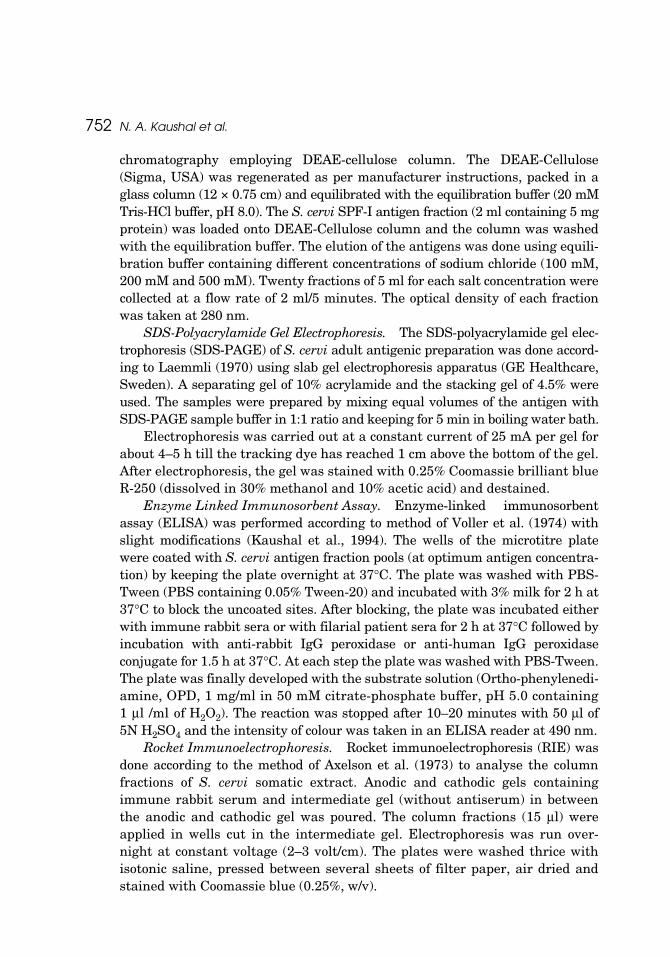
The SFP-I fraction was further purified using DEAE-cellulose anion-exchange chromatography, which resulted in the separation of 4 proteinpeaks. The elution profile of SFP-I on DEAE-cellulose column is shown inFigure 2. All the column fractions were analysed by Rocket immunoelectro-phoresis using rabbit anti-ScA serum and 4 antigen pool fractions (DFP-I,DFP-II, DFP-III and DFP-IV) were made based on the results of Rocketimmunoelectrophoresis (data not shown). The reactivities of purified antigen
Table 1: Analysis of S. cervi fraction pools from Sephacryl S-200 column.
Fractionpools
Fraction number
Precipitin peaksin RIE
ELISA Titre IRS1
ELISA OD (1: 500 dil)
CIEIHS2 NHS3
SFP-I 10–15 9–11 1:256000 2.742 0.361 13–14SFP-II 16–19 3–6 1:128000 1.389 0.451 8–9SFP-III 20–24 2–3 1:16000 0.896 0.418 3–4SFP-IV 27–30 3a – 0.23 0.167 NDSFP-V 35–38 2a – 0.34 0.151 ND
1IRS = rabbit anti-S. cervi adult serum; 2IHS = filarial patient serum pool,3NHS = normal humanserum pool.aVery faint antigenic peaks; All antigen fractions were used at 0.1 μg/well concentration;Antibody titre of IRS could not be determined for antigen fractions SFP-1V and SFP-V as theELISA OD was very low for these fractions.
S. cervi Antigen Fraction for Filaria Diagnosis 755
pool fractions (DFP-I to DFP-IV) were tested in ELISA using rabbit anti-ScAserum and the pool fractions DFP-I, DFP-III and DFP-IV showed high reac-tivity (antibody titre of 1:32,000 for DFP-I, 1:16000 for DFP-III and 1:64000for DFP-IV) with rabbit anti-ScA serum pool while pool fraction DFP-IIshowed comparatively low antibody titre (1:4000). The CIE analysis of theseantigen fractions, using rabbit anti-ScA serum pool, revealed 11 and 8 pre-cipitin peaks in DFP-I and DFP-III, respectively, while only 2–3 precipitinpeaks were observed in antigen pool fractions DFP-II and DFP-IV (Table 2).The reactivities of antigen fractions (DFP-I to DFP-IV) were further testedwith filarial patient serum (W. bancrofti and B. malayi) and normal humanserum pools in ELISA. Out of the 4 antigen fraction pools, DFP-IV showedhigh ELISA reactivity (OD490 >2.0) with both bancroftian and brugianpatient serum pools as compared to the other three antigen pool fractions(Table 2). The S. cervi DFP-IV fraction also showed high ELISA reactivitywith immune rabbit serum against B. malayi adult antigen but no signifi-cant reactivity was observed with rabbit anti- A. ceylanicum serum (data notshown).
Figure 2: Ion-exchange chromatography of S. cervi SFP-I antigen fraction using DEAE Cellulosecolumn. The SFP-I fraction was applied to DEAE-Cellulose column and column was washedwith 20 mM Tris-HCl buffer, pH 8.0, the antigens were eluted with same buffer containingdifferent salt concentrations. The antigen pools were made based on RIE results. DFP-I pool offractions 3–5; DFP-II pool of fractions 7–11; DFP-III pool of fractions 28–33; DFP-IV pool offractions 41–45.
756 N. A. Kaushal et al.
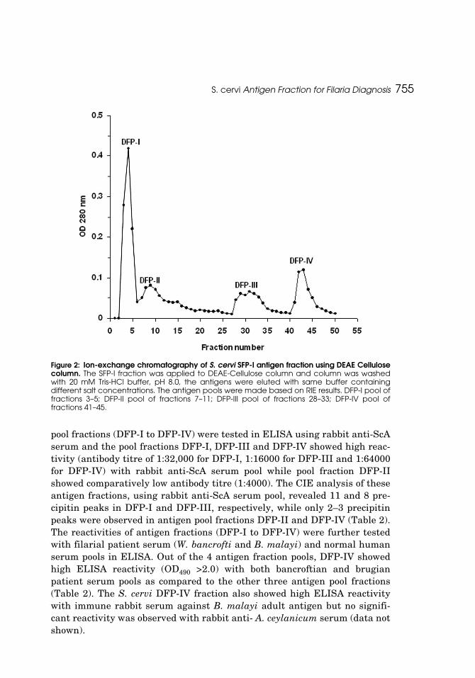
In order to test the specificity of S. cervi DFP-IV purified fraction, thereactivity of the DFP-IV fraction was further tested using filarial (W. bancrofti),non-filarial (ascaris and hookworm) and normal human sera in ELISA.Twenty sera from filarial patients, 5 each of ascaris and hookworm infectionand 10 normal human sera were used in ELISA and the results are shown inFigure 3. The DFP-IV antigen fraction showed high reactivity with filarial
Table 2: Analysis of S. cervi fraction pools from DEAE-cellulose column.
Fractionpools
Fraction number
Precipitinpeaks in RIE
ELISA Titre IRS1
ELISA OD (1: 500 dil)
CIEIHS12 IHS23 NHS4
DFP-I 3–5 9–10 1:32000 0.812 0.762 0.288 11DFP-II 6–11 2–3 1:4000 0.441 0.424 0.251 2–3DFP-III 28–33 5–7 1:16000 1.289 1.318 0.368 8DFP-IV 41–45 2–3 1:64000 2.381 2.294 0.224 2–3
1IRS = rabbit anti-S. cervi adult serum; 2IHS1= W. bancrofti serum pool. 3IHS2 = B. malayi serumpool; 4NHS = normal human serum pool.DFP-I, DFP-II and DFP-III fractions were used at 0.06 μg/well and DFP-IV fraction used at 0.03 μg/well concentration.
Figure 3: Reactivity of S. cervi purified DFP-IV antigen fraction with filarial and non-filarialserum pools in ELISA. 20 sera from filarial patients, 5 each of ascaris and hookworm infectionand 10 normal human sera were tested at 1:500 dilutions and OD 490 nm was read usingMolecular Devices Spectramax190 plus microplate ELISA reader.
S. cervi Antigen Fraction for Filaria Diagnosis 757
patients sera (mean OD490 of 1.998) and did not show any significant reactiv-ity with the ascaris (mean OD490 of 0.383), hookworm (mean OD490 of 0.439)and normal human sera (mean OD490 of 0.242).
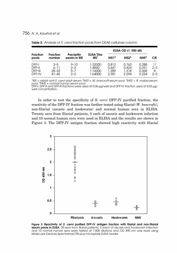
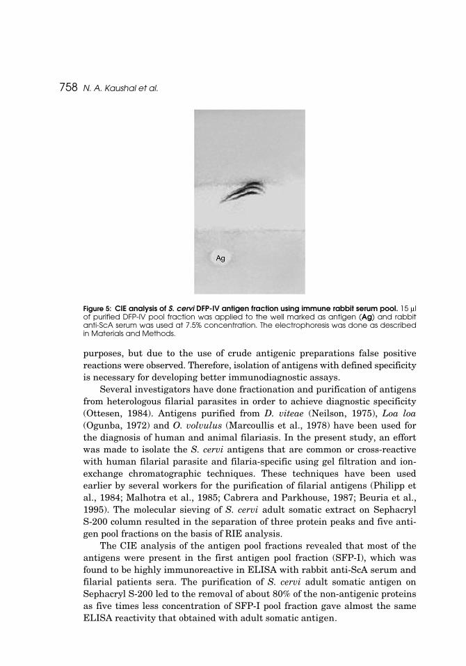
The SDS-polyacrylamide gel electrophoresis analysis revealed 2 majorand one minor protein band (mol. wt. range 65–70 kDa) in S. cervi DFP-IVantigen fraction while 35–40 protein bands (mol. wt. range 10–200 kDa) wereobserved in somatic antigenic preparation of S. cervi adults (Fig. 4). The DFP-IV purified fraction was further analysed by Crossed immunoelectrophoresisusing rabbit anti-ScA serum and the CIE pattern is shown in Figure 5. TheCIE analysis revealed 3 antigens in S. cervi DFP-IV purified fraction.
DISCUSSION
The complex nature and extensive cross-reactivity of the filarial parasiteantigens with other nematode parasites is responsible for most of the non-specific reactions in immunological assays employed for filarial diagnosis. Theantigens collected by in vitro maintenance of human filarial parasites orobtained from the filarial patients sera were found to be specific for immmun-odiagnosis of filariasis (Harinath et al., 1984; Reddy et al., 1986), however,getting sufficient amount of the antigenic material for large-scale use inimmunoassays is the major limitation. This has put impetus for the use ofheterologous antigens prepared from animal filarial parasites for diagnostic
Figure 4: SDS-Polyacrylamide gel electrophoretic analysis of S. cervi DFP-IV antigen fraction.The ScA (A) and purified DFP-IV pool fraction (B) were run on 10% acrylamide gel.
758 N. A. Kaushal et al.
purposes, but due to the use of crude antigenic preparations false positivereactions were observed. Therefore, isolation of antigens with defined specificityis necessary for developing better immunodiagnostic assays.
Several investigators have done fractionation and purification of antigensfrom heterologous filarial parasites in order to achieve diagnostic specificity(Ottesen, 1984). Antigens purified from D. viteae (Neilson, 1975), Loa loa(Ogunba, 1972) and O. volvulus (Marcoullis et al., 1978) have been used forthe diagnosis of human and animal filariasis. In the present study, an effortwas made to isolate the S. cervi antigens that are common or cross-reactivewith human filarial parasite and filaria-specific using gel filtration and ion-exchange chromatographic techniques. These techniques have been usedearlier by several workers for the purification of filarial antigens (Philipp etal., 1984; Malhotra et al., 1985; Cabrera and Parkhouse, 1987; Beuria et al.,1995). The molecular sieving of S. cervi adult somatic extract on SephacrylS-200 column resulted in the separation of three protein peaks and five anti-gen pool fractions on the basis of RIE analysis.
The CIE analysis of the antigen pool fractions revealed that most of theantigens were present in the first antigen pool fraction (SFP-I), which wasfound to be highly immunoreactive in ELISA with rabbit anti-ScA serum andfilarial patients sera. The purification of S. cervi adult somatic antigen onSephacryl S-200 led to the removal of about 80% of the non-antigenic proteinsas five times less concentration of SFP-I pool fraction gave almost the sameELISA reactivity that obtained with adult somatic antigen.
Figure 5: CIE analysis of S. cervi DFP-IV antigen fraction using immune rabbit serum pool. 15 μlof purified DFP-IV pool fraction was applied to the well marked as antigen (Ag) and rabbitanti-ScA serum was used at 7.5% concentration. The electrophoresis was done as describedin Materials and Methods.
S. cervi Antigen Fraction for Filaria Diagnosis 759
Further purification of S. cervi SFP-I antigen fraction on DEAE-Cellulosecolumn resulted in the isolation of DFP-IV antigen fraction with bettersensitivity as evident by its high ELISA reactivity with filarial patient sera.The SDS-PAGE and CIE analysis revealed simple antigenic make-up (2–3antigens) of DFP-IV fraction. The ion-exchange chromatography has beenused earlier by different workers for the purification of filarial antigens.Subrahmanyam et al. (1974) fractionated W. bancrofti patients sera on aDEAE-cellulose column. Fractionation of W. bancrofti microfilarial solubleantigen was carried out using Sephadex G-150 gel filtration method followed byDEAE-cellulose column chromatography (Kaliraj et al., 1982). Ion-exchangechromatography was also used by other workers to obtain the antigen fraction(s)with enhanced sensitivity and specificity (Lavebrett et al., 1994). A highlyimmunogenic fraction of O. volvulus having molecular weight of 10–40 kDawas purified by using ion exchange chromatography (Guzman et al., 2002).The W. bancrofti and B. malayi are the two causative agents for lymphaticfilariasis in India and the reactivity of DFP-IV antigen fraction with both W.bancrofti and B. malayi patients sera suggest that DFP-IV antigen fractioncannot be used to differentiate bancroftian and brugian infections.
The crude somatic antigens of filarial parasites have been shown to becross-reactive with antibodies present in sera from other helminth infections,such as ascaris and hookworms (Dissanayake and Ismail, 1980; Kaushalet al., 1984, Akao et al., 1991). In the present study, the purified S. cervi anti-gen fraction (DFP-IV) did not show cross-reactivity with antibodies present insera from ascaris and hookworm infection and may have potential for specificdetection of anti-filarial antibodies in patients sera. Our studies on measuringIgG4 antibody responses in filarial patients sera using S. cervi DFP-IV anti-gen fraction have shown potential for immunodiagnosis of human filariasis(unpublished data). However, further work on evaluation of S. cervi DFP-IVantigen fraction using large number of sera samples from different filarial andnon-filarial infections is required to establish its filaria-specificity and its sub-sequent use for developing an immunodiagnostic test for human filariasis.
Therefore, in the present study we have been able to isolate an antigenfraction DFP-IV from S. cervi adults employing gel filtration and ion exchangechromatography. Our studies have also demonstrated the filaria-specificity(W. bancrofti/B. malayi) of DFP-IV fraction as it did not show cross-reactivitywith sera from other nematode infections (ascaris and hookworm), therebysuggesting the potential of S. cervi DFP-IV antigen fraction for immunodiag-nosis of human filariasis.
ACKNOWLEDGMENTS
The authors are thankful to the Director, CDRI, Lucknow for providing thenecessary facilities. Four of the authors (NS, AT, SKS and HM) are grateful to
760 N. A. Kaushal et al.
the Council of Scientific and Industrial Research for financial support. CDRICommunication no. 5104
REFERENCES
Agrawal, V.K. and Sashindran, V.K. (2006). Lymphatic Filariasis in India: Problems,challenges and new initiatives. Med J Arm For India 62:359–362.
Akao, N., Kondo, K. Fujita, K. (1991). Immunoblot analysis of Dirofilaria immitisrecognized by infected humans. Ann. Trop. Med. Parasitol. 85:455–460.
Axelsen, N.H., Kroll, J., Weeke, B. (1973). Intermediate gel in crossed and in fusedrocket immunoelectrophoresis. Scand. J. Immunol. Suppl. 1:71–77.
Beuria, M.K., Bal, M., Das, M.K. (1995). Allergic reactivity and IgG subclasses to a pro-teinase fraction of Setaria digitata in filariasis. J. Helminthol. 69:181–185.
Cabrera, Z., Parkhouse, R.M.E. (1987). Isolation of an antigenic fraction for diagnosisof onchocerciasis. Para. Immunol. 9:39–48.
Dissanayake, S., Ismail, M.M. (1980). Antigen of Setaria digitata: cross-reaction withsurface antigens of Wuchereria bancrofti microfilariae and serum antibodies of W.bancrofti-infected subjects. Bull. WHO 58: 649–654.
Dissanayake, S., Ismail, M.M. (1981). Antibody determination in the diagnosis ofWuchereria bancrofti infection in man. Bull. WHO 59:753–757.
Forsyth, K.P., Copeman, D.B., Anders, R.F., Mitchell, G.F. (1981). The major radioiodi-nated cuticular antigens of Onchocerca gibsoni microfilariae are neither speciesnor onchocerca specific. Acta Tropica 38:343–352.
Grady, C.A., de Rochars, M.B., Direny, A.N., Orelus, J.N., Wendt, J., Radday, J.,Mathieu, E., Roberts, J.M., Streit, T.G., Addiss, D.G., Lammie, P.J. (2007).Endpoints for lymphatic filariasis programs. Emerg. Infect Dis. 13:608–610.
Guzman, G.E., Lavebratt, C., Lujan, R., Akuffo, H. (2002). Diagnosis of onchocerciasisusing highly specific and sensitive native proteins. Scand. J. Infect. Dis. 34:583–590.
Harinath, B.C., Malhotra, A. Ghirnikar, S.N., Annodate, S.D., Isaacs, V.P., Bharti,M.S. (1984). Field evaluation of ELISA using Wuchereria bancrofti mf ES antigenfor bancroftian filariasis. Bull. WHO 62:941–966.
Kaliraj, P. Harinath, B.C., Ghirnikar, S.N. (1982). Fractionation & evaluation ofWuchereria bancrofti microfilarial antigens in immunodiagnosis of bancroftianfilariasis. Indian J. Exp. Biol. 20:440–444.
Kaushal, N.A., Hussain, R., Ottesen, E.A. (1984). Excretory-secretory and somaticantigens in the diagnosis of human filariasis. Clin. Exp. Immunol. 56:567–576.
Kaushal, N.A., Kaushal, D.C., Ghatak, S. (1987). Identification of antigenic proteins ofSetaria cervi by immunoblotting technique. Immunol. Invest. 16: 139–149.
Kaushal, N.A., Kaushal, D.C., Ghosh, S., Talwar, G.P. (1994). Monoclonal antibodiesagainst antigenic epitopes common between Setaria cervi and Brugia malayi. Ind.J. Exp. Biol. 32:371–375.
Laemmli, U.K. (1970). Cleavage of structural proteins during the assembly of the headof bacteriophage T4. Nature 227:680–685.
Lavebratt, C., Dalhammar, G., Adamafio, N.A., Dejerud, U.N., Mingarini, K.,Ingemarsson, K., Opoku, N., Akuffo, H.O. (1994). A simple dot blot assay adapt-able for field use in the diagnosis of onchocerciasis: preparation of an adult worm
S. cervi Antigen Fraction for Filaria Diagnosis 761
antigen fraction which enhances sensitivity and specificity. Trans. Roy. Soc. Trop.Med. Hyg. 88:303–306.
Lowry O.H., Rosebrough N.J., Farr A.L., Randall R.J. (1951). Protein measurementwith the Folin phenol reagent. J. Biol. Chem. 193: 265–275.
Malhotra, A., Kaushal, N.A., Ghatak, S. (1986). Protein and antigenic composition ofadult and microfilarial stages of Setaria cervi. Immunol. Invest. 15:505–519.
Malhotra, A., Prasad, G.B.K.S., Harinath, B.C. (1985). Detection and isolation of filarialantigen from hydrocoele fluid and its use in diagnosis. Ind. J. Exp. Biol. 23:180–182.
Mercoullis, G. Salonen, E.M., Grasbeck, R. (1978). Sequential affinity chromatographyfor the purification of antigens extracted from Onchocerca volvulus adultworms.Tropmed. Parasitol. 29: 39–48.
Mohan S., Kaushal N.A., Misra A., Kaushal, D.C., Katiyar, J.C. and Ghatak, S (1988):Ancylostoma ceylanicum: Protein and antigenic composition of adult and larvalstages. Immun. Investig. 17: 295–308.
Mustafa, H., Srivastava, N., Kaushal, D.C. and Kaushal, N.A. (1996). Analysis andpotential of excretory-secretory antigens of Setaria cervi for immunodiagnosis ofhuman filariasis. Indian J. Exp. Biol. 34:508–512.
Neilson, J.T. (1975). Fractionation of a soluble somatic extract and solubilized cuticularextracts of Dipetalonema viteae adult worms. J. Parasitol. 61:785–793.
Ogunba, E.O. (1972). Serological investigations with Loa loa antigens. J. Helminthol.46: 241–250.
Ottesen, E. A. (1984). Immunological aspects of lymphatic filariasis and onchocerciasisin man. Trans. Roy. Soc. Trop. Med. Hyg., 78 (Suppl):9–18.
Parkhouse, R.M., Almond, N.M., Cabrera, Z., Harnett, W., (1987) Nematode antigensin protection, diagnosis and pathology. Vet. Immunol. Immunopathol. 17: 313–324.
Philipp M., Gomez-Priego A., Parkhouse R.M., Davies M.W., Clark N.W., Ogilvie B.M.,Beltran-Hernandez F. (1984). Identification of an antigen of Onchocerca volvulusof possible diagnostic use. Parasitology 89:295–309.
Reddy, M.V.R., Prasad, G.B.K.S., Harinath, B.C. (1986). Isolation and evaluation ofantigens from microfilaraemia plasma and immune complexes for diagnosis ofbancroftian filariasis. Ind. J. Pathol. Microbiol. 29:179–188.
Singh, S.K., Kaushal, D.C., Murthy, P.K., Kaushal, N.A. (2007). Partial purification andcharacterization of acetylcholinesterase isozymes from adult bovine filarial parasiteSetaria cervi. Ind. J. Biochem. Biophys. 44:379–385.
Song, K.H., Hayasaki, M., Cho, K.W., Lee, S.E., Kim, D.H., (2002) Cross-reactivitybetween sera from dogs experimentally infected with Dirofilaria immitis andcrude extract of Toxocara canis. Korean J. Parasitol. 40:195–198.
Subrahmanyam, D., Chaudhury, S., Jain, S., (1974) Immunological investigations infilariasis. Ind. J. Pathol. Bacteriol. 17:135–141.
Swada, T., Sato, K., Sato, S. (1969). Studies on skin test antigen FST for immunodiag-nosis of filariasis. I. Electrophoretic analysis and fractionation of antigen FST.Jap. J. Exp. Med. 39:427–433.
Voller, A., Bidwell, D., Huldt, G., Engvall, E. (1974). A microplate method of enzyme-linked immunosorbent assay and its application to malaria. Bull. WHO 51:209–211.