Myxoma and Vaccinia Viruses Bind Differentially to Human Leukocytes
Upload
independentCategory
view
3download
0
I O D I N A T I N G A B I L I T Y OF VARIOUS L E U K O C Y T E S A N D T H E I R B A C T E R I C I D A L A C T I V I T Y *
BY S. R. SIMMONS AND M. L. KARNOVSKY
(From the Department of Biological Chemistry, Harvard Medical School, Boston, ~assachusetts 02115)
(Received for publication 13 February 1973)
A major function of mammalian phagocytes such as the tissue macrophage and circulating polymorphonuclear leukocyte (PMN) 1 is to ingest and kill invading micro- organisms. Recently considerable data have been presented in the literature to clarify the linkage between the metabolism of the phagocyte and the killing of the ingested microorganism. The metabolic perturbations that accompany the process of phago- cytosis in PMN include a cyanide-insensitive respiratory burst and an increased pro- duction of H20,. Work in our laboratory (1) has indicated that a flavoenzyme, in- sensitive to cyanide and found in human and guinea pig PMN, might be responsible for the alterations in metabolism associated with phagocytosis, by promoting the oxidation of NADH by molecular oxygen to yield H202 (2). A seminal study linking leukocyte metabolism and killing is that of Klebanoff (3), who showed that human and guinea pig PMN are capable of fixing iodine (provided as iodide) covalently to ingested bacteria. This reaction employs H20:,~ and is a function of the activity of the myeloperoxidase of the lysosomal granules of the PMN. The system has since been found to exhibit a marked toxic activity toward bacteria, fungi, and viruses (4-6), and is clearly linked to the respiratory changes in phagocytizing PMN noted above.
The mechanism of the bacterial killing per se is not yet known, but actual iodina- tion is not necessarily involved, since killing in such systems occurs when bromide or chloride replace iodide. However, by including labeled iodide in the system one may obtain a useful measure of the peroxidase-peroxide reaction (i.e., lslI-fixation). Re- cently, Pincus and Klebanoff (7) described a method for quantitatively measuring iodination of bacteria by PMN from various patients, including those suffering from chronic granulomatous disease, whose PMN are unable to generate significant H202 (8) [for review see (9)]. The method is based on the precipitation by trichloroacetic acid of iodine covalently bound to protein. The assay was not, however, correlated kinetically with the degree of ingestion of particles.
Work on the peroxide-mediated microbicidal phenomenon has been focussed pre- dominantly on PMN. A purpose of our investigation was to draw a comparison among
* Supported by Research Grant AI-03200 from the National Institutes of Health. 1 Abbreviations used in this paper: AIK, alkaline isotonic KC1; AGH solution, 0.1% bovine
serum albumin and 5.6 mM glucose in modified Hanks' solution; CGD, chronic granuloma- tous disease; CDG-PMN, PMN from children with chronic granulomatous disease; GP, guinea pig; KRP, Krebs-Ringer phosphate; M, mouse; MAC, peritoneal macrophages; MN, monocytes; PIVIN, polymorphonuclear leukocytes; PS, polystyrene; TB, tubercle bacilli.
44 TgE J O U R N A L OF E X P E R I M E N T A L M E D I C I N E • V O L U M E 138, 1973
on July 30, 2016jem
.rupress.orgD
ownloaded from
Published July 1, 1973
S. R. SIMMONS AND M. L. KARNOVSKY 45
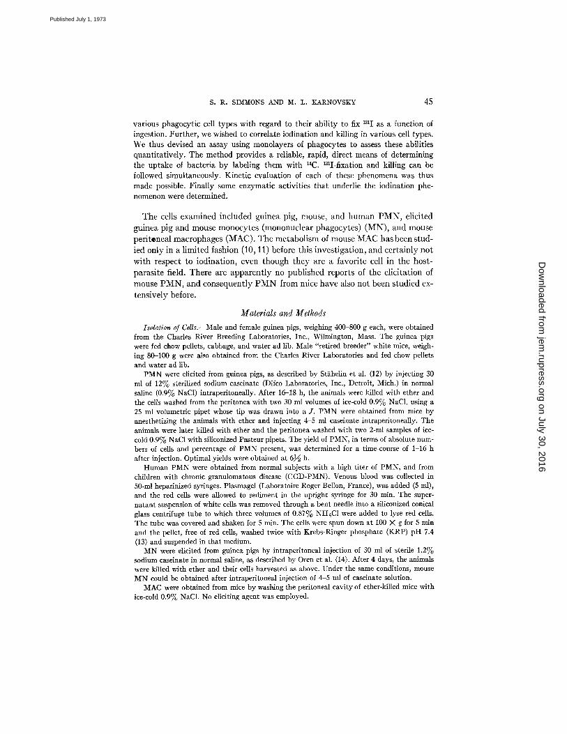
various phagocytic cell types with regard to their ability to fix 1311 as a function of ingestion. Further, we wished to correlate iodination and killing in various cell types. We thus devised an assay using monolayers of phagocytes to assess these abilities quantitatively. The method provides a reliable, rapid, direct means of determining the uptake of bacteria by labeling them with 14C. 131I-fixation and killing can be followed simultaneously. Kinetic evaluation of each of these phenomena was thus made possible. Finally some enzymatic activities that underlie the iodination phe- nomenon were determined.
T h e cells examined inc luded guinea pig, mouse, and h u m a n P M N , elici ted
guinea p ig and mouse monocy te s (mononuclear phagocytes) ( M N ) , and mouse
per i tonea l macrophages (MAC) . T h e me tabo l i sm of mouse M A C has been stud-
ied only in a l imi ted fashion (10, 11) before this inves t iga t ion , and ce r ta in ly no t
wi th respect to iodinat ion, even though t h e y are a favor i te cell in the host-
paras i te field. The re are appa ren t l y no publ i shed repor ts of the e l ic i ta t ion of
mouse P M N , and consequen t ly P M N f rom mice have also no t been s tudied ex-
tens ive ly before.
Materials and Methods
Isdation of CeUs.--Male and female guinea pigs, weighing 400-800 g each, were obtained from the Charles River Breeding Laboratories, Inc., Wilmington, Mass. The guinea pigs were fed chow pellets, cabbage, and water ad lib. Male "retired breeder" white mice, weigh- ing 80-100 g were also obtained from the Charles River Laboratories and fed chow pellets and water ad lib.
PMN were elicited from guinea pigs, as described by StKhelin et al. (12) by injecting 30 ml of 12% sterilized sodium caseinate (Difco Laboratories, Inc., Detroit, Mich.) in normal saline (0.9% NaC1) intraperitoneally. After 16-18 h, the animals were killed with ether and the cells washed from the peritonea with two 30 ml volumes of ice-cold 0.9% NaC1, using a 25 ml volumetric pipet whose tip was drawn into a J. PMN were obtained from mice by anesthetizing the animals with ether and injecting 4-5 ml caseinate intraperitoneally. The animals were later killed with ether and the peritonea washed with two 2-ml samples of ice- cold 0.9% NaC1 with siliconized Pasteur pipets. The yield of PMN, in terms of absolute num- bers of cells and percentage of PMN present, was determined for a time-course of 1-16 h after injection. Optimal yields were obtained at 61/6 h.
Human PMN were obtained from normal subjects with a high titer of PMN, and from children with chronic granulomatous disease (CGD-PMN). Venous blood was collected in 50-ml heparinized syringes. Plasmagel (Laboratoire Roger Bellon, France), was added (5 ml), and the red cells were allowed to sediment in the upright syringe for 30 min. The super- natant suspension of white cells was removed through a bent needle into a siliconized conical glass centrifuge tube to which three volumes of 0.87% NH4C1 were added to lyse red cells. The tube was covered and shaken for 5 rain. The cells were spun down at 100 X g for 5 rain and the pellet, free of red cells, washed twice with Krebs-Ringer phosphate (KRP) pH 7.4 (13) and suspended in that medium.
MN were elicited from guinea pigs by intraperitoneal injection of 30 ml of sterile 1.2% sodium caseinate in normal saline, as described by Oren et al. (14). After 4 days, the animals were killed with ether and their cells harvested as above. Under the same conditions, mouse MN could be obtained after intraperitoneal injection of 4-5 ml of caseinate solution.
MAC were obtained from mice by washing the peritoneal cavity of ether-killed mice with ice-cold 0.9% NaCI. No eliciting agent was employed.
on July 30, 2016jem
.rupress.orgD
ownloaded from
Published July 1, 1973

4 6 IODINATION AND KILLING BY LEUKOCYTES
After they were harvested, cells were filtered through nylon gauze, and were spun down at 10°C, <100 X g for 10 min. When it appeared necessary, the pellet was suspended in 1 2 ml 0.9% NaC1 and diluted to 20 ml with distilled water. After a few seconds, an appro- priate amoun t of 9% NaCI was added to bring the solution to isotonicity. This osmotic shock served to lyse red blood cells without appreciable damage to leukocytes. However, prepara- tions of white cells containing m a n y erythrocytes were discarded. After a 10 min centrifuga- tion at < 100 X g, the pellet was washed twice with K R P pH 7.4 (13). The final suspension of cells was in KRP. All glassware used in collection and centrlfugation had been siliconized.
Suspensions of P M N were ca. 95% pure with some M N and lymphocytes. The M N sus- pensions contained ca. 80% mononuclear phagocytes with P M N consti tut ing the bulk of the impurity, and again some lymphocytes. The peritoneal macrophage preparations were 60-70% macrophages with m a ny lymphocytes (11, 14). The final monolayers (see below) were always further enriched with respect to the relevant cell type in each case because lym- phocytes did not stick to the plastic dishes, and P M N were less adherent than M N or MAC. Thus , these monolayers were at least 90% pure.
Preparation of Mondayers.--Monolayers were prepared by adding 1.0 ml of cell suspensions (5-10 X 106 white cells per milliliter) to tissue culture dishes (Falcon Plastics, Div. of Bio- Quest, Oxnard, Calif., 3.5 cm diameter) cut down to fit into a Nuclear Chicago gas flow counter, Model D47 (Nuclear-Chicago Corp., Des Plaines, Ill.). They were incubated 1 h a t 37°C in a Visidome incubator (Greiner Scientific Corp., New York). Monolayers and superna tant cells were swirled gently every 15 rain to insure homogeneity. Supernatant suspensions were poured off and the monolayers rinsed in three washes of K R P (for killing studies) or of a special medium for iodination studies. The dishes were drained of the original fluid a few seconds before test media were added. The medium for studies of iodination con- sisted of 0.1% bovine serum albumin and 5.6 m M glucose in modified Hanks ' solution (3), and will be referred to as AGH solution.
Uptake of 131I and [14ClTubercle Bacilli.--The method of Michell et al. (15) for following ingestion of 14C-labeled particles by cells in monolayers was adapted to examine, s imultane- ously, the uptake of 131I as a funct ion of time and load of microbial particles. To prepared monolayers were added 0.90 ml portions of serum solution, consisting of 10% guinea pig serum (Grand Island Biological Co., Grand Island, N. Y.) in the iodination medium above (AGH). After 6 min, 100/~1 portions of 10 - 5 M Na[ containing 4 #Ci /ml Na131I (New Eng- land Nuclear, Boston, Mass.) were added to sets A and B of monolayers and 100 #1 of 10 - 5 YI N a 127I to set C. 10 min later, 0.10-ml portions of a suspension of [14C]tubercle bacilli were added to sets B and C, whereas 0.10-ml portions of AGH alone were added to set A. Dead tubercle bacilli, labeled with 14C, had been prepared as described elsewhere (16) and sus- pended in the AGH. The monolayers were then covered and incubated for periods of 0-60 min at 37°C with occasional gentle swirling. After this incubation, the dishes were drained, washed in six beakers containing cold 0.9% NaC1 and 10 -6 M NaI (nonradioactive) and air dried. The set of dishes lacking radioisotope but exposed to ~4C-Iabeled particles (set C) was counted with a gas flow counter, to monitor ingestion. The 14C and /or 131I of sets A and B were similarly counted. Monolayers were digested with 0.5 N NaOH and their protein assayed (17). In all calculations for protein, allowance was made for the contribution of [14C]tubercle bacilli.
Iodinatlon of [14C]Tubercle Bacilli in the Absence of Phagocyles.--The experiments pro- ceeded essentially as described above, except tha t no phagocytes were present and dishes with particles were incubated for 20 rain. At the end of this t ime the contents of the dishes were transferred quant i ta t ively to centrifuge tubes, spun a t 10,000 X g for 10 min at 4°C, and the pellets washed three times with cold 0.9% NaCI-10 - 6 M NaI (unlabeled). The pellets were then transferred, in aqueous suspension (1 ml) to their original dishes, dried, and counted.
Uptake of 131I and Inert Particles.--Monolayers of phagocytic cells were exposed to lalI in the presence and absence of a noniodinatahle particle, polystyrene (PS) (18). This control
on July 30, 2016jem
.rupress.orgD
ownloaded from
Published July 1, 1973

S. R. SIMMONS AND M. L. KARNOVSKY 47
monitored entry into the cell of iodide specifically attributable to the uptake of external me- dium during phagocytosis. The procedure was as described above, but 0.10 mI of a suspension of PS (0.81 ~m diameter, Difeo Laboratories) in AGH was substituted for [14C]tubercle bacilli. The set of dishes C, lacking radioisotope but exposed to PS, was examined micro- scopically to confirm ingestion of particles by the phagocytes. The uptake of 131I in the pres- ence and absence of PS was determined by gas flow counting, as before.
t~gect of Cyanide on Iodination by "Resting" Cells.--Tbe effect of cyanide on nonphago- cytizing cells was examined to determine whether the iodination observed in such cells could be a function of heine enzyme activity. Mono]ayers were exposed to 0.90 ml AGH solution containing or lacking 1.2 mM KCN pH 7.4 for 6 rain, after which 100/~l 10 -5 M NaI con- taining 131I (0.4/zCi) were added. Mter i0 rain, 0.10 ml of AGH was added. Dishes were drained, washed, and counted as before, after a 20 min incubation.
Measurement of Ingestion and Killing.--To monolayers of phagocytes were added 0.90 ml portions of KRP glucose (7.5 mM)--5% guinea pig serum heated at 56°C for 30 rain, at 37°C. After 16 rain, 0.10-ml portions of ice-cold live [14C]Escherichia coli were added, and the covered dishes incubated at 37°C for 0-90 rain. E. coli had been grown to stationary phase in the presence of D-[t4C]glucose (uniformly labeled) (New England Nuclear) and washed to a constant specific activity. "I he multiplicity of E. coli per leukocyte was deter- mined. At the end of the incubations, monolayers were drained and washed six times in cold 0.9% NaC], all under sterile conditions. Those dishes to be used for assaying total uptake of E. coli were dried and counted on the gas-flow counter, and their protein contents assayed. To the other monolayer dishes, placed on ice, were added ice-cold 1.0-ml portions of sterile 0.5% Triton X-100 (Rohm and Haas Co., Philadelphia, Pa.) in 0.9% NaC1. The monolayers were then suspended and lysed by rapid pipetting with sterile Pasteur pipets and 0.10-ml samples were serially diluted, for plating, in sterile 0.9% NaC1. Other samples were taken for protein analysis. The effects of such a concentration of Triton X-100 on both the viability of 1". coli and the intactness of the phagocytes examined had been determined previously. This substance at this concentration in no way impaired the viability of E. toll, whereas it promoted the release of phagocytic cells from dishes and lysis of those cells (;>90%). In parallel with every kilting experiment, a test was run for the viability of E. eoli with time in the incubation medium, without mono]ayers. The experimental procedure is outlined in Fig. 1. After washing of all dishes had been completed, unexposed phagocytes in monolayer form were passed through the wash solutions to check for contamination by 14C and by viable bacteria.
After the monolayers were lysed as above and diluted, the samples were plated on "Certa Plate Antibiotic No. 3" (Hospital Service Tech. Corp., North Andover, Mass.). After a 16 h overnight incubation at 37°C, the bacterial plates were visually counted.
Enzymatic Activities of Phagocytes.-- NADH and NADPH oxidases: The assay was based on the method described by Cagan
(1, 19). Washed suspensions of phagocytes were resuspended in alkaline isotonic KC1 (AIK: 0.32 ml of I0 mg/ml KHCO3 in 100 ml of 1.15% KC1), 30% by volume. The suspensions of cells were then homogenized on ice in a Potter-Elvehjem homogenizer with a mechanically driven Tefon pestle (Arthur H. Thomas Co., Philadelphia, Pa.) to give >90% breakage as determined by microscopic observation. The homogenates were spun at 15,000 M g for 30 min at 4°C and the resulting supernatants were carefully removed and assayed. A sample of the homogenate, before centrifugation was saved for protein determination.
NADH and NADPH oxidase assays (for both cyanide-sensitive and cyanide-insensitive activity) were performed at 37°C in a Perkin-Elmer Model 202 spectrophotometer (Perkin- Elmer Coq)., Instrument Div., Norwalk, Conn.), monitoring the optical density at 340 nm. The cuvettes (1 cm path length) contained a total volume of 1 mI. NADH and NADPH (Sigma Chemical Co., St. Louis, Mo.) each dissolved in 0.10 M potassium phosphate
on July 30, 2016jem
.rupress.orgD
ownloaded from
Published July 1, 1973

48 IODINATION AND KILLING BY LEUKOCYTES
Tissue Culture Dishes (37°C)
+ Monolayers (Cells) - Monolayers l I
+ 0.9 ml KRP-Glucose-Serum
+ 0. I0 ml live~4C]E, col____~i in 0.9% cold NaCI
I. Draln~ rinse 6 times in i. Drain, rinse 6 times 0.97° NaCl, place on ice
L 2. Add 1.0 ml 0.5% Triton 2.
X-IO0 in 0.9% NaCI~ ice- cold, and suspend
3. Serially dilute 0.I0 ml 3. aliquots and plate
Calculate number of i nges t ed [14C E. c e l l still viable ]-
Incubation Time
16 rain $
I 0-90 rain
t I Serially dilute
in 0.9% NaCI~ and dry 0.I0 ml aliquots
I and plate I Count on gas-flow counter
I Add 0.5N NaOH to each d i s h and a s s a y p r o t e i n
I Calculate number of Calculate number
I C a l c u l a t e t o t a l number of v i a b l e
FIG. 1. Flow sheet of the kinetic assay of bactericidal activity. (See text for determina- tion of killing efficiency.)
buffer p H 7.0 to give a concentration of 1.6 X 10 -a M. The pyridine nucleotide concentra- tions were calculated from the value of E ~ ~ = 6.22 X 10 a liters-tool -1. Assay mixtures con- tained 0.10 ml 10 -2 M KCN, p H 7.0 (or 0.10 ml 0.10 M potassium phosphate pH 7.0), 0.30 ml superna tant preparation (1 mg superna tant protein, or 0.30 ml AIK), 0.50 ml 0.10 M potass ium phosphate buffer, p H 7.0, and 0.10 ml (1.6 X 10 -4 retool) of the above solution of N A D H or N A D P H (or 0.10 ml of buffer). The reaction was initiated by the addition of pyridine nucleotide, and the decrease in optical density was followed with time. Activity, based on extrapolation of an initial linear decrease, was expressed as nanomoles of nucleotide rain -1 mg -1 homogenate protein or for 107 cells.
Peroa:idase: The peroxidase method was based on that used by Michel] et aI. (20), a modi- fication of tha t of Maehly (21). Suspensions of cells were homogenized as a 30% suspension in AIK, as described above, and samples taken for protein analysis. The assay mixtures were incubated at 37°C in a Perkin-Elmer spectrophotometer monitoring the appearance of color a t 470 nm. To the cuvettes (1 cm pa th length) were added 0.2 ml of 0.1 M sodium phosphate buffer, p H 7.0, 1.0 ml of 20 m M guaiacol (0.22 ml liquid diluted to 100 ml with water), 0.2 ml of 1.6% Tri ton X-100, 0.1-0.3 mg homogenate protein in AIK, and water to a total volume of 3 ml. The reaction was started by the addition of 10 #1 H202 solution (final concentration ~ 0 . 6 raM). The initial reaction rate was calculated by using /£]~,1 = 26.6 X 10 a liters- mo1-1 for tetraguaiacol, as nanomoles tetraguaiacol evolved rain -1 mg -a homogenate protein or for 107 cells.
Catalase: The catalase assay, run at 25°C, was essentially tha t of Baudhuin et aL (22) adapted by Michell et al. (20). Washed phagocytes were resuspended in 0.34 M sucrose solu- tion (pH adjusted to 7.2 with NaHCO,) to give a 30% suspension. Cells were then homoge- nized on ice to give maximal cell breakage and minimal lysosomal breakage as determined
on July 30, 2016jem
.rupress.orgD
ownloaded from
Published July 1, 1973

S. R. SIMMONS AND M. L. KARNOVSKY 49
by Michell et al. (20). A sample of homogenate was taken for determination of protein, and the remainder spun at 15,000 X g for 60 min at 4°C. The supernatant was carefully pipetted off and used for assay. By definition, one unit of catalase activity destroys 90% of the H20~ m i n - ' for a 50 ml assay volume at 25°C.
RESULTS
Ingestion and Iodination.--
Ingestion of [14C]tubercle bacilli: Uptake of ['4C]tubercle bacilli ([14C]TB), de- termined as a function of time, is represented in Fig. 2. Classical saturation curves for uptake with time (Fig. 2) were noted for all five cell types. For an average load per monolayer dish of 2.2-2.5 mg dry weight ['4C]TB (adequate to permit maximal rates) ingestion was linear for 10 min.
Fixation of iodide: The uptake of ['4C]TB in the presence of 13'I is represented by Fig. 3. There is a 5 to 10 rain lag in the fixation of '3'I compared to the inges- tion of [14C]TB (particles). The ratio of fixed '3'I to ingested ['4C]TB is not a con- stant with time but reaches a constant maximum 25-30 rain after the initiation of particle uptake under the conditions used. Fixation of 'aI to ['4C]TB after in-
o_ 2 x
. c a .
0
o I
• - GP-PMN 0 - M - P M N
• - GP-MN a - - M - M N [ ] - M - M A C
i I ,5 ~o 3'o 4o
Time (minutes)
Fro. 2. The rate of uptake of []4C]tubercle bacilli by five types of phagocytes (2.2-2.5 mg dried tubercle bacilli present).
on July 30, 2016jem
.rupress.orgD
ownloaded from
Published July 1, 1973

50 I O D I N A T I O N A N D K I L L I N G B Y L I L U K O C Y T E S
o_
o D. .
m I
E
E
I
I I I
3 0 0
I 0 0
I 0 2 0 3,0 4 0 5 0 e o
Time (minutes)
FIG. 3. Ingestion and iodination of dead tubercle bacilli. The rate of uptake of [14C]tubercle bacilli (solid circles: X 10 -3) and [14C]tubercle bacilli plus 181I (open circles: ;< 10 -~) are given for guinea pig PMN. The counts due to 131I fixed are approximately two orders of magnitude greater than those for 14C. All data expressed per milligram of phagocyte protein (left hand ordinate scale). The ratios of fixation of 131I to ingestion of [14C]tubercle bacilli at each time are given as solid triangles (right hand ordinate scale).
cubat ion for 20 rain in the absence of monolayers of phagocyt ic cells was found to be insignificant.
An experiment was designed to show whether or not 131I up take during phago- cytosis was s imply an ar t i fact of " leakage" of iodide during the ingestion of solids. Polystyrene (PS) was used as the particle. F ixat ion of 13~I was approxi- ma te ly the same, with or without PS (which, as assessed by microscopic ob- servation, was readi ly phagocytized) and was v i r tua l ly l inear to 60 min. Such fixation in the absence and presence of inert (noniodinatable) (18) part icles var ied from one cell type to another, and in the cases of P M N and M N was always minimal compared with the iodinat ion observed in the presence of [14C]TB or E. coIi, i.e., about 1%.
I t was desirable to determine whether or not the concentrat ion of iodide usual ly used in these studies, 10 -6 M, was l imiting for iodide fixation at the t imes and part icle loads employed. This concentration of iodide was used by Klebanoff in his original paper (3). Dur ing the experiments to measure uptake of iodide as a function of the concentrat ion of N a I and with s tandard loads of [~4C]TB, i t had been observed tha t phagocytes, especially P M N , stuck to their dishes very poorly at iodide concentrations >_ 10 .3 M. However, when counts due to iodine fixation were adjusted for loss of protein from the dishes, the uptake of 131I per milligram protein was found to increase with increasing iodide concentration. Uptake of 1311 had still not reached saturat ion at applied
on July 30, 2016jem
.rupress.orgD
ownloaded from
Published July 1, 1973

S. R. S IMMONS A N D M. L. K A R N O V S K ¥ 51
iodide concentrations of 10 .2 M, whereas cellular protein attached to dishes had decreased to 10% of the protein present on dishes with 10 -6 M NaI. I t was thus impossible to saturate the 1311 fixation mechanism if reasonably intact mono- layers were to be preserved. Thereafter, 10 -6 M NaI and 0.4/zCi/ml 131I per dish were used for most experiments to obtain 131I counts in a manageable range for counting and to minimize toxic effects.
The uptake of 131I per milligram of phagocyte protein was a function of the load of particles presented to the five different types of cells. Uptake was linear with load for all cells only up to 0.5 mg of [14C]TB. Total uptake of iodide peaked at loads of [14C]TB between 1 and 2 rag. Fixation of iodide by PMN was almost an order of magnitude greater than that by MN; iodination by MAC was vir- tually negligible. Perhaps the optimal way in which to compare different types of phagocytes for their ability to iodinate is to examine the "maximal iodina- tion ratio", i.e., the fixation of ~3~I per unit of t4C particles ingested. The maxi- mum occurs at a load of 0.25-0.50 mg [t4C]TB per dish (Fig. 4), and cannot keep
l - - GP-PMN & - - GP-MN o - - M-PMN
2 ' ~ , - - M-MN [3 - - M-MAC
0 X
b e
_ 7
~ 6 A
b.
4 ~
5
2
I
[ ~ I I D" I I~ I - - ' - - - - ¢ 1 1
I 2 :5 4 5
[ L4C ] T B P e r D i s h ( r ag )
FIG. 4. The iodination ratios (1311/14C, i.e., iodine fixed per unit of bacteria ingested) derived for five types of phagocytes exposed to various loads of [14C]tubercle bacilli. (See Materials and Methods for details.)
on July 30, 2016jem
.rupress.orgD
ownloaded from
Published July 1, 1973

52 IODINATION AND KILLING BY LEUKOCYTES
pace with the uptake (phagocytosis) of particles beyond that load. This situa- tion could be due to a number of factors, including limitation in the available H~O~ and/or iodide, or perhaps a limiting rate of degranulation of the cells after a certain rate of ingestion is achieved, which would then control the ac- tivation of peroxidase. I t was impossible to get reliable measurements of inges- tion below a load of 0.25 mg [14CJTB per dish because of the low counts of 14C to be observed, and a particle with higher specific activity would therefore have been preferable. However, it is still reasonable to make comparisons among cell types with respect to their maximum iodination ratios under the conditions specified. This comparison is made in Table I, where the superiority of PMN, particularly guinea pig PMN, in iodination during phagocytosis is evident.
T A B L E I
Optimal Iodination Ratios for Monolayers of Various Cells*
Species
Cells Guinea pig Mouse
PMN 335 45 MN 7.0 1.4 MAC - - 0.35
* Expressed as the ratio of 1311 fixed (cpm-mg -1 phagocyte protein) to the 14C ingested (cpm.mg -1 phagocyte protein). The phagocytes were permitted to ingest 14C-labeled tu- bercle bacilli in a medium containing 1~11-. Values are computed for 3 X 10⁵ cpm I - ap- plied; the specific activity of the bacteria was 2.5 X 10⁴ cpm.mg -1 protein. Data are cor- rected for fixation in the absence of ingestible particles, (See Materials and Methods). qihe numbers given are averages from two experiments each performed in duplicate or triplicate, and are for loads of particles and a period of incubation yielding an uptake of 1311 linear with ingestion of [14C]tubercle bacilli.
A dramatic case of impaired iodination in cells normally extremely capable in that function is that of the P M N from patients with chronic granulomatous disease (CGD). A comparison of such cells with normal human P M N is seen in Table II . The inability of C G D - P M N to generate peroxide, due probably to a de- ficiency of N A D H oxidase (8, 9), has thus resulted in marked diminution of the "iodination ratio" (i.e., the fixation of iodine per unit of ingested particles). Uptake of particles by C G D - P M N was about half of that by normal human P M N in this particular case. A depression in particle uptake by CGD vs. nor- mal P M N has been denied previously, but ingestion in such published cases has been monitored microscopically rather than with radioactively-labeled particles in monolayers. However, using the monolayer method, Michell (unpublished data) found an uptake of [14C]starch by monolayers of P M N from two pa- tients that was normal. The depression observed here may be within limits of biological variability, or conceivably there may be differences between normal and CGD human P M N with respect to their ingestion of different kinds of par-
on July 30, 2016jem
.rupress.orgD
ownloaded from
Published July 1, 1973

S. R. SIMMONS AND M. L. KARNOVSKY 53
ticle. The decrease in iodination is, however, two orders of magnitude, much greater than could be explained by decreased ingestion.
I t is also worth noting that "resting" iodination was inhibited in CGD cells by an order of magnitude, which would implicate H~O2 in resting as well as
phagocytic iodination. In order to obtain some clue to the nature of the fixation
of iodide observed for nonphagocyfizing cells, we at tempted to inhibit such io- dination. Myeloperoxidase and possibly catalase have been implicated in the process (4), and both are inhibited by 1 mM cyanide (23). Iodination by resting
cells in the presence of 1 mM cyanide was observed to be maximally inhibited in the case of P M N (75-80 %), and minimally inhibited in mouse MAC (22 %). Ap- parent ly resting iodination in MAC is largely independent of a heme enzyme. The data are presented in Table I I I .
Ingestion and Kill ing.-- [14C]E. coli were used for monitoring ingestion and the
fate of the organism. Live E. coli did not replicate in the medium employed for the studies of ingestion, as assessed by optical density measurements made over
a 90 rain period. This removed a possible complication.
TABLE II Uptake of 1311 and [14C]TB By Human PMN Monolayers*
Resting Phagocytizing Ratio
Cells (131I) (131I -]- 14C) (14C) 1~11/14C~
Normal PMN 8,150 272,000 3,880 67 CGD-PMN§ 870 3,530 1,810 0.47
* Uptake expressed as cpm.mg -1 phagocyte protein. [I-] = 10 -6 M. The load of par- ticles applied per dish, where appropriate, was 2.5 mg [14C]TB (dry weight), and the time of exposure to particles was 20 rain. ~lhe radioactivity was normalized to 3 X 10 5 cpm 131I- applied. Other conditions as in Table I. Experiment was performed in triplicate.
:~ Expressed as the ratio of 131I (cpm.mg -1 phagocyte protein) associated with the in- gestion of particles, to the 14C (cpm.mg -1 phagocyte protein) of the TB particles ingested.
§ Cells obtained by Dr. Robert Baehner from a patient with chronic granulomatous disease (CGD).
TABLE III Influence of Cyanide on Iodination by Monolayers of "Resting" Cells*
Cells PMN MN MAC
Species Guinea pig Mouse Guinea pig Mouse Mouse
-- Cyanide 9,000 4,600 2,500 2,000 1,000 + Cyanide 1,300 1,150 1,200 1,100 800 ~o Inhibition by CN- 85 75 52 45 20
* Data as cpm.mg -1 cell protein, normalized to 3 X 10⁵ epm aaI applied; final [I-] = 10 -6 M. Experiments were performed in triplicate, and incubation time with iodide was 30 rain. Where present, KCN was added to dishes to give a final concentration of 1 raM; mono- layers were then incubated for 10 rain before addition of iodide.
on July 30, 2016jem
.rupress.orgD
ownloaded from
Published July 1, 1973

54 IODINATION AND KILLING BY LEUKOCYTES
Guinea pig serum, heated at 56°C before use, was employed in the medium to promote ingestion. In order to make possible determinations of killing efficiency of each phagocyte in different experiments, we included in each trial a kinetic study of the bactericidal activity of the medium itself. Given the change in the ingestion of E. coli by a monolayer of phagocytes for an interval of time, one could perform a stepwise integration to arrive cumulatively at the percentage of viable ingested [14C]E. coli in a monolayer 5-90 min after ingestion had com- menced, if there were no killing by the phagocyte and no internal bacterial repli- cation. This value is represented by the expression
V~nt = ~ t fmi AUi X 100. i=o Ut
In this expression one makes use of the total number of bacteria that have been ingested after t rain (Ut), the increment in the number of particles ingested over a limited interval of time (A U~), and the average viability of the E. coli pres- ent in the medium (in the absence of phagocytes) after a given interval of time, as the fraction of the bacteria which were originally viable at to (fmi). The last function thus allows for any toxic effects of the medium.
The assumption is made that viable bacteria and those that may have died in the external medium as the experiment proceeds are phagocytized equally. That this is an approximation is indicated by the fact that bacteria killed by extreme means that might affect their surface (heat or UV) are not ingested as rapidly as live E. coli under our conditions. For this reason it is well to choose sera and other conditions that are minimally damaging to the bacteria.
One can compare the curve resulting from all the values of I/'mt plotted against time with the curve resulting from the experinaental determination of the percentage of viable intracellular E. coil, Vk~ -- 100 (number of live E. coli)/ (number of ingested E. coli). Knowing these two values for each point in time, one can determine the cumulative killing efficiency for each point in time as lO0(Vmt-Vkt) . (Vm~) -1. Of course, much simplification results from the use of a medium or serum completely nontoxic to E. coll. In that case the assumption made above is eliminated and further, the expression for the killing efficiency reduces to lO0-Vkt (since Vm, would be 100). Occasionally, negative values were obtained for the "killing efficiency", due to replication of bacteria within phagocytes that killed poorly?
Killing by phagocytic cells was studied for loads of E. coli that covered two ranges: a low multiplicity of about 5:1 and a high multiplicity of about 100:1. At low loads a linear uptake of bacteria continuing for 90 rain was apparent for all cell types examined. Similar studies at much higher loads (ca. 100:1) re- vealed saturation of the ingestion process in the case of elicited monocytes, whereas PMN and MAC ingested [I~C]E. coli at a constant rate for 90 rain (cf.
2A simple computer program has been developed in this laboratory to plot the time- course of ingestion and the killing efficiency.
on July 30, 2016jem
.rupress.orgD
ownloaded from
Published July 1, 1973

S. R. SIMMONS AND M. L. KARNOVSKY 55
plateau effect in Fig. 2 for a saturating load of particles). The various cells in- gested E. coli in a relation to each other comparable to that for the tubercle bacillus (Fig. 2). The killing efficiencies at low loads (Fig. 5) are seen to be the greatest for PMN. However they are followed very closely by the values ob- tained for MN and MAC. At such loads the bactericidal activity of the cells is not being stressed too severely. Data for three cell types (GP-PMN, M-MN, and M-MAC) at high loads, presented in Fig. 6, reveal a much greater disparity under such a stress. Mouse MN deal the least efficiently with ingested [~4C]E. coli, guinea pig PMN exert the most efficient bactericidal activity, and mouse
o>, [,
o_
=
I 00
90
eo
7O
60 -
50 -
40 -
30 -
20 " I~
I 0
^ J IC ~" 3'0 ," 5'0 :o ¢o 8'o ,o'
Time (minutes)
I ~ GP-PMN x - - M-PMN &- - GP-MN
o - - M-MN I - - M - M A C
FIG. 5. The "killing efficiency," or percentage of ingested viable [14C]E. coli that were killed by monolayers of five types of phagocytes. The abscissa refers to the period over which low loads of E. coli were presented to the phagocytes (multiplicity ca. 5:1).
MAC fall between, behaving more like P M N than MN. Other studies at high loads with human and mouse PMN and guinea pig MN have indicated that PMN from various species behave similarly to one another and are consist- ently superior MN in their killing of E. coli.
The data would thus indicate that the bactericidal capacity of these cells is being stressed earlier at higher loads, whether because of limiting rates of gene- ration of H202, degranulation, digestion by lysosomal enzymes, or other factors is uncertain.
N A D H Oxidase, Peroxidase, and Catalase.--Activities of enzymes were cal- culated with respect to numbers of phagocytes. The results for NADH oxidase, peroxidase, and catalase, are presented in Table IV. NADH oxidase was as- sayed by the disappearance of NADH, on the hypothesis that it would prove to
on July 30, 2016jem
.rupress.orgD
ownloaded from
Published July 1, 1973

56 I O D 1 N A T I O N A N D K I L L I N G BY L E U K O C Y T E S
50
A
0
~: -50
-I00
20 40 60 80
Time {minutes}
FIG. 6. T h e " k i l l i n g e f f i c i ency , " o r p e r c e n t a g e of i n g e s t e d v i a b l e [ l a c ] E . coli t h a t w e r e
k i l led b y t h r e e t y p e s of p h a g o c y t e s . T h e a b s c i s s a r e fe r s to t h e p e r i o d o v e r w h i c h h i g h l oads of
E. coli w e r e p r e s e n t e d to t h e p h a g o c y t e s ( m u l t i p l i c i t y ca . 1 0 0 : 1 ) . N e g a t i v e v a l u e s i n d i c a t e
p r o l i f e r a t i o n of b a c t e r i a i n t r a c e l l u l a r l y .
T A B L E I V
Enzyme Activities of Phagocytic Cells*
Cells PMN MAC
Species Guinea pig Mouse Mouse Mouse
NADH oxidase: + C N - - - C N -
Peroxidaseli
Catalase**
1 .94- 0.4 (3~ 2 .04- o.2 (2;
340 4- 10 (2)
110 (1)
0.89 4- 0.05 (3) 0.89 4- 0.05 (3)
~70 4- 50 (2)
0.5 (1)
MN
Human Guinea pig
0.46§ 3.2 -4- 0.4 (3) 3 . 2 ± 0.4 (3)
1,400¶ 170 4- 60 (2)
80 (1) 81 (1)
2.4-4- 0.1 (2) 2.7 4- 0.4 (2) 2.4 -4- 0.1 (2) 2.7 4- 0.4 (2)
110 4-30 (2) s.3 -*- 2 (2)
2.7 (1) 6.2 (1)
* Means for different batches of cells and average errors are gxven where possible; values in parentheses refer to the number of batches of cells examined. Where present, cyanide was at 1 mM concentrations. Values are calculated for celt preparations which are 100% of the type designated, e.g., 100% MN.
:~ Data as nanomoles 02 (or NADH). 10 -7 cells-min -1. § Results from Baehner and Karnovsky (8). [1 Data as nanomoles tetraguaiacol. 10 -7 cells-mln -1. ¶ Performed, in sucrose medium, by R. L. Baehner.
** Data as milliunits. 10 - 7 ceils (see Materials and Methods).
on July 30, 2016jem
.rupress.orgD
ownloaded from
Published July 1, 1973

S. R. SIMMONS AND M. L. KARNOVSK¥ 57
be the most likely source of H202 in conjunction with enhanced 02 consumption, judging from data for guinea pig PMN (9). The enzyme itself has been partially characterized only for guinea pig PMN by Cagan (19), and some effort has been spent in studying it in human PMN by Baehner et al. (2). The NADH oxidase (K,~ = 1 X 10 .3 M) of guinea pig PMN was observed, for example, to have a K~ two and a half times that of the enzyme from human PMN at the optimum pH of 4.5 (2). The activities were largely cyanide-insensitive, which would rule out a participation by peroxidase. In parallel with these experiments, assays for soluble cyanidednsensitive NADPH oxidase activity were under-taken. The only case for which some such activity was significant was that of guinea pig MN, whose oxidase activity with NADPH was 50% of that with NADH, in the presence of cyanide.
I t was discovered here, in the course of peroxidase assays on cell homogenates in AIK and sucrose that sucrose (at about 0.25 M) could cause up to 50 % in- hibition of peroxidase assayed by the guaiacol method (20, 21). Therefore, all peroxidase assays were performed on AIK homogenates, with the exception of that performed on a human PMN homogenate. I t is probable that human PMN peroxidase activity is substantially higher than that listed. Nevertheless, it can be seen that human PMN contain notably more peroxidase than do the other cells examined. Mouse MAC, as has been confirmed by other investigators on macrophages (24), contain negligible amounts of peroxidase, amounts that could easily be accounted for by very slight contamination by MN or PMN.
Catalase activities summarized in Table IV are seen to be dramatically less for mouse cells than for guinea pig and human cells.
DISCUSSION
The introduction of the monolayer technique to iodination studies has pro- vided a finer measure of the correlation between particle ingestion and iodine fixation. Iodination has been seen to be virtually a linear function of particle uptake within certain limits of time and iodinatable particle load, an observa- tion that one would not expect if "iodination" were simply an artifact of, or limited by, diffusion of I- . This function is an important consideration when dealing with quantitative differences in iodination among cell types. If, for ex- ample, a cell's ability to iodinate during phagocytosis were to be used as a tool in the detection of a metabolic defect, such as that of CGD, it would have to be considered in the light of quantitative measures of ingestion per se. In the par- ticular example studied here, however, the defect in iodination by CGD-PMN is dramatic. The discrepancies observed in ingestion of particles revealed in ex- periments using monolayers of PMN from a CGD patient are negligible in com- parison with the differences in ability to iodinate. Pincus and Klebanoff (7) previously noted a great depression in iodination by CGD-PMN, but did not simultaneously determine ingestion on a comparably accurate scale.
I t is also worthy of notice that iodination by resting cells is a variable phe-
on July 30, 2016jem
.rupress.orgD
ownloaded from
Published July 1, 1973

58 I O D I N A T I O N AND K I L L I N G BY LEUKOCYTES
nomenon from one cell type to the next. Susceptibility of this function to in- hibition by cyanide was greatest for PMN, less for MN, and least for mouse MAC. The cyanide-insensitive residual activity appeared about the same for all cells, but it is not known if it represents simply a "blank" value. The inhibition observed with MN and PMN indicates that in these cell types iodide fixation is not due simply to nonenzymatic fixation of iodide, but is probably dependent on a heme enzyme and/or on an active transport of the iodide into the cells. If resting iodination is at all comparable to the phenomenon observed in phago- cytizing PMN, then one would certainly expect peroxidase to play a major role, and peroxidase is markedly inhibited by cyanide. If iodide enters the cell by an active process, the situation might be comparable to that observed in the thyroid gland. There, active transport of iodide is inhibited by cyanide and DNP, in vitro (25). As has been noted in the literature (26-28) myeloperoxidase, where present, is latent and granule-bound in phagocytes. Such latency might be expected to limit catalysis by myeloperoxidase in resting cells. One could, al- ternatively, invoke the action of soluble catalase (also inhibited by cyanide), but, given its small ability to catalyze iodination and its very low activity in mouse phagocytes, the function of catalase in promoting iodination by resting cells seems dubious. Some degranulation and release of peroxidase might be oc- curring in so-called resting cells that might then account for the fixation of 1~1I observed when phagocytosis was not occurring.
In view of the possible role of pinocytosis in fixing iodine, one might speculate on whether ingestion by pinocytosis is competitive with that by phagocytosis. One might question if it is valid to subtract resting level iodination (possibly attributable to pinocytosis) from phagocytically-associated iodination for calcu- lations of iodination ratios as a measure of a cell's iodinating capability. The answer is not critical in cases where the level of iodination during phagocytosis far exceeds that at "rest" (as for PMN), but in less extreme instances (as for mouse MN), the correction could diminish a measure of the cell's iodinating activity, and in turn, the estimation of available H202 during phagocytosis. Perhaps the best evidence that fixation and/or facilitated entry of iodide in resting cells is not inhibited by phagocytosis was provided by an experiment in which fixation of iodide by resting guinea pig PMN and by those phagocytiz- ing a noniodinatable particle, PS, was measured with time. The two curves were virtually superimposable over 60 min. Therefore, whate'~er mechanism was responsible for the resting iodination was still apparently active during ingestion of PS. The same hierarchical arrangement of phagocytes with respect to iodination, indicated by Table I, pertains, however, if one does not subtract the resting level of iodination. This ranking seems to reflect, grossly, the cellular levels of peroxidase activity.
A quantitative determination of the various enzymes in the different cell types examined here is essential to an understanding of the varying degrees of iodination and killing in these cells. However, it does not necessarilv furnish a
on July 30, 2016jem
.rupress.orgD
ownloaded from
Published July 1, 1973

S. R. SIMMONS AND M. L. KARNOVSKY 59
complete explanation of the process. One complication whose role is difficult to evaluate is the issue of enzyme localization. Peroxidase, as mentioned above, is localized in the leukocyte's granule fraction and is latent until the granule merges in vivo with a phagocytic (or pinocytic) vacuole or is lysed in vitro with an agent such as Triton X-100. After release in vivo the activity would be manifest in the vacuole. Catalase is present in the soluble cytoplasmic fraction, at least in PMN (20). NADH oxidase of guinea pig and human PMN is either granule-associated or soluble, depending on whether the leukocytes are homoge- nized in sucrose or AIK (2). I t thus becomes a problem to assess how much of the NADH oxidase-generated H202 is available to peroxidase or to cytoplasmic catalase in vivo. Further complications could arise if degranulation occurred with varying degrees of delay for different types of phagocytes relative to the production of H202 during phagocytosis (29). For example, an initial rapid generation of H202 relative to a slow degranulation could favor catalatic de- composition, rendering the peroxide less useful for iodination than were H202 generation to be more gradual, favoring peroxidatic activity of myeloperoxidase released from the granules.
With such variables still undefined, it is impossible to arrive at a realistic equation correlating iodination in the phagocytes and the activities of NADH oxidase, catalase, and peroxidase, even if the H202 stemmed only from the action of NADH oxidase, a matter of some disagreement in the literature (9, 30).
The absence of myeloperoxidase in the mouse macrophage, and in most macrophages (31), provides an interesting subject for conjecture. I t is generally thought that tissue macrophages are the result of the maturation of monocytes (32), and maturation in vitro of circulating horse blood monocytes to a cell species very similar to the macrophage has been achieved by Bennett and Cohn (33). These workers noted that the cultivation of monocytes resulted in an increase in cell size, acid hydrolases, the number of mitochondria and of granules, all observations consistent with maturation to macrophages. Peroxi- dase has been observed in circulating MN (31), and it is reported above that mouse MN do contain peroxidase. If mouse peritoneal MAC are the products of maturation of mouse MN the peroxidase would have to have been inacti- vated, presumably leading to a change in the phagocyte's microbicidal arsenal. Yet in the present studies, the disappearance of peroxidase, and the absence of a significant iodination potential do not appear to have radically impaired the ability of MAC to kill E. coli. On the contrary, they were more efficient killers than were mouse or guinea pig MN. A general parallel between efficiencies of iodination and killing can be arrived at for PMN vs. MN, but mouse MAC present a paradox in the context of this study. PMN possess multiple micro- bially lethal agents, including cationic proteins and lysozyme (26) as well as a superior iodinating potential. Elicited MN are less impressive in their potential, and certainly their performance in killing E. coli was inferior to that of PMN,
on July 30, 2016jem
.rupress.orgD
ownloaded from
Published July 1, 1973

60 IODINATION AND K I L L I N G BY LEUKOCVTES
especially at high loads. Mouse MAC, predicted to be the least effective on the basis of the criteria just mentioned, compared favorably with PMN in micro- bicidal activity toward E. coll.
Perhaps the most valid comparison employing the criteria of iodinating and killing capabilities is that between normal human- and CGD-PMN. For these cells, only the generation of H202 (presumably arising from the levels of cyanide- insensitive NADH oxidase) (8, 9) appears to differ, and here, certainly, the disparity in iodinating potentials, correlated with the radically differing micro- bicidal activities noted in other laboratories, (34, 35) is most dramatic.
I t can be seen from the killing studies performed with monolayers that killing efficiency varies with time and the ratio of microbes to phagocytes (and there- fore, with the rate of ingestion). I t is interesting that PMN displayed their most notable superiority to MN against high titers of bacteria, that is, under a stress- ful condition not employed in the killing studies with suspensions of phagocytes described in the literature. Low multiplicities are employed by most workers who study killing of bacteria by suspensions of phagocytes primarily because larger changes can be measured in total bacterial viability compared to the original number of viable bacteria. However, the use of high multiplicities does offer an opportunity, in comparative studies, to "exaggerate" differences in levels of bactericidal activity between different cell types. Since PMN are the first phagocytes to arrive at a site of infection, to be replaced somewhat later by mononuclear phagocytes, there may be a correlation between cells which seemingly (as observed here) can handle high bacterial loads most efficiently in vitro and cells which must do so in vivo. The monolayer technique for simultaneously following the kinetics of ingestion and of killing under a wide range of conditions provides a tool for investigating some of these complex questions.
SUMMARY
A rapid method that employs monolayers of different phagocytic cells, pri- marily from guinea pigs and mice, has allowed a kinetic determination of (a) ingestion by these cells of labeled particles, (b) fixation of 1~1I and (c) micro- bicidal activity in the cells after periods as short as 5 t of exposure of bacteria to phagocytes. Phagocytes so examined included polymorphonuclear leukocytes (PMN) elicited into the peritoneal cavity, elicited peritoneal mononuclear cells (monocytes) (MN), and peritoneal macrophages (MAC) obtained simply by lavage. Circulating PMN from normal human subjects and from children afflicted with chronic granulomatous disease were also studied.
The potential for generation of H202 (a key component of the iodinating system) of all the normal cells studied, gauged by their content of cyanide- insensitive NADH oxidase, seemed comparable. Peroxidase levels varied widely, and were highest in PMN and almost undetectable in MAC. Catalase was at negligible levels in all the cell types obtained from mice. The fixation of
on July 30, 2016jem
.rupress.orgD
ownloaded from
Published July 1, 1973

S. R. SIMMONS AND M. L. KARNOVSKY 61
1311 by phagocytes ingesting 14C-labeled dead tubercle bacilli appeared to be primarily a function of the cellular peroxidase content. Thus, mouse macro- phages, with virtually no peroxidase, displayed no fixation of iodide. PMN proved far more able to fix 13q during phagocytosis than did MN. In experi- ments comparing P M N from normal human subjects and from children with chronic granulomatous disease (CGD), a sex-linked condition characterized by a deficiency of H202 production during phagocytosis and low microbicidal activity, the iodination ratio of CGD cells was dramatically less than that of normal PMN (by about two orders of magnitude). Capacity for iodination was correlated with bactericidal activity toward E. coli.
At low bacterial loads (ca. 5 : 1), phagocytes killed efficiently, and little dis- crepancy in ability among cell types was apparent. Under the stress of higher loads of 14C-labeled E. coli (ca. 100:1), differences in bactericidal activity were exaggerated, and a substantial disparity between MN and PMN was observed in favor of the latter. The hierarchy for killing efficiencies therefore agreed with that for iodination, with one notable exception: mouse MAC were consistently competent in their killing activity, more so than MN, even though they virtually lack peroxidase and the ability to iodinate ingested bacteria.
The authors are deeply indebted to Elvera A. Glass for valuable assistance and advice, and to Dr. R. Baehner for human white cells, and for performing some preliminary studies of bactericidal activity.
REFERENCES
1. Cagan, R. H., and M. L. Karnovsky. 1964. Enzymatic basis of the respiratory stimulation during phagocytosis. Nature (Lond.). 204:255.
2. Baehner, R. L., N. Gilman, and M. L. Karnovsky. 1970. Respiration and glucose oxidation in human and guinea pig leukocytes: comparative studies. J. Clin. Invest. 49:692.
3. Klebanoff, S. J. 1967. Iodination of bacteria: a bactericidal mechanism. J. Exp. Med. 126:1063.
4. Klebanoff, S. J. 1969. The antimicrobial activity of catalase at acid pH. Proc. Soc. Exp. Biol. Ivied. 132:571.
5. Lehrer, R. I. 1969. Antifungal effects of peroxidase systems. J. Bacteriol. 99:361. 6. Belding, M. E., S. J. Klebanoff, and C. G. Ray. 1970. Peroxidase-mediated viru-
cidal systems. Science (Wash. D. C.). 167:195. 7. Pincus, S. H., and S. J. Klebanoff. 1971. Quantitative leukocyte iodination. N.
Engl. J. Med. 284:744. 8. BAEHNER, R. L., AND M. L. KARNOVSK¥. 1968. Deficiency of reduced NADH
oxidase in chronic granulomatous disease. Science (Wash. D. C.). 162:1277. 9. Karnovsky, M. L. 1973. Chronic granulomatous disease-pieces of a cellular and
molecular puzzle. Fed. Proc. 32:1527. 10. Karnovsky, M. L., S. Simmons, E. A. Glass, A. W. Shafer, and P. D'Arcy Hart.
1970. Metabolism of Macrophages. In Mononuclear Phagocytes. Ralph Van Furth, editor. Blackwell Scientific Publications, Ltd., Oxford. 103-120.
on July 30, 2016jem
.rupress.orgD
ownloaded from
Published July 1, 1973

62 I O D I N A T I O N A N D K I L L I N G B Y L E U K O C Y T E S
11. Stubbs, M., A. V. Kfihner, E. A. Glass, J. R. David, and M. L. Karnovsky. 1973. Metabolic and functional studies on activated mouse macrophages. J. Exp. Med. 137:537.
12. St/~helin, H., E. Suter, and M. L. Karnovsky. 1956. Studies on the interaction between phagocytes and tubercle bacilli. J. Exp. Med. 104:121.
13. Umbreit, W. W., R. H. Burris, and J. F. Stauffer. 1964. In Manometric Tech- niques and Tissue Metabolism. Burgess Publishing Company, Minneapolis, Minn. 149.
14. Oren, R., A. E. Farnham, K. Saito, E. M. Milofsky, and M. L. Karnovsky. 1963. Metabolic patterns in three types of phagocytizing cells. J. Cell Biol. 17:487.
15. Michell, R. H., S. J. Pancake, J. Noseworthy, and M. L. Karnovsky. 1969. Measurement of rates of phagocytosis: the use of cellular monolayers. J. Cell Biol. t0:216.
16. Long, E. R., R. J. Anderson, D. Rittenberg, M. L. Karnovsky, and H. J. Hen- derson. 1955. The carbon metabolism of the tubercle bacillus. Am. Rev. Tuberc. 71:609.
17. Lowry, O. H., N. J. Rosebrough, A. L. Farr, and R. J. Randall. 1951. Protein measurement with the Folin phenol reagent. J. Biol. Chem. 193:265.
18. Singer, J. M., and C. J. Van Oss. 1969. Radioiodination of latex particles. J. Reticuloendothel. Soc. 6:281.
19. Cagan, R. H. 1965. NADH oxidase of leukocytes: localization, characterization, and role in phagocytosis. Doctorate Thesis. Harvard University Press, Cam- bridge, Mass.
20. Michell, R. H., M. J. Karnovsky, and M. L. Karnovsky. 1970. The distributions of some granule-associated enzymes in guinea pig polymorphonuclear leuko- cytes. Biochem. J. 116:207.
21. Maebly, A. C. 1954. Catalases and Peroxidases. In Methods of Biochemical Analysis. D. Glick, editor. Interscience Publishers, Inc., New York. 1:386.
22. Baudhuin, P., H. Beaufay, Y. Rahman-Li, P. Z. Sellinger, R. Wattiaux, P. Jacques, and C. de Duve. 1964. Intracellular distribution of monoamine oxidase, aspartate aminotransferase, atanine aminotransferase, D-amino acid oxidase, and catalase in rat liver tissue. Biochem. J. 92:179.
23. Agner, K. 1943. Verdoperoxidase. Adv. Enzymol. Relat. Subj. Biochem. 3:137. 24. Fedorko, M. E., and J. G. Hirsch. 1970. Structure of monocytes and macro-
phages. Semin. Hematol. 7:109. 25. Klebanoff, S. J. 1965. In Physiology and Biophysics. T. C. Ruch and H. D.
Patton, editors. W. B. Saunders Company, Philadelphia, Pa. 1152. 26. Cohn, A. Z., and J. G. Hirsch. 1960. The influence of phagocytosis on the intra-
cellular distribution of granule-associated components of polymorphonuclear leukocytes. J. Exp. Med. 112:1015.
27. Baggiolini, M., J. G. Hirsch, and C. de Duve. 1970. Further biochemical and morphological studies of granule fractions from rabbit heterophil leukocytes. J. Cell Biol. 45:586.
28. Bainton, D. F., and M. G. Farquhar. 1968. Differences in enzyme contents of azurophil and specific granules of polymorphonuclear leukocytes. J. Cell Biol. 39:299.
29. Bainton, D. F. 1972. Origin, Content and Fate of PMN Granules. In Phagocytic
on July 30, 2016jem
.rupress.orgD
ownloaded from
Published July 1, 1973

S. R. SIMMONS AND M. L. KARNOVSKY 63
Mechanisms in Health and Disease. R. C. Williams and H. H. Fudenberg, editors. Intercontinental Medical Books Corp., New York. 123.
30. Romeo, D., G. Zabucchi, M. R. Soranzo, and F. Rossi. 1971. Macrophage metab- olism: Activation of NADPH oxidation by phagocytosis. Biochem. Biophys. Res. Commun. 46:1056.
31. Cline, M. J. 1970. Bactericidal activity of human macrophages: Analysis of fac- tors influencing the killing of Listeria monocytogenes. Infect. and Immun. 2:156.
32. Ebert, R. H., and H. W. Florey. 1939. The extravascular development of the monocyte observed in vivo. Brit. J. Exp. Pathol. 20:342.
33. Bennet, W. E., and Z. A. Cohn. 1966. The isolation and selected properties of blood monocytes. J. Exp. Med. 123:145.
34. Good, R. A., P. G. Quie, B. Windhorst, A. R. Page, G. E. Rodey, J. White, J. J. Wolfson, and B. H. Holmes. 1968. Fatal (Chronic) Granulomatous Disease of Childhood: A hereditary defect of leukocyte function. Semin. Hematol. 5:215.
35. Davis, W. C., H. Huber, S. D. Douglas, and H. H. Fudenberg. 1968. A defect in the circulating mononuclear phagocytes in Chronic Granulomatous Dis- ease of childhood. J. Immunol. 101:1093.
on July 30, 2016jem
.rupress.orgD
ownloaded from
Published July 1, 1973
Copyright © 2022 FDOKUMEN