Opening the Courthouse Doors: The Need for More Hispanic Judges
Investigation of Folate Pathway Gene Polymorphisms and theIncidence of Neural Tube Defects in a Texas Hispanic Population
Robert Barber,*,† Stuart Shalat,‡ Kate Hendricks,§ Brigitte Joggerst,‡ Russell Larsen,§Lucina Suarez,§ and Richard Finnell*,†,1
*Center for Environmental and Rural Health, Texas A&M University, College Station, Texas; †Center for Human Molecular Genetics,
Molecular Genetics and Metabolism 70, 45–52 (2000)doi:10.1006/mgme.2000.2991, available online at http://www.idealibrary.com on
University of Nebraska Medical Center, Omaha, Nebraska 68198-5455; ‡Environmental and Occupational Health Sciences Institute,al Tub
d in re
UMDNJ, Rutgers, Piscataway, New Jersey; and §Texas Neur
Received March 2, 2000, an
Neural tube defects (NTDs) are multifactorial intheir etiology, having both genetic and environmen-tal factors contributing to their development. Re-cent evidence demonstrates that periconceptionalsupplementation of the maternal diet with a multi-vitamin containing folic acid significantly reducesthe occurrence and recurrence risk for having apregnancy complicated by NTDs. Unfortunately,the mechanism underlying the beneficial effects offolic acid remains unknown. NTD surveillance datafrom the Texas–Mexico border show that the highNTD rate (28/10,000 live births) noted during the1990–1991 Cameron county NTD cluster was super-imposed on a background Cameron county NTDrate (16/10,000 live births) which is considerablyhigher than that generally noted in the UnitedStates (8–10/10,000 live births). These data suggestthat genetic factors as well as transient environ-mental factors may contribute to the etiology of theNTDs. Furthermore, clinical and experimental evi-dence imply that allelic forms of genes involvedwith folate metabolism and/or transport may ex-plain some of the observed variation in the NTDrates found across different populations. Two folatepathway genes were selected for evaluation in thisstudy. The loci investigated included two knownalleles of the 5,10-methylenetetrahydrofolate reduc-tase (MTHFR) gene, as well as the promoter regionof the folate receptor-a (FR-a) gene. Odds ratios(ORs) for the C677T polymorphism in the MTHFR
1 To whom reprint requests should be addressed at Center forHuman Molecular Genetics, 985455 Nebraska Medical Center,Omaha, NE 68198-5455. Fax: (402) 559-4001. E-mail:[email protected].
45
e Defect Project, Texas Department of Health, Austin, Texas
vised form March 23, 2000
gene were 1.8 (CI 0.47–6.8) for heterozygosity and1.8 (CI 0.35–9.4) for homozygosity for the mutant677T allele, relative to wildtype homozygotes. Theodds ratio for the heterozygosity for the A1298Cpolymorphism in the same gene was 1.1 (CI 0.09–14).No individuals homozygous for the 1298C allelewere observed. The OR for heterozygosity of FR-agene polymorphisms detected at nucleotide 762 andat nucleotides 610/631 was 1.4 and 0.7, respectively.Neither of the FR-a polymorphisms was observed inthe homozygous condition. No statistically signifi-cant associations were observed for any of the poly-morphisms examined, as the 95% confidence inter-vals for all of the ORs included one. However, thefrequency of the MTHFR 677T allele in the largelyHispanic control group from Texas was signifi-cantly different from other populations (P < 0.005),and among the highest reported for any controlpopulations examined. © 2000 Academic Press
It is well established that periconceptional folatesupplementation reduces the risk of a neural tubedefect (NTD) in the fetus (1–12). The fact that in-creased folate intake lowers this risk indicates thatfolic acid supplementation is a significant modulatorof NTD risk. Although folate levels of women havingan NTD-affected pregnancy have often been ob-served to be low, they are still within the clinicallynormal range (13). However, periconceptional folatesupplementation eliminates at most 50–70% of NTDrisk (2–11), and this does not occur equally in all
populations (14,15).It has been suggested that altered folate metabo-lism or transport within either the mother or child
1096-7192/00 $35.00Copyright © 2000 by Academic Press
All rights of reproduction in any form reserved.
tsez
hcn
acNosi
ER ET
could be responsible for NTDs (16–18). It has alsobeen suggested that genetically determined defi-ciencies of enzymes involved in folate and homocys-teine metabolism could explain the borderline lowfolate levels and increased homocysteine levels ob-served among the mothers of NTD infants. Elevatedhomocysteine levels could impair the closure of theneural tube and lead to a NTD, perhaps by workingas an N-methyl-D-aspartate (NMDA) receptor an-agonist (19). Multivitamins containing folic acidupplementation may provide sufficiently high lev-ls of folate to compensate for malfunctioning en-ymes, thereby rescuing the normal phenotype.Several enzymes involved in folate metabolism
ave been investigated as candidate genes for in-reased NTD susceptibility. These include methio-ine synthase (MS), cystathionine b-synthase
(CBS), and methylenetetrahydrofolate reductase(MTHFR). While mutations in the genes coding MSand CBS have not been shown to be associated withNTDs (20–22), a mutation in MS has been shown tobe associated with increased plasma homocysteinelevels that may indirectly contribute to NTD risk(20).
Most attempts to resolve the genetic factors influ-encing NTD etiology have focused on the MTHFRgene, which encodes an enzyme that converts homo-cysteine to methionine while transforming the pri-mary circulating form of folate (5-methyltetra-hydrofolate) into the metabolically active form (tet-rahydrofolate). In this way, it serves to help regulatethe partitioning of folate metabolites within the cell(23). It has been discovered that a C . T transitionat nucleotide 677 (677C . T) of this gene leads to thesubstitution of valine at residue 222 (A222V) in theprocessed protein (24). This substitution renders theenzyme thermolabile, and less active than the wild-type form (24–26). Reduced activity is apparent inhomozygous and, to a lesser extent, heterozygousindividuals. Although homozygous MTHFR 677T in-dividuals have been shown to possess decreased se-rum folate levels and increased homocysteine levels(26,27), red blood cell folate was found to be in-creased in one study (25) and decreased in another(28). Homozygosity, and to a lesser extent heterozy-gosity, for the MTHFR 677T allele in children hasbeen linked to an increased risk of NTDs in Dutchand Irish populations (25,29). However, numerousother studies have failed to observe an association
46 BARB
(14,15,30–34).Although a second polymorphism in the MTHFR
gene (1298A . C) was not shown to be associated
with an elevated NTD risk (26), a combination ofheterozygosity for both polymorphisms (677C . Tnd 1298A . C) has been observed to slightly in-rease homocysteine levels, as well as the risk forTDs (26). The two polymorphisms have never been
bserved to occur on the same allele (26), and con-equently, homozygosity for both polymorphisms, ift occurs, must be exceedingly rare.
A second candidate gene, folate receptor-a (FR-a),which is responsible for the receptor-mediatedtransport of folate into selected cell types, was alsoinvestigated. FR-a is one isoform within a family offolate receptors that is primarily expressed in thefetal part of the placenta, as well as in the maternalkidney and choroid plexus. The FR-a regulates fo-late transport from maternal blood into fetal circu-lation (35). A mutation in this gene could thereforeresult in a diminished fetal folate uptake. However,recent studies suggest that mutations in the FR-acoding region are not associated with an increasedrisk of NTDs (18,36). This finding is a result ofseveral years of investigation that revealed surpris-ingly little variation at the nucleotide level withinthe coding region of the FR-a gene. Only two nucle-otide changes within the FR-a coding region havebeen detected after examination of nearly 2000 in-dividuals. These observations were made in five sep-arate studies, conducted in two laboratories, utiliz-ing single-strand conformational polymorphismanalysis (SSCP), dideoxyDNA fingerprinting (ddF),or DNA sequence analysis. Of the two nucleotidechanges discovered, only one occurred in a NTD-affected individual. This change was a synonymouspolymorphism that arose de novo within the stopcodon. The second alteration involved an amino acidsubstitution, but was detected in an individual whowas phenotypically normal with respect to neuraltube closure. We focused on the presumed promoterregion of this gene, believing that differences in theextent of gene expression may regulate susceptibil-ity to NTDs.
The purpose of this study was to identify the prev-alence of selected polymorphisms in two folate path-way genes and determine if they were associatedwith an increased risk for neural tube defects in alargely Hispanic population residing on the Texas–Mexican border.
MATERIALS AND METHODS
AL.
A 1990–1991 cluster of NTD births in Browns-ville, Texas was the impetus for the CDC, the Texas
Ci
GENE
Department of Health, and Texas A&M Universityto initiate a case–control study in several Texascounties. The population-based case–control studywas designed to identify maternal and paternal riskfactors including environmental contaminants, oc-cupational exposures, infectious agents, and geneticfactors that might be contributing to a high preva-lence of neural tube defects which continues to be aconcern in this population. In this article, wepresent the results of the genetic analysis of a sub-population of these studies.
Study Population
Cases and controls were drawn from pregnanciesin the 14 Texas counties that directly border Mexico,that delivered or were terminated between June 1,1995 and September 30, 1998. Cases were identifiedthrough the following data sources: hospitals, birth-ing centers, ultrasound centers, abortion centers,prenatal clinics, genetics clinics, and birth atten-dants (midwives and nonhospital physicians). Caseswere defined as terminations (spontaneous or elec-tive abortions) and live or stillbirths with a diagno-sis of an NTD. An NTD was defined as spina bifida(ICD 741), anencephaly (ICD 740), or encephalocele(ICD 742.0). Controls were healthy live births thatoccurred in the same counties during that time pe-riod. A more detailed description of the study popu-lation has been previously reported (37).
With this protocol, 189 case-women and 289 con-trol-women were identified and 185 and 288, respec-tively, were ascertained. Informed consent and bloodsamples could be obtained for 101 case families andfor 139 control families. The Texas Department ofHealth maintains consent forms. The study protocolwas reviewed and approved by the Texas A&M Uni-versity internal review board (IRB) and the TexasDepartment of Health IRB. An additional IRB ap-proval was obtained from the University of Medicineand Dentistry New Jersey concerning the statisticalanalysis of the genetic data.
Genetic Analysis
Blood was dried on Gutherie cards and stored at220°C until genomic DNA was extracted by thePuregene method (Gentra, Minneapolis, MN) andresuspended in 20 ml of TE (pH 7.5). Approximately0.5–1.0 ml of DNA was used as template in each PCR
FOLATE PATHWAY
amplification.To detect polymorphisms associated with the
FR-a gene, the P1 promoter region was amplified
with the following primers: FR-aP1.UP: 59GAGTT-GGGGATGGAAGGAGAGC39 FR-aP1.DN: 59AGG-
AGGGAGTGGGAATG39. Amplifications were runn 25-ml reactions on a Robocycler Gradient thermo-
cycler (Stratagene, La Jolla, CA). The following ther-mal profile was used: one cycle at 95°C for 5 min,followed by 40 cycles at 95°C for 1 min, 61°C for 1min, 72°C for 2 min, followed by a final, 5-min ex-tension at 72°C. Sigmataq DNA polymerase (Sigma,St Louis, MO) was used with MgCl2 at a final con-centration 1.5 mM.
Dideoxy fingerprinting of the FR-a P1 PCR prod-uct was performed using three primers and theThermoSequenase cycle sequencing kit (AmershamLife Sciences, Arlington Heights, IL), following themanufacturer’s protocol, adapted from four bases toone base per sample. The primer sequences, anneal-ing temperatures, and ddNTP used in each reactionwere: S1: 59TGTGGCTGGTGATGGGTGAC39, 62°C,ddATP; S2: 59GGATCTCATTTTCCTCATCTGC3963°C, ddGTP; S3: 59GATGAGGAAAATGAGAT-CCG39 59°C, ddCTP. Electrophoresis was carriedout on a native 8% polyacrylamide gel (Genomyx,Foster City, CA) at 30W and 4°C for 2.5 h. Gels werethen dried and exposed to Biomax MR film (Kodak,Rochester, NY) at room temperature for 2 days.
To detect polymorphisms within the MTHFRgene, the regions surrounding nucleotides 677 and1298 were amplified with the following primers:MTHFR677.UP: 59TTGAGGCTGACCTGAAGCAC39,MTHFR677.DN: 59ATGTCGGTGCATGCCTTCAC39,MTHFR1298.UP: 59 GGACTACTACCTCTTCTACCT39, and MTHFR1298.DN: 59 ATGAACCAGGGTC-CCCACTC 39 (38). Amplifications were run in 25 mlreactions under the same conditions used for amplifi-cation of the FR-a P1 region.
Dideoxy fingerprinting was performed with se-quencing primers located between the PCR primersand downstream of the respective polymorphic nucle-otides: MTHFR677.ddf: 59CATGCCTTCACAAAGC-GG39 for the C677T polymorphism and MTHFREX7.ddf: 59TCCCCACTCCAGCATCAC39 for the A1298Cpolymorphism. Dideoxy termination reactions werecarried out as described for FR-a. The use of ddATPand ddGTP in conjunction with a reverse-oriented se-quencing primer enabled the unambiguous detectionof the C677 and T677 as well as the A1298 and C1298alleles in either the homozygous or heterozygous state.
47POLYMORPHISMS
Statistical Analysis
Data analysis was performed with SAS softwareprogram. To assess relative risk associated with ge-
ER ET
notype, odds ratios (ORs), 95% confidence intervals,and exact probabilities with Fisher’s Exact test wereperformed. A x2-Goodness-of-Fit test was used tocompare the background prevalence of different ge-notypes. To test for data consistency, the data werechecked for impossible combinations of genotypesfor mothers and children (a mother homozygous forone genotype could not be a parent to a child withhomozygosity for the other genotype). No such im-possible combinations were detected.
RESULTS
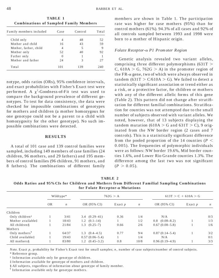
A total of 101 case and 139 control families weresampled, including 149 members of case families (24children, 96 mothers, and 29 fathers) and 195 mem-bers of control families (96 children, 91 mothers, and8 fathers). The combinations of different family
TABLE 1Combinations of Sampled Family Members
Family members included Case Control Total
Child only 4 48 52Mother and child 16 43 59Mother, father, child 4 5 9Mother only 52 40 92Father only 0 1 1Mother and father 24 3 27
Total 101 139 240
TAOdds Ratios and 95% CIs for Children and Moth
for Folate Rec
Wildtype* 7
OR n OR (95% CI)
ChildrenOnly children† 1 3/41 3.4 (0.29–41)Mother available‡ 1 18/43 1.2 (0.1–14)All children§ 1 21/84 1.3 (0.25–7)
MothersOnly mothers¶ 1 64/37 1.3 (0.4–4.5)Child available‡ 1 19/43 0.57 (0.06–5.4)All mothers§ 1 83/80 1.2 (0.45–3.2)
Note. Exact p, probability for Fisher’s Exact test for small sam* Reference group.
48 BARB
† Information available only for genotype of children.‡ Information available for genotype of mothers and children.§ All subjects, regardless of information about genotype of family me¶ Information available only for genotype mothers.
members are shown in Table 1. The participationrate was higher for case mothers (95%) than forcontrol mothers (61%). 94.3% of all cases and 92% ofall controls sampled between 1993 and 1998 wereborn to a mother of Hispanic origin.
Folate Receptor-a P1 Promoter Region
Genetic analysis revealed two variant alleles,comprising three different polymorphisms (631T .C, 610A . G, 762G . A) in the promoter region ofthe FR-a gene, two of which were always observed intandem (631T . C/610A . G). We failed to detect astatistically significant association or trend either asa risk, or a protective factor, for children or motherswith any of the different allelic forms of this gene(Table 2). This pattern did not change after stratifi-cation for different familial combinations. Stratifica-tion for counties was not attempted due to the smallnumber of subjects observed with variant alleles. Wenoted, however, that of 13 subjects displaying thetandem mutation (610A . G and 631T . C), 9 orig-inated from the NW border region (2 cases and 7controls). This is a statistically significant differencefrom the pooled proportion of the 14 counties (P ,0.005). The frequencies of polymorphic individualswere as follows: NW border 19.6%, Mid border coun-ties 1.6%, and Lower Rio Grande counties 1.3%. Thedifference among the last two was not significant(P . 0.05).
2rom Different Familial Sampling Combinations-a Mutations
A 613T . C 1 610A . G
xact p n OR (95% CI) Exact p n
0.36 1/4 N/A 0/31 1/2 0.8 (0.08–8.2) 1 1/30.66 2/6 0.67 (0.08–5.8) 1 1/6
0.77 9/4 0.87 (0.14–5.4) 1 3/21 1/4 N/A 0/10.8 10/8 0.96 (0.19–4.9) 1 3/3
, number of case subjects/number of control subjects.
AL.
BLEers feptor
62G .
E
ples; n
mber.

ctgggOwr6s
ic
aotiscc00
iMcp
ily me
Methyleneterahydrofolate Reductase
MTHFR 677C . T polymorphism. Of 24 casehildren, 3 were homozygous CC (677CC) comparedo 19 of 93 controls. There were 17 case heterozy-otes (60/93 controls), while 4 cases were homozy-ous for the T allele (14/93 controls). With homozy-osity for CC considered to be the reference group,Rs for heterozygosity (CT) and homozygosity (TT)ere 1.8 (95% CI 0.5–6.8) and 1.8 (95% CI 0.4–9.4),
espectively (Table 3). Thus, the presence of the77TT genotype, although elevated in cases, was notignificantly associated with NTD risk (P . 0.05).To assess sampling bias, we computed ORs for
children sampled within different family combina-tions. Either there was no information about otherfamily members available, or there were childrenfrom families where additional information aboutmothers could be obtained. Although confidence in-tervals were quite large due to the small number ofcase children, the trends were similar for each strat-ification. Among children, there was a trend towardsa slightly higher NTD risk associated with the 677Tallele. This trend was not statistically significant.Interestingly, the same 677T allele in mothers (ho-mozygous and heterozygous) was observed to pos-sess a slight, nonsignificant protective effect (Table3). We also analyzed the data following stratification
TAORs and 95% CIs for Children and Mothers
for MTHFR 6
CC*
OR n OR (95% CI)
ChildrenOnly children† 1 1/12 0.8 (0.07–10.0)Mother available‡ 1 2/7 1.7 (0.31–9.2)All children§ 1 3/19 1.8 (0.47–6.8)
MothersOnly mothers¶ 1 15/9 0.9 (0.35–2.3)Child available‡ 1 8/7 0.3 (0.07–0.94)All mothers§ 1 23/16 0.6 (0.30–1.33)
Note. Exact p, probability for two-tailed Fisher’s Exact for sma* Reference group.† Information available only for genotype of children.‡ Information available for genotype of mothers and children.§ All subjects, regardless of information about genotype of fam¶ Information available only for genotype mothers.
FOLATE PATHWAY
by county of residence. We found similar trends forORs for mothers and children, but none reachedstatistical significance (data not shown).
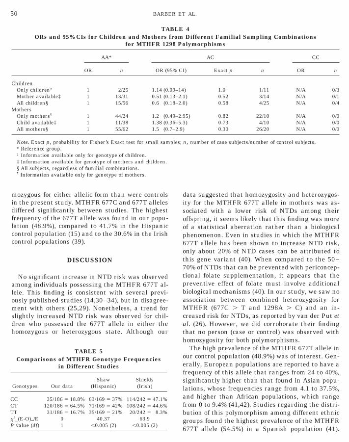
MTHFR 1298A . C polymorphism. No associa-tion or trend was observed for the 1298A . C poly-morphism, either as a risk, or as a protective factor(P , 0.05). The mutant 1298C allele was observedn the homozygous condition in only four controlhildren (Table 4).
Combined heterozygosity for MTHFR 677C . Tnd 1298A . C polymorphisms. No individual wasbserved to be homozygous for both the 677T andhe 1298C polymorphisms. Combined heterozygos-ty (heterozygosity for both loci) was detected in 55ubjects (22 cases and 33 controls). Of those, 16 werease children and 16 control children. The OR forombined heterozygosity for children is 0.8 (95% CI.3–2.5), which is not statistically significant (P ..05).
Comparison of MTHFR 677C ; T polymorphismn control populations. The frequencies of the
THFR genotypes in our control population wereompared with those of control populations fromrevious publications (Table 5). We used a x2 anal-
ysis to compare the frequencies of the MTHFR ge-notypes in our control population with control pop-ulations from other publications (Table 5). Weobserved that the frequencies for the MTHFR 677Cand 677T alleles were statistically significantly dif-ferent between studies (P , 0.005). In the x2 values
3Different Familial Sampling Combinationslymorphism
TT
ct p n OR (95% CI) Exact p n
2/29 2.4 (0.12–46) 1 1/571 15/31 1.2 (0.15–9.0) 1 3/955 17/60 1.8 (0.35–9.4) 0.68 4/14
42/28 1.8 (0.44–7.3) 0.5 12/4049 8/27 0.15 (0.02–0.89) 0.05 2/1226 50/55 0.6 (0.23–1.59) 0.34 14/16
ples; n, number of case subjects/number of control subjects.
mber.
49POLYMORPHISMS
BLEfrom77 Po
CT
Exa
10.0.
10.0.
ll sam
GENE
for comparison of Texas data (from the presentstudy) with data from California (15) and Ireland(39), Hispanic controls were more likely to be ho-
midflcc
alomsd
cath
oefslafC
TxP
C
M
5
ozygous for either allelic form than were controlsn the present study. MTHFR 677C and 677T allelesiffered significantly between studies. The highestrequency of the 677T allele was found in our popu-ation (48.9%), compared to 41.7% in the Hispanicontrol population (15) and to the 30.6% in the Irishontrol populations (39).
DISCUSSION
No significant increase in NTD risk was observedmong individuals possessing the MTHFR 677T al-ele. This finding is consistent with several previ-usly published studies (14,30–34), but in disagree-ent with others (25,29). Nonetheless, a trend for
lightly increased NTD risk was observed for chil-ren who possessed the 677T allele in either theomozygous or heterozygous state. Although our
TABLE 5Comparisons of MTHFR Genotype Frequencies
in Different Studies
Genotypes Our dataShaw
(Hispanic)Shields(Irish)
C 35/186 5 18.8% 63/169 5 37% 114/242 5 47.1%T 120/186 5 64.5% 71/169 5 42% 108/242 5 44.6%
TAORs and 95% CIs for Children and Mothers
for MTHFR 12
AA*
OR n OR (9
hildrenOnly children† 1 2/25 1.14 (0.Mother available‡ 1 13/31 0.51 (0.All children§ 1 15/56 0.6 (0.othersOnly mothers¶ 1 44/24 1.2 (0.Child available‡ 1 11/38 1.38 (0.All mothers§ 1 55/62 1.5 (0.
Note. Exact p, probability for Fisher’s Exact test for small sam* Reference group.† Information available only for genotype of children.‡ Information available for genotype of mothers and children.§ All subjects, regardless of familial combinations.¶ Information available only for genotype of mothers.
0 BARBE
bg6
T 31/186 5 16.7% 35/169 5 21% 20/242 5 8.3%2_(E-O)_/E 0 40.37 63.9value (df) 1 ,0.005 (2) ,0.005 (2)
data suggested that homozygosity and heterozygos-ity for the MTHFR 677T allele in mothers was as-sociated with a lower risk of NTDs among theiroffspring, it seems likely that this finding was moreof a statistical aberration rather than a biologicalphenomenon. Even in studies in which the MTHFR677T allele has been shown to increase NTD risk,only about 20% of NTD cases can be attributed tothis gene variant (40). When compared to the 50–70% of NTDs that can be prevented with periconcep-tional folate supplementation, it appears that thepreventive effect of folate must involve additionalbiological mechanisms (40). In our study, we saw noassociation between combined heterozygosity forMTHFR (677C . T and 1298A . C) and an in-reased risk for NTDs, as reported by van der Put etl. (26). However, we did corroborate their findinghat no person (case or control) was observed withomozygosity for both polymorphisms.The high prevalence of the MTHFR 677T allele in
ur control population (48.9%) was of interest. Gen-rally, European populations are reported to have arequency of this allele that ranges from 24 to 40%,ignificantly higher than that found in Asian popu-ations, whose frequencies range from 4.1 to 37.5%,nd higher than African populations, which rangerom 0 to 9.4% (41,42). Studies regarding the distri-
4Different Familial Sampling Combinationslymorphisms
AC CC
) Exact p n OR n
) 1.0 1/11 N/A 0/3) 0.52 3/14 N/A 0/1) 0.58 4/25 N/A 0/4
5) 0.82 22/10 N/A 0/0) 0.73 4/10 N/A 0/0
0.30 26/20 N/A 0/0
n, number of case subjects/number of control subjects.
AL.
h
C
BLEfrom98 Po
5% CI
09–1413–2.118–2.0
49–2.936–5.37–2.9)
ples;
R ET
ution of this polymorphism among different ethnicroups found the highest prevalence of the MTHFR77T allele (54.5%) in a Spanish population (41).
GENE
Papapetrou et al. (14) have suggested that homozy-gosity for MTHFR 677T is only a risk factor forNTDs in some ethnic groups and not in others. Theynote that in populations where the T allele is foundto be a risk factor for NTDs, specifically the Dutchand Irish populations, the background frequency ofthis allele is low (Dutch, 26%; Irish, 28%). In popu-lations with high background frequencies of the677T allele, such as the British (36%) or Mexican–American (49%), an association between MTHFRgenotype and NTDs could not be established.
The conflicting data that have been reported forMTHFR genotype and NTD risk could be partiallyexplained by the complexity of NTD etiology. Underthe multifactorial threshold model, MTHFR wouldrepresent one locus among many that contribute toNTD liability. Within this model, high backgroundfrequencies of the MTHFR 677T allele could func-tion to increase the overall susceptibility of a popu-lation to NTD risk, while not closely segregatingwith the disease. In this hypothetical instance, al-leles at additional loci that are associated with NTDrisk would occur at low frequencies and representthe critical alleles that could, along with key envi-ronmental factors, “push” an individual over thedisease threshold and determine the final NTDprevalence in a population. In this example, it wouldbe these additional, rare alleles that would be ob-served to segregate with the disease phenotype. Inother populations where the MTHFR 677T alleleoccurred infrequently, the MTHFR genotype mightbe the allele that would be observed to segregatewith the disease phenotype. If this hypothetical sit-uation were accurate, specific alleles that influencedNTD sensitivity would be expected to show a statis-tically significant association with the disease phe-notype in populations where the background fre-quency of the allele was low, but only be weaklyassociated in populations where the allele was morecommon.
Increasingly, studies of possible causes of NTDs re-port that interactions among different factors (gene–gene, gene–nutrition, gene–environment) may confera higher risk for NTDs than any of these factors alone.Our data suggest that if there is an NTD risk associ-ated with the MTHFR 677T allele, it is small, andinsufficient to explain the vast majority of NTDs inHispanics. It is also possible that different NTD typesmay have different etiologies and different known risk
FOLATE PATHWAY
factors may explain the different etiologic fractions ofthese birth defects. Future studies are needed whichinclude genetic, nutritional, and environmental data
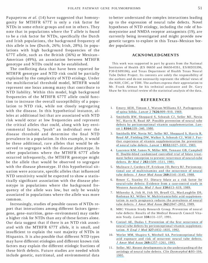
to better understand the complex interactions leadingup to the expression of neural tube defects. Novelhypotheses of NTD etiology, including the role of ho-mocysteine and NMDA receptor antagonists (19), arecurrently being investigated and might provide newgenetic targets to explore in this Texas–Mexican bor-der population.
ACKNOWLEDGMENTS
This work was supported in part by grants from the NationalInstitutes of Health (ES 06650 and 06650-03S1, ES/HD35396,P30-ES09106), and Texas Department of Health Texas NeuralTube Defect Project. Its contents are solely the responsibility ofthe authors and do not necessarily represent the official views ofthe NIH, CDC, or TDH. The authors express their appreciation toMr. Frank Aleman for his technical assistance and Dr. GaryShaw for his critical review of the statistical analysis of the data.
REFERENCES
1. Emery AEH, Timson J, Watson-Williams EJ. Pathogenesisof spina bifida. Lancet 2 7626:909–910, 1969.
2. Smithells RW, Sheppard S, Schorah CJ, Seller MJ, NevinNC, Harris R, Read AP. Possible prevention of neural tubedefects by periconceptional vitamin supplementation. Lan-cet 1 8164:339–340, 1980.
3. Smithells RW, Nevin NC, Seller MJ, Sheppard S, Harris R,Read AP, Fielding DW, Walker S, Schorah CJ, Wild J. Fur-ther experience of vitamin supplementation for preventionof neural tube defects. Lancet 1 8332:1027–1031, 1983.
4. Laurence KM, James N, Miller MH, Tennant GB, CampbellH. Double-blind randomised controlled trial of folate treat-ment before conception to prevent recurrence of neural-tubedefects. Br Med J 282:1509–1511, 1981.
5. Mulinare J, Cordero JF, Erickson JD, Berry RJ. Periconcep-tional use of multivitamins and the occurrence of neuraltube defects. J Amer Med Assoc 260:3141–3145, 1988.
6. Bower C, Stanley FJ. Dietary folate as a risk factor forneural-tube defects: Evidence from a case-control study inWestern Australia. Med J Aust 150:613–619, 1989.
7. Milunsky A, Jick H, Jick SS, Bruell CL, MacLaughlin DS,Rothman KJ, Willett W. Multivitamin/folic acid supplemen-tation in early pregnancy reduces the prevalence of neuraltube defects. J Amer Med Assoc 262:2847–2852, 1989.
8. MRC Vitamin Study Research Group. Prevention of neuraltube defects: Results of the Medical Research Council Vita-min Study. Lancet 338:131–137, 1991.
9. Czeizel AE, Dudas I. Prevention of the first occurrence ofneural tube defects by periconceptional vitamin supplemen-tation. N Engl J Med 327:1832–1835, 1992.
10. Werler MM, Shapiro S, Mitchell AA. Periconceptional folicacid exposure and risk of occurrent neural tube defects.
51POLYMORPHISMS
J Amer Med Assoc 269:1257–1261, 1993.
11. Seller, MJ. Recent developments in the understanding of theaetiology of neural tube defects. Clin Dysmorphol 4:93–104,1995.
1
1
1
ER ET
12. Berry RJ, Li Z, Erickson JD, et al. Prevention of neural tubedefects with folic acid in China. N Engl J Med 341:1485–1490, 1999.
3. Kirke PN, Molloy AM, Daly LE, Burke H, Weir DG, ScottJM. Maternal plasma folate and vitamin B12 are indepen-dent risk factors for neural tube defects. Q J Med 86:703–708, 1993.
4. Papapetrou C, Lynch SA, Burn J, Edwards YH. Methyl-enetetrahydrofolate reductase and neural tube defects. Lan-cet 348:58, 1997.
5. Shaw GM, Rozen R, Finnell RH, Wasserman CR, LammerEJ. Maternal vitamin use, genetic variation of infant meth-ylenetetrahydrofolate reductase, and risk for spina bifida.Amer J Epidemiol 148:30–37, 1998.
16. Finnell RH, Greer KA, Barber RC, Piedrahita JA. Neuraltube and craniofacial defects with special emphasis on folatepathway genes. Crit Rev Oral Biol Med 9:38–53, 1998.
17. Christensen B, Rosenblatt DS. Effects of folate deficiency onembryonic development. Bailleres Clin Haematol 8:617–637,1995.
18. Barber RC, Shaw GM, Lammer EJ, Greer KA, Biela TA,Lacey SW, Wasserman CR, Finnell RH. Lack of associationbetween mutations in the folate receptor-a gene and spinabifida. Amer J Med Genet 76:310–317, 1998.
19. Rosenquist TH, Schneider AM, Monaghan DT. N-methyl-D-aspartate receptor agonists modulate homocysteine-induceddevelopmental abnormalities. FASEB J 13:1523–1531, 1999.
20. van der Put NM, van der Molen EF, Kluijtmans LA, HeilSG, Trijbels JM, Eskes TK, Van Oppenraaij-Emmerzaal D,Banerjee R, Blom HJ. Sequence analysis of the coding regionof human methionine synthase: Relevance to hyperhomocys-teinemia in neural tube defects and vascular diseases. QJ Med 90:511–517, 1997.
21. Steegers-Theunissen RP, Boers GH, Trijbels FJ, FinkelsteinJD, Blom HJ, Thomas CM, Borm GF, Wouters MG, EskesTK. Maternal Hyperhomocysteinemia: A risk factor for neu-ral tube defects? Metabolism 43:1475–1480, 1994.
22. Shaw GM, Todoroff K, Finnell RH, Lammer EJ, Leclerc D,Gravel RA, Rozen R. Infant methionine synthase variantsand risk for spina bifida. J Med Genet 36:86–87, 1999.
23. Goyette P, Summer JS, Milos R, Duncan AMV, RosenblattDS, Matthews RG, Rozen R. Human methylenetetrahydro-folate reductase: Isolation of cDNA, mapping and mutationidentification. Nat Genet 7:195–200, 1994.
24. Frosst P, Blom HJ, Milos R, Goyette P, Sheppard CA, Mat-thews RG, Boers GJ, den Heijer M, Kluijtmans LA, van denHeuvel LP, et al. A candidate genetic risk factor for vasculardisease: A common mutation in methylenetetrahydrofolatereductase. Nat Genet 10:111–113, 1995.
25. van der Put NM, Steegers-Theunissen RP, Frosst P, TrijbelsFJ, Eskes TK, van den Heuvel LP, Mariman EC, den HeyerM, Rozen R, Blom HJ. Mutated methylenetetrahydrofolatereductase as a risk factor for spina bifida. Lancet 346:1070–1071, 1995.
26. van der Put NM, Gabreels F, Stevens EM, Smeitink JA,
52 BARB
Trijbels FJ, Eskes TK, van den Heuvel LP, Blom HJ. Asecond common mutation in the methylene–tetrahydrofo-late reductase gene: An additional risk for neural-tube de-fects? Amer J Hum Genet 62:1044–1051, 1998.
27. van der Put NMJ, van den Heuvel LP, Steeger-TheunissenRPM, Trijbles FJJM. Decreased MTHFR activity due to the677 C ; T mutation in families with spina bifida. J MolecMed 74:691–694, 1996.
28. Molloy AM, Daly S, Mills JL, Kirke PN, Whitehead AS,Ramsbottom D, Conley MR, Weir DG, Scott JM. Thermo-labile variant of 5,10-MTHFR associated with low red-cellfolates. implications for folate intake recommendations.Lancet 349:1591–1593, 1997.
29. Eskes TK. Folates and the fetus. Eur J Obstet GynecolReprod Biol 71:105–111, 1997.
30. de Franchis R, Sebastio G, Mandato C, Andria G, Mastroia-covo P. Spina bifida, 677C-Tmutation and role of folate.Lancet 346:1703, 1995.
31. Wilcken DEL, Wang XL. Relevance to spina bifida of mu-tated MTHFR. Lancet 347:340, 1996.
32. Mornet E, Muller F, Lenvoise-Furet A, Delezoide AL, ColJY, Simon-Bouy B, Serre JL. Screening of the C677T muta-tion on the MTHFR gene in French patients with NTDs.Hum Genet 100:512–514, 1997.
33. Speer MC, Worley G, Mackey JF, et al. The thermolabilevariant of MTHFR is not a major risk factor for NTDs inAmerican Caucasians. Neurogenetics 1:490–150, 1997.
34. Koch MC, Stegemann K, Ziegler A, et al. Evaluation of theMTHFR C677T allele and the MTHFR gene locus in a Germanspina bifida population. Eur J Pediatr 157:487–492, 1998.
35. Prasad PD, Ramamoorthy S, Moe AJ, Smith CH, LeibachFH, Ganapathy V. Selective expression of the high-affinityisoform of the folate receptor (FR-a) in the human placentalsyncytiotrophoblast and choriocarcinoma cells. BiochemBiophys Acta 1223:71–75, 1994.
36. Heil SG, van der Put NM, Trijbels FJ, Gabreels FJ, BlomHJ. Molecular genetic analysis of human folate receptors inneural tube defects. Eur J Hum Genet 7:393–396, 1999.
37. Hendricks KA, Simpson JS, Larsen RD. Neural tube defectsalong the Texas–Mexican border, 1993–1995. Amer J of Epi149:1119–1127, 1999.
38. Trembath D, Sherbondy AL, Vandyke DC, Shaw GM, Todor-off K, Lammer EJ, Finnell RH, Marker S, Lerner G, MurrayJC. Analysis of selected folate pathway genes, PAX3, andhuman T in a Midwestern neural tube defect population.Teratology 59:331–341, 1999.
39. Shields DC, Kirke PN, Mills JL, Ramsbottom D, Molloy AM,Burke H, Wier DG, Scott JM, Whitehead AS. The “thermo-labile” variant of methylenetetrahydrofolate reductase andneural tube defects: An evaluation of genetic risk and therelative imrtance of the genotypes of the embryo and themother. Amer J Hum Genet 64:1045–1055, 1999.
40. Posey DL, Khoury MJ, Mulinare J, Adams MJ Jr, Ou CY. Ismutated MTHFR a risk factor for neural tube defects? Lan-cet 347:686–687, 1996.
41. Pepe G, Vanegas OC, Giusti B, Brunelli T, et al. Heteroge-neity in world distribution of the thermolabile C677T mu-tation in 5,10-methylenetetrahydrofolate reductase. Amer J
AL.
Hum Genet 63:917–920, 1998.
42. Schneider JA, Rees DC, Liu YT, Clegg JB. Worldwide dis-tribution of a common methylenetetrahydrofolate reductasemutation. Amer J Hum Genet 62:1258–1260, 1998.
Copyright © 2022 FDOKUMEN