Invasive Aspergillosis in the Intensive Care Unit
12
Aspergillosis in the ICU • CID 2007:45 (15 July) • 205 REVIEW ARTICLE Invasive Aspergillosis in the Intensive Care Unit Wouter Meersseman, 1 Katrien Lagrou, 2 Johan Maertens, 3 and Eric Van Wijngaerden 1 1 Department of General Internal Medicine, 2 Medical Diagnostic Sciences, and 3 Department of Hematology, Gasthuisberg University Hospital, Leuven, Belgium Data regarding the incidence of invasive aspergillosis (IA) in the intensive care unit (ICU) are scarce, and the incidence varies. An incidence of 5.8% in a medical ICU has been reported. The majority of patients did not have a hematological malignancy, and conditions such as chronic obstructive pulmonary disease and liver failure became recognized as risk factors. Diagnosis of IA remains difficult. Mechanical ventilation makes it difficult to interpret clinical signs, and radiological diagnoses are clouded by underlying lung pathologies. The significance of a positive respiratory culture result is greatly uncertain, because cultures of respiratory spec- imens have low sensitivity (50%) and specificity (20%–70%, depending on whether the patient is immuno- compromised). The use of serologic markers has never been validated in an ICU population. Limited experience with the detection of galactomannan in bronchoalveolar lavage fluid specimens has yielded promising results. Because of a delay in the diagnosis of IA, the mortality rate exceeds 50%. Recently, our therapeutic arma- mentarium against IA has improved. Data concerning the safety and efficacy of new antifungal agents in the ICU setting, however, are lacking. Aspergillus species are ubiquitous soil inhabitants; if the conidia are inhaled into the respiratory tract, they can cause life-threatening disease. Invasive aspergillosis (IA) is a major cause of morbidity and mortality in severely immunocompromised patients. The bulk of literature about IA involves patients with classic risk factors for IA, such as prolonged neutropenia and hematopoietic stem cell transplantation [1–3]. However, a broad group of patients who are admitted to intensive care units (ICUs) may also be susceptible to these infections. IS IA A PROBLEM IN THE ICU? Autopsy studies have revealed the emergence of Asper- gillus species as major pathogens, as well as the expan- sion of the spectrum of patients at risk for IA. In a nonselected patient population at an academic hospital, the prevalence of invasive fungal infection increased Received 22 December 2006; accepted 24 March 2007; electronically published 13 June 2007. Reprints or correspondence: Dr. Wouter Meersseman, Dept. of General Internal Medicine, Gasthuisberg University Hospital, Herestraat 49, 3000 Leuven, Belgium ([email protected]). Clinical Infectious Diseases 2007; 45:205–16 2007 by the Infectious Diseases Society of America. All rights reserved. 1058-4838/2007/4502-0010$15.00 DOI: 10.1086/518852 from 2.2% to 5.1% over a 12-year period, largely in association with an increase in the rate of Aspergillus infection [4]. However, estimates about the incidence of IA among critically ill patients are sparse and vari- able. For various reasons, figures about the true inci- dence of IA are difficult to generate. First, with cultures that are positive for Aspergillus species, discriminating between colonization and infection remains challeng- ing. Second, very few institutions perform postmortem examinations routinely, although in most cases, this is the only way to prove the definite nature of the diag- nosis [5–7]. Third, characteristic radiological signs of IA are usually absent in the nonneutropenic ICU pa- tient. Finally, to date, the diagnostic utility of recently available non–culture based microbiological tools, in- cluding PCR for the detection of fungal antigens and the detection of Aspergillus-specific DNA, has not been properly validated in the nonhematology ICU popu- lation. In addition, the European Organization for Re- search and Treatment of Cancer/Mycosis Study Group (EORTC/MSG) guidelines were not designed for pa- tient categories other than patients with cancer and patients who have undergone bone marrow transplan- tation [3]. The available studies of ICU patients are summarized in table 1 [4–6, 8, 10–14]. In our medical ICU, we by guest on May 4, 2016 http://cid.oxfordjournals.org/ Downloaded from
Transcript of Invasive Aspergillosis in the Intensive Care Unit

Aspergillosis in the ICU • CID 2007:45 (15 July) • 205
R E V I E W A R T I C L E
Invasive Aspergillosis in the Intensive Care Unit
Wouter Meersseman,1 Katrien Lagrou,2 Johan Maertens,3 and Eric Van Wijngaerden1
1Department of General Internal Medicine, 2Medical Diagnostic Sciences, and 3Department of Hematology, Gasthuisberg University Hospital,Leuven, Belgium
Data regarding the incidence of invasive aspergillosis (IA) in the intensive care unit (ICU) are scarce, and the
incidence varies. An incidence of 5.8% in a medical ICU has been reported. The majority of patients did not
have a hematological malignancy, and conditions such as chronic obstructive pulmonary disease and liver
failure became recognized as risk factors. Diagnosis of IA remains difficult. Mechanical ventilation makes it
difficult to interpret clinical signs, and radiological diagnoses are clouded by underlying lung pathologies. The
significance of a positive respiratory culture result is greatly uncertain, because cultures of respiratory spec-
imens have low sensitivity (50%) and specificity (20%–70%, depending on whether the patient is immuno-
compromised). The use of serologic markers has never been validated in an ICU population. Limited experience
with the detection of galactomannan in bronchoalveolar lavage fluid specimens has yielded promising results.
Because of a delay in the diagnosis of IA, the mortality rate exceeds 50%. Recently, our therapeutic arma-
mentarium against IA has improved. Data concerning the safety and efficacy of new antifungal agents in the
ICU setting, however, are lacking.
Aspergillus species are ubiquitous soil inhabitants; if the
conidia are inhaled into the respiratory tract, they can
cause life-threatening disease. Invasive aspergillosis (IA)
is a major cause of morbidity and mortality in severely
immunocompromised patients. The bulk of literature
about IA involves patients with classic risk factors for
IA, such as prolonged neutropenia and hematopoietic
stem cell transplantation [1–3]. However, a broad group
of patients who are admitted to intensive care units
(ICUs) may also be susceptible to these infections.
IS IA A PROBLEM IN THE ICU?
Autopsy studies have revealed the emergence of Asper-
gillus species as major pathogens, as well as the expan-
sion of the spectrum of patients at risk for IA. In a
nonselected patient population at an academic hospital,
the prevalence of invasive fungal infection increased
Received 22 December 2006; accepted 24 March 2007; electronically published13 June 2007.
Reprints or correspondence: Dr. Wouter Meersseman, Dept. of General InternalMedicine, Gasthuisberg University Hospital, Herestraat 49, 3000 Leuven, Belgium([email protected]).
Clinical Infectious Diseases 2007; 45:205–16� 2007 by the Infectious Diseases Society of America. All rights reserved.1058-4838/2007/4502-0010$15.00DOI: 10.1086/518852
from 2.2% to 5.1% over a 12-year period, largely in
association with an increase in the rate of Aspergillus
infection [4]. However, estimates about the incidence
of IA among critically ill patients are sparse and vari-
able. For various reasons, figures about the true inci-
dence of IA are difficult to generate. First, with cultures
that are positive for Aspergillus species, discriminating
between colonization and infection remains challeng-
ing. Second, very few institutions perform postmortem
examinations routinely, although in most cases, this is
the only way to prove the definite nature of the diag-
nosis [5–7]. Third, characteristic radiological signs of
IA are usually absent in the nonneutropenic ICU pa-
tient. Finally, to date, the diagnostic utility of recently
available non–culture based microbiological tools, in-
cluding PCR for the detection of fungal antigens and
the detection of Aspergillus-specific DNA, has not been
properly validated in the nonhematology ICU popu-
lation. In addition, the European Organization for Re-
search and Treatment of Cancer/Mycosis Study Group
(EORTC/MSG) guidelines were not designed for pa-
tient categories other than patients with cancer and
patients who have undergone bone marrow transplan-
tation [3].
The available studies of ICU patients are summarized
in table 1 [4–6, 8, 10–14]. In our medical ICU, we
by guest on May 4, 2016
http://cid.oxfordjournals.org/D
ownloaded from

Tabl
e1.
Rele
vant
epid
emio
logi
cal
stud
ies
ofin
vasi
veas
perg
illos
is(IA
)in
the
inte
nsiv
eca
reun
it(IC
U).
Stu
dyty
pe,s
tudy
Year
No.
ofpa
tient
sD
urat
ion
ofst
udy
Type
ofst
udy
Aim
ofth
est
udy
Aut
opsy
prot
ocol
aIn
cide
nce
ofIA
Impo
rtan
tfin
ding
s
Stu
dies
that
exam
ined
the
inci
denc
eof
IAth
atw
asw
ides
prea
din
hosp
ital(
and
not
confi
ned
toth
eIC
U)
Gro
llet
al.
[4]
1996
8000
12ye
ars
Ret
rosp
ectiv
e,si
ngle
-cen
tre
Des
crib
ing
tren
dsin
post
mor
tem
epid
emio
logy
ofIF
IYe
s3.
1%In
crea
sing
tren
dsin
inci
denc
eof
IA,
com
pare
dw
ithin
va-
sive
cand
idia
sis
Cor
nille
tet
al.
[8]
2006
886
year
sC
ombi
ned
retr
ospe
ctiv
ean
dpr
ospe
ctiv
eco
hort
,w
ith47
%IC
Upa
tient
sC
ompa
ring
feat
ures
ofIA
inne
utro
peni
can
dno
nneu
trop
enic
patie
nts
No
15ca
ses/
year
Ove
rall
mor
talit
yra
te,7
1%;
rate
for
nonn
eutr
open
icpa
-tie
nts,
89%
Stu
dies
that
spec
ifica
llyex
amin
edth
ein
cide
nce
ofIA
inth
eIC
U
Bul
paet
al.
[9]
2001
234
year
sC
ase
serie
sof
patie
nts
with
CO
PD
ina
mix
edIC
UD
escr
ibin
gIA
inpa
tient
sw
ithC
OP
Dw
how
ere
adm
itted
toth
eIC
UYe
s…
Mor
talit
yin
patie
nts
with
CO
PD
who
unde
rwen
tve
nti-
latio
n,10
0%
Mee
rsse
man
etal
.[1
0]20
0312
73
year
sR
etro
spec
tive,
sing
le-c
ente
r,m
edic
alIC
UD
eter
min
ing
the
inci
denc
eof
IAin
am
edic
alIC
UYe
s5.
8%IA
incr
easi
ngly
reco
gniz
edin
patie
nts
with
out
clas
sic
risk
fact
ors
Gar
nach
o-M
onte
roet
al.
[11]
2005
1756
9m
onth
sM
ultip
le-c
entr
epr
ospe
ctiv
est
udy
of73
mix
edIC
Us
Des
crib
ing
char
acte
ristic
sof
patie
nts
with
spu-
tum
sam
ples
posi
tive
for
Asp
ergi
llus
spec
ies
in73
ICU
s
No
1.1%
Mor
talit
yra
tefo
rpa
tient
sco
lo-
nize
dw
ithA
sper
gillu
ssp
e-ci
es,
50%
;ra
tefo
rpa
tient
sco
nsid
ered
toha
veIA
,80
%
Vand
ewou
deet
al.
[12]
2006
172
7ye
ars
Ret
rosp
ectiv
e,si
ngle
-cen
ter,
mix
edIC
UD
escr
ibin
gch
arac
teris
tics
ofpa
tient
sw
ithsp
u-tu
msa
mpl
espo
sitiv
efo
rA
sper
gillu
ssp
ecie
sN
o0.
33%
Mor
talit
yra
tefo
rno
nhem
atol
-og
ypa
tient
s,60
%;
for
pa-
tient
sw
ithIA
,77
%;
for
col-
oniz
edpa
tient
s,40
%
Oth
erst
udie
sb
Roo
sen
etal
.[5
]20
0010
01
year
Ret
rosp
ectiv
e,si
ngle
-cen
ter,
med
ical
ICU
Com
parin
gpr
emor
tem
diag
nosi
san
dau
tops
yfin
ding
s(a
llca
uses
ofde
ath)
Yes
15%
IAis
am
ore
impo
rtan
tm
isse
ddi
agno
sis
ina
med
ical
ICU
than
any
othe
rill
ness
Valle
set
al.
[13]
2002
677
year
sP
rosp
ectiv
eco
hort
stud
yat
2m
ixed
ICU
sD
escr
ibin
gpa
tient
sw
ithho
spita
l-acq
uire
dpn
eu-
mon
iaw
how
ere
adm
itted
toth
eIC
UN
o19
%IA
was
the
seco
ndm
ost
fre-
quen
tca
use
ofH
AP
requ
ir-in
gIC
Uad
mis
sion
;CO
PD
was
asi
gnifi
cant
risk
fact
or
Dim
opou
los
etal
.[6
]20
0422
21
year
Ret
rosp
ectiv
e,si
ngle
-cen
ter,
mix
edIC
UC
ompa
ring
prem
orte
mdi
agno
sis
and
auto
psy
findi
ngs
(all
caus
esof
deat
h)Ye
s3.
7%In
6of
14ca
ses
with
maj
orm
isse
ddi
agno
ses,
IAw
asre
spon
sibl
e
Kum
aret
al.
[14]
2006
2154
15ye
ars
Ret
rosp
ectiv
e,m
ultip
le-c
entr
eco
hort
Det
erm
inin
gth
eim
pact
ofan
timic
robi
alth
erap
yfo
ral
lpat
ient
sw
ithse
ptic
shoc
kw
how
ere
adm
itted
toth
eIC
U
No
0.7%
No
data
for
prov
enca
ses;
in-
clus
ion
was
base
dso
lely
oncu
lture
resu
lts
NO
TE
.C
OP
D,c
hron
icob
stru
ctiv
epu
lmon
ary
dise
ase;
HA
P,ho
spita
l-acq
uire
dpn
eum
onia
;IFI
,inv
asiv
efu
ngal
infe
ctio
n.a
Stu
dies
inw
hich
auto
psy
was
perf
orm
edin
150
%of
case
s.b
Mor
ege
nera
laut
opsy
stud
ies
orst
udie
sth
atex
amin
edth
eet
iolo
gyof
pneu
mon
iain
the
ICU
.
by guest on May 4, 2016
http://cid.oxfordjournals.org/D
ownloaded from
Aspergillosis in the ICU • CID 2007:45 (15 July) • 207
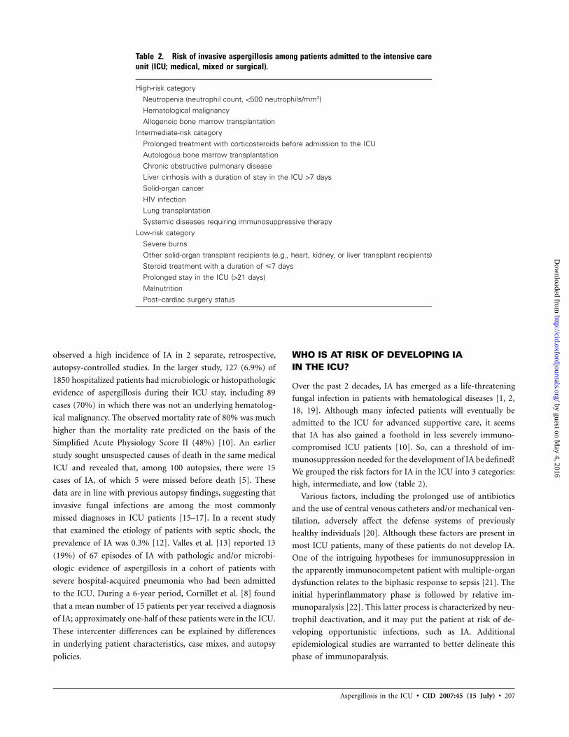
Table 2. Risk of invasive aspergillosis among patients admitted to the intensive careunit (ICU; medical, mixed or surgical).
High-risk categoryNeutropenia (neutrophil count, !500 neutrophils/mm3)Hematological malignancyAllogeneic bone marrow transplantation
Intermediate-risk categoryProlonged treatment with corticosteroids before admission to the ICUAutologous bone marrow transplantationChronic obstructive pulmonary diseaseLiver cirrhosis with a duration of stay in the ICU 17 daysSolid-organ cancerHIV infectionLung transplantationSystemic diseases requiring immunosuppressive therapy
Low-risk categorySevere burnsOther solid-organ transplant recipients (e.g., heart, kidney, or liver transplant recipients)Steroid treatment with a duration of �7 daysProlonged stay in the ICU (121 days)MalnutritionPost–cardiac surgery status
observed a high incidence of IA in 2 separate, retrospective,
autopsy-controlled studies. In the larger study, 127 (6.9%) of
1850 hospitalized patients had microbiologic or histopathologic
evidence of aspergillosis during their ICU stay, including 89
cases (70%) in which there was not an underlying hematolog-
ical malignancy. The observed mortality rate of 80% was much
higher than the mortality rate predicted on the basis of the
Simplified Acute Physiology Score II (48%) [10]. An earlier
study sought unsuspected causes of death in the same medical
ICU and revealed that, among 100 autopsies, there were 15
cases of IA, of which 5 were missed before death [5]. These
data are in line with previous autopsy findings, suggesting that
invasive fungal infections are among the most commonly
missed diagnoses in ICU patients [15–17]. In a recent study
that examined the etiology of patients with septic shock, the
prevalence of IA was 0.3% [12]. Valles et al. [13] reported 13
(19%) of 67 episodes of IA with pathologic and/or microbi-
ologic evidence of aspergillosis in a cohort of patients with
severe hospital-acquired pneumonia who had been admitted
to the ICU. During a 6-year period, Cornillet et al. [8] found
that a mean number of 15 patients per year received a diagnosis
of IA; approximately one-half of these patients were in the ICU.
These intercenter differences can be explained by differences
in underlying patient characteristics, case mixes, and autopsy
policies.
WHO IS AT RISK OF DEVELOPING IAIN THE ICU?
Over the past 2 decades, IA has emerged as a life-threatening
fungal infection in patients with hematological diseases [1, 2,
18, 19]. Although many infected patients will eventually be
admitted to the ICU for advanced supportive care, it seems
that IA has also gained a foothold in less severely immuno-
compromised ICU patients [10]. So, can a threshold of im-
munosuppression needed for the development of IA be defined?
We grouped the risk factors for IA in the ICU into 3 categories:
high, intermediate, and low (table 2).
Various factors, including the prolonged use of antibiotics
and the use of central venous catheters and/or mechanical ven-
tilation, adversely affect the defense systems of previously
healthy individuals [20]. Although these factors are present in
most ICU patients, many of these patients do not develop IA.
One of the intriguing hypotheses for immunosuppression in
the apparently immunocompetent patient with multiple-organ
dysfunction relates to the biphasic response to sepsis [21]. The
initial hyperinflammatory phase is followed by relative im-
munoparalysis [22]. This latter process is characterized by neu-
trophil deactivation, and it may put the patient at risk of de-
veloping opportunistic infections, such as IA. Additional
epidemiological studies are warranted to better delineate this
phase of immunoparalysis.
by guest on May 4, 2016
http://cid.oxfordjournals.org/D
ownloaded from

Tabl
e3.
Tool
sfo
rdi
agno
sis
ofin
vasi
veas
perg
illos
isan
dap
plic
abili
tyin
the
inte
nsiv
eca
reun
it(IC
U).
Dia
gnos
ticto
olC
hara
cter
istic
findi
ngN
o.of
patie
nts
[ref
eren
ce]
App
licab
ility
for
the
ICU
Com
men
ts
CT
Hal
osi
gn25
prov
enca
ses
[53]
No;
sign
arriv
esto
oea
rly(5
days
befo
reth
eon
set
ofdi
seas
e)(fi
gure
1)
Not
spec
ific
for
Asp
ergi
llus
spec
ies
(als
oot
her
mol
ds)
CT
Cre
scen
tsi
gn25
prov
enca
ses
[53]
No;
obsc
ured
byat
elec
tasi
s,A
RD
S,
and/
orpl
eura
leff
usio
n(fi
gure
1)
CT
ofte
nno
tfe
asib
lein
apa
tient
with
ahi
ghfr
actio
nof
insp
ired
oxyg
enH
isto
path
olog
icev
iden
ceA
cute
lybr
anch
ing
(45�
),se
ptat
edhy
phae
mai
nly
inlu
ngtis
sue
129
(56
prov
en)[
10];a
100
(15
prov
en)[
5]a
Yes,
glob
alst
anda
rdB
iops
ies
ofte
nno
tfe
asib
lein
pa-
tient
sw
ithth
rom
bocy
tope
nia
ora
high
frac
tion
ofin
spire
dox
ygen
Cul
ture
Gro
wth
onS
abou
raud
agar
172
(17
prov
en)[
12];a
36(5
prov
en)[
11];a
1209
(24
cent
ers)
[55]
;26
0(3
1pr
oven
)[30
]M
oder
ate
appl
icab
ility
for
both
cultu
rean
dm
icro
scop
yas
are
sult
ofpo
orse
nsiti
vity
and
spec
ifici
ty
Isol
atio
nof
the
spec
ies
take
sse
v-er
alda
ys;
50%
ofca
ses
are
mis
sed
onth
eba
sis
ofcu
lture
and
mic
rosc
opy
findi
ngs;
dis-
crim
inat
ion
ofco
loni
zatio
nve
r-su
sin
vasi
vedi
seas
eis
diffi
cult;
posi
tive
pred
ictiv
eva
lue
in-
crea
ses
with
incr
ease
dim
mun
osup
pres
sion
Dire
ctm
icro
scop
yPA
S,
Gro
cott
stai
n,ca
lcofl
uor
visu
aliz
a-tio
nof
hyph
alel
emen
ts(n
oton
lyA
s-pe
rgill
ussp
ecie
s),r
apid
test
172
(17
prov
en)[
12];a
36(5
prov
en)[
11];a
1209
(24
cent
ers)
[55]
;26
0(3
1pr
oven
)[30
]S
ame
asab
ove
Sam
eas
abov
e
Gal
acto
man
nan
seru
mas
say
Pol
ysac
char
ide
rele
ased
byth
efu
ngus
inth
eev
ent
ofin
vasi
vene
ss(t
hres
h-ol
d,0.
5–1.
5ng
/mL)
[56]
Not
test
edin
the
ICU
Inth
eno
nneu
trop
enic
,crit
ical
lyill
patie
nt,b
ronc
hoal
veol
arflu
idm
aype
rfor
mbe
tter
than
seru
mP
CR
DN
Am
ater
ialo
fA
sper
gillu
sfu
mig
atus
[57]
Not
test
edin
the
ICU
Inth
eno
nneu
trop
enic
,crit
ical
lyill
patie
nt,b
ronc
hoal
veol
arflu
idm
aype
rfor
mbe
tter
than
bloo
db-(1
,3)d
-glu
can
Fung
alce
llw
allc
ompo
nent
61[5
8]O
nly
1st
udy
Not
spec
ific
for
Asp
ergi
llus
spe-
cies
;al
sopr
esen
tin
yeas
tsan
dba
cter
ia;
may
beus
eful
asa
nega
tive
pred
icto
rof
fung
alin
fect
ion
NO
TE
.A
RD
S,a
cute
resp
irato
rydi
stre
sssy
ndro
me;
PAS
,pe
riodi
cac
idS
chiff
.a
Stu
dies
confi
ned
toth
eIC
U.
by guest on May 4, 2016
http://cid.oxfordjournals.org/D
ownloaded from

Aspergillosis in the ICU • CID 2007:45 (15 July) • 209
Figure 1. Chest radiograph for a patient with chronic obstructive pulmonary disease (COPD) who was receiving steroids and who was admitted tothe intensive care unit because of an exacerbation of COPD with respiratory failure. Patchy, hazy infiltrates with a predominantly peripheral localizationand a right-side pleural effusion were seen. Bronchoalveolar lavage (BAL) culture was positive for Haemophilus influenzae and negative for fungi. Theresults of a serum galactomannan test were negative but revealed a value of 2.6 ng/mL in the BAL fluid specimen. Despite administration of caspofungin(the patient was experiencing renal failure), the patient died. Autopsy revealed invasive aspergillosis that was confined to the lungs.
Patients in the ICU (medical and surgical) are often treated
with steroids. Recent work concluded that the mortality rate
is reduced if patients with septic shock who have adrenal dys-
function receive hydrocortisone for a 7-day period [23]. How-
ever, in vitro pharmacological concentrations of hydrocortisone
accelerate the growth of Aspergillus species [24]. Clearly, high
steroid intake diminishes both lines of cellular defense against
IA (i.e., macrophages and neutrophils). This has been dem-
onstrated in hematopoietic stem cell transplant recipients who
received prolonged courses of steroids for the treatment of
graft-versus-host disease [25, 26]. Palmer et al. [27] reported
that the threshold steroid concentration varies according to the
type of patient, and they emphasized that underlying lung dis-
ease is a risk factor for IA even when low doses of steroids are
administered. Cases of IA have even been reported in associ-
ation with inhaled steroids [28]. Additional studies are needed
to investigate whether administration of the 7-day course of
hydrocortisone (200 mg/day) to patients with septic shock puts
them at risk of developing IA, knowing that recognition of
fungal infection may be delayed, because the anti-inflammatory
properties of steroids blunt the signs of infection [29].
Two at-risk groups not included in the EORTC/MSG defi-
nitions stand out with regard to IA: patients with chronic ob-
structive pulmonary disease (COPD) and patients with cirrho-
sis. Patients with COPD have an increasingly recognized risk
of developing IA, and in some institutions, cases of IA among
patients with COPD outnumber those cases in “classic” patients
[30]. Bulpa et al. [9] analyzed a group of 16 patients with COPD
who had proven or probable IA and who required ICU ad-
mission. All patients were receiving steroid treatment. The out-
come was invariably poor. This is in accordance with the find-
ings of Rello et al. [31], who described another 8 patients with
COPD and IA, among whom the outcome was universally fatal.
Hepatic failure is generally not recognized as a risk factor
for IA. A literature review revealed that 5 of 14 previously
reported cases of IA in seemingly immunocompetent hosts
were associated with liver disease [32]. Our study revealed 3
fatal cases of IA [10]. Patients with cirrhosis experience de-
pressed phagocytosis, which may increase their risk for severe
infections [33].
It is expected that new risk categories of IA will arise as new
immunosuppressive agents, such as alemtuzumab and etaner-
cept (a TNF-a blocker), are made available [34, 35].
DO PATIENTS ACQUIRE IA IN THE ICU?
There are numerous sources of Aspergillus species for patients
in the ICU. Some studies suggest that fungal colonization of
the lungs is present before entry into the hospital [36]. It is
believed that the primary ecological niche is decomposing ma-
by guest on May 4, 2016
http://cid.oxfordjournals.org/D
ownloaded from

210 • CID 2007:45 (15 July) • Meersseman et al.
Figure 2. Chest radiograph for a liver transplant recipient revealing predominantly right-side air-space disease. No nodular lesions are seen. Findingsare compatible with the diagnosis of pneumonia. Chest CT was not feasible because of the high fraction of inspired oxygen requirements. Bronchoalveolarlavage culture results were negative for bacteria and fungi (while the patient was receiving broad-spectrum antibiotics). Results of tests for galactomannanin serum were negative. The patient died, and autopsy revealed disseminated aspergillosis.
terial. However, aerosolized spores may become a potential
source of infection through improperly cleaned ventilation sys-
tems, water systems, or even computer consoles [37]. The use
of high-efficiency particulate air filtration reduces the risk of
IA but does not reduce it to zero, probably in part because
patients may be colonized before admission to the ICU, and
partly because of breaks in airflow [38]. Pittet et al. [39] de-
scribed 2 patients who developed fatal IA in the ICU. In ret-
rospect, high concentrations of airborne Aspergillus spores
could be found in close proximity to an air filter change in the
ICU. In addition to the airborne route, contaminated water
has been implicated as a source of infection [40, 41]. To our
knowledge, a study of ventilators as a source of infection has
not been undertaken. Of note, the development of IA depends
on an interplay between the inoculating dose, the ability of the
host to resist infection, and the virulence of the organism.
In the retrospective study performed in our unit, 63 (62%)
of 102 patients with a culture positive for Aspergillus species
had received the positive culture result within 1 week after
admission to the ICU. Almost all patients were undergoing
mechanical ventilation, and the mean duration of ICU stay was
20 days. Of the patients with proven cases, 18 (69%) of 26 with
an underlying hematological malignancy and 11 (37%) of 30
without a malignancy had clinical evidence of IA at the time
of admission to the ICU [10]. However, there is no consensus
about the incubation period; estimates range from 2 days to 3
months [42]. Moreover, culture results and clinical evidence
alone are not reliable predictors for invasive disease. The con-
cept that increasing fungal burden associated with specific ICU
treatments for diseases other than IA (e.g., steroid therapy for
septic shock) parallels the progression from subclinical to clin-
ical aspergillosis needs to be explored using more-sensitive
markers (e.g., PCR). PCR of respiratory secretion specimens
as a modality for surveillance is an interesting topic for research.
DISEASE MANIFESTATIONS IN THE ICU
There are several manifestations of IA disease in the ICU [43–
52]. There are 3 types of pulmonary pathogen–host interactions
[43]. The most frequent interaction is colonization of the air-
ways; this can be present in patients with defective mucociliary
clearance and structural changes in the bronchial wall [44].
These changes are present in almost every patient who is un-
dergoing mechanical ventilation, making them particularly sus-
ceptible to colonization. IA will not develop in these patients
unless a critical level of immunodeficiency has been reached.
The second type of interaction is “allergic” in nature and is
beyond the scope of this review. The most relevant form of
interaction for ICU physicians is the invasive disease that de-
velops in persons with impaired immunity. The lungs and si-
nuses are implicated in 190% of these cases. The aggressive
angioinvasive form of IA is frequently encountered in neutro-
by guest on May 4, 2016
http://cid.oxfordjournals.org/D
ownloaded from

Figure 3. Chest radiograph (A) and CT (B) for a patient who was receiving high-dose steroids because of graft-versus-host disease 4 months afterundergoing bone marrow transplantation for acute myeloid leukemia. Chest radiography revealed a right-side pleural effusion and adjacent lunginfiltrate. CT confirmed a right-side complicated parapneumonic effusion, a mass filled partially with air between the fourth and fifth rib (with partialdestruction of the bone), and a wedge-shaped infiltrate on the left side. The pleural fluid culture grew Aspergillus fumigatus. Findings are compatiblewith a bronchopleural fistula, secondary to rupture of a cavitating infiltrate and adjacent bone destruction.
by guest on May 4, 2016
http://cid.oxfordjournals.org/D
ownloaded from

Figure 4. Chest radiograph (A) and CT (B) obtained 2 months after kidney transplantation in a patient with end-stage diabetes. Bilateral lower lobecavities with adjacent pleural effusion on the right side are seen. Transbronchial biopsy revealed Aspergillus fumigatus. The serum and bronchoalveolarlavage galactomannan levels were 0.1 and 5.7 ng/mL, respectively. Despite the administration of antifungal treatment, the patient died of provenAspergillus endocarditis of the tricuspid valve.
by guest on May 4, 2016
http://cid.oxfordjournals.org/D
ownloaded from

Aspergillosis in the ICU • CID 2007:45 (15 July) • 213
Table 4. Prediction scoring model and probability of invasiveaspergillosis (IA).
ScoreNo. of
patientsNo. (%) of
patients with IA
0 119 3 (2.5)1–2 106 11 (10.3)3–4 25 10 (40)�5 10 7 (70)
NOTE. Scoring model: �2 consecutive, positive airway samples, 1; sampleobtained by invasive procedure, 1; leukemia, 2; corticosteroid treatment, 2;neutropenia, 5. Data are from [30].
penic patients, whereas cavitating infiltrates are observed most
frequently in patients who are receiving steroids, patients with
COPD, patients with cirrhosis, solid-organ transplant recipi-
ents, et cetera. In lung transplant recipients, anastomotic in-
fections are the most frequently occurring presentations [45,
46]. Other, rarer presentations include endocarditis, wound in-
fection, mediastinitis (after cardiac surgery), infection of vas-
cular grafts, and osteomyelitis; these are occasionally a problem
in immunocompromised patients or during epidemic out-
breaks. A detailed description of all disease entities is beyond
the scope of this article and was recently reviewed elsewhere
[47, 48]. Infection of the CNS is frequently an ominous sign
and may arise from hematogenous seeding (for which the lung
is the most common primary site), from spread of the pathogen
from the sinuses, or after neurosurgery.
The pathogenesis of IA in patients with steroid-associated
immunosuppression differs greatly from that in neutropenic
patients. Data demonstrate that pathologic lesions are often
widespread and that death is related to a high fungal burden
in neutropenic animals, whereas the pathogenesis in nonneu-
tropenic, steroid-treated animals is driven by an adverse in-
flammatory host response that is frequently confined to the
lungs, with a low fungal burden in the lung parenchyma and
other organs [53, 54].
Clinical signs are usually nonspecific and do not necessarily
differ from those for other causes of nosocomial pneumonia.
In addition, critically ill patients with prolonged stays in the
ICU often develop pulmonary infiltrates, atelectasis, and/or
acute respiratory distress syndrome (ARDS), whereas patients
with prior lung disease (e.g., COPD) may present with pre-
existing cavities noted by conventional chest radiography.
ARE THE AVAILABLE DIAGNOSTIC TOOLSAPPLICABLE TO PATIENTS IN THE ICU?
Making a timely diagnosis of IA in the ICU population is
probably even more challenging than establishing an early di-
agnosis in patients with hematologic disease. Basically, this is
because the index of suspicion is lower in the ICU population,
because most patients do not belong to one of the well-estab-
lished risk groups. Moreover, the diagnostic tools were devel-
oped in hematology patients. In general, a diagnosis is made
on the basis of a combination of compatible clinical findings,
abnormal radiologic findings, and microbiologic confirmation
or on the basis of histologic proof of tissue invasion by the
fungus [55]. Table 3 presents an overview of the available di-
agnostic tools.
Over the past few years, lung CT has become one of the most
important diagnostic tools. Diagnostic signs of angioinvasive pul-
monary mycosis—not only that due to Aspergillus species, but
occasionally that due to Mucorales species—include single or
multiple small nodules with the halo sign. It should be recognized
that the utility of this sign has been evaluated almost exclusively
in neutropenic patients [61]. In other groups, including ICU
patients, similar CT findings are frequently absent, and if the
signs are present, they are far less specific [10, 12]. Many ICU
patients have nonspecific interfering radiologic abnormalities as-
sociated with atelectasis or ARDS (figures 1–4).
A positive result of a culture of a respiratory specimen or
positive findings of a direct microscopic examination is present
in only one-half of patients with IA [55, 59]. The predictive
value of a positive culture result depends largely on whether
the patient is immunocompromised and ranges from 20% to
80% [60]. Given the ubiquitous nature of Aspergillus spores,
differentiation of colonization from infection remains problem-
atic. Two studies have examined the significance of isolation
of Aspergillus species in ICU patients and have confirmed the
poor positive predictive values [12, 61]. However, although
culture and microscopic examination of respiratory tract sam-
ples are performed on a regular basis in most ICUs (once or
twice weekly, as a means of surveillance), it is not an appropriate
guide for clinical practice.
Serologic testing techniques based on the detection of cir-
culating fungal cell wall components, such as galactomannan
(GM) or b-d-glucan, and detection of circulating fungal DNA
by PCR techniques hold promise for patients with hematologic
malignancy, but they have not been systematically studied for
the diagnosis of IA in the ICU. GM and b-d-glucan are poly-
saccharide fungal cell wall components that are released during
tissue invasion and that can be detected in specimens of body
fluids (e.g., serum and bronchoalveolar lavage fluid) obtained
from patients with IA [56, 62]. Studies of neutropenic patients
have revealed high rates of sensitivity (67%–100%) and spec-
ificity (86%–99%) [58, 63–65]. However, in a retrospective ob-
servational study of a medical ICU population, serum GM was
elevated in only 53% of patients with IA [10]. Detection of
serum GM is probably not a sensitive marker for IA (especially
in nonneutropenic patients), as demonstrated in lung and liver
transplant recipients [57, 66]. Viable fungi can endure in the
lung tissue (with encapsulation by an inflammatory process),
whereas circulating markers can remain undetectable because
by guest on May 4, 2016
http://cid.oxfordjournals.org/D
ownloaded from

214 • CID 2007:45 (15 July) • Meersseman et al.
of clearance by circulating neutrophils. Bronchoalveolar lavage
fluid could be a better specimen for GM detection. The use of
b-d-glucan detection in the ICU is hampered by false-positive
results (associated with the use of albumin, wound gauze, he-
modialysis, and bacterial infections) [67]. GM detection yields
fewer false-positive results, although the use of b-lactam an-
tibiotics, such as piperacillin-tazobactam, may also pose a prob-
lem [68]. Thus far, no prospective data on PCR detection are
available for ICU patients [69].
Critical care physicians need a helpful instrument to guide
clinical practice. We are currently exploring the role of GM in
bronchoalveolar lavage in a broad group of critically ill patients
who are at risk of acquiring IA. It may result in an algorithm
that is able to identify an invasive mold infection at an early
stage or that can rule out infection in high-risk, critically ill
patients. Meanwhile, the prediction model involving currently
available diagnostic tools (i.e., risk factors and culture results)
proposed by Bouza et al. (table 4) [30] can be used.
ANTIFUNGALS FOR THE TREATMENT OF IAIN THE ICU
Amphotericin B has been the mainstay of the treatment of IA
for a long time. However, this formulation is renowned for
being associated with serious adverse effects (e.g., nephrotox-
icity, hypokalemia, and fever). These events often result in the
use of suboptimal dosing regimens. Fortunately, over the past
few years, lipid-based formulations of amphotericin B and new
antifungal drugs with more favorable tolerability and safety
profiles (including voriconazole, posaconazole, and the echin-
ocandins) have become available as alternatives [1, 2].
Recently, voriconazole, a derivative of fluconazole, has be-
come the new standard of care for treating IA. A significantly
better outcome (response rate, 52.8% vs. 30.6%) was dem-
onstrated in a randomized study that compared initial treat-
ment with voriconazole versus conventional amphotericin B
[70]. Posaconazole is a new, oral, broad-spectrum triazole that
is effective against several fungi that are resistant to most other
antifungals; it is well tolerated and holds promise as a pro-
phylactic agent in neutropenic patients [71]. It can be used as
an alternative agent in salvage therapy [72]. Caspofungin, mi-
cafungin, and anidulafungin belong to a new class of antifungal
drugs, the echinocandins, which act by inhibiting the synthesis
of b-(1,3)-d-glucan in the fungal cell wall. Echinocandins dis-
play activity against Aspergillus species, as demonstrated in sev-
eral studies of salvage therapy, but convincing data on its use
as first-line treatment are still lacking [73]. (The latter criticism
also applies to first-line treatment with lipid-based formulations
of amphotericin B [74].)
However, most patients who were recruited in these first-
and second-line treatment studies were experiencing an un-
derlying hematological disorder or were transplant recipients.
These studies usually exclude patients with baseline character-
istics that are commonly seen in ICU patients, including pa-
tients with liver function abnormalities, coagulation disorders,
or renal dysfunction and patients in need of advanced cardio-
vascular or pulmonary support, including mechanical venti-
lation. Nonneutropenic ICU patients and patients who are not
transplant recipients largely tend to be underrepresented in all
major trials; given the impact of these comorbidities, lower
response rates can be anticipated.
In addition, many aspects of antifungal therapy that are rel-
evant to the ICU population have not been sufficiently ad-
dressed in clinical studies, including the pharmacokinetic pro-
file of antifungals in patients with underlying renal, hepatic,
and/or cardiac dysfunction; the dose-response relationship; the
best route of administration (oral, enteral, or parenteral); the
monitoring of drug-related toxicities (e.g., how to monitor vor-
iconazole-induced visual disturbances in sedated patients); and,
especially, drug interactions with frequently used “ICU drugs.”
The echinocandins have not been studied as first-line therapy
but offer the advantage of being free of nephrotoxicity; dose
adjustments are not required in the event of renal failure or in
patients who are undergoing continuous hemofiltration. In ad-
dition, few clinically significant drug-drug interactions have
been reported.
FUTURE DIRECTIONS
In an era of increased availability of new immunosuppressive
drugs and better intensive care, with prolonged patient survival,
we can expect a continuing increase in the incidence of IA. The
occurrence of IA in the ICU usually entails a poor prognosis,
despite major recent improvements in the diagnosis and treat-
ment of IA in patients with hematologic diseases. Multicenter
studies are warranted, to explore the exact incidence of IA in
the ICU and to better delineate the difference between hospital-
acquired, ICU-acquired, and community-acquired aspergillosis.
Evaluating the value of galactomannan, b-d-glucan, and PCR
in nonneutropenic, critically ill patients with different sample
types (and, especially, with respiratory samples) is urgently
needed, as is a better delineation of the patient population at
risk for IA in the broad group of critically ill patients. Finally,
antifungal pharmacokinetics, pharmacodynamics, and inter-
actions with other drugs need to be explored more thoroughly.
Meanwhile, all new diagnostic techniques and therapeutic mea-
sures must be validated against postmortem findings, because
only proven cases of IA offer the most valuable information.
Acknowledgments
Potential conflicts of interest. W.M. has been a member of the speak-ers’ bureau for Pfizer. J.M. has been a consultant for Merck, Gilead Sciences,Pfizer, Schering-Plough, and Zeneus Pharma and is a member of the speak-ers’ bureau of Merck. E.V.W. has been a consultant for Merck and Pfizer
by guest on May 4, 2016
http://cid.oxfordjournals.org/D
ownloaded from

Aspergillosis in the ICU • CID 2007:45 (15 July) • 215
and is a member of the speakers’ bureaus of Merck and Pfizer. K.L.: noconflicts.
References
1. Segal BH, Walsh TJ. Current approaches to diagnosis and treatmentof invasive aspergillosis. Am J Respir Crit Care Med 2006; 173:707–17.
2. Patterson TF. Advances and challenges in management of invasivemycoses. Lancet 2005; 366:1013–25.
3. Ascioglu S, Rex JH, de Pauw B, et al., on behalf of the Invasive FungalInfections Cooperative Group of the European Organization for Re-search and Treatment of Cancer and Mycoses Study Group of theNational Institute of Allergy and Infectious Diseases. Defining oppor-tunistic invasive fungal infections in immunocompromised patientswith cancer and hematopoietic stem cell transplants: an internationalconsensus. Clin Infect Dis 2002; 34:7–14.
4. Groll AH, Shah PM, Mentzel C, Schneider M, Just-Nuebling G, Hueb-ner K. Trends in the postmortem epidemiology of invasive fungal in-fections at a university hospital. J Infect 1996; 33:23–32.
5. Roosen J, Frans E, Wilmer A, Knockaert D, Bobbaers H. Comparisonof premortem clinical diagnoses in critically ill patients and subsequentautopsy findings. Mayo Clin Proc 2000; 75:562–7.
6. Dimopoulos G, Piagnerelli M, Berre J, Salmon I, Vincent JL. Postmortem examination in the intensive care unit: still useful? IntensiveCare Med 2004; 30:2080–5.
7. Esteban A, Fernandez-Segoviano P. Is autopsy dead in the ICU? In-tensive Care Med 2003; 29:522–5.
8. Cornillet A, Camus C, Nimubona S, et al. Comparison of epidemio-logical, clinical and biological features of invasive aspergillosis in neu-tropenic and nonneutropenic patients: a 6-year survey. Clin Infect Dis2006; 43:577–84.
9. Bulpa PA, Dive AM, Garrino MG, et al. Chronic obstructive pulmonarydisease patients with invasive pulmonary aspergillosis: benefits of in-tensive care? Intensive Care Med 2001; 27:59–67.
10. Meersseman W, Vandecasteele SJ, Wilmer A, Verbeken E, PeetermansWE, Van Wijngaerden E. Invasive aspergillosis in critically ill patientswithout malignancy. Am J Respir Crit Care Med 2004; 170:621–5.
11. Garnacho-Montero J, Amaya-Villar R, Ortiz-Leyba C, et al. Isolationof Aspergillus spp. from the respiratory tract in critically ill patients:risk factors, clinical presentation and outcome. Crit Care 2005; 9:R191–9.
12. Vandewoude KH, Blot SI, Depuydt P, et al. Clinical relevance of as-pergillus isolation from respiratory tract samples in critically ill patients.Crit Care 2006; 10:R31.
13. Valles J, Mesalles E, Marsical D, et al. A 7-year prospective study ofsevere-hospital acquired pneumonia requiring ICU admission. Inten-sive Care Med 2003; 29:1981–8.
14. Kumar A, Roberts D, Wood KE, et al. Duration of hypotension beforeinitiation of effective antimicrobial therapy is the critical determinantof survival in human septic shock. Crit Care Med 2006; 34:1589–96.
15. Silfvast T, Takkunen O, Kolho E, Andersson L, Rosenberg P. Char-acteristics of discrepancies between clinical and autopsy diagnoses inthe intensive care unit: a 5-year review. Intensive Care Med 2003; 29:321–4.
16. Mort TC, Yeston NS. The relationship of pre mortem diagnoses andpost mortem findings in a surgical intensive care unit. Crit Care Med1999; 27:299–303.
17. Combes A, Mokhtari M, Couvelard A, et al. Clinical and autopsydiagnoses in the intensive care unit: a prospective study. Arch InternMed 2004; 164:389–92.
18. Denning DW. Therapeutic outcome in invasive aspergillosis. Clin InfectDis 1996; 23:608–15.
19. Stevens DA, Kan VL, Judson MA. Practice guidelines for diseasescaused by aspergillus. Clin Infect Dis 2000; 30:696–709.
20. Polderman KH, Girbes ARJ. Central venous catheter use, part 2: in-fectious complications. Intensive Care Med 2002; 28:18–28.
21. Hartemink KJ, Paul MA, Spijkstra JJ, Girbes A, Polderman KH. Im-
munoparalysis as a cause for invasive aspergillosis? Intensive Care Med2003; 29:2068–71.
22. Kox WJ, Volk T, Kox SN, et al. Immunomodulatory therapies in sepsis.Intensive Care Med 2000; 26(Suppl 1):S124–8.
23. Annane D, Sebille V, Charpentier C, et al. Effect of treatment with lowdoses of hydrocortisone and fludrocortisone on mortality in patientswith septic shock. JAMA 2002; 288:862–71.
24. Lionakis MS, Kontoyiannis DP. Glucocorticoids and invasive fungalinfections. Lancet 2003; 362:1828–38.
25. O’Donnell MR, Schmidt GM, Tegtmeier BR, et al. Prediction of sys-temic fungal infection in allogeneic marrow recipients: impact of am-photericin prophylaxis in high-risk patients. J Clin Oncol 1994; 12:827–34.
26. Martino R, Subira M, Rovira M, et al. Invasive fungal infections afterallogeneic peripheral blood stem cell transplantation: incidence andrisk factors in 395 patients. Br J Haematol 2002; 116:475–82.
27. Palmer LB, Greenberg HE, Schiff MJ. Corticosteroid treatment as arisk factor for invasive aspergillosis in patients with lung disease. Thorax1991; 46:15–20.
28. Leav BA, Fanburg B, Hadley S. Invasive pulmonary aspergillosis as-sociated with high-dose inhaled fluticasone. N Engl J Med 2000; 343:586.
29. Graham BS, Tucker WS Jr. Opportunistic infections in endogenousCushing’s syndrome. Ann Intern Med 1984; 101:334–8.
30. Bouza E, Guinea J, Pelaez T, Perez-Molina J, Alcala L, Munoz P. Work-load due to Aspergillus fumigatus and significance of the organism inthe microbiology laboratory of a general hospital. J Clin Microbiol2005; 43:2075–9.
31. Rello J, Esandi ME, Mariscal D, Gallego M, Domingo C, Valles J.Invasive pulmonary aspergillosis in patients with chronic obstructivepulmonary disease: report of eight cases and review. Clin Infect Dis1998; 26:1473–5.
32. Ascah KJ, Hyland RH, Hutcheon MA, et al. Invasive aspergillosis in a“healthy” patient. Can Med Assoc J 1984; 131:332–5.
33. Bailey RJ, Woolf IL, Cullens H, Williams R. Metabolic inhibition ofpolymorphonuclear leucocytes in fulminant hepatic failure. Lancet1976; 1:1162–3.
34. Martin SI, Marty FM, Fiumara K, Treon SP, Gribben JG, Baden LR.Infectious complications associated with alemtuzumab use for lym-phoproliferative disorders. Clin Infect Dis 2006; 43:16–24.
35. Warris A, Bjorneklett A, Gaustad P, et al. Invasive aspergillosis asso-ciated with infliximab therapy. N Engl J Med 2001; 344:1099–100.
36. Lass-Florl C, Salzer G, Schmid T, Rabl W, Ulmer H, Dierichi M. Pul-monary Aspergillus colonization in humans and its impact on man-agement of critically ill patients. Br J Haematol 1999; 104:745–7.
37. Warris A, Verweij PE. Clinical implications of environmental sourcesfor Aspergillus. Med Mycol 2005; 43(Suppl 1):S59–65.
38. Munoz P, Guinea J, Pelaez T, Duran C, Blanco JL, Bouza E. Nosocomialinvasive aspergillosis in a heart transplant recipient acquired during abreak in the HEPA air filtration system. Transpl Infect Dis 2004; 6:50–4.
39. Pittet D, Huguenin T, Dharan S, et al. Unusual cause of lethal pul-monary aspergillosis in patients with chronic obstructive pulmonarydisease. Am J Respir Crit Care Med 1996; 154:541–4.
40. Anaissie EJ, Stratton SL, Dignani MC, et al. Pathogenic Aspergillusspecies recovered from a hospital water system: a 3-year prospectivestudy. Clin Infect Dis 2002; 34:780–9.
41. Warris A, Voss A, Abrahamsen TG, Verwije PE. Contamination ofhospital water with Aspergillus fumigatus and other molds. Clin InfectDis 2002; 34:1159–60.
42. Carreras A. Preventing exposure to moulds. Clin Microbiol Infect2006; 12:S77–83.
43. Soubani AO, Chandrasekar PH. The clinical spectrum of pulmonaryaspergillosis. Chest 2002; 121:1988–99.
44. Hope WW, Walsh TJ, Denning DW. The invasive and saprophyticsyndromes due to Aspergillus spp. Med Mycol 2005; 43(Suppl 1):S207–38.
by guest on May 4, 2016
http://cid.oxfordjournals.org/D
ownloaded from

216 • CID 2007:45 (15 July) • Meersseman et al.
45. Nathan SD, Shorr AF, Schmidt ME, Burton NA. Aspergillus and en-dobronchial abnormalities in lung transplant recipients. Chest 2000;118:403–7.
46. Mehrad B, Paciocco G, Martinez FJ, Ojo TC, Ianettoni MD, Lynch JP.Spectrum of Aspergillus infection in lung transplant recipients: caseseries and review of the literature. Chest 2001; 119:169–75.
47. Pasqualotto AC, Denning DW. Post-operative aspergillosis. Clin Mi-crobiol Infect 2006; 12:1060–76.
48. Nunley DR, Gal AA, Vega JD, Perlino C, Smith P, Lawrence CE. Sap-rophytic fungal infections and complications involving the bronchialanastomosis following human lung transplantation. Chest 2002; 122:1185–91.
49. Lin SJ, Schranz J, Teutsch M. Aspergillosis case-fatality rate: systematicreview of the literature. Clin Infect Dis 2001; 32:358–66.
50. Marr KA, Patterson T, Denning DW. Aspergillosis: pathogenesis, clin-ical manifestations and therapy. Infect Dis Clin North Am 2002; 16:875–94.
51. Denning DW, Riniotis K, Dobrashian R, Sambatakou H. Chronic cav-itary and fibrosing pulmonary and pleural aspergillosis: case series,proposed nomenclature change, and review. Clin Infect Dis 2003;37(Suppl 3):S265–80.
52. Patterson TF, Kirkpatrick WR, White M, et al. Invasive aspergillosis:disease spectrum, treatment practices, and outcome. Medicine (Bal-timore) 2000; 79:250–60.
53. Balloy V, Huerre M, Latge JP, Chignard M. Differences in patterns ofinfection and inflammation for corticosteroid treatment and chemo-therapy in experimental invasive pulmonary aspergillosis. Infect Im-mun 2005; 73:494–503.
54. Chamilos G, Luna M, Lewis R, et al. Invasive fungal infections inpatients with hematological malignancies in a tertiary care center: anautopsy study over a 15-year period (1989–2003). Haematologica2006; 91:986–9.
55. Hope WW, Walsh TJ, Denning DW. Laboratory diagnosis of invasiveaspergillosis. Lancet Infect Dis 2005; 5:609–22.
56. Mennink-Kersten MA, Donelly JP, Verweij PE. Detection of circulatinggalactomannan for the diagnosis and management of invasive asper-gillosis. Lancet Infect Dis 2004; 4:349–57.
57. Kwak E, Husain S, Obman A, et al. Efficacy of galactomannan antigenin the Platelia Aspergillus enzyme immunoassay for diagnosis of in-vasive aspergillosis in liver transplant recipients. J Clin Microbiol2004; 42:435–8.
58. Ostrosky-Zeichner L, Alexander BD, Kett DH, et al. Multicenter clinicalevaluation of the (1r3) b-d-glucan assay as an aid to diagnosis offungal infections in humans. Clin Infect Dis 2005; 41:654–9.
59. Tarrand JJ, Lichterfeld M, Warraich I, et al. Diagnosis of invasive septatemold infections: a correlation of microbiological culture and histologicor cytologic examination. Am J Clin Pathol 2003; 119:854–8.
60. Perfect JR, Cox GM, Lee JY, et al. The impact of culture isolation of
aspergillus species: a hospital-based survey of aspergillosis. Clin InfectDis 2001; 33:1824–33.
61. Caillot D, Casasnovas O, Bernard A, et al. Improved management ofinvasive pulmonary aspergillosis in neutropenic patients using earlythoracic computed tomographic scan and surgery. J Clin Oncol1997; 15:139–47.
62. Klont R, Messink-Kersten M, Verwije PE. Utility of Aspergillus antigendetection in specimens other than serum specimens. Clin Infect Dis2004; 39:1467–74.
63. Maertens J, Van Eldere J, Verhaegen J, Verbeken Verschakelen J, Boo-gaerts M. Use of circulating galactomannan screening for early diag-nosis of invasive aspergillosis in allogeneic stem cell transplant recip-ients. J Infect Dis 2002; 186:1297–306.
64. Pfeiffer C, Fine J, Safdar N. Diagnosis of invasive aspergillosis using agalactomannan assay: a meta-analysis. Clin Infect Dis 2006; 42:1417–27.
65. Pazos C, Ponton J, Del Palacio A. Contribution of 1,3 beta-d glucanchromogenic assay to diagnosis and therapeutic monitoring of invasiveaspergillosis in neutropenic adult patients: a comparison with serialscreening for circulating galactomannan. J Clin Microbiol 2005; 43:299–305.
66. Husain S, Kwak E, Obman A, et al. Prospective assessment of PlateliaAspergillus galactomannan antigen for the diagnosis of invasive asper-gillosis in lung transplant recipients. Am J Transplant 2004; 4:796–802.
67. Digby J, Kalbfleisch J, Glenn A, Larsen A, Browder W, Williams D.Serum glucan levels are not specific for presence of fungal infectionsin intensive care unit patients. Clin Diagn Lab Immunol 2003; 10:882–5.
68. Sulahian A, Touratier S, Ribaud P. False positive test for aspergillusantigenemia related to concomitant administration of piperacillin andtazobactam. N Engl J Med 2003; 349:2366–77.
69. Donnelly J. Polymerase chain reaction for diagnosing invasive asper-gillosis: getting closer but still a ways to go. Clin Infect Dis 2006; 42:487–9.
70. Herbrecht R, Denning DW, Patterson TF, et al. Voriconazole versusamphotericin B for primary therapy of invasive aspergillosis. N EnglJ Med 2002; 347:408–15.
71. Cornely O, Maertens J, Winston DJ, et al. Posaconazole vs. fluconazoleor itraconazole in patients with neutropenia. N Engl J Med 2007; 356:348–59.
72. Walsh TJ, Raad I, Patterson TF, et al. Treatment of invasive aspergillosiswith posaconazole in patients who are refractory or intolerant of con-ventional therapy: an externally controlled trial. Clin Infect Dis 2007;44:2–12.
73. Denning DW. Echinocandin antifungal drugs. Lancet 2003; 362:1142–51.
74. Sastry P, Parikh P, Kulkarni P, Bhagwart R, Gadade H. Use of liposomalamphotericin B in bone marrow transplant. J Postgrad Med 2005; 51:S49–52.
by guest on May 4, 2016
http://cid.oxfordjournals.org/D
ownloaded from