Intrahost and Interhost Variability of the HIV Type 1 nef Gene in Brazilian Children
Upload
independentCategory
view
2download
0
Intrahost distribution and trasmission of a new species of
cyclopoid copepod endosymbiotic to a freshwater snail, Pomacea
canaliculata (Caenogastropoda, Ampullariidae) from Argentina
C. D. GAMARRA-LUQUES, I. A. VEGA, E. KOCH AND A. CASTRO-VAZQUEZ
Laboratory of Physiology (IHEM-CONICET), Department of Morphology and Physiology, School of Medicine, University
of Cuyo, Casilla de Correo 33, M5500 Mendoza, Argentina.
Key words: Ozmanidae; Ozmana haemophila; Ozmana huarpium, n. sp.; pallial organs; haemocoel
ABSTRACT: A new species of cyclopoid copepod, Ozmana huarpium, is described as a symbiont to Pomacea
canaliculata (Lamarck 1822) (Caenogastropoda, Ampullariidae). Rather large numbers (about one hundred
copepods per snail) were found, although there was no evidence of harm to the host. To our knowledge, O.
haemophila (symbiont to P. maculata), and the currently described species, O. huarpium, are the only cope-
pod species ever recorded as endosymbionts to freshwater invertebrates. While O. haemophila is restricted to
the haemocoel of its host, O. huarpium predominate in the penis sheath, the ctenidium and the mantle cavity,
figuring in these pallial organs 63-65 % of total mature forms. The sex ratio of the symbiont is skewed to the
female side in these organs, specially in male hosts. The hypothesis that a special female tropism for the male
host’s pallial organs might ensure interindividual transmission of the symbiont was tested, with indications
that the symbiont is mainly transmitted during copulation.
BIOCELL
2004, 28(3): 155-164
ISSN 0327 - 9545
PRINTED IN ARGENTINA
Introduction
Pomacea maculata Perry 1810 (Caenogastropoda,
Ampullariidae) hosts a cyclopoid copepod, Ozmana
haemophila Ho & Thatcher 1989, Ozmanidae (Ho and
Thatcher, 1989) that occurs in rather small numbers
(1-14) in the haemolymph of this large snail (up to 14
cm in shell length). The life cycle of O. haemophila is
unknown, except for the occurrence of most develop-
mental stages in the snail’s haemolymph.
In the present paper we describe another ozmanid
species, which was found living in high numbers (up to
182) in Pomacea canaliculata (Lamarck 1822)
(Caenogastropoda, Ampullariidae). This snail is smaller
than P. maculata (only up to 6 cm in shell length) and it
is distributed in both tropical and subtropical zones,
including the Plata and Amazon basins (Estebenet and
Martín, 2002); in parts of the latter basin P. canaliculata
may be sympatric to P. maculata. Various other organ-
isms have been either reported or proposed as symbi-
otic partners or parasites to P. canaliculata: a prokariont
(Castro-Vazquez et al., 2002), two ciliates
(Parasicuophora ampullariarum and P. corderoi;
Gascón, 1975), a rotifer (genus Abrochtha Bryce 1910,
Philodinavidae, Gamarra-Luques & Castro-Vazquez,
unpublished findings), and some temnocephalid
platyhelmynthes (Damborenea, 1998; Damborenea and
Cannon, 2001), hirudinea (Damborenea and Gullo,
1996) and digenea (Hamann, 1992; Keawjam et al.,
1993).
Address correspondence to: Dr. Alfredo Castro-Vazquez.Laboratorio de Fisiologia, Facultad de Ciencias Médicas, UNCuyo.
Casilla de Correo 33. (5500) Mendoza, ARGENTINA.
TEL: +54 (261) 413-5006 int. 2715.FAX: +54 (261) 449-4117
E-mail: [email protected] on March 5, 2004. Accepted on June 9, 2004.
156 CARLOS D. GAMARRA-LUQUES et al.
Material and Methods
Copepods were obtained mostly from mature snails
of a cultured strain of Pomacea canaliculata, although
some observations were also made on field-collected
animals (Rosedal and Regatas Lakes, Palermo Park,
Buenos Aires, Argentina). Since then, the lake was
cleaned up and the original population was extinguished,
but voucher (alcohol preserved) specimens of the origi-
nal population and of the cultured strain were deposited
at the collection of Museo Argentino de Ciencias Natu-
rales (Buenos Aires, Argentina; lots MACN-In 35707
and MACN-In 36046, respectively). Histological ob-
servations were made on material fixed in Bouin’s fluid,
embedded in paraffin and stained with haematoxylin
and eosin.
Cultured snails were maintained under conditions
of artificial lighting (14 h per day) and temperature (23-
25 °C), and were fed ad libitum on a mixed diet (let-
tuce, rodent food pellets and toilet paper).
Copepods were looked for in the heart and the
perintestinal and peripharyngeal haemocoel sinuses
under a stereoscopic microscope. The mantle cavity (and
dependent organs: ctenidium, osphradium, vagina and
penis sheath) and the intestine (both mucosa and con-
tent), were also investigated for copepods (a thin water
jet was produced with a hypodermic syringe to wash
out the copepods from the different surfaces and col-
lect them in Petri dishes).
After fixation in 70% alcohol for 30 min, the cope-
pods were quickly passed through 80% alcohol, and
stained (22 mg/dL eosin, 30 mg/dL phosphotungstic
acid, dissolved in 96% alcohol). They were then washed
in 96º alcohol, cleared in phenol/xylene (1:3, v/v) and
mounted in Eukitt.
One hundred and thirty two female individuals and
98 male individuals were examined for morphological
description of the new species. All these specimens were
obtained from the haemocoel, mantle cavity, ctenidium
and penis sheath´s groove of living snails. Also, 62 im-
mature individuals of the same origin (all of them in
copepodid stages) were studied. Type material was de-
posited at the Invertebrate collection of the Museo
Argentino de Ciencias Naturales (MACN-In), Buenos
Aires, Argentina, and at the Museo de La Plata (MLP),
La Plata, Argentina.
For studying the intrahost distribution of copepods,
adult female (shell length 36.7-50.2 mm) and male snails
(shell length 39.5-51.6 mm) were sacrificed, and cope-
pods carefully recovered in Petri dishes (as described
above), counted and classified as either immatures, adult
males or adult females; the number of egg-bearing fe-
males was also recorded. The sex ratio was calculated
as M/(M+F), M and F being number of males and fe-
males, respectively.
For studying the way of transmission of the sym-
biont two different sets of observations were run. First,
non-infested adult snails were used (shell length >35
mm), which were obtained by culturing newly hatched
snails of both sexes in the laboratory, preventing their
contact with infested snails. Infested snails were ob-
tained from aquaria in which at least one infested snail
was found (we have previously determined that infesta-
tion of snails by the copepod was always present in snails
inhabiting with infested snails). Different experimental
groups of adult snails were made: (1) infested males
with non-infested females; (2) infested females with
non-infested males; (3) infested males with non-infested
males; (4) infested females with non-infested females.
The initially non-infested animals were sacrificed 15-
20 and 30-40 days after the groups were formed, and
the total number of copepods (mostly mature males and
females) in the perintestinal sinus and the pallial or-
gans was recorded.
Furthermore, we observed the behaviour of adult
copepods of both sexes that were kept in Petri dishes
containing paper-filtered aquarium water at 24 ºC. Wa-
ter was changed daily and the copepods survival was
recorded daily for 10 days (a copepod was considered
dead when no movements were elicited by gentle stimu-
lation with a micropipette tip). The fate of eggs was
also recorded during the same period.
Statistical evaluation of differences between two
groups was made by means of Student’s t test, while
multigroup comparisons were made by Kruskal-Wallis
one-way analysis of variance, followed by Dunn test.
Results
Family Ozmanidae Ho et Thatcher 1989.
Genus Ozmana Ho et Thatcher 1989.
Ozmana huarpium Gamarra-Luques et Castro-
Vazquez, n. sp.
Description:
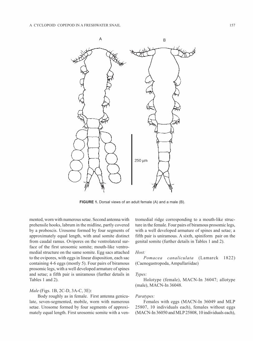
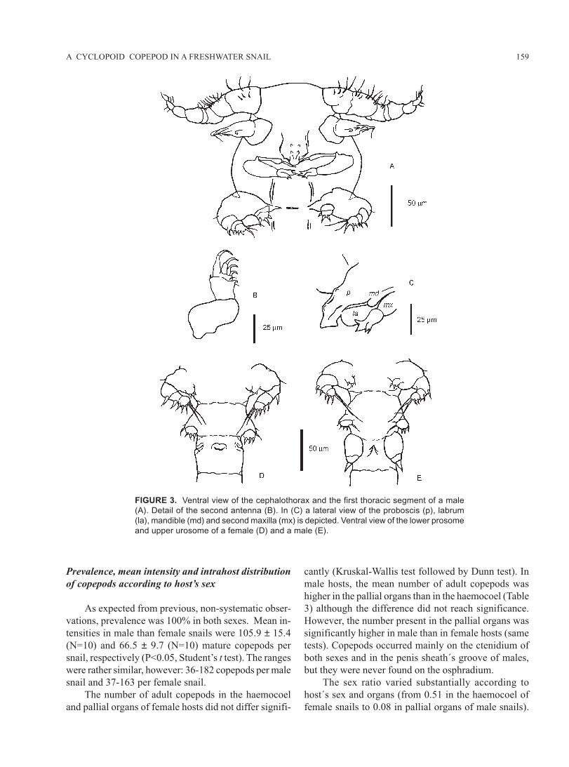
Female (Figs.1A, 2A-B, 3D):
Body modified, elongated. Urosome distinctly nar-
rower than prosome. Urosome and appendages with well
defined sclerites. Cephalothorax rounded, including first
leg-bearing somite. First antenna cylindrical, seven-seg-
157A CYCLOPOID COPEPOD IN A FRESHWATER SNAIL
FIGURE 1. Dorsal views of an adult female (A) and a male (B).
mented, worn with numerous setae. Second antenna with
prehensile hooks, labrum in the midline, partly covered
by a proboscis. Urosome formed by four segments of
approximately equal length, with anal somite distinct
from caudal ramus. Ovipores on the ventrolateral sur-
face of the first urosomic somite; mouth-like ventro-
medial structure on the same somite. Egg sacs attached
to the ovipores, with eggs in linear disposition, each sac
containing 4-6 eggs (mostly 5). Four pairs of biramous
prosomic legs, with a well developed armature of spines
and setae; a fifth pair is uniramous (further details in
Tables 1 and 2).
Male (Figs. 1B, 2C-D, 3A-C, 3E):
Body roughly as in female. First antenna genicu-
late, seven-segmented, mobile, worn with numerous
setae. Urosome formed by four segments of approxi-
mately equal length. First urosomic somite with a ven-
tromedial ridge corresponding to a mouth-like struc-
ture in the female. Four pairs of biramous prosomic legs,
with a well developed armature of spines and setae; a
fifth pair is uniramous. A sixth, spiniform pair on the
genital somite (further details in Tables 1 and 2).
Host:
Pomacea canaliculata (Lamarck 1822)
(Caenogastropoda, Ampullariidae)
Types:
Holotype (female), MACN-In 36047; allotype
(male), MACN-In 36048.
Paratypes:
Females with eggs (MACN-In 36049 and MLP
25807, 10 individuals each), females without eggs
(MACN-In 36050 and MLP 25808, 10 individuals each),
158 CARLOS D. GAMARRA-LUQUES et al.
and males (MACN-In 36051 and MLP 25806, 10 indi-
viduals each).
Location:
All these specimens were obtained from the
haemocoel, mantle cavity, ctenidium and penis sheath´s
groove of Pomacea canaliculata cultured in Mendoza,
Argentina.
Type locality:
The culture strain was originated from animals col-
lected at the Rosedal Lake (Palermo Park, Buenos Aires,
Argentina), and since we also confirmed the presence
of these copepods in animals collected at the Regatas
Lake (within the same park), we are hereby designating
Palermo, in Buenos Aires city, as the type locality.
Etymology:
huarpium (= of the Huarpes), a genitive noun, re-
fers to the Huarpe people, whose culture once predomi-
nated in Mendoza, Argentina.
Survival and behaviour of copepods outside the snails
Eighty adult copepods of both sexes were still alive
one day after being removed from the snails and placed
in aquarium water. All the egg-bearing females had re-
leased their eggs by that day, and neither hatching of
the released eggs was observed during the 10 days pe-
riod of observation, nor additional egg sacs appeared
attached to the females ovipores. Copepods died during
the subsequent days, a few of them having survived up
to seven days in such conditions.
Both the adults and the copepodid forms of O.
huarpium were unable to swim in aquarium water; they
were only able to crawl over short distances on the bot-
tom surface of the Petri dish and over the mucous clumps,
to which they usually remained attached and motionless.
When stimulated with a micropipette they showed a clear
thigmotropic response, turning to the pipette tip and at-
taching to it. When obliged to leave they seldom attempt
to flight by crawling, but they started a series of ventral
flexions and extensions of the urosome over the prosome,
from which no locomotion resulted.
Spontaneous behaviour of both females and males
also included rapid and simultaneous abduction and
adduction movements of the right and left first anten-
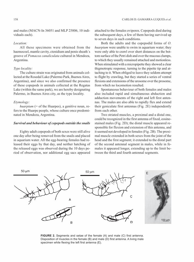
nas. The males are also able to rapidly flex and extend
their geniculate first antennas (Fig. 2E) independently
from each other.
Two striated muscles, a proximal and a distal one,
could be recognized in the first antenna of fixed, eosine-
stained males (Fig. 2D); the distal muscle appeared re-
sponsible for flexion and extension of this antenna, and
it seemed not developed in females (Fig. 2B). The proxi-
mal muscle extended in both sexes from the joint of the
head and the first segment; it extended to the distal part
of the second antennal segment in males, while in fe-
males it appeared longer, extending up to the limit be-
tween the third and fourth antennal segments.
FIGURE 2. Segments and setae of the female (A) and male (C) first antenna.
Disposition of muscles in the female (B) and male (D) first antenna. A living male
specimen while flexing the left first antenna (E).
E
100 µm
159A CYCLOPOID COPEPOD IN A FRESHWATER SNAIL
FIGURE 3. Ventral view of the cephalothorax and the first thoracic segment of a male
(A). Detail of the second antenna (B). In (C) a lateral view of the proboscis (p), labrum
(la), mandible (md) and second maxilla (mx) is depicted. Ventral view of the lower prosome
and upper urosome of a female (D) and a male (E).
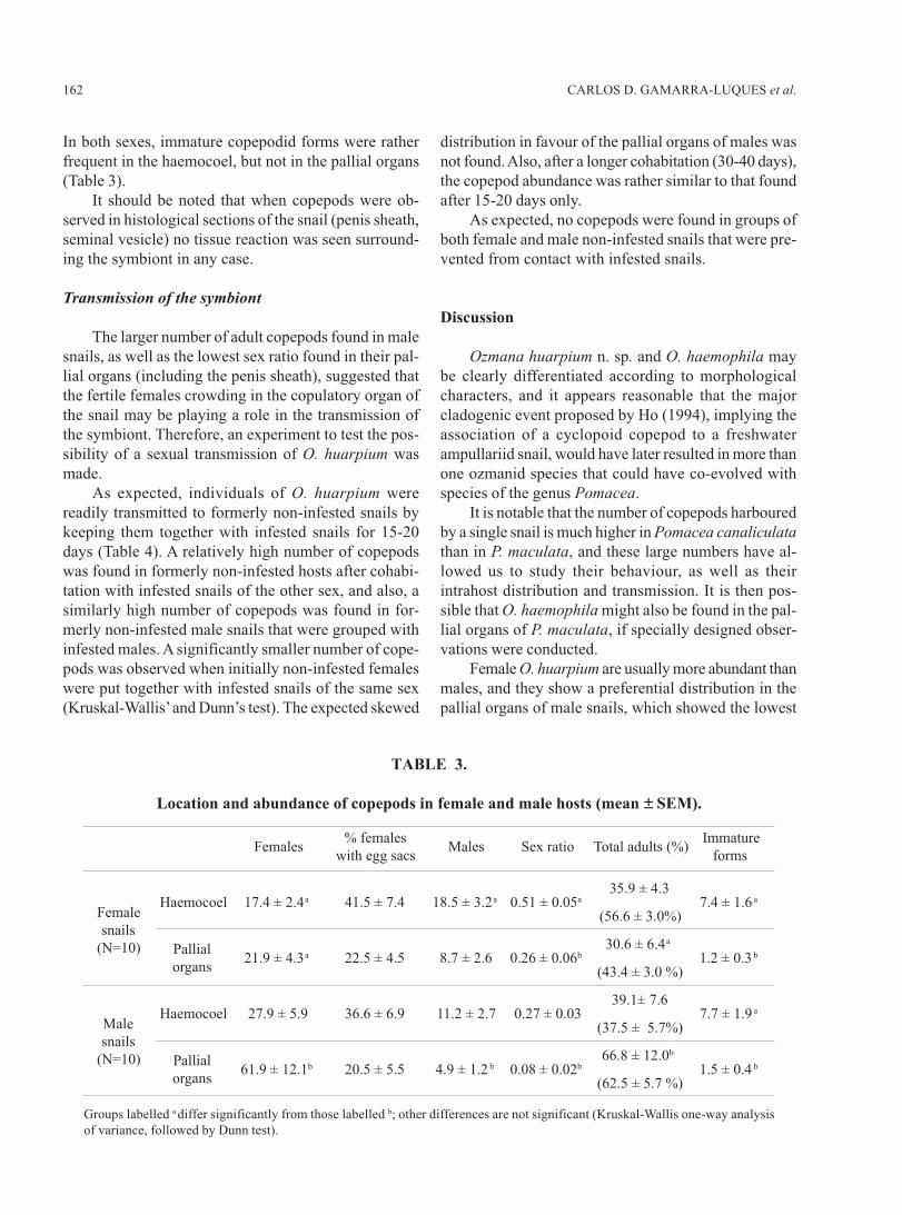
Prevalence, mean intensity and intrahost distribution
of copepods according to host’s sex
As expected from previous, non-systematic obser-
vations, prevalence was 100% in both sexes. Mean in-
tensities in male than female snails were 105.9 ± 15.4
(N=10) and 66.5 ± 9.7 (N=10) mature copepods per
snail, respectively (P<0.05, Student’s t test). The ranges
were rather similar, however: 36-182 copepods per male
snail and 37-163 per female snail.
The number of adult copepods in the haemocoel
and pallial organs of female hosts did not differ signifi-
cantly (Kruskal-Wallis test followed by Dunn test). In
male hosts, the mean number of adult copepods was
higher in the pallial organs than in the haemocoel (Table
3) although the difference did not reach significance.
However, the number present in the pallial organs was
significantly higher in male than in female hosts (same
tests). Copepods occurred mainly on the ctenidium of
both sexes and in the penis sheath´s groove of males,
but they were never found on the osphradium.
The sex ratio varied substantially according to
host´s sex and organs (from 0.51 in the haemocoel of
female snails to 0.08 in pallial organs of male snails).
160 CARLOS D. GAMARRA-LUQUES et al.
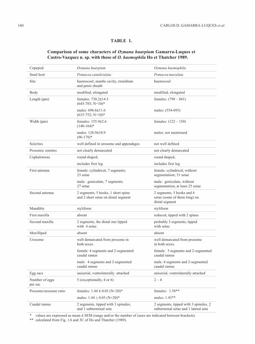
TABLE 1.
Comparison of some characters of Ozmana huarpium Gamarra-Luques et
Castro-Vazquez n. sp. with those of O. haemophila Ho et Thatcher 1989.
Copepod Ozmana huarpium Ozmana haemophila
Snail host Pomacea canaliculata Pomacea maculata
Site haemocoel, mantle cavity, ctenidium haemocoel
and penis sheath
Body modified, elongated modified, elongated
Length (µm) females: 730.2±14.5 females: (798 – 841)
(645-785; N=10)*
males: 696.6±11.6 males: (554-693)
(635-752; N=10)*
Width (µm) females: 155.9±2.6 females: (122 – 150)
(140-164)*
males: 128.9±10.9 males: not mentioned
(96-178)*
Sclerites well defined in urosome and appendages not well defined
Prosomic somites not clearly demarcated not clearly demarcated
Cephalotorax round shaped, round shaped,
includes first leg includes first leg
First antenna female: cylindrical, 7 segments; female: cylindrical, without
23 setae segmentation; 31 setae
male: geniculate, 7 segments; male: geniculate, without
27 setae segmentation; at least 25 setae
Second antenna 2 segments; 5 hooks, 1 short spine 2 segments; 5 hooks and 4
and 2 short setae on distal segment setae (some of them long) on
distal segment
Mandible styliform styliform
First maxilla absent reduced, tipped with 2 spines
Second maxilla 2 segments, the distal one tipped probably 3 segments, tipped
with 4 setae. with setae
Maxilliped absent absent
Urosome well demarcated from prosome in well demarcated from prosome
both sexes. in both sexes.
female: 4 segments and 2-segmented female: 3 segments and 2-segmented
caudal ramus caudal ramus
male: 4 segments and 2-segmented male: 4 segments and 2-segmented
caudal ramus caudal ramus
Egg sacs uniserial, ventrolaterally attached uniserial, ventrolaterally attached
Number of eggs 5 (exceptionally, 4 or 6) 2 – 4
per sac
Prosome/urosome ratio females: 1.44 ± 0.03 (N=20)* females: 1.58**
males: 1.44 + 0.03 (N=20)* males: 1.43**
Caudal ramus 2 segments, tipped with 3 spinules, 2 segments, tipped with 3 spinules, 2
and 1 subterminal seta subterminal setae and 1 lateral seta
* values are expressed as mean ± SEM (range and/or the number of cases are indicated between brackets).
** calculated from Fig. 1A and 3C of Ho and Thatcher (1989).
161A CYCLOPOID COPEPOD IN A FRESHWATER SNAIL
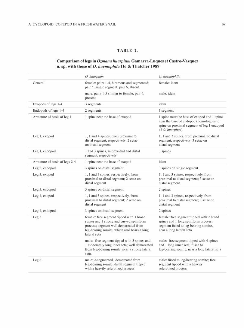
TABLE 2.
Comparison of legs in Ozmana huarpium Gamarra-Luques et Castro-Vazquez
n. sp. with those of O. haemophila Ho & Thatcher 1989
O. huarpium O. haemophila
General female: pairs 1-4, biramous and segmented; female: idem
pair 5, single segment; pair 6, absent.
male: pairs 1-5 similar to female; pair 6, male: idem
present
Exopods of legs 1-4 3 segments idem
Endopods of legs 1-4 2 segments 1 segment
Armature of basis of leg 1 1 spine near the base of exopod 1 spine near the base of exopod and 1 spine
near the base of endopod (homologous to
spine on proximal segment of leg 1 endopod
of O. huarpium)
Leg 1, exopod 1, 1 and 4 spines, from proximal to 1, 1 and 3 spines, from proximal to distal
distal segment, respectively; 2 setae segment, respectively; 3 setae on
on distal segment distal segment
Leg 1, endopod 1 and 3 spines, in proximal and distal 3 spines
segment, respectively
Armature of basis of legs 2-4 1 spine near the base of exopod idem
Leg 2, endopod 3 spines on distal segment 3 spines on single segment
Leg 3, exopod 1, 1 and 3 spines, respectively, from 1, 1 and 3 spines, respectively, from
proximal to distal segment; 2 setae on proximal to distal segment; 3 setae on
distal segment distal segment
Leg 3, endopod 3 spines on distal segment 2 spines
Leg 4, exopod 1, 1 and 3 spines, respectively, from 1, 1 and 3 spines, respectively, from
proximal to distal segment; 2 setae on proximal to distal segment; 3 setae on
distal segment distal segment
Leg 4, endopod 3 spines on distal segment 2 spines
Leg 5 female: free segment tipped with 3 broad female: free segment tipped with 2 broad
spines and 1 strong and curved spiniform spines and 1 long spiniform process;
process; segment well demarcated from segment fused to leg-bearing somite,
leg-bearing somite, which also bears a long near a long lateral seta
lateral seta
male: free segment tipped with 3 spines and male: free segment tipped with 4 spines
1 moderately long inner seta; well demarcated and 1 long inner seta; fused to
from leg-bearing somite, near a strong lateral leg-bearing somite, near a long lateral seta
seta.
Leg 6 male: 2-segmented, demarcated from male: fused to leg-bearing somite; free
leg-bearing somite; distal segment tipped segment tipped with a heavily
with a heavily sclerotized process sclerotized process
162 CARLOS D. GAMARRA-LUQUES et al.
In both sexes, immature copepodid forms were rather
frequent in the haemocoel, but not in the pallial organs
(Table 3).
It should be noted that when copepods were ob-
served in histological sections of the snail (penis sheath,
seminal vesicle) no tissue reaction was seen surround-
ing the symbiont in any case.
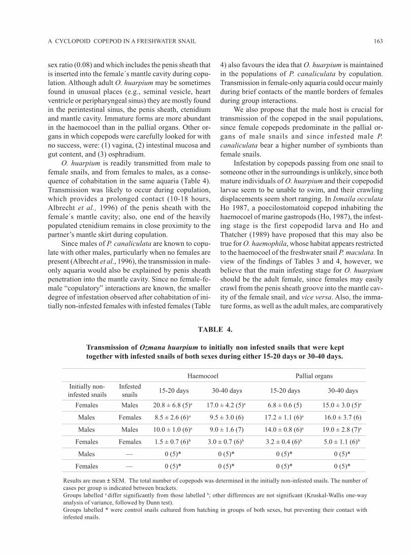
Transmission of the symbiont
The larger number of adult copepods found in male
snails, as well as the lowest sex ratio found in their pal-
lial organs (including the penis sheath), suggested that
the fertile females crowding in the copulatory organ of
the snail may be playing a role in the transmission of
the symbiont. Therefore, an experiment to test the pos-
sibility of a sexual transmission of O. huarpium was
made.
As expected, individuals of O. huarpium were
readily transmitted to formerly non-infested snails by
keeping them together with infested snails for 15-20
days (Table 4). A relatively high number of copepods
was found in formerly non-infested hosts after cohabi-
tation with infested snails of the other sex, and also, a
similarly high number of copepods was found in for-
merly non-infested male snails that were grouped with
infested males. A significantly smaller number of cope-
pods was observed when initially non-infested females
were put together with infested snails of the same sex
(Kruskal-Wallis’ and Dunn’s test). The expected skewed
distribution in favour of the pallial organs of males was
not found. Also, after a longer cohabitation (30-40 days),
the copepod abundance was rather similar to that found
after 15-20 days only.
As expected, no copepods were found in groups of
both female and male non-infested snails that were pre-
vented from contact with infested snails.
Discussion
Ozmana huarpium n. sp. and O. haemophila may
be clearly differentiated according to morphological
characters, and it appears reasonable that the major
cladogenic event proposed by Ho (1994), implying the
association of a cyclopoid copepod to a freshwater
ampullariid snail, would have later resulted in more than
one ozmanid species that could have co-evolved with
species of the genus Pomacea.
It is notable that the number of copepods harboured
by a single snail is much higher in Pomacea canaliculata
than in P. maculata, and these large numbers have al-
lowed us to study their behaviour, as well as their
intrahost distribution and transmission. It is then pos-
sible that O. haemophila might also be found in the pal-
lial organs of P. maculata, if specially designed obser-
vations were conducted.
Female O. huarpium are usually more abundant than
males, and they show a preferential distribution in the
pallial organs of male snails, which showed the lowest
TABLE 3.
Location and abundance of copepods in female and male hosts (mean ± SEM).
Groups labelled a differ significantly from those labelled b; other differences are not significant (Kruskal-Wallis one-way analysis
of variance, followed by Dunn test).
163A CYCLOPOID COPEPOD IN A FRESHWATER SNAIL
sex ratio (0.08) and which includes the penis sheath that
is inserted into the female´s mantle cavity during copu-
lation. Although adult O. huarpium may be sometimes
found in unusual places (e.g., seminal vesicle, heart
ventricle or peripharyngeal sinus) they are mostly found
in the perintestinal sinus, the penis sheath, ctenidium
and mantle cavity. Immature forms are more abundant
in the haemocoel than in the pallial organs. Other or-
gans in which copepods were carefully looked for with
no success, were: (1) vagina, (2) intestinal mucosa and
gut content, and (3) osphradium.
O. huarpium is readily transmitted from male to
female snails, and from females to males, as a conse-
quence of cohabitation in the same aquaria (Table 4).
Transmission was likely to occur during copulation,
which provides a prolonged contact (10-18 hours,
Albrecht et al., 1996) of the penis sheath with the
female´s mantle cavity; also, one end of the heavily
populated ctenidium remains in close proximity to the
partner’s mantle skirt during copulation.
Since males of P. canaliculata are known to copu-
late with other males, particularly when no females are
present (Albrecht et al., 1996), the transmission in male-
only aquaria would also be explained by penis sheath
penetration into the mantle cavity. Since no female-fe-
male “copulatory” interactions are known, the smaller
degree of infestation observed after cohabitation of ini-
tially non-infested females with infested females (Table
4) also favours the idea that O. huarpium is maintained
in the populations of P. canaliculata by copulation.
Transmission in female-only aquaria could occur mainly
during brief contacts of the mantle borders of females
during group interactions.
We also propose that the male host is crucial for
transmission of the copepod in the snail populations,
since female copepods predominate in the pallial or-
gans of male snails and since infested male P.
canaliculata bear a higher number of symbionts than
female snails.
Infestation by copepods passing from one snail to
someone other in the surroundings is unlikely, since both
mature individuals of O. huarpium and their copepodid
larvae seem to be unable to swim, and their crawling
displacements seem short ranging. In Ismaila occulata
Ho 1987, a poecilostomatoid copepod inhabiting the
haemocoel of marine gastropods (Ho, 1987), the infest-
ing stage is the first copepodid larva and Ho and
Thatcher (1989) have proposed that this may also be
true for O. haemophila, whose habitat appears restricted
to the haemocoel of the freshwater snail P. maculata. In
view of the findings of Tables 3 and 4, however, we
believe that the main infesting stage for O. huarpium
should be the adult female, since females may easily
crawl from the penis sheath groove into the mantle cav-
ity of the female snail, and vice versa. Also, the imma-
ture forms, as well as the adult males, are comparatively
TABLE 4.
Transmission of Ozmana huarpium to initially non infested snails that were kept
together with infested snails of both sexes during either 15-20 days or 30-40 days.
Results are mean ± SEM. The total number of copepods was determined in the initially non-infested snails. The number of
cases per group is indicated between brackets.
Groups labelled a differ significantly from those labelled b; other differences are not significant (Kruskal-Wallis one-way
analysis of variance, followed by Dunn test).
Groups labelled * were control snails cultured from hatching in groups of both sexes, but preventing their contact with
infested snails.
164 CARLOS D. GAMARRA-LUQUES et al.
infrequent in the pallial organs. We hypothesize that the
armature of O. huarpium may permit to readily traverse
the soft tissues of its host, specially the rather thin layer
of tissues separating the mantle cavity from the
perintestinal sinus.
The predominance of female copepods in the pal-
lial organs is interesting, since it is possible that the
mantle cavity and its organs should exert some form of
attraction for adult females, or, alternatively, that the
haemocoel should be exerting it for males. Another in-
teresting possibility would be that some unknown con-
dition in the mantle organs might be shifting somehow
sexual differentiation to the female side, while the
haemocoel might be shifting it to the male side.
The rather large number of adult copepods found
in the perintestinal sinus, the more equilibrated sex ra-
tio, as well as the higher incidence of immature forms,
all these data suggest that reproduction of O. huarpium
occurs mainly in the haemocoel of its host. From there,
many females (and some males) may traverse the rather
thin layer of tissues separating the perintestinal sinus
from the mantle cavity. Once there, the interlamellar
spaces of the ctenidium and the penis sheath´s groove
may provide places for fixation and shelter against the
water currents in the mantle cavity (in this context, it is
puzzling that the osphradium, also a multilamellar struc-
ture, was always found devoid of copepods). Finally,
the large penis sheath and the lengthy copulation of P.
canaliculata may be providing the occasion for some
female copepods (and some males) to crawl into the
female´s mantle cavity and ctenidium during male-to-
female transmission. Alternatively, during female-to-
male transmission, they may crawl from the female´s
ctenidium to the nearby attached penis sheath, and from
there to the rest of the male´s mantle cavity. Afterwards,
some degree of reproduction may occur in the recently
colonized mantle organs (since a small proportion of
immature forms are also found there) and finally, many
males (and some females) may cross the barrier sepa-
rating the mantle cavity from the perintestinal sinus,
thereby closing the circle.
Aknowledgements
This work was supported by grants from CONICET,
ANPCyT and the National University of Cuyo (Argen-
tina). We are grateful to Drs. Cristina Damborenea and
Néstor J. Cazzaniga for critical reading of the manuscript.
References
Albrecht EA, Carreño NB, Castro-Vazquez A (1996). A quantitative study of copulation and spawning in the South American apple-snail,
Pomacea canaliculata (Prosobranchia, Ampullariidae). Veliger 39: 142-147.
Castro-Vazquez A, Albrecht EA, Vega IA, Koch E, Gamarra-Luques C (2002). Pigmented corpuscles in the midgut gland of Pomacea
canaliculata and other Neotropical apple-snails (Prosobranchia, Ampullariidae): A possible symbiotic association. Biocell 26: 101-
109.
Damborenea MC, Gullo BS (1996). Hirudíneos asociados a la cavidad paleal de Pomacea canaliculata (Lamarck 1822) (Gastropoda
Ampullariidae) en el Balneario Bagliardi, Río de la Plata, Argentina. Neotropica 42: 97-101.
Damborenea MC (1998). Distribution patterns of temnocephalids commensal with Crustacea and Mollusca from Argentina. Hydrobiologia
383: 269-274.
Damborenea MC, Cannon LRG (2001). On Neotropical Temnocephala (Platyhelminthes). J Nat Hist 35: 1103-1118.
Estebenet AL, Martín PR (2002). Pomacea canaliculata (Gastropoda: Ampullariidae): Life-history traits and their plasticity. Biocell 26:
83-89.
Gascón A (1975). Dos ciliados del género Parasicuophora parásitos de Pomacea canaliculata. Revista de Biología del Uruguay 3: 111-
125.
Hamann MI (1992). Catadiscus pomaceae sp. n. (Trematoda, Paramphistomatidae) from Pomacea canaliculata (Lamarck 1801)
(Prosobranchia, Ampullariidae). Mem I Oswaldo Cruz 87: 9-14.
Ho JS (1987). Larval stages of Ismalia occulata Ho, 1981 and the affinity of Splanchnotrophidae (Copepoda: Poecilostomatoida).
Researches on Crustacea 16: 67-83.
Ho JS (1994). Origin and Evolution of the Parasitic Cyclopoid Copepods. Int J Parasitol 24: 1293-1300.
Ho JS, Thatcher VE (1989). A new family of cyclopoid copepods (Ozmanidae) parasitic in the hemocoel of a snail from the Brazilian
Amazon. J Nat Hist 23: 903-911.
Keawjam RS, Poonswad P, Upatham ES, Banpavichit S (1993). Natural parasitic infection of the golden apple snail, Pomacea canaliculata.
Southeast Asian J Trop Med Public Health 24: 170-177.
Copyright © 2022 FDOKUMEN