The prevalence of endosymbiotic bacteria in Culicoides biting ...
290
The prevalence of endosymbiotic bacteria in Culicoides biting midges and the distribution of Torix group Rickettsia in arthropod hosts Thesis submitted in accordance with the requirements of the University of Liverpool for the degree of Doctor in Philosophy Jack David Rogers Pilgrim February 2020
-
Upload
khangminh22 -
Category
Documents
-
view
0 -
download
0
Transcript of The prevalence of endosymbiotic bacteria in Culicoides biting ...

The prevalence of endosymbiotic bacteria in
Culicoides biting midges and the distribution of
Torix group Rickettsia in arthropod hosts
Thesis submitted in accordance with the requirements of the University of Liverpool for the
degree of Doctor in Philosophy
Jack David Rogers Pilgrim
February 2020
i
The prevalence of endosymbiotic bacteria in Culicoides biting midges and the distribution of
Torix-group Rickettsia in arthropod hosts
Abstract
Culicoides biting midges (Diptera: Ceratopogonidae), are the vectors of several viruses affecting
livestock including bluetongue and Schmallenberg viruses. There are no effective control methods of
the vectors, and disease control therefore relies on vaccines which, given the rapid emergence/spread
of the viruses, are often not available. Thus, there is increasing interest in the heritable bacteria
(endosymbionts) of invertebrates as they present novel targets for control initiatives. For example,
the endosymbiont Wolbachia is capable of inducing a “virus blocking” phenotype in mosquito hosts.
Previous studies on biting midges, have revealed infections with the endosymbiotic bacteria,
Wolbachia and Cardinium. However, other common symbionts, such as Rickettsia, are underexplored.
I first clarify which Culicoides vector species are appropriate for further study of Cardinium-midge
interactions. Reinvestigation of a previous UK screening study indicates spurious identification of
Cardinium infection in the vector species C. pulicaris, as a result of inappropriate methodology and
interpretation. In addition, this chapter establishes associations between mitochondrial haplotypes
(mitotypes), used as a phylogeographic marker, and Cardinium infection in the globally important
vector C. imicola. The concordance of mitotypes and Cardinium infection in populations of C. imicola
from different geographic regions suggests a potential confounding of future biodiversity studies
which fail to consider the presence of Cardinium.
I then describe the results of a targeted screening of Culicoides populations for Rickettsia symbionts.
Through conventional PCR, I demonstrated that Rickettsia represent a widespread but previously
overlooked association, reaching high frequencies in midge populations and present in over a third of
the species tested. Sequence typing clusters the Rickettsia within the Limoniae group of the genus, a
group known to infect several aquatic and haematophagous taxa. Considering the presence of
Rickettsia in several vector species, this result highlights the need to establish the impact of this newly-
found association on vector competence. Leading from this, I describe the tropism of a Rickettsia
endosymbiont present in the Scottish Highland midge, Culicoides impunctatus. Fluorescence in-situ
hybridisation (FISH) and transmission electron microscopy (TEM) analysis indicated the presence of
Rickettsia bacteria in ovarian tissue and the ovarian suspensory ligament suggesting a novel germline
targeting strategy. In addition, Rickettsia presence in the fat body of larvae indicates potential host
fitness effects to be investigated in the future.
In the final study, I investigate inadvertent amplification of Rickettsia DNA through the common
taxonomic identification technique of DNA barcoding. Through collaboration with the Barcoding of
Life Data System (BOLD) curators, I undertook a systematic survey to determine the scale of this
phenomenon, as well as investigate its potential to unveil new Rickettsia-infected host species. My
results determine that unintended Rickettsia amplification is common and should be considered when
designing future barcoding studies. In addition, a new wealth of host information was uncovered
which can inform future directions of investigation pertaining to Rickettsia.
ii
Preface
This thesis is based on research carried out at the Institute of Infection and Global Health at
the University of Liverpool. The work in this thesis is my own, with the following exceptions:
In chapters 2 and 3, the collection and identification of Culicoides from Sweden and France
were undertaken by Dr Jan Chirico (National Veterinary institute, Uppsala, Sweden) and Dr
Claire Garros (CIRAD, Agricultural Research for Development, Montpellier, France). All other
collections were undertaken by the author. In chapter 3 and 5, DNA extracts of Rickettsia
positive taxa were provided by Panupong Thongprem.
In chapter 4, electron microscopy imaging was undertaken by Alison Beckett (University of
Liverpool).
The phylogeographic analyses undertaken in chapter 3 were published in Environmental
Microbiology, 19(10): pp.4238-4255. This publication also includes genomic analysis
undertaken by Dr Stefanos Siozios (University of Liverpool).
Jack Pilgrim

iii
Acknowledgements
I’m grateful to the Biotechnology and Biological Sciences Research Council Doctoral Training
Partnership programme for allowing me to undertake this project. I want to extend my
gratitude to my supervisors Professor Matthew Baylis and Professor Greg Hurst. I would
particularly like to thank Matthew for giving me opportunities to undertake field work in
Africa and the United States; and Greg for his enthusiasm, patience and kind words. Thanks
go to the Hurst group who have made all the lab work here so enjoyable. In particular, I would
like to thank Dr Stefanos Siozios for his assistance in teaching and guidance over the past 4
years and more importantly his friendship. Many thanks to Matthew Palmer, Charlie
Winstanley, Vinnie Keenan and Lukasz Lukomski for agreeing to venture into the Scottish
Highlands with me in order to assist with collections of midges. Thank you to Chloé Leseney
for her friendship and keeping me awake when I was falling asleep during those long hours in
the microscope room. Finally, a special thanks go to my parents for their love and support
which has been invaluable.
‘For an instance of insects endued with a spear, I shall, for its peculiarity, pitch upon one of
the smallest, if not the very smallest of all of the Gnat-kind…called midges.’
Physico-Theology: or a Demonstration of the Being and Attributes of God from His Works of
Creation. p.191
Reverand W. Derham, 171
iv
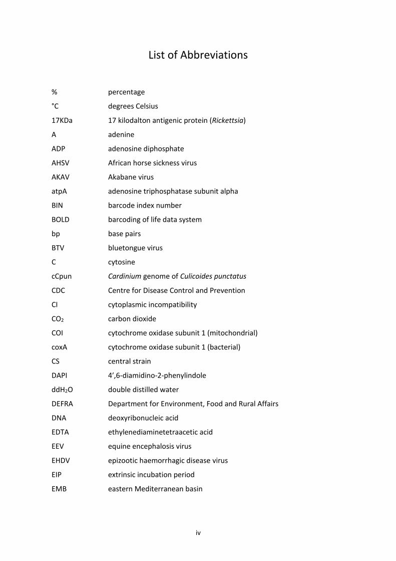
List of Abbreviations
% percentage
°C degrees Celsius
17KDa 17 kilodalton antigenic protein (Rickettsia)
A adenine
ADP adenosine diphosphate
AHSV African horse sickness virus
AKAV Akabane virus
atpA adenosine triphosphatase subunit alpha
BIN barcode index number
BOLD barcoding of life data system
bp base pairs
BTV bluetongue virus
C cytosine
cCpun Cardinium genome of Culicoides punctatus
CDC Centre for Disease Control and Prevention
CI cytoplasmic incompatibility
CO2 carbon dioxide
COI cytochrome oxidase subunit 1 (mitochondrial)
coxA cytochrome oxidase subunit 1 (bacterial)
CS central strain
DAPI 4′,6-diamidino-2-phenylindole
ddH2O double distilled water
DEFRA Department for Environment, Food and Rural Affairs
DNA deoxyribonucleic acid
EDTA ethylenediaminetetraacetic acid
EEV equine encephalosis virus
EHDV epizootic haemorrhagic disease virus
EIP extrinsic incubation period
EMB eastern Mediterranean basin
v
ExoSAP exonuclease/shrimp alkaline phosphatase
FISH fluorescence in-situ hybridisation
G guanine
GIS geographic information system
gltA citrate synthase subunit alpha
GyrB Gyrase B
Hg inch of mercury (pressure)
ITS internal transcriber sequences
Ka/Ks non-synonymous/synonymous mutation ratio
Km kilometers
kV kilovolts
LED light emitting diode
ln likelihood score
m/s metres per second MALDI-TOF-MS
matrix-assisted laser desorption/ionization time-of flight mass spectrometry
min(s) minute(s)
ml Microlitres
ML maximum likelihood
MLST multi locus sequence typing
mM micromolar
MST minimum spanning tree
mtDNA mitochondrial deoxyribonucleic acid
NaCl sodium chloride
NCBI National Center for Biotechnology Information
NUMT nuclear mitochondrial DNA
OIE World Organisation for Animal Health
OSL ovarian suspensory ligament
OsO4 osmium tetroxide
PBF post blood feeding
PCR polymerase chain reaction
vi
pmol Picomoles
Q phred score
qPCR real-time polymerase chain reaction
qRT-PCR real-time reverse transcription polymerase chain reaction
RiCNE Rickettsia genome of Culicoides newsteadi N5
RNA ribonucleic acid
rRNA ribosomal ribonucleic acid
SBV Schmallenberg virus
SLV single locus variant
SNP single nucleotide polymorphism
sp. species (individual)
spp. species (plural)
T Thymine
TEM transmission electron microscopy
Tris-HCl tris(hydroxymethyl)aminomethane
UK United Kingdom
UPGMA Unweighted Pair Group Method with Arithmetic Mean
USA United States of America
W Watts
w/v weight/volume ratio
WMB western Mediterranean basin
wsp Wolbachia surface protein
μl microlitres
μm micrometres
vii
List of Tables
Table 1.1: Colonisation attempts of Culicoides species. ………………………………………………………20
Table 2.1.1: COI and Gyrase B gene primer attributes. ………………………………………………….…….42
Table 2.1.2: COI and Gyrase B PCR cycling conditions. …………………………………………………………42
Table 2.2: Accession numbers of taxa used for Gyrase B nested primer design and reference
sequences in Figure 2.5. ………………………………………………………………………………………………….….43
Table 2.3.1: Sequencing of COI amplicons from samples initially identified as Culicoides
pulicaris from Lewis et al. (2014) and this study, suggesting cross-contamination with
Culicoides punctatus in Cardinium positives from Lewis’ study. …………………………………………..49
Table 2.3.2: PCR amplification status of Culicoides species of interest with two sets of COI
primers. ………………………………………………………………………………………………………………………….….49
Table 2.4: GyrB conventional and nested PCR assay results. ……………………………………………….53
Table 2.5.1: Fisher’s exact results for Cardinium infection comparisons of populations of the
same species but at different sites. …………………………………………………………………………………….58
Table 2.5.2: Fisher’s exact results for Cardinium infection comparisons of females and males
of the same species at the same site. …………………………………………………………………………………58
Table 3.1: Housekeeping and 17KDa gene primer attributes. ………………………………………………80
Table 3.2.1: 17KDa conventional PCR assay results for Rickettsia-positive Culicoides. ………….85
Table 3.2.2: 17KDa conventional PCR assay results for Rickettsia-negative Culicoides. …………86
Table 3.3: Genetic characteristics of housekeeping and 17KDa alleles. ……………………………….90
Table 3.4: Rickettsia strains recovered from Culicoides midges, with allelic profiles. …………….90
Table 3.5: Evidence of atpA intragenic recombination in Hilara interstincta and Culicoides
impunctatus isolates. …………………………………………………………………………………………………………96
viii
Table 3.6: The comparison of tree topologies of the unconstrained concatenated dataset
against topologies where host monophyly was enforced using the Shimodaira-Hasegawa (SH)
test. …………………………………………………………………………………………………………………………………103
Table 5.1: NCBI matches mistaken for true mtDNA barcodes and their homology to Rickettsia
COI. ………………………………………………………………………………………………………………………………….156
Table 5.2: Mitochondrial COI and bacterial gene primers used for re-barcoding and multilocus
phylogenetic analysis. ………………………………………………………………………………………………………162
Table 5.3: Primer pairs involved in the unintended amplification of 753 Rickettsia COI from
BOLD projects. ………………………………………………………………………………………………………………….167
Table 5.4.1: Homology of Rickettsia groups and Wolbachia to the most common forward
primer (CLepFolF) attributed to bacterial COI amplification from arthropod DNA extracts…169
Table 5.4.2: Homology of Rickettsia groups and Wolbachia to the most common reverse
primer (CLepFolR) attributed to bacterial COI amplification from arthropod DNA extracts…169
Table 5.5: The barcoding success rate of taxa which gave at least one bacteria COI inadvertent
amplification with an adjusted Rickettsia prevalence based on the total number of arthropods
in project. …………………………………………………………………………………………………………………………179
ix
List of Figures
Figure 1.1: A 16S rRNA phylogenetic tree with molecular clock showing the evolutionary
relationships among lineages within the Rickettsiaceae family of Rickettsiales. …………….…27
Figure 2.1: Quality control of DNA extracts labelled as Culicoides pulicaris from Lewis et al.’s
study using HCO1490/LCO2198 primers (Folmer et al. 1994). ………………………………………….….48
Figure 2.2: Detection limits of GyrB conventional and nested PCR. ……………………………………..51
Figure 2.3: The comparison of Cardinium prevalence rates in Culicoides species detected by
both GyrB conventional and nested PCR assays. ………………………………………………………………….52
Figure 2.4.1.: Palearctic Culicoides trapping and endosymbiont distribution map. …………….56
Figure 2.4.2: Afrotropical Culicoides trapping and endosymbiont distribution map. ……………57
Figure 2.5: A maximum likelihood phylogram based on a 1300 bp region of the GyrB gene…59
Figure 2.6: mtDNA haplotype networks of Cardinium infected and uninfected Culicoides
imicola. ………………………………………………………………………………………………………………………………61
Figure 3.1: Prevalence of Rickettsia positive Culicoides species. ………………………………………….88
Figure 3.2: A map depicting Rickettsia strain distribution across the UK, Sweden and Corsica,
France. ……………………………………………………………………………………………………………………………….93
Figure 3.3: Mirrored maximum likelihood trees of the atpA gene either side of a
recombination breaking point. ……………………………………………………………………………………………95
Figure 3.4.1: Rickettsia 16S maximum likelihood phylogeny. ………………………………………………97
Figure 3.4.2: Rickettsia 17KDa maximum likelihood phylogeny. ………………………………………….98
Figure 3.4.3: Rickettsia atpA maximum likelihood phylogeny. …………………………………………….99
Figure 3.4.4: Rickettsia coxA maximum likelihood phylogeny. …………………………………………..100
Figure 3.4.5: Rickettsia gltA maximum likelihood phylogeny. …………………………………………...101
x
Figure 3.5: Phylogenetic placement of the Rickettsia symbionts of Culicoides midges based on
a concatenated gene set of 16S, 17KDa, atpA, coxA and gltA genes. …………………….……………102
Figure 4.1: FISH and TEM images of stage 1 C. impunctatus and C. nubeculosus eggs. ………..128
Figure 4.2: FISH images of C. impunctatus egg chambers at different developmental stages of
oogenesis. ………………………………………………………………………………………………………………………..129
Figure 4.3: Rickettsia localisation in C. impunctatus adult connective tissues associated with
both the midgut and ovaries via FISH imaging. …………………………………………………………….……131
Figure 4.4: FISH and TEM analysis of C. impunctatus spermathecae. …………………………………133
Figure 4.5: Kaplan-Meier survival curve monitoring cumulative survival of C. impunctatus
larvae over time. …………………………………………………………………………………………………………….135
Figure 4.6: Transmitted light microscope images of different life stages of C. impunctatus. 137
Figure 4.7: FISH imaging analysis of an L3 C. impunctatus larva. ………………………………………..138
Figure 5.1: Workflow of the project demonstrating the acquisition and fates of contaminant
and non-contaminant COI barcoding sequences. ………………………………………………………………158
Figure 5.2: Cladogram of the maximum likelihood tree of 1,126 alphaproteobacteria COI
contaminants retrieved from BOLD projects incorporating 185,000 arthropod specimens. 166
Figure 5.3.1: Cladogram of a maximum likelihood tree of 753 COI Rickettsia contaminants
retrieved from BOLD projects incorporating 185,000 arthropod specimens. ……………………170
Figure 5.3.2: Identical cladogram tree to Figure 5.3.1. but taxa are separated into the Leech
and Limoniae subclades of the Torix Rickettsia group. ………………………………………………………171
Figure 5.4: Collection sites of the 753 COI Rickettsia contaminants retrieved from BOLD
projects. ……………………………………………………………………………………………………………………..……173
Figure 5.5: Phylogram of the maximum likelihood (ML) tree of 106 COI Rickettsia
contaminants based on the concatenation of 4 loci; 16S, 17KDa, gltA and COI. ………………...174
Figure 5.6: Phylogram of a maximum likelihood (ML) tree of 55 COI Rickettsia contaminants
giving a host barcode. ……………………………………………………………………………………………………….177
Table of Contents Chapter 1: General Introduction ........................................................................... 1
1.1 Vector-borne diseases ............................................................................................................ 2
1.1.1 Overview ................................................................................................................................ 2
1.1.2 Endosymbiont-based control of vector-borne diseases ........................................................ 3
1.2 Culicoides biting midges .......................................................................................................... 6
1.2.1 The significance of Culicoides as vectors and pests ............................................................... 6
1.2.2 Biology of Culicoides .............................................................................................................. 9
1.2.3 Culicoides in the UK .............................................................................................................. 11
1.2.4 Control of Culicoides and their viruses ................................................................................ 13
1.2.5 Taxonomic classification of Culicoides ................................................................................. 16
1.2.6 Laboratory rearing of Culicoides .......................................................................................... 18
1.3 Endosymbionts ............................................................................................................................ 21
1.3.1 Primary and secondary endosymbionts............................................................................... 21
1.3.2 Endosymbiont detection ...................................................................................................... 22
1.3.3 Endosymbionts of Culicoides ............................................................................................... 24
1.3.4 Cardinium ............................................................................................................................. 24
1.3.5 Rickettsia .............................................................................................................................. 26
1.3.6 Endosymbionts and the confounding of DNA barcoding studies ........................................ 28
1.4 Thesis aims .................................................................................................................................. 30
Chapter 2: Assessing the Phylogeographic Distribution of Candidatus Cardinium hertigii in
Culicoides with a Focus on Two UK Vector Species .............................................. 33
2.1 Abstract ....................................................................................................................................... 34
2.2 Introduction ................................................................................................................................ 35
2.3 Methods ...................................................................................................................................... 39
2.3.1 Culicoides collection and identification ............................................................................... 39
2.3.2 DNA extraction ..................................................................................................................... 39
2.3.3 COI and Cardinium screening ............................................................................................... 40
2.3.4 Nested primer design and sensitivity ................................................................................... 41
2.3.5 Mapping Cardinium spatial distribution .............................................................................. 44
2.3.6 Sanger sequencing ............................................................................................................... 44
2.3.7 Phylogenetic analysis ........................................................................................................... 44
2.3.8 mtDNA haplotype networks of Cardinium infected and uninfected C. imicola................... 45
2.3.9 Cardinium prevalence comparisons of site, species and sex ............................................... 45
2.4 Results ......................................................................................................................................... 46
2.4.1 Clarification of the Cardinium infection status of C. pulicaris and C. punctatus ................. 46
2.4.2 Nested PCR sensitivity .......................................................................................................... 47
2.4.3. An extended Cardinium survey of Culicoides species and the detection of low titre
infections ....................................................................................................................................... 50
2.4.4. Cardinium prevalence comparisons of site, species and sex .............................................. 55
2.4.5. Phylogenetic analysis .......................................................................................................... 55
2.4.6 C. imicola haplotype network analysis ................................................................................. 60
2.5 Discussion .................................................................................................................................... 63
Chapter 3: Limoniae Group Rickettsia are Widespread in Culicoides and Reach High
Frequency .......................................................................................................... 71
3.1 Abstract ....................................................................................................................................... 72
3.2 Introduction ................................................................................................................................ 73
3.3 Methods ...................................................................................................................................... 77
3.3.1 Culicoides collection identification and DNA extractions .................................................... 77
3.3.2. PCR screening ...................................................................................................................... 77
3.3.3 Multilocus typing gene choice ............................................................................................. 78
3.3.4 Multigenic strain typing ....................................................................................................... 81
3.3.5 Phylogenetic analysis ........................................................................................................... 81
3.3.6 Nucleotide sequence accession numbers ............................................................................ 83
3.4 Results ......................................................................................................................................... 84
3.4.1 Prevalence of Rickettsia in biting midges ............................................................................ 84
3.4.2 The detection of Rickettsia coinfections .............................................................................. 89
3.4.3 Individual allele information ................................................................................................ 89
3.4.4 Multilocus strain typing and geographic comparison .......................................................... 91
3.4.5 Phylogenetic analysis ........................................................................................................... 92
3.5 Discussion .................................................................................................................................. 105
Chapter 4: The Tropism and Transtadial Transmission of a Rickettsia Endosymbiont in
Culicoides impunctatus ..................................................................................... 114
4.1 Abstract ..................................................................................................................................... 115
4.2 Introduction .............................................................................................................................. 116
4.3 Methods .................................................................................................................................... 121
4.3.1 The collection of Culicoides impunctatus ........................................................................... 121
4.3.2 Larval rearing ..................................................................................................................... 122
4.3.3. Dissections of adults ......................................................................................................... 123
4.3.4 Tissue preparation and fluorescence in-situ hybridisation (FISH) ..................................... 124
4.3.5. Transmission electron microscopy ................................................................................... 125
4.4 Results ....................................................................................................................................... 127
4.4.1 Rickettsia infection during oogenesis ................................................................................ 127
4.4.2 Germline targeting of Rickettsia via the ovarian suspensory ligament ............................. 127
4.4.3 FISH and transmission electron microscopy of spermathecae .......................................... 132
4.4.4 Subcellular location and associations ................................................................................ 134
4.4.5 The rearing of Culicoides impunctatus ............................................................................... 134
4.4.6. Larval tissue localisation ................................................................................................... 136
4.5 Discussion .................................................................................................................................. 139
Chapter 5: The Investigation of Rickettsia Host and Bacterial Diversity through DNA
Barcoding ......................................................................................................... 148
5.1. Abstract .................................................................................................................................... 149
5.2 Introduction .............................................................................................................................. 150
5.3 Methods .................................................................................................................................... 155
5.3.1 The erroneous amplification of Rickettsia through barcoding .......................................... 155
5.3.2 BOLD datasets acquisition ................................................................................................. 155
5.3.3 Assessment of non-target microbe amplicons .................................................................. 157
5.3.4 Phylogenetic analysis ......................................................................................................... 159
5.3.5 Host barcoding ................................................................................................................... 161
5.3.6 Assessment of barcoding success ...................................................................................... 163
5.3.7 Assessment of primer bias ................................................................................................. 164
5.4 Results ....................................................................................................................................... 165
5.4.1 Rickettsia is the most common bacterial contaminant following barcoding .................... 165
5.4.2 COI primer attributes ......................................................................................................... 165
5.4.3 The diversity of non-target Rickettsia ................................................................................ 168
5.4.4. Rickettsia multilocus phylogenetic analysis ...................................................................... 172
5.4.5 The hidden host diversity of Torix Rickettsia ..................................................................... 175
5.4.6 Rickettsia co-infections ...................................................................................................... 176
5.4.7 Barcoding success of taxa .................................................................................................. 178
5.5 Discussion .................................................................................................................................. 181
Chapter 6: General Discussion and Future Directions ........................................ 191
6.1 Overview ................................................................................................................................... 192
6.2 The re-examination of the Cardinium infection status of Culicoides ........................................ 193
6.3 The role of Rickettsia in Culicoides biology ............................................................................... 194
6.4 Rickettsia transmission strategies ............................................................................................. 197
6.5 Rickettsia and Cardinium confounding of DNA barcoding........................................................ 200
6.6 Future directions ....................................................................................................................... 202

Bibliography ..................................................................................................... 206
Appendix 1. ..................................................................................................................................... 257
Appendix 2. ..................................................................................................................................... 261
Appendix 3. ..................................................................................................................................... 264

1
Chapter 1: General Introduction
2
1.1 Vector-borne diseases
1.1.1 Overview
Vector-borne diseases are infections transmitted by blood-feeding arthropods. The most
well-known examples of these infections are pathogens spread by mosquitoes, although tick
and midge-borne diseases are also of importance (Gubler, 2009). For example, Anopheles and
Aedes mosquito species transmit malaria and flaviviruses (dengue, Zika and chikungunya),
causing hundreds of thousands of human deaths every year (World Health Organisation,
2019). Additionally, mosquitoes can transmit filarial nematodes including Brugia malayi,
which can cause lymphatic filariasis (elephantiasis); a disease leading to disability and social
stigma in developing nations (Zeldenryk et al., 2011). Vector borne diseases are also of
importance in the veterinary world including pathogens such as tick-borne Babesia sp.
(Babesiosis in several mammals and birds) and mosquito transmitted Dirofilaria immitis
(Heart worm in dogs and cats) (Morchón et al., 2012; Dantas-Torres, Alves and Uilenberg,
2016).
Conventional vector control approaches have heavily relied on the removal of breeding sites
and insecticides (Flores and O’Neill, 2018). However, these are proving insufficient to cope
with host population density increases as a result of urbanisation particularly in tropical
regions (Pang, Mak and Gubler, 2017). Additionally, meteorological factors can influence the
reproduction and survival of vectors meaning that climatic variables can influence invasive
ranges and population sizes (Takken and Knols, 2007). Indeed, approximately a third of
emerging diseases are deemed to be vector-borne (Jones et al., 2008), suggesting new health
interventions are of pressing need.
3
One vector control approach aims to reduce vector population numbers. Population
suppression approaches include sterile insect technique (SIT) where male vectors are
sterilised via irradiation or chemical treatment so females fail to produce offspring after
mating (Lees et al., 2015); and the release of transgenic insects carrying a dominant lethal
(RIDL) which leads to larval or pupal mortality when the lethal gene is switched on in the wild
(Phuc et al., 2007; Wise De Valdez et al., 2011). The shortcomings of these approaches include
the limited epidemiological evidence available to suggest effectiveness, the reduced fitness
of lab-reared mosquitoes compared to their wild counterparts (particularly irradiated insects)
(Bowman, Donegan and McCall, 2016), and the negative public perception of releasing
genetically modified insects into nature (Flores and O’Neill, 2018). An approach which aims
to overcome some of these obstacles is the use of endosymbionts (bacteria residing within
the body or cells of insect hosts).
1.1.2 Endosymbiont-based control of vector-borne diseases
Endosymbiont protection of Dipteran hosts was first observed in Drosophila fruit flies infected
naturally with Wolbachia, which were protected against fungal and viral pathogens
(Panteleev et al., 2007; Hedges et al., 2008; Teixeira, Ferreira and Ashburner, 2008). Further
experimental work demonstrated the ability to transinfect mosquito species with the
Wolbachia strain wMel, leading to a blocking effect of dengue virus, chikungunya virus, yellow
fever virus, West Nile virus and Zika virus (Bian et al., 2010b; Blagrove et al., 2012; Hussain et
al., 2012; van den Hurk et al., 2012; Chouin-Carneiro et al., 2019). The further Wolbachia
strains wAlbA, wAu and wMelPop also appear to perturb pathogen transmission suggesting
multiple routes of disease control investigation (Moreira et al., 2009; Walker et al., 2011; Ant
et al., 2018). The potential to deploy these effects in wild populations of mosquitoes is
4
enabled by Wolbachia-induced cytoplasmic incompatibility (CI) (Ant et al., 2019). This is the
induction of embryo death in mating between infected males and uninfected females, with
all other crosses remaining viable (Werren and O’Neill, 1997). The significance of this effect is
the rapid spread of the symbiont through a population before gaining stability at a near 100%
prevalence rate. This is contingent on an invasion threshold being reached dependent on the
“strength” of CI, vertical transmission, and fitness cost to infected females (Fine, 1978;
Stouthamer, Breeuwer and Hurst, 1999). Large initial releases aid in achieving the invasion
threshold, and persistence may then occur through the CI phenotype without onward
releases. Viral suppression is dependent on both host and Wolbachia genetic backgrounds.
For example, when the natural strain of Aedes albopictus (wAlbB) was transferred to the naïve
host, Aedes aegypti, a stronger inhibitory effect of dengue virus was observed than in the
native host (Bian et al., 2010). Furthermore, high endosymbiont densities, and subsequently
a greater virus inhibition effect has been observed when Drosophila strains of Wolbachia
(wMel and wMelPop) are transinfected into mosquitoes (Blagrove et al., 2012).
Several mechanisms have been proposed for Wolbachia-mediated virus blocking. These
include immune-effector modulation and competition for resources (Sinkins, 2013; Lindsey et
al., 2018). With respect to the latter, the bacterium’s relationship with host lipid metabolism
has attracted attention. Flaviviruses, with a limited genetic repertoire, rely on host-derived
components for maintenance, such as cholesterol (Osuna-Ramos, Reyes-Ruiz and Del Ángel,
2018). During flavivirus infection, host cholesterol levels can modulate cell entry, replication,
assemblage and host interferon responses (Rothwell et al., 2009; Upla, Hyypiä and
Marjomäki, 2009; Heaton et al., 2010). Furthermore, dengue virus has been suggested to
5
indirectly increase intracellular cholesterol through reducing the expression of
intramembrane proteins related to cholesterol metabolism (Tree et al., 2019). Subsequently,
as Wolbachia also relies on such host-derived components, cholesterol has been viewed as
the target by which the endosymbiont suppresses viral titres through competition (Caragata
et al., 2013; Sinkins, 2013; Molloy et al., 2016; Frentiu, 2017). Furthermore, the localisation
of Wolbachia within cells is mediated by the actin cytoskeleton which is also used to transport
virus within endosomes (Sheehan et al., 2016). Subsequently, a pre-existing Wolbachia
infection within a cell could lead to disruption of such virion trafficking (Geoghegan et al.,
2017). Although, direct interactions are likely to explain partially Wolbachia-mediated virus
blocking, the independent distribution of wMelPop and Dengue virus in Aedes aegypti
(Moreira et al., 2009) suggests other means should also be considered. Priming of the immune
system by the endosymbiont has been proposed as such a mechanism due to the
upregulation of several immune genes, including components of the Toll-pathway, which are
major regulators of flavivirus replication (Xi, Ramirez and Dimopoulos, 2008; Pan et al., 2012).
Encouragingly, the results of field-based CI-inducing symbiont interventions indicate a viable
method to control arboviruses (Hoffmann et al., 2014; Schmidt et al., 2017). For example,
since the 2011 release of wMel transinfected Aedes aegypti in Northern Australia, there has
been no local transmission of dengue fever where the bacterium is established (Ritchie, 2018).
Importantly, as symbionts are naturally occurring and ubiquitous, public acceptance of these
Wolbachia-based initiatives may be seen as more acceptable than other genetic modification
interventions. Furthermore, this protective phenotype appears to be associated with RNA
viruses leading to the potential for symbiont-based biocontrol in major midge-borne
6
pathogens such as bluetongue virus (BTV), African horse sickness (AHSV) and Schmallenberg
virus (SBV). Finally, Wolbachia can be used as part of an incompatible insect technique (IIT),
which is a modified version of SIT where the endosymbiont is used to effectively sterilise
males without the fitness costs encountered by irradiation (Zheng et al., 2019). When males
are released in high enough numbers, vector population collapse occurs due to incompatible
matings. Despite these promising field advancements, symbionts are often a neglected factor
in non-mosquito-borne arbovirus disease dynamics.
1.2 Culicoides biting midges
1.2.1 The significance of Culicoides as vectors and pests
Culicoides (Diptera: Ceratopogonidae) biting midges are blood-feeding flies which range from
1-3 mm in length (Mellor, Boorman and Baylis, 2000). As well as being a biting nuisance to
humans, a number of Culicodies species are able to transmit filarial nematodes and protozoan
parasite to birds, humans, and other animals (Yates, Lowrie and Eberhard, 1982; Linley,
1985a; Veiga et al., 2018). Although other haematophagous flies from the family
Ceratopogonidae are known as vertebrate parasites, the dominant research interest for the
genus Culicoides are the numerous viruses (Reoviridae and Peribunyaviridae families) they
transmit (Mellor, Boorman and Baylis, 2000; Sick et al., 2019). Pathogens affecting ruminants
include BTV, SBV, Akabane virus (AKAV) and epizootic haemorrhagic disease virus (EHDV).
Other viruses are known to affect equids including AHSV and equine encephalosis virus (EEV),
which are of great significance for animal welfare, as well as the horse racing industry
(Bachanek-Bankowska et al., 2009).
7
Apart from the economic and animal health implications of these veterinary viruses, the
human pathogen oropouche virus, observed in the Americas (during the 1950s), makes these
vectors additionally of human health importance (Anderson et al., 1961). Indeed, a recent
review (Sick et al., 2019) suggests Culicoides are possibly a neglected vector of human viruses,
with the lack of information deriving from a bias towards surveillance of mosquitoes.
Considering the Peribunyaviridae contain human pathogens, and that this virus family are
largely associated with Culicoides, it would be unsurprising to observe future midge-borne
outbreaks of human viruses.
SBV is a negative-sense single-stranded RNA member of the Bunyaviridae family and only
recently emerged in 2011 in the German/Dutch border region (Elbers et al., 2013), causing
disease primarily in sheep along with less frequent cases in cattle. Whilst SBV infections in
adult animals are usually subclinical, vertical transmission of SBV from mother to progeny can
result in severe and fatal congenital malformations along with still births and abortion
(European Food Safety Authority, 2016). Since its first detection, multiple outbreaks of SBV
have been reported in a range of European countries including Belgium, United Kingdom,
Luxembourg, France, Spain and Italy (Beer, Conraths and Van Der Poel, 2012), and has also
been suspected in Africa (Leask, Botha and Bath, 2013). The Palearctic biting midges
predominantly considered as vectors are the C. obsoletus complex and C. imicola. This was
confirmed by infection experiments undertaken in field-caught midges of both groups (Pagès
et al., 2018). Additionally, C. punctatus, C. imicola and the C. pulicaris/C. newsteadi complexes
were also positive for the viral genome by qRT-PCR suggesting they may also contribute to
8
the epidemiology of SBV (Rasmussen et al., 2012; Larska et al., 2013; Balenghien et al., 2014;
Ségard et al., 2018)
BTV is an OIE (World Organisation for Animal Health) list A pathogen, meaning it has the
potential for very rapid spread and can cause serious socio-economic consequence due to its
impact on the international trade of animals. Clinical signs of BTV in ruminants include
depression, pyrexia, serous nasal discharge, hypersalivation, facial oedema, as well as
hyperaemia and ulceration of the oral cavity (Maclachlan et al., 2009). Death can occur as a
result of disease or euthanasia on animal welfare grounds. Bluetongue was first described in
sheep from South Africa in the late 18th century (Callis and Kramer, 1997). However,
throughout the 20th century, several incursions of BTV serotype 9 from Africa have occurred
in the Mediterranean basin leading to outbreaks in the Iberian Peninsula (1950s and 1960s)
(Mellor and Boorman, 1995), as well as Greece, Turkey and Bulgaria (1990s) (Mellor et al.,
1985; Boorman, 1986b). These invasions were attributed to the invasion of the Afrotropical
vector C. imicola into southern Europe. However, the 2006 outbreak of BTV serotype 8 across
Europe, including the UK, was spread by the common Palearctic species C. obsoletus. This
outbreak of BTV caused serious economic and animal health damage to the European
livestock industries (Wilson and Mellor, 2009) as it spread through a serologically naïve and
highly susceptible ruminant population. The cost of the UK epidemic was estimated to cost
up to £485M with losses in livestock productivity, restrictions on UK agricultural exports and
employment impacts cited as the main contributors to this figure (Pirbright Institute, 2009).
9
Like mosquitoes, only female Culicoides will search for vertebrate hosts to acquire a blood
meal in order to allow for the full development of eggs. If these females feed on viraemic
blood, then they have the capacity to contribute to the virus transmission cycle (Boorman,
1974). After blood-feeding, there are many barriers which the virus must overcome in order
for successful vertebrate to vertebrate transmission to occur (Fu et al., 1999). First, the virus
must be able to invade, and then bypass, the gut epithelium to make its way into the
haemolymph and secondary organs, such as the fat body. Second, the pathogen must evade
host immune responses before reaching the salivary glands where it proliferates. Finally, the
host must live long enough for sustained transmission to occur. With respect to the latter, an
important concept to consider is the extrinsic incubation period (EIP)(Mills et al., 2017a),
which is the time it takes for a virus to initially enter the female Culicoides through feeding,
until the time that the virus is able to be transmitted via the salivary glands (Pinheiro,
Travassos da Rosa and Travassos da Rosa, 1981; Kedmi et al., 2010). This varies widely with
virus and invertebrate host, but in the case of BTV serotype 11 and C. sonorensis, the EIP is
estimated at between 18-22 days at 21°C (Mullens et al., 1995). The mechanical transmission
of viruses has also been noted in Culicoides transmitting fowl pox (Fukuda et al., 1979).
However, it is unknown if Culicoides can mechanically transmit BTV or SBV. More recently,
there has been a suggestion that certain viruses can be vertically transmitted through
Culicoides’ eggs with SBV being detected in the eggs of C. punctatus (Larska et al., 2013).
1.2.2 Biology of Culicoides
Over 1,400 Culicoides species are currently recognised within the genus and they occur
globally with the exceptions of New Zealand, Patagonia, Hawaii and Antarctica (Mellor,
Boorman and Baylis, 2000). Culicoides are holometabolous and have four larval stages as well
10
as a pupal and imago stage. Egg hatching times, as well as larval and pupal development
periods are dependent on species as well as ambient temperature (Meiswinkel, 1989) with a
full lifecycle taking between less than two weeks to several months (Mellor, Boorman and
Baylis, 2000). Cigar-shaped eggs are often laid in bunches and are initially translucent before
darkening with maturation. The presence of moisture is one of the most important
determinants for suitable larval habitats (Blackwell, Young and Mordue, 1994; Lardeux and
Ottenwaelder, 1997; Meiswinkel, 1997), although some species such as C. imicola will drown
in flooded breeding-sites (Nevill, 1969). Thus, immatures are found in a diverse range of
habitats including animal dung, tree-holes, soil, swamps, beaches, rivers, marshes and bogs
(Linley and Davies, 1971; Kline and Axtell, 1976; Meiswinkel, 1989; Blackwell, Young and
Mordue, 1994). Larvae are considered omnivores and can subsist on detritus and algae
(Aussel and Linley, 1994), but some species are predatory, having been observed to feed on
nematodes (Kettle, Wild and Elson, 1975; Boorman, 1985). In temperate countries with a
prolonged cold winter, fourth stage instars will undergo diapause (Kettle, 1977). The
continuation of development into pupa has been linked to temperature and increased day
length although exact cues remain unknown (Searle et al., 2014; Lühken et al., 2015; White
et al., 2017). Pre-pupae will generally rise to the surface of breeding habitats where
development continues in loose debris. The pupal period is much shorter than larval stages,
usually lasting for only a few days (Barceló and Miranda 2018).
Most adult Culicoides species are crepuscular in activity (Kettle, 1977; Mellor, Boorman and
Baylis, 2000) although others are present diurnally (Mercer et al., 2009). The life-span of
adults is short with most individuals surviving between 2-3 weeks with exceptions living for
11
periods of up to 90 days (Mellor, Boorman and Baylis, 2000). Flight activity is limited to
behaviours such as feeding, mating, host seeking and ovipositing (Campbell and Kettle, 1976;
Bishop et al., 1995) and is determined by light intensity, temperature and humidity (Kettle,
1957; Blackwell, 1997). Adults generally find it difficult to fly in wind speeds greater than 3
m/s and will retreat to shelter in rainy conditions (Carpenter et al., 2008). Although the flight
range of adults is generally limited to a few hundred metres from breeding sites, long range
dispersal has been described spanning thousands of kilometres. Simulation models suggests
that wind streams have facilitated the dispersal of populations from Indonesia to Australia
(Eagles et al., 2014) as well as Corsica to Spain (Jacquet et al., 2016). Mating is thought to
occur through swarms consisting of a much higher proportion of males than females (1
female: 200 males) (Downes, 1950, 1955; Zimmerman, Barker and Turner, 1982). The
seasonality of Culicoides is determined by climatic and meteorological variables. In temperate
regions, numbers are much reduced during winter but during spring, increased temperatures
and day-length leads to a population increase which tends to peak a few weeks after the
hottest time of year (Kline and Axtell, 1976; Mellor, Boorman and Baylis, 2000). Additionally,
the number of generations per year will affect population peaks; univoltine species
(undergoing 1 gonotrophic cycle per year) will have greater numbers at the start of the season
compared to multivoltine species, where populations peak later (Mellor, Boorman and Baylis,
2000).
1.2.3 Culicoides in the UK
At least 46 species of Culicoides have been described in the UK, of which 38 have been
associated with virus transmission (Wittmann and Baylis, 2000). Of note are the obsoletus
and pulicaris species complexes which are of concern for future UK outbreaks of BTV and SBV.
12
Apart from C. chiopterus, all other members of the obsoletus complex are known to be
putative vectors of BTV via qRT-PCR screening of field-caught populations (Meiswinkel et al.,
2007; Foxi et al., 2016). Furthermore, C. scoticus, C. chiopterus and C. obsoletus of the
obsoletus complex have been implicated as putative vectors of SBV (Elbers et al., 2013). Like
other temperate regions, adult Culicoides in the UK tend to emerge from spring (April
onwards) with numbers reducing towards autumn (November) (Mellor, Boorman and Baylis,
2000). However, phenology varies by species with C. impunctatus observed between May and
September (Holmes and Boorman, 1987). Aside from seasonality, the number of generations
throughout a season are also important in determining timing and spread of midge-borne
diseases in the UK, with known species having between 1-3 generations per year (Baylis et al.,
1997; Sanders et al., 2011). Of interest is C. impunctatus, which appears to be univoltine in
the South of England but bivoltine in Scotland (Hill, 1947; Blackwell et al., 1992).
Culicoides impunctatus, commonly known as the “Scottish Highland midge”, is a UK species
of note due to the large numbers observed in Western Scotland. The comparatively small
wing-length (approximately 1.5mm) and unmarked thorax makes C. impunctatus easy to
distinguish from other species in the same subgenus (Culicoides), such as C. fagineus
(Campbell and Pelham-Clinton, 1960). C. impunctatus is prevalent throughout mostly
Northern Europe although they can be found as far South as the Iberian Peninsula (Hill, 1947;
Campbell and Pelham-Clinton, 1960; John Boorman, 1986b; Rawlings, 1996). The autogenic
lifestyle of C. impunctatus (it does not require a blood meal for its initial gonotrophic cycle),
and its prolificity in peat bog breeding sites has led to its abundance and is a well-known biting
nuisance (Boorman and Goodard, 1970). Indeed, Boorman (1986a) attributed between 70-
13
95% of Culicoides attacks on humans to C. impunctatus. This biting nuisance contributes
negatively to two of Scotland’s major industries, Forestry and Tourism. The effects on the
former, are as a result of “midge attacks” disrupting arborists’ vision while operating
machinery and refuelling chainsaws, with up to 20% of summer working hours being lost per
year (Hendry and Godwin, 1988). The economic impact on tourism (worth over £2 billion each
year to the Scottish economy), however, is far more difficult to quantify due to the transient
nature of visitors (Hendry, 1996). Some impact, however, is clear: a questionnaire study,
exploring visitor’s attitudes to midges during the summer months suggested roughly half
would not visit Scotland at the same time of year again. This economic cost prompted several
studies focusing on the breeding-sites and seasonality of C. impunctatus in the hope of finding
a suitable control intervention (Blackwell et al., 1992; Blackwell, 1997). However, the broad
breeding site range was shown to be a major limiting factor for restricting numbers (Blackwell,
Young and Mordue, 1994). Subsequently, the problem of C. impunctatus, as a pest remains
unsolved.
1.2.4 Control of Culicoides and their viruses
During outbreaks of BTV, movement restrictions of livestock and their products are
implemented to stifle the spread of viruses into new regions (Carpenter et al., 2013). In the
case of UK outbreaks, this involves the active testing of animals within a 20 km zone of
confirmed cases. Additionally serological surveillance of sentinel ruminants and
entomological surveillance is implemented within a 100 km protection zone where
movement is only allowed within this zone (Defra, 2014). Movements in and out of these
zones is permitted during a “vector-free period”, which can be declared if there is evidence
of a lack of circulating virus along with a lack of vector activity through surveillance and
14
temperature thresholds (Defra, 2014). There have also been suggestions for farming practices
to accommodate for potential teratogenic effects caused by midge-borne viruses such as SBV,
which cause no or minor disease in adults (Wernike, Hoffmann and Beer, 2013). For example,
the breeding seasons of sheep can be adjusted to ensure the exposure of young livestock to
infected vectors (during peak midge activity) so the dam can acquire immunity before
conceiving. However, this is impractical because of year to year differences in peak vector
activity, as well as a reluctance to change livestock-management from farmers (Dwyer et al.,
2007; Carpenter, Mellor and Torr, 2008; Wernike et al., 2014).
Control strategies for BTV and SBV outside of vector control thus rely on immunity acquired
through vaccination (Szmaragd et al., 2010; Wernike, Hoffmann and Beer, 2013; Defra, 2014).
The successful vaccination of millions of cattle during the 2006-2009 BTV European outbreak
demonstrated that immunisation can play a major role in the control of epidemics. In the UK,
the >80% uptake rate of vaccination by farmers was deemed a contributing factor to the lack
of BTV infections observed in the UK during 2008 (Szmaragd et al., 2010). However, the
presence of 27 circulating serotypes complicate application of vaccine strategies.
Furthermore, segmented viruses, such as BTV and AHSV, show reassortment of genome
segments in hosts co-infected with more than one serotype (Verwoerd et al., 1972; Roy,
Mertens and Casal, 1994; Roy, 2017). This can produce novel strains of the virus with a
combination of characteristics of the original parental serotypes (Maan et al., 2011; Zientara
and Sánchez-Vizcaíno, 2013). Subsequently, such events create difficulties when producing
vaccines, and often outbreaks can only be controlled by vaccines after a time-delay of
serotyping and developing an effective vaccine (Calvo-Pinilla et al., 2014; Zulu and Venter,
15
2014; Nomikou et al., 2015; Harrup, Miranda and Carpenter, 2016). Furthermore, the use of
live pentavalent BTV vaccines, which comprise five attenuated strains, has led to an increase
in genetic diversity of serotypes circulating in the field (Batten et al., 2008). This perpetuates
existing concerns over the reliance on live vaccines as it has been suggested that this could
play a role in accelerating the evolution of BTV via horizontal gene transfer (Shaw et al., 2012).
Other limitations of reactive control measures include the detection failure of new outbreaks
by both farmers and vets (Elbers et al., 2010). Additionally, the unpredictable nature of midge-
borne outbreaks can cause problems in anticipating the emergence of outbreaks. An example
of this is the disappearance and re-emergence of SBV in the UK, within a few years, which has
created problems for both prophylactic and reactive control measures (Stokes, Baylis and
Duncan, 2016; Stokes et al., 2018). Further, the historic incursions of the vector species C.
imicola into continental Europe, as a result of climate change and wind-transport events
(Mellor and Boorman, 1995; Purse et al., 2005; Venail et al., 2012; Jacquet et al., 2016), has
also highlighted the challenge of mapping Culicoides movement.
Considering all the above, proactive measures such as vector control are seen to be more
attractive options in controlling the spread of these arboviruses (Harrup, Miranda and
Carpenter, 2016). Current interventions targeting adults include insecticide treatments of
livestock, moving livestock into sheltered housing during peak activities of biting, and the use
of repellents (Carpenter, Mellor and Torr, 2008; Harrup, Miranda and Carpenter, 2016; Snyder
et al., 2016). For immature stages, the elimination of breeding sites through insecticides and
pathogen application has been trialled with some success (Ansari, Carpenter and Butt, 2010;
Harrup, Miranda and Carpenter, 2016). However, due to the broad range of habitats, the
16
disruption of breeding sites is impractical. Finally, insecticide use can lead to unwanted
ecological effects due to their non-specific effects (Carpenter, Mellor and Torr, 2008; Harrup,
Miranda and Carpenter, 2016).
1.2.5 Taxonomic classification of Culicoides
An issue with advancing Culicoides control initiatives is the problem of predicting and
monitoring vector species due to an underdeveloped taxonomic classification system.
Taxonomic classification of adults has primarily relied on wing-spot patterns for adults and
head capsule morphology for larvae with multiple keys having been devised (Meiswinkel,
1994; Rawlings, 1996; Mathieu et al., 2012; Bellis, Halling and Anderson, 2015). Certain
species are morphometrically similar such as C. pulicaris, C. lupicaris, C. bysta and C.
newsteadi which make up what is known as the pulicaris complex (Yildirim et al., 2019).
Similarly, the obsoletus complex encompasses several cryptic species including C. obsoletus,
C. dewulfi, C. chiropterus, and C. scoticus (Downes and Kettle, 1952; Nielsen and Kristensen,
2011). Although identification through morphological assessment is possible, this requires
laborious geometric analysis of various structures including sex organs, spermathecae and
maxillary measurements (Downes and Kettle, 1952; Campbell and Pelham-Clinton, 1960;
Mathieu et al., 2012). This difficulty in classification is problematic because species complexes
contain some members which have been established as vectors and other which have not.
For example, members of both obsoletus and pulicaris complexes have different levels of
susceptibility to the same serotype of BTV (Carpenter et al., 2006).
17
To overcome these issues in identification, molecular techniques have been developed. This
can involve multiplex PCR (Nolan et al., 2007) of DNA marker regions or matrix-assisted laser
desorption/ionization time-of flight mass spectrometry (MALDI-TOF-MS) which identifies
species based on protein signatures (Uhlmann et al., 2014). Furthermore, Internal transcriber
sequences (ITS) of ribosomal DNA are occasionally used, but these often lack resolution to
differentiate sister species (Cêtre-Sossah et al., 2004; Gomulski et al., 2006). Latterly,
mitochondrial markers such as DNA barcodes, using the sequence of the Cytochrome oxidase
subunit 1 gene (COI), have been widely utilised (Pagès et al., 2009; Lassen et al., 2012; Ander,
Troell and Chirico, 2013; Nielsen and Kristensen, 2015). Following barcoding, it is possible to
allocate the mtDNA reference sequence to other barcodes based on percentage similarity.
Traditionally, sequences with >98% homology are deemed the same species, although this
presumes that the minimal interspecific mtDNA divergence between sister species is greater
than maximum intraspecific divergence i.e. a “barcoding gap” exists (Hebert et al., 2003;
Wiemers and Fiedler, 2007). Following barcode allocation, it may be possible to return to the
voucher specimen, if tissues of taxonomic interest are retained (e.g. head, terminal abdomen
and wings), to identify characters which can be used to identify this species in future studies
(Nielsen and Kristensen, 2015). Alternatively, through deposition of the sequences into an
online database, it is possible for studies to remove the need for morphological assessment
altogether (Ratnasingham and Hebert, 2007). Barcoding is not only useful in identifying
cryptic vector species, but can allow for the discovery of putative new Culicoides species to
be investigated in the future (Sarvašová et al., 2017).

18
1.2.6 Laboratory rearing of Culicoides
In order to investigate the role of Culicoides in virus transmission cycles, and to investigate
life stages outside of the short adult season, this requires the rearing and maintenance of
laboratory colonies (Hill, 1947; Megahed, 1956; Kettle, Wild and Elson, 1975; Boorman, 1985;
Linley, 1985b; Mullens and Velten, 1994). However, Culicoides research has lagged behind
that of many other invertebrate vectors due to logistical problems, such as the midges’
reluctance to copulate in confined spaces (Linley, 1968; Nayduch et al., 2014). A further
limitation to utilising cultivated Culicoides for infection experiments, is the trade-off between
temperature dependent life-cycle times and mortality. When Culicoides adults and larvae are
maintained at higher temperatures their survival rate is lower (Hill, 1947; Wellby et al., 1996;
Wittmann, Mellor and Baylis, 2002). However, larval and pupal periods are also shorter with
these elevated temperatures allowing for quicker access to study materials.
Initial failed attempts at colonisation of C. imicola in South Africa was followed by the first
successful establishment in the USA vector species C. sonorensis, which could mate without
displaying swarming behaviour and had a comparatively quick life cycle (Boorman, 1974).
Aside from C. sonorensis, success with C. nubeculosus rearing has led to extant colonies of
both species being maintained in the USA (ARS-USDA; Kansas) and UK (Pirbright Institute;
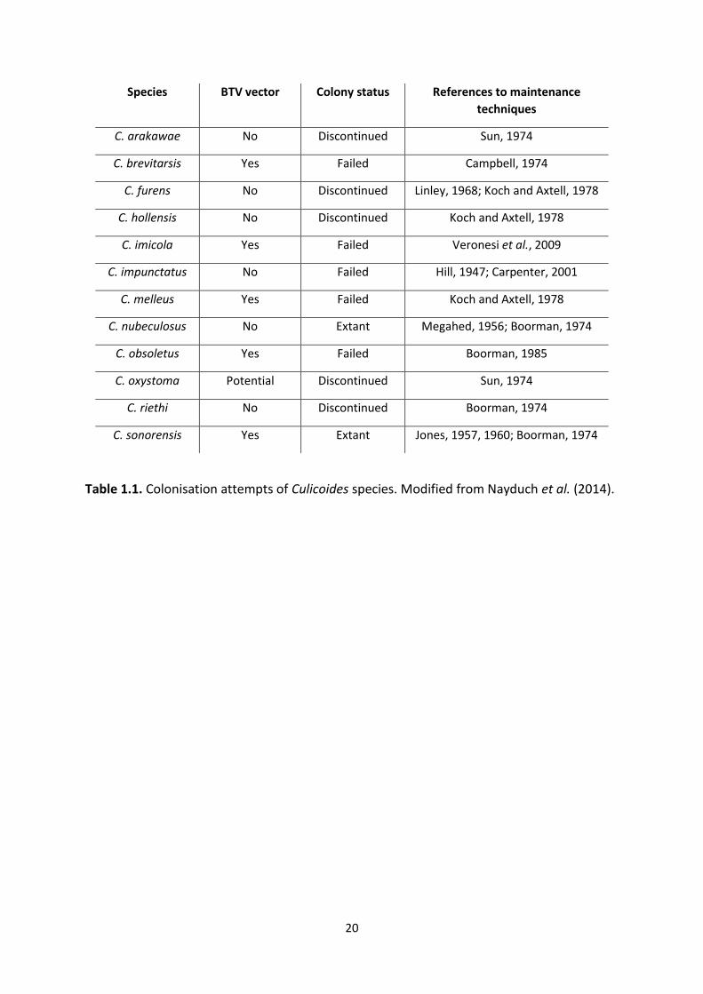
Surrey) (Nayduch et al., 2014; Table 1.1.). As only C. sonorensis is a vector of BTV, the major
data pertaining to experimental infections have been acquired from this species (Chandler et
al., 1985; Nunamaker et al., 1997; Fu et al., 1999). However, an interest in colonisation is not
limited to only vector species. For example, the laboratory rearing of pest species, such as C.
impunctatus (Hill, 1947; Carpenter, 2001), has been suggested to allow for integral studies of
aggregation cues to increase trapping efficiency (Bhasin, Mordue and Mordue, 2001).
19
Furthermore, laboratory investigations into ovipositing and fecundity of this pest species
could lead to control interventions at breeding-sites (Carpenter, Mordue and Mordue, 2001).
Maintenance of adults has been achieved using cardboard pill boxes or large mesh cages
where midges are fed on sucrose-soaked cotton wool, or via artificial blood-feeding devices
if initiation of a gonotrophic cycle is needed (Boorman, 1974; Barceló and Miranda, 2018).
After mating and blood-feeding, ovipositing occurs on damp filter paper, although in the case
of C. impunctatus, Sphagnum spp. moss can be used to increase ovipositing success
(Carpenter, Mordue and Mordue, 2001). However, after hatching, maintenance of larvae has
proven more difficult due to problems in identifying suitable substrates for development. Due
to the diverse natural larval habitats of Culicoides (see section 1.2.2), it is unlikely that a single
larval medium will be appropriate for universal use (Carpenter, 2001). Indeed, it should be
noted that both C. sonorensis and C. nubeculosus develop in heavily manured soils, which may
account for their rearing success using an organically polluted substrate (Hribar, 1990). In
these cases, larval pans containing manure/soil and water, were optimised to include liver
powder, yeast and wheat germ to promote bacterial growth (Megahed, 1956; Boorman,
1974). However, when introducing other important vector species into this system (e.g. C.
obsoletus), high larval mortality was observed (Boorman, 1974). Another problem in
investigating larval rearing conditions is the visualision of immatures through these
substrates, which can make it difficult to assess mortality rates (Carpenter, 2001).
Furthermore, visualisation problems complicate the monitoring of life stage development
times, which is a hindrance to understanding the bionomics of many species (Barceló and
Miranda, 2018). To overcome this, the rearing of larvae on agar dishes, with nematodes as a
20
Species BTV vector Colony status References to maintenance
techniques
C. arakawae No Discontinued Sun, 1974
C. brevitarsis Yes Failed Campbell, 1974
C. furens No Discontinued Linley, 1968; Koch and Axtell, 1978
C. hollensis No Discontinued Koch and Axtell, 1978
C. imicola Yes Failed Veronesi et al., 2009
C. impunctatus No Failed Hill, 1947; Carpenter, 2001
C. melleus Yes Failed Koch and Axtell, 1978
C. nubeculosus No Extant Megahed, 1956; Boorman, 1974
C. obsoletus Yes Failed Boorman, 1985
C. oxystoma Potential Discontinued Sun, 1974
C. riethi No Discontinued Boorman, 1974
C. sonorensis Yes Extant Jones, 1957, 1960; Boorman, 1974
Table 1.1. Colonisation attempts of Culicoides species. Modified from Nayduch et al. (2014).
21
diet, has been trialled with success (Kettle, Wild and Elson, 1975; Linley, 1979; Aussel and
Linley, 1994; Barceló and Miranda, 2018). This has allowed for the small-scale maintenance
of populations used for developmental, behavioural and diet studies (Linley, 1985b; Barceló
and Miranda, 2018).
1.3 Endosymbionts
1.3.1 Primary and secondary endosymbionts
Insects harbour a diverse range of endosymbionts and their association with hosts range from
obligate mutualism to facultative parasitism (Kikuchi, 2009). Obligate endosymbionts tend to
have long shared evolutionary history with their hosts and are termed “primary” symbionts
(Ferrari and Vavre, 2011). A well-known example is Buchnera aphidicola, which provides
essential amino acids to its aphid host, Acyrthosiphon pisum, with this association occurring
for between 160-280 million years (Moran et al., 1993). “Secondary” symbionts, on the other
hand, are organisms that have a long-term association with their host but are not required
for survival or fundamental physiological processes, although they may induce a range of
phenotypic effects (Ferrari and Vavre, 2011). These facultative associations can come in
several forms:
1) Symbiont protection to their hosts from natural enemies. The best studied example
of this interaction occurs between the symbiont Hamiltonella defensa in pea aphids
(Acyrthosiphon pisum) and a parasitoid wasp (Aphidius ervi). In this case, A. pisum
harbouring Hamiltonella had reduced levels of mortality after attack by A. ervi (Oliver
et al., 2003). A further example of parasitoid resistance is the protection of Drosophila
hydei by Spiroplasma infection (Xie, Vilchez and Mateos, 2010).
22
2) Resilience to harsh environments through mechanisms such as thermal tolerance
(Corbin et al., 2017). An example being Anaplasma phagocytophilum, which induces
their tick hosts to express an antifreeze glycoprotein gene to enhance their survival
under cold conditions (Neelakanta et al., 2010).
3) Symbionts can also induce reproductive manipulations. These often come in the form
of biasing sex ratios to favour female offspring (Werren, Baldo and Clark, 2008; Ferrari
and Vavre, 2011). This occurs due to the mostly maternal route by which
endosymbionts are vertically transmitted, although paternal transmission has been
rarely described in some cases (Moran and Dunbar, 2006; Watanabe et al., 2014). As
males are often (but not always) an evolutionary dead end, this leads to symbiont and
female host fitness becoming entwined. Sex ratio distorting phenotypes include male-
killing (e.g. Rickettsia in ladybirds; Wolbachia in butterflies), parthenogenesis
induction (e.g. Wolbachia, Rickettsia and Cardinium in parasitoid wasps) and
feminisation of males (e.g Wolbachia in woodlice) (Juchault et al., 1994; Werren et al.,
1994; Dyson, Kamath and Hurst, 2002; Zchori-Fein et al., 2002; Giorgini et al., 2010).
Another strategy to improve female host fitness is via cytoplasmic incompatibility (CI)
(see section 1.1.2), which has evolved independently at least twice in the symbionts
Wolbachia and Cardinium (Werren and O’Neill, 1997; Gotoh, Noda and Ito, 2007) and
is crucial in driving the symbiont into populations for vector-control initiatives.
1.3.2 Endosymbiont detection
Detection of symbionts was initially conducted through Giemsa staining of insect tissues and
then later transmission electron microscopy (TEM) (Hertig and Wolbach, 1924; Suitor and
Weiss, 1961; Roshdy, 1968). However, a lack of genetic information made classification of
23
these bacteria difficult with many symbionts being termed “Rickettsia-like microorganisms”
up until PCR and sequencing technologies allowed for the gathering of DNA sequence data
and phylogenetic information. Subsequently, these advances have allowed for the large-scale
targeted screening of symbionts, which has been invaluable in assessing symbiotic
associations in time and space (Simões et al., 2011; Weinert et al., 2015). Due to low titres
leading to false negatives through conventional PCR, more sensitive assays such as nested
and quantitative PCR have been used recently (Simoncini et al., 2001; Wolfgang et al., 2009;
Mee et al., 2015). This has been important as many symbiont-induced host phenotypes are
titre dependent. For example, low-titre Wolbachia infections can lead to a weakened CI
phenotype in Dosophila melanogaster (Merçot and Charlat, 2004). One problem of relying
solely on sensitive molecular techniques, to define endosymbiotic associations, is the
possibility of false positives through laboratory or natural contamination of a symbiont from
a parasitoid of the targeted host (Wolfgang et al., 2009; Ramage et al., 2017). Therefore, true
endosymbiotic associations can only be truly confirmed through symbiont-specific imaging
techniques such as fluorescence in-situ hybridisation (FISH) of host tissues (Koga, Tsuchida
and Fukatsu, 2009). The localisation of symbionts to specific tissues can also give information
on transmission strategies of the symbiont as well as provide clues to host effects. For
example, the detection of Blattabacterium, in the fat body urocytes of cockroaches, is linked
to the assistance of nitrogen metabolism within these cells (Sabree, Kambhampati and
Moran, 2009). Furthermore, identification of tropisms to the germline are also important in
order to confirm symbiont vertical transmission and reproductive manipulations (Werren,
Baldo and Clark, 2008).

24
1.3.3 Endosymbionts of Culicoides
Despite an increasing interest in insects and their endosymbionts, there are few studies
relating to biting midges and their microbiota. A 16S metagenomic screening of C. sonorensis
gut samples revealed amplicons allied to several genera, including Rickettsia (Campbell et al.,
2004), albeit with no phylogenetic or population-based information. Despite this, a
subsequent targeted screening attempt (Lewis et al., 2014) failed to detect any Rickettsia
symbionts. Nakamura et al. (2009) initially discovered the symbionts Cardinium and
Wolbachia in Japanese Culicoides before subsequent findings in midges from Australasia and
Palearctic regions (Morag et al., 2012; Lewis et al., 2014; Mee et al., 2015; Pagès et al., 2017).
Despite Wolbachia being the more widely researched of the two endosymbionts, interest has
focussed on Cardinium as its discovery in a blood-feeding insect is the first of its kind
(Nakamura et al., 2009). Of the Culicoides species known to be infected with Cardinium; C.
imicola, C. obsoletus, C. pulicaris, C. punctatus and C. brevitarsis are known vectors of
bluetongue, having been directly linked with epidemics of BTV (Muller et al., 1982; Goffredo
et al., 2015).
1.3.4 Cardinium
Candidatus Cardinium hertigii (Bacteroides) is a genus of bacteria that has multiple effects on
the host biology of numerous arthropods. It is estimated between 6-13% of arthropod species
carry Cardinium (Weinert et al., 2015), making it the third most common endosymbiont-
arthropod interaction documented (behind Wolbachia ~52% and Rickettsia ~24%). This
endosymbiont forms “hot spots” in arachnids such as spiders and mites, but also has an
affinity for parasitoid wasps and whiteflies (Zchori-Fein et al., 2004; Gotoh, Noda and Ito,
2007; Duron et al., 2008; Nakamura et al., 2009). Classification of Cardinium is based on the
25
Gyrase B and 16S rRNA genes which suggest the presence of at least four groups designated
as A, B, C and D (Nakamura et al., 2009; Edlund et al., 2012). Group A is the largest and
encompasses a variety of hosts including diaspidid scale insects, planthoppers and spider
mites (Gruwell, Wu and Normark, 2009; Nakamura et al., 2009); B includes parasitic
nematodes (Noel and Atibalentja, 2006; Brown et al., 2018); C is so far exclusive to Culicoides
biting midges (Nakamura et al., 2009; Morag et al., 2012; Mee et al., 2015; Pagès et al., 2017);
and D is the most recent addition found in the marine copepod Nitocra spinipes (Edlund et
al., 2012). Additionally, the recent discovery of a Cardinium infected non-marine ostracod
(Schön et al., 2019), which doesn’t place in any of these groups based on a 16S phylogeny,
suggests the host range of this symbiont is more extensive than previously thought.
Cardinium can induce CI in both spider-mites and parasitoid wasps (Hunter, Perlman and
Kelly, 2003; Gotoh, Noda and Ito, 2007; Perlman, Kelly and Hunter, 2008; Ros and Breeuwer,
2009), parthenogenesis (Zchori-Fein et al., 2002), as well as inducing feminisation in several
spider mite species (Weeks, Marec and Breeuwer, 2001; Groot and Breeuwer, 2006).
Cardinium also has the capacity to improve host fitness by increasing longevity in the wasp
Encarsia inaron (White et al., 2011) and fecundity in the mite Metaseiulus occidentalis (Weeks
and Stouthamer, 2004). B-vitamin provisioning has also been a suggested phenotype, which
is of significance for any Cardinium-infected haematophagous arthropods, as blood is
deficient in such micronutrients (Rio, Attardo and Weiss, 2016). However, this appears not to
be the case in Cardinium-infected Culicoides punctatus, where the recently published genome
(cCpun) suggests the symbiont is a biotin sink as opposed to a provider (Siozios et al., 2019).
26
1.3.5 Rickettsia
The genus Rickettsia lies within the alphaproteobacteria, and are members of the
Rickettsiaceae family being closely related to Wolbachia (Order: Rickettsiales) (Williams,
Sobral and Dickerman, 2007). Rickettsia was initially identified as the causative agent of the
human disease epidemic typhus, spread by the human biting louse Pediculus humanus (Gross,
1996). Further severe vertebrate diseases caused by Rickettsia were then discovered
associated with ticks, including Rocky Mountain spotted fever (Fricks, 1916). Although these
early discoveries of Rickettsia involved a vertebrate host as part of the transmission cycle, this
is atypical of the genus, with subsequent findings of Rickettsia groups with no known
secondary host (Perlman, Hunter and Zchori-Fein, 2006; Weinert et al., 2009; Figure 1.1.).
Subsequently, the known host range of this group now encompasses a wide range of aquatic,
terrestrial and blood-feeding taxa identified including water beetles, sand flies, leeches and
amoebae (Dyková et al., 2003; Kikuchi and Fukatsu, 2005; Reeves, Kato and Gilchriest, 2008;
Küchler, Kehl and Dettner, 2009). The discovery of male-killing Rickettsia in Adalia ladybirds
in the 1990s suggested this genus also contributed to reproductive effects similar to other
endosymbionts such as Wolbachia (Werren et al., 1994). Indeed, since then, parthenogenesis
induction has also been observed in the parasitoid wasps Pnigalio soemius and
Neochrysocharis formosa (Hagimori et al., 2006; Giorgini et al., 2010). Similar to Wolbachia,
Rickettsia symbiont infection appears to be protective against some pathogens; enhancing
resistance of aphids (Acyrthosiphon pisum) to fungal attack, and whiteflies (Bemisia tabaci)
to bacterial challenge (Łukasik et al., 2013; Hendry, Hunter and Baltrus, 2014). Multiple
vertical and horizontal transmission routes have been established for the Rickettsia genus.
Like the common endosymbionts Wolbachia and Cardinium, most members are maternally
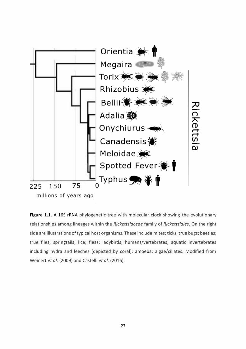
27
Figure 1.1. A 16S rRNA phylogenetic tree with molecular clock showing the evolutionary
relationships among lineages within the Rickettsiaceae family of Rickettsiales. On the right
side are illustrations of typical host organisms. These include mites; ticks; true bugs; beetles;
true flies; springtails; lice; fleas; ladybirds; humans/vertebrates; aquatic invertebrates
including hydra and leeches (depicted by coral); amoeba; algae/ciliates. Modified from
Weinert et al. (2009) and Castelli et al. (2016).
28
transmitted (Weinert et al., 2009). However, a peculiarity of note is the paternal transmission
observed in the leafhopper Nephotettix cincticeps (Watanabe et al., 2014).This additional
vertical transmission route has been suggested to allow for the drive of a symbiont into a
population when maternal transmission is incomplete. Rickettsia has also been suggested to
have strategies allowing for host shifts, which include blood feeding where co-feeding on
vertebrate hosts allows for the transmission of spotted fevers (Wood and Artsob, 2012).
Additionally, there is some evidence to suggest plant-feeders could acquire Rickettsia with
phloem acting as an intermediary reservoir (Chrostek et al., 2017). This suggestion is
supported by the transmission of a Belli group Rickettsia from infected whiteflies to non-
infected conspecifics feeding sequentially on the same leaves (Caspi-Fluger et al., 2012). In
addition, a Rickettsia member is the causative agent of the plant disease, papaya bunchy top
disease (Luis-Pantoja et al., 2015). Subsequently, Rickettsia represents an intriguing
endosymbiotic group with numerous transmission routes and ecological anomalies.
1.3.6 Endosymbionts and the confounding of DNA barcoding studies
Although mtDNA barcoding has been used widely for the taxonomic classification of
Culicoides (see section 1.2.5), the presence of symbionts can often confound studies using
this technique. As endosymbionts and mitochondria are generally co-inherited together (via
the maternal line), they are often observed in linkage disequilibrium when a symbiont
selective sweep occurs in a population. Subsequently, indirect selection on mtDNA can also
occur in insects containing maternally inherited endosymbionts (Hurst and Jiggins, 2005). This
can lead to the confounding effects of biodiversity studies where one of the assumptions is
that the mtDNA marker is under neutral selection. Specific effects can include the fragmenting
29
or homogenising of mitochondrial lineages. For example, the erasure of mtDNA diversity
within a population can occur as a result of selective sweeps driven by a strain conferring CI,
which can lead to an underestimation of diversity (Ballard et al., 1996; Jiggins, 2003;
Shoemaker et al., 2004; Zhang et al., 2013). Conversely, when more than one symbiont strain
enters a population, this can lead to the maintenance of divergent mtDNA haplotypes
(Schulenburg et al., 2002; Jiggins, 2003; Kvie et al., 2013). Finally, the conflation of differing
species through past mitochondrial introgression can occur when an endosymbiont transfers
during a hybridisation event, driving into a population and carrying with it the heterospecific
mtDNA (Jiggins, 2003; Whitworth et al., 2007; Raychoudhury et al., 2009; Jäckel, Mora and
Dobler, 2013).
Another confounding effect of symbionts is related to the inadvertent amplification of
bacteria DNA (Linares et al., 2009; Smith et al., 2012; Ceccarelli, Haddad and Ramírez, 2016).
As the hypothetical bacterial ancestor of mitochondria (proto-mitochondria) is thought to be
closely related to Wolbachia and Rickettsia (Wang and Wu, 2015), DNA barcoding can
erroneously amplify these symbionts instead of the host target. This phenomenon was first
described when the barcoding of lepidopterans retrieved several Wolbachia COI sequences
(Linares et al., 2009). Since then, a large scale systematic review using the Barcoding Of Life
Data System (BOLD), assessing over 2 million sequences, revealed that 0.16% of barcoding
attempts of insects led to similar unintended Wolbachia amplifications (Smith et al., 2012).
More recently, Rickettsia has also been observed to produce similar outcomes. For example,
the erroneously barcoding amplification of Rickettsia has been observed in two families of
spider (Anyphaenidae and Dysderidae) (Řezáč et al., 2014; Ceccarelli, Haddad and Ramírez,
30
2016). Subsequently, this widespread direct and indirect confounding suggests the
interpretation of barcoding studies should only be considered in the context of past and
present symbiont infections.
1.4 Thesis aims
Overall, this thesis had 3 key aims:
1) To investigate the occurrence and distribution of Cardinium and Rickettsia in Culicoides
from Palearctic and Afrotropical regions.
2) To assess potential transmission dynamics and host effects of Rickettsia in Culicoides
through phylogenetic analysis and the localisation of the symbiont to specific tissues.
3) To determine the confounding effects of Rickettsia and Cardinium on DNA barcoding
studies.
The first section of this thesis aimed to investigate the symbiotic interactions of Cardinium
and Culicoides through a targeted screening of biting midge species. Although, Culicoides
populations from various biogeographic regions were assessed, a specific focus was given to
UK species to identify candidates to undertake further host fitness and infection work.
Specifically, this relied on rescreening the only two UK vector species identified as being
Cardinium-infected, C. pulicaris and C. punctatus (Lewis et al., 2014). It was of importance to
assess these species for polymorphic infections as this would provide a suitable population
for vector competence experiments. This is because the naturally uninfected individuals from
these populations could be used as negative controls without the confounding effects of
antibiotic use. This initial investigation also aimed to assess the prevalence and distribution
of Cardinium infections in Culicoides to assess clustering patterns of geography and host
31
phylogeny. Finally, as symbionts can confound the interpretation of biodiversity studies which
use DNA barcoding, the well-known vector species C. imicola is used to assess if mtDNA
haplotypes reflect Cardinium presence rather than host gene flow patterns.
Research on symbionts of Culicoides species is in its infancy. Subsequently, it was unsurprising
when the common Rickettsia endosymbiont was uncovered through the serendipitous
shallow whole genome sequencing of C. newsteadi by a colleague (Stefanos Siozios, University
of Liverpool). With limited opportunities to identify an in vivo system to study midge-
symbiont interactions arising from the first section of this thesis, a focus was shifted to
identify such a system in relation to Rickettsia, which required its own targeted screening
presented in chapter 3. This chapter not only described the host diversity of Rickettsia, but
through prevalence and phylogenetic analysis, an aim was to identify ecological dynamics
which are imperative in understanding the potential for novel biological intervention
involving Rickettsia in the future.
These symbiont screens then informed the fourth chapter, which describes the tissue
tropisms of Rickettsia in the UK pest species C. impunctatus. Despite previous difficulties in
rearing C. impunctatus (Carpenter, 2001), cultivation of immatures for processing were
successful, which allowed for tropism information to be gathered for various life stages.
Through transmission electron microscopy (TEM) and fluorescence in-situ hybridisation (FISH)
the cellular and subcellular localisation of Rickettsia in both somatic and germline tissues is
described. This advances the understanding of Rickettsia-midge interactions by contributing
knowledge to transmission dynamics as well as potential host effects induced by the
symbiont. Due to Rickettsia’s potential to increase host fitness (Kikuchi and Fukatsu, 2005),

32
identification of such effects would be essential for any future control program for this
economically important pest species.
Comparing sequences identified in the Rickettsia screening of chapter 3 to online databases,
it was found that hundreds of supposed mitochondrial (barcode) sequences from Genbank
(NCBI; http://www.ncbi.nlm.nih.gov/genbank/) and BOLD (Barcoding of Life Data System;
http://www.boldsystems.org/) had a high homology to Culicoides Rickettsia. This indicated
barcoding studies were often amplifying Rickettsia DNA instead of host mtDNA.
Subsequently, through a systematic review of in silico barcoding sequences, the final section
of this thesis investigates how common this phenomenon of inadvertent Rickettsia
amplification is. Through special access to DNA extracts producing erroneous Rickettsia
amplifications (provided by BOLD), the screening and sequencing of housekeeping genes used
in chapter 3 is used to expand our phylogenetic knowledge of the Rickettsia genus. A further
aim of this study was to access if DNA barcoding could be used as an adjunctive to targeted
Rickettsia screens to uncover host new host interactions which would inform future study
systems of the underexplored “ancestral” group. Finally, a discussion of the results aims to
summarise the findings from all chapters with a focus on the prospects of Rickettsia’s
contribution to arbovirus transmission dynamics.
33
Chapter 2: Assessing the Phylogeographic Distribution of Candidatus Cardinium hertigii in Culicoides with a Focus on
Two UK Vector Species
34
2.1 Abstract
The endosymbiont Candidatus Cardinium hertigii (Bacteroidetes), has previously been
described in Culicoides (Diptera: Ceratopogonidae) populations from Japan, Australia, Israel,
Spain and UK, with the latter account suggesting infection of both C. pulicaris and C.
punctatus, putative vectors of bluetongue virus in Europe. Due to the lack of a model UK
Culicodies species to test Cardinium-midge interactions, this chapter aimed to assess the
suitability of both species. The attempted replication of the previous UK Cardinium screening
of C. pulicaris and C. punctatus showed several errors in the original methodology and
interpretation of Cardinium infection status, suggesting neither are suitable for future in vivo
work. In addition, nested PCR screening was undertaken for 338 individuals of 24 species of
biting midges from both Palearctic and Afrotropical regions to assess patterns of geography
and host phylogeny. Infections were observed in 64 individuals out of 9 species, with
prevalence ranging from low and intermediate, to fixation. These data corroborate previous
estimates of Cardinium prevalence in midges. Phylogenetic analysis based on the Gyrase B
gene grouped new isolates within “group C” of the genus, a clade which has so far been
exclusive to Culicoides, and in which all Culicoides Cardinium infections described to date lie.
Additionally, the concordance of mitochondrial haplotypes (mitotypes) and Cardinium
infection in populations of C. imicola suggests a potential confounding of phylogeographic
studies based on this marker. The biological significance of Cardinium in midges, and effects
on vector capacity, are still to be elucidated.
35
2.2 Introduction
Worldwide, biting midges of the genus Culicoides (Diptera: Ceratopogonidae) are known to
transmit more than 50 arboviruses. Notably, bluetongue virus (BTV) and Schmallenberg virus
(SBV) pose a great threat to livestock and wild animal welfare (Mellor, Boorman and Baylis,
2000). Outbreaks of BTV have caused serious economic damage to the European livestock
industry (Wilson and Mellor, 2009). For example, a single epidemic of BTV in the UK has been
estimated to potentially cost up to £485M with losses in livestock productivity, restrictions on
UK agricultural exports and employment impacts cited as the main contributors to this figure
(Pirbright Institute, 2009). Current control methods of BTV rely on vaccines which, due to the
quick emergence and uncertainty of circulating strains, can be ineffective. The recent
introduction of the vector species Culicoides imicola into continental Europe, as a result of
climate change and wind-transport events (Purse et al., 2005; Venail et al., 2012; Jacquet et
al., 2016), has also highlighted the challenge of mapping midge-borne pathogens. Thus, novel
vector control measures are seen to be more attractive options in controlling the spread of
these arboviruses (Carpenter, Mellor and Torr, 2008).
Intracellular bacteria (endosymbionts) and insects frequently form symbiotic associations,
some of which have been implicated in host defence against pathogens. As such, this
symbiosis can have profound effects on the biology, ecology and evolutionary dynamics of
host-pathogen interactions (Ferrari and Vavre, 2011). Importantly, the endosymbiont
Wolbachia has demonstrated the ability to transinfect mosquito species leading to a blocking
effect of dengue virus, chikungunya virus, yellow fever virus and West Nile virus (Bian et al.,
2010; Blagrove et al., 2012; Hussain et al., 2012; van den Hurk et al., 2012). The potential to
36
extrapolate these effects to wild populations of Aedes mosquitoes is enabled by Wolbachia-
induced cytoplasmic incompatibility (CI); embryo death in mating between infected males
and uninfected females (Werren and O’Neill, 1997; Bourtzis et al., 2014). CI is used as
mechanism to drive the Wolbachia into a population to distribute this virus blocking
phenotype into a naïve population (Mouton et al., 2003; Miller, Ehrman and Schneider, 2010;
Hoffmann et al., 2011, 2014). In addition, this can allow for the suppression of vector numbers
when releasing Wolbachia-infected male Aedes aegypti (Mains et al., 2019; Zheng et al.,
2019). Encouragingly, the results of field-based CI-inducing symbiont interventions indicate a
viable method to control arboviruses (Hoffmann et al., 2014). Importantly, this protective
phenotype appears to be associated with RNA viruses leading to the potential for symbiont-
based biocontrol in major midge-borne pathogens such as BTV and SBV.
Candidatus Cardinium hertigii (Bacteroidetes) is an endosymbiont which has been widely
associated with Culicoides (Nakamura et al., 2009; Morag et al., 2012; Mee et al., 2015; Pagès
et al., 2017). Of the Culicoides species known to be infected with Cardinium; C. imicola, C.
obsoletus, C. pulicaris, C. punctatus and C. brevitarsis are known vectors of BTV, having been
directly linked with epidemics of bluetongue (Muller et al., 1982; Goffredo et al., 2015). It is
estimated up to 13% of arthropod species carry Cardinium (Weinert et al., 2015) with induced
phenotypes including the reproductive manipulations feminisation (Weeks, Marec and
Breeuwer, 2001; Groot and Breeuwer, 2006) and parthenogenesis (Zchori-Fein et al., 2002).
CI has also been observed in Cardinium in both spider-mites and parasitoid wasps (Hunter,
Perlman and Kelly, 2003; Gotoh, Noda and Ito, 2007; Perlman, Kelly and Hunter, 2008; Ros
37
and Breeuwer, 2009) suggesting this could be a driving mechanism for future introductions
into Culicoides populations.
Mitochondrial DNA (mtDNA) markers have been widely used in the taxonomic classification
of animals due to its high evolutionary rate, suitable effective population size and low
recombination rate when compared to nuclear markers (Hurst and Jiggins, 2005). As well as
being utilised as a molecular technique to identify arthropods, mtDNA “barcodes” have
frequently been used in phylogeographic studies to detect biodiversity and dispersal events
(Craft et al., 2010; Papadopoulou et al., 2011; Baselga et al., 2013). Despite this utility, these
studies can occasionally be confounded by the linkage disequilibrium of endosymbionts and
host mtDNA. As endosymbionts enter a naïve population, selective sweeps of the bacteria
can occur and with it the homogenisation of linked mtDNA haplotypes, leading to the
apparent absence of biodiversity (Hurst and Jiggins, 2005). Conversely, the introduction of
more than one endosymbiont in linkage disequilibrium with an mtDNA haplotype can lead to
a perceived genetic population structure when there is a lack of one. Subsequently, inferences
from barcoding studies can only be interpreted in the context of recent infection (or lack
thereof) with endosymbiotic bacteria. This may therefore confound attempts to reconstruct
vector history of invasion and spread.
The motivation for this study stems from the need for a model system to study Cardinium-
Culicoides interactions. First, the phylogeographic distribution of Cardinium infections is
investigated in Culicoides populations from Palearctic and Afrotropical regions through
screening using a nested PCR. A focus is placed on C. pulicaris and C. punctatus, two UK vector
species described as being Cardinium-infected (Lewis et al., 2014), with the intention of

38
assessing their suitability for future in vivo work. A polymorphically infected population
(containing infected and uninfected individuals), with high numbers in the wild, is preferable
for such studies due to readily available negative (uninfected) controls. Finally, this chapter
aims to establish associations between mitochondrial haplotypes (mitotypes) and Cardinium
infection in the globally important vector Culicoides imicola, as this could have implications
for inferring movement of this species into naïve geographic areas.
39
2.3 Methods
2.3.1 Culicoides collection and identification
Overall, 338 specimens of 24 Culicoides species were collected using light traps from May
2007 to July 2016 across 17 sites spanning France, South Africa, Sweden and the UK.
Populations collected by the author included all midges collected from 2015 onwards from
the UK and South Africa. Other specimens were provided by Ken Sherlock (Institute of
Infection and Global Health, Liverpool, UK), Dr Jan Chirico (National Veterinary institute,
Uppsala, Sweden) and Dr Claire Garros (CIRAD, Agricultural Research for Development,
Montpellier, France). Those obtained by the author were collected through the use of both
CDC (6-volt motorbike battery) and Onderstepoort light traps (mains) utilising an ultraviolet
Light Emitting Diode (LED). Generally, traps were placed above two metres at sites before
dusk and collected at dawn the next day. A downward draught from a fan directed insects
into a beaker filled with water and a small amount of detergent to break surface tension. After
collection, all midge specimens were stored in 70% ethanol for preservation before being
sexed and separated morphologically down to the species level using relevant keys (Downes
and Kettle 1952; Campbell and Pelham-Clinton 1960; Delécolle 1985; Meiswinkel 1994).
Sampled species included both vectors and non-vectors of BTV. Morphological identification
of Cardinium positive specimens was confirmed by sequencing a fragment of the
mitochondrial cytochrome c oxidase subunit 1 (COI) barcode (Pagès et al., 2009; Ander, Troell
and Chirico, 2013; Nielsen and Kristensen, 2015).
2.3.2 DNA extraction
DNA extractions were prepared based on the protocol of Ander et al. (2013). Briefly, whole
individual insects were washed in molecular grade water, transferred to blue roll and dried at
40
room temperature for approximately two minutes. Using a sterile pipette tip with a melted
tip, the specimen was transferred to an Eppendorf tube, where 30 μL of PrepMan Ultra (Life
Technologies, Inc., Carlsbad, CA, USA) was added. The specimen was homogenized using the
same sterile pipette tip until no macroscopic structure was visible and then briefly
centrifuged. The preparation was incubated at 100 °C for ten minutes, and samples were then
kept on ice for two minutes. Remaining fragments of tissue were removed from the
homogenate by centrifugation at 12,000 × g for three minutes, before 20 μL of the
supernatant was transferred to a fresh Eppendorf tube. Before the above fresh samples were
available, DNA extracts from a previous study by Lewis et al. (2014), describing the positive
infection status of C. pulicaris and C. punctatus in the UK, were used to test screening assays.
2.3.3 COI and Cardinium screening
Amplification of the COI gene was initially assessed as a means of quality control by
conventional PCR assay using the commonly used primer set HCO1490/LCO2198 (Folmer et
al., 1994). For individuals which failed to produce a positive result, a secondary set of primers
C1-J-1718/C1-N-2191 were utilised (Dallas et al., 2003). Samples that did not amplify with
either primer set were deemed to contain low quality DNA and were removed from further
analysis. DNA extracts which passed quality control were then screened for Cardinium using
conventional and nested primers amplifying partial sequences of the Gyrase B (GyrB) gene.
PCR assays consisted of a total of 15 μL per well, comprising of 7.5 μL GoTaq® Hot Start
Polymerase (Promega), 5.1 μL nuclease free water, 0.45 μL forward and reverse primers
(concentration 10 pmol/μL) and 1.5 μL DNA template. PCR products were separated on 1%
agarose gels stained with Midori Green Nucleic Acid Staining Solution (Nippon Genetics
41
Europe). Primer sequence details and PCR cycling conditions can be found in Tables 2.1.1 and
2.1.2.
2.3.4 Nested primer design and sensitivity
Nested primers were designed by aligning known Cardinium hertigii GyrB sequences (Table
2.2.) available in GenBank (NCBI) using the ClustalW algorithm in Mega6 (Tamura et al., 2013).
In order to identify the correct reading frame, both complementary strands in all three
reading frames were translated via the ExPasy translate tool (Gasteiger et al., 2003) into 6
different amino acid sequences. The result that gave the longest amino acid sequence, before
a stop codon was encountered, was deemed the correct reading frame. Specific nested
primers were then designed based on homology inferred from the multiple alignment of GyrB
sequences using the online tool PriFi (Fredslund et al., 2005) and its default parameters. The
stable anchoring of the primers’ 3’ termini was ensured by the inclusion of two-fold
degenerate sites at the third position of codons to avoid effects of base-wobble pairing. The
diagnostic sensitivity of conventional and nested PCR assays was quantified by serially diluting
Cardinium positive DNA extracts using 10-fold dilutions of nuclease-free water. Dilutions
ranged from undiluted to 10-6 for both C. punctatus (n=5) and C. imicola (n=2) DNA templates.
Nested PCR is prone to contamination and subsequently false positives. Precautions
undertaken to avoid this outcome include repeated disinfecting of surfaces, separation of lab
space between sample preparation and amplification handling, the use of filter tips and
multiple (four) negative controls.
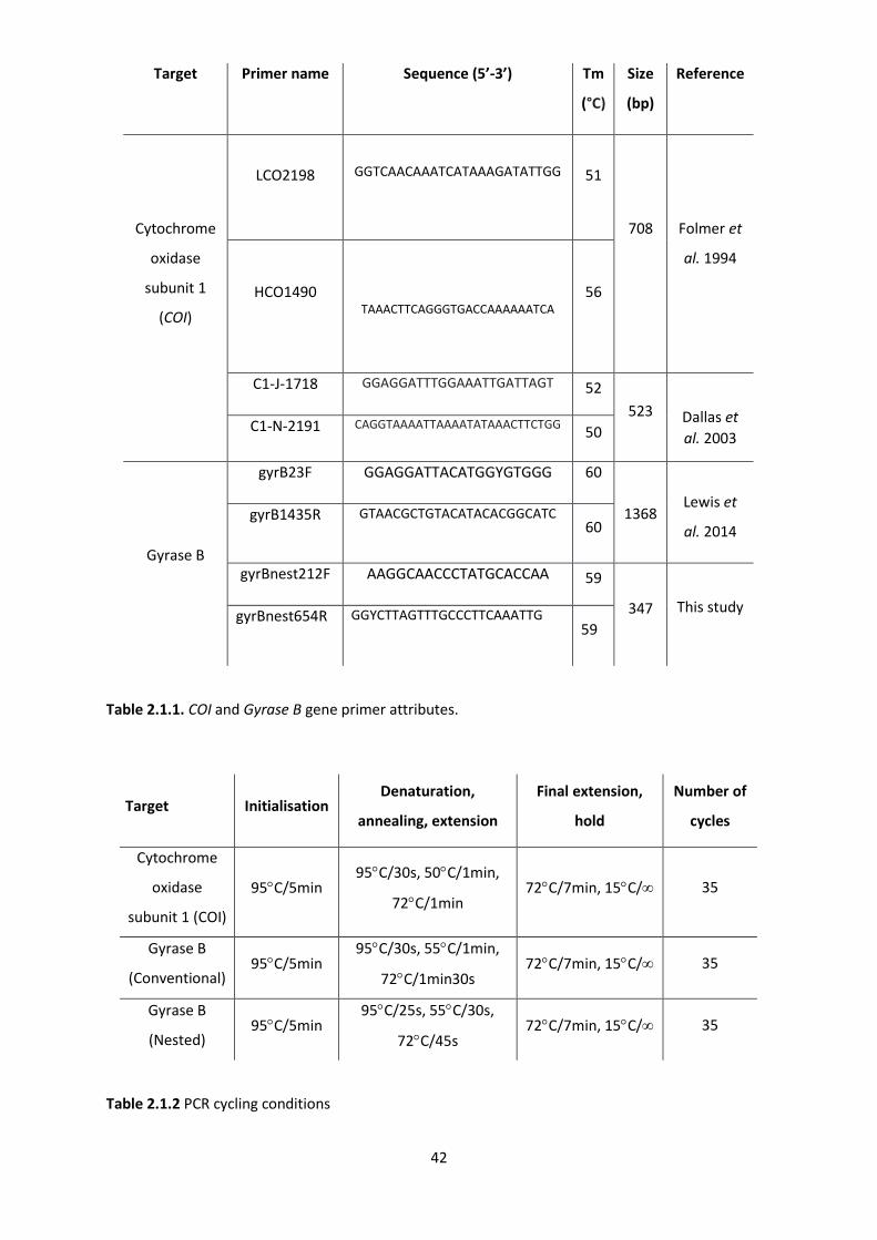
42
Table 2.1.1. COI and Gyrase B gene primer attributes.
Table 2.1.2 PCR cycling conditions
Target Primer name Sequence (5’-3’) Tm
(°C)
Size
(bp)
Reference
Cytochrome
oxidase
subunit 1
(COI)
LCO2198
GGTCAACAAATCATAAAGATATTGG
51
708
Folmer et
al. 1994
HCO1490
TAAACTTCAGGGTGACCAAAAAATCA
56
C1-J-1718 GGAGGATTTGGAAATTGATTAGT 52
523
Dallas et
al. 2003 C1-N-2191 CAGGTAAAATTAAAATATAAACTTCTGG
50
Gyrase B
gyrB23F GGAGGATTACATGGYGTGGG 60
1368
Lewis et
al. 2014 gyrB1435R GTAACGCTGTACATACACGGCATC
60
gyrBnest212F AAGGCAACCCTATGCACCAA 59
347
This study gyrBnest654R GGYCTTAGTTTGCCCTTCAAATTG
59
Target Initialisation Denaturation,
annealing, extension
Final extension,
hold
Number of
cycles
Cytochrome
oxidase
subunit 1 (COI)
95C/5min 95C/30s, 50C/1min,
72C/1min 72C/7min, 15C/ 35
Gyrase B
(Conventional) 95C/5min
95C/30s, 55C/1min,
72C/1min30s 72C/7min, 15C/ 35
Gyrase B
(Nested) 95C/5min
95C/25s, 55C/30s,
72C/45s 72C/7min, 15C/ 35
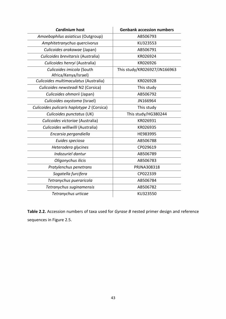
43
Cardinium host Genbank accession numbers
Amoebophilus asiaticus (Outgroup) AB506793
Amphitetranychus quercivorus KU323553
Culicoides arakawae (Japan) AB506791
Culicoides brevitarsis (Australia) KR026924
Culicoides henryi (Australia) KR026926
Culicoides imicola (South Africa/Kenya/Israel)
This study/KR026927/JN166963
Culicoides multimaculatus (Australia) KR026928
Culicoides newsteadi N2 (Corsica) This study
Culicoides ohmorii (Japan) AB506792
Culicoides oxystoma (Israel) JN166964
Culicoides pulicaris haplotype 2 (Corsica) This study
Culicoides punctatus (UK) This study/HG380244
Culicoides victoriae (Australia) KR026931
Culicoides williwilli (Australia) KR026935
Encarsia pergandiella HE983995
Euides speciosa AB506788
Heterodera glycines CP029619
Indozuriel dantur AB506789
Oligonychus ilicis AB506783
Pratylenchus penetrans PRJNA308318
Sogatella furcifera CP022339
Tetranychus pueraricola AB506784
Tetranychus suginamensis AB506782
Tetranychus urticae KU323550
Table 2.2. Accession numbers of taxa used for Gyrase B nested primer design and reference
sequences in Figure 2.5.
44
2.3.5 Mapping Cardinium spatial distribution
Collection sites of Culicoides populations were plotted against Cardinium infection prevalence
based on the nested PCR assay and mapped using Quantum GIS v3 software (Quantum GIS,
2016). The world borders shapefile was obtained from Thematicmappings.org under an
Attribution-Share Alike License (Bjorn Sandvik, 2009), accessed December 2017.
2.3.6 Sanger sequencing
Amplicons identified by gel electrophoresis were subsequently purified enzymatically
(ExoSAP) before being sent for sequencing if in batches of <24 (GATC Biotech AG, Konstanz,
Germany) or sequenced using a BigDye® Terminator v3.1 kit (Thermo Scientific, Waltham,
USA), and capillary sequenced on a 3500 xL Genetic Analyser (Applied Biosystems, Austin,
USA) for larger batch numbers. Amplicons were sequenced through both strands for GyrB and
through single strands for COI. Generated chromatograms were assessed visually for correct
base calls with consensus sequences being created in Ugene v1.22 (Okonechnikov et al.,
2012).
2.3.7 Phylogenetic analysis
The relatedness of Cardinium strains from different host species was analysed using
nucleotide sequences from conventional PCR. GyrB was chosen for phylogenetic analysis
because it has a higher divergence when compared to the conserved 16S rRNA gene, another
gene used in Cardinium phylogeny reconstruction. Cardinium GyrB sequences from groups A,
B and C were retrieved from GenBank (NCBI) (Table 2.2.) to improve phylogenetic resolution.
Sequences were aligned using the “L-INS-I” algorithm in MAFFT v7 (Katoh and Standley, 2013).
A maximum likelihood (ML) phylogeny was inferred with RAxML v8 (Stamatakis, 2014) using
1000 rapid bootstrap replicates and using the GTR + I + G model which was selected with
45
jModelTest 2 (Darriba et al., 2012) using the Akaike information criterion, with the topology
search taking the best of Subtree Pruning and Nearest Neighbour Interchange rearranging.
Finally, trees were drawn using the EvolView (He et al., 2016) online tree annotation and
visualization tool.
2.3.8 mtDNA haplotype networks of Cardinium infected and uninfected C. imicola
Phylogenetic relationships between Cardinium positive and negative C. imicola from South
Africa were assessed using a haplotype network of a 517 bp region of the COI gene. The
network was then extended to include C. imicola from France, Corsica (this study) as well as
Portugal and Israel (Dallas et al. 2003; Accession numbers: AF078098–AF078100, AF080531,
AF080532, AF080534, AF080535, AJ549393–AJ549426) where varying Cardinium infection
status had previously been described. Analysis and visualisation was undertaken using the
TCS haplotype network algorithm generated in PopART v1 (Leigh and Bryant, 2015).
2.3.9 Cardinium prevalence comparisons of site, species and sex
Differences in Cardinium prevalence were tested for each collection site, species and sex using
the two tailed Fisher’s exact test. More specifically, this was achieved by comparing the same
species at different collection sites where at least one population had a Cardinium positive.
All statistical analyses in this study was undertaken using Rstudio version 1.1.419 (Rstudio
Team, 2015).
46
2.4 Results
2.4.1 Clarification of the Cardinium infection status of C. pulicaris and C. punctatus
The rescreening of DNA extracts from the Lewis study provided contrasting results with those
described in the original study. Of the 26 C. punctatus DNA extracts screened, 25/26 (96%)
were positive for Cardinium, all of which were validated for sufficient DNA quality via PCR
amplification of the COI subunit using the HCO1490/LCO2198 primer set. Despite adequately
replicating Lewis’ findings for C. punctatus, only 12/39 (31%) C. pulicaris samples were
positive when using the HCO1490/LCO2198 primer pair, as opposed to 39/39 (100%) claimed
in the previous study. Incidentally, the 12 COI positive samples were the same DNA extracts
which came back PCR positive for the Cardinium GyrB screen as documented by Lewis.
Furthermore, fresh C. pulicaris caught in the autumn of 2015 did not show positive signals for
amplification of COI using HCO1490/LCO2198 primers. This led to the likelihood that there
was a polymorphism at one of the forward or reverse primer sites designed to amplify COI in
C. pulicaris. To test this, a new COI locus was chosen to observe whether the previous
negative samples would become positive. The secondary quality control primers C1-J-
1718/C1-N-2191 (Dallas et al., 2003) amplified all the C. pulicaris DNA extracts confirming this
premise (Table 2.3.2).
Although all of these samples had now been confirmed to have adequate DNA quality, there
were still the unresolved issues of how Lewis had detected COI positive C. pulicaris with the
invalid HCO1490/LCO2198 primers, and why the re-screened HCO1490/LCO2198 positives
were coincidentally the only samples to come back positive for Cardinium. Obtaining the raw
data from Lewis’ study resolved the first of these issues. Examination of Lewis’ gel images
47
corroborated my re-screening data (Figure 2.1.); 12/39 C. pulicaris samples gave positive
bands after amplification with HCO1490/LCO2198 primers despite Lewis claiming that all
passed the COI quality control screen. To explain the matter of why there were any
HCO1490/LCO2198 amplified C. pulicaris extracts to begin with, there were several possible
explanations: the DNA extracts had been cross contaminated with other Culicoides’ extracts
through a pipetting error; the individuals had been misidentified when morphologically
distinguishing the Culicoides species and were in fact another species; or the individuals
morphologically appeared as C. pulicaris but were an unidentified cryptic species which lack
the polymorphism at the HCO1490/LCO2198 site and are infected with Cardinium. To
distinguish between these possibilities, Sanger sequencing of the purified PCR product of both
HCO1490 and C1-N-2191 was undertaken and compared to known mitochondrial DNA
barcodes on Genbank. BLASTn alignment identified the HCO1490 product to be 99%
homologous to C. punctatus COI (Accession number: KX064676), whereas the C1-N-2191
product was 100% identical to C. pulicaris COI (Accession number: KJ624116). As both loci
overlap with each other, these data implied cross contamination of C. pulicaris and C.
punctatus DNA extracts (Table 2.3.1.).
2.4.2 Nested PCR sensitivity
The serial dilutions of Cardinium positive samples saw an increase in sensitivity by between
10- and 100-fold lower detection limit for nested compared to the
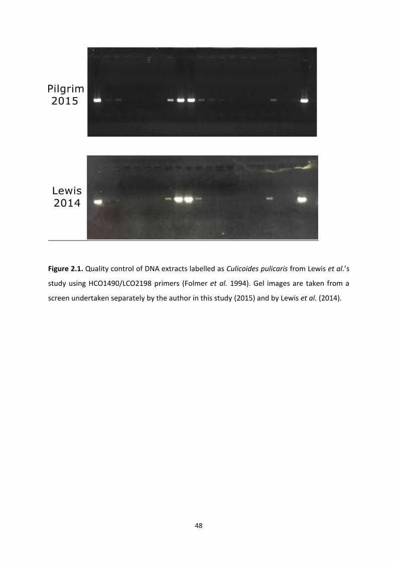
48
Figure 2.1. Quality control of DNA extracts labelled as Culicoides pulicaris from Lewis et al.’s
study using HCO1490/LCO2198 primers (Folmer et al. 1994). Gel images are taken from a
screen undertaken separately by the author in this study (2015) and by Lewis et al. (2014).
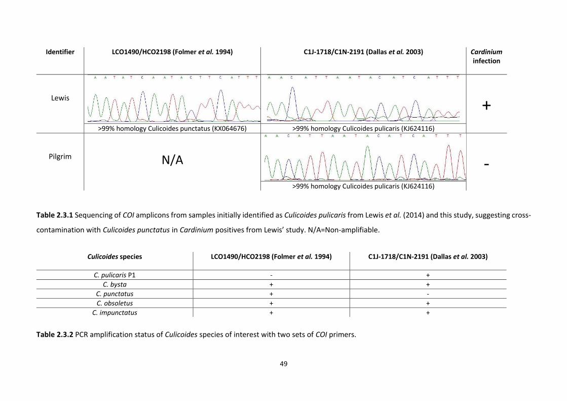
49
Table 2.3.1 Sequencing of COI amplicons from samples initially identified as Culicoides pulicaris from Lewis et al. (2014) and this study, suggesting cross-
contamination with Culicoides punctatus in Cardinium positives from Lewis’ study. N/A=Non-amplifiable.
Table 2.3.2 PCR amplification status of Culicoides species of interest with two sets of COI primers.
Identifier LCO1490/HCO2198 (Folmer et al. 1994)
C1J-1718/C1N-2191 (Dallas et al. 2003) Cardinium infection
Lewis
+
>99% homology Culicoides punctatus (KX064676) >99% homology Culicoides pulicaris (KJ624116)
Pilgrim
N/A
-
>99% homology Culicoides pulicaris (KJ624116)
Culicoides species LCO1490/HCO2198 (Folmer et al. 1994)
C1J-1718/C1N-2191 (Dallas et al. 2003)
C. pulicaris P1 - +
C. bysta + +
C. punctatus + -
C. obsoletus + +
C. impunctatus + +
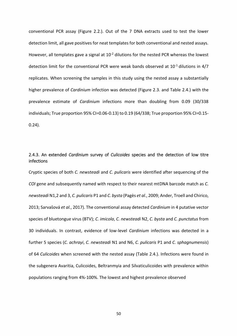
50
conventional PCR assay (Figure 2.2.). Out of the 7 DNA extracts used to test the lower
detection limit, all gave positives for neat templates for both conventional and nested assays.
However, all templates gave a signal at 10-2 dilutions for the nested PCR whereas the lowest
detection limit for the conventional PCR were weak bands observed at 10-1 dilutions in 4/7
replicates. When screening the samples in this study using the nested assay a substantially
higher prevalence of Cardinium infection was detected (Figure 2.3. and Table 2.4.) with the
prevalence estimate of Cardinium infections more than doubling from 0.09 (30/338
individuals; True proportion 95% CI=0.06-0.13) to 0.19 (64/338; True proportion 95% CI=0.15-
0.24).
2.4.3. An extended Cardinium survey of Culicoides species and the detection of low titre
infections
Cryptic species of both C. newsteadi and C. pulicaris were identified after sequencing of the
COI gene and subsequently named with respect to their nearest mtDNA barcode match as C.
newsteadi N1,2 and 3, C. pulicaris P1 and C. bysta (Pagès et al., 2009; Ander, Troell and Chirico,
2013; Sarvašová et al., 2017). The conventional assay detected Cardinium in 4 putative vector
species of bluetongue virus (BTV); C. imicola, C. newsteadi N2, C. bysta and C. punctatus from
30 individuals. In contrast, evidence of low-level Cardinium infections was detected in a
further 5 species (C. achrayi, C. newsteadi N1 and N6, C. pulicaris P1 and C. sphagnumensis)
of 64 Culicoides when screened with the nested assay (Table 2.4.). Infections were found in
the subgenera Avaritia, Culicoides, Beltranmyia and Silvaticulicoides with prevalence within
populations ranging from 4%-100%. The lowest and highest prevalence observed
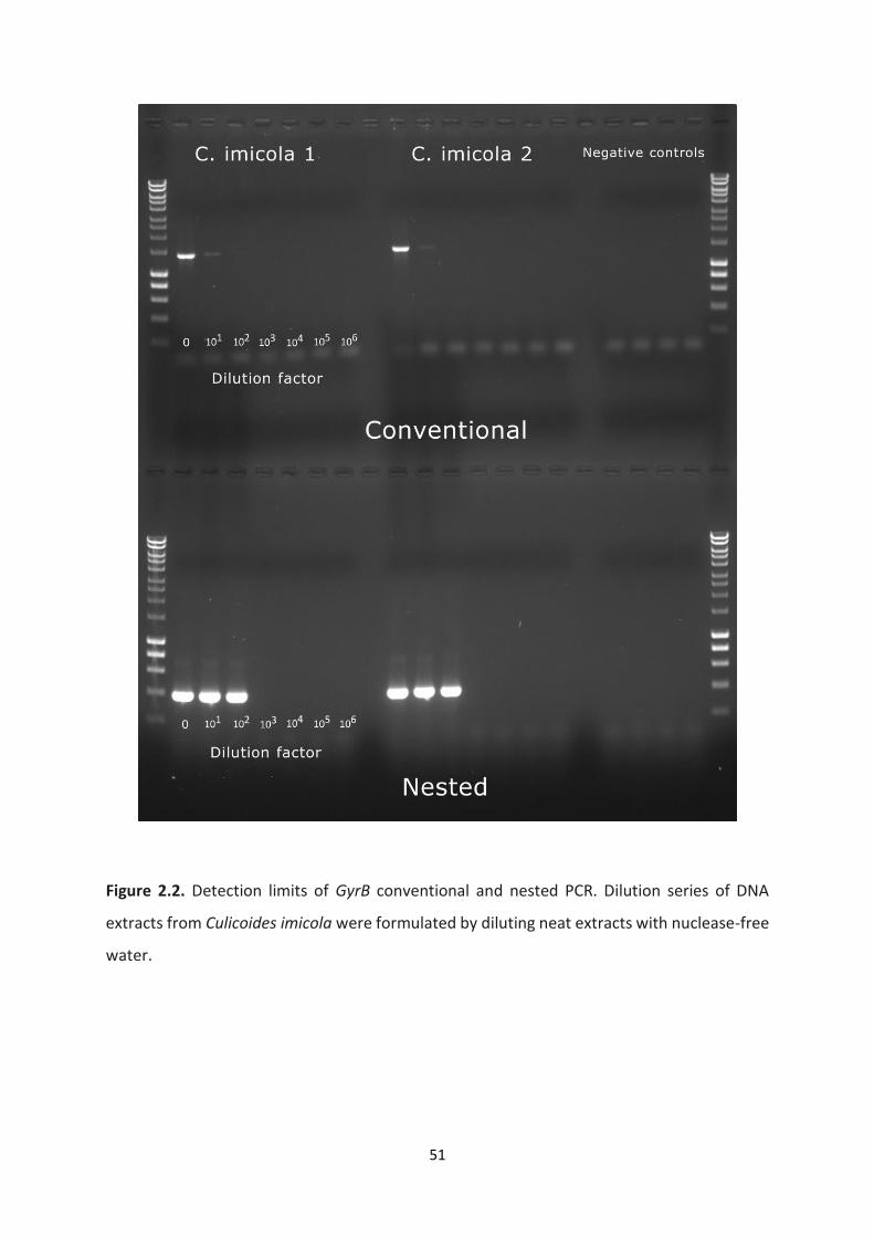
51
Figure 2.2. Detection limits of GyrB conventional and nested PCR. Dilution series of DNA
extracts from Culicoides imicola were formulated by diluting neat extracts with nuclease-free
water.
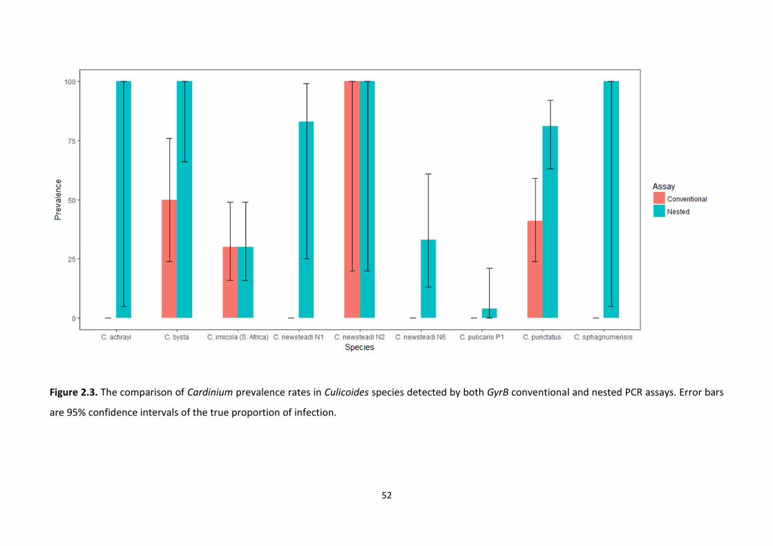
52
Figure 2.3. The comparison of Cardinium prevalence rates in Culicoides species detected by both GyrB conventional and nested PCR assays. Error bars
are 95% confidence intervals of the true proportion of infection.
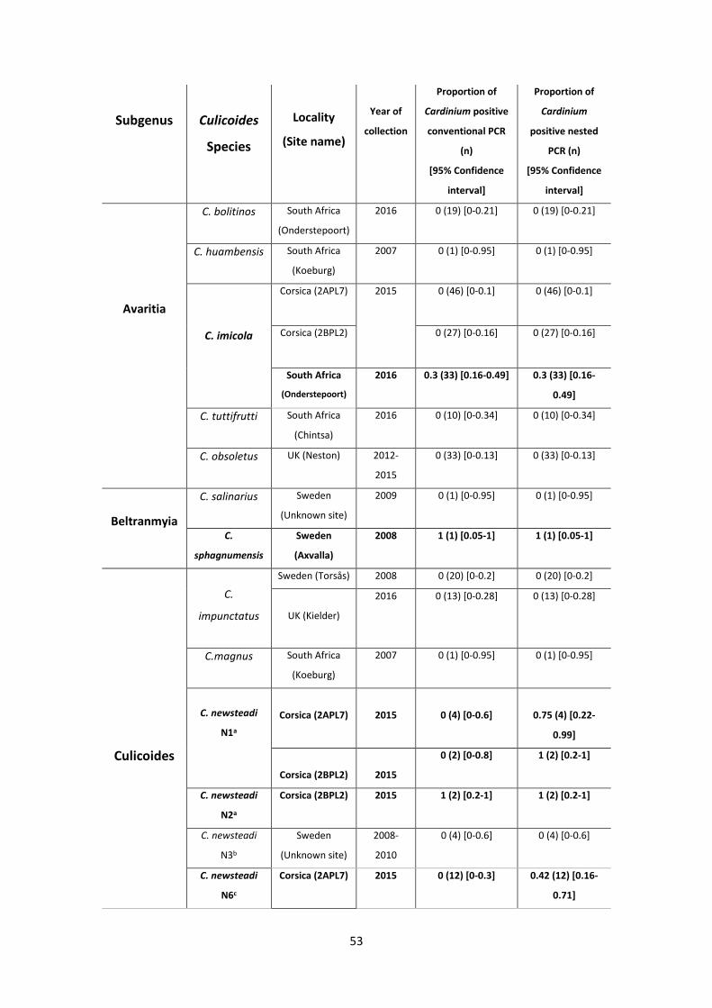
53
Subgenus
Culicoides
Species
Locality
(Site name)
Year of
collection
Proportion of
Cardinium positive
conventional PCR
(n)
[95% Confidence
interval]
Proportion of
Cardinium
positive nested
PCR (n)
[95% Confidence
interval]
Avaritia
C. bolitinos South Africa
(Onderstepoort)
2016 0 (19) [0-0.21] 0 (19) [0-0.21]
C. huambensis South Africa
(Koeburg)
2007 0 (1) [0-0.95] 0 (1) [0-0.95]
C. imicola
Corsica (2APL7) 2015 0 (46) [0-0.1] 0 (46) [0-0.1]
Corsica (2BPL2) 0 (27) [0-0.16] 0 (27) [0-0.16]
South Africa
(Onderstepoort)
2016 0.3 (33) [0.16-0.49] 0.3 (33) [0.16-
0.49]
C. tuttifrutti South Africa
(Chintsa)
2016 0 (10) [0-0.34] 0 (10) [0-0.34]
C. obsoletus UK (Neston) 2012-
2015
0 (33) [0-0.13] 0 (33) [0-0.13]
Beltranmyia
C. salinarius Sweden
(Unknown site)
2009 0 (1) [0-0.95] 0 (1) [0-0.95]
C.
sphagnumensis
Sweden
(Axvalla)
2008 1 (1) [0.05-1] 1 (1) [0.05-1]
Culicoides
C.
impunctatus
Sweden (Torsås) 2008 0 (20) [0-0.2] 0 (20) [0-0.2]
UK (Kielder)
2016 0 (13) [0-0.28] 0 (13) [0-0.28]
C.magnus South Africa
(Koeburg)
2007 0 (1) [0-0.95] 0 (1) [0-0.95]
C. newsteadi
N1a
Corsica (2APL7)
2015
0 (4) [0-0.6]
0.75 (4) [0.22-
0.99]
Corsica (2BPL2)
2015
0 (2) [0-0.8]
1 (2) [0.2-1]
C. newsteadi
N2a
Corsica (2BPL2) 2015 1 (2) [0.2-1] 1 (2) [0.2-1]
C. newsteadi
N3b
Sweden
(Unknown site)
2008-
2010
0 (4) [0-0.6] 0 (4) [0-0.6]
C. newsteadi
N6c
Corsica (2APL7) 2015 0 (12) [0-0.3] 0.42 (12) [0.16-
0.71]
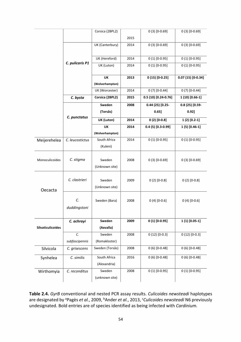
54
Corsica (2BPL2)
2015
0 (3) [0-0.69] 0 (3) [0-0.69]
C. pulicaris P1
UK (Canterbury) 2014 0 (3) [0-0.69] 0 (3) [0-0.69]
UK (Hereford) 2014 0 (1) [0-0.95] 0 (1) [0-0.95]
UK (Luton) 2014 0 (1) [0-0.95] 0 (1) [0-0.95]
UK
(Wolverhampton)
2013 0 (15) [0-0.25] 0.07 (15) [0-0.34]
UK (Worcester) 2014 0 (7) [0-0.44] 0 (7) [0-0.44]
C. bysta Corsica (2BPL2) 2015 0.5 (10) [0.24-0.76] 1 (10) [0.66-1]
C. punctatus
Sweden
(Torsås)
2008
0.44 (25) [0.25-
0.65]
0.8 (25) [0.59-
0.92]
UK (Luton) 2014 0 (2) [0-0.8] 1 (2) [0.2-1]
UK
(Wolverhampton)
2014 0.4 (5) [0.3-0.99] 1 (5) [0.46-1]
Meijerehelea C. leucostictus
South Africa
(Kuleni)
2014 0 (1) [0-0.95] 0 (1) [0-0.95]
Monoculicoides
C. stigma
Sweden
(Unknown site)
2008
0 (3) [0-0.69]
0 (3) [0-0.69]
Oecacta
C. clastrieri
Sweden
(Unknown site)
2009
0 (2) [0-0.8]
0 (2) [0-0.8]
C.
duddingstoni
Sweden (Bara)
2008
0 (4) [0-0.6]
0 (4) [0-0.6]
Silvaticulicoides
C. achrayi Sweden
(Axvalla)
2009 0 (1) [0-0.95] 1 (1) [0.05-1]
C.
subfascipennis
Sweden
(Romakloster)
2008 0 (12) [0-0.3] 0 (12) [0-0.3]
Silvicola C. grisescens Sweden (Torsås) 2008 0 (6) [0-0.48] 0 (6) [0-0.48]
Synhelea C. similis South Africa
(Alexandria)
2016 0 (6) [0-0.48] 0 (6) [0-0.48]
Wirthomyia C. reconditus Sweden
(unknown site)
2008 0 (1) [0-0.95] 0 (1) [0-0.95]
Table 2.4. GyrB conventional and nested PCR assay results. Culicoides newsteadi haplotypes are designated by aPagès et al., 2009, bAnder et al., 2013, cCulicoides newsteadi N6 previously undesignated. Bold entries are of species identified as being infected with Cardinium.
55
from populations with more than a few individuals were from C. pulicaris P1 (Prevalence
proportion=0.04; True proportion 95% CI=0.01-0.21) and C. bysta (Prevalence proportion=1;
True proportion 95% CI=0.66-1) respectively.
2.4.4. Cardinium prevalence comparisons of site, species and sex
Where there were multiple populations of the same species at different geographical
locations, most showed no evidence of heterogeneity in the fraction infected (C. newsteadi,
C. pulicaris, C. punctatus) apart from C. imicola populations from South Africa and Corsica
(Fisher’s exact, P<0.01) (Table 2.5.1). Although infected C. imicola were observed at an
intermediate prevalence in Onderstepoort, South Africa (Prevalence proportion=0.33; True
proportion 95% CI=0.16-0.49), no infection was detected in the same species in Corsica (True
proportion 95% CI=0-0.06). Sex ratio differences were not observed within infected species
of C. punctatus (Fisher’s exact, p=0.76) or C. imicola (Fisher’s exact, p=0.68) (Table 2.5.2). No
clear pattern of spatial distribution was observed between the three geographical regions of
the study (Figure 2.4.1 and 2.4.2.).
2.4.5. Phylogenetic analysis
Phylogenetic analysis based on a 1300 bp region of the GyrB gene confirmed a monophyletic
clade; grouping all Culicoides Cardinium isolates in group C of the genus where all previously
described sequences of Culicoides clustered (Figure 2.5.). Sequences isolated from this study
(C. newsteadi N2, C. bysta, C. imicola and C. punctatus) grouped neither based on subgenus
nor geography. Culicoides newsteadi N2 and C. bysta from the subgenus Culicoides both
formed separate clades with C. ohmorii (subgenus Hoffmania) and C. imicola/C. oxystoma
(Subgenus Avaritia/Remmia) respectively. Sequence divergence between C. newsteadi N2
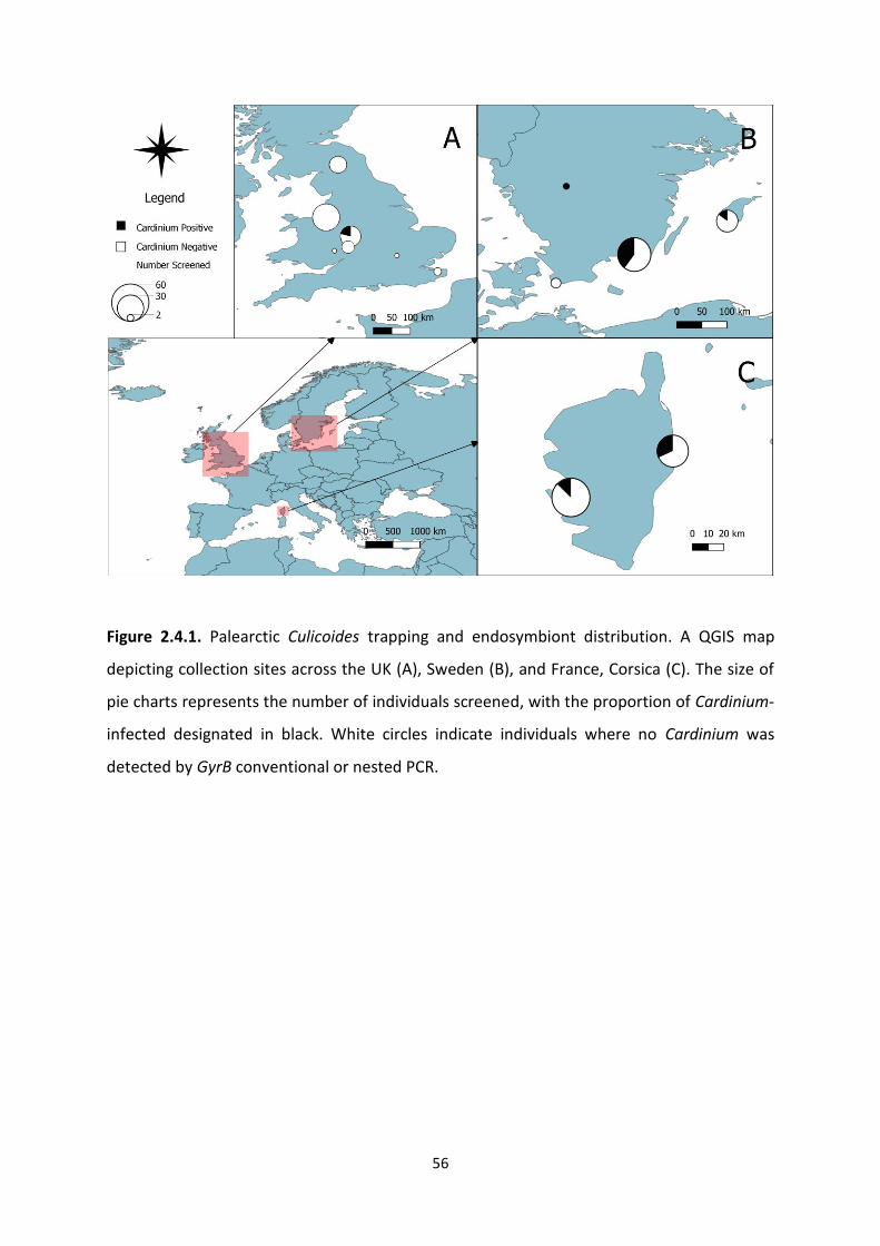
56
Figure 2.4.1. Palearctic Culicoides trapping and endosymbiont distribution. A QGIS map
depicting collection sites across the UK (A), Sweden (B), and France, Corsica (C). The size of
pie charts represents the number of individuals screened, with the proportion of Cardinium-
infected designated in black. White circles indicate individuals where no Cardinium was
detected by GyrB conventional or nested PCR.
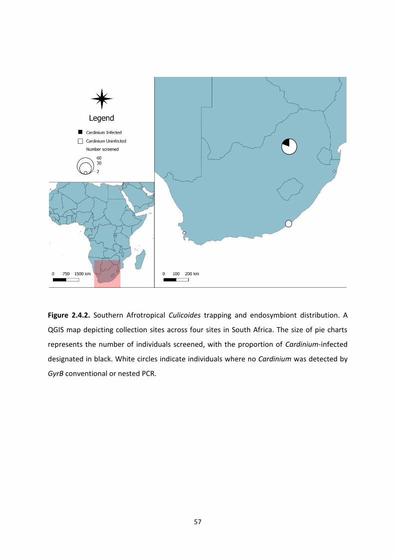
57
Figure 2.4.2. Southern Afrotropical Culicoides trapping and endosymbiont distribution. A
QGIS map depicting collection sites across four sites in South Africa. The size of pie charts
represents the number of individuals screened, with the proportion of Cardinium-infected
designated in black. White circles indicate individuals where no Cardinium was detected by
GyrB conventional or nested PCR.

58
Site 1 Site 2 Culicoides species
p value
South Africa (Onderstepoort)
Corsica (2APL7) C. imicola <0.01
South Africa (Onderstepoort)
Corsica (2BPL2) C. imicola <0.01
Corsica (2BPL2) Corsica (2APL7) C. newsteadi N1 1
Sweden (Torsås) UK (Luton) C. punctatus 1
Sweden (Torsås) UK (Wolverhampton)
C. punctatus 1
UK (Luton) UK (Wolverhampton)
C. punctatus 1
Corsica (2BPL2) Corsica (2APL7) C. newsteadi N6 0.5
Table 2.5.1. Fisher’s exact results for Cardinium infection comparisons of populations of the
same species but at different sites.
Culicoides species Site p value
C. imicola Onderstepoort 0.68
C. punctatus Torsas 0.76
Table 2.5.2. Fisher’s exact results for Cardinium infection comparisons of females and males
of the same species at the same site.
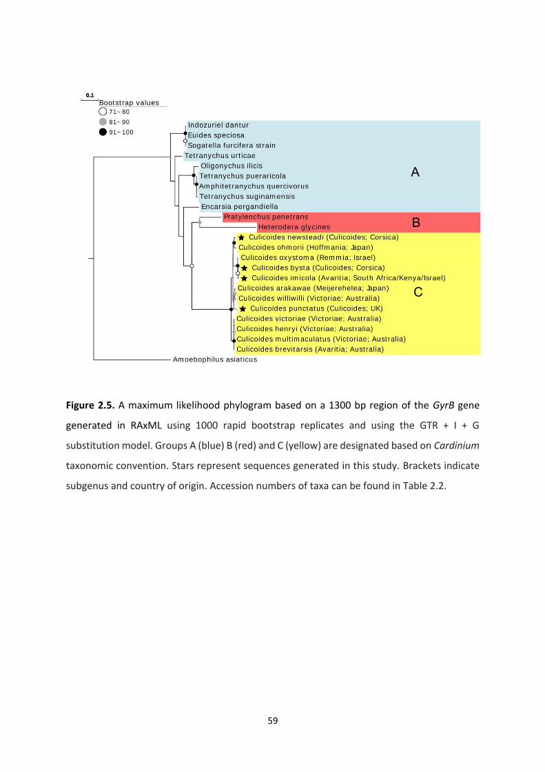
59
Figure 2.5. A maximum likelihood phylogram based on a 1300 bp region of the GyrB gene
generated in RAxML using 1000 rapid bootstrap replicates and using the GTR + I + G
substitution model. Groups A (blue) B (red) and C (yellow) are designated based on Cardinium
taxonomic convention. Stars represent sequences generated in this study. Brackets indicate
subgenus and country of origin. Accession numbers of taxa can be found in Table 2.2.
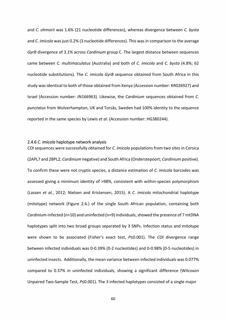
60
and C. ohmorii was 1.6% (21 nucleotide differences), whereas divergence between C. bysta
and C. imicola was just 0.2% (3 nucleotide differences). This was in comparison to the average
GyrB divergence of 3.1% across Cardinium group C. The largest distance between sequences
came between C. multimaculatus (Australia) and both of C. imicola and C. bysta (4.8%; 62
nucleotide substitutions). The C. imicola GyrB sequence obtained from South Africa in this
study was identical to both of those obtained from Kenya (Accession number: KR026927) and
Israel (Accession number: JN166963). Likewise, the Cardinium sequences obtained from C.
punctatus from Wolverhampton, UK and Torsås, Sweden had 100% identity to the sequence
reported in the same species by Lewis et al. (Accession number: HG380244).
2.4.6 C. imicola haplotype network analysis
COI sequences were successfully obtained for C. imicola populations from two sites in Corsica
(2APL7 and 2BPL2; Cardinium negative) and South Africa (Onderstepoort; Cardinium positive).
To confirm these were not cryptic species, a distance estimation of C. imicola barcodes was
assessed giving a minimum identity of >98%, consistent with within-species polymorphism
(Lassen et al., 2012; Nielsen and Kristensen, 2015). A C. imicola mitochondrial haplotype
(mitotype) network (Figure 2.6.) of the single South African population, containing both
Cardinium-infected (n=10) and uninfected (n=9) individuals, showed the presence of 7 mtDNA
haplotypes split into two broad groups separated by 3 SNPs. Infection status and mitotype
were shown to be associated (Fisher’s exact test, P≤0.001). The COI divergence range
between infected individuals was 0-0.39% (0-2 nucleotides) and 0-0.98% (0-5 nucleotides) in
uninfected insects. Additionally, the mean variance between infected individuals was 0.077%
compared to 0.37% in uninfected individuals, showing a significant difference (Wilcoxon
Unpaired Two-Sample Test, P≤0.001). The 3 infected haplotypes consisted of a single major
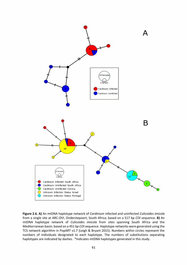
61
Figure 2.6. A) An mtDNA haplotype network of Cardinium infected and uninfected Culicoides imicola from a single site at ARC-OVI, Onderstepoort, South Africa; based on a 517 bp COI sequence. B) An mtDNA haplotype network of Culicoides imicola from sites spanning South Africa and the Mediterranean basin; based on a 451 bp COI sequence. Haplotype networks were generated using the TCS network algorithm in PopART v1.7 (Leigh & Bryant 2015). Numbers within circles represent the numbers of individuals designated to each haplotype. The numbers of substitutions separating haplotypes are indicated by dashes. *Indicates mtDNA haplotypes generated in this study.
62
group (8 individuals) and 2 haplotypes containing singletons. After extending the haplotype
network to include C. imicola populations from the western Mediterranean basin (WMB)
(Corsica and Portugal) and the eastern Mediterranean basin (EMB) (Israel), 12 haplotypes
were observed with 0-1.77% range of divergence over a 451 bp region. A majority of the
available COI sequences (24/29) from Israel clustered in the main haplotype associated with
Cardinium infection in South African individuals (Figure 2.6.). Another association was
identified between COI sequences from Portugal and Corsica which shared 3 haplotypes. The
two predominant haplotypes from EMB and WMB were separated by two SNPs either side of
the main Cardinium uninfected haplotype from South Africa.

63
2.5 Discussion
A main aim of this study was to explore the potential of C. pulicaris and C. punctatus as
suitable model organisms to undertake future UK-based work pertaining to Culicoides-
Cardinium interactions. Overall, the erroneous methodology and interpretation of a previous
study, assessing these two species (Lewis et al., 2014), has led to a false estimation of
Cardinium prevalence in C. pulicaris which required correction. First, the majority of C.
pulicaris extracts from Lewis were incorrectly interpreted as passing quality control when, in
fact, positive PCR bands were not observed (Figure 2.1.). Usually, negative PCR results are
interpreted as poor DNA quality, containing inhibitors, or containing a polymorphism at a
primer binding site. The latter appears to be true in this case, with inclusion of a secondary
set of primers (Dallas et al., 2003) amplifying the COI gene in all C. pulicaris extracts.
Apart from the poor interpretation of quality control results by Lewis, the author’s screening
of UK C. pulicaris from archived samples (2014) has identified a 7% prevalence through nested
PCR screening compared to the 26% prevalence inferred by Lewis using a conventional assay.
This higher prevalence is likely a result of cross-contamination from Cardinium positive C.
punctatus samples which were stored in the same cryobox. This is supported by the successful
sequencing of both C. pulicaris and C. punctatus COI sequences from the same extracts
labelled as C. pulicaris by Lewis (Table 2.3.1). The successful replication of raw screening data
from both Lewis and the author (Figure 2.1.) suggests any cross-contamination occurred at
the time of the original study in 2014. The renewed low Cardinium prevalence recorded for
C. pulicaris in the UK has implications for potential future in vivo work. As the more abundant
of the two species from UK collections, C. pulicaris (as opposed to C. punctatus) would be the
preferred candidate for any future work examining the vector competence or other
64
phenotypic effects relating to Cardinium. The initial intermediate prevalence suggested by
Lewis could have opened the possibility of case-control studies investigating the effects of
Cardinium on wild C. pulicaris populations. However, the single low-titre individual (1/27)
found to be infected suggests the numbers required to do such a study would be too large.
Despite the low titre and prevalence of infection observed here, a cryptic species of the
pulicaris complex, C. bysta, was found to be infected at fixation in Corsica, France. If
Cardinium does influence vectorial capacity, this result highlights the importance of using
molecular markers alongside morphological data to decipher cryptic species which vary in
their endosymbiont infection status. The high prevalence of Cardinium in the only other
infected UK species, C. punctatus, would require antibiotics to cure infection in order to
obtain a control group for experimental analysis. More importantly, due to the limited
numbers of wild C. punctatus, finding material to initiate a colony is the major issue of using
this species as a model organism.
The extended screening of midge populations in both Palearctic and Afrotropical regions
produced varying prevalence ratios of Cardinium. Most populations did not show any signs of
Cardinium infection (22/33 populations), 5 populations showed signs of low-intermediate
prevalence, and 6 were at fixation for Cardinium although a few of these had low sample
sizes. This varying prevalence is similar to the study by Mee et al. (2015), in which 3/26
positive species had a fixed infection, and 6 carried Cardinium in under 20% of individuals
sampled, with the remainder being of variable intermediate prevalence. However, Mee’s
study (2015) also detected at least one Cardinium positive individual in each population
screened which is at odds to the author’s findings and another study by Pagès et al. (2017),
65
investigating Cardinium distribution in Culicoides from Spain. It is possible that the apparent
“hotspot” in Australia is as a result of a more sensitive assay, as qPCR is often preferred to
nested PCR in screening (Ikewaki et al., 2003; Hyong et al., 2008). Alternatively, a temperature
effect could explain the discrepancies observed between these bioclimatic zones. Morag et
al. (2012) found a positive correlation between land surface temperature and Cardinium
prevalence in Culicoides. Similarly, a recent meta-analysis (Charlesworth et al., 2018) found a
a clear positive effect of temperature between arthropods and Cardinium infection rates
when assessing arthropods in 137 countries. Further climatic effects have also been
implicated in endosymbiont titre fluctuation (Hurst et al., 2000; Corbin et al., 2017) and
subsequently detection rate. Therefore, it is possible that both the varying Cardinium titres
and prevalence observed in this study, could be accounted for by temperature. An alternative
hypothesis is that invasive ranges may play a role in prevalence heterogeneity between
populations. For example, the loss of Wolbachia has been observed in invasive populations
of the thrip Pezothrips kellyanus, possibly due to selection pressures that vary throughout
host ranges (Nguyen, Spooner-Hart and Riegler, 2016).
A species which has come under scrutiny in the past due to its incursions into southern
Europe, leading to outbreaks of BTV in Europe, is C. imicola (Mellor et al., 1985; Boorman,
1986b; Jacquet et al., 2015). The absence of Cardinium in C. imicola populations from Corsica,
in this chapter, was recapitulated in a recent study from Spain (Pagès et al., 2017), where lack
of infection was also observed. No difference in prevalence was seen with the additional
nested screening indicating a lack of low-titre infections. However, the presence of Cardinium
in Israel (Morag et al., 2012) suggests infection heterogeneity exists across the Mediterranean
66
basin. To investigate this further, mtDNA haplotypes and the infection status of C. imicola
individuals within and between populations were compared. Endosymbionts can lead to
selective sweeps that result in a reduction in diversity of mtDNA markers. This has been
shown for Tetranychus truncatus mites, where haplotype diversity was greater in Wolbachia
uninfected individuals compared to those infected (Zhang et al., 2013). The haplotype
networks produced from this study (Figure 2.6.), suggests Cardinium infection in Culicoides is
another example of a selective sweep of the symbiont which facilitates the spread of specific
mtDNA haplotypes. The low diversity observed between C. imicola infected mtDNA
haplotypes when compared to those uninfected suggests a historic selective sweep
(homogenising mtDNA haplotypes) followed by the gradual accumulation of mutations and
subsequently new haplotypes. This interpretation is supported by the two minor infected
haplotypes differing by one nucleotide difference. Alternative explanations for these data
would be a bottleneck or founder event, although low mtDNA diversity due to endosymbiont
effects in C. imicola is more parsimonious (Hurst and Jiggins, 2005). Although there is a
structure observed between infection status and mtDNA haplotype, the presence of 2
uninfected individuals in the main infected haplotype in C. imicola from South Africa suggests
imperfect vertical endosymbiont transmission.
Infection heterogeneity between C. imicola populations from the EMB and WMB could be
explained by the dispersal of both infected and uninfected founding populations to both
areas. Indeed, a recent study (Jacquet et al., 2016) suggests the dispersal of C. imicola to the
WMB was as a result of wind-dispersal of populations from Corsica, suggesting a lack of
Cardinium infection was as a result of absence in dispersed populations. Further to this, Israeli
67
(EMB) haplotypes clustered within the main Cardinium infected mitotype observed in South
Africa, whereas the Portuguese (WMB) haplotypes all clustered within an uninfected Corsican
mitotype. Although, the infection statuses of these Portuguese and Israeli individuals are
unknown, the recent Pagès (2017) screening for Cardinium found no indication of C. imicola
infection in the WMB (Spain) but others (Morag et al., 2012) have observed the symbiont to
be present in the EMB (Israel). The matrilineal subdivision in Cardinium infected South African
C. imicola and uninfected Corsican C. imicola (Figure 2.6.) is similar to observations by several
studies (Dallas et al., 2003; Nolan et al., 2008; Calvo et al., 2009) which all noted a remarkable
divergence between C. imicola mitotypes from the EMB and WMB. This suggests that the
linkage disequilibrium of Cardinium and mitochondria offers insights into symbiont gene flow
within the Mediterranean basin but may not assist in elucidating host gene flow. For example,
if dispersal events occur involving Cardinium-infected C. imicola, selective sweeps of the
symbiont may erase any previously detectable biodiversity. This is reminiscent of similar
patterns observed in other insects with endosymbiont infections: the mosquito vector, Culex
pipiens (Rasgon, Cornel and Scott, 2006), the parasitoid wasp, Nasonia vitripennis
(Raychoudhury et al., 2010) and the ladybird Adalia bipunctata (Schulenburg et al., 2002). In
all these cases, the frequency of mtDNA haplotypes are more closely associated to the
endosymbionts in a population rather than geography. Cryptic species can also offer
explanations for endosymbiont infection heterogeneity. However, there was no evidence of
cryptic species between C. imicola populations based on COI sequence divergence ranges of
<2% (Lassen et al., 2012; Nielsen and Kristensen, 2015). Overall, this suggests that a non-
biased marker is needed for biogeographic studies aiming to evaluate the population
structure of C. imicola.
68
Analysis of the relatedness of strains using the GyrB locus in this study (Figure 2.5.)
corroborate a recent study by Siozios et al. (2019) placing the Culicoides Cardinium as a
monophyletic sister clade to Cardinium group B from parasitic plant nematodes. Mee et al.
(2015) has previously suggested that Cardinium strains cluster by geography, however, we
find a sporadic distribution of strains pertaining to location; the two strains identified from
Corsica, group with strains from Japan, Israel and Southern Africa instead of each other.
Although symbionts have been known to cluster based on location due to geographic barriers
(Russell et al., 2009), the ability of Culicoides to disperse over long ranges (Burgin et al., 2013;
Jacquet et al., 2016) can explain why we do not see a similar scenario in Cardinium-infected
midges.
It is curious to note that although Cardinium was detected in five species in this study, all of
which are vectors of BTV (C. newsteadi, C. pulicaris, C. punctatus, C. bysta and C. imicola), C.
obsoletus (the major Palearctic vector of BTV) in the UK appears to be infection free. Pagès
et al. (2017), however, have recently shown very low infection rates in C. obsoletus from Spain
(2/304 individuals). Despite this, such low prevalence indicates Cardinium is most likely not
relevant to vector biology at a population level. There is a particular interest in varying
endosymbiont titres of vector species as they could influence potential biocontrol effects
including viral blockage and host longevity (Hoffmann, Ross and Rašić, 2015). Aside from
environmental effects, endosymbiont density can be determined by a range of biotic factors
including bacterial strain and sex (Duron, Fort and Weill, 2007; Tortosa et al., 2010).
Considering the discovery of SBV transovarial transmission in C. punctatus (Larska et al.,
2013), and Cardinium presence in ovaries (Nakamura et al., 2009; Morag et al., 2012), direct
69
interactions between Cardinium and SBV could alter rates of viral vertical transmission via
the production of antiviral metabolites (Joyce et al., 2011) or resource competition (Caragata
et al., 2013; Frentiu, 2017). However, in contrast with other symbiont-insect associations, the
biological significance of this Cardinium clade in Culicoides remains unknown and requires
further research. Despite a likely vertical transmission, the lack of sex-ratio distortion
observed in this study in the two species which contained both males and females (C. imicola
and C. punctatus; Table 2.5.2), suggests the induction of parthenogenesis, feminisation or
male-killing is unlikely to be associated with Cardinium in this instance.
Other Cardinium strains have been implicated in cytoplasmic incompatibility (CI) in the wasp
Encarsia pergandiella (Hunter, Perlman and Kelly, 2003), the planthopper Sogatella furcifera
(Zhang, Zhao and Hong, 2012) and spider mites (Gotoh, Noda and Ito, 2007). The recent
publication of the C. punctatus Cardinium genome by Siozios et al. (2019) has suggested
possible unique genes related to CI. These are a family of endonucleases (DUF1703) whose
paralogs are associated with the induction of CI in Wolbachia (Beckmann, Ronau and
Hochstrasser, 2017). The presence of low Wolbachia densities have been suggested to lead
to a weak sperm modification ability and a reduced CI effect (Kondo et al., 2002). If indeed
midge Cardinium does induce CI, low titres described in this study and elsewhere could
suggest a weak CI effect in some host species. The low-density infections observed here along
with the geographical variability in Cardinium infection status, has important implications for
any future Cardinium-based vector control initiatives through effects on population dynamics.
This is especially relevant if the strategy is reliant on the driving-mechanism of CI. Certain
symbionts of haematophagus insects have also been shown to enable the synthesis of B
vitamins due to their scarce availability in a blood diet (Rio et al. 2016). However, there is no
70
indication of this from genomic data, with no biotin or other B vitamin biosynthetic pathways
present; indeed, a biotin transport system indicates Cardinium is a B vitamin sink rather than
a provider (Siozios et al., 2019).
To conclude, the Cardinium infection status of Culicoides pulicaris has been reassessed to
suggest this species is not suitable for further in vivo work relating to the symbiont in the UK.
The difficulties of culturing and maintaining Culicoides colonies (Mullen et al., 1985; Jennings
and Mellor, 1988; Nayduch et al., 2014) has so far stunted investigations into the role of
Cardinium. However, Cardinium has been shown to disrupt immune gene expression in cell
culture models (Nakamura et al., 2011). Subsequently, cell-lines could potentially be used as
an alternative to whole-midge assays in order to assess future host phenotypic effects of
Cardinium. Finally, the linkage disequilibrium between Cardinium and mitochondria suggests
a potential to confound dispersal studies utilising the COI gene as a phylogeographic marker.
Subsequently, due to the importance of C. imicola as a vector in Africa and Europe, the
utilisation of mtDNA markers to assess C. imicola population structure should be met with
caution.
71
Chapter 3: Limoniae Group Rickettsia are Widespread in Culicoides and Reach High Frequency
72
3.1 Abstract
There is increasing interest in the heritable bacteria of invertebrate vectors of disease as they
present a novel target for control initiatives. Previous studies on biting midges (Culicoides
spp.), known to transmit several RNA viruses of veterinary importance, have revealed
infections with the endosymbiotic bacteria, Wolbachia and Cardinium. However, rickettsial
symbionts in these vectors are underexplored. Screening of 493 Culicoides individuals from
30 Palearctic, Nearctic or Afrotropical species revealed Rickettsia represent a widespread but
previously overlooked association, reaching high frequencies in midge populations and
present in 37% of the species tested. Phylogenetic analysis of a multigene dataset clusters the
Rickettsia within the Limoniae clade of the genus, a group known to infect several aquatic and
hematophagous taxa. Given the importance of biting midges as vectors, a key area of future
research is to establish the impact of this endosymbiont on vector competence.
73
3.2 Introduction
Heritable bacteria represent an important component of the biology of many arthropods.
Carried by over half of all species (Weinert et al., 2015), many vertically transmitted microbes
contribute to host function. This contribution is most commonly through specific services,
such as nutrient provisioning or protection (Douglas, 2009; Oliver et al., 2009; Jaenike et al.,
2010). Conversely, their maternal-inheritance has led symbionts to favour production of
daughters by their host, leading to the evolution of systems biasing the offspring sex ratio
towards females (reproductive parasitism) (Hurst and Frost 2015). The strength of symbiont
impact on individual biology, combined with the high frequency with which arthropod species
are infected with symbionts, has led to intense study.
Particular attention has been focussed on symbiont/host interactions in vector species.
Through the induction of cytoplasmic incompatibility, the endosymbiont Wolbachia prevents
the formation of viable progeny between infected males and uninfected females in various
dipterans including Drosophila spp. and Aedes spp. (Werren, Baldo and Clark, 2008). With
respect to the latter, not only can this incompatibility enable vector population suppression
but, through unknown mechanisms, a strong RNA virus resistance phenotype (Moreira et al.,
2009; Bian et al., 2010; Blagrove et al., 2012; van den Hurk et al., 2012). Furthermore,
experimental evidences show that both Wolbachia and another proteobacteria,
Wigglesworthia, can act as obligate (required) symbionts, provisioning blood sucking vector
hosts with B vitamins that are lacking in a blood-diet (Rio et al. 2016). This provisioning has
evolved independently in bed bugs (Cimex lectularius; Nikoh et al. 2014), tsetse flies (Glossina
sp.; Akman et al. 2002; Snyder et al. 2010; Rio et al. 2012) and lice (Pediculus schaeffi; Boyd
74
et al. 2014). Additional genomic surveys suggest that other proteobacterial symbionts
including Coxiella are involved in metabolic homeostasis (Zhong, Jasinskas and Barbour, 2007;
Manzano-Marín et al., 2015; Smith et al., 2015). As such, these symbioses can have profound
effects on the biology, ecology and evolutionary dynamics of vector-pathogen interactions.
Rickettsia (Class: Alphaproteobacteria; Order: Rickettsiales) symbionts are obligate
intracellular bacteria most notable for containing species pathogenic to vertebrates, such as
Rickettsia prowazekii, the causative agent of louse-borne Typhus fever, Rickettsia rickettsii
(Rocky Mountain spotted fever) and Rickettsia conorii (Boutonneuse or Mediterranean
spotted fever). Despite this, vertebrate disease-causing Rickettsia are atypical of the genus as
a whole (Perlman et al. 2006; Weinert et al. 2009), and many Rickettsia are maintained
without infectious transfer. Members are known to induce a variety of reproductive
manipulations, including male-killing in ladybird beetles (Adalia bipunctata) (Werren et al.,
1994; Hurst et al., 1999; Majerus et al., 1999) and parthenogenesis induction in parasitoid
wasps (Pnigalio soemius; Neochrysocharis formosa) (Hagimori et al., 2006; Giorgini et al.,
2010). Rickettsia symbiont infection can also be protective, enhancing resistance of aphids
(Acyrthosiphon pisum) to fungal attack, and whiteflies (Bemisia tabaci) to bacterial challenge
(Łukasik et al., 2013; Hendry, Hunter and Baltrus, 2014). Of significance to the study of
vectors, Rickettsia are also known to increase the competence of Bemisia whiteflies for
transmission of tomato leaf curl virus (Kliot et al., 2014). Members of the genus can also be
insect-vectored plant pathogens in their own right, for example, underlying papaya bunchy
top disease (Luis-Pantoja et al., 2015). As such, symbiosis with Rickettsia is biologically
75
important at the individual and population level, and both as vectored disease agents in
themselves and as a symbiont facilitating the spread of other diseases.
Previous studies of Culicoides symbionts have screened extensively for Cardinium and
Wolbachia infections (Nakamura et al., 2009; Morag et al., 2012; Lewis et al., 2014; Mee et
al., 2015; Pagès et al., 2017), with most failing to report presence of Rickettsia. However, a
16S metagenomic screening project in C. sonorensis gut samples revealed amplicons allied to
Rickettsia (Campbell et al., 2004), albeit with no phylogenetic or population-based
information. Further, when a colleague (Dr Stefanos Siozios, University of Liverpool)
performed a shallow whole-genome sequencing of C. newsteadi N5, he recovered a near
complete genome of an uncharacterized Rickettsia species related to the Limoniae clade of
Rickettsia, a group known to infect several aquatic and hematophagous taxa.
Due to the genetic diversity and various phenotypes associated with endosymbionts, systems
of strain typing have been developed. For Wolbachia, the surface protein gene wsp was
initially used as a genetic marker (Zhou, Rousset and O’Neil, 1998). However, evidence of
diversifying selection (Jiggins, Hurst and Yang, 2002) and recombination (Baldo, Lo and
Werren, 2005) made this gene unsuitable for isolate characterization if used alone. Further
to this, horizontal gene transfer events between genes (intergenic recombination) detected
in Wolbachia suggested a single-locus approach to strain typing was inappropriate (Baldo et
al. 2006). Subsequently, multigenic approaches have been seen as a superior tool to assess
genetic relatedness between endosymbionts. A commonly used method, multi locus
sequence typing (MLST), defines the haplotypes of individual strains based on house-keeping
76
genes and was initially used as a surveillance tool for pathogenic bacteria (Maiden et al.,
1998). However, more recently, this approach has included non-pathogenic bacteria (Baldo
et al. 2006; Cai et al. 2007) for population and evolutionary analyses, as well as the association
of particular strains with phenotypes. The conserved nature of housekeeping genes provides
enough discrimination over an evolutionary timescale to be useful in typing isolates, as well
as being less likely to undergo diversifying selection and intragenic recombination, which can
disguise relationships among strains. For each locus, sequences are designated unique alleles
and by combining these from the same isolate, an allelic profile is created. No weighting is
given to single nucleotide changes or recombination exchanges meaning strains can be
compared even when recombination is frequent in a genus. Thus, this system can allow for
the accurate tracing of endosymbionts between geographic locations and insect
communities. However, the comparison of allelic profiles can only give limited information
related to evolutionary history and fails to distinguish recently diverged strains meaning
concatenated gene phylogenies are seen as a suitable alternative approach (Bleidorn and
Gerth 2017).
Subsequently, this chapter aimed to initially identify Rickettsia’s occurrence and distribution
through a targeted Culicoides screen. Isolates were then assessed via a strain typing system
similar to MLST, before a concatenated gene phylogeny was constructed to give better
phylogenetic resolution after the investigation of any potential confounding recombination
events.
77
3.3 Methods
3.3.1 Culicoides collection identification and DNA extractions
A majority of collection, identification and DNA extractions of Culicoides details can be found
in chapter 2. Slight differences include the inclusion of C. sonorensis populations of midges
for screening from California, USA (collected February/March 2018); a major vector of BTV in
North America. Collection of C. sonorensis was undertaken using carbon-dioxide baited traps
and confirmation of identification was performed with the help of Dr Alec Gerry and Xinmi
Zhang (UC Riverside, USA) using wing morphology. DNA extractions of 29 individual C.
sonorensis and 10 pools of 5 individuals were prepared using Qiagen DNEasy kits (Qiagen.
Inc., Valencia, CA, USA) following the manufacturer’s instructions for insect tissue (Qiagen,
2006). This brings the total number of specimens in this study to 493 specimens of 30
Culicoides species spanning Palearctic, Nearctic and Afrotropical ecozones.
3.3.2. PCR screening
Presence of Rickettsia was initially assessed by PCR assay using Rickettsia-specific primers
designed to amplify a 320-bp region of the 17kDa surface antigen precursor. Cycling
conditions were as follows: initial denaturation at 95°C for 5 min, followed by 35 cycles of
denaturation (94°C, 30 sec), annealing (54°C, 30 sec), extension (72°C, 120 sec), and a final
extension at 72°C for 7 min. Amplicons identified by gel electrophoresis were subsequently
purified enzymatically (ExoSAP) and Sanger sequenced as described in chapter 2.
78
3.3.3 Multilocus typing gene choice
Based on previous studies (Fournier et al. 2003; Weinert et al. 2009; Li et al. 2010;
Machtelinckx et al. 2012; Santibáñez et al. 2013) that profile Rickettsia diversity, the 16S
rRNA, gltA (citrate synthase), coxA (cytochrome oxidase) and atpA (ATP synthase) genes were
chosen as indicators of genetic relatedness between isolates. With the consideration that
these housekeeping loci may be too conserved to resolve recently diverged strains, the 17KDa
gene, was included to allow for higher resolution in typing, alongside an inference of selection
pressure due to the divergent nature of antigen genes compared to housekeeping genes.
Briefly, 16S has a structural role in the ribosomal protein scaffold; gltA encodes a subunit of
the Citrate Synthase enzyme utilised in the Kreb’s cycle; coxA encodes a catalytic subunit of
the respiratory electron transport chain; atpA is a subunit of the enzyme ATP synthase
assisting in the conversion of Adenosine diphosphate (ADP) to Adenosine triphosphate (ATP);
and finally, 17KDa encodes an antigen of the outer membrane protein, thought to assist in
the adhesion of Rickettsia to host cells. Culicoides sonorensis individuals and pools were
screened using all 5 primers in contrast to other populations where the 17KDa primers were
used alone. Insects from positive pooled C. sonorensis were rescreened to identify infected
individuals.
Primers to amplify these loci (Table 3.1.) were designed in Geneious (Kearse et al., 2012)
based on the C. newsteadi N5 (RiCNE) genome (BioProject accession number PRJNA376033;
Dr Stefanos Siozios) and available conserved gene sequences so that they could amplify across
several Rickettsia groups but would not cross-amplify any alpha-proteobacteria outgroups.
All PCR amplifications were performed under the same 17KDa assay conditions described
above. Sequencing through both strands allowed for the clarification of ambiguous base calls
79
as well as giving greater sequence coverage at individual loci. Raw sequences were edited in
UGENE (Okonechnikov et al., 2012) and alignments for each locus were generated in MEGA6
using the ClustalW algorithm (Tamura et al., 2013).
A profile of each locus was constructed by calculating GC content, nucleotide diversity per
site (π) and the percentage of variable sites using DNAsp v5 (Librado and Rozas 2009). As
phylogenetic inferences can be complicated by recombination, the presence of intragenic and
intergenic recombination was investigated. The program RDP v4 (Martin et al., 2015) was
used to test for intragenic recombination using the MaxChi, RDP, Chimaera, Bootscan and
GENECONV algorithms with the following criteria to assess a true recombination positive: a
p-value of <0.001, sequences were considered linear with 1000 permutations being
performed. As these algorithms are prone to false positives, a recombination event was
considered likely if more than one algorithm gave a significant p-value, as well as being
confirmed by visual inspection. To this end, maximum likelihood trees either side of putative
recombination breaking points were generated for inspection of congruency. For any
intragenic recombination events detected, the locus for that strain was removed from
concatenation. Intergenic recombination was also assessed by comparing single-gene trees.
Additionally, all protein coding loci (17KDa, atpA, coxA and gltA) were investigated for
evidence of diversifying selection at the gene level via pairwise non-
synonymous/synonymous rate ratio analysis (Ka/Ks ratio).
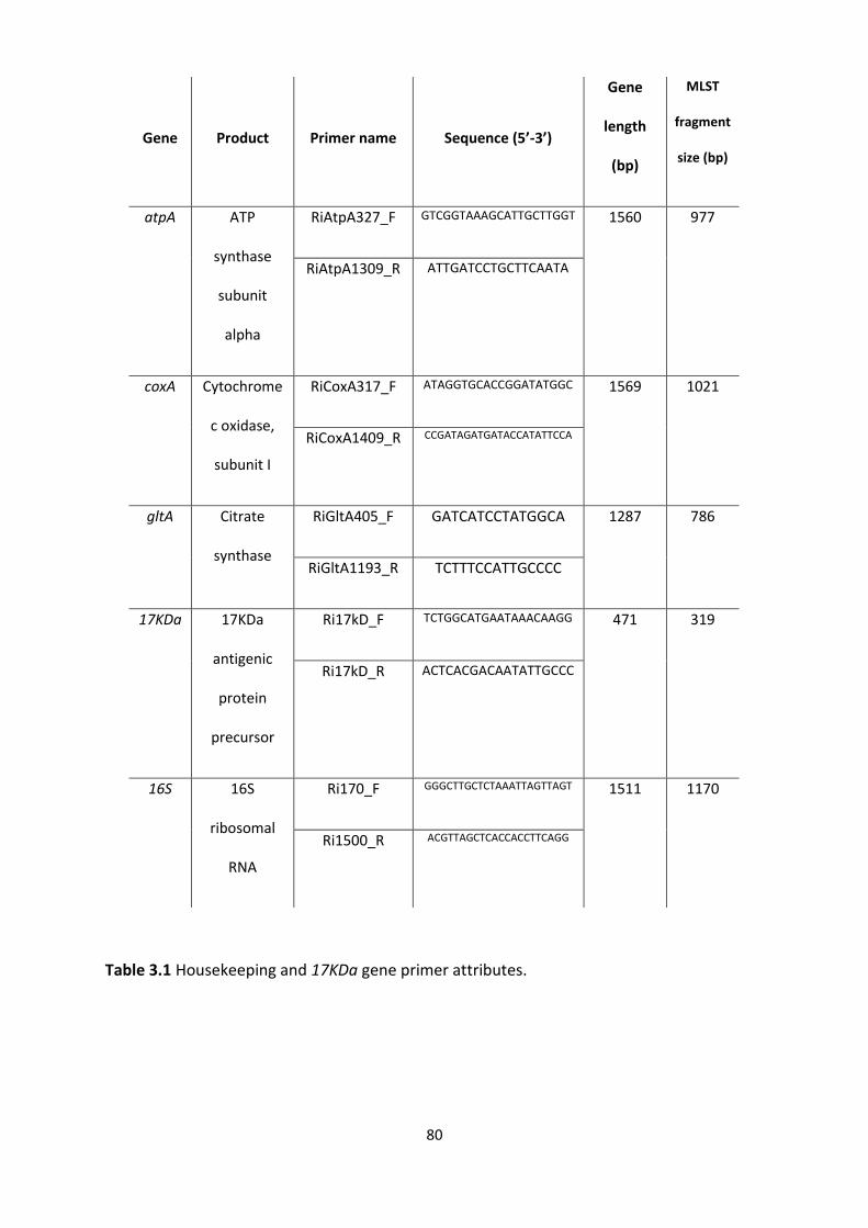
80
Table 3.1 Housekeeping and 17KDa gene primer attributes.
Gene
Product
Primer name
Sequence (5’-3’)
Gene
length
(bp)
MLST
fragment
size (bp)
atpA ATP
synthase
subunit
alpha
RiAtpA327_F GTCGGTAAAGCATTGCTTGGT 1560 977
RiAtpA1309_R ATTGATCCTGCTTCAATA
coxA Cytochrome
c oxidase,
subunit I
RiCoxA317_F ATAGGTGCACCGGATATGGC 1569 1021
RiCoxA1409_R CCGATAGATGATACCATATTCCA
gltA Citrate
synthase
RiGltA405_F GATCATCCTATGGCA 1287 786
RiGltA1193_R TCTTTCCATTGCCCC
17KDa 17KDa
antigenic
protein
precursor
Ri17kD_F TCTGGCATGAATAAACAAGG 471 319
Ri17kD_R ACTCACGACAATATTGCCC
16S 16S
ribosomal
RNA
Ri170_F GGGCTTGCTCTAAATTAGTTAGT 1511 1170
Ri1500_R ACGTTAGCTCACCACCTTCAGG
81
3.3.4 Multigenic strain typing
Following the convention of multilocus sequence typing (MLST), all unique genotypes were
designated allele numbers (used as a unique identifier) which, when combined at all loci,
produce an allelic profile (Maiden et al., 1998). Aside from identifying specific isolates, this
multigenic approach also allows for clonal complexes to be identified (conventionally allelic
profiles which are identical at three or more loci). Allelic profiles and complexes were
designated based on unweighted pair group method with arithmetic mean (UPGMA) cluster
analysis and visualised as a minimum spanning tree (MST) implemented by Bionumerics v7
(Applied Maths 2012). Quantum GIS software v3 (Quantum GIS Development Team, 2016)
was then used to map strains to investigate geographic patterns of relatedness.
3.3.5 Phylogenetic analysis
As UPGMA analysis does not take into consideration substitution models of character
changes, the addition of other Rickettsia-positive taxa, provided by Panupong Thongprem
(University of Liverpool) (Appendix 1), were included in a further phylogenetic analysis.
Sanger sequence visualisation and analysis was performed by the author for all loci except
gltA which was used in the initial screening for Thongprem’s screen. After checking for
recombination events likely to affect phylogenetic inferences, the relatedness of midge
Rickettsia within the Rickettsiaceae was assessed using the concatenation of the 5 individual
genes. First, sequences of 16S rRNA, 17KDa, atpA, coxA and gltA from selected Rickettsia
groups were extracted and concatenated using SequenceMatrix v1.8 (Vaidya, Lohman and
Meier, 2011). Sequences were then combined with others selected from across the genus
(Weinert et al. 2009) obtained from GenBank (NCBI). Strains included both the ancestral
“Hydra” (Megaira) group as well as derived pathogenic isolates (Spotted fever and
82
Typhus). For some of the taxa included, only 16S or gltA sequences were available, and so
missing data was allowed in the alignment where a gene was absent. According to Wiens
(2006), missing character data should not affect phylogenetic resolution, and is only likely to
be an issue for incomplete data if the number of characters is very low. The sequences for
each locus were aligned using the “L-INS-I” algorithm in MAFFT v7.4 (Katoh and Standley
2013) before a partition scheme was devised in order to allow for an optimised substitution
model for each gene using ModelFinder (Kalyaanamoorthy et al., 2017). Model selection
included free rate as well as traditional gamma distributed rate categories which may
“underfit” multi-gene data. Edge-proportional models were included where varying branch
lengths were allowed in different partitions before proportionally rescaling when devising the
multi-gene tree. The selected models were as follows: 16S and GTR+F+R4; 17KDa and
TVM+F+I+G4; atpA and GTR+F+I+G4; coxA and TVM+F+I+G4; gltA and GTR+F+R3. A maximum
likelihood (ML) phylogeny was estimated with IQTree (Nguyen et al., 2015) using the
concatenated alignment of 4403 nucleotides and 5000 ultrafast bootstraps. Various
constrained tree searches alongside the Shimodaira-Hasegawa (SH) test (Significance
threshold p<0.001) were implemented in IQTree to test alternative hypotheses of monophyly
with respect to host taxa. The hypothesis test statistic, for a comparison of alternative
topologies, was achieved by obtaining a maximised likelihood score (-ln) for each comparison.
Finally, trees were drawn and annotated using the EvolView (He et al., 2016) online tree
annotation and visualization tools.

83
3.3.6 Nucleotide sequence accession numbers
Culicoides' COI barcodes and sequences generated for individual Rickettsia loci in this study
were deposited in GenBank under deposition numbers KY765346-KY765408 and KY778697-
KY778698.

84
3.4 Results
3.4.1 Prevalence of Rickettsia in biting midges
Screening of field collected midge specimens using the 17KDa gene revealed Rickettsia
infections in 155 of 493 (31%) individuals and 11 of 30 (37%) Culicoides species sampled
(Tables 3.2.1 and 3.2.2). Rickettsia-positive species of biting midge were recorded across
Culicoides subgenera. Infection was identified across the subgenera Beltranmyia (1/3
species), Culicoides (7/11 species), Monoculicoides (2/3) and Oecacta (1/4 species) [as
determined by Borkent (2016)]. There was no apparent host sex bias in the presence of
Rickettsia for either C. pulicaris P1 (UK) (Fisher’s two-tailed test; p=1) or C. impunctatus
(Fisher’s two-tailed test; p=0.36), the only infected species with both host sexes available to
compare.
Rickettsia was found in all individuals in 16 of the 20 positive populations screened, with
prevalence being at low or intermediate prevalence in the remaining 4 (1 C. newsteadi N1
population and 3 C. impunctatus populations) (Table 3.2.1. and 3.2.2; Figure 3.1.). Where
multiple samples of particular species were tested, there was no significant difference in the
fraction infected (C. impunctatus populations from Bala vs Kielder in the UK, N1=31, N2=23,
Fisher’s two-tailed test; p=0.37). Mitochondrial DNA barcoding of infected (KY765353) and
uninfected (KY765354) individuals of C. impunctatus confirmed these individuals shared a
barcode, consistent with infection showing within-species polymorphism.
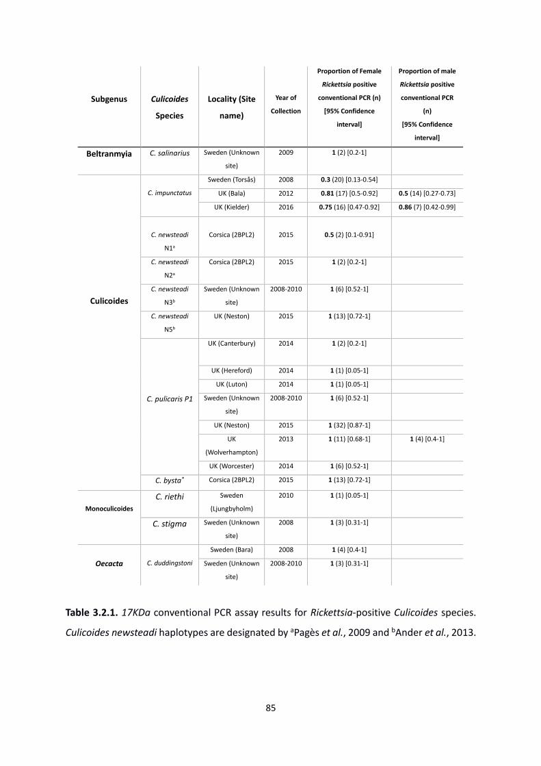
85
Table 3.2.1. 17KDa conventional PCR assay results for Rickettsia-positive Culicoides species.
Culicoides newsteadi haplotypes are designated by aPagès et al., 2009 and bAnder et al., 2013.
Subgenus
Culicoides
Species
Locality (Site
name)
Year of
Collection
Proportion of Female
Rickettsia positive
conventional PCR (n)
[95% Confidence
interval]
Proportion of male
Rickettsia positive
conventional PCR
(n)
[95% Confidence
interval]
Beltranmyia C. salinarius Sweden (Unknown
site)
2009 1 (2) [0.2-1]
Culicoides
C. impunctatus
Sweden (Torsås) 2008 0.3 (20) [0.13-0.54]
UK (Bala) 2012 0.81 (17) [0.5-0.92] 0.5 (14) [0.27-0.73]
UK (Kielder) 2016 0.75 (16) [0.47-0.92] 0.86 (7) [0.42-0.99]
C. newsteadi
N1a
Corsica (2BPL2)
2015
0.5 (2) [0.1-0.91]
C. newsteadi
N2a
Corsica (2BPL2) 2015 1 (2) [0.2-1]
C. newsteadi
N3b
Sweden (Unknown
site)
2008-2010 1 (6) [0.52-1]
C. newsteadi
N5b
UK (Neston) 2015 1 (13) [0.72-1]
C. pulicaris P1
UK (Canterbury) 2014 1 (2) [0.2-1]
UK (Hereford) 2014 1 (1) [0.05-1]
UK (Luton) 2014 1 (1) [0.05-1]
Sweden (Unknown
site)
2008-2010 1 (6) [0.52-1]
UK (Neston) 2015 1 (32) [0.87-1]
UK
(Wolverhampton)
2013 1 (11) [0.68-1] 1 (4) [0.4-1]
UK (Worcester) 2014 1 (6) [0.52-1]
C. bysta* Corsica (2BPL2) 2015 1 (13) [0.72-1]
Monoculicoides
C. riethi Sweden
(Ljungbyholm)
2010 1 (1) [0.05-1]
C. stigma Sweden (Unknown
site)
2008 1 (3) [0.31-1]
Oecacta
C. duddingstoni
Sweden (Bara) 2008 1 (4) [0.4-1]
Sweden (Unknown
site)
2008-2010 1 (3) [0.31-1]
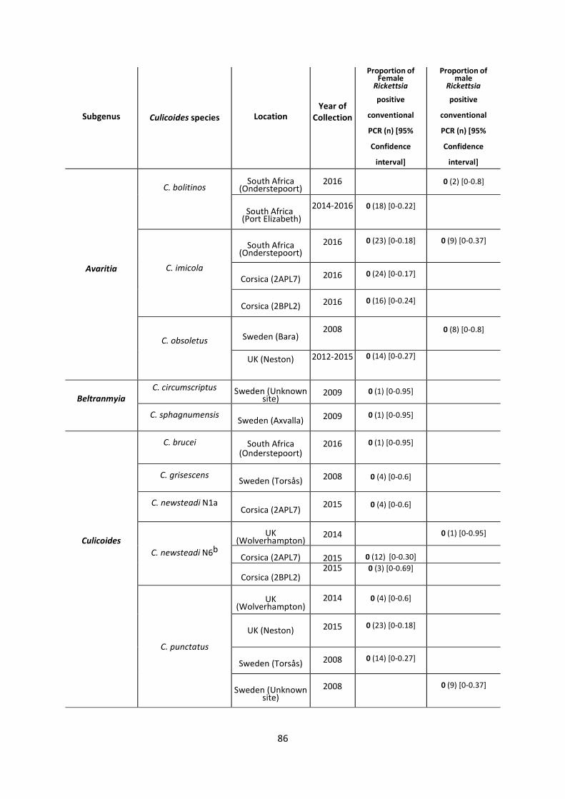
86
Subgenus
Culicoides species
Location
Year of Collection
Proportion of Female
Rickettsia
positive
conventional
PCR (n) [95%
Confidence
interval]
Proportion of male
Rickettsia
positive
conventional
PCR (n) [95%
Confidence
interval]
Avaritia
C. bolitinos
South Africa
(Onderstepoort)
2016
0 (2) [0-0.8]
South Africa (Port Elizabeth)
2014-2016
0 (18) [0-0.22]
C. imicola
South Africa
(Onderstepoort)
2016
0 (23) [0-0.18]
0 (9) [0-0.37]
Corsica (2APL7)
2016
0 (24) [0-0.17]
Corsica (2BPL2)
2016
0 (16) [0-0.24]
C. obsoletus
Sweden (Bara)
2008
0 (8) [0-0.8]
UK (Neston)
2012-2015 0 (14) [0-0.27]
Beltranmyia C. circumscriptus
Sweden (Unknown site)
2009
0 (1) [0-0.95]
C. sphagnumensis
Sweden (Axvalla)
2009
0 (1) [0-0.95]
Culicoides
C. brucei
South Africa
(Onderstepoort)
2016
0 (1) [0-0.95]
C. grisescens
Sweden (Torsås)
2008
0 (4) [0-0.6]
C. newsteadi N1a
Corsica (2APL7)
2015
0 (4) [0-0.6]
C. newsteadi N6b
UK
(Wolverhampton)
2014
0 (1) [0-0.95]
Corsica (2APL7)
2015
5
0 (12) [0-0.30]
Corsica (2BPL2)
2015
0 (3) [0-0.69]
C. punctatus
UK
(Wolverhampton)
2014
0 (4) [0-0.6]
UK (Neston)
2015
0 (23) [0-0.18]
Sweden (Torsås)
2008
0 (14) [0-0.27]
Sweden (Unknown
site)
2008
0 (9) [0-0.37]
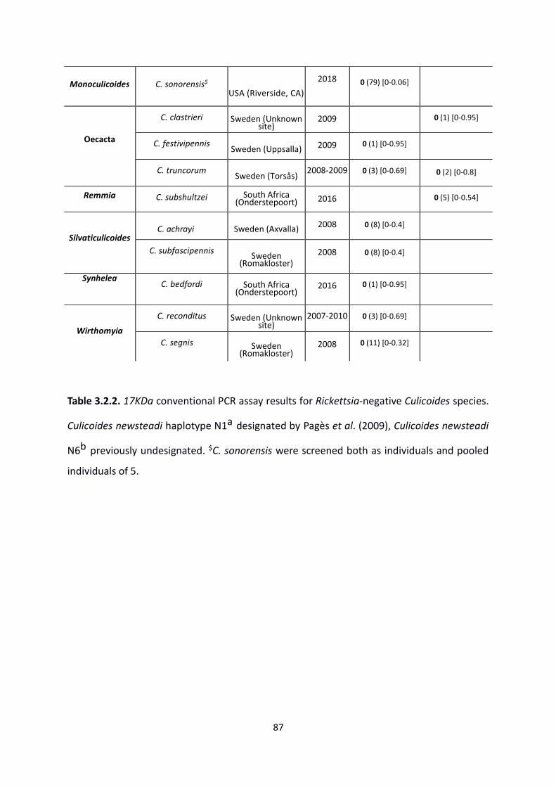
87
Monoculicoides
C. sonorensis$
USA (Riverside, CA)
2018
0 (79) [0-0.06]
Oecacta
C. clastrieri
Sweden (Unknown
site)
2009
0 (1) [0-0.95]
C. festivipennis
Sweden (Uppsalla)
2009
0 (1) [0-0.95]
C. truncorum
Sweden (Torsås)
2008-2009
0 (3) [0-0.69]
0 (2) [0-0.8]
Remmia
C. subshultzei South Africa
(Onderstepoort)
2016
0 (5) [0-0.54]
Silvaticulicoides
C. achrayi
Sweden (Axvalla)
2008
0 (8) [0-0.4]
C. subfascipennis
Sweden (Romakloster)
2008
0 (8) [0-0.4]
Synhelea
C. bedfordi South Africa
(Onderstepoort)
2016
0 (1) [0-0.95]
Wirthomyia
C. reconditus
Sweden (Unknown
site)
2007-2010
0 (3) [0-0.69]
C. segnis
Sweden (Romakloster)
2008
0 (11) [0-0.32]
Table 3.2.2. 17KDa conventional PCR assay results for Rickettsia-negative Culicoides species.
Culicoides newsteadi haplotype N1a designated by Pagès et al. (2009), Culicoides newsteadi
N6b previously undesignated. $C. sonorensis were screened both as individuals and pooled
individuals of 5.
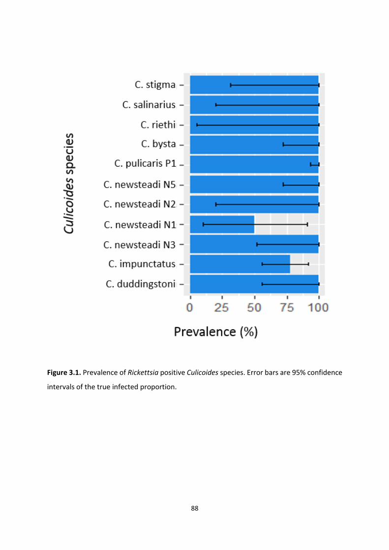
88
Figure 3.1. Prevalence of Rickettsia positive Culicoides species. Error bars are 95% confidence
intervals of the true infected proportion.
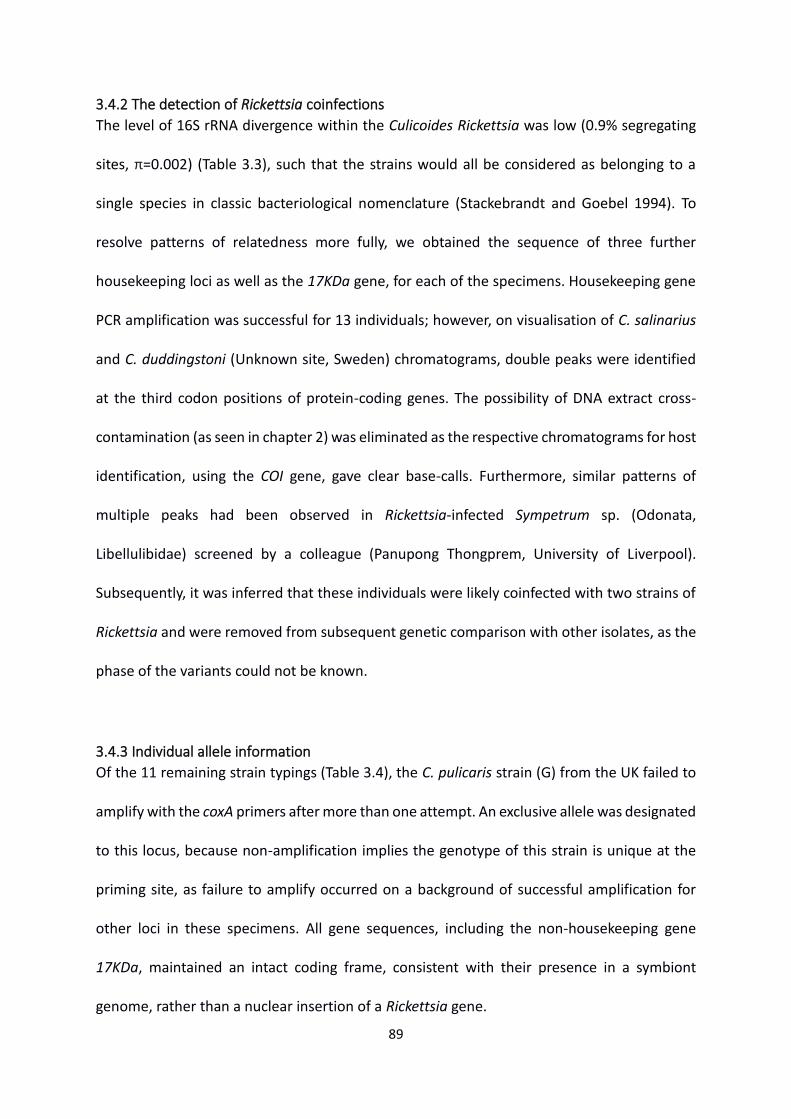
89
3.4.2 The detection of Rickettsia coinfections
The level of 16S rRNA divergence within the Culicoides Rickettsia was low (0.9% segregating
sites, π=0.002) (Table 3.3), such that the strains would all be considered as belonging to a
single species in classic bacteriological nomenclature (Stackebrandt and Goebel 1994). To
resolve patterns of relatedness more fully, we obtained the sequence of three further
housekeeping loci as well as the 17KDa gene, for each of the specimens. Housekeeping gene
PCR amplification was successful for 13 individuals; however, on visualisation of C. salinarius
and C. duddingstoni (Unknown site, Sweden) chromatograms, double peaks were identified
at the third codon positions of protein-coding genes. The possibility of DNA extract cross-
contamination (as seen in chapter 2) was eliminated as the respective chromatograms for host
identification, using the COI gene, gave clear base-calls. Furthermore, similar patterns of
multiple peaks had been observed in Rickettsia-infected Sympetrum sp. (Odonata,
Libellulibidae) screened by a colleague (Panupong Thongprem, University of Liverpool).
Subsequently, it was inferred that these individuals were likely coinfected with two strains of
Rickettsia and were removed from subsequent genetic comparison with other isolates, as the
phase of the variants could not be known.
3.4.3 Individual allele information
Of the 11 remaining strain typings (Table 3.4), the C. pulicaris strain (G) from the UK failed to
amplify with the coxA primers after more than one attempt. An exclusive allele was designated
to this locus, because non-amplification implies the genotype of this strain is unique at the
priming site, as failure to amplify occurred on a background of successful amplification for
other loci in these specimens. All gene sequences, including the non-housekeeping gene
17KDa, maintained an intact coding frame, consistent with their presence in a symbiont
genome, rather than a nuclear insertion of a Rickettsia gene.
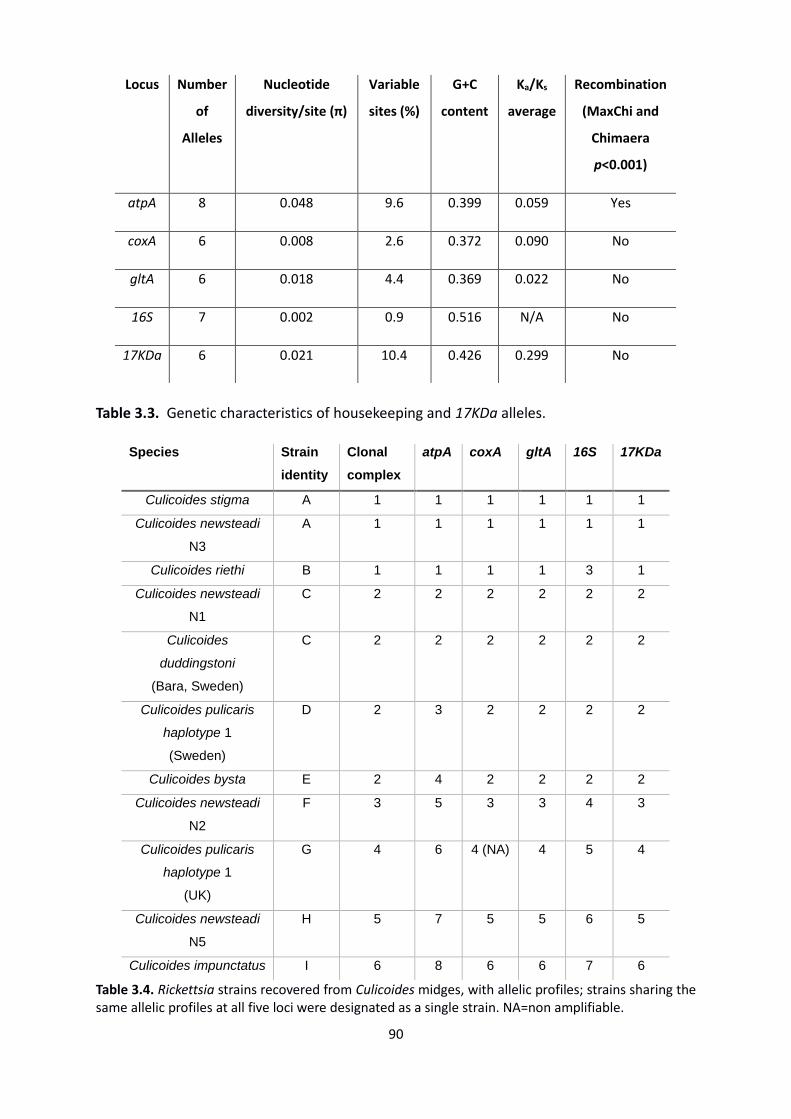
90
Table 3.3. Genetic characteristics of housekeeping and 17KDa alleles.
Table 3.4. Rickettsia strains recovered from Culicoides midges, with allelic profiles; strains sharing the same allelic profiles at all five loci were designated as a single strain. NA=non amplifiable.
Locus Number
of
Alleles
Nucleotide
diversity/site (π)
Variable
sites (%)
G+C
content
Ka/Ks
average
Recombination
(MaxChi and
Chimaera
p<0.001)
atpA 8 0.048 9.6 0.399 0.059 Yes
coxA 6 0.008 2.6 0.372 0.090 No
gltA 6 0.018 4.4 0.369 0.022 No
16S 7 0.002 0.9 0.516 N/A No
17KDa 6 0.021 10.4 0.426 0.299 No
Species Strain
identity
Clonal
complex
atpA coxA gltA 16S 17KDa
Culicoides stigma A 1 1 1 1 1 1
Culicoides newsteadi
N3
A 1 1 1 1 1 1
Culicoides riethi B 1 1 1 1 3 1
Culicoides newsteadi
N1
C 2 2 2 2 2 2
Culicoides
duddingstoni
(Bara, Sweden)
C 2 2 2 2 2 2
Culicoides pulicaris
haplotype 1
(Sweden)
D 2 3 2 2 2 2
Culicoides bysta E 2 4 2 2 2 2
Culicoides newsteadi
N2
F 3 5 3 3 4 3
Culicoides pulicaris
haplotype 1
(UK)
G 4 6 4 (NA) 4 5 4
Culicoides newsteadi
N5
H 5 7 5 5 6 5
Culicoides impunctatus I 6 8 6 6 7 6
91
The most polymorphic housekeeping locus was atpA, with 9.6% variable sites and the highest
level of nucleotide diversity per site (π=0.048) (Table 3.3.). All genes showed average Ka/Ks of
less than 1, indicating that the genes were subject to purifying selection, conforming to the
general requirements for reliable indicators of genetic relatedness between bacterial isolates.
Predictably, as an antigenic protein with less intense purifying selection and potential
episodes of positive selection, 17KDa had a greater average Ka/Ks (0.299) than the other loci;
although no signs of positive selection were observed at the gene-level.
Whilst there was evidence the strains found within Culicoides were closely related, some loci
demonstrated 100% sequence identity with Rickettsia strains from other taxa. These included
the partial gltA sequences of C. impunctatus, which was identical to the Rickettsia symbionts
of the beetle Deronectes platynotus (Dytiscidae; FM177878) (Küchler et al., 2009), the
Dipteran fly Chrysotimus flaviventris (Dolichopodidae; JQ925578) (Martin et al., 2013) and the
spider Pityohyphantes phrygianus (Linyphiidae; DQ 231491) (Goodacre et al., 2006), and the
partial 16S sequences of clonal complex 2 strains (C. duddingstoni; Bara, Sweden), C. pulicaris
haplotype 1 (Sweden), C. newsteadi N1, C. bysta which were identical to the 16S sequence of
the Rickettsia in the cranefly Limonia chorea (Limoniidae; AF322443). Furthermore, a coxA
995 bp region of the Hemipteran bug Macrolophus sp. Rickettsia 1 (Miridae; HE583223)
(Machtelinckx et al., 2012) was >99% similar to all Culicoides’ strains except for C. impunctatus.
3.4.4 Multilocus strain typing and geographic comparison
I next examined the relationship of the Rickettsia strains from different Culicoides’ species
using allelic profiles across loci (Figure 3.2.). The number of alleles per locus ranged from 6 to
8, with most allelic profiles obtained from different host populations (9/11) being unique.
92
Furthermore, of these 9 unique allelic profiles, 4 (F, G, H and I) shared no alleles with other
strains (Table 3.4.). Allelic profiles which were shared by more than one host species were
designated as central strains (CSs); whereas isolates which varied at one locus to these CSs
were termed single locus variants (SLVs). Together the CSs and SLVs form clonal complexes, as
they are presumed to be closely related. Two clonal complexes were identified in this study
(Figure 3.2.); the central strain A from C. stigma and C. newsteadi N3 formed clonal complex
1 with the SLV strain from C. riethi (B); whereas the central strain C from C. newsteadi N1 and
C. duddingstoni (Bara, Sweden) formed clonal complex 2 with the SLV strains from C. pulicaris
P1 (Sweden) (D) and C. bysta (E).
When strains were mapped by geographic location (Figure 3.2.), no clear pattern between
host, location and isolate was observed. Strain C was observed in both Corsica, France and
Sweden in C. newsteadi N1 and C. duddingstoni respectively; Culicoides species of differing
subgenera. However, C. impunctatus contained the same strain in 3 populations in the UK
(Bala and Kielder), as well as Sweden (Torsås).
3.4.5 Phylogenetic analysis
Aside from biting midges, sequences of all 5 strain-typing genes were generated in the
following host taxa; Hilara interstincta (Diptera: Empididae), Zavrelimyia sp. (Diptera:
Chironomidae), Glyptotendipes sp. (Diptera: Chironomidae), Macrolophus pygmaeus
(Hemiptera: Miridae), Pyrrhosoma nymphula (Odonata: Coenagrionidae), Enallagma
cyathingerum (Odonata: Coenagrionidae), Coenagrion mercuraile and Coenagrion puella
(Odonata: Coenagrionidae). Further taxa which gave incomplete profiles were an unidentified
Chironomidae sp. and the black fly Simulium aureum (Appendix 1). The atpA gene was the
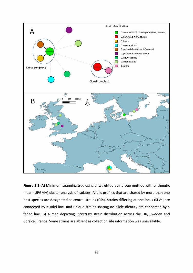
93
Figure 3.2. A) Minimum spanning tree using unweighted pair group method with arithmetic
mean (UPGMA) cluster analysis of isolates. Allelic profiles that are shared by more than one
host species are designated as central strains (CSs). Strains differing at one locus (SLVs) are
connected by a solid line, and unique strains sharing no allele identity are connected by a
faded line. B) A map depicting Rickettsia strain distribution across the UK, Sweden and
Corsica, France. Some strains are absent as collection site information was unavailable.
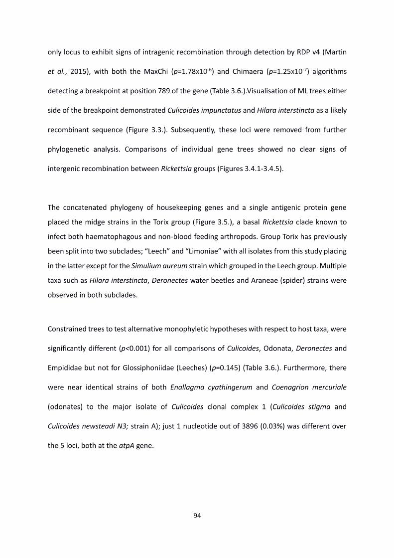
94
only locus to exhibit signs of intragenic recombination through detection by RDP v4 (Martin
et al., 2015), with both the MaxChi (p=1.78x10⁻⁶) and Chimaera (p=1.25x10⁻⁷) algorithms
detecting a breakpoint at position 789 of the gene (Table 3.6.).Visualisation of ML trees either
side of the breakpoint demonstrated Culicoides impunctatus and Hilara interstincta as a likely
recombinant sequence (Figure 3.3.). Subsequently, these loci were removed from further
phylogenetic analysis. Comparisons of individual gene trees showed no clear signs of
intergenic recombination between Rickettsia groups (Figures 3.4.1-3.4.5).
The concatenated phylogeny of housekeeping genes and a single antigenic protein gene
placed the midge strains in the Torix group (Figure 3.5.), a basal Rickettsia clade known to
infect both haematophagous and non-blood feeding arthropods. Group Torix has previously
been split into two subclades; “Leech” and “Limoniae” with all isolates from this study placing
in the latter except for the Simulium aureum strain which grouped in the Leech group. Multiple
taxa such as Hilara interstincta, Deronectes water beetles and Araneae (spider) strains were
observed in both subclades.
Constrained trees to test alternative monophyletic hypotheses with respect to host taxa, were
significantly different (p<0.001) for all comparisons of Culicoides, Odonata, Deronectes and
Empididae but not for Glossiphoniidae (Leeches) (p=0.145) (Table 3.6.). Furthermore, there
were near identical strains of both Enallagma cyathingerum and Coenagrion mercuriale
(odonates) to the major isolate of Culicoides clonal complex 1 (Culicoides stigma and
Culicoides newsteadi N3; strain A); just 1 nucleotide out of 3896 (0.03%) was different over
the 5 loci, both at the atpA gene.
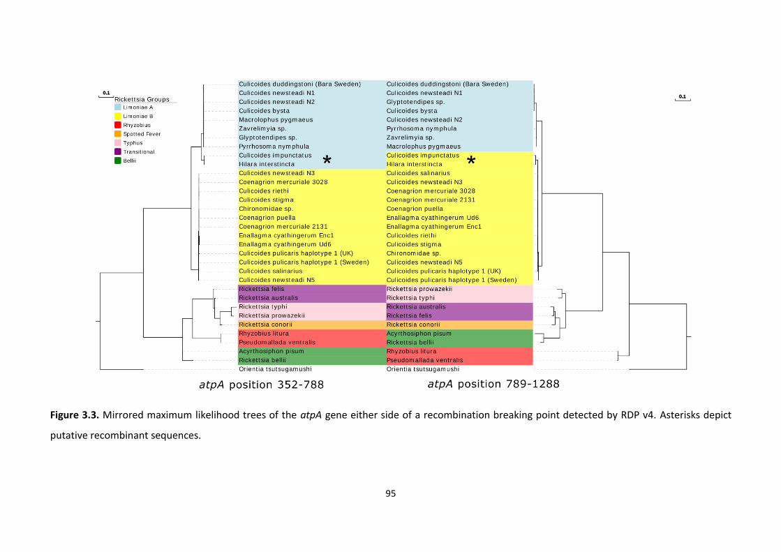
95
Figure 3.3. Mirrored maximum likelihood trees of the atpA gene either side of a recombination breaking point detected by RDP v4. Asterisks depict
putative recombinant sequences.
96
Table 3.5. Evidence of atpA intragenic recombination in Hilara interstincta and Culicoides
impunctatus isolates as determined with RDP4. Minor parent = Parent contributing the
smaller fraction of sequence. Major parent = Parent contributing the larger fraction of
sequence.
Putative recombinant
sequence
Putative minor
parental sequence
Putative major
parental sequence
RDP p value
Maxchi p value
Chimaera p value
Culicoides impunctatus/Hilara
interstincta
Coenagrion puella
Glyptotendipes sp.
1.50E-02 1.78E-06 1.25E-07
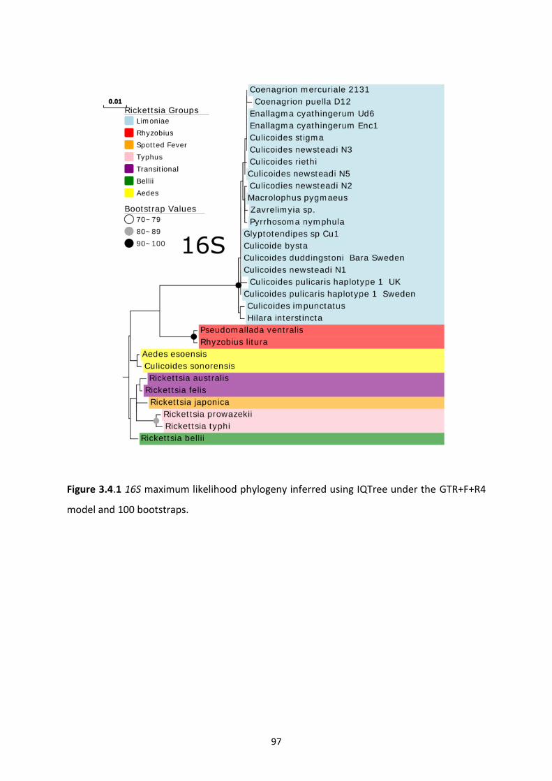
97
Figure 3.4.1 16S maximum likelihood phylogeny inferred using IQTree under the GTR+F+R4
model and 100 bootstraps.
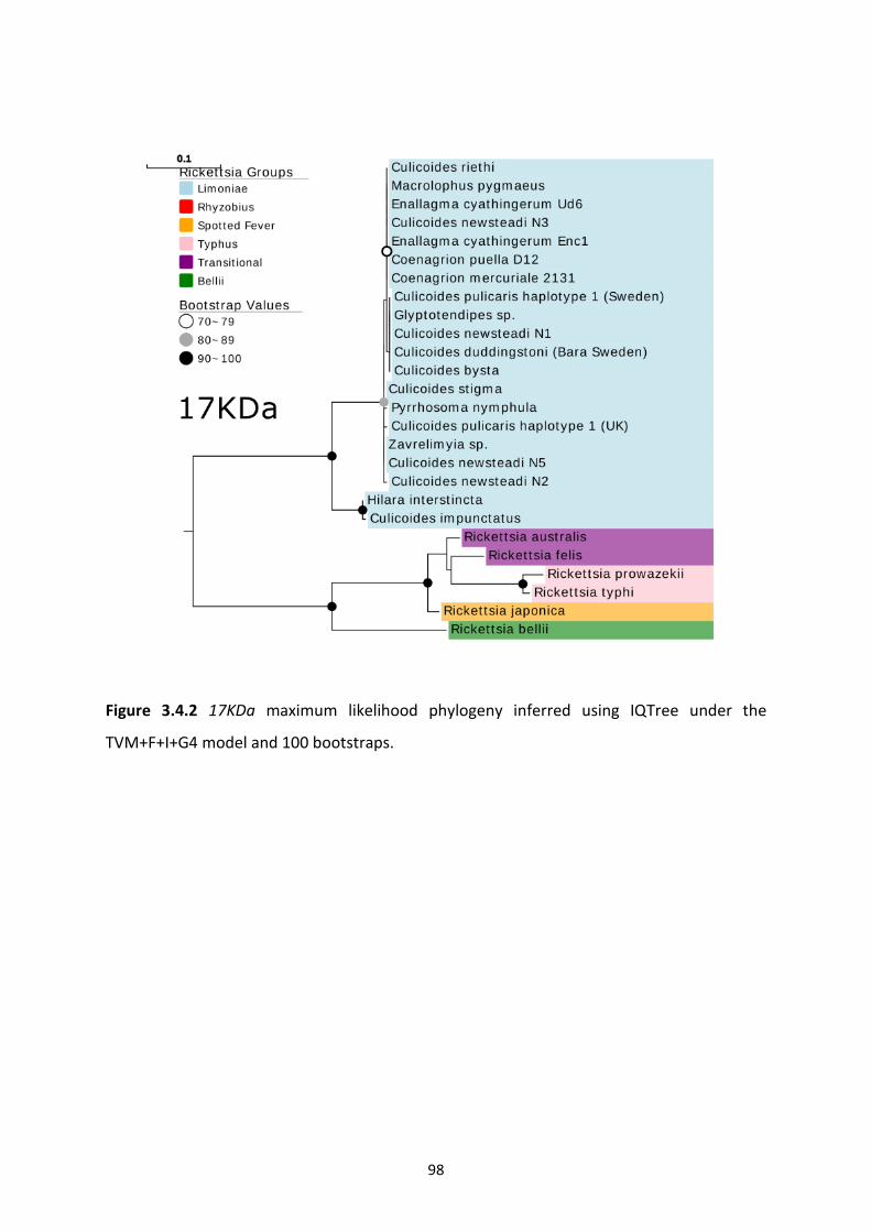
98
Figure 3.4.2 17KDa maximum likelihood phylogeny inferred using IQTree under the
TVM+F+I+G4 model and 100 bootstraps.
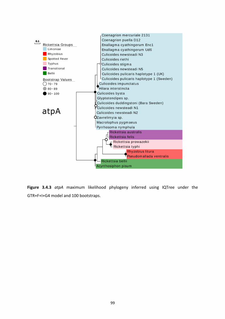
99
Figure 3.4.3 atpA maximum likelihood phylogeny inferred using IQTree under the
GTR+F+I+G4 model and 100 bootstraps.
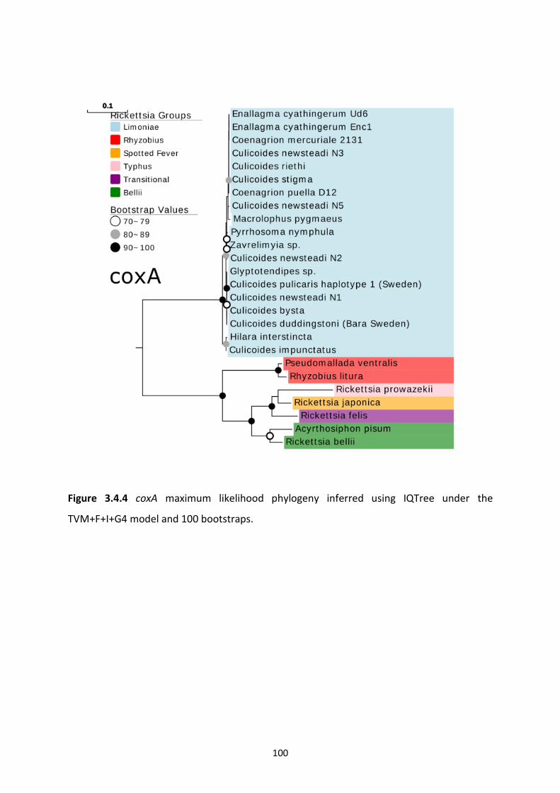
100
Figure 3.4.4 coxA maximum likelihood phylogeny inferred using IQTree under the
TVM+F+I+G4 model and 100 bootstraps.
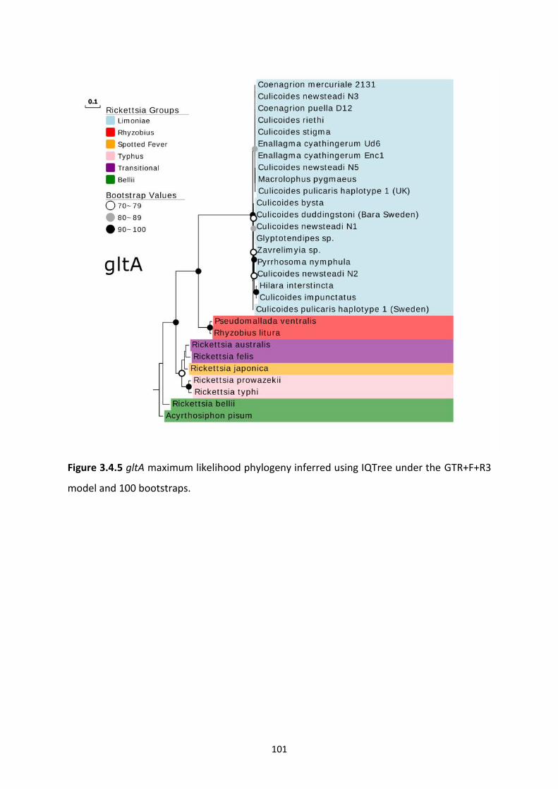
101
Figure 3.4.5 gltA maximum likelihood phylogeny inferred using IQTree under the GTR+F+R3
model and 100 bootstraps.
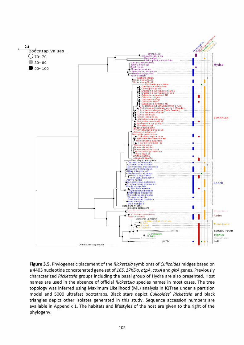
102
Figure 3.5. Phylogenetic placement of the Rickettsia symbionts of Culicoides midges based on a 4403 nucleotide concatenated gene set of 16S, 17KDa, atpA, coxA and gltA genes. Previously characterized Rickettsia groups including the basal group of Hydra are also presented. Host names are used in the absence of official Rickettsia species names in most cases. The tree topology was inferred using Maximum Likelihood (ML) analysis in IQTree under a partition model and 5000 ultrafast bootstraps. Black stars depict Culicoides’ Rickettsia and black triangles depict other isolates generated in this study. Sequence accession numbers are available in Appendix 1. The habitats and lifestyles of the host are given to the right of the phylogeny.
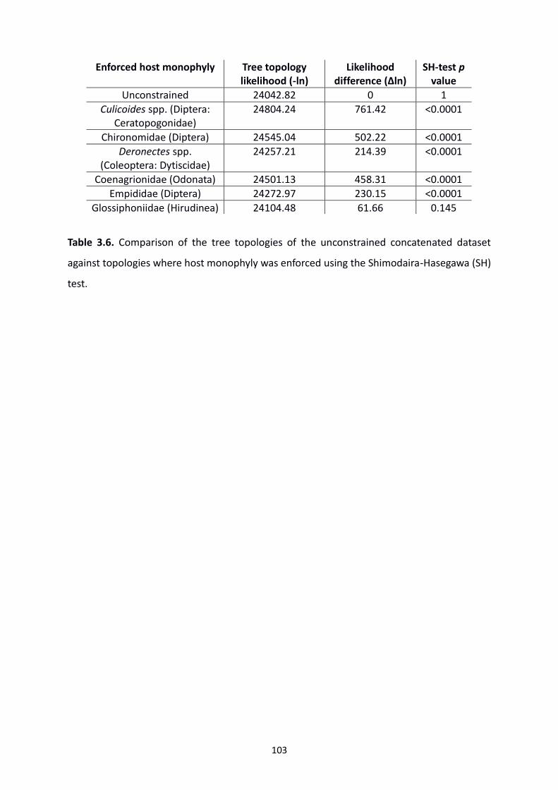
103
Table 3.6. Comparison of the tree topologies of the unconstrained concatenated dataset
against topologies where host monophyly was enforced using the Shimodaira-Hasegawa (SH)
test.
Enforced host monophyly Tree topology likelihood (-ln)
Likelihood difference (Δln)
SH-test p value
Unconstrained 24042.82 0 1
Culicoides spp. (Diptera: Ceratopogonidae)
24804.24 761.42 <0.0001
Chironomidae (Diptera) 24545.04 502.22 <0.0001
Deronectes spp. (Coleoptera: Dytiscidae)
24257.21 214.39 <0.0001
Coenagrionidae (Odonata) 24501.13 458.31 <0.0001
Empididae (Diptera) 24272.97 230.15 <0.0001
Glossiphoniidae (Hirudinea) 24104.48 61.66 0.145
104
In contrast, strains from Culicoides impunctatus, which also placed in the Limoniae group,
showed a divergence of 3.2% when compared to the same strain from clonal complex 1.
Despite all midge strains from Palearctic regions clustering in the Limoniae clade, an isolate
was detected from a single Culicoides sonorensis which was placed with an isolate discovered
recently in the mosquito Aedes esoensis from the demilitarized zone in Korea (Maina et al.,
2017) (Figure 3.5.). Only the 16S locus amplified with this sample, with all other primers failing
to generate a band for sequencing. Despite this, the clade described in this study as the
“Aedes” group after the initial host taxa it was discovered (as is convention for the genus), is
well supported and places basal to derived pathogenic groups but is a derivative of Torix and
Rhizobius clades.
105
3.5 Discussion
In this study, we show Limoniae group Rickettsia are common in biting midges, and thus
represent a previously unrecognized component of the biology of this important vector group.
Previous work on Culicoides, using conventional PCR to establish the presence of the heritable
symbiont Cardinium, revealed interspecies infection rates ranging from 16% to 29%
(Nakamura et al., 2009; Lewis et al., 2014; Mee et al., 2015). Thus, our PCR screen suggests
that Rickettsia is the most common symbiont of Culicoides identified so far, being present in
11 of 30 species tested (37%) and in 100% of specimens examined in 9 of the Rickettsia
positive species. Hence, this Rickettsia clade represents an important associate found widely
in Culicoides midges. It is noteworthy that our assessment of incidence is conservative, being
based on a conventional PCR assay which will likely report false negatives for low titre
infections.
A Rickettsia with 99% identity to Torix Leech isolates has previously been described in
Culicoides sonorensis by Campbell et al. (Campbell et al., 2004). Although the sequence of
the strain from this midge is not available, due to the similar homology found in this study
between our Rickettsia and the same leeches, it can be presumed that these are of the same
clade. Subsequently, a focussed screening of Culicoides sonorensis from California, USA was
undertaken due primarily to its significance as a major vector of BTV in North America. A
single individual from a population of Culicoides sonorensis (0/29 extracted individuals and
1/10 pooled samples) was found to be PCR positive. This individual, identified from a pool of
5 midges, was positive through 16S PCR screening which was later confirmed as Rickettsia by
Sanger sequencing. Other housekeeping gene markers and 17KDa screening gave negative
results, indicating a divergent isolate compared to the Torix Rickettsia found in other
106
Culicoides species. Furthermore, through phylogenetic analysis (Figure 3.5.), the isolate was
shown to cluster in a sister group of Rickettsia clades occasionally known to induce
pathogenicity in vertebrates. A similar isolate was also recently reported in Aedes mosquitoes
from the demilitarised zone of Korea. The placement as a sister clade of derived pathogenic
isolates, alongside the presence in two blood feeders and its low prevalence, suggests a
possible haematophagous transmission route of this Rickettsia. Subsequently, the Rickettsia
isolate reported here may play a role in animal and/or human health which requires further
investigation.
Of note is the absence of Limoniae Rickettsia in this C. sonorensis Californian population.
Campbell et al. (2004) found Torix Rickettsia in 3/10 individuals from Colorado suggesting it
would be likely to be detected in the author’s screen considering the higher sample size. The
lack of detection could be due to geographical barriers, such as the Rocky Mountains, between
the two populations. Regardless, the heterogeneous infection status of C. sonorensis in the
USA may have an impact on spatiotemporal infectious disease dynamics of BTV, if indeed the
presence of this endosymbiont affects vectorial capacity. Both C. pulicaris and C. newsteadi
complexes are thought to be vectors of BTV, with this study demonstrating them both to be
Rickettsia infected. Despite this, the major vectors of BTV in Europe and Africa, C. obsoletus
and C. imicola respectively, are shown to be uninfected.
The Torix group of Rickettsia has been recorded previously in an array of invertebrate species.
Many of these species share ecological characteristics including an aquatic phase and
predatory larval stages (e.g. biting midges, diving beetles, leeches, crane flies) (Figure 3.5.).
Others are notable for haematophagy (e.g. biting midges, leeches, sandflies). Moreover, no
107
secondary associations with vertebrate hosts or pathogenicity have been associated so far
with this Rickettsia group. Due to the initial lack of available Torix Rickettsia homologues of
the different loci amplified in this study, a multi-locus typing scheme was used to assess
relatedness between midge Rickettsia strains. This revealed 2 clonal complexes of close
relatedness. The presence of 2 identical strains in differing hosts, as is seen in C. stigma/C.
newsteadi N3 and C. duddingstoni/C. newsteadi N1, indicate a lack of host specificity and
suggests frequent host shifts. A similar scenario has previously been observed in the
Panamanian leaf-cutting ants Acromyrmex echinatior and Acromyrmex insinuator, where
shared Wolbachia isolates were observed in the cohabiting species (Van Borm et al., 2003).
The further inclusion of additional sequences from other Torix-infected taxa, showed near
identical strains between a Culicoides isolate in C. stigma/C. newsteadi N3, and the odonates
Enallagma cyathingerum and Coenagrion mercurial (Figure 3.5.). This identity suggests recent
host shifts are not limited to closely related taxa but can extend to horizontal transmission of
distantly related taxa. The pattern contrasts with the midge Cardinium (group C, chapter 2)
which is apparently restricted in its incidence. Host shift events have been created under
experimental conditions with Wolbachia from Drosophila simulans being transmitted to the
parasitic wasp, Leptopilina, before undergoing vertical transmission in this novel host (Heath
et al., 1999). Due to the predaceous nature of Torix hosts, it is possible that contamination
from ingested prey is accounting for the observed similarity in strains. However, the
observation of multiple populations of C. impunctatus from different locations containing
identical strains (Figure 3.2.), indicates this is unlikely as it is improbable that multiple
populations acquired identical Rickettsia isolates from their environment by chance alone.
108
Despite this, imaging techniques such as electron microscopy or Fluorescence in-situ
hybridisation (FISH) is needed in order to confirm a true endosymbiosis (see chapter 4).
The sporadic patterns of Rickettsia across a range of both related and unrelated host taxa
raises the question of how this Rickettsia endosymbiont is maintained in populations.
Endosymbiotic bacteria maintain high prevalence rates in populations via high fidelity
maternal transmission often leading to co-cladogenesis between symbiont and host (Chen, Li
and Aksoy, 1999; Clark et al., 2000). However, incongruencies between host and symbiont
phylogenies in Wolbachia suggests host shifts also occur as a strategy to explore new niches
(Turelli et al., 2018). The significantly worse topologies produced when host monophyly is
enforced in this study (Table 3.6.), suggests this lack of co-speciation is also the case with Torix
Rickettsia. Therefore, the high prevalence and close relatedness of midge Rickettsia strains in
distantly related hosts indicate a mixture of vertical and horizontal transmission.
Previous reports have also suggested mixed transmission strategies for Rickettsia (Perlman,
Hunter and Zchori-Fein, 2006; Caspi-Fluger et al., 2012). Due to the lifestyles of
haematophagous hosts which spread pathogenic Rickettsia strains to vertebrates, it has been
assumed that blood-feeding on common infected hosts has led to host shifts in these groups.
This is supported by other Rickettsiales families, Ehrlichia, Anaplasma and Orientia, all
sustaining infection through blood-feeding in experimental settings (Saito and Walker 2015;
Ogden et al. 2003; Frances et al. 2000). Although this may be the case for some Rickettsiales,
the presence of non-blood feeders in the Torix group, suggests alternative routes of
transmission may be occurring.
109
As host shifts of endosymbionts necessitate intimate interactions between current and
putative hosts, predation and parasitism have been proposed to account for horizontal
transfer events (Heath et al., 1999; Noda et al., 2001; Ahmed et al., 2015; Mascarenhas et al.,
2016). All of the dipteran hosts so far identified in the Limoniae subclade of Torix have larval
stages which are predatory. In addition, the adults of spiders, damselflies, predatory bugs and
dragonflies are also predators. Despite this, the isolate from the black fly Simulium aureum,
which was placed in the Torix subclade Leech, has no predatory life stages (although it is
possible filter feeders could acquire rickettsial symbionts from their diet due to symbionts in
protozoans). Another connection of note is the aquatic lifestyle of a majority of taxa in the
Torix group. Weinert et al. (2009) have previously suggested host switching could be occurring
between Rickettsia-infected freshwater taxa and arthropods, specifically via protists. This is
on account of the ancestral Rickettsia Hydra group (recently renamed Megaira), which
contains aquatic ciliate hosts. Additionally, the freshwater amoeboid parasite of fish,
Nuclearia pattersoni, has been shown to belong to the Torix group (Dyková et al., 2003). An
alternative hypothesis to explain host-shifts includes the transmission via endoparasitoids.
Torix-infected parasitoid wasps Asobara tabida and Quadrastichus mendeli should be
considered for such events although the latter was shown to contain a different strain of
Rickettsia to its gall wasp host Leptocybe invasa (Gualtieri et al., 2017). The transmission
strategies of Torix Rickettsia are discussed further in chapters 4 and 5.
The segregation of the Torix group into Leech and Limoniae groups had originally been
suggested by Perotti et al. (2006), although only limited taxa and sequence data were
available. The inclusion of the genes atpA, coxA and 17KDa in this project allowed for the
confident placement of sequences from previous studies which had relied primarily on just
110
one of 16S or gltA genes. Common taxa were observed in both Limoniae and Leech subclades
including, spiders, empidid dance flies and Deronectes water beetles. Despite this pattern
suggesting a common transmission route of the endosymbiont as mentioned above, the
divergence into two subclades could offer different effects on host biology. So far, the only
genome available comes from Culicoides newsteadi N5 (RiCNE) in the Limoniae group (Pilgrim
et al., 2017). However, comparative analysis with a Leech counterpart may offer insights into
differences between accessory genomes and subsequently specific effects on arthropod
hosts.
A further intriguing finding was the presence of coinfections of Rickettsia in Culicoides with
multiple strains being detected in C. salinarius and C. duddingstoni. Coinfections of different
Wolbachia strains have been attributed to exogenous acquirement through wounds of
cohabiting insects, as well as prey-predator or parasitoid interactions (Rigaud and Juchault
1995; Werren et al. 1995). Although coinfections can lead to competition for resources and
space, there may be a selection advantage for multiple infections as a result of an increased
potential to expand genetic repertoire through horizontal gene transfer. The restrictive
lifestyle of intracellular bacteria can often lead to limited opportunities for bacterial genome
evolution. However, through mobile genetic elements facilitated by plasmids and
bacteriophages, this can lead to the novel exchange of genes between symbionts infecting the
same cell (Frost et al., 2005; Kent et al., 2011). Furthermore, theory predicts coinfections
persist if a coinfected female produces more coinfected daughters than a single infected
female produces singly infected daughters. A specific evolutionary advantage for coinfections
of maternally inherited endosymbionts is the induction of bi-directional cytoplasmic
incompatibility (CI); the failure of reproduction in male and females containing different

111
Wolbachia strains. However, CI has not been observed in Rickettsia and so multiple infections
may provide a fitness advantage for infected individuals in a different way (discussed further
in chapter 5).
At least one recombination event, such as those facilitated by coinfections, was observed for
the atpA gene in Hilara intersincta and Culicoides impunctatus (Table 3.5. and Figure 3.3.). As
recombination can confound the true relationship between isolates, the loci of both strains
were removed from phylogenetic analysis. This is the first time recombination has been
detected in the Torix group of Rickettsia, although similar genetic reshuffling has also been
observed in Transitional, Rhizobius and Adalia groups, in the genes coxA and gltA (Weinert et
al. 2009). Rickettsia plasmids have been described several times across the genus, implicating
them as facilitators for such processes (Ogata et al., 2005; Baldridge et al., 2007; Blanc et al.,
2007). However, although the Torix genome of Culicoides newsteadi N5 (RiCNE) identified a
tra conjugative DNA transfer element, it was not clear if this was present as part of a
chromosome or a low-copy number plasmid (Pilgrim et al., 2017). The common
recombination across the genus (Ogata et al., 2005; Blanc et al., 2007; El Karkouri et al., 2016)
led to the comparison of single gene trees to uncover any intergenic recombination but
topologies appeared similar (Figures 3.4.1-3.4.5).
The impact of this Rickettsia on host biology is uncertain. Rickettsia infections are known to
be associated with a variety of reproductive manipulations of their host (reproductive
parasitisms), including male-killing in ladybird beetles (Werren et al., 1994) and
parthenogenesis induction in parasitoids (Hagimori et al., 2006; Giorgini et al., 2010).
However, equal likelihood of male and female midges being infected indicates sex ratio
112
distortion is unlikely to be a phenotype for the Rickettsia in midges. Further to this, Rickettsia
felis represents an obligate symbiont in book lice (Liposcelis bostrychophila) required for egg
production (Perotti et al., 2006). However, the sporadic distribution of Rickettsia across
diverse taxa suggests a lack of co-speciation making it unlikely that the host requires symbiont
presence for its function.
Overall, the data suggests the Torix group Rickettsia identified in this study may have some
facultative (ecologically contingent) benefit to their host. Indeed, Rickettsia from this clade
have been linked with a fitness benefit (increased body size) in leeches (Kikuchi and Fukatsu
2005). Endosymbionts of haematophagous arthropods occasionally have the capacity to
synthesise B vitamins to supplement their host’s lacking diets. This has previously been
recorded for Wigglesworthia symbionts in tsetse flies and Wolbachia in Cimex bedbugs
(Snyder et al., 2010; Nikoh et al., 2014). However, with the exception of a reduced pathway
for folate biosynthesis (also found in other Rickettsia), RiCNE lacks known pathways for the
biosynthesis of cofactors and B-vitamins (Pilgrim et al., 2017).
A peculiarity of note is the detection of coexisting infected and uninfected individuals in C.
impunctatus populations, a scenario contrary to the more common fixed infections observed
in this study. However, low titre infections in those testing negative for Rickettsia cannot be
ruled out (chapter 2 and Mee et al. 2015). An alternative explanation for this difference is that
the strain in C. impunctatus has a different role in its host in comparison to the other isolates
at fixation in midges. Rickettsia surface antigens, like 17KDa, have previously been identified
to be evolving under positive selection and may have key roles in host adherence and
infiltration (Blanc et al., 2005), although Ka/Ks ratios suggest this might not be the case here.
113
A major research effort for the future lies in identifying the impact of Rickettsia on host
biology.
In conclusion, this study has identified a common but neglected association between
Rickettsia and biting midges. Given the importance of biting midges as vectors, two key areas
of future research are to establish the impact of Rickettsia presence on vector competence.
Symbionts may reduce vector competence (as in Wolbachia in Aedes aegypti), increase it (as
for Rickettsia in Bemisia tabaci) or have no impact. Rickettsia infections are also known to
affect host dispersal tendencies, with Torix Rickettsia-infected spiders showing lower
motivation for dispersal (Goodacre et al., 2009). Symbiont impact on either of these
characteristics would significantly alter the local and spatial spread of vector-borne infections,
and thus pressingly deserve attention.
114
Chapter 4: The Tropism and Transtadial Transmission of a Rickettsia Endosymbiont in Culicoides impunctatus
115
4.1 Abstract
Previous work in this thesis has shown Rickettsia (group Limoniae) is widespread in midge
populations with infection often reaching fixation. The maintenance of Rickettsia
endosymbionts in insect populations is achieved through both vertical and horizontal
transmission routes. For example, the presence of the symbiont in the follicle cells and
salivary glands of Bemisia whiteflies allows Rickettsia transmission via the germline and plants
respectively. Through fluorescence in-situ hybridisation (FISH) and transmission electron
microscopy screening, this study describes the pattern of Rickettsia tissue tropisms observed
in the Scottish highland midge, Culicoides impunctatus. Of note, is the infection of the ovarian
suspensory ligament, indicating a novel germline targeting strategy. Additionally, localisation
in ovaries and larval fat bodies suggests transtadial transmission is a major route of ensuring
maintenance of Rickettsia within C. impunctatus populations. Aside from providing insights
into transmission strategies, Rickettsia presence in the fat body of larvae indicates potential
host fitness and vector effects to be investigated in the future.
116
4.2 Introduction
Endosymbiotic bacteria of arthropods primarily maintain themselves within a population
through maternal transmission to offspring. In insects there are three distinct methods of
ensuring such vertical transmission occurs (Russell, Chappell and Sullivan, 2019).
1) Soma-soma interactions; a rare occurrence in arthropods, although observed in the
viviparous tsetse fly where developing embryos gain Wigglesworthia infections via milk gland
secretions (Rio et al., 2012).
2) Germline-germline transmission; where certain Wolbachia strains of Drosophila
melanogaster localise in the germline continuously throughout development (Veneti et al.,
2004; Serbus and Sullivan, 2007; Toomey et al., 2013).
3) Soma-germline transmission; the best-known example of which involves the deliverance
of endosymbionts from specialised cells (bacteriocytes) to gametes or developing embryos.
The latter is the most commonly described and is hypothesised to be as a result of
evolutionary histories (endosymbiotic events pre-date germline appearance) and selection
pressures (restriction to somatic tissues prevents detrimental local effects during gamete
development) (Russell, Chappell and Sullivan, 2019). Furthermore, the sequestering of
endosymbionts by the soma offers a large selection pool for occupation by the germline (Mira
and Moran, 2002; Ikuta et al., 2016). Subsequently, the success of vertical propagation is
often determined by both germline and somatic bacteria populations.
Although bacteriocytes primarily transmit microbes upon which their host is dependent for
survival (primary endosymbionts), facultative (secondary) endosymbionts also utilise somatic

117
tissues to target the germline. However, due to differing evolutionary histories, varying routes
of inheritance occur. For example, Buchnera aphidicola, a primary endosymbiont of the pea
aphid which provides essential amino acids to its host, co-exists with the thermal and parasite
protective secondary endosymbiont Serratia symbiotica (Douglas, 2002; Oliver et al., 2003;
Burke, Fiehn and Moran, 2010). Although Buchnera utilises the exocytosis of bacteriocytes,
Serratia co-opts endocytic mechanisms via the haemolymph with subsequent segregation of
both endosymbionts after the formation of embryonic bacteriocytes (Koga et al., 2012).
Many endosymbionts exhibit evidence of both vertical and horizontal (mixed) transmission.
This is exemplified in D. melanogaster, where microinjection of Wolbachia led to infection of
the germline after localisation in the haemolymph and somatic stem cells (Frydman et al.,
2006). Thus, symbionts newly infecting somatic tissues can transfer into the germ line, and
this provides a pathway for host shifts to occur. Furthermore, soma infections are often linked
to host fitness effects. For example, close association with uric acid-containing cells (urocytes)
by Blattabacterium in cockroaches is associated with nitrogen recycling into amino acids
(Sacchi et al., 1998; Sabree, Kambhampati and Moran, 2009), whilst the presence of
Wolbachia in Drosophila muscles and nervous tissue is correlated with olfactory-cued
locomotion (Peng et al., 2008). Although these associations are well-characterised in some
symbioses, many recently identified insect-bacteria associations are yet to be investigated.
Thus, the localisation of symbionts to both somatic and germline tissues can offer insights
into these intimate interactions.
118
Rickettsia are an intracellular group of alphaproteobacteria which demonstrate mixed modes
of transmission. The horizontal and vertical transmission strategies of Rickettsia have allowed
for the infection of a diverse range of organisms including protists, arthropods, plant and
vertebrates (Weinert et al., 2009; Zchori-Fein, 2011). Haematophagous spread of pathogenic
Rickettsia to vertebrates has been well documented, and phloem-feeding insects have been
implicated in transmission of Rickettsia to plants (Brumin, Levy and Ghanim, 2012; Caspi-
Fluger et al., 2012). Rickettsia also has the unusual ability to infect sperm head nuclei, allowing
for paternal inheritance, which can combine with maternal transmission to drive a costly
symbiont into the population (Watanabe et al., 2014). Subsequently, the multiple
transmission pathways of Rickettsia makes this symbiont a model organism for providing
insights into transitions between mutualism and pathogenicity (Merhej and Raoult, 2011).
Previous work in this thesis (chapter 3) has demonstrated the presence of Limoniae Rickettsia
in several species of Culicoides although host effects and transmission routes are yet to be
determined. The absence of sex-ratio distortion and obligate mutualism (not all individuals
were infected within a population) suggests the lack of a reproductive parasitism. By
exclusion, these indicate the drive of this endosymbiont is related to a facultative benefit.
Indeed, a Torix group Rickettsia in leeches is positively correlated with body size (Kikuchi and
Fukatsu, 2005). Furthermore, as Culicoides transmit several veterinary viruses there is
particular interest in the effects of Rickettsia on vectorial capacity. For example, Rickettsia
infection of Bemisia tabaci has been shown to increase the transmission of tomato yellow leaf
curl virus (Kliot et al., 2014).
119
The biting midge Culicoides impunctatus is prevalent across Northern Europe but is most
abundant in the Highlands of Scotland. Here they are a biting nuisance with “midge attacks”
accountable for significant economic impact through losses in tourist and forestry industries
(Hendry and Godwin, 1988; Hendry, 1996). The abundance of C. impunctatus in Scotland is
likely due to a large number of acidic boggy breeding sites. Further, their ability to reproduce
once in the absence of a blood meal (autogeny) means huge numbers can develop even where
vertebrate hosts are not available (Boorman and Goodard, 1970; Blackwell et al., 1992;
Blackwell, Young and Mordue, 1994).
Despite their commonness in the field, attempts at laboratory-rearing C. impunctatus have
proven largely unsuccessful (Hill, 1947; Carpenter, 2001). A main barrier to cultivation of this
species is the lack of identification of a suitable diet. The two extant Culicoides species
colonised (C. sonorensis and C. nubeculosus) rely on substrates, such as liver powder and
wheat germ, which encourage microbial growth in larval pans (Boorman, 1974). However,
previous attempts utilising this method in C. impunctatus have been unsuccessful (Carpenter,
2001). In addition, developmental stages are difficult to observe with these methods.
Subsequently, the investigation of Culicoides bionomics often utilises agar dishes to gain basic
developmental information which can inform future cultivation attempts (Barceló and
Miranda, 2018). Additionally, this technique can allow for the investigation of life stage-
specific traits.
In order to investigate potential phenotypic effects on the host as well as symbiont
transmission strategies, both the tissue and subcellular tropisms of Rickettsia must be
120
elucidated. Subsequently, this study assesses both germline and somatic tissues of C.
impunctatus for Rickettsia infection via transmission electron microscopy (TEM) and
fluorescence in-situ hybridisation (FISH).
121
4.3 Methods
4.3.1 The collection of Culicoides impunctatus
Collections of Culicoides impunctatus for imaging of adult tissues were carried out at multiple
sites in the United Kingdom; Dumfries, Kielder forest, Loch Lomond and Fort William between
June 2017 and September 2018. For midges not used for ovipositing, individual Culicoides
were allowed to rest on the author’s arm before aspiration into 1.5 ml Eppendorf tubes with
10% sucrose-soaked cotton wool placed at the bottom and damp sphagnum moss filled in the
lid to maintain a high humidity. Tubes were then placed horizontally on sticky tape in
Tupperware boxes before incubation at 23 °C. For ovipositing Culicoides, female midges were
collected by allowing feeding on the author and field assistants’ forearms from the Loch
Lomond site (May 2018). When midges were observed to be replete (approximately after 5
minutes), or the insect released mouth parts from the skin, they were aspirated and stored in
a plastic container at ambient temperature (13-17°C) until the end of the collection session.
Ovipositing containers were assembled based on previous studies undertaken by Carpenter
(2001). Briefly, approximately 50 Culicoides were transferred to containers consisting of
cylindrical pill boxes (Watkins and Doncaster, UK; 64 mm diameter x 60mm depth), with
cotton wool soaked in 10% w/v sucrose solution (replaced every two days) placed on top of a
fine net meshing which covered the tops of the pillboxes. For ovipositing areas, 50 ml Falcon
tube lids were filled with damp sphagnum moss (B and Q, UK) by soaking in 1% nipagin
dissolved in distilled water and squeezing until drops could be counted, before being placed
on top of damp filter paper. This was secured by cutting a cylindrical hole in the bottom of
the pill box. These were then transferred to 19 x 12 x 8 cm Tupperware boxes containing a
122
50 ml beaker of saturated sodium sulphate (Na2SO4) which has previously been described to
maintain humidity at >90% when between 20-25°C (Winston and Bates, 1960). These were
then transported to a laboratory in Liverpool before the Tupperware boxes were placed in an
incubator where they were maintained at 23°C with a photoperiod of 12 Light: 12 Dark hours.
4.3.2 Larval rearing
Due to C. impunctatus’ small size and vulnerability to drowning on surface films under
laboratory conditions it is unlikely that this species oviposit in open water (Carpenter, 2001).
This alongside the observation by Carpenter that L1 instars of C. impunctatus get trapped in
agar solutions of >1% led to the rearing on 0.5% agar; petri dishes used were 100 mm in
diameter and x 15 mm in depth with agar made up from deionised water poured to a depth
of 5 mm. Eggs oviposited onto the sphagnum moss substrate were picked individually with a
fine paintbrush or the damp edge of a sharpened tungsten needle and placed onto agar dishes
(100-180 eggs per dish; n=3 dishes) and spaced evenly apart. Culicoides impunctatus
identification was confirmed by the distinctive brown heads of larvae (Kettle and Lawson,
1952) as well as identification of ovipositing adults in pill boxes. Two larval diets were briefly
trialled for suitability; Banana worms (Panagrellus nepenthicola), and the ciliate, Volvox
carteri. Subsequently, Banana worms were chosen because larvae were seen to actively
attack the worms once introduced, whereas the surface tension from Volvox prevented larvae
from moving freely in the agar. Banana worms were cultured using the manufacturer’s
instructions (Ron’s worms; Amazon UK) before a fine paintbrush was used to place the
nematodes in 2-3 ml of deionised water and spread evenly across each agar dish. This was
performed daily, providing nutrition and ensuring the agar did not desiccate. Larvae were
then stored under the same temperature and photoperiods as adults previously mentioned.
123
Allocation of larval instars were designated by head capsule length measurements as
described by Kettle and Lawson (1952). A Kaplan-Meier estimator was used to measure
cumulative survival over time as well as larval periods. Survival curve censoring of individuals
was undertaken for larvae used for imaging assays, which were retrieved from the agar dishes
using a coiled piece of tungsten wire.
4.3.3. Dissections of adults
Post blood-feeding in the wild, Culicoides were processed after carefully timed transportation
to a laboratory in Liverpool. Individuals were sacrificed at various time points post blood
feeding (PBF); 0 hours (non-blood fed), 12 hours PBF, 48 hours PBF and 120 hours PBF. First,
Culicoides were chilled in the freezer for immobilisation before being placed in a drop of
Phosphate-buffered saline on a petri dish. Midges were then killed by piercing the thorax with
a sharp tungsten needle before being confirmed as C. impunctatus. Ovaries were exposed in
various ways through dissection under a stereoscopic microscope:
1) By a midline incision of the ventral abdomen
2) By shelling away cuticle from abdominal segment 8 and repeating proximally
3) By quick evisceration through holding the dorsal thorax and pulling the posterior abdomen
with forceps. Any other tissues which could also be recovered were then stored separately
for further investigation of somatic tropism.
Time points for sacrifice were chosen as a result of a study by Carpenter (2001) which
identified developmental stages of forming eggs as a function of time after blood-feeding.
Stages were confirmed by using a system developed by Linley (1965) which was subsequently
modified by Campbell and Kettle (1975):
124
stage 1) No observation of yolk within the oocyte;
stage 2) Yolk can be identified within the oocyte;
stage 3) Yolk proteins occupy up to three-quarters of the oocyte;
stage 4) The oocyte is elongated and no longer oval, resembling the mature egg;
stage 5) Egg fully mature, with chorion visible.
4.3.4 Tissue preparation and fluorescence in-situ hybridisation (FISH)
Tissue samples were initially stored in Carnoy’s solution (chloroform:ethanol:glacial acetic
acid, 6:3:1) overnight in 100 μl wells covered in Parafilm, but were difficult to retrieve due to
their size and tendency to stick to the sides of wells. Subsequently, preparation was changed
to direct fixing onto poly-L-lysine covered slides which allowed for adherence of the tissues
on the slide without problems of disruption when undergoing subsequent washes. Tissues
examined included eggs of different developmental stage, Malpighian tubules, midgut,
foregut, hindgut, fat body and salivary glands. Additionally, crushed spermathecae were
prepared for visualisation of the spermatophore and spermatids contained within. This was
achieved by separating paired spermathecae from ovaries with diligent attempts to prevent
any contamination from any ovaries that might be Rickettsia-infected. Spermathecae were
then suspended in Phosphate-buffered saline and allowed to dry before pressing a coverslip
over the slide to break open the tissue.
For fluorescence in-situ hybridisation (FISH) imaging, the above tissues were fixed for one
hour in Carnoy’s solution and tissues cleared by treating with 6% H2O2 in ethanol for 2 hours.
125
Two pre-hybridisation washes were undertaken using wash buffer (20 mM Tris-HCl, pH 8.0,
50mM NaCl, 0.01% sodium dodecyl sulphate, 5 mM EDTA). Hybridisation was performed
overnight in hybridisation buffer (20 mM Tris-HCl, pH 8.0, 90mM NaCl, 0.01% sodium dodecyl
sulphate, 30% formamide) containing 10 pmol/ml of the Rickettsia specific probe [5’-
CCATCATCCCCTACTACA-(ATTO 633)-3’] adapted from Perotti et al. (2006) which were
checked for specificity against the Torix 16S gene of Culicoides newsteadi (RiCNE; Bioproject
accession number PRJNA376033). After hybridisation, the samples were thoroughly washed
twice in wash buffer and slide mounted in Vectashield with DAPI (Vector Laboratories) and
viewed under a Zeiss LSM 880 BioAFM confocal microscope. The specificity of the detection
and any autofluorescent properties of midge tissue was assessed using Rickettsia-free midges
(Culicoides nubeculosus; Pirbright Institute) as negative controls. Further negative controls
were used when fresh C. nubeculosus were not available by using probe-free samples. For
each tissue, at least 5 specimens were viewed under the microscope to confirm
reproducibility. Optical sections (0.7μm thick) were prepared from each specimen to create a
Z-stack image to be processed in ImageJ. All FISH imaging equipment and technical assistance
was provided by the Liverpool Centre for Cell Imaging (University of Liverpool).
4.3.5. Transmission electron microscopy
As host-seeking Culicoides mate prior to blood feeding it was possible to examine spermatids
from female spermathecae. Ovaries and spermathecae were prepared for transmission
electron microscopy (TEM) as follows. Tissues were dissected into 2% (w/v)
paraformaldehyde + 2.5% (w/v) glutaraldehyde in 0.1M phosphate buffer (pH 7.4). Fixative
was then changed for 2.5% (w/v) glutaraldehyde in 0.1M phosphate buffer (pH 7.4). Heavy
metal staining consisted of 2% (w/v) OsO4 in ddH2O, followed by 1% (w/v) Tannic acid in
126
ddH2O and then 1% (w/v) aqueous uranyl acetate. To prevent precipitation artefacts the
tissue was washed copiously with ddH2O between each staining step. Fixation and staining
steps were performed in a Pelco Biowave®Pro (Ted Pella Inc.Redding California, USA) at 100W
20Hg, for 3 mins and 1min respectively. Dehydration was in a graded ethanol series before
filtration and embedding in medium premix resin (TAAB, Reading, UK). For TEM, 70-74 nm
serial sections were cut using a UC6 ultra microtome (Leica Microsystems, Wetzlar, Germany)
and collected on Formvar (0.25% (w/v) in chloroform, TAAB, Reading, UK) coated Gilder 200
mesh copper grids (GG017/C, TAAB, Reading, UK). Images were acquired on a 120 kV Tecnai
G2 Spirit BioTWIN (FEI, Hillsboro, Oregon, USA) using a MegaView III camera and analySIS
software (Olympus, Germany). Dissections and initial fixation were performed by the author
with subsequent staining and imaging undertaken with assistance from Alison Beckett and
the Electron microscopy unit (Institute of Translational Medicine; University of Liverpool).

127
4.4 Results
4.4.1 Rickettsia infection during oogenesis
Underdeveloped (stage 1) eggs contained clusters of Rickettsia predominantly in oocytes with
no presence of yolk deposition seen (Figures 4.1. and 4.2.). Signal was strongest in the oocyte
with bacteria also being observed within nurse cells. Electron microscopy images suggest the
Rickettsia are perinuclear rather than within the nurse cell nuclei themselves (Figure 4.1.).
The follicular epithelium is also infected with bacteria seen in transit between follicle cells and
the oocyte. No signal was observed in Rickettsia-free Culicoides nubeculosus or probe-free
negative controls. After 48 hours post-blood feeding, yolk deposition is seen as a clouding in
the (stage 2) oocyte although individual yolk granules are not able to be seen. Clusters of
bacteria no longer occupy nurse cells but still predominantly fill the oocyte (Figure 4.2.). In
stage 4 eggs, yolk granules become visible and appear to harbour sparse numbers of bacteria
with a predominant localisation in follicle cells (Figure 4.2.). However, in certain focal planes
Rickettsia are seen to remain in the oocyte cytoplasm but presents only at the periphery of
the oocyte.
4.4.2 Germline targeting of Rickettsia via the ovarian suspensory ligament
During dissections of adult female C. impunctatus it was often possible to retrieve most of the
gut, Malphigian tubules and paired ovaries. This allowed for comparing localisation patterns
in the somatic tissues as well as the germline. Initial examination of adult specimens at low
magnification gave a consistent pattern of strong localised signal at the anterior/posterior
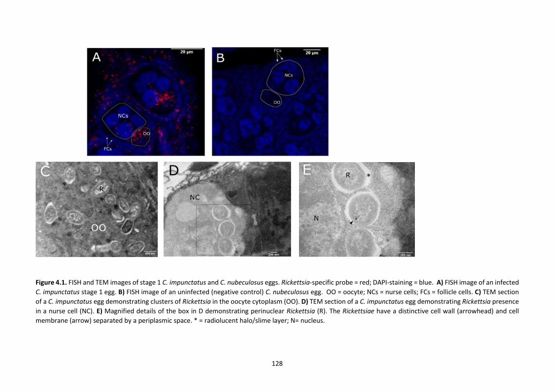
128
Figure 4.1. FISH and TEM images of stage 1 C. impunctatus and C. nubeculosus eggs. Rickettsia-specific probe = red; DAPI-staining = blue. A) FISH image of an infected
C. impunctatus stage 1 egg. B) FISH image of an uninfected (negative control) C. nubeculosus egg. OO = oocyte; NCs = nurse cells; FCs = follicle cells. C) TEM section
of a C. impunctatus egg demonstrating clusters of Rickettsia in the oocyte cytoplasm (OO). D) TEM section of a C. impunctatus egg demonstrating Rickettsia presence
in a nurse cell (NC). E) Magnified details of the box in D demonstrating perinuclear Rickettsia (R). The Rickettsiae have a distinctive cell wall (arrowhead) and cell
membrane (arrow) separated by a periplasmic space. * = radiolucent halo/slime layer; N= nucleus.
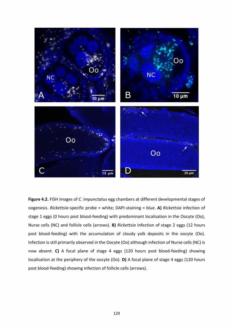
129
Figure 4.2. FISH images of C. impunctatus egg chambers at different developmental stages of
oogenesis. Rickettsia-specific probe = white; DAPI-staining = blue. A) Rickettsia infection of
stage 1 eggs (0 hours post blood-feeding) with predominant localisation in the Oocyte (Oo),
Nurse cells (NC) and follicle cells (arrows). B) Rickettsia infection of stage 2 eggs (12 hours
post blood-feeding) with the accumulation of cloudy yolk deposits in the oocyte (Oo).
Infection is still primarily observed in the Oocyte (Oo) although infection of Nurse cells (NC) is
now absent. C) A focal plane of stage 4 eggs (120 hours post blood-feeding) showing
localisation at the periphery of the oocyte (Oo). D) A focal plane of stage 4 eggs (120 hours
post blood-feeding) showing infection of follicle cells (arrows).
130
midgut junction but not throughout the majority of the alimentary tract (Figure 4.3.).
Additionally, on examination at a higher magnification, it was difficult to place the signal in
any of the midgut epithelial cells. Examination of further individuals led to the discovery that
both the midgut junction and ovaries were connected by a fibrous piece of tissue which was
to be later identified as the ovarian suspensory ligament. This structure, otherwise known as
the “median ligament”, was seen to pair off and loop down from the midgut attachment site
before attaching at the apex of the ovary.
Infection of the suspensory ligament apparatus was strongest at the anterior-posterior
midgut junction (Figure 4.3.) although this could be attributed to connective tissue density. It
was possible to follow the signal down the suspensory ligament where the structure became
continuous with the terminal filaments of ovarioles and the ovarian epithelial sheath; the
structure separating ovarioles from one another. Strength of infection was consistent in the
ovarian epithelial sheath over the long axis of the ovary with neighbouring immature egg
chambers seen to be heavily infected. Bacteria could be seen migrating from this densely
populated structure into neighbouring follicle cells and further into oocytes themselves,
which again, had the distinct pattern of infection both in undifferentiated oocyte cytoplasm
as well as nurse cells. Within the ovary, the germarium appeared to be no more infected than
the rest of the ovarian tissue. In one individual, the attachment of the suspensory ligament to
a sparsely cellular but strongly signalled structure is thought to be part of a lobe of the fat
body (Figure 4.3.) although the ethanol based (Carnoy’s) fixative diminishes lipids leading to
ambiguity of identification when observed under transmission light. Signal was observed in
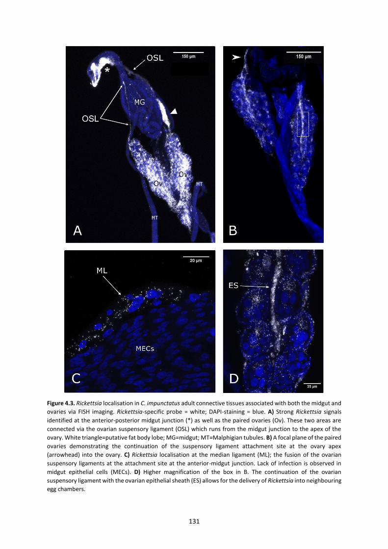
131
Figure 4.3. Rickettsia localisation in C. impunctatus adult connective tissues associated with both the midgut and
ovaries via FISH imaging. Rickettsia-specific probe = white; DAPI-staining = blue. A) Strong Rickettsia signals
identified at the anterior-posterior midgut junction (*) as well as the paired ovaries (Ov). These two areas are
connected via the ovarian suspensory ligament (OSL) which runs from the midgut junction to the apex of the
ovary. White triangle=putative fat body lobe; MG=midgut; MT=Malphigian tubules. B) A focal plane of the paired
ovaries demonstrating the continuation of the suspensory ligament attachment site at the ovary apex
(arrowhead) into the ovary. C) Rickettsia localisation at the median ligament (ML); the fusion of the ovarian
suspensory ligaments at the attachment site at the anterior-midgut junction. Lack of infection is observed in
midgut epithelial cells (MECs). D) Higher magnification of the box in B. The continuation of the ovarian
suspensory ligament with the ovarian epithelial sheath (ES) allows for the delivery of Rickettsia into neighbouring
egg chambers.

132
the crop of the foregut in one sample although it is possible that this is part of the connective
tissue matrix continuous with the median ligament. Fragility of tissues when dissecting often
impaired the recovery of the completely intact connective tissue; some samples only showed
signal at the attachment sites of the ovarian ligament suggesting the snapping of the structure
during processing. No signal was observed in Malpighian tubules, heads, salivary glands or
other alimentary structures except for the 1 individual which demonstrated infection in the
crop.
4.4.3 FISH and transmission electron microscopy of spermathecae
Rickettsia was further detected in crushed spermathecae from fertilised females (Figure 4.4).
DAPI staining gave an unspecific signal making it difficult to distinguish spermatids from the
rest of the acellular spermatophore. However, Rickettsia signal was detected diffusely in
samples. This observation suggested possible male horizontal transmission (to mates) and/or
vertical transmission (to offspring). Alternatively, this signal could reflect presence in
maternally-derived spermathecal structures. Subsequently, TEM sections of spermathecae
were assessed to clarify the nature of this signal. Overall, infected spermathecae showed no
sign of bacteria in sperm heads or tails of spermatids, nor in the acellular matrix (Figure 4.4.).
However, Rickettsia was identified in the maternally derived spermathecal epithelium,
occasionally associated with actin-like filaments. Unfortunately, due to the difficulties of lab
maintenance of Culicoides, a crossing-system to definitively rule out paternal transmission
was not possible.
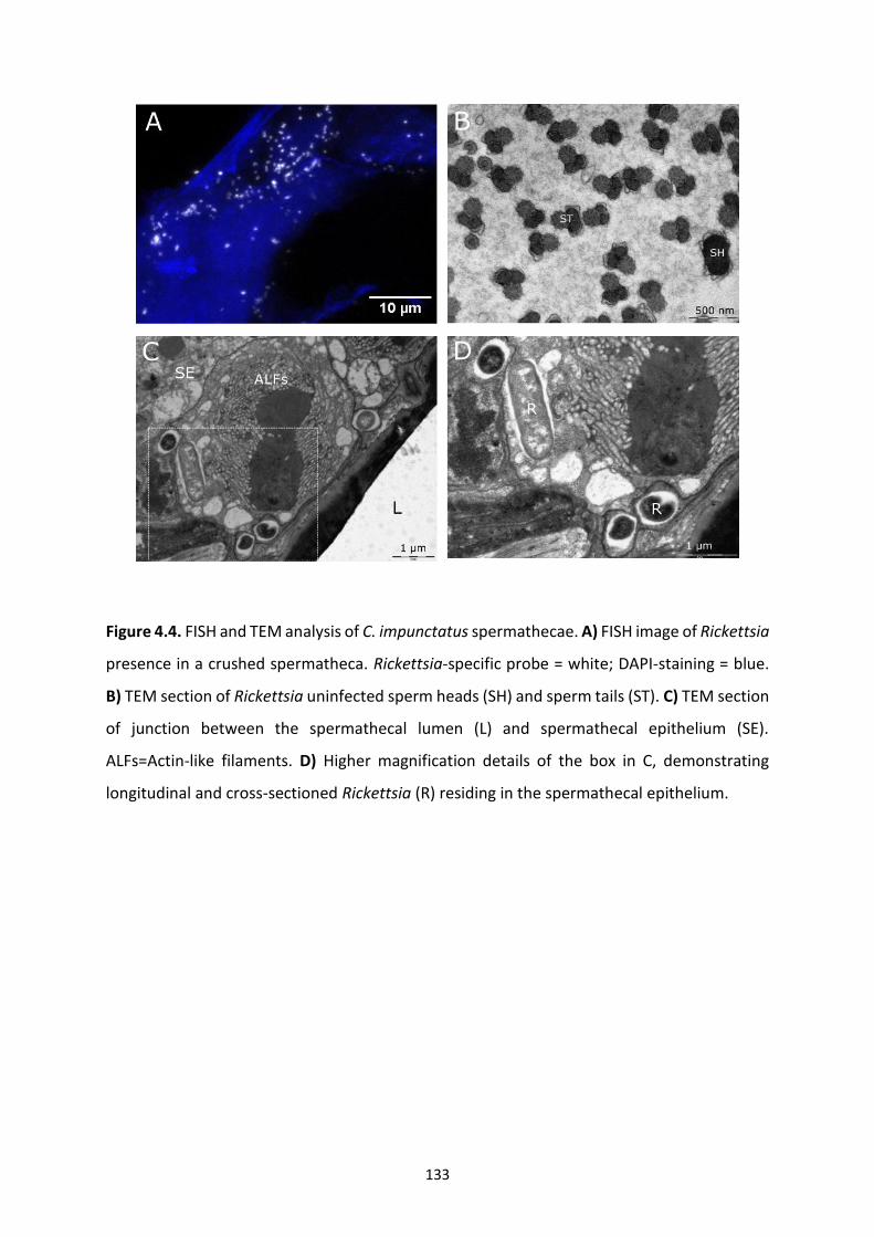
133
Figure 4.4. FISH and TEM analysis of C. impunctatus spermathecae. A) FISH image of Rickettsia
presence in a crushed spermatheca. Rickettsia-specific probe = white; DAPI-staining = blue.
B) TEM section of Rickettsia uninfected sperm heads (SH) and sperm tails (ST). C) TEM section
of junction between the spermathecal lumen (L) and spermathecal epithelium (SE).
ALFs=Actin-like filaments. D) Higher magnification details of the box in C, demonstrating
longitudinal and cross-sectioned Rickettsia (R) residing in the spermathecal epithelium.
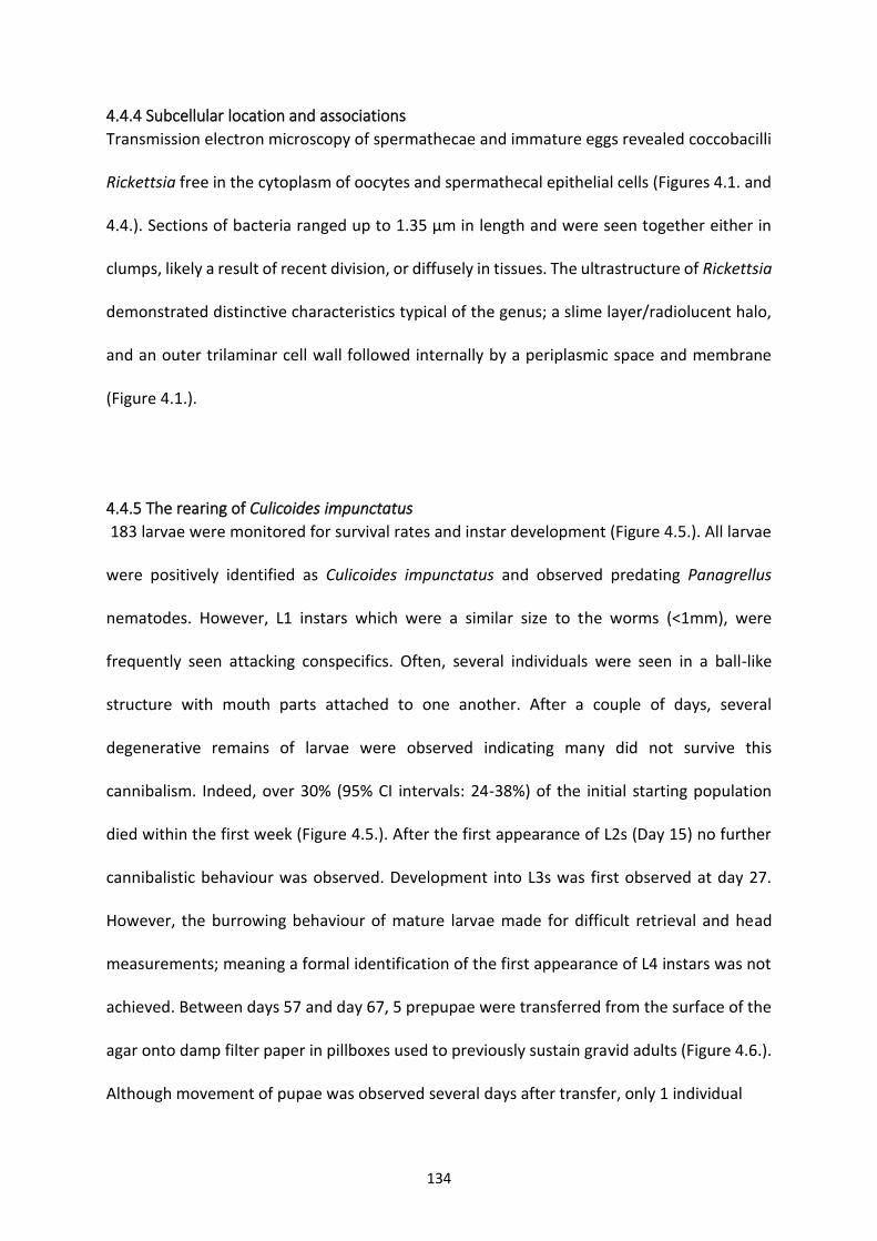
134
4.4.4 Subcellular location and associations
Transmission electron microscopy of spermathecae and immature eggs revealed coccobacilli
Rickettsia free in the cytoplasm of oocytes and spermathecal epithelial cells (Figures 4.1. and
4.4.). Sections of bacteria ranged up to 1.35 μm in length and were seen together either in
clumps, likely a result of recent division, or diffusely in tissues. The ultrastructure of Rickettsia
demonstrated distinctive characteristics typical of the genus; a slime layer/radiolucent halo,
and an outer trilaminar cell wall followed internally by a periplasmic space and membrane
(Figure 4.1.).
4.4.5 The rearing of Culicoides impunctatus
183 larvae were monitored for survival rates and instar development (Figure 4.5.). All larvae
were positively identified as Culicoides impunctatus and observed predating Panagrellus
nematodes. However, L1 instars which were a similar size to the worms (<1mm), were
frequently seen attacking conspecifics. Often, several individuals were seen in a ball-like
structure with mouth parts attached to one another. After a couple of days, several
degenerative remains of larvae were observed indicating many did not survive this
cannibalism. Indeed, over 30% (95% CI intervals: 24-38%) of the initial starting population
died within the first week (Figure 4.5.). After the first appearance of L2s (Day 15) no further
cannibalistic behaviour was observed. Development into L3s was first observed at day 27.
However, the burrowing behaviour of mature larvae made for difficult retrieval and head
measurements; meaning a formal identification of the first appearance of L4 instars was not
achieved. Between days 57 and day 67, 5 prepupae were transferred from the surface of the
agar onto damp filter paper in pillboxes used to previously sustain gravid adults (Figure 4.6.).
Although movement of pupae was observed several days after transfer, only 1 individual
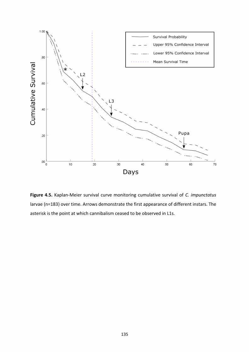
135
Figure 4.5. Kaplan-Meier survival curve monitoring cumulative survival of C. impunctatus
larvae (n=183) over time. Arrows demonstrate the first appearance of different instars. The
asterisk is the point at which cannibalism ceased to be observed in L1s.

136
melanised with none eventually emerging as adults. Development into pupae took a
minimum of 56 days and the mean survival time of larvae was 19 days.
4.4.6. Larval tissue localisation
Due to difficulties in the manipulation and dissection of such small larvae, whole insect FISH
assays were performed. Out of 10 L3 larvae assessed of unknown infection status, 4 showed
a positive signal in the terminal abdominal (anal) segment fat body (Figure 4.7.) with 3 of
these also demonstrating infection in the heads. One individual of the 4 positives showed
sporadic multifocal infections across the rest of the length of the body. Focal “Burst” patterns
of signal in the terminal abdominal segment suggest infections of globe-like structures such
as cells or lipid droplets of the fat body. Of the infections in the head, although the exact
tissue could not definitively be deduced, Rickettsia was detected closely associated with the
head body wall (within 0-3 μm proximity of the autofluorescent cuticle of each focal plane).
Concurrent presence in the large fat bodied anal segment (Figure 4.6.) of the larvae alongside
the fat body’s frequent attachment to the body wall suggests the pericerebral fat body is the
most likely tissue affected, although further work is needed to confirm this. DAPI staining was
poor in the heads of larvae making it difficult to ascertain the positions of nuclei. However,
infected regions often surrounded circular areas (3-5 μm in length) lacking infection which
are potentially uninfected nuclei (Figure 4.7). Instead of imaging pupae, attempts were made
to confirm transgenerational transmission by monitoring and screening newly emerged
adults. Unfortunately, none of the 5 reaching pupation emerged as adults.
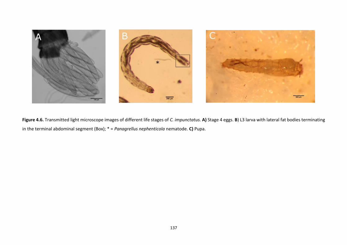
137
Figure 4.6. Transmitted light microscope images of different life stages of C. impunctatus. A) Stage 4 eggs. B) L3 larva with lateral fat bodies terminating
in the terminal abdominal segment (Box); * = Panagrellus nephenticola nematode. C) Pupa.
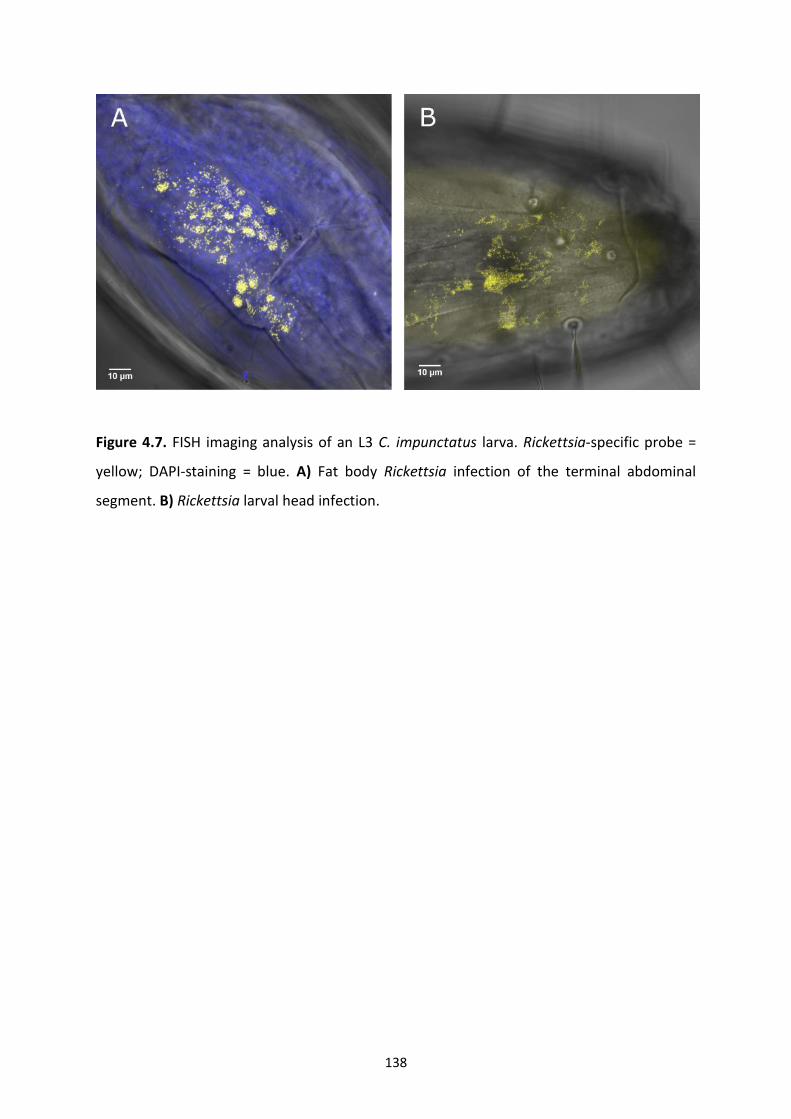
138
Figure 4.7. FISH imaging analysis of an L3 C. impunctatus larva. Rickettsia-specific probe =
yellow; DAPI-staining = blue. A) Fat body Rickettsia infection of the terminal abdominal
segment. B) Rickettsia larval head infection.
139
4.5 Discussion
The transmission routes of Rickettsia endosymbionts and their effects on biting midges are
unknown but can be informed by symbiont tissue localisation. A major finding of this study is
the Rickettsia infection of the ovarian suspensory ligament indicating a novel means of
endosymbiont germline targeting (Figures 4.3.). The ovarian suspensory ligament of insects is
a connective tissue which fuses the terminal filaments of ovarioles together at the anterior
apex of the ovary. Commonly, the ligaments of each ovary join to form what is known as the
“median ligament” which can attach to several tissues including the dorsal diaphragm, body
wall, heart or fat body (Storto, 1994; Szklarzewicz et al., 2007). In the case of Culicoides
impunctatus, my findings suggest the median ligament fuses at the anterior posterior midgut
junction, although it is possible that the ligament is part of a more extensive matrix with
several attachment points: the fragile and thin nature of the ovarian suspensory ligament
make it likely that this can be excluded in dissected or sectioned whole insects.
Endosymbionts which undergo mixed modes of transmission, such as Rickettsia, require
mechanisms of cell-to-cell transfer to ensure migration from the soma to the germline (Allen
et al., 2007; Landmann et al., 2010; Stoll et al., 2010). The ligament itself is thought to consist
of muscle and fibrous tissue, with our data showing the presence of both small and large
nuclei (Figure 4.3.) consistent with Curtin and Jones’ (1961) description of the Aedes aegypti
suspensory ligament. As Rickettsia have been noted to utilise actin for movement within and
between cells, this offers a possible mechanism for how the bacteria are able to pass through
this structure (Teysseire, Chiche-Portiche and Raoult, 1992; Heinzen et al., 1993; Gouin et al.,
1999; Reed et al., 2014).
140
Aside from host-derived bacteriocytes transferring primary endosymbionts to ovaries,
facultative bacteria possess innate qualities allowing them to target the germline (Russell and
Moran, 2005; Frydman et al., 2006; Weiss et al., 2006; Nakayama et al., 2015). Thus,
determining these mechanisms of germline infection is of interest as it allows for insights into
the initiation of novel insect-endosymbiont interactions. The continuation of the suspensory
ligament with the ovarian epithelial sheath (Figure 4.3.) has previously been described in
mosquitoes (Clements, 1992) and suggests an efficient route for ensuring transport of
Rickettsia to egg chambers via follicle cells. The passage of Rickettsia through the follicle cells
of ovarioles has been previously described in the ladybird Adalia bipunctata and the whitefly
Bemisia tabaci (Sokolova, Zinkevich and Zakharov, 2002; Brumin, Levy and Ghanim, 2012). In
these instances, Rickettsia is thought to migrate through follicle cells due to contact with
infected haemocytes (Chen, Campbell and Purcell, 1996; Hurst, Walker and Majerus, 1996).
Other studies have suggested bacteriocytes could be a means of transovarial transmission for
Rickettsia (Gottlieb et al., 2006, 2008), although further investigation suggested this was not
a primary route of ovary-targeting in all cases (Brumin, Levy and Ghanim, 2012).
Subsequently, infection of connective tissue directly linking the germline, offers a novel
alternative route for the infection of developing eggs.
Multiple means of germline targeting have been previously observed in Wolbachia, where
the endosymbiont utilises different germ line and somatic tissues depending on strain and
host. In most Drosophila species, infections tend to occur in both somatic stem cell niches and
germline stem cells, with both niches allowing for germline entry (Miller and Riegler, 2006;
Toomey et al., 2013). However, a select few Wolbachia strains appear to exhibit strict tropism
141
to somatic stem cells. The utilisation of somatic tissues as an indirect route of Wolbachia
germline colonisation appears to also occur in filarial nematodes (Onchocercidae family)
(Fischer et al., 2011; Landmann et al., 2012).
Our findings of Rickettsia infection in follicle cells but with limited infection of mature oocytes
(Figure 4.2.), corroborates previous observations in the whitefly Bemisia tabaci (Brumin, Levy
and Ghanim, 2012). In this case, heavy Rickettsia infection in immature oocytes, but not
mature stages, was attributed to younger eggs being more permeable than their mature
counterparts. The Rickettsia infection and close contact of the epithelial sheath to the follicle
cells offers a suitable mechanism for ensuring persistent follicular infection (Figure 4.3.).
Furthermore, as only a few bacteria cells are required to ensure subsequent infection of life
stages (Watanabe et al., 2014), remnant Rickettsia in the oocyte periphery of mature eggs
(Figure 4.2.) appears to be sufficient for transtadial transmission (Figure 4.7.). The co-option
of yolk granules by endosymbionts to gain entry into egg chambers via endocytosis (Herren
et al., 2013) can be dismissed in this case as Rickettsia are seen present in stage 1 oocytes
before the accumulation of yolk (Figures 4.1. and 4.2.).
While maternal transmission is the primary transmission route for endosymbionts, horizontal
transfer can lead to increased gene flow between co-habiting symbionts (Russell, Corbett-
Detig and Cavanaugh, 2017), expand host-range (Stahlhut et al., 2010; Morrow et al., 2014)
and negate the effects of detrimental mutations (Muller’s ratchet) which often occur in
intracellular bacteria (Moran, 1996). Horizontal transmission to vertebrate or invertebrate
hosts, via haematophagy, is unlikely due to a lack of observed signal in salivary glands, and
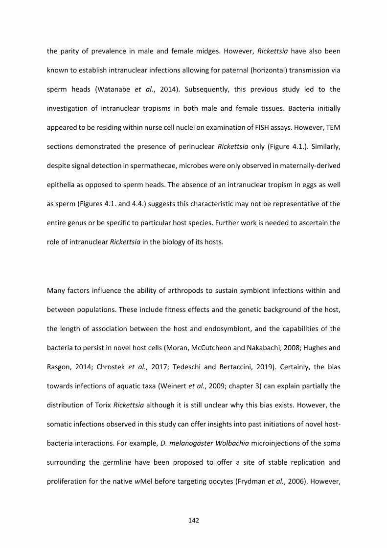
142
the parity of prevalence in male and female midges. However, Rickettsia have also been
known to establish intranuclear infections allowing for paternal (horizontal) transmission via
sperm heads (Watanabe et al., 2014). Subsequently, this previous study led to the
investigation of intranuclear tropisms in both male and female tissues. Bacteria initially
appeared to be residing within nurse cell nuclei on examination of FISH assays. However, TEM
sections demonstrated the presence of perinuclear Rickettsia only (Figure 4.1.). Similarly,
despite signal detection in spermathecae, microbes were only observed in maternally-derived
epithelia as opposed to sperm heads. The absence of an intranuclear tropism in eggs as well
as sperm (Figures 4.1. and 4.4.) suggests this characteristic may not be representative of the
entire genus or be specific to particular host species. Further work is needed to ascertain the
role of intranuclear Rickettsia in the biology of its hosts.
Many factors influence the ability of arthropods to sustain symbiont infections within and
between populations. These include fitness effects and the genetic background of the host,
the length of association between the host and endosymbiont, and the capabilities of the
bacteria to persist in novel host cells (Moran, McCutcheon and Nakabachi, 2008; Hughes and
Rasgon, 2014; Chrostek et al., 2017; Tedeschi and Bertaccini, 2019). Certainly, the bias
towards infections of aquatic taxa (Weinert et al., 2009; chapter 3) can explain partially the
distribution of Torix Rickettsia although it is still unclear why this bias exists. However, the
somatic infections observed in this study can offer insights into past initiations of novel host-
bacteria interactions. For example, D. melanogaster Wolbachia microinjections of the soma
surrounding the germline have been proposed to offer a site of stable replication and
proliferation for the native wMel before targeting oocytes (Frydman et al., 2006). However,
143
it should be noted that this did not work for non-native strains of Wolbachia. The proximity
of the OSL to the midgut (Figure 4.3.) suggests predation may be a viable route of horizontal
transmission. However, there are harsh environmental conditions needed to initiate
infections within the gut epithelium including high pH, reactive oxygen species and peristaltic
movements (Vallet-Gely, Lemaitre and Boccard, 2008). Subsequently, infection via
endoparasitism of the major fat body tissues of larvae (or pupae), can offer an alternative
route due to the fat body’s association with the ovarian suspensory ligament (Storto, 1994).
Thus, such soma-germline infection dynamics could offer efficient strategies for both
horizontal and vertical transmission. This pattern corroborates findings in chapter 3 and
chapter 5 where the coalescing of Rickettsia strains in diverse host taxa suggests frequent
host shifts.
Aside from predatory and parasitoid interactions, it would be of particular interest to see
whether the various physiologies of the suspensory ligament in different hosts can predict
rickettsial infection. For example, the robber fly Asilius crabroniformis’ ovarian suspensory
ligament does not form a median ligament, but attaches to the body wall via the outer
peritoneal sheath of ovaries instead of the terminal filaments (Owsley, 1946). Similar
physiological barriers of infection have been proposed in the Spiroplasma endosymbiont of
Drosophila melanogaster, where the inability to host shift has been suggested to be due to
the divergence of yolk proteins between hosts (Nakayama et al., 2015).
Previous examples of endosymbiont fat body infections of Culicoides includes the observation
of “Rickettsia-like” organisms by Wolbach and Hertig (1924) and “twinkling” symbionts under
144
polarised light by Lawson (1951). Most of the larval fat body comprises of two bands
extending down the lateral abdomen terminating in large lobes of the terminal segment
(Figure 4.6. and Lawson, 1951). Rickettsia appear to be one of few known endosymbionts to
reside in the fat body; the others being Blattabacterium of cockroaches (Brooks, 1970) and
Wolbachia in a variety of insects (Cheng et al., 2000; Clark et al., 2005; Hughes et al., 2011).
The presence of Wolbachia in the fat body, an important endocrine tissue, has been
associated with effects on host glucose and glycogen metabolism via altered enzyme activity
and insulin signalling (Ikeya et al., 2009; Voronin et al., 2016). Not only is the fat body
metabolically active, allowing for the bacterial sequestering of metabolite precursors, but fat
cells during larval and pupal development are refractive to degradation (Nelliot, Bond and
Hoshizaki, 2006; Aguila et al., 2007). Thus, this tissue offers a suitable niche in arthropods for
stable division and proliferation of endosymbionts.
Aside from Rickettsia observed in the abdominal fat bodies of larvae, another heavily infected
area was the head (Figure 4.7.). Although it is not clear exactly which tissues are affected by
the Rickettsia, the pericerebral fat body and cerebral ganglia (brain) are two candidates for
future consideration. Endosymbiont brain infections have been proposed to lead to
behavioural modifications (Albertson et al., 2013; Strunov et al., 2017) and early death in
Drosophila fruit flies (Min and Benzer, 1997). Additionally, the fat body surrounding the brain
has been demonstrated to play a different physiological role to that in the abdomen. Indeed,
unique insulin signalling pathways have been observed in the head fat bodies of D.
melanogaster leading to increased longevity as a result of inhibited senescence (Hwangbo et
al., 2004).
145
The presence of Rickettsia in the fat body of Culicoides, a vector of several veterinary viruses,
raises questions on their effects on vector competence. Bluetongue virus (BTV) and epizootic
haemorrhagic disease (EHDV) of ruminants replicate in the fat body of midges (Fu et al., 1999;
Mills et al., 2017b) before travelling to the salivary glands, suggesting direct interactions
between Rickettsia and the virus could be occurring. For example, competition for lipids
between bacteria and virus have been suggested to influence viral titres (Caragata et al.,
2013; Sinkins, 2013; Geoghegan et al., 2017; Schultz et al., 2017). Additionally, antimicrobial
peptides are synthesised in the fat body (Arrese and Soulages, 2010), again suggesting
Rickettsia effects on vectorial capacity warrants further investigation. Furthermore, Rickettsia
localisation to ovaries can indicate a possible role in autogeny (the development of eggs
without a blood meal) in C. impunctatus (Boorman and Goddard, 1970). Indeed, Rickettsia
felis is necessary for egg development in the booklouse Liposcelis bostrychophila. As autogeny
is responsible for the pest burden of C. impunctatus, this could offer a target for population
suppression in the future.
Although several Culicoides species have previously been reared on a Panagrellus nematode
diet (Linley, 1979, 1985b; Mullens and Velten, 1994; Barceló and Miranda, 2018), the results
in this chapter represent the first documented case in C. impunctatus, and provides a first
step to making this a tractable system for investigating the effect of the symbiont on the host.
Barceló et al. (2018) used such a diet recently with successful rearing and emergence of 5/6
species within a month (mean time 28.2 days). However, in the current study, development
rates were comparatively slow, with the first observations of pupa at day 56 (Figure 4.5.).
Furthermore, only 5/183 larvae pupated with none subsequently emerging. The emergence
146
of C. impunctatus in a natural soil habitat at 16-19°C by Hill (1947) took up to 5 months but
with no information of developmental stages; replicate breeding pots at 23-24°C showed no
emergence. Subsequently, the temperature used in this study may have been inappropriate
for maintaining high levels of survival in C. impunctatus. However, thermal environment may
not completely account for difficulties in rearing C. impunctatus, with insufficient aeration of
culturing media and inappropriate diet also being proposed (Carpenter, 2001). With respect
to the latter, although all instars were seen attacking nematodes, cannibalism was observed
only in L1 instars (Figure 4.5.) indicating possible difficulties in predation of Panagrellus due
to size. Alternatively, cannibalism is a natural behaviour of L1s which may be an inherent
problem in rearing C. impunctatus (Hill, 1947). An interesting hypothesis to test in the future
is whether cannibalism can facilitate the spread of Rickettsia in naïve populations. Initial
infections of a few individuals could lead to this outcome if several larvae predate on a single
(infected) conspecific as observed in this study. Furthermore, experimental evidence suggests
the cannibalism of isopods can lead to the initiation of novel Wolbachia infections in naïve
hosts (Le Clec’h et al., 2013). Overall, the reasons for the slow development and poor survival
observed here in comparison with previous attempts is unknown. Further optimisation of
rearing was planned to be investigated further, but the unusually hot and dry summer of 2018
prevented sufficient numbers being collected.
In conclusion, this study has identified several somatic and germline infections of the
endosymbiont Rickettsia in larval and adult life stages. Infection of the ovarian suspensory
ligament, a continuation of the ovarian epithelial sheath, has identified a potential novel
means of endosymbiont germline targeting. Further, somatic tissue infections of note include

147
the fat body of larvae and spermathecal epithelium of adult females which could have
implications for host effects, as well as symbiont and arbovirus transmission dynamics.
148
Chapter 5: The Investigation of Rickettsia Host and Bacterial Diversity through DNA Barcoding
149
5.1. Abstract
The Barcode of Life Data System (BOLD) is a sequence database specifically designed for the
curation of DNA barcodes, a widely used tool for species identification. Despite its utility, DNA
barcoding can be confounded by the unintended amplification of endosymbionts. Previous
studies have focussed primarily on the inadvertent amplification of Wolbachia due to its
commonness in arthropods. However, closely related endosymbionts such as Rickettsia
remain unexplored. Through the serendipitous discovery of Rickettsia amplicons in BOLD, this
chapter describes the host and bacterial diversity revealed though a systematic review of
>185,000 barcodes. Rickettsia is observed in approximately 0.4% of barcode submissions and
is over twice as likely to be amplified as Wolbachia (0.165%). Through phylogenetic analysis
of BOLD DNA extracts, the Torix group of Rickettsia are shown to account for 95% of all
unintended amplifications from the genus. Furthermore, re-barcoding of extracts uncovers a
new and substantial host diversity associated with Rickettsia, including phloem-feeding bugs,
parasitoid wasps, blood-feeding flies and several detritivores. This suggests barcoding can be
used as an adjunctive to targeted screens in discovering novel Rickettsia-host interactions.
The inadvertent amplification of Rickettsia should be considered during the design of
barcoding studies to avoid erroneous interpretation of species identification and population
biodiversity.
150
5.2 Introduction
DNA barcoding is a technique which uses a mitochondrial DNA (mtDNA) reference sequence
to identify existing species and aid in the discovery of new species (Hebert et al., 2003). As
well as being utilised as an identification system of individuals, mtDNA barcodes have also
been used in phylogeographic studies to detect biodiversity and dispersal events of ancestors,
including those of Culicoides biting midges (Craft et al., 2010; Papadopoulou et al., 2011;
Baselga et al., 2013; Jacquet et al., 2015). The success of barcoding can be attributed to the
“universal” nature of DNA barcoding primers used, making it a simple and standardised
method. Barcoding has been particularly useful in the designation of arthropod species which
can be difficult to assign taxonomic status to, based on morphological assessment alone.
Ambiguities can arise from the presence of cryptic or new species within populations or
because certain life stages of arthropods are problematic to classify (Kekkonen and Hebert
2014; Ashfaq et al. 2016; Horecka et al. 2017; Kanturski et al. 2018). In addition, the advent
of sequencing technology has allowed for “metabarcoding” of environmental samples, which
can allow for environmental monitoring as well as diet analysis studies (Xiong et al., 2017;
Kimmerling et al., 2018; Robeson et al., 2018; Yan et al., 2018). The widespread uptake of
DNA barcoding as a technique has led to the deposition and curation of millions of barcode
sequences in databases such as the Barcode of Life Data System (BOLD;
http://www.boldsystems.org/).
Like most methods, there are practical limitations to DNA barcoding. The COI (Cytochrome
oxidase subunit I) gene used in DNA barcoding was initially chosen due to its high evolutionary
rate, suitable effective population size and low recombination rate when compared to nuclear
markers (Hurst and Jiggins 2005). A further important assumption was that COI was a neutral
151
genetic marker i.e. little selection was occurring at this gene. However, by virtue of COI being
a functional gene, both purifying and positive selection have been observed at this locus
(Ward and Holmes 2007; Castoe et al. 2008). Significantly, indirect selection on mtDNA can
also occur in insects containing maternally inherited endosymbionts (Hurst and Jiggins 2005).
This event arises as a result of the coinheritance of symbiont and mitochondria and can have
several confounding effects on perceived population structures.
1) Through the erasure of mtDNA diversity within a population as a result of selective sweeps
driven by an endosymbiont reproductive parasitism, such as cytoplasmic incompatibility (CI),
which can lead to an underestimation of diversity (Ballard et al., 1996; Jiggins, 2003;
Shoemaker et al., 2004; Zhang et al., 2013).
2) Via the mistaken delineation of insect species occurring when isolating mechanisms (e.g.
CI) of more than one endosymbiont strain is present within a population, leading to the
segregation of mtDNA haplotypes (Schulenburg et al., 2002; Jiggins, 2003; Kvie et al., 2013).
3) Though the conflation of interspecific lineages through past mitochondrial introgression of
hybridising species (Jiggins, 2003; Whitworth et al., 2007; Raychoudhury et al., 2009, 2010;
Jäckel, Mora and Dobler, 2013).
The above confounding effects have been limited to Wolbachia and Cardinium, until a
previous example of identical barcodes in two damselfly species (Coenagrion puella and
Coenagrion pulchellum; Freeland and Conrad, 2002) was judged to be as a result of mtDNA
interspecific introgression linked to Rickettsia infection (Panupong Thongprem, University of
Liverpool; unpublished data). Consequently, Rickettsia infections also appear to have the
potential to confound barcoding studies when interpreting mtDNA divergence.
152
A further problem associated with DNA barcoding is the accidental amplification of an
unintended COI sequence instead of a target barcode. The universal nature of barcoding
primers can lead to unintended amplifications through contamination from human handling
(Ratnasingham and Hebert 2007), as well as organisms associated with the intended host
target, such as parasitoids (Ramage et al., 2017). These occur despite the attempted
development of taxa-specific barcoding primers (Hebert et al., 2004). Further, there may be
amplification of mitochondrial gene copies that are present in the nuclear genome (NUMTs),
and these may render barcodes unreadable using the Sanger method (Song et al., 2008).
It is also possible to experience the amplification of bacterial COI from DNA barcoding of
arthropods (Linares et al., 2009; Smith et al., 2012; Řezáč et al., 2014; Ceccarelli, Haddad and
Ramírez, 2016). In the largest investigation into such microbial amplification, Smith et al.
(2012) found numerous Wolbachia COI hits when exploring barcodes derived from BOLD.
Although it was concluded that Wolbachia presence would rarely interfere with intended
target COI amplification, it was noted that DNA barcoding could inadvertently reveal
previously unknown Wolbachia-insect associations, as well as providing additional bacterial
phylogenetic information.
The only other microbe demonstrated to cause erroneous amplification is Rickettsia, which is
unsurprising considering it belong to the Rickettisales order of bacteria alongside Wolbachia.
For example, erroneous amplification of a Rickettsia endosymbiont has been detected in the
Anyphaenidae and Dysderidae families of spider (Řezáč et al., 2014; Ceccarelli, Haddad and
Ramírez, 2016). Amaurobioides africana, an intertidal spider of the southern coast of Africa,
153
was noted to amplify Rickettsia in 6/19 individuals via a barcoding primer set containing
several degenerate sites. Although the inclusion of degenerate sites in primer design is often
used to increase sensitivity, this is likely to reduce specificity and subsequently increase
chances of microbe COI amplification. Furthermore, the multilocus Rickettsia designation
system devised in chapter 3 included CoxA (the bacterial equivalent of COI) and, when
compared to BOLD and NCBI databases, returned several hundred unintended database
entries that were the result of barcoding projects. These observations indicate that
inadvertent Rickettsia amplification warrants further investigation.
Rickettsia are most commonly regarded as human pathogens vectored by haematophagous
arthropods, being responsible for epidemic typhus and various “spotted fevers” (Cowan,
2000; Parola et al., 2013). Therefore, the initial research focus on Rickettsia pertained to
disease treatment and vector control. However, in subsequent years, knowledge of the genus
has greatly expanded and has been found to include hosts which include non-blood feeding
arthropods, as well as amoebae and leeches (Dyková et al., 2003; Perlman, Hunter and Zchori-
Fein, 2006; Weinert et al., 2009) and it is likely that vertebrate disease-causing Rickettsia are
atypical of the genus as a whole. Although some members are known to induce male-killing
in ladybirds, (Werren et al., 1994; Hurst et al., 1999; Majerus et al., 1999) and
parthenogenesis in parasitoid wasps (Hagimori et al., 2006; Giorgini et al., 2010), the
interactions between a majority of new strains and their hosts remain unexplored. This
includes, specifically, the Torix group of Rickettsia which encompasses a variety of hosts that
have mostly been associated with a semi-aquatic habitat (chapter 3). Further work is needed
to explore Torix interactions, especially as many hosts are vectors of vertebrate diseases.
154
These include: Simulium sp. (black flies); Phlebotomus chinensis and Lutzomyia apache (sand
flies); Nosopsyllus laeviceps (fleas) and Culicoides spp. (biting midges) (Reeves et al. 2008; Li
et al. 2016; Song et al. 2018; chapter 3). Additionally, many of these insects are difficult to
maintain in lab culture, leading to the pressing need to find model systems to elucidate
Rickettsia effects on vectorial capacity.
Considering the above, and with special permission to access BOLD trace files, this chapter
intended to uncover the extent of inadvertent Rickettsia COI amplification through barcoding.
This study specifically aimed to address the questions:
1) Is this phenomenon common and, if so, is Rickettsia likely to affect the integrity of future
barcoding studies?
2) Is Rickettsia amplification a result of an affinity to frequently used barcoding primers?
3) Can barcoding be used as an adjunctive to targeted Rickettsia screens to discover new host
taxa?
4) What phylogenetic information can be gathered from non-target Rickettsia COI?
155
5.3 Methods
5.3.1 The erroneous amplification of Rickettsia through barcoding
As part of the multilocus analysis from chapter 3 describing the analysis of several Rickettsia
housekeeping genes, it was noted that Rickettsia coxA (the bacterial equivalent of COI) was
found to match to several sequences on Genbank deposited as invertebrate mitochondrial
sequences (NCBI; http://www.ncbi.nlm.nih.gov/genbank/; accessed January 2018) (Table
5.1.). This match indicated barcoding studies were occasionally amplifying Rickettsia DNA
instead of host mtDNA. Further to this, searching BOLD, a depository of >6 million COI mtDNA
sequences, hundreds of hits were observed with high homology to both Limoniae and Leech
Rickettsia subclades. Subsequently, as the record details of these matches were not publicly
available, Professor Alex Smith (University of Guelph, Canada), who had previously worked
on non-target Wolbachia amplification associated with BOLD (Smith et al., 2012) was
contacted for further investigation.
5.3.2 BOLD datasets acquisition
As BOLD archives all Sanger sequence traces, non-target sequences from the database were
requested. Subsequently, COI barcoding data deriving from a screening project, totalling
>185K arthropod specimens from 21 countries, were provided. COI sequences provided by
BOLD were generally from somatic tissues (legs are often used in order to retain most of the
specimen for further analyses if necessary), but also rarely included abdominal tissues. The
first dataset made available included 3,829 sequences deemed as contaminant sequences as
a result of not matching initial morphotaxa assignment. The second dataset included 55,271
specimens judged to not contain erroneous amplicons. A remaining 126K specimens were not
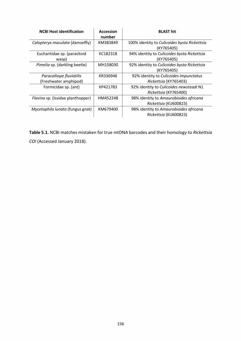
156
NCBI Host identification Accession number
BLAST hit
Calopteryx maculata (damselfly) KM383849 100% identity to Culicoides bysta Rickettsia (KY765405)
Eucharitidae sp. (parasitoid wasp)
KC182318 94% identity to Culicoides bysta Rickettsia (KY765405)
Pimelia sp. (darkling beetle) MH158030 92% identity to Culicoides bysta Rickettsia (KY765405)
Paracalliope fluviatilis (Freshwater amphipod)
KR336946 92% identity to Culicoides impunctatus Rickettsia (KY765403)
Formicidae sp. (ant) KP421783 92% identity to Culicoides newsteadi N1 Rickettsia (KY765400)
Flavina sp. (Issidae planthopper) HM452248 98% identity to Amaurobioides africana Rickettsia (KU600823)
Mycetophila lunata (fungus gnat) KM679400 98% identity to Amaurobioides africana Rickettsia (KU600823)
Table 5.1. NCBI matches mistaken for true mtDNA barcodes and their homology to Rickettsia
COI (Accessed January 2018).
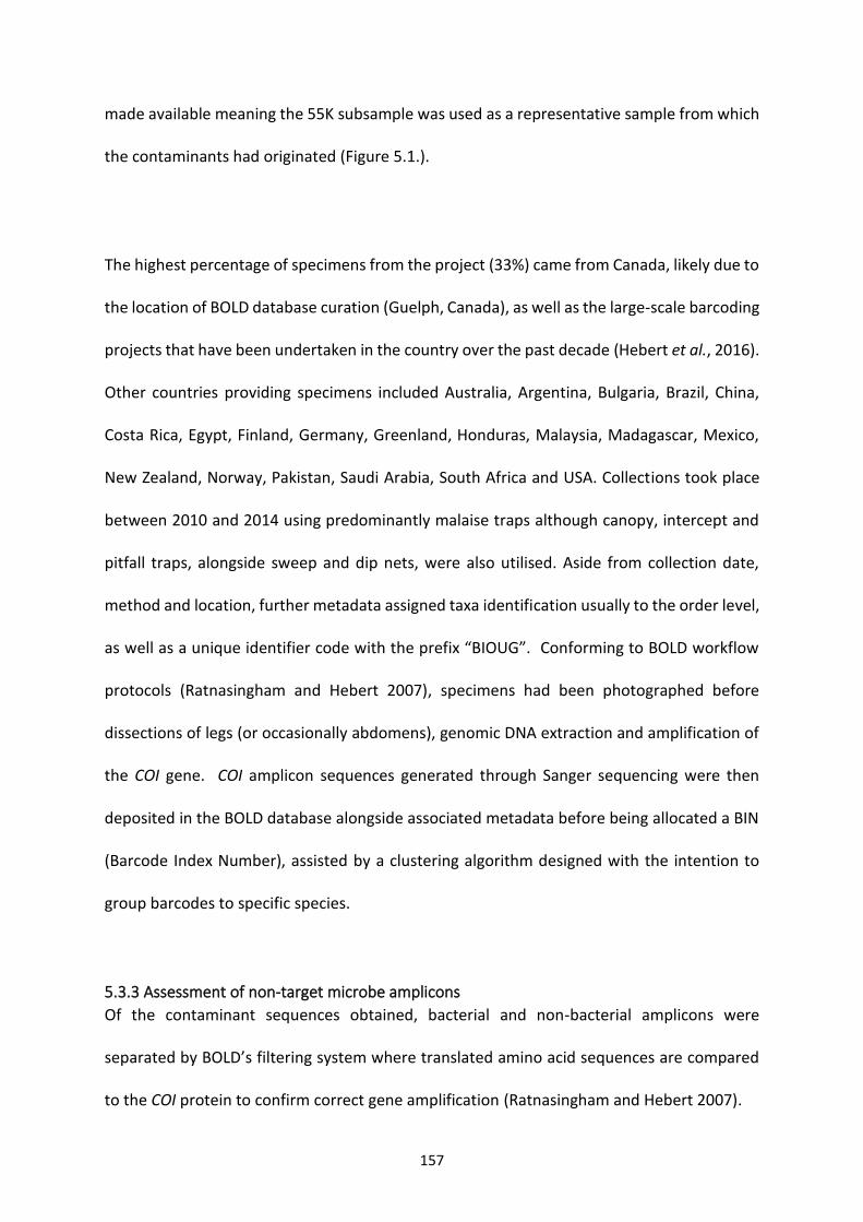
157
made available meaning the 55K subsample was used as a representative sample from which
the contaminants had originated (Figure 5.1.).
The highest percentage of specimens from the project (33%) came from Canada, likely due to
the location of BOLD database curation (Guelph, Canada), as well as the large-scale barcoding
projects that have been undertaken in the country over the past decade (Hebert et al., 2016).
Other countries providing specimens included Australia, Argentina, Bulgaria, Brazil, China,
Costa Rica, Egypt, Finland, Germany, Greenland, Honduras, Malaysia, Madagascar, Mexico,
New Zealand, Norway, Pakistan, Saudi Arabia, South Africa and USA. Collections took place
between 2010 and 2014 using predominantly malaise traps although canopy, intercept and
pitfall traps, alongside sweep and dip nets, were also utilised. Aside from collection date,
method and location, further metadata assigned taxa identification usually to the order level,
as well as a unique identifier code with the prefix “BIOUG”. Conforming to BOLD workflow
protocols (Ratnasingham and Hebert 2007), specimens had been photographed before
dissections of legs (or occasionally abdomens), genomic DNA extraction and amplification of
the COI gene. COI amplicon sequences generated through Sanger sequencing were then
deposited in the BOLD database alongside associated metadata before being allocated a BIN
(Barcode Index Number), assisted by a clustering algorithm designed with the intention to
group barcodes to specific species.
5.3.3 Assessment of non-target microbe amplicons
Of the contaminant sequences obtained, bacterial and non-bacterial amplicons were
separated by BOLD’s filtering system where translated amino acid sequences are compared
to the COI protein to confirm correct gene amplification (Ratnasingham and Hebert 2007).
158
Figure 5.1. Workflow of the project demonstrating the acquisition and fates of contaminant and non-contaminant COI barcoding sequences.
159
Sequences were then compared against a variety of potential contaminants including
Wolbachia and human homologues. Bacterial identity was also confirmed by measuring GC
content, as microbes do not contain the distinctive arthropod mtDNA AT bias (Smith et al.,
2012). The confirmed microbial sequences were then aligned using the “L-INS-I” algorithm in
MAFFT v7.4 (Katoh and Standley 2013) before using Gblocks (Castresana, 2000) to exclude
areas of the alignment with excessive gaps or poor alignment; a less stringent selection was
chosen through parameters allowing smaller final conserved blocks and gap
positions.ModelFinder (Kalyaanamoorthy et al., 2017) then determined the TIM3+F+G4
model to be used after selection based on default “auto” parameters using the Bayesian
information criteria. A maximum likelihood (ML) phylogeny was then estimated with IQTree
(Nguyen et al., 2015) using an alignment of 559 nucleotides and 1000 standard bootstraps.
The Rickettsiales genera Ehrlichia, Anaplasma, Neorickettsia, Orientia, Wolbachia
(Supergroups A, B, E, F, H) and Rickettsia were included in the analysis as references. Quantum
GIS software v3 (Quantum GIS Development Team, 2016) was then used to map BOLD-
associated Rickettsia with their collection site. Finally, trees were drawn and annotated based
on host taxa (order) using the EvolView (He et al., 2016) online tree annotation and
visualisation tools.
5.3.4 Phylogenetic analysis
Since original BOLD genomic DNA extracts are archived at the Biodiversity institute of Ontario,
it was possible to request Rickettsia-containing samples of interest. This allowed for both
validation and further phylogenetic analysis based on more housekeeping genes other than
COI. As Torix Rickettsia are known to be at high prevalence within populations (chapter 3), it
was likely that most of the genetically similar Rickettsia from the same taxa (order) and
160
collection sites would be from the same host species. Subsequently, aside from selecting
samples spread across the whole of the phylogeny, a mixture of 190 DNA extracts were
chosen based on varied collection site and host order.
The details of multilocus PCR screening and phylogenetic analysis of Rickettsia can be found
in chapter 3. However, slight variations include the exclusion of the atpA gene due to
observed recombination at this locus. Furthermore, the amplification conditions for the
17KDa locus was changed because the single Leech reference DNA extract (Host: Simulium
aureum) failed to amplify with the primer set Ri_17KD_F/ Ri_17KD_R from chapter 3.
Subsequently, a 17KDa alignment of Rickettsia groups originating from the genomes of
Spotted fever, Typhus, Transitional, Belli, Limoniae and the newly available Megaira group
(provided by Prof. R. S. Coyne, Dr T. Doak, and Prof. M. Lynch) was generated to design a new
set of primers using the online tool PriFi (Fredslund et al., 2005). The chosen locus was
ensured to overlap with already characterised regions of the gene assessed in chapter 3. Once
multilocus profiles of the Rickettsia had been established, samples amplifying at least 3 out
of 4 genes (16S, 17KDa, coxA and gltA) were concatenated before being analysed for
recombination within and between genes using RDP v4 (Martin et al., 2015) (details chapter
3). These sequences were then assessed again through ML phylogenetic analysis to
differentiate samples into Rickettsia subclades. The selected models used in the concatenated
partition scheme were as follows: 16S and GTR+F+R2; 17KDa and GTR+F+G4; coxA and
TVM+F+I+G4; gltA and TVM+F+I+G4. Accession numbers for all reference Rickettsia and
outgroup sequences can be found in Appendix 2.
161
5.3.5 Host barcoding
Aside from phylogenetic placement of these Rickettsia-containing samples, attempts were
made to extract an mtDNA barcode in order to uncover the host diversity of infected
specimens. Previous erroneous amplification of Rickettsia through DNA barcoding had
occurred in the bed bug Cimex lectularius, with a recovery of the true barcode after using the
primer set C1-J-1718/HCO1490 (Panupong Thongprem, University of Liverpool; unpublished
data), which amplifies a shortened 455 bp sequence within the COI locus. Subsequently, all
samples were screened using these primers or a further set of secondary COI primers
(LCOt_1490/ MLepR1) if the first failed to give an adequate host barcode. All COI and
Rickettsia multilocus screening primers, including references, are available in Table 5.2.
Cycling conditions for COI PCRs were as follows: initial denaturation at 95°C for 5 min,
followed by 35 cycles of denaturation (94°C, 30 sec), annealing (50°C, 60 sec), extension (72°C,
90 sec), and a final extension at 72°C for 7 min. Rickettsia and host amplicons identified by gel
electrophoresis were subsequently purified enzymatically (ExoSAP) and Sanger sequenced
through both strands as described in chapter 2. Forward and reverse reads were assessed in
UGENE (Okonechnikov et al., 2012) to create a consensus sequence by eye with a cut-off
phred (Q) score (Ewing and Green 1998) of 20; any ambiguous base calls below this threshold
were attributed as “N” according to IUPAC nucleotide nomenclature . Primer regions were
trimmed from barcodes before being matched to Genbank and BOLD databases by BLAST
based on default parameters and an e-value threshold of <1e-85. Host taxonomy was
determined by a barcode-based assignment of the closest BLAST hit, under the following
criteria modified from Ramage et al. (2017):

162
Gene Product Primer
name
Sequence (5’-3’) Product
length
(bp)
Reference
COI
Cytochrome
c oxidase,
subunit I
C1-J-1718 GGAGGATTTGGAAATTGATTAGT 455 Folmer et
al. 1994;
Dallas et
al. 2003
HCO1490
TAAACTTCAGGGTGACCAAAAAATCA
LCOt_1490 TGTAAAACGACGGCCAGTGG
TCAACAAATCATAAAGATATT
GG
310 Simon et
al. 1994;
Hajibabaei
et al.
2006
MLepR1 CCTGTTCCAGCTCCATTTTC
LepF1 ATTCAACCAATCATAAAGATAT 320 Hebert et
al. 2004;
Fisher and
Smith
2008
C_ANTMR1
D_RonIIdeg
_R
GGRGGRTARAYAGTTCATCCWG
TWCC
gltA Citrate
synthase
RiGltA405_
F
GATCATCCTATGGCA 786 Pilgrim et
al. 2017
RiGltA1193
_R
TCTTTCCATTGCCCC
17KDa 17KDa
antigenic
protein
precursor
Ri_Meg17k
D_F
TGGYATGAATAARCAAGGTGG 319 This study
Ri_Meg17k
D_R
ATACTCACGACAATAYTGCCC
16S 16S
ribosomal
RNA
Ri170_F GGGCTTGCTCTAAATTAGTTAGT 1170 Pilgrim et
al. 2017 Ri1500_R ACGTTAGCTCACCACCTTCAGG
Table 5.2. Mitochondrial COI and bacterial gene primers used for re-barcoding and multilocus
phylogenetic analysis.
163
1) Species level designation for at least 98% sequence identity over at least 90% of the
barcode.
2) Genus level designation for at least 95% sequence identity and 90% overlap.
3) Family level designation for at least 80% sequence identity and 90% overlap.
Additionally, all sequences were required to be at least >200 bp in length. Newly identified
hosts which amplified with at least 2 out of 4 Rickettsia genes were then placed
phylogenetically using the same methods as above before being mapped by lifestyle and diet.
5.3.6 Assessment of barcoding success
One of the factors determining a successful COI bacterial amplification is the initial failure of
an extract to amplify mtDNA. Subsequently, to determine the likelihood of this event within
taxa, we used the 55K representative dataset to evaluate failure rates. To this end, all taxa at
the order level, which gave at least one non-target bacteria COI hit were assessed. The
barcoding success rate was determined as the proportion of specimens which matched initial
morphotaxa assignment and were not removed after BOLD quality control (Ratnasingham
and Hebert 2007). With respect to the latter, chromatograms were allocated a Q score for
each nucleotide position as well as a mean value for the entire barcode. Generally poor and
failed trace files were defined as mean Q scores of <30, although designation of a submitted
BOLD sequence was still ultimately determined by the submitter. As the total Rickettsia count
was from a larger dataset than the one made available, an adjusted prevalence for each taxa
was calculated based on the 55K representative dataset.

164
5.3.7 Assessment of primer bias
A further determining factor for erroneous amplification of bacteria is primer site matching
to respective microbe sequences. Subsequently, pairwise homology of the primer set
predominantly used for BOLD barcode screening and Rickettsia or Wolbachia was assessed
using Mega v6 (Tamura et al., 2013).
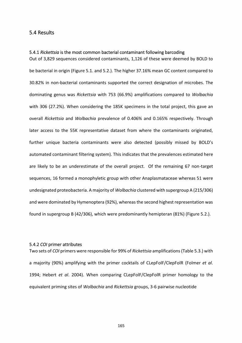
165
5.4 Results
5.4.1 Rickettsia is the most common bacterial contaminant following barcoding
Out of 3,829 sequences considered contaminants, 1,126 of these were deemed by BOLD to
be bacterial in origin (Figure 5.1. and 5.2.). The higher 37.16% mean GC content compared to
30.82% in non-bacterial contaminants supported the correct designation of microbes. The
dominating genus was Rickettsia with 753 (66.9%) amplifications compared to Wolbachia
with 306 (27.2%). When considering the 185K specimens in the total project, this gave an
overall Rickettsia and Wolbachia prevalence of 0.406% and 0.165% respectively. Through
later access to the 55K representative dataset from where the contaminants originated,
further unique bacteria contaminants were also detected (possibly missed by BOLD’s
automated contaminant filtering system). This indicates that the prevalences estimated here
are likely to be an underestimate of the overall project. Of the remaining 67 non-target
sequences, 16 formed a monophyletic group with other Anaplasmataceae whereas 51 were
undesignated proteobacteria. A majority of Wolbachia clustered with supergroup A (215/306)
and were dominated by Hymenoptera (92%), whereas the second highest representation was
found in supergroup B (42/306), which were predominantly hemipteran (81%) (Figure 5.2.).
5.4.2 COI primer attributes
Two sets of COI primers were responsible for 99% of Rickettsia amplifications (Table 5.3.) with
a majority (90%) amplifying with the primer cocktails of CLepFolF/ClepFolR (Folmer et al.
1994; Hebert et al. 2004). When comparing CLepFolF/ClepFolR primer homology to the
equivalent priming sites of Wolbachia and Rickettsia groups, 3-6 pairwise nucleotide
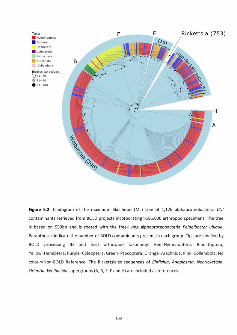
166
Figure 5.2. Cladogram of the maximum likelihood (ML) tree of 1,126 alphaproteobacteria COI
contaminants retrieved from BOLD projects incorporating >185,000 arthropod specimens. The tree
is based on 559bp and is rooted with the free-living alphaproteobacteria Pelagibacter ubique.
Parantheses indicate the number of BOLD contaminants present in each group. Tips are labelled by
BOLD processing ID and host arthropod taxonomy: Red=Hymenoptera; Blue=Diptera;
Yellow=Hemiptera; Purple=Coleoptera; Green=Psocoptera; Orange=Arachnida; Pink=Collembola; No
colour=Non-BOLD Reference. The Rickettsiales sequences of Ehrlichia, Anaplasma, Neorickettsia,
Orientia, Wolbachia supergroups (A, B, E, F and H) are included as references.
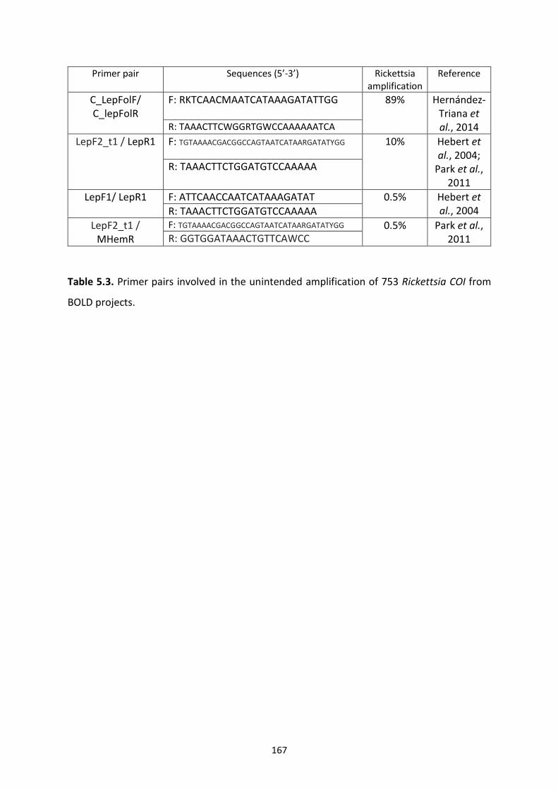
167
Primer pair Sequences (5’-3’) Rickettsia amplification
Reference
C_LepFolF/ C_lepFolR
F: RKTCAACMAATCATAAAGATATTGG 89% Hernández-Triana et al., 2014 R: TAAACTTCWGGRTGWCCAAAAAATCA
LepF2_t1 / LepR1 F: TGTAAAACGACGGCCAGTAATCATAARGATATYGG 10% Hebert et al., 2004;
Park et al., 2011
R: TAAACTTCTGGATGTCCAAAAA
LepF1/ LepR1 F: ATTCAACCAATCATAAAGATAT 0.5% Hebert et al., 2004 R: TAAACTTCTGGATGTCCAAAAA
LepF2_t1 / MHemR
F: TGTAAAACGACGGCCAGTAATCATAARGATATYGG 0.5% Park et al., 2011 R: GGTGGATAAACTGTTCAWCC
Table 5.3. Primer pairs involved in the unintended amplification of 753 Rickettsia COI from
BOLD projects.
168
differences were observed across the 26 nucleotide C_LepFolF and 1-4 nucleotides across
C_LepFolR (Tables 5.4.1 and 5.4.2.). Although all Rickettsia and Wolbachia contained a SNP at
the 3’ priming site (position 24) of CLepFolR, Torix Rickettsia (Culicoides newsteadi N5; Pilgrim
et al., 2017) was the only sequence to not contain a similar SNP at the 3’ priming end of
CLepFolF (position 23).
5.4.3 The diversity of non-target Rickettsia
BOLD Rickettsia contaminants were dominated by the Torix group with 716/753 (95.1%)
forming a clade with Limoniae and Leech reference sequences generated in chapter 3, which
overlapped with the BOLD COI barcode locus by approximately 350 bp (Figure 5.3.1.). The
remaining 37 Rickettsia clustered with groups Transitional (n=15), Belli (n=9), Rhyzobius (n=1)
and 12 were undesignated. Breaking this down at a host taxa level: 294 (39%) were present
in Hymenoptera; 187 (25%) in Diptera; 177 in Hemiptera (24%); 40 each in Coleoptera and
Psocodea (5% each); 8 in Arachnida (1%); 4 in Trichoptera (0.5%); and single cases of
Thysanoptera, Diplopoda and Dermaptera (0.3% each).
Using the reference sequences for both Limoniae and Leech subclades of the Torix group it
was possible to tentatively allocate the Rickettsia sequences to either subclade (Figure 5.3.2.).
Hymenoptera and Diptera were present in both subclades, while 168/170 Hemiptera were
allocated to the Leech group. Conversely, a single coleopteran out of 33 was allocated in the
Leech group with the remainder placed in the Limoniae subclade. A majority of Diptera were
not able to be placed in either subclade with strong support Mapping the 753 Rickettsia to
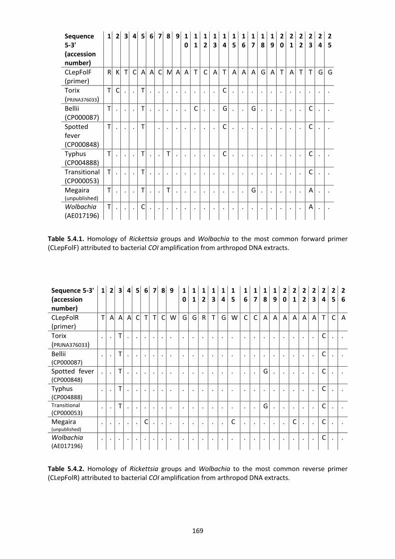
169
Table 5.4.1. Homology of Rickettsia groups and Wolbachia to the most common forward primer (CLepFolF) attributed to bacterial COI amplification from arthropod DNA extracts.
Sequence 5-3' (accession number)
1 2 3 4 5 6 7 8 9 10
11
12
13
14
15
16
17
18
19
20
21
22
23
24
25
26
CLepFolR (primer)
T A A A C T T C W G G R T G W C C A A A A A A T C A
Torix (PRJNA376033)
. . T . . . . . . . . . . . . . . . . . . . . C . .
Bellii (CP000087)
. . T . . . . . . . . . . . . . . . . . . . . C . .
Spotted fever (CP000848)
. . T . . . . . . . . . . . . . . G . . . . . C . .
Typhus (CP004888)
. . T . . . . . . . . . . . . . . . . . . . . C . .
Transitional
(CP000053) . . T . . . . . . . . . . . . . . G . . . . . C . .
Megaira (unpublished)
. . . . . C . . . . . . . . C . . . . . C . . C . .
Wolbachia (AE017196)
. . . . . . . . . . . . . . . . . . . . . . . C . .
Table 5.4.2. Homology of Rickettsia groups and Wolbachia to the most common reverse primer (CLepFolR) attributed to bacterial COI amplification from arthropod DNA extracts.
Sequence 5-3' (accession number)
1 2 3 4 5 6 7 8 9 10
11
12
13
14
15
16
17
18
19
20
21
22
23
24
25
CLepFolF (primer)
R K T C A A C M A A T C A T A A A G A T A T T G G
Torix (PRJNA376033)
T C . . T . . . . . . . . C . . . . . . . . . . .
Bellii (CP000087)
T . . . T . . . . . C . . G . . G . . . . . C . .
Spotted fever (CP000848)
T . . . T
. . . . . . . C . . . . . . . . C . .
Typhus (CP004888)
T . . . T . . T . . . . . C . . . . . . . . C . .
Transitional (CP000053)
T . . . T . . . . . . . . . . . . . . . . . C . .
Megaira (unpublished)
T . . . T . . T . . . . . . . . G . . . . . A . .
Wolbachia (AE017196)
T . . . C . . . . . . . . . . . . . . . . . A . .
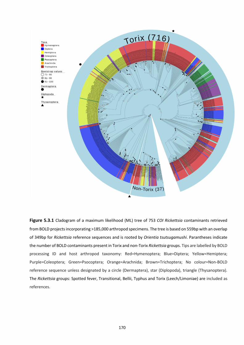
170
Figure 5.3.1 Cladogram of a maximum likelihood (ML) tree of 753 COI Rickettsia contaminants retrieved
from BOLD projects incorporating >185,000 arthropod specimens. The tree is based on 559bp with an overlap
of 349bp for Rickettsia reference sequences and is rooted by Orientia tsutsugamushi. Parantheses indicate
the number of BOLD contaminants present in Torix and non-Torix Rickettsia groups. Tips are labelled by BOLD
processing ID and host arthropod taxonomy: Red=Hymenoptera; Blue=Diptera; Yellow=Hemiptera;
Purple=Coleoptera; Green=Psocoptera; Orange=Arachnida; Brown=Trichoptera; No colour=Non-BOLD
reference sequence unless designated by a circle (Dermaptera), star (Diplopoda), triangle (Thysanoptera).
The Rickettsia groups: Spotted fever, Transitional, Bellii, Typhus and Torix (Leech/Limoniae) are included as
references.
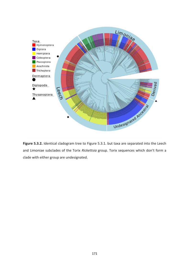
171
Figure 5.3.2. Identical cladogram tree to Figure 5.3.1. but taxa are separated into the Leech
and Limoniae subclades of the Torix Rickettsia group. Torix sequences which don’t form a
clade with either group are undesignated.
172
collection site (Figure 5.4.) revealed arthropod infections predominantly from Canada with
other locations in South/Central America, Europe, Africa and Asia.
5.4.4. Rickettsia multilocus phylogenetic analysis
To confirm the allocation of these sequences and to better place the undesignated Torix
Rickettsia, a subsample of 190 Rickettsia-containing taxa were screened with 2 further
housekeeping genes (16S, gltA) and the antigenic 17KDa protein gene. 120 extracts
successfully amplified and gave a high-quality sequence for at least one gene. No intragenic
or intergenic recombination was detected for any of the genes. A phylogram including 106
taxa containing a concatenation of at least 3 of the 4 Rickettsia genes of interest (including
COI) confirmed the initial designation of Leech and Limoniae Rickettsia, while placing the
undesignated, largely Dipteran group, as an early branch of the Limoniae subclade (Figure
5.5.). Through inclusion of multilocus profiles from Torix Rickettsia not associated with BOLD
samples (Appendix 2), it became clear that both Leech and Limoniae subclades were
paraphyletic with respect to host taxa. For example, Odonata were distributed sporadically
across the Limoniae clade, while specific families (Hemiptera: Psyliidae and Hymenoptera:
Diapriidae) were found present in both Leech and Limoniae groups. A full list of multilocus
profiles and Rickettsia group designation can be found in Appendix 3.
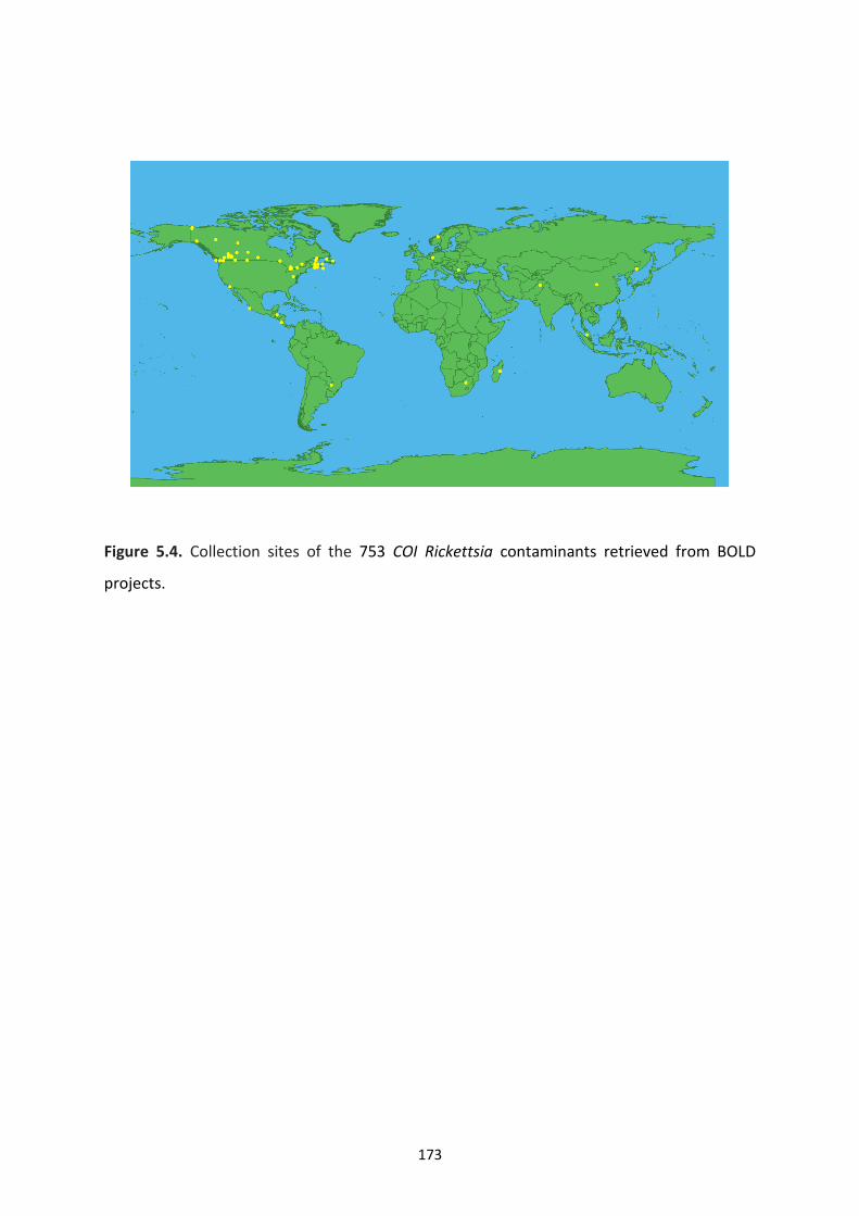
173
Figure 5.4. Collection sites of the 753 COI Rickettsia contaminants retrieved from BOLD
projects.
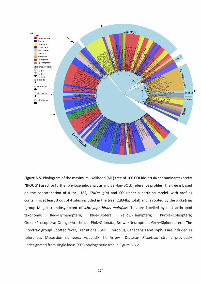
174
Figure 5.5. Phylogram of the maximum likelihood (ML) tree of 106 COI Rickettsia contaminants (prefix
“BIOUG”) used for further phylogenetic analysis and 53 Non-BOLD reference profiles. The tree is based
on the concatenation of 4 loci; 16S, 17KDa, gltA and COI under a partition model, with profiles
containing at least 3 out of 4 sites included in the tree (2,834bp total) and is rooted by the Rickettsia
(group Megaira) endosymbiont of Ichthyophthirius multifiliis. Tips are labelled by host arthropod
taxonomy: Red=Hymenoptera; Blue=Diptera; Yellow=Hemiptera; Purple=Coleoptera;
Green=Psocoptera; Orange=Arachnida; Pink=Odonata; Brown=Neuroptera; Grey=Siphonoptera. The
Rickettsia groups Spotted fever, Transitional, Bellii, Rhizobius, Canadensis and Typhus are included as
references (Accession numbers: Appendix 2). Arrow= Dipteran Rickettsia strains previously
undesignated from single locus (COI) phylogenetic tree in Figure 5.3.2.
175
5.4.5 The hidden host diversity of Torix Rickettsia
Through the discovery of Rickettsia NCBI hits labelled as arthropod mtDNA (Table 5.1.), it
became clear that host diversity of Torix Rickettsia was wider than previously thought.
Identified erroneous amplifications of Rickettsia from previous studies include Calopteryx
maculata (Damselflies); Eucharitidae (Parasitoid wasps); Pimelia sp. (Darkling beetles);
Formicidae (Ants); Flavina sp. (planthoppers); Mycetophila lunata (Fungus gnats) and
Paracalliope fluviatilis, a freshwater amphipod. Although taxonomic classification for most
BOLD IDs are initially given through morphological assessment, this is usually only down to
the order level. Subsequently, in order to refine the taxonomy further of the Rickettsia-
containing specimens, attempts were made to re-barcode DNA extracts using a different set
of primers to the ones used originally in the initial barcoding project. Successful barcodes
were obtained from 84 of the 190-subsample set using the primer pair C1-J-1718/HCO1490
(Table 5.2.). A further 5 Diptera returned a barcode after a secondary amplification with the
primer pair LCOt_1490/MLepR1 and a single hymenopteran produced a barcode with primers
LepF1/C_ANTMR1D.
Out of 28 Diptera producing successful barcodes, those conforming to the requirements of a
species, genus or family allocation (see methods for details) and representing novel
Rickettsia-hosts included the Fungus gnat Boletina villosa and the Marsh fly Pherbellia
tenuipes; while a single specimen was classified as belonging to the genus Cerodontha (Leaf-
miner flies). Several Dipteran families were also identified including: Sciaridae (Dark-winged
fungus-gnat); Cecidomyiidae (Gall-midges); Mycetophilidae (Fungus-gnats); Chironomidae
(Chironomids); Dolichopodidae (Long-legged flies); Limoniidae (Crane flies); Empididae
(Balloon flies); Psilidae (Rust flies); Hybotidae (Dance flies) and Tabanidae (Horseflies). From
176
27 Hymenoptera barcodes several parasitoid wasp families were revealed including:
Megaspilidae; Ceraphronidae; Diapriidae; Platygastridae; Eulophidae; Eurytomidae and
Mymaridae (Fairy wasps). Of the 15 Hemipteran barcodes, the planthopper families: Issidae,
Ricaniidae, Achilidae and Flatidae (subfamily: Aurrenchorhyna) were observed. At the species
level, the phloem-feeding Psyllidae Chamaepsylla hartigii and Cacopsylla melanoneura were
identified. Coleopteran families included: Staphylinidae (Rove beetles); Dystiscidae (water
beetles) and Elateridae (Click beetles) with a single barcode indicating the Marsh beetle
Prionocyphon limbatus as a host species. Out of 11 screened Psocodea, 10 from Canada gave
an identity of >99% to the barklouse Philotarsus californicus while the remaining individual
from Honduras was classified to the barklouse family Myopscocidae. Single barcodes were
recovered for the orders Thysanoptera, Trombidiformes and Trichopteran which were refined
down to the families Thripidae (Thrips), Calyptostomatidae (Water mites) and the species
Lepidostoma hoodi (Caddisfly) respectively. Notably, the single specimens of Dermaptera and
Diplopoda failed to barcode; full details of barcode success of BOLD Rickettsia extracts
including BLAST hits can be found in Appendix 3. Noteworthy lifestyles of hosts were then
mapped on a phylogram (Figure 5.6.) of the Rickettsia genus to assess any significant patterns.
Similar to when taxa were mapped across the phylogeny (Figure 5.5.), feeding habits such as
herbivory, predation and haematophagy were not correlated with any particular Rickettsia
clade. Furthermore, parasitoidism and aquatic lifestyles were seen across the phylogeny.
5.4.6 Rickettsia co-infections
In concordance with observations in chapter 3, double peaks were occasionally found at third
codon sites in chromatogram trace files from protein coding genes. This suggests co-infecting
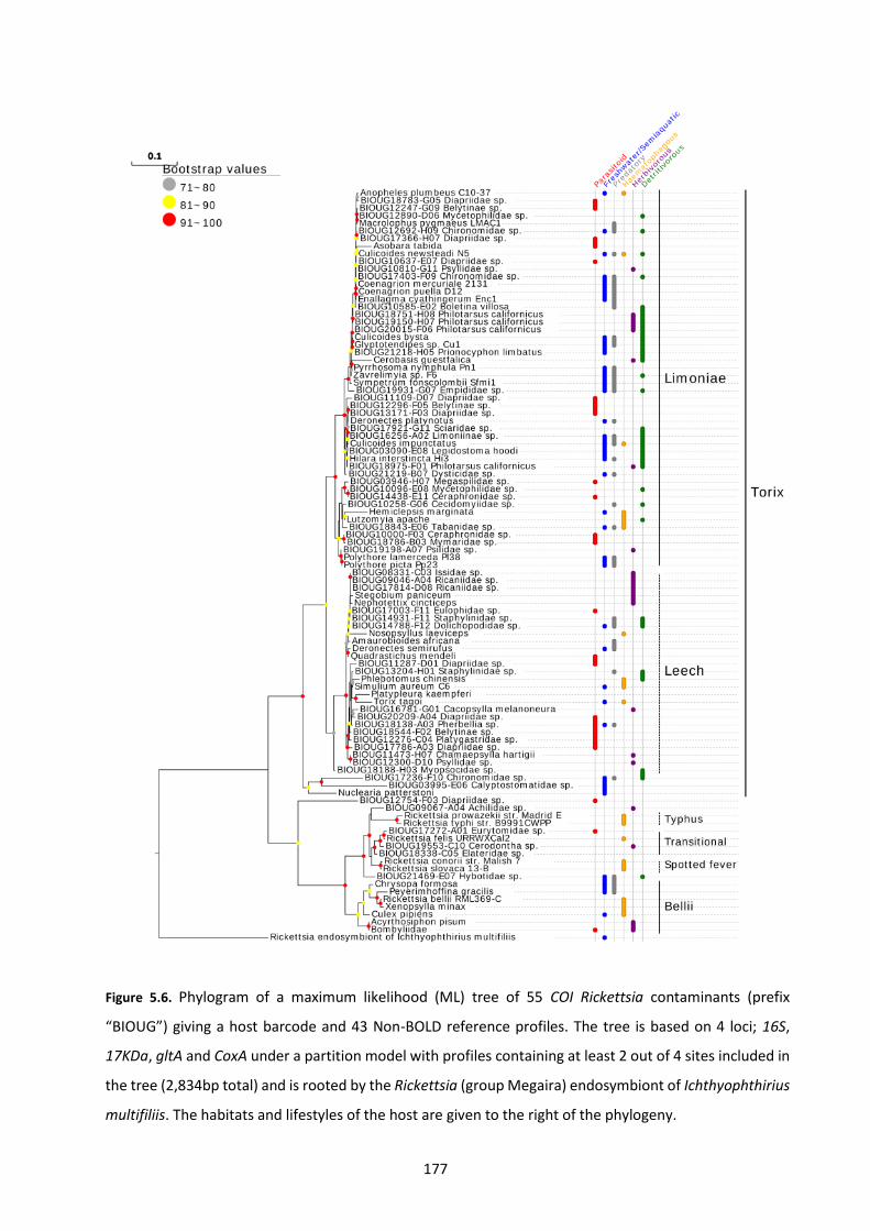
177
Figure 5.6. Phylogram of a maximum likelihood (ML) tree of 55 COI Rickettsia contaminants (prefix
“BIOUG”) giving a host barcode and 43 Non-BOLD reference profiles. The tree is based on 4 loci; 16S,
17KDa, gltA and CoxA under a partition model with profiles containing at least 2 out of 4 sites included in
the tree (2,834bp total) and is rooted by the Rickettsia (group Megaira) endosymbiont of Ichthyophthirius
multifiliis. The habitats and lifestyles of the host are given to the right of the phylogeny.
178
Rickettsia strains in hosts is a widespread phenomenon of the Torix group. To ensure cross-
contamination between DNA extracts had not occurred, the original trace files from
attempted BOLD COI amplifications were visualised to confirm double peaks had also
occurred at the time of initial processing. Co-infections were observed in 6/10 Philotarsus
californicus as well as a minority of Psilidae (1/3), Sciaridae (1/4), Chironomidae (1/4) and
Diapriidae (1/18) (Appendix 3).
5.4.7 Barcoding success of taxa
An available BOLD project of attempted barcodes associated with the contaminants
contained 55,271 out of 185K arthropods originally used in the overall study. The 3 classes of
Insecta (n=49,688), Arachnida (n=3,626) and Collembola (n=1,957) accounted for >99.8% of
total specimens (Figure 5.1.). Successful amplification and sequencing of COI was achieved in
43,246 specimens (78.1%) of the genomic extracts, but when assessed at the order level
success rates varied (Table 5.5.). The likely explanation for this variation is taxa-specific
divergence of sequences at priming sites.
The number of each taxonomic order giving at least 1 Rickettsia amplification was then
calculated and adjusted based on the total number of specimens in the project to allow for a
prevalence estimate. Overall, Hymenoptera, Diptera and Hemiptera were the three most
represented taxa associated with Rickettsia COI amplification (87.4%). Similarly, on
assessment of a subsample from the project where the contaminants originated, a majority
(77.9%) of the dataset were also accounted for by these three orders. After adjusting the
prevalence to take into account the number of inaccessible specimens, Trichoptera (2.27%),
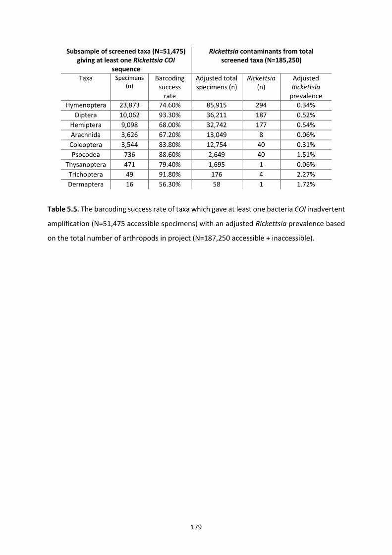
179
Subsample of screened taxa (N=51,475) giving at least one Rickettsia COI
sequence
Rickettsia contaminants from total screened taxa (N=185,250)
Taxa Specimens (n)
Barcoding success
rate
Adjusted total specimens (n)
Rickettsia (n)
Adjusted Rickettsia
prevalence
Hymenoptera 23,873 74.60% 85,915 294 0.34%
Diptera 10,062 93.30% 36,211 187 0.52%
Hemiptera 9,098 68.00% 32,742 177 0.54%
Arachnida 3,626 67.20% 13,049 8 0.06%
Coleoptera 3,544 83.80% 12,754 40 0.31%
Psocodea 736 88.60% 2,649 40 1.51%
Thysanoptera 471 79.40% 1,695 1 0.06%
Trichoptera 49 91.80% 176 4 2.27%
Dermaptera 16 56.30% 58 1 1.72%
Table 5.5. The barcoding success rate of taxa which gave at least one bacteria COI inadvertent
amplification (N=51,475 accessible specimens) with an adjusted Rickettsia prevalence based
on the total number of arthropods in project (N=187,250 accessible + inaccessible).
180
Dermaptera (1.72%) and Psocodea (1.51%) were the most likely taxa to give an inadvertent
Rickettsia amplification. Despite Hemiptera and Diptera having a similar estimated prevalence
(0.54% and 0.52%), Hemiptera were much more likely to fail to barcode (67.2% vs 93.3%)
indicating the true Dipteran prevalence is likely to be higher, as a barcoding failure is
necessary to amplify non-target bacteria COI.
181
5.5 Discussion
The utility of DNA barcoding as a technique is highlighted by its widespread use in taxonomic
and other biodiversity studies (Goldstein and DeSalle 2019). However, this study
demonstrates that the integrity of this approach can be compromised through the erroneous
amplification of Rickettsia DNA. For such a non-target amplification to occur, two criteria must
be fulfilled:
1) An unintended target must complement barcoding primer sites and
2) The unintended target must hybridise in preference to the intended target during PCR
cycling.
With respect to the first, the few bacteria likely to share homology with mitochondria are the
Rickettsiales order of alphaproteobacteria. The hypothetical bacterial ancestor of
mitochondria (proto-mitochondria) is thought to be phylogenetically embedded within the
Rickettsiales (Sassera et al. 2011; Ferla et al. 2013; Wang and Wu 2015), although its
evolutionary origin is still debated (Martijn et al., 2018; Muñoz-Gómez et al., 2019). Regarding
the second, if both target and non-target COI can hybridise during amplification, then
annealing steps early on in PCR cycling are critical in subsequent preferential amplification. It
is likely that this amplification preference is determined by gene copy number (Linares et al.,
2009) but it has also been suggested to be affected by DNA extraction protocols or variety of
Taq polymerase utilised (Smith et al., 2012). Despite this, the amplification of more than one
COI gene often results in “messy” chromatograms during Sanger sequencing. Subsequently,
the clean traces of non-target COI, as observed in this study, are likely to be the result of initial
primer mismatching with the intended host target.
182
Due to its abundance across arthropod taxa (Hilgenboecker et al. 2008; Zug and Hammerstein
2012), many previous reports have proposed Wolbachia (Rickettsiales: Anaplasmataceae) to
be the main bacterial contaminant associated with barcoding. Smith et al. (2012)
demonstrated Wolbachia was present in multiple BOLD COI traces related to insects although,
more often than not, endosymbiont presence did not interfere with barcoding. However,
other Rickettsiales, namely from the family Rickettsiaceae, have not been considered as
significant by-products until recently (Řezáč et al., 2014; Ceccarelli, Haddad and Ramírez,
2016). In a demonstration of how unintended Rickettsia amplifications can affect
phylogeographic studies relying on DNA barcoding, a Rickettsia COI was conflated with a
species of freshwater amphipod, Paracalliope fluvitalis (Lagrue et al., 2016). Subsequently,
supposed unique mtDNA haplotypes were allocated to a particular collection site, whereas
this merely demonstrated the presence of Rickettsia in the lake. A consequence of this
misamplification is that the large-scale use of DNA barcoding can allow for the uncovering of
a large, and previously unidentified, host range of Rickettsia. Therefore, the Rickettsia hits
identified from BOLD in this study has led to a unique opportunity to further investigate both
host and bacteria diversity.
In the current study, Wolbachia and Rickettsia accounted for most bacteria contaminants
resulting from DNA barcoding (>94%). Notably, Wolbachia from supergroup A was the main
subgroup of the genus identified in accordance with the previous study by Smith et al. (2012).
Additionally, our 0.165% prevalence estimate of barcoded Wolbachia is the same as the
frequency estimated in the same study. However, when comparing the two
alphaproteobacteria, there were over twice as many Rickettsia contaminants (n=753) than
183
Wolbachia (n=306). The global estimates of arthropod Wolbachia and Rickettsia host species
when compared in the same analysis (Weinert et al., 2015) are given as between 48-57% and
20-42% respectively, leading to the question: why is Rickettsia over-represented through
barcoding? There are several possibilities:
1) The projects from where the contaminants originated contained more arthropods
harbouring Rickettsia than Wolbachia;
2) Rickettsia-containing taxa are more likely to fail to barcode leading to an increased
likelihood of bacteria-COI amplification;
3) Rickettsia strains are a better match to barcoding priming sites than Wolbachia;
4) Tissues used for DNA barcoding generally contain more Rickettsia.
Unfortunately, an accurate assessment of 1 and 2 cannot be made without the calculation of
a true endosymbiont prevalence through a targeted screen. However, neither of these taxa
biases appear likely: despite there being fewer Wolbachia COI sequences from Hymenoptera
(the most represented insect order) than Rickettsia detected in this study, Wolbachia-
infection is deemed to be much more common in Hymenoptera overall (Ahmed et al., 2013;
Weinert et al., 2015).
Subsequently, primer and tissue biases are both possible explanations for the large number
of Rickettsia observed. When assessing primer site homology with Rickettsia and Wolbachia
genomes, the Torix Rickettsia from Culicoides newsteadi N5 (Pilgrim et al., 2017) was the only
sequence to not contain a SNP at the 3’ binding site of the forward primer, from the primer
set CLepFolF/CLepFolR, responsible for a majority of attempted barcodes in the project (Table
184
5.4.1.). As the 3’ priming site is responsible for efficient primer annealing, and most of the
Rickettsia amplified belonged to the Torix group, then this could account, at least partially,
for why Rickettsia is overrepresented in DNA barcoding. Furthermore, as arthropod legs are
generally used in barcoding, another explanation is that Rickettsia is more common in somatic
tissues when compared to Wolbachia. There are multiple examples of both Wolbachia and
Rickettsia tropisms to various somatic tissues (Hurst et al. 1996; Dobson et al. 1999; Brumin,
Levy and Ghanim, 2012; Pietri et al. 2016; chapter 4). However, there are no recorded
examples of Rickettsia exclusively residing in reproductive tissues; in contrast, some
Wolbachia strains have been known to exclusively associate with ovaries (Cheng et al., 2000;
Zouache et al., 2009). Regardless, as titres are likely to be higher for both endosymbionts in
the abdomen, DNA barcoding is likely to underestimate infection rates for both bacteria.
Just as there is a bias for Rickettsia amplification over Wolbachia, there is an
overrepresentation of Torix group Rickettsia within the genus (Figure 5.3.1.). Again, both
primer and taxa biases could account for this. Indeed, the same SNP suggested to hinder
primer annealing in Wolbachia, above, is present in all the major Rickettsia groups except
Torix. An alternative explanation is that blood-feeding taxa, strongly associated with derived
Rickettsia clades, are underrepresented in the project although the Belli and Transitional
groups containing non-blood feeders are also in a minority. It is also possible that ancestral
group Rickettsia, such as Torix, are simply more common than these derived clades. Certainly,
a focus on derived Rickettsia clades was initially driven by medically important strains (e.g.
Rickettsia prowazekii), with subsequent studies showing that the Torix group had been
neglected (Perlman, Hunter and Zchori-Fein, 2006; Weinert et al., 2009; Pilgrim et al., 2017).
185
Overall, Hymenoptera, Diptera and Hemiptera were the three most represented taxa
associated with Rickettsia COI amplification (87.4%); with also a majority (77.9%) of the
project being accounted for by these three orders. Subsequently, it is unlikely that there is a
strong inherent bias for these host taxa in comparison to other insects identified as hosts in
this study. Indeed, when adjusted for relative prevalence, Psocodea and Trichoptera were
estimated to be the most likely taxa to harbour Rickettsia (Table 5.5.). Furthermore, by
calculating barcode success rate at an order level, Hemiptera were deemed to fail barcoding
(either lack of amplification and/or quality sequence) more so than Diptera despite having a
similar adjusted prevalence. As an increased barcoding failure rate is associated with non-
target COI amplification, it is likely there are more Torix Rickettsia-associated Diptera than
Hemiptera in the BOLD database. This is in accordance with targeted Rickettsia screens which
have demonstrated mostly Dipteran hosts in the Torix group (Vandekerckhove et al. 2005;
Reeves et al. 2008; Weinert et al. 2009; Martin et al. 2013; Pilgrim et al. 2017).
Although Rickettsia will only interfere with barcoding in a minority of cases (~0.4%), it is likely
that the redesign of screening primers for some taxa harbouring the endosymbiont will need
to be considered. For example, a colleague (Panupong Thongprem, University of Liverpool)
demonstrated a case of COI primers (LCO1490/HCO2198; Folmer et al. 1994) amplifying
Rickettsia in a bed bug Cimex lectularius population, which is polymorphically infected. Based
on gel visualisation of amplicons, no COI amplification was detected in Rickettsia uninfected
individuals and a weak band was observed in Rickettsia infected individuals. On sequencing
this was confirmed as Leech group Rickettsia. It was then possible to rescue mtDNA
amplification by using the alternative set of primers used in this study (C1-J-1718/HCO1490;
186
Table 5.2.) to re-barcode “contaminant” specimens. In a demonstration of the challenges
faced when redesigning primers to be taxa-specific, Hebert et al. (2004) established the
LepF1/LepR1 primer set to assist in increasing the barcoding specificity of targeting
Lepidoptera. Despite this, not only have this primer set since been used widely for a variety
of arthropod taxa (Smith et al., 2012), subsequent Wolbachia amplification has often been
described (Linares et al., 2009; Smith et al., 2012).
As the in silico and empirical evidence suggests Rickettsia COI amplification is not uncommon,
why has this phenomenon not been described more widely before? The conduction of a
previous large-scale non-target COI study using BOLD specimens (Smith et al., 2012), revealed
only Wolbachia hits through comparison with a Wolbachia-specific reference library and was
thus likely to miss Rickettsia. Additionally, there has been a lack of Torix Rickettsia COI
homologues to compare barcodes to until recently, where a multilocus identification system,
including COI was described (chapter 3). Indeed, out of the contaminant dataset received in
this study, some of the Rickettsia contaminants were tentatively described by BOLD as
Wolbachia probably due to the absence of publicly available Rickettsia COI to compare.
A further important finding from this study is the uncovered host diversity of Rickettsia
through DNA barcoding. As the host effects, especially of Torix group Rickettsia, are unknown,
this leads to a great opportunity to identify model organisms to assess the effects of the
bacteria on arthropods and vice-versa. At the order level, previously unidentified Torix
Rickettsia-associated taxa detected from the BOLD database included Trichoptera (caddis
flies), Trombidiformes (water mites), Thysanoptera (thrips), Diplopoda (millipedes) and
187
Dermaptera (earwigs). Aside from insects and arachnids, the identification of a Rickettsia COI
from the freshwater amphipod, Paracalliope fluvitalis in Genbank (Lagrue et al., 2016)
suggests the Malacostraca should also be further investigated for their role in Rickettsia
ecology. Caution needs to be taken when interpreting what these newly found associations
mean, as mere presence of Rickettsia DNA does not definitively indicate an endosymbiotic
association. Indeed, parasitism or ingestion of symbiont-infected biota can also result in PCR
detection (Plantard et al., 2012; Le Clec’h et al., 2013; Ramage et al., 2017).
The above taxa join the previously known Torix Rickettsia-infected arthropods of Diptera,
Hemiptera, Hymenoptera, Coleoptera, Psocodea and Araneae (Goodacre et al. 2006; Perotti
et al. 2006; Küchler et al. 2009; Machtelinckx et al. 2012; Martin et al. 2013; Gualtieri et al.
2017). However, the ability to re-barcode some of the DNA extracts responsible for Rickettsia
COI amplification made it possible to refine the taxonomy of hosts even further. Taxa of note
included detritivores residing in woodlands, such as Sciaridae (Dark-winged fungus-gnat),
Mycetophilidae (Fungus-gnats), Philotarsus californicus (Barklice), and the tree-hole dwelling
beetle Prionocyphon limbatus (Figure 5.6. and Appendix 3) indicating a novel ecological niche
for Torix-Rickettsia which has primarily been associated with taxa residing near rivers.
Furthermore, the discovery of new terrestrial hosts such as, Aurrenchoryna (Hemiptera) and
parasitoid wasps confirm Torix Rickettsia to be an ecological component to be investigated in
both terrestrial and aquatic ecosystems.
The varied ecological niches of Torix Rickettsia, alongside sporadic distribution of the host
taxa across the Rickettsia phylogeny (indicating frequent host shifts), suggests environmental
188
sources of this endosymbiont should be considered. Many parasitoid wasps have been
associated with facilitating such novel bacteria-host associations (Vavre et al., 1999; Huigens
et al., 2004; Johannesen, 2017). Specifically, parasitoids have been considered as phoretic
vectors of endosymbionts via sequential stabbing of an infected host and then an uninfected
host (Gehrer and Vorburger 2012; Ahmed et al. 2015), although sustained transmission from
host to wasp has also been suggested (Vavre et al., 1999; Chiel et al., 2009; Johannesen,
2017). Thus, it is unsurprising to find many Rickettsia-associated parasitoids present in this
study, which may be acting as an intermediary for the endosymbiont’s promiscuity. The most
common hymenopteran identified from the targeted re-barcoding is from the Diapriidae
wasp family known to primarily infect various Dipterans. Several Torix-associated families of
the true flies, which have been known to act as hosts to these wasps include the
Ceratopogonidae (e.g. Culicoides spp.), Tabanidae, Mycetophilidae, Sciaridae, Chironomidae
and Psilidae (Wright, Geering and Ashby, 1947; Nixon, 1957, 1980; Wild, 1972; Hellqvist,
1994). Notably, one of the few taxa identified as an alternative host to flies is the rove beetle
(Staphylinidae) (Nixon, 1980), another Rickettsia-infected taxon recognised in this study. The
Rickettsia presence in the Platygastridae family of parasitoid wasps is also intriguing because
the major subfamily, Platygastrinae, are often coupled with Torix-associated gall midges
(Cecidomyiidae) (Austin, Johnson and Dowton, 2005). Finally, nearly half of the hosts of
another Rickettsia-associated parasitoid family, Mymaridae, parasitise Auchenorrhyncha
hemipterans (Huber, 1986), which are over-represented in this study.
Despite endoparastioids clearly being a hotspot for Rickettsia association, it is also possible
for alternative routes such as predation (Le Clec’h et al., 2013) and phloem-feeding (Caspi-
189
Fluger et al., 2012; Li et al., 2017) to allow for horizontal transmission. Phloem-feeding
Psyllids, identified as Rickettsia-infected in this study, are a candidate for such future
investigations. Recently, there has been increasing interest in the role plants play in
endosymbiont transmission (Reviewed by Chrostek et al. 2017). Circumstantial evidence of
herbivorous insects sharing common plants and symbionts (Sintupachee et al., 2006; Stahlhut
et al., 2010; Morrow et al., 2014) has been superseded by direct evidence of sustained
Wolbachia and Serratia transmission in the adult progeny of Bemisia whiteflies and Aphis
fabae aphids infected by phloem-feeding (Li et al., 2017; Pons et al., 2019). Additionally, other
endosymbionts, including Rickettsia, Cardinium and Arsenophonus, have been found in
phloem (Bressan, Terlizzi and Credi, 2012; Caspi-Fluger et al., 2012; Gonella et al., 2015)
increasing the likelihood of widespread symbiont transmission between phloem-feeders.
The establishment of a new arthropod-endosymbiont interaction is easier if the donor and
recipient share a similar host genetic background (Łukasik et al., 2015). Subsequently, it is
likely that the development of several horizontal transmission strategies in different
ecosystems, as discussed above, increases the chances of overcoming such host shift barriers.
Indeed, if multiple horizontal transmission paths do exist, this could account for the diverse
plethora of infected taxa, as well as arthropods harbouring more than one strain of symbiont
(Vavre et al., 1999; Morrow et al., 2014). In addition, shared host-shifting mechanisms
support observations in this study and chapter 3, where common taxa were observed in both
subclades of the Torix group; Leech and Limoniae (Figures 5.5 and 5.6). Further work is
needed to assess if these subclades should be distinguished based on functionality, or if they
should be classed as a single group for ease of study.
190
Finally, the discovery of a tabanid fly harbouring Rickettsia adds to the existing
haematophagous taxa previously identified. Other Torix-associated blood-feeders include
vectors of disease such as Simulium sp. (chapter 3), Anopheles plumbeus (unpublished data),
Phlebotomus chinensis (Li et al., 2016), Lutzomyia apache (Reeves, Kato and Gilchriest, 2008),
Nosopsyllus laeviceps (Song et al., 2018) and Culicoides spp. (chapter 3). Additionally, this
study has uncovered a Rickettsia-infected psyllid (Cacopsylla melanoneura), a major vector of
phytoplasma. Subsequently, the question of Rickettsia vector-competence effects proposed
in previous chapters of this thesis are clearly of widespread relevance.
To conclude, this study has shown that the widely-used technique of DNA barcoding can be
confounded by the inadvertent amplification of Rickettsia, particularly of the Torix group. Not
only does this have implications for the design of barcoding studies incorporating Rickettsia-
infected taxa, but due to the unknown host effects and transmission strategies of these
endosymbionts, barcoding has been shown to act as an important tool in informing future
directions of investigation involving the understudied Torix Rickettsia.
191
Chapter 6: General Discussion and Future Directions
192
6.1 Overview
Diseases transmitted by Culicoides are of both animal and public health importance (Mellor,
Boorman and Baylis, 2000; Sick et al., 2019). Furthermore, the economic significance in both
vector and pest midge species, has led to targeted research on these tiny flies. Predictive
climate models indicate that continued emergence of outbreaks in previously naïve areas is
likely (Kilpatrick et al., 2008; Gould and Higgs, 2009; Weaver and Reisen, 2010). Additionally,
there is an increasing concern of the surfacing of novel diseases, transmitted by Culicoides,
which could affect humans (Sick et al., 2019). Currently, vaccination and vector control have
been the focus of such interventions. In the case of immunisation, vaccination schemes have
proven to be effective in some cases, especially in the only known BTV outbreak in the UK in
2007 (Szmaragd et al., 2010). However, a reliance on the knowledge of circulating virus
strains, and lengthy vaccine stockpiling times are needed to ensure this strategy is effective.
Vector control, on the other hand has relied on the removal of breeding sites and moving
cattle into screened housing, which have logistical and farmer compliance issues, and has
proven ineffective (Dwyer et al., 2007; Carpenter, Mellor and Torr, 2008). Subsequently, the
need for novel control interventions is necessary.
Vector control has advanced considerably over the past decade, with the release of transgenic
and sterilised mosquitoes garnering much attention (Flores and O’Neill, 2018). Success stories
in field trials also include endosymbiont-based strategies. After the introduction of Aedes
aegypti mosquitoes containing the wMel strain of Wolbachia, no new cases of dengue have
been observed in Northern Australia (Ritchie, 2018). Since this initial program, concurrent
efforts have been expanded to 21 countries spanning Asia and the Americas (World Mosquito
193
Program, 2019). Importantly, this includes a randomised control trial in Indonesia, where the
large-scale release of Wolbachia-infected Ae. aegypti is being undertaken, with an aim to
measure dengue fever incidence reduction in treated versus untreated areas (Anders et al.,
2018). This contrasts with the original Australian initiative where no controls were utilised
(O’Neill, 2012). Importantly, as symbionts are naturally occurring and ubiquitous, public
acceptance of these Wolbachia-based initiatives are generally seen as more acceptable than
other genetic modification interventions (Flores and O’Neill, 2018).
Considering the important contribution of symbionts to the control of vector competence,
there is a lack of work relating to the symbionts of biting midges. Previous studies have
uncovered Wolbachia and Cardinium in populations from around the world, but host effects
are unknown and studies have been limited by the difficult initiation and maintenance of
midge colonies (Nakamura et al., 2009; Morag et al., 2012; Lewis et al., 2014; Mee et al., 2015;
Pagès et al., 2017). Subsequently, this thesis aimed to develop our understanding of known
midge symbionts and their potential influences on host biology. Listed below is a summary of
findings and their significance, followed by suggested future research directions.
6.2 The re-examination of the Cardinium infection status of Culicoides
The presence of low-titre Cardinium infections in chapter 2 highlights the need for additional
studies to assess potential host effects conferred by Cardinium. For example, low-titre
Wolbachia infections in Drosophila paulistorum can lead to bi-directional cytoplasmic
incompatibility (CI), suggesting these symbiont interactions could be missed by non-sensitive
assays such as conventional PCR (Miller, Ehrman and Schneider, 2010). It is conceivable that
194
a CI phenotype involving Cardinium will also be determined by symbiont load. Subsequently,
when Cardinium effects on their midge hosts are formally identified, research may move away
from investigating only infections with high symbiont titres. Therefore, the utilisation of
nested PCR provides an efficient methodology to profile such low-tire infections in a range of
Culicoides species.
The clarification of a low Cardinium infection rate in C. pulicaris is important due to this
species being previously considered as a candidate for future Cardinium work as a result of
erroneously inferred high Cardinium prevalence (Lewis et al., 2014). Furthermore, due to the
limited numbers of wild C. punctatus (infected at high prevalence) inside and outside of the
UK, advancements in ascertaining the endosymbiont’s effects on Culicoides biology are likely
to derive from more common vector species infected with Cardinium, such as C. imicola.
Crucially, for further investigations into Cardinium interactions, problems in overcoming the
rearing and maintaining of colonies must also be achieved (Nayduch et al., 2014).
6.3 The role of Rickettsia in Culicoides biology
A targeted screening revealed Limoniae Rickettsia reached high frequencies in midge
populations and was present in 37% of the species tested. Importantly, infections were
observed in the BTV vector species C. pulicaris and C. newsteadi complexes. Shortly after the
publication of this data (Pilgrim et al., 2017), Rickettsia was identified in the Australasian
vector C. brevitarsis, during a metagenomic screen of guts (Mee, 2017). Furthermore, in a
separate study, Rickettsia was identified in the obsoletus complex and C. sonorensis
(Möhlmann, 2019). Overall, the high frequency of Limoniae Rickettsia infections identified in
195
these studies and chapter 3, suggest this is an important, but previously unrecognized,
component of the biology of Culicoides. Although Rickettsia are known to have both
reproductive and fitness benefits (Hagimori et al., 2006; Giorgini et al., 2010; Himler et al.,
2011), the impact of Limoniae Rickettsia on host biology is uncertain. Of relevance to
Culicoides are potential effects relating to B vitamin provisioning, vectorial capacity, dispersal
behaviour and reproduction manipulation. These are now discussed below:
Symbiont provisioning of B vitamins has been recorded in several blood feeding insects
including Wigglesworthia in Tsetse flies and Wolbachia in bed bugs (Snyder et al., 2010; Nikoh
et al., 2014). However, this appears to be unlikely for Rickettsia and Culicoides; with the
exception of a reduced pathway for folate biosynthesis, no known pathways for the
biosynthesis of cofactors and B-vitamins have been observed through genomic analysis
(Pilgrim et al., 2017). Furthermore, as only females require a blood meal to reproduce, it is
likely that B vitamin supplementation may occur at different life stages through other diet
components, such as from nectar (adults) or detritus/small arthropods (larvae).
Symbiont influences on vector competence have focussed predominantly on Wolbachia and
mosquitoes due to their virus blocking effect (Bian et al., 2010; Blagrove et al., 2012; Hussain
et al., 2012; van den Hurk et al., 2012; Chouin-Carneiro et al., 2019). However, other
endosymbionts are likely to have similar modifying effects. Indeed, the presence of Rickettsia
in the fat body of Culicoides is of potential relevance to vector competence as BTV and
epizootic haemorrhagic disease (EHDV) of ruminants replicate in this tissue (Fu et al., 1999;
196
Mills et al., 2017b) before travelling to the salivary glands. This suggests that direct
interactions between Rickettsia and the virus could be occurring.
The natural Rickettsia infections observed in Culicoides vector species (C. obsoletus, C.
sonorensis, C. pulicaris, C. newsteadi) contrasts with Aedes aegypti populations, where
natural Wolbachia infections are absent or present at low prevalence (Gloria-Soria, Chiodo
and Powell, 2018; Kulkarni et al., 2019). Endosymbiont transinfections usually leads to a
higher virus blocking impact compared to natural infections possibly due to the evolution of
immune tolerance to the naturally occurring symbionts (Sinkins, 2013). For example, the
transinfection of wMel in Aedes albopictus gave a reduced virus blocking effect when
compared to the same artificial infection created in Aedes aegypti (Blagrove et al., 2012) and
was deemed to be as a result of Aedes albopictus’ immunotolerance to Wolbachia (Ae.
albopictus harbours a natural Wolbachia double infection). Despite this, immune priming has
been shown to only partially account for virus blocking with perturbation of intracellular
trafficking and competition for resources also being hypothesised to account for virus load
decreases (Caragata et al., 2013; Geoghegan et al., 2017; Schultz et al., 2017). Overall, this
implies that natural infections have the potential to alter arbovirus transmission dynamics.
Conversely, Wolbachia infections have also been shown to lead to a virus potentiating effects
in insects (Graham et al., 2012; Dodson et al., 2014). Although the mechanism of this virus
titre alteration is unknown, natural Rickettsia infections appear to also lead to a similar effect
with an increase in vector competence observed in Bemisia white flies transmitting tomato
yellow leaf curl virus (Kliot et al., 2014). Whether there is a virus blocking or potentiating
197
effect induced by Rickettsia in Culicoides, both are of interest for predicting outcomes of
future midge-borne diseases.
The presence of Rickettsia in the ovaries of C. impunctatus suggests potential reproductive
effects could be occurring. Rickettsia infections are known to be associated with a variety of
reproductive manipulations of their host, including male-killing in ladybird beetles (Werren et
al., 1994) and parthenogenesis induction in parasitoid wasps (Hagimori et al., 2006; Giorgini
et al., 2010). However, equal likelihood of male and female midges being infected indicates
sex ratio distortion is unlikely to be a phenotype for the Rickettsia in midges. The autogenous
nature of some Culicoides species including C. impunctatus (Boorman and Goodard, 1970),
suggests Rickettsia could be facilitating the initial gonotrophic cycle of these species. Blood-
feeding is a costly behaviour due to the dangers that midges encounter when straying from
sheltered habitats. Subsequently, facilitation of oogenesis without a blood meal would
increase host fitness (Bradshaw et al., 2018).
6.4 Rickettsia transmission strategies
As well as informing host phenotypic effects, investigating Rickettsia localisation to tissues
can help elucidate transmission strategies of the bacteria. The discovery of somatic and
germline infections corroborate previous studies of Rickettsia (Gottlieb et al., 2006; Perotti et
al., 2006; Küchler, Kehl and Dettner, 2009; Brumin, Levy and Ghanim, 2012). Presence in the
ovaries and larvae suggests high fidelity maternal transmission and substantiates the findings
of high prevalence in Culicoides populations from chapter 3. In the leaf hopper Nephotettix
198
cincticeps (Watanabe et al., 2014), paternal transmission is thought to assist in driving a costly
symbiont into a new population. However, there was no intrasperm infection observed in C.
impunctatus spermatozoa through TEM. To truly confirm a lack of paternal transmission,
crossing experiments are needed, although the absence of an available C. impunctatus colony
makes this problematic. However, a recent crossing study in the bed bug Cimex lectularius
infected with Leech group Rickettsia revealed a lack of paternal transmission (Panupong
Thongprem, University of Liverpool; unpublished data). This suggests paternal transmission
of Torix group Rickettsia are likely to be atypical of the clade.
The infection of the ovarian suspensory ligament (OSL) in C. impuncatus is intriguing, as it
offers a novel route of germline targeting. As a connective tissue attaching the ovaries to
various insect tissues including the body wall and fat body (Storto, 1994; Szklarzewicz et al.,
2007), the OSL is an obvious target for symbionts requiring localisation to the ovaries. The
OSL’s continuation with the ovarian epithelial sheath suggests this is an efficient route of
targeting all developing egg chambers. Despite this, a time series is needed to definitively
confirm the infection of the OSL precedes the infection of ovaries. The follicular epithelium
infection observed in C. impunctatus is reminiscent of Adalia ladybird and Bemisia whitefly
infections (Sokolova, Zinkevich and Zakharov, 2002; Brumin, Levy and Ghanim, 2012),
although in these cases it is suggested to be as a result of infected haemocytes transferring
the symbiont from haemolymph to follicular cells (Chen, Campbell and Purcell, 1996; Hurst,
Walker and Majerus, 1996). Some studies have also proposed bacteriocytes as a means of
transovarial transmission for Rickettsia (Gottlieb et al., 2006, 2008) in Bemisia, although
further investigation suggested this was not a primary route of ovary-targeting in all cases
199
(Brumin, Levy and Ghanim, 2012). Infection of connective tissue directly linking the germline,
as observed in this thesis, suggests there may be multiple routes of germline targeting
dependant on Rickettsia and host genetic background. This is certainly the case in Wolbachia
where access to ovaries can be via somatic or germline stem cells depending on symbiont
strain (Frydman et al., 2006; Serbus and Sullivan, 2007; Toomey et al., 2013).
The infection of somatic tissues can also offer insights into the evolutionary history of the
symbiont’s past transmission routes. For example, Wolbachia infections of Drosophila
melanogaster in somatic tissues can replicate after microinjection (Frydman et al., 2006). This
has been proposed to offer a site of stable replication and proliferation for a newly introduced
symbiont in a naïve host, before targeting oocytes. In the case of C. impunctatus the
attachment site is the anterior/posterior midgut junction which suggests possible past
ingestion of Rickettsia-infected taxa could be followed by the passage into the suspensory
ligament and the germline. Another possible route of host shifting is via entry into the fat
body through parasitism. Although fat body infection was observed in larval stages, this was
difficult to assess in adults; only parous individuals were available for evaluation in the current
study which have depleted fat bodies. Subsequently, nulliparous females will be needed in
the future to confirm adult fat body infection.
Moreover, insights into transmission dynamics can be provided by phylogenetic information.
Obligate symbionts, which share a long evolutionary history with their hosts, will have
congruent phylogenies as a result of co-cladogenesis (Ferrari and Vavre, 2011). On the other
hand, where bacteria and host phylogenies do not mirror each other, this suggests frequent
200
host shifts are likely to occur (Turelli et al., 2018). The phylogenies of Torix Rickettsia
generated in chapters 3 and 5 show a clear lack of clustering between strains and host
taxonomy, indicating host shifts are rampant. There are several possible environmental
sources to initiate novel Rickettsia-host interactions, with the various lifestyles of Torix hosts
suggesting multiple horizontal transmission paths could exist. These include parasitic wasps,
or routes via ingestion of phloem or Rickettsia-infected prey. Parasitoids have been
considered as phoretic vectors of endosymbionts via sequential stabbing of an infected host
and then an uninfected host (Gehrer and Vorburger, 2012; Ahmed et al., 2015). The
uncovering of Rickettsia associations in several wasp families, and their hosts, suggests this is
a viable route. However, it has been noted previously, that because many successful
parasitisms lead to the death of the host, this is likely to hinder a symbiont host shift (Vavre
et al., 1999). This project has also unveiled phloem-feeders (Hemiptera; Psyllidae) associated
with Torix-Rickettsia infection, suggesting a further potential horizontal transmission route. A
Belli group Rickettsia has previously been transmitted from infected Bemisia white flies to
uninfected individuals via phloem (Caspi-Fluger et al., 2012) increasing the likelihood that this
could be common across the genus.
6.5 Rickettsia and Cardinium confounding of DNA barcoding
Endosymbionts can impact the interpretation of DNA barcoding studies in two ways:
1) Through the co-inheritance of mtDNA and symbionts leading to difficulties in
interpreting mtDNA divergence patterns (Hurst and Jiggins, 2005).

201
2) Via the inadvertent amplification of endosymbionts instead of the targeted host
arthropod mitochondria (Smith et al., 2012; Ceccarelli, Haddad and Ramírez, 2016).
The first of these is observed in chapter 2, relating to Cardinium-infected Culicoides imicola,
the primary vector of BTV in Afrotropical regions. The Cardinium infection status of this midge
species is intriguing due to its presence and absence across the Mediterranean basin (Morag
et al., 2012; Pagès et al., 2017). The linkage disequilibrium of Cardinium and mitochondria
suggests the dispersal of Cardinium-infected C. imicola into the Eastern Mediterranean basin
(EMB) but not the Western basin (WMB) from Africa. Although this makes mtDNA useful for
understanding symbiont gene flow, the selective sweeps of symbionts mean interpretation
of host gene flow is difficult. For example, if a Cardinium sweep homogenises the mtDNA
diversity in a region where a recent incursion has occurred, then it wipes out previous gene
flow patterns which can no longer be interpreted (Hurst and Jiggins, 2005). This suggests that
a non-biased marker is needed for biogeographic studies aiming to evaluate the population
structure of C. imicola. This is important for understanding C. imicola dispersal, which has
come under scrutiny previously due to their incursions into Europe leading to the spread of
BTV in serologically naïve animals (Mellor et al., 1985; J. Boorman, 1986b).
The inadvertent amplification of Rickettsia DNA, as opposed to host DNA, was observed in
Chapter 5. Previously, Wolbachia has been deemed the most important bacterial
contaminant confounding this method (Smith et al., 2012). However, the demonstration that
Rickettsia is over twice as likely to be amplified than Wolbachia suggests the former is more
important to consider when designing barcoding studies. Related to this, the
overrepresentation of erroneous Torix Rickettsia amplification could be rectified by using
202
specifically designed primers as opposed to commonly used universal primers. The use of the
primer set C1-J-1718/HCO1490 managed to rescue the amplification of mtDNA in problematic
host taxa and so should be considered as candidate primers for future studies. Despite this,
the inadvertent amplification of Torix Rickettsia has had the unintended benefit of expanding
our knowledge of this clade of the genus. For example, the uncovering of tabanid flies and
psyllids add to existing knowledge of Torix-Rickettsia associated vectors including:
Nosopsyllus fleas (Song et al., 2018); Lutzomyia sand flies (Reeves, Kato and Gilchriest, 2008);
Simulium black flies (Panupong Thongprem, University of Liverpool; unpublished data) and
Culicoides biting midges. In addition, through a recent targeted screening undertaken by the
author, the mosquito Anopheles plumbeus has been identified as harbouring Torix Rickettsia.
6.6 Future directions
The widespread presence of Limoniae Rickettsia in Culicoides is of great significance due to
their potential to affect vectorial capacity. Considering this, the identification of a suitable
model organism and system is a priority. The lack of available biting midge colonies is a clear
hinderance in this instance, with only C. sonorensis and C. nubeculosus publicly available
(Nayduch et al., 2014); only the former of which is a BTV vector. This study failed to find
Limoniae Rickettsia in C. sonorensis wild populations and the extant “AA” colony deriving from
Californian populations (held at the Pirbright institute; Surrey, UK). However, Rickettsia have
been identified in a colony originating from C. sonorensis in Colorado (held at the ARS-USDA;
Kansas, USA) (Möhlmann, 2019). This colony is polymorphically infected (40% positive),
meaning that curing via antimicrobials is not necessary to obtain negative controls for any
future studies. In addition, C. sonorensis is the gold standard for work investigating BTV
infection dynamics (Nayduch et al., 2014), making this the most promising avenue for future
203
work pertaining to vector competence involving Rickettsia. At the time of writing (January
2020), the author is in contact with the USDA to negotiate working with their Rickettsia
infected C. sonorensis colony.
A future focus on both indirect and direct effects on vector competence should be considered.
A comparative transcriptomic study of Rickettsia positive and negative C. sonorensis would
allow for the identification of immune genes possibly responsible for influencing viral titres.
The presence of Rickettsia in fat bodies, and the perceived relevance of virus maintenance as
a result of intracellular cholesterol modification, suggests direct resource competition may be
occurring between symbiont and virus (Caragata et al., 2013; Sinkins, 2013; Frentiu, 2017;
Geoghegan et al., 2017). Therefore, cholesterol quantification assays, in a Rickettsia infected
cell line, would allow for the assessment of lipid utilisation by the symbiont (Geoghegan et
al., 2017). Rickettsia-Culicoides interactions should not be limited to investigations solely
relating to vector competence. Cytoplasmic incompatibility, which has evolved independently
at least twice in the symbionts Cardinium and Wolbachia (Werren and O’Neill, 1997; Gotoh,
Noda and Ito, 2007), should be investigated in Limoniae Rickettsia. This is relevant to both
vector and pest species as a population suppression technique (through the release of
infected males; Mains et al., 2019) as well as a mechanism to drive the symbiont into an
uninfected target population (through the release of infected males and females; Hoffmann
et al., 2011).
The interactions between gut microbiota and Rickettsia should also be considered in futures
studies due to the numerous effects gut bacteria can have on host biology, including the
204
protection against pathogens (Cirimotich, Ramirez and Dimopoulos, 2011). However, studies
(Wong et al., 2011; Rottschaefer and Lazzaro, 2012) have mostly focussed on endosymbiont
effects on pathogenic bacteria challenges, as opposed to already established gut flora. Ye et
al. (2017) identified that Drosophila gut microbiota diversity can be reduced in the presence
of Wolbachia although the causative mechanism has still not been elucidated (Simhadri et al.,
2017). Specifically, relating to Culicoides endosymbionts, Cardinium does not appear to
correlate with any broad changes in the taxonomic diversity of gut bacteria (Mee, 2017).
Despite this, Rickettsia’s effect on midge gut microbiota are still to be elucidated and should
be considered once a suitable system of investigation is in place.
The infection rates of both Rickettsia and Cardinium described in this thesis should be met
with caution as facultative endosymbiont prevalences are known to fluctuate with time. For
example, Bemisia white fly Rickettsia infections in the USA were observed to reach fixation
from 1% prevalence from 2000 to 2006, before recently being observed at 37% prevalence in
2017 (Bockoven et al., 2019). This endosymbiont fluctuation can be attributed to abiotic
factors such as temperature tolerance, as well as host-specific factors or parasitoid pressure
(Bordenstein and Bordenstein, 2011; Smith et al., 2015; Bockoven et al., 2019). Therefore, the
monitoring of both symbiont prevalence and titre fluctuation over time should continue in
field Culicoides. Additionally, studies should also consider Culicoides’ endosymbiont co-
infections, which can lead to competition and reduction/clearance of one competitor
(Jaenike, 2009; Kriesner et al., 2013; Rossi et al., 2015). Subsequently, C. bysta and C.
newsteadi N2 are both candidates for the monitoring of such symbiont dynamics, due to their
harbouring of both Rickettsia and Cardinium.
205
Finally, a focus should be put on the economically important pest species C. impunctatus, with
Rickettsia presence likely to be of great interest to the Scottish tourism and forestry industries
(Hendry and Godwin, 1988; Hendry, 1996). Unfortunately, the two past attempts to
investigate C. impunctatus through initiating colonies have failed (Hill, 1947; Carpenter,
2001). In both previous cases, rearing to pupation either failed or life stages were not visible
to assess development. Work undertaken in this thesis appears to have overcome the
problem of provisioning larvae with a suitable diet to achieve pupation. Although only a
minority developed to pupation, this indicates progress towards the goal of initiating a
sustainable colony. The establishment of a future C. impunctatus colony will allow for the
investigation of Rickettsia effects on reproductive parameters. Specifically, future work
should focus on the potential for Rickettsia to influence fecundity and autogeny. Indeed,
Rickettsia felis has been shown necessary for oogenesis in the booklouse Liposcelis
bostrychophila (Perotti et al., 2006).

206
Bibliography
207
Aguila, J.R. et al., 2007. The role of larval fat cells in adult Drosophila melanogaster. Journal
of Experimental Biology, 210(6), pp.956–963.
Ahmed, M.Z. et al., 2013. Consistently high incidence of Wolbachia in global fig wasp
communities. Ecological Entomology, 38(2), pp.147–154.
Ahmed, M.Z. et al., 2015. The intracellular bacterium Wolbachia uses parasitoid wasps as
phoretic vectors for efficient horizontal transmission. PLoS Pathogens, 11(2):
e1004672.
Akman, L. et al., 2002. Genome sequence of the endocellular obligate symbiont of tsetse
flies, Wigglesworthia glossinidia. Nature genetics, 32(3), pp.402–407.
Albertson, R. et al., 2013. Mapping Wolbachia distributions in the adult Drosophila brain.
Cellular Microbiology, 15(9), pp.1527–1544.
Allen, J.M. et al., 2007. Evolutionary relationships of “Candidatus Riesia spp.,”
endosymbiotic Enterobacteriaceae living within hematophagous primate lice. Applied
and Environmental Microbiology, 73(5), pp.1659–1664.
Ander, M., Troell, K. and Chirico, J., 2013. Barcoding of biting midges in the genus Culicoides:
A tool for species determination. Medical and Veterinary Entomology, 27(3), pp.323–
331.
Anders, K.L. et al., 2018. The AWED trial (Applying Wolbachia to Eliminate Dengue) to assess
the efficacy of Wolbachia-infected mosquito deployments to reduce dengue incidence
in Yogyakarta, Indonesia: Study protocol for a cluster randomised controlled trial.
Trials, 19(1).
Anderson, C.R. et al., 1961. Oropouche virus: a new human disease agent from Trinidad,
West Indies. The American journal of tropical medicine and hygiene, 10, pp.574–578.
Ansari, M.A., Carpenter, S. and Butt, T.M., 2010. Susceptibility of Culicoides biting midge
larvae to the insect-pathogenic fungus, Metarhizium anisopliae: Prospects for
bluetongue vector control. Acta Tropica, 113(1), pp.1–6.
Ant, T. H. et al., 2018. The Wolbachia strain wAu provides highly efficient virus transmission
blocking in Aedes aegypti. PLoS Pathogens, 14(1): e1006815.
208
Ant, T.H. et al., 2020. Wolbachia transinfections in Culex quinquefasciatus generate
cytoplasmic incompatibility. Insect Molecular Biology, 29(1), pp.1-8.
Applied Maths NV, 2012. Bionumerics. Available at: http://www.applied-maths.com
[Accessed 25 August, 2016].
Arrese, E.L. and Soulages, J.L., 2010. Insect Fat Body: Energy, Metabolism, and Regulation.
Annual Review of Entomology, 55(1), pp.207–225.
Ashfaq, M., Hebert, P.D.N. and Naaum, A., 2016. DNA barcodes for bio-surveillance:
Regulated and economically important arthropod plant pests. Genome, 59(11), pp.933–
945.
Aussel, J.P. and Linley, J.R., 1994. Natural food and feeding behavior of Culicoides furens
larvae (Diptera, Ceratopogonidae). Journal of Medical Entomology, 31(1), pp.99–104.
Austin, A.D., Johnson, N.F. and Dowton, M., 2005. Systematics, evolution, and biology of
scelionid and platygastrid wasps. Annual Review of Entomology, 50(1), pp.553–582.
Bachanek-Bankowska, K. et al., 2009. Une avancée dans la construction d’une base de
données moléculaire sur le virus de la peste équine africaine. Revue d’élevage et de
médecine vétérinaire des pays tropicaux, 62(2–4): 174.
Baldo, L., Hotopp, J.C.D., et al., 2006. Multilocus sequence typing system for the
endosymbiont Wolbachia pipientis. Applied and Environmental Microbiology, 72(11),
pp.7098–7110.
Baldo, L., Bordenstein, S., et al., 2006. Widespread recombination throughout Wolbachia
genomes. Molecular Biology and Evolution, 23(2), pp.437–449.
Baldo, L., Lo, N. and Werren, J.H., 2005. Mosaic nature of the Wolbachia surface protein.
Journal of Bacteriology, 5406–5418(187): 15.
Baldridge, G.D. et al., 2007. Transposon insertion reveals pRM, a plasmid of Rickettsia
monacensis. Applied and Environmental Microbiology, 73(15), pp.4984–4995.
Balenghien, T. et al., 2014. The emergence of Schmallenberg virus across Culicoides
communities and ecosystems in Europe. Preventive Veterinary Medicine, 116(4),
pp.360–369.
209
Ballard, J.W.O. et al., 1996. Reduced variation in Drosophila simulans mitochondrial DNA.
Genetics, 144(4), pp.1519–1528.
Barceló, C. and Miranda, M.A., 2018. Bionomics of livestock-associated Culicoides (biting
midge) bluetongue virus vectors under laboratory conditions. Medical and Veterinary
Entomology, 32(2), pp.216–225.
Baselga, A. et al., 2013. Whole-community DNA barcoding reveals a spatio-temporal
continuum of biodiversity at species and genetic levels. Nature Communications, 4:
1892.
Batten, C.A. et al., 2008. A European field strain of bluetongue virus derived from two
parental vaccine strains by genome segment reassortment. Virus Research, 137(1),
pp.56–63.
Baylis, M. et al., 1997. The spatial and seasonal distribution of African horse sickness and its
potential Culicoides vectors in Morocco. Medical and Veterinary Entomology, 203–
212(11): 3.
Beckmann, J.F., Ronau, J.A. and Hochstrasser, M., 2017. A Wolbachia deubiquitylating
enzyme induces cytoplasmic incompatibility. Nature Microbiology, 1:17007.
Beer, M., Conraths, F.J. and Van Der Poel, W.H.M., 2012. ‘Schmallenberg virus’ – a novel
orthobunyavirus emerging in Europe. Epidemiology and Infection, 141(1), pp.1–8.
Bellis, G.A., Halling, L. and Anderson, S.J., 2015. Pictorial key to adult female Culicoides
Latreille, 1809 (Diptera: Ceratopogonidae) from the Northern Territory, Western
Australia and South Australia. Austral Entomology, 54(1), pp.28–59.
Bhasin, A., Mordue, A.J. and Mordue, W., 2001. Field studies on efficacy of host odour baits
for the biting midge Culicoides impunctatus in Scotland. Medical and Veterinary
Entomology, 15(2), pp.147–156.
Bian, G. et al., 2010. The endosymbiotic bacterium Wolbachia induces resistance to dengue
virus in Aedes aegypti. PLoS pathogens, 6(4): e1000833.
Birley, M.H. and Boorman, J.P.T., 1982. Estimating the survival and biting rates of
haematophagous Insects, with particular reference to the Culicoides obsoletus group
210
(Diptera, Ceratopogonidae) in Southern England. The Journal of Animal Ecology, 51(1):
135.
Bishop, A.L. et al., 1995. The effects of habitat on the distribution of Culicoides brevitarsis
kieffer (diptera: Ceratopogonidae) during its resting phase. Australian Journal of
Zoology, 43(5), pp.531–539.
Bjorn Sandvik, 2009. World Borders Shapefile. Available at:
http://thematicmapping.org/downloads/world_borders.php [Accessed 4 April, 2019].
Blackwell, A., Mordue, A.J., Young, M.R., et al., 1992. Bivoltinism, survival rates and
reproductive characteristics of the Scottish biting midge, Culicoides impunctatus
(Diptera, Ceratopogonidae) in Scotland. Bulletin of Entomological Research, 82(3),
pp.299–306.
Blackwell, A., 1997. Diel flight periodicity of the biting midge Culicoides impunctatus and the
effects of meteorological conditions. Medical and Veterinary Entomology, 11(4),
pp.361–367.
Blackwell, A., Mordue (Luntz), A.J., Young, M.R., et al., 1992. The swarming behaviour of the
Scottish biting midge, Culicoides impunctatus (Diptera: Ceratopogonidae). Ecological
Entomology, 17(4), pp.319–325.
Blackwell, A., Young, M.R. and Mordue, W., 1994. The microhabitat of Culicoides
impunctatus (Diptera: Ceratopogonidae) larvae in Scotland. Bulletin of Entomological
Research, 84(3), pp.295–301.
Blagrove, M.S.C. et al., 2012. Wolbachia strain wMel induces cytoplasmic incompatibility
and blocks dengue transmission in Aedes albopictus. Proceedings of the National
Academy of Sciences of the United States of America, 109(1), pp.255–260.
Blanc, G. et al., 2007. Lateral gene transfer between obligate intracellular bacteria: Evidence
from the Rickettsia massiliae genome. Genome Research, 17(11), pp.1657–1664.
Blanc, G. et al., 2005. Molecular evolution of Rickettsia surface antigens: evidence of
positive selection. Molecular biology and evolution, 22(10), pp.2073–2083.
Bleidorn, C. and Gerth, M., 2018. A critical re-evaluation of multilocus sequence typing
211
(MLST) efforts in Wolbachia. FEMS Microbiology Ecology, 94(1): fix163.
Bockoven, A. A. et al., 2019. What goes up might come down: the spectacular spread of an
endosymbiont is followed by its decline a decade later. Microbial Ecology, [Epub ahead
of print]. doi: 10.1007/s00248-019-01417-4.
Boorman, J. and Goodard, P., 1970. Observations on the biology of Culicoides impunctatus
Goetgh. (Dipt., Ceratopogonidae) in southern England. Bulletin of entomological
research, 60(2), pp.189–98.
Boorman, J., 1974. The maintenance of laboratory colonies of Culicoides variipennis (Coq.),
C. nubeculosus (Mg.) and C. riethi Kieff. (Diptera, Ceratopogonidae). Bulletin of
Entomological Research, 64: 371.
Boorman, J., 1986. British Culicoides (Diptera: Ceratopogonidae): Notes on distribution and
biology. Entomologist’s Gazette, 37, pp.253–266.
Boorman, J., 1986. Presence of bluetongue virus vectors on Rhodes. The Veterinary record,
118(1): 21.
Boorman, J., 1985. Rearing Culicoides obsoletus (Diptera, Ceratopogonidae) on agar cultures
of nematodes. Progress in clinical and biological research, 178, pp.229-31.
Bordenstein, Sarah R. and Bordenstein, Seth R., 2011. Temperature affects the tripartite
interactions between bacteriophage WO, Wolbachia, and cytoplasmic incompatibility.
PLoS ONE, 6(12): e29106.
Van Borm, S. et al., 2003. Cloning and sequencing of wsp encoding gene fragments reveals a
diversity of co-infecting Wolbachia strains in Acromyrmex leafcutter ants. Molecular
Phylogenetics and Evolution, 26(1), pp.102–109.
Bourtzis, K. et al., 2014. Harnessing mosquito-Wolbachia symbiosis for vector and disease
control. Acta Tropica, 132(1), pp.150-163 .
Bowman, L. R., Donegan, S. and McCall, P. J., 2016. Is dengue vector control deficient in
effectiveness or evidence?: Systematic review and meta-analysis. PLOS Neglected
Tropical Diseases, 10(3): e0004551.
Boyd, B.M. et al., 2014. Genome Sequence of Candidatus Riesia pediculischaeffi,
212
endosymbiont of chimpanzee lice, and genomic comparison of recently acquired
endosymbionts from human and chimpanzee lice. G3: Genes, Genomes, Genetics,
4(11), pp.2189–2195.
Bradshaw, W.E. et al., 2018. Evolutionary transition from blood feeding to obligate
nonbiting in a mosquito. Proceedings of the National Academy of Sciences of the United
States of America, 115(5), pp.1009–1014.
Bressan, A., Terlizzi, F. and Credi, R., 2012. Independent origins of vectored plant pathogenic
bacteria from arthropod-associated Arsenophonus Endosymbionts. Microbial Ecology,
63(3), pp.628–638.
Brooks, M.A., 1970. Comments on the classification of intracellular symbiotes of
cockroaches and a description of the species. Journal of Invertebrate Pathology, 16(2),
pp.249–258.
Brown, A.M.V. et al., 2018. Comparative genomics of Wolbachia-Cardinium dual
endosymbiosis in a plant-parasitic nematode. Frontiers in Microbiology, 9,: 2482.
Brumin, M., Levy, M. and Ghanim, M., 2012. Transovarial transmission of Rickettsia spp. and
organ-specific infection of the whitefly Bemisia tabaci. Applied and Environmental
Microbiology, 78(16), pp.5565–5574.
Burgin, L.E. et al., 2013. Investigating incursions of bluetongue virus using a model of long-
distance Culicoides biting midge dispersal. Transboundary and Emerging Diseases,
60(3), pp.263–272.
Burke, G., Fiehn, O. and Moran, N., 2010. Effects of facultative symbionts and heat stress on
the metabolome of pea aphids. ISME Journal, 4(2), pp.242–252.
Cai, H. et al., 2007. Genotypic and phenotypic characterization of Lactobacillus casei strains
isolated from different ecological niches suggests frequent recombination and niche
specificity. Microbiology, 153(8), pp.2655–2665.
Callis, J. and Kramer, T., 1997. Infectious disease of livestock with special reference to
Southern Africa. Veterinary Immunology and Immunopathology, 58(2): 194.
Calvo-Pinilla, E. et al., 2014. Recombinant vaccines against bluetongue virus. Virus Research,
213
182, pp.78–86.
Calvo, J.H. et al., 2009. Variations in the mitochondrial cytochrome c oxidase subunit I gene
indicate northward expanding populations of Culicoides imicola in Spain. Bulletin of
entomological research, 99(6), pp.583–591.
Campbell, C.L. et al., 2004. Culture-independent analysis of midgut microbiota in the
arbovirus vector Culicoides sonorensis (Diptera: Ceratopogonidae). Journal of Medical
Entomology, 41(3), pp.340–348.
Campbell, J.A. and Pelham-Clinton, E.C., 1960. A taxonomic review of the British species of
Culicoides Latreille (Diptera, Ceratopogonidæ). Proceedings of the Royal Society of
Edinburgh (B), 67(3), pp.181–302.
Campbell, M.M. and Kettle, D.S., 1976. Number of adult Culicoides brevitarsis kieffer
(Diptera: Ceratopogonidae) emerging from bovine dung exposed under different
conditions in the field. Australian Journal of Zoology, 24(1), pp.1–6.
Campbell, M.M. and Kettle, D.S., 1975. Oogenesis in Culicoides brevitarsis kieffer (Diptera:
Ceratopogonidae) and the development of a plastron-like layer on the egg. Australian
Journal of Zoology, 23(2), pp.151–157.
Caragata, E.P. et al., 2013. Dietary cholesterol modulates pathogen blocking by Wolbachia.
PLoS Pathogens, 9(6): e1003459.
Carpenter, S., Szmaragd, C., et al., 2008. An assessment of Culicoides surveillance techniques
in northern Europe: Have we underestimated a potential bluetongue virus vector?
Journal of Applied Ecology, 45(4), pp.1237–1245.
Carpenter, S., 2001. Colonisation and dispersal studies of the Scottish biting midge,
Culicoides impunctatus Goetghebuer. University of Aberdeen.
Carpenter, S., Mordue, A.J. and Mordue, W., 2001. Oviposition in Culicoides impunctatus
under laboratory conditions. Entomologia Experimentalis et Applicata, 101(2), pp.123–
129.
Carpenter, S. et al., 2006. Oral susceptibility to bluetongue virus of Culicoides (Diptera:
Ceratopogonidae) from the United Kingdom. Journal of Medical Entomology, 43(1),
214
pp.73–78.
Carpenter, S., Mellor, P.S. and Torr, S.J., 2008. Control techniques for Culicoides biting
midges and their application in the U.K. and northwestern Palaearctic. Medical and
Veterinary Entomology, 122(3), pp.175–187.
Carpenter, S., Mordue, W. and Mordue, J., 2008. Selection of resting areas by emerging
Culicoides impunctatus (Diptera: Ceratopogonidae) on downy birch (Betula pubescens).
International Journal of Pest Management, 54(1), pp.39–42.
Carpenter, S. et al. 2008. An assessment of Culicoides surveillance techniques in northern
Europe: Have we underestimated a potential bluetongue virus vector? Journal of
Applied Ecology, 45(4), pp.1237 - 1245.
Carpenter, S. et al., 2013. Culicoides biting midges, arboviruses and public health in Europe.
Antiviral Research, 100(1), pp.102–13.
Carpenter, S. et al., 2015. Vector competence of Culicoides for arboviruses: Three major
periods of research, their influence on current studies and future directions. OIE Revue
Scientifique et Technique, 34(1), pp.97–112.
Caspi-Fluger, A. et al., 2012. Horizontal transmission of the insect symbiont Rickettsia is
plant-mediated. Proceedings of the Royal Society B: Biological Sciences, 279(1734),
pp.1791–1796.
Castelli, M., Sassera, D. and Petroni, G., 2016. Biodiversity of “non-model” Rickettsiales and
their association with aquatic organisms. [Book] "Rickettsiales: Biology, Molecular
Biology, Epidemiology, and Vaccine Development." Springer, pp. 59–91.
Castoe, T.A. et al., 2008. Adaptive evolution and functional redesign of core metabolic
proteins in snakes. PLoS ONE, 3(5): e2201.
Castresana, J., 2000. Selection of conserved blocks from multiple alignments for their use in
phylogenetic analysis. Molecular Biology and Evolution, 17(4):540-552.
Ceccarelli, F.S., Haddad, C.R. and Ramírez, M.J., 2016. Endosymbiotic Rickettsiales
(Alphaproteobacteria) from the spider genus Amaurobioides (Araneae: Anyphaenidae).
Journal of Arachnology, 44(2), pp.251–253.

215
Cêtre-Sossah, C. et al., 2004. Molecular detection of Culicoides spp. and Culicoides imicola,
the principal vector of bluetongue (BT) and African horse sickness (AHS) in Africa and
Europe. Veterinary Research, 35(3), pp.325–337.
Chandler, L.J. et al., 1985. The virogenesis of bluetongue virus in Culicoides variipennis.
Progress in clinical and biological research, 178, pp.245–53.
Charlesworth, J. et al., 2019. Wolbachia, Cardinium and climate: an analysis of global data.
Biology Letters, 15(8): 20190273.
Chen, D.Q., Campbell, B.C. and Purcell, A.H., 1996. A new Rickettsia from a herbivorous
insect, the pea aphid Acyrthosiphon pisum (Harris). Current Microbiology, 33(2),
pp.123–128.
Chen, X., Li, S. and Aksoy, S., 1999. Concordant evolution of a symbiont with its host insect
species: Molecular phylogeny of genus Glossina and its bacteriome-associated
endosymbiont, Wigglesworthia glossinidia. Journal of Molecular Evolution, 48(1),
pp.49-58.
Cheng, Q. et al., 2000. Tissue distribution and prevalence of Wolbachia infections in tsetse
flies, Glossina spp. Medical and Veterinary Entomology, 14(1), pp.44–50.
Chiel, E. et al., 2009. Almost there: Transmission routes of bacterial symbionts between
trophic levels. PLoS ONE, 4(3): e4767.
Chouin-Carneiro, T. et al., 2020. Wolbachia strain wAlbA blocks Zika virus transmission in
Aedes aegypti. Medical and Veterinary Entomology, 34(1), pp.116-119.
Chrostek, E. et al., 2017. Horizontal transmission of intracellular insect symbionts via plants.
Frontiers in Microbiology, 8: 2237.
Cirimotich, C. M., Ramirez, J. L. and Dimopoulos, G., 2011. Native microbiota shape insect
vector competence for human pathogens. Cell Host and Microbe, 10(4), pp.307–310.
Clark, M.A. et al., 2000. Cospeciation between bacterial endosymbionts (Buchnera) and a
recent radiation of aphids (Uroleucon) and pitfalls of testing for phylogenetic
congruence. Evolution, 54(2), pp.517–525.
Clark, M.E. et al., 2005. Widespread prevalence of Wolbachia in laboratory stocks and the
216
implications for Drosophila research. Genetics, 170(4), pp.1667–1675.
Le Clec’h, W. et al., 2013. Cannibalism and predation as paths for horizontal passage of
Wolbachia between terrestrial Isopods. PLoS ONE, 8(4): e60232.
Clements, A., 1992. [Book] The biology of mosquitoes: v.1: Development, nutrition and
reproduction. Chapman and Hall, p. 509.
Corbin, C. et al., 2017. Heritable symbionts in a world of varying temperature. Heredity, 118,
pp.10–20.
Cowan, G., 2000. Rickettsial diseases: The typhus group of fevers - A review. Postgraduate
Medical Journal, 76(895), pp.269–272.
Craft, K.J. et al., 2010. Population genetics of ecological communities with DNA barcodes: An
example from New Guinea Lepidoptera. Proceedings of the National Academy of
Sciences, 107(11), pp.5041–5046.
Curtin, T.J. and Jones, J.C., 1961. The mechanism of ovulation and oviposition in Aedes
aegypti. Annals of the Entomological Society of America, 54, p.295.
Dallas, J.F. et al., 2003. Phylogenetic status and matrilineal structure of the biting midge,
Culicoides imicola, in Portugal, Rhodes and Israel. Medical and Veterinary Entomology,
17(4), pp.379–387.
Dantas-Torres, F., Alves, L. C. and Uilenberg, G., 2016. [Book chapter] Babesiosis. Arthropod
Borne Diseases. Springer, pp. 347-354.
Darriba, D. et al., 2012. JModelTest 2: More models, new heuristics and parallel computing.
Nature Methods, 9: 772.
DEFRA, 2014. GB bluetongue virus disease control strategy, Available at: www.gov.uk/defra
[Accessed 20 November, 2015].
Delécolle, J.C., 1985. Nouvelle contribution à l’étude systématique et iconographique des
espéces du genre Culicoides (Diptera: Ceratopogonidae) du Nord-Est de la France.
Université Louis Pasteur du Strasbourg.
Dobson, S.L. et al., 1999. Wolbachia infections are distributed throughout insect somatic

217
and germ line tissues. Insect Biochemistry and Molecular Biology, 29(2), pp.153–160.
Dodson, B. L. et al., 2014. Wolbachia Enhances West Nile Virus (WNV) Infection in the
Mosquito Culex tarsalis. PLoS Neglected Tropical Diseases, 8(7): e2965.
Douglas, A.E., 2002. Nutritional interactions in insect-microbial symbioses: Aphids and their
symbiotic bacteria Buchnera. Annual Review of Entomology, 43(1), pp.17–37.
Douglas, A.E., 2009. The microbial dimension in insect nutritional ecology. Functional
Ecology, 23(1), pp.38–47.
Downes, J.A., 1950. Habits and life-cycle of Culicoides nubeculosus mg. Nature, 166(4221),
pp.510–511.
Downes, J.A., 1955. Observations on the swarming flight and mating of Culicodies (Diptera:
Ceratopogonidae). Transactions of the Royal Entomological Society of London, 106(5),
pp.213–236.
Downes, J.A. and Kettle, D.S., 1952. Descriptions of three species of Culicoides Latreille
(Diptera: Ceratopogonidae) new to science, together with notes on, and a revised key
to the British species of the pulicaris and obsoletus groups. Proceedings of the Royal
Entomological Society of London B, 21, pp.3–78.
Duron, O. et al., 2008. High incidence of the maternally inherited bacterium Cardinium in
spiders. Molecular Ecology, 17(6), pp.1427–1437.
Duron, O., Fort, P. and Weill, M., 2007. Influence of aging on cytoplasmic incompatibility,
sperm modification and Wolbachia density in Culex pipiens mosquitoes. Heredity, 98,
pp.368–374.
Dwyer, J. et al., 2007. Understanding and influencing positive behaviour change in farmers
and land managers - a project for Defra. DEFRA Report. Available at:
https://www.academia.edu/2604701/Understanding_and_influencing_positive_behavi
our_change_in_farmers_and_land_managers [Accessed 20 November, 2015].
Dyková, I. et al., 2003. Nuclearia pattersoni sp. n. (Filosea), a new species of amphizoic
amoeba isolated from gills of roach (Rutilus rutilus), and its rickettsial endosymbiont.
Folia Parasitologica, 50(3), pp.161–170.
218
Dyson, E.A., Kamath, M.K. and Hurst, G.D.D., 2002. Wolbachia infection associated with all-
female broods in Hypolimnas bolina (Lepidoptera: Nymphalidae): Evidence for
horizontal transmission of a butterfly male killer. Heredity, 88(3), pp.166–171.
Eagles, D. et al., 2014. Long-distance aerial dispersal modelling of Culicoides biting midges:
Case studies of incursions into Australia. BMC Veterinary Research, 10: 135.
Edlund, A. et al., 2012. Antibiotic-induced change of bacterial communities associated with
the copepod Nitocra spinipes. PLoS ONE, 7(3): e33107.
Elbers, A.R. et al., 2010. To report or not to report: a psychosocial investigation aimed at
improving early detection of avian influenza outbreaks. Rev Sci Tech, 29(3), pp.435–
449.
Elbers, A.R.W. et al., 2013. Schmallenberg virus in Culicoides spp. biting midges, the
Netherlands, 2011. Emerging Infectious Diseases, 19(1), pp.106–109.
European Food safety Authority, 2016. Schmallenberg virus: State of Art. EFSA Journal,
12(5): 3681.
Ewing, B. and Green, P., 1998. Base-calling of automated sequencer traces using phred. II.
Error probabilities. Genome Research, (8)3, pp.186-194.
Ferla, M.P. et al., 2013. New rRNA gene-based phylogenies of the Alphaproteobacteria
provide perspective on major groups, mitochondrial ancestry and phylogenetic
instability. PLoS ONE, 8(12): e83383.
Ferrari, J. and Vavre, F., 2011. Bacterial symbionts in insects or the story of communities
affecting communities. Philosophical transactions of the Royal Society of London. Series
B, Biological sciences, 366(1569), pp.1389–1400.
Fine, P.E.M., 1978. On the dynamics of symbiote-dependent cytoplasmic incompatibility in
culicine mosquitoes. Journal of Invertebrate Pathology, 31(1), pp.8–10.
Fisher, B. and Smith, A.M., 2008. A revision of Malagasy species of Anochetus mayr and
Odontomachus latreille (hymenoptera: formicidae), PLOS ONE, 3(5): e1787.
Flores, H.A. and O’Neill, S.L., 2018. Controlling vector-borne diseases by releasing modified
mosquitoes. Nature Reviews Microbiology, 16(8), pp.508–518.
219
Folmer, O. et al., 1994. DNA primers for amplification of mitochondrial cytochrome c
oxidase subunit I from diverse metazoan invertebrates. Molecular Marine Biology and
Biotechnology, 3(5), pp.294–299.
Fournier, P.E. et al., 2003. Gene sequence-based criteria for identification of new Rickettsia
isolates and description of Rickettsia heilongjiangensis sp. nov. Journal of Clinical
Microbiology, 41(12), pp.5456–5465.
Foxi, C. et al., 2016. Role of different Culicoides vectors (Diptera: Ceratopogonidae) in
bluetongue virus transmission and overwintering in Sardinia (Italy). Parasites and
Vectors, 9(1): 440.
Frances, S.P. et al., 2000. Transmission of Orientia tsutsugamushi, the aetiological agent for
scrub typhus, to co-feeding mites. Parasitology, 6, pp.601–607.
Fraune, S. and Bosch, T.C.G., 2007. Long-term maintenance of species-specific bacterial
microbiota in the basal metazoan Hydra. Proceedings of the National Academy of
Sciences of the United States of America, 104(32), pp.13146–13151.
Fredslund, J. et al., 2005. PriFi: Using a multiple alignment of related sequences to find
primers for amplification of homologs. Nucleic Acids Research, 1(33), pp.516–20.
Freeland, J.R. and Conrad, K.F., 2002. Genetic similarity within and among populations of
the variable and Azure damelflies (Coenagrion pulchellum and C. puella). Hydrobiologia,
479(1–3), pp.69–73.
Frentiu, F.D., 2017. Lipids and Pathogen Blocking by Wolbachia. Trends in Parasitology,
33(12), pp.916–917.
Fricks, L.D., 1915. Rocky Mountain Spotted Fever: A report of its investigation and the
measures undertaken for its eradication during 1914. Public Health Reports (1896-
1970), 30(3), pp.148–165.
Frost, L.S. et al., 2005. Mobile genetic elements: The agents of open source evolution.
Nature Reviews Microbiology, 3(9), pp.722–732.
Frydman, H.M. et al., 2006. Somatic stem cell niche tropism in Wolbachia. Nature,
441(7092), pp.509–512.
220
Fu, H. et al., 1999. The barriers to bluetongue virus infection, dissemination and
transmission in the vector, Culicoides variipennis (Diptera: Ceratopogonidae). Archives
of Virology, 144(4), pp.747–761.
Fukuda, T. et al., 1979. Experimental transmission of fowl pox by Culicoides arakawae.
National Institute of Animal Health quarterly, 19(3), pp.104–105.
Gasteiger, E. et al., 2003. ExPASy: The proteomics server for in-depth protein knowledge and
analysis. Nucleic Acids Research, 31(13), pp.3784–3788.
Gehrer, L. and Vorburger, C., 2012. Parasitoids as vectors of facultative bacterial
endosymbionts in aphids. Biology Letters, 8(4), pp.613–615.
Geoghegan, V. et al., 2017. Perturbed cholesterol and vesicular trafficking associated with
dengue blocking in Wolbachia-infected Aedes aegypti cells. Nature Communications,
8(1): 526.
Gerth, M. et al., 2017. Green lacewings (Neuroptera: Chrysopidae) are commonly associated
with a diversity of rickettsial endosymbionts. Zoological Letters, 3: 12.
Giorgini, M. et al., 2010. Rickettsia symbionts cause parthenogenetic reproduction in the
parasitoid wasp Pnigalio soemius (Hymenoptera: Eulophidae). Applied and
Environmental Microbiology, 76(8), pp.2589–2599.
Gloria-Soria, A., Chiodo, T.G. and Powell, J.R., 2018. Lack of evidence for natural Wolbachia
infections in Aedes aegypti (Diptera: Culicidae). Journal of medical entomology, 55(5),
pp.1354–1356.
Goffredo, M. et al., 2015. Vector species of Culicoides midges implicated in the 2012‑2014
Bluetongue epidemics in Italy. Veterinaria italiana, 51(2), pp.131–138.
Goldstein, P. and DeSalle, R., 2019. Review and Interpretation of Trends in DNA Barcoding.
Frontiers in Ecology and Evolution, 7: 302.
Gomulski, L.M. et al., 2006. Phylogeny of the subgenus Culicoides and related species in
Italy, inferred from internal transcribed spacer 2 ribosomal DNA sequences. Medical
and Veterinary Entomology, 20(2), pp.229–238.
Gonella, E. et al., 2015. Plant-mediated interspecific horizontal transmission of an
221
intracellular symbiont in insects. Scientific Reports, 13(5): 15811.
González, M. et al., 2013. A survey of Culicoides developmental sites on a farm in northern
Spain, with a brief review of immature habitats of European species. Veterinary
Parasitology, 191(1–2), pp.81–93.
Goodacre, S.L. et al., 2009. Microbial modification of host long-distance dispersal capacity.
BMC Biology, 7: 32.
Goodacre, S.L. et al., 2006. Wolbachia and other endosymbiont infections in spiders.
Molecular Ecology, 15(2), pp.517–527.
Gotoh, T., Noda, H. and Ito, S., 2007a. Cardinium symbionts cause cytoplasmic
incompatibility in spider mites. Heredity, 98(1), pp.13–20.
Gottlieb, Y. et al., 2006. Identification and localization of a Rickettsia sp. in Bemisia tabaci
(Homoptera: Aleyrodidae). Applied and Environmental Microbiology, 5(72), pp.3646–
3652.
Gottlieb, Y. et al., 2008. Inherited intracellular ecosystem: symbiotic bacteria share
bacteriocytes in whiteflies. The FASEB Journal, 22(7), pp.2591–2599.
Gouin, E. et al., 1999. A comparative study of the actin-based motilities of the pathogenic
bacteria Listeria monocytogenes, Shigella flexneri and Rickettsia conorii. Journal of cell
science, 112(11), pp.1697–1708.
Gould, E.A. and Higgs, S., 2009. Impact of climate change and other factors on emerging
arbovirus diseases. Transactions of the Royal Society of Tropical Medicine and Hygiene,
103(2), pp.109–121.
Graham, R. I. et al., 2012. Wolbachia in a major African crop pest increases susceptibility to
viral disease rather than protects. Ecology Letters, 15(9); pp. 993-1000.
Groot, T.V.M. and Breeuwer, J.A.J., 2006. Cardinium symbionts induce haploid thelytoky in
most clones of three closely related Brevipalpus species. Experimental and Applied
Acarology, 39(3–4), pp.257–271.
Gross, L., 1996. How Charles Nicolle of the Pasteur Institute discovered that epidemic
typhus is transmitted by lice: reminiscences from my years at the Pasteur Institute in
222
Paris. Proceedings of the National Academy of Sciences of the United States of America,
93(20), pp.10539–10540.
Gruwell, M.E., Wu, J. and Normark, B.B., 2009. Diversity and phylogeny of Cardinium
(Bacteroidetes) in armored scale insects (Hemiptera: Diaspididae). Annals of the
Entomological Society of America, 102(6), pp.1050–1061.
Gualtieri, L. et al., 2017. Life inside a gall: closeness does not favour horizontal transmission
of Rickettsia between a gall wasp and its parasitoid. FEMS microbiology ecology, 93(7).
Gubler, D. J., 2009. Vector-borne diseases. Revue Scientifique et Technique de l’OIE, 28(2),
pp. 583–588.
Hagimori, T. et al., 2006. The first finding of a Rickettsia Bacterium associated with
parthenogenesis induction among insects. Current Microbiology, 52(2),
pp.97–101.
Hajibabaei, M. et al., 2006. DNA barcodes distinguish species of tropical Lepidoptera.
Proceedings of the National Academy of Sciences of the United States of America,
103(4), pp.968-971.
Haqshenas, G. et al., 2019. A role for the insulin receptor in the suppression of dengue virus
and zika virus in Wolbachia-infected mosquito cells. Cell Reports, 26(3), pp.529-535.
Harrup, L.E. et al., 2013. Larval development and emergence sites of farm-associated
Culicoides in the United Kingdom. Medical and Veterinary Entomology, 27(4), pp.441–
449.
Harrup, L.E., Miranda, M.A. and Carpenter, S., 2016. Advances in control techniques for
Culicoides and future prospects. Veterinaria Italiana, 52(3–4), pp.247–264.
He, Z. et al., 2016. Evolview v2: an online visualization and management tool for customized
and annotated phylogenetic trees. Nucleic Acids Research, 44(1), pp.236–241.
Heath, B.D. et al., 1999. Horizontal transfer of Wolbachia between phylogenetically distant
insect species by a naturally occurring mechanism. Current Biology, 9(6), pp.313–316.
Heaton, N.S. et al., 2010. Dengue virus nonstructural protein 3 redistributes fatty acid
synthase to sites of viral replication and increases cellular fatty acid synthesis.
223
Proceedings of the National Academy of Sciences of the United States of America,
107(40), pp.17345–17350.
Hebert, P.D.N. et al., 2003. Biological identifications through DNA barcodes. Proceedings of
the Royal Society B: Biological Sciences, 270(1512), pp.313–321.
Hebert, P.D.N. et al., 2004. Ten species in one: DNA barcoding reveals cryptic species in the
neotropical skipper butterfly Astraptes fulgerator. Proceedings of the National
Academy of Sciences of the United States of America, 101(41), pp.14812–14817.
Hebert, P.D.N. et al., 2016. Counting animal species with DNA barcodes: Canadian insects.
Philosophical Transactions of the Royal Society B: Biological Sciences, 371(1702).
Hedges, L.M. et al., 2008. Wolbachia and virus protection in insects. Science, 322(5902):
702.
Heinzen, R.A. et al., 1993. Directional actin polymerization associated with spotted fever
group Rickettsia infection of Vero cells. Infection and Immunity, 61(5), pp.1926–1935.
Hellqvist, S., 1994. Biology of Synacra sp. (Hym., Diapriidae), a parasitoid of Bradysia
paupera (Dipt., Sciaridae) in Swedish greenhouses. Journal of Applied Entomology,
117(1–5), pp.491–497.
Hendry, G.A.F., 1996. Midges in Scotland, Edinburgh: Mercat Press.
Hendry, G.A.F. and Godwin, G., 1988. Biting midges in Scottish forestry: a costly irritant or a
trivial nuisance? Scottish Forestry, 42, pp.113-119.
Hendry, T.A., Hunter, M.S. and Baltrus, D.A., 2014. The facultative symbiont Rickettsia
protects an invasive whitefly against entomopathogenic Pseudomonas syringae strains.
Applied and Environmental Microbiology, 80(23), pp.7161–7168.
Hernández-Triana, L.M., et al. 2014. Recovery of DNA barcodes from blackfly museum
specimens (Diptera: Simuliidae) using primer sets that target a variety of sequence
lengths. Molecular Ecology Resources, 14(3), pp.508-518.
Herren, J.K. et al., 2013. Vertical transmission of a Drosophila endosymbiont via cooption of
the yolk transport and internalization machinery. mBio, 4(2), pp.e00532-00512.
224
Hertig, M. and Wolbach, S.B., 1924. Studies on Rickettsia-Like Micro-Organisms in Insects.
The Journal of medical research, 44(3), pp.329–374.
Hilgenboecker, K. et al., 2008. How many species are infected with Wolbachia? - A statistical
analysis of current data. FEMS Microbiology Letters, 281(2), pp.215–220.
Hill, M.A., 1947. The life-cycle and habits of Culicoides impunctatus goetghebuer and
Culicoides obsoletus meigen, together with some observations on the life-cycle of
Culicoides odibilis austen, Culicoides pallidicornis kieffer, Culicoides cubitalis edwards
and Culicoides. Annals of Tropical Medicine and Parasitology, 41(1), pp.55–115.
Himler, A. G. et al., 2011. Rapid spread of a bacterial symbiont in an invasive whitefly is
driven by fitness benefits and female bias. Science, 332(6026), pp.254-256.
Hoffmann, A. A. et al., 2011. Successful establishment of Wolbachia in Aedes populations to
suppress dengue transmission. Nature, 476(7361), pp.454–457.
Hoffmann, A.A. et al., 2014. Stability of the wMel Wolbachia infection following invasion
into Aedes aegypti populations. PLoS Neglected Tropical Diseases, 8(9): e3115.
Hoffmann, A.A., Ross, P.A. and Rašić, G., 2015. Wolbachia strains for disease control:
ecological and evolutionary considerations. Evolutionary Applications, 8(8), pp.751–
768.
Holmes, P.R. and Boorman, J.P.T., 1987. Light and suction trap catches of Culicoides midges
in southern England. Medical and Veterinary Entomology, 1(4), pp.349–359.
Horecka, B. et al., 2017. Usefulness of DNA barcoding for identification of preimaginal stages
species of the Mecinini: Genus Miarus (Schoenherr, 1826) and Cleopomiarus (Pierce,
1919) (Coleoptera: Curculionidae). Folia Biologica (Poland), 65(1).
Hribar, L.J., 1990. A review of methods for recovering biting midge larvae (Diptera:
Ceratopogonidae) from substrate samples. Journal of Agricultural Entomology, 71,
pp.71–77.
Huber, J., 1986. Systematics, biology, and hosts of the Mymaridae and Mymarommatidae
(Insecta: Hymenoptera): 1758-1984. Entomography, 4, pp.185–243.
Hughes, G.L. et al., 2011. Wolbachia infections are virulent and inhibit the human malaria
225
parasite Plasmodium falciparum in Anopheles gambiae. PLoS Pathogens, 7(5):
e1002043.
Hughes, G.L. and Rasgon, J.L., 2014. Transinfection: A method to investigate Wolbachia-host
interactions and control arthropod-borne disease. Insect Molecular Biology, 23(2),
pp.141–151.
Huigens, M.E. et al., 2004. Natural interspecific and intraspecific horizontal transfer of
parthenogenesis-inducing Wolbachia in Trichogramma wasps. Proceedings of the Royal
Society B: Biological Sciences, 271(1538), pp.509–515.
Hunter, M.S., Perlman, S.J. and Kelly, S.E., 2003. A bacterial symbiont in the Bacteroidetes
induces cytoplasmic incompatibility in the parasitoid wasp Encarsia pergandiella.
Proceedings of the Royal Society B: Biological Sciences, 270(1529), pp.2185–2190.
van den Hurk, A.F. et al., 2012. Impact of Wolbachia on infection with chikungunya and
yellow fever viruses in the mosquito vector Aedes aegypti. PLoS Neglected Tropical
Diseases, 6(11): e1892.
Hurst, G. D. D., Walker, L. E. and Majerus, M. E. N., 1996. Bacterial infections of hemocytes
associated with the maternally inherited male-killing trait in British populations of the
two spot ladybird, Adalia bipunctata. Journal of Invertebrate Pathology, 68(3), pp.286-
292.
Hurst, G.D.D. et al., 1999. Invasion of one insect species, Adalia bipunctata, by two different
male-killing bacteria. Insect Molecular Biology, 8(1), pp.133–139.
Hurst, G.D.D. et al., 2000. Male killing Wolbachia in Drosophila: a temperature sensitive trait
with a threshold density. Genetics, 156(2), pp.699–709.
Hurst, G.D.D. and Frost, C.L., 2015. Reproductive parasitism: Maternally inherited symbionts
in a biparental world. Cold Spring Harbor Perspectives in Biology, 7(5): a017699.
Hurst, G.D.D. and Jiggins, F.M., 2005. Problems with mitochondrial DNA as a marker in
population, phylogeographic and phylogenetic studies: the effects of inherited
symbionts. Proceedings Biological sciences / The Royal Society, 272(1572), pp.1525–
1534.
226
Hurst, G.D.D., Walker, L.E. and Majerus, M.E.N., 1996. Bacterial infections of hemocytes
associated with the maternally inherited male-killing trait in British populations of the
two spot ladybird, Adalia bipunctata. Journal of Invertebrate Pathology, 68(3), pp.286–
292.
Hussain, M. et al., 2012. Effect of Wolbachia on replication of West Nile Virus in mosquito
cell line and adult mosquitoes. Journal of Virology, 87(2), pp.851–858.
Hwangbo, D.S. et al., 2004. Drosophila dFOXO controls lifespan and regulates insulin
signalling in brain and fat body. Nature, 429(6991), pp.562–566.
Hyong, S.K. et al., 2008. Comparison of conventional, nested, and real-time PCR assays for
rapid and accurate detection of Vibrio vulnificus. Journal of Clinical Microbiology, 46(9),
pp.2992–2998.
Ikewaki, J. et al., 2003. Real-time PCR assay compared to nested PCR and antigenemia
assays for detecting cytomegalovirus reactivation in adult T-cell leukemia-lymphoma
patients. Journal of Clinical Microbiology, 41(9), pp.4382–4387.
Ikeya, T. et al., 2009. The endosymbiont Wolbachia increases insulin/IGF-like signalling in
Drosophila. Proceedings of the Royal Society B: Biological Sciences, 276(1674),
pp.3799–3807.
Ikuta, T. et al., 2016. Surfing the vegetal pole in a small population: Extracellular vertical
transmission of an "intracellular" deep-sea clam symbiont. Royal Society Open Science,
3(5): 160130.
Iturbe-Ormaetxe, I., Walker, T. and O’ Neill, S.L., 2011. Wolbachia and the biological control
of mosquito-borne disease. EMBO reports, 12(6), pp.508–518.
Jäckel, R., Mora, D. and Dobler, S., 2013. Evidence for selective sweeps by Wolbachia
infections: Phylogeny of Altica leaf beetles and their reproductive parasites. Molecular
Ecology, 22(16), pp.4241–4255.
Jacquet, S. et al., 2015. Colonization of the Mediterranean basin by the vector biting midge
species Culicoides imicola: An old story. Molecular Ecology, 24(22), pp.5707–5725.
Jacquet, S. et al., 2016. Range expansion of the bluetongue vector, Culicoides imicola, in
227
continental France likely due to rare wind-transport events. Scientific Reports, 6:
27247.
Jaenike, J. 2009. Coupled population dynamics of endosymbionts within and between hosts.
Oikos, 118(3), pp.353-362.
Jaenike, J. et al., 2010. Adaptation via symbiosis: Recent spread of a Drosophila defensive
symbiont. Science, 329(5988), pp.212–215.
Jennings, D.M. and Mellor, P.S., 1988. The vector potential of British Culicoides species for
bluetongue virus. Veterinary Microbiology, 17(1), pp.1–10.
Jiggins, F.M., 2003. Male-killing Wolbachia and mitochondrial DNA: Selective sweeps, hybrid
introgression and parasite population dynamics. Genetics, 164(1), pp.5–12.
Jiggins, F.M., Hurst, G.D.D. and Yang, Z., 2002. Host-symbiont conflicts: positive selection on
an outer membrane protein of parasitic but not mutualistic Rickettsiaceae. Molecular
Biology and Evolution, 19(8), pp.1341–1349.
Johannesen, J., 2017. Tracing the history and ecological context of Wolbachia double
infection in a specialist host (Urophora cardui)—parasitoid (Eurytoma serratulae)
system. Ecology and Evolution, 7(3), pp.986–996.
Jones, R. H., 1957. The laboratory colonization of Culicoides variipennis (Coq.)1. Journal of
Economic Entomology, 50(1), pp.107–108.
Jones, R. H., 1960. Mass-production methods for the colonization of Culicoides variipennis
sonorensis. Journal of Economic Entomology, 53(5), pp.731–735.
Jones, K. E. et al. 2008. Global trends in emerging infectious diseases, Nature, 451(7181),
pp.990–993.
Joyce, J.D. et al., 2011. Interactions between La Crosse virus and bacteria isolated from the
digestive tract of Aedes albopictus (Diptera: Culicidae). Journal of Medical Entomology,
48(2), pp.389–394.
Juchault, P. et al., 1994. New evidence for feminizing bacteria in terrestrial isopods:
Evolutionary implications. Comptes Rendus de l’Academie des Sciences - Serie III,
317(3), pp.225–230.
228
Kalyaanamoorthy, S. et al., 2017. ModelFinder: Fast model selection for accurate
phylogenetic estimates. Nature Methods, 14(6), pp.587–589.
Kanturski, M. et al., 2018. DNA barcoding and a precise morphological comparison revealed
a cryptic species in the Nippolachnus piri complex (Hemiptera: Aphididae: Lachninae).
Scientific Reports, 8(1): 8998.
El Karkouri, K. et al., 2016. Origin and evolution of rickettsial plasmids. PLoS ONE, 11(2):
e0147492.
Katoh, K. and Standley, D.M., 2013. MAFFT Multiple Sequence Alignment Software Version
7: Improvements in performance and usability. Molecular Biology and Evolution, 30(4),
pp.772–780.
Kawafune, K. et al., 2015. Two different rickettsial bacteria invading Volvox carteri. PLoS
ONE, 10(2): e0116192.
Kearse, M. et al., 2012. Geneious. Bioinformatics (Oxford, England).
Kedmi, M. et al., 2010. Assessment of the productivity effects associated with epizootic
hemorrhagic disease in dairy herds. Journal of Dairy Science, 93(6), pp.2486–2495.
Kekkonen, M. and Hebert, P.D.N., 2014. DNA barcode-based delineation of putative species:
Efficient start for taxonomic workflows. Molecular Ecology Resources, 14(4), pp.706–
715.
Kent, B.N. et al., 2011. Complete bacteriophage transfer in a bacterial endosymbiont
(Wolbachia) determined by targeted genome capture. Genome Biology and Evolution,
3, pp.209–218.
Kettle, D.S., 1977. Biology and Bionomics of Bloodsucking Ceratopogonids. Annual Review of
Entomology, 22, pp.33–51.
Kettle, D.S., 1957. Preliminary observations on weather conditions and the activity of biting
flies. Proceedings of the Royal Entomological Society of London. Series A, General
Entomology, 32(103), pp.13-20.
Kettle, D.S. and Lawson, J.W.H., 1952. The early stages of British biting midges Culicoides
Latreille (Diptera: Ceratopogonidae) and allied Genera. Bulletin of Entomological
229
Research, 43(3), pp.421–467.
Kettle, D.S., Wild, C.H. and Elson, M.M., 1975. A new technique for rearing individual
Culicoides larvae (Diptera: Ceratopogonidae). Journal of Medical Entomology, 12(2),
pp.263–264.
Kikuchi, Y. and Fukatsu, T., 2005. Rickettsia infection in natural leech populations. Microbial
Ecology, 49(2), pp.265–271.
Kikuchi, Y., 2009. Endosymbiotic bacteria in insects: Their diversity and culturability.
Microbes and Environments, 24(3), pp.195–204.
Kikuchi, Y. et al., 2002. Novel clade of Rickettsia spp. from leeches. Applied and
Environmental Microbiology, 68(2), pp.999–1004.
Kilpatrick, A.M. et al., 2008. Temperature, viral genetics, and the transmission of West Nile
virus by Culex pipiens mosquitoes. PLoS Pathogens, 4(6): e1000092.
Kimmerling, N. et al., 2018. Quantitative species-level ecology of reef fish larvae via
metabarcoding. Nature Ecology and Evolution, 2(2), pp.306–316.
Kline, D.L. and Axtell, R.C., 1976. Salt marsh Culicoides (Diptera: ceratopogonidae): Species,
seasonal abundance, and comparison of trapping methods. Mosquito News, 36(1),
pp.1–10.
Kliot, A. et al., 2014. Implication of the bacterial endosymbiont Rickettsia spp. in interactions
of the whitefly Bemisia tabaci with Tomato yellow leaf curl virus. Journal of Virology,
88(10), pp.5652–5660.
Koch, H. and Axtell, R., 1978. Autogeny and rearing of Culicoides furens, C. hollensis and C.
melleus (Diptera: Ceratopogonidae) from coastal North Carolina. Mosquito News, 38(2),
pp.240-244.
Koga, R. et al., 2012. Cellular mechanism for selective vertical transmission of an obligate
insect symbiont at the bacteriocyte-embryo interface. Proceedings of the National
Academy of Sciences, 109(20), pp.1230–1237.
Koga, R., Tsuchida, T. and Fukatsu, T., 2009. Quenching autofluorescence of insect tissues
for in situ detection of endosymbionts. Applied Entomology and Zoology, 44(2): 281.
230
Kondo, N. et al., 2002. Prevailing triple infection with Wolbachia in Callosobruchus chinensis
(Coleoptera: Bruchidae). Molecular Ecology, 11(2), pp.167–180.
Kriesner, P. et al., 2013. Rapid sequential spread of two Wolbachia variants in Drosophila
simulans. PLoS Pathogens, 9(9): e1003607.
Küchler, S.M., Kehl, S. and Dettner, K., 2009. Characterization and localization of Rickettsia
sp. in water beetles of genus Deronectes (Coleoptera: Dytiscidae). FEMS Microbiology
Ecology, 68(2), pp.201–211.
Kulkarni, A. et al., 2019. Wolbachia pipientis occurs in Aedes aegypti populations in New
Mexico and Florida, USA. Ecology and Evolution, 9(1), pp.6148–6156.
Kvie, K.S. et al., 2013. Deep sympatric mtDNA divergence in the autumnal moth (Epirrita
autumnata). Ecology and Evolution, 1(3), pp.126–144.
Lagrue, C. et al., 2016. Genetic structure and host-parasite co-divergence: Evidence for trait-
specific local adaptation. Biological Journal of the Linnean Society, 118(2), pp.344–358.
Landmann, F. et al., 2010. Asymmetric Wolbachia segregation during Early Brugia malayi
embryogenesis determines its distribution in adult host tissues. PLoS Neglected
Tropical Diseases, 4(7): e758.
Landmann, F. et al., 2012. Both asymmetric mitotic segregation and cell-to-cell invasion are
required for stable germline transmission of Wolbachia in filarial nematodes. Biology
Open, 1(6): 536–547.
Lardeux, F.J.R. and Ottenwaelder, T., 1997. Density of larval Culicoides belkini (Diptera:
Ceratopogonidae) in relation to physicochemical variables in different habitats. Journal
of Medical Entomology, 34(4), pp.387–395.
Larska, M. et al., 2013. Detection of the Schmallenberg virus in nulliparous Culicoides
obsoletus/scoticus complex and C. punctatus-The possibility of transovarial virus
transmission in the midge population and of a new vector. Veterinary Microbiology,
166(3–4), pp.467–473.
Lassen, S.B. et al., 2012. Molecular differentiation of Culicoides biting midges (Diptera:
Ceratopogonidae) from the subgenus Culicoides Latreille in Denmark. Parasitology
231
Research, 110(5), pp.1765–1771.
Lawson, J.W.H., 1951. The anatomy and morphology of the earky stages of Culicoides
nubeculosus Meigen (Diptera: Ceratopogonidae = Heleidae). Transactions of the Royal
Entomological Society of London, 102(9), pp.511–574.
Leask, R., Botha, A.M. and Bath, G.F., 2013. Schmallenberg virus - Is it present in South
Africa? Journal of the South African Veterinary Association, 84(1), pp.1–4.
Lees, R. S. et al., 2015. Back to the future: the sterile insect technique against mosquito
disease vectors, Current Opinion in Insect Science, 10, pp.156–162.
Leigh, J.W. and Bryant, D., 2015. Popart: Full-Feature Software for Haplotype Network
Construction. Methods in Ecology and Evolution, 6(9), pp.1110–1116.
Lewis, S.E. et al., 2014. First detection of endosymbiotic bacteria in biting midges Culicoides
pulicaris and Culicoides punctatus, important Palaearctic vectors of bluetongue virus.
Medical and Veterinary Entomology, 28(4), pp.453–456.
Li, A.Y. et al., 2010. High prevalence of Rickettsia gravesii sp. nov. in Amblyomma
triguttatum collected from feral pigs. Veterinary Microbiology, 146(1–2), pp.59–62.
Li, K. et al., 2016. Diversity of bacteriome associated with Phlebotomus chinensis (Diptera:
Psychodidae) sand flies in two wild populations from China. Scientific Reports, 6: 36406.
Li, S.J. et al., 2017. Plant-mediated horizontal transmission of Wolbachia between whiteflies.
ISME Journal, 11(4), pp.1019–1028.
Librado, P. and Rozas, J., 2009. DnaSP v5: A software for comprehensive analysis of DNA
polymorphism data. Bioinformatics, 25(11), pp.1451–1452.
Linares, M.C. et al., 2009. High mitochondrial diversity in geographically widespread
butterflies of Madagascar: A test of the DNA barcoding approach. Molecular
Phylogenetics and Evolution, 50(3), pp.485–495.
Lindsey, A.R.I. et al., 2018. Conflict in the intracellular lives of endosymbionts and viruses: A
mechanistic look at Wolbachia-mediated pathogen-blocking. Viruses, 10(4): 141.
Linley, J.R., 1985a. Biting midges (Diptera: Ceratopogonidae) as vectors of nonviral animal
232
pathogens. Journal of Medical Entomology, 22(6), pp.589–599.
Linley, J.R., 1968. Colonization of Culicoides furens. Annals of the Entomological Society of
America, 61(6), pp.1486–1490.
Linley, J.R., 1985b. Growth and survival of Culicoides melleus larvae (Diptera:
Ceratopogonidae) on four prey organisms. Journal of medical entomology, 22(2),
pp.178–189.
Linley, J.R., 1979. Nutrient agar mixtures for culture of food organisms for predaceous
Culicoides larvae (Diptera: Ceratopogonidae). Journal of Medical Entomology, 16(2),
pp.171–172.
Linley, J.R., 1965. The ovarian cycle and egg stage in Leptoconops (Holoconops) becquaerti
(Kieff.) (Diptera, Ceratopogonidae). Bulletin of Entomological Research, 56(1), pp.37–
56.
Linley, J.R. and Davies, J.B., 1971. Sandflies and Tourism in Florida and the Bahamas and
Caribbean Area. Journal of Economic Entomology, 64(1), pp.264–278.
Lühken, R. et al., 2015. Effects of temperature and photoperiod on the development of
overwintering immature Culicoides chiopterus and C. dewulfi. Veterinary Parasitology,
214(1–2), pp.195–199.
Luis-Pantoja, M. et al., 2015. Rickettsia-related bacteria associated with papaya plants
showing bunchy top disease in Cuba. Journal of General Plant Pathology, 81(2),
pp.166–168.
Łukasik, P. et al., 2015. Horizontal transfer of facultative endosymbionts is limited by host
relatedness. Evolution, 69(1), pp.2757–2766.
Łukasik, P. et al., 2013. Protection against a fungal pathogen conferred by the aphid
facultative endosymbionts Rickettsia and Spiroplasma is expressed in multiple host
genotypes and species and is not influenced by co-infection with another symbiont.
Journal of Evolutionary Biology, 26(12), pp.2654–2661.
Maan, S. et al., 2011. Complete genome characterisation of a novel 26th bluetongue virus
serotype from Kuwait. PLoS ONE, 6(10): e26147.
233
Machtelinckx, T. et al., 2012. Microbial community of predatory bugs of the genus
Macrolophus (Hemiptera: Miridae). BMC Microbiology, 12(1): 9.
Maclachlan, N.J. et al., 2009. The Pathology and Pathogenesis of Bluetongue. Journal of
Comparative Pathology, 141(1), pp.1–16.
Maiden, M.C. et al., 1998. Multilocus sequence typing: a portable approach to the
identification of clones within populations of pathogenic microorganisms. Proceedings
of the National Academy of Sciences of the United States of America, 95(6), pp.3140–5.
Maina, A.N. et al., 2017. Molecular characterization of novel mosquito-borne Rickettsia spp.
from mosquitoes collected at the Demilitarized Zone of the Republic of Korea. PLoS
ONE, 12(11): e0188327.
Mains, J.W. et al., 2019. Localized control of Aedes aegypti (Diptera: Culicidae) in Miami, FL,
via inundative releases of Wolbachia-infected male mosquitoes. Journal of Medical
Entomology, 56(5), pp.1296–1303.
Majerus, T.M.O. et al., 1999. Molecular identification of a male-killing agent in the ladybird
Harmonia axyridis (Pallas) (Coleoptera: Coccinellidae). Insect Molecular Biology, 8(4),
pp.551–555.
Manzano-Marín, A. et al., 2015. Solving a bloody mess: B-vitamin independent metabolic
convergence among gammaproteobacterial obligate endosymbionts from blood-
feeding arthropods and the Leech haementeria officinalis. Genome Biology and
Evolution, 7(10), pp.2871–2884.
Martijn, J. et al., 2018. Deep mitochondrial origin outside the sampled alphaproteobacteria.
Nature, 557(7703), pp.101–105.
Martijn, J. et al., 2015. Single-cell genomics of a rare environmental alphaproteobacterium
provides unique insights into Rickettsiaceae evolution. ISME Journal, 9(11), pp.2373–
2385.
Martin, D.P. et al., 2015. RDP4: Detection and analysis of recombination patterns in virus
genomes. Virus Evolution, 1(1), pp.1–5.
Martin, O.Y. et al., 2013. Infections with the microbe Cardinium in the Dolichopodidae and
234
other Empidoidea. Journal of insect science (Online), 13: 47.
Martin, O.Y. et al., 2013. Infections with Wolbachia, Spiroplasma, and Rickettsia in the
Dolichopodidae and other Empidoidea. Infection, Genetics and Evolution, 13, pp.317–
330.
Mascarenhas, R.O. et al., 2016. Wolbachia in guilds of Anastrepha fruit flies (Tephritidae)
and parasitoid wasps (Braconidae). Genetics and Molecular Biology, 39(4), pp.600–610.
Mathieu, B. et al., 2012. Development and validation of IIKC: An interactive identification
key for Culicoides (Diptera: Ceratopogonidae) females from the Western Palaearctic
region. Parasites and Vectors, 5: 137.
McDermott, E.G. et al., 2015. Bluetongue virus infection creates light averse Culicoides
vectors and serious errors in transmission risk estimates. Parasites and Vectors, 8: 460.
Mee, P.T. et al., 2015. Detection of low-level Cardinium and Wolbachia infections in
Culicoides. Applied and Environmental Microbiology, 81(18), pp.6177–6188.
Mee, P.T., 2017. [Thesis] Profiling the occurrence of bacterial endosymbionts in Australian
Culicoides species and an investigation of possible interactions with the host insect.
University of Melbourne.
Megahed, M.M., 1956. A culture method for Culicoides nubeculosus (meigen) (diptera:
Ceratopogonidae) in the laboratory, with notes on the biology. Bulletin of
Entomological Research, 47(1), pp.107–114.
Meiswinkel, R., 1989. Afrotropical Culicoides: a redescription of C. (Avaritia) imicola Kieffer,
1913 (Diptera: Ceratopogonidae) with description of the closely allied C. (A.) bolitinos
sp. nov. reared from the dung of the African buffalo, blue wildebeest and cattle in
South Africa. The Onderstepoort journal of veterinary research, 56(1), pp.23–39.
Meiswinkel, R., 1997. Discovery of a Culicoides imicola-free zone in South Africa: Preliminary
notes and potential significance. Onderstepoort Journal of Veterinary Research, 64(1),
pp.81–86.
Meiswinkel, R. et al., 2007. Potential new Culicoides vector of bluetongue virus in northern
Europe. Veterinary Record, 161(16), pp.564–565.
235
Meiswinkel, R., 1994. Preliminary Wing Picture Atlas of Afrotropical Culicoides (119 Species),
Pretoria, South Africa.
Meiswinkel, R. and Elbers, A.R.W., 2016. The dying of the light: Crepuscular activity in
Culicoides and impact on light trap efficacy at temperate latitudes. Medical and
Veterinary Entomology, 30(1), pp.53–63.
Mellor, P.S. et al., 1985. Culicoides imicola: a bluetongue virus vector in Spain and Portugal.
Veterinary Record, 116(22), pp.589–590.
Mellor, P.S. and Boorman, J., 1995. The transmission and geographical spread of African
horse sickness and bluetongue viruses. Annals of Tropical Medicine and Parasitology,
89(1), pp.1–15.
Mellor, P.S., Boorman, J. and Baylis, M., 2000. Culicoides biting midges: their role as
arbovirus vectors. Annual review of entomology, 45(1), pp.307–340.
Mercer, D.R. et al., 2003. Biting Rates and Developmental Substrates for Biting Midges
(Diptera: Ceratopogonidae) in Iquitos, Peru. Journal of Medical Entomology, 40(6),
pp.807–812.
Merçot, H. and Charlat, S., 2004. Wolbachia infections in Drosophila melanogaster and D.
simulans: Polymorphism and levels of cytoplasmic incompatibility. In Genetica. pp. 51–
59.
Merhej, V. and Raoult, D., 2011. Rickettsial evolution in the light of comparative genomics.
Biological Reviews, 86(2), pp.379–405.
Miller, W.J. and Riegler, M., 2006. Evolutionary dynamics of wAu-like Wolbachia variants in
neotropical Drosophila spp. Applied and Environmental Microbiology, 72(1), pp.826–
835.
Miller, W.J., Ehrman, L. and Schneider, D., 2010. Infectious speciation revisited: Impact of
symbiont-depletion on female fitness and mating behavior of Drosophila paulistorum.
PLoS Pathogens, 6(12): e1001214.
Mills, M.K. et al., 2017a. Culicoides–virus interactions: infection barriers and possible factors
underlying vector competence. Current Opinion in Insect Science, 22, pp.7–15.
236
Mills, M.K. et al., 2017b. Dynamics of epizootic hemorrhagic disease virus infection within
the vector, Culicoides sonorensis (Diptera: Ceratopogonidae) B. J. Mans, ed. PLOS ONE,
12(11): e0188865.
Min, K.-T. and Benzer, S., 1997. Wolbachia, normally a symbiont of Drosophila, can be
virulent, causing degeneration and early death. Proceedings of the National Academy
of Sciences, 94(20), pp.10792–10796.
Mira, A. and Moran, N.A., 2002. Estimating population size and transmission bottlenecks in
maternally transmitted endosymbiotic bacteria. Microbial Ecology, 44(2), pp.137–143.
Möhlmann, T., 2019. [Thesis] Mosquitoes, midges and microbiota: European vector diversity
and the spread of pathogens. Wageningen University.
Molloy, J.C. et al., 2016. Wolbachia modulates lipid metabolism in Aedes albopictus
mosquito cells. Applied and Environmental Microbiology, 82(10), pp.3109–3120.
Morag, N. et al., 2012. Prevalence of the symbiont Cardinium in Culicoides (Diptera:
Ceratopogonidae) vector species is associated with land surface temperature. FASEB
Journal, 26(10), pp.4025–4034.
Moran, N.A. et al., 1993. A molecular clock in endosymbiotic bacteria is calibrated using the
insect hosts. Proceedings of the Royal Society of London. Series B: Biological Sciences,
253(1337), pp.167–171.
Moran, N.A., 1996. Accelerated evolution and Muller’s rachet in endosymbiotic bacteria.
Proceedings of the National Academy of Sciences of the United States of America, 93(7),
pp.2873–2878.
Moran, N.A. and Dunbar, H.E., 2006. Sexual acquisition of beneficial symbionts in aphids.
Proceedings of the National Academy of Sciences, 103(34), pp.12803–12806.
Moran, N.A., McCutcheon, J.P. and Nakabachi, A., 2008. Genomics and evolution of
heritable bacterial symbionts. Annual Review of Genetics, 42(1), pp.165–190.
Morchón, R. et al., 2012. Heartworm disease (Dirofilaria immitis) and their vectors in Europe
- new distribution trends. Frontiers in Physiology, 3: 196.
Moreira, L.A. et al., 2009. A Wolbachia symbiont in Aedes aegypti limits infection with
237
Dengue, Chikungunya, and Plasmodium. Cell, 139(7), pp.1268–1278.
Morrow, J.L. et al., 2014. Tropical tephritid fruit fly community with high incidence of shared
Wolbachia strains as platform for horizontal transmission of endosymbionts.
Environmental Microbiology, 16(12), pp.3622–3637.
Mouton, L. et al., 2003. Strain-specific regulation of intracellular Wolbachia density in
multiply infected insects. Molecular Ecology, 12(12), pp.3459–3465.
Mullen, G.R. et al., 1985. Laboratory infections of Culicoides debilipalpis and C. stellifer
(Diptera: Ceratopogonidae) with bluetongue virus. Prog Clin Biol Res, 178, pp.239–243.
Mullens, B.A. and Velten, R.K., 1994. Rearing Culicoides variipennis sonorensis (Diptera:
Ceratopogonidae) on agar and nematodes. Journal of Medical Entomology, 31(1),
pp.175–177.
Mullens, B.A. et al., 1995. Effects of temperature on virogenesis of bluetongue virus
serotype 11 in Culicoides variipennis sonorensis. Medical and Veterinary Entomology,
9(1), pp.71–76.
Mullens, B.A., Sarto Monteys, V. and Przhiboro, A.A., 2008. Mermithid parasitism in
Ceratopogonidae: A literature review and critical assessment of host impact and
potential for biological control of Culicoides. Russian Entomology Journal, 17(1), pp.87–
113.
Muller, M.J. et al., 1982. [Conference material] Culicoides brevitarsis (Diptera:
Ceratopogonidae) as a vector of arboviruses in Australia. Proceedings of the 3rd
Symposium on Arbovirus Research in Australia.
Muñoz-Gómez, S.A. et al., 2019. An updated phylogeny of the Alphaproteobacteria reveals
that the parasitic Rickettsiales and Holosporales have independent origins. eLife, 8:
e42535.
Nakamura, Y. et al., 2011. Differentially expressed genes in silkworm cell cultures in
response to infection by Wolbachia and Cardinium endosymbionts. Insect Molecular
Biology, 20(3), pp.279–289.
Nakamura, Y. et al., 2009. Prevalence of Cardinium bacteria in planthoppers and spider
238
mites and taxonomic revision of “Candidatus Cardinium hertigii” based on detection of
a new Cardinium group from biting midges. Applied and Environmental Microbiology,
75(21), pp.6757–6763.
Nakayama, S. et al., 2015. Can maternally inherited endosymbionts adapt to a novel host?
Direct costs of Spiroplasma infection, but not vertical transmission efficiency, evolve
rapidly after horizontal transfer into D. melanogaster. Heredity, 114(6), pp.539–543.
Nayduch, D. et al., 2014. Studying Culicoides vectors of BTV in the post-genomic era:
Resources, bottlenecks to progress and future directions. Virus Research, 182, pp.43–
49.
Neelakanta, G. et al., 2010. Anaplasma phagocytophilum induces Ixodes scapularis ticks to
express an antifreeze glycoprotein gene that enhances their survival in the cold. Journal
of Clinical Investigation, 120(9), pp.3179–3190.
Nelliot, A., Bond, N. and Hoshizaki, D.K., 2006. Fat-body remodeling in Drosophila
melanogaster. genesis, 44(8), pp.396–400.
Nevill, E.M., 1969. The morphology of the immature stages of some South African Culicoides
species (Diptera: Ceratopogonidae). Onderstepoort Journal of Veterinary Research,
36(2), pp.265–283.
Nguyen, D.T., Spooner-Hart, R.N. and Riegler, M., 2016. Loss of Wolbachia but not
Cardinium in the invasive range of the Australian thrips species, Pezothrips kellyanus.
Biological Invasions, 18(1), pp.197–214.
Nguyen, L.T. et al., 2015. IQ-TREE: A fast and effective stochastic algorithm for estimating
maximum-likelihood phylogenies. Molecular Biology and Evolution, 32(1), pp.268–274.
Nielsen, S.A. and Kristensen, M., 2015. Delineation of Culicoides species by morphology and
barcode exemplified by three new species of the subgenus Culicoides (Diptera:
Ceratopogonidae) from Scandinavia. Parasites and Vectors, 8(1): 151.
Nielsen, S.A. and Kristensen, M., 2011. Morphological and molecular identification of
species of the Obsoletus group (Diptera: Ceratopogonidae) in Scandinavia. Parasitology
Research, 109(4), pp.1133–1141.
239
Nikoh, N. et al., 2014. Evolutionary origin of insect-Wolbachia nutritional mutualism.
Proceedings of the National Academy of Sciences, 111(28), pp.10257–10262.
Nixon, G., 1980. Handbooks for the Identification of British Insects: Diapriidae (Diapriinae),
Royal Entomological Society of London.
Nixon, G., 1957. Hymenoptera, Proctotrupoidea, Diapriidae, subfamily Belytinae. Handbooks
for the Identification of British Insects no. 8(3dii), Royal Entomological Society of
London.
Noda, H. et al., 2001. Wolbachia infection shared among planthoppers (Homoptera:
Delphacidae) and their endoparasite (Strepsiptera: Elenchidae): a probable case of
interspecies transmission. Molecular Ecology, 10(8), pp.2101–2106.
Noel, G.R., 2006. “Candidatus Paenicardinium endonii”, an endosymbiont of the plant-
parasitic nematode Heterodera glycines (Nemata: Tylenchida), affiliated to the phylum
Bacteroidetes. International Journal of Systematic and Evolutionary Microbiology,
56(7), pp.1697–1702.
Nolan, D. V. et al., 2008. Incursion and range expansion in the bluetongue vector Culicoides
imicola in the Mediterranean basin: A phylogeographic analysis. Medical and
Veterinary Entomology, 22(4), pp.340–351.
Nolan, D. V. et al., 2007. Rapid diagnostic PCR assays for members of the Culicoides
obsoletus and Culicoides pulicaris species complexes, implicated vectors of bluetongue
virus in Europe. Veterinary Microbiology, 124(1–2), pp.82–94.
Nomikou, K. et al., 2015. Widespread reassortment shapes the evolution and epidemiology
of bluetongue virus following European invasion G. Tiao, ed. PLOS Pathogens, 11(8):
e1005056.
Nunamaker, R.A. et al., 1997. bluetongue virus in laboratory-reared Culicoides variipennis
sonorensis: Applications of dot-blot, ELISA, and immunoelectron microscopy. Journal of
Medical Entomology, 34(1), pp.18–23.
O’Neill, S., 2012. [Newsletter] Eliminate dengue our challenge. Program Newsletter.
Available at: http://www.eliminatedengue.com/library/media/document/edp-
240
program_overview.pdf [Accessed 13 September, 2019].
Ogata, H. et al., 2005. The genome sequence of Rickettsia felis identifies the first putative
conjugative plasmid in an obligate intracellular parasite. PLoS Biology, 3(8): e248.
Ogden, N.H. et al., 2003. Transmission of Anaplasma phagocytophilum to Ixodes ricinus ticks
from sheep in the acute and post-acute phases of infection. Infection and Immunity,
71(4), pp.2071–2078.
Okonechnikov, K. et al., 2012. Unipro UGENE: A unified bioinformatics toolkit.
Bioinformatics, 28(8), pp.1166–1167.
Oliver, K.M. et al., 2003. Facultative bacterial symbionts in aphids confer resistance to
parasitic wasps. Proceedings of the National Academy of Sciences, 100(4), pp.1803–
1807.
Oliver, K.M. et al., 2010. Facultative symbionts in aphids and the horizontal transfer of
ecologically important traits. Annual Review of Entomology, 55(1), pp.247–266.
Osuna-Ramos, J.F., Reyes-Ruiz, J.M. and del Ángel, R.M., 2018. The role of host cholesterol
during Flavivirus infection. Frontiers in Cellular and Infection Microbiology, 8: 388.
Owsley, W.B., 1946. The comparative morphology of internal structures of the Asilidae
(Diptera). Annals of the Entomological Society of America, 39, p.52.
Pagès, N. et al., 2009. Identification of cryptic species of Culicoides (Diptera:
Ceratopogonidae) in the subgenus Culicoides and development of species-specific PCR
assays based on barcode regions. Veterinary Parasitology, 165(3–4), pp.298–310.
Pagès, N. et al., 2017. First detection of Wolbachia-infected Culicoides (Diptera:
Ceratopogonidae) in Europe: Wolbachia and Cardinium infection across Culicoides
communities revealed in Spain. Parasites and Vectors, 10(1), p.582.
Pagès, N. et al., 2018. Schmallenberg virus detection in Culicoides biting midges in Spain:
First laboratory evidence for highly efficient infection of Culicoides of the Obsoletus
complex and Culicoides imicola. Transboundary and Emerging Diseases, 65(1), pp.e1–
e6.
Pan, X. et al., 2012. Wolbachia induces reactive oxygen species (ROS)-dependent activation
241
of the Toll pathway to control dengue virus in the mosquito Aedes aegypti. Proceedings
of the National Academy of Sciences, 109(1), pp.e23–e31.
Pang, T., Mak, T. K. and Gubler, D. J. 2017 Prevention and control of dengue—the light at
the end of the tunnel. The Lancet Infectious Diseases, 17(3), pp.e79–e87.
Panteleev, D.. Y. et al., 2007. The endosymbiotic bacterium Wolbachia enhances the
nonspecific resistance to insect pathogens and alters behavior of Drosophila
melanogaster. Russian Journal of Genetics, 43(9), pp.1066–1069.
Papadopoulou, A. et al., 2011. Testing the species–genetic diversity correlation in the
Aegean archipelago: Toward a haplotype-based macroecology? The American
Naturalist, 178(2), pp.241–255.
Park, D. S. et al., 2011. Barcoding bugs: Dna-based identification of the true bugs (insecta:
Hemiptera: Heteroptera). PLoS ONE, 6(4): e18749.
Parola, P. et al., 2013. Update on tick-borne rickettsioses around the world: a geographic
approach. Clinical Microbiology Reviews, 26(4), pp.657–702.
Peng, Y. et al., 2008. Wolbachia infection alters olfactory-cued locomotion in Drosophila
spp. Applied and Environmental Microbiology, 74(13), pp.3943–3948.
Penz, T. et al., 2012. Comparative genomics suggests an independent origin of cytoplasmic
incompatibility in Cardinium hertigii N. A. Moran, ed. PLoS Genetics, 8(10): e1003012.
Perlman, S.J., Hunter, M.S. and Zchori-Fein, E., 2006. The emerging diversity of Rickettsia.
Proceedings of the Royal Society B, 273(1598), pp.2097–2106.
Perlman, S.J., Kelly, S.E. and Hunter, M.S., 2008. Population biology of cytoplasmic
incompatibility: maintenance and spread of Cardinium symbionts in a parasitic wasp.
Genetics, 178(2), pp.1003–1011.
Perotti, M.A. et al., 2006. Rickettsia as obligate and mycetomic bacteria. The FASEB journal :
official publication of the Federation of American Societies for Experimental Biology,
20(13), pp.2372–2374.
Phuc, H. et al. 2007. Late-acting dominant lethal genetic systems and mosquito control. BMC
Biology, 5: 11.
242
Pietri, J.E., DeBruhl, H. and Sullivan, W., 2016. The rich somatic life of Wolbachia.
MicrobiologyOpen, 5(6), pp.923–936.
Pilgrim, J. et al., 2017. Torix group Rickettsia are widespread in Culicoides biting midges
(Diptera: Ceratopogonidae), reach high frequency and carry unique genomic features.
Environmental Microbiology, 19(10), pp.4238–4255.
Pinheiro, F.P., Travassos da Rosa, A.P.A. and Travassos da Rosa, J.F.S., 1981. Oropouche
virus. I. A review of clinical, epidemiological, and ecological findings. American Journal
of Tropical Medicine and Hygiene, 30(1), pp.149–160.
Pirbright Institute, 2009. The economic and social impact of the Institute for Animal Health’s
work on bluetongue disease (BTV-8). Available at:
http://www.pirbright.ac.uk/ecosoc/docs/Blue-Tongue-case-study.pdf [Accessed
December 8, 2016].
Plantard, O. et al., 2012. Detection of Wolbachia in the tick Ixodes ricinus is due to the
presence of the Hymenoptera endoparasitoid Ixodiphagus hookeri. PLoS ONE, 7(1):
e30692.
Pons, I. et al., 2019. Circulation of the cultivable symbiont Serratia symbiotica in aphids is
mediated by plants. Frontiers in Microbiology, 10, p.764.
Purse, B. V. et al., 2005. Climate change and the recent emergence of bluetongue in Europe.
Nature Reviews Microbiology, 3(2), pp.171–181.
Qiagen, 2006. Purification of total DNA from insects using the DNeasy® Blood and Tissue Kit
Title. Available at:
https://www.qiagen.com/gb/resources/resourcedetail?id=cabd47a4-cb5a-4327-b10d-
d90b8542421eandlang=en [Accessed February 5, 2018].
Quantum GIS Development Team, 2016. Quantum GIS Geographic Information System.
Open Source Geospatial Foundation Project.
Ramage, T. et al., 2017. A DNA barcode-based survey of terrestrial arthropods in the Society
Islands of French Polynesia: host diversity within the SymbioCode Project. European
Journal of Taxonomy, 272, pp. 1-13.
243
Rasgon, J.L., Cornel, A.J. and Scott, T.W., 2006. Evolutionary history of a mosquito
endosymbiont revealed through mitochondrial hitchhiking. Proceedings of the Royal
Society B: Biological Sciences, 273(1594), pp.1603–1611.
Rasmussen, L.D. et al., 2012. Culicoids as vectors of Schmallenberg virus. Emerging
Infectious Diseases, 18(7): pp.1204–1206.
Ratnasingham, S. and Hebert, P.D.N., 2007. BOLD: The Barcode of Life Data System:
Barcoding. Molecular Ecology Notes, 7(3), pp.355–364.
Rawlings, P., 1996. A key, based on wing patterns of biting midges (genus Culicoides Latreille
- Diptera: Ceratopogonidae) in the Iberian Peninsula, for use in epidemiological studies.
Graellsia, 52, pp.57–71.
Raychoudhury, R. et al., 2009. Modes of acquisition of Wolbachia: Horizontal transfer,
hybrid introgression, and codivergence in the Nasonia species complex. Evolution,
63(1), pp.165–183.
Raychoudhury, R. et al., 2010. Phylogeography of Nasonia vitripennis (Hymenoptera)
indicates a mitochondrial–Wolbachia sweep in North America. Heredity, 104(3),
pp.318–326.
Reed, S.C.O. et al., 2014. Rickettsia actin-based motility occurs in distinct phases mediated
by different actin nucleators. Current Biology, 24(1), pp.98–103.
Reeves, W.K., Kato, C.Y. and Gilchriest, T., 2008. Pathogen screening and bionomics of
Lutzomyia apache (Diptera: Psychodidae) in Wyoming, USA. Journal of the American
Mosquito Control Association, 24(3), pp.444–447.
Řezáč, M. et al., 2014a. Integrative taxonomy and evolutionary history of a newly revealed
spider Dysdera ninnii complex (Araneae: Dysderidae). Zoological Journal of the Linnean
Society, 172(2), pp.451–474.
Rigaud, T. and Juchault, P., 1995. Success and failure of horizontal transfers of feminizing
Wolbachia endosymbionts in woodlice. Journal of Evolutionary Biology, 8(2), pp.249–
255.
Rio, R.V.M. et al., 2012. Insight into the transmission biology and species-specific functional
244
capabilities of Tsetse (Diptera: Glossinidae) obligate symbiont Wigglesworthia. mBio,
3(1), pp.1–13.
Rio, R.V.M., Attardo, G.M. and Weiss, B.L., 2016. Grandeur alliances: Symbiont metabolic
integration and obligate arthropod hematophagy. Trends in Parasitology, 32(9),
pp.739–749.
Ritchie, S.A., 2018. Wolbachia and the near cessation of dengue outbreaks in Northern
Australia despite continued dengue importations via travellers. Journal of Travel
Medicine, 25(1): tay084.
Robeson, M.S. et al., 2018. Assessing the utility of metabarcoding for diet analyses of the
omnivorous wild pig (Sus scrofa). Ecology and Evolution, 8(1), pp.185–196.
Ros, V.I.D. and Breeuwer, J.A.J., 2009. The effects of, and interactions between, Cardinium
and Wolbachia in the doubly infected spider mite Bryobia sarothamni. Heredity, 102(4),
pp.413–422.
Roshdy, M.A., 1968. A rickettsia-like microorganism in the tick Ornithodoros savignyi;
Observations on its structure and distribution in the tissues of the tick. Journal of
Invertebrate Pathology, 11(2), pp.155–169.
Rossi, P. et al., 2015. Mutual exclusion of Asaia and Wolbachia in the reproductive organs of
mosquito vectors. Parasites and Vectors, 8:278.
Rothwell, C. et al., 2009. Cholesterol biosynthesis modulation regulates dengue viral
replication. Virology, 389(1–2), pp.8–19.
Roy, P., 2017. Bluetongue virus structure and assembly. Current Opinion in Virology, 24,
pp.115–123.
Roy, P., P.C. Mertens, P. and Casal, I., 1994. African horse sickness virus structure.
Comparative Immunology, Microbiology and Infectious Diseases, 17(3–4), pp.243–273.
RStudio, Rs., 2015. RStudio: Integrated Development for R. [Online] RStudio, Inc., Boston,
MA.
Russell, J.A. et al., 2009. Specialization and geographic isolation among Wolbachia
symbionts from ants and lycaenid butterflies. Evolution, 63(3), pp.624–640.
245
Russell, J.A. and Moran, N.A., 2005. Horizontal transfer of bacterial symbionts: Heritability
and fitness effects in a novel aphid host. Applied and Environmental Microbiology,
71(12), pp.7987–7994.
Russell, S.L., Chappell, L. and Sullivan, W., 2019. A symbiont’s guide to the germline. In
Current Topics in Developmental Biology. pp. 315–351.
Russell, S.L., Corbett-Detig, R.B. and Cavanaugh, C.M., 2017. Mixed transmission modes and
dynamic genome evolution in an obligate animal–bacterial symbiosis. The ISME
Journal, 11(6), pp.1359–1371.
Sabree, Z.L., Kambhampati, S. and Moran, N.A., 2009a. Nitrogen recycling and nutritional
provisioning by Blattabacterium, the cockroach endosymbiont. Proceedings of the
National Academy of Sciences, 106(46), pp.19521–19526.
Sacchi, L. et al., 1998. Ultrastructural studies of the fat body and bacterial endosymbionts of
Cryptocercus punctulatus Scudder (Blattaria: Cryptocercidae). Symbiosis, 25(1–3),
pp.251–269.
Saito, T.B. and Walker, D.H., 2015. A tick vector transmission model of monocytotropic
Ehrlichiosis. Journal of Infectious Diseases, 212(6), pp.968–977.
Sanders, C.J. et al., 2011. Influence of season and meteorological parameters on flight
activity of Culicoides biting midges. Journal of Applied Ecology, 48(6), pp.1355–1364.
Santibáñez, S. et al., 2013. Usefulness of rickettsial PCR assays for the molecular diagnosis of
human Rickettsioses. Enfermedades Infecciosas y Microbiologia Clinica, 31(5), pp.283–
288.
Sarvašová, A. et al., 2017. Description of Culicoides (Culicoides) bysta n. sp., a new member
of the Pulicaris group (Diptera: Ceratopogonidae) from Slovakia. Parasites and Vectors,
10(1): 279.
Sassera, D. et al., 2011. Phylogenomic evidence for the presence of a flagellum and cbb 3
oxidase in the free-living mitochondrial ancestor. Molecular Biology and Evolution,
28(12), pp.3285–3296.
Schmidt, T.L. et al., 2017. Local introduction and heterogeneous spatial spread of dengue-
246
suppressing Wolbachia through an urban population of Aedes aegypti. PLOS Biology,
15(5): e2001894.
Schön, I. et al., 2019. Novel Cardinium strains in non-marine ostracod (Crustacea) hosts from
natural populations. Molecular Phylogenetics and Evolution, 130, pp.406–415.
Schrallhammer, M. et al., 2013. ‘Candidatus Megaira polyxenophila’ gen. nov., sp. nov.:
Considerations on evolutionary history, host range and shift of early divergent
Rickettsiae S. A. Ralph, ed. PLoS ONE, 8(8): e72581.
Schulenburg, J.H.G.V.D. et al., 2002. History of infection with different male-killing bacteria
in the two-spot ladybird beetle Adalia bipunctata revealed through mitochondrial DNA
sequence analysis. Genetics, 10(3), pp.1075–1086.
Schultz, M.J. et al., 2017. Variable inhibition of zika virus replication by different Wolbachia
strains in mosquito cell cultures. Journal of Virology, 91(14).
Searle, K.R. et al., 2014. Environmental drivers of Culicoides phenology: How important is
species-specific variation when determining disease policy? PLoS ONE, 9(11): e111876.
Ségard, A. et al., 2018. Schmallenberg virus in Culicoides Latreille (Diptera:
Ceratopogonidae) populations in France during 2011-2012 outbreak. Transboundary
and Emerging Diseases, 65(1), pp.e94–e103.
Serbus, L.R. and Sullivan, W., 2007. A cellular basis for Wolbachia recruitment to the host
germline. PLoS Pathogens, 3(12): e190.
Service, M.W., 1977. A critical review of procedures for sampling populations of adult
mosquitoes. Bulletin of Entomological Research, 67(3), pp.343–382.
Shaw, A.E. et al., 2013. Reassortment between two serologically unrelated bluetongue virus
strains is flexible and can involve any genome segment. Journal of Virology, 87(1),
pp.543–557.
Sheehan, K.B. et al., 2016. Identification and characterization of a candidate Wolbachia
pipientis type IV effector that interacts with the actin cytoskeleton. mBio, 7(4): e00622-
16.
Shoemaker, D.D. et al., 2004. Decreased diversity but increased substitution rate in host
247
mtDNA as a consequence of Wolbachia endosymbiont infection. Genetics, 168(4),
pp.2049–2058.
Sick, F. et al., 2019. Culicoides biting midges—underestimated vectors for arboviruses of
public health and veterinary importance. Viruses, 11(4): 376.
Simhadri, R. K. et al. 2017. The gut commensal microbiome of Drosophila melanogaster is
modified by the endosymbiont Wolbachia. mSphere, 2(5), pp.e00287-17.
Simões, P.M. et al., 2011. Wolbachia detection: An assessment of standard PCR Protocols.
Molecular Ecology Resources, 11(3), pp.567–572.
Simoncini, L. et al., 2001. Real-time PCR for quantification of the bacterial endosymbionts
(Wolbachia) of filarial nematodes. Parassitologia, 43(4), pp.173-178.
Sinkins, S.P., 2013. Wolbachia and arbovirus inhibition in mosquitoes. Future Microbiology,
8(10), pp.1249–1256.
Sintupachee, S. et al., 2006. Closely related Wolbachia strains within the pumpkin arthropod
community and the potential for horizontal transmission via the plant. Microbial
Ecology, 51(3), pp.294–301.
Siozios, S. et al., 2019a. The draft genome of strain cCpun from biting midges confirms insect
Cardinium are not a monophyletic group and reveals a novel gene family expansion in a
symbiont. PeerJ, 7: e6448.
Smith, A. H. et al., 2015. Patterns, causes and consequences of defensive microbiome
dynamics across multiple scales. Molecular Ecology, 24(5), pp.1135-1149.
Smith, M.A. et al., 2012. Wolbachia and DNA barcoding insects: Patterns, potential, and
problems J. H. Badger, ed. PLoS ONE, 7(5): e36514.
Smith, T.A. et al., 2015. A Coxiella-like endosymbiontis a potential vitamin source for the
lone star tick. Genome Biology and Evolution, 7(3), pp.831–838.
Snyder, A.K. et al., 2010. Nutrient provisioning facilitates homeostasis between tsetse fly
(Diptera: Glossinidae) symbionts. Proceedings. Biological sciences / The Royal Society,
277(1692), pp.2389–2397.
248
Snyder, D. et al., 2016. Insecticidal sugar baits for adult biting midges. Medical and
Veterinary Entomology, 30(2), pp.209–217.
Sokolova, M.I., Zinkevich, N.S. and Zakharov, I.A., 2002. Bacteria in ovarioles of females from
maleless families of ladybird beetles Adalia bipunctata L. (Coleoptera: Coccinellidae)
naturally infected with Rickettsia, Wolbachia, and Spiroplasma. Journal of Invertebrate
Pathology, 79(2), pp.72–79.
Song, H. et al., 2008. Many species in one: DNA barcoding overestimates the number of
species when nuclear mitochondrial pseudogenes are coamplified. Proceedings of the
National Academy of Sciences, 105(36), pp.13486–13491.
Song, S. et al., 2018. Diversity of Rickettsia species in border regions of northwestern China.
Parasites and Vectors, 11(1): 634.
Stalhut, J.K. et al., 2010. The mushroom habitat as an ecological arena for global exchange
of Wolbachia. Molecular Ecology, 19(9), pp.1940–1952.
Stamatakis, A., 2014. RAxML Version 8: A tool for Phylogenetic Analysis and Post-Analysis of
Large Phylogenies. Bioinformatics, 30(9): 1312–1313.
Steinke, S., Lühken, R. and Kiel, E., 2014. Assessment of the abundance of Culicoides
chiopterus and Culicoides dewulfi in bovine dung: A comparison of larvae extraction
techniques and emergence traps. Veterinary Parasitology, 205(1–2), pp.255–262.
Stokes, J.E. et al., 2018. Survey to determine the farm-level impact of Schmallenberg virus
during the 2016–2017 United Kingdom lambing season. Veterinary Record, 183(22),
pp.690–690.
Stokes, J.E., Baylis, M. and Duncan, J.S., 2016. A freedom from disease study: Schmallenberg
virus in the south of England in 2015. Veterinary Record, 179(17): 435.
Stoll, S. et al., 2010. Bacteriocyte dynamics during development of a holometabolous insect,
the carpenter ant Camponotus floridanus. BMC Microbiology, 10(1): 308.
Storto, P.D., 1995. The Insect Ovary: Ultrastructure, previtellogenic growth and evolution.
Invertebrate Reproduction and Development, 28(3), pp.217–218.
Strunov, A. et al., 2017. Restricted distribution and lateralization of mutualistic Wolbachia in
249
the Drosophila brain. Cellular Microbiology, 19(1): e12639.
Suitor, E.C. and Weiss, E., 1961. Isolation af a rickettsia-like microorganism (Wolbachia
persica, n. sp.) From Argas persicus (Oken). The Journal of Infectious Diseases, 108(1),
pp.95–106.
Sun, W. K. C., 1974. Laboratory colonization of biting midges (Diptera: Ceratopogonidae).
Journal of Medical Entomology, 11(1), pp. 71–73.
Szklarzewicz, T. et al., 2007. Structure of the ovaries and oogenesis in Cixius nervosus
(Cixiidae), Javesella pellucida and Conomelus anceps (Delphacidae) (Insecta, Hemiptera,
Fulgoromorpha). Arthropod Structure and Development, 36(2), pp.199–207.
Szmaragd, C. et al., 2010. The spread of bluetongue virus serotype 8 in Great Britain and its
control by vaccination. PLoS ONE, 5(2): e9353.
Takken, W. and Knols, B. G. J. 2007. [Book] Emerging pests and vector-borne diseases in
Europe: ecology and control of vector-borne diseases, Wageningen Academic
Publishers, pp. 97-185.
Tamura, K. et al., 2013. MEGA6: Molecular evolutionary genetics analysis version 6.0.
Molecular Biology and Evolution, 30(12), pp.2725–2729.
Tedeschi, R. and Bertaccini, A., 2019. Transovariol transmission in insect vectors. In
Phytoplasmas: Plant Pathogenic Bacteria - II: Transmission and Management of
Phytoplasma - Associated Diseases. Springer, p. 116.
Teixeira, L., Ferreira, Á. and Ashburner, M., 2008. The bacterial symbiont Wolbachia induces
resistance to RNA viral infections in Drosophila melanogaster. PLoS Biology, 6(12):
e1000002.
Teysseire, N., Chiche-Portiche, C. and Raoult, D., 1992. Intracellular movements of Rickettsia
conorii and R. typhi based on actin polymerization. Research in Microbiology, 143(9),
pp.821–829.
Thompson, G.M., Jess, S. and Murchie, A.K., 2013. Differential emergence of Culicoides
(Diptera: Ceratopogonidae) from on-farm breeding substrates in Northern Ireland.
Parasitology, 140(6), pp.699–708.

250
Toomey, M.E. et al., 2013. Evolutionarily conserved Wolbachia-encoded factors control
pattern of stem-cell niche tropism in Drosophila ovaries and favor infection.
Proceedings of the National Academy of Sciences, 110(26), pp.10788–10793.
Tortosa, P. et al., 2010. Wolbachia age-sex-specific density in Aedes albopictus: A host
evolutionary response to Cytoplasmic Incompatibility? A. K. Chippindale, ed. PLoS ONE,
5(3): e9700.
Tree, M.O. et al., 2019. Dengue virus reduces expression of low-density lipoprotein
receptor-related protein 1 to facilitate replication in Aedes aegypti. Scientific Reports,
9(1): 6352.
Turelli, M. et al., 2018. Rapid global spread of wRi-like Wolbachia across multiple
Drosophila. Current Biology, 28(6), pp.963–971.
Uhlmann, K.R. et al., 2014. Species determination of Culicoides biting midges via peptide
profiling using matrix-assisted laser desorption ionization mass spectrometry. Parasites
and Vectors, 7(1): 392.
Upla, P., Hyypiä, T. and Marjomäki, V., 2009. Role of lipid rafts in virus infection. Future
Virology, 4(5), pp.487–500.
Vaidya, G., Lohman, D.J. and Meier, R., 2011. SequenceMatrix: Concatenation software for
the fast assembly of multi-gene datasets with character set and codon information.
Cladistics, 27(2), pp.171-180.
Vallet-Gely, I., Lemaitre, B. and Boccard, F., 2008. Bacterial strategies to overcome insect
defences. Nature Reviews Microbiology, 6(4), pp.302–313.
Vandekerckhove, T.T; Swings, J.G; Mertens, J; Gillis, M., 2005. Rickettsia limoniae sp. nov.,
intracellular symbiont of the cranefly Limonia chorea (Hexapoda, Diptera, Limoniidae)
and so far the earliest diverging species in Rickettsia phylogeny. unpublished. Available
at:
https://www.ncbi.nlm.nih.gov/Taxonomy/Browser/wwwtax.cgi?mode=Infoandid=1725
66 [Accessed 20 October, 2016].
Vannini, C. et al., 2005. A bacterium belonging to the Rickettsiaceae family inhabits the
251
cytoplasm of the marine ciliate Diophrys appendiculata (Ciliophora, Hypotrichia).
Microbial Ecology, 49(3), pp.434–442.
Vavre, F. et al., 1999. Phylogenetic evidence for horizontal transmission of Wolbachia in
host- parasitoid associations. Molecular Biology and Evolution, 16(12), pp.1711–1723.
Veiga, J. et al., 2018. Culicoides paolae and C. circumscriptus as potential vectors of avian
haemosporidians in an arid ecosystem. Parasites and Vectors, 11(1): 524.
Veronesi, E. et al., 2009. Life-history parameters of Culicoides (Avaritia) imicola Kieffer in the
laboratory at different rearing temperatures. Veterinary Parasitology, 163(4), pp.370-
373.
Venail, R. et al., 2012. Assessing diversity and abundance of vector populations at a national
scale: Example of Culicoides surveillance in France after bluetongue virus emergence. In
Arthropods as Vectors of Emerging Diseases. pp. 77–102.
Veneti, Z. et al., 2004. Heads or tails: Host-parasite interactions in the Drosophila-Wolbachia
system. Applied and Environmental Microbiology, 70(9), pp.5366–5372.
Verwoerd, D.W. et al., 1972. Structure of the bluetongue virus capsid. J. Virol., 10(4),
pp.783–794.
Viennet, E. et al., 2011. Assessment of vector/host contact: comparison of animal-baited
traps and UV-light/suction trap for collecting Culicoides biting midges (Diptera:
Ceratopogonidae), vectors of Orbiviruses. Parasites and Vectors, 4(1): 119.
Voronin, D. et al., 2016. Glucose and glycogen metabolism in Brugia malayi is associated
with Wolbachia symbiont fitness C. K. S. Carlow, ed. PLOS ONE, 11(4): e0153812.
Walker, T. et al., 2011. The wMel Wolbachia strain blocks dengue and invades caged Aedes
aegypti populations. Nature, 476(7361), pp. 450–453.
Wang, Z. and Wu, M., 2015. An integrated phylogenomic approach toward pinpointing the
origin of mitochondria. Scientific Reports, 5(1): 7949.
Ward, R.D. and Holmes, B.H., 2007. An analysis of nucleotide and amino acid variability in
the barcode region of cytochrome c oxidase I (cox1) in fishes. Molecular Ecology Notes,
7(6), pp.899–907.
252
Watanabe, K. et al., 2014. Intrasperm vertical symbiont transmission. Proceedings of the
National Academy of Sciences, 111(20), pp.7433–7437.
Weaver, S.C. and Reisen, W.K., 2010. Present and future arboviral threats. Antiviral
Research, 85(2), pp.328–345.
Weeks, A.R., Marec, F. and Breeuwer, J.A.J., 2001. A mite species that consists entirely of
haploid females. Science, 292(5526), pp.2479-82.
Weeks, A.R. and Stouthamer, R., 2004. Increased fecundity associated with infection by a
Cytophaga –like intracellular bacterium in the predatory mite, Metaseiulus occidentalis.
Proceedings of the Royal Society of London. Series B: Biological Sciences, 271(4): S193–
S195.
Weinert, L.A. et al., 2009. Evolution and diversity of Rickettsia bacteria. BMC Biology, 7(1): 6.
Weinert, L.A. et al., 2015. The incidence of bacterial endosymbionts in terrestrial
arthropods. Proceedings of the Royal Society B: Biological Sciences, 282(1807):
20150249.
Weiss, B.L. et al., 2006. Interspecific transfer of bacterial endosymbionts between tsetse fly
species: Infection establishment and effect on host fitness. Applied and Environmental
Microbiology, 72(11), pp.7013–7021.
Wellby, M.P. et al., 1996. Effect of temperature on survival and rate of virogenesis of African
horse sickness virus in Culicoides variipennis sonorensis (Diptera: Ceratopogonidae) and
its significance in relation to the epidemiology of the disease. Bulletin of Entomological
Research, 86(6), pp.715–720.
Wernike, K. et al., 2014. Schmallenberg virus—Two years of experiences. Preventive
Veterinary Medicine, 116(4), pp.423–434.
Wernike, K., Hoffmann, B. and Beer, M., 2013. Schmallenberg Virus. In Developments in
Biologicals. pp. 175–182.
Werren, J.H. et al., 1994. Rickettsial relative associated with male killing in the ladybird
beetle (Adalia bipunctata). Journal of Bacteriology, 176(2), pp.388–394.
Werren, J.H., Baldo, L. and Clark, M.E., 2008. Wolbachia: master manipulators of
253
invertebrate biology. Nature Reviews Microbiology, 6(10), pp.741–751.
Werren, J.H. and O’Neill, S.L., 1997. The evolution of heritable symbionts. In Influential
Passengers: Inherited Microorganisms and Arthropod Reproduction. pp. 3–41.
Werren, J.H., Zhang, W. and Guo, L.R., 1995. Evolution and phylogeny of Wolbachia:
reproductive parasites of arthropods. Proceedings of the Royal Society of London.
Series B: Biological Sciences, 261(1360), pp.55–63.
White, J.A. et al., 2011. Endosymbiont costs and benefits in a parasitoid infected with both
Wolbachia and Cardinium. Heredity, 106(4), pp.585–591.
White, S.M. et al., 2017. Mechanistic model for predicting the seasonal abundance of
Culicoides biting midges and the impacts of insecticide control. Parasites and Vectors,
10(1): 162.
Whitworth, T.. et al., 2007. DNA barcoding cannot reliably identify species of the blowfly
genus Protocalliphora (Diptera: Calliphoridae). Proceedings of the Royal Society B:
Biological Sciences, 274(1619), pp.1731–1739.
Wiemers, M. and Fiedler, K., 2007. Does the DNA barcoding gap exist? – a case study in blue
butterflies (Lepidoptera: Lycaenidae). Frontiers in Zoology, 4(1): 8.
Wiens, J.J., 2006. Missing data and the design of phylogenetic analyses. Journal of
Biomedical Informatics, 39(1), pp.34–42.
Wild, C., 1972. A study of the inquilines of some Victorian social insects. La Trobe University,
Melbourne.
Williams, K.P., Sobral, B.W. and Dickerman, A.W., 2007. A robust species tree for the
alphaproteobacteria. Journal of Bacteriology, 189(13), pp.4578–4586.
Wilson, A.J. and Mellor, P.S., 2009. Bluetongue in Europe: past, present and future.
Philosophical transactions of the Royal Society of London. Series B, Biological sciences,
364(1530), pp.2669–2681.
Winston, P.W. and Bates, D.H., 1960. Saturated solutions for the control of humidity in
biological research. Ecology, 41(1), pp.232–237.
254
Wise De Valdez, M. R. et al., 2011. Genetic elimination of dengue vector mosquitoes.
Proceedings of the National Academy of Sciences of the United States of America,
108(12), pp.4772–4775.
Wittmann, E.J. and Baylis, M., 2000. Climate change: Effects on Culicoides-transmitted
viruses and implications for the UK. The Veterinary Journal, 160(2), pp.107–117.
Wittmann, E.J., Mellor, P.S. and Baylis, M., 2002. Effect of temperature on the transmission
of orbiviruses by the biting midge, Culicoides sonorensis. Medical and Veterinary
Entomology, 16(2), pp.147–156.
Wolfgang, A. et al., 2009. Evidence for low-titre infections in insect symbiosis: Wolbachia in
the bark beetle Pityogenes chalcographus (Coleoptera, Scolytinae). Environmental
Microbiology, 11(8), pp.1923-33.
Wood, H. and Artsob, H., 2012. Spotted fever group rickettsiae: A brief review and a
canadian perspective. Zoonoses and Public Health, 59, pp.65–79.
World Mosquito Program, 2019. The World Mosquito Program’s project sites around the
world. World Mosquito Program. Available at:
https://www.worldmosquitoprogram.org/ [Accessed August 30, 2019].
Wright, D.W., Geering, Q.A. and Ashby, D.G., 1947. The insect parasites of the Carrot fly,
Psila rosae , fab. Bulletin of Entomological Research, 37(4), pp.507–529.
Xi, Z., Ramirez, J.L. and Dimopoulos, G., 2008. The Aedes aegypti toll pathway controls
dengue virus infection. PLoS Pathogens, 4(7): e1000098.
Xie, J., Vilchez, I. and Mateos, M., 2010. Spiroplasma bacteria enhance survival of Drosophila
hydei attacked by the parasitic wasp Leptopilina heterotoma. PLoS ONE, 5(8): e12149.
Xiong, M. et al., 2017. Molecular dietary analysis of two sympatric felids in the Mountains of
Southwest China biodiversity hotspot and conservation implications. Scientific Reports,
7(1): 41909.
Yan, D.F. et al., 2018. High-throughput eDNA monitoring of fungi to track functional
recovery in ecological restoration. Biological Conservation, 217, pp. 113-120.
Yang, A., Narechania, A. and Kim, E., 2016. Rickettsial endosymbiont in the “early-diverging”
255
streptophyte green alga Mesostigma viride. Journal of Phycology, 52(2), pp.219–229.
Yates, J.A., Lowrie, R.C. and Eberhard, M.L., 1982. Development of Tetrapetalonema
llewellyni to the infective stage in Culicoides hollensis. The Journal of Parasitology,
68(2): 293.
Ye, Y. H. et al., 2017. Gut microbiota in Drosophila melanogaster interacts with Wolbachia
but does not contribute to Wolbachia-mediated antiviral protection. Journal of
Invertebrate Pathology, 143, pp.18-25.
Yildirim, A. et al., 2019. Genetic diversity of Culicoides species within the Pulicaris complex
(Diptera: Ceratopogonidae) in Turkey inferred from mitochondrial COI gene sequences.
Acta Tropica, 190, pp.380–388.
Zchori-Fein, E. et al., 2001. A newly discovered bacterium associated with parthenogenesis
and a change in host selection behavior in parasitoid wasps. Proceedings of the
National Academy of Sciences, 98(22), pp.12555–12560.
Zchori-Fein, E., 2004. Characterization of a “Bacteroidetes” symbiont in Encarsia wasps
(Hymenoptera: Aphelinidae): proposal of “Candidatus Cardinium hertigii.” International
Journal of Systematic and Evolutionary Microbiology, 54(3), pp.961–968.
Zchori-Fein, E., 2011. Manipulative Tenants, CRC Press.
Zeldenryk, L. M. et al., 2011. The emerging story of disability associated with lymphatic
filariasis: A critical review. PLoS Neglected Tropical Diseases, 5(12): e1366.
Zhang, X.-F., Zhao, D.-X. and Hong, X.-Y., 2012. Cardinium —the leading factor of
cytoplasmic incompatibility in the planthopper Sogatella furcifera doubly infected with
Wolbachia and Cardinium. Environmental Entomology, 41(4), pp.833–840.
Zhang, Y.-K. et al., 2013. Wolbachia play an important role in affecting mtDNA variation of
Tetranychus truncatus (Trombidiformes: Tetranychidae). Environmental Entomology,
42(6), pp.1240–1245.
Zheng, X. et al., 2019. Incompatible and sterile insect techniques combined eliminate
mosquitoes. Nature, 572, pp.56–61.
Zhong, J., Jasinskas, A. and Barbour, A.G., 2007. Antibiotic treatment of the tick vector
256
Amblyomma americanum reduced reproductive fitness. PLOS ONE, 2(5): e405.
Zhou, W., Rousset, F. and O’Neil, S., 1998. Phylogeny and PCR-based classification of
Wolbachia strains using wsp gene sequences. Proceedings. Biological sciences / The
Royal Society, 265(1395), pp.509–515.
Zientara, S. and Sánchez-Vizcaíno, J.M., 2013. Control of bluetongue in Europe. Veterinary
Microbiology, 165(1–2), pp.33–37.
Zimmerman, R.H., Barker, S.J. and Turner, E.C., 1982. Swarming and mating behavior of a
natural population of Culicoides variipennis (Diptera: Ceratopogonidae). Journal of
Medical Entomology, 19(2), pp.151–156.
Zouache, K. et al., 2009. Persistent Wolbachia and cultivable bacteria infection in the
reproductive and somatic tissues of the mosquito vector Aedes albopictus. PLoS ONE,
4(7): e6388.
Zug, R. and Hammerstein, P., 2012. Still a host of hosts for Wolbachia: Analysis of recent
data suggests that 40% of terrestrial arthropod species are infected. PLoS ONE, 7(6):
e38544.
Zulu, G.B. and Venter, E.H., 2014. Evaluation of cross-protection of bluetongue virus
serotype 4 with other serotypes in sheep. Journal of the South African Veterinary
Association, 85(1): 1041.
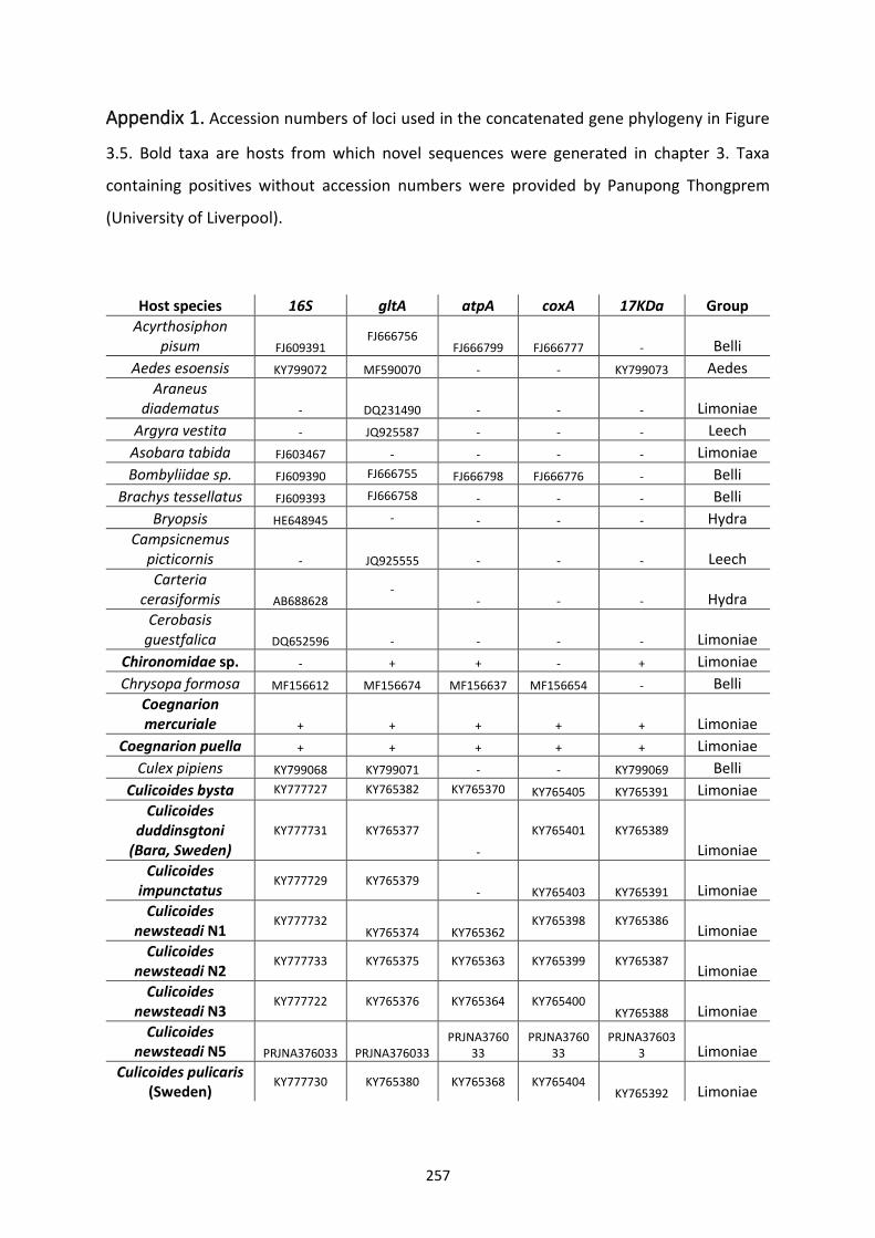
257
Appendix 1. Accession numbers of loci used in the concatenated gene phylogeny in Figure
3.5. Bold taxa are hosts from which novel sequences were generated in chapter 3. Taxa
containing positives without accession numbers were provided by Panupong Thongprem
(University of Liverpool).
Host species 16S gltA atpA coxA 17KDa Group
Acyrthosiphon pisum FJ609391
FJ666756 FJ666799 FJ666777 - Belli
Aedes esoensis KY799072 MF590070 - - KY799073 Aedes
Araneus diadematus - DQ231490 - - - Limoniae
Argyra vestita - JQ925587 - - - Leech
Asobara tabida FJ603467 - - - - Limoniae
Bombyliidae sp. FJ609390 FJ666755 FJ666798 FJ666776 - Belli
Brachys tessellatus FJ609393 FJ666758 - - - Belli
Bryopsis HE648945 - - - - Hydra
Campsicnemus picticornis - JQ925555 - - - Leech
Carteria cerasiformis AB688628
- - - - Hydra
Cerobasis guestfalica DQ652596 - - - - Limoniae
Chironomidae sp. - + + - + Limoniae
Chrysopa formosa MF156612 MF156674 MF156637 MF156654 - Belli
Coegnarion mercuriale + + + + + Limoniae
Coegnarion puella + + + + + Limoniae
Culex pipiens KY799068 KY799071 - - KY799069 Belli
Culicoides bysta KY777727 KY765382 KY765370 KY765405 KY765391 Limoniae
Culicoides duddinsgtoni
(Bara, Sweden)
KY777731 KY765377
-
KY765401 KY765389
Limoniae
Culicoides impunctatus
KY777729 KY765379 - KY765403 KY765391 Limoniae
Culicoides newsteadi N1
KY777732 KY765374 KY765362
KY765398 KY765386 Limoniae
Culicoides newsteadi N2
KY777733 KY765375 KY765363 KY765399 KY765387 Limoniae
Culicoides newsteadi N3
KY777722 KY765376 KY765364 KY765400 KY765388 Limoniae
Culicoides newsteadi N5 PRJNA376033 PRJNA376033
PRJNA376033
PRJNA376033
PRJNA376033 Limoniae
Culicoides pulicaris (Sweden)
KY777730 KY765380 KY765368 KY765404 KY765392 Limoniae
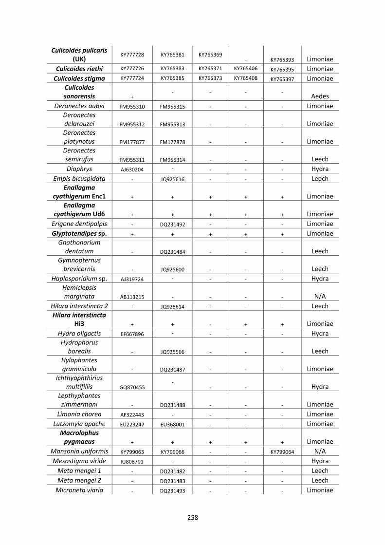
258
Culicoides pulicaris (UK)
KY777728 KY765381 KY765369 - KY765393 Limoniae
Culicoides riethi KY777726 KY765383 KY765371 KY765406 KY765395 Limoniae
Culicoides stigma KY777724 KY765385 KY765373 KY765408 KY765397 Limoniae
Culicoides sonorensis +
- - - - Aedes
Deronectes aubei FM955310 FM955315 - - - Limoniae
Deronectes delarouzei FM955312 FM955313 - - - Limoniae
Deronectes platynotus FM177877 FM177878 - - - Limoniae
Deronectes semirufus FM955311 FM955314 - - - Leech
Diophrys AJ630204 - - - - Hydra
Empis bicuspidata - JQ925616 - - - Leech
Enallagma cyathigerum Enc1 + + + + + Limoniae
Enallagma cyathigerum Ud6 + + + + + Limoniae
Erigone dentipalpis - DQ231492 - - - Limoniae
Glyptotendipes sp. + + + + + Limoniae
Gnathonarium dentatum - DQ231484 - - - Leech
Gymnopternus brevicornis - JQ925600 - - - Leech
Haplosporidium sp. AJ319724 - - - - Hydra
Hemiclepsis marginata AB113215 - - - - N/A
Hilara interstincta 2 - JQ925614 - - - Leech
Hilara interstincta Hi3 + + - + + Limoniae
Hydra oligactis EF667896 - - - - Hydra
Hydrophorus borealis - JQ925566 - - - Leech
Hylaphantes graminicola - DQ231487 - - - Limoniae
Ichthyophthirius multifiliis GQ870455
- - - - Hydra
Lepthyphantes zimmermani - DQ231488 - - - Limoniae
Limonia chorea AF322443 - - - - Limoniae
Lutzomyia apache EU223247 EU368001 - - - Limoniae
Macrolophus pygmaeus + + + + + Limoniae
Mansonia uniformis KY799063 KY799066 - - KY799064 N/A
Mesostigma viride KJ808701 - - - - Hydra
Meta mengei 1 - DQ231482 - - - Leech
Meta mengei 2 - DQ231483 - - - Leech
Microneta viaria - DQ231493 - - - Limoniae
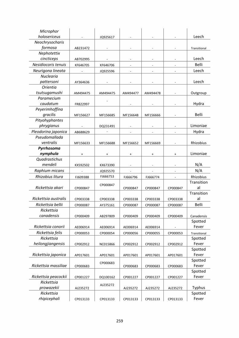
259
Microphor holosericeus - JQ925617 - - - Leech
Neochrysocharis formosa AB231472 - - - - Transitional
Nephotettix cincticeps AB702995 - - - - Leech
Nesidiocoris tenuis KF646705 KF646706 - - - Belli
Neurigona lineata - JQ925596 - - - Leech
Nuclearia pattersoni AY364636 - - - - Leech
Orientia tsutsugamushi AM494475 AM494475 AM494477 AM494478 - Outgroup Paramecium
caudatum FR822997 -
- - - Hydra
Peyerimhoffina gracilis MF156627 MF156685 MF156648 MF156666 - Belli
Pityohyphantes phrygianus - DQ231491 - - - Limoniae
Pleodorina japonica AB688629 - - - - Hydra
Pseudomallada ventralis MF156633 MF156688 MF156652 MF156669 - Rhizobius
Pyrrhosoma nymphula + + + + + Limoniae
Quadrastichus mendeli KX592502 KX673390 - - - N/A
Raphium micans - JQ925570 - - - N/A
Rhizobius litura FJ609388 FJ666753 FJ666796 FJ666774 - Rhizobius
Rickettsia akari CP000847 CP000847
CP000847 CP000847 CP000847
Transitional
Rickettsia australis CP003338 CP003338 CP003338 CP003338 CP003338
Transitional
Rickettsia bellii CP000087 AY375161 CP000087 CP000087 CP000087 Belli
Rickettsia canadensis CP000409 AB297809 CP000409 CP000409 CP000409 Canadensis
Rickettsia conorii AE006914 AE006914 AE006914 AE006914 -
Spotted Fever
Rickettsia felis CP000053 CP000054 CP000056 CP000055 CP000053 Transitional Rickettsia
heilongjiangensis CP002912 NC015866 CP002912 CP002912 CP002912
Spotted Fever
Rickettsia japonica AP017601 AP017601 AP017601 AP017601 AP017601
Spotted Fever
Rickettsia massiliae CP000683 CP000683
CP000683 CP000683 CP000683
Spotted Fever
Rickettsia peacockii CP001227 DQ100162 CP001227 CP001227 CP001227
Spotted Fever
Rickettsia prowazekii AJ235272
AJ235272 AJ235272 AJ235272 AJ235272 Typhus
Rickettsia rhipicephali CP013133 CP013133 CP013133 CP013133 CP013133
Spotted Fever
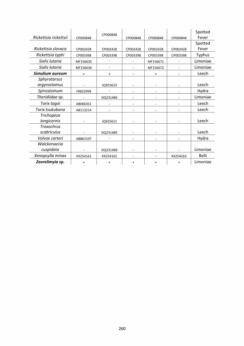
260
Rickettsia rickettsii CP000848 CP000848
CP000848 CP000848 CP000848
Spotted Fever
Rickettsia slovaca CP002428 CP002428 CP002428 CP002428 CP002428
Spotted Fever
Rickettsia typhi CP003398 CP003398 CP003398 CP003398 CP003398 Typhus
Sialis lutaria MF156635 - - MF156671 - Limoniae
Sialis lutaria MF156636 - - MF156672 - Limoniae
Simulium aureum + + - + - Leech
Sphyrotarsus argyrostomus - JQ925623 - - - Leech
Spirostomum FR822999 - - - - Hydra
Theridiidae sp. - DQ231486 - - - Limoniae
Torix tagoi AB066351 - - - - Leech
Torix tsukubana AB113214 - - - - Leech
Trichopeza longicornis - JQ925611 - - - Leech
Troxochrus scabriculus - DQ231485 - - - Leech
Volvox carteri AB861537 - - - - Hydra
Walckenaeria cuspidata - DQ231489 - - - Limoniae
Xenopsylla minax KX254161 KX254162 - - KX254163 Belli
Zavrelimyia sp. + + + + + Limoniae
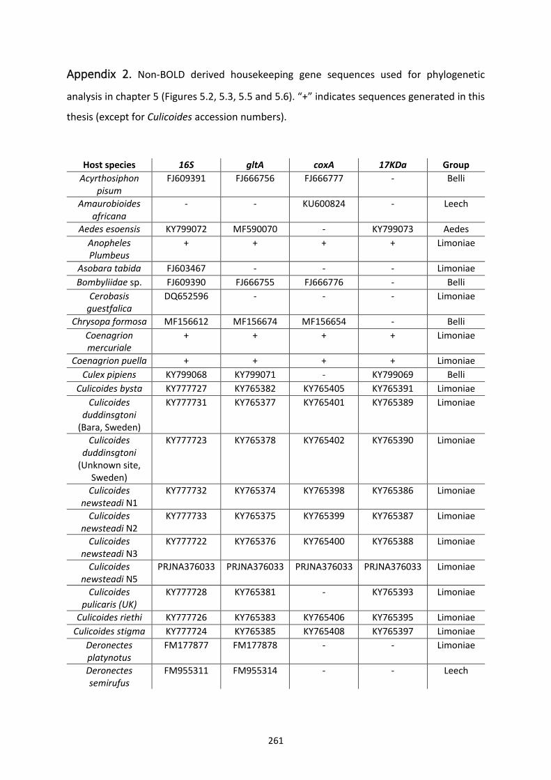
261
Appendix 2. Non-BOLD derived housekeeping gene sequences used for phylogenetic
analysis in chapter 5 (Figures 5.2, 5.3, 5.5 and 5.6). “+” indicates sequences generated in this
thesis (except for Culicoides accession numbers).
Host species 16S gltA coxA 17KDa Group
Acyrthosiphon pisum
FJ609391 FJ666756 FJ666777 - Belli
Amaurobioides africana
- - KU600824 - Leech
Aedes esoensis KY799072 MF590070 - KY799073 Aedes
Anopheles Plumbeus
+ + + + Limoniae
Asobara tabida FJ603467 - - - Limoniae
Bombyliidae sp. FJ609390 FJ666755 FJ666776 - Belli
Cerobasis guestfalica
DQ652596 - - - Limoniae
Chrysopa formosa MF156612 MF156674 MF156654 - Belli
Coenagrion mercuriale
+ + + + Limoniae
Coenagrion puella + + + + Limoniae
Culex pipiens KY799068 KY799071 - KY799069 Belli
Culicoides bysta KY777727 KY765382 KY765405 KY765391 Limoniae
Culicoides duddinsgtoni
(Bara, Sweden)
KY777731 KY765377 KY765401 KY765389 Limoniae
Culicoides duddinsgtoni
(Unknown site, Sweden)
KY777723 KY765378 KY765402 KY765390 Limoniae
Culicoides newsteadi N1
KY777732 KY765374 KY765398 KY765386 Limoniae
Culicoides newsteadi N2
KY777733 KY765375 KY765399 KY765387 Limoniae
Culicoides newsteadi N3
KY777722 KY765376 KY765400 KY765388 Limoniae
Culicoides newsteadi N5
PRJNA376033 PRJNA376033 PRJNA376033 PRJNA376033 Limoniae
Culicoides pulicaris (UK)
KY777728 KY765381 - KY765393 Limoniae
Culicoides riethi KY777726 KY765383 KY765406 KY765395 Limoniae
Culicoides stigma KY777724 KY765385 KY765408 KY765397 Limoniae
Deronectes platynotus
FM177877 FM177878 - - Limoniae
Deronectes semirufus
FM955311 FM955314 - - Leech
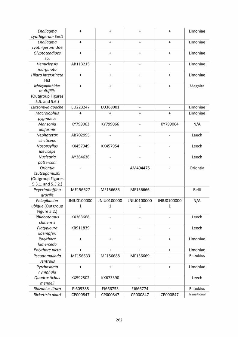
262
Enallagma cyathigerum Enc1
+ + + + Limoniae
Enallagma cyathigerum Ud6
+ + + + Limoniae
Glyptotendipes sp.
+ + + + Limoniae
Hemiclepsis marginata
AB113215 - - - Limoniae
Hilara interstincta Hi3
+ + + + Limoniae
Ichthyophthirius multifiliis
(Outgroup Figures 5.5. and 5.6.)
+ + + + Megaira
Lutzomyia apache EU223247 EU368001 - - Limoniae
Macrolophus pygmaeus
+ + + + Limoniae
Mansonia uniformis
KY799063 KY799066 - KY799064 N/A
Nephotettix cincticeps
AB702995 - - - Leech
Nosopsyllus laeviceps
KX457949 KX457954 - - Leech
Nuclearia pattersoni
AY364636 - - - Leech
Orientia tsutsugamushi
(Outgroup Figures 5.3.1. and 5.3.2.)
- - AM494475 - Orientia
Peyerimhoffina gracilis
MF156627 MF156685 MF156666 - Belli
Pelagibacter ubique (Outgroup
Figure 5.2.)
JNIU01000001
JNIU01000001
JNIU01000001
JNIU01000001
N/A
Phlebotomus chinensis
KX363668 - - - Leech
Platypleura kaempferi
KR911839 - - - Leech
Polythore lamerceda
+ + + + Limoniae
Polythore picta + + + + Limoniae
Pseudomallada ventralis
MF156633 MF156688 MF156669 - Rhizobius
Pyrrhosoma nymphula
+ + + + Limoniae
Quadrastichus mendeli
KX592502 KX673390 - - Leech
Rhizobius litura FJ609388 FJ666753 FJ666774 - Rhizobius
Rickettsia akari CP000847 CP000847 CP000847 CP000847 Transitional
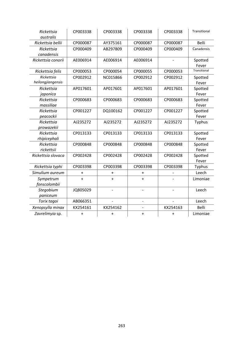
263
Rickettsia australis
CP003338 CP003338 CP003338 CP003338 Transitional
Rickettsia bellii CP000087 AY375161 CP000087 CP000087 Belli
Rickettsia canadensis
CP000409 AB297809 CP000409 CP000409 Canadensis
Rickettsia conorii AE006914 AE006914 AE006914 - Spotted Fever
Rickettsia felis CP000053 CP000054 CP000055 CP000053 Transitional
Rickettsia heilongjiangensis
CP002912 NC015866 CP002912 CP002912 Spotted Fever
Rickettsia japonica
AP017601 AP017601 AP017601 AP017601 Spotted Fever
Rickettsia massiliae
CP000683 CP000683 CP000683 CP000683 Spotted Fever
Rickettsia peacockii
CP001227 DQ100162 CP001227 CP001227 Spotted Fever
Rickettsia prowazekii
AJ235272 AJ235272 AJ235272 AJ235272 Typhus
Rickettsia rhipicephali
CP013133 CP013133 CP013133 CP013133 Spotted Fever
Rickettsia rickettsii
CP000848 CP000848 CP000848 CP000848 Spotted Fever
Rickettsia slovaca CP002428 CP002428 CP002428 CP002428 Spotted Fever
Rickettsia typhi CP003398 CP003398 CP003398 CP003398 Typhus
Simulium aureum + + + - Leech
Sympetrum fonscolombii
+ + + - Limoniae
Stegobium paniceum
JQ805029 - - - Leech
Torix tagoi AB066351 - - - Leech
Xenopsylla minax KX254161 KX254162 - KX254163 Belli
Zavrelimyia sp. + + + + Limoniae

264
Appendix 3. Re-barcoding status and nearest BLAST hit (NCBI) of mtDNA COI arthropod DNA
extracts accessed for further analysis in chapter 5, along with the success of multilocus
Rickettsia profiles with allocated Rickettsia group (based on phylogenetic analysis) and co-
infection status.
BOLD ID Host
taxonomy
Nearest host
barcoding
BLAST hit
gltA 16S 17k
Da Co-
infection Rickettsia
group
BIOUG03090-
E08 Trichoptera
100% identity to
Lepidostoma
hoodi
KM533759
Caddis fly
+ - + - Limoniae
BIOUG03644-
G07 Diptera - + + - - Limoniae
BIOUG03946-
H07 Hymenoptera
86% identity to
Megaspilidae
sp. KP693295
Parasitoid wasp
+ + + - Limoniae
BIOUG03995-
E06 Arachnida
99% identity to Calyptostomatidae sp. MG317701
Water mite
+ - - - Torix
BIOUG05036-
F05 Hymenoptera - + + - - Limoniae
BIOUG05952-
E04 Diptera
99% identity to
Sciaridae sp.
KR435639 Dark
winged fungus
gnat
+ - - - Torix
BIOUG05952-
F01 Diptera
99% identity to
Sciaridae sp.
KR435639 Dark
winged fungus
gnat
- - - - Torix
BIOUG05952-
F09 Diptera
99% identity to
Sciaridae sp.
KR435639 Dark
winged fungus
gnat
- - - - Torix
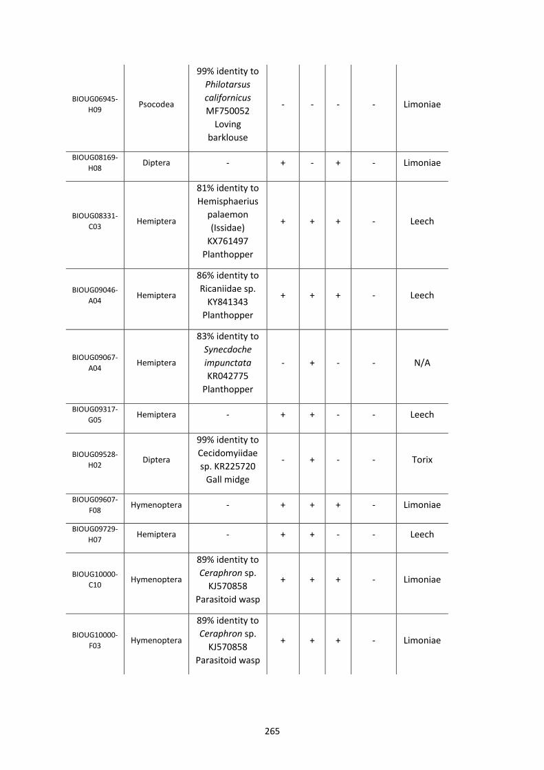
265
BIOUG06945-
H09 Psocodea
99% identity to
Philotarsus
californicus
MF750052
Loving
barklouse
- - - - Limoniae
BIOUG08169-
H08 Diptera - + - + - Limoniae
BIOUG08331-
C03 Hemiptera
81% identity to
Hemisphaerius
palaemon
(Issidae)
KX761497
Planthopper
+ + + - Leech
BIOUG09046-
A04 Hemiptera
86% identity to
Ricaniidae sp.
KY841343
Planthopper
+ + + - Leech
BIOUG09067-
A04 Hemiptera
83% identity to
Synecdoche
impunctata
KR042775
Planthopper
- + - - N/A
BIOUG09317-
G05 Hemiptera - + + - - Leech
BIOUG09528-
H02 Diptera
99% identity to
Cecidomyiidae
sp. KR225720
Gall midge
- + - - Torix
BIOUG09607-
F08 Hymenoptera - + + + - Limoniae
BIOUG09729-
H07 Hemiptera - + + - - Leech
BIOUG10000-
C10 Hymenoptera
89% identity to
Ceraphron sp.
KJ570858
Parasitoid wasp
+ + + - Limoniae
BIOUG10000-
F03 Hymenoptera
89% identity to
Ceraphron sp.
KJ570858
Parasitoid wasp
+ + + - Limoniae
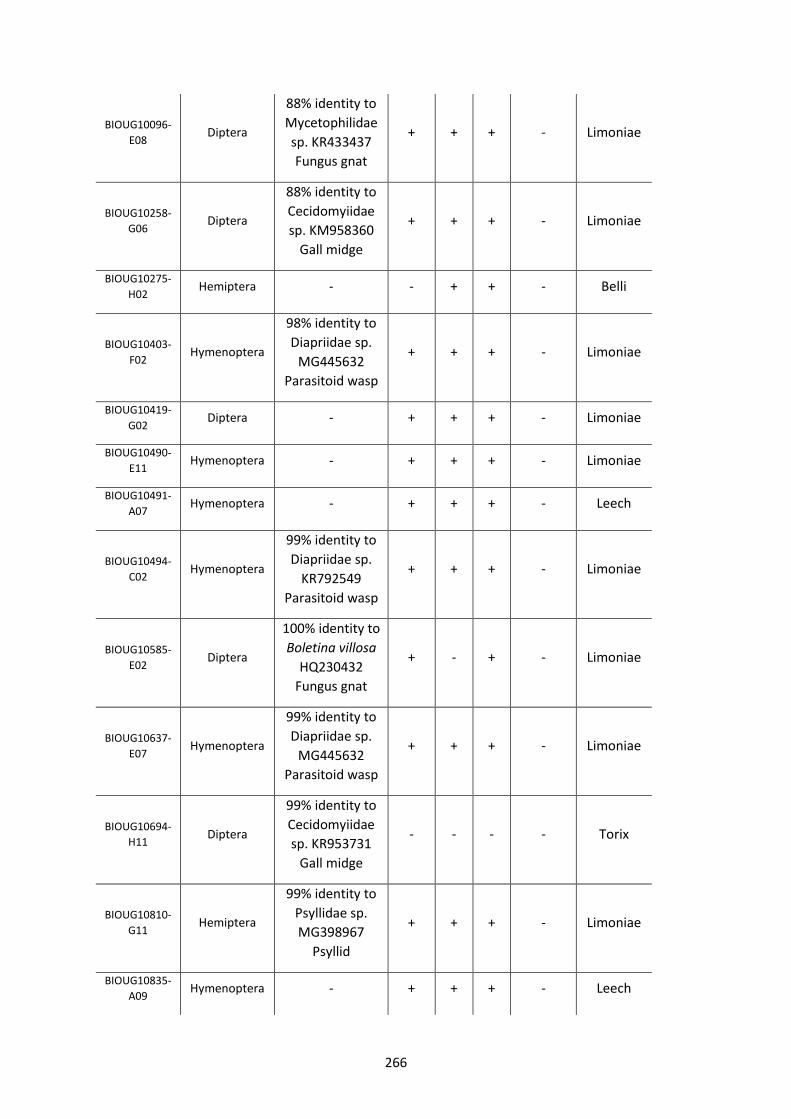
266
BIOUG10096-
E08 Diptera
88% identity to
Mycetophilidae
sp. KR433437
Fungus gnat
+ + + - Limoniae
BIOUG10258-
G06 Diptera
88% identity to
Cecidomyiidae
sp. KM958360
Gall midge
+ + + - Limoniae
BIOUG10275-
H02 Hemiptera - - + + - Belli
BIOUG10403-
F02 Hymenoptera
98% identity to
Diapriidae sp.
MG445632
Parasitoid wasp
+ + + - Limoniae
BIOUG10419-
G02 Diptera - + + + - Limoniae
BIOUG10490-
E11 Hymenoptera - + + + - Limoniae
BIOUG10491-
A07 Hymenoptera - + + + - Leech
BIOUG10494-
C02 Hymenoptera
99% identity to
Diapriidae sp.
KR792549
Parasitoid wasp
+ + + - Limoniae
BIOUG10585-
E02 Diptera
100% identity to
Boletina villosa
HQ230432
Fungus gnat
+ - + - Limoniae
BIOUG10637-
E07 Hymenoptera
99% identity to
Diapriidae sp.
MG445632
Parasitoid wasp
+ + + - Limoniae
BIOUG10694-
H11 Diptera
99% identity to
Cecidomyiidae
sp. KR953731
Gall midge
- - - - Torix
BIOUG10810-
G11 Hemiptera
99% identity to
Psyllidae sp.
MG398967
Psyllid
+ + + - Limoniae
BIOUG10835-
A09 Hymenoptera - + + + - Leech
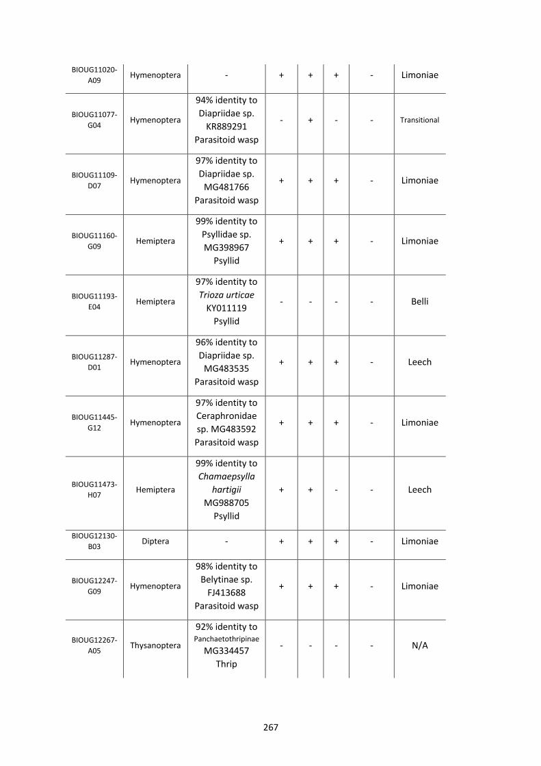
267
BIOUG11020-
A09 Hymenoptera - + + + - Limoniae
BIOUG11077-
G04 Hymenoptera
94% identity to
Diapriidae sp.
KR889291
Parasitoid wasp
- + - - Transitional
BIOUG11109-
D07 Hymenoptera
97% identity to
Diapriidae sp.
MG481766
Parasitoid wasp
+ + + - Limoniae
BIOUG11160-
G09 Hemiptera
99% identity to
Psyllidae sp.
MG398967
Psyllid
+ + + - Limoniae
BIOUG11193-
E04 Hemiptera
97% identity to
Trioza urticae
KY011119
Psyllid
- - - - Belli
BIOUG11287-
D01 Hymenoptera
96% identity to
Diapriidae sp.
MG483535
Parasitoid wasp
+ + + - Leech
BIOUG11445-
G12 Hymenoptera
97% identity to
Ceraphronidae
sp. MG483592
Parasitoid wasp
+ + + - Limoniae
BIOUG11473-
H07 Hemiptera
99% identity to
Chamaepsylla
hartigii
MG988705
Psyllid
+ + - - Leech
BIOUG12130-
B03 Diptera - + + + - Limoniae
BIOUG12247-
G09 Hymenoptera
98% identity to
Belytinae sp.
FJ413688
Parasitoid wasp
+ + + - Limoniae
BIOUG12267-
A05 Thysanoptera
92% identity to Panchaetothripinae
MG334457
Thrip
- - - - N/A
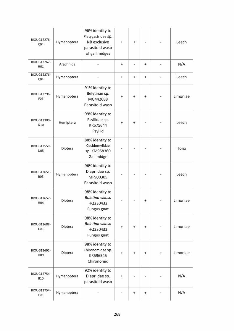
268
BIOUG12276-
C04 Hymenoptera
96% identity to
Platygastridae sp.
NB exclusive
parasitoid wasp
of gall midges
+ + - - Leech
BIOUG12267-
H01 Arachnida - + - + - N/A
BIOUG12276-
C04 Hymenoptera - + + + - Leech
BIOUG12296-
F05 Hymenoptera
91% identity to
Belytinae sp.
MG442688
Parasitoid wasp
+ + + - Limoniae
BIOUG12300-
D10 Hemiptera
99% identity to
Psyllidae sp.
KR575644
Psyllid
+ + - - Leech
BIOUG12559-
D05 Diptera
88% identity to Cecidomyiidae
sp. KM958360
Gall midge
- - - - Torix
BIOUG12651-
B03 Hymenoptera
96% identity to
Diapriidae sp.
MF900305
Parasitoid wasp
- - - - Leech
BIOUG12657-
H04 Diptera
98% identity to
Boletina villosa
HQ230432
Fungus gnat
- - + - Limoniae
BIOUG12688-
E05 Diptera
98% identity to
Boletina villosa
HQ230432
Fungus gnat
+ + + - Limoniae
BIOUG12692-
H09 Diptera
98% identity to
Chironomidae sp.
KR596545
Chironomid
+ + + + Limoniae
BIOUG12754-
B10 Hymenoptera
92% identity to
Diapriidae sp.
parasitoid wasp
+ - - - N/A
BIOUG12754-
F03 Hymenoptera - - + + - N/A
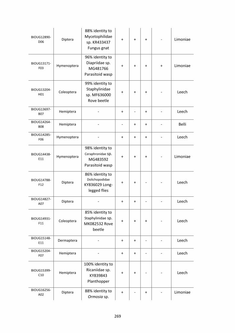
269
BIOUG12890-
D06 Diptera
88% identity to
Mycetophilidae
sp. KR433437
Fungus gnat
+ + + - Limoniae
BIOUG13171-
F03 Hymenoptera
96% identity to
Diapriidae sp.
MG481766
Parasitoid wasp
+ + + + Limoniae
BIOUG13204-
H01 Coleoptera
99% identity to
Staphylinidae
sp. MF636000
Rove beetle
+ + + - Leech
BIOUG13697-
B07 Hemiptera - + - + - Leech
BIOUG14264-
B08 Hemiptera - - + + - Belli
BIOUG14285-
F06 Hymenoptera - + + + - Leech
BIOUG14438-
E11 Hymenoptera
98% identity to
Ceraphronidae sp.
MG483592
Parasitoid wasp
+ + + - Limoniae
BIOUG14788-
F12 Diptera
86% identity to Dolichopodidae
KY836029 Long-
legged flies
+ + - - Leech
BIOUG14827-
A07 Diptera - + + - - Leech
BIOUG14931-
F11 Coleoptera
85% identity to
Staphylinidae sp.
MK082532 Rove
beetle
+ + + - Leech
BIOUG15148-
E11 Dermaptera - + + - - Leech
BIOUG15204-
F07 Hemiptera - + + - - Leech
BIOUG15399-
C10 Hemiptera
100% identity to
Ricaniidae sp.
KY839843
Planthopper
+ + - - Leech
BIOUG16256-
A02 Diptera 88% identity to
Ormosia sp. + - + - Limoniae
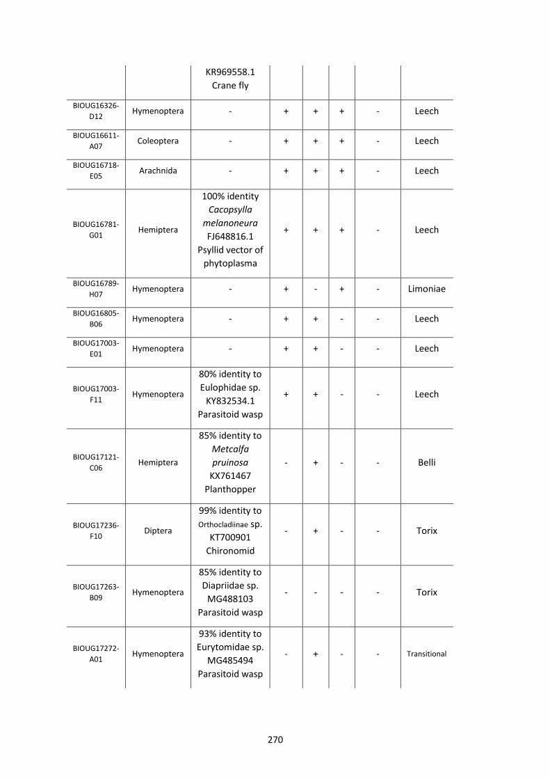
270
KR969558.1
Crane fly
BIOUG16326-
D12 Hymenoptera - + + + - Leech
BIOUG16611-
A07 Coleoptera - + + + - Leech
BIOUG16718-
E05 Arachnida - + + + - Leech
BIOUG16781-
G01 Hemiptera
100% identity
Cacopsylla
melanoneura
FJ648816.1
Psyllid vector of
phytoplasma
+ + + - Leech
BIOUG16789-
H07 Hymenoptera - + - + - Limoniae
BIOUG16805-
B06 Hymenoptera - + + - - Leech
BIOUG17003-
E01 Hymenoptera - + + - - Leech
BIOUG17003-
F11 Hymenoptera
80% identity to
Eulophidae sp.
KY832534.1
Parasitoid wasp
+ + - - Leech
BIOUG17121-
C06 Hemiptera
85% identity to
Metcalfa
pruinosa
KX761467
Planthopper
- + - - Belli
BIOUG17236-
F10 Diptera
99% identity to
Orthocladiinae sp.
KT700901
Chironomid
- + - - Torix
BIOUG17263-
B09 Hymenoptera
85% identity to
Diapriidae sp.
MG488103
Parasitoid wasp
- - - - Torix
BIOUG17272-
A01 Hymenoptera
93% identity to
Eurytomidae sp.
MG485494
Parasitoid wasp
- + - - Transitional
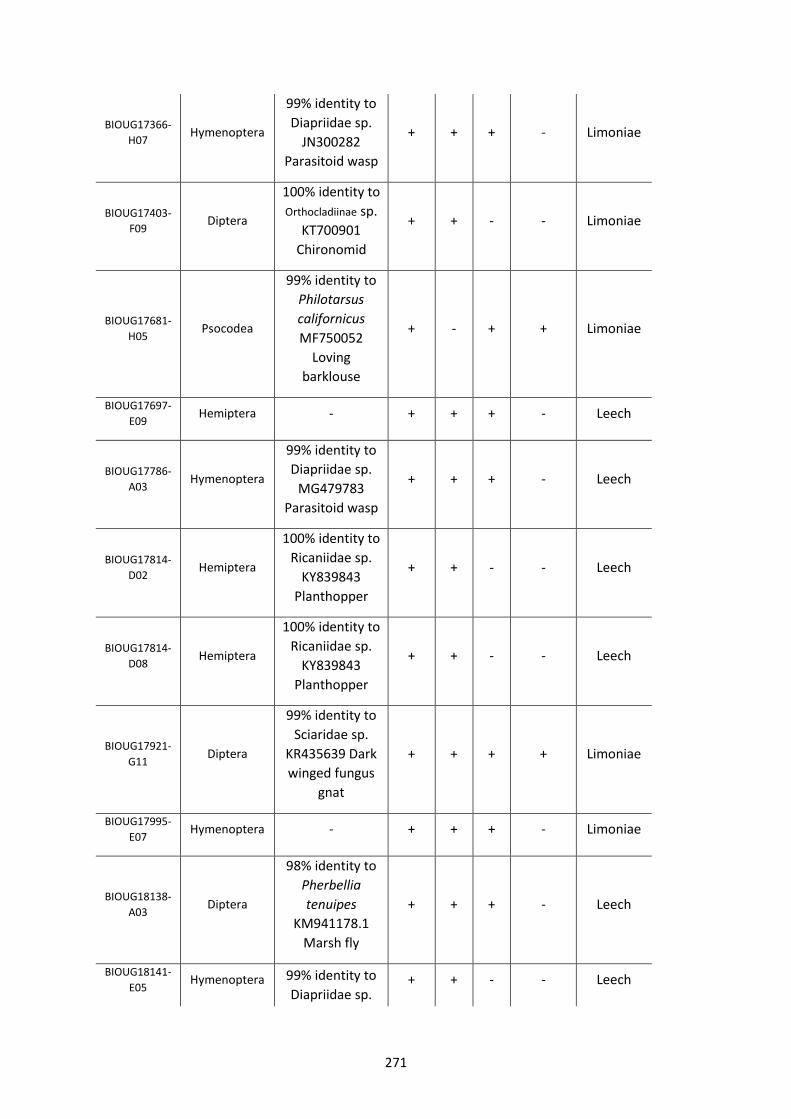
271
BIOUG17366-
H07 Hymenoptera
99% identity to
Diapriidae sp.
JN300282
Parasitoid wasp
+ + + - Limoniae
BIOUG17403-
F09 Diptera
100% identity to
Orthocladiinae sp.
KT700901
Chironomid
+ + - - Limoniae
BIOUG17681-
H05 Psocodea
99% identity to
Philotarsus
californicus
MF750052
Loving
barklouse
+ - + + Limoniae
BIOUG17697-
E09 Hemiptera - + + + - Leech
BIOUG17786-
A03 Hymenoptera
99% identity to
Diapriidae sp.
MG479783
Parasitoid wasp
+ + + - Leech
BIOUG17814-
D02 Hemiptera
100% identity to
Ricaniidae sp.
KY839843
Planthopper
+ + - - Leech
BIOUG17814-
D08 Hemiptera
100% identity to
Ricaniidae sp.
KY839843
Planthopper
+ + - - Leech
BIOUG17921-
G11 Diptera
99% identity to
Sciaridae sp.
KR435639 Dark
winged fungus
gnat
+ + + + Limoniae
BIOUG17995-
E07 Hymenoptera - + + + - Limoniae
BIOUG18138-
A03 Diptera
98% identity to
Pherbellia
tenuipes
KM941178.1
Marsh fly
+ + + - Leech
BIOUG18141-
E05 Hymenoptera 99% identity to
Diapriidae sp. + + - - Leech
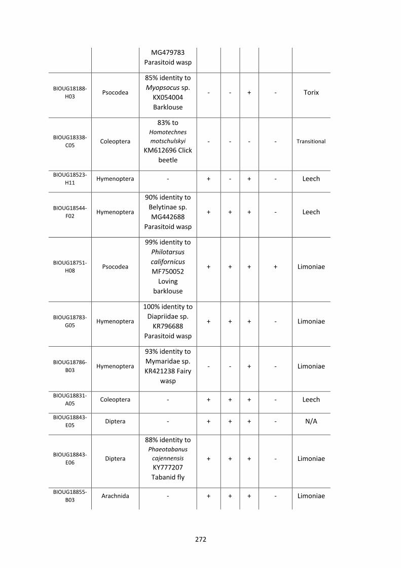
272
MG479783
Parasitoid wasp
BIOUG18188-
H03 Psocodea
85% identity to
Myopsocus sp.
KX054004
Barklouse
- - + - Torix
BIOUG18338-
C05 Coleoptera
83% to Homotechnes
motschulskyi
KM612696 Click
beetle
- - - - Transitional
BIOUG18523-
H11 Hymenoptera - + - + - Leech
BIOUG18544-
F02 Hymenoptera
90% identity to
Belytinae sp.
MG442688
Parasitoid wasp
+ + + - Leech
BIOUG18751-
H08 Psocodea
99% identity to
Philotarsus
californicus
MF750052
Loving
barklouse
+ + + + Limoniae
BIOUG18783-
G05 Hymenoptera
100% identity to
Diapriidae sp.
KR796688
Parasitoid wasp
+ + + - Limoniae
BIOUG18786-
B03 Hymenoptera
93% identity to
Mymaridae sp.
KR421238 Fairy
wasp
- - + - Limoniae
BIOUG18831-
A05 Coleoptera - + + + - Leech
BIOUG18843-
E05 Diptera - + + + - N/A
BIOUG18843-
E06 Diptera
88% identity to Phaeotabanus
cajennensis
KY777207
Tabanid fly
+ + + - Limoniae
BIOUG18855-
B03 Arachnida - + + + - Limoniae
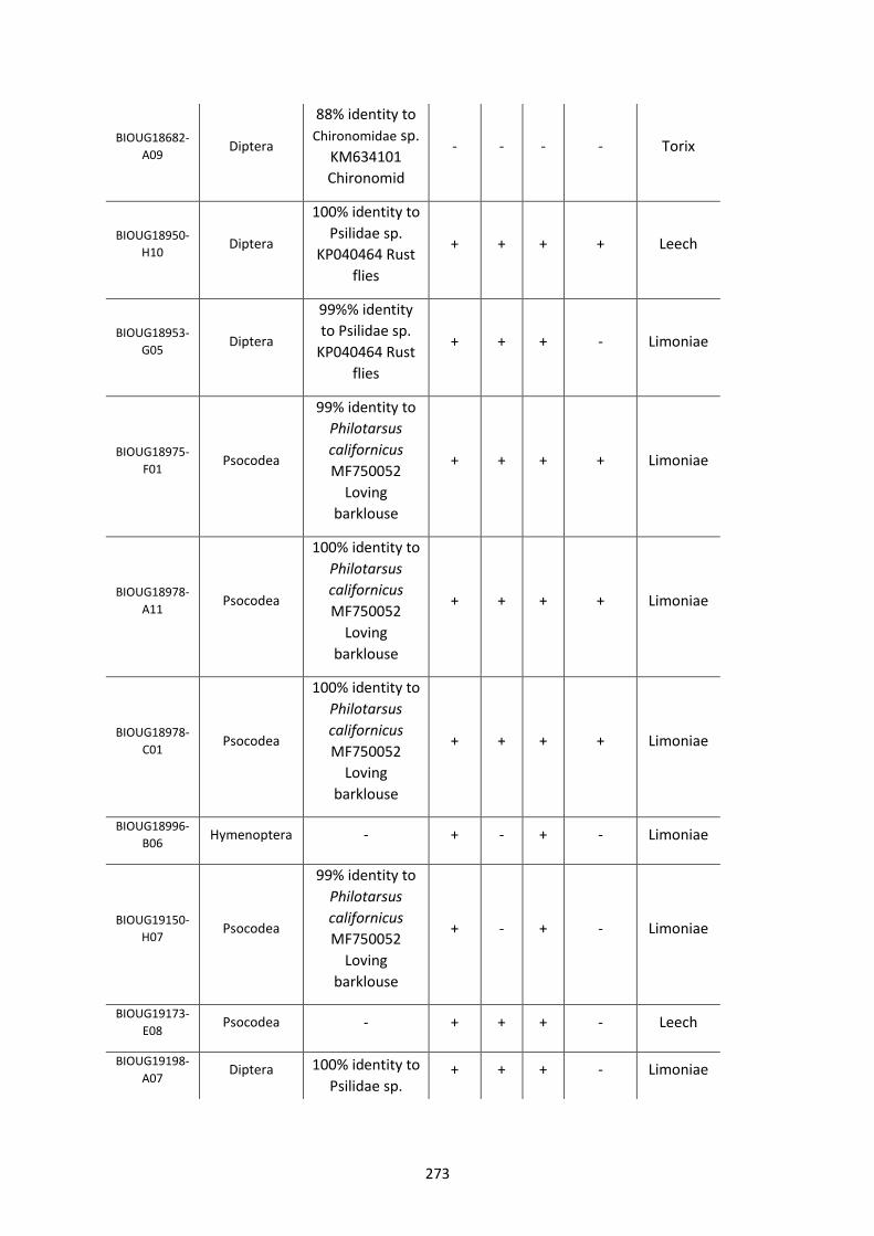
273
BIOUG18682-
A09 Diptera
88% identity to
Chironomidae sp.
KM634101
Chironomid
- - - - Torix
BIOUG18950-
H10 Diptera
100% identity to
Psilidae sp.
KP040464 Rust
flies
+ + + + Leech
BIOUG18953-
G05 Diptera
99%% identity
to Psilidae sp.
KP040464 Rust
flies
+ + + - Limoniae
BIOUG18975-
F01 Psocodea
99% identity to
Philotarsus
californicus
MF750052
Loving
barklouse
+ + + + Limoniae
BIOUG18978-
A11 Psocodea
100% identity to
Philotarsus
californicus
MF750052
Loving
barklouse
+ + + + Limoniae
BIOUG18978-
C01 Psocodea
100% identity to
Philotarsus
californicus
MF750052
Loving
barklouse
+ + + + Limoniae
BIOUG18996-
B06 Hymenoptera - + - + - Limoniae
BIOUG19150-
H07 Psocodea
99% identity to
Philotarsus
californicus
MF750052
Loving
barklouse
+ - + - Limoniae
BIOUG19173-
E08 Psocodea - + + + - Leech
BIOUG19198-
A07 Diptera 100% identity to
Psilidae sp. + + + - Limoniae
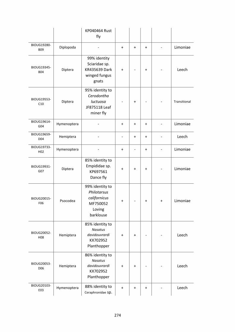
274
KP040464 Rust
fly
BIOUG19280-
B09 Diplopoda - + + + - Limoniae
BIOUG19345-
B04 Diptera
99% identity
Sciaridae sp.
KR435639 Dark
winged fungus
gnats
+ - + - Leech
BIOUG19553-
C10 Diptera
95% identity to
Cerodontha
luctuosa
JF875118 Leaf
miner fly
- + - - Transitional
BIOUG19614-
G04 Hymenoptera - + + + - Limoniae
BIOUG19659-
D04 Hemiptera - - + + - Leech
BIOUG19733-
H02 Hymenoptera - + - + - Limoniae
BIOUG19931-
G07 Diptera
85% identity to
Empididae sp.
KP697561
Dance fly
+ + + - Limoniae
BIOUG20015-
F06 Psocodea
99% identity to
Philotarsus
californicus
MF750052
Loving
barklouse
+ - + + Limoniae
BIOUG20052-
H08 Hemiptera
85% identity to Nasatus
davidouvrardi
KX702952
Planthopper
+ + - - Leech
BIOUG20053-
D06 Hemiptera
86% identity to Nasatus
davidouvrardi
KX702952
Planthopper
+ + - - Leech
BIOUG20103-
E03 Hymenoptera 88% identity to
Ceraphronidae sp. + + + - Leech
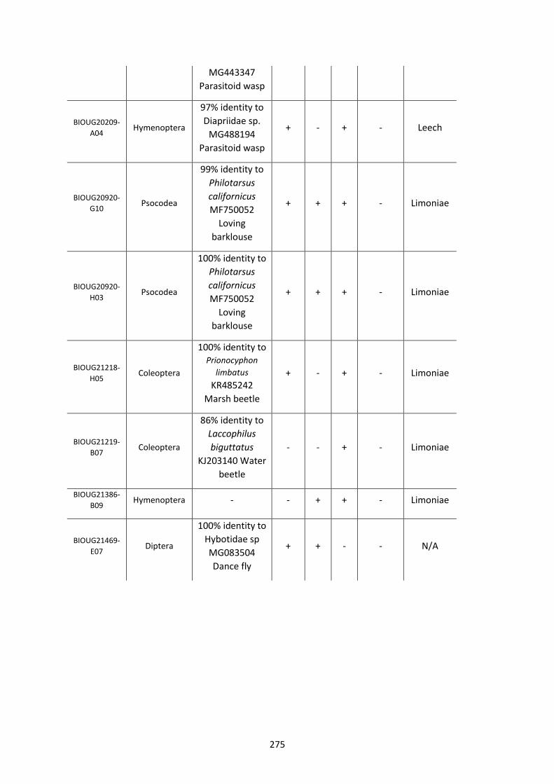
275
MG443347
Parasitoid wasp
BIOUG20209-
A04 Hymenoptera
97% identity to
Diapriidae sp.
MG488194
Parasitoid wasp
+ - + - Leech
BIOUG20920-
G10 Psocodea
99% identity to
Philotarsus
californicus
MF750052
Loving
barklouse
+ + + - Limoniae
BIOUG20920-
H03 Psocodea
100% identity to
Philotarsus
californicus
MF750052
Loving
barklouse
+ + + - Limoniae
BIOUG21218-
H05 Coleoptera
100% identity to Prionocyphon
limbatus
KR485242
Marsh beetle
+ - + - Limoniae
BIOUG21219-
B07 Coleoptera
86% identity to
Laccophilus
biguttatus
KJ203140 Water
beetle
- - + - Limoniae
BIOUG21386-
B09 Hymenoptera - - + + - Limoniae
BIOUG21469-
E07 Diptera
100% identity to
Hybotidae sp
MG083504
Dance fly
+ + - - N/A