International Journal of Toxicology oculata Nannochloropsis Safety Assessment of EPA-Rich Polar...
17
http://ijt.sagepub.com/ International Journal of Toxicology http://ijt.sagepub.com/content/early/2014/10/08/1091581814553453 The online version of this article can be found at: DOI: 10.1177/1091581814553453 published online 9 October 2014 International Journal of Toxicology Michael L. Kagan, Dexter W. Sullivan, Jr, Shayne C. Gad and Corey M. Ballou oculata Nannochloropsis Safety Assessment of EPA-Rich Polar Lipid Oil Produced From the Microalgae Published by: http://www.sagepublications.com On behalf of: American College of Toxicology can be found at: International Journal of Toxicology Additional services and information for http://ijt.sagepub.com/cgi/alerts Email Alerts: http://ijt.sagepub.com/subscriptions Subscriptions: http://www.sagepub.com/journalsReprints.nav Reprints: http://www.sagepub.com/journalsPermissions.nav Permissions: What is This? - Oct 9, 2014 OnlineFirst Version of Record >> by guest on October 21, 2014 ijt.sagepub.com Downloaded from by guest on October 21, 2014 ijt.sagepub.com Downloaded from
-
Upload
independent -
Category
Documents
-
view
0 -
download
0
Transcript of International Journal of Toxicology oculata Nannochloropsis Safety Assessment of EPA-Rich Polar...

http://ijt.sagepub.com/International Journal of Toxicology
http://ijt.sagepub.com/content/early/2014/10/08/1091581814553453The online version of this article can be found at:
DOI: 10.1177/1091581814553453
published online 9 October 2014International Journal of ToxicologyMichael L. Kagan, Dexter W. Sullivan, Jr, Shayne C. Gad and Corey M. Ballou
oculataNannochloropsisSafety Assessment of EPA-Rich Polar Lipid Oil Produced From the Microalgae
Published by:
http://www.sagepublications.com
On behalf of:
American College of Toxicology
can be found at:International Journal of ToxicologyAdditional services and information for
http://ijt.sagepub.com/cgi/alertsEmail Alerts:
http://ijt.sagepub.com/subscriptionsSubscriptions:
http://www.sagepub.com/journalsReprints.navReprints:
http://www.sagepub.com/journalsPermissions.navPermissions:
What is This?
- Oct 9, 2014OnlineFirst Version of Record >>
by guest on October 21, 2014ijt.sagepub.comDownloaded from by guest on October 21, 2014ijt.sagepub.comDownloaded from

Original Article
Safety Assessment of EPA-Rich PolarLipid Oil Produced From the MicroalgaeNannochloropsis oculata
Michael L. Kagan1, Dexter W. Sullivan Jr2, Shayne C. Gad2,and Corey M. Ballou2
AbstractAlmega PL is an eicosapentaenoic acid-rich o-3 oil that is isolated from Nannochloropsis oculata algae and developed as a dietarysupplement. The safety of the algal oil was evaluated in 14- and 90-day studies in Sprague-Dawley rats by oral gavage at dose levelsof 0, 250, 500, and 2500 mg/kg/d and 0, 200, 400, and 2000 mg/kg/d, respectively. No mortalities occurred and no signs of toxicitywere observed during the studies. No treatment-related effects were seen for body weight, food consumption, ophthalmology,neurological effects, urinalysis, clinical pathology, gross pathology, organ weights, or histopathology. Although statistically signif-icant effects were noted for some end points, none were considered to be of toxicological significance. The no observed adverseeffect level for Almega PL was 2000 mg/kg/d. Additionally, Almega PL was not mutagenic in Salmonella typhimurium or Escherichiacoli, did not induce chromosome aberrations in Chinese hamster ovary cells, and did not induce genotoxic effects in vivo in ratbone marrow erythrocytes.
KeywordsNannochloropsis oculata, algae, o-3, dietary supplement, eicosapentaenoic acid, EPA, docosahexaneoic acid, DHA, toxicity,genotoxicity
Introduction
Nannochloropsis is a small green microalga genus that is well
known due to its nutritional value and ability to produce valu-
able lipophilic and lipophobic materials.1 In the aquaculture
industry, Nannochloropsis is extensively used as feedstock in
growing small zooplanktons such as rotifers and in fish hatch-
eries2 and for producing green water.3 In addition, the bio-
chemical composition of Nannochloropsis makes it a
valuable food source for animals and humans.1
Nannochloropsis oculata is 1 of the 6 species contained in
the genus Nannochloropsis and was originally isolated off the
coast of Scotland. Species of the genus Nannochloropsis have
been shown to contain high concentrations of eicosapentaenoic
acid (EPA) while containing no docosahexaneoic acid
(DHA).4,5 Nannochloropsis oculata is a rich source of EPA,
and the oil extracted from the green microalgae is one of the
few sources of EPA that does not also contain DHA.4 Recent
studies comparing EPA and DHA have suggested that not only
is the EPA component primarily responsible for reducing
inflammation6 and depression7 but that any DHA component
acts to block the activity of EPA.8 Therefore, the oil extracted
from N oculata presents a means of maximizing the available
levels and desired beneficial aspects of EPA as evidenced in
humans.4
Almega PL is an EPA-rich o-3 oil isolated from N oculata.
It is being developed by Qualitas Health Ltd, (Jerusalem,
Israel) as a dietary supplement ingredient with a suggested
daily intake of 300 to 1250 mg (2.0-20.8 mg/kg/d for a 60-kg
person), thus providing a maximum of 312.5 mg/d o-3 fatty
acids.9 To evaluate the potential toxicity of Almega PL, a
battery of Good Laboratory Practice (GLP)-compliant in vivo
and in vitro assays were performed. In rat models, a 14-day
maximum-tolerated dose (MTD) study and a 90-day repeated
dose subchronic toxicity study were performed. Clinical obser-
vations, clinical chemistry, hematology, urinalysis, functional
observational batteries (FOBs), and gross and microscopic
pathology were conducted to determine whether or not
repeated oral exposure of Almega PL resulted in any signs of
toxicity. Additionally, to evaluate the genotoxic potential of
Almega PL, a bacterial reverse mutation assay, an in vitro
mammalian chromosome aberration test in Chinese Hamster
1 Qualitas Health Ltd, Jerusalem, Israel2 Gad Consulting Services, Cary, North Carolina, USA
Corresponding Author:
Michael L. Kagan, Qualitas Health Ltd, 19 Hartom St, Jerusalem 91450, Israel.
Email: [email protected]
International Journal of Toxicology1-16ª The Author(s) 2014Reprints and permission:sagepub.com/journalsPermissions.navDOI: 10.1177/1091581814553453ijt.sagepub.com
by guest on October 21, 2014ijt.sagepub.comDownloaded from
(Cricetulus griseus) ovary (CHO) cells, and an in vivo micro-
nucleus assay in rats were performed.
Materials and Methods
Test Item
Almega PL (Lots 130202a and 130610; Qualitas Health Ltd) is
a nonpurified, dark green oily paste obtained from N oculata.
The source biomass, N oculata, is cultured in raceway-type
pools and harvested daily followed by extraction of the oil from
the biomass, resulting in Almega PL. Almega PL consists of
approximately 55% total fatty acids, of which 25% is EPA.
Almega PL also contains chlorophyll (approximately 3%) and
15% polar lipids (phospholipids and glycolipids).
The test item was analyzed for the presence of organochlor-
ine and organophosphate pesticides. No pesticides were found.
An analysis for dioxin, dioxin-like polychlorinated biphenyls
(PCBs), and nondioxin-like PCBs indicated that the levels of
these substances are below the maximum levels for marine oils
as listed by the World Health Organization and when adjusted
for fat content. Toxin analyses conducted on Almega PL indi-
cated that no natural product toxins were detected above detec-
tion limits including microcystins/nodularin, anatoxin-a,
cylindrospermopsin, paralytic shellfish toxin/saxitoxins, oka-
daic acid, and brevetoxins.10
Chemicals and Materials
Extra virgin olive oil used as the vehicle for the in vivo studies
was supplied by Qualitas Health Ltd except for in the micro-
nucleus assay where it was supplied by Pharmaseed Ltd (Ness
Ziona, Israel). 2-Nitrofulorene and 2-aminoanthracene were
obtained from Sigma-Aldrich (Munich, Germany). 9-
Aminoacridine was obtained from Novel Organic Synthesis
(Andhra Pradesh, India). 4-Nitroquinoline-1-oxide, sodium
azide, ethyl methanesulfonate, cyclophosphamide monohy-
drate, and colchicine were obtained from Sigma-Aldrich (St
Louis, Missouri). Dimethyl sulfoxide (DMSO) was obtained
from RFCL Ltd (Faridabad, India). Oxoid nutrient broth No.
2 was obtained from Oxoid Ltd (Basingstoke, Hampshire,
England). Fetal bovine serum was obtained from Sera Labora-
tories (West Sussex, United Kingdom). Methanol was obtained
from Merck Specialties Private Limited (Mumbai, India).
Potassium phosphate monobasic, sodium phosphate dibasic,
and acridine orange hydrochloride hydrate were obtained
from Sigma-Aldrich (Buchs, Switzerland). DPX (Distyrene,
Plasticizer, Xylene) mountant was obtained from SD Fine
Chemicals Limited (Mumbai, India).
Animals and Organisms
For the 14-day MTD study performed at Pharmaseed Ltd, 24
(12 male and 12 female) 8-week-old Sprague-Dawley SD rats
obtained from Harlan (Israel) were housed in polyethylene
cages (3/cage). The acclimatization period was 3 days prior
to testing. Initial mean group body weights were 237 g for
males and 181 g for females. The minimum and maximum
initial weight in the groups were within a range of +20% of
the groups mean weight. Room temperature was maintained at
20�C to 24�C with a relative humidity of 30% to 70%, a min-
imum of 15 air changes/h and a 12-hour light and 12-hour dark
cycle. Autoclaved and acidified drinking water (pH between
2.5 and 3.5) and commercial rodent diet (Teklad Certified Glo-
bal 18% Protein Diet cat # 2018SC) were provided ad libitum.
For the 90-day repeated dose, subchronic toxicity assay per-
formed at Advinus Therapeutics, Ltd (Bangalore, India), male
and female Sprague-Dawley rats raised in house by Advinus
Therapeutics were used. The acclimatization period was 5 days
prior to testing. The rats were 6 to 7 weeks old at the beginning
of the assay. Initial mean group body weights ranged from 145
to 148 g for the male rats and 127 to 130 g for the female rats.
At the beginning of treatment, the weight variation of the test
animals did not exceed +20% of the mean body weight in each
group and sex. Rats were housed in groups of 2 per sex per cage
in solid floor standard polysulfone cages with stainless top
grills for providing pelleted food and drinking water in poly-
carbonate bottles with stainless steel sipper tubes in rooms with
12 hours light cycles. Steam sterilized clean corn cob was used
as bedding and changed along with the cage once during the
acclimatization period and twice a week during the treatment
period. Room temperature was maintained at 19�C to 24�Cwith a relative humidity of 58% to 68%, with a minimum of
12 air changes/h. Filtered deep-bore well water and Teklad
Global 14% protein rodent maintenance diet (Harlan Labora-
tories, An Venray, the Netherlands) were provided ad libitum.
The Salmonella typhimurium and Escherichia coli test
strains for the bacterial reverse mutation assays performed at
Advinus Therapeutics Ltd were obtained, respectively, from
Health Protection Agency National Collection of Type Cul-
tures (London, Great Britain) and The National Collection of
Industrial and Marine Bacteria Ltd (Scotland, United King-
dom). The CHO cell line CHO-51, (ATCC CCL-61, Lot
4765275) for the chromosome aberration assay performed at
Advinus Therapeutics Ltd were obtained from the American
Type Culture Collection (Manassas, Virginia).
For the in vivo erythrocyte micronucleus test performed at
Advinus Therapeutics Ltd, male and female Sprague-Dawley
rats (in-house random bred) were acclimated for 5 days before
dosing. The rats were at least 10 weeks old (range, 10-11 weeks)
at the beginning of the assay. Initial body weights ranged from
250 to 253 g for the male rats and 188 to 190 g for the female
rats. All rats were visually inspected prior to the start of treat-
ment. Rats were individually housed during acclimatization and
treatment in standard polysulfone cages with stainless steel top
grills in rooms with 12 hours light cycles. Steam sterilized corn
cob was used as bedding which along with cages was changed at
least once a week. Room temperature was maintained at 20�C to
23�C with a relative humidity of 65% to 67% and 12 to 15 high-
efficiency particulate air-filtered air changes/h. Deep bore-well
water (passed through activated charcoal filter and exposed to
ultraviolet [UV] rays) and Teklad Certified (2014C) Global 14%Protein Rodent Maintenance pellets were provided ad libitum.
2 International Journal of Toxicology
by guest on October 21, 2014ijt.sagepub.comDownloaded from

Guidelines
Because the 14-day rat study was a dose-range finding (DRF)
study and used to determine the MTD of Almega PL as well as
the doses for the subsequent subchronic (90 day) study, it was
not performed in full compliance with GLP guidelines. The
subchronic toxicity study in rats was performed in accordance
with the OECD Guideline for Testing of Chemicals, (408):
Repeated Dose 90-Day Oral Toxicity Study in Rodents11 and
under GLP in accordance with OECD12 Principles of GLP for
the Testing of Chemicals. The bacterial reverse mutation
(Ames) assays were performed in accordance with the OECD13
Guideline 471 for testing of chemicals: Bacterial Reverse
Mutation Test and under GLP in accordance with OECD14
Principles of GLP. The chromosome aberration assay was per-
formed in accordance with the OECD15 Guideline 473 for
testing of chemicals: In Vitro Mammalian Chromosome Aber-
ration Test and under GLP in accordance with OECD14 Prin-
ciples of GLP. The rat micronucleus assay was performed in
accordance with the OECD16 Guideline 474 for testing of che-
micals: Mammalian Erythrocyte Micronucleus Test and under
GLP in accordance with OECD12 Principles of GLP.
Experimental Design
Maximum-tolerated dose oral toxicity study. Groups of 3 male and
3 female SD rats each were administered a single dose of 0, 5%(250 mg/kg), 10% (500 mg/kg), or 50% (2500 mg/kg) Almega
PL by oral gavage on day 1. The administrations were per-
formed using 16-gauge gavage needles. The vehicle was warm
extra virgin olive oil, and the dose volume was 5 mL/kg. Fol-
lowing the treatment period, all rats were monitored for 14 days
and euthanized on day 15.
Rats were observed twice daily for signs of morbidity and
mortality and once daily for clinical signs until study termina-
tion. Body weights were recorded prior to the initial dosing,
twice weekly during the study, and at study necropsy. On day
15, rats were euthanized by carbon dioxide asphyxiation. After
killing the animals, the organs and tissues identified in Table 1
were removed and examined for gross pathological anomalies.
Subchronic oral toxicity study. Groups of 10 male and 10 female
Sprague-Dawley rats each were administered 0, 250, 500, or
2500 mg/kg Almega PL in extra virgin olive oil by oral gavage
once daily for 90 days. Doses were administered using dispo-
sable plastic syringes attached with a metal feeding cannula.
All doses were administered at a constant dose volume of
5 mL/kg. Following the treatment period, all animals were
euthanized on day 91.
Rats were observed twice daily for signs of morbidity and
mortality and once daily for clinical signs until study termina-
tion. Detailed clinical examinations were performed prior to
the item administration on day 1 and at weekly intervals (+2
days) thereafter. Body weights were recorded on day 1 prior to
the test item administration and weekly thereafter except for
week 13 when the body weights were recorded on day 5 of the
week. Fasting body weights were recorded prior to necropsy on
day 91. Food consumption was measured once per week on the
same days that body weight measurements were made. At the
end of the study, rats were fasted overnight (water allowed),
anesthetized with isoflurane, and exsanguinated.
Ophthalmological examination. Prior to the start of treatment (on
day 5 of acclimatization), ophthalmological examinations were
performed for all animals with an ophthalmoscope. Mydriasis
was induced before examination using a solution of 1% tropi-
camide. The eyes were examined in subdued light. Subdued
light was maintained in the animal room for the remainder of
the day. These procedures were repeated at the end of the
treatment period for all animals.
Functional observational battery tests. To assess the behavioral
and neurological status of each rat, FOB tests were performed
for all of the group rats on day 90 of the treatment period. The
FOB tests included home cage observations for posture and for
presence or absence of abnormal vocalizations and convulsion;
observations during removal from rat from home cage and
handling; open field observations; and functional tests includ-
ing sensory evaluation, landing hindlimbs foot splay, grip per-
formance, and motor activity (Table 2).
Urinalysis. Before killing, urine was collected from all rats in
urine collection tubes. Each rat was placed in specially fabri-
cated cages overnight (water allowed) and the next morning the
urine was collected for analysis. Urinalysis parameters exam-
ined in the collected samples included color, clarity, bilirubin,
erythrocytes, glucose, ketone bodies, leukocytes, nitrite, pro-
teins, pH, specific gravity, urobilinogen, and volume. Urine
was also subjected to microscopic examination for sediments
such as crystals, epithelial cells, and casts.
Clinical pathology. At the end of the treatment period, rats were
fasted overnight (water allowed) after which blood was col-
lected from the retro-orbital sinus plexus with fine capillary
tubes under isoflurane anesthesia. An aliquot of blood was
collected in tubes containing 3.2% sodium citrate solution for
determination of coagulation parameters, and the remaining
blood was collected into K2EDTA and lithium heparainized
Table 1. List of Evaluated Organs.
Organ/tissue Organ/tissue Organ/tissue
Adrenal glands Lungs Seminal vesiclesBrain Mandibular lymph node SkinCecum Mesenteric lymph node SpleenColon Mammary gland StomachDuodenum Esophagus TestesEpididymitis Ovaries ThymusEyes Pancreas TongueHeart Parathyroid glands TracheaIleum Thyroid Urinary bladderJejunum Pituitary UterusKidneys Prostate gland VaginaLarynx Rectum Sciatic nerveLiver Salivary glands
Kagan et al 3
by guest on October 21, 2014ijt.sagepub.comDownloaded from

tubes for hematology and clinical chemistry examinations
(Table 3).
Pathology. All animals were fasted overnight prior to terminal
killing on day 91. At the scheduled termination, all survivors
were euthanized by exsanguination under isoflurane anesthesia.
The organs identified in Table 4 were removed, weighed, and
examined for gross and microscopic pathological anomalies.
Bacterial reverse mutation assays. The mutagenic potential of
Almega PL was evaluated using the bacterial reverse mutation
assay (Ames test) using standard plate incorporation (experi-
ment I) and preincubation (experiment II) methods. The ability
of Almega PL to induce mutagenicity in S typhimurium tester
strains TA98, TA100, TA1535, and TA1537 and E coli strain
WP2uvrA was assessed in the presence and absence of rat
microsomal fraction S9 mix. Based on the observations of
preliminary experiments to evaluate toxicity, 5000 mg/plate
was selected as the highest dose for all test strains and condi-
tions. A stock solution of 50 000 mg/mL test item in DMSO was
prepared by mixing 500 mg Almega PL in DMSO and bringing
the volume to 10 mL in a volumetric flask with DMSO. For
experiment I, 4 lower dose levels (50, 158, 500, and 1581 mg/
plate) were prepared by serially diluting the stock solution with
DMSO. For experiment II, 4 lower dose levels (100, 266, 707,
and 1880 mg/plate) were prepared by serially diluting the stock
solution with DMSO. The positive controls in the absence of
S9 mix were 2-nitrofluorene for S typhimurium TA98, sodium
azide for S typhimurium TA100 and TA1535, 9-aminoacridine
for S typhimurium TA1537, and 4-nitroquinoline-1-oxide for
E coli WP2uvrA. The positive control for all bacterial strains in
the presence of S9 mix was 2-aminoanthracene. The vehicle
control for all strains in the presence or absence of S9 mix was
DMSO (100 mL).
Each of the tester strains from the master plates were inocu-
lated into tubes containing Oxoid Nutrient broth No. 2 and then
incubated at 37�C + 1�C for approximately 17 hours. For
experiment I, the bacterial suspensions were exposed to the test
item, vehicle, and the positive controls in the presence or
absence of the exogenous metabolic activation system. These
bacterial suspensions were then mixed with overlay agar and
plated immediately onto minimal medium, viz, his� for S
typhimurium and trp� for E coli, respectively. The plates were
then incubated at 37�C + 1�C for 67 hours. Revertant colonies
Table 3. Clinical Pathology Investigations.
Parameter Abbreviations
HematologyDLC, G/L and % HCT, L/LHGB, g/L MCH, pgMCHC, g/L MCV, fLMPV, fL Plat, G/LRBC, T/L Retic, T/L and %WBC, G/L
CoagulationPT, s APTT, s
Clinical chemistryA/G ALT, U/LAlb, g/L ALP, U/LAST, U/L BUN, mmol/LCa, mmol/L Cl, mEq/LCreat, mmol/L GGT, U/LGlob, g/L Glu, mmol/LPi, mmol/L K, mEq/LNa, mEq/L TBil, mmol/LTChol, mmol/L TPro, mmol/L
Trig, mmol/L Urea,a mmol/L
Abbreviations: DLC, differential leukocyte count; HCT, hematocrit; HGB,hemoglobin; MCH, mean corpuscular hemoglobin; MCHC, mean corpuscularhemoglobin concentration; MCV, mean corpuscular volume; MPV, mean plate-let volume; Plat, platelets; RBC, red blood corpuscles; Retic, reticulocytes;WBC, white blood corpuscles; PT, prothrombin time; APTT, activated partialthromboplastin time; A/G, albumin/globulin ratio (calculated values); ALT, ala-nine aminotransferase; Alb, albumin; ALP, alkaline phosphatase; AST, aspartateaminotransferase; BUN, blood urea nitrogen; Ca, calcium; Cl, chloride; Creat,creatine; GGT, g-glutamyl transpeptidase; Glob, globulin (calculated values);Glu, glucose; Pi, inorganic phosphorous; K, potassium; Na, sodium; TBil, totalbilirubin; TChol, total cholesterol; TPro, total plasma protein; Trig, triglycer-ides; urea, urea (calculated values).aUrea values are not reported separately as both BUN and urea values are thesame when expressed in mmol/L.
Table 2. Functional Observational Battery (FOB) Tests.
Observations during removal of rat from home cage and handling
Ease of removal from homecage
Salivation
Handling reactivity Skin/fur examinationPalpebral closure Perineum wetnessEye examination RespirationPiloerection Muscle toneLacrimation Extensor thrust responseOpen field observations
Gait Bizarre behaviorPosture UrinationMobility score DefecationArousal level RearingClonic and tonic movements VocalizationsStereotypic behaviors
Functional test—sensory reactivity measurements
Approach response Tail-pinch responseTouch response Pupillary responseClick response Aerial righting reflexFunctional test—landing hindlimbs foot splay
The landing hind limb foot splay was performed by dropping an animalon a surface from a short height and measuring the distancebetween the feet upon landing.
Functional test—grip performance
Hindlimbs and forelimbs grip performance was tested over 3 trialsusing a computerized dual grip strength meter.
Functional test—motor activity
The motor activity of each rat was measured using an automatedanimal activity measuring system equipped with a computeranalyzer over a 30-minute duration.
Physiological observations and body weight measurements
Rectal body temperature, �C Rat weight at the end of functionaltests
4 International Journal of Toxicology
by guest on October 21, 2014ijt.sagepub.comDownloaded from

were counted manually, and the plates were examined for bac-
terial background lawn. For experiment II, the test constituents
were mixed with the bacteria inside tubes, incubated in an
incubator shaker for 30 minutes at 37�C + 1�C, mixed with
overlay agar, and plated immediately onto minimal medium
his� for S typhimurium and trp� for E coli, respectively. The
plates were then incubated at 37�C + 1�C for 67 hours. Rever-
tant colonies were counted manually, and the plates were
examined for bacterial background lawn.
The tests were considered positive for mutagenicity if the
increase in mean revertants at the peak of the dose response
was equal to or greater than twice the mean vehicle control
value for TA98, TA100, or WP2uvrA or equal to or greater
than 3 times the mean vehicle control value for TA1535 or
TA1537. Data were not analyzed statistically.
Chromosome aberration assay. The potential of Almega PL to
induce structural chromosome aberrations in cultured mamma-
lian cells was evaluated using CHO cells. The maximum con-
centrations of Almega PL tested were selected based on
preliminary range finding test in which concentrations between
17.5 and 35 mg/mL with 4-hour exposure (presence of S9) and
35 and 70 mg/mL with 4-hour exposure (absence of S9) caused
cytotoxicity of at least 50% cell growth inhibition compared to
the vehicle control. Similarly, cytotoxicity was seen at concen-
trations between 4.4 and 8.45 mg/mL with 21-hour exposure in
the absence of metabolic activation. A stock solution of 10 000
mg/mL Almega PL was prepared by mixing 250 mg Almega PL
in DMSO and bringing the volume to 25 mL with DMSO in a
volumetric flask. Almega PL was exposed to CHO cells in
duplicate at concentrations of 0 (DMSO negative control),
1.9, 6, and 19 mg/mL and at 0 (DMSO negative control), 4,
13, and 40 mg/mL of the medium in the presence and absence of
metabolic activation, respectively, with 4-hour exposure.
Additionally, Almega PL was exposed to CHO cells in duplicate
at concentrations of 0 (DMSO negative control), 1.75, 3.5, and
7 mg/mL of the medium in the absence of metabolic activation,
with 21-hour exposure. Ethyl methanesulfonate (600 mg/mL)
was used as the positive control in the absence of S9 mix,
and cyclophosphamide monohydrate (55 mg/mL) was used as
the positive control in the presence of S9 mix. Following expo-
sure of the cell cultures to the test items for 4 or 21 hours, they
were treated with Colchicine to arrest the cells in a metaphase-
like stage of mitosis (c-metaphase). Cells were then harvested
and chromosome preparations were made. Preparations were
stained, and metaphases were analyzed under microscope for
chromosomal aberrations. Concurrent cytotoxicity for all treated
and control cultures was also assessed.
In vivo micronucleus assay. The micronucleus assay was per-
formed in order to determine the potential of Almega PL to
cause genotoxicity in the form of micronuclei containing lag-
ging chromosome fragments (clastogenicity) or whole chromo-
somes (aneugenicity). Prior to the main experiment, a DRF
study was performed to determine the MTD of Almega PL and
to select the doses for the main experiment. Due to a lack of
observed toxicity in the DRF study, the MTD was set at 2000
mg/kg/d, the highest dose tested. For the main study, groups of
5 male and 5 female Sprague-Dawley rats each were adminis-
tered 0, 500, 1000, or 2000 mg/kg/d Almega PL in extra virgin
olive oil by oral gavage, 2 times, at an interval of 24 hours. Five
male and five female rats served as positive controls and
received 15 mg/kg cyclophosphamide monohydrate in sterile
water (10 mL/kg) as a single intraperitoneal injection. The
vehicle control and Almega PL-treated rats were observed
twice daily for clinical signs and mortality on days 1 and 2 and
once on day 3. Positive control rats were observed twice for
clinical signs and mortality on day 1 and once on day 2. Body
Table 4. Tissue Collection and Organ Weighing.
Tissue/organ Tissue/organ Tissue/organ
Adrenalsa Kidneysa Sciatic nervesAll gross lesions Lacrimal glands Seminal vesicles and coagulating glandsa
Aorta Larynx SkinBone marrow smear Livera Spinal cord (cervical, mid thoracic, lumbar)Brain (cerebrum, cerebellum, and
medulla)aLungs (with bronchi and bronchioles) Spleena
Cecum Mandibular lymph nodes Sternum with marrowColon Mesenteric lymph nodes StomachDuodenum Esophagus Testesa
Epididymidesa Ovariesa Thymusa
Eyes with optic nerve Oviducts Thyroid with parathyroidsa
Mammary gland Pancreas TongueFemur bone with distal joint Pharynx TracheaFemoral muscle Pituitarya UretersHearta Prostatea Urinary bladderIleum with Peyer patches Rectum Uterus with cervixa
Jejunum Salivary glands (sublingual, submandibular, andparotid)
Vagina
aOrgans weighed.
Kagan et al 5
by guest on October 21, 2014ijt.sagepub.comDownloaded from
weights of rats were recorded on days 1, 2, and 3, except for the
body weights of positive control animals which were recorded
on days 1 and 2. All rats were anesthetized with isoflurane and
killed 23 to 24 hours following the second treatment of Almega
PL. After killing, 1 femur was removed from either side of the
body after clearing the musculature. Femur heads were
trimmed to expose the marrow canals, femur bone marrow was
flushed, collected in centrifuge tubes, and smears were pre-
pared and stained. Prepared slides were scored (minimum of
2000 polychromatic erythrocytes [PCE] from each rat) for the
incidence of micronuclei in immature erythrocytes or PCEs and
the proportion of immature erythrocytes among total erythro-
cytes (number of PCE divided by number of total red blood
corpuscle [RBC]) was determined for each rat by counting at
least 200 to 256 erythrocytes.
Statistical Analyses
Maximum-tolerated dose oral toxicity study. For the MTD oral
toxicity study, group means and standard deviations were cal-
culated for male and female body weights. Statistical analysis
was performed by 2-way analysis of variance (ANOVA) fol-
lowed by Bonferroni post hoc analysis.
Subchronic oral toxicity study. For all quantitative variables like
functional observation, parameters (neuromuscular observa-
tions, motor activity, body weight, and body temperature) were
tested for normality and homogeneity of variances within the
group before performing a 1-factor ANOVA modeling by treat-
ment groups. When the data were found to be heterogeneous, a
suitable transformation was performed. In the case of nonpara-
metric data, the Kruskal-Wallis test was used when the var-
iances were significantly different. Comparison of means
between treatment groups and control group was done using
Dunnett test when the overall treatment ‘‘F’’ test was found
significant. The data pertaining to male and female rats were
evaluated separately. All analyses and comparisons were eval-
uated at the 5% (P ¼ 0.05) level.
Chromosome aberration assay. Statistical analysis of the experi-
mental data was carried out using validated SYSTAT Statisti-
cal package ver. 12.0. Data were analyzed for proportions of
aberrant metaphases in each sample excluding gaps as aberra-
tions were analyzed. Pooled data from each test concentration
and the positive control were compared with the vehicle control
using Fisher exact test. All analysis and comparisons were
evaluated at 5% (P ¼ 0.05) level.
In vivo micronucleus assay. The statistical analysis of the experi-
mental data was carried out using validated copies of the
SYSTAT Statistical package ver. 12.0. All quantitative vari-
ables like change in net body weight were tested for normality
(Shapiro-Wilk test) and homogeneity (Levene test) of within
group variance before performing ANOVA. For percentages,
data were normalized using square root transformation before
ANOVA. Comparison of means between the control and the
treatment groups was done using Dunnett test where ‘‘F’’ test
was found to be significant in ANOVA. All analyses and com-
parisons were evaluated at 5% (P < 0.05) level.
Results
Maximum-Tolerated Dose Oral Toxicity Study
No mortalities occurred during the study. No abnormal clinical
signs were observed in any of the animals. Body weight
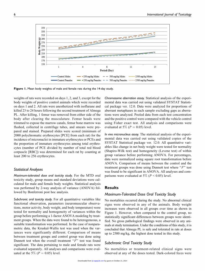
increases were observed in all groups over time as shown in
Figure 1. However, when compared to the control group, no
statistically significant differences between groups were identi-
fied. No gross pathological findings were observed in any ani-
mals at study termination. Under the conditions of this study, it is
concluded that Almega PL is safe and tolerated in rats at doses
up to 2500 mg/kg, the highest dose tested in this study.
Subchronic Oral Toxicity Study
No mortalities or treatment-related clinical signs were
observed at any of the doses tested. Dark-colored feces were
Figure 1. Mean body weights of male and female rats during the 14-day study.
6 International Journal of Toxicology
by guest on October 21, 2014ijt.sagepub.comDownloaded from

observed in all rats of the high-dose group. The dark-
colored feces were attributed to the high chlorophyll content
of Almega PL and considered a treatment-related nonad-
verse effect. No statistically significant changes were
observed in food consumption for any treated group as com-
pared to the control group. No treatment-related changes
were observed in body weights at any of the tested doses
in either sex during the entire treatment period (Figure 2).
The results of ophthalmological examinations were normal
for all of the study animals.
Neurological examinations including functional tests
were conducted on both male and female rats and were gener-
ally negative. Statistically significant differences were identi-
fied for forelimbs grip strength in mid-dose males and low- and
high-dose females, hindlimbs foot splay in mid- and high-dose
females, body temperature in high-dose males and low- and
high-dose females, and motor activity in low- and mid-dose
females (Table 5). However, these changes were considered
to be incidental as either the changes were of minimal magni-
tude or not dose dependent.
Figure 2. Mean body weights of male and female rats during the 90-day study.
Table 5. Selected Results From the Functional Observation Battery.a
Sex Males Females
Dose, mg/kg/d 0 250 500 2500 0 250 500 2500
Forelimbs grip strength(GF)
1125.70 +80.22
1076.00 +46.54
1062.10 +19.46b
1160.60 +61.08
953.10 +17.39
996.10 +37.07b
961.80 +31.83
911.50 +32.71b
Hindlimbs foot splay,mm
84.50 +24.52
99.90 +12.37
80.00 +13.26
89.10 +12.96
52.10 +9.90
58.20 + 9.82 73.90 +12.61b
70.80 +9.65b
Body temperature, �C 36.61 36.67 37.09 37.36b 38.09 37.18b 37.76 37.21b
Motor activityAmbulatory time—interval 2, s
202.80 +37.95
222.10 +56.22
203.00 +44.12
219.90 +41.87
171.10 +45.09
209.80 +43.53
222.10 +26.81b
200.30 +37.43
Horizontal counts—interval 3
758.70 +213.88
864.10 +223.02
711.60 +227.76
815.20 +150.60
648.80 +325.66
1024.70 +202.89b
899.10 +246.39
914.40 +327.63
Ambulatorycounts—interval 3
508.20 +194.10
557.50 +165.95
472.20 +164.30
507.70 +133.73
404.70 +227.92
682.00 +161.77b
580.20 +197.71
581.90 +220.37
Ambulatorycounts—total
2955.90 +672.60
2940.80 +675.53
2676.40 +566.06
2471.10 +742.46
2515.90 +841.83
3128.50 +415.33
3482.70 +758.07b
3082.70 +898.64
aData for all end points except body temperature are presented as mean values+ standard deviations with N¼ 10; body temperature presented as mean with N¼ 10.bStatistically significant from the vehicle control group (P � 0.05).
Kagan et al 7
by guest on October 21, 2014ijt.sagepub.comDownloaded from

Data from the hematology, coagulation, and clinical chem-
istry analyses performed on day 91 are presented in Tables 6
and 7 for males and females, respectively. Statistically signif-
icant changes in hematology parameters were seen in high-dose
males including hematocrit (HCT), mean corpuscular
hemoglobin concentration (MCHC), and mean platelet volume
(MPV); MPV was also statistically different in mid-dose males.
In female rats, statistically significant hematological changes
including RBC, HCT, mean corpuscular hemoglobin, MCHC,
and MPV were seen in the mid- and high-dose groups. These
Table 6. Summary of Hematology, Coagulation, and Clinical Chemistry Values on Day 91—Males.a
Parameter 0 mg/kg/d 250 mg/kg/d 500 mg/kg/d 2500 mg/kg/d
HematologyWBC, G/L 9.72 + 2.64 9.28 + 2.21 9.14 + 1.85 9.04 + 1.39RBC, T/L 9.14 + 0.30 9.23 + 0.27 9.21 + 0.27 9.47 + 0.25HGB, g/L 168 + 5 166 + 4 166 + 4 170 + 5HCT, L/L 0.520 + 0.014 0.519 + 0.013 0.522 + 0.017 0.537 + 0.017b
MCV, fL 56.9 + 1.1 56.3 + 1.0 56.7 + 1.1 56.7 + 1.7MCH, pg 18.4 + 0.3 18.0 + 0.3 18.0 + 0.4 18.0 + 0.6MCHC, g/L 323 + 4 321 + 4 318 + 6 317 + 4Plat, G/L 859 + 73 898 + 114 902 + 114 837 + 98MPV, fL 8.3 + 0.7 8.4 + 0.6 8.8 + 0.6b 9.9 + 0.8b
Neut, % 19.2 + 4.6 19.2 + 5.9 16.5 + 4.9 16.7 + 4.7Lymp, % 76.6 + 4.7 76.7 + 6.3 79.4 + 5.2 79.3 + 5.4Mono, % 1.8 + 0.5 1.8 + 0.4 1.9 + 0.4 2.0 + 0.7Eosi, % 0.7 + 0.1 0.6 + 0.2 0.8 + 0.4 0.7 + 0.2Baso, % 0.4 + 0.1 0.3 + 0.1 0.3 + 0.1 0.3 + 0.1Neut A, G/L 1.83 + 0.57 1.72 + 0.53 1.47 + 0.39 1.50 + 0.43Lymp A, G/L 7.48 + 2.30 7.18 + 1.95 7.30 + 1.77 7.19 + 1.32Mono A, G/L 0.18 + 0.08 0.16 + 0.06 0.17 + 0.03 0.18 + 0.05Eosi A, G/L 0.07 + 0.02 0.06 + 0.01 0.07 + 0.02 0.06 + 0.01Baso A, G/L 0.04 + 0.02 0.03 + 0.02 0.03 + 0.01 0.03 + 0.01Retic, % 2.46 + 0.28 2.51 + 0.21 2.51 + 0.09 2.31 + 0.24Retic A, T/L 0.224 + 0.023 0.231 + 0.019 0.231 + 0.009 0.219 + 0.024
CoagulationPT, s 17.2 + 0.6 16.7 + 1.1 16.4 + 0.4 16.5 + 0.7APTT, s 16.2 + 1.7 15.5 + 2.9 12.9 + 2.1b 14.6 + 3.4
Clinical chemistryGlu, mmol/L 7.53 + 0.66 7.46 + 0.45 7.54 + 0.83 7.79 + 0.97BUN, mmol/L 4.43 + 0.51 4.79 + 0.39 4.88 + 0.41 5.41 + 0.43b
Creat, mmol/L 31 + 9 30 + 6 28 + 8 25 + 7AST, U/L 86 + 21 91 + 14 77 + 9 94 + 11ALT, U/L 56 + 7 56 + 8 60 + 8 78 + 13b
GGT, U/L 1 + 1 1 + 1 1 + 1 1 + 1ALP, U/L 118 + 23 108 + 16 113 + 15 120 + 20TBil, mmol/L 1.66 + 0.63 1.33 + 0.61 1.45 + 0.54 1.35 + 0.75TChol, mmol/L 2.65 + 0.17 2.66 + 0.23 2.63 + 0.29 3.20 + 0.43b
Trig, mmol/L 0.55 + 0.10 0.50 + 0.15 0.61 + 0.12 0.54 + 0.09TPro mmol/L 71.3 + 2.9 72.2 + 2.4 73.1 + 1.5 71.8 + 3.5Alb, g/L 42.2 + 1.8 42.3 + 1.6 42.7 + 0.6 42.8 + 1.9Pi, mmol/L 1.94 + 0.23 2.00 + 0.18 2.06 + 0.20 2.05 + 0.14Ca, mmol/L 2.65 + 0.07 2.63 + 0.06 2.61 + 0.07 2.51 + 0.09b
Na, mEq/L 147.2 + 2.2 148.6 + 0.5 148.7 + 0.5 148.8 + 1.0K, mEq/L 4.09 + 0.17 4.32 + 0.26 4.07 + 0.35 4.47 + 0.39b
Cl, mEq/L 111.5 + 1.1 112.2 + 0.7 111.9 + 0.6 112.1 + 1.2A/G, ratio 1.46 + 0.10 1.42 + 0.11 1.41 + 0.10 1.49 + 0.14Glob, g/L 29.0 + 1.9 29.9 + 2.0 30.4 + 1.8 29.0 + 2.6
Abbreviations: WBC, white blood corpuscles; RBC, red blood corpuscles; HGB, hemoglobin; HCT, hematocrit; MCV, mean corpuscular volume; MCH, meancorpuscular hemoglobin; MCHC, mean corpuscular hemoglobin concentration; Plat, platelets; MPV, mean platelet volume; Neut, neutrophils; Lymp, lymphocytes;Mono, monocytes; Eosi, eosinophils; Baso, basophils; Retic, reticulocyte counts; PT, prothrombin time; APTT, activated partial thromboplastin time; Glu, glucose;BUN, blood urea nitrogen; Creat, creatine; AST, aspartate aminotransferase; ALT, alanine aminotransferase; GGT, g-glutamyl transpeptidase; ALP, alkalinephosphatase; TBil, total bilirubin; TChol, total cholesterol; Trig, triglycerides; TPro, total plasma protein; Alb, albumin; Pi, inorganic phosphorous; Ca, calcium; Na,sodium; K, potassium; Cl, chloride; A/G, albumin/globulin ratio (calculated values).aAll data are presented as mean values + standard deviations with N ¼ 10.bStatistically significant from the vehicle control group (P � 0.05).
8 International Journal of Toxicology
by guest on October 21, 2014ijt.sagepub.comDownloaded from

changes were considered incidental due to the lack of a dose–
response relationship and/or the changes being within the phy-
siological range. For coagulation parameters, a decrease in
activated partial thromboplastin time was seen in mid-dose
males, and an increase in prothrombin time was seen in high-
dose females. These values were considered incidental due to
the lack of a dose response and the minor magnitude of the
changes. Clinical chemistry values including blood urea nitro-
gen (BUN), alanine aminotransferase (ALT), total cholesterol
(TChol), calcium (Ca), and potassium (K) were statistically
Table 7. Summary of Hematology, Coagulation, and Clinical Chemistry Values on Day 91—Females.a
Parameter 0 mg/kg/d 250 mg/kg/d 500 mg/kg/d 2500 mg/kg/d
HematologyWBC, G/L 6.58 + 2.22 6.62 + 1.43 6.56 + 1.27 7.57 + 2.17RBC, T/L 8.21 + 0.28 8.45 + 0.22 8.68 + 0.32b 8.59 + 0.26b
HGB, g/L 156 + 4 156 + 3 159 + 5 159 + 4HCT, L/L 0.483 + 0.014 0.490 + 0.011 0.503 + 0.016b 0.500 + 0.014b
MCV, fL 58.9 + 1.2 58.0 + 1.3 58.0 + 1.5 58.3 + 0.8MCH, pg 19.0 + 0.5 18.5 + 0.3b 18.3 + 0.4b 18.5 + 0.3b
MCHC, g/L 322 + 4 319 + 4 316 + 5b 317 + 4b
Plat, G/L 868 + 122 943 + 120 949 + 140 982 + 105MPV, fL 9.1 + 1.1 8.6 + 0.6 9.7 + 0.9b 9.9 + 0.4b
Neut, % 15.3 + 4.2 14.5 + 5.6 16.6 + 5.7 12.0 + 2.2Lymp, % 80.6 + 4.6 81.7 + 5.8 79.5 + 6.2 84.1 + 2.3Mono, % 1.9 + 0.8 1.5 + 0.3 1.7 + 0.6 1.8 + 1.1Eosi, % 0.9 + 0.2 0.8 + 0.2 0.9 + 0.4 0.9 + 0.3Baso, % 0.3 + 0.1 0.3 + 0.1 0.3 + 0.1 0.3 + 0.1Neut A, G/L 1.00 + 0.37 0.98 + 0.56 1.10 + 0.51 0.89 + 0.24Lymp A, G/L 5.32 + 1.92 5.38 + 1.10 5.20 + 1.01 6.39 + 1.93Mono A, G/L 0.13 + 0.06 0.10 + 0.04 0.12 + 0.05 0.13 + 0.08Eosi A, G/L 0.06 + 0.01 0.05 + 0.02 0.06 + 0.02 0.07 + 0.03Baso A, G/L 0.02 + 0.01 0.02 + 0.01 0.02 + 0.01 0.02 + 0.01Retic, % 2.84 + 0.54 2.58 + 0.39 2.37 + 0.39 2.30 + 0.65Retic A, T/L 0.233 + 0.040 0.217 + 0.031 0.205 + 0.030 0.197 + 0.053
CoagulationPT, s 16.5 + 1.0 16.5 + 0.7 17.2 + 0.8 18.2 + 0.9b
APTT, s 14.6 + 3.1 14.8 + 2.2 13.2 + 3.6 13.4 + 2.5Clinical chemistry
Glu, mmol/L 7.36 + 0.52 7.76 + 0.51 8.13 + 1.52 7.36 + 0.71BUN, mmol/L 4.59 + 0.44 5.17 + 0.53 5.02 + 0.65 5.65 + 0.76b
Creat, mmol/L 30 + 8 27 + 9 27 + 9 33 + 6AST, U/L 118 + 22 87 + 14b 83 + 10b 92 + 14b
ALT, U/L 50 + 4 50 + 8 45 + 8 64 + 8b
GGT, U/L 0 + 1 1 + 1 0 + 1 2 + 1b
ALP, U/L 85 + 14 87 + 12 87 + 25 98 + 16TBil, mmol/L 2.04 + 1.17 1.78 + 0.76 2.31 + 1.19 1.68 + 0.65TChol, mmol/L 3.10 + 0.27 3.23 + 0.16 3.07 + 0.35 3.60 + 0.35b
Trig, mmol/L 0.47 + 0.09 0.45 + 0.05 0.44 + 0.10 0.50 + 0.09TPro, mmol/L 69.9 + 2.6 70.7 + 2.2 71.0 + 3.4 73.6 + 2.7b
Alb, g/L 42.4 + 0.7 44.2 + 3.1 42.6 + 1.8 42.75 + 2.2b
Pi, mmol/L 1.73 + 0.22 1.79 + 0.14 1.80 + 0.22 1.89 + 0.18Ca, mmol/L 2.59 + 0.07 2.59 + 0.05 2.53 + 0.18 2.62 + 0.09Na, mEq/L 147.9 + 1.5 147.6 + 1.0 147.9 + 1.4 148.7 + 0.9K, mEq/L 3.83 + 0.55 3.78 + 0.14 3.83 + 0.24 4.03 + 0.40Cl, mEq/L 112.6 + 1.0 112.2 + 1.0 112.6 + 0.9 112.9 + 1.0A/G, ratio 1.55 + 0.14 1.72 + 0.42 1.51 + 0.12 1.82 + 0.16b
Glob, g/L 27.5 + 2.5 26.5 + 3.6 28.4 + 2.3 26.2 + 1.8
Abbreviations: WBC, white blood corpuscles; RBC, red blood corpuscles; HGB, hemoglobin; HCT, hematocrit; MCV, mean corpuscular volume; MCH, meancorpuscular hemoglobin; MCHC, mean corpuscular hemoglobin concentration; Plat, platelets; MPV, mean platelet volume; Neut, neutrophils; Lymp, lymphocytes;Mono, monocytes; Eosi, eosinophils; Baso, basophils; Retic, reticulocyte counts; PT, prothrombin time; APTT, activated partial thromboplastin time; Glu, glucose;BUN, blood urea nitrogen; Creat, creatine; AST, aspartate aminotransferase; ALT, alanine aminotransferase; GGT, g-glutamyl transpeptidase; ALP, alkalinephosphatase; TBil, total bilirubin; TChol, total cholesterol; Trig, triglycerides; TPro, total plasma protein; Alb, albumin; Pi, inorganic phosphorous; Ca, calcium; Na,sodium; K, potassium; Cl, chloride; A/G, albumin/globulin ratio (calculated values).aAll data are presented as mean values + standard deviations with N ¼ 10.bStatistically significant from the vehicle control group (P � 0.05).
Kagan et al 9
by guest on October 21, 2014ijt.sagepub.comDownloaded from

different in the high-dose males when compared to controls.
The BUN, aspartate aminotransferase, ALT, g-glutamyl trans-
peptidase, TChol, total plasma protein, albumin, and albumin/
globulin ratio were statistically different in high-dose females
when compared to controls. Aspartate aminotransferase was
also statistically different in low- and mid-dose females. The
minimal increases in ALT activity (39% for males and 28% for
females) and TChol levels (21% for males and 16% for
females) seen at the highest dose (2500 mg/kg/d) were consid-
ered to be a test item-related, nonadverse effect due to the
severity of change being limited and below the threshold of
concern. In addition, the increased ALT activity was not
accompanied by correlating histologic changes in the liver. All
other statistically significant changes in clinical chemistry were
of a low magnitude and within the physiological range and
therefore considered toxicologically insignificant. No
treatment-related changes were seen in any of the urine para-
meters analyzed in terminally killed animals.
When compared to the control groups, no significant
changes attributed to treatment were seen in organ weights or
organ to body weight ratios for male or female animals at any
dose level evaluated (Tables 8 and 9). Statistically significant
changes were limited to increased thyroid weights (absolute
and relative) in low- and high-dose males, increased adrenals
weight in mid- (absolute) and high-dose (relative) females, and
increased relative liver weights in high-dose females. These
were considered incidental due to the absence of clinical signs
and histopathological changes.
Compared to their respective control groups, no test item-
related gross pathological lesions were evident in male or
female rats at terminal necropsy (day 91). No incidental find-
ings were seen in males. Incidental findings were found in
females as dilated uterus noted across the tested groups (2 per
test group including controls). The dilated uterus findings were
confirmed microscopically (mild to severe dilation across all
treatment groups including controls) and was not considered to
Table 8. Summary of Terminal Fasting Body Weights and Absolute Organ Weights on Day 91.a
Group
Males Females
0mg/kg/d
250mg/kg/d
500mg/kg/d
2500mg/kg/d
0mg/kg/d
250mg/kg/d
500mg/kg/d
2500mg/kg/d
Terminal fasting BW, g 420.72 +37.17
429.34 +37.14
421.29 +32.71
424.90 +32.04
252.36 +14.06
260.70 +14.49
259.48 +18.03
243.39 +12.44
Adrenals, g 0.0538 +0.0068
0.0544 +0.0055
0.0566 +0.0055
0.0572 +0.0048
0.0602 +0.0061
0.0643 +0.0050
0.0674 +0.0036b
0.0653 +0.0071
Brain, g 1.9636 +0.0804
2.0037 +0.0770
1.9731 +0.0938
2.0226 +0.1018
1.8508 +0.0689
1.8205 +0.0531
1.8805 +0.0527
1.8486 +0.0489
Epididymides, g 1.3437 +0.0321
1.3612 +0.0729
1.3159 +0.0566
1.3492 +0.1044
– – – –
Heart, g 1.3063 +0.1234
1.3493 +0.0890
1.3235 +0.1517
1.3993 +0.1269
0.8871 +0.0659
0.9263 +0.0507
0.9490 +0.0702
0.9084 +0.0849
Kidneys, g 2.2479 +0.1927
2.2841 +0.1547
2.1974 +0.2168
2.3741 +0.2300
1.4327 +0.0863
1.4397 +0.0871
1.4984 +0.1006
1.4403 +0.0869
Liver, g 11.2948 +1.5264
11.1451 +1.3716
11.3951 +1.1856
12.0852 +1.3774
6.4700 +0.3399
6.6165 +0.3849
6.5445 +0.5758
6.7227 +0.5350
Ovaries – – – – 0.0886 +0.0109
0.0794 +0.0138
0.0916 +0.0104
0.0899 +0.0143
Pituitary, gc 0.0128 +0.0021
0.0122 +0.0023
0.0122 +0.0028
0.0132 +0.0014
0.0141 +0.0014
0.0140 +0.0019
0.0142 +0.0035
0.0145 +0.0020
Prostate, g 1.1370 +0.1241
1.0702 +0.1731
1.1452 +0.1348
1.1797 +0.1418
– – – –
Seminal vesicles andcoagulating glands, g
1.8415 +0.2714
1.7696 +0.3712
1.7238 +0.1837
1.8494 +0.2565
– – – –
Spleen, g 0.7734 +0.0990
0.7919 +0.1067
0.07675 +0.0964
0.8095 +0.1058
0.6358 +0.0525
0.6008 +0.0362
0.6382 +0.1240
0.6201 +0.1160
Testes, g 3.8115 +0.1292
3.8862 +0.2085
3.8779 +0.2073
3.8856 +0.1647
– – – –
Thymus, g 0.4015 +0.0399
0.4209 +0.0750
0.4144 +0.0477
0.4041 +0.0814
0.3118 +0.0455
0.3084 +0.0466
0.3120 +0.0357
0.3741 +0.1857
Thyroid with parathyroids, gc 0.0282 +0.0041
0.0351 +0.0067b
0.0315 +0.0055
0.0394 +0.0049b
0.0259 +0.0040
0.0310 +0.0056
0.0299 +0.0087
0.0275 +0.0040
Uterus with cervix, g – – – – 0.7654 +0.3443
0.7138 +0.2771
0.8041 +0.5670
0.8099 +0.4375
aAll data are presented as mean values + standard deviations with N ¼ 10.bStatistically significant from the vehicle control group (P � 0.05).cData recorded on day 93.
10 International Journal of Toxicology
by guest on October 21, 2014ijt.sagepub.comDownloaded from

be treatment related. All other microscopic findings (inflam-
matory foci in esophageal adnexal tissue, proteinaceous mate-
rial in kidney tubules, harderian gland alterations, hepatocyte
vacuolation, mesenteric lymph node erythrophagocytosis,
mandibular lymph node plasmacytosis and/or erythrophagocy-
tosis, unilateral ovarian cysts, prostate mononuclear cell infil-
tration, and rectum nematode) occurred at low frequencies in
the control and high-dose groups and were not considered treat-
ment related.
Under the conditions of this study, the ‘‘no observed adverse
effect level’’ (NOAEL) for Almega PL would be 2500 mg/kg/d
based on a lack of treatment-related effects at 2500 mg/kg/d,
the highest dose evaluated in both male and female rats. How-
ever, because dose formulation analyses revealed consistently
lower (approximately �20%) concentrations of the test item
from the corresponding theoretical doses, the highest intended
dose of 2500 mg/kg/d would result in an actual dose of 2000
mg/kg/d. Therefore, the NOAEL for the subchronic oral
gavage administration of Almega PL in rats is determined as
2000 mg/kg/d.
Bacterial Reverse Mutation Assays
Genotypic characterization of tester bacteria indicated that the
assays were valid because (1) all S typhimurium strains demon-
strated the requirement of histidine amino acid for their growth
and E coli strain WP2uvrA demonstrated the requirement of
tryptophan amino acid for its growth, (2) TA98, TA100, and
WP2uvrA demonstrated their typical resistance to ampicillin,
(3) the presence of characteristic mutations like the rfa muta-
tion was demonstrated by all S typhimurium strains by their
sensitivity to crystal violet, (4) The uvrA mutation in the E coli
strain and the uvrB mutation in the S typhimurium strains were
demonstrated by their sensitivity to UV light, (5) all of the
tester strains produced spontaneous revertant colonies which
were within the frequency ranges of historical control data, and
(6) the numbers of revertant colonies in the positive control
groups were greater than 3 times the number of revertant colo-
nies in the corresponding negative controls.
The summarized results for experiments I and II are pre-
sented in Tables 10 and 11, respectively. Slight precipitation of
Table 9. Summary of Organ to Body Weight Ratios on Day 91.a
Group
Males Females
0mg/kg/d
250mg/kg/d
500mg/kg/d
2500mg/kg/d
0mg/kg /d
250mg/kg/d
500mg/kg/d
2500mg/kg/d
Adrenals, % 0.0128 +0.0017
0.0128 +0.0016
0.0135 +0.0017
0.0135 +0.0015
0.0239 +0.0028
0.0247 +0.0021
0.0261 +0.0021
0.0268 +0.0024b
Brain, % 0.4690 +0.0319
0.4695 +0.0406
0.4697 +0.0246
0.4776 +0.0315
0.7354 +0.0474
0.7001 +0.0411
0.7276 +0.0498
0.7607 +0.0283
Epididymides, % 0.3216 +0.0291
0.3186 +0.0249
0.3136 +0.0221
0.3188 +0.0296
– – – –
Heart, % 0.3107 +0.0157
0.3150 +0.0156
0.3141 +0.0267
0.3292 +0.0135
0.3515 +0.0165
0.3556 +0.0148
0.3665 +0.0260
0.3728 +0.0225
Kidneys, % 0.5353 +0.0324
0.5337 +0.0338
0.5216 +0.0309
0.5585 +0.0265
0.5682 +0.0274
0.5538 +0.0447
0.5787 +0.0389
0.5918 +0.0207
Liver, % 2.6760 +0.1536
2.5917 +0.1568
2.7044 +0.1747
2.8375 +0.1362
2.5676 +0.1381
2.5415 +0.1492
2.5239 +0.1673
2.7601 +0.1251b
Ovaries – – – – 0.0351 +0.0038
0.305 +0.0054
0.0353 +0.0031
0.0369 +0.0056
Pituitary, % 0.0030 +0.0005
0.0028 +0.0005
0.0029 +0.0006
0.0031 +0.0002
0.0056 +0.0006
0.0054 +0.0007
0.0055 +0.0013
0.0059 +0.0007
Prostate, % 0.2733 +0.0475
0.2521 +0.0527
0.2730 +0.0363
0.2796 +0.0428
– – – –
Seminal vesicles andcoagulating glands, %
0.4386 +0.0609
0.4170 +0.1066
0.4097 +0.0385
0.4363 +0.0595
– – – –
Spleen, % 0.1835 +0.0117
0.1850 +0.0239
0.1825 +0.0216
0.1905 +0.0208
0.2524 +0.0227
0.2309 +0.0161
0.2462 +0.0434
0.2537 +0.0388
Testes, % 0.9117 +0.0790
0.9084 +0.0542
0.9252 +0.0840
0.9192 +0.0818
– – – –
Thymus, % 0.0958 +0.0100
0.0983 +0.0178
0.0988 +0.0133
0.0947 +0.0157
0.1234 +0.0158
0.1179 +0.0136
0.1203 +0.0121
0.1550 +0.0811
Thyroid with parathyroids, % 0.0067 +0.0007
0.0082 +0.0014b
0.0075 +0.0012
0.0093 +0.0011b
0.0103 +0.0015
0.0119 +0.0022
0.0115 +0.0034
0.0113 +0.0017
Uterus with cervix, % – – – – 0.3042 +0.1424
0.2732 +0.1023
0.3102 +0.2222
0.3324 +0.1753
aAll data are presented as mean values + standard deviations with N ¼ 10.bStatistically significant from the vehicle control group (P � 0.05).
Kagan et al 11
by guest on October 21, 2014ijt.sagepub.comDownloaded from

Almega PL was visually observable in all test strains both with
and without S9 mix at concentrations of 1581 mg/plate and
higher in experiment I and 1880 mg/plate and higher in experi-
ment II. However, this slight precipitation did not interfere with
the scoring of the bacterial background lawn intensity. No
toxicity was observed up to the highest concentration of 5000
mg/plate for experiment I or II, as the intensity of the bacterial
background lawn as well as the mean number of revertant
colonies were comparable to the DMSO control. For both
experiment I and II, no positive mutagenic responses were
observed up to the top dose of 5000 mg/plate in any of the tester
strains both in the presence and absence of metabolic activa-
tion. It is concluded that all criteria for the bacterial reverse
mutation assays are valid and that under the conditions of this
assay, Almega PL was not mutagenic at any of the doses tested.
Chromosome Aberration Assay
The results for the chromosome aberration assay are presented
in Table 12. After 4 hours of exposure and in the presence of
metabolic activation, cytotoxicity was seen at 19 mg/mL, the
highest concentration tested. Similarly, cytotoxicity was seen
at the highest doses tested in the absence of metabolic activa-
tion following 4 hours of exposure (40 mg/mL) and 21 hours of
exposure (7 mg/mL). For all doses and times, and in the absence
or presence of metabolic activation, no positive incidence of
chromosomal aberrations was observed following treatment
with Almega PL. The in vitro mammalian chromosome aberra-
tion assay is considered valid because (1) the incidence of
aberrations in the vehicle control cultures is in the range of
in-house historical control data and (2) the positive control
substances produced a significant increase in the incidence of
aberrations compared to the respective vehicle control.
In Vivo Micronucleus Assay
No mortality was seen in any of the tested animals, and body
weight gains were comparable to vehicle controls. No
treatment-related clinical signs were observed in any dose
groups except for dark-colored feces seen at 1000 and 2000
mg/kg/d on days 2 and 3. The dark-colored feces were attrib-
uted to the high chlorophyll content of Almega PL and consid-
ered a treatment-related nonadverse effect. Data for the total
RBC ratio and micronucleus incidence are summarized in
Table 13. No significant increases in micronucleated PCE were
observed between mice treated with Almega PL and the vehicle
control. A significant decrease in PCE:total RBC ratio was seen
in males at 500 and 2000 mg/kg/d and for combined sex at 500
Table 10. Summary of Results of Initial Mutation Assay.
Treatment, mg/plate
No. of revertants/platea
TA98 TA100 TA1535 TA1537 WP2uvrA (pKM101)
Vehicle control—DMSO (100 mL)With metabolic activation 21 + 3 112 + 4 18 + 1 10 + 2 137 + 4Without metabolic activation 24 + 3 106 + 8 19 + 1 11 + 2 132 + 4
50With metabolic activation 20 + 2 100 + 4 16 + 1 10 + 2 132 + 5Without metabolic activation 21 + 1 99 + 4 16 + 2 10 + 2 131 + 3
158With metabolic activation 22 + 3 108 + 4 16 + 2 10 + 1 132 + 3Without metabolic activation 21 + 4 106 + 7 16 + 3 10 + 3 130 + 3
500With metabolic activation 21 + 5 101 + 3 16 + 3 11 + 2 136 + 4Without metabolic activation 20 + 1 102 + 5 16 + 4 9 + 1 126 + 6
1581With metabolic activation 20 + 2 101 + 10 14 + 3 11 + 2 135 + 5Without metabolic activation 23 + 1 105 + 9 17 + 1 10 + 2 132 + 5
5000With metabolic activation 21 + 3 103 + 4 16 + 1 11 + 2 135 + 4Without metabolic activation 22 + 4 107 + 8 17 + 3 10 + 3 135 + 5
Positive controlWith metabolic activation 544 + 8b 787 + 9b 132 + 3b 123 + 6b 591 + 4c
Without metabolic activation 231 + 6d 578 + 6e 131 + 4e 125 + 4f 605 + 10g
Abbreviations: DMSO, dimethyl sulfoxide; SD, standard deviation.aValues are means + SDs of 3 replicates and are rounded off to the nearest whole number.bTA98, TA100, TA1535, and TA1537 with activation: 2-aminoanthracene (4 mg/plate).cWP2uvrA (pKM101) with activation: 2-aminoanthracene (30 mg/plate).dTA98 without activation: 2-nitrofluorene (2 mg/plate).eTA100 and TA1535 without activation: sodium azide (1 mg/plate).fTA1537 without activation: 9-aminoacridine (50 mg/plate).gP2uvrA (pKM101) without activation: 4-nitroquinoline-1-oxide (4 mg/plate).
12 International Journal of Toxicology
by guest on October 21, 2014ijt.sagepub.comDownloaded from
and 1000 mg/kg/d. However, the values were comparable to
historical data ranges and therefore determined to be nonbio-
logically relevant. The test was considered valid because (1)
the incidence of micronucleated PCE in the vehicle control
group was within the historical control range, (2) the positive
control animals demonstrated a significant increase in micro-
nucleated PCE compared to controls, and (3) at the end of the
study, the weight variation in animals was minimal and did not
exceed +20% of the mean weight of each sex.
Discussion
Omega-3 fatty acids possess significant health benefits for
brain function and development as well as cardiovascular con-
ditions.17,18 Currently most of these fatty acids isolated for
human consumption are sourced from fish. However, due to
a limited amount of global fish stocks, alternative sources of
fatty acids are needed. Potential substitutes include oil extracts
from plants, krill, microalgae, and microalgae-like protists.
Of the potential substitutes, algae may be one of the more
promising alternatives. Due to their small size, algae are easy to
grow on a large scale. Algae do not require freshwater or arable
land and are capable of producing significant amounts of fatty
acids even under adverse conditions.18,19 In the past, the
potential for using microalgal oils on a larger scale has been
limited by cost, extraction, and purification methods.17 At pres-
ent, various strains of algae are being used for the production of
fatty acids for human consumption. For example, Schizochy-
trium sp and/or Crypthecodinium cohnii are included in infant
formulas, food additives, cosmetics, and pharmaceutical
products.17
Nannochloropsis oculata cultures have been used as a
source of live food for rotifers in marine fish hatcheries and
as crustacean feed for over 30 years.20 No data were located in
the published literature indicating a history of consumption of
N oculata-derived oil products by humans, either in conven-
tional food or dietary supplements. Currently, Almega PL is not
regulated by the US Food and Drug Administration and has not
been determined Generally Recognized As Safe (GRAS) as a
food ingredient. However, EPA has been evaluated and
approved in GRAS notifications and food additive petitions for
use as an o-3 fatty acid. In addition, the FDA has stated that
dietary supplements should not recommend or suggest in their
labeling a daily intake exceeding 2 g EPA and DHA.9
Previously, the acute and subchronic oral toxicities of
N oculata biomass were evaluated in male and female
Sprague-Dawley rats.1 The oral median lethal dose of N ocu-
lata was greater than 12 g/kg and the subchronic toxicity was
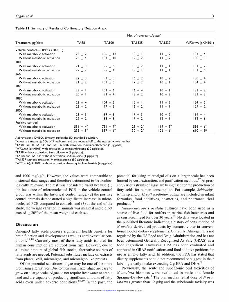
Table 11. Summary of Results of Confirmatory Mutation Assay.
Treatment, mg/plate
No. of revertants/platea
TA98 TA100 TA1535 TA1537 WP2uvrA (pKM101)
Vehicle control—DMSO (100 mL)With metabolic activation 23 + 2 106 + 12 18 + 1 11 + 2 134 + 4Without metabolic activation 26 + 4 103 + 10 19 + 2 11 + 2 130 + 3
100With metabolic activation 21 + 3 95 + 5 18 + 2 11 + 1 131 + 2Without metabolic activation 22 + 2 95 + 4 19 + 1 11 + 1 131 + 5
266With metabolic activation 22 + 3 93 + 3 16 + 2 10 + 2 130 + 4Without metabolic activation 21 + 2 101 + 5 17 + 2 10 + 1 134 + 4
707With metabolic activation 23 + 1 103 + 6 16 + 4 10 + 1 131 + 2Without metabolic activation 20 + 1 93 + 4 18 + 2 10 + 2 131 + 3
1880With metabolic activation 22 + 4 104 + 6 15 + 1 11 + 2 134 + 5Without metabolic activation 22 + 2 97 + 3 16 + 2 11 + 1 129 + 2
5000With metabolic activation 23 + 3 99 + 6 17 + 3 10 + 2 134 + 4Without metabolic activation 22 + 2 98 + 9 17 + 2 12 + 1 132 + 6
Positive controlWith metabolic activation 556 + 4b 791 + 7b 128 + 2b 119 + 3b 596 + 6c
Without metabolic activation 235 + 5d 587 + 6e 130 + 2e 126 + 6f 610 + 5g
Abbreviations: DMSO, dimethyl sulfoxide; SD, standard deviation.aValues are means + SDs of 3 replicates and are rounded off to the nearest whole number.bTA98, TA100, TA1535, and TA1537 with activation: 2-aminoanthracene (4 mg/plate).cWP2uvrA (pKM101) with activation: 2-aminoanthracene (30 mg/plate).dTA98 without activation: 2-nitrofluorene (2 mg/plate).eTA100 and TA1535 without activation: sodium azide (1 mg/plate).fTA1537 without activation: 9-aminoacridine (50 mg/plate).gWP2uvrA(pKM101) without activation: 4-nitroquinoline-1-oxide (4 mg/plate).
Kagan et al 13
by guest on October 21, 2014ijt.sagepub.comDownloaded from

Tab
le12.
Sum
mar
yR
esults
ofC
hro
moso
me
Aber
ration
Ass
ayW
ith
and
Without
Met
abolic
Act
ivat
ion.a
Tre
atm
ent,mg
/mL
Exposu
re,h
No.ofm
etap
has
essc
ore
d
No.(%
)ofm
etap
has
esw
ith
aber
rationsb
Tota
lno.(%
)ofab
erra
nt
met
aphas
esc
Cel
lgr
ow
thin
hib
itio
n,%
Gap
sBre
aks
Exch
ange
s
Cs
Ct
Cs
Ct
Cs
Ct
RC
Incl
udin
gga
ps
excl
udin
gga
ps
With
met
abolic
activa
tion
DM
SO(1
50mL
)4
200
00
00
00
00
00
1.9
4200
1(0
.5)
1(0
.5)
00
00
02
(1.0
)0
26
64
200
02
(1.0
)0
00
00
2(1
.0)
042
19
4200
00
01
(0.5
)0
00
1(0
.5)
1(0
.5)
51
CPA
55
4200
42
(21.0
)9
(4.5
)1
(0.5
)70
(35.0
)42
(21.0
)31
(15.5
)0
158
(79.0
)127
(63.5
)d38
Without
met
abolic
activa
tion
DM
SO(1
50mL
)4
200
01
(0.5
)0
4(2
.0)
00
05
(2.5
)4
(2.0
)0
44
200
00
03
(1.5
)0
00
3(1
.5)
3(1
.5)
20
13
4200
2(1
.0)
3(1
.5)
1(0
.5)
4(2
.0)
00
010
(5.0
)5
(2.5
)25
40
4200
3(1
.5)
7(3
.5)
1(0
.5)
5(2
.5)
00
013
(6.5
)5
(2.5
)51
EM
S600
4200
15
(7.5
)19
(9.5
)51
(25.5
)91
(45.5
)0
33
(16.5
)4
(2.0
)144
(72.0
)132
(66.0
)d31
DM
SO(1
50mL
)21
200
1(0
.5)
1(0
.5)
00
00
02
(1.0
)0
01.7
521
200
01
(0.5
)0
1(0
.5)
00
02
(1.0
)1
(0.5
)26
3.5
21
200
00
01
(0.5
)0
00
1(0
.5)
1(0
.5)
39
721
200
1(0
.5)
2(1
.0)
02
(1.0
)0
00
5(2
.5)
2(1
.0)
52
EM
S600
21
200
9(4
.5)
33
(16.5
)6
(3.0
)91
(45.5
)9
(4.5
)45
(22.5
)0
121
(60.5
)107
(53.5
)d35
Abbre
viat
ions:
DM
SO,dim
ethyl
sulfo
xid
e;C
s,ch
rom
oso
me
type;
Ct,
chro
mat
idty
pe;
RC
,ri
ng
chro
moso
me;
EM
S,et
hyl
met
han
esulfo
nat
e.a A
ber
rant
met
aphas
esin
cludin
gga
ps
are
not
subje
ctto
stat
istica
lan
alys
is.
bV
alues
are
the
sum
oftw
ore
plic
ates
and
the
valu
esin
par
enth
esis
repre
sent
%.
c Met
aphas
epla
tes
with
one
or
more
aber
rations
are
consi
der
edas
one
met
aphas
epla
tew
ith
aber
rations.
dSt
atis
tica
llysi
gnifi
cant
from
the
vehic
leco
ntr
olgr
oup
(P�
0.0
5).
14 by guest on October 21, 2014ijt.sagepub.comDownloaded from

6 g/kg/d. However, this study focused only on the potential
hepatotoxicity and nephrotoxicity of N oculata. Furthermore,
the study was limited to the evaluation of N oculata biomass
and did not evaluate the potential toxicity of oil extracted from
the biomass. Therefore, the studies reported herein provide new
and relevant data on the safety of N oculata (as Almega PL) oil
extract.
The oral administration of Almega PL at concentrations of
up to 2000 mg/kg/d was well-tolerated by both male and female
rats in the 14-day MTD study and the GLP-compliant 90-day
subchronic toxicity study. Almega PL had no adverse effects
on health or growth as measured by survival, appearance, beha-
vior, food consumption, body weight, or body weight gain. No
treatment-related effects were seen for ophthalmology, neuro-
logical examinations, hematology, coagulation, clinical chem-
istry, gross pathology, absolute and relative organ weights, or
histopathology in animals of any dose group. Although statis-
tical significance was noted for several end points in the 90-day
subchronic toxicity study, these were not considered to be bio-
logically relevant or attributable to Almega PL because these
effects were (1) incidental or sporadic in nature, (2) within the
range of historical control data for rats of this age and strain,
and/or (3) occurred in the absence of corroborative clinical or
histopathological changes.
The battery of GLP-compliant in vitro and in vivo genotoxi-
city studies provide overall support that Almega PL is not
genotoxic. In 2 independent bacterial reverse mutation assays,
Almega PL was negative for mutagenicity at levels up to 5000
mg/plate using S typhimurium strains TA98, TA100, TA1535,
and TA1537 and E coli strain WP2uvrA, both with and without
metabolic activation. Almega PL was not clastogenic as eval-
uated by the in vitro chromosome aberration assay in the CHO
cell line at concentrations of 1.9, 6, or 19 mg/mL and 4, 13, or
40 mg/mL with or without metabolic activation, respectively,
for 4 hours exposure as well as concentrations of 1.75, 3.5, or 7
mg/mL without metabolic activation for 21 hours of exposure.
When evaluated in the in vivo erythrocyte micronucleus assay,
Almega PL was not clastogenic or aneugenic. No treatment-
related structural and/or numerical chromosomal damage was
seen in immature erythrocytes in rats administered Almega PL
at doses of 500, 1000, or 2000 mg/kg/d.
The available human toxicity data for Almega PL are lim-
ited to a randomized, crossover study conducted in 10 male
patients (aged 18-45 years) which compared the appearance
of fatty acids in the blood plasma of healthy humans after
consumption of a high fat meal followed by either Almega
PL or a commercially available krill oil supplement (NKO;
Neptune Technologies & Bioresources Inc, Quebec, Canada).4
Both oils were encapsulated in gelatin soft capsules manufac-
tured by Catalent (Eberbach, Germany). The capsules provided
approximately 1.5 g EPA and no DHA for Almega PL or 1.02 g
EPA and 0.54 g DHA for krill oil. Both oils were well tolerated.
Plasma samples collected before and 0.5 to 10 hours after
capsule administration indicated time-dependent increases in
EPA for both oils with the concentration of EPA being higher
with algal oil than with krill oil at multiple time points.
Described here is the nonclinical safety assessment (and
limited clinical assessment) of the algal oil Almega PL, which
is being developed as a dietary supplement in the United States,
providing a vegetarian and sustainable source of o-3 fatty acids
Table 13. Summary of PCE:Total RBC Ratio and Micronucleus Incidence.
Sex and dose,mg/kg/d
No. ofrats
PCE: Total RBC ratio
TotalPCEa
No. of PCEwith MN
PCE with MN
No. ofPCE/group
No. ofRBC/group
Ratio,mean + SD % Mean
Male—0 5 559 1134 0.49 + 0.01 10 265 4 0.04 0.04 + 0.04Male—500 5 531 1122 0.47 + 0.01b 10 337 5 0.05 0.05 + 0.03Male—1000 5 534 1116 0.48 + 0.01 10 643 7 0.07 0.07 + 0.04Male—2000 5 520 1096 0.47 + 0.01b 10 274 6 0.06 0.06 + 0.06Malec—15 5 455 1118 0.41 + 0.01b 10 317 129 1.25 1.25 + 0.26d
Female—0 5 549 1134 0.48 + 0.01 10 520 7 0.07 0.07 + 0.04Female—500 5 536 1117 0.48 + 0.01 10 359 6 0.06 0.06 + 0.04Female—1000 5 501 1066 0.47 + 0.01 10 296 6 0.06 0.06 + 0.04Female—2000 5 535 1111 0.48 + 0.01 10 881 3 0.03 0.03 + 0.02Femalec—15 5 446 1100 0.41 + 0.02b 10 535 125 1.19 1.18 + 0.43d
Combined—0 10 1108 2268 0.49 + 0.01 20 785 11 0.05 0.05 + 0.04Combined—500 10 1067 2239 0.48 + 0.01b 20 696 11 0.05 0.05 + 0.04Combined—1000 10 1035 2182 0.47 + 0.01b 20 939 13 0.06 0.06 + 0.04Combined—2000 10 1055 2207 0.48 + 0.01 21 155 9 0.04 0.04 + 0.05Combinedc—15 10 901 2218 0.41 + 0.01b 20 852 254 1.22 1.21 + 0.34d
Abbreviations: RBC, red blood corpuscle; SD, standard deviation.aA minimum of 2000 polychromatic erythrocytes (PCEs) were scored from each rat for the incidence of PCE with micronuclei (MN). Total PCE included PCEscounted to determine PCE: Total RBC ratio.bSignificantly lower than the control group by Dunnett test.cPositive control (cyclophosphamide) administered as a single intraperitoneal injection.dSignificantly higher than the control group by Dunnett test.
Kagan et al 15
by guest on October 21, 2014ijt.sagepub.comDownloaded from
in the diet, primarily EPA. Based on the results of the GLP-
compliant nonclinical studies, the NOAEL for the algal oil is
2000 mg/kg/d, which will provide a reasonable margin of
safety (96-fold) over the suggested maximum oral intake of
1250 mg/d Almega PL (20.8 mg/kg/d for a 70-kg individual).
Acknowledgments
This work was supported by Qualitas Health Ltd.
Author Contributions
M. Kagan contributed to conception and design, acquisition, critically
revised the article, gave final approval, and agrees to be accountable for
all aspects of work ensuring integrity and accuracy. D. Sullivan contrib-
uted to analysis and interpretation, drafted the article, critically revised
the article, gave final approval, and agree to be accountable for all aspects
of work ensuring integrity and accuracy. S. Gad contributed to analysis
and interpretation, critically revised the article, gave final approval, and
agrees to be accountable for all aspects of work ensuring integrity and
accuracy. C. Ballou contributed to interpretation, drafted the article,
critically revised the article, gave final approval, and agrees to be accoun-
table for all aspects of work ensuring integrity and accuracy.
Declaration of Conflicting Interests
The author(s) declared the following potential conflicts of interest
with respect to the research, authorship, and/or publication of this
article: Michael L. Kagan is an employee of Qualitas Health Ltd.
Funding
The author(s) received no financial support for the research, authorship,
and/or publication of this article.
References
1. Kafaie S, Loh SP, Mohtarrudin N. Acute and sub-chronic toxico-
logical assessment of Nannochloropsis oculata in rats. Afr J Agr
Res. 2012;7(7):1220-1225.
2. Rocha JM, Garcia JE, Henriques MH. Growth aspects of the
marine microalga Nannochloropsis gaditana. Biomol Eng.
2003;20(4-6):237-242.
3. Spolaore P, Joannis-Cassan C, Duran E, Isambert A. Commercial
applications of microalgae. J Biosci Bioeng. 2006;101(2):87-96.
4. Kagan ML, West AL, Zante C, Calder PC. Acute appearance of
fatty acids in human plasma—a comparative study between polar-
lipid rich oil from the microalgae Nannochloropsis oculata and
krill oil in healthy young males. Lipids Health Dis. 2013;12:102.
5. Werman MJ, Sukenik A, Mokady S. Effects of the marine uni-
cellular alga Nannochloropsis sp. to reduce the plasma and liver
cholesterol levels in male rats fed on diets with cholesterol. Biosci
Biotechnol Biochem. 2003;67(10):2266-2268.
6. Simopoulos A. Omega-3 fatty acids in inflammation and autoim-
mune diseases. J Am Coll Nutr. 2001;21(6):495-505.
7. Peet M, Horrobin DF. A dose-ranging study of the effects of
ethyl-eicosapentaenoate in patients with ongoing depression
despite apparently adequate treatment with standard drugs. Arch
Gen Psychiatry. 2002;59(10):913-919.
8. Sublette ME, Ellis SP, Geant AL, Mann JJ. Meta-analysis of the
effects of eicosapentaenoic acid (EPA) in clinical trials in depres-
sion. J Clin Psychiatry. 2011;72(12):1577-1584.
9. Qualitas Health Inc. New Dietary Ingredient Notification for
Almega PLTM. Texas: Imperial; 2014.
10. Aubel M. Microcystin/nodularin, anatoxin-a, cylindrospermop-
sin, paralytic shellfish toxins (saxitoxins), okadaic acid, brevetox-
ins data report. FL: Green Water Laboratories; 2013:1-4.
11. Organisation for Economic Cooperation and Development.
Repeated Dose 90-Day Oral Toxicity Study in Rodents. OECD
Guidelines for the Testing of Chemicals. Paris: Organisation for
Economic Cooperation and Development; 1998.
12. Organisation for Economic Cooperation and Development.
OECD Principles of Good Laboratory Practice (as Revised in
1997): Council Decision [C(97) 186/Final]. Paris: Organisation
for Economic Cooperation and Development; 1997.
13. Organisation for Economic Cooperation and Development.
Guideline No. 471 for Testing of Chemicals. Bacterial Reverse
Mutation Test, Adopted: July 21, 1997. Paris: Organisation for
Economic Cooperation and Development; 1997.
14. Organisation for Economic Cooperation and Development. Prin-
ciples of Good Laboratory Practice No 1 ENV/MC/CHEM(98)17.
Paris: Organisation for Economic Cooperation and Development;
1998.
15. Organisation for Economic Cooperation and Development.
Guideline No. 473 for Testing of Chemicals: In Vitro Mammalian
Chromosome Aberration Test, Adopted: July 21, 1997. Paris:
Organisation for Economic Cooperation and Development; 1997.
16. Organisation for Economic Cooperation and Development.
Guideline No. 474 for Testing of Chemicals: Mammalian Ery-
throcyte Micronucleus Test, Adopted on 21st July, 1997. Paris:
Organisation for Economic Cooperation and Development;
1997.
17. Lenihan-Geels G, Bishop KS, Ferguson LR. Alternative sources
of omega-3 fats: Can we find a sustainable substitute for fish.
Nutrients. 2013;5(4):1301-1315.
18. Adarme-Vega TC, Thomas-Hall SR, Schenk PM. Towards sus-
tainable sources for omega-3 fatty acids production. Curr Opin
Biotechnol. 2014;26:14-18.
19. Schenk PM, Thomas-Hall SR, Stephens E, et al. Second genera-
tion biofuels: high-efficiency microalgae for biodiesel produc-
tion. Bioenerg Res. 2008;1(1):20-43.
20. Okauchi M. An assessment of the beneficial roles of Nannochlor-
opsis oculata in larval rearing of marine fish. Fish Res Agency.
2004(Suppl 1):83-90.
16 International Journal of Toxicology
by guest on October 21, 2014ijt.sagepub.comDownloaded from