AILA 2014 Program booklet - International Association of ...
Upload
khangminh22Category
view
0download
0
International Association of Surgeons,
Gastroenterologists and Oncologists
Continuing Medical Education: Advanced Post-Graduate Course in Yokohama 2017
June 7th (Wed), 2017PACIFICO Yokohama Conference Center
Chair of IASGO-CME Advanced Post-Graduate Course in Yokohama 2017:
Hironori Kaneko, M.D., Ph.D., F.A.C.S. Department of Surgery, School of Medicine, Toho University


IASGO-CME in Yokohama 2017June 7th (Wed)
■■ Greetings
It is a great pleasure to serve as the head of the International Association of Surgeons, Gastroenterologists, and Oncologists (IASGO) Continuing Medical Education: Advanced Post-Graduate Course to be held in Yokohama city on June 7th, 2017.The local organizing committee is planning to provide keynote lectures on the latest information and advanced knowledge. If the IASGO is to flourish it must extend its roots across the globe and become an international organization. We must go global as quickly as possible so our surgeons can continue to grow professionally and contribute to education on gastrointestinal disease around the world.
We hope this IASGO-CME: Advanced Post-Graduate Course in Yokohama 2017 will inspire doctors who are in leadership positions to share the spirit, joy and challenging but rewarding experience of being a surgeon and pass on the art and science of gastrointestinal surgery to today’s young doctors. This meeting has always been a source of lively discussions and we look forward to this year’s meeting continuing in that tradition.
While this meeting provides us with a forum for handing down well established gastroenterology and knowledge of oncology. It opens a window onto the world from which we can look at the most recently introduced treatment procedures from across the globe that are helping to improve patients’ prognosis and quality of life.
The IASGO-CME: Advanced Post-Graduate Course will be held in Yokohama, a famous waterfront destination and one of Japan’s oldest international ports. Offering the perfect environment to balance serious academic work with relaxation, it’s a place where people can get together and wind down as they enjoy the cool sea breeze blowing over Yokohama Bay.
We look forward to receiving your attendance, your abstract submissions and seeing you at Yokohama.
� Hironori�Kaneko,�M.D.,�Ph.D.,�F.A.C.S. Chair of IASGO-CME Advanced Post-Graduate Course in Yokohama 2017
― 3 ―

IASGO-CME in Yokohama 2017June 7th (Wed)
■■ Access■Map
PACIFICO Yokohama1-1-1 Minato Mirai, Nishi-ku, Yokohama 220-0012, JapanTEL : +81-45-221-2155
― 4 ―

IASGO-CME in Yokohama 2017June 7th (Wed)
■■ Floor■Map
4F4F
421 422 423 424
419
418
417
416
415
414
PACIFICO Yokohama Conference Center
Presentation Room(416+417)
Poster and Exhibition(414+415)
Registration Desk
Head Office
― 5 ―

IASGO-CME in Yokohama 2017June 7th (Wed)
■■ General■Information■for■Participants
◆ REGISTRATION ◆• Location of Registration Desk: Foyer, PACIFICO Yokohama Conference Center 4F
• Registration Time: June 7th (Wed) 8:30-17:00
• Registration Fee: Registration fee (Medical student: free, Member: 5,000 JPY, Non member: 10,000 JPY) is payable by
cash only at the registration desk.
◆ LUNCHEON�SEMINAR ◆• Box lunches will be provided at the Luncheon Seminar.
◆ MEETING�VENUE ◆• Use of Cameras and Videos: To record presentation data on cameras, camcorders, and mobile phones are strictly prohibited.
• Smoking: No smoking in any part of the venue.
◆ SECRETARIAT ◆• Congress Secretariat: c/o Congress Corporation Kohsai-kaikan Bldg., 5-1 Kojimachi, Chiyoda-ku, Tokyo 102-8481, Japan TEL: +81-3-5216-5318 / FAX: +81-3-5216-5552 E-mail: [email protected]
◆ AWARDS ◆ Distinguished posters will be awarded. The Awarding Ceremony will be held during the Closing
Ceremony.
― 6 ―

IASGO-CME in Yokohama 2017June 7th (Wed)
■■ Sponsors■List
Alfresa Pharma CorporationAMCO INC.
ASAHI KASEI PHARMA CORPORATIONBristol-Myers Squibb K.K.
Chugai Pharmaceutical Co., Ltd.Covidien Japan Inc.
CSL Behring K.K.DAIICHI SANKYO COMPANY, LIMITED
Eli Lilly Japan K.K.Fuji Systems Corporation
Johnson & Johnson K.K. Medical CompanyKaken Pharmaceutical Co.,Ltd.
Medinet Co., Ltd.Merck Serono
Mylan EPD G.K.Olympus Medical Science Sales CO., LTD.
ONO PHARMACEUTICAL CO., LTD.Otsuka Pharmaceutical Factory, Inc.
SHIONOGI & CO., LTD.Smith & Nephew KKStryker Japan K.K.
TAIHO PHARMACEUTICAL CO., LTD.Takeda Pharmaceutical Company Limited
Yakult Honsha Co.,Ltd.Yoshindo Inc.
(As of May 24th, 2017)
― 7 ―


― Program ―
IASGO-CME in Yokohama 2017June 7th (Wed)
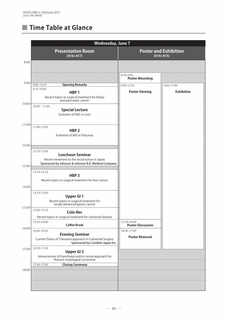
■■ Time■Table■at■Glance
9:00 - 9:10 Opening Remarks
17:40-17:50 Closing Ceremony
9:10-10:00HBP 1
Recent topics in surgical treatment for biliary and pancreatic cancer
10:00 - 11:00Special LectureEvolution of MIS in Liver
8:30-9:00Poster Mounting
9:00-15:35
Poster Viewing
15:35-16:00Poster Discussion
16:00-17:00
Poster Removal
9:00-17:00
Exhibition
11:00-12:00HBP 2
Evolution of MIS in Pancreas
13:10-14:10HBP 3
Recent topics in surgical treatment for liver cancer
14:10-15:00Upper GI 1
Recent topics in surgical treatment for locally advanced gastric cancer
16:00-16:50 Evening Seminar
Current Status of Transanal approach in Colorectal Surgery Sponsored by Covidien Japan Inc.
16:50-17:40Upper GI 2
Advancement of transhiatal and/or cerival approach for thoracic esophageal carcinoma
15:35-16:00Coffee Break
15:00-15:35 Colo-RecRecent topics in surgical treatment for colorectal disease
12:10-13:00Luncheon Seminar
Recent treatment to the rectal tumor in JapanSponsored by Johnson & Johnson K.K. Medical Company
Wednesday, June 7
Presentation Room(416+417)
Poster and Exhibition (414+415)
8:00
9:00
10:00
11:00
12:00
13:00
14:00
15:00
16:00
17:00
18:00
― 10 ―

IASGO-CME in Yokohama 2017June 7th (Wed)
■■ Program
June�7�(Wed),�PACIFICO�Yokohama�Conference�Center
9:00 ~ 9:10 Opening Remarks
9:10 ~ 10:00 HBP 1:Recent topics in surgical treatment for biliary and pancreatic cancer
【Chairs】 Wataru Kimura ( Gastroenterological, General, Breast&Thyroid Surgery, Yamagata University Faculty of Medicine)
Michiaki Unno (Department of Surgery, Tohoku University)
【Speakers】 HBP1-1 Suguru Yamada ( Department of Gastroenterological Surgery, Nagoya University Graduate School of
Medicine)“�Preoperative�treatment�in�locally�advanced�pancreatic�cancer-�Evaluation�of�availability�of�gemcitabine�plus�nab-paclitaxel�-”
HBP1-2 Itaru Endo (Department of Gastroenterological Surgery, Yokohama City University)“�Impact�of� surgical� resection�on� the�survival�of� initially�unresectable�cholangiocarcinoma”
HBP1-3 Satoshi Hirano ( Department of Gastroenterological Surgery II, Hokkaido University Graduate School of Medicine)
“�Conversion�surgery�for�initially�unresectable�pancreatic�cancer”
10:00 ~ 11:00 Special Lecture:Evolution of MIS in Liver
【Chairs】 Hironori Kaneko (Department of Surgery, School of Medicine, Toho University) Go Wakabayashi (Department of Surgery, Ageo Central General Hospital)
【Speakers】 SL-1 Brice GAYET ( Digestive and Oncologic Surgery, Institut Mutualiste Montsouris (IMM) University
Paris Descarte)“Laparoscopic�liver:�Pr�Gayet’s�experience”
SL-2 Ho-Seong Han (Seoul National University Bundang Hospital)“From�the�past�to�current�status�in�laparoscopic�liver�resection”
― 11 ―

IASGO-CME in Yokohama 2017June 7th (Wed)
11:00 ~ 12:00 HBP 2:Evolution of MIS in Pancreas
【Chairs】 Hiroki Yamaue ( Second Department of Surgery, Wakayama Medical University School of Medicine) Katsuhiko Yanaga (Department of Surgery, The Jikei Univeisity School of Medicine)
【Speakers】 HBP2-1 Masafumi Nakamura ( Department of Surgery and Oncology, Graduate School of Medical Sciences,
Kyushu University)“Minimally�invasive�pancreatic�resection�for�cancer”
HBP2-2 Tang Chung-Ngai ( Department of Surgery, Pamela Youde Nethersole Eastern Hospital)“Challenges�of�Robotic�Pancreatectomy”
HBP2-3 Kyoichi Takaori ( Department of Surgery, Kyoto University Graduate School of Medicine)“Laparoscopic�Pancreatic�Resections:�Toward�International�Consensus”
HBP2-4 Kenichi Hakamada ( Gastroenterological Surgery, Hirosaki University)“Robotic�pancreatic�surgery,�an�update”
12:10 ~ 13:00 Luncheon Seminar:Recent treatment to the rectal tumor in Japan� Sponsored�by�Johnson�&�Johnson�K.K.�Medical�Company
【Chair】 Yojiro Hashiguchi (Professor of Department of Surgery, Teikyo University)
【Speakers】 LS-1 Hiroaki Nozawa (Department of Surgical Oncology, The University of Tokyo)
“�Minimally�invasive�approaches�to�gastrointestinal�stromal�tumors�of�the�lower�rectum”
LS-2 Eiji Sunami ( Director of Department of Surgical Oncology, Japanese Red Cross Medical Center)“�Minimally�invasive�approaches�for�the�rectal�cancer�with�fewer�complications.”
13:10 ~ 14:10 HBP 3:Recent topics in surgical treatment for liver cancer
【Chairs】 Kiyoshi Hasegawa ( Hepato-Biliary-Pancreatic Surgery Division, Department of Surgery, Graduate School of Medicine, The University of Tokyo)
Keiichi Kubota (Second Department of Surgery, Dokkyo Medical University)
【Speakers】 HBP3-1 Kyung-Suk Suh ( Department of Surgery, Seoul National University College of Medicine)
“Paradigm�change�in�liver�surgery”
HBP3-2 Susumu Eguchi ( Department of Surgery, Nagasaki University Graduate School of Biomedical Sciences)“Liver�transplantation�for�metastatic�liver�tumors”
HBP3-3 Akinobu Taketomi (Department of Gastroenterological Surgery, Hokkaido University)“Hepatectomy�for�hepatocellular�carcinoma�with�bile�duct�tumor�thrombus.”
― 12 ―

IASGO-CME in Yokohama 2017June 7th (Wed)
HBP3-4 Satoru Imura (Department of Digestive Surgery and Transplantation, Tokushima University)“�Treatment�strategy�for�advanced�hepatocellular�carcinoma�with�macroscopic�portal�invasion”
14:10 ~ 15:00 Upper GI 1:Recent topics in surgical treatment for locally advanced gastric cancer
【Chairs】 Yasuhiro Kodera ( Department of Gastroenterological Surgery, Nagoya University Graduate School of Medicine)
Kazuhiro Yoshida ( Department of Surgical Oncology, Gifu University, Graduate School of Medicine)
【Speakers】 UG1-1 Young-Woo Kim ( Department of Cancer Control and Population Health, Graduate School of Cancer
Science and Policy, National Cancer Center)“�Spade�Shaped�Anastomosis�Following�Proximal�Gastrectomy�Using�Double�Uncut�Stapler�Fixing�Posterior�Wall�of�Esophagus�to�Anterior�Wall�of�the�Stomach�(SPADE�Operation)”
UG1-2 Yasuhiro Kodera ( Department of Gastroenterological Surgery, Nagoya University Graduate School of Medicine)
“Standard�D2�dissection�for�clinically�Stage�II/III�gastric�cancer�of�the�upper�stomach:�An�evidence-based�update”
UG1-3 Kazuhiro Yoshida ( Department of Surgical Oncology, Gifu University, Graduate School of Medicine)“Surgical�treatment�for�adenocarcinoma�of�GEJ”
15:00 ~ 15:35 Colo-Rec:Recent topics in surgical treatment for colorectal disease
【Chairs】 Toshiaki Watanabe ( Department of Surgical Oncology and Vascular Surgery, The University of Tokyo)
Shigeki Yamaguchi ( Department of Colorectal surgery, Saitama Medical University International Medical Center)
【Speakers】 CR-1 Keiji Koda (Department of Surgery, Teikyo University Chiba Medical Center)
“�Recent�progress� in�the�surgical� treatment�of�rectal�cancer�to�achieve�a�better�prognosis”
CR-2 Kazuhiro Sakamoto ( Department of Coloproctological Surgery, Juntendo University Faculty of Medicine)
“�Advanced�techniques�in�the�minimal�invasive�surgery�for�colorectal�cancer�-the�cutting�edge�in�the�quarter�century-”
15:35 ~ 16:00 Coffee Break and Poster Discussion
― 13 ―

IASGO-CME in Yokohama 2017June 7th (Wed)
16:00 ~ 16:50 Evening Seminar:Current Status of Transanal approach in Colorectal Surgery� Sponsored�by�Covidien�Japan�Inc.�
【Chair】 Kimihiko Funahashi (Department of Surgery, School of Medicine, Toho University)
【Speakers】 ES-1 Koji Okabayashi (Department of Surgery, Keio University School of Medicine)
“�TaTME�for�surgery�in�ulcerative�colitis:� technical�challenges�and�future�prospects”
ES-2 Junichi Koike (Department of Surgery, School of Medicine, Toho University)“TaTME�followed�by�Reduced�Port�Surgery�for�lower�rectal�cancer.”
16:50 ~ 17:40 Upper GI 2:Advancement of transhiatal and/or cerival approach for thoracic esophageal carcinoma
【Chairs】 Yasuyuki Seto ( Department of Gastrointestical Surgery, The University of Tokyo, Graduate School of Medicine)
Hisahiro Matsubara ( Department of Frontier Surgery Graduate School of Medicine, Chiba University)
【Speakers】 UG2-1 Yasuyuki Seto ( Department of Gastrointestical Surgery, The University of Tokyo, Graduate School of
Medicine)“�Non-transthoracic�robot-assisted�radical�esophagectomy”
UG2-2 Hitoshi Fujiwara ( Division of Digestive Surgery, Department of Surgery, Kyoto Prefectural University of Medicine)
“Single-port�mediastinoscope-assisted� transhiatal�esophagectomy� for�thoracic�esophageal�cancer”
UG2-3 Hirofumi Kawakubo (Department of Surgery, School of Medicine, Keio Univesrity)“�Thoracoscopic�esophagectomy�with�hybrid�position� �(prone�+�left�lateral�decubitus�position)”
17:40 ~ 17:50 Closing Ceremony
― 14 ―

― Abstracts ―

IASGO-CME in Yokohama 2017June 7th (Wed)
■■ HBP1-1■ June■7■(Wed) 9:10 ~ 10:00
Suguru�YamadaDepartment of Gastroenterological Surgery, Nagoya University Graduate School of Medicine
Education:3/2007 Ph.D. Gastroenterological Surgery, Nagoya University, Graduate School of Medicine, Nagoya, Japan3/1997 M.D. Nagoya University School of Medicine, Nagoya, Japan4/1991 - 3/1997 Nagoya University School of Medicine, Nagoya, Japan
Postdoctoral Training:1/2009 - 12/2010 Research Fellow, Harvard Medical School and Massachusetts General Hospital Cancer Center, Surgical Oncology4/2007 - 12/2008 Clinical Fellow, Gastroenterological Surgery, Nagoya University, Nagoya, Japan4/2004 - 3/2007 Research Fellow, Gastroenterological Surgery, Nagoya University, Nagoya, Japan4/2003 - 3/2004 Chief Resident, General Surgery, Komaki City Hospital, Komaki city, Japan4/1999 - 3/2003 Resident, General Surgery, Komaki City Hospital, Komaki city, Japan5/1997 - 3/1999 Intern, General Training, Komaki City Hospital, Komaki city, Japan
Professional Societies:American Association for Cancer Research, American Society of Clinical Oncology, American College of Surgeons, Japan Surgical Society, Japanese Society of Gastroenterological Surgery, Japan Society of Clinical Oncology, Japan Surgical Association, Japan Pancreas Society, Japan Biliary Association, Japanese Society of Gastroenterology, Japanese Society of Hepato-Biliary-Pancreatic Surgery, Japanese Society of Hepatology, Japan Society for Endoscopic Surgery, Japanese Cancer Association, Japan Gastric Cancer Association
― 16 ―

IASGO-CME in Yokohama 2017June 7th (Wed)
Preoperat ive treatment in local ly advanced pancreat ic cancer- Evaluation of availability of gemcitabine plus nab-paclitaxel -
Suguru Yamada1), Tsutomu Fujii2), Yasuhiro Kodera1)
1) Department of Gastroenterological Surgery, Nagoya University Graduate School of Medicine 2) Department of Surgery and Sciences, Graduate School of Medicine and Phamaceutical Sciences, University
of Toyama
【Background】 In recent years, the advance of chemo or chemo-radiotherapy in the field of pancreatic cancer has gradually improved the survival outcomes. Especially, the introduction of gemcitabine plus nab-paclitaxel therapy against the locally advanced pancreatic cancer since the MPACT trial was reported has been actively taken in the clinics and expected to improve the outcomes as preoperative treatment. The resectability of pancreatic cancer is defined based on the NCCN guidelines and Japan Pancreas Society also advocated simpler criteria in 2016. When our 353 patients with resected pancreatic cancer without preoperative treatment were analyzed, the median survival was 30.8 months in resectable patients, 14.9 in BR-PV, 14.2 in BR-A, and 11.0 in UR-LA. As proposed in the NCCN guidelines, preoperative treatment is recommended in BR disease, whereas, chemo or chemo-radiotherapy is recommended in UR-LA disease. Recently, the concept of “conversion surgery” is advocated and actively conducted against the UR-LA case after chemo or chemo-radiotherapy. However, the details regarding the regimen chosen and optimum duration of chemotherapy, and operative indication are still unclear.
【Results】 In our department, a total of 83 patients underwent gemcitabine plus nab-paclitaxel between 2015 and 2016. The breakdown was as follows; 12 in BR, 16 in UR-LA, 12 in UR-M, 21 in postoperative recurrence, 16 in gemcitabine plus nab-paclitaxel/paclitaxel ip., and 6 in gemcitabine plus nab-paclitaxel/radiation. When the adverse event was investigated limited in the patients with preoperative treatment, the event of grade 3 or more was 52.5% in leucopenia, 75.0% in neutropenia, 20% in anemia, 17.5% in thrombocytopenia, 12.5% in peripheral neuropathy, 20% in grade 2 acomia, and 10% in fever. In 12 patients with BR cancer, disease control rate was 83.3%, resection rate was 75%, and R0 ratio was 77.8%. On the other hand, in 16 patients with UR-LA cancer, disease control rate was 87.5%, resection rate was 31.3%, and R0 ratio was 100%.
【Conclusions】 Gemcitabine plus nab-paclitaxel as preoperative treatment was safe and feasible. Now, the clinical trial is on-going in BR cancer regarding gemcitabine plus nab-paclitaxel vs. FOLFIRINOX as preoperative treatment in our institution. On the other hand, as for the operative indication and timing of conversion surgery for UR-LA cancer, we try to perform the resection for the patient who received the chemotherapy for more than 8 months, had RECIST SD or more, normalization of tumor marker, and PS 0 or more. However, further investigation is required to decide the operative indication of conversion surgery and predictive factor of preoperative treatment.
― 17 ―

IASGO-CME in Yokohama 2017June 7th (Wed)
■■ HBP1-2■ June■7■(Wed) 9:10 ~ 10:00
Itaru�EndoDepartment of Gastroenterological Surgery, Yokohama City UniversityTitle MD, PhD
Education: Yokohama City University School of Medicine, 1, April, 1979 - 31, March, 1985, Doctor of Medicine, Yokohama, Japan
Work experience:1985 Licensed Physician & Board Certification1987 Fujisawa Municipal Hospital, Department of Surgery,1988 Instructer, Teikyo University Mizonokuchi Hospital, Surgery1994 Clinical investigator, UCLA Dumont Liver Transplantation Center 2002 Assistant Professor, Gastroenterological Surgery, Yokohama City University:2006 Research Fellow, Memorial Sloan-Kettering Cancer Center, New York, USA, Hepatobiliary
Service2009 Professor and Chairman, Gastroenterological Surgery, Yokohama City University2016 Deputy director, Yokohama City University Hospital.
Executive Board Member:Japanese Society of Hepato-Biliary-Pancreatic SurgeryJapanese Society of Gastroenterological SurgeryJapan Biliary Association
Councilor:Japan Surgical SocietyJapanese Society of Gastroenterological SurgeryJapan Society of Clinical OncologyJapan Surgical Association
Specialty and Present Interest:HBP surgery, Image-guided surgery, Immunomodulation, Septic DIC & organ failure, Education for young surgeons.
― 18 ―

IASGO-CME in Yokohama 2017June 7th (Wed)
Impact of surgical resection on the survival of initially unresectable cholangiocarcinoma
Itaru Endo
Department of Gastroenterological Surgery, Yokohama City University
Surgical resection of perihilar cholangiocarcinoma remains a challenge for surgeons. In advanced cases, achieving an R0 resection is sometimes difficult because of the anatomic constraints of the hepatic hilum. Due to its anatomical relationship vis-à-vis vasculature in the porta hepatis, en-bloc vascular resection with hepatectomy should be performed for such advanced cases as a radical resection. Currently, portal resection has been rendered standard in right hemihepatectomy with caudate lobectomy. However, arterial resection and reconstruction is still controversial due to high operative mortality and low long-term survival. To achieve R0 resection, it is essential to make a precise preoperative diagnosis of tumor extension using several imaging modalities. Since it is difficult to comprehend three-dimensional structures of the hepatic arteries by conventional 2DCT alone, virtual 3D simulation helps surgeons to achieve R0 resection with minimized risk of postoperative morbidities. Furthermore, advanced cholangiocarcinoma are often accompanied with lymph node metastasis. In such instances, survival after surgical resction alone is unsatisfactory. Therefore, some surgeons have started clinical trials concerning multidisplinary treatment including neoadjuvant chemotherapy. If R0 resection could be achieved, improvement of long-term outcome can be expected even patients who underwent en bloc vascular resection.
― 19 ―

IASGO-CME in Yokohama 2017June 7th (Wed)
■■ HBP1-3■ June■7■(Wed) 9:10 ~ 10:00
Satoshi�HiranoDepartment of Gastroenterological Surgery II, Hokkaido University Graduate School of Medicine
Education:1998 Ph.D. (Dr. of Medical Science), Hokkaido University (Thesis: An experimental study for
anti-infectability of grafts replaced in portal vein)1988 M.D. Hokkaido University School of Medicine, Sapporo, JAPAN
Research and Professional Experiences:Nov.2011-present Professor and Chairman, Department of Gastroenterological Surgery II
(reorganized from Dept. of Surgical Oncology), Division of Surgery, Hokkaido University Graduate School of Medicine
2008-2011 Professor, 2nd Department of Surgery, Hokkaido University Hospital (Concurrent)
2005-2011 Associate professor, Department of Surgical Oncology, Division of Surgery, Hokkaido University Graduate School of Medicine
2005 Overseas clinical study at St James’s University Hospital (HPB and Transplant Services), Leeds, United Kingdom
2003-2005 Instructor, 2nd Department of Surgery, Hokkaido University Hospital2000-2003 Assistant professor, 2nd Department of Surgery, Hokkaido University Hospital1998-2000 Medical Staff in 2nd Department of Surgery Hokkaido University Hospital Beginning the work on the hepatobiliary and pancreatic surgery 1992-1994 Research Fellow, 2nd Department of Surgery, Hokkaido University School of
Medicine1988-1991 Resident in hospitals affiliated with 2nd Department of Surgery, Hokkaido
University Hospital, Sapporo, Japan1988 Passed the Examination of National Board
Major surgical interests: # Surgery for Hepato-biliary and pancreatic neoplasms, chronic disease, and traumatic injury # Acute care surgery
Memberships:1. Fellow of American College of Surgeons (FACS)2. International Hepato-Pancreato-Biliary Association:Active member 3. Asian-Pacific Hepato-Pancreato-Biliary Association:Active member4. International Association for Surgeons and Gastroenterologists:Active member5. Japanese Surgical Society: Director6. Japanese Society of Gastrointestinal Surgery: Councilor 7. Japanese Society of Hepato-Biliry-Pancreatic Surgery: Director8. Japan biliary association: Director9. Japanese Pancreas Society: Councilor10. Japanese society of acute care surgery: Director
― 20 ―

IASGO-CME in Yokohama 2017June 7th (Wed)
Conversion surgery for initially unresectable pancreatic cancer
Satoshi Hirano, Toshimichi Asano, Toru Nakamura
Department of Gastroenterological Surgery II, Hokkaido University Graduate School of Medicine
Recently we have encountered patients with initially unresectable pancreatic cancer (URPC) who survived over a few years owing to newly established effective chemo- or chemoradiotherapy. Conversion surgery (CS) is one of a treatment of choices for patients who could respond to such non-surgical therapies. Although a few case reports for complete remission of advanced pancreatic cancer has been published, most patients with URPC survived very short periods. They could alive 8-10 months in median even administered by brand-new most powerful regimens such as FOLFIRINOX or gemcitabine combined with nab-paclitaxel. The adverse effects caused by therapeutic agents might limit the duration of chemotherapy. Therefore, patients who respond to the non-surgical treatments must be candidates for CS if the change in therapy could be beneficial for the patients. It has been still unknown that who should be conversed to surgical intervention from non-surgical therapy, when the CS should be performed, what kinds of operative procedure should be performed, and how to manage the postoperative patients. Although the CS for URPC is still under investigation, some published data and experiences of our own will be discussed in the lecture.
― 21 ―

IASGO-CME in Yokohama 2017June 7th (Wed)
■■ Special■Lecture-1■ June■7■(Wed) 10:00 ~ 11:00
Brice�GAYETInstitut Mutualiste Montsouris (IMM)University Paris Descartes
Education:Degree in general anatomy and organogenesis in 1977Medical Degree in 1980, Ph D in 1982Professor of Anatomy (1986) and then Professor of Digestive Surgery in 1992
Hospital Appointments:Head of Digestive Diseases Department, Université Paris Descartes,Since 2010, he is a permanent member of ISIR, the robotic institute of University of Paris, associated to CNRS and INSERM.
Profile:He had a diploma in computer science from the Jussieu Faculty of Science at the University of Paris. Dr Gayet was Professor of Anatomy before becoming Professor of Digestive Surgery.Until last year he was member of the French University National Council (CNU) and president of the French Society of Endoscopic Surgery (SFCE). Additionally, Professor Gayet has been involved with health care at the national level by working as a counselor to the ministry of health in France, Bernard Kouchner. He was at the forefront of laparoscopy in the end-1980s. His department - gastroenterology, endoscopy, surgery - has become world renowned in multi-disciplinary disease management that focused on treating patients with complex gastrointestinal (600 proctectomies, 450 esophagectomies by laparoscopy and hepatopancreatobiliary diseases (over 770 hepatectomies and 310 pancreatectomies by laparoscopy).Pr Gayet is the author or coauthor of more than 605 publications on google scholar including over 237 original articles cited more than 7000 times (H index 38). He has given over 800 presentations or lectures in his career and has edited 130 videos on laparoscopic techniques.His last publication includes at least one educative video on any single laparoscopic liver or pancreatic procedure (Laparoscopic Liver, Pancreas, and Biliary Surgery: Textbook and Illustrated Video Atlas, Wiley ed).
― 22 ―

IASGO-CME in Yokohama 2017June 7th (Wed)
Laparoscopic liver: Pr Gayet’s experience
Brice GAYET
digestive and oncologic surgery, Institut Mutualiste Montsouris (IMM) University Paris Descarte
With advancement in surgical techniques, minimally invasive techniques are increasingly being used for liver resection. Historically, the main barriers to the use of laparoscopy in liver surgery were, in addition to the technical complexity of interventions and lack of suitable instrumentation, the risk of air embolism associated with pneumoperitoneum, the difficulty of locating intraparenchymal lesions and difficulties in achieving hemostasis. The development of new techniques and new instruments have gradually allowed the evolution of laparoscopic hepatectomy techniques, allowing major liver resection even as described here in case of IVC, portal or bile involvement. A system to determine the difficulty level for laparoscopic liver resection (LLR) is essential because it is necessary to gradually increase surgical skills according to the experience level before performing technically-demanding procedures. Based on operative time, blood loss and conversion rate, this new three-level stratification system allows a classification of each procedure as low, intermediate, or high difficulty. This practical classification system of technical difficulty may promote education, development, and safe dissemination of LLR. Although most of the cases could be operated laparoscopically, there are very few case reports and series describing thoracoscopic approach for liver tumors especially those located in segment VII, VIII and IVa immediately under the diaphragm. Abdominal approah is possible but should first control the upper part of the liver while a thoracoscopic approach is especially useful in recurrent tumors located in posterosuperior segments as laparoscopic approach is more difficult and time consuming due to dense adhesions. Finally, using many videoclips from our atlas (*) we will try to show how a surgeon should be able to control any type of bleeding including repair of large vessels by a fully laparoscopic approach before moving to advanced procedures.
(*) “Laparoscopic Liver, Pancreas, and Biliary Surgery: Textbook and Illustrated Video Atlas”, Wiley ed.
― 23 ―

IASGO-CME in Yokohama 2017June 7th (Wed)
■■ Special■Lecture-2■ June■7■(Wed) 10:00 ~ 11:00
Ho-Seong�Han,�M.D.,�Ph.DSeoul National University Bundang Hospital
EDUCATION & DEGREES:1978-1984 M.D., Seoul National University College of Medicine1986-1988 M.S., Seoul National University College of Medicine1989-1993 Ph.D., Seoul National University College of Medicine
POSITIONS:1984-1985 Intern, Seoul National University Hospital1985-1989 Resident, Department of Surgery Seoul National University Hospital 1989-1993 Assistant professor, Department of Surgery, Gyeongsang National University College of Medicine 1993-2003 Associate professor & Chairman of Department of Surgery, Ewha Womans University College of Medicine2003- Present. Professor of Department of Surgery Seoul National University College of Medicine2012- 2016 Director of Comprehensive Cancer Center Vice President in Cancer and Neuroscience Seoul National University Bundang Hospital
MEMBERSHIP:President , Korean Study Group of Laparoscopic Liver Surgery (2008- Present)Chairman of Board of Directors , Korean Society of Laparoscopic & Endoscopic Surgeons (2016 – Present)*President , Korean Society of Traumatology (2015- Present)Past President , Korean Study Group of Pancreas Surgery (2012- 2014)Past President , Korean Society of Surgical Metabolism and Nutrition (2014 – 2016) Past Chairman of Board of Directors , Korean Society of Surgical Oncology (2014 – 2016)President of Organizing Committee, PENSA 2018
― 24 ―

IASGO-CME in Yokohama 2017June 7th (Wed)
From the past to current status in laparoscopic liver resection
Ho-Seong Han
Seoul National University Bundang Hospital
With many reports on encouraging outcomes, laparoscopic liver resection has been accepted as attractive alternative for open liver resection. In early ear of laparoscopic liver resection, there has been many limitations on the procedure. First, laparoscopic liver resection has been limited to easily accessible lesions. Second, this procedure is not suitable when tumor is close to major hepatic vein or IVC. Third, anatomic liver resection was still difficult. As the experience with this procedure grows, its prior indications and contraindications can be changed accordingly. Previously, the indications for laparoscopic liver resection have been limited to tumors in the peripheral portion of the antero-lateral segments of the liver. In contrast, lesions in the posterior or superior part of the liver (segments I, VII, VIII and the superior part of IV) are considered by most surgeons to be poor indications for laparoscopic liver resection. With the introduction of flexible endoscopy, high definition imaging and various kinds of equipment, operative field has been much improved. When the tumor is centrally located (tumor is close to major hepatic vein or IVC, performing laparoscopic liver resection has been considered contraindicated due to difficulty in bleeding control, risk of major hemorrhage. Recent development of instrument for parenchymal dissection has made laparoscopic liver resection more safe and meticulous than before. The type of resection also may depend on the remaining liver’s functional capacity. The patients with HCC usually have poor liver function due to chronic liver disease or liver cirrhosis. Therefore it would be recommendable to resect as minimal as possible without jeopardizing oncologic safety. Anatomical liver resection may be advantageous in terms of preserving remaining liver volume and eradicating tumor completely in some cases. Anatomical liver resection can be performed in many ways. Glissonian pedicle approach is one method for anatomical liver.
― 25 ―

IASGO-CME in Yokohama 2017June 7th (Wed)
■■ HBP2-1■ June■7■(Wed) 11:00 ~ 12:00
Masafumi�NakamuraDepartment of Surgery and Oncology, Graduate School of Medical Sciences, Kyushu University
Education:1982-1988 M.D., Faculty of Medicine, Kyushu University, Japan.1995-1999 Ph.D. (Medical Science), Graduate School of Medical Sciences, Kyushu University,
Fukuoka, Japan1999-2001 Research Fellow, Cancer Biology Program, Harvard University
Professional experience:1988-1992 Resident, Dept. Surgery1, Kyushu Univ. and affiliated hospitals, Fukuoka, Japan1992-1995 Resident, National Cancer Center Hospital, Tokyo, Japan2001-2003 The head surgeon, Shin-Kokura Hospital, Kitakyushu, Japan2003-2011 Assistant Professor, (2005-Associate Prof.) Kyushu University2011-2015 Chairperson and Professor, Dept. Digestive Surgery, Kawasaki Medical School,
Kurashiki, Japan2015- Chairperson and Professor, Dept. Surgery and Oncology, Kyushu University
Committee Service:President: Japanese Society for Endoscopic Pancreatic SurgeryBoard of directors: Japanese Society of Hepato-Biliary-Pancreatic Surgery, Japan Society for Endoscopic SurgeryCouncilor: Japan Surgical Association, Japan Society for Biological Therapy, Japan Pancreas Society, Japan Surgical Society, Japanese Society of Gastroenterological SurgeryEditorial Board: Journal of Hepato-Biliary-Pancreatic Sciences, Journal of Japan Society for Endoscopic Surgery, Journal of Laparoendoscopic & Advanced Surgical Techniques (JLAST), International Journal of Clinical Oncology
Prize:Long-term fellowship; Human Frontier Science Program 2000 (Strasburg, France)The most outstanding research paper award in FEBS letter in 2002Grant-in-Aid, Japanese Society of strategies for Cancer Research and Therapy 2004The Best Presentation Award for the Video Presentation, JSS 2013Best Doctor 2014-2015, 2016-2017
Interests:HBP surgery and Laparoscopic surgeryMultidisciplinary therapy of pancreatic cancerIPMN
― 26 ―

IASGO-CME in Yokohama 2017June 7th (Wed)
Minimally invasive pancreatic resection for cancer
Masafumi Nakamura
Department of Surgery and Oncology, Graduate School of Medical Sciences, Kyushu University
First minimally invasive pancreatectomy (MIPR) started as laparoscopic pancreatoduodenectomy (LPD) in 1994. Laparoscopic distal pancreatectomy (LDP) was first performed in 1996. They have gradually evolved during the following decades and is currently utilized for treatment of benign and low-grade malignancies of the pancreas. As for LDP, we performed multicenter comparative study of LDP and open distal pancreatectomy (ODP) using propensity score-matching of more than 2,000 patients in Japan and reported LDP was associated with more favorable perioperative outcomes than ODP, such as lower rates of intraoperative transfusion, clinical grade of pancreatic fistula and morbidity and shorter hospital stay, but a longer operative time. These data showed that LDP is low risk and high return method at least for benign pancreatic tumors. Oncological advantages of LDP for invasive cancers have not been proved by RCT. However, several papers retrospectively revealed that LDP for cancer was technically feasible and oncological outcome was not inferior to that of open method in terms of the rate of R0 resection and the number of dissected lymph nodes. Meanwhile, there are some limitation of LDP for cancer, i.e., positive resection margin after LDP for cancer was more often in low-volume hospitals compared with in high-volume hospitals, open approach was favored for large tumors invading adjacent organs. LPD has reported to be associated with shorter hospital stay and less blood loss compared with open-PD (OPD) and there was no difference in mortality between two methods in high-volume center. In addition, patients who underwent LPD received adjuvant chemotherapy sooner after surgery than patients who underwent OPD. However, these results were limited to high-volume centers in previous reports. The nation-wide risk of mortality rate for patients who underwent LPD was twice as high as that for patients who underwent OPD. Therefore, LPD is best performed at institutions with surgeons who have extensive experience in pancreatic resection and in advanced laparoscopic procedures. Meanwhile, robot assisted procedure is expected to overcome the complicated reconstruction process, which is the largest technical hurdle in LPD. However, this issue is not so much simple because robotic PD needed longer operation time than LPD. A smaller incision and earlier postoperative recovery appear to be potential advantages of MIPR for patients with cancer. However, the safety of this procedure should be ensured before the method can be widely recommended.
― 27 ―

IASGO-CME in Yokohama 2017June 7th (Wed)
■■ HBP2-2■ June■7■(Wed) 11:00 ~ 12:00
Tang�Chung-NgaiDepartment of Surgery, Pamela Youde Nethersole Eastern Hospital, Chai Wan, Hong Kong
Current Appointments:• Consultant Surgeon, Chief of Service• Chief of Hepatobiliary Surgery• Director of Minimal Access Surgery Training Centre (MASTC)• Deputy Hospital Chief Executive of Pamela Youde Nethersole Eastern Hospital• Honorary Consultant Surgeon (Hong Kong Sanatorium)• Honorary Clinical Associate Professor (The Chinese University of Hong Kong)• Honorary Clinical Associate Professor (The University of Hong Kong)• Honorary Professor (Tung Wah College, Hong Kong)• Founding President of the Hong Kong Society of Robotic Surgery• President-elect of Clinical Robotic Surgery Association (CRSA)My clinical excellence in HPB/General Surgery is well exemplified by the following innovations and significant breakthrough in surgical practice, techniques and approachConsultant Surgeon / Chief of Hepatobiliary Surgery / Chief of Service (Surgery):❖ Pioneer of MAS Hepatobiliary Surgery in HK, performed the first laparoscopic Whipple operation in
HK in 2006, the first Robotic-assisted liver resection & Whipple operation in HK in 2009❖ The first qualified Robotic General Surgeon in HK, accumulated personal experience more than 500
cases of Robotic-assisted hepatobiliary surgery / general surgery since 2009, with published series in numerous peer-reviewed journals showing favorable results as compared to open and laparoscopic counterparts
❖ Maintained the biggest series of laparoscopic biliary operation (>300 cases), laparoscopic liver resection (>300 cases) and laparoscopic Whipple operation (>75 cases) in HK
❖ Published extensively on MAS HPB surgery with close to 100 original publications in peer-reviewed journal / book chapter, and delivered about 100 invited lectures on laparoscopic and robot-assisted HPB surgery in local and international symposia
a) Director of Minimal Access Surgery Training Centre (MASTC):❖ Organized training workshops for local/regional doctors and nurses, and more than 15000
healthcare professionals were benefited. Both basic and advanced laparoscopic (General surgery / Urology) surgery courses were recognized as mandatory trainings by College of Surgeons of HK since 2010
❖ Piloted MAS Competence Assessment Model to ensure staff competency in PYNEH, later rolled out and adopted by HA COC (Surgery) and College of Surgeons of HK in 2010
❖ Chairman of MAS Subspecialty Group of COC (Surgery) since 2011❖ Director of numerous International Symposia (IESS, HPB-MAS, Asia-CRSA & IASGO HK Chapter)❖ Convener of International Association of Surgeons, Gastroenterologists & Oncologists (IASGO),
Hong Kong Chapter, organizing post-graduate training courses for local and regional specialists❖ Co-director of Robotic HPB & Upper GI Training Program conducted in Grosetto, Italy since 2011❖ International Faculty of the biggest MAS training Centre in the world (IRCAD/EITS of Strasbourg,
France) since 2010.
― 28 ―

IASGO-CME in Yokohama 2017June 7th (Wed)
Challenges of Robotic Pancreatectomy
Tang Chung-Ngai
Department of Surgery, Pamela Youde Nethersole Eastern Hospital, Chai Wan, Hong Kong
Complex pancreatic surgery remains the hurdle for minimal access surgery because of the technical challenges of controlling hemorrhage from major vessels and reconstructing the biliary and pancreatic ducts with acceptable morbidity. It is one of the most challenging and complex procedures encountered by the general surgeon. Conventional laparoscopic Whipple’s Operation did not gain broad acceptance due to the complexity of the procedure, the accuracy needed to perform the operation, and the steep learning curve required to master the procedures. Robotic surgical systems have been recently introduced to enhance a surgeon’s dexterity in the surgical field through a magnified three-dimensional view, instruments with seven degrees of freedom, and intuitive hand-control movements. Up till now, few data are available comparing a robotic approach to open Whipple’s Operation. Complication and mortality rates are comparable to those of open surgery. However, oncological outcome are lacking in the literature. The current evidence demonstrated that robotic Whipple’s Operation is feasible and safe in selected patients. However, the procedure should be performed by a surgical team expert in pancreatic and laparoscopic surgery in properly selected patients. Larger series and controlled trials comparing robotic and open Whipple’s Operation are needed in order to fully explore these potential advantages.
― 29 ―

IASGO-CME in Yokohama 2017June 7th (Wed)
■■ HBP2-3■ June■7■(Wed) 11:00 ~ 12:00
Kyoichi�Takaori,�M.D.,�Ph.D.,�F.A.C.S.��Secretary General, IASGODirector, Pancreas Cancer UnitKyoto University Hospital
BIOGRAPHY: Dr. Kyoichi Takaori is a pancreatic surgeon who has extensive experiences of open, laparoscopic, and robotic surgery. His academic career includes Professor of Surgery at Asahi University and Assistant Professor of Physiology and Biophysics at University of Arkansas for Medical Sciences. Thorough his career as a surgeon, he has struggled to improve the prognosis of pancreatic cancer, which is known as the worst malignancy. First, he has focused on early detection of pancreatic cancer so that surgeons can offer truly curative operations to the patients. In 2003, with Dr. Ralph Hruban, Dr. Takaori organized International Expert Meeting on Precursor Lesions of Pancreatic Cancer and created international consensus on the classification of pancreatic intraepithelial neoplasia (PanIN) and intraductal papillary mucinous neoplasm (IPMN). World Health Organization has adopted this classification system, which helps investigators better understand the precursor lesions. Furthermore, in pursuit of early diagnosis and treatment of pancreatic cancer in high-risk individuals, he has founded a Japanese Familial Pancreatic Cancer Registry and is promoting collateral studies as the chairman of the registry committee of Japan Pancreas Society. Second, he has endeavored to improve the surgical techniques for pancreatic malignancies. In order to improve local control and to perform more oncologic resections, he has refined artery-first pancreatoduodenectomy and developed new techniques of artery-first distal pancreatectomy, and artery-first DP-CAR by utilizing the “Tora-no-Ana” approach. He is practicing the artery-first approach in all open, laparoscopic and robotic surgery. Third, he is a great believer of multi-disciplinary approach and presently directing the multi-disciplinary team as the Head of Pancreatic Cancer Unit at Kyoto University. Recently, he has conducted international collaborative study on the clinical managements of pancreatic cancer. Last but not least, Dr. Takaori serves patient’s activities on the Medical Advisory Board for PanCAN Japan and the Scientific Advisory Board for Pancreatic Cancer UK. He is contributing to scientific journals as the Vice Editor-in-Chief of Pancreatology and Consultant Editor of Digestive Surgery, the official journal of the International Association of Surgeons, Gastroenterologists and Oncologists (IASGO). Dr. Takaori has served as the International Coordinator of IASGO Educational & Training Projects along with late Professor Nikolaos Lygidakis. In 2015, he was appointed to the Secretary General of IASGO and is organizing IASGO World Congresses as well as many Postgraduate Courses for the purpose of “Globalization of Medical Knowledge” all over the world.
― 30 ―

IASGO-CME in Yokohama 2017June 7th (Wed)
Laparoscopic Pancreatic Resections: Toward International Consensus
Kyoichi Takaori
Department of Surgery, Kyoto University Graduate School of Medicine
Since 1990’s, laparoscopic approaches have been applied to a number of pancreatic resections in the West and East. Laparoscopic pancreatic resections have been preformed in patients with a variety of disease including chronic pancreatitis, pancreatic trauma, and neoplasms of the pancreas; e.g., insulinoma, mucinous cystic neoplasm, intraductal papillary mucinous neoplasm, etc. Laparoscopic pancreatic resections with en bloc lymph node dissection have also been performed for invasive carcinomas. Nowadays, it became a worldwide consensus that laparoscopic distal pancreatectomies with or without spleen preservation may benefit the patients with reduced postoperative pain, shorter hospital stay, quicker recovery to normal activity and better cosmetic appearances based on retrospective analyses of collective series. Oncologic outcomes including the number of harvested lymph nodes, R0 ratio, and long-term survival after laparoscopic distal pancreatectomy in the setting of pancreatic cancer appear compatible to those after open surgery, if performed by experienced surgeons. In contrast, outcomes of laparoscopic proximal pancreatectomies remain controversial. Although the resection parts of laparoscopic pancreaticoduodenectomy and laparoscopic duodenum-preserving pancreatic head resection are technically feasible, laparoscopic reconstruction after proximal pancreatectomies is not well established yet. In some high-volume centers in US, India , China , Korea and other countr ies have reported large ser ies of laparoscopic pancreaticoduodenectomy with outcomes of mortality rates around 3-5%. According to National Clinical Database (NCD) in Japan, the mortality rate after pancreaticoduodenectomy, mostly by open approaches, is 1.3% and the mortality rate of laparoscopic pancreaticoduodenectomy should be lowered to this level. Besides, according to the data analysis in US, the mortality rate of laparoscopic pancreaticoduodenectomy was significantly higher in low volume hospitals, suggesting it may not be a safe operation during the early phase of learning curve. To justify laparoscopic pancreaticoduodenectomy, it is mandatory to demonstrate not only clinical benefits but also the safety that should not be inferior to open surgery. There have been many attempts to establish the international consensus about the indication, techniques, and managements for laparoscopic and robotic pancreaticoduodenectomy. However, due to lack of high-level evidence and due to the technical challenges in this complicated procedure, the consensus has not been reached yet. It will be most beneficial both for surgeons and for patients if the consensus can show guidelines for those surgeons who start laparoscopic pancreaticoduodenectomy.
― 31 ―

IASGO-CME in Yokohama 2017June 7th (Wed)
■■ HBP2-4■ June■7■(Wed) 11:00 ~ 12:00
Kenichi�Hakamada,�M.D.,�Ph.D.Professor of Surgery, Hirosaki University
EDUCATION:1985 M.D. Hirosaki University School of Medicine, Japan1995 Ph.D. Hirosaki University School of Medicine, Japan
PRESENT APPOINTMENTS:2008- Present Professor and Chairman, Department of Gastroenterological Surgery, Hirosaki University Graduate School of Medicine2010- Present Professor, Department of Pediatric Surgery2008- Present Director, Department of Gastroenterological Surgery, Breast Surgery, and Thyroid
Surgery, Hirosaki University Hospital2012-Present Director, Operative theatre, Hirosaki University Hospital
PROFESSIONAL HISTORY:Internship Nakadoori Hospital, Akita, Japan 1985-1986Residency Okinawa Chubu Hospital, Okinawa, Japan 1986-1988Fellowship Department of Surgery, Hirosaki University 1988-1995Attending staff Assistant Professor of Surgery, Hirosaki University 1995-2001 Lecturer of Surgery, Hirosaki University 2001-2005 Associate Professor of Surgery, Hirosaki University 2005-2008
BOARD CERTIFICATION:Board Certified Surgeon of the Japan Surgical Society 2002-PresentBoard Certified Surgeon in Gastroenterology 2003-PresentBoard Certified Surgeon in Endoscopic Surgery 2005-2009Board Certified Hepatologist of the Japan Society of Hepatology 2005-PresentBoard Certified Transplant Surgeon 2012-Present
SOCIETY MEMBERSHIPS and ADVISORY BOARDS:Japan Surgical Society, Board MemberJapanese Society of Gastroenterological Surgery, Member of the Board of directors, Board member, Japanese Society of Hepato-Biliary-Pancreatic Surgery, Board MemberJapanese Society of the Acute Care Surgery, Member of the Board of directors, Board memberJapan Society for Endoscopic Surgery, Board MemberJapan Society for Transplantation, Board MemberJapanese Liver Transplantation Society, Board MemberJapan Pancreas Association, Board MemberInternational member: IASGO, AHPBA, IHPBA, IPA, ILTS
― 32 ―

IASGO-CME in Yokohama 2017June 7th (Wed)
Robotic pancreatic surgery, an update
Kenichi Hakamada
Gastroenterological Surgery, Hirosaki University
Since the first reports of laparoscopic pancreaticoduodenectomy by Ganger and Pomp and distal pancreatectomy by Cuschieri in 1994, minimally invasive pancreatectomy still remains a challenge because of its technical difficulty and significant postoperative morbidity. Robotic platform is expected to overcome such technical difficulties of laparoscopic approach by providing stable, magnified 3-D visualization and improving the surgeons’ dexterity. During pancreatic resection, fine dissection is always required in complex anatomy, especially in case of malignancy. During reconstruction, fine and multiple anastomoses are mandatory after pancreaticoduodenectomy. However, the use of robot is still quite limited in some institutes. One reason is high cost, and others are several shortcomings of the present robot system. It requires two experienced surgeons, one at console and one at operative field. Some devices, such as curved harmonic scalpels, staples with different cartilage, are not available now. The current status of robotic pancreatectomy is surveyed.
Robotic pancreaticoduodenectomy (RPD) More than 430 cases of RPD are reported in 7 articles from high volume centers with 20 cases/center until 2016. Most centers limit the indication to tumors without need for vascular resection, or without adjacent organ invasion. However, during learning curve, indication has been changing. Mean operative time was around 8 hours. Operative morbidity (30%-70%), Mortality and length of hospital stay ware comparable with laparoscopic and open surgeries. Oncological outcome remains unclear.
Robotic distal pancreatectomy (RDP) There are several meta-analyses describing decreased morbidity and earlier recovery by minimally invasive distal pancreatectomy compared to open approach. As for RDP, about 400 cases are reported in 9 articles from high volume centers. Malignancy includes 20%-80% of all cases. The operative time is longer in RDP except in one institute. Postoperative outcomes of both morbidity and hospital stay are comparable between RDP and Laparoscopic PD.
Despite theoretical advantage of robotic pancreatectomy, the real benefit by the use of robot is still undetermined yet. Anyway, the number of reports is quite limited and the robotic system remains immature. It is too early to draw any conclusion about robotic pancreatectomy. We should continue to challenge to improve the surgical outcome of minimally invasive pancreatic surgery.
― 33 ―

IASGO-CME in Yokohama 2017June 7th (Wed)
■■ Luncheon■Seminar-1■ June■7■(Wed) 12:10 ~ 13:00
Hiroaki�NozawaDepartment of Surgical Oncology, The University of Tokyo
EDUCATION:April 1995-March 1999Ph.D. Surgical Oncology, Graduate School of Medicine, University of TokyoApril 1989-March 1993M.D., School of Medicine, University of Tokyo
PROFESSIONAL WORK EXPERIENCE:April 2017-Associate Professor , Department of Surgical Oncology, University of Tokyo HospitalMarch 2016-Lecturer , Department of Surgical Oncology, University of Tokyo HospitalApril 2008-Assistant Professor , Department of Surgical Oncology, University of Tokyo HospitalApril 2006-General Surgeon, Odaira-memorial Tokyo Hitachi HospitalSeptember 2002-Postdoctoral Scholar , Diabetes Center and Comprehensive Cancer Center, University of California San Francisco, USAApril 2001-Senior Resident , Department of Surgical Oncology, University of Tokyo HospitalApril 1999-Senior Resident , Department of Surgery, Tokyo Post and Telecommunication HospitalJune 1994-Junior Resident , Department of Surgery, Cancer Institute Hospital, June 1993-Junior Resident , Department of Surgery, 1st Department of Surgery, University of Tokyo Hospital
― 34 ―

IASGO-CME in Yokohama 2017June 7th (Wed)
Minimally invasive approaches to gastrointestinal stromal tumors of the lower rectum
Hiroaki Nozawa, Toshiaki Watanabe
Department of Surgical Oncology, The University of Tokyo
Gastrointestinal stromal tumors (GISTs) are malignant neoplasms that predominantly arise from the stomach and small intestine. GISTs of the lower rectum are rare, but they are difficult to be managed as they can frequently invade the adjacent organs in the pelvis and sphincter muscles. Since surgical resection is the mainstay of treatment of GISTs, rectal GISTs are likely to require extensive surgery. Efforts have been made for the purpose of preserving sphincter and urinary functions via at least two approaches. One is neoadjuvant therapy using imatinib, and another is laparoscopic surgery. Imatinib is a selective BCR-ABL1 kinase inhibitor that was originally developed as a therapeutic drug for chronic myeloid leukemia. It is also effective for GISTs by binding c-kit and inhibiting its downstream signal transduction pathway. Imatinib improved the prognosis for patients with unresectable / refractory GISTs, and moreover recent studies demonstrated that preoperative imatinib treatment might be also useful in reducing the tumor size in many GISTs. Laparoscopic surgery has been gaining acceptance in many surgical specialties including colorectal surgery. Previous studies showed that the application of the minimally invasive surgical techniques to the treatment of GISTs of the stomach provides a shorter hospital stay and comparable long-term oncologic outcomes to open surgery. In addition, several reports of laparoscopic resection of rectal GISTs have been published. The combination of these minimally invasive treatments is expected to contribute to a better oncological outcome as well as improved quality of life after surgery for GISTs of the lower rectum.
― 35 ―

IASGO-CME in Yokohama 2017June 7th (Wed)
■■ Luncheon■Seminar-2■ June■7■(Wed) 12:10 ~ 13:00
Eiji�SunamiDirector of Department of Surgical Oncology, Japanese Red Cross Medical Center
Titles and degrees:1990- M.D.2003- Ph.D.
Field of Specialization:Surgical Oncology (Colon and Rectal cancer)
Education:1984-1990 Faculty of Medicine, The University of Tokyo1996-2000 Department of Surgical Oncology, Graduate School of Medicine, The University of
Tokyo
Professional Experience:1990-1991 Resident of Surgery, the University of Tokyo Hospital1991-1992 Associate Surgeon, Kantou Rousai Hospital, Japan1992-1995 Resident-fellow, National Cancer Center Hospital, Japan1995-1996 Surgeon, the First Department of Surgery, the University of Tokyo, Japan2000-2002 Chief Surgeon, Department of Surgery, Yaizu City Hospital, Japan2002-2005 Instructor, Department of Colorectal Surgery and Department of Surgical Oncology,
the University of Tokyo, Japan2005-2007 Research fellow, John Wayne Cancer Institute, USA2007-2010 Instructor, Department of Colorectal Surgery and Department of Surgical Oncology,
the University of Tokyo, Japan2010-2015 Lecturer, Department of Colorectal Surgery and Department of Surgical Oncology,
the University of Tokyo, Japan2015- Director of Department of Surgical Oncology, Japanese Red Cross Medical Center,
Japan
Membership in Medical and Scientific Society: 1) International Society of University Colon & Rectal Surgeons 2) AACR 3) Japan Surgical Society 4) Japanese cancer association 5) Japan Society of Clinical Oncology 6) The Japan Society of Coloproctology 7) The Japanese Society of gastroenterological Surgery 8) The Japanese Society of gastroenterology 9) Japan gastroenterological endoscopy society10) The Japan Society for Endoscopic Surgery
― 36 ―

IASGO-CME in Yokohama 2017June 7th (Wed)
Minimally invasive approaches for the rectal cancer with fewer complications.
Eiji Sunami
Director of Department of Surgical Oncology, Japanese Red Cross Medical Center
The minimally invasive procedure such as laparoscopic surgery for the colorectal cancer has become widespread in Japan. Large scale randomized controlled trial had been conducted to confirm the non-inferiority of laparoscopic surgery to open surgery for colon cancer in Japan. According to this trial, laparoscopic surgery was associated with less blood loss, a shorter time to pass first flatus, decreased use of analgesics, shorter hospital stay, and lower morbidity rate. 5-year overall survival was about the same. Technical feasibility of laparoscopic surgical procedure for rectal cancer including TME, lateral pelvic lymph node dissection, and ISR has also been shown by other trials in Japan. Based on these backgrounds, more than half of the colorectal cancer patients has been treated by laparoscopically. However, several procedures such as lateral pelvic lymph node dissection and lower rectal cancer surgery are sometimes very difficult to perform laparoscopically, especially in cases of patients with narrow pelvis or obesity. To achieve totally minimally invasive therapy for the colorectal cancer patients, both intraoperative minimally invasive approach such as laparoscopic surgery and perioperative fewer complications play very important roles. Anastomotic leakage is one of the most undesirable post-operative complications of colorectal surgery. The incidence of anastomotic leakage is reported to reach 1.7 % in colonic cancer surgery and 9.7 % in low anterior resection according to the Japanese large scale database (National clinical database). Anastomotic leakage may cause higher incidence of local recurrence and poor survival and may impair function and quality of life after sphincter-saving operative intervention for rectal cancer. Risk factor analyses for anastomotic leakage has been widely reported. The knowledge of these risk factors may support procedure-related decisions and may help to reduce the leakage rate. In this presentation, laparoscopic procedure for rectal cancer and technics to reduce anastomotic leakage will be presented.
― 37 ―

IASGO-CME in Yokohama 2017June 7th (Wed)
■■ HBP3-1■ June■7■(Wed) 13:10 ~ 14:10
Kyung-Suk�SuhDepartment of SurgerySeoul National University College of MedicineSeoul National University Hospital
Prof. Kyung-Suk Suh is Professor of Department of Surgery, Seoul National University College of Medicine.Prof. Suh graduated from Seoul National University College of Medicine with his medical degree in 1984 and completed his internship and residency in Department of Surgery at Seoul National University Hospital, receiving his diploma in General Surgery in 1989.Since 1993, Prof. Suh held a number of professional positions at the Seoul National University Hospital in Seoul, including instructor, assistant Prof., associate Prof., Prof., chairman of Department of Surgery Seoul national university College of Medicine and Chief of Seoul National University Hospital Transplant Center. Since 2011, Prof. Suh have been taking a number of roles, including Director of the International affairs in Korean Society of Organ Transplantation (2011-), , Chairman in Korean Association of HBP Surgery(2015-), President of the International Living Donor Liver Transplantation Study Group(2015-) and Chairman of Korean Surgical Society(2016-).Prof. Suh’s major fields of interest are Liver Transplantation, Oncological Surgery for Hepato-biliary Carcinoma.
― 38 ―

IASGO-CME in Yokohama 2017June 7th (Wed)
Paradigm change in liver surgery
Kyung-Suk Suh
Department of Surgery, Seoul National University College of Medicine
The rationale for anatomic resection is based on the assumption that the hepatocellular carcinoma (hcc) invades the nearby portal vein branches and makes daughter nodules. So the anatomic resection means the resection of the tumor-bearing portal territory. There have been several reports showing the more favorable prognosis in patients underwent anatomic resection than those underwent non-anatomical resection. But still, there are controversies about this procedure. There are several technical misconceptions in the anatomic resection. For example, in the anterior sectionectomy, some surgeons emphasize that the right hepatic vein and the middle hepatic vein should be fully visualized after resection. However, the concept of the anatomical resection is based on portal tributaries not on hepatic veins. And even smaller resection around the tumor would be done according to the concept of the anatomic resection. Many patients with hcc have cirrhosis and so larger volume of resection sometimes causes poor prognosis. And rapid regeneration due to large volume of resection may be a risk factor for tumor growth. Another consideration in HCC resection is the effect of hypoxia, congestion and inflammation of the liver on HCC. Several reports showed the hypoxia, congestion and inflammation are risk factors of tumor recurrence. With technical advancement of laparoscopic device such as 3 dimensional and flexible scope, the laparoscopically unfavorably located, such as segment 7 and 8, tumors could be laparoscopically resected recently. In Living donor hepatectomy is now well established surgical procedure. However, a large abdominal incision is still required especially for a right liver graft. The prospect of this large incisional scar may make some live donors and even surgeon reluctant to undergo the procedure due to concerns about self-image; this may be especially true for young women. We started to perform minimal incisional donor right hepatectomy assisted by laparoscopy or using hand assisted device first. But now, pure donor laparoscopic technique is exclusively used in my center. In conclusion, in hepatic resection for HCC, anatomic resection is important but also the amount of resection, ischemia and congestion after resection should be considered. And so “functional resection” rather than “anatomical resection” would be the better approach. And the laparoscopic hepatic resection could be done irrespective of tumor location. Futhermore, pure laparoscopic donor hepatectomy is the standard procedure in my center.
― 39 ―

IASGO-CME in Yokohama 2017June 7th (Wed)
■■ HBP3-2■ June■7■(Wed) 13:10 ~ 14:10
Susumu�Eguchi,�M.D.,�Ph.D.,�F.A.C.S.,�F.E.B.S.Professor and ChairmanDepartment of Surgery, Nagasaki University Graduate School of Biomedical Sciences
Education:Medical School Nagasaki University School of Medicine, Nagasaki, Japan, Graduated in 1992Post-graduate School Nagasaki University Graduate School of Biomedical Sciences, Department of Surgery, PhD course,
4/1994-3/1998
Medical Training & Professional career:Nagasaki University Hospital, Department of Surgery II, 6/1992-4/1993Nagasaki Municipal Hospital, Department of Surgery, 11/1993-3/1994Cedars-Sinai Medical Center, Department of Surgery, Surgical Research, 6/1994-3/1997 Nagasaki Memorial Hospital, Department of Surgery, 1/1999-3/1999National Tsushima Hospital, Department of Surgery, Chief 4/1999-1/2000Nakatsushima Hospital, Department of Surgery, Chief 2/2000-3/2000Prefectural Shimabara Onsen Hospital, Department of Surgery4/2000-3/2001Groningen University Hospital, Department of Surgery, Liver Transplantation and Hepatobiliary Surgery Clinical fellow, 4/2003-3/2005Nagasaki University Graduate School of Biomedical Sciences, Department of Surgery, Professor 1/2012 presentGuangzhou First Municipal People’s Hospital, Visiting professor 8/2012-presentSyzganovs’ National Scientific Centre of Surgery, Almaty, Kazakhstan, Professor emeritus 2013-presentNagasaki University hospital, Deputy hospital director10/2013-3/2016Nagasaki University hospital, International Medical Center director 4/2016-The Second Affiliated Hospital to Nanchang University, Professor emeritus 8/2016-
Career position:< Editorial board >SURGERY TODAY (Associate editor, 2007-present)HEPATOLOGY RESEARCH (2008-present)Investigative Applied Medicine and Science (2009-present)Journal of Hepato-Biliary-Pancreatic Science (-present)International Journal of Clinical Oncology (IJCO) (2016-present)< Certification >Course for multi-organ procurement procedure, Leiden, The Netherlands (12/16, 2003)Assesser of objective structured clinical examination (OSCE)(2006-present)The Society for Testing English Proficiency (STEP) Practical English Skill, 1st degree DiplomaThe European Diploma in Transplantation Surgery for Multi- organ retrieval and Liver Transplantation.(Organized by the European Union of Medical Specialists/UEMS, the European Board of Surgery/EBS and the European Society of Organ Transplantation/ ESOT)
― 40 ―

IASGO-CME in Yokohama 2017June 7th (Wed)
Liver transplantation for metastatic liver tumors
Susumu Eguchi, Takanobu Hara, Mitsuhisa Takatsuki, Akihiko Soyama, Masaaki Hidaka
Department of Surgery, Nagasaki University Graduate School of Biomedical Sciences
Liver transplantation for metastatic neuroendocrine tumor (NET) in the liver used to be the main reason for liver transplantation for metastatic liver tumor. It is reported that liver transplantation in selected patients with nonresectable metastatic NET in the liver had favorable outcomes equivalent to liver transplantation for hepatocellular carcinoma. Recently, liver transplantation for colorectal liver metastasis (CRLM) has recently attracted attention. According to SECA study, liver transplantation for non-resectable CRLM had a high rate of recurrence but a favorable prognosis. Further discussion on patient selection, attempts at immunosuppressive therapy, and combination with chemotherapy and treatment at the time of relapse is required in order to improve the outcomes of liver transplantation for metastatic liver tumor.
― 41 ―

IASGO-CME in Yokohama 2017June 7th (Wed)
■■ HBP3-3■ June■7■(Wed) 13:10 ~ 14:10
Akinobu�Taketomi,�MD,�PhD.ProfessorDepartment of Gastroenterological Surgery IGraduate School of Medicine, Hokkaido University
EDUCATION: 1996 PhD Kyushu University1990 MD Kyushu University
PROFESSIONAL APPOINTMENTS AND RESEARCH EXPERIENCE: November 2011 – Present Professor, Department of Gastroenterological Surgery I, Hokkaido UniversityApril 2003 – October 2011 Assistant professor, Department of Surgery II, Kyushu University HospitalApril 2001 - March 2003 Chief surgeon, Department of Surgery, Nakatsu Municipal HospitalSeptember 1998 - March 2001 Post-doctoral research fellow, Huntsman Cancer Institute, University of Utah, USAApril 1996 - August 1998 Fellow, National Kyushu Cancer Center (Department of Digestive Organs)April 1991 - March 1992 Resident, Hiroshima Red Cross Hospital & Atomic-bomb Survivors Hospital June 1990 - March 1991 Resident, Department of Surgery II, Kyushu University Hospital
PROFESSIONAL AFFILIATIONS AND ACTIVITIESInternational Liver Transplant CongressInternational Hepato-Pancreato Biliary AssociationJapan Surgical SocietyJapanese Society of Gastroenterological SurgeryJapanese Society of Hepato-Biliary-Pancreatic SurgeryThe Japan Society of HepatologyJapanese Cancer AssociationJapan Society of Clinical OncologyJapan Surgical AssociationJapan Society of Medical Oncology
― 42 ―

IASGO-CME in Yokohama 2017June 7th (Wed)
Hepatectomy for hepatocellular carcinoma with bile duct tumor thrombus.
Akinobu Taketomi, Tatsuya Orimo, Hideki Yokoo, Hirofumi Kamachi, Toshiya Kamiyama
Department of Gastroenterological Surgery, Hokkaido University
<Backgrounds> This study aimed to evaluate the short- and long-term outcomes of hepatectomy for hepatocellular carcinoma (HCC) with bile duct tumor thrombus (BDTT), including cases with obstructive jaundice. <Methods> The study reviewed 42 HCC patients with BDTT (21 microscopic and 21 acroscopic BDTT cases), including six patients who needed preoperative biliary drainage due to obstructive jaundice, and 732 HCC patients without BDTT. The authors analyzed the impact of BDTT on the surgical outcomes and assessed the outcomes of hepatectomy for patients presenting with obstructive jaundice. <Results> The HCC patients with BDTT, almost all with stage 3 or 4 disease, had increased alpha-fetoprotein expression, larger tumors, and more portal vein invasion status compared to that of non-BDTT group. The survival of the HCC patients with BDTT was significantly inferior to that of the patients without BDTT (p = 0.0003). Survival did not differ significantly between the HCC patients with BDTT and those without BDTT when the two groups were matched by stage (p = 0.3366). The HCC patients with BDTT who presented with obstructive jaundice demonstrated outcomes similar to those for the HCC patients with BDTT who did not present with obstructive jaundice in terms of the overall survival rate (p = 0.5469). The perioperative outcomes for the HCC patients with BDTT did not depend on the presence or absence of preoperative jaundice. No patients in either BDTT group demonstrated 90-day mortality in this study. <Conclusions> Hepatectomy should be considered for HCC patients with BDTT, even for patients with obstructive jaundice, because the surgical outcomes equivalent to those for HCC without BDTT can be achieved.
― 43 ―

IASGO-CME in Yokohama 2017June 7th (Wed)
■■ HBP3-4■ June■7■(Wed) 13:10 ~ 14:10
Satoru�ImuraDepartment of Digestive Surgery and Transplantation, Tokushima University
EDUCATION:Degrees/ Diplomas/ Licensures and Certifications1997 M.D. Tokushima University, School of Medicine2004 Ph.D. Tokushima UniversityPROFESSIONAL TRAINING and EMPLOYMENT: a) Academic Appointment 2004-2013 Assistant Professor, Department of Surgery Tokushima University, Tokushima, Japan 2014- Project Professor, Department of Minimum invasive and Telesurgery Tokushima University Hospital, Tokushima Japan b) Previous Appointments 1997-1998 Resident, Department of Surgery Tokushima University Hospital, Tokushima, Japan 1998-1999 Medical Staff, Department of Surgery, Takamatsu Municipal Hospital, Takamatsu, Japan 1999-2000 Medical Staff, Department of Surgery Shikoku Chuo Hospital, Shikoku chuo, Japan 2000-2001 Medical Staff, Department of Surgery Anan Kyoei Hospital, Naka, Japan 2001-2004 Fellow, Department of Surgery Tokushima University Hospital, Tokushima, Japan c) Clinical Experience Discipline: General Surgery Specialty: Liver SurgeryPROFESSIONAL AFFILIATIONS AND ACTIVITIES:Japan Surgical SocietyThe Japanese Society of Gastroenterological SurgeryThe Japan Society of HepatologyThe Japanese Association of Hepato-Biliary-Pancreatic SurgeryThe Japanese Society of GastroenterologyJapan Society for Endoscopic SurgeryThe Japan Society of Clinical OncologyJapanese Cancer AssociationThe Japan Society for TransplantationAmerican College of SurgeonsEuropean Society for Organ TransplantationInternational Association of Surgeons, Gastroenterologists and Oncologists etc.BOARD CERTIFICATION:Japan Surgical SocietyThe Japanese Society of Gastroenterological SurgeryThe Japan Society of HepatologyThe Japanese Association of Hepato-Biliary-Pancreatic SurgeryThe Japanese Society of GastroenterologyJapan Society for Endoscopic SurgeryFellow of American College of Surgeons
― 44 ―

IASGO-CME in Yokohama 2017June 7th (Wed)
Treatment strategy for advanced hepatocellular carcinoma with macroscopic portal invasion
Satoru Imura, Mitsuo Shimada
Department of Digestive Surgery and Transplantation, Tokushima University
Background: Treatment strategy for advanced hepatocellular carcinoma (HCC) remains unclear. We have reported the anti-tumor effects of IFN α on invasion, proliferation, in vitro and vivo (J Surg Res 2012), and the effect of our systemic adjuvant IFP therapy consisting of IFN, 5FU and CDDP after hepatectomy (Hx) on advanced HCC with portal vein tumor thrombus (PVTT) in major branch or first branches. (HGE 2008).Aims: To evaluate the effect of (i) IFP after hepatectomy (Hx), (ii) conversion Hx after HAIC, (iii) sorafenib for for advanced HCC with PVTT in major branch or first branches (Vp3 or more).Methods: (i) Sixteen patients who had HCC with Vp3 or more were retrospectively divided into two groups: control (n=8) and IFP group (n=8), in which one cycle of IFN and systemic intravenous administration of 5FU and CDDP was done as soon as possible after surgery. (ii) Conversion Hx after HAIC was performed (initially unresectable). (iii) Sorafenib therapy was introduced for 38 cases. Among them, 9 cases were HCC with Vp3 or more.Results: (i) The OS in IFP group was higher than in control group (1year: 100% vs. 0%, 3 year: 88% vs. 0%), Regarding the DFS, the DFS in IFP group was also significantly better than control group (1year: 50% vs. 0%, 2 year: 50% vs. 0%). Regarding the recurrent patterns, in the IFP group, 60% patients had controllable recurrent tumors (<3 nodules) in the remnant liver, although all patients with recurrence had uncontrollable recurrence in the control group. (ii) Lobectomy and PVTT removal was done for all cases. Almost all tumor or PVTT were revealed pathologically non-viable. Although follow-up period was short, new lesion was not observed yet. (iii) OS in patients with Vp3 or more was significantly worse than that in PVTT negative group. All patients categorized PD after evaluation in early period.Conclusions: Sorafenib could not control HCC with Vp3 or more. Our adjuvant IFP therapy after radical Hx may improve survival of HCC with macroscopic portal invasion. In preceding HAIC patients, the timing can be performed radical Hx should be checked.
― 45 ―

IASGO-CME in Yokohama 2017June 7th (Wed)
■■ Upper■GI■1-1■ June■7■(Wed) 14:10 ~ 15:00
Young-Woo�Kim�MD,�PhD,�FRCS,Department of Cancer Control and Population Health, Graduate School of Cancer Science and Policy, National Cancer Center
Education:1988 Seoul National University College of Medicine1992 Seoul National University Postgraduate School, M.Sc1998 Seoul National University Postgraduate School, Ph DFaculty Appointments:1996.5-1997.8 Fellow, Department of General Surgery, Ewha Womans University Mokdong Hospital1997.9-1999.8 Instructor, Department of General Surgery College of Medicine, Ewha Womans University1999.9-2002.8 Assistant Professor, Department of General Surgery College of Medicine, Ewha Womans
University2002.9- staff, Center for Gastric Cancer, National Cancer Center2006. 3- Chief Scientist, Gastric Cancer Branch, Division of Common Cancers, Research Institute,
National Cancer Center2006.8-2012.1. Head, Center for Gastric Cancer, National Cancer Center2007.12-2010.1 Chief, Gastric Cancer Branch, Research Institute2007.12-2009.1 Head, Division of Translational & Clinical Research I2008.12-2010.1 Chief, Gastric Cancer Branch, Research Institute, National Cancer Center2012.1 – present2009.8 – 2015.2 Chief, Department of Surgery, Hospital, National Cancer Center2014.3.1- present Professor. Department of Cancer Control and Policy, Graduate School of Cancer Science
and PolicyResearch & Training:1997 Department of Surgery, - University of Maryland, Baltimore, USA2008.1-2 TU Munchen, The Royal Marsden Hospital, Hospital Ambroise Pare,S. Camillo Hospital2014 6-8 Honorary Visiting Professor, Imperial College of LondonAwards:2005 The Best Poster of the Session Prize, 6th International Gastric Cancer Congress2008 Young Surgeons Award, Seoul National University Department of Surgery Alumni Association2008 Excellent achievement Award, Strategic Subject, Ministry of Knowledge Economy2009 Excellent Investigator Award, Korean Cancer Association2011 A medal for distinguished services at 10th Anniversary of NCC, Award of Minister of Health and Welfare 2011 Karl Storz EAES Award (Best of EAES), 19th EAES Meeting2015 Excellent Investigator Award (National Cancer Center)2016 Niall O’Higgins Best Proffered Paper Award (ESSO 36 Congress)2016 Outstanding Research Award, Minister of Health and Welfare of KoreaSocial Activities:Chair Korean Robotic Gastric Surgery Study Group(KRSG)Chair Multidisciplinary Treatment Committee, Korean Society of Clinical Oncoogy (KSCO)Chair Information Committee, Korean Gastric Cancer Association (KGCA) Chair Treasurer, Korean Association of Robotic Surgery (KAROS)President Korean Society of Patient Blood Management (KPBM)President 2018 International Society of Medical innovation and Technology (iSMIT)member Research Committee, European Association of Endoscopic Surgery and other interventional
techniques (EAES)
― 46 ―

IASGO-CME in Yokohama 2017June 7th (Wed)
Spade Shaped Anastomosis Following Proximal Gastrectomy Using Double Uncut Stapler Fixing Posterior Wall of Esophagus to Anterior Wall of the Stomach (SPADE Operation)
Young-Woo Kim
Department of Cancer Control and Population Health, Graduate School of Cancer Science and Policy, National Cancer Center
Optimizing operative extent of gastrectomy according to the disease status and preservation of gastric function to improve quality of life of the gastric cancer patient is more and more important as proportion of early gastric cancer has been increased up to 70 % in Korea and japan and enormous development of minimally invasive surgery. For the proximally located early gastric cancer, still lots of surgeons continue to do total gastrectomy due to lack of conviction of value of proximal gastrectomy and relenting reflux and stricture after operation. Main concern is how to prevent reflux in reconstruction of esophagus and remnant stomach. Recent systemic review concluded that esophagogastrostomy with addition of anti-reflux procedures. Simple esophagogastrostomy should be avoided and other complicated procedures like jejunal interposition or double tract anastomosis are not recommended because there are limitations in terms of nutritional benefit, morbidities, operative time, cost, and difficulty in surveillance of remnant stomach, etc. We have experienced several kinds of anastomosis and devised new innovative reconstruction which fulfils essential points to prevent reflux. We thought 1) his angle 2) intraabdominal anastomosis 3) sphincter action or peristaltic anti-reflux mechanism and basically esophagogastrostomy to ensure direct 100 % food passage into normal physiologic path of food and complete surveillance. Mainly inspired by Kamikawa’s procedure, we could invent new surgery of SPADE operation, whicj resembles shape of spade and comprised of esophagogastropexy with double uncut staplers and hand sewn esophagojejunostomy. We report only limited number of patients but their clinical outcome is outstandingly excellent. No patients complaints any reflux symptoms and quality of life is good. There must be further research and development for proximal gastrectomy and proper reconstruction. We propose this SPDE operation should be explored with multicenter clinical trail to prove benefits and pitfalls.
― 47 ―

IASGO-CME in Yokohama 2017June 7th (Wed)
■■ Upper■GI■1-2■ June■7■(Wed) 14:10 ~ 15:00
Yasuhiro�Kodera,�MD,�PhDDepartment of Gastroenterological Surgery, Nagoya University Graduate School of Medicine
Education:Graduated Nagoya University, School of Medicine in 1985, and underwent training at Komaki Municipal Hospital (Aichi, Japan), Cancer Center Hospital (Tokyo, Japan), and Nagoya University Hospital (Aichi, Japan)
Licensure and Certification: 1985 Japanese Medical License, Registration No. 288476 1990 Certified by Japan Surgical Society, Certificate Number 5281 1992 Certified by Educational Commission for Foreign Medical Graduates, Certificate Number 430-788-0 1994 Ph. D., Nagoya University School of Medicine 1996 Certified by the Japanese Society of Gastroenterological Surgery Certificate number 399 2009 Fellow of the American College of Surgeons (F.A.C.S)
Hospital Appointments: 1994/6~2001/12 Consulting Surgeon at Department of Gastroenterological Surgery, Aichi Cancer Center, Aichi, Japan 2002/1 Teaching Staff at the Department of Gastroenterological Surgery, Nagoya University Graduate School of Medicine 2011/8 Current position
Board of Directors:Japan Surgical SocietyJapanese Gastric Cancer AssociationJapanese Society of Clinical Oncology
Associate Editor:Gastric Cancer
Editorial Board Member:Japanese Journal of Clinical Oncology, World Journal of Surgery, European Journal of Surgical Oncology
― 48 ―

IASGO-CME in Yokohama 2017June 7th (Wed)
Standard D2 dissection for clinically Stage II/III gastric cancer of the upper stomach: An evidence-based update
Yasuhiro Kodera
Department of Gastroenterological Surgery, Nagoya University Graduate School of Medicine
In D2 dissection for gastric cancer of the upper-third stomach, the standard procedure is total gastrectomy with D2 dissection, and splenectomy had been mandatory for complete dissection of lymph nodes at the splenic hilum and along the distal part of the splenic artery. Splenectomy is known to be the cause of higher morbidity, which often led to mortality in the British and Dutch D1 versus D2 trial in the 1990’s. As a consequence, surgeons in the West eventually abandoned splenectomy while the procedure continued to be the standard in Japan. The Japan Clinical Oncology Group (JCOG) conducted a randomized trial, JCOG0110, in which 505 patients were accrued to clarify survival benefit of splenenectomy for cancer of the upper-third stomach excluding tumors that are located on the major curvature. Splenectomy was associated with higher morbidity and larger blood loss, but the operation time was similar. The 5-year survivals were 75.1% and 76.4% in the splenectomy and spleen preservation groups, respectively. The hazard ratio was 0.88 (90.7%, confidence interval 0.67-1.16) (<1.21); thus, the noninferiority of spleen preservation was confirmed (P = 0.025). This trial became the only trial to statistically prove non-inferiority of spleen-preserving gastrectomy. In addition, bursectomy had often been performed in conjunction with D2 dissection, in part to remove micrometastases within the bursa omentalis. This had been considered particularly important to treat T3/T4 gastric cancer located on the posterior wall. The Japanese surgeons gradually abandoned this time-consuming procedure, but a small-scale randomized trial conducted by a study group in Osaka suggested benefit of bursectomy. This led JCOG to conduct a large-scale (N=1,204) confirmatory randomized trial, JCOG1001, to prove superiority of D2 dissection with bursectomy versus D2 without bursectomy. Bursectomy resulted in greater amount of blood loss and higher incidence of pancreatic fistula but was not associated with significantly superior survival rate (HR: 1.07, 95% CI: 0.81~1.42). Subset analysis revealed no benefit for cancers located on the posterior wall or with serosal invasion. These phase III trials justified “modified D2” in which both splenectomy and bursectomy are omitted, and an omentectomy versus non-omentectomy trial could be one of candidates for the forthcoming phase III trial. Minimally invasiveness and low morbidity so as to move on to the adjuvant therapies without untoward delay has grown to be of greater importance in the current landscape of gastric cancer surgery.
― 49 ―

IASGO-CME in Yokohama 2017June 7th (Wed)
■■ Upper■GI■1-3■ June■7■(Wed) 14:10 ~ 15:00
Kazuhiro�YoshidaProfessor and Chairman Department of Surgical Oncology, Gifu University, Graduate School of MedicineDirector of Cancer Center, Gifu University Hospital
Education: 1984 M.D. Hiroshima University, School of Medicine 1990 Ph.D. Graduated from Postgraduate Research Institute, Hiroshima University
Professional training and Employment:1984.4. – 1985.3. Resident in Surgery, Hiroshima University 1985.4. – 1987.3. Medical Staff in Surgery, Matsuyama Red Cross Hospital, Matsuyama1987.4. – 1990.3. Postgraduate student of Hiroshima University1990.4. – 1991.3 Research Associate at the Department of Pathology, Hiroshima University
School of Medicine1991.4. – 1992.3. Research fellow at the Department of Surgical Oncology, Research Institute for
Radiation Biology and Medicine1992.4. – 2002.6. Research Associate at the Department of Surgical Oncology, Research Institute
for Radiation Biology and Medicine, Hiroshima University(1994.7. – 1996.6. Research Scientist at John Radcliffe Hospital, University of Oxford, UK. )2002.6. – 2007.7. Assistant Professor at the Hiroshima University Hospital2007.8. – present Professor and Chairman Dept. Surgical Oncology, Gifu University, Graduate
School of Medicine2008.4. – present Director of Cancer Center, Gifu University Hospital
Congress President:・President, the 15th Annual Meeting of the Japanese Society of Gastrointestinal Surgery, JDDW 2017・President elect, the 57th Annual Meeting of the Japan Society of Clinical Oncology 2019
Council member: ・Japanese Society of Gastrointestinal surgery (Board of Directors, Chair of International Committee)・Japan Society of Clinical Oncology (Board of Directors, Chair of International Committee)・ Japanese Gastric Cancer Association (Board of Directors, Chair of research promotion committee,
member of International Committee)・Japan Digestive Disease Week (JDDW) (Board of Directors, member of International Committee)・Japan Surgical Society (Member of International Committee)・ Japanese Society of endoscopic surgery ・ Japanese Cancer Association・ Japanese Esophageal Society ・ Japanese Society of Gastrointestinal Carcinogenesis (Board of Directors)・ Japanese Gastroenterological Association・ Japan Surgical Association ・ Japanese Society of BRM
― 50 ―

IASGO-CME in Yokohama 2017June 7th (Wed)
Surgical treatment for adenocarcinoma of GEJ
Kazuhiro Yoshida
Department of Surgical Oncology, Gifu University, Graduate School of Medicine
Japanese guide line for the treatment of gastric cancer has demonstrated the standard treatment strategy as follows; early gastric cancer (GC) can be treated with ESD or laparoscopic surgery, while patients with advanced cancer including stage II and III, open surgery+D2 LN dissection followed by S-1 administration for one year is recommended. Laparoscopic resection is still under clinical trial. The major two topics of under discussion at present are the treatment of gastro-esophageal junction (GEJ) cancer and conversion therapy on stage IV gastric cancer. Siewert type I tumor is treated as esophageal cancer and type III as gastric cancer. However, in type II, there still exist controversy concerning the extent of LN dissection and reconstruction methods. According to the nationwide survey, tumors less than 4 cm in type II carcinoma, metastasis to LN No 4, 5 6 are very rare and total gastrectomy is not necessary oncologically. However, reconstruction method is another topic because reflux esophagitis is one of the main concern after proximal gastrectomy due to adenocarcinoma of GEJ. Recent topics will be presented in the session.
― 51 ―

IASGO-CME in Yokohama 2017June 7th (Wed)
■■ Colo-Rec-1■ June■7■(Wed) 15:00 ~ 15:35
Keiji�KodaProfessor Department of SurgeryTeikyo University Chiba Medical Center
Education: 1978-84: Chiba University, School of Medicine, Japan
Postgraduate Training: 1984-1985: Resident in Surgery, Chiba University Hospital, Chiba, Japan 1985-1988: Medical Staff in Surgery, Chiba Municipal Hospital, National Narashino Hospital,
National Shimoshizu Hospital, Chiba, Japan1988-1989: Research worker, Gastrointestinal Immunology, State University of New York, Stony
Brook, NY 1989: Research worker, Immunology, Scripps Clinic, San Diego, CA1989-1990: Research Fellow, Cancer Immunology and Surgery, University of California, San
Diego, CA1988-1990: Research worker, Brunswick Biotechnetics. Inc., San Diego, CA1990-1998: Medical Staff, Department of General Surgery, Chiba University Hospital, Chiba,
Japan 1998-2000: Associate Professor of Medicine, Department of Surgery, Faculty of Medicine,
Tsukuba University, Tsukuba, Japan2000-2006: Associate Professor of Medicine, Department of Academic Surgery, Chiba University,
Chiba, Japan2006-present: Professor and Chairman, Department of Surgery, Teikyo University Chiba Medical
Center, Chiba, Japan
Board member:Japan Society of Coloproctology(JSCP)Japanese Society of Stoma and Continence Rehabilitation(JSSCR; director general) International Society of University Colon and Rectal Surgeons(ISUCRS)
Membership: Japan Surgical Society, Japan Surgical Association, The Japanese Society of Gastroenterological Surgery, Japan Society of Clinical Oncology, Japan Society for Biological Therapy, Japan Gastroenterological Endoscopy Society, The Japanese Cancer Association, Japan Society for Endoscopic Surgery, International Association of Surgeons, Gastroenterologists and Oncologists (IASGO), American Society for Clinical Oncology (ASCO)
Surgical activity and research:Colorectal surgery, Proctology, Chemotherapy for colorectal cancer, Laparoscopic surgery, LAR syndrome
― 52 ―

IASGO-CME in Yokohama 2017June 7th (Wed)
Recent progress in the surgical treatment of rectal cancer to achieve a better prognosis
Keiji Koda, Chihiro Kosugi, Kiyohiko Shuto, Masato Yamazaki, Hiroaki Shimizu
Department of Surgery, Teikyo University Chiba Medical Center
In recent years, there has been remarkable progress in the surgical technique of rectal cancer treatment. Robotic surgery and transanal mesorectal excision (TaTME) for rectal cancer surgery are the two latest methods. Since these methods are not widely performed in Japan, the prognostic values of these approaches remain to be seen, although they are expected to have a certain role in a certain group of patients. As for the therapeutic approach that is closely associated with the better postoperative prognosis during the last few decades, total mesorectal excision (TME) is widely accepted as achieving a better prognosis than previous general surgical methods. However, in TME, there are several points that are difficult to perform. Preservation of the neurovascular bundle one of the points that need care, since there is no absolute border between the mesorectum and the bundle, and dissection to preserve the bundle needs experience. In addition, preoperative neoadjuvant therapy including radiation plus several types of chemotherapy has been shown to benefit a certain group of patients. However, on the other hand, CRT is reported to facilitate distant metastases in case local recurrence occurs. There seems to be a group of patients who shows unfavorable outcome that is associated with poor prognosis, resulting in the whole treatment not to become an absolute method that leads to the better prognosis. Lateral pelvic lymph node dissection has long been performed in Japan, and some benefit has been reported when performed properly. The surgical technique is difficult and complicated, especially when functional preservation is attempted during open surgery, which may be one of the reasons that the technique is not necessarily associated with a better prognosis, as had been anticipated. However, recent progress in laparoscopic surgery may have made the procedure easier to perform under better vision and with an improved apparatus; a better prognosis may be anticipated in at least a certain group of patients. Postoperative adjuvant therapy contributes to a better prognosis, but it will be discussed elsewhere.
― 53 ―

IASGO-CME in Yokohama 2017June 7th (Wed)
■■ Colo-Rec-2■ June■7■(Wed) 15:00 ~ 15:35
Kazuhiro�SakamotoDepartment of Coloproctological Surgery, Juntendo University Faculty of Medicine
Education:1992 Ph.D.(Doctorate of Medical Science), School of Medicine, Juntendo University1984 M.D. Juntendo University School of Medicine
Professional Training and Employment:2009- Professor in Surgery, Juntendo University Hospital, Department of Coloproctological
Surgery, Tokyo2005-2009 Associate Professor in Surgery, Juntendo University Hospital, Department of
Coloproctological Surgery, Tokyo2004-2005 Research fellow in Department of Surgery, University of Hawaii, USA2003-2004 Assistant Professor in Surgery, Juntendo University Hospital, Department of
Coloproctological Surgery, Tokyo 1986-2003 Medical Staff in Surgery, Juntendo University Hospital, Department of
Coloproctological Surgery, Tokyo1984-1986 Resident in Surgery, Juntendo University Hospital, Department of Coloproctological
Surgery, Tokyo1984 Passed the Examination of National Board
Certificates: 1988 Certificate of Japanese Surgical Society2005 Certificate of Japanese Society for Endoscopic Surgery2015 Certificate of da Vinci Surgical System Training
Research Interests:・Colorectal cancer・Minimally invasive surgery・Surgical oncology・Chemotherapy
― 54 ―

IASGO-CME in Yokohama 2017June 7th (Wed)
Advanced techniques in the minimal invasive surgery for colorectal cancer -the cutting edge in the quarter century-
Kazuhiro Sakamoto, Hirohiko Kamiyama, Makoto Takahashi, Yutaka Kojima, Yuichi Tomiki
Department of Coloproctological Surgery, Juntendo University Faculty of Medicine
Laparoscopic surgery (LS) for colorectal cancer (CRC) now has a quarter century of history, and still keeps on evolving. LS for CRC was performed for early cancer in the beginning, and was applied for advanced cancer later. In the short-term outcomes, LS for CRC has been proved to be minimally invasive, since it causes less postoperative pain, requires a shorter postoperative hospital stay, and allows quick resume of social activity for patients. Long-term outcomes have been shown to be equivalent between LS and conventional open surgery (OS) by large-scale randomized control trials; COST, CLASSIC and COLOR. The JCOG0404 Study was conducted with 1057 patients of cStage II/III CRC in Japan. Superiority of LS over OS was not observed in the long-term outcomes due to excellent prognosis in both groups, however, favorable outcomes of LS has been finally shown in Japan, as well. Now that 57% of operations for CRC were LS in 2013 according to the survey of the Japan Society for Endoscopic Surgery (JSES). Difficulties and challenges to overcome those always exist behind successful progression in LS for CRC. Although, in the Japanese Colorectal Cancer Treatment Guidelines, the current indication of LS to cStage II/III of colonic and rectosigmoid cancer is still judged based on the skill level of the individual surgical teams, cases of LS for advanced CRC is larger than that for early CRC in Japan according to the survey of JSES. Recently, the laparoscopic approach has been applied for more complicated procedures in patients with advanced CRC, such as multivisceral resection, lateral pelvic lymph node (LPLN) dissection and paraaortic lymph node (PALN) dissection. Those new procedures will be presented in the paper. Those procedures for patients with further advanced colorectal cancer, such as T4 diseases or isolated PALN metastases were safe and feasible under appropriate selection of cases. Over the past two decades, treatment for colorectal cancer has advanced rapidly. Laparoscopic surgery now plays an important role in minimally invasive treatment options for colorectal cancer, and is expected to show further advances in the future by our persistent challenges.
― 55 ―

IASGO-CME in Yokohama 2017June 7th (Wed)
■■ Evening■Seminar-1■ June■7■(Wed) 16:00 ~ 16:50
Koji�OkabayashiAssistant ProfessorDivision of Colorectal SurgeryDepartment of SurgeryKeio University
Positions Held Prior to Present Appointment:1999 Passed the Examination of National Board1999-2000 Resident in Surgery, Keio University2000-2001 Medical Staff in Surgery, Hamamatsu Red Cross Hospital, Shizuoka2001-2002 Medical Staff in Surgery, Hiratsuka City Hospital, Kanagawa2002-2006 Resident in Surgery, Keio University2006-2008 Medical Staff in Surgery, Hiratsuka City Hospital, Kanagawa2008-2010 Assistant Attending Surgeon, Division of Colorectal Surgery, Department of Surgery, Keio University2010-2012 Research Fellow, Division of Surgery, Department of Surgery and Cancer, Imperial College London2012-present Assistant Attending Surgeon, Division of Colorectal Surgery, Department of Surgery, Keio University
Broad Certification:Board Certified SurgeonBoard Certified Surgeon in GastroenterologyBoard Certified Coloproctologist
Membership of Societies:American Society of Colon and Rectal SurgeonsJapanese Society of SurgeryJapanese Society of GastroenterologyJapanese Association of Colo-ProctologyJapanese Society of Gastroenterological SurgeryJapanese Association of Cancer TreatmentJapanese Society of Medical OncologyJapanese Society of Endoscopic Surgeons
― 56 ―

IASGO-CME in Yokohama 2017June 7th (Wed)
TaTME for surgery in ulcerative colitis: technical challenges and future prospects
Koji Okabayashi, Hirotoshi Hasegawa, Masashi Tsuruta, Takashi Ishida, Yuko Kitagawa
Department of Surgery, Keio University School of Medicine
Introduction With increasing experience with laparoscopic colorectal surgery, restorative proctocolectomy for ulcerative colitis (UC) is performed using laparoscopic approach. This procedure is an extensive operation with multiple steps and often takes longer operation time, resulting in huge surgeons’ stress. Today, trans-anal total mesorectal excision (TaTME) has been gaining acceptance for very low rectal cancer surgery. TaTME is considered in patients with obesity and narrow pelvis as an alternative approach to conventional laparoscopic surgery. It has, however, remained unclear how TaTME should be applied in surgery in UC. The objectives of this talk is to introduce our initial experience of TaTME for surgery in UC and discuss technical challenges and future prospects of this procedure.Procedure There are two procedures according to the degree of preservation of anal sphincter (IAA: ileo-anal anastomosis, or IACA: ileo-anal canal anastomosis). In TaTME, critical anatomic landmarks has not been fully appreciated yet. In our institution, the identification of prostate (or vagina) before starting TaTME is considered the most important operative step. In IAA, the dissection of rectal mucosa is started at the level of dentate line and performed up to the level of prostate under direct view. After that, single port device (SILS™ Port, Covidien Japan) is attached in anus and TaTME is performed. After the removal of large intestine, hand-sewn anastomosis is performed. In IACA, the dissection of rectal mucosa is started at the level of 4 cm from dentate line under laparoscopic or direct view. Postate is easily found out at this point. TaTME is conducted using single port device (SILS™ Port, Covidien Japan). Purse-string suture is performed at the edge of rectum and the stapled anastomosis is performed using EEA 25mm (Covidien Japan). To conserve operative time, these operative steps have been carried out with two surgical team approach, where the dissection is performed from above and below simultaneously. Especially, two team approach facilitates completion of pelvic dissection as abdominal to pelvic field communication helps the identification of anatomical landmarks.Conclusion The technique of TaTME is absolutely useful in terms of improving pelvic access and shortening operation time. Although further technical improvement and development of devices are necessary especially in stapled anastomosis, TaTME is an enormous innovation and is extremely promising for surgery in UC.
― 57 ―

IASGO-CME in Yokohama 2017June 7th (Wed)
■■ Evening■Seminar-2■ June■7■(Wed) 16:00 ~ 16:50
Junichi�Koike,�M.D.,�Ph.D.Department of Surgery, School of Medicine, Toho University
Education:1983-1989: Toho University School of Medicine, Tokyo, Japan.
Professional Training and Employment:1989-1991: Surgical trainee, Toho University School of Medicine, Omori hospital, Tokyo, Japan.1991-1997: Resident, First Department of Surgery, Toho University School of Medicine, Tokyo,
Japan.1997-2000: Visiting associate, N.I.E.H.S. NIH, NC, USA1998: Young investigator award, 92th AACR2000-2001: Resident, First Department of Surgery, Toho University School of Medicine, Tokyo,
Japan.2001-2003: Division chief of Surgery, Kawasaki social insurance hospital, Kawasaki, Japan2003-2009: Resident, Department of General and Gastroenterological Surgery, Toho University
School of Medicine, Tokyo, Japan.2009-2016: Assistant professor, Department of General and Gastroenterological Surgery, Toho
University School of Medicine, Tokyo, Japan. 2016- : Associate professor, Department of General and Gastroenterological Surgery, Toho
University School of Medicine, Tokyo, Japan.
Membership: Japan Surgical Society, Japanese Society of Gastroenterological Surgery, Japan Society of ColoproctologyJapan Society for Endoscopic SurgeryJapan Society of Clinical Oncology
― 58 ―

IASGO-CME in Yokohama 2017June 7th (Wed)
TaTME followed by Reduced Port Surgery for lower rectal cancer.
Junichi Koike, Kimihiko Funahashi
Department of Surgery, School of Medicine, Toho University
Introduction: TransAnal total mesorectal excision (taTME) for distal rectal cancer facilitates dissection of the mesorectum regardless of the patient body habitus, and provides extensive rectal mobilization and safety surgical margin under good visibility. Furthermore in abdominal approach, we think that Reduced Port Surgery (RPS) using ileostoma site enable us less invasive surgery. We describe the surgical technique about taTME followed by RPS. Operative techniques: Transanal approach is started under direct vision to retain the space for the multiplatform followed by insulfation. The dissection of mesorecutum is performed as deep as we can use laparoscopic instruments, after transanal phase, we move to abdominal phase by RPS using the future ileostoma site. Supralevator space dissection follows loose connective tissue inside the endpelvic fascia as a landmark on the posterior wall using the TAMIS platform. Laterally, dissection proceeds inside the pelvic splanchnic nerves as a landmark. Although dissection through loose connective tissue outside the splanchnic nerves is possible, care is required to avoid nerve damage. In males, dissection between the prostate/seminal vesicles and anterior rectal wall is considered difficult, but magnification facilitates it. Dissection proceeds cephalad from the rectum along the prostate midline to the pouch of Douglas, followed by right and left dissection with awareness of the anterior rectal wall. Using the lateral pelvic fascia enclosing the prostate and rectum as a landmark, transection is done as far from the prostate as possible to prevent neurovascular bundle injury. Abdominal procedures: With easy anal side connection through the anterior and posterior walls of the rectum, lateral transection while avoiding neurovascular bundle injury achieves complete extirpation.Summary: When performing taTME for distal rectal cancer, magnification achieves safer TME and decreases transabdominal procedures. However, proficiency in recognizing anatomical landmarks from the anal side is essential to avoid nerve injury.
― 59 ―

IASGO-CME in Yokohama 2017June 7th (Wed)
■■ Upper■GI■2-1■ June■7■(Wed) 16:50 ~ 17:40
Yasuyuki�Seto�M.D.,�ph.D.Department of Gastrointestical Surgery, The University of Tokyo, Graduate School of Medicine
Education:1984 University of Tokyo, Department of Medicine
Professional Training and Employment:1984-1985 Intern in Anesthesiology, JR Center Hospital Intern in 1st Dept.of Surgery, University of Tokyo1985-1987 Stuff in Surgery, Japan Labor Health and Welfare Organization, Kanto Rosai Hospital1987-1988 Stuff in Surgery, Meiwakai Nakadori General Hospital1988-1992 Stuff in 1st Dept.of Surgery, University of Tokyo1992-1993 Chief Resident in Surgery, National Cancer Center Hospital1993-2000 Stuff in 1st Dept.of Surgery, University of Tokyo2000-2003 Vice Director, Meiwakai Nakadori General Hospital2003-2005 Chief in Dept.of Gastroenterological Surgery, Cancer Institute Hospital, Japanese Foundation for Cancer Research2005-2008 Chief in Gastroenterological Center, Dept.of Gastroenterological Surgery, Cancer Institute ARIAKE Hospital, Japanese Foundation for Cancer Research2008-present Professor in Dept.of Gastrointestinal Surgery, The University of Tokyo, Graduate School of Medicine
― 60 ―

IASGO-CME in Yokohama 2017June 7th (Wed)
Non-transthoracic robot-assisted radical esophagectomy
Yasuyuki Seto
Department of Gastrointestical Surgery, The University of Tokyo, Graduate school of Medicine
Background: Surgical treatment is still main stream for esophageal cancer. In radical esophagectomy, three fields’ lymph node dissection, cervical, mediastinal, and abdominal regions, is standard procedure. Frequent complications after radical esophagectomy are well known. Japanese nation-wide database (National Clinical Database) shows that 1/3 patients underwent the esophagectomy by MIE (minimally invasive esophagectomy), while the conventional open procedures were done among 2/3 patients. According to those results, the morbidity was significantly less in open group than MIE group (Ann Surg 2014;260:259-66). MIE was shown to fail to decrease the morbidity. Therefore, the prevention of post-operative complications, especially pneumonia, is most important issue yet. Methods: With the aim of achieving lymph node dissection equivalent to the conventional procedure (open or VATS) and decreasing the development of post-operative pulmonary complications simultaneously, we developed the novel procedure, non-transthoracic radical esophagectomy by using da Vinci. It is the combination of transhiatal robotic manipulation for the middle and lower mediastinum and a video-assisted transcervical procedure for the upper mediastinum. Results: That procedure has been performed in 66 cases with esophageal cancer, to date. No postoperative pneumonia occurred and the number of harvested mediastinal lymph nodes was equal to the conventional open surgery. Furthermore, the QOLs after surgery were observed to be better as compared to the conventional groups. Concusions: Non-transthoracic robot-assisted esophagectomy offers a new radical procedure for esophageal cancer.
― 61 ―

IASGO-CME in Yokohama 2017June 7th (Wed)
■■ Upper■GI■2-2■ June■7■(Wed) 16:50 ~ 17:40
Hitoshi�Fujiwara�MD,�PhDAssociate professorDivision of Digestive Surgery, Department of Surgery, Kyoto Prefectural University of Medicine
Education:1989, graduated from Kyoto Prefectural University of Medicine (KPUM), MD1994-1998, Graduate school of KPUM (cancer immunotherapy) 1994-1996, Visiting Research Fellow at Sapporo Medical University (cancer gene therapy)1999, PhD
Professional Background:1989-1991, Resident, Department of Surgery at KPUM Hospital1991-1994, Medical Staff, Social Insurance Kyoto Hospital1998-2000, Senior Resident, Department of Surgery, KPUM hospital2000-2005, Assistant Professor, Division of Digestive Surgery, KPUM2005-2009, Lecturer, Division of Digestive Surgery, KPUM2009-2014, Senior Lecturer, Division of Digestive Surgery, KPUM2014-present, Associate Professor, Division of Digestive Surgery, KPUM2007-present, Chief of Esophageal Surgery Section, Division of Digestive Surgery
Recent Research Interest:Surgical technique for minimally invasive esophagectomyMultimodal treatment for esophageal cancer
Recent Publications:1. Fujiwara H, Shiozaki A, Konishi H, et al. Perioperative outcomes of single-port mediastinoscope-
assisted transhiatal esophagectomy for thoracic esophageal cancer.Dis. Esophagus 2017 (in press)2. Fujiwara H, Shiozaki A, Konishi H, et al.Hand-assisted laparoscopic transhiatal esophagectomy with
a systematic procedure for en bloc infracarinal lymph node dissection.Dis. Esophagus 29:131-138. 2016
3. Fujiwara H, Shiozaki A, Konishi H, et al. Single-Port Mediastinoscopic Lymphadenectomy Along the Left Recurrent Laryngeal Nerve.Ann Thorac Surg. 100: 1115-1117. 2015
― 62 ―

IASGO-CME in Yokohama 2017June 7th (Wed)
Single-port mediastinoscope-assisted transhiatal esophagectomy for thoracic esophageal cancer
Hitoshi Fujiwara
Division of Digestive Surgery, Department of Surgery, Kyoto Prefectural University of Medicine
Background: Mediastinoscope-assisted transhiatal esophagectomy (MATHE) is a minimally invasive option for thoracic esophageal cancer with the potential benefit of decreasing pulmonary complications by avoiding one-lung ventilation or a transthoracic procedure. However, the conventional MATHE procedure is less radical than transthoracic esophagectomy due to operative view limitations and insufficient mediastinal lymphadenectomy. In upper mediastinal dissection, the conventional MATHE procedure only provides esophageal mobilization with or without lymph node sampling. We developed a novel MATHE procedure with en bloc mediastinal lymphadenectomy by introducing a single-port laparoscopic technique.Methods: The patient was placed in a supine position with bilateral lung ventilation. The upper mediastinal dissection, using a left cervical approach, was performed with a single-port mediastinoscopic technique. A laparoscope was used as a ‘mediastinoscope’. The lymph nodes along the right RLN were dissected under direct vision using a right cervical approach. Bilateral cervical approaches were followed by hand-assisted laparoscopic transhiatal esophagectomy.Results: A single-port technique provides a favorable expansion of the mediastinal space by carbon dioxide insufflation, and improves the visibility and handling in the deep mediastinum around the aortic arch, allowing for en bloc lymphadenectomy in the upper mediastinum including the subaortic arch lymph nodes. In addition, a hand-assisted laparoscopic transhiatal procedure allows for en bloc lymphadenectomy in the middle and lower mediastinum including the subcarinal and bilateral main bronchial lymph nodes. Cervical and transhiatal procedures were performed safely and carefully under video-assisted magnified vision according to the standardized procedure with an appropriate operative field expansion using retractors.Conclusions: single-port mediastinoscope-assisted transhiatal esophagectomy is feasible as a novel radical surgery for esophageal squamous cell carcinoma or thoracic esophageal cancer.
― 63 ―

IASGO-CME in Yokohama 2017June 7th (Wed)
■■ Upper■GI■2-3■ June■7■(Wed) 16:50 ~ 17:40
Hirofumi�KawakuboDepartment of Surgery, School of Medicine, Keio Univesrity
Educational background1988 Entered to Keio University School of Medicine, Tokyo, Japan1994 Keio University School of Medicine, Tokyo, Japan, M.D.2007 Keio University School of Medicine, Tokyo, Japan, Ph. D.
Professional positionsMay. 1994 – Apr1995 Junior Resident in Department of Surgery, Keio University School of
Medicine, Tokyo, Japan May. 1995 – May. 1996 Medical Staff in Surgery, Saiseikai Utsunomiya Hospital, Tochigi, JapanJun. 1996 – Apr. 1998 Medical Staff in Surgery, Tochigi Cancer Center, Tochigi, JapanMay. 1998 – Apr. 1999 Senior Resident in Department Surgery, Keio University School of
Medicine, Tokyo, JapanMay. 1999 – Apr. 2000 Chief Resident in Department of Surgery, Keio University School of
Medicine, Tokyo, JapanMay. 2000 – Apr. 2002 Medical Staff in Department of Surgery, Ootawara Red-cross Hospital,
Tochigi, JapanMay. 2002 – Sep. 2002 Medical Staff in Department of Surgery, Saitama Shakaihoken Hospital,
Saitama, JapanOct. 2002 – Apr. 2005 Research fellow in Surgical Department of Surgery Oncology, Massachusetts
General Hospital, Harvard Medical School, Boston, USAMay. 2005 – Apr. 2011 Medical Staff in Department of Surgery, Kawasaki Municipal Hospital,
Kanagawa, JapanMay. 2011 – present Assistant Professor, Department of Surgery, School of Medicine, Keio
University, Tokyo, Japan
Professional membershipsJapanese Society of Surgery Japanese Society of Gastroenterological SurgeryJapanese Society of Gastroenterological endoscopyJapanese Society for Endoscopic surgeryJapanese Gastric Cancer AssociationJapan Esophageal SocietyJapan Society of Clinical OncologyJapanese Association for Thoracic SurgeryInternational Society of Esophageal DiseaseAmerican Association of Cancer Research
― 64 ―

IASGO-CME in Yokohama 2017June 7th (Wed)
Thoracoscopic esophagectomy with hybrid position (prone + left lateral decubitus position)
Hirofumi Kawakubo, Yuko Kitagawa
Department of Surgery, School of Medicine, Keio Univesrity
The global incidence of esophageal cancer has increased in the past decades. Esophageal cancer has one of the highest malignant potentials of any tumor. Three-field lymph node dissection, dissection of cervical, mediastinal and abdominal lymph nodes is standard procedure for surgically curable esophageal cancer located in the middle or upper thoracic esophagus in Japan. Recently, the adoption of minimal invasive esophagectomy has increased in many countries. Several reports have shown its feasibility and curability. Another distinctive advantage of minimally invasive esophagectomy is the magnifying effect of thoracoscope. We developed a hybrid of the prone and left lateral decubitus positions for thoracoscopic esophagectomy (TE). The patient is fixed on the operating table with the semi-prone position and we can easily change patient positions from the left lateral decubitus position to the prone position and vice versa using rotation system of the operation table. The upper mediastinal procedure is performed with the patient in the left lateral decubitus position, while the middle and lower mediastinal procedures are performed with the patient in the prone position with artificial pneumothorax (7mmHg). We introduced hybrid-TE for the following thee reasons: (1) Mobilization and lymphadenectomy around the middle and lower esophagus are easier in the prone position. Thanks to artificial pneumothorax and the gravity, the middle and lower mediastinum are opened, and which give us good surgical field. (2) Lymphadenectomy along the left recurrent laryngeal nerve (RLN) is more reliable and precise when performed in the left lateral decubitus position. We can dissect lymph node around the RLN higher position in the upper mediastinum. (3) Unexpected events requiring conversion to thoracotomy (e.g., massive bleeding, injury of other organs, dense intrathoracic adhesion, resection of adjacent organs) are easier to deal with in the left lateral decubitus position. The magnifying effect of thoracoscope enables us to perform more precise surgery and preserve nerve and vessels, and hybrid position is thought to be feasible and effective methods.
― 65 ―


― Poster ―

IASGO-CME in Yokohama 2017June 7th (Wed)
P-1 Eight cases of gastric tube perforation after esophagectomy for esophageal cancer.Naoki�Iwata,�Masahiko�Koike,�Daisuke�Kobayashi,�Chie�Tanaka,�Yasuhiro�KoderaDepartment of Gastoenterological Surgery (Surgery II), Nagoya University Graduate School of Medicine
【Background】Gastric tube is the first choice as an esophageal substitute for reconstruction after esophagectomy. As recent advances in the surgical technique and postoperative care have afforded better prognosis for esophageal cancer patients, postoperative complication in gastric tube have increased. Gastric tube perforation is rare but life-threatening complication due to non-malignant ulceration. Ulcer formation is often asymptomatic and so in some cases instances of hematemesis or shock.
【Patients and method】From 1995 to 2015, 8 cases of gastric tube perforation were encountered in authors institution records of these 8 patients were retrospectively reviewed to determine demographic data, diagnosis, and perioperative course.
【Results】The reconstructive route was, in all patients, posterior mediastinum in route. The perforation occurred from 9 to99 months after initial surgery. In 4 of 8 patients, the site of perforation were part of cardiovascular system and all of them had died. The others had ulcer in the gastric tube penetrating into the right lung, main bronchus or thoracic cavity. Four cases of 8 underwent neoadjuvant chemotherapy or chemoradiotherapy. H2 blocker and NSAIDs had prescribed in 4 and 5 patients, respectively.
【Discussion】The frequency of peptic ulcer in reconstructed gastric tubes was reported to be 6.6% - 19.4%. Helicobacter pylori infection, decreased blood supply, stasis or bile reflux have been suggested as a factor in ulceration, however, the etiology of gastric tube ulceration remains controversial. From these 8cases, it is impossible to find precise etiology of this serious complication.Whatever the etiological factors are, to avoid this live-threatening complication, careful management is necessary. Endoscopic surveillance could be a help to find peptic ulcer before perforation. What is most important is to explain to patients about serious complication; gastric tube ulcer. Once the patients understand its seriousness, they won’t hesitate to undergo endoscopy and may consider various risk factor, such as NSAIDs, smoking and stress.In 2004, to avoid gastric tube perforation, we decided to prescribe proton pump inhibitor, to conduct endoscopic surveillance and to explain to patients about ulceration in gastric tube and use of nonsteroidal anti-inflammatory drugs (NSAIDs) has been increasingly associated with gastrointestinal toxicity, as well as gastric tube perforation.
P-2 Laparoscopic endoscopic cooperative surgery with nonexposure technique for gastric malignancyTakashi�Mitsui,�Susumu�Aikou,�Hiroharu�Yamashita,�Sachiyo�Nomura,�Yasuyuki�SetoDepartment of Gastrointestinal Surgery, Graduate School of Medicine, The University of Tokyo
Laparoscopic endoscopic cooperative surgery carry risks of intraabdominal bacterial contamination and tumor seeding because of the necessity of gastric perforation.To prevent these problems we have been developed a novel surgery with nonexposure technique, termed non-exposed endoscopic wall-inversion surgery (NEWS). We evaluated the feasibility and safety of NEWS for gastric submucosal tumor and early gastric cancer without lymphnode metastasis. In the consecutive 27 cases, NEWS was feasible and safe, and was performed within a reasonable time. NEWS can be a useful option for laparoscopic local resection as a minimal invasive surgery.
― 68 ―

IASGO-CME in Yokohama 2017June 7th (Wed)
P-3 Outcomes of salvage esophagectomy for cT4 esopahgeal cancerMasato�Nishida1),�Kazuhiko�Mori2),�Hiroharu�Yamashita1),�Sachiyo�Nomura1),�Yasuyuki�Seto1)1 Department of Gastrointesinal Surgery, The University of Tokyo Hospital 2 Gastrointestinal Surgery , Mitsui Memorial Hospital
Concurrent chemo-radiotherapy (CCRT) is considered initial treatment for advanced esophageal cancers with direct invasion to adjacent organs such as trachea, bronchus or descending aorta (cT4b). When CCRT does not achieve complete remission of primary tumor in spite of resolution of direct invasion, salvage esophagectomy is applied. Physical status of patients is generally deteriorated after CCRT. Instead of always applying systematic 3-field lymphadenectomy, we try to dissect the regional lymphnodes that are estimated metastasis before CCRT. The process of repairing tissue damage of CCRT always accompanys fibrotic change. Tissue fibrosis makes the surgical procedure difficult, especially in dissecting between primary tumor and trachea or thoracic aorta. We should carefully avoid unnecessary injury. It is also necessary to be cautious in postoperative management. Twenty five cases of salvage surgery since January, 2006 were retrospectively analyzed regarding the short- and long-term outcomes. The rate of R0 resection was 60%. The postoperative complication rate was 44% and in-hospital mortality rate was 12%. One-year overall survival rate was 47% in all patients and 85% in R0 resection cases. The incidence of postoperative morbidity and mortality of salvage surgery is higher comparing with typical esophagectomy reported previously. It is very important to minimize the surgical insult as long as it assures the radicality of surgery. Salvage esophagectomy can improves overall survival when R0 resection is achieved.
P-4 Conversion surgery for gastric cancer with peritoneal metastasisMasaki�Nakamura,�Mikihito�Nakamori,�Toshiyasu�Ojima,�Masahiro�Katsuda,�Hiroki�YamaueSecond Department of Surgery, Wakayama Medical University
【BACKGROUND】 Patients with peritoneal dissemination (P1) or positive peritoneal cytology (CY1) have significantly poor prognosis. We have performed pretherapeutic staging laparoscopy (SL) to diagnose peritoneal dissemination for patients with large type 3, type 4 or serosa-invasive gastric cancer. When peritoneal dissemination disappears by chemotherapy for patients with CY1 or P1, we perform the conversion surgery (CS). The aim of this study is to clarify the clinical significance of CS for patients with CY1 or P1.
【METHODS】 We retrospectively analyzed clinical outcomes of 115 patients (Male: 76, Female: 39) with advanced gastric cancer (type4, large type3, serosa-invasion) who underwent SL between 2005 from 2014. We examined usefulness of CS for patients with CY1 or P1.
【RESULTS】 CY0P0, CY1P0 and P1 were found in 56, 26 and 33 patients, respectively. The median survival time (MST) of patients with CY0P0, CY1P0 and P1 were 38, 21 and 12 months, and patients with CY1P0 or P1 had significantly poorer prognosis than patients with CY0P0 (CY0P0 vs CY1P0; p=0.031, CY0P0 vs P1; p < 0.001). In patients with CY1P0, 18 of 24 patients who received chemotherapy underwent the second look SL, and 12 patients (50 %) underwent CS (R0) as peritoneal cytology turned negative. These regimens of chemotherapy were S-1/CDDP (n=9), Docetaxel/CDDP/S-1 (n=2) and S-1/Docetaxel (n=1), and the median number of treatment courses was 5 courses. The frequency of partial response by chemotherapy was significantly higher in the CS group than in the non-CS group (p=0.019). Moreover, the frequency of more than 5 courses of chemotherapy was higher in the CS group than in the non-CS group (p=0.049). The independent prognostic factor of patients with CY1P0 was only the non-CS. The MST of patients with or without CS were 40 and 11 months, respectively, and the survival of patients with CS were significantly longer than that without CS (p < 0.001). Then, there was no difference in overall survival between patients with CS and patients with CY0P0 from the first (p=0.889). All patients with P1 received chemotherapy, and 7 of these patients underwent the second look SL. As peritoneal metastasis of 5 patients with P1 (15.2%) disappeared by chemotherapy, they underwent CS (R0). The median relapse free survival time of these patients was 12 months. The relapse free survival time of one patient with Grade 3 in pathological response was 43 months, and overall survival time was 69 months.
【CONCLUSION】 This study suggests that the conversion surgery would contribute to improve the prognosis of patients with CY1P0. However, as outcomes of patients with P1 are not enough, the new therapeutic strategy is necessary.
― 69 ―

IASGO-CME in Yokohama 2017June 7th (Wed)
P-5 Safety of Central Venous Access PortsMakoto�Toda,�Shuichiro�Sugawara,�Toshihiro�Watanabe,�Ichiro�Hirai,�Wataru�KimuraYamagata University Faculty of Medicine First Department of Surgery
【Background】 Central venous access ports (CV ports) are now frequently used in cases receiving chemotherapy or nutrition support. The purpose of this study is to reveal the complications and the outcomes of the CV ports.
【Methods】 Fifty-five patients who implanted with CV ports were evaluated retrospectively. 【Results】 The 6 complications were arisen when CV ports were implanted, including 4 cases of the artery punctures and 2 of arrhythmias due to guide wires.Seven patients (12.7%) had the complications after implantations. There were 2 local infections, 3 catheter related blood stream infection (CRBSI), 1 bending and 1 malposition of the catheters. In a catheter breakage case, Interventional radiology was necessary to remove the catheter from the right atrium.The median follow-up period was 281 days. Twenty-eight patients out of 55 were then using the CV ports. Nineteen patients were able to use the ports until their deaths. Eight patients received the surgery for removal of the ports. Only 4 cases needed the removal of the ports because of complications.
【Conclusion】 Implantation of the CV ports were able to be performed safety. Severe complications were rare when the ports were using.
P-6 Perioperative Serum Carcinoembryonic Antigen Levels Predict Recurrence and Survival of Patients with Pathological T2-4 Gastric Cancer Treated with Curative GastrectomyHiroaki�Uda,�Mitsuro�Kanda,�Chie�Tanaka,�Michitaka�Fujiwara,�Yasuhiro�KoderaDepartment of Gastroenterological Surgery (Surgery II), Nagoya University Graduate School of Medicine
【Backgroud/Aims】The optimal application of blood markers facilitates stratification and disease monitoring of patients with advanced gastric cancer (GC). The aim of the present study was to evaluate the significance of perioperative serum levels of carcinoembryonic antigen(CEA) and carbohydrade antigen(CA)19-9 for predicting recurrence and long-term survival after patients with pT2-4 GC undergo curative gastrectomy.
【Methods】This study included 251 patients with radically resected pT2-4 GC without preoperative treatment. Perioperative levels of CEA and CA19-9 correlated with postoperative long-term outcomes and recurrence patterns.
【Results】Preoperative CEA>5.0 ng/ml was an independent prognostic factor of overall survivals.Elevation of both preoperative CEA and CA19-9 levels showed no synergistic adverse effect on prognosis. Preoperative levels of these markers achieved superior predictive performance compared with postoperative values.Prognosis was significantly and gradually distinguished through changes in CEA levels before and after surgery.Adverse prognosis significantry associated with persistent elevation of CEA levels,which were not significantly associated with hematogenous recurrence,particularly after gastrectomy.
【Conclusion】Determination of perioperative CEA levels facilitated improved predictions of reccurence and prognosis after patients with pT2-4 underwent curative gastrectomy.
― 70 ―

IASGO-CME in Yokohama 2017June 7th (Wed)
P-7 Clinical utility of the platelet-lymphocyte ratio as a predictor of postoperative complications after radical gastrectomy for clinical T2-4 gastric cancerKenichi�Inaoka,�Mitsuro�Kanda,�Chie�Tanaka,�Daisuke�Kobayashi,�Yasuhiro�KoderaDepartment of Gastroenterological Surgery (Surgery II), Nagoya University Graduate School of Medicine
Patients undergoing a gastrectomy with systemic lymphadenectomy for gastric cancer occasionally experience clinically relevant postoperative complications. Development of a prediction tool based on preoperatively determined factors would be helpful to identify individuals at risk of postoperative complications for whom precise informed consent and tailored perioperative management could be provided. In the present study, the predictive values for postoperative complications after gastrectomy were compared among candidate preoperative factors to identify a simple and sensitive marker. Three-hundred and twelve patients with previously untreated clinical T2-4 gastric cancer who underwent a D2 standard gastrectomy (distal gastrectomy or total gastrectomy) were included in the analysis. Correlations between 21 parameters that can be determined by preoperative routine blood tests and clinically relevant postoperative complications (grade II or higher according to the Clavien-Dindo classification) were evaluated. The optimal cutoff values and clinical significance of the selected markers were further evaluated by subgroup analyses according to age, body mass index, operative procedure and clinical disease stage. Sixty-six patients (21.1%) experienced grade II or higher postoperative complications. The platelet-lymphocyte ratio (PLR, total lymphocyte count / platelet count × 100) exhibited the highest area under the curve value (0.639) for predicting postoperative complications among the 21 parameters, and the optimal cutoff value was determined to be 0.71 (sensitivity = 70%, specificity = 56%). In the univariate analysis, the odds ratio of a low PLR for the occurrence of postoperative complications was 2.94 (95% confidence interval 1.66–5.35, P < 0.001), and a multivariate binomial logistic analysis involving other potential risk factors identified a low PLR as an independent risk factor for postoperative complications (odds ratio 3.32, 95% confidence interval 1.82–6.25, P < 0.001). In subgroups classified according to age, body mass index, operative procedure and clinical disease stage, the low PLR group exhibited an increased incidence of postoperative complications. The preoperative PLR is a simple and useful predictor of complications after curative gastrectomy in patients with clinical T2-4 gastric cancer.
P-8 The Globulin-to-albumin ratio predicts outcome after curative resection for patients with gastric cancer compared with the conventional inflammation-based prognostic systemsTakayuki�Shimizu,�Mitsuru�Ishizuka,�Akihito�Abe,�Taku�Aoki,�Keiichi�KubotaSecond Department of Surgery, Dokkyo Medical University
【Backgrounds & Objectives】 Globulin-to-albumin ratio (GAR) is useful for predicting the prognosis of patients with various cancers. However, there is no report of comparison between GAR and the conventional inflammation-based prognostic system such as Glasgow prognostic score (GPS) and neutrophil-to-lymphocyte ratio (NLR) for the prognostication of gastric cancer (GC) patients. Our purpose is to compare GAR with GPS and NLR in prognostication after curative resection for GC patients.
【Methods】 We retrospectively reviewed 305 patients undergoing curative resection for GC. Univariate and multivariate analyses using the Cox proportional hazard model were performed to detect clinical characteristics that correlated with overall survival (OS), and their cut-off values were identified using receiver operating characteristic (ROC) curve analyses. Kaplan-Meier analysis and the log-rank test were used for comparison of OS and cancer-specific survival (CSS).
【Results】 Multivariate analysis using 10 clinical characteristics selected by univariate analyses revealed that GAR (>0.80/<0.80) was significantly associated with OS (HR, 2.774; 95% CI, 1.244–6.186; P = 0.013), as well as tumor-node-metastasis stage (II, III/I) (HR, 3.566; 95% CI, 1.389–9.156; P = 0.008). Kaplan-Meier analysis and log-rank test demonstrated that OS and CSS of patients with high GAR (>0.80) were significantly poorer than those with low GAR (≤0.80).
【Conclusions】 GAR is superior to GPS and NLR for prognostication after curative surgery for patients with GC.
― 71 ―

IASGO-CME in Yokohama 2017June 7th (Wed)
P-9 FAM46C serves as a predictor of recurrences in patients with resectable gastric cancerHaruyoshi�Tanaka,�Mitsuro�Kanda,�Chie�Tanaka,�Daisuke�Kobayashi,�Naoki�Iwata,��Hideki�Takamai,�Yukiko�Niwa,�Masamichi�Hayashi,�Suguru�Yamada,�Tsutomu�Fujii,��Goro�Nakayama,�Hiroyuki�Sugimoto,�Masahiko�Koike,�Michitaka�Fujiwara,�Yasuhiro�KoderaDepartment of Gastroenterological Surgery (Surgery II), Nagoya University Graduate School of Medicine
【Background】 Gastric cancer (GC) relapse can occur even if curative gastrectomy resection is achieved. Biomarkers predicting recurrence are needed to provide appropriate postoperative surveillance and a perioperative therapeutic strategy.
【Methods】 A global expression profiling was performed using tissues from patients who had metastatic GC. mRNA expression analysis, direct nucleotide sequencing, bisulfite sequencing and copy number assays of eleven GC cell lines were performed. Expression levels of primary GC tissues from 129 patients who underwent curative GC resection were determined and the correlation with clinicopathological factors including postoperative outcome was analyzed.
【Results】 Family with sequence similarity 46, member C (FAM46C) was identified as a candidate biomarker. Levels of FAM46C mRNA differed among GC cell lines. Point mutations in FAM46C were detected in five GC cell lines accompanied with reduced FAM46C transcription. DNA methylation was not detected at the designated portion within the promoter region of FAM46C gene. Copy number alterations were found in six GC cell lines with differing FAM46C transcription levels. Reduced FAM46C mRNA expression levels were detected in 117 (91%) GC specimens compared with adjacent noncancerous tissues. Low FAM46C expression levels were significantly associated with larger macroscopic GC tumor sizes. The low FAM46C expression group was likely to have shorter disease-free survival than the high group (5-year survival rates were 58% and 86%, respectively, P=0.001). In multivariable analysis, a low FAM46C level was identified as an independent risk factor for recurrence after curative resection [hazard ratio 4.61 (2.15–9.89), P<0.001].
【Conclusions】 Reduced GC expression of FAM46C is a potential biomarker to predict recurrence after curative gastrectomy.
P-10 Prognostic significance of serum p53 autoantibodies in the patients with pT1N0 esophageal carcinomaNobuki�Ishioka,�Takashi�Suzuki,�Tatsuki�Nanami,�Yoko�Oshima,�Satoshi�Yajima,��Kimihiko�Funahashi,�Hironori�Kaneko,�Hideaki�ShimadaDepartment of Surgery, School of Medicine, Toho University
【Background and purpose】 The serum p53 antibody (s-p53-Abs) was frequently positive in the patients with lymph node metastasis even in superficial esophageal cancer. There is little information about the positive rate in lymph node negative superficial carcinoma. Therefore, we evaluated the prognostic significance of s-p53-Abs in patients with T1N0 esophageal cancer.
【Patients and method】A total of 32 patients with T1N0 esophageal squamous cell carcinoma, surgically treated without any neoadjuvant therapy between 2009 to March 2017 at Toho University hospital, were retrospectively analyzed clinicopathological significance of preoperative s-p53-Abs. s-p53-Abs positive group and negative group were compared using Fischer’s exact test. Overall survival and relapse-free survival rate were evaluated by Log-rank test.
【Results】A total of five patients (15.6%) were positive for s-p53-Abs before surgery. There was no significant differences in clinicopathological factors between sero-positive group and sero-negative group. Three of the five sero-positive patients (60%) and three of the 27 sero-negative patients (11.1%) developed tumor recurrence. Sero-positive patients more likely developed recurrence than sero-negative patients (p=0.034). Although there was no significant difference in overall survival (p = 0.685), sero-positive patients showed statistically significant worse relapse-free survival than sero-negative patients (p <0.001).
【Discussion and Conclusion】Although small number of patients with T1N0 esophageal squamous cell carcinoma showed s-p53-Abs, sero-positive patients more likely developed recurrent tumors than sero-negative patients. Even for T1N0 patients, more radical lymph node dissection and/or adjuvant chemotherapy for sero-positive patients can be considered. Further more large number of studies should be planned to make conclusive results.
― 72 ―

IASGO-CME in Yokohama 2017June 7th (Wed)
P-11 Negative conversion of high titer of serum p53 antibodies in a patient with gastric cancer at 31months after surgeryKenji�Murayama,�Yoko�Oshima,�Kimihiko�Funahashi,�Hironori�Kaneko,�Hideaki�ShimadaDepartment of Surgery, School of Medicine, Toho University
We monitored serum p53 antibodies (s-p53-Abs) titers in a 68-year-old man with gastric adenocarcinoma for over 4 years after surgery. We performed distal gastrectomy with D2 lymph node dissection. Pathological stage was Stage IB (T2N0M0). After that, we monitored for 4 years about s-p53-Ab titers and it consistently decreased, without disease recurrence. The s-p53-Abs titer remained positive even after two years. It decreased to 16.5, 4.46, 2.66, 1.55, and 1.18U/ml, three-month, seven-month, one-year, two years and three-years after surgery, respectively. As it indicates, s-p53-Abs finally converted to be negative at 31 postoperative months. Although s-p53-Abs titers were slightly positive even over two years after surgery, no recurrent sign was observed by computed tomography examination as of today. This case report showed that changing pattern of s-p53-Abs titer after surgery may be useful to identify the patients without recurrence. The patient with such consistent decreased pattern of s-p53-Abs titers showed natural course of disease-free advanced gastric cancer. Further studies were required to gain a more precise understanding of the clinical impact of s-p53-Abs titer monitoring in gastric adenocarcinoma.
P-12 Salvage hepatectomy for recurrent hepatocellular carcinoma after radiofrequency ablation and/or transcatheter arterial chemoembolizationTatsuya�Orimo,�Toshiya�Kamiyama,�Hideki�Yokoo,�Hirofumi�Kamachi,�Akinobu�TaketomiDepartment of Gastroenterological Surgery I, Hokkaido University Graduate School of Medicine
【Purpose】 This study aimed to evaluate the short- and long-term surgical outcomes of salvage hepatectomy for recurrent hepatocellular carcinoma (HCC) after radiofrequency ablation (RFA) and/or transcatheter arterial chemoembolization (TACE).
【Methods】 We reviewed surgical outcomes of 93 recurrent HCC patients after RFA and/or TACE and 490 HCC patients without preoperative treatment.
【Results】 Recurrent HCC after RFA and/or TACE had lower age, more hepatitis C virus infection, advanced stage, more tumor number, poorer differentiation, and more portal vein invasion status than primary HCC cases. The perioperative outcomes did not depend on the presence or absence of preoperative RFA and/or TACE. The survival of the recurrent HCC after RFA and/or TACE was significantly inferior to that of the patients without preoperative treatment in terms of the survival (p = 0.0005) and recurrence (p = 0.0026). Univariate and multivariate analyses indicated alpha-fetoprotein expression, tumor size, differentiation, portal vein invasion, liver cirrhosis, stage, and preoperative RFA and/or TACE were independent prognostic factors for survival among all HCC cases. Differentiation and portal vain invasion were independent prognostic factors for survival among recurrent HCC after RFA and/or TACE.
【Conclusions】 Recurrent HCC after RFA and/or TACE showed higher pathological malignancy than that without preoperative treatment. Short outcome between two groups were similar, but long outcome of salvage group was poor. Especially, salvage hepatectomy for cases with poor differentiation and portal vein invasion requires careful observation because of poor prognosis.
― 73 ―

IASGO-CME in Yokohama 2017June 7th (Wed)
P-13 Preoperative prognostic nutritional index (PNI) predicts both short and long-term outcomes after liver resection for hepatocellular carcinoma.Yu�Saito,�Satoru�Imura,�Yuji�Morine,�Tetsuya�Ikemoto,�Mitsuo�ShimadaDepartment of Surgery, Tokushima University
【Background】The prognostic nutritional index (PNI) is one of immune parameters calculated based on the serum albumin concentration and the total lymphocyte count. The aim of this study was to investigate the prognostic significance of the PNI for both short and long term outcomes after liver resection (Hx) for hepatocellular carcinoma (HCC) and correlation between PNI and other immune parameters.
【Method】Between 2006 and 2014, 162 (without any previous treatment) of 229 surgically treated HCC patients were retrospectively analyzed. The cut off value of the preoperative PNI was 45.0. Patients were divided into two groups, PNI low (n=76) and high (n=86) group. Clinicopathological factors were compared between the two groups.
【Results】Preoperative PNI significantly correlated with age, ICG and serum GOT levels. PNI significantly correlated with other parameters such as neutrophil / lymphocyte ratio (NLR) and aspartate aminotransferase to neutrophil ratio index (ALRI). In short term outcomes, PNI low group had significantly more blood loss, and a longer hospital stays than patients in the PNI-high group. PNI low group were more likely to have postoperative complications (> Clavien-Dindo Classification III). The overall survival rate in PNI low group tended to be worse than that in the PNI high group (p=0.06). The disease-free survival rate in PNI low group was significantly worse than that in the PNI high group (20.5 vs. 48.7 %, 5 year SR, p=0.03). On multivariate analysis, Low PNI was an independent prognostic factor for disease free survival (HR 1.71, p= 0.035).
【Conclusion】The preoperative PNI might be a significant prognostic factor for evaluating both short and long-term outcomes after Hx for HCC.
P-14 Hepatectomy and prognoses for hepatocellular carcinoma originated from caudate lobeShingo�Shimada,�Toshiya�Kamiyama,�Hideki�Yokoo,�Tatsuya�Orimo,�Akinobu�TaketomiDepartment of Gastroenterological Surgery I, Hokkaido University Graduate School of Medicine
【Introduction】Caudate lobe has anatomical specificities as followings. 1) Caudate branches of portal vein are directly arising from right and left branches of portal vein. 2) Hepatic vein of caudate lobe are directly flowed into inferior vena cava.
【Patients and Methods】We analyzed 51 patients with hepatocellular carcinoma (HCC) originated from caudate lobe among 996 patients with HCC who were underwent initial hepatectomy between January 1990 and November 2014. We analyzed hepatectomy, prognoses, and recurrence patterns.
【Results】Male/Female 42 cases/9 cases (82%/18%), median age was 63 years old (45-81). As for tumor numbers, simple/multiple 40 cases/11 cases (78%/22%). Mean maximum tumor size was 4.3 ± 2.4 (0.8-10.6) cm.As for hepatectomy, isolated caudate lobectomy was 35 cases (total lobectomy was 7 cases, partial lobectomy was 28 cases). Caudate lobectomy combined with other hepatic lobe was 16 cases. Isolated caudate lobectomy had significantly shorter operation time than caudate lobectomy combined with other hepatic lobe (296 ± 99 minutes vs. 388 ± 108 minutes; p=0.05). However, amount of blood loss was not significant. We analyzed between total lobectomy and partial lobectomy among the isolated caudate lobectomy. Total lobectomy had significantly longer operation time (358 ± 89 minutes vs. 278 ± 86 minutes; p=0.05), significantly greater amount of blood loss (1376 ± 1244 ml vs. 544 ± 818 ml; p=0.05) than partial lobectomy. Five year overall survival (OS) rates and median survival times (MST) were 58 %,82 months, respectively. Median relapse free survival (RFS) time was 16 months . Recurrence cases was 37 cases (73%). Recurrence parts were followings; remnant liver was 35 cases (95%), lung was 6 cases (16%), adrenal gland was 2 cases (5%), brain was 1 case (3%), bone was 2cases (5%), lymph node was 4 cases (11%). Initial recurrence patterns were followings; remnant liver was 35 cases (95%), lung was 4 cases (11%).Isolated caudate lobectomy had 24 initial recurrence cases (69%), and remnant liver was 22 cases (92%), lung was 2 cases (8%). Caudate lobectomy combined with other hepatic lobe had 13 initial recurrence cases (81%), and remnant liver was 13 cases (100%), lung was 2 cases (15%). Prognosis for recurrence was not significant between isolated caudate lobectomy and caudate lobectomy combined with other hepatic lobe.
【Conclusions】Recurrence patterns and prognoses of patients with HCC are almost equivalent between isolated caudate lobectomy and caudate lobectomy combined with other hepatic lobe. We should select optimal procedure in consideration of liver function reserved and curability.
― 74 ―

IASGO-CME in Yokohama 2017June 7th (Wed)
P-15 Predicting microscopic vascular invasion and satellite nodules for solitary ≤5cm hepatocellular carcinomaMasaki�Ueno,�Manabu�Kawai,�Shinya�Hayami,�Seiko�Hirono,�Hiroki�YamaueSecond Department of Surgery, Wakayama Medical University
【Background and aim】 Microscopic vascular invasion (MVI) or satellite nodules (MSN) around an early-stage hepatocellular carcinoma (HCC) nodule have been revealed to be a prognostic factor. Therefore, predicting the MVI or MSN has become the next concern. Recent development of imaging diagnosis could depict tumor in detail and made it possible to predict MVI or MSN. Also measuring tumor markers such as alpha-fetoprotein (AFP), lens culinaris agglutinin-reactive fraction of AFP (AFP-L3) and des-gamma-carboxy prothrombin (DCP) were revealed to be useful for predicting tumor malignancy. In current study, we aimed to investigate the most valuable predictor for MVI or MSN under the situation that the surveyed population was limited to HCC with 5cm or smaller single nodule and without major vessel invasion.
【Methods】 Clinical and pathological data including followings: serum AFP, AFP-L3, and DCP levels, tumor size, and tumor margin pattern were reviewed. Potent risk factors of MVI or MSN were screened by univariate logistic regression analyses. Independent risk factors of MVI or MSN were extracted by multivariate logistic analyses.
【Results】 In this study 140 solitary HCC patients with 5cm or smaller in size and without major vessel invasion were retrospectively analyzed. In univariate analysis, each tumor marker and tumor margin pattern were recognized as potent predictors. However, areas under the receiver operating characteristics curve of AFP, AFP-L3, and DCP were 0.67, 0.61, and 0.69, respectively. This revealed that these tumor markers, as continuous variables, were not powerful predictors. Therefore, based on their normal upper limitation values (20ng/ml, 10%, and 40mAU/mL), we made categorical data of tumor marker positivity (none-, single-, double-, and triple-positive). As a result, Multivariate analysis revealed that triple positive tumor markers and tumor margin pattern were significant predictors for MVI or MSN. The odds ratio (95% confidence interval) were 14.8 (1.71 – 128.3) and 2.44 (1.02 – 5.86), respectively. The incidence of MVI or MSN was 60% among patients with both positive predictors.
【Conclusions】 Triple positive status of following three tumor markers; AFP, AFP-L3, and DCP were clinically most useful for predicting microscopic vascular invasion or microscopic satellite nodules. This might be helpful for making treatment strategy for solitary small HCC.
P-16 A panel of serum autoantibodies in Japanese patients with hepatocellular carcinomaRei�Okada1),�Hideaki�Shimada1),�Masatoshi�Tagawa2),�Yuichiro�Otuka1),�Hironori�Kaneko1)1 Department of Surgery, School of Medicine, Toho University 2 Division of Pathology and Cell Therapy, Chiba Cancer Center
【Background】 Conventional serum markers frequently yield negative results in the early stages of hepatocellular carcinoma (HCC). Serum IgG autoantibodies to tumor-associated antigens have been reported even in the early stages. However, very little information is available for Japanese patients with HCC. The aim of this study was to profile serum autoantibodies in Japanese patients with HCC, using enzyme-linked immunosorbent assay (ELISA) systems based on tumor-associated antigens (TAAs)/antigenic fragments.
【Methods】 Sera were obtained from patients with HCC at stages I (n=11), II (n=30), and III (n=9). A total of 18 serum autoantibodies were detected using ELISA. The tumor-associated antigens used were RalA, Hsp70, p90, KM-HN-1, Ny-ESO-1, Galectin-1, Sui1, p53, Annexin A2, Prx6, VEGF, c-myc, HCC-22-5, p62, HCA25a, HER2, Hsp40, and Cyclin B1.
【Results】 The positive rates of autoantibodies against RalA (24%), Hsp70 (22%), and P90 (20%) were the highest. Among 50 patients who showed sero-positivity, 40 (80%) showed sero-positivity for at least one antibody and 27 (54%) showed sero-positivity for two or more antibodies. The positive rates of serum autoantibodies at each stage were as follows: 91% for stage I, 80% for stage II, and 67% for stage III.
【Conclusions】 We developed ELISA systems to detect autoantibodies and successfully profiled 18 serum autoantibodies in Japanese patients with HCC. As pleural antibody responses were detected even in patients with stage I tumors, combination assay for these autoantibodies may be useful to detect early-stage HCC. Clinical trial registration number: UMIN 000014530.
― 75 ―

IASGO-CME in Yokohama 2017June 7th (Wed)
P-17 Living donor liver transplantation for hepatocellular carcinoma after Heavy-ion irradiation therapy: report of 2 cases.Huang�Yu,�Masaaki�Hidaka,�Akihiko�Soyama,�Tomohiko�Adachi,�Shinichiro�Ono,��Tota�Kugiyama,�Takanobu�Hara,�Satomi�Okada,�Mitsuhisa�Takatsuki,�Susumu�EguchiDepartment of Surgery, Nagasaki University Graduate School of Biomedical Sciences
Heavy-ion irradiation therapy (HRT) has currently been developed as effective modality to treat hepatocellular carcinoma (HCC) with cirrhosis. We herein report 2 cases of HCC which were previously treated with HRT, and finally required living donor liver transplantation (LDLT) for accompanied decompensated cirrhosis. Case 1: A 57-year-old female underwent LDLT for HCC with decompensated HCV-related cirrhosis as Child-Pugh score of 9. She previously underwent successful HRT for HCC in right posterior segment 2 years before LDLT, but developed new HCC in left lateral segment. During LDLT, there was severe adhesion between the lesion treated with HRT and diaphragm, so that the patient required the combined resection of diaphragm with whole liver. The extended left liver graft was implanted without any troubles, and the patient is currently doing well with good graft function, 14 months after LDLT. In liver specimen, there was no viable HCC after HRT lesion. Case 2: A 60-year-old male underwent LDLT for multiple HCCs within Milan criteria with decompensated HCV-related cirrhosis as Child-Pugh score of 10. He previously underwent successful HRT for HCC in S4 2 years before LDLT, but developed multiple HCCs which were treated with TACE. During LDLT, there was severe fibrosis around hilum, and right gastroepiploic artery was used for arterial reconstruction because the recipient hepatic artery was injured during hilar dissection. The extended left liver graft was implanted and the patient is currently doing well with good graft function, 8 months after LDLT. There was no viable HCC in S4, treated with HRT in liver specimen. HRT is the powerful treatment to control HCC, but it cause very severe fibrosis and adhesion. Attention should be paid not to damage the surrounding tissue and organs when LDLT is considered as the treatment of choice for HCC, which previously was treated with HRT.
P-18 Functional liver volume measurement using 99mTc-galactosyl human serum albumin scintigraphy single-photon emission computed tomography predicts liver failureTakashi�Murakami,�Takafumi�Kumamoto,�Yu�Sawada,�Ryusei�Matsuyama,�Itaru�EndoDepartment of Gastroenterological Surgery, Yokohama City University Graduate School of Medicine
【Introduction】 Preoperative assessment of remnant liver function is essential for patients who undergo liver resection. The aim of the present study was to reveal significance of functional liver volume measurement using 99mTc-galactosyl human serum albumin (GSA) scintigraphy single-photon emission computed tomography/computed tomography (SPECT/CT) imaging.
【Methods】 A total of 39 patients with colorectal liver metastasis or hepatocellular carcinoma who underwent portal vein embolization (PVE) prior to liver resection were analyzed. Functional liver volume was evaluated by GSA SPECT/CT. The percentage of the non-tumorous remnant liver volume (%LV) and that of functional remnant liver volume (%FLV) were estimated before and after PVE, respectively. The prediction score (PS; Yamanaka et al. Ann Surg. 1994; 219: 342-6), which consists of %LV, indocyanine green retention rate, and patient’s age, was used for predicting postoperative liver failure. The modified PS (mPS) was calculated from %FLV instead of %LV. Accuracy for predicting liver failure was compared between mPS and PS.
【Results】 The %LV significantly increased after PVE from 40.3 to 52.8% (P<0.01). The %FLV was also significantly increased from 43.1 to 61.5% (P<0.01). The relative increment was 5.9% greater in the %FLV compared to the %LV (P=0.01). Postoperative liver failure with grade B or C (ISGLS definition) occurred in 2 cases. Area under the receiver operating characteristic curve (AUC) were 0.674 for PS, and 0.913 for mPS, respectively.
【Conclusions】 The present study suggests that functional liver volume measurement using 99mTc-galactosyl human serum albumin scintigraphy single-photon emission computed tomography is useful to predict liver failure.
― 76 ―

IASGO-CME in Yokohama 2017June 7th (Wed)
P-19 Experience of Laparoscopic Liver Resection using Indocyanine green Fluorescence Imaging SystemYuichiro�Otsuka,�Yoshihisa�Kubota,�Toshio�Katagiri,�Yuko�Sawaguchi,�Kazutaka�Kimura,��Yu�Matsumoto,�Tetsuya�Maeda,�Jun�Ishii,�Masaru�Tsuchiya,�Kimihiko�Funahashi,��Hironori�KanekoDepartment of Surgery, School of Medicine, Toho University
【Introduction】 Laparoscopic liver resection (LLR) is now widely used as a minimally invasive surgical option for benign and malignant liver disease. However, the endoscopic approach is limited by absence of tactile sense and the difficulty in establishing anatomic orientation. Indocyanine green (ICG) fluorescence imaging system has been developed as a tool for diagnosis and real-time surgical navigation. It has also been available for laparoscopic setting. We describe the usefulness of ICG fluorescence imaging in LLR.
【Methods】 A high-end full high-definition laparoscopic camera system equipped with integrated filters for optimal detection of near-infrared fluorescence was used in patients received LLR for liver tumor.In identifying portal parenchymal territory with negative contrast emission, ICG was administered intravenously after the selective occlusion of the portal pedicles prior to anatomically oriented laparoscopic hepatic transection. In detecting the localization of liver tumors, ICG was administered intravenously within the 2 weeks before surgery for preoperative evaluation of hepatic functional reserve. Emitted liver tumors by uptake of ICG were inspected during the surgery.In detecting bile leakage, trans-catheter cholangiography by intra-biliary injection of ICG was performed after the completion of hepatectomy. Emitted leakage from the bile duct was inspected, and was repaired.
【Results】 ICG fluorescence imaging system was used in 13 patients received LLR for primary liver cancer in 8 patients, for metastatic liver cancer in 4 patients and benign disease in 1 patient. All of 13 LLRs were successfully performed. This system was helpful in terms of identification of the hepatic portal territory, liver tumors and the bile leakage after liver transection.
【Conclusions】 The ICG fluorescence imaging system could be clinically useful for diagnostic and navigation tool in ensuring safe and precise LLR.
P-20 Validation study of preoperative prognostic score for perihilar cholangiocarcinomaMisa�Noguchi,�Takehiro�Noji,�Hiroki�Saito,�Yoshitsugu�Nakanishi,�Toshimichi�Asano,��Yuma�Ebihara,�Yo�Kurashima,�Toru�Nakamura,�Soichi�Murakami,�Takahiro�Tsuchikawa,��Keisuke�Okamura,�Toshiaki�Shichinohe,�Satoshi�HiranoHokkaido University Graduate School of Medicine
【Objectives】 We presented that Preoperative Prognostic Score (PPS) using platelet-lymphocyte ratio (>150), serum C-reactive protein (>0.5 mg/dL), albumin (<3.5 g/dL), and CEA (>7.0 ng/mL) levels could predict postoperative survival resection of perihilar cholangiocarcinoma. This study sought to investigate that PPS reflected the prognosis for other groups.
【Methods】 We evaluated 38 consecutive patients who underwent curative resection for perihilar cholangiocarcinoma. All patients underwent major hepatectomy and caudate lobe and bile duct resection. We investigated the relationship between PPS and prognosis.
【Results】 Patients with PPS 0, 1, 2, 3 had a 3-year disease specific survival of 90.9, 65.6, 50.0, 33.0% , respectively. Patients with PPS 0, 1, 2, 3 had a 5-year disease specific survival of 68.2, 65.6, 25.0, 0%, respectively. There were no differences in the 5-year survival according to the each PPS. But there was difference in the 5-year overall survival between PPS 0+1 group and PPS 2+3 groups (p=0.006). There was difference in the 5-year disease specific survival between PPS 0+1 group and PPS 2+3 groups (p=0.024). There was difference in the 5-year disease free survival between PPS 0+1 group and PPS 2+3 groups (p=0.018).
【Conclusions】 The PPS could reflect the prognosis but further study is needed to validate the PPS. Because study number is small and the observation period is short.
― 77 ―

IASGO-CME in Yokohama 2017June 7th (Wed)
P-21 Radical resection of an initially unresectable locally advanced pancreatic ductal adenocarcinoma after combination chemotherapy with gemcitabine, TS-1, and nafamostat mesilateHironari�Kawai,�Koichiro�Haruki,�Hiroaki�Shiba,�Tadashi�Uwagawa,�Taro�Sakamoto,��Katsuhiko�YanagaDepartment of Surgery, The Jikei University School of Medicine
【Background】 We initiated a phase II trial of gemcitabine and TS-1 in combination with a regional arterial infusion of nafamostat mesilate for patients with unresectable pancreatic ductal adenocarcinoma (PDAC). Here we report a successful conversion case of microscopic curative resection for an initially unresectable locally advanced PDAC after the above-mentioned combination chemotherapy.
【Case presentation】 A 62-year-old female presented with complaints of yellow skin, anorexia, and vomiting. Abdominal enhanced computed tomography revealed a 26-mm hypovascular pancreatic tumor in the uncinate process of the pancreas. The tumor demonstrated >180° encasement of the circumference of the superior mesenteric plexus. Based on a diagnosis of unresectable PDAC, the patient was treated with gemcitabine and TS-1 with an arterial infusion of nafamostat mesilate. After six courses of chemotherapy, enhanced computed tomography revealed a decrease in the pancreatic tumor size, with no lymph node or distant metastasis and decreased encasement to 120° of the circumference of the superior mesenteric plexus, for which radical pancreaticoduodenectomy was performed. The patient made a satisfactory recovery without complications and was discharged on postoperative day 19. Pathological diagnosis showed microscopic R0 resection.
【Conclusion】 Combination chemotherapy with gemcitabine, TS-1, and intraarterial nafamostat mesilate could be an effective treatment strategy for advanced PDAC.
P-22 Significance of abdominal contamination for the development of postoperative pancreatic fistulasAsahi�Sato,�Toshihiko�Masui,�Takayuki�Anazawa,�Kyoichi�Takaori,�Shinji�UemotoDivision of Hepato-Biliary-Pancreatic Surgery and Transplantation, Department of Surgery, Kyoto University
【Backgrounds】Pancreatic fistulas are one of the most frequent morbidities after pancreaticoduodenectomy and sometimes contribute to some lethal situations such as hemorrhage and sepsis. Some reports have already suggested a relationship between bacterial infections and postoperative pancreatic fistulas, although details of the mechanisms, especially involved in hemorrhage in association with the fistulas have not been elucidated. This study retrospectively examined the relationship between positive drainage culture and hemorrhage associated with pancreatic fistulas after pancreaticoduodenectomy.
【Methods】 From January 2012 to December 2016, 179 consecutive patients underwent pancreaticoduodenectomy at our institution. We retrospectively reviewed the patients’ demographic data, perioperative laboratory data, operation-related factors and drainage culture results. The clasification of postoperative pancreatic fistulas were performed in accordance with
【Results】 All patients were operated by pancreatico-jejunal anastomosis (Kakita;125, modified Blumgart 54). Thirty-one (17%) patients had clinically relevant ( ≧ Grade B) postoperative pancreatic fistulas, whereas forty-five (25%) patients experienced positive drainage culture tests. Multivariable analysis revealed that the level of 1POD drain amylase and positive drainage culture were independently associated with clinically relevant postoperative pancreatic fistulas. Hemorrhage had occured in 9 (5%) patients. Prevalence of Candida albicans (p<0.001) or multiple microorganisms (p=0.038) in the lavage culture significantly correlated with hemorrhage associated with pancreatic fistulas. Seventy-five percent (6/8) of patients who experienced abdominal Candida contamination suffered potentially lethal hemorrhagic complications and needed intervention. Detection of multiple microorganisms seemed to be reduced after introducing modified Blumgart procedure (Kakita: modified Blumgart = 8.8%:1.9%), but further investigation will be needed to reveal the mechanisms.
【Conclusions】 A positive abdominal drainage culture is associated with the development of pancreatic fistulas. Moreover, the presence of Candida albicans and multiple microorganisms in drainage fluid may be a risk factor for hemorrhagic complications.
― 78 ―

IASGO-CME in Yokohama 2017June 7th (Wed)
P-23 Laparoscopic pancreatectomy using 3D-CT angiography and 3D printingYasuhisa�Mori,�Takao�Ohtsuka,�Kohei�Nakata,�Yoshihiro�Miyasaka,�Masafumi�NakamuraDepartment of Surgery and Oncology, Graduate School of Medical Sciences, Kyushu University
The preoperative assessment of the precise locations of the tumor, splenic vessels, and main pancreatic duct, is needed to perform minimally invasive laparoscopic pancreatic surgery safely such as splenic vessels preserving distal pancreatectomy or enucleation of the pancreatic tumor. Three-dimensional (3D)-CT angiography is useful to evaluate the relevant anatomy of various operations, and more recently 3D printing has been reported to be helpful to understand surgical anatomy without special technique and to plan the appropriate operative procedure during orthopedic surgery and neurosurgery. On the other hand, few reports have been shown in the field of pancreatic surgery using 3D printing. We have several experiences using the 3D printing models for laparoscopic enucleation of the pancreatic tumor or splenic vessels preserving distal pancreatectomy, and the aim of this study was to evaluate the efficacy and feasibility of 3D printing models during laparoscopic pancreatic surgery. After obtaining the imaging data of the pancreatic parenchyma, spleen, portal vein, and splenic vessels based on the 4-phase multi-detector computed tomography, the models of pancreas were created by 3D printer. These models contributed to understanding the precise location of the tumor, main pancreatic duct, and splenic vessels. These preoperative imaging was also helpful to propose preoperative planning for operation procedure. Furthermore, we could handle the models nearby laparoscopic monitor during operation and could understand the anatomy easily and quickly. In addition, this model seems to be helpful for patients to understand the operative procedure during informed consent.
P-24 Appropriate drain management after pancreatectomy in short-term outcome and long-term outcomeManabu�Kawai,�Seiko�Hirono,�Ken-ichi�Okada,�Motoki�Shimizu,�Atsushi�Shimizu,��Yuji�Kitahata,�Ryohei�Kobayashi,�Masaki�Ueno,�Shinya�Hayami,�Hiroki�YamaueSecond Department of Surgery, Wakayama Medical University
【Background】 A multicenter randomized controlled trial published in 2014 demonstrated that pancreaticoduodenectomy without intraperioneal drainage significantly increased the incidence of severe complications and contributed to increased mortality. Appropriate drain management after pancreaticoduodenectomy have remained still controversial.
【Postoperative drain management based on evidence】 A non-randomized prospective study was conducted to determine whether the period of drain insertion influences intra-abdominal infections. We prospectively assigned the patients into 2 groups: group I (n = 52, drain to be removed on postoperative day 8); group II (n = 52, drain to be removed on postoperative day 4). The incidence of pancreatic fistula was significantly lower in POD 4 (3.6%) than in POD 8 (23%) (P =0.0038). The incidence of intra-abdominal infections, including intra-abdominal abscess and infected intra-abdominal collections, were significantly reduced in POD 4 (7.7%) compared with POD 8 (38%) (P =0 .0003). Intraoperative bleeding (>1500 mL), operative time (>420 min, and the period of drain insertion (POD8) were significant risk factors of intra-abdominal infections. Moreover, drain removal on POD8 was the only independent risk factor for intra-abdominal infections by multivariate analysis (odds ratio; 6.7). This study has concluded that postoperative complications rates including pancreatic fistula and intra-abdominal infections were significantly lower when the prophylactic drains were to be removed on POD 4.
【Drain management to predict pancreatic fistula】 The data of 1,239 patients who had undergone PD were retrospectively reviewed by a survey of high-volume PD centers in Japan. A drain amylase level more than 4,000 IU/L on postoperative day (POD) 1 was proposed as the cut-off level to predict clinical relevant pancreatic fistula by the receiver operating characteristic curve. The sensitivity, specificity, and accuracy of this cut-off level were 62.2, 89.0, and 84.8%, respectively. A multivariate logistic regression analysis revealed that male, intraoperative bleeding >1,000 ml, soft pancreas, and drain amylase level on POD 1 more than 4,000 IU/L were the significant predictive factors for clinical pancreatic fistula. Amylase value more than 4,000 U/L in drains on POD 1 was correlated with a predictive risk factor for developing clinically-relevant pancreatic fistula (grade B/C) by performing.
【Conclusion】 It is important that prophylactic intraperitoneal drainage after pancreaticoduodenectomy should be removed as early as possible to prevent postoperative complications. Moreover, assessment of drain amylase value on POD1 could provide an early stratification of patients at low-risk or high-risk of developing pancreatic fistula. The identification of high risk of pancreatic fistula in the early period could offer useful information to tailor postoperative management after pancreaticoduodenectomy.
― 79 ―

IASGO-CME in Yokohama 2017June 7th (Wed)
P-25 Perioperative management and outcome after total pancreatectomyYuko�Sawaguchi,�Tetsuya�Maeda,�Yuichiro�Otsuka,�Kimihiko�Funahashi,�Hironori�KanekoDepartment of Surgery, School of Medicine, Toho University
【Introduction】 Total pancreatectomy (TP) was tended to be avoided because of the concerns about the deterioration the quality of life, but TP has become to be performed safely because of the improvement of the surgical technique and peri-operative management recently. We aimed to investigate the operative outcome a single institution series of TP retrospectively.
【Method】 From 2006 to 2016, 24 consecutive patients who had undergone curative TP. (Study 1) Study 1 was to investigated the operative outcome in our hospital. (Study 2) This study population was divided into two groups based on the period retrospectively, 12 patients was early period group from 2006 to 2011, and the other patients was late period group from 2012 to 2016. Patients of early period group took pancreatin mainly, and of late period group took pancrelipase.
【Result】 (Study 1) The median age was 70.5-year old. About half of patients had several comorbidity, heart, pulmonary, renal, and diabetes mellitus. The median insulin dose was 19U/day, and the median hemoglobin A1c was 6.7% at discharge. 20 patients could be rehabilitated. (Study 2) Total protein at 3 months after surgery and TCHO at 12 months after surgery were significantly different (P<0.05). But other nutrition status was not significantly different (body weight loss, Albumin, ChE, Cholesterol, Hb A1c.
【Summary】 In this study, systemic and nutritional status of the patients had undergone TP was within the acceptable range preoperatively, but it was necessary consideration to the pre-operative management because of several comorbidities. In addition, TP tends to be increased surgical stress in order to ensure the curability, so it was not even less mortality. However, it can be also peri- and post-operative management, so it is possible adaptation if there is sufficient consideration to the patient’s background and it has been expected for improving the prognosis. It seemed that it was necessary long-term careful follow-up sequentially.
P-26 A case of pancreatic neuroendocrine tumor and serous cystic neoplasms in patient with von Hippel-Lindau diseaseShimpei�Maeda,�Fuyuhiko�Motoi,�Masamichi�Mizuma,�Naoaki�Sakata,�Hideo�Ohtsuka,��Kei�Nakagawa,�Hiroki�Hayashi,�Takanori�Morikawa,�Takeshi�Naitoh,�Takashi�Kamei,��Michiaki�UnnoDepartment of Surgery, Tohoku University Graduate School of Medicine
【Introduction】 von Hippel-Lindau disease (VHL) is a dominantly inherited multi-system syndrome with neoplastic hallmarks. Because of the diverse multi-system effects of this disease, careful and selective treatment should be provided to individuals. Simple cysts, serous cystic neoplasms (SCNs) and neuroendocrine tumors (PNETs) of the pancreas have associated with VHL. We report a case of patient with VHL, successfully underwent distal pancreatectomy for PNET with complete replacement of pancreas by multiple cysts and SCNs, in which pancreatic function could be preserved.
【Case report】 A 39-year-old female with a history of hemangioblastomas of the cerebellum and retina, and renal cell carcinomas, was referred to our institution for treatment of PNET associated with multiple cystic lesions of whole pancreas. Abdominal computed tomography (CT) showed a well enhanced mass, 4cm in diameter, in the tail of pancreas and complete replacement of pancreas by multiple cysts and SCNs. Endoscopic ultrasound-guided fine-needle aspiration from the mass in the pancreatic tail showed features of PNET. Magnetic resonance cholangiopancreatography showed innumerable cystic lesions of the whole pancreas and no detectable main pancreatic duct. On 18F-fluorodeoxyglucose-positron emission tomography (FDG-PET), intense FDG uptake were seen in the PNET and the right ilium. CT-guided needle biopsy revealed the iliac lesion to be marginal zone lymphoma which doesn’t require immediate treatment. A distal pancreatectomy was performed while transecting the pancreas through the cystic lesions using integrated energy device. The main pancreatic duct was invisible, and the cut edge of the pancreas was sutured as firm as possible. Histologic examination of the resected pancreas revealed G1 PNET and serous cystic adenomas. The patient made an uneventful recovery with no symptoms of pancreatic endocrine and exocrine insufficiency.
【Conclusions】 To the best of our knowledge, a case of patient underwent distal pancreatectomy for PNET with multiple cystic lesions of whole pancreas has not been described in literature. Functional preservation should be considered in the treatment of a patient with VHL.
― 80 ―

IASGO-CME in Yokohama 2017June 7th (Wed)
P-27 MiR-143 induces growth suppression and increased chemosensitivity in breast cancer cellsYoshihisa�Tokumaru,�Takao�Takahashi,�Kazuya�Yamaguchi,�Manabu�Futamura,��Kazuhiro�YoshidaDepartment of Surgical Oncology, Gifu University School of Medicine
KRAS mutation has been known as the frequent oncogenic alteration in digestive organ cancers. However, KRAS mutation of breast cancer is reported to be less than 5%. On the other hand, the increased activity of PI3K/AKT and/or ERK/MAPK signal pathways is well known in breast cancer. We developed a novel synthesized miR-143(syn-miR-143) and we investigated about the cell growth suppression by syn-miR-143 that targets KRAS gene in breast cancer cell lines. The expression levels of miR-143 were downregulated in breast cancer cell lines compared with that of normal mammary gland cell line MCF10A. The cell growth suppression was observed when transfected with syn-miR-143 in the breast cancer cell lines, MCF-7 and MB-231. The expression levels of KRAS were downregulated in both cell lines after the transfection, which was evaluated by western blot(WB) analysis. Similar results were observed in qRT-PCR. We next examined the effector signaling pathways of KRAS. As a result, the expression levels of AKT and ERK were significantly decreased by the transfection with syn-miR-143. Based on these results, our observations in the current study indicates the possibility of the suppressive effect on PI3K/AKT and ERK/MAPK signal pathways by targeting KRAS. Furthermore, we found that syn-miR-143 also targets BRCA1 and that the alteration of chemosensitivity after the transfection with syn-miR-143 exist in the breast cancer cells.
P-28 Comparison of right-sided and left-sided colorectal cancer with special reference to clinicopathological features at the tumor frontYuka�Okada1),�Yojiro�Hashiguchi1),�Hideki�Ueno3),�Fukuo�Kondo2),�Keiji�Matsuda1)1 Department of Surgery, Teikyo University 2 Department of Pathology, Teikyo University Hospital 3 Department of Surgery, National Defense Medical College
【Background】 Different responses between right-sided and left-sided colorectal cancer (CRC) to chemotherapy have attracted renewed interests concerning its biological differences by sidedness. We have reported the importance of pathological features at the tumor front as an indicator for biological behavior of CRC.
【Aim】 To compare clinicopathological features at the tumor front of CRC by sidedness. 【Patients and methods】 In 160 patients who underwent potentially curative surgery with Stage I-III CRC (2007 to 2008), pathological review for tumor budding (BD), poorly differentiated clusters (PDC), desmoplastic reaction (DR), neural invasion (PN) were performed. Other conventional pathological factors in the Japanese Classification of Colorectal Carcinoma 8th edition such as tumor depth, lymph node involvement, tumor differentiation, lymphatic invasion, and venous invasion were also evaluated. Associations among prognostic factors and sidedness were analyzed using chi-square tests. Survival rates were calculated using the Kaplan–Meier method, and comparisons were made using the log-rank test.
【Results】 Ninety-two male and 68 female patients were involved. Mean age was 67.2 (30-89). There were 60 patients with right-side tumor and 100 patients with left-sided cancer. There was no significant difference between right-side and left-sided CRC in the distribution of gender, tumor depth, lymph node involvement, tumor stage, venous invasion, BD, PDC, DR, and PN. However, there were significant differences in the incidence of poorly differentiated tumor (right vs. left: 6.7% vs. 1.0%; p=0.015) and tumor with lymphatic invasion (ly23; right vs. left: 16.7% vs. 4.0%; p=0.006). 5-year relapse-free survival for patients with right-sided and left-side CRC were 88.2% and 83.3%, respectively (p=0.51).
【Conclusion】 Right-sided CRC was associated with more poorly differentiated tumor and more pronounced lymphatic invasion. However, there was no prognostic impact by sidedness of CRC.
― 81 ―

IASGO-CME in Yokohama 2017June 7th (Wed)
P-29 H i s t o g r a m a n a l y s i s o f d i f f u s i o n - w e i g h t e d i m a g i n g t o e v a l u a t e clinicopathologic features in colorectal cancer.Yumiko�Takahashi,�Koichi�Hayano,�Gaku�Ohira,�Hideaki�Miyauchi,�Hisahiro�MatsubaraDepartment of Frontier Surgery, Chiba University
【Introduction】 It is well-known that malignancies have a structural heterogeneity in the tumor. On the other hand, diffusion-weighted imaging (DWI) has been reported to have associations with clinicopathologic factors, treatment response, and prognosis in various types of cancer. In this study, we applied histogram analysis to DWI to assess structural heterogeneity of colorectal cancer, and evaluate the clinical usefulness of histogram analysis of DWI in colorectal cancer.
【Methods】 Fifty-eight colorectal cancer patients who underwent a preoperative MRI with DWI were retrospectively investigated. Apparent diffusion coefficient (ADC) of the tumor was measured, and histogram analysis was applied to DWI, and Kurtosis and Skewness of the tumor were also measured. ADC and these histogram parameters were compared with pathological features such as T/N/M stage, lymphatic invasion (ly), venous invasion (v), infiltrating type, and overall survival (OS).
【Results】 ADC of the tumor was significantly different between tumors with v positive and those with v negative (1.40 ±0.26 vs. 1.15 ± 0.20, p=0.005), and Skewness of the tumor was significantly different between tumors with lymph node metastases and those without (1.24 ± 0.25 vs. 1.19 ± 0.26, p=0.04). Kurtosis and Skewness have significant associations with OS in Cox regression analysis (p=0.01, p=0.005). The higher Kurtosis and Skewness were related to poorer prognosis in Kaplan-Meyer analysis (p=0.001 and 0.03, log-rank).
【Conclusion】 Histogram analysis of DWI can be a new imaging biomarker reflecting pathologic features of colorectal cancer.
P-30 Surgical debulking and chemotherapy for synchronous metastatic colorectal cancerMitsunori�Ushigome,�Junichi�Koike,�Takamaru�Koda,�Hiroyuki�Shiokawa,�Akiharu�Kurihara,��Yuko�Sawaguchi,�Mayu�Goto,�Satoru�Kagami,�Shigeki�Imamura,�Hideaki�Shimada,��Hironori�Kaneko,�Kimihiko�FunahashiDepartment of Surgery, School of Medicine, Toho University
【Aim】 To evaluate the contribution to prognosis of the primary tumor resection for synchronous metastatic colorectal cancer (mCRC) retrospectively.
【Patients and methods】 We evaluated 211 synchronous mCRC between 2009 and 2013. We classified the 211 mCRC into the three groups: the patients with R0 resection for mCRC (Group A), the patients treated with intensive chemotherapy after a primary tumor was resected (Group B), and the patients treated with intensive chemotherapy alone (Group C). We evaluated overall survival (OS), and factors that influence prognosis in multivariate analysis among the three groups.
【Results】 An OS of Group A, B and C were 68.7 months, 29.2 months and 6 months, respectively. Primary tumor resection for mCRC contributed to the good prognosis significantly; An OS in the patients with primary tumor resection showed better compared to the patients without primary tumor resection. An OS in the patients treated with chemotherapy in the B and C group significantly extended compared to the non-enforcement group. Factors that influence prognosis in multivariate analysis were negative tumor marker, weakness of distant metastasis, chemotherapy, low N / L ratio, and primary resection.
【Conclusions】 Primary tumor resection and planned systemic chemotherapy contributes to the good prognosis.
― 82 ―

IASGO-CME in Yokohama 2017June 7th (Wed)
P-31 The efficacy of preoperative measurement of a serum anti-p53 antibody level compared with CEA and CA19-9 as a prognostic marker.Hiromitsu�Iwamoto,�Shozo�Yokoyama,�Kenji�Matsuda,�Kouichi�Tamura,�Hiroki�YamaueSecond Department of Surgery, Wakayama Medical University, School of Medicine
【Introduction】A p53 gene mutation occurs very frequently in patients with various malignant tumors including colorectal cancer. Furthermore an anti-p53 antibody appears frequently in serum of patients with colorectal cancer. In this study, we try to clarify the efficacy of the measurement of a serum anti-p53 antibody level compared with serum CEA and CA19-9 levels as a preoperative test.
【Patients and Methods】We measured serum anti-p53 antibody, CEA, and CA19-9 levels in 185 patients with colorectal cancer, underwent surgical treatment in Our Hospital between June, 2008 and December, 2009. We evaluated the efficacy as a preoperative test in terms of the detection of the cancer patient and the prediction of a risk of a recurrence and a prognosis.
【Results】A Positive rate (PR) of p53 was significantly higher than those of CEA and CA19-9. A PR in combination measurement of p53+CEA was significantly higher than any other combination.On the other hand, by an univariate analysis, in terms of 5y-disease free survival (DFS) of 148 patients with stage 1-3, positive rates of lymph node metastasis, CEA, CA19-9, CEA and CA19-9, CEA or CA19-9 and CEA or CA19-9 or p53 were significant prognostic factors, whereas by a multivariate analysis, a positive rate of lymph node metastasis was a significant prognostic factor. Moreover, by an univariate analysis, in terms of 5y-over all survival (OS) of all patients, positive rates of lymph node metastasis, liver metastasis, peritoneum metastasis, the other metastasis, CEA, CA19-9, CEA and CA19-9, CEA or CA19-9, CEA and CA19-9 and p53 and CEA or CA19-9 or p53 were significant prognostic factors, whereas by a multivariate analysis, positive rates of lymph node metastasis, liver metastasis and the other metastasis were significant prognostic factors.
【Conclusion】We demonstrated that a measurement of serum p53 was as efficient as that of serum CEA and CA19-9 in patients with colorectal cancer. Whereas it may be less efficient as a preoperative predictive test in terms of 5y-DFS and 5y-OS.
P-32 Evaluation of colorectal anastomotic perfusion by intra-operative transanal ICG infrared fluorescence imagingHiroyuki�Amagai,�Hideaki�Miyauchi,�Yorihiko�Muto,�Gaku�Ohira,�Hisahiro�MatsubaraDepartment of Frontier surgery, Chiba University
【Background】 The postoperative incidence of anastomotic leakage is said to be approximately 10%. Local blood flow is considered to be a major cause of anastomostic leakage. Although the usefulness of the indocyanine green (ICG) infrared fluorescence imaging has been reported, unstable quantification of ICG luminance is a problem. Therefore, we conducted the research on the new evaluation method of ICG infrared fluorescence imaging from the luminal side. Focusing on the patterns of the blood vessels and background visualized by ICG fluorescence imaging, the local blood flow was classified into four stages of vessel staining pattern (VSP). C1 is the state of the entire black view. C2 is the state that thick white blood vessels can’t be observed on the white background. C3 is the state that thick white vessels can be observed on the white background. C4 is a state that white branches of the capillaries can be observed.
【Purpose】 The purpose of this study is to verify the usefulness of VSP in assessing the local blood flow. 【Objectives and Method】 17 patients who undergo sigmoid colon resection or low anterior resection between June 2015 and February 2017 were examined. When their anastomoses were completed in operations, ICG was injected intravenously. Then anastomotic blood flow was measured transanally by near infrared scope. Examination items are VSP and luminance. We measured local blood flow by VSP and luminance, and examined correlation between blood flow and anastomotic leakage.
【Characteristics of Patients】 17 patients (4 female / 13 male) with mean age of 66 years were analyzed. 7 patients with sigmoid colon resection and 10 patients with rectal resection were included. Laparoscopic surgery was done in 7 cases, and open abdominal surgery, in 10 cases. Side to side anastomosis was done in 7 cases, and end to side, in 10 cases. Splenic flexure mobilization was done in 2 cases, high ligation of IMA in 9 cases, and covering ileostomy in 2 cases. There 3 cases of anastomotic leakage, and 2 anastomotic stenoses.
【Result】 There was no significant correlation between the luminance peak value and leakage. There was no significant correlation between time to dyeing start and leakage.There was no significant correlation between ``the time from dyeing start to maximum`` and leakage. There are significantly more cases of leakage in VSP C1-2 groups, compared to C3-4 groups.
【Conclusion】 Measurement of VSP during an operation may be useful for assessment of intestinal blood flow to predict anastomotic leakage.
― 83 ―

IASGO-CME in Yokohama 2017June 7th (Wed)
P-33 Long-term assessment of anorectal function after extensive resection of the internal anal sphincter for treatment of low-lying rectal cancer near the anusHiroyuki�Shiokawa,�Kimihiko�Funahashi,�Junichi�Koike,�Mitsunori�Ushigome,�Satoru�Kagami,�Takamaru�Koda,�Yasuo�Nagashima,�Tomoaki�Kaneko,�Yu�Yoshino,�Akiharu�Kurihara,��Hironori�KanekoDepartment of Surgery, School of Medicine, Toho University
【Objectives】 Intersphincteric resection (ISR) for low-lying rectal cancer (LRC) may induce major problems associated with anorectal function. In this study, we assessed the severity of ISR-induced impairment in anorectal function.
【Methods】 Forty-five patients followed up regularly ≥ 2 years after diverting ileostoma closure were eligible. The patients underwent ISR (n = 35) or conventional coloanal anastomosis without resection of the internal anal sphincter (IAS) (n = 10) for treatment of LRC from January 2000 to December 2011. We retrospectively compared anorectal function (stool frequency, urgency, Wexner incontinence scale [WIS] score, and patient satisfaction with bowel movement habits on a visual analog scale [VAS] score) for ≥ 2 years after stoma closure between the two groups.
【Results】 The median follow-up period was 4.0 years (range, 2.0–6.5 years). Seventeen (48.6%) patients who underwent ISR had poor anorectal function, including two with complete incontinence. Significant differences were found between the groups in the incidence of urgency (p = 0.042), WIS score (p = 0.024), and defecation disorder with a WIS score of ≥ 10 (p = 0.034), but not in stool frequency. Based on the VAS score, 45.7% of patients who underwent ISR were dissatisfied with their bowel movement habits (p = 0.041).
【Conclusions】 Extensive resection of the IAS has negative short- and long-term effects on anorectal function.
P-34 Interspincteric resection for lower rectal cancer: oncological results and postoperative function.Kazuo�Narushima,�Keiji�Koda,�Hiroaki�Shimizu,�Kiyohiko�Shuto,�Chihiro�KosugiDepartment of Surgery, Teikyo University Chiba Medical Center
【Background】 In recent years, intersphincteric resection (ISR) and additional partial external sphincteric resecion (pESR) with coloanal anastomosis has been proposed to avoid permanent colostomy for rectal cancer located at near the anorectal junction. We investigated curability and functional results in ISR and additional pESR for patients with low rectal cancer.
【Materials and Methods】 From November 2007 to December 2015, 52 patients underwent ISR and/or pESR for very low rectal cancer located at the anorectal junction. Preoperative chemotherapy with VEGF-A antibody or EGFR antibody (NAC: n=13) or chemo-radiotherapy (CRT: n=9) was performed in patients with cT4 tumors and/or cN3 who agreed to preoperative adjuvant.
【Results】 The mean 5-year local recurrence rate was 17.3 %, and the 5-year disease-free and overall survival rates were 63.2 and 80.5 %, respectively. According to ypStage, the 5-year disease-free rate of ypStageI and II were 100 % and 87.8 %, respectively, and overall survival rate were 95.5 % and 100 %, respectively. However, the 5-year disease-free and overall survival rates of ypStageIII were 45.8 and 62.2 %, respectively. The incidences of postoperative complication with/without NAC or CRT were 18.2 % and 33.3 %, respectively. Of the 52 patients, 46 received diverting ileostomy closure. The Wexner incontinence score at 1 year postoperatively was determined for all patients received diverting ileostomy closure. The median Wexner incontinence score at 1 year after diverting ileostomy closure was 11.0 ± 5.6. There was not statistical significant in the median Wexner incontinence score between NAC or CRT group and non-NAC or CRT group.
【Conclusion】 Local curability was obtained by ISR with pESR, CRT, NAC in patients with very low rectal cancer. Adjuvant and/or neoadjuvant therapy should be considered for the StageIII patients.
― 84 ―

IASGO-CME in Yokohama 2017June 7th (Wed)
P-35 Differences in innervated neurons of the internal anal sphincter based on age and sex: a histological studyTomoaki�Kaneko1),�Tetsuo�Nemoto2),�Junichi�Koike1),�Kimihiko�Yoshida1),�Mayu�Goto1),��Mitsunori�Ushigome1),�Yasuo�Nagashima1),�Akiharu�Kurihara1),�Hiroyuki�Shiokawa1),��Takayuki�Suzuki1),�Takamaru�Koda1),�Satoru�Kagami1),�Kazutoshi�Shibuya2),�Kimihiko�Funahashi1)1 Department of Surgery, School of Medicine, Toho University 2 Department of Surgical Pathology, Toho University School of Medicine
【Background】 Previous studies indicate sex and age differences in anal sphincter function, but few morphological studies have focused on the quality and quantity of the nerves that control the sphincter muscles.
【Aim】 This study aimed to determine whether there are morphological and quantitative sex and age differences in the nerves in the conjoined longitudinal muscle.
【Patients and methods】 This was a single-center, retrospective study using surgical specimens from 44 patients who underwent abdominoperineal resection between 2003 and 2012. Hematoxylin-eosin- and S-100-stained peripheral nerves (nerve fibers and ganglion cells) in the conjoined longitudinal muscle beneath the dentate line were observed microscopically. A qualitative examination assessed the degeneration score, which was based on the presence or absence of karyopyknosis, vacuolar degeneration, acidophilic degeneration of the cytoplasm, denucleation, and adventitial neuronal changes. For quantitative examinations, each neuronal and muscular area was traced to calculate the neuronal area ratio in S-100-immunostained photomicrographs at the observation site.
【Results】 Women had significantly fewer in quantity of nerves than men. Elderly individuals (aged ≥ 80 years) had significantly fewer in quantity of nerves than young ones. Furthermore, elderly individuals tended to show greater morphological changes that appeared to be due to degeneration.
【Conclusions】 Our findings suggest that anal hypofunction in female and older individuals may result from differences in the quantity and quality of the neurons controlling the anal sphincter muscle.
P-36 Evaluation of the Surgical Treatment for Elderly Patients with Ulcerative ColitisNatsuki�Matsuo,�Michio�Itabashi,�Shimpei�Ogawa,�Takeshi�Ohki,�Masakazu�YamamotoInstitution of Gastroenterology, Department of Surgery, Tokyo Women’s Medical University
【Introduction】 Recently, the more population of patients with ulcerative colitis (UC) increased, the more the rage of age seemed to be expanded. Along with the aging of the population, there is an increasing trend of not only patients aged with juvenile onset but also older onset patients. We examined surgical cases of elderly patients with UC, defined as over 65 years old in this study, in our department.
【Methods】 Between January 2000 and March 2017, among 159 patients who underwent surgical treatment for severe UC, we found 19 elderly patients, 13 men and 6 women. The ratio of the elderly patients was 11.9%. The median age of them during the operation was 73.68 (66-85) years. The median age of patients who were diagnosed as UC was 62.73 (25-77.0) years old.
【Results】 When over 65-year-old patients underwent IACA or IAA and ileostomy, 6 cases were emergency and 13 were non-emergency. 17 patients have had type of total colitis and the others were left-sided. The older onset was 76.3%. In terms of surgical indication, bleeding was 5.2%, megacolon was 10.5%, cancer or dysplasia was 26.3%, perforation was 5.2% and incurable was10.2%. 5-year overall survival was 78.9%. Only one patient died because of septic shock which was related to primary disease. Four patients died from other disease. All of them was treated with conservative treatment since their youth.
【Conclusion】 Elderly patients with UC could be at high risk of postoperative complications and tend to extend the hospitalization period because of non-primary. Older onset patients were better prognosis than old patients with long term conservative treatment. Maintaining nutritional status during conservative treatment, raising activity of daily living (ADL) and keeping good performance status seem necessary to reduce short-term postoperative complications.
― 85 ―

IASGO-CME in Yokohama 2017June 7th (Wed)
P-37 Laparoscopic suture rectopexy in the management of patients over 90 years of age with refractory full-thickness rectal prolapse: Our experience with five patientsKimihiko�Funahashi,�Akiharu�Kurihara,�Mitsunori�Ushigome,�Satoru�Kagami,��Takayuki�Suzuki,�Takamaru�Koda,�Kazutaka�Kimura,�Junichi�Koike,�Hiroyuki�Shiokawa,��Shigeki�Imamura,�Yasuyuki�Miura,�Hironori�KanekoDepartment of Surgery, School of Medicine, Toho University
【Introduction】A full-thickness rectal prolapse (FTRP) develops frequently in the elderly. The development of FTRP greatly impairs a patient’s quality of life (QOL). We report the successful surgical management of five patients over 90 years of age with FTRP by using laparoscopic suture rectopexy (lap-suture rectopexy).
【Methods】The median age was 92 years (range, 90 to 100 years). Three of the five patients had recurrent FTRP that had been previously managed by a transperineal procedure. Massive protrusion of the rectal wall ranged from 50 mm to 150mm. For all patients, FTRP greatly affected the QOL in an adverse way. They had no severe comorbidities preoperatively. All patients were classified as American Society of Anesthesiologists physical status II.
【Results】We completed a lap-suture rectopexy for all patients with the cooperation of the anaesthesiologists. Although there were no deaths, medical complications were observed in three patients: two had mild heart failure (Clavien-Dindo classification, grade I), and one had aspiration pneumonitis (Clavien-Dindo classification, grade III). There were no complications associated with lap-suture rectopexy. All the patients were able to return home after surgery.
【Conclusion】These experiences suggest that lap-suture rectopexy without resection may be justified in selected patients over 90 years of age who are active and in good general health. However, in the case of the very elderly, more cautious preoperative evaluation of operative risks and more careful perioperative management are required to avoid medical complications.
P-38 Decision of operative procedure for rectal prolapse based on sacral fixation of rectumAkiharu�Kurihara,�Kimihiko�Funahashi,�Junichi�Koike,�Hideaki�Shimada,�Hironori�KanekoDepartment of Surgery, School of Medicine, Toho University
【Introduction】Abdominal operations for rectal prolapse generally have superior overall results. However, morbidity and mortality is slightly higher with abdominal approach.We propose an algorithm based on sacral fixation of rectum.
【Method】The case which enforced the operation from June, 2008 to April, 2016.The age is less than 80 and performance status (PS) is good chooses abdominal operation, except it chooses perineal operation.Followed this algorithm, we recognized a high recurrence rate.Therefore, we decided to change a procedure by sacral fixation of rectum that evaluated by defecography since April, 2012.If the fixation is poor and PS is good, we choose abdominal operation, the other cases choose perineal operation.
【Result】148 patients were operated in the period (93 abdominal, 55 perineal).The age was 17-100 years old (median 71y abdominal, 79y perineal, P<0.01).The recurrence accepted it for 31 of 148 cases (20.9%) (Old algorithm 51%, new one12%, P<0.01, Odds ratio8.1) (11.8% abdominal 36.4%perineal, P<0.01, Odds Ratio4.3)The operative time was for 44-645 minutes (average 252.1min abdominal, 151.6min perineal, P<0.01).Hospital stay was on 7~80 day (average 16.9d abdominal, 18.1d perineal, P=0.5).The complications were higher than the grade II in the Clavien-Dindo classification for 17 cases. (17 abdominal, 1 perineal).
【Conclusion】We propose an algorithm based on sacral fixation of rectum that can serve as a guide to determining the prolapse operation that will achieve the best possible postoperative outcomes for individual patients.
― 86 ―


Copyright © 2022 FDOKUMEN