Synthesis and Supramolecular Organization of Regioregular Polythiophene Block Oligomers
C
orrection
•
The Journal of Experimental Medicine
Carlin et al. Vol. 194, No. 10, November 19, 2001. Pages 1507–1517.
Due to some typographical errors, some information in the table was incorrect. The corrected table appears below.
Table I.
Inhibitory KIRs Induce Capture of Target Cell HLA-C
NK cell Target cellNumber of
conjugates imaged
Percentage of conjugates exhibiting clustering of GFP at the immune synapse
Percentage of conjugates showing transfer of GFP to NK cell
YTS/KIR2DL1 221/Cw6-GFP 415 66 49 (74)YTS/ECO 221/Cw6-GFP 207 2 5YTS/MOCK 221/Cw6-GFP 122 2 2YTS/KIR2DL1 221/Cw4-GFP 20 80 65 (81)YTS/KIR2DL1 221/Cw6-TMG-GFP 89 76 48 (63)YTS/KIR2DL1 221/Cw3-GFP 174 5 3
Various transfectants of 721.221 were imaged when conjugated to various YTS transfectants as listed. Table lists the percentage of cellconjugates for which GFP-tagged HLA-C clustered at the intercellular contact and the percentage of cell conjugates for which HLA-Cw6-GFP transferred to the NK cell. Numbers in parenthesis denote the percentage of those conjugates exhibiting clustering of GFPat the immune synapse that also show transfer of GFP into NK cell.
on March 18, 2016
jem.rupress.org
Dow
nloaded from
Published November 19, 2001 on M
arch 18, 2016jem
.rupress.orgD
ownloaded from
Published November 19, 2001
http://jem.rupress.org/content/suppl/2001/12/05/194.10.1507.DC1.html Supplemental Material can be found at:
on March 18, 2016
jem.rupress.org
Dow
nloaded from
Published November 19, 2001 on M
arch 18, 2016jem
.rupress.orgD
ownloaded from
Published November 19, 2001
on March 18, 2016
jem.rupress.org
Dow
nloaded from
Published November 19, 2001 on M
arch 18, 2016jem
.rupress.orgD
ownloaded from
Published November 19, 2001
on March 18, 2016
jem.rupress.org
Dow
nloaded from
Published November 19, 2001 on M
arch 18, 2016jem
.rupress.orgD
ownloaded from
Published November 19, 2001
on March 18, 2016
jem.rupress.org
Dow
nloaded from
Published November 19, 2001 on M
arch 18, 2016jem
.rupress.orgD
ownloaded from
Published November 19, 2001
on March 18, 2016
jem.rupress.org
Dow
nloaded from
Published November 19, 2001 on M
arch 18, 2016jem
.rupress.orgD
ownloaded from
Published November 19, 2001
on March 18, 2016
jem.rupress.org
Dow
nloaded from
Published November 19, 2001
on March 18, 2016
jem.rupress.org
Dow
nloaded from
Published November 19, 2001
J. Exp. Med.
The Rockefeller University Press • 0022-1007/2001/11/1507/11 $5.00Volume 194, Number 10, November 19, 2001 1507–1517http://www.jem.org/cgi/content/full/194/10/1507
1507
Intercellular Transfer and Supramolecular Organization of Human Leukocyte Antigen C at Inhibitory Natural Killer Cell Immune Synapses
✪
Leo M. Carlin, Konstantina Eleme, Fiona E. McCann,
and Daniel M. Davis
Department of Biological Sciences, Sir Alexander Fleming Building, Imperial College of Science, Technology, and Medicine, London SW7 2AZ, United Kingdom
Abstract
After accumulation of target cell human leukocyte antigen (HLA)-C at inhibitory natural killer(NK) cell immune synapses, some HLA-C transfers from target cells to NK cell plasma mem-branes and cytoplasm. This unexpected intercellular transfer of HLA-C is dependent on NKreceptor recognition, since HLA-Cw6 or -Cw4 but not -Cw3 transfer to an NK transfectantexpressing killer Ig-like receptor (KIR)2DL1. Strikingly, live-cell time-lapse laser scanningconfocal microscopy shows vesicles containing target cell green fluorescent protein–taggedHLA-C migrating away from immune synapses into NK cells. Unlike clustering of HLA-C atthe immune synapse, intercellular transfer of HLA-C is dependent on NK cell ATP, but nottarget cell ATP. However, the intercellular transfer of HLA-C is not dependent on active po-lymerization of the actin cytoskeleton. In addition, different arrangements of HLA-C are seenat inhibitory NK immune synapses, and these alter as NK synapses mature, but in a fashion dis-tinct from that seen upon T cell activation.
Key words: MHC class I • KIR • green fluorescent protein • immunosurveillance • laser scanning confocal microscopy
Introduction
As an essential component of the innate immune system,NK cells participate in cytokine production and cytotoxic-ity against tumor and virus-infected cells independent ofprior antigen stimulation (1). One way that NK cells surveya cell’s state of health is to check for expression of MHCclass I protein. This strategy for detecting diseased cells, tocheck for an error in the expression of self-proteins ratherthan directly detecting nonself molecules, is called the‘missing self’ hypothesis (2). Human NK cells detect MHCclass I protein via a variety of Ig-like and C-type lectin re-ceptors (for a review, see references 3–6). Killer Ig-like re-ceptors (KIRs)
*
with two Ig domains, KIR2DL1 andKIR2DL2 recognize the MHC class I proteins, HLA-Cw4or -Cw6, and HLA-Cw3 or -Cw7, respectively (7, 8). At
intercellular contacts between NK and target cells, i.e., at NKcell immune or immunological synapses, KIRs accumulatewith their cognate MHC class I ligands in membranedomains segregated from domains containing intercellularadhesion molecule 1 and lymphocyte function–associatedantigen 1 (9). Oligomerization of KIRs in the presence ofcobalt or zinc (10–13) may facilitate such KIR/MHC ag-gregation at immune synapses (9, 14, 15).
Various immune receptors redistribute at the cell surfaceduring immunosurveillance (16–19). T cell receptors andadhesion molecules cluster in unexpectedly large segregatedmembrane domains, i.e., supramolecular activation clusters,at immune synapses (20). In a model system using T cellsinteracting with protein-rich two dimensional lipid bilay-ers, peptide–MHC proteins were seen to initially accumu-late in a ring around adhesion proteins and then move intoa central patch upon recognition by TCRs (21). These andother observations (22, 23) suggest an important role incontrolling T cell signaling (24–26), though the context inwhich immune synapse formation is important in vivo re-mains to be clarified (27, 28). Interestingly, the inhibitoryNK cell immune synapse has inverted arrangements ofMHC, intercellular adhesion molecule 1, and lymphocyte
✪
The online version of this article contains supplemental material.Address correspondence to Daniel M. Davis, Dept. of Biological Sci-
ences Sir Alexander Fleming Bldg, Imperial College of Science, Technol-ogy, and Medicine, South Kensington, London SW7 2AZ, UK. Phone:44-207-594-5420; Fax: 44-207-584-2056; E-mail: [email protected]
*
Abbreviations used in this paper:
BDM, 2,3-butanedione monoxime;GFP, green fluorescent protein; KIR, killer Ig-like receptor; LSCM, laserscanning confocal microscopy; SHP, Src homology 2 domain-containingtyrosine phosphatase; SMIC, supramolecular inhibition clusters.
1508
Human Leukocyte Antigen C at Inhibitory Natural Killer Cell Synapses
function associated antigen 1 in comparison to the matureactivating T cell synapse (9, 20, 21; for a review, see refer-ence 26). Here, we employed live-cell time-lapse laserscanning confocal microscopy (LSCM) to visualize ingreater detail, the molecular recognition of HLA-C at hu-man NK cell immune synapses. Unexpectedly, we ob-served intercellular transfer and dynamic organization ofHLA-C at inhibitory human NK cell immune synapses.
Materials and Methods
Cell Lines and Transfectants.
721.221, an EBV-transformed Bcell line selected to lack endogenous expression of MHC class Iprotein (29), was transfected to express COOH-terminal greenfluorescent protein (GFP; reference 30)-tagged HLA-Cw3, -Cw4,and -Cw6, as described previously (9). 721.221 transfected withCOOH-terminal GFP-tagged HLA-Cw6-TMG, i.e., HLA-Cw6with the transmembrane region from HLA-G was also describedpreviously (9). NK transfectants, YTS/Eco and YTS/KIR2DL1,were also described previously (31).
Preparation of Peripheral Blood NK Cells.
NK cells were ac-quired from lymphocyte-enriched buffy coat residues derivedfrom multiple healthy donors (National Blood Transfusion Ser-vice, London, UK). PBMCs were isolated from the buffy coat bycentrifugation on Ficoll-Paque Plus (Amersham Pharmacia Bio-tech) according to the manufacturer’s instructions. Peripheralblood lymphocytes were cultured with irradiated (20,000 rad)721.221 cells in a 4:3 ratio (10
6
PBL: 7.5
�
10
5
irradiated 721.221/ml) for the generation of NK lines. The culture medium con-sisted of RPMI 1640 (GIBCO BRL) with 10% human serum(type AB; Sigma-Aldrich) supplemented with 1 mM
L
-glutamine,1 mM sodium pyruvate, 1 mM penicillin-streptomycin, 1 mMMEM nonessential amino acids, and 20
�
M
�
-mercaptoethanol(GIBCO BRL). 6–7 d later, pure NK cell cultures were obtainedby removal of contaminating T cells stained with anti-CD3 mAb(UCHT1; BD PharMingen) by FACS
®
(Beckman Coulter). PureCD3
�
CD56
�
NK lines were cultured in RPMI 1640 (GIBCOBRL) with 10% human serum (type AB; Sigma-Aldrich) supple-mented with 1 mM
L
-glutamine, 1 mM sodium pyruvate, 1 mMpenicillin-streptomycin, 1 mM MEM nonessential amino acids,20
�
M
�
-mercaptoethanol (GIBCO BRL), and 100 U/ml h-r-IL-2 (Roche Laboratories) and stimulated with irradiated 721.221cells in a 4:3 ratio (10
6
PBL: 7.5
�
10
5
irradiated 721.221 cells/ml)immediately after sorting. NK cells were restimulated every 5–6 dwith fresh medium containing IL-2. NK lines were phenotypedwith respect to the levels of expression of CD16 (3G8; BDPharMingen), KIR2DL1 (HP3E4; BD PharMingen), KIR2DL2(DX27; BD PharMingen) Ig-like Transcript-2 (HP-F1, a giftfrom M. López-Botet, Universitat Pompeu Fabra, Barcelona,Spain) and were periodically monitored to be CD3-negative andCD56-positive by antibody staining (with mAb UCHT1 andB159; BD PharMingen).
LSCM.
For live-cell imaging, NK cells and target cells (
�
10
6
of each) were resuspended in 1 ml tissue culture medium con-taining no selective antibiotics. The cells were mixed and incu-bated in conical end centrifuge tubes for 45 min at 37
�
C, 5%CO
2
unless otherwise indicated. After incubation, cells had fallenunder gravity to the bottom of the tube. Cells were not centri-fuged together, as in previous work (9). Excess media was re-moved and the cells were resuspended in
�
50
�
l tissue culturemedium and 7
�
l was sealed between a plain glass slide andcover-slip. Live cells were imaged immediately by LSM510
(Zeiss) or TCS SP2 (Leica) using a 40
�
oil-immersion objective(numerical aperture 1.3 or 1.25). For 3D reconstructions of inter-acting cells, images were taken every 0.3
�
m through the axisperpendicular to intercellular contact. A composite of these im-ages was then either color coded for depth or projected as a 3Dmodel of the cell conjugate.
Superior images to those seen previously (9) were obtained asthe designs of the microscopes used have various features ensur-ing maximum light yield and high quality images. This isachieved by Digital Signal Processors, accurate centration ofpinholes, new scanning mirrors, Acousto-Optical Tunable Fil-ters, and fiber optic feeds for laser excitation, short light pathswith improved dichroic mirrors and highly sensitive photomul-tipliers.
Percentages of the total amount of MHC at the synapse andwithin the NK cell were obtained by calculating the mean fluo-rescence intensity within various regions within cell conjugates.Numbers of synapses, occurrence of intercellular transfer ofHLA-C and different distributions of HLA-C were acquired byscanning samples using transmitted light views, and characterizingany conjugates seen by their distribution of fluorescence. 721.221transfectants were easily distinguished from NK cells or NKtransfectants by their size and/or their bright GFP fluorescence.In all time-lapse images, zero times are arbitrarily defined.
Western Blot Analysis.
0.5
�
10
6
YTS/KIR2DL1 cells werepreviously incubated with 0.5
�
10
6
cells of either 221/Cw6-GFP or 221/Cw3-GFP and then purified by FACS
®
. These werethen lysed in NuPage™ sample buffer (Invitrogen) containingthe reducing agent DTT and analyzed by SDS-PAGE. Proteinswere transferred to nitrocellulose and the membrane blockedwith 5% milk powder in 0.1% Tween 20/PBS for 1 h. The blotwas stained with anti-GFP mAb (JL-8; CLONTECH) diluted1:7,000 in blocking solution, washed three times in 0.1% Tween20/PBS, stained for 1 h with HRP-conjugated goat anti–mouseIg (Pierce Chemical Co.), diluted 1:50,000 in blocking solution,and finally washed five times in 0.1% Tween 20/PBS. Immu-noreactive bands were visualized by chemiluminescence accord-ing to manufacturer’s instructions (SuperSignal West Pico; PierceChemical Co.).
Depletion of ATP and Impairment of Actin Cytoskeleton.
Stocksolutions of reagents (all purchased from Sigma-Aldrich unlessotherwise stated) were made up as follows: 3 M sodium azide indeionized water, 13 mM antimycin-A in DMSO, 1 mM cyto-chalasin D in DMSO, and 200 mM 2, 3-butanedione monoxime(BDM) in PBS, pH 7.4. For the depletion of ATP, NK and tar-get cells where preincubated separately at 37
�
C, 5% CO
2
in tissueculture medium with 50 mM sodium azide for 2 h, or glucose-free RPMI 1640 (Sigma-Aldrich) with 13
�
M antimycin-A for1 h. Where the drug was used to deplete ATP in both cells, theNK and target cells where then mixed together and incubated fora further hour. Where the drug was only used in one populationof cells (i.e., the NK or target cells), the cells were first washedand resuspended in tissue culture medium, or glucose-free RPMI1640 (Sigma-Aldrich) if antimycin-A was used, and then mixedtogether. The cytoskeletal inhibitor, cytochalasin D, which capsthe end of actin microfilaments (32), was used in a similarmethod to the other drugs but added to both cell populations at aconcentration of 10
�
M and incubated for 1 h before the cellswere mixed. BDM, which is known to target myosin and potas-sium channels (33, 34), was used at a final concentration of 20mM in tissue culture medium and the cells were incubated sepa-rately for an hour before they were mixed and incubated to-gether for a further hour.
1509
Carlin et al.
Staining with mAbs.
Live YTS/KIR2DL1 and 221/Cw6-GFP cells were mixed and incubated as described previously,then fixed and permeabilized with Cytofix/Cytoperm (BDPharMingen) according to manufacturer’s instructions. Cells werethen incubated with 0.5% Triton X-100/PBS (pH 7.4) for 15min before blocking with 5% BSA/0.1% Tween 20/PBS for 1 hat room temperature. Cells were then labeled with HC10 (panclass I MHC mAb; American Type Collection Culture) followedby Alexa-568
®
conjugated goat anti–mouse Ig (MolecularProbes) in the blocking solution. After incubations with each an-tibody, cells were washed three times with 0.1% Tween 20/PBS.Slides were then prepared as for imaging live cells.
Flow Cytometry and FACS
®
.
2
�
10
6
NK cells and 2
�
10
6
target cells were washed and resuspended in 1 ml tissue culturemedium with no selective antibiotics, mixed, and incubated at37
�
C, 5% CO
2
for 1 h (see Fig. 2 a, b, and d) or various times(see Fig. 2 c). Cells were then incubated on ice for 5 min andvortexed gently to split up conjugates. The samples were fixed in4% paraformaldehyde (Sigma-Aldrich) and stained with a Cy-Chrome™ conjugated mAb against CD56 (B159; BD PharMin-gen) for separation of the CD56-positive population by FACS
®
.Cells were analyzed by flow cytometry (CELLQuest™; BectonDickinson).
Online Supplemental Material.
Fig1cVideo1 and Fig3bVideo2are movies of time-lapse confocal microscopy data, individualframes of which are shown in Fig. 1 c and 3 b, respectively.Fig6bVideo3 is a 360
�
rotating projection around the y-axis of thecells in Fig. 6 b. Fig6cVideo4 is a 360
�
rotating projection of thecells at the first time point of Fig. 6 c.
Results
Peripheral Blood NK Cell Lines Capture Target CellHLA-C.
A CD56
�
CD16
�
CD3
�
NK cell line, K010,derived from peripheral blood of healthy donors, was phe-notyped by flow cytometry. 50% of cells in this line werefound to express KIR2DL1 (or KIR2DS1, since the mAbused cannot distinguish the intracellular portion of KIR), 39%expressed KIR2DL2 (or KIR2DS2), and 0% expressed Ig-like Transcript 2. After incubation for 30 min at 37
�
C, 5%CO
2
, with transfectants of 721.221 expressing GFP-conju-gated HLA-Cw6 (221/Cw6-GFP), clustering of GFP wasseen in
�
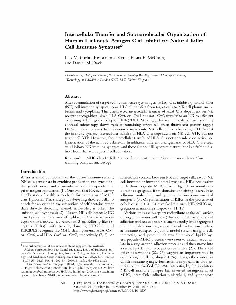
50% of NK/target cell intercellular contacts. Thisis consistent with KIR2DL1-induced clustering of HLA-Cw6 at NK immune synapses as described previously (9).However, when imaging live conjugates of NK and 221/Cw6-GFP cells by confocal microscopy in the currentstudy, GFP-tagged HLA-Cw6 (HLA-Cw6-GFP) couldalso be seen on the NK cell, both at the cell surface and insmall intracellular vesicles, only in conjugates at which in-hibitory synapses were evident. Two peripheral blood NKcells simultaneously surveying a single 221/Cw6-GFP cellcan be seen in Fig. 1 a. HLA-C has transferred only intothe NK cell at which HLA-C clustered at the immune syn-apse, consistent with KIR2DL1-induced clustering and in-tercellular transfer of HLA-Cw6.
Images taken every 0.3
�
m through cell conjugates, per-pendicular to the immune synapses revealed that much ofthe captured HLA-Cw6-GFP is on the NK plasma mem-brane with some in the cytoplasm. This is represented in
Figure 1. HLA-Cw6-GFP transfers from target cells to NK cells at theinhibitory NK immune synapse. (a) A single 221/Cw6-GFP cell is seeninteracting with two peripheral blood NK cells (NK line K010) demon-strating selective clustering and intercellular transfer of HLA-Cw6-GFP atthe immune synapse. Figure shows the transmitted light overlaid withGFP fluorescence (left) and the corresponding GFP fluorescence only(right). Clustering of HLA-Cw6-GFP at the immune synapse and trans-ferred vesicles of GFP are evident in the lower K010 NK cell only. (b)Vesicles containing HLA-Cw6-GFP, transferred from the target cell tothe NK cell, are present on the NK cell surface and cytoplasm. Left panelshows a single 0.9 �m optical slice of GFP fluorescence in a K010 NKcell conjugated to a 221/Cw6-GFP target cell (bottom of the panel).HLA-Cw6-GFP is clustered at the immune synapse and there is alsotransfer of HLA-Cw6-GFP onto the NK cell surface. Images were takenevery 0.3 �m through the cell perpendicular to the immune synapse anda composite of these images was then color coded for depth through theNK cell (right). The color scale used is below the panel; 0 �m (blue) rep-resents the upper surface and 3.5 �m (red) the lower surface of the NKcell. Red and white arrows indicate vesicles containing HLA-Cw6-GFPon the plasma membrane and in the cytoplasm of the NK cell, respec-tively. (c) Top panel shows the transmitted light image overlaid with GFPfluorescence; lower three frames depict identical fields of view at differenttimes, showing a vesicle containing HLA-Cw6-GFP moving away fromthe immune synapse into the NK cell. The white arrow indicates the po-sition of the vesicle in the first frame. In all images, white scale bars repre-sent 5 �m. Cells shown are representative of numerous cells in at leastthree independent experiments.
1510
Human Leukocyte Antigen C at Inhibitory Natural Killer Cell Synapses
Fig. 1 b (right), in which GFP fluorescence has been col-ored according to its depth within the NK cell. Blue- andred-colored patches represent GFP fluorescence located on,or near to, the upper and lower NK cell surfaces, respec-tively. Green colored patches locate GFP
�
1.5–2
�
m be-neath the upper cell surface within the cytoplasm.
Using time-lapse imaging, vesicles containing HLA-Cw6-GFP are clearly seen migrating away from the im-mune synapse and into the cytoplasm of an NK cell fromanother peripheral blood NK cell line, K007, which ex-presses KIR2DL1 (Fig. 1 c and Fig1cVideo1). Vesicles mi-grated with nonBrownian direction, reminiscent of migra-tion of vesicles along the cytoskeleton. The speed of one ofthese vesicles, or group of vesicles, at room temperaturewas calculated to be 0.04
�
m s
�
1
.
NK Cell Transfectants Expressing KIR2DL1 Capture TargetCell HLA-C.
To determine if intercellular transfer ofHLA-C is dependent on recognition by KIRs, we used aCD28
�
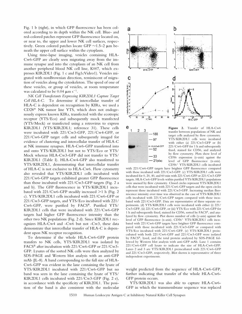
NK tumor line YTS, which does not endoge-nously express known KIRs, transfected with the ecotropicreceptor (YTS/Eco) and subsequently mock transfected(YTS/Mock) or transfected using a retrovirus to expressKIR2DL1 (YTS/KIR2DL1; reference 31). These cellswere incubated with 221/Cw3-GFP, 221/Cw4-GFP, or221/Cw6-GFP target cells and subsequently imaged forevidence of clustering and intercellular transfer of HLA-Cat NK immune synapses. HLA-Cw6-GFP transferred intoand onto YTS/KIR2DL1 but not to YTS/Eco or YTS/Mock whereas HLA-Cw3-GFP did not transfer to YTS/KIR2DL1 (Table I). HLA-Cw4-GFP also transferred toYTS/KIR2DL1, demonstrating that intercellular transferof HLA-C is not exclusive to HLA-Cw6. Flow cytometryalso revealed that YTS/KIR2DL1 cells incubated with221/Cw6-GFP targets exhibited greater GFP fluorescencethan those incubated with 221/Cw3-GFP targets (Fig. 2 aand b). The GFP fluorescence in YTS/KIR2DL1 incu-bated with 221/Cw6-GFP steadily increased
�
1 h (Fig. 2c). YTS/KIR2DL1 cells incubated with 221/Cw6-GFP or221/Cw3-GFP targets, and YTS/Eco incubated with 221/Cw6-GFP, were purified by FACS
®
. Purified YTS/KIR2DL1 cells that were incubated with 221/Cw6-GFPtargets had higher GFP fluorescence intensity than theother two NK populations (Fig. 2 d). Since KIR2DL1 rec-ognizes HLA-Cw4 and -Cw6 but not -Cw3, these datademonstrate that intercellular transfer of HLA-C is depen-dent upon NK receptor recognition.
To determine if the whole HLA-Cw6-GFP proteintransfers to NK cells, YTS/KIR2DL1 was isolated byFACS
®
after incubation with 221/Cw6-GFP or 221/Cw3-GFP. Lysates of the sorted NK cells were then analyzed bySDS-PAGE and Western blot analysis with an anti-GFPmAb (JL-8). A band corresponding to the full size of HLA-Cw6-GFP was evident in the lane containing the lysate ofYTS/KIR2DL1 incubated with 221/Cw6-GFP but noband was seen in the lane containing the lysate of YTS/KIR2DL1 cells incubated with 221/Cw3-GFP (Fig. 2 e),in accordance with the specificity of KIR2DL1. The posi-tion of the band is also consistent with the molecular
weight predicted from the sequence of HLA-Cw6-GFP,further indicating that transfer of the whole HLA-Cw6-GFP protein occurs.
YTS/KIR2DL1 was also able to capture HLA-Cw6-GFP in which the transmembrane sequence was replaced
Figure 2. Transfer of HLA-Cw6transfer between populations of NK andtarget cells analyzed by flow cytometry.YTS/KIR2DL1 cells were incubatedwith either (a) 221/Cw3-GFP or (b)221/Cw6-GFP for 1 h and subsequentlyfixed, stained for CD56, and analyzedby flow cytometry. Plots show level ofCD56 expression (y-axis) against thelevel of GFP fluorescence (x-axis).CD56� YTS/KIR2DL1 cells incubated
with 221/Cw6-GFP targets have brighter GFP fluorescence comparedwith those incubated with 221/Cw3-GFP. (c) YTS/KIR2DL1 cells wereincubated for 0, 20, 40, and 60 min with 221/Cw6-GFP or 221/Cw3-GFPtargets. HLA-Cw6-GFP levels within purified YTS/KIR2DL1 populationswere assessed by flow cytometry. Closed circles represent YTS/KIR2DL1cells that were incubated with 221/Cw6-GFP targets and the open circlesrepresent those incubated with 221/Cw3-GFP. Increasing median fluo-rescence intensity over time was observed in the case of YTS/KIR2DL1cells incubated with 221/Cw6-GFP targets compared with those incu-bated with 221/Cw3-GFP. Data are representative of three separate ex-periments. (d) YTS/KIR2DL1 cells were incubated with either (i) 221/Cw3-GFP, (ii) 221/Cw6-GFP, or (iii) YTS/Eco with 221/Cw6-GFP for1 h and subsequently fixed, stained for CD56, sorted by FACS®, and ana-lyzed by flow cytometry. Plot shows number of cells (y-axis) against thelevel of GFP fluorescence (x-axis). CD56� YTS/KIR2DL1 cells incu-bated with 221/Cw6-GFP targets have brighter GFP fluorescence com-pared with those incubated with 221/Cw3-GFP or compared withYTS/Eco incubated with 221/Cw6-GFP. (e) YTS/KIR2DL1 prein-cubated with both 221/Cw6-GFP and 221/Cw3-GFP were isolatedby FACS®, lysed, and the total protein analyzed by SDS-PAGE fol-lowed by Western blot analysis with anti-GFP mAb. Lane 1 contains221/Cw6-GFP cell lysate to indicate the size of HLA-Cw6-GFP.Lanes 2 and 3 are YTS/KIR2DL1 preincubated with 221/Cw6-GFPand 221/Cw3-GFP, respectively. Blot shown is representative of threeindependent experiments.
1511
Carlin et al.
with that of HLA-G, which lacks a critical transmembranecysteine (221/Cw6-TMG-GFP; Table I). Thus, althoughrecognition of HLA-Cw6 by a subset of NK cells is depen-dent on a cysteine within the transmembrane sequence(Cys309; reference 35), capture of HLA-Cw6 by YTS/KIR2DL1 is independent of this substitution.
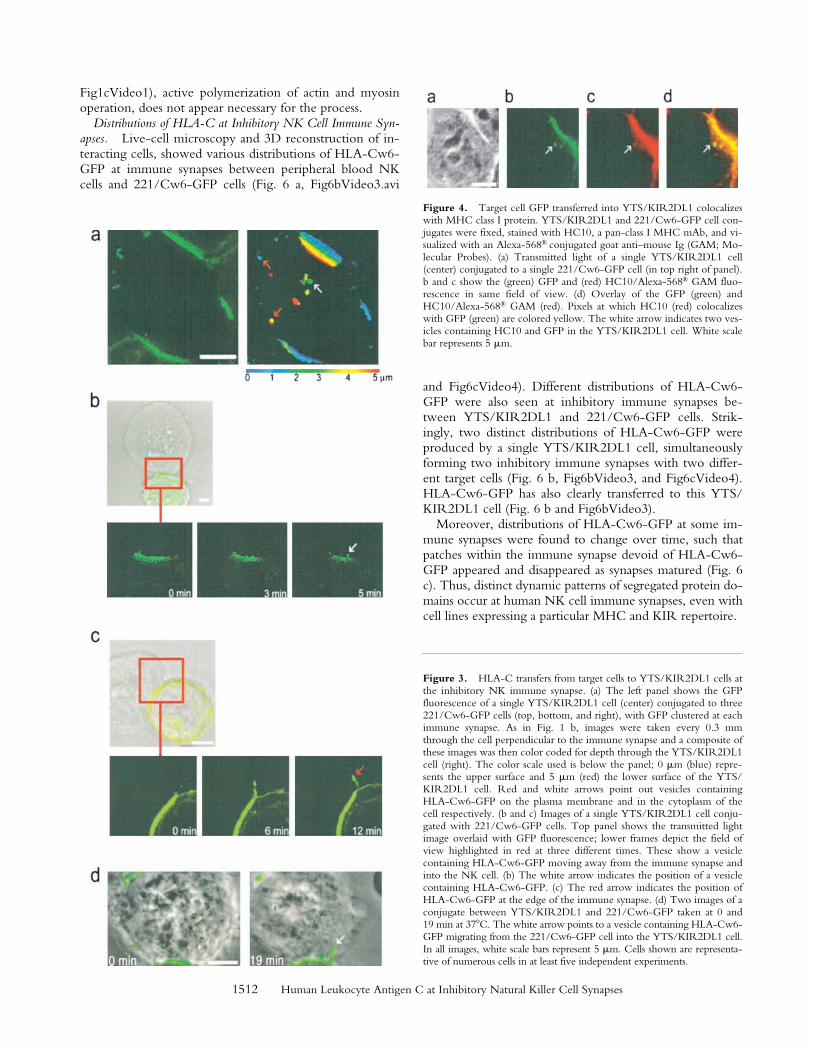
Like the transfer of HLA-C into peripheral blood NKcells (Fig. 1), target cell HLA-Cw6-GFP transferred toboth the plasma membrane and the cytoplasm of YTS/KIR2DL1 (Fig. 3 a). In time-lapse images, target cell HLA-Cw6-GFP was seen moving away from the immune syn-apse and into YTS/KIR2DL1, both from the center of theimmune synapse into the cytoplasm of YTS/KIR2DL1(Fig. 3 b) and from the edge of the synapse directly ontothe NK cell surface (Fig. 3 c). Transfer of target cell HLA-Cw6-GFP into YTS/KIR2DL1 was seen at both roomtemperature (Fig. 3 b, Fig3cVideo2 and Fig. 3 c) and at37
�
C, using a heated microscope stage (Fig. 3 d). However,it proved difficult to routinely image cells at 37
�
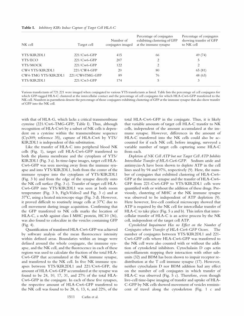
C due tocell movement during image acquisition. Confirming thatthe GFP transferred to NK cells marks the location ofHLA-C, a mAb against class I MHC protein, HC10 (36),was also found to colocalize in the vesicles containing GFP(Fig. 4).
Quantification of transferred HLA-Cw6-GFP was achievedby software analysis of the mean fluorescence intensitywithin defined areas. Boundaries within an image weredefined around the whole conjugate, the immune syn-apse, and the NK cell, and the fluorescence in each of theseregions was used to calculate the fraction of the total HLA-Cw6-GFP that accumulated at the NK immune synapse,and transferred to the NK cell. In five NK immune syn-apses between YTS/KIR2DL1 and 221/Cw6-GFP, theamount of HLA-Cw6-GFP accumulated at the synapse wasfound to be 24, 10, 17, 31, and 27% of the total HLA-Cw6-GFP in the conjugate. In each of these five synapses,the respective amount of HLA-Cw6-GFP transferred tothe NK cell was found to be 28, 6, 13, 6, and 22%, of the
total HLA-Cw6-GFP in the conjugate. Thus, it is likelythat variable amounts of target cell HLA-C transfer to NKcells, independent of the amount accumulated at the im-mune synapse. However, differences in the amount ofHLA-C transferred into the NK cells could also be ac-counted for if each NK cell, before imaging, surveyed avariable number of target cells capturing some HLA-Cfrom each.
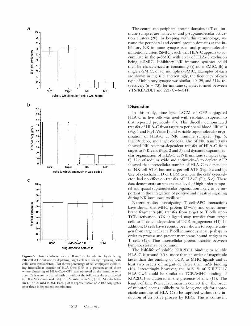
Depletion of NK Cell ATP but not Target Cell ATP InhibitsIntercellular Transfer of HLA-Cw6-GFP.
Sodium azide andantimycin-A have been shown to deplete ATP in the celllines used by 94 and 97%, respectively (9). Here, the num-ber of conjugates that exhibited clustering of HLA-Cw6-GFP at the immune synapse and the transfer of HLA-Cw6-GFP from 221-Cw6-GFP to YTS/KIR2DL1 cells werequantified with or without the addition of these drugs. Pre-viously, clustering of MHC at the NK immune synapsewas observed to be independent of ATP depletion (9).Here however, live-cell confocal microscopy showed thatATP is required by the NK cell for intercellular transfer ofHLA-C to take place (Fig. 5 a and b). This infers that inter-cellular transfer of HLA-C is an active process by the NKcell, independent of the target cell ATP.
Cytoskeletal Impairment Has no Effect on the Number ofConjugates where Transfer of HLA-Cw6-GFP Occurs.
Thenumber of conjugates between YTS/KIR2DL1 and 221-Cw6-GFP cells where HLA-Cw6-GFP was transferred tothe NK cell were also counted with or without the addi-tion of cytoskeletal inhibitors. Cytochalasin D caps actinmicrofilaments stopping their interaction with other sub-units (32) and BDM has been shown to impair receptor re-distribution at the T cell immune synapse (17). However,neither cytochalasin D nor BDM addition had any effecton the number of cell conjugates in which transfer ofHLA-C was observed (Fig. 5 c). Therefore, even thoughlive-cell time-lapse imaging of transfer and uptake of HLA-C-GFP by NK cells showed movement of vesicles reminis-cent of travel along the cytoskeleton (Fig. 1 c and
Table I.
Inhibitory KIRs Induce Capture of Target Cell HLA-C
NK cell Target cellNumber of
conjugates imaged
Percentage of conjugates exhibiting clustering of GFP at the immune synapse
Percentage of conjugates showing transfer of GFP to NK cell
YTS/KIR2DL1 221/Cw6-GFP 415 66 49 (74)YTS/ECO 221/Cw6-GFP 207 2 5YTS/MOCK 221/Cw6-GFP 122 2 2CW4 YTS/KIR2DL1 221/CW4-GFP 20 80 65 (81)CW4-TMG YTS/KIR2DL1 221/CW6TMG-GFP 89 76 48 (63)YTS/KIR2DL1 221/Cw3-GFP 174 5 3
Various transfectants of 721.221 were imaged when conjugated to various YTS transfectants as listed. Table lists the percentage of cell conjugates forwhich GFP-tagged HLA-C clustered at the intercellular contact and the percentage of cell conjugates for which HLA-Cw6-GFP transferred to theNK cell. Numbers in parenthesis denote the percentage of those conjugates exhibiting clustering of GFP at the immune synapse that also show transferof GFP into the NK cell.
1512
Human Leukocyte Antigen C at Inhibitory Natural Killer Cell Synapses
Fig1cVideo1), active polymerization of actin and myosinoperation, does not appear necessary for the process.
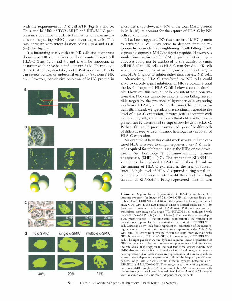
Distributions of HLA-C at Inhibitory NK Cell Immune Syn-apses.
Live-cell microscopy and 3D reconstruction of in-teracting cells, showed various distributions of HLA-Cw6-GFP at immune synapses between peripheral blood NKcells and 221/Cw6-GFP cells (Fig. 6 a, Fig6bVideo3.avi
and Fig6cVideo4). Different distributions of HLA-Cw6-GFP were also seen at inhibitory immune synapses be-tween YTS/KIR2DL1 and 221/Cw6-GFP cells. Strik-ingly, two distinct distributions of HLA-Cw6-GFP wereproduced by a single YTS/KIR2DL1 cell, simultaneouslyforming two inhibitory immune synapses with two differ-ent target cells (Fig. 6 b, Fig6bVideo3, and Fig6cVideo4).HLA-Cw6-GFP has also clearly transferred to this YTS/KIR2DL1 cell (Fig. 6 b and Fig6bVideo3).
Moreover, distributions of HLA-Cw6-GFP at some im-mune synapses were found to change over time, such thatpatches within the immune synapse devoid of HLA-Cw6-GFP appeared and disappeared as synapses matured (Fig. 6c). Thus, distinct dynamic patterns of segregated protein do-mains occur at human NK cell immune synapses, even withcell lines expressing a particular MHC and KIR repertoire.
Figure 3.
HLA-C transfers from target cells to YTS/KIR2DL1 cells atthe inhibitory NK immune synapse. (a) The left panel shows the GFPfluorescence of a single YTS/KIR2DL1 cell (center) conjugated to three221/Cw6-GFP cells (top, bottom, and right), with GFP clustered at eachimmune synapse. As in Fig. 1 b, images were taken every 0.3 mmthrough the cell perpendicular to the immune synapse and a composite ofthese images was then color coded for depth through the YTS/KIR2DL1cell (right). The color scale used is below the panel; 0
�
m (blue) repre-sents the upper surface and 5
�
m (red) the lower surface of the YTS/KIR2DL1 cell. Red and white arrows point out vesicles containingHLA-Cw6-GFP on the plasma membrane and in the cytoplasm of thecell respectively. (b and c) Images of a single YTS/KIR2DL1 cell conju-gated with 221/Cw6-GFP cells. Top panel shows the transmitted lightimage overlaid with GFP fluorescence; lower frames depict the field ofview highlighted in red at three different times. These show a vesiclecontaining HLA-Cw6-GFP moving away from the immune synapse andinto the NK cell. (b) The white arrow indicates the position of a vesiclecontaining HLA-Cw6-GFP. (c) The red arrow indicates the position ofHLA-Cw6-GFP at the edge of the immune synapse. (d) Two images of aconjugate between YTS/KIR2DL1 and 221/Cw6-GFP taken at 0 and19 min at 37
�
C. The white arrow points to a vesicle containing HLA-Cw6-GFP migrating from the 221/Cw6-GFP cell into the YTS/KIR2DL1 cell.In all images, white scale bars represent 5
�
m. Cells shown are representa-tive of numerous cells in at least five independent experiments.
Figure 4. Target cell GFP transferred into YTS/KIR2DL1 colocalizeswith MHC class I protein. YTS/KIR2DL1 and 221/Cw6-GFP cell con-jugates were fixed, stained with HC10, a pan-class I MHC mAb, and vi-sualized with an Alexa-568® conjugated goat anti–mouse Ig (GAM; Mo-lecular Probes). (a) Transmitted light of a single YTS/KIR2DL1 cell(center) conjugated to a single 221/Cw6-GFP cell (in top right of panel).b and c show the (green) GFP and (red) HC10/Alexa-568® GAM fluo-rescence in same field of view. (d) Overlay of the GFP (green) andHC10/Alexa-568® GAM (red). Pixels at which HC10 (red) colocalizeswith GFP (green) are colored yellow. The white arrow indicates two ves-icles containing HC10 and GFP in the YTS/KIR2DL1 cell. White scalebar represents 5 �m.
1513
Carlin et al.
The central and peripheral protein domains at T cell im-mune synapses are named c- and p-supramolecular activa-tion clusters (20). In keeping with this terminology, wename the peripheral and central protein domains at the in-hibitory NK immune synapse as c- and p-supramolecularinhibition clusters (SMIC), such that HLA-C appears to ac-cumulate in the p-SMIC with areas of HLA-C exclusionbeing c-SMIC. Inhibitory NK immune synapses couldthen be characterized as containing (a) no c-SMIC, (b) asingle c-SMIC, or (c) multiple c-SMIC. Examples of eachare shown in Fig. 6 d. Interestingly, the frequency of eachtype of inhibitory synapse was similar, 40, 29, and 31%, re-spectively (
n
73), for immune synapses formed betweenYTS/KIR2DL1 and 221/Cw6-GFP.
Discussion
In this study, time-lapse LSCM of GFP-conjugatedHLA-C in live cells was used with resolution superior tothat reported previously (9). This directly demonstratedtransfer of HLA-C from target to peripheral blood NK cells(Fig. 1 and Fig1cVideo1) and variable supramolecular orga-nization of HLA-C at NK immune synapses (Fig. 6,Fig6bVideo3, and Fig6cVideo4). Use of NK transfectantsshowed NK receptor-dependent transfer of HLA-C fromtarget to NK cells (Figs. 2 and 3) and dynamic supramolec-ular organization of HLA-C at NK immune synapses (Fig.6). Use of sodium azide and antimycin-A to deplete ATPshowed that intercellular transfer of HLA-C is dependenton NK cell ATP, but not target cell ATP (Fig. 5 a and b).Use of cytochalasin D or BDM to impair the cells’ cytoskel-eton had no effect on transfer of HLA-C (Fig. 5 c). Thesedata demonstrate an unexpected level of high order tempo-ral and spatial supramolecular organization likely to be im-portant in the integration of positive and negative signalingduring NK immunosurveillance.
Recent studies investigating T cell–APC interactionshave shown that MHC protein (37–39) and other mem-brane fragments (40) transfer from target to T cells uponTCR activation. OX40 ligand may transfer from targetcells to T cells independent of TCR engagement (41). Inaddition, B cells have recently been shown to acquire anti-gen from target cells at a B cell immune synapse, perhaps inorder to process and present membrane-bound antigens toT cells (42). Thus intercellular protein transfer betweenlymphocytes may be common.
The half-life of soluble KIR2DL1 binding to solubleHLA-C is around 0.3 s, more than an order of magnitudefaster than the binding of TCR to MHC ligands and atleast two orders of magnitude faster than mAb binding(10). Interestingly however, the half-life of KIR2DL1/HLA-Cw6 could be similar to TCR/MHC binding, ifKIR2DL1 is clustered in the presence of zinc (11). Thelength of time NK cells remain in contact (i.e., the orderof minutes) seems unlikely to be long enough for appre-ciable amounts of HLA-C to be captured without the in-duction of an active process by KIRs. This is consistent
Figure 5. Intercellular transfer of HLA-C can be inhibited by depletingNK cell ATP but not by depleting target cell ATP or by impairing bothcells’ actin cytoskeleton. Plot shows percentage of cell conjugates exhibit-ing intercellular transfer of HLA-Cw6-GFP as a percentage of thosewhere clustering of HLA-Cw6-GFP was observed at the immune syn-apse. Cells were incubated with or without the following drugs as labeled(a) 50 mM sodium azide, (b) 13 �M antimycin-A, (c) 10 �M cytochala-sin D, or 20 mM BDM. Each plot is representative of �100 conjugatesover three independent experiments.
1514
Human Leukocyte Antigen C at Inhibitory Natural Killer Cell Synapses
with the requirement for NK cell ATP (Fig. 5 a and b).Thus, the half-life of TCR/MHC and KIR/MHC pro-teins may be similar in order to facilitate a common mech-anism of capturing MHC protein from target cells. Thismay correlate with internalization of KIR (43) and TCR(44) after ligation.
It is interesting that vesicles in NK cells and membranedomains at NK cell surfaces can both contain target cellHLA-C (Figs. 1, 3, and 4), and it will be important tocharacterize these vesicles and domains fully. There is evi-dence that tumor, dendritic, and EBV-transformed B cellscan secrete vesicles of endosomal origin or ‘exosomes’ (45,46). However, constitutive secretion of MHC protein in
exosomes is too slow, at
�
10% of the total MHC proteinin 24 h (46), to account for the capture of HLA-C by NKcells reported here.
It has been suggested (37) that transfer of MHC proteinto activated T cells may serve to dampen immune re-sponses by fratricide, i.e., neighboring T cells killing T cellsexpressing captured MHC/antigenic peptide. However, asimilar function for transfer of MHC protein between lym-phocytes could not be attributed to the transfer of targetcell HLA-C to NK cells, as HLA-C transferred to NK cellswould not usually present an antigenic peptide and, in gen-eral, HLA-C serves to inhibit rather than activate NK cells.
Alternatively, HLA-C transferred to NK cells couldserve to directly signal inhibition of NK cytotoxicity untilthe level of captured HLA-C falls below a certain thresh-old. However, this would not be consistent with observa-tions that NK cells cannot be inhibited from killing suscep-tible targets by the presence of bystander cells expressinginhibitory HLA-C, i.e., NK cells cannot be inhibited intrans (8). Instead, we speculate that continually assessing thelevel of HLA-C expression, through serial encounter withneighboring cells, could help set a threshold at which a sin-gle cell can be determined to express low levels of HLA-C.Perhaps this could prevent unwanted lysis of healthy cellsof different type with an intrinsic heterogeneity in levels ofHLA-C expression.
An example of how this could work would be if the cap-tured HLA-C served to simply sequester a key NK mole-cule required for inhibition, such as the KIRs or the down-stream Src homology 2 domain–containing tyrosinephosphatase, (SHP)-1 (47). The amount of KIR/SHP-1sequestered by captured HLA-C would then depend onthe amount of HLA-C expressed in the area of surveil-lance. A high level of HLA-C captured during serial en-counters with several targets would then lead to a highamount of KIR/SHP-1 being sequestered. This in turn
Figure 6.
Supramolecular organization of HLA-C at inhibitory NKimmune synapses. (a) Image of 221/Cw6-GFP cells surrounding a pe-ripheral blood K010 NK cell (left) and the supramolecular organization ofHLA-Cw6-GFP at the two immune synapses formed (right panels). (b)First panel shows an overlay of HLA-Cw6-GFP fluorescence and thetransmitted light image of a single YTS/KIR2DL1 cell conjugated withtwo 221/Cw6-GFP cells (far left of frame). The next three frames depicta 3D reconstruction of the same cells, demonstrating the formation oftwo distinct supramolecular organizations by a single YTS/KIR2DL1cell. Cartoons below each frame represent the orientation of the interact-ing cells in each frame, with green spheres representing the 221/Cw6-GFP cells. (c) Left panel shows the transmitted light image overlaid withGFP fluorescence of 221/Cw6-GFP cells surrounding a YTS/KIR2DL1cell. The right panels show the dynamic supramolecular organization ofGFP fluorescence at the two immune synapses indicated. White arrowsindicate SMIC that disappear in the next frame; red arrows indicate newSMIC that were absent from the previous frame. In all images, white scalebars represent 5
�
m. Cells shown are representative of numerous cells inat least three independent experiments. d shows the frequency of differentpatterns of p- and c-SMIC at the immune synapse between YTS/KIR2DL1 and 221/Cw6-GFP. Two images of each type of organization,i.e., no c-SMIC, single c-SMIC, and multiple c-SMIC are shown withthe percentage that each was observed given below. A total of 73 synapseswere analyzed over at least three independent experiments.
1515
Carlin et al.
would then leave a lower amount of KIR/SHP-1 availablefor inhibition at a given NK synapse and hence a higheramount of HLA-C at that immune synapse may then berequired to trigger inhibition.
An emerging theme in contemporary molecular immu-nology is that cell surface receptors need to be consideredas part of supramolecular complexes of proteins and lipidsthat facilitate specific receptor conformations and distinctdistributions at cell surfaces. That different arrangements ofsegregated protein domains (Fig. 6) can occur at inhibitoryimmune synapses raises a number of important new ques-tions. For example, does supramolecular organization ofHLA-C at the inhibitory synapse influence the outcome ofthe intercellular communication and how is the organiza-tion biophysically controlled? Clustering of similarly sizedproteins could account for the segregation of protein do-mains at immune synapses (48, 49), but could not predictthe specific arrangements of such segregated domains seenat the synapses. It has also been suggested that cell surfaceproteins are recruited to a particular localization within cellmembranes through noncovalent interactions with choles-terol-rich membrane microdomains known as ‘lipid rafts’(50). Indeed, clustering of lipid rafts induced by costimula-tion of T cells suggests an immunological importance oflipid raft motility (51, 52), but their role in immune syn-apse formation remains to be clarified. However, recentdata shows that engagement of HLA-C by KIR blocks re-distribution of lipid rafts (53), suggesting that organizationof lipid rafts is not responsible for the different arrange-ments of HLA-C at NK inhibitory immune synapses seenin Fig. 6, Fig6bVideo3, and Fig6cVideo4.
In summary, both intercellular transfer and supramolecu-lar organization of MHC protein at immune synapses em-body new high order aspects of molecular recognition byNK cells. The NK immune synapse undoubtedly includesadditional cell surface proteins critical to the intercellularcommunication, for example, HLA-E/CD94/NKG2 (54)the activating NK receptors including NKG2D, NKp44,NKp46, and NKp30 (55, 56) and their ligands, such asMIC (57) and viral hemagglutinin (58). An important nextgoal is to image the organization of these and other recep-tors at NK immune synapses.
We are grateful to Aaron Rae and Jonathan Styles for assistancewith cell sorting and to Klaus Suhling and Margaret Dallman forcritical reading of the manuscript.
This work is supported by grants from the Medical ResearchCouncil, the Biotechnology and Biological Sciences ResearchCouncil, and The Royal Society.
Submitted: 13 July 2001Revised: 26 September 2001Accepted: 10 October 2001
References1. Trinchieri, G. 1989. Biology of natural killer cells. Adv. Im-
munol. 47:187–376.2. Ljunggren, H.G., and K. Karre. 1990. In search of the ‘miss-
ing self’: MHC molecules and NK cell recognition. Immunol.Today. 11:237–244.
3. Lanier, L.L. 1998. NK cell receptors. Annu. Rev. Immunol.16:359–393.
4. Long, E.O. 1999. Regulation of immune responses throughinhibitory receptors. Annu. Rev. Immunol. 17:875–904.
5. Long, E.O., and S. Rajagopalan. 2000. HLA class I recogni-tion by killer cell Ig-like receptors. Semin. Immunol. 12:101–108.
6. Raulet, D.H., R.E. Vance, and C.W. McMahon. 2001.Regulation of the natural killer cell receptor repertoire.Annu. Rev. Immunol. 19:291–330.
7. Colonna, M., E.G. Brooks, M. Falco, G.B. Ferrara, and J.L.Strominger. 1993. Generation of allospecific natural killercells by stimulation across a polymorphism of HLA-C. Sci-ence. 260:1121–1124.
8. Colonna, M., G. Borsellino, M. Falco, G.B. Ferrara, and J.L.Strominger. 1993. HLA-C is the inhibitory ligand that deter-mines dominant resistance to lysis by NK1- and NK2-specificnatural killer cells. Proc. Natl. Acad. Sci. USA. 90:12000–12004.
9. Davis, D.M., I. Chiu, M. Fassett, G.B. Cohen, O. Mandel-boim, and J.L. Strominger. 1999. The human natural killercell immune synapse. Proc. Natl. Acad. Sci. USA. 96:15062–15067.
10. Vales-Gomez, M., H.T. Reyburn, M. Mandelboim, and J.L.Strominger. 1998. Kinetics of interaction of HLA-C ligandswith natural killer cell inhibitory receptors. Immunity. 9:337–344.
11. Vales-Gomez, M., R.A. Erskine, M.P. Deacon, J.L.Strominger, and H.T. Reyburn. 2001. The role of zinc in thebinding of killer cell Ig-like receptors to class I MHC pro-teins. Proc. Natl. Acad. Sci. USA. 98:1734–1739.
12. Rajagopalan, S., and E.O. Long. 1998. Zinc bound to thekiller cell-inhibitory receptor modulates the negative signal inhuman NK cells. J. Immunol. 161:1299–1305.
13. Fan, Q.R., E.O. Long, and D.C. Wiley. 2000. Cobalt-medi-ated dimerization of the human natural killer cell inhibitoryreceptor. J. Biol. Chem. 275:23700–23706.
14. Boyington, J.C., S.A. Motyka, P. Schuck, A.G. Brooks, andP.D. Sun. 2000. Crystal structure of an NK cell immuno-globulin-like receptor in complex with its class I MHCligand. Nature. 405:537–543.
15. Fan, Q.R., E.O. Long, and D.C. Wiley. 2001. Crystal struc-ture of the human natural killer cell inhibitory receptorKIR2DL1-HLA-Cw4 complex. Nat. Immunol. 2:452–460.
16. McCloskey, M.A., and M.M. Poo. 1986. Contact-inducedredistribution of specific membrane components: local accu-mulation and development of adhesion. J. Cell Biol. 102:2185–2196.
17. Wülfing, C., and M.M. Davis. 1998. A receptor/cytoskeletalmovement triggered by costimulation during T cell activa-tion. Science. 282:2266–2269.
18. Wülfing, C., M.D. Sjaastad, and M.M. Davis. 1998. Visualiz-ing the dynamics of T cell activation: intracellular adhesionmolecule 1 migrates rapidly to the T cell/B cell interface andacts to sustain calcium levels. Proc. Natl. Acad. Sci. USA. 95:6302–6307.
19. Krummel, M.F., M.D. Sjaastad, C. Wulfing, and M.M.Davis. 2000. Differential clustering of CD4 and CD3zetaduring T cell recognition. Science. 289:1349–1352.
20. Monks, C.R., B.A. Freiberg, H. Kupfer, N. Sciaky, and A.Kupfer. 1998. Three-dimensional segregation of supramolec-
1516 Human Leukocyte Antigen C at Inhibitory Natural Killer Cell Synapses
ular activation clusters in T cells. Nature. 395:82–86.21. Grakoui, A., S.K. Bromley, C. Sumen, M.M. Davis, A.S.
Shaw, P.M. Allen, and M.L. Dustin. 1999. The immunolog-ical synapse: a molecular machine controlling T cell activa-tion. Science. 285:221–227.
22. Stinchcombe, J.C., D.C. Barral, E.H. Mules, S. Booth, A.N.Hume, L.M. Machesky, M.C. Seabra, and G.M. Griffiths.2001. Rab27a is required for regulated secretion in cytotoxicT lymphocytes. J. Cell Biol. 152:825–834.
23. Wülfing, C., A. Bauch, G.R. Crabtree, and M.M. Davis.2000. The vav exchange factor is an essential regulator in ac-tin-dependent receptor translocation to the lymphocyte-anti-gen-presenting cell interface. Proc. Natl. Acad. Sci. USA. 97:10150–10155.
24. Delon, J., and R.N. Germain. 2000. Information transfer atthe immunological synapse. Curr. Biol. 10:R923–R933.
25. Dustin, M.L., and J.A. Cooper. 2000. The immunologicalsynapse and the actin cytoskeleton: molecular hardware for Tcell signaling. Nat. Immunol. 1:23–29.
26. Bromley, S.K., W.R. Burack, K.G. Johnson, K. Somersalo,T.N. Sims, C. Sumen, M.M. Davis, A.S. Shaw, P.M. Allen,and M.L. Dustin. 2001. The immunological synapse. Annu.Rev. Immunol. 19:375–396.
27. Dustin, M.L., P.M. Allen, and A.S. Shaw. 2001. Environ-mental control of immunological synapse formation and du-ration. Trends. Immunol. 22:192–194.
28. Friedl, P., and M. Gunzer. 2001. Interaction of T cells withAPCs: the serial encounter model. Trends. Immunol. 22:187–191.
29. Shimizu, Y., and R. DeMars. 1989. Production of humancells expressing individual transferred HLA-A,-B,-C genesusing an HLA-A,-B,-C null human cell line. J. Immunol. 142:3320–3328.
30. Tsien, R.Y. 1998. The green fluorescent protein. Annu. Rev.Biochem. 67:509–544.
31. Cohen, G.B., R.T. Gandhi, D.M. Davis, O. Mandelboim,B.K. Chen, J.L. Strominger, and D. Baltimore. 1999. The se-lective downregulation of class I major histocompatibilitycomplex proteins by HIV-1 protects HIV-infected cells fromNK cells. Immunity. 10:661–671.
32. Cooper, J.A. 1987. Effects of cytochalasin and phalloidin onactin. J. Cell Biol. 105:1473–1478.
33. Urwyler, N., P. Eggli, and H.U. Keller. 2000. Effects of themyosin inhibitor 2,3-butanedione monoxime (BDM) on cellshape, locomotion and fluid pinocytosis in human polymor-phonuclear leucocytes. Cell. Biol. Int. 24:863–870.
34. Schlichter, L.C., P.A. Pahapill, and I. Chung. 1992. Dual ac-tion of 2,3-butanedione monoxime (BDM) on K� current inhuman T lymphocytes. J. Pharmacol. Exp. Ther. 261:438–446.
35. Davis, D.M., O. Mandelboim, I. Luque, E. Baba, J. Boyson,and J.L. Strominger. 1999. The transmembrane sequence ofhuman histocompatibility leukocyte antigen (HLA)-C as adeterminant in inhibition of a subset of natural killer cells. J.Exp. Med. 189:1265–1274.
36. Sernee, M.F., H.L. Ploegh, and D.J. Schust. 1998. Why cer-tain antibodies cross-react with HLA-A and HLA-G: epitopemapping of two common MHC class I reagents. Mol. Immu-nol. 35:177–188.
37. Huang, J.F., Y. Yang, H. Sepulveda, W. Shi, I. Hwang, P.A.Peterson, M.R. Jackson, J. Sprent, and Z. Cai. 1999. TCR-Mediated internalization of peptide-MHC complexes ac-quired by T cells. Science. 286:952–954.
38. Hwang, I., J.F. Huang, H. Kishimoto, A. Brunmark, P.A.
Peterson, M.R. Jackson, C.D. Surh, Z. Cai, and J. Sprent.2000. T cells can use either T cell receptor or CD28 recep-tors to absorb and internalize cell surface molecules derivedfrom antigen-presenting cells. J. Exp. Med. 191:1137-1148.
39. Patel, D., P. Arnold, G. White, J. Nardella, and M. Mannie.1999. Class II MHC/peptide complexes are released fromAPC and are acquired by T Cell responders during specificantigen recognition. J. Immunol. 163:5201–5210.
40. Hudrisier, D., J. Riond, H. Mazarguil, J.E. Gairin, and E.Joly. 2001. Cutting edge: CTLs rapidly capture membranefragments from target cells in a TCR signaling-dependentmanner. J. Immunol. 166:3645–3649.
41. Baba, E., Y. Takahashi, J. Lichtenfeld, R. Tanaka, A.Yoshida, K. Sugamura, N. Yamamoto, and Y. Tanaka. 2001.Functional CD4 T cells after intercellular molecular transferof 0X40 ligand. J. Immunol. 167:875–883.
42. Batista, F.D., D. Iber, and M.S. Neuberger. 2001. B cells ac-quire antigen from target cells after synapse formation. Na-ture. 411:489–494.
43. Huard, B., L. Karlsson, and F. Triebel. 2001. KIR down-regulation on NK cells is associated with down-regulation ofactivating receptors and NK cell inactivation. Eur. J. Immu-nol. 31:1728–1735.
44. Salio, M., S. Valitutti, and A. Lanzavecchia. 1997. Agonist-induced T cell receptor down-regulation: molecular require-ments and dissociation from T cell activation. Eur. J. Immu-nol. 27:1769–1773.
45. Wolfers, J., A. Lozier, G. Raposo, A. Regnault, C. Thery, C.Masurier, C. Flament, S. Pouzieux, F. Faure, T. Tursz, et al.2001. Tumor-derived exosomes are a source of shared tumorrejection antigens for CTL cross-priming. Nat. Med. 7:297–303.
46. Raposo, G., H.W. Nijman, W. Stoorvogel, R. Liejendekker,C.V. Harding, C.J. Melief, and H.J. Geuze. 1996. B lympho-cytes secrete antigen-presenting vesicles. J. Exp. Med. 183:1161–1172.
47. Burshtyn, D.N., A.M. Scharenberg, N. Wagtmann, S. Raja-gopalan, K. Berrada, T. Yi, J.P. Kinet, and E.O. Long. 1996.Recruitment of tyrosine phosphatase HCP by the killer cellinhibitor receptor. Immunity. 4:77–85.
48. van der Merwe, P.A., S.J. Davis, A.S. Shaw, and M.L. Dus-tin. 2000. Cytoskeletal polarization and redistribution of cell-surface molecules during T cell antigen recognition. Semin.Immunol. 12:5–21.
49. Wild, M.K., A. Cambiaggi, M.H. Brown, E.A. Davies, H.Ohno, T. Saito, and P.A. van der Merwe. 1999. Dependenceof T cell antigen recognition on the dimensions of an acces-sory receptor-ligand complex. J. Exp. Med. 190:31–41.
50. Simons, K., and E. Ikonen. 1997. Functional rafts in cellmembranes. Nature. 387:569–572.
51. Viola, A., S. Schroeder, Y. Sakakibara, and A. Lanzavecchia.1999. T lymphocyte costimulation mediated by reorganiza-tion of membrane microdomains. Science. 283:680–682.
52. Janes, P.W., S.C. Ley, and A.I. Magee. 1999. Aggregation oflipid rafts accompanies signaling via the T cell antigen recep-tor. J. Cell Biol. 147:447–461.
53. Lou, Z., D. Jevremovic, D.D. Billadeau, and P.J. Leibson.2000. A balance between positive and negative signals in cy-totoxic lymphocytes regulates the polarization of lipid raftsduring the development of cell-mediated killing. J. Exp.Med. 191:347–354.
54. Braud, V.M., D.S. Allan, C.A. O’Callaghan, K. Soderstrom,A. D’Andrea, G.S. Ogg, S. Lazetic, N.T. Young, J.I. Bell,

1517 Carlin et al.
J.H. Phillips, et al. 1998. HLA-E binds to natural killer cellreceptors CD94/NKG2A, B and C. Nature. 391:795–799.
55. Moretta, A., C. Bottino, M. Vitale, D. Pende, C. Cantoni,M.C. Mingari, R. Biassoni, and L. Moretta. 2001. Activatingreceptors and coreceptors involved in human natural killercell-mediated cytolysis. Annu. Rev. Immunol. 19:197–223.
56. Lanier, L.L. 2001. On guard-activating NK cell receptors.Nat. Immunol. 2:23–27.
57. Bauer, S., V. Groh, J. Wu, A. Steinle, J.H. Phillips, L.L.
Lanier, and T. Spies. 1999. Activation of NK cells and T cellsby NKG2D, a receptor for stress- inducible MICA. Science.285:727–729.
58. Mandelboim, O., N. Lieberman, M. Lev, L. Paul, T.I. Ar-non, Y. Bushkin, D.M. Davis, J.L. Strominger, J.W. Yew-dell, and A. Porgador. 2001. Recognition of haemagglutininson virus-infected cells by NKp46 activates lysis by humanNK cells. Nature. 409:1055–1060.
Copyright © 2022 FDOKUMEN