Interaction Profiles of Protein Kinase−Inhibitor Complexes and Their Application to Virtual...
13
Interaction Profiles of Protein Kinase-Inhibitor Complexes and Their Application to Virtual Screening Claudio Chuaqui, Zhan Deng, and Juswinder Singh* Computational Drug Design Group, Department of Research Informatics, Biogen Idec, Inc., 14 Cambridge Center, Cambridge, Massachusetts 01242 Received August 19, 2004 A major challenge facing structure-based drug discovery efforts is how to leverage the massive amount of experimental (X-ray and NMR) and virtual structural information generated from drug discovery projects. Many important drug targets have large numbers of protein-inhibitor complexes, necessitating tools to compare and contrast their similarities and differences. This information would be valuable for understanding potency and selectivity of inhibitors and could be used to define target constraints to assist virtual screening. We describe a profile-based approach that enables us to capture the conservation of interactions between a set of protein- ligand receptor complexes. The use of profiles provides a sensitive means to compare multiple inhibitors binding to a drug target. We demonstrate the utility of profile-based analysis of small molecule complexes from the protein-kinase family to identify similarities and differences in binding of ATP, p38, and CDK2 compounds to kinases and how these profiles can be applied to differentiate the selectivity of these inhibitors. Importantly, our virtual screening results demonstrate superior enrichment of kinase inhibitors using profile-based methods relative to traditional scoring functions. Interaction-based analysis should provide a valuable tool for understanding inhibitor binding to other important drug targets. Introduction The rapid growth in the number of protein-small molecule complexes from X-ray crystallography and NMR has helped drive the success of structure-based drug design (SBDD) and virtual screening (VS) ap- proaches for lead discovery. 1-5 The application of SBDD to the discovery and optimization of drug candidates has led to a tremendous number of protein-ligand com- plexes for targets 6 including HIV protease, carbonic anhydrase, thrombin, and neuroamidase. With such large amounts of data being generated, fully leveraging this information hinges on the ability to organize, analyze, and mine the structural data in order to derive knowledge that may be applied to drug discovery. To this end we have developed the structural interaction fingerprint (SIFt) methodology and in a recent paper demonstrated how it could effectively organize, visualize, and analyze protein-ligand com- plexes. 7 SIFt was also shown to be an effective molecular filter to improve the results obtained from VS. In this paper we extend the SIFt methodology to derive an interaction profile-based approach we term profile-SIFt, or p-SIFt. The p-SIFt is derived from a collection of SIFts that measures the conservation of interactions observed in clusters of protein-ligand complexes. The p-SIFt approach is analogous to profile- based techniques that have proven to be very useful in the analysis and database mining of groups of protein sequences 8-10 and structures. 11-13 The sequence profile is constructed from a set of multiply aligned sequences or structures of a probe family and is used to identify distant relationships to a database of target proteins. The profile is essentially a sequence position-specific scoring matrix encoding the probability of finding any of the 20 amino acid residues at that position in the target. In the case of p-SIFt, the SIFts derived from a set of probe structures are used to derive a position- dependent profile encoding the probability that a given interaction at that position is present. The probe set of structures may correspond to entire gene families, e.g., kinases, or to subfamilies of structures representing ligands with a particular activity or selectivity profile. To address the limitations inherent in traditional scoring functions, 14-20 a variety of “knowledge-based” or “target-biased” approaches have been developed that impose constraints based on ligand or receptor phar- macophores thought to be required for activity. 19,21-27 The tractability of knowledge-based VS for lead dis- covery 3,28-31 is highlighted by recent papers reporting the identification of potent lead (nM) compounds, using receptor-based docking in the case of Chk1 kinase, 22 and ligand-based VS for the discovery of a 25 nM inhibitor against TGF-RI. 32 In both of these examples, the success of the VS strategy was dependent on the application of constraints derived from knowledge of how small molecule inhibitors bind at the ATP site of protein kinases. These constraints typically filter virtual libraries based on the presence of kinase binding motifs, or on the ability to satisfy key interactions with the receptor. However, the ability to apply constraints during VS that predict the selectivity of inhibitors for one kinase over another is a much more challenging problem that has not been widely addressed. The protein kinase family exemplifies the challenges faced with the large amount of structural data being generated not only on specific drug targets but also at the gene family level. 33-35 For example, there exist over * To whom correspondence should be addressed. Tel: (617) 679- 2027. Fax: (617) 679-2616. E-mail: [email protected]. 121 J. Med. Chem. 2005, 48, 121-133 10.1021/jm049312t CCC: $30.25 © 2005 American Chemical Society Published on Web 12/16/2004
-
Upload
independent -
Category
Documents
-
view
3 -
download
0
Transcript of Interaction Profiles of Protein Kinase−Inhibitor Complexes and Their Application to Virtual...
Interaction Profiles of Protein Kinase-Inhibitor Complexes and TheirApplication to Virtual Screening
Claudio Chuaqui, Zhan Deng, and Juswinder Singh*
Computational Drug Design Group, Department of Research Informatics, Biogen Idec, Inc., 14 Cambridge Center,Cambridge, Massachusetts 01242
Received August 19, 2004
A major challenge facing structure-based drug discovery efforts is how to leverage the massiveamount of experimental (X-ray and NMR) and virtual structural information generated fromdrug discovery projects. Many important drug targets have large numbers of protein-inhibitorcomplexes, necessitating tools to compare and contrast their similarities and differences. Thisinformation would be valuable for understanding potency and selectivity of inhibitors and couldbe used to define target constraints to assist virtual screening. We describe a profile-basedapproach that enables us to capture the conservation of interactions between a set of protein-ligand receptor complexes. The use of profiles provides a sensitive means to compare multipleinhibitors binding to a drug target. We demonstrate the utility of profile-based analysis ofsmall molecule complexes from the protein-kinase family to identify similarities and differencesin binding of ATP, p38, and CDK2 compounds to kinases and how these profiles can be appliedto differentiate the selectivity of these inhibitors. Importantly, our virtual screening resultsdemonstrate superior enrichment of kinase inhibitors using profile-based methods relative totraditional scoring functions. Interaction-based analysis should provide a valuable tool forunderstanding inhibitor binding to other important drug targets.
Introduction
The rapid growth in the number of protein-smallmolecule complexes from X-ray crystallography andNMR has helped drive the success of structure-baseddrug design (SBDD) and virtual screening (VS) ap-proaches for lead discovery.1-5 The application of SBDDto the discovery and optimization of drug candidates hasled to a tremendous number of protein-ligand com-plexes for targets6 including HIV protease, carbonicanhydrase, thrombin, and neuroamidase.
With such large amounts of data being generated,fully leveraging this information hinges on the abilityto organize, analyze, and mine the structural data inorder to derive knowledge that may be applied to drugdiscovery. To this end we have developed the structuralinteraction fingerprint (SIFt) methodology and in arecent paper demonstrated how it could effectivelyorganize, visualize, and analyze protein-ligand com-plexes.7 SIFt was also shown to be an effective molecularfilter to improve the results obtained from VS.
In this paper we extend the SIFt methodology toderive an interaction profile-based approach we termprofile-SIFt, or p-SIFt. The p-SIFt is derived from acollection of SIFts that measures the conservation ofinteractions observed in clusters of protein-ligandcomplexes. The p-SIFt approach is analogous to profile-based techniques that have proven to be very useful inthe analysis and database mining of groups of proteinsequences8-10 and structures.11-13 The sequence profileis constructed from a set of multiply aligned sequencesor structures of a probe family and is used to identifydistant relationships to a database of target proteins.
The profile is essentially a sequence position-specificscoring matrix encoding the probability of finding anyof the 20 amino acid residues at that position in thetarget. In the case of p-SIFt, the SIFts derived from aset of probe structures are used to derive a position-dependent profile encoding the probability that a giveninteraction at that position is present. The probe set ofstructures may correspond to entire gene families, e.g.,kinases, or to subfamilies of structures representingligands with a particular activity or selectivity profile.
To address the limitations inherent in traditionalscoring functions,14-20 a variety of “knowledge-based”or “target-biased” approaches have been developed thatimpose constraints based on ligand or receptor phar-macophores thought to be required for activity.19,21-27
The tractability of knowledge-based VS for lead dis-covery3,28-31 is highlighted by recent papers reportingthe identification of potent lead (nM) compounds, usingreceptor-based docking in the case of Chk1 kinase,22 andligand-based VS for the discovery of a 25 nM inhibitoragainst TGFâ-RI.32 In both of these examples, thesuccess of the VS strategy was dependent on theapplication of constraints derived from knowledge ofhow small molecule inhibitors bind at the ATP site ofprotein kinases. These constraints typically filter virtuallibraries based on the presence of kinase binding motifs,or on the ability to satisfy key interactions with thereceptor. However, the ability to apply constraintsduring VS that predict the selectivity of inhibitors forone kinase over another is a much more challengingproblem that has not been widely addressed.
The protein kinase family exemplifies the challengesfaced with the large amount of structural data beinggenerated not only on specific drug targets but also atthe gene family level.33-35 For example, there exist over
* To whom correspondence should be addressed. Tel: (617) 679-2027. Fax: (617) 679-2616. E-mail: [email protected].
121J. Med. Chem. 2005, 48, 121-133
10.1021/jm049312t CCC: $30.25 © 2005 American Chemical SocietyPublished on Web 12/16/2004
100 protein kinase small molecule complexes36 that havebeen deposited in the public domain, which compriseexamples from 34 different kinase family members.Since the majority of kinase inhibitors bind to aconserved ATP site on the enzyme,36 the ability tounderstand the selectivity profile for an inhibitor iscritical to avoid downstream toxicity issues as well asenable “target-hopping”, where an inhibitor to a givenkinase is used to discover a lead inhibitor for a newtarget.32
In this paper we describe the application of p-SIFt toanalyze the similarities and differences between ATP,p38, and CDK2 inhibitors binding to the protein kinasefamily. In addition, we demonstrate the ability of p-SIFtto not only enrich for p38 and CDK2 inhibitors, but also,importantly show how it can enrich selectively. Finally,we will demonstrate how p-SIFt can serve as a frame-work to generate target-specific knowledge-based filtersfor VS as well as provide an understanding of theinteraction patterns responsible for inhibitor selectivity.
Methods
1. Structure Preparation and Docking Method-ology. The work reported in this study is based onexperimentally determined X-ray cocrystal structuresof protein kinases complexed with ATP, ATP analogues,and small molecule inhibitors. A panel of 93 X-raycrystal structures of protein kinase-ligand complexeswas selected from the PDB. The selection criteriaincluded the following: (i) the structures must becomplexed with small molecules (either ATP, ATPanalogues, or inhibitors) present in their ATP bindingpockets; and (ii) most of the ATP binding site residuesare visible and present in the crystal structures.
We used the crystal structures of p38 in complex witha pyridinyl imidazole inhibitor SB203580 (PDB code1a9u) and of CDK2 complexed with 4-[3-hydroxy-anilino]-6,7-dimethoxyquinazoline (PDB code 1di8) forour docking studies. In each case the ligand-binding sitewas defined from the bound ligand using a cutoff of 10Å. Bound waters were removed from the binding sites,and the receptors were protonated at pH 7.4.
The set of known inhibitors of p38 was chosen to spanseveral major p38 inhibitor chemotypes and selectedfrom those reviewed by Adams and Lee.37 Inhibitors ofCDK2 were 54 actives collected from the literature.27
These known actives for p38 and CDK2 were combinedwith 1000 small molecules compiled internally. Toensure diversity, the decoy set was selected on the basisof structural and property diversity using the extendedconnectivity fingerprints (ECFP), molecular weight, andLogP in Pipeline pilot.38 A 3D version of the liganddatabase was generated with the program Omega,39
with options set to generate flexible ring conformers.The docking program FlexX 40,41 in Sybyl42 was used
to dock onto the crystal structures of p38 and CDK2.In each study 30 ligand poses generated by FlexX wereretained for subsequent analyses. The FlexX scoringfunction was used for scoring the docking.
2. Construction of SIFts. SIFt is a method forrepresenting and analyzing 3D protein-ligand bindinginteractions. Key to this approach is the generation ofan interaction fingerprint that translates 3D structural
binding information from a protein-ligand complex intoa one-dimensional binary string. The SIFt protocol andimplementation reported previously were used to gener-ate the interaction fingerprints utilized in this study.A uniform list of 56 residues involved in ligand bindingwas used for both kinase structure analysis and dockingstudies.
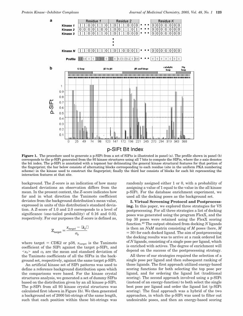
3. Calculation of the p-SIFts. A structural interac-tion fingerprint profile (p-SIFt) represents the degreeto which interactions are conserved across a set ofligand-receptor complexes. The p-SIFt, P(r), is derivedfrom an array, denoted below as b, of SIFt patterns, andits derivation from a set of SIFts is shown in Figure 1a.The array has length N for the total number of protein-ligand complexes and width K of SIFt fingerprints bits.The value of each element of P(r) is derived by averagingthe elements in each column of the SIFt matrix, yieldinga numerical interaction frequency that varies from 0 to1 for unobserved to always present, respectively. TheSIFt array, b, and resulting P(r) are given by,
and
where bi,r is the binary bit value in the SIFt i)1,N atposition r)1,K. The value in the p-SIFt at position r isgiven by,
4. Measurement of Similarity between SIFtsand/or p-SIFts. We have used the Tanimoto43 coef-ficient to measure the similarity between two SIFts,between two p-SIFts, and between a SIFt and a p-SIFt.The Tanimoto coefficient (Tc) between a p-SIFt, P(r), andSIFt, a, is defined as,
where K denotes the number of bits defining the SIFtand p-SIFt. Other similarity metrics were tested, in-cluding the cosine coefficient, Pearson’s correlationcoefficient, Dice coefficient, and city block distance, andwere found to not affect the relative rankings.44-47 Ashas been described previously, a set of SIFt patternscan be clustered using the Tanimoto similarity measureby applying standard hierarchical clustering algo-rithms.45,48
The statistical Z-score was employed to measure howsignificant the similarity between a SIFt and a targetp-SIFt (i.e., a group of structures) is above a certain
b ) (b1,1 b1,2 b1,3 · · · b1,K
b2,1 b2,2 b2,3 · · · b2,K
···.
bN,1 bN,2 bN,3 · · · bN,K
)P(r) ) [P1 P2 P3 P4 PK ]
P(r) ) ∑i)1
N
bi,r /N
Tc )
∑i)1
K
Pi ‚ai
∑i)1
K
Pi2 + ∑
i)1
K
ai2 - ∑
i)1
K
Pi ‚ai
122 Journal of Medicinal Chemistry, 2005, Vol. 48, No. 1 Chuaqui et al.
background. The Z-score is an indication of how manystandard deviations an observation differs from themean. In the present context, the Z-score indicates howfar and in what direction the Tanimoto coefficientdeviates from the background distribution’s mean value,expressed in units of this distribution’s standard devia-tion. A Z-score of 1.0 and 2.0 corresponds to a level ofsignificance (one-tailed probability) of 0.16 and 0.02,respectively. For our purposes the Z-score is defined as,
where target ) CDK2 or p38, xtarget is the Tanimotocoefficient of the SIFt against the target p-SIFt, and<xb> and σb are the mean and standard deviation ofthe Tanimoto coefficients of all the SIFts in the back-ground set, respectively, against the same target p-SIFt.
An artificial kinase set of SIFt patterns was used todefine a reference background distribution upon whichthe comparisons were based. For the kinase crystalstructures analysis, we generated a set of dummy SIFtsbased on the distribution given by an all kinase p-SIFt.The p-SIFt from all 93 kinase crystal structures wascalculated first (shown in Figure 1b). We then generateda background set of 2000 bit-strings of the same length,such that each position within these bit-strings was
randomly assigned either 1 or 0, with a probability ofassigning a value of 1 equal to the value in the all kinasep-SIFt. For the database enrichment experiment, weused all the docking poses as the background set.
5. Virtual Screening Protocol and Postprocess-ing. In this paper, we explored three strategies for VSpostprocessing. For all three strategies a list of dockingposes was generated using the program FlexX, and thetop 30 poses were retained using the FlexX scoringfunction.40 The output obtained from docking N ligandsis then an NxM matrix consisting of M poses (here, M) 30) for each docked ligand. The aim of postprocessingthe docking results was to arrive at a rank ordered listof N ligands, consisting of a single pose per ligand, whichis enriched with actives. The degree of enrichment willdepend on the success of the postprocessing strategy.
All three of our strategies required the selection of asingle pose per ligand and then subsequent ranking ofthose ligands. The first approach utilized energy-basedscoring functions for both selecting the top pose perligand, and for ordering the ligand list (traditionalscoring). The second approach involved using a p-SIFt(instead of an energy-function) to both select the singlebest pose per ligand and order the ligand list (p-SIFtscoring). The final approach was a hybrid of the twoapproaches, in which the p-SIFt was used to filter outundesirable poses, and then an energy-based scoring
Figure 1. The procedure used to generate a p-SIFt from a set of SIFts is illustrated in panel (a). The profile shown in panel (b)corresponds to the p-SIFt generated from the 93 kinase structures using all 7 bits to compute the SIFts, where the x-axis denotesthe bit index. The p-SIFt is annotated with a topmost bar delineating the general kinase structural features for that portion ofthe fingerprint; the bar below consists of alternating blocks corresponding to each residue (site in the uniform PKA numberingscheme) in the kinase used to construct the fingerprint; finally the third bar consists of blocks for each bit representing theinteraction features at that site.
Ztarget )xtarget - ⟨xb⟩
σb
Protein Kinase-Inhibitor Complexes Journal of Medicinal Chemistry, 2005, Vol. 48, No. 1 123
function was used to select the best pose per ligand andto create an ordered list of ligands (hybrid scoring).
In all three cases, the overall postprocessing schemeused to triage ligand placements, or poses, generatedfrom docking consisted of four general steps, namely,
A. rescoring: each pose generated (Nx30) is scoredusing standard scoring functions and p-SIFts
B. filtering: unrealistic poses are removed from thepool to be considered further.
C. final pose selection: a single pose per ligand isselected.
D. ranking of ligand list: the N ligands are rankordered.
The three scoring and postprocessing strategies wehave applied utilized different strategies to carry outsteps A-D as summarized in Table 1. For the tradi-tional scoring and hybrid scoring schemes, docked poses
were rescored using several widely applied scoringfunctions computed using the Cscore49 utility in Sybyl.42
For the p-SIFt scoring and hybrid scoring protocols wealso computed Z between the SIFt for the pose and thetarget p-SIFt, Ztarget , where target ) CDK2 or p38.
The filtering step B applied in the hybrid scoringscheme involved filtering out any poses having ZCDK2< 4.5 and Zp38 < 5.0, for VS against CDK2 or p38,respectively. The Ztarget cutoffs were chosen to be at thelowest value of the Ztarget distribution observed for theCDK2 and p38 X-ray structures. In addition, a canonicalinteraction filter was applied to each pose such thatSIFts not satisfying the subset of interactions havingan interaction frequency of 1 in the all-kinase p-SIFt,shown in Figure 1b, were filtered out (see section 2).
It is during the filtering step that incorrect ligandposes can be eliminated from the pool of poses that will
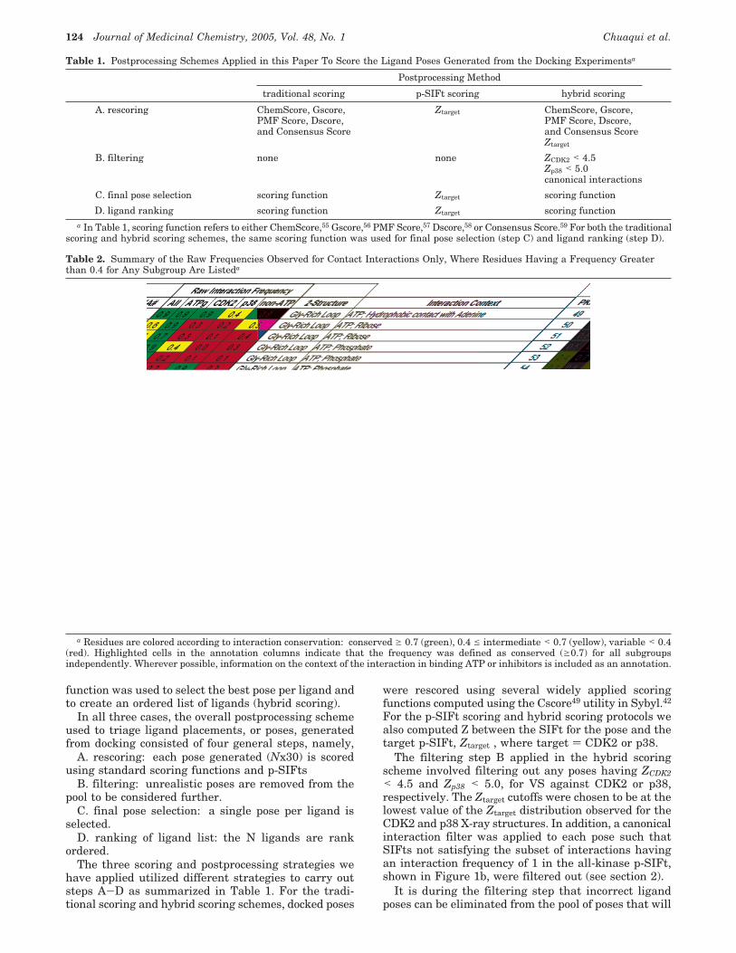
Table 1. Postprocessing Schemes Applied in this Paper To Score the Ligand Poses Generated from the Docking Experimentsa
Postprocessing Method
traditional scoring p-SIFt scoring hybrid scoring
A. rescoring ChemScore, Gscore,PMF Score, Dscore,and Consensus Score
Ztarget ChemScore, Gscore,PMF Score, Dscore,and Consensus ScoreZtarget
B. filtering none none ZCDK2 < 4.5Zp38 < 5.0canonical interactions
C. final pose selection scoring function Ztarget scoring function
D. ligand ranking scoring function Ztarget scoring functiona In Table 1, scoring function refers to either ChemScore,55 Gscore,56 PMF Score,57 Dscore,58 or Consensus Score.59 For both the traditional
scoring and hybrid scoring schemes, the same scoring function was used for final pose selection (step C) and ligand ranking (step D).
Table 2. Summary of the Raw Frequencies Observed for Contact Interactions Only, Where Residues Having a Frequency Greaterthan 0.4 for Any Subgroup Are Listeda
a Residues are colored according to interaction conservation: conserved g 0.7 (green), 0.4 e intermediate < 0.7 (yellow), variable < 0.4(red). Highlighted cells in the annotation columns indicate that the frequency was defined as conserved (g0.7) for all subgroupsindependently. Wherever possible, information on the context of the interaction in binding ATP or inhibitors is included as an annotation.
124 Journal of Medicinal Chemistry, 2005, Vol. 48, No. 1 Chuaqui et al.
be considered for final selection. The aim is thereforeto reduce the number of false positive poses whileretaining the plausible true positive poses. The filteringstep is optional and for comparison purposes wasomitted in order to generate results based only onfunctions (traditional scoring) and only on p-SIFTs (p-SIFt scoring).
Step C involves selecting a single pose per ligand fromthe set of poses that have passed all of the filters, if any,applied in step B. Enrichment curves and factors werecomputed by rank ordering (step D) the final set ofligand poses using the schemes outlined in Table 1.
Results1. Implementation of the SIFt Methodology for
the Kinase Family. The work presented in this paperis based on 93 X-ray structures representing 21 differentprotein kinases complexed with ATP, ATP analogues,and a variety of small molecule inhibitors. Of the 56ligand binding site residues used to construct the SIFts,those playing a significant role in interactions withligands are listed in Table 2, along with their uniformprotein kinase A (PKA) residue numbering.
The results of the hierarchical clustering of SIFtscomputed for the 93 kinase structures were describedin the original SIFt paper7 and revealed three majorclusters representing three dominant interaction pat-terns present in the ligand-kinase complexes. Cluster1 is composed of nine structures of small moleculeinhibitors interacting with p38 kinase (herein referredto as the p38 cluster). Similarly, Cluster 2 is composedof 20 structures for complexes involving inhibitors ofCDK2 kinase (denoted as the CDK2 cluster). The largestdistinct group, Cluster 3, is made up of nine ATP and16 ATP analogues complexed with different kinases,
which will be termed the ATP-group (ATPg) cluster. Theremaining 42% of the structures do not belong to anyparticular cluster. It is noteworthy that the hierarchicalclustering procedure, based solely on ligand-receptorinteraction features, is able to group structures intomeaningful clusters where variable ligands have similarinteractions with a fixed receptor (p38 and CDK2clusters) and where very similar ligands interact in ahighly conserved way with a diverse set of kinasereceptors (ATPg cluster).
2. Interaction Profiles and Profile-Profile Analy-sis. Each SIFt cluster represents a set of structureshaving similar conserved and variable interactions. Oneapproach to represent the degree of interaction conser-vation for any set of structures is to define a p-SIFt fromthe SIFts of those structures, where the p-SIFt mea-sures the degree of conservation of an interactionfeature at that residue. The p-SIFts may be derivedusing a reduced set of interaction features to representeach interaction. Thus, while the standard SIFts utilizes7 bits to characterize the interaction at each residue, asimplified p-SIFt may be derived from only the interac-tion frequencies of the contact bit at each residue.
To simplify the analyses, results presented in thissection were based on contact-only p-SIFts. The upperpanel of Figure 1 illustrates the general methodologyused to derive a p-SIFt from a set of SIFts.
As an initial application, p-SIFts provide a useful toolto overview the interaction patterns observed betweenligands and protein kinases. For this purpose, it isconvenient to define categories from the contact-onlyp-SIFts to characterize the observed interactions, e.g.,conserved g 0.7, 0.4 e intermediate < 0.7, variable <0.4, as denoted by dashed lines on the plot in Figure1b. It should be noted that the p-SIFts themselves are
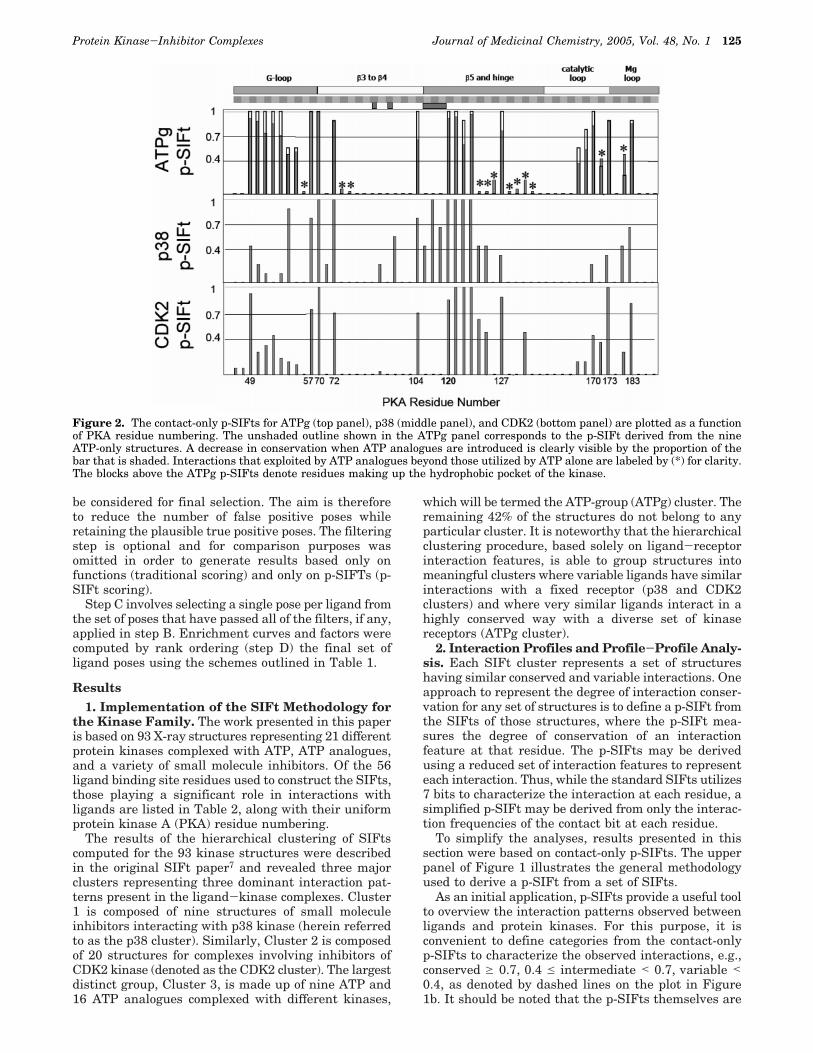
Figure 2. The contact-only p-SIFts for ATPg (top panel), p38 (middle panel), and CDK2 (bottom panel) are plotted as a functionof PKA residue numbering. The unshaded outline shown in the ATPg panel corresponds to the p-SIFt derived from the nineATP-only structures. A decrease in conservation when ATP analogues are introduced is clearly visible by the proportion of thebar that is shaded. Interactions that exploited by ATP analogues beyond those utilized by ATP alone are labeled by (*) for clarity.The blocks above the ATPg p-SIFts denote residues making up the hydrophobic pocket of the kinase.
Protein Kinase-Inhibitor Complexes Journal of Medicinal Chemistry, 2005, Vol. 48, No. 1 125
not particularly sensitive to minor variations in thecutoffs used for binning the interaction frequencies. Theoverall distribution of conserved, intermediate, andvariable interactions observed overall and for the ATPg,p38, and CDK2 clusters are summarized in Table 2.
Conservation in ATP and ATP Analogues. The25 members of the ATPg cluster consist of nine struc-tures of ATP complexed with three different kinases and16 structures of ATP analogues complexed with sixkinases. The ATPg p-SIFt computed from the ATPgcluster SIFts is shown in the top panel of Figure 2. Forcomparison we have also plotted the p-SIFt derivedusing only the 9 ATP structures in the ATPg cluster.For the nine ATP complexes, 18 out of 23 contacts areclassified as conserved between the kinases and theribose, triphosphate, and adenine moieties. Moreover,for the nine ATP structures there are no completelyvariable positions. Interestingly, even for these ATP-only structures, four interactions lie in the intermediateconservation range. Interactions between the γ-phos-phate and residues 54 and 55, making up the tip of theglycine-rich loop in the kinases, are dependent on theconformation of this flexible region of the binding siteand are observed only in approximately half of thestructures. Contact between the â-phosphate of ATP andresidue 171 is primarily determined by the conformationof the ATP phosphate groups. In approximately 60% ofthe structures, the R-â phosphate pyrophosphate bondis rotated such that the â-phosphate is oriented awayfrom residue 171 and toward the glycine-rich loop(Figure 3; PDB code 1atp). It is noteworthy that inseveral of these structures a water molecule is observedto take the place of the rotated â-phosphate and formwater-mediated interaction between ATP and residue171.50 Finally, contact between the adenine ring of ATPand residue 183 is largely a function of the side-chainidentity. No contact is observed for the ATP structuresthat have Ala at this position (50%) whereas Thr andVal side-chains are able to contact the adenine ringeither directly or via water-mediated interaction.
When the ATP analogues are considered in additionto the ATP complexes, the degree of variability in-creases. In particular, interactions with residues 104,122, and 168 shift from conserved to variable. Theextent of variability is clear when the ATPg p-SIFt iscompared to the ATP only p-SIFt, as shown in Figure2. The contacts that are not fully conserved for the ATPgcluster are colored yellow in Figure 3a and fall into theintermediate (∼21%) and variable (∼33%) ranges. Nev-ertheless, the ATPg p-SIFt reveals a high degree ofinteraction conservation as annotated in Table 2 andcolored green in Figure 3a. Of the 33 contacts observedacross the ATPg, ∼46% are classified as conserved (seeTable 3). The patterns of conserved interactions forATPg ligands define an ATP-like binding signature andprovide a baseline for comparison when analyzing non-ATP small molecule inhibitors
Non-ATP p-SIFts and Difference Profiles. Thecontact p-SIFts derived for the ATPg, CDK2, and P38clusters plotted in Figure 2 measure the degree ofinteraction conservation for each group of structures.From the p-SIFts, it is evident that CDK2 and p38inhibitors share some common binding interactions asobserved between ATP and some regions of the kinase
domain while displaying marked differences in others.A convenient way to compare directly the p-SIFts is todefine a difference profile computed by the directsubtraction of one p-SIFt from another. The differenceprofiles provide insight into how the interaction patternsobserved for known kinase inhibitors differ from thosedetailed above for ATP. To this end, we have defineddifference profiles p38-ATPg, p38-CDK2, and CDK2-ATPg, plotted in Figure 4.
For the p38-ATPg and p38-CDK2 difference profiles,the key distinctions are determined in part by theidentity of the residue at position 120. Referred to asthe “gatekeeper” residue, it controls the relative accessto the hydrophobic pocket of the ATP site, a region notoccupied by ATP. Bulky residues at position 120, suchas the Phe in CDK2, restrict access to the hydrophobic
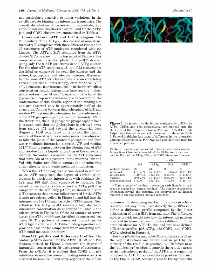
Figure 3. In panels a-c the binned contact-only p-SIFts forATPg, CDK2, and p38, respectively, are mapped onto thestructure of the complex between ATP and PKA (PDB code1atp) using the values and color scheme introduced in Table2. Panel d highlights key areas of difference in the interactionpatterns observed for ATP, CDK2, and p38 identified from thedifference profiles.
Table 3. Summary of Conserved, Intermediate, and VariableInteractions Observed across All of the 93 Kinase Structuresand for Each of the ATPg, P38, and CDK2 Structure Clustersa
cluster
ALL ATPg P38 CDK2
contact residues 56 33 26 29conserved 11 (19.6%) 15 (45.5%) 10 (38.5%) 12 (41.4%)intermediate 6 (10.7%) 7 (21.2%) 8 (30.8%) 5 (17.2%)variable 39 (69.6%) 11 (33.3%) 8 (30.8%) 12 (41.4%)unique conserved 5 (8.9%) 9 (27.3%) 4 (15.4%) 6 (20.7%)
a Total number of residues interacting with ligands in eachgroup is denoted as “contact residues”. The number of conservedinteraction beyond the canonical set observed for all ligandsappears in the row labeled “unique conserved”.
126 Journal of Medicinal Chemistry, 2005, Vol. 48, No. 1 Chuaqui et al.
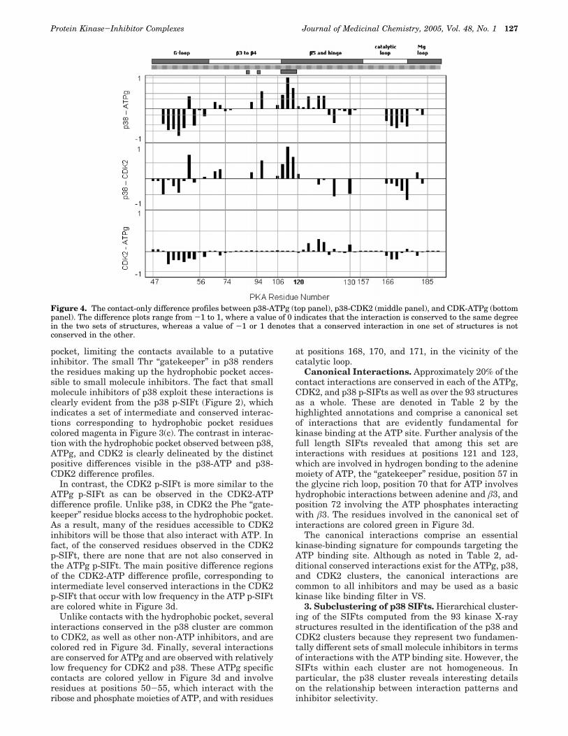
pocket, limiting the contacts available to a putativeinhibitor. The small Thr “gatekeeper” in p38 rendersthe residues making up the hydrophobic pocket acces-sible to small molecule inhibitors. The fact that smallmolecule inhibitors of p38 exploit these interactions isclearly evident from the p38 p-SIFt (Figure 2), whichindicates a set of intermediate and conserved interac-tions corresponding to hydrophobic pocket residuescolored magenta in Figure 3(c). The contrast in interac-tion with the hydrophobic pocket observed between p38,ATPg, and CDK2 is clearly delineated by the distinctpositive differences visible in the p38-ATP and p38-CDK2 difference profiles.
In contrast, the CDK2 p-SIFt is more similar to theATPg p-SIFt as can be observed in the CDK2-ATPdifference profile. Unlike p38, in CDK2 the Phe “gate-keeper” residue blocks access to the hydrophobic pocket.As a result, many of the residues accessible to CDK2inhibitors will be those that also interact with ATP. Infact, of the conserved residues observed in the CDK2p-SIFt, there are none that are not also conserved inthe ATPg p-SIFt. The main positive difference regionsof the CDK2-ATP difference profile, corresponding tointermediate level conserved interactions in the CDK2p-SIFt that occur with low frequency in the ATP p-SIFtare colored white in Figure 3d.
Unlike contacts with the hydrophobic pocket, severalinteractions conserved in the p38 cluster are commonto CDK2, as well as other non-ATP inhibitors, and arecolored red in Figure 3d. Finally, several interactionsare conserved for ATPg and are observed with relativelylow frequency for CDK2 and p38. These ATPg specificcontacts are colored yellow in Figure 3d and involveresidues at positions 50-55, which interact with theribose and phosphate moieties of ATP, and with residues
at positions 168, 170, and 171, in the vicinity of thecatalytic loop.
Canonical Interactions. Approximately 20% of thecontact interactions are conserved in each of the ATPg,CDK2, and p38 p-SIFts as well as over the 93 structuresas a whole. These are denoted in Table 2 by thehighlighted annotations and comprise a canonical setof interactions that are evidently fundamental forkinase binding at the ATP site. Further analysis of thefull length SIFts revealed that among this set areinteractions with residues at positions 121 and 123,which are involved in hydrogen bonding to the adeninemoiety of ATP, the “gatekeeper” residue, position 57 inthe glycine rich loop, position 70 that for ATP involveshydrophobic interactions between adenine and â3, andposition 72 involving the ATP phosphates interactingwith â3. The residues involved in the canonical set ofinteractions are colored green in Figure 3d.
The canonical interactions comprise an essentialkinase-binding signature for compounds targeting theATP binding site. Although as noted in Table 2, ad-ditional conserved interactions exist for the ATPg, p38,and CDK2 clusters, the canonical interactions arecommon to all inhibitors and may be used as a basickinase like binding filter in VS.
3. Subclustering of p38 SIFts. Hierarchical cluster-ing of the SIFts computed from the 93 kinase X-raystructures resulted in the identification of the p38 andCDK2 clusters because they represent two fundamen-tally different sets of small molecule inhibitors in termsof interactions with the ATP binding site. However, theSIFts within each cluster are not homogeneous. Inparticular, the p38 cluster reveals interesting detailson the relationship between interaction patterns andinhibitor selectivity.
Figure 4. The contact-only difference profiles between p38-ATPg (top panel), p38-CDK2 (middle panel), and CDK-ATPg (bottompanel). The difference plots range from -1 to 1, where a value of 0 indicates that the interaction is conserved to the same degreein the two sets of structures, whereas a value of -1 or 1 denotes that a conserved interaction in one set of structures is notconserved in the other.
Protein Kinase-Inhibitor Complexes Journal of Medicinal Chemistry, 2005, Vol. 48, No. 1 127
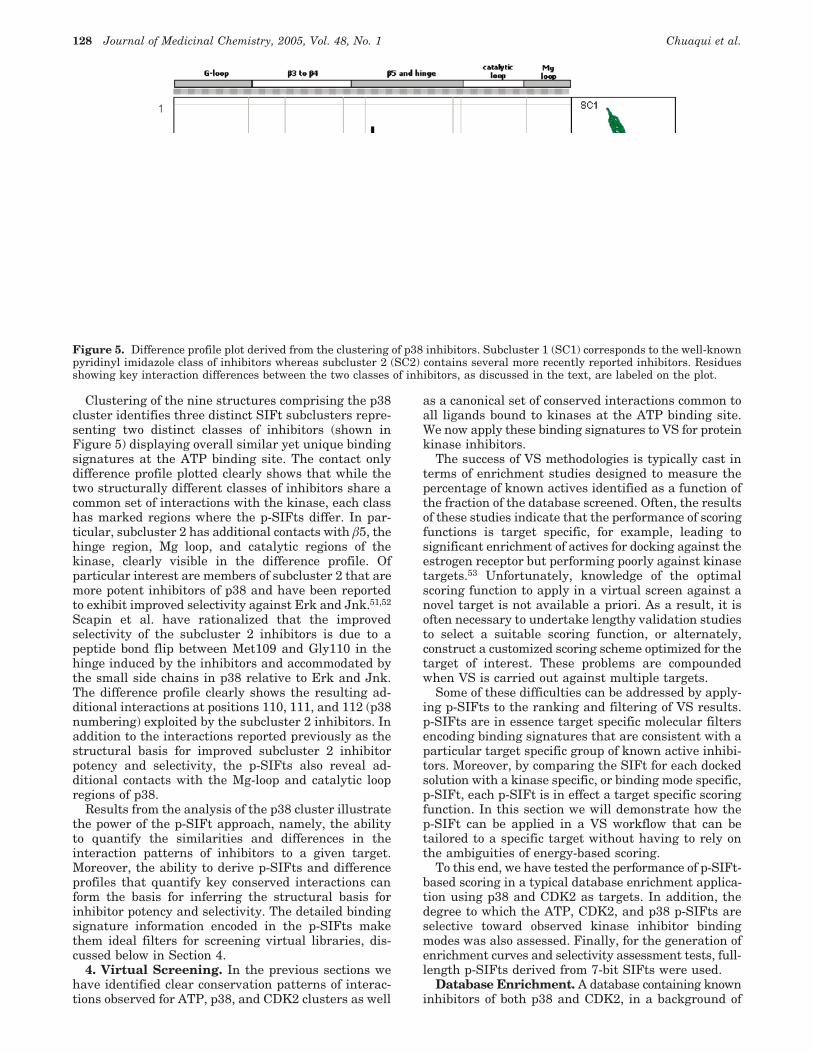
Clustering of the nine structures comprising the p38cluster identifies three distinct SIFt subclusters repre-senting two distinct classes of inhibitors (shown inFigure 5) displaying overall similar yet unique bindingsignatures at the ATP binding site. The contact onlydifference profile plotted clearly shows that while thetwo structurally different classes of inhibitors share acommon set of interactions with the kinase, each classhas marked regions where the p-SIFts differ. In par-ticular, subcluster 2 has additional contacts with â5, thehinge region, Mg loop, and catalytic regions of thekinase, clearly visible in the difference profile. Ofparticular interest are members of subcluster 2 that aremore potent inhibitors of p38 and have been reportedto exhibit improved selectivity against Erk and Jnk.51,52
Scapin et al. have rationalized that the improvedselectivity of the subcluster 2 inhibitors is due to apeptide bond flip between Met109 and Gly110 in thehinge induced by the inhibitors and accommodated bythe small side chains in p38 relative to Erk and Jnk.The difference profile clearly shows the resulting ad-ditional interactions at positions 110, 111, and 112 (p38numbering) exploited by the subcluster 2 inhibitors. Inaddition to the interactions reported previously as thestructural basis for improved subcluster 2 inhibitorpotency and selectivity, the p-SIFts also reveal ad-ditional contacts with the Mg-loop and catalytic loopregions of p38.
Results from the analysis of the p38 cluster illustratethe power of the p-SIFt approach, namely, the abilityto quantify the similarities and differences in theinteraction patterns of inhibitors to a given target.Moreover, the ability to derive p-SIFts and differenceprofiles that quantify key conserved interactions canform the basis for inferring the structural basis forinhibitor potency and selectivity. The detailed bindingsignature information encoded in the p-SIFts makethem ideal filters for screening virtual libraries, dis-cussed below in Section 4.
4. Virtual Screening. In the previous sections wehave identified clear conservation patterns of interac-tions observed for ATP, p38, and CDK2 clusters as well
as a canonical set of conserved interactions common toall ligands bound to kinases at the ATP binding site.We now apply these binding signatures to VS for proteinkinase inhibitors.
The success of VS methodologies is typically cast interms of enrichment studies designed to measure thepercentage of known actives identified as a function ofthe fraction of the database screened. Often, the resultsof these studies indicate that the performance of scoringfunctions is target specific, for example, leading tosignificant enrichment of actives for docking against theestrogen receptor but performing poorly against kinasetargets.53 Unfortunately, knowledge of the optimalscoring function to apply in a virtual screen against anovel target is not available a priori. As a result, it isoften necessary to undertake lengthy validation studiesto select a suitable scoring function, or alternately,construct a customized scoring scheme optimized for thetarget of interest. These problems are compoundedwhen VS is carried out against multiple targets.
Some of these difficulties can be addressed by apply-ing p-SIFts to the ranking and filtering of VS results.p-SIFts are in essence target specific molecular filtersencoding binding signatures that are consistent with aparticular target specific group of known active inhibi-tors. Moreover, by comparing the SIFt for each dockedsolution with a kinase specific, or binding mode specific,p-SIFt, each p-SIFt is in effect a target specific scoringfunction. In this section we will demonstrate how thep-SIFt can be applied in a VS workflow that can betailored to a specific target without having to rely onthe ambiguities of energy-based scoring.
To this end, we have tested the performance of p-SIFt-based scoring in a typical database enrichment applica-tion using p38 and CDK2 as targets. In addition, thedegree to which the ATP, CDK2, and p38 p-SIFts areselective toward observed kinase inhibitor bindingmodes was also assessed. Finally, for the generation ofenrichment curves and selectivity assessment tests, full-length p-SIFts derived from 7-bit SIFts were used.
Database Enrichment. A database containing knowninhibitors of both p38 and CDK2, in a background of
Figure 5. Difference profile plot derived from the clustering of p38 inhibitors. Subcluster 1 (SC1) corresponds to the well-knownpyridinyl imidazole class of inhibitors whereas subcluster 2 (SC2) contains several more recently reported inhibitors. Residuesshowing key interaction differences between the two classes of inhibitors, as discussed in the text, are labeled on the plot.
128 Journal of Medicinal Chemistry, 2005, Vol. 48, No. 1 Chuaqui et al.
1000 diverse commercially available compounds wasdocked against the X-ray structures of CDK2 (PDB code1di8) and p38 (PDB code 1a9u). The ability of our p-SIFtVS protocol to identify known actives was quantified bycomputing enrichment curves plotting the percentageof actives recovered as a function of the percentage ofthe database screened.
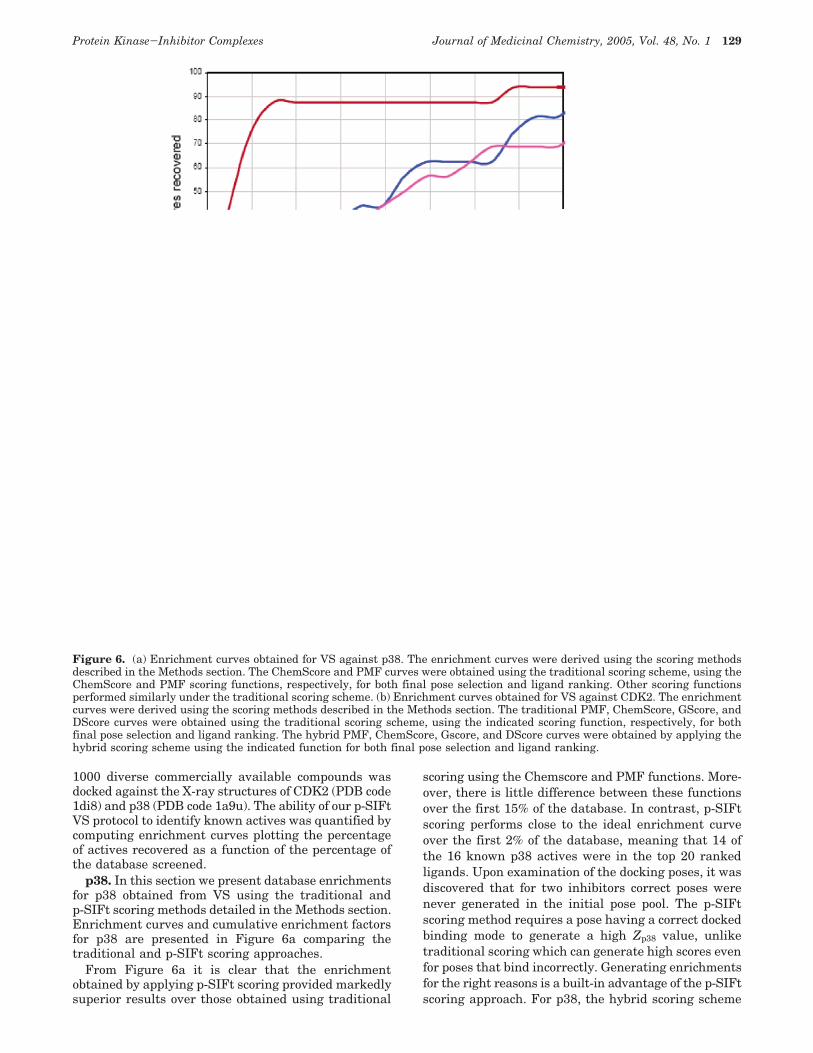
p38. In this section we present database enrichmentsfor p38 obtained from VS using the traditional andp-SIFt scoring methods detailed in the Methods section.Enrichment curves and cumulative enrichment factorsfor p38 are presented in Figure 6a comparing thetraditional and p-SIFt scoring approaches.
From Figure 6a it is clear that the enrichmentobtained by applying p-SIFt scoring provided markedlysuperior results over those obtained using traditional
scoring using the Chemscore and PMF functions. More-over, there is little difference between these functionsover the first 15% of the database. In contrast, p-SIFtscoring performs close to the ideal enrichment curveover the first 2% of the database, meaning that 14 ofthe 16 known p38 actives were in the top 20 rankedligands. Upon examination of the docking poses, it wasdiscovered that for two inhibitors correct poses werenever generated in the initial pose pool. The p-SIFtscoring method requires a pose having a correct dockedbinding mode to generate a high Zp38 value, unliketraditional scoring which can generate high scores evenfor poses that bind incorrectly. Generating enrichmentsfor the right reasons is a built-in advantage of the p-SIFtscoring approach. For p38, the hybrid scoring scheme
Figure 6. (a) Enrichment curves obtained for VS against p38. The enrichment curves were derived using the scoring methodsdescribed in the Methods section. The ChemScore and PMF curves were obtained using the traditional scoring scheme, using theChemScore and PMF scoring functions, respectively, for both final pose selection and ligand ranking. Other scoring functionsperformed similarly under the traditional scoring scheme. (b) Enrichment curves obtained for VS against CDK2. The enrichmentcurves were derived using the scoring methods described in the Methods section. The traditional PMF, ChemScore, GScore, andDScore curves were obtained using the traditional scoring scheme, using the indicated scoring function, respectively, for bothfinal pose selection and ligand ranking. The hybrid PMF, ChemScore, Gscore, and DScore curves were obtained by applying thehybrid scoring scheme using the indicated function for both final pose selection and ligand ranking.
Protein Kinase-Inhibitor Complexes Journal of Medicinal Chemistry, 2005, Vol. 48, No. 1 129
was found to offer no improvement in enrichment overthat obtained from using p-SIFt scoring.
CDK2. Enrichment curves were derived using tradi-tional scoring, p-SIFt scoring, and hybrid scoring, arepresented in Figure 6b for docking against CDK2. Astriking feature of these curves is that all of the hybridscoring scheme variants performed better than thetraditional and p-SIFt schemes irrespective of whatscoring function was used for pose selection and rank-ing. It would appear that once the majority of theincorrect poses that contribute to false positive scoresare filtered out, the differences between scoring func-tions visible in the results using these functions alone(traditional scoring) is factored out. Enrichments ob-tained using p-SIFt scoring are comparable to tradi-tional scoring up to 6% of the database screened andsignificantly better at higher levels.
Attaining database enrichments for CDK2 compa-rable to those obtained for p38 is a considerably morechallenging task for VS. The large gatekeeper residuein CDK2 restricts the number of residues accessible inthe ATP binding site. The p-SIFt for CDK2 samplesfewer residues compared to p38 and conserved interac-tions are distributed over a relatively small spatialregion. As a result, in the CDK2 there are fewerconstraints to generate ligand placements and it istherefore easier to generate poses that satisfy conservedinteractions in CDK2 compared to p38 where theresidues of the hydrophobic pocket are accessible. Ineffect, the CDK2 p-SIFt is less selective against falseposes as evidenced by the superior performance ofp-SIFt scoring for p38 versus CDK2.
Selectivity Assessment. The difference profilespresented in Section 2 exhibit clear regions where ATPg,CDK2, and p38 inhibitors bind to kinases in uniqueways. These observations suggest that p-SIFts can beused to model the selectivity of inhibitors based on thetypes of interactions they are able to satisfy whenbinding to the kinase. To validate the use of p-SIFts asselectivity filters, we have carried out self-recognitionexperiments using the set of 93 X-ray structures as atest data set. For this purpose, ATPg, CDK2, and p38validation p-SIFts were derived. For CDK2 and ATPg∼50% of the structures in each group were selectedrandomly to derive the p-SIFt, whereas for p38, onlythe SIFts corresponding to subcluster 1 were used. Theremaining ATPg, CDK2, and p38 structures were notused to derive the validation p-SIFts. For each p-SIFt,Ztarget values were then computed against each of the93 kinase structures in order to assess the ability ofp-SIFts to recognize members of their own group. Forthe p-SIFts to serve as effective molecular filters, thep38 p-SIFt needs to generate statistically significantlyhigher Zp38 against the p38 cluster X-ray structuresrelative to the remaining structures, whereas the CDK2and ATPg p-SIFts should perform well against theCDK2 and ATPg structures, respectively.
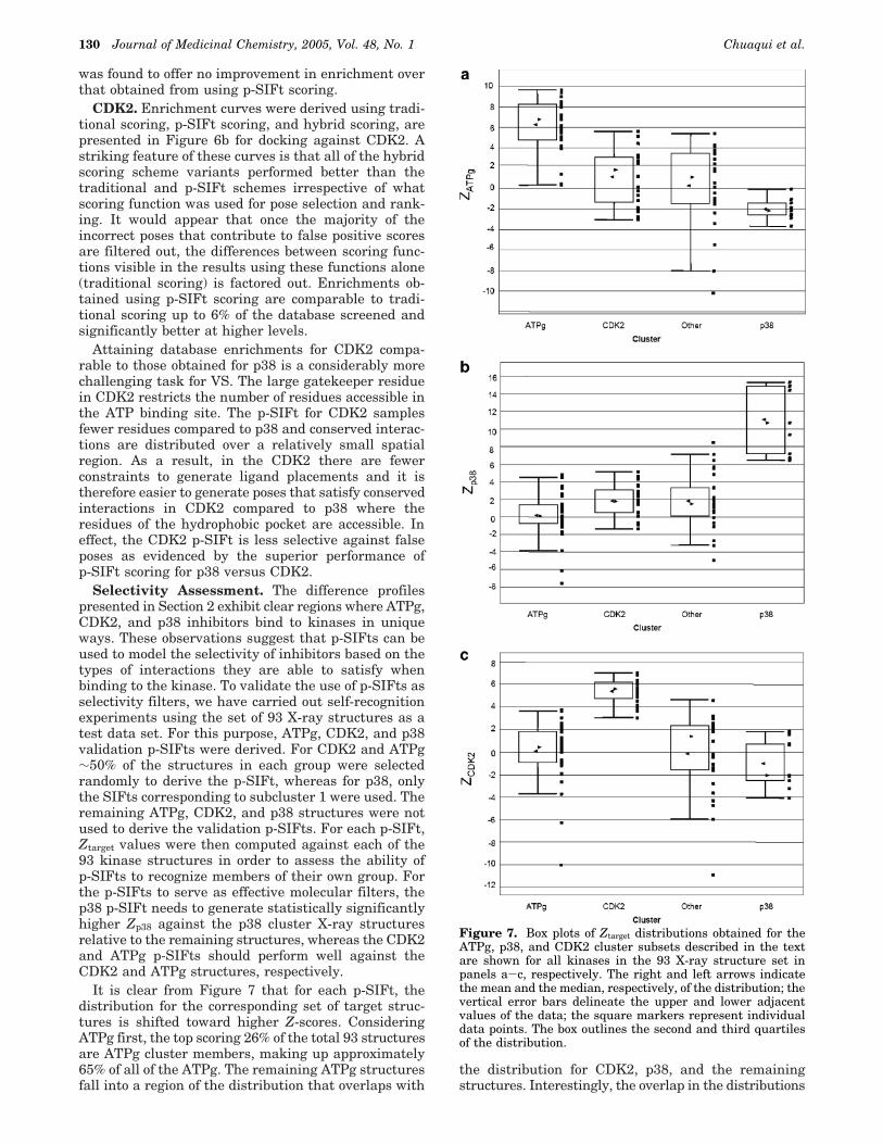
It is clear from Figure 7 that for each p-SIFt, thedistribution for the corresponding set of target struc-tures is shifted toward higher Z-scores. ConsideringATPg first, the top scoring 26% of the total 93 structuresare ATPg cluster members, making up approximately65% of all of the ATPg. The remaining ATPg structuresfall into a region of the distribution that overlaps with
the distribution for CDK2, p38, and the remainingstructures. Interestingly, the overlap in the distributions
Figure 7. Box plots of Ztarget distributions obtained for theATPg, p38, and CDK2 cluster subsets described in the textare shown for all kinases in the 93 X-ray structure set inpanels a-c, respectively. The right and left arrows indicatethe mean and the median, respectively, of the distribution; thevertical error bars delineate the upper and lower adjacentvalues of the data; the square markers represent individualdata points. The box outlines the second and third quartilesof the distribution.
130 Journal of Medicinal Chemistry, 2005, Vol. 48, No. 1 Chuaqui et al.
may be rationalized in terms of the p-SIFt similaritiesdiscussed in Section 3. The similarity between the ATPgand CDK2 p-SIFts, discussed previously, has the con-sequence that 90% of the CDK2 structures overlap in Zwith the lowest scoring 35% of the ATPg. This overlapexists primarily because the ATPg p-SIFt is in essencederived from a subset of the interactions sampled byCDK2. However, the differences between the ATP andCDK2 interaction patterns are captured in the CDK2p-SIFt. Consequently, the highest segment in the dis-tribution shown in Figure 7c contains 19 of 20 CDK2structures and overlaps with only 2 ATPg structures.
The greatest separation in Z-score distributions isobtained for p38, due primarily from p-SIFt featuresreflecting conserved residues in the hydrophobic pocketof the ATP binding site. The p38 structures fall into twogroups in the distribution, dividing neatly between thehighest scoring subcluster 1 structures, used to derivethe p-SIFt, and slightly lower scoring subcluster 2examples. The latter set overlaps in Zp38 with 1qpe,1pme, and 3erk. Both 1pme and 3erk are examples ofcomplexes between pyridinyl imidazole compounds com-plexed with variants of Erk2, whereas 1qpe is a struc-ture of PP2 complexed with Lck. As in p38, the “gate-keeper residue” in Lck is also Thr, and the two kinaseshave relatively similar ATP binding sites. These ex-amples highlight the fact that the p-SIFts are able tocapture similarities in interaction patterns arising, onone hand from ligand similarity (1pme and 3erk), andon the other, from binding site similarity. Finally, wefind that that using a multistructure p-SIFt rather thana single structure (1a9u) yields improved separationbetween Z-score distributions.
Discussion and Conclusion
This paper introduces interaction profiling (p-SIFt)as a new and powerful approach to understand smallmolecule protein interactions. Sequence profile ap-proaches have previously been shown to successfullydetect remote protein sequence and structural relation-ships. Our results are the first demonstration, to ourknowledge, that this approach seems well suited toanalysis of protein-inhibitor complexes.
The implementation of the p-SIFt methodology andunderlying SIFts is very flexible. The SIFts themselvescan be constructed from any number of interactionfeatures or bits, whereas the p-SIFts can be derived tocapture very general interaction patterns, for example,a canonical kinase binding signature, or rather, capturea very focused set of interactions, as in the example ofthe p38 subcluster difference profile. We used simpleaveraging of the bit values to construct the profiles,although more sophisticated weighting schemes, asdescribed extensively in the literature,54 can also beused in order to improve the sensitivity of the p-SIFt.The weighting scheme may be useful to reduce thecontribution of highly correlated bits in the profile orto add weight to bits that are most correlated withinhibitor activity. In addition to the Tanimoto coef-ficient, several other well-known similarity metrics canbe applied to compare the bit-strings, such as the cosinecoefficient and measures of the Euclidean distance.44-47
The results from the profile analysis of ATP, p38, andCDK2 show that these profiles can be useful to quickly
define similarities and differences between groups ofsmall molecule inhibitors and these can be applied toVS. We believe that these examples are generallyapplicable to other important protein-drug targets. Itis apparent that in order to be competitive with ATP,small molecule inhibitors must satisfy a set of canonicalinteractions, common to those made by the adeninemoiety of ATP, along the hinge and â3-â5 regions ofthe kinase. In contrast, conserved interactions involvingthe ATP-phosphates and ribose ring are not a require-ment for small molecule inhibition. With few exceptions,small molecule inhibitors and ATP share the same“anchoring” interactions at the hinge region of thebinding site, but not necessarily the interactions key tometal binding and phosphorylation. Our profile-profilecomparisons also reveal that, unlike ATP, on average,small molecules interact with the kinase in the regionof the hydrophobic pocket when accessible and extendfurther along the hinge toward the solvent interface ofthe ATP binding site.
We find that when comparing the overall conservedand variable interactions observed in different kinasesubgroups, the coarse-grained contact-only p-SIFts pro-vide a clear and uncluttered analysis. However, fordetailed analyses and in VS applications, the full-featured interaction p-SIFt is most useful.
Virtual screening, in general, suffers from a signal-to-noise problem: the true ligand binding mode is moreoften than not buried in a background of high scoringincorrect ligand placements.22 Because of this high falsepositive problem inherent in VS, the ability to filter out“bogus” poses is critical.27 Our results show that theZ-score computed against a p-SIFt is a sensitive mea-sure, able to separate ligands that are binding to areceptor with a required interaction pattern from highscoring VS poses that are binding inconsistently withknown ligands to the desired target. Of course, p-SIFtfiltering may also miss out novel inhibitor bindingmodes (false negatives). However, the p-SIFt approachmay be also used to identify novel binding modes byretaining poses that are most dissimilar to the p-SIFtderived from known binding modes. This approach couldbe extended in order to select a diverse set of bindingmodes spanning a range of similarity with the p-SIFt.
Cross-docking experiments present a particularlychallenging problem for VS. Docking tends to generatemany bogus poses per ligand, creating the daunting taskof detecting the correct poses from many incorrect ligandplacements. To further complicate matters, in cross-docking some of the incorrect poses will correspond toinhibitors of another kinase and therefore containkinase-binding motifs, increasing the probability thatthey will score as false-positive hits in the VS. However,our results demonstrate that the p-SIFt-based scoringscheme can serve as a flexible and transferable targetspecific approach to VS lead discovery.
Given the rapid growth in the number of availableX-ray structures, it should be possible to eventuallyconstruct and screen against a virtual selectivity panelof p-SIFts for multiple drug targets in much the sameway that inhibitors are routinely tested against a panelof in vitro inhibition assays. The resulting virtualselectivity panel could be precomputed for ligands invirtual libraries, thus providing an annotation that
Protein Kinase-Inhibitor Complexes Journal of Medicinal Chemistry, 2005, Vol. 48, No. 1 131
could be mined when selective inhibitors to any targetare desired. In addition, predicted cross-reactivity to agiven drug target could be an effective starting pointfor lead discovery for novel targets, an approach thathas been demonstrated to be fruitful for protein kinases.
Acknowledgment. The authors would like to thankChristian Lemmen, Marcus Gastreich, and Rajiah Den-ny for their help with the docking experiments. We alsothank Rainer Fuchs, Herman van Vlijmen, AlexeyLugovskoy, and Jennifer Campbell for helpful commentson the manuscript.
Appendix
The abbreviations of common amino acids are inaccordance with the recommendations of IUPAC. Ad-ditional abbreviations: ATP, adenosine triphosphate;ATPg, adenosine triphosphate group; CDK2, cyclindependent kinase 2; Chk1, checkpoint kinase 1; EFCP,extended connectivity fingerprint; Erk, extracellularsignal-regultaed kinase; Jnk, c-Jun N-terminal kinase;NMR, nuclear magnetic resonance; p38, p38 mitogen-activated protein kinase; PKA, protein kinase A; PDB,protein databank; p-SIFt, profile structural interactionfingerprint; SBDD, structure-based drug design; SC1,subcluster 1; SC2, subcluster2; SIFt, structural interac-tion fingerprint; TGFâ-RI, transforming growth factorbeta receptor I; VS, virtual screening.
Supporting Information Available: A table listing the93 X-ray structures and corresponding ligands used in thispaper is available free of charge via the Internet at http://pubs.acs.org.
References(1) Alvarez, J. C. High-throughput docking as a source of novel
drug leads. Curr. Opin. Chem. Biol. 2004, DOI 10.1016/j.cbpa.2004.05.001.
(2) Perola, E.; Walters, W. P.; Charifson, P. S. A detailed comparisonof current docking and scoring methods on systems of pharma-ceutical relevance. Proteins 2004, 56, 235-249.
(3) Muegge, I.; Enyedy, I. J. Virtual screening for kinase targets.Curr. Med. Chem. 2004, 11, 693-707.
(4) Lyne, P. D. Structure-based virtual screening: an overview.Drug. Discovery Today 2002, 7, 1047-1055.
(5) Bajorath, J. Integration of virtual and high-throughput screen-ing. Nat. Rev. Drug Discovery 2002, 1, 882-894.
(6) Chalk, A. J.; Worth, C. L.; Overington, J. P.; Chan, A. W.PDBLIG: Classification of Small Molecular Protein Binding inthe Protein Data Bank. J. Med. Chem. 2004, 47, 3807-3816.
(7) Deng, Z.; Chuaqui, C.; Singh, J. Structural interaction finger-print (SIFt): a novel method for analyzing three-dimensionalprotein-ligand binding interactions. J. Med. Chem. 2004, 47,337-344.
(8) Gribskov, M.; McLachlan, A. D.; Eisenberg, D. Profile analysis:detection of distantly related proteins. Proc. Natl. Acad. Sci.U.S.A. 1987, 84, 4355-4358.
(9) Gribskov, M.; Luthy, R.; Eisenberg, D. Profile analysis. MethodsEnzymol. 1990, 183, 146-159.
(10) Wang, G.; Dunbrack, R. L., Jr. Scoring profile-to-profile sequencealignments. Protein Sci. 2004, 13, 1612-1626.
(11) Mehta, P. K.; Argos, P.; Barbour, A. D.; Christen, P. Recognizingvery distant sequence relationships among proteins by familyprofile analysis. Proteins 1999, 35, 387-400.
(12) Rice, D. W.; Eisenberg, D. A 3D-1D substitution matrix forprotein fold recognition that includes predicted secondary struc-ture of the sequence. J. Mol. Biol. 1997, 267, 1026-1038.
(13) Koonin, E. V.; Wolf, Y. I.; Aravind, L. Protein fold recognitionusing sequence profiles and its application in structural genom-ics. Adv. Protein Chem. 2000, 54, 245-275.
(14) Stahl, M.; Rarey, M. Detailed analysis of scoring functions forvirtual screening. J. Med. Chem. 2001, 44, 1035-1042.
(15) McGovern, S. L.; Caselli, E.; Grigorieff, N.; Shoichet, B. K. Acommon mechanism underlying promiscuous inhibitors fromvirtual and high-throughput screening. J. Med. Chem. 2002, 45,1712-1722.
(16) Bissantz, C.; Folkers, G.; Rognan, D. Protein-based virtualscreening of chemical databases. 1. Evaluation of differentdocking/scoring combinations. J. Med. Chem. 2000, 43, 4759-4767.
(17) Kontoyianni, M.; McClellan, L. M.; Sokol, G. S. Evaluation ofdocking performance: comparative data on docking algorithms.J. Med. Chem. 2004, 47, 558-565.
(18) Schulz-Gasch, T.; Stahl, M. Binding site characteristics instructure-based virtual screening: evaluation of current dockingtools. J. Mol. Model. 2003, 9, 47-57.
(19) Jansen, J.; Martin, E. Target-biased scoring approaches andexpert systems in structure based virtual screening. Curr. Opin.Chem. Biol. 2004, in press.
(20) Wang, R.; Lu, Y.; Wang, S. Comparative evaluation of 11 scoringfunctions for molecular docking. J. Med. Chem. 2003, 46, 2287-2303.
(21) Gohlke, H.; Hendlich, M.; Klebe, G. Knowledge-based scoringfunction to predict protein-ligand interactions. J. Mol. Biol.2000, 295, 337-356.
(22) Lyne, P. D.; Kenny, P. W.; Cosgrove, D. A.; Deng, C.; Zabludoff,S.; Wendoloski, J. J.; Ashwell, S. Identification of compoundswith nanomolar binding affinity for checkpoint kinase-1 usingknowledge-based virtual screening. J. Med. Chem. 2004, 47,1962-1968.
(23) Paul, N.; Kellenberger, E.; Bret, G.; Muller, P.; Rognan, D.Recovering the true targets of specific ligands by virtual screen-ing of the protein data bank. Proteins 2004, 54, 671-680.
(24) Jacoby, E.; Schuffenhauer, A.; Floersheim, P. Chemogenomicsknowledge-based strategies in drug discovery. Drug News Per-spect. 2003, 16, 93-102.
(25) Klon, A. E.; Glick, M.; Thoma, M.; Acklin, P.; Davies, J. W.Finding more needles in the haystack: A simple and efficientmethod for improving high-throughput docking results. J. Med.Chem. 2004, 47, 2743-2749.
(26) Fradera, X.; Mestres, J. Guided docking approaches to structure-based design and screening. Curr. Top. Med. Chem. 2004, 4,687-700.
(27) Claussen, H.; Gastreich, M.; Apelt, V.; Greene, J.; Hindle, S. A.;Lemmen, C. The FlexX Database Docking Environment -Rational Exctraction of Receptor Based Pharmacophores. Curr.Drug Discovery Technol. 2004, 1, 49-60.
(28) Cavasotto, C. N.; Abagyan, R. A. Protein flexibility in liganddocking and virtual screening to protein kinases. J. Mol. Biol.2004, 337, 209-225.
(29) Friesner, R. A.; Banks, J. L.; Murphy, R. B.; Halgren, T. A.;Klicic, J. J.; Mainz, D. T.; Repasky, M. P.; Knoll, E. H.; Shelley,M.; Perry, J. K.; Shaw, D. E.; Francis, P.; Shenkin, P. S. Glide:a new approach for rapid, accurate docking and scoring. 1.Method and assessment of docking accuracy. J. Med. Chem.2004, 47, 1739-1749.
(30) Vangrevelinghe, E.; Zimmermann, K.; Schoepfer, J.; Portmann,R.; Fabbro, D.; Furet, P. Discovery of a potent and selectiveprotein kinase CK2 inhibitor by high-throughput docking. J.Med. Chem. 2003, 46, 2656-2662.
(31) Kroemer, R. T.; Vulpetti, A.; McDonald, J. J.; Rohrer, D. C.;Trosset, J. Y.; Giordanetto, F.; Cotesta, S.; McMartin, C.; Kihlen,M.; Stouten, P. F. Assessment of docking poses: interactions-based accuracy classification (IBAC) versus crystal structuredeviations. J. Chem. Inf. Comput. Sci. 2004, 44, 871-881.
(32) Singh, J.; Chuaqui, C. E.; Boriack-Sjodin, P. A.; Lee, W. C.; Pontz,T.; Corbley, M. J.; Cheung, H. K.; Arduini, R. M.; Mead, J. N.;Newman, M. N.; Papadatos, J. L.; Bowes, S.; Josiah, S.; Ling,L. E. Successful shape-based virtual screening: the discoveryof a potent inhibitor of the type I TGFâ receptor kinase (TâRI).Bioorg. Med. Chem. Lett. 2003, 13, 4355-4359.
(33) Cohen, P. Protein kinasessthe major drug targets of the twenty-first century? Nat. Rev. Drug. Discovery 2002, 1, 309-315.
(34) ter Haar, E.; Walters, W. P.; Pazhanisamy, S.; Taslimi, P.; Pierce,A. C.; Bemis, G. W.; Salituro, F. G.; Harbeson, S. L. Kinasechemogenomics: targeting the human kinome for target valida-tion and drug discovery. Mini Rev. Med. Chem. 2004, 4, 235-253.
(35) Manning, G.; Whyte, D. B.; Martinez, R.; Hunter, T.; Sudar-sanam, S. The protein kinase complement of the human genome.Science 2002, 298, 1912-1934.
(36) Vieth, M.; Higgs, R. E.; Robertson, D. H.; Shapiro, M.; Gragg,E. A.; Hemmerle, H. Kinomics-structural biology and chemoge-nomics of kinase inhibitors and targets. Biochim. Biophys. Acta2004, 1697, 243-257.
(37) Adams, J.; Lee, D. Recent progress towards the identificationof selective inhibitors of serine/threonine protein kinases. Curr.Opin. Drug Discovery Dev. 1999, 2, 96-109.
(38) PipelinePilot; Scitegic Inc., San Diego, California, version 3.0.1.0.(39) Omega; Openeye Scientific Software Inc., Santa Fe, New Mexico,
version 1.8.(40) Rarey, M.; Kramer, B.; Lengauer, T.; Klebe, G. A fast flexible
docking method using an incremental construction algorithm.J. Mol. Biol. 1996, 261, 470-489.
132 Journal of Medicinal Chemistry, 2005, Vol. 48, No. 1 Chuaqui et al.
(41) Kramer, B.; Rarey, M.; Lengauer, T. Evaluation of the FLEXXincremental construction algorithm for protein-ligand docking.Proteins 1999, 37, 228-241.
(42) Sybyl; Tripos, Inc., St. Louis, MO, version 6.9.1.(43) Willett, P. Chemical similarity searching. J. Chem. Inf. Comput.
Sci. 1998, 38, 983-996.(44) Chen, X.; Reynolds, C. H. Performance of similarity measures
in 2D fragment-based similarity searching: comparison ofstructural descriptors and similarity coefficients. J Chem. Inf.Comput. Sci. 2002, 42, 1407-1414.
(45) Raymond, J. W.; Blankley, C. J.; Willett, P. Comparison ofchemical clustering methods using graph- and fingerprint-basedsimilarity measures. J. Mol. Graph. Model. 2003, 21, 421-433.
(46) Willett, P. Similarity-based approaches to virtual screening.Biochem. Soc. Trans. 2003, 31, 603-606.
(47) Willett, P. Evaluation of molecular similarity and moleculardiversity methods using biological activity data. Methods Mol.Biol. 2004, 275, 51-64.
(48) Dubes, R.; Jain, A. K. Clustering methodologies in exploratoryanalysis. Adv. Comput. 1980, 19, 113-228.
(49) CScore; Tripos, Inc., St. Louis, MO, version in Sybyl 6.9.1.(50) See structures with PDB codes: 1atp, 1phk, 2phk, and 1ql6.(51) Fitzgerald, C. E.; Patel, S. B.; Becker, J. W.; Cameron, P. M.;
Zaller, D.; Pikounis, V. B.; O’Keefe, S. J.; Scapin, G. Structuralbasis for p38alpha MAP kinase quinazolinone and pyridol-pyrimidine inhibitor specificity. Nat. Struct. Biol. 2003, 10, 764-769.
(52) Scapin, G. Structural biology in drug design: selective proteinkinase inhibitors. Drug Discovery Today 2002, 7, 601-611.
(53) Halgren, T. A.; Murphy, R. B.; Friesner, R. A.; Beard, H. S.; Frye,L. L.; Pollard, W. T.; Banks, J. L. Glide: a new approach forrapid, accurate docking and scoring. 2. Enrichment factors indatabase screening. J. Med. Chem. 2004, 47, 1750-1759.
(54) Vingron, M.; Sibbald, P. R. Weighting in sequence space: acomparison of methods in terms of generalized sequences. Proc.Natl. Acad. Sci. U.S.A. 1993, 90, 8777-8781.
(55) Eldridge, M.; Murray, C. W.; Auton, T. A.; Paolini, G. V.; Lee,R. P. Empirical scoring functions: I. The development of a fastemprical scoring function to estimate the binding affinity ofligands in receptor complexes. J. Comput.-Aided Mol. Des. 1997,11, 425-445.
(56) Jones, G.; Willett, P.; Glen, R. C.; Leach, A. R.; Taylor, R.Development and validation of a genetic algorithm for flexibledocking. J. Mol. Biol. 1997, 267, 727-748.
(57) Muegge, I.; Martin, Y. C. A general and fast scoring functionfor protein-ligand interactions: a simplified potential approach.J. Med. Chem. 1999, 42, 791-804.
(58) Meng, C.; Shoichet, B. K.; Kuntz, I. D. Automated docking withgrid-based energy evaluation. J. Comput. Chem. 1992, 13, 505-524.
(59) Charifson, P. S.; Corkery, J. J.; Murcko, M. A.; Walters, W. P.Consensus scoring: A method for obtaining improved hit ratesfrom docking databases of three-dimensional structures intoproteins. J. Med. Chem. 1999, 42, 5100-5109.
JM049312T
Protein Kinase-Inhibitor Complexes Journal of Medicinal Chemistry, 2005, Vol. 48, No. 1 133