Integrity of White Matter Microstructure in Alcoholics With and Without Korsakoff’s Syndrome
14
Integrity of White Matter Microstructure in Alcoholics With and Without Korsakoff’s Syndrome Shailendra Segobin, 1,2,3,4 Ludivine Ritz, 1,2,3,4 Coralie Lannuzel, 1,2,3,4 C eline Boudehent, 1,2,3,4,5 Franc¸oisVabret, 1,2,3,4,5 Francis Eustache, 1,2,3,4 H ele ` ne Beaunieux, 1,2,3,4 and Anne-Lise Pitel 1,2,3,4 * 1 INSERM, Caen, France 2 Universit e De Caen Basse-Normandie, Caen, France 3 Ecole Pratique Des Hautes Etudes, Caen, France 4 Centre Hospitalier Universitaire, Caen, France 5 Centre Hospitalier Universitaire, Service D’addictologie, Caen, France r r Abstract: Alcohol dependence results in two different clinical forms: “uncomplicated” alcoholism (UA) and Korsakoff’s syndrome (KS). Certain brain networks are especially affected in UA and KS: the frontocerebellar circuit (FCC) and the Papez circuit (PC). Our aims were (1) to describe the pro- file of white matter (WM) microstructure in FCC and PC in the two clinical forms, (2) to identify those UA patients at risk of developing KS using their WM microstructural integrity as a bio- marker. Tract-based spatial statistics and nonparametric voxel-based permutation tests were used to compare diffusion tensor imaging (DTI) data in 7 KS, 20 UA, and 14 healthy controls. The two patient groups were also pooled together and compared to controls. k-means classifications were then performed on mean fractional anisotropy values of significant clusters across all subjects for two fiber tracts from the FCC (the middle cerebellar peduncle and superior cerebellar peduncle) and two tracts from the PC (fornix and cingulum). We found graded effects of WM microstructural abnormalities in the PC of UA and KS. UA patients classified at risk of developing KS using fiber tracts of the PC from DTI data also had the lowest scores of episodic memory. That finding sug- gests that WM microstructure could be used as a biomarker for early detection of UA patients at risk of developing KS. Hum Brain Mapp 00:000–000, 2015. V C 2015 Wiley Periodicals, Inc. Key words: alcoholism; Korsakoff’s syndrome; frontocerebellar circuit; Papez’s circuit; tract-based spatial statistics; classification r r INTRODUCTION The effects of chronic and excessive alcohol consump- tion on the human brain and cognition result, among others, in two different clinical forms of alcohol- dependence, which differ mainly by the extent of brain damage [Pitel et al., 2012]. The more severe clinical form is the Korsakoff’s syndrome (KS) [Korsakoff, 1889], which is defined by permanent and debilitating neurological com- plications that arise from a combination of heavy alcohol *Correspondence to: Anne-Lise Pitel, GIP Cyceron, Bd Becquerel, Caen Cedex 14074, France. E-mail: [email protected] Received for publication 17 November 2014; Revised 19 March 2015; Accepted 21 March 2015. DOI: 10.1002/hbm.22808 Published online 00 Month 2015 in Wiley Online Library (wileyonlinelibrary.com). r Human Brain Mapping 00:00–00 (2015) r V C 2015 Wiley Periodicals, Inc.
Transcript of Integrity of White Matter Microstructure in Alcoholics With and Without Korsakoff’s Syndrome

Integrity of White Matter Microstructure inAlcoholics With and Without Korsakoff’s Syndrome
Shailendra Segobin,1,2,3,4 Ludivine Ritz,1,2,3,4 Coralie Lannuzel,1,2,3,4
C�eline Boudehent,1,2,3,4,5 Francois Vabret,1,2,3,4,5 Francis Eustache,1,2,3,4
H�elene Beaunieux,1,2,3,4 and Anne-Lise Pitel1,2,3,4*
1INSERM, Caen, France2Universit�e De Caen Basse-Normandie, Caen, France
3Ecole Pratique Des Hautes Etudes, Caen, France4Centre Hospitalier Universitaire, Caen, France
5Centre Hospitalier Universitaire, Service D’addictologie, Caen, France
r r
Abstract: Alcohol dependence results in two different clinical forms: “uncomplicated” alcoholism(UA) and Korsakoff’s syndrome (KS). Certain brain networks are especially affected in UA and KS:the frontocerebellar circuit (FCC) and the Papez circuit (PC). Our aims were (1) to describe the pro-file of white matter (WM) microstructure in FCC and PC in the two clinical forms, (2) to identifythose UA patients at risk of developing KS using their WM microstructural integrity as a bio-marker. Tract-based spatial statistics and nonparametric voxel-based permutation tests were used tocompare diffusion tensor imaging (DTI) data in 7 KS, 20 UA, and 14 healthy controls. The twopatient groups were also pooled together and compared to controls. k-means classifications werethen performed on mean fractional anisotropy values of significant clusters across all subjects fortwo fiber tracts from the FCC (the middle cerebellar peduncle and superior cerebellar peduncle)and two tracts from the PC (fornix and cingulum). We found graded effects of WM microstructuralabnormalities in the PC of UA and KS. UA patients classified at risk of developing KS using fibertracts of the PC from DTI data also had the lowest scores of episodic memory. That finding sug-gests that WM microstructure could be used as a biomarker for early detection of UA patients atrisk of developing KS. Hum Brain Mapp 00:000–000, 2015. VC 2015 Wiley Periodicals, Inc.
Key words: alcoholism; Korsakoff’s syndrome; frontocerebellar circuit; Papez’s circuit; tract-basedspatial statistics; classification
r r
INTRODUCTION
The effects of chronic and excessive alcohol consump-tion on the human brain and cognition result, amongothers, in two different clinical forms of alcohol-dependence, which differ mainly by the extent of braindamage [Pitel et al., 2012]. The more severe clinical form isthe Korsakoff’s syndrome (KS) [Korsakoff, 1889], which isdefined by permanent and debilitating neurological com-plications that arise from a combination of heavy alcohol
*Correspondence to: Anne-Lise Pitel, GIP Cyceron, Bd Becquerel,Caen Cedex 14074, France. E-mail: [email protected]
Received for publication 17 November 2014; Revised 19 March2015; Accepted 21 March 2015.
DOI: 10.1002/hbm.22808Published online 00 Month 2015 in Wiley Online Library(wileyonlinelibrary.com).
r Human Brain Mapping 00:00–00 (2015) r
VC 2015 Wiley Periodicals, Inc.
consumption and thiamine deficiency. Patients with KSsuffer from retrograde [Kopelman, 1989; for review Oscar-Berman, 2012] and anterograde [Fama et al., 2012 forreview] amnesia, as well as ataxia Sullivan et al., 2000],visuospatial deficits [Jacobson et al., 1990], and executivedysfunctions [Oscar-Berman, 2012 for review]. Postmortem[Harper, 2009; Harper and Kril, 1990; Mayes et al., 1988;Victor et al., 1971] and neuroimaging studies [Colchesteret al., 2001; Krabbendam et al., 2000; Pitel et al., 2009; Sulli-van and Marsh, 2003; Sullivan et al., 1999] revealed struc-tural brain abnormalities in KS patients, especially in thethalamus, cingulate cortex, cerebellum, mammillarybodies, and white matter (WM) tracts of the superior cere-bellar vermis [Harper et al., 2003]. Studies using magneticresonance imaging (MRI) have also shown shrinkage ofthe frontal and parietal cortices [Christie et al., 1988]. Theimpact of the pathology on the hippocampus is still underdebate with some studies reporting the region as pre-served [Colchester et al., 2001; Squire et al., 1990], whileothers found it damaged [Sullivan and Marsh, 2003; Visseret al., 1999].
The other clinical form of alcohol-dependence refers topatients often considered as “Uncomplicated Alcoholics”(UA) [Pitel et al., 2009, 2012; for review Oscar-Bermanet al., 2014; Zahr, 2014]. Those patients, equally coined as“detoxified alcoholics” [Chanraud et al., 2007 for exam-ple], or “non-Korsakoff alcoholics” [Parsons, 1998 forexample] are those without ostensible and severe neuro-logical complications or liver dysfunctions [Alexander-Kaufman et al., 2006; Harper, 2007; Harper and Matsu-moto, 2005; Matsumoto, 2009; for review Oscar-Bermanet al., 2014; Zahr, 2014]. This clinical form is characterizedby its heterogeneity but is known to result in mild-to-moderate cognitive deficits [Parsons and Nixon, 1998; Sul-livan et al., 2000] and brain damage [Chanraud et al.,2007; Rosenbloom et al., 2003]. The neuropsychologicalprofile includes impairment of working memory andexecutive functions such as planning, organization, cate-gorization, flexibility, inhibition, and deduction of rules[Ambrose et al., 2001; Ihara et al., 2000; No€el et al., 2001;Pitel et al., 2007]. Episodic memory is also affected in UAwith both encoding and retrieval processes beingimpaired [No€el et al., 2012; Pitel et al., 2007]. Neuroimag-ing investigations revealed gray matter volume losses inthe frontal, parietal, and medial temporal lobes, the cere-bellar cortex, cerebellar vermis, as well as subcorticalstructures including the thalamus and the caudatenucleus [Chanraud et al., 2007; Pfefferbaum et al., 1992;Shear et al., 1996; Sullivan, 2003; Sullivan et al., 2003].Neuropathological studies reported a disruption of thecytoskeleton and WM shrinkage especially in the corpuscallosum, superior frontal cortex, anterior superior cere-bellar vermis, and the limbic system [Chanraud et al.,2009; Harris et al., 2008; Pfefferbaum et al., 2006, 2009].While postmortem examination of the human brain didnot indicate demyelination because of the inherent rapiddisintegration of cellular membranes following death,
animal studies have shown thinning of myelin sheaths[Phillips et al., 1991]. Differences in microstructural integ-rity have also been found in vivo in diffusion tensorimaging (DTI) studies of alcoholic patients having lowerfractional anisotropy (FA) in the genu of the corpus cal-losum in men and the centrum semiovale in women [Pfef-ferbaum and Sullivan, 2005]. When WM fiber tracts weredefined a priori, lower FA values were revealed in themesencephalic and pontine region, superior longitudinalfasciculus, external capsule, fornix, and cingulum [Chan-raud et al., 2009; Harris et al., 2008; Pfefferbaum et al.,2006, 2009].
In both UA and KS, two brain networks and associatedcognitive functions are predominantly affected: the fronto-cerebellar circuit (FCC) [Chanraud et al., 2010a,b] and thePapez circuit (PC) [Aggleton, 2012; Parsons, 1998]. TheFCC, identified in non-human primates using viral trans-neuronal tracing technology [Kelly and Strick, 2003], con-sists of two distinct, parallel closed-loops within thecorticothalamo-cerebellar circuitry. The first one underliesexecutive functions and includes Brodmann areas 9 and 46of the dorsolateral prefrontal cortex, which receives inputfrom the cerebellar crus I and II through the thalamus,and projects back to the cerebellum through the pons. Thesecond one contributes to motor functions and encom-passes the motor cortex, which receives input from lobulesIV–VI of the cerebellar vermis through the thalamus andfeeds back to the cerebellum via the pons [Chanraud et al.,2010a,b]. DTI technique and tractography analyses [Oishiet al., 2011] have shown that the superior cerebellarpeduncle connects the cerebellum to the thalamus, whichis then connected to the dorsolateral prefrontal cortexthrough the anterior limb of the internal capsule and theanterior corona radiata. The feedback loop goes from thedorsolateral prefrontal cortex back to the pons via cortico-pontine tracts and back to the cerebellum through themiddle cerebellar peduncle. The PC [Aggleton and Brown,1999; Papez, 1937], involved in episodic memory, includesthe hippocampal formation, which connects to the mam-millary bodies via the fornix. The thalamus then receivesinformation from the mammillary bodies via the mammi-lothalamic tract. The anterior limb of the internal capsuleconnects the thalamus to the cingulate gyrus, which is inturn connected back to the hippocampus through the cing-ulum bundle.
The PC and the FCC are affected in both clinical forms,but not to the same extent, hence, the difference of clinicalseverity between UA and KS patients. In fact, the compari-son between these two clinical forms of alcohol-dependence revealed patterns of differences and similar-ities in the profiles of cognitive impairments and brainstructural deficits. Neuropsychological and neuroimagingstudies have shown that impairment of the FCC is gener-ally comparable in both UA and KS, whereas gradedeffects are observed for the PC [Pitel et al., 2008, 2012].More precisely, it has been shown that while deficits inworking memory and executive functions did not differ
r Segobin et al. r
r 2 r

significantly between UA and KS patients, deficits in epi-sodic memory are more severe in KS patients compared toUA [Butters and Brandt, 1985; Fama et al., 2012; Pitelet al., 2008]. Volumetric analyses of gray matter [Harper,2009; Harper et al., 2003; Pitel et al., 2009, 2012; Sullivanand Pfefferbaum, 2009] have also indicated that nodesbelonging to the PC including the medial thalami andmammillary bodies were more severely affected in KScompared to UA patients. Similar degrees of shrinkagehave been observed in both patient groups in some of thenodes of the FCC including the frontal cortex but not inthe pons and cerebellum, for which graded effects wereobserved [Sullivan and Pfefferbaum, 2009]. The few stud-ies that have compared UA and KS patients regardingWM volumes indicated that the cerebellar WM [Kril et al.,1997], corpus callosum, and thalamic radiations were moreseverely damaged in KS than in UA [Harper et al., 2003;Pitel et al., 2012].
The comparison of neuropsychological functioningbetween UA and KS patients gave rise to the hypothesisthat the effects of chronic alcohol consumption lie along acontinuum from mild-to-moderate impairments in UA tosevere ones in patients with KS [Butters and Brandt, 1985;Parsons, 1998; Ryback, 1971]. The existence of a continuumbetween these two clinical forms reflects the heterogeneitywithin the UA group with some patients having preservedresults similar to those of healthy controls (HC), whileothers have severe deficits close to those of KS patients[Pitel et al., 2008]. The latter ones are at risk to developsevere alcohol-related neurological complications but theyoften go undiagnosed [Pitel et al., 2011] and therefore donot receive appropriate treatment. Early identification ofthese patients would help clinicians in optimizing treat-ment outcome. Clinical, neuropsychological, and macro-structural brain biomarkers of risks for KS have beenproposed. Previous studies have suggested that a sub-group of UA patients with episodic memory impairments[Pitel et al., 2008] and thalamic shrinkage [Pitel et al., 2012]close to those of KS patients can be identified. This sub-group of patients may even be clinically defined by thepresence of signs of Wernicke’s encephalopathy [Pitelet al., 2011]. How WM microstructure can also be used asa biomarker of KS has never been explored. The WMmicrostructure and structural connectivity has never beeninvestigated in KS and, therefore, the integrity of the fibersbundles has never been quantitatively compared betweenUA and KS. Comparisons of WM have so far been limitedto neuropathological investigations [Harper, 2009] and onevoxel-based morphometry (VBM) study of WM volumes[Pitel et al., 2012]. Those studies gave first insights regard-ing WM volumes but did not provide a clear picture of thedifferences in the microstructure of WM bundles. The latteris better represented by observing the differences in FA val-ues of WM fiber tracts, obtained through DTI studies,under the assumption that FA is a structural biomarkerthat depicts WM disruption involving myelin, cytoskeleton,and the axons’ microtubule system [Pfefferbaum et al.,
2006]. We should, however, bear in mind that in absence ofsound methodological procedures, measurements of FAvalues could turn out to be artefactual instead of reflectingimpairments due to factors inherent to alcohol-dependence.One example is the “correspondence problem” [Smithet al., 2006] following poor spatial normalization. A pre-served WM tract would be observed as impaired if whatare being effectively measured are FA values in crossingfibers which are inherently lower.
The first objective of the present study was, therefore, todescribe the WM microstructure in UA and KS comparedto HC using a voxel-wise approach. We hypothesizegraded effects of compromised WM integrity in the bun-dles of the PC (KS<UA<HC) but not in those of the FCC([KS 5 UA]<HC). Since the UA group is classically heter-ogeneous, the second objective was to identify UA patientsat risk of developing KS through the analysis of WMintegrity across the tracts belonging to the FCC and PC.
MATERIALS AND METHODS
Participants
Twenty-seven patients (22 men, 5 women) with alcohol-dependence (DSM IV criteria) [American Psychiatric Asso-ciation, 1994] and 14 healthy subjects (9 men, 5 women)were included in the study. To be included, all partici-pants had to be between 18- and 70-year old, and to haveFrench as their native language. No participant had acomorbid psychiatric disorder (no other axis 1 of the DSMIV as evaluated by MINI 500) [American Psychiatric Asso-ciation, 1994], was under psychotropic medication, had ahistory of serious chronic pathology (diabetes, hepatitis,HIV, endocrinal disorder, as revealed by participants’blood tests), neurological problems (traumatic head injurycausing loss of consciousness for >30 min, epilepsy,stroke, etc.) that might have affected cognitive function.No participant fulfilled the DSM-IV criteria for abuse ofanother substance over the last 3 months, nor filled theDSM-IV criteria for dependence of another substance(except tobacco). They had not taken any other psychoac-tive substance for more than 5 times over the last month(except alcohol for the patients) and had not participatedin any neuropsychological study or had any neuropsycho-logical evaluation during the previous year. All partici-pants gave their informed consent to the study, which wasapproved by the local ethics committee. Their demograph-ical details are summarized in Table 1. All patients wererecruited for this study while being inpatients at CaenUniversity Hospital. The study was carried out in linewith the Declaration of Helsinki [1964].
Of those 27 patients, seven (six men, one woman) filledthe DSM IV criteria for persisting amnestic disorder [Amer-ican Psychiatric Association, 1994] and were, therefore,diagnosed as KS patients. All KS patients had a history ofheavy drinking (longer than 20 years), a Mini-Mental State
r Integrity of White Matter Microstructure in Alcoholics r
r 3 r

(MMS) [Folstein et al., 1975] score of at least 20, were absti-nent for at least 7 days and were diagnosed with severelyimpaired episodic memory as revealed by a neuropsycho-logical examination. The consequences of their memoryimpairments were such that none of the KS were able to goback to their previous jobs and all of them lived in shel-tered accommodation or were inpatients waiting for a placein an institution. It was difficult to obtain accurate informa-tion about their alcohol intake due to their amnesia. Thebackground information for the KS came mainly from fam-ily members and medical records. For each KS patient, theselection was made according to a codified procedure in aFrench officially registered center for addiction. The case ofeach patient was examined by a multidisciplinary teammade up of specialists in cognitive neuropsychology andbehavioral neurology. Clinical and neuroimaging investiga-tions ruled out other possible causes of memory impair-ments (particularly focal brain damage).
The 20 alcoholic patients without KS were considered asUA patients. They were recruited by clinicians while beinginpatients for alcohol-dependence at Caen University Hos-pital. Although patients were early in abstinence (2.4 6 3.1days of sobriety prior to inclusion), none of them pre-sented physical symptoms of alcohol withdrawal asassessed by the Cushman’s scale [Cushman et al., 1985] atinclusion. They were interviewed with the Alcohol UseDisorders Identification Test (AUDIT) [Gache et al., 2005])and a modified version of the semistructured lifetimedrinking history [Pfefferbaum et al., 1988]. Measuresincluded the duration of alcohol use (in years), alcoholmisuse (in years), alcohol dependence (in years), numberof withdrawals, and daily alcohol consumption prior totreatment (in units, a standard drink corresponding to abeverage containing 10 g of pure alcohol).
The control group (HC) was recruited locally mainly byword of mouth and to match the demographics of the UA
TABLE 1. Demographical, clinical, and neuropsychological description of the alcoholics with and without Korsakoff’s
syndrome and control participants
Variable HC(n 5 14) UA(n =2 0) KS(n 5 7) Between-group comparisons
Age (years) 45.4 6 6.9 45.2 6 8.1 55.3 6 7.8 HC 5 UA; HC<KS[31–55] [34–63] [44–67] UA<KS
Education (years) 11.5 6 2.5 11.9 6 1.7 9.4 6 2.8 HC 5 UA; HC 5 KS[7–15] [9–15] [6–15] UA<KS
Alcohol Use Disorders Test (AUDIT) 3.2 6 1.4 29.2 6 7.5 16.6 6 14.0 HC<UA; HC<KS[0–5] [9–39] [1–37] UA<KS
Beck Depression Index (BDI) 3.6 6 3.2 13.2 6 7.5 9.0 6 10.0 HC<UA; HC 5 KS[0–9] [2–27] [0–29] UA 5 KS
Mini Mental State (MMS) (/30) 28.4 6 1.1 27.4 6 2.2 22.3 6 3.3 HC 5 UA; HC<KS[27–30] [21–30] [18–27] UA<KS
MATTIS Total score 141 6 2.0 136 6 6.7 114 6 12.3 HC<UA; HC<KS[136–144] [119–143] [95–132] UA<KS
STAIa A 27.2 6 7.2 31.6 6 11.0 33.4 6 6.4 HC 5 UA; HC 5 KS[20–47] [20–59] [25–42] UA 5 KS
STAIa B 33.4 6 7.0 44.4 6 12.3 42.0 6 11.1 HC<UA; HC 5 KS[23–47] [28–66] [26–56] UA 5 KS
Fagerstromb 0.7 6 1.8 4.8 6 3.6 N/A HC<UA[0-6] [0–14]
Abstinence before inclusion (days) N/A 2.4 6 3.1 53.7 6 29.4 UA<SK[1–13] [1–100]
Alcohol Use (years) N/A 29.6 6 9.3 NR NA[18–51]
Alcohol misuse (years) N/A 18.3 6 8.7 NR NA[2–30]
Alcohol dependence (years) N/A 9.5 6 6.7 NR NA[1–26]
Number of previous detoxifications N/A 3.3 6 2.6 NR NA[0–11]
HC 5 Healthy Controls; UA 5 Uncomplicated Alcoholics; KS 5 Alcoholics with Korsakoff’s Syndrome.NR 5 data not reported since it was not possible to obtain complete and accurate information regarding alcohol history in all KSpatients.Mean 6 standard deviation and range [minimum – maximum] are reported.*Mann–Whitney U tests P< 0.05.aState-Trait Anxienty Inventory for adults, Y-A for “state anxiety” and Y-B for “trait anxiety” [Spielberger et al., 1983].bFagerstrom [Heatherton et al., 1991].
r Segobin et al. r
r 4 r
patients. Inclusion criteria were: a minimum MMS score of26 or a minimum MATTIS [Mattis, 1976] score of 129, anda maximum Beck Depression Index [Beck et al., 1961] of29. The maximum score at the AUDIT was six for womenand seven for men.
UA and HC were age- and education-matched (P 5 0.723and P 5 0.76, respectively). KS differed from both HC andUA in age, education (years of schooling) and MMSEscores. Age, education, depression (Beck Depression Inven-tory, and anxiety scores (State-Trait Anxiety Inventory,STAI) for adults with two forms Y-A for “state anxiety”and Y-B for “trait anxiety”) [Spielberger et al., 1983] as wellas nicotine dependence level (Fagerstrom Test) [Heathertonet al., 1991] are reported in Table 1.
All participants underwent a neuropsychological exami-nation assessing intellectual abilities (Information andMatrix Reasoning subtests of the WAIS III [Wechsler,2001a]), global cognitive function (MMSE) [Folstein et al.,1975] and episodic memory (the French version of the Freeand Cued Selective Reminding Test [FSCRT]) [Grober andBuschke, 1987; Van der Linden, 2004]. Neuropsychologicalperformances are reported in Table 2.
DTI Data Acquisition
All participants underwent a DTI sequence on the Phi-lips Achieva 3T MRI scanner (The Netherlands). 70 slices(slice thickness of 2mm, no gap) were acquired axiallyusing a diffusion weighted imaging spin echo sequence(32 directions at b 5 1,000 s/mm2, repetition time 5 10,000ms; echo time 5 82 ms; flip angle 5 90�, field of view 5 2243 224 mm2; matrix 5 112 3 112 and in-plane resolutionof 2 3 2 mm2; one no-diffusion weighted image atb 5 0 s/mm2 was also acquired).
DTI Data Processing
The diffusion-weighted images (DWI) for all subjectswere first preprocessed to create FA images using the FSL
Diffusion Toolbox (FDT; http://fsl.fmrib.ox.ac.uk/fsl/fslwiki/FDT) that is part of FSL 5.0 toolbox for medicalimage analysis [Smith et al., 2004]. The FA images were fur-ther processed using Tract-Based Spatial Statistics (TBSS)for subsequent voxelwise statistical analysis [Smith et al.,2006]. TBSS presents an improvement on classical voxelwiseapproaches like VBM. More specifically, it first addressesthe “correspondence problem” faced by standard registra-tion algorithms where it is difficult to gauge whether theobserved differences are indeed due to differences in tissuevolumes/density or are artefactual modifications that resultfrom local misalignment. The issue becomes more pertinentin the case of WM tracts where a higher level of precisionis required to ensure that the FA values contained in thevoxels come from exactly the same part of WM tract acrossall subjects. TBSS addresses the problem by tailoring thenon-linear registration algorithm to the requirements of theDTI data, followed by projection onto a tract representationthat is alignment invariant (referred to as the mean FA skel-eton). Such an approach also removes the need for apply-ing a spatial smoothing for which the choice of thesmoothing kernel is deemed to be done in an arbitrarymanner, and results known to be highly dependent on thekernel size [Jones et al., 2005]. Smoothing increases partialvolume effects between tissues such that it is difficult todifferentiate between WM differences that are due to thebiological mechanism under investigation or an artefactualmeasurement due to a mixture of tissues.
For each subject, the 32 DWI images were first correctedfor distortions due to Eddy currents and aligned to theb 5 0 s/mm2 image using rigid-body registration formotion correction [Jenkinson et al., 2002]. FA images werethen created by fitting a tensor model to the diffusionimages and were further processed using TBSS. Briefly, allsubjects’ FA data were aligned into MNI space using thenonlinear registration tool (FNIRT), which uses a b-splinerepresentation of the registration warp field [Rueckertet al., 1999] resulting in FA maps of matrix size of 182 3
218 3 182 and voxel size of 1 3 1 3 1 mm3. Next, themean FA image was calculated and thinned to create a
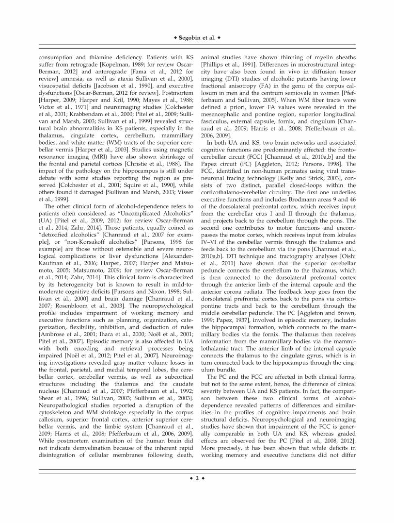
TABLE 2. Neuropsychological description of the alcoholics with and without Korsakoff’s syndrome and controls
Cognitive functions Tasks HC UA KS Tests*
Intellectual abilities(WAIS III subtests)a
Information 9.1 6 3.0 6.04 6 3.0 5.2 6 3.1 HC>UA>KSMatrix reasoning 10.4 6 2.2 7.71 6 2.26 5.0 6 4.4 HC>UA>KS
Episodic memory FSCRT sum of 3 free recalls 34.0 6 4.9 28.5 6 7.8 5.4 6 2.9 HC>UA>KSWorking memory
(MEM-III subtests)aForward visuospatial span 8.5 6 2.3 5.8 6 1.5 5.1 6 0.7 HC>UA>KSBackward visuospatial span 8.7 6 2.5 5.3 6 1.3 3.9 6 0.7 HC>UA; HC>KS
HC 5 Healthy Controls; UA 5 Uncomplicated Alcoholics; KS 5 Alcoholics with Korsakoff’s Syndrome.(Mean 6 standard deviation) and range [minimum – maximum] are reported.Mann–Whtney U tests P< 0.05.aStandard scores.¥: comparison with KS not done as no data available.WAIS III Wechsler Adults Intelligence Scale.FSCRT: Free and Cued Selective Reminding Test.
r Integrity of White Matter Microstructure in Alcoholics r
r 5 r

mean FA skeleton, which represents the centers of alltracts common to the group. Each subject’s aligned FAimage was threshold at 0.3 to exclude low FA values thatcould be contaminated with partial volume effects fromother non-white-matter tissues and to minimize intersub-ject variability. The resulting image is then projected ontothe mean skeleton by filling every voxel of the latter withthe maximum FA value that lies perpendicular to the skel-eton structure. Voxel-based statistics are performed onthese “skeletonized” images.
Statistical Analyses
Comparison of WM integrity in HC, UA patients
and KS patients in the whole-brain (voxel-based
analysis)
Nonparametric permutation tests [Nichols and Holmes,2002] were performed between HC and UA; HC and KS;and UA and KS groups. Age was included as a covariateto account for between-group differences (KS being olderthan the two other groups). For each between-group com-parison, 5,000 permutations were done, and the data cor-rected for multiple comparisons (FWE, P< 0.05) usingthreshold-free cluster enhancement (TFCE) [Smith andNichols, 2009] for cluster-wise correction. This statisticaltoolbox is implemented within FSL 5.0. (http://fsl.fmrib.ox.ac.uk/fsl/fslwiki/Randomise).
Identification of UA patients at risk of developing KS
1. Because of the heterogeneity within the UA group, UAand KS patients were pooled together to form a singlegroup of alcohol-dependant patients (UASK). TheUASK group was then compared to the HC groupusing nonparametric permutation tests (5,000 permuta-tions, FWE P< 0.05, TFCE).
2. The John Hopkins University International Consortiumfor Brain Mapping (JHU-ICBM) DTI-81 WM atlas [Oishiet al., 2011], implemented as an atlas tool in FSL 5.0(http://fsl.fmrib.ox.ac.uk/fsl/fslwiki/Atlases), was thenused to extract and binarise clusters highlighted in theprevious between-group analysis (UASK vs HC). Onlythe WM fiber tracts that belong to the FCC and PCwere used to produce “fiber-cluster masks.” These“fiber-cluster masks” were then employed to extract themean FA value within each tract belonging to the FCCand PC for each subject. Focus was laid on a couple offiber tracts only for each circuit. In that respect, thecingulum and the fornix were chosen for the PC andthe middle cerebellar peduncle and superior cerebellarpeduncle for the FCC. These are the tracts that arebelieved to offer less variability in terms of specificity tothe PC and FCC, respectively, having a lower involve-ment in fibers connecting other parts of the brain orinvolved in other circuitry.
3. For each fiber tract, a k-means clustering classificationwas then performed on the mean FA values, with thealgorithm constrained to separate the 41 participantsinto two groups. The aim was to find which WM fibertract enables the identification of some UA patients clas-sified into the same group as KS patients. A reliableclassification would include all HC into one group, andall KS patients into the other group with the heteroge-neous UA group fitting into either of these. Those UAwho will be sorted within the same group as the KSpatients will be deemed as UA_HIGH patients (for highrisk of developing KS) while those who will be sortedwithin the same group as the HC will be labelledUA_LOW patients (for low risk of developing KS). It ishypothesized that a more robust classification will beobtained when using FA values in the fibers of the PCthan in those of the FCC. Comparisons of episodicmemory performance among the subsequent subgroupsshould help in asserting the robustness of the classifica-tion step.
4. Between-group comparisons of episodic memory per-formance:
Episodic memory scores were compared between thegroups of HC, UA_LOW, UA_HIGH, and KS usingnonparametric Mann–Whitney U tests. The hypothesisis that the scores on the episodic memory test inUA_LOW patients will differ significantly from theUA_HIGH and KS patients but not from HC.UA_HIGH patients are expected to differ significantlyfrom HC and UA_LOW and KS patients.
RESULTS
Comparison of WM Integrity in HC, UA Patients
and KS Patients
UA versus HC
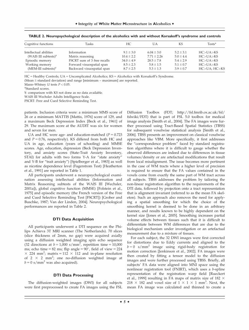
Nonparametric permutation tests (FWE, P< 0.05)between UA and HC showed lower FA values in fibertracts spread across the whole brain including the corpuscallosum (Tmax 5 6.01; k 5 6,258; g2 5 0.17), the anteriorlimb of the internal capsule (Tmax 5 5.86; k =742; g2 =0.10),the anterior corona radiata (Tmax 5 5.52; k 5 2,215;g2 5 0.16), the fornix (Tmax 5 5.91; k 5 560; g2 5 0.15); thecingulum (Tmax 5 4.95; k 5 821; g2 5 0.14); the middle cere-bellar peduncle (Tmax 5 5.17; k 5 1,320; g2 5 0.11), and thesuperior cerebellar peduncle Tmax 5 4.92; k 5 252; g2 5 0.15)as shown in Figure 1.
Note: Values for effect size (g2) have been calculatedfrom T-scores resulting from the nonparametric permuta-tion tests.
KS versus HC
The profile of WM structural impairment in KS versusHC was similar to that between UA and HC. FA values
r Segobin et al. r
r 6 r
were lower in KS than in HC in the same tracts asreported above but with higher T and k values: corpus cal-losum (Tmax 5 10.52; k 5 7,279; g2 5 0.26), anterior coronaradiata (Tmax 5 7.45; k 5 2,936; g2 5 0.31), anterior limb ofthe internal capsule (Tmax 5 7.00; k 5 1,179; g2=0.23); cingu-lum (Tmax 5 6.93; k 5 869; g2 5 0.14); the fornix (Tmax 5 6.60;k 5 693; g2 5 0.31); the middle cerebellar peduncle(Tmax 5 6.30; k 5 1,153; g2 5 0.27), and the superior cerebel-lar peduncle Tmax 5 7.27; k 5 270; g2=0.35).
UA versus KS
Group differences between UA and KS were foundmainly in the corpus callosum (Tmax 5 4.93; k 5 4,453;g2 5 0.139) and anterior corona radiata (Tmax 5 4.9; k 5 932;g2 5 0.13). Other tracts with significant differences werealso observed but they either had low T-values or theircluster sizes were small. For example, anterior limb of theinternal capsule (Tmax 5 3.59; k 5 163; g2 5 0.11); cingulum(Tmax 5 3.61; k 5 103; g2 5 0.14), and the fornix (Tmax 5 3.81;k 5 107; g2 5 0.16). There were no significant differences inthe middle and superior cerebellar peduncles.
There was no gender effect in the UA and KS groupsacross all WM fiber tracts. In the control group, differences
between men and women were significant for the cingu-lum (Mann–Whitney U test, P 5 0.014). There was no cor-relation between FA measures and beck depression indexscores or years of education across all tracts for all threegroups. There was also no correlation between FA meas-ures and the number of detoxifications in the UA group.The same analysis could not be performed for the KSgroup since it was not possible to obtain complete andaccurate information regarding alcohol history for thesepatients.
Identification of UA Patients at Risk of
Developing KS
Comparison of WM integrity between HC and UASK
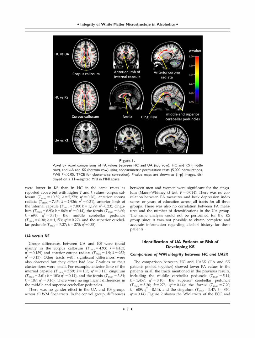
The comparison between HC and UASK (UA and SKpatients pooled together) showed lower FA values in thepatients in all the tracts mentioned in the previous results,including the middle cerebellar peduncle (Tmax 5 5.14;k 5 1,457; g2 5 0.10); the superior cerebellar peduncle(Tmax 5 5.20; k 5 278; g2 5 0.14); the fornix (Tmax 5 7.20;k 5 609; g2 5 0.14), and the cingulum (Tmax 5 5.47; k 5 840;g2 5 0.14). Figure 2 shows the WM tracts of the FCC and
Figure 1.
Voxel by voxel comparisons of FA values between HC and UA (top row), HC and KS (middle
row), and UA and KS (bottom row) using nonparametric permutation tests (5,000 permutations,
FWE P< 0.05, TFCE for cluster-wise correction). P-value maps are shown as (1-p) images, dis-
played on a T1-weighted MRI in MNI space.
r Integrity of White Matter Microstructure in Alcoholics r
r 7 r

PC that are significantly disrupted in the UASK groupcompared to the HC group.
Identification of UA at risk to develop KS
for each fiber tract
For all participants, mean FA values were extractedfrom significant fiber-clusters (see above) for the middleand superior cerebellar peduncles, representing tracts fromthe FCC and the cingulum and fornix, representing tractsfrom the PC. A k-means classification was then performedon those tracts. Figure 3 represents the distribution of themean FA values for each selected tract identified in theWM atlas [Oishi et al., 2011] to be part of the FCC and PC.
With regard to WM tracts within the PC, the k-meansclassification conducted on the FA values in the cingulumand fornix showed an expected classification of all HC inone group and all KS patients in another. Some UA
patients were classified in the same group as HC and canbe therefore considered as UA_LOW, while others wereclassified in the same group as KS and were thus qualifiedas UA_HIGH. The same patients were classified asUA_HIGH for both the cingulum and the fornix, exceptfor two of them (one for cingulum and one for fornix).However, these two patients were those with the lowestFA values in the subsequent UA_LOW class. Comparisonsof mean FA values among the four subgroups for both thecingulum and the fornix (ANOVA Kruskal-Wallis) haveshown that the controls did not differ from the UA_LOWgroup (P 5 1). KS differed significantly from HC(P< 0.001) and UA_LOW (P 5 0.01) but not UA_HIGH.UA_HIGH was significantly different from HC (P< 0.001),and UA_LOW (P 5 0.01).
Regarding the tracts of the FCC, the results of the classi-fication seemed less reliable since each of the two groupsidentified included a mix of KS patients, UA patients, and
Figure 2.
Voxel by voxel comparisons of FA values between HC and UASK using non-parametric permuta-
tion tests (5,000 permutations, FWE P< 0.05, and TFCE for cluster-wise correction). P-value
maps are shown as (1-p) images, displayed on a T1-weighted MRI in MNI space.
r Segobin et al. r
r 8 r

HC. Moreover, the number and the identity of the UApatients classified as UA_HIGH completely differedbetween the two tracts of the FCC.
Comparison of episodic memory performance
in the UA_HIGH and UA_LOW groups
Since a better classification of patients for UA_HIGH andUA_LOW were obtained for the two WM tracts of the PC asopposed to the FCC, we compared episodic memory per-formances between the two subgroups of UA identified withthe classification analysis conducted on FA values in the for-nix and cingulum individually. Nonparametric Mann–Whit-ney U tests showed that UA_LOW did not differ from HC(P 5 0.37 for the classification using the fornix and P 5 0.32for the classification using the cingulum). UA_LOW differedfrom UA_HIGH (P 5 0.038 for the classification using thefornix and P 5 0.025 for the classification using the cingu-lum). UA_HIGH differed significantly from HC (P 5 0.002for both fornix and cingulum) and KS (P 5 0.001 for classifi-cation with the fornix and P 5 0.002 for classification withthe cingulum). The latter was significantly different to all theother three subgroups for both classifications (P< 0.001).These results are illustrated in Figure 4.
We conducted the same analysis on the performanceson matrix reasoning, and forward and backward block
spans but found insignificant differences between the UAsubgroups for neither the cingulum nor the fornix (datanot shown). Hence, the ability to identify UA patients atrisk of developing KS seems to be specific to performancesin episodic memory tasks.
Note that we have also tested classification with the cor-pus callosum but it yielded poor results (data not shownsince the corpus callosum is neither part of the FCC, northe PC).
DISCUSSION
The first aim of this study was to describe the profile ofmicrostructural WM integrity in UA and KS in the wholebrain, at a voxel-level, since most of the previous studieshad been carried out using a region of interest approach(with anatomical regions defined a priori). In accordancewith neuropathological [Harper et al., 2003; Kril et al.,1997] and neuroimaging studies [Chanraud et al., 2010a,b;Pfefferbaum and Sullivan, 2005; Pfefferbaum et al., 2006,2009], the present TBSS analysis of FA values in UArevealed widespread compromised WM microstructureincluding notably fibers of the FCC and the PC. Using astringent statistical threshold (TFCE and FWE, P< 0.05),our voxel-based analyses have successfully replicated pre-vious DTI results in fibers defined a priori [Pfefferbaum
Figure 3.
Distribution of the mean FA values for the selected WM tracts within the FCC (mcp 5 middle
cerebellar peduncle, scp 5 superior cerebellar peduncle) and the PC (fornix and cing 5 cingulum).
Horizontal black lines represent the separation between the two identified clusters.
r Integrity of White Matter Microstructure in Alcoholics r
r 9 r

et al., 2009] such as the superior longitudinal fasciculus,external capsule, fornix, and cingulum. Moreover, ouranalyses revealed compromised WM integrity in otherfiber tracts such as the internal capsule, cerebralpeduncles, corona radiata, and thalamic radiations, whichhas also been reported in a TBSS study comparing alco-holics who have been abstinent for at least 5 years withHC [Fortier et al., 2014]. Contrary to previous neuropatho-logical [Harper et al., 2003] and neuroimaging studies[Chanraud et al., 2009; Pfefferbaum et al., 2009], we alsofound microstructural abnormalities in the middle andsuperior cerebellar peduncles. The use of a voxel-basedapproach may have enabled the observation of the latterfinding since a ROI approach, which would average theFA values within a region defined a priori, would notreveal any localized impairments within those fibers.Thus, WM abnormalities in the middle and superior cere-bellar peduncles revealed by TBSS suggest that disruptionmay be more localized than spread-throughout in thosefibers.
Our study is the first to evaluate WM microstructuralintegrity in KS patients. When KS patients were comparedto HC, the profile of WM abnormalities was similar to thatobserved between UA and HC, in agreement with the pat-terns of GM and WM shrinkage found in a recent VBMstudy [Pitel et al., 2012]. Compromised WM microstructurein the middle and superior cerebellar peduncles is in-linewith previous investigations that have shown significantshrinkage of the cerebellar WM of KS patients [Harper et al.,2003; Kril et al., 1997] and in proteomics studies wherechanges in the levels of thiamine-dependent enzymes havebeen observed [Alexander-Kaufman et al., 2006].
Previous neuropsychological and neuroimaging (struc-tural and functional) studies have hypothesized thatanterograde amnesia in KS patients is essentially due to adisconnection within the PC [Nahum et al., 2014; Warring-ton and Weiskrantz, 1982]. More specifically, findings ofabnormalities in diencephalic structures, including thethalamus and mammillary bodies, and cortical structuresfrom the frontal and medial temporal lobes have pushedtoward the hypothesis of a disruption between nodesbelonging to this neural network [Aup�ee et al., 2001; Kimet al., 2009; Renou et al., 2008]. Our study provides consol-idating evidence of WM disruptions in the PC that islinked to episodic memory deficits and therefore amnesia[Kessels and Kopelman, 2012 for review). Based on thesubstantial role of the cerebellum in cognitive processesand its connections with cortical areas, damaged FCC hasbeen hypothesized to be involved in working memory andexecutive dysfunction in KS [Wijnia and Goossensen,2010]. Our data confirm compromised WM integrity in thecerebellum and especially disruption of the middle andsuperior cerebellar peduncles. In summary, cognitive defi-cits observed in KS patients, including amnesia, stemsfrom a disconnection of neural networks, which can inturn be due to abnormalities in the nodes of the net-work(s), disruption of WM tracts linking those nodes, orabnormal synaptic activity between the nodes. While ourstudy confirms the disruption of WM tracts in specificbrain networks, and another study showed an absence ofatrophy within connected regions [Nahum et al., 2014], itis still difficult to evaluate the cascade of events (neuro-transmission dysfunction—local or global network disrup-tion—cellular damage/atrophy) that effectively governsthe pathophysiological mechanism of KS when using across-sectional paradigm,. Longitudinal studies arerequired to concretely establish this mechanism.
Our direct voxel-based comparison between UA and KSpatients showed significant differences mainly in the cor-pus callosum, which follows previous volumetric studiesthat has reported further volume loss in the corpus cal-losum in alcoholic patients with Wernicke’s Encephalop-athy than those alcoholic patients without [Lee et al., 2005]and the other one between UA and KS [Pitel et al., 2012].The abnormalities in the microstructural integrity of thecorpus callosum have also been hypothesized to be due tothiamine deficiency as observed in a study with rats [Heet al., 2007].
Contrary to our initial hypotheses, a clear graded effectof deficits in the PC was not observed. This can be attrib-uted to our sample size and to the heterogeneity of theUA group. As neurological complications from UA to KSlie along a continuum [Ryback, 1971], it is difficult toobserve a distinct pattern of microstructural degradationbetween these 2 groups, justifying the need to divide theUA group into subgroups to better observe their underly-ing pathological mechanisms. By first combining the UAand KS groups, the statistical power to detect all regionsaffected in alcohol dependent patients was increased
Figure 4.
Episodic memory performance (sum of the three free-recalls of
FCSRT) in the HC,KS and subgroups UA_LOW and UA_HIGH.
UA_LOW and UA_HIGH have been identified using k-means
classification conducted on means FA values of significant fiber-
clusters in the fornix and the cingulum bundle.
*: sig different from HC; §: sig different from UA_LOW; ¥: sig
different from UA_HIGH
r Segobin et al. r
r 10 r

[Monnig et al., 2013], ensuring that the subsequent classifi-cation step did not disregard any WM tracts that arepotential structural biomarkers for identifying alcoholics atrisk to develop neurological complications such as KS.
The classification step revealed that the use of DTI maybe particularly relevant as a structural biomarker towardthe early identification of UA patients at risk of developingKS. The early identification is important for clinicians toapply the correct and optimal treatment with the aim ofpreventing severe, debilitating and irreversible neurologicalcomplications. Our analysis provides consolidating evi-dence of the PC, as opposed to the FCC, being an appro-priate neuroanatomical substrate for identifying UApatients that can potentially develop KS [Pitel et al., 2012].The statistical comparisons of the mean FA values betweenthe subgroups of subjects have confirmed the robustness ofthe classification step. The fact that there is a clear separa-tion between HC and KS as well as consistent classificationof the UA subgroups for the cingulum and fornix comple-ments volumetric findings that have shown graded effectsin the mammillary bodies, the hippocampus and the thala-mus [Sullivan and Pfefferbaum, 2009]. In a previous study[Pitel et al., 2012], the volume of the thalamus was foundto be comparable between some of the UA patients andthe KS ones, reinforcing neuropsychological data that haveshown the same trend in episodic memory deficits [Pitelet al., 2008]. While the volume of the thalamus was notexplored in the present study, we confirm that UA patientsclassified at risk to develop KS based on WM microstruc-tural abnormalities in the PC had the lowest episodic mem-ory scores. Interestingly, it was also observed that 4patients out of 10 classified as UA_LOW filled none ofCaine’s criteria for Wernicke’s encephalopathy [Caine et al.,1997; Pitel et al., 2011], four patients filled one criterion andtwo filled more than two criteria. For the UA_HIGH sub-group, two patients filled no criterion, four filled one crite-rion and four filled more than two criteria. The number ofsigns of Wernicke’s encephalopathy did not differ betweenthe UA_LOW and UA_HIGH (Chi-squared test, data notshown). Our refined neuroimaging analyses confirm that aneuropsychological evaluation, especially targeting episodicmemory, is highly recommended in clinical settings, whereneuroimaging tools are not available, to identify patients atrisk of developing KS. Taken together, the analysis ofmicrostructural integrity within the fornix and cingulum,in combination with scores of episodic memory, thalamicvolume and signs of Wernicke’s encephalopathy, couldgive a reliable depiction of whether a UA patient is at riskof developing KS. Our study is thus a positive iteration tothe heuristic value of the continuity hypothesis [Buttersand Brandt, 1985; Parsons, 1998; Pitel et al., 2008; Ryback,1971]. While this inherent heterogeneity in the UA groupenabled us to detect alcoholics at risk to develop neurologi-cal complications, the currently used average neuropsycho-logical, structural and functional description of a UA groupdoes not reflect the heterogeneity of individual profiles inclinical settings.
CONCLUSIONS AND FURTHER WORKS
TBSS has allowed us to describe the profile of WMintegrity at a voxel-level in UA and KS patients. While thechronological position of WM disruption is still unknownin the cascade of structural and functional events that gov-ern the pathophysiological mechanism underlying the neu-rotoxicity of alcohol, we have shown the potential of DTIdata to identify UA at risk of developing KS. The methodpaves the way for more in depth analysis of this subgroupin order to better understand the mechanism underlyingthese two clinical forms. Multi-modal neuroimaging, com-bined with biological and neuropsychological analyses willenable researchers to explore the characteristics of theseclinical forms in terms of detailed microstructure, regionalvolume, function, enzyme metabolism, and cognitive defi-cits. The specificity of the subgroup of patients at risk todevelop KS is also likely to be confirmed via longitudinalstudies in which the progress of the pathology can bemonitored and the efficiency of the treatment can beassessed and optimized.
REFERENCES
Aggleton JP (2012): Multiple anatomical systems embedded withinthe primate medial temporal lobe: Implications for hippocam-pal function. Neurosci Biobehav Rev 36:1579–1596.
Aggleton JP, Brown MW (1999): Episodic memory, amnesia, andthe hippocampal-anterior thalamic axis. Behav Brain Sci 22:425–444.
Alexander-Kaufman K, James G, Sheedy D, Harper C, MatsumotoI (2006): Differential protein expression in the prefrontal whitematter of human alcoholics: A proteomics study. Mol Psychia-try 11:56–65.
Ambrose ML, Bowden SC, Whelan G (2001): Working memoryimpairments in alcohol-dependent participants without clinicalamnesia. Alcohol Clin Exp Res 25:185–191.
American Psychiatric Association (1994): Diagnostic and StatisticalManual of Mental Disorders, 4th ed. Washington, DC: Ameri-can Psychiatric Association.
Aup�ee AM, Desgranges B, Eustache F, Lalev�ee C, de la Sayette V,Viader F, Baron JC (2001): Voxel-based mapping of brainhypometabolism in permanent amnesia with PET. Neuroimage13:1164–1173.
Beck A, Ward C, Mendelson M, Mock J, Erbaugh J (1961): Aninventory for measuring depression. Arch Gen Psychiatry 4:561–571.
Butters N, Brandt J (1985): The continuity hypothesis: The rela-tionship of long-term alcoholism to the Wernicke-korsakoffsyndrome. Recent Dev Alcohol 3:207–226.
Caine D, Halliday GM, Kril JJ, Harper CG (1997): Operational cri-teria for the classification of chronic alcoholics: Identification ofwernicke’s encephalopathy. J Neurol Neurosurg Psychiatry 62:51–60.
Chanraud S, Martelli C, Delain F, Kostogianni N, Douaud G,Aubin HJ, Reynaud M, Martinot JL (2007): Brain morphometryand cognitive performance in detoxified alcohol-dependentswith preserved psychosocial functioning. Neuropsychophar-macology 32:429–438.
r Integrity of White Matter Microstructure in Alcoholics r
r 11 r

Chanraud S, Reynaud M, Wessa M, Penttil€a J, Kostogianni N,Cachia A, et al. (2009): Diffusion tensor tractography in mesen-cephalic bundles: Relation to mental flexibility in detoxifiedalcohol-dependent subjects. Neuropsychopharmacology 34:1223–1232.
Chanraud S, Pitel AL, Rohlfing T, Pfefferbaum A, Sullivan EV(2010a): Dual tasking and working memory in alcoholism:Relation to frontocerebellar circuitry. Neuropsychopharmacol-ogy 35:1868–1878.
Chanraud S, Zahr N, Sullivan EV, Pfefferbaum A (2010b): MR dif-fusion tensor imaging: A window into white matter integrityof the working brain. Neuropsychol Rev 20:209–225.
Christie JE, Kean DM, Douglas RHB, Engleman HM, St Clair D,Blackburn IM (1988): Magnetic resonance imaging in pre-seniledementia of the Alzheimer-type, multi-infarct dementia andkorsakoff’s syndrome. Psychol Med 18:319–329.
Colchester A, Kingsley D, Lasserson D, Kendall B, Bello F, Rush C,Stevens TG, Goodman G, Heilpern G, Stanhope N, KopelmanMD (2001): Structural MRI volumetric analysis in patients withorganic amnesia, 1: Methods and comparative findings acrossdiagnostic groups. J Neurol Neurosurg Psychiatry 71:13–22.
Cushman P Jr, Forbes R, Lerner W, Stewart M (1985): Alcoholwithdrawal syndromes: clinical management with lofexidine.Alcohol Clin Exp Res 9:103–108.
Fama R, Pitel AL, Sullivan EV (2012): Anterograde episodic mem-ory in korsakoff syndrome. Neuropsychol Rev 22:93–104.
Folstein MF, Folstein SE, McHugh PR (1975): Mini-mental state. Apractical method for grading the cognitive state of patients forthe clinician. J Psychiatr Res 12:189–198.
Fortier CB, Leritz EC, Salat DH, Lindemer E, Maksimovskiy AL,Shepel J, Williams V, Venne JR, Milberg WP, McGlinchey RE(2014): Widespread effects of alcohol on white matter micro-structure. Alcohol Clin Exp Res 38:2925–2933.
Gache P, Michaud P, Landry U, Accietto C, Arfaoui S, Wenger O,Daeppen JB (2005): The alcohol use disorders identification test(AUDIT) as a screening tool for excessive drinking in primarycare: Reliability and validity of a french version. Alcohol ClinExp Res 29:2001–2007.
Grober E, Buschke H (1987): Genuine memory deficits in demen-tia. Dev Neuropsychol 3:13–36.
Harper C (2007): The neurotoxicity of alcohol. Hum Exp Toxicol26:251–257.
Harper C (2009): The neuropathology of alcohol-related braindamage. Alcohol Alcohol 44:136–140.
Harper C, Matsumoto I (2005): Ethanol and brain damage. CurrOpin Pharmacol 5:73–78.
Harper C, Dixon G, Sheedy D, Garrick T (2003): Neuropathologi-cal alterations in alcoholic brains. Studies arising from the newsouth wales tissue resource centre. Prog Neuropsychopharma-col Biol Psychiatry 27:951–961.
Harper CG, Kril JJ (1990): Neuropathology of alcoholism. AlcoholAlcohol 25:207–216.
Harris GJ, Jaffin SK, Hodge SM, Kennedy D, Caviness VS,Marinkovic K, Papadimitriou GM, Makris N, Oscar-Berman M(2008): Frontal white matter and cingulum diffusion tensorimaging deficits in alcoholism. Alcohol Clin Exp Res 32:1001–1013.
He X, Sullivan EV, Stankovic RK, Harper CG, Pfefferbaum A(2007): Interaction of thiamine deficiency and voluntary alcoholconsumption disrupts rat corpus callosum ultrastructure. Neu-ropsychopharmacology 32:2207–2216.
Heatherton TF, Kozlowski LT, Frecker RC, Fagerstr€om KO (1991):The fagerstr€om test for nicotine dependence: A revision of thefagerstr€om tolerance questionnaire. Br J Addict 86:1119–1127.
Ihara H, Berrios GE, London M (2000): Group and case study ofthe dysexecutive syndrome in alcoholism without amnesia.J Neurol Neurosurg Psychiatry 68:731–737.
Jacobson RR, Acker CF, Lishman WA (1990): Patterns of neuro-psychological deficit in alcoholic korsakoff’s syndrome. Psy-chol Med 20:321–334.
Jenkinson M, Bannister P, Brady M, Smith S (2002): Improvedoptimization for the robust and accurate linear registration andmotion correction of brain images. NeuroImage 17:825–841.
Jones DK, Symms MR, Cercignani M, Howard RJ (2005): Theeffect of filter size on VBM analyses of DT-MRI data. Neuro-Image 26:546–554.
Kelly RM, Strick PL (2003): Cerebellar loops with motor cortexand prefrontal cortex of a nonhuman primate. J Neurosci 23:8432–8444.
Kessels RP, Kopelman MD (2012): Context memory in Korsakoff’ssyndrome. Neuropsychol Rev 22:117–131.
Kim E, Ku J, Namkoong K, Lee W, Lee KS, Park J-Y, Lee SY, KimJJ, Kim SI, Jung YC (2009): Mammillothalamic functional con-nectivity and memory function in wernicke’s encephalopathy.Brain 132:369–376.
Kopelman Michael D (1989): Remote and autobiographical mem-ory, temporal context memory and frontal atrophy in korsakoffand alzheimer patients. Neuropsychologia 27:437–460.
Korsakoff S (1889): Etude medico-psychologique sur une formedes maladies de la m�emoire. Revue Philosophie 20:501–530.
Krabbendam L, Visser PJ, Derix MMA, Verhey F, Hofman P,Verhoeven W, Tuinier S, Jolles J (2000): Normal cognitive per-formance in patients with chronic alcoholism in contrast topatients with korsakoff’s syndrome. J Neuropsychiatry Clin Neu-rosci 12:44–50.
Kril JJ, Halliday GM, Svoboda MD, Cartwright H (1997): The cere-bral cortex is damaged in chronic alcoholics. Neuroscience 79:983–998.
Lee S-T, Jung Y-M, Na DL, Park SH, Kim M (2005): Corpus cal-losum atrophy in wernicke’s encephalopathy. J Neuroimaging15:367–372.
Matsumoto I (2009): Proteomics approach in the study of thepathophysiology of alcohol-related brain damage. AlcoholAlcohol 44:171–176.
Mattis S (1976): Mental status examination for organic mental syn-drome in the elderly patients. In: Bellak L, Karasu T, editors.Geriatrics Psychiatry: A Handbook for Psychiatrists and Pri-mary Care Physicians. New York: Grune & Stratton. pp 77–121.
Mayes AR, Meudell PR, Mann D, Pickering A (1988): Location oflesions in korsakoff’s syndrome: Neuropsychological and neu-ropathological data on two patients. Cortex 24:367–388.
Monnig MA, Caprihan A, Yeo RA, Gasparovic C, Ruhl DA, LysneP, Bogenschutz MP, Hutchison KE, Thoma RJ (2013): Diffusiontensor imaging of white matter networks in individuals withcurrent and remitted alcohol use disorders and comorbid con-ditions. Psychol Addict Behav 27:455–465.
Nahum L, Pignat JM, Bouzerda-Wahlen A, Gabriel D, LiveraniMC, Lazeyras F, Ptak R, Richiardi J, Haller S, Thorens G,Zullino DF, Guggisberg AG, Schnider A (2014): Neural Corre-late of Anterograde Amnesia in Wernicke-Korsakoff Syn-drome. Brain Topogr [Epub ahead of print].
r Segobin et al. r
r 12 r

Nichols TE, Holmes AP (2002): Nonparametric permutation testsfor functional neuroimaging: A primer with examples. HumBrain Mapp 15:1–25.
No€el X, Van der Linden M, Schmidt N (2001): Supervisory atten-tional system in nonamnesic alcoholic men. Arch Gen Psychia-try 58:1152–1158.
No€el X, Van Der Linden M, Brevers D, Campanella S, Hanak C,Kornreich C, Verbanck P (2012): The contribution of executivefunctions deficits to impaired episodic memory in individualswith alcoholism. Psychiatry Res 198:116–122.
Oishi K, Faria A, Van Zijl PMC, Mori S (2011): An atlas of humanwhite matter, 2nd ed. Amsterdam, Netherlands: Elsevier.Academic Press.
Oscar-Berman M (2012): Function and dysfunction of prefrontalbrain circuitry in alcoholic korsakoff’s syndrome. Neuropsy-chol Rev 22:154–169.
Oscar-Berman M, Valmas MM, Sawyer KS, Ruiz SM, Luhar RB,Gravitz ZR (2014): Profiles of impaired, spared, and recoveredneuropsychologic processes in alcoholism. Handb Clin Neurol125:183–210.
Papez J (1937): A proposed mechanism of emotion. Arch NeurolPsychiatry 28:725–743.
Parsons OA (1998): Neurocognitive deficits in alcoholics and socialdrinkers: A continuum? Alcohol Clin Exp Res 22:954–961.
Parsons OA, Nixon SJ (1998): Cognitive functioning in sober socialdrinkers: A review of the research since 1986. J Stud Alcohol59:180–190.
Pfefferbaum A, Sullivan EV (2005): Disruption of brain white mat-ter microstructure by excessive intracellular and extracellularfluid in alcoholism: Evidence from diffusion tensor imaging.Neuropsychopharmacology 30:423–432.
Pfefferbaum A, Rosenbloom MJ, Crusan K, Jernigan TL (1988):Brain CT changes in alcoholics: Effects of age and alcohol con-sumption. Alcohol Clin Exp Res 12:81–87.
Pfefferbaum A, Lim KO, Zipursky RB, Mathalon DH, RosenbloomMJ, Lane B, Ha CN, Sullivan EV (1992): Brain gray and whitematter volume loss accelerates with aging in chronic alco-holics: A quantitative MRI study. Alcohol Clin Exp Res 16:1078–1089.
Pfefferbaum A, Adalsteinsson E, Sullivan EV (2006): Supratento-rial profile of white matter microstructural integrity in recover-ing alcoholic men and women. Biol Psychiatry 59:364–372.
Pfefferbaum A, Rosenbloom MJ, Rohlfing T, Sullivan EV (2009):Degradation of association and projection white matter sys-tems in alcoholism detected with quantitative fiber tracking.Biol Psychiatry 65:680–690.
Phillips DE, Krueger SK, Rydquist JE (1991): Short- and long-termeffects of combined pre- and postnatal ethanol exposure (threetrimester equivalency) on the development of myelin andaxons in rat optic nerve. Int J Dev Neurosci 9:631–647.
Pitel AL, Beaunieux H, Witkowski T, Vabret F, Guillery-Girard B,Quinette P, Desgranges B, Eustache F (2007): Genuine episodicmemory deficits and executive dysfunctions in alcoholic sub-jects early in abstinence. Alcohol Clin Exp Res 31:1169–1178.
Pitel AL, Beaunieux H, Witkowski T, Vabret F, De la Sayette V,Viader F, Desgranges B, Eustache F (2008): Episodic and work-ing memory deficits in alcoholic korsakoff patients: The conti-nuity theory revisited. Alcohol Clin Exp Res 32:1229–1241.
Pitel AL, Aup�ee AM, Ch�etelat G, M�ezenge F, Beaunieux H, De laSayette V, Viader F, Baron JC, Eustache F, Desgranges B(2009): Morphological and glucose metabolism abnormalities
in alcoholic korsakoff’s syndrome: Group comparisons andindividual analyses. PloS One 4:e7748
Pitel AL, Zahr NM, Jackson K, Sassoon SA, Rosenbloom MJ,Pfefferbaum A, Sullivan EV (2011): Signs of preclinical wer-nicke’s encephalopathy and thiamine levels as predictors ofneuropsychological deficits in alcoholism without korsakoff’ssyndrome. Neuropsychopharmacology 36:580–588.
Pitel AL, Ch�etelat G, Le Berre AP, Desgranges B, Eustache F,Beaunieux H (2012): Macrostructural abnormalities in korsak-off syndrome compared with uncomplicated alcoholism. Neu-rology 78:1330–1333.
Renou P, Ducreux D, Batouche F, Denier C (2008): Pure and acutekorsakoff syndrome due to a bilateral anterior fornix infarc-tion. Arch Neurol 65:1252–1253.
Rosenbloom MJ, Sullivan EV, Pfefferbaurn A (2003): Use of MRIto determine brain damage in alcoholics. Alcohol Res Health27:146–152.
Rueckert D, Sonoda LI, Hayes C, Hill DL, Leach MO, Hawkes DJ(1999): Nonrigid registration using free-form deformations: Appli-cation to breast MR images. IEEE Trans Med Imaging 18:712–721.
Ryback R (1971): The continuum and specificity of the effects ofalcohol on memory. A Review. Q J Stud Alcohol 32:991–1016.
Shear PK, Sullivan EV, Lane B, Pfefferbaurn A (1996): Mammillarybody and cerebellar shrinkage in chronic alcoholics with andwithout amnesia. Alcohol Clin Exp Res 20:1489–1495.
Smith SM, Nichols TE (2009): Threshold-free cluster enhancement:Addressing problems of smoothing, threshold dependence andlocalisation in cluster inference. NeuroImage 44:83–98.
Smith SM, Jenkinson M, Woolrich MW, Beckmann CF, BehrensTE, Johansen-Berg H, Bannister PR, De Luca M, Drobnjak I,Flitney DE, Niazy RK, Saunders J, Vickers J, Zhang Y, DeStefano N, Brady JM, Matthews PM (2004): Advances in func-tional and structural MR image analysis and implementationas FSL. Neuroimage 23 Suppl 1:S208–S219.
Smith SM, Jenkinson M, Johansen-Berg H, Rueckert D, NicholsTE, Mackay CE, Watkins KE, Ciccarelli O, Cader MZ,Matthews PM, Behrens TE (2006): Tract-based spatial statistics:Voxelwise analysis of multi-subject diffusion data. NeuroImage31:1487–1505.
Spielberger CD, Gorsuch RL, Lushene R, Vagg PR, Jacobs GA(1983): Manual for the State-Trait Anxiety Inventory (form Y).Palo Alto: Consulting Psychologists Press.
Squire LR, Amaral DG, Press GA (1990): Magnetic resonanceimaging of the hippocampal formation and mammillary nucleidistinguish medial temporal lobe and diencephalic amnesia.J Neurosci 10:3106–3117.
Sullivan EV (2003): Compromised pontocerebellar and cerebello-thalamocortical systems: Speculations on their contributions tocognitive and motor impairment in nonamnesic alcoholism.Alcohol Clin Exp Res 27:1409–1419.
Sullivan EV, Marsh L (2003): Hippocampal volume deficits inalcoholic korsakoff’s syndrome. Neurology 61:1716–1719.
Sullivan EV, Pfefferbaum A (2009): Neuroimaging of theWernicke-korsakoff syndrome. Alcohol Alcohol 44:155–165.
Sullivan EV, Lane B, Deshmukh A, Rosenbloom MJ, Desmond JE,Lim KO, Pfefferbaum A (1999): In vivo mammillary body vol-ume deficits in amnesic and nonamnesic alcoholics. AlcoholClin Exp Res 23:1629–1636.
Sullivan EV, Deshmukh A, Desmond JE, Lim KO, Pfefferbaum A(2000): Cerebellar volume decline in normal aging, alcoholism,and korsakoff’s syndrome: Relation to ataxia. Neuropsychol-ogy 14:341–352.
r Integrity of White Matter Microstructure in Alcoholics r
r 13 r

Sullivan EV, Rosenbloom MJ, Serventi KL, Deshmukh A,Pfefferbaum A (2003): Effects of alcohol dependence comorbid-ity and antipsychotic medication on volumes of the thalamusand pons in schizophrenia. Am J Psychiatry 160:1110–1116.
Van der Linden M (2004): L’�evaluation des troubles de lam�emoire : Pr�esentation de quatre tests de m�emoire �episodique.Collectif 2004.
Victor M, Adams RD, Collins GH (1971): The Wernicke- Korsakoffsyndrome. Philadelphia: F.A. Davis Co.
Visser PJ, Krabbendam L, Verhey FRJ, Hofman PAM, VerhoevenWMA, Tuinier S, Wester A, Den Berg YW, Goessens LF, WerfYD, Jolles J (1999): Brain correlates of memory dysfunction in
alcoholic Korsakoff’s syndrome. J Neurol Neurosurg Psychia-try 67:774–778.
Warrington EK, Weiskrantz L (1982): Amnesia: A disconnectionsyndrome? Neuropsychologia 20:233–248.
Wechsler D (2001): WAIS-III : Wechsler Adult Intelligence Scale,3rd ed. Canada: Harcourt.
Wijnia JW, Goossensen A (2010): Cerebellar neurocognition andkorsakoff’s syndrome: An hypothesis. Med Hypotheses 75:266–268.
Zahr NM (2014): Structural and microstructral imaging of thebrain in alcohol use disorders. Handb Clin Neurol 125:275–290.
r Segobin et al. r
r 14 r