Influence of weak hip abductor muscles on joint contact forces during normal walking: probabilistic...
8
Influence of weak hip abductor muscles on joint contact forces during normal walking: A probabilistic modeling analysis Giordano Valente a,b,n , Fulvia Taddei a , Ilse Jonkers c Q1 a Laboratorio di Tecnologia Medica, Istituto Ortopedico Rizzoli, Bologna, Italy b Department of Industrial Engineering, University of Bologna, Italy c Department of Biomedical Kinesiology, K.U. Leuven, Belgium article info Article history: Accepted 30 June 2013 Keywords: Musculoskeletal modeling Dynamic simulations Monte-Carlo analysis Weak abductor muscles Joint contact forces abstract The weakness of hip abductor muscles is related to lower-limb joint osteoarthritis, and joint overloading may increase the risk for disease progression. The relationship between muscle strength, structural joint deterioration and joint loading makes the latter an important parameter in the study of onset and follow- up of the disease. Since the relationship between hip abductor weakness and joint loading still remains an open question, the purpose of this study was to adopt a probabilistic modeling approach to give insights into how the weakness of hip abductor muscles, in the extent to which normal gait could be unaltered, affects ipsilateral joint contact forces. A generic musculoskeletal model was scaled to each healthy subject included in the study, and the maximum force-generating capacity of each hip abductor muscle in the model was perturbed to evaluate how all physiologically possible configurations of hip abductor weakness affected the joint contact forces during walking. In general, the muscular system was able to compensate for abductor weakness. The reduced force-generating capacity of the abductor muscles affected joint contact forces to a mild extent, with 50th percentile mean differences up to 0.5 BW (maximum 1.7 BW). There were greater increases in the peak knee joint loads than in loads at the hip or ankle. Gluteus medius, particularly the anterior compartment, was the abductor muscle with the most influence on hip and knee loads. Further studies should assess if these increases in joint loading may affect initiation and progression of osteoarthritis. & 2013 Elsevier Ltd. All rights reserved. 1. Introduction Mechanical factors related to joint loading constitute a major cause in the development and progression of osteoarthritis (OA) (Lafeber et al., 2006). Excessive joint loading during normal daily activities and obesity, in conjunction with local joint vulnerabilities, are confirmed risk factors in the etiopathogenesis of OA, particularly at the hip and knee joints (Felson, 2004). The weakness of hip abductor muscles has been related to joint OA, for example, the level of gluteus medius atrophy was found to correlate significantly to radiographic signs of hip OA in both ipsilateral and contralateral legs (Amaro et al., 2007). Significant weakness of hip abductors was also observed in knee OA subjects (Costa et al., 2010; Hinman et al., 2010), although a causal relationship between muscle weakness and disease onset was not established. Impaired function of the hip abductor muscles is traditionallyassociated with excessive pelvic drop towards the contralateral limb (Trendelenburg gait). This pelvic instability has been documented in patients with hip OA and after total joint replacement (Beaulieu et al., 2010; Madsen et al., 2004; Jandrić, 1997). However, changes in gait parameters may simply reflect an abnormal gait pattern so as to avoid pain on weight-bearing structures rather than abductor muscle weakness. In fact, recent studies showed that even severe hip abductor weakness does not lead to significant pelvic drop (Kendall et al., 2010, 2013; Henriksen et al., 2009), demonstrating that weakness of the hip abductors is not sufficient to have a positive Trendelenburg test. Hip abductor weakness was related to excessive medial tibiofemoral compartment loading with consequent risk for disease progression in knee OA patients (Mündermann et al., 2005; Chang et al., 2005), and was found to contribute to functional limitations of patients with total knee arthroplasty (Piva et al., 2011). The suggested relationship between muscle strength, articular loading and structural deterioration makes joint loading a relevant parameter in the detection of onset and follow-up of degenerative joint disease in the lower limb (Wilson et al., 2006; Wu et al., 2000). However, joint loading cannot be easily studied in vivo. The only direct way to measure joint contact forces in vivo implies the use of instrumented prostheses. This approach is, however, limited to a few subjects and is representative of a post-operative 1 2 3 4 5 6 7 8 9 10 11 12 13 14 15 16 17 18 19 20 21 22 23 24 25 26 27 28 29 30 31 32 33 34 35 36 37 38 39 40 41 42 43 44 45 46 47 48 49 50 51 52 53 54 55 56 57 58 59 60 61 62 63 64 65 66 67 68 69 70 71 72 73 74 75 76 77 78 79 80 81 82 83 84 85 86 87 88 89 90 91 92 93 Contents lists available at SciVerse ScienceDirect journal homepage: www.elsevier.com/locate/jbiomech www.JBiomech.com Journal of Biomechanics 0021-9290/$-see front matter & 2013 Elsevier Ltd. All rights reserved. http://dx.doi.org/10.1016/j.jbiomech.2013.06.030 n Corresponding author at: Laboratorio di Tecnologia Medica, Istituto Ortopedico Rizzoli, via di Barbiano 1/10, 40136 Bologna, Italy. Tel.: +39 051 6366554. E-mail address: [email protected] (G. Valente). Please cite this article as: Valente, G., et al., Influence of weak hip abductor muscles on joint contact forces during normal walking: A probabilistic modeling analysis. Journal of Biomechanics (2013), http://dx.doi.org/10.1016/j.jbiomech.2013.06.030i Journal of Biomechanics ∎ (∎∎∎∎) ∎∎∎–∎∎∎
-
Upload
independent -
Category
Documents
-
view
0 -
download
0
Transcript of Influence of weak hip abductor muscles on joint contact forces during normal walking: probabilistic...
Influence of weak hip abductor muscles on joint contact forces duringnormal walking: A probabilistic modeling analysis
Giordano Valente a,b,n, Fulvia Taddei a, Ilse Jonkers cQ1
a Laboratorio di Tecnologia Medica, Istituto Ortopedico Rizzoli, Bologna, Italyb Department of Industrial Engineering, University of Bologna, Italyc Department of Biomedical Kinesiology, K.U. Leuven, Belgium
a r t i c l e i n f o
Article history:
Accepted 30 June 2013
Keywords:
Musculoskeletal modelingDynamic simulationsMonte-Carlo analysisWeak abductor musclesJoint contact forces
a b s t r a c t
The weakness of hip abductor muscles is related to lower-limb joint osteoarthritis, and joint overloadingmay increase the risk for disease progression. The relationship between muscle strength, structural jointdeterioration and joint loading makes the latter an important parameter in the study of onset and follow-up of the disease. Since the relationship between hip abductor weakness and joint loading still remainsan open question, the purpose of this study was to adopt a probabilistic modeling approach to giveinsights into how the weakness of hip abductor muscles, in the extent to which normal gait could beunaltered, affects ipsilateral joint contact forces. A generic musculoskeletal model was scaled to eachhealthy subject included in the study, and the maximum force-generating capacity of each hip abductormuscle in the model was perturbed to evaluate how all physiologically possible configurations of hipabductor weakness affected the joint contact forces during walking. In general, the muscular system wasable to compensate for abductor weakness. The reduced force-generating capacity of the abductormuscles affected joint contact forces to a mild extent, with 50th percentile mean differences up to 0.5 BW(maximum 1.7 BW). There were greater increases in the peak knee joint loads than in loads at the hip orankle. Gluteus medius, particularly the anterior compartment, was the abductor muscle with the mostinfluence on hip and knee loads. Further studies should assess if these increases in joint loading mayaffect initiation and progression of osteoarthritis.
& 2013 Elsevier Ltd. All rights reserved.
1. Introduction
Mechanical factors related to joint loading constitute a majorcause in the development and progression of osteoarthritis (OA)(Lafeber et al., 2006). Excessive joint loading during normal dailyactivities and obesity, in conjunction with local joint vulnerabilities,are confirmed risk factors in the etiopathogenesis of OA, particularlyat the hip and knee joints (Felson, 2004). The weakness of hipabductor muscles has been related to joint OA, for example, the levelof gluteus medius atrophy was found to correlate significantly toradiographic signs of hip OA in both ipsilateral and contralateral legs(Amaro et al., 2007). Significant weakness of hip abductors was alsoobserved in knee OA subjects (Costa et al., 2010; Hinman et al., 2010),although a causal relationship betweenmuscle weakness and diseaseonset was not established. Impaired function of the hip abductormuscles is traditionally associated with excessive pelvic drop towardsthe contralateral limb (Trendelenburg gait). This pelvic instability has
been documented in patients with hip OA and after total jointreplacement (Beaulieu et al., 2010; Madsen et al., 2004; Jandrić,1997). However, changes in gait parameters may simply reflect anabnormal gait pattern so as to avoid pain on weight-bearingstructures rather than abductor muscle weakness. In fact, recentstudies showed that even severe hip abductor weakness does notlead to significant pelvic drop (Kendall et al., 2010, 2013; Henriksenet al., 2009), demonstrating that weakness of the hip abductors is notsufficient to have a positive Trendelenburg test. Hip abductorweakness was related to excessive medial tibiofemoral compartmentloading with consequent risk for disease progression in knee OApatients (Mündermann et al., 2005; Chang et al., 2005), and wasfound to contribute to functional limitations of patients with totalknee arthroplasty (Piva et al., 2011).
The suggested relationship between muscle strength, articularloading and structural deterioration makes joint loading a relevantparameter in the detection of onset and follow-up of degenerativejoint disease in the lower limb (Wilson et al., 2006; Wu et al.,2000). However, joint loading cannot be easily studied in vivo.The only direct way to measure joint contact forces in vivo impliesthe use of instrumented prostheses. This approach is, however,limited to a few subjects and is representative of a post-operative
123456789101112131415161718192021222324252627282930313233343536373839404142434445464748495051525354555657585960616263646566
676869707172737475767778798081828384858687888990919293
Contents lists available at SciVerse ScienceDirect
journal homepage: www.elsevier.com/locate/jbiomechwww.JBiomech.com
Journal of Biomechanics
0021-9290/$ - see front matter & 2013 Elsevier Ltd. All rights reserved.http://dx.doi.org/10.1016/j.jbiomech.2013.06.030
n Corresponding author at: Laboratorio di Tecnologia Medica, Istituto OrtopedicoRizzoli, via di Barbiano 1/10, 40136 Bologna, Italy. Tel.: +39 051 6366554.
E-mail address: [email protected] (G. Valente).
Please cite this article as: Valente, G., et al., Influence of weak hip abductor muscles on joint contact forces during normal walking: Aprobabilistic modeling analysis. Journal of Biomechanics (2013), http://dx.doi.org/10.1016/j.jbiomech.2013.06.030i
Journal of Biomechanics ∎ (∎∎∎∎) ∎∎∎–∎∎∎

situation. Alternatively, computer models of the musculoskeletalsystem in combination with dynamic simulations of motion, havebeen increasingly used (Pandy and Andriacchi, 2010; Erdemiret al., 2007) to calculate joint contact forces, providing a valuableapproach to joint loading analyses (Lenaerts et al., 2009; Steeleet al., 2011; Taddei et al., 2012).
To the authors' knowledge, the relationship between the weak-ness of hip abductor muscles and lower-limb joint contact forceshas not been extensively studied. Lenaerts et al., 2008 used asubject-specific model with halved force-generating capacity ofthe hip abductor muscles to calculate hip contact forces duringwalking using inverse dynamics and static optimization methods.Compared to the model with full force-generating capacity,increased muscle activation was found for these muscles, evenup to full activation, but changes in hip contact forces did notresult. In a more recent study, van der Krogt et al., 2012 analyzedthe effect of local muscle weakness on gait impairment andmuscle forces during walking, using a scaled generic musculoske-letal model and the Computed Muscle Control (CMC) algorithm(Thelen and Anderson, 2006). They concluded that normal gaitcould still be achieved, at a higher cost, when removing mostmuscles, and gait was most impaired when hip abductors andankle plantarflexors were weakened in the model.
However, none of the above studies provided a complete over-view on the relationship between hip abductor weakness and lower-limb joint loading. Lenaerts et al., 2008 limited the analysis on hipcontact forces in one gait trial, and did not analyze the effect on theknee; in the second study (van der Krogt et al., 2012), muscleweakness was not related to joint contact forces. In addition, nostudies were found that included a structured approach to analyzehow weakness of the individual compartments of hip abductormuscles correlated with altered joint loading.
Therefore, the aim of the present study was to evaluate the effect ofweakness of the different compartments of the hip abductor muscleson joint contact forces of the ipsilateral lower limb, in the extent towhich normal gait remained unaltered. To address this question,
simulations of gait of healthy adult subjects were first generated usinga generic musculoskeletal model with representative muscle strength.Then, a probabilistic modeling approach was adopted performinga Monte-Carlo analysis. This allowed evaluation of the effect of allphysiologically possible configurations of force-generating capacity ofthe hip abductors on joint contact forces, and to investigate whichwere the most influential muscle compartments. The statisticalanalysis of the results was focused on the gait instants of maximumjoint loads, particularly relevant for overloading risk.
2. Materials and methods
2.1. Human experiments
Five healthy male subjects (age: 2672 yrs; mass: 82710 kg; height: 18272 cm)gave informed consent to participate in this study, approved by the local institutionalresearch board. The subjects were fitted with markers for 3D motion capturing,merging the Plug-in-gait (Davis et al., 1991) and the MOCAP (Software for interactivemusculoskeletal modeling (SIMM), Motion Analysis Corp., Santa Rosa, USA) markersets. After a static trial, the subjects were asked to walk at a self-selected speed in astraight line along a 10 mwalkway. Throughout all trials, kinematics was measured at100 Hz using a Vicon system (Oxford Metrics, Oxford, UK). In a pre-processing phase,the marker coordinates were filtered and smoothed using Woltring's quintic splineroutine (Woltring, 1986), implemented in Workstation (Vicon Workstation 5.2 beta 20,Oxford Metrics). Ground reaction forces were measured at 1000 Hz using two AMTIforce plates (Advanced Mechanical Technology Inc., Watertown, MA) embedded in thewalkway. Surface electromyography (EMG) signals of major lower-limb muscles (i.e.gluteus maximus, rectus femoris, vastus lateralis, biceps femoris long head, semi-membranosus, tibialis anterior, gastrocnemius) were recorded at 1000 Hz using awireless EMG device (Aurion, Italy). EMG data were rectified, bi-directionally low-passfiltered at 6 Hz, and normalized to the peak values of predicted muscle activations (vander Krogt et al., 2012).
2.2. Baseline gait simulations
A generic musculoskeletal model (Delp et al., 1990) with 10 rigid bodies, 23degrees of freedom and 92 musculotendon actuators was scaled to match eachsubject's dimensions and inertial properties, using the experimentally measuredposition of markers placed on anatomical landmarks and body mass during
123456789101112131415161718192021222324252627282930313233343536373839404142434445464748495051525354555657585960616263646566
676869707172737475767778798081828384858687888990919293949596979899100101102103104105106107108109110111112113114115116117118119120121122123124125126127128129130131132
Fig. 1. Comparison between standard deviations of muscle activations predicted with the baseline model (dark gray areas) and experimental EMG recordings (light grayareas), for the muscles of which EMG was available. Data were averaged over five subjects. EMG data were rectified, bi-directionally low-pass filtered at 6 Hz, and normalizedto the peak values of predicted muscle activations.
G. Valente et al. / Journal of Biomechanics ∎ (∎∎∎∎) ∎∎∎–∎∎∎2
Please cite this article as: Valente, G., et al., Influence of weak hip abductor muscles on joint contact forces during normal walking: Aprobabilistic modeling analysis. Journal of Biomechanics (2013), http://dx.doi.org/10.1016/j.jbiomech.2013.06.030i

standing trials. For a representative trial of each subject, OpenSim (Delp et al.,2007) was used to generate and analyze simulations of walking using the scaledmodels. Inverse kinematics and Residual Reduction Algorithm (RRA) (Delp et al.,2007) were used in conjunction with the measured marker trajectories and groundreaction forces to enforce dynamic consistency. Then CMC (Thelen and Anderson,2006) was used to calculate the optimal muscle forces that generated the necessaryjoint moments to track the kinematics produced by RRA. The lower-limb jointcontact forces were calculated using the Joint Reaction analysis (Steele et al., 2011).The predicted muscle activations were in line with the literature and generallyconsistent with the experimental EMG recordings (Fig. 1), with some discrepancyin the hamstring muscles during the stance phase of gait.
2.3. Perturbed gait simulations
A Monte-Carlo analysis was performed to evaluate the sensitivity of jointcontact forces to the weakness of hip abductor muscles. Seven stochastic inputvariables were defined, i.e. the maximum isometric force of each muscle actuatormodeling the hip abductors of the ipsilateral leg, including gluteus mediusanterior (GMedA), middle (GMedM) and posterior (GMedP), gluteus minimusanterior (GMinA), middle (GMinM) and posterior (GMinP), and tensor fascia latae(TFL). The effect of the stochastic input variables was evaluated on the followingstochastic output variables: 12 joint contact force variables, i.e. antero-posterior(Fx), proximo-distal (Fy) medio-lateral (Fz) components of the ipsilateral hip, kneeand ankle joint contact forces, and their respective magnitudes (F).
Each input variable was randomly sampled independently with a uniform distribu-tion between zero and the maximum isometric force values (Table A1 of the Appendix)
of the baseline model. This hypothesis allowed to analyze to which extent the weaknessof the hip abductor muscles could be tolerated (van der Krogt et al., 2012). A LatinHypercube Sampling (LHS) strategy (McKay et al., 1979) was applied to perform theMonte-Carlo simulations, generating an efficient sampling of the input variables fromtheir distribution. This allowed to include all possible configurations of the differentvariable values: for each variable, n values were randomly distributed with one fromeach equiprobable interval (0, 1/n), (1/n, 2/n),…, (1�1/n, 1), and they were randomlypermuted. This strategy, unlike simple random sampling, ensured a full coverage of therange of each variable by maximally stratifying each marginal distribution. Table A2 ofthe Appendix reports the LHS of the seven input variables. For each set of generatedsample values, a correspondingmusculoskeletal model with the altered set of maximumisometric forces was created. Consequently, new CMC solutions were calculated withthe resulting modified models that aimed to track the original kinematics. Normal gaitkinematics was considered successfully tracked if the difference in all joint anglesbetween perturbed and baseline simulations did not exceed one degree (Thelen andAnderson, 2006; van der Krogt et al., 2012). If normal gait kinematics could be achieved,further statistical analyses were performed on the output variables. The models werefree to adopt any muscle force values within the imposed maximum force-generatingcapacity. Finally, subsequent Joint Reaction analyses calculated the perturbed jointcontact forces.
A convergence criterion was defined as a stopping rule for the Monte-Carlosimulations. Convergence was defined as when the mean and standard deviation ofall variables over the final 10% of the simulations were less than 5% of the overallmean and standard deviation, respectively. 200 simulations, with the correspond-ing sets of input variables, ensured that convergence was reached. Approximately250-h computation time was needed to run a total of 1000 simulations (200 persubject) on a common desktop computer.
123456789101112131415161718192021222324252627282930313233343536373839404142434445464748495051525354555657585960616263646566
676869707172737475767778798081828384858687888990919293949596979899100101102103104105106107108109110111112113114115116117118119120121122123124125126127128129130131132
Fig. 2. Variations in the components of the calculated joint contact forces (BW) following the statistical perturbation of the hip abductor muscles strength in the simulatedgait cycle. The curves show the standard deviations of the perturbed simulations (shaded area), run within the Monte Carlo analysis, compared to the baseline simulation(solid line). The values were averaged over five subjects. Force components are: antero-posterior (Fx), proximo-distal (Fy), medio-lateral (Fz) and magnitude (F).
G. Valente et al. / Journal of Biomechanics ∎ (∎∎∎∎) ∎∎∎–∎∎∎ 3
Please cite this article as: Valente, G., et al., Influence of weak hip abductor muscles on joint contact forces during normal walking: Aprobabilistic modeling analysis. Journal of Biomechanics (2013), http://dx.doi.org/10.1016/j.jbiomech.2013.06.030i
2.4. Statistical analysis
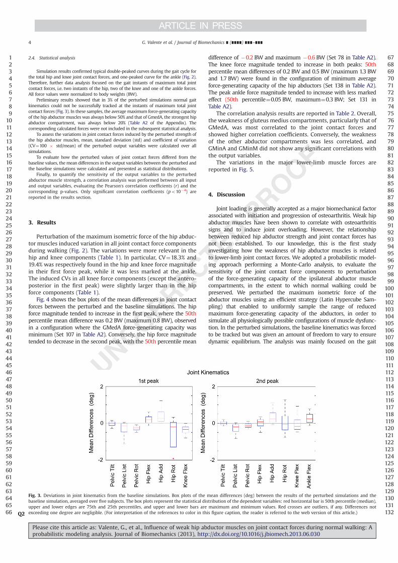
Simulation results confirmed typical double-peaked curves during the gait cycle forthe total hip and knee joint contact forces, and one-peaked curve for the ankle (Fig. 2).Therefore, further data analysis focused on the gait instants of maximum total jointcontact forces, i.e. two instants of the hip, two of the knee and one of the ankle forces.All force values were normalized to body weights (BW).
Preliminary results showed that in 3% of the perturbed simulations normal gaitkinematics could not be successfully tracked at the instants of maximum total jointcontact forces (Fig. 3). In these samples, the average maximum force-generating capacityof the hip abductor muscles was always below 50% and that of GmedA, the strongest hipabductor compartment, was always below 20% (Table A2 of the Appendix). Thecorresponding calculated forces were not included in the subsequent statistical analysis.
To assess the variations in joint contact forces induced by the perturbed strength ofthe hip abductor muscles, mean, standard deviation (std) and coefficient of variation(CV¼100 " std/mean) of the perturbed output variables were calculated over allsimulations.
To evaluate how the perturbed values of joint contact forces differed from thebaseline values, the mean differences in the output variables between the perturbed andthe baseline simulations were calculated and presented as statistical distributions.
Finally, to quantify the sensitivity of the output variables to the perturbedabductor muscle strength, a correlation analysis was performed between all inputand output variables, evaluating the Pearson's correlation coefficients (r) and thecorresponding p-values. Only significant correlation coefficients (po10�4) arereported in the results section.
3. Results
Perturbation of the maximum isometric force of the hip abduc-tor muscles induced variation in all joint contact force componentsduring walking (Fig. 2). The variations were more relevant in thehip and knee components (Table 1). In particular, CV¼18.3% and19.4% was respectively found in the hip and knee force magnitudein their first force peak, while it was less marked at the ankle.The induced CVs in all knee force components (except the antero-posterior in the first peak) were slightly larger than in the hipforce components (Table 1).
Fig. 4 shows the box plots of the mean differences in joint contactforces between the perturbed and the baseline simulations. The hipforce magnitude tended to increase in the first peak, where the 50thpercentile mean difference was 0.2 BW (maximum 0.8 BW), observedin a configuration where the GMedA force-generating capacity wasminimum (Set 107 in Table A2). Conversely, the hip force magnitudetended to decrease in the second peak, with the 50th percentile mean
difference of �0.2 BW and maximum �0.6 BW (Set 78 in Table A2).The knee force magnitude tended to increase in both peaks: 50thpercentile mean differences of 0.2 BW and 0.5 BW (maximum 1.3 BWand 1.7 BW) were found in the configuration of minimum averageforce-generating capacity of the hip abductors (Set 138 in Table A2).The peak ankle force magnitude tended to increase with less markedeffect (50th percentile¼0.05 BW, maximum¼0.3 BW; Set 131 inTable A2).
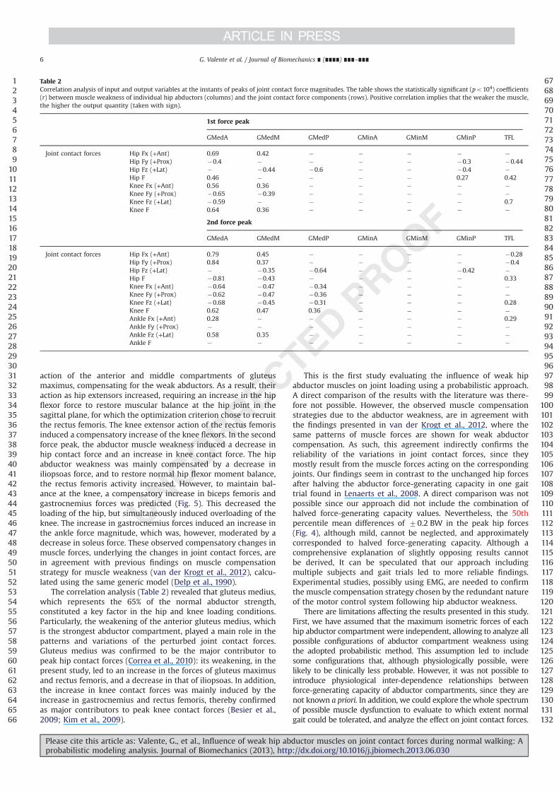
The correlation analysis results are reported in Table 2. Overall,the weakness of gluteus medius compartments, particularly that ofGMedA, was most correlated to the joint contact forces andshowed higher correlation coefficients. Conversely, the weaknessof the other abductor compartments was less correlated, andGMinA and GMinM did not show any significant correlations withthe output variables.
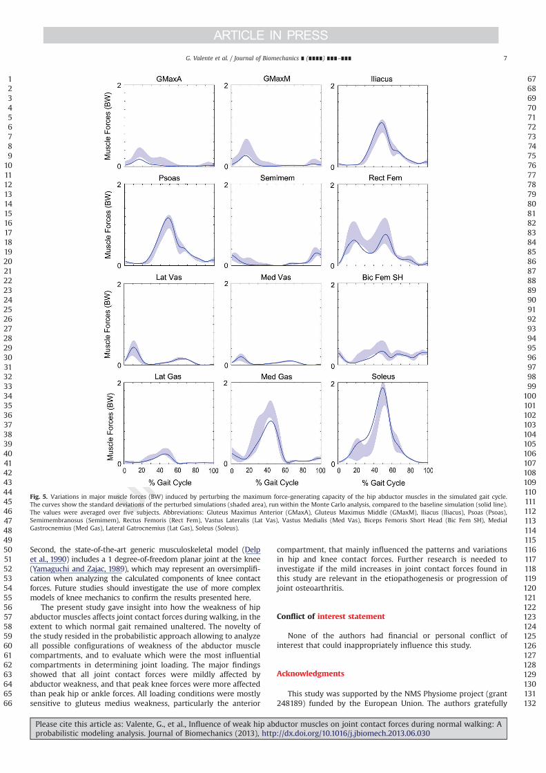
The variations in the major lower-limb muscle forces arereported in Fig. 5.
4. Discussion
Joint loading is generally accepted as a major biomechanical factorassociated with initiation and progression of osteoarthritis. Weak hipabductor muscles have been shown to correlate with osteoarthritissigns and to induce joint overloading. However, the relationshipbetween reduced hip abductor strength and joint contact forces hasnot been established. To our knowledge, this is the first studyinvestigating how the weakness of hip abductor muscles is relatedto lower-limb joint contact forces. We adopted a probabilistic model-ing approach performing a Monte-Carlo analysis, to evaluate thesensitivity of the joint contact force components to perturbationof the force-generating capacity of the ipsilateral abductor musclecompartments, in the extent to which normal walking could bepreserved. We perturbed the maximum isometric force of theabductor muscles using an efficient strategy (Latin Hypercube Sam-pling) that enabled to uniformly sample the range of reducedmaximum force-generating capacity of the abductors, in order tosimulate all physiologically possible configurations of muscle dysfunc-tion. In the perturbed simulations, the baseline kinematics was forcedto be tracked but was given an amount of freedom to vary to ensuredynamic equilibrium. The analysis was mainly focused on the gait
123456789101112131415161718192021222324252627282930313233343536373839404142434445464748495051525354555657585960616263646566
676869707172737475767778798081828384858687888990919293949596979899100101102103104105106107108109110111112113114115116117118119120121122123124125126127128129130131132
Fig. 3. Deviations in joint kinematics from the baseline simulations. Box plots of the mean differences (deg) between the results of the perturbed simulations and thebaseline simulation, averaged over five subjects. The box plots represent the statistical distribution of the dependent variables: red horizontal bar is 50th percentile (median),upper and lower edges are 75th and 25th percentiles, and upper and lower bars are maximum and minimum values. Red crosses are outliers, if any. Differences notexceeding one degree are negligible. (For interpretation of the references to color in this figure caption, the reader is referred to the web version of this article.)Q2
G. Valente et al. / Journal of Biomechanics ∎ (∎∎∎∎) ∎∎∎–∎∎∎4
Please cite this article as: Valente, G., et al., Influence of weak hip abductor muscles on joint contact forces during normal walking: Aprobabilistic modeling analysis. Journal of Biomechanics (2013), http://dx.doi.org/10.1016/j.jbiomech.2013.06.030i

instants of maximum total joint contact forces, particularly relevant inthe context of detecting overloading risk.
The perturbed simulations revealed that the muscular system wasable to compensate for weakness of the hip abductor muscles, underthe assumption of optimal conditions of motor control strategy in thecalculation of muscle forces (Thelen and Anderson, 2006). Specifically,normal gait kinematics was successfully tracked in 97% of the simu-lations after changes in compensatory muscle forces. Indeed, fewexceptions of unsuccessful kinematics tracking (with a high level ofweakness highlighted in Table A2 of the Appendix) were observed inthe first force peaks (Fig. 3). These cases were, however, excluded fromstatistical analyses. This preliminary finding corroborates a computa-tional study (van der Krogt et al., 2012) where 100% weaknessof gluteus medius was predicted to be tolerated in some subjects,as well as experimental studies showing no correlations between hipabductor weakness and pelvic drop during walking in normal subjects(Kendall et al., 2010, 2013).
We found that weakness of the hip abductor muscles mainlyaffected hip and knee contact forces during normal walking(Fig. 2). The statistical perturbation of hip abductor strength ledto appreciable variability in joint contact forces (CV¼19% in thefirst peak of knee force magnitude). In general, the calculatedmean differences from the baseline simulations showed largerdifferences in the knee force components compared to the hip(Figs. 2 and 4), particularly in the second force peak. However, theamount of difference was not marked: the maximum meandifferences in joint force magnitude were up to 1.7 BW at theknee, occurring with the lowest average abductor strength, andthe 50th percentiles were up to 0.5 BW, approximately corre-sponding to halved force-generating capacity (Fig. 4).
In the first peak of joint contact forces, a concurrent increase inhip and knee forces was induced, due to the synergistic action offlexor and extensor muscles spanning the hip and knee joints(Fig. 5). This might be explained by the marked compensatory
123456789101112131415161718192021222324252627282930313233343536373839404142434445464748495051525354555657585960616263646566
676869707172737475767778798081828384858687888990919293949596979899100101102103104105106107108109110111112113114115116117118119120121122123124125126127128129130131132
Table 1
Variation in the perturbed joint contact forces induced by the abductor muscle weakness during the Monte Carlo simulations: descriptive statistics parameters (mean, stdand CV) at the peaks of hip, knee and ankle joint force magnitude expressed in BW. Force components are: antero-posterior (Fx), proximo-distal (Fy), medio-lateral (Fz) andmagnitude (F).
1st peak Hip Knee
Force component Fx Fy Fz F Fx Fy Fz F
Mean (BW) 0.27 �4.11 0.88 4.21 0.77 �2.66 �0.11 2.78Std (BW) 0.20 0.74 0.26 0.77 0.26 0.49 0.06 0.54CV 73.4% 18.0% 29.7% 18.3% 34.0% 18.3% 54.7% 19.4%
2nd peak Hip Knee
Force component Fx Fy Fz F Fx Fy Fz F
Mean (BW) �2.35 �3.94 0.59 4.64 �1.14 �3.88 �0.13 4.05Std (BW) 0.36 0.35 0.13 0.45 0.20 0.45 0.06 0.49CV 15.3% 8.9% 21.9% 9.7% 17.3% 11.7% 47.9% 12.0%
Peak Ankle
Force component Fx Fy Fz F
Mean (BW) �1.80 �4.00 �0.26 4.40Std (BW) 0.18 0.21 0.05 0.23CV 9.9% 5.2% 20.1% 5.2%
Fig. 4. Deviations in the perturbed joint contact forces from the baseline simulations. Box plots of the mean differences (BW) between the results of the perturbedsimulations and the baseline simulation, averaged over five subjects. The box plots represent the statistical distribution of the dependent variables: red horizontal bar is 50thpercentile (median), upper and lower edges are 75th and 25th percentiles, and upper and lower bars are maximum and minimum values. Red crosses are outliers, if any.Force components are: antero-posterior (Fx), proximo-distal (Fy), medio-lateral (Fz) and magnitude (F). (For interpretation of the references to color in this figure caption, thereader is referred to the web version of this article.)
G. Valente et al. / Journal of Biomechanics ∎ (∎∎∎∎) ∎∎∎–∎∎∎ 5
Please cite this article as: Valente, G., et al., Influence of weak hip abductor muscles on joint contact forces during normal walking: Aprobabilistic modeling analysis. Journal of Biomechanics (2013), http://dx.doi.org/10.1016/j.jbiomech.2013.06.030i
action of the anterior and middle compartments of gluteusmaximus, compensating for the weak abductors. As a result, theiraction as hip extensors increased, requiring an increase in the hipflexor force to restore muscular balance at the hip joint in thesagittal plane, for which the optimization criterion chose to recruitthe rectus femoris. The knee extensor action of the rectus femorisinduced a compensatory increase of the knee flexors. In the secondforce peak, the abductor muscle weakness induced a decrease inhip contact force and an increase in knee contact force. The hipabductor weakness was mainly compensated by a decrease iniliopsoas force, and to restore normal hip flexor moment balance,the rectus femoris activity increased. However, to maintain bal-ance at the knee, a compensatory increase in biceps femoris andgastrocnemius forces was predicted (Fig. 5). This decreased theloading of the hip, but simultaneously induced overloading of theknee. The increase in gastrocnemius forces induced an increase inthe ankle force magnitude, which was, however, moderated by adecrease in soleus force. These observed compensatory changes inmuscle forces, underlying the changes in joint contact forces, arein agreement with previous findings on muscle compensationstrategy for muscle weakness (van der Krogt et al., 2012), calcu-lated using the same generic model (Delp et al., 1990).
The correlation analysis (Table 2) revealed that gluteus medius,which represents the 65% of the normal abductor strength,constituted a key factor in the hip and knee loading conditions.Particularly, the weakening of the anterior gluteus medius, whichis the strongest abductor compartment, played a main role in thepatterns and variations of the perturbed joint contact forces.Gluteus medius was confirmed to be the major contributor topeak hip contact forces (Correa et al., 2010): its weakening, in thepresent study, led to an increase in the forces of gluteus maximusand rectus femoris, and a decrease in that of iliopsoas. In addition,the increase in knee contact forces was mainly induced by theincrease in gastrocnemius and rectus femoris, thereby confirmedas major contributors to peak knee contact forces (Besier et al.,2009; Kim et al., 2009).
This is the first study evaluating the influence of weak hipabductor muscles on joint loading using a probabilistic approach.A direct comparison of the results with the literature was there-fore not possible. However, the observed muscle compensationstrategies due to the abductor weakness, are in agreement withthe findings presented in van der Krogt et al., 2012, where thesame patterns of muscle forces are shown for weak abductorcompensation. As such, this agreement indirectly confirms thereliability of the variations in joint contact forces, since theymostly result from the muscle forces acting on the correspondingjoints. Our findings seem in contrast to the unchanged hip forcesafter halving the abductor force-generating capacity in one gaittrial found in Lenaerts et al., 2008. A direct comparison was notpossible since our approach did not include the combination ofhalved force-generating capacity values. Nevertheless, the 50thpercentile mean differences of 70.2 BW in the peak hip forces(Fig. 4), although mild, cannot be neglected, and approximatelycorresponded to halved force-generating capacity. Although acomprehensive explanation of slightly opposing results cannotbe derived, It can be speculated that our approach includingmultiple subjects and gait trials led to more reliable findings.Experimental studies, possibly using EMG, are needed to confirmthe muscle compensation strategy chosen by the redundant natureof the motor control system following hip abductor weakness.
There are limitations affecting the results presented in this study.First, we have assumed that the maximum isometric forces of eachhip abductor compartment were independent, allowing to analyze allpossible configurations of abductor compartment weakness usingthe adopted probabilistic method. This assumption led to includesome configurations that, although physiologically possible, werelikely to be clinically less probable. However, it was not possible tointroduce physiological inter-dependence relationships betweenforce-generating capacity of abductor compartments, since they arenot known a priori. In addition, we could explore the whole spectrumof possible muscle dysfunction to evaluate to which extent normalgait could be tolerated, and analyze the effect on joint contact forces.
123456789101112131415161718192021222324252627282930313233343536373839404142434445464748495051525354555657585960616263646566
676869707172737475767778798081828384858687888990919293949596979899100101102103104105106107108109110111112113114115116117118119120121122123124125126127128129130131132
Table 2
Correlation analysis of input and output variables at the instants of peaks of joint contact force magnitudes. The table shows the statistically significant (po104) coefficients(r) between muscle weakness of individual hip abductors (columns) and the joint contact force components (rows). Positive correlation implies that the weaker the muscle,the higher the output quantity (taken with sign).
1st force peak
GMedA GMedM GMedP GMinA GMinM GMinP TFL
Joint contact forces Hip Fx (+Ant) 0.69 0.42 � � � � �
Hip Fy (+Prox) �0.4 � � � � �0.3 �0.44Hip Fz (+Lat) � �0.44 �0.6 � � �0.4 �
Hip F 0.46 � � � � 0.27 0.42Knee Fx (+Ant) 0.56 0.36 � � � � �
Knee Fy (+Prox) �0.65 �0.39 � � � � �
Knee Fz (+Lat) �0.59 � � � � � 0.7Knee F 0.64 0.36 � � � � �
2nd force peak
GMedA GMedM GMedP GMinA GMinM GMinP TFL
Joint contact forces Hip Fx (+Ant) 0.79 0.45 � � � � �0.28Hip Fy (+Prox) 0.84 0.37 � � � � �0.4Hip Fz (+Lat) � �0.35 �0.64 � � �0.42 �
Hip F �0.81 �0.43 � � � � 0.33Knee Fx (+Ant) �0.64 �0.47 �0.34 � � � �
Knee Fy (+Prox) �0.62 �0.47 �0.36 � � � �
Knee Fz (+Lat) �0.68 �0.45 �0.31 � � � 0.28Knee F 0.62 0.47 0.36 � � � �
Ankle Fx (+Ant) 0.28 � � � � � 0.29Ankle Fy (+Prox) � � � � � � �
Ankle Fz (+Lat) 0.58 0.35 � � � � �
Ankle F � � � � � � �
G. Valente et al. / Journal of Biomechanics ∎ (∎∎∎∎) ∎∎∎–∎∎∎6
Please cite this article as: Valente, G., et al., Influence of weak hip abductor muscles on joint contact forces during normal walking: Aprobabilistic modeling analysis. Journal of Biomechanics (2013), http://dx.doi.org/10.1016/j.jbiomech.2013.06.030i
Second, the state-of-the-art generic musculoskeletal model (Delpet al., 1990) includes a 1 degree-of-freedom planar joint at the knee(Yamaguchi and Zajac, 1989), which may represent an oversimplifi-cation when analyzing the calculated components of knee contactforces. Future studies should investigate the use of more complexmodels of knee mechanics to confirm the results presented here.
The present study gave insight into how the weakness of hipabductor muscles affects joint contact forces during walking, in theextent to which normal gait remained unaltered. The novelty ofthe study resided in the probabilistic approach allowing to analyzeall possible configurations of weakness of the abductor musclecompartments, and to evaluate which were the most influentialcompartments in determining joint loading. The major findingsshowed that all joint contact forces were mildly affected byabductor weakness, and that peak knee forces were more affectedthan peak hip or ankle forces. All loading conditions were mostlysensitive to gluteus medius weakness, particularly the anterior
compartment, that mainly influenced the patterns and variationsin hip and knee contact forces. Further research is needed toinvestigate if the mild increases in joint contact forces found inthis study are relevant in the etiopathogenesis or progression ofjoint osteoarthritis.
Conflict of interest statement
None of the authors had financial or personal conflict ofinterest that could inappropriately influence this study.
Acknowledgments
This study was supported by the NMS Physiome project (grant248189) funded by the European Union. The authors gratefully
123456789101112131415161718192021222324252627282930313233343536373839404142434445464748495051525354555657585960616263646566
676869707172737475767778798081828384858687888990919293949596979899100101102103104105106107108109110111112113114115116117118119120121122123124125126127128129130131132
Fig. 5. Variations in major muscle forces (BW) induced by perturbing the maximum force-generating capacity of the hip abductor muscles in the simulated gait cycle.The curves show the standard deviations of the perturbed simulations (shaded area), run within the Monte Carlo analysis, compared to the baseline simulation (solid line).The values were averaged over five subjects. Abbreviations: Gluteus Maximus Anterior (GMaxA), Gluteus Maximus Middle (GMaxM), Iliacus (Iliacus), Psoas (Psoas),Semimembranosus (Semimem), Rectus Femoris (Rect Fem), Vastus Lateralis (Lat Vas), Vastus Medialis (Med Vas), Biceps Femoris Short Head (Bic Fem SH), MedialGastrocnemius (Med Gas), Lateral Gatrocnemius (Lat Gas), Soleus (Soleus).
G. Valente et al. / Journal of Biomechanics ∎ (∎∎∎∎) ∎∎∎–∎∎∎ 7
Please cite this article as: Valente, G., et al., Influence of weak hip abductor muscles on joint contact forces during normal walking: Aprobabilistic modeling analysis. Journal of Biomechanics (2013), http://dx.doi.org/10.1016/j.jbiomech.2013.06.030i
thank Christophe Meyer (University Hospital Pellenberg, Belgium)for the gait analysis data.
Appendix A. Supporting information
Supplementary data associated with this article can be found in theonline version at http://dx.doi.org/10.1016/j.jbiomech.2013.06.030.
References
Amaro, A., Amado, F., Duarte, J.A., Appell, H.J., 2007. Gluteus medius muscle atrophyis related to contralateral and ipsilateral hip joint osteoarthritis. InternationalJournal of Sports Medicine 28 (12), 1035–1039.
Beaulieu, M.L., Lamontagne, M., Beaulé, P.E., 2010. Lower limb biomechanics duringgait do not return to normal following total hip arthroplasty. Gait & Posture32 (2), 269–273.
Besier, T.F., Fredericson, M., Gold, G.E., Beaupre, G.S., Delp, S.L., 2009. Knee muscleforces during walking and running in patellofemoral pain patients and pain-free controls. Journal of Biomechanics 42 (7), 898–905.
Chang, A., Hayes, K., Dunlop, D., Song, J., Hurwitz, D., Cahue, S., Sharma, L., 2005. Hipabduction moment and protection against medial tibiofemoral osteoarthritisprogression. Arthritis and Rheumatism 52 (11), 3515–3519.
Correa, T.A., Crossley, K.M., Kim, H.J., Pandy, M.G., 2010. Contributions of individualmuscles to hip joint contact force in normal walking. Journal of Biomechanics43 (8), 1618–1622.
Costa, R.A., de Oliveira, L.M., Hiroko Watanabe, S., Jones, A., Natour, J., 2010.Isokinetic assessment of the hip muscles in patients with osteoarthritis of theknee. Clinics 65 (12), 1253–1259.
Davis, R.B., Õunpuu, S., Tyburski, D., Gage, J.R., 1991. A gait analysis data collectionand reduction technique. Human Movement Science 10 (5), 575–587.
Delp, S.L., Loan, J.P., Hoy, M.G., Zajac, F.E., Topp, E.L., Rosen, J.M., 1990. An interactivegraphics-based model of the lower extremity to study orthopaedic surgicalprocedures. IEEE Transactions on Biomedical Engineering 37 (8), 757–767.
Delp, S.L., Anderson, F.C., Arnold, A.S., Loan, P., Habib, A., John, C.T., Guendelman, E.,Thelen, D.G., 2007. OpenSim: open-source software to create and analyzedynamic simulations of movement. IEEE Transactions on Biomedical Engineer-ing 54, 1940–1950.
Erdemir, A., McLean, S., Herzog, W., van den Bogert, A.J., 2007. Model-basedestimation of muscle forces exerted during movements. Clinical Biomechanics(Bristol, Avon) 22 (2), 131–154.
Felson, D.T., 2004. Obesity and vocational and avocational overload of the joint asrisk factors for osteoarthritis. Journal of Rheumatology 31 (70), 2–5.
Henriksen, M., Aaboe, J., Simonsen, E.B., Alkjaer, T., Bliddal, H., 2009. Experimentallyreduced hip abductor function during walking: Implications for knee jointloads. Journal of Biomechanics 42 (9), 1236–1240.
Hinman, R.S., Hunt, M.A., Creaby, M.W., Wrigley, T.V., McManus, F.J., Bennell, K.L.,2010. Hip muscle weakness in individuals with medial knee osteoarthritis.Arthritis Care & Research 62 (8), 1190–1193.
Jandrić, S., 1997. Muscle parameters in coxarthrosis. Medicinski pregled 50 (7-8),301–304.
Kendall, K.D., Schmidt, C., Ferber, R., 2010. The relationship between hip-abductorstrength and the magnitude of pelvic drop in patients with low back pain.Journal of Sport Rehabilitation 19 (4), 422–435.
Kendall, K.D., Patel, C., Wiley, J.P., Pohl, M.B., Emery, C.A., Ferber, R., 2013. Stepstoward the validation of the trendelenburg test: the effect of experimentallyreduced hip abductor muscle function on frontal plane mechanics. ClinicalJournal of Sport Medicine 23 (1), 45–51.
Kim, H.J., Fernandez, J.W., Akbarshahi, M., Walter, J.P., Fregly, B.J., Pandy, M.G., 2009.Evaluation of predicted knee-joint muscle forces during gait using an instru-mented knee implant. Journal of Orthopaedic Research 27 (10), 1326–1331.
Lafeber, F.P.J.G., Intema, F., Van Roermund, P.M., Marijnissen, A.C.A., 2006. Unload-ing joints to treat osteoarthritis, including joint distraction. Current opinion inrheumatology 18 5, 519–525.
Lenaerts, G., De Groote, F., Demeulenaere, B., Mulier, M., Van der Perre, G., Spaepen,A., Jonkers, I., 2008. Subject-specific hip geometry affects predicted hip jointcontact forces during gait. Journal of Biomechanics 41 (6), 1243–1252.
Lenaerts, G., Mulier, M., Spaepen, A., Van der Perre, G., Jonkers, I., 2009. Aberrantpelvis and hip kinematics impair hip loading before and after total hipreplacement. Gait & Posture 30 (3), 296–302.
Madsen, M.S., Ritter, M.A., Morris, H.H., Meding, J.B., Berend, M.E., Faris, P.M.,Vardaxis, V.G., 2004. The effect of total hip arthroplasty surgical approach ongait. Journal of Orthopaedic Research 22 (1), 44–50.
McKay, M.D., Beckman, R.J., Conover, W.J., 1979. Comparison of Three Methods forSelecting Values of Input Variables in the Analysis of Output from a ComputerCode. Technometrics 21 (2), 239–245.
Mündermann, A., O' Dyrby, C., Andriacchi, T.P., 2005. Secondary gait changesin patients with medial compartment knee osteoarthritis: increased loadat the ankle, knee, and hip during walking. Arthritis and Rheumatism 52 (9),2835–2844.
Pandy, M.G, Andriacchi, T.P., 2010. Muscle and joint function in human locomotion.Annual Review of Biomedical Engineering 12, 401–433.
Piva, S.R., Teixeira, P.E.P., Almeida, G.J.M., Gil, A.B., DiGioia III, A.M., Levison, T.J.,Kelley Fitzgerald, G., 2011. Contribution of hip abductor strength to physicalfunction in patients with total knee arthroplasty. Physical Therapy 91 (2),225–233.
Steele, K.M., DeMers, M.S., Schwartz, M.H., Delp, S.L., 2011. Compressive tibiofe-moral force during crouch gait. Gait & Posture 35 (4), 556–560.
Taddei, F., Martelli, S., Valente, G., Benedetti, M.G., Leardini, A., Manfrini, M.,Viceconti, M., 2012. Femoral loads during gait in a patient with massive skeletalreconstruction. Clinical Biomechanics (Bristol, Avon) 27 (3), 273–280.
Thelen, D.G., Anderson, F.C., 2006. Using computed muscle control to generateforward dynamic simulations of human walking from experimental data.Journal of Biomechanics 39 (6), 1107–1115.
Wilson, W., van Burken, C., van Donkelaar, C., Buma, P., van Rietbergen, B., Huiskes,R., 2006. Causes of Mechanically Induced Collagen Damage in ArticularCartilage. Journal of Orthopaedic Research 2, 220–228.
Woltring, H.J., 1986. A Fortran package for generalized, cross-validatory splinesmoothing and differentiation. Advances in Engineering Software 8 (2), 104–113.
Wu, J.Z., Herzog, W., Epstein, M., 2000. Joint contact mechanics in the early stagesof osteoarthritis. Medical Engineering & Physics 22 (1), 1–12.
Yamaguchi, G.T., Zajac, F.E., 1989. A planar model of the knee joint to characterizethe knee extensor mechanism. Journal of Biomechanics 22 (1), 1–10.
van der Krogt, M.M., Delp, S.L., Schwartz, M.H., 2012. How robust is human gait tomuscle weakness? Gait & Posture 36 (1), 113–119.
1234567891011121314151617181920212223242526272829303132333435363738394041424344
45464748495051525354555657585960616263646566676869707172737475767778798081828384858687
G. Valente et al. / Journal of Biomechanics ∎ (∎∎∎∎) ∎∎∎–∎∎∎8
Please cite this article as: Valente, G., et al., Influence of weak hip abductor muscles on joint contact forces during normal walking: Aprobabilistic modeling analysis. Journal of Biomechanics (2013), http://dx.doi.org/10.1016/j.jbiomech.2013.06.030i