Increasing Silver Nanowire Network Stability through Small ...
9
nanomaterials Article Increasing Silver Nanowire Network Stability through Small Molecule Passivation Alexandra Madeira 1 , Marie Plissonneau 1 , Laurent Servant 2 , Irene A. Goldthorpe 3 and Mona Tréguer-Delapierre 1, * 1 CNRS, Institut de Chimie de la Matière Condensée de Bordeaux, University Bordeaux, UMR 5026, 33687 Pessac, France; [email protected](A.M.); [email protected] (M.P.) 2 Institut des Sciences Moléculaires, University of Bordeaux, UMR 5255 33405 TALENCE CEDEX, France; [email protected] 3 Department of Electrical & Computer Engineering and The Waterloo Institute for Nanotechnology, University of Waterloo, Waterloo, ON N2L 3G1, Canada; [email protected] * Correspondence: [email protected]; Tel.: +33-5-4000-6333 Received: 15 May 2019; Accepted: 14 June 2019; Published: 20 June 2019 Abstract: Silver nanowire (AgNW) transparent electrodes show promise as an alternative to indium tin oxide (ITO). However, these nanowire electrodes degrade in air, leading to significant resistance increases. We show that passivating the nanowire surfaces with small organic molecules of 11-mercaptoundecanoic acid (MUA) does not affect electrode transparency contrary to typical passivation films, and is inexpensive and simple to deposit. The sheet resistance of a 32 nm diameter silver nanowire network coated with MUA increases by only 12% over 120 days when exposed to atmospheric conditions but kept in the dark. The increase is larger when exposed to daylight (588%), but is still nearly two orders of magnitude lower than the resistance increase of unpassivated networks. The difference between the experiments performed under daylight versus the dark exemplifies the importance of testing passivation materials under light exposure. Keywords: silver; nanowire; passivation; 11-mercaptoundecanoic acid 1. Introduction Ag nanowire networks have numerous technologically relevant properties that have been explored for use in strain sensors, transparent flexible conductors, light emitting diodes, e-paper, self-healing electronic devices, liquids crystal displays, artificial skins, and solar cells [1]. Their application as transparent electrodes in particular, has received wide attention in the past several years [2,3]. Commercially used transparent electrodes are typically films of metal oxides such as indium tin oxide (ITO), however, these metal oxides have limited mechanical flexibility and require high temperatures and vacuum for deposition. Nanowire (NW) networks have emerged as an attractive replacement since solution-synthesized Ag nanowires can be mass produced, and can be easily deposited on a surface using roll-to-roll industrial processes at room temperature [4–8]. They are also mechanically flexible and can have conductivity and transparency values as high as metal-oxide based transparent electrodes [1]. However, one of the outstanding problems of these nanowire films is their chemical instability [9–12]. Ag is prone to oxidation and sulfidation when exposed to air, resulting in a deterioration of electrical properties [9,13–16]. The metal corrodes due to chemical reactions between its surface and the hydrogen sulfide, water, or carbonyl sulfide (OCS) in air. Analyses of the NW surfaces performed by Elechiguerra [9], Deignan [12], and Chen [17] all show the main corrosion artifact to be Ag 2 S in the form of nanoparticles or a discontinuous shell. This corrosion has adverse effects on the electrical Nanomaterials 2019, 9, 899; doi:10.3390/nano9060899 www.mdpi.com/journal/nanomaterials
-
Upload
khangminh22 -
Category
Documents
-
view
1 -
download
0
Transcript of Increasing Silver Nanowire Network Stability through Small ...
nanomaterials
Article
Increasing Silver Nanowire Network Stabilitythrough Small Molecule Passivation
Alexandra Madeira 1, Marie Plissonneau 1, Laurent Servant 2, Irene A. Goldthorpe 3 andMona Tréguer-Delapierre 1,*
1 CNRS, Institut de Chimie de la Matière Condensée de Bordeaux, University Bordeaux, UMR 5026,33687 Pessac, France; [email protected](A.M.); [email protected] (M.P.)
2 Institut des Sciences Moléculaires, University of Bordeaux, UMR 5255 33405 TALENCE CEDEX, France;[email protected]
3 Department of Electrical & Computer Engineering and The Waterloo Institute for Nanotechnology,University of Waterloo, Waterloo, ON N2L 3G1, Canada; [email protected]
* Correspondence: [email protected]; Tel.: +33-5-4000-6333
Received: 15 May 2019; Accepted: 14 June 2019; Published: 20 June 2019
Abstract: Silver nanowire (AgNW) transparent electrodes show promise as an alternative to indiumtin oxide (ITO). However, these nanowire electrodes degrade in air, leading to significant resistanceincreases. We show that passivating the nanowire surfaces with small organic molecules of11-mercaptoundecanoic acid (MUA) does not affect electrode transparency contrary to typicalpassivation films, and is inexpensive and simple to deposit. The sheet resistance of a 32 nm diametersilver nanowire network coated with MUA increases by only 12% over 120 days when exposed toatmospheric conditions but kept in the dark. The increase is larger when exposed to daylight (588%),but is still nearly two orders of magnitude lower than the resistance increase of unpassivated networks.The difference between the experiments performed under daylight versus the dark exemplifies theimportance of testing passivation materials under light exposure.
Keywords: silver; nanowire; passivation; 11-mercaptoundecanoic acid
1. Introduction
Ag nanowire networks have numerous technologically relevant properties that have been exploredfor use in strain sensors, transparent flexible conductors, light emitting diodes, e-paper, self-healingelectronic devices, liquids crystal displays, artificial skins, and solar cells [1]. Their applicationas transparent electrodes in particular, has received wide attention in the past several years [2,3].Commercially used transparent electrodes are typically films of metal oxides such as indium tin oxide(ITO), however, these metal oxides have limited mechanical flexibility and require high temperaturesand vacuum for deposition. Nanowire (NW) networks have emerged as an attractive replacementsince solution-synthesized Ag nanowires can be mass produced, and can be easily deposited on asurface using roll-to-roll industrial processes at room temperature [4–8]. They are also mechanicallyflexible and can have conductivity and transparency values as high as metal-oxide based transparentelectrodes [1]. However, one of the outstanding problems of these nanowire films is their chemicalinstability [9–12].
Ag is prone to oxidation and sulfidation when exposed to air, resulting in a deterioration ofelectrical properties [9,13–16]. The metal corrodes due to chemical reactions between its surface andthe hydrogen sulfide, water, or carbonyl sulfide (OCS) in air. Analyses of the NW surfaces performedby Elechiguerra [9], Deignan [12], and Chen [17] all show the main corrosion artifact to be Ag2S inthe form of nanoparticles or a discontinuous shell. This corrosion has adverse effects on the electrical
Nanomaterials 2019, 9, 899; doi:10.3390/nano9060899 www.mdpi.com/journal/nanomaterials
Nanomaterials 2019, 9, 899 2 of 9
properties of AgNW electrodes. For example, Moon et al. have observed a 240% increase in thesheet resistance of electrodes based on poly(vinylpyrrolidone) (PVP)-stabilized Ag nanowires aftertwo months of air-storage [18]. Electrodes made of thinner AgNW@PVP (25 nm in diameter) werenon-conductive after only a couple of weeks [12].
To enhance the chemical stability of AgNW networks in air, they can be coated with thin protectivelayers. However, for many applications, a suitable passivation layer is lacking due to the stringentrequirements. The passivation layer must be optically transparent, inexpensive, easy to deposit, andmechanically flexible so that the advantages of the silver nanowire electrode can be fully realized.In addition, in devices such as solar cells and light emitting diodes (LEDs), the nanowire passivationlayer must also permit the flow of current from the electrode into or out of the device.
Several AgNW transparent electrode passivation materials with many of the above properties, mostcommonly conductive polymers, graphene, and thin metal oxide layers, have been studied [19–23].The performance of PEDOT:PSS (poly(3,4-ethylenedioxythiophene)-poly(styrenesulfonate)) as apassivation material is insufficient, with the sheet resistance of the silver nanowire electrode increasingnearly 9% in less than five days of exposure to air [24,25]. Protection with graphene flakes is similarlyunsuitable, with resistance increasing by almost 50% over eight days at 70 C [26]. Layer(s) of graphenedeposited by chemical vapor deposition are more effective [27]. Impermeable to numerous gasmolecules, they effectively protect the metal from harmful gases such as hydogen sulfide or carbonylsulfide. However, their potential integration into industrial devices is limited because the deposition ofone layer of graphene onto metal networks is difficult and expensive, and subsequent functionalizationis not straightforward. Coating the AgNW networks with thin oxide layers is also frequently used toachieve both stability and robustness of the transparent electrodes. For example, Song et al. showedthe enhanced chemical stability of titania coated AgNW electrodes [19]. However, a degradation of themechanical properties of the hybrid electrodes was observed since the titania did not bond well to theunderlying substrate. Moreover, all these passivation layers reduce the transparency of the electrodes.PEDOT:PSS, graphene, and TiO2 decreased the electrode transparency by 8, 2 and 3 percentage points,respectively [25–27], which are significant amounts considering a transparency > 90% is typicallydesired for several applications.
Instead of coating a passivating material over the entire electrode surface as in the paragraphabove, coating the surfaces of the AgNWs only with an organic short molecule has many advantages.Functionalization of the metal nanowires with strongly bound ligands enhance their chemical stability,and in many cases does not affect the transparency and conductivity of an electrode [28–30]. It shouldalso not have any effect on the electrode’s mechanical flexibility, and the process is cost-effective.Idier et al. evaluated the benefit of the passivating a AgNW electrode with triphenylphosphine(PPh3) [28]. Although the PPh3-protected AgNW network showed higher chemical stability to air thanPVP-coated ones, there was still a 500% increase in resistance after 110 days. Liu et al. recently testedthe passivation abilities of several different organothiols. For example, with 2-mercaptobenzimidazole(MBI), the resistance increase of a AgNW network was limited to only 67% after 120 days [30]. However,these latter tests were performed in a chamber without exposure of the AgNWs to light. We will showhere that testing passivation materials in light is critical.
Since short molecules have many attractive qualities as a passivation strategy, it is worth seeingif other molecules can be more effective. Testing in light must also done. The short molecule11-mercaptoundecanoic acid (MUA) can easily attach to Ag through a thiolate bond and has beenwidely used to modify Ag and Au surfaces [31–34]. We recently showed using surface enhancedRaman spectroscopy that the PVP remaining on AgNWs, after their synthesis, could easily be replacedby MUA molecules by a simple immersion of the NWs in a solution of the thiolated compound [35].A MUA monolayer on Ag forms a packing order due to the electrostatic and Van der Waals forcesbetween the chains [33], which in turn can act as a barrier against corrosion. Moreover, by saturating thesurface of AgNW with sulfur functions, the formation of extra Ag2S could be avoided as every surfaceatom of silver would already be bonded to sulfur. Thus, the key advantage of the MUA molecule is its
Nanomaterials 2019, 9, 899 3 of 9
small size (~20 Å), and its ability to self-assemble onto the surface of silver in alcohol yielding to theformation of a compact and stable monolayer. Furthermore, the possibility to post-functionalize thecarboxylated-terminated monolayer can be interesting for manipulating the metal surface properties(wettability, permeability, conductivity, (bio-)sensing efficiency etc.).
Very recently, AgNWs were passivated with alkanethiolate (octadecanethiol, ODT), a similarmolecule to MUA but with 18 carbon chains instead of 10, and having no acidic function at the moleculeend [29,30]. An 18% resistance increase was measured after 30 days versus 169% for unpassivatednanowires. However, the nanowires were embedded in an epoxy resin and thus the passivationeffectiveness of the molecule alone is unclear. Furthermore, the surface of a nanowire network cannot becovered in a non-conductive polymer if used in a solar cell or LED since the electrode is not electricallyaccessible. In a later study, the effectiveness of ODT was studied without embedding the nanowiresin a resin, but only tests in the dark were performed whereas in applications, AgNW electrodes willabsolutely be exposed to light [30].
In this work, we explore the effectiveness of MUA in reducing the corrosion tendency of AgNWs inair. In our previous paper, only a preliminary investigation of the effectiveness of MUA on the electricalstability of a Ag nanowire network was done [35]. Here, we conduct stability tests of MUA-coatedAgNWs under both daylight and in the dark, compare its efficacy to a PPh3 coating, report results onnanowires of different diameters, and determine the effect of MUA on the transparency and initialsheet resistance of the nanowire network. We also report a more effective strategy of passivating thenanowires with MUA after their formation into electrodes.
2. Materials and Methods
Chemicals. Silver nitrate (AgNO3, 99.99%), PVP (average Mw ~1,300,000 g·mol−1), sodiumbromide (NaBr), 1,2-propanediol (ACS reagent, ≥99.5%), triphenylphosphine (PPh3, ≥95%) and11-mercaptoundecanoic acid (MUA, 98%) were procured from Sigma Aldrich (Saint Louis, MO, USA),and used without further purification.
Ag nanowire synthesis. AgNWs were synthesized using a modified protocol based on a methodrecently patented [36]. This approach produces uniform silver nanowires in high yield and in a singlerun. First, 166 mg of PVP and 25 mL of 1,2-propanediol were mixed for 45 min at 160 C. Then 50 µlof NaBr (50 mM) dissolved in 1,2-propanediol was added. After another 15 min, a syringe pumpwas used to continuously add 10 mL of AgNO3 (100 mM) dissolved in 1,2-propanediol, at a rate of0.15 mL/min for 1 h. Statistical analysis TEM imaging revealed an average diameter of 32 ± 10 nm andan average length of 40 ± 22 µm. Nanowires of 70 ± 12 nm (and length of 80 ± 25 µm) were producedby decreasing the amount of PVP added (150 mg instead of 166 mg). At the end of the synthesis, theAg nanowires have a passivation layer of PVP (~3 nm thick) that avoids aggregation in solution.
Nanowire network preparation and passivation. The NW solution (5.7 mg/mL) was depositedon 24 × 32 mm glass substrates (Roth glass) by Mayer rod coating [7]. Four coats in 4 orthogonaldirections were applied so that a random NW network was achieved. The samples were then annealedat 160 C for 30 min to reduce the resistance between overlapping NWs. Silver paste and copper tapewas affixed to the ends of the electrodes to allow for electrical measurements.
Due to the strong affinity between the thiol function and silver, MUA can easily replace PVP onthe wire surface [35]. To perform this surface modification, the NWs on glass were immersed in an18 mM solution of MUA in ethanol. Continuous stirring of the MUA over the NWs was achieved byplacing the containers on a rocking platform for 1 h 30 min. A similar protocol was used to passivatenanowire networks with PPh3 (18 mM). A simple schematic of the overall process is illustrated inthe Scheme 1. In this experimental condition, the MUA fully saturated the surface. Previous Ramaninvestigations evidenced that a saturated layer was produced within 70 min, with a MUA solution of5 mM [35].
Nanomaterials 2019, 9, 899 4 of 9Nanomaterials 2019, 9, x FOR PEER REVIEW 4 of 9
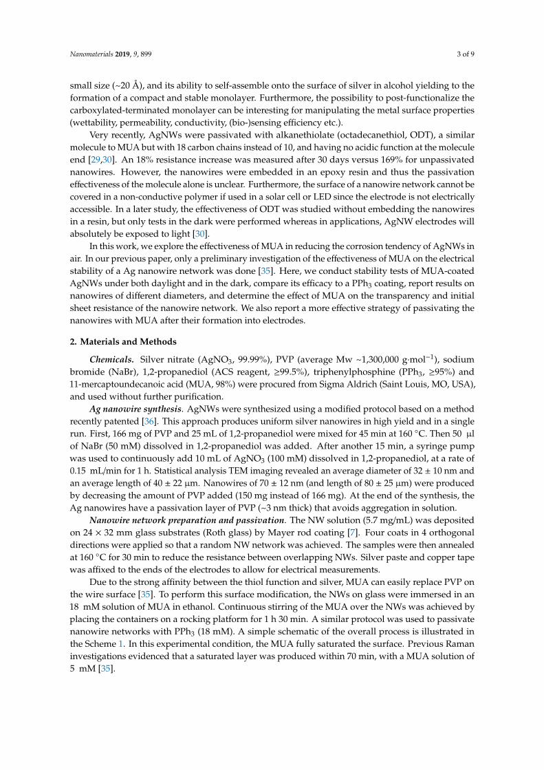
Scheme 1. Fabrication procedure of a passivated Ag nanowire (NW) network.
Characterization. Samples were imaged with scanning electron microscopy (SEM) and atomic electron microscopy. X-Ray Photoelectron spectroscopy (XPS) measurements were conducted with a Thermo Fisher Scientific K-Alpha spectrometer (Waltham, MA, USA), and spectra were fitted and analyzed with Thermo Scientific Avantage software (Waltham, MA, USA). The initial resistances of the electrodes were measured by a four-point probe measurement. Quoted resistances are the average resistance of eight electrodes prepared in the same way. For the stability data, the relative changes in the sheet resistance were instead measured by two-point probe measurements, since the repeated measurements required in the lifetime study could damage the electrode at the contact points. Transparency values were measured at 550 nm with plain glass as a reference. Initial transparency measurements were obtained with a UV-Visible SHIMADZU spectrophotometer (Kyoto, Japon), then a handheld window tint meter was used throughout the remainder of the lifetime studies.
Stability tests over time. Electrodes were stored in the ambient atmosphere of the laboratory (average relative humidity 80%). Some electrodes were exposed to daylight and the others were stored in the dark. Electrical and optical measurements were performed over a period of 4 months, with the optical measurement being done less frequently since the latter measurement caused some nanowires come off the glass.
3. Results
Scheme 1 depicts the route adopted to passivate the surface of PVP-coated AgNW network. This strategy of passivating the nanowires after their formation into electrodes, rather than
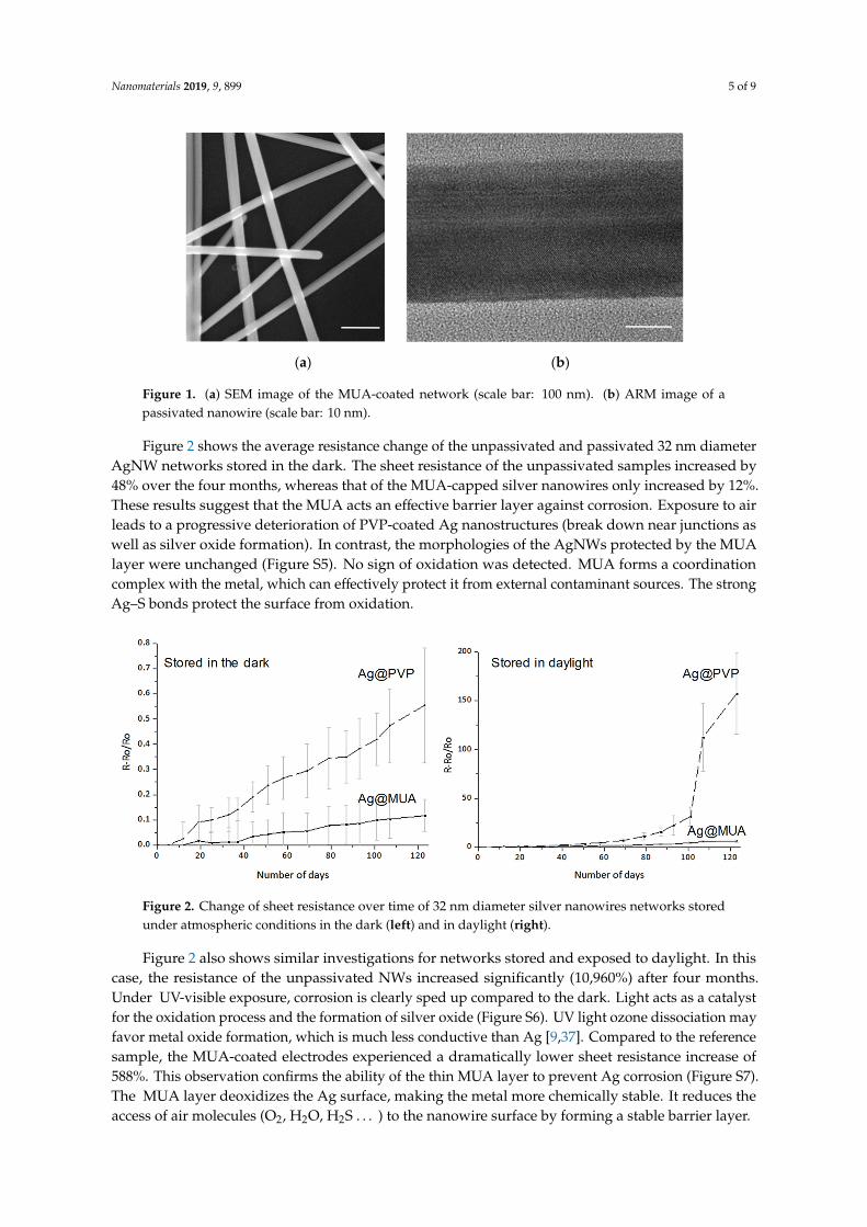
before, was necessary to achieve a uniform dispersion of nanowires in the network. When the NWs were passivated with MUA in suspension, such as done in our prior work [35], there was significant aggregation when they were subsequently deposited as a film due to the weak repulsion between MUA-capped AgNWs (Figure S1). The agglomeration affected the transparency of the NW electrodes passivated before their formation into networks (Table S1). Immersing the AgNW networks in a solution of MUA resulted in a uniform layer of MUA on the AgNWs and no MUA on the open areas of the substrate (Figure 1a and Figure S2). The MUA layer on the nanowires is so thin that atomic resolution microscopy (ARM) and elemental analysis were difficult (Figure 1b, Figures S3 and S4).
(a) (b)
Scheme 1. Fabrication procedure of a passivated Ag nanowire (NW) network.
Characterization. Samples were imaged with scanning electron microscopy (SEM) and atomicelectron microscopy. X-Ray Photoelectron spectroscopy (XPS) measurements were conducted witha Thermo Fisher Scientific K-Alpha spectrometer (Waltham, MA, USA), and spectra were fitted andanalyzed with Thermo Scientific Avantage software (Waltham, MA, USA). The initial resistancesof the electrodes were measured by a four-point probe measurement. Quoted resistances are theaverage resistance of eight electrodes prepared in the same way. For the stability data, the relativechanges in the sheet resistance were instead measured by two-point probe measurements, since therepeated measurements required in the lifetime study could damage the electrode at the contact points.Transparency values were measured at 550 nm with plain glass as a reference. Initial transparencymeasurements were obtained with a UV-Visible SHIMADZU spectrophotometer (Kyoto, Japon), thena handheld window tint meter was used throughout the remainder of the lifetime studies.
Stability tests over time. Electrodes were stored in the ambient atmosphere of the laboratory(average relative humidity 80%). Some electrodes were exposed to daylight and the others were storedin the dark. Electrical and optical measurements were performed over a period of 4 months, with theoptical measurement being done less frequently since the latter measurement caused some nanowirescome off the glass.
3. Results
Scheme 1 depicts the route adopted to passivate the surface of PVP-coated AgNW network.This strategy of passivating the nanowires after their formation into electrodes, rather than
before, was necessary to achieve a uniform dispersion of nanowires in the network. When the NWswere passivated with MUA in suspension, such as done in our prior work [35], there was significantaggregation when they were subsequently deposited as a film due to the weak repulsion betweenMUA-capped AgNWs (Figure S1). The agglomeration affected the transparency of the NW electrodespassivated before their formation into networks (Table S1). Immersing the AgNW networks in asolution of MUA resulted in a uniform layer of MUA on the AgNWs and no MUA on the open areasof the substrate (Figure 1a and Figure S2). The MUA layer on the nanowires is so thin that atomicresolution microscopy (ARM) and elemental analysis were difficult (Figure 1b, Figures S3 and S4).
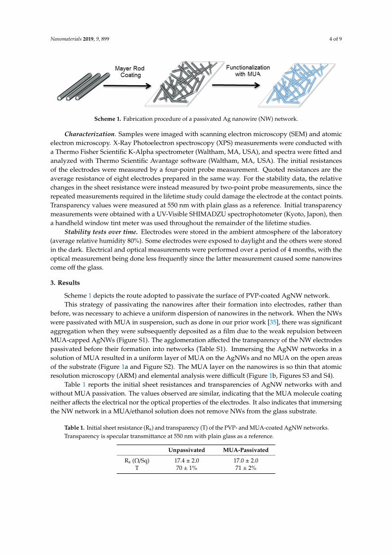
Table 1 reports the initial sheet resistances and transparencies of AgNW networks with andwithout MUA passivation. The values observed are similar, indicating that the MUA molecule coatingneither affects the electrical nor the optical properties of the electrodes. It also indicates that immersingthe NW network in a MUA/ethanol solution does not remove NWs from the glass substrate.
Table 1. Initial sheet resistance (Rs) and transparency (T) of the PVP- and MUA-coated AgNW networks.Transparency is specular transmittance at 550 nm with plain glass as a reference.
Unpassivated MUA-Passivated
Rs (Ω/Sq) 17.4 ± 2.0 17.0 ± 2.0T 70 ± 1% 71 ± 2%
Nanomaterials 2019, 9, 899 5 of 9
Nanomaterials 2019, 9, x FOR PEER REVIEW 4 of 9
Scheme 1. Fabrication procedure of a passivated Ag nanowire (NW) network.
Characterization. Samples were imaged with scanning electron microscopy (SEM) and atomic electron microscopy. X-Ray Photoelectron spectroscopy (XPS) measurements were conducted with a Thermo Fisher Scientific K-Alpha spectrometer (Waltham, MA, USA), and spectra were fitted and analyzed with Thermo Scientific Avantage software (Waltham, MA, USA). The initial resistances of the electrodes were measured by a four-point probe measurement. Quoted resistances are the average resistance of eight electrodes prepared in the same way. For the stability data, the relative changes in the sheet resistance were instead measured by two-point probe measurements, since the repeated measurements required in the lifetime study could damage the electrode at the contact points. Transparency values were measured at 550 nm with plain glass as a reference. Initial transparency measurements were obtained with a UV-Visible SHIMADZU spectrophotometer (Kyoto, Japon), then a handheld window tint meter was used throughout the remainder of the lifetime studies.
Stability tests over time. Electrodes were stored in the ambient atmosphere of the laboratory (average relative humidity 80%). Some electrodes were exposed to daylight and the others were stored in the dark. Electrical and optical measurements were performed over a period of 4 months, with the optical measurement being done less frequently since the latter measurement caused some nanowires come off the glass.
3. Results
Scheme 1 depicts the route adopted to passivate the surface of PVP-coated AgNW network. This strategy of passivating the nanowires after their formation into electrodes, rather than
before, was necessary to achieve a uniform dispersion of nanowires in the network. When the NWs were passivated with MUA in suspension, such as done in our prior work [35], there was significant aggregation when they were subsequently deposited as a film due to the weak repulsion between MUA-capped AgNWs (Figure S1). The agglomeration affected the transparency of the NW electrodes passivated before their formation into networks (Table S1). Immersing the AgNW networks in a solution of MUA resulted in a uniform layer of MUA on the AgNWs and no MUA on the open areas of the substrate (Figure 1a and Figure S2). The MUA layer on the nanowires is so thin that atomic resolution microscopy (ARM) and elemental analysis were difficult (Figure 1b, Figures S3 and S4).
(a) (b)
Figure 1. (a) SEM image of the MUA-coated network (scale bar: 100 nm). (b) ARM image of apassivated nanowire (scale bar: 10 nm).
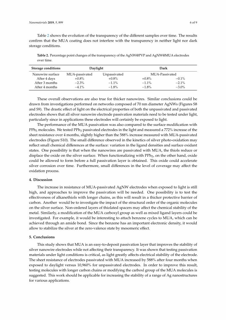
Figure 2 shows the average resistance change of the unpassivated and passivated 32 nm diameterAgNW networks stored in the dark. The sheet resistance of the unpassivated samples increased by48% over the four months, whereas that of the MUA-capped silver nanowires only increased by 12%.These results suggest that the MUA acts an effective barrier layer against corrosion. Exposure to airleads to a progressive deterioration of PVP-coated Ag nanostructures (break down near junctions aswell as silver oxide formation). In contrast, the morphologies of the AgNWs protected by the MUAlayer were unchanged (Figure S5). No sign of oxidation was detected. MUA forms a coordinationcomplex with the metal, which can effectively protect it from external contaminant sources. The strongAg–S bonds protect the surface from oxidation.
Nanomaterials 2019, 9, x FOR PEER REVIEW 5 of 9
Figure 1. (a) SEM image of the MUA-coated network (scale bar: 100 nm). (b) ARM image of a passivated nanowire (scale bar: 10 nm).
Table 1 reports the initial sheet resistances and transparencies of AgNW networks with and without MUA passivation. The values observed are similar, indicating that the MUA molecule coating neither affects the electrical nor the optical properties of the electrodes. It also indicates that immersing the NW network in a MUA/ethanol solution does not remove NWs from the glass substrate.
Table 1. Initial sheet resistance (Rs) and transparency (T) of the PVP- and MUA-coated AgNW networks. Transparency is specular transmittance at 550 nm with plain glass as a reference.
Unpassivated MUA-Passivated Rs (Ω/Sq) 17.4 ± 2.0 17.0 ± 2.0
T 70 ± 1% 71 ± 2%
Figure 2 shows the average resistance change of the unpassivated and passivated 32 nm diameter AgNW networks stored in the dark. The sheet resistance of the unpassivated samples increased by 48% over the four months, whereas that of the MUA-capped silver nanowires only increased by 12%. These results suggest that the MUA acts an effective barrier layer against corrosion. Exposure to air leads to a progressive deterioration of PVP-coated Ag nanostructures (break down near junctions as well as silver oxide formation). In contrast, the morphologies of the AgNWs protected by the MUA layer were unchanged (Figure S5). No sign of oxidation was detected. MUA forms a coordination complex with the metal, which can effectively protect it from external contaminant sources. The strong Ag–S bonds protect the surface from oxidation.
Figure 2. Change of sheet resistance over time of 32 nm diameter silver nanowires networks stored under atmospheric conditions in the dark (left) and in daylight (right).
Figure 2 also shows similar investigations for networks stored and exposed to daylight. In this case, the resistance of the unpassivated NWs increased significantly (10,960%) after four months. Under UV-visible exposure, corrosion is clearly sped up compared to the dark. Light acts as a catalyst for the oxidation process and the formation of silver oxide (Figure S6). UV light ozone dissociation may favor metal oxide formation, which is much less conductive than Ag [9,37]. Compared to the reference sample, the MUA-coated electrodes experienced a dramatically lower sheet resistance increase of 588%. This observation confirms the ability of the thin MUA layer to prevent Ag corrosion (Figure S7). The MUA layer deoxidizes the Ag surface, making the metal more chemically stable. It reduces the access of air molecules (O2, H2O, H2S…) to the nanowire surface by forming a stable barrier layer.
Figure 2. Change of sheet resistance over time of 32 nm diameter silver nanowires networks storedunder atmospheric conditions in the dark (left) and in daylight (right).
Figure 2 also shows similar investigations for networks stored and exposed to daylight. In thiscase, the resistance of the unpassivated NWs increased significantly (10,960%) after four months.Under UV-visible exposure, corrosion is clearly sped up compared to the dark. Light acts as a catalystfor the oxidation process and the formation of silver oxide (Figure S6). UV light ozone dissociation mayfavor metal oxide formation, which is much less conductive than Ag [9,37]. Compared to the referencesample, the MUA-coated electrodes experienced a dramatically lower sheet resistance increase of588%. This observation confirms the ability of the thin MUA layer to prevent Ag corrosion (Figure S7).The MUA layer deoxidizes the Ag surface, making the metal more chemically stable. It reduces theaccess of air molecules (O2, H2O, H2S . . . ) to the nanowire surface by forming a stable barrier layer.
Nanomaterials 2019, 9, 899 6 of 9
Table 2 shows the evolution of the transparency of the different samples over time. The resultsconfirm that the MUA coating does not interfere with the transparency in neither light nor darkstorage conditions.
Table 2. Percentage point changes of the transparency of the AgNW@PVP and AgNW@MUA electrodesover time.
Storage conditions Daylight Dark
Nanowire surface MUA-passivated Unpassivated MUA-PassivatedAfter 4 days +0.8% +0.8% +0.8% −0.1%
After 3 months −2.3% −1.1% −1.1% −2.1%After 4 months −4.1% −1.8% −1.8% −3.0%
These overall observations are also true for thicker nanowires. Similar conclusions could bedrawn from investigations performed on networks composed of 70 nm diameter AgNWs (Figures S8and S9). The drastic effect of light on the electrical properties of both the unpassivated and passivatedelectrodes shows that all silver nanowire electrode passivation materials need to be tested under light,particularly since in applications these electrodes will certainly be exposed to light.
The performance of the MUA passivation was also compared to the surface modification withPPh3 molecules. We tested PPh3 passivated electrodes in the light and measured a 772% increase of thesheet resistance over 4 months, slightly higher than the 588% increase measured with MUA-passivatedelectrodes (Figure S10). The small difference observed in the kinetics of silver photo-oxidation mayreflect small chemical differences at the surface: variation in the ligand densities and surface oxidantstates. One possibility is that when the nanowires are passivated with MUA, the thiols reduce ordisplace the oxide on the silver surface. When functionalizing with PPh3, on the other hand, oxidecould be allowed to form before a full passivation layer is obtained. This oxide could acceleratesilver corrosion over time. Furthermore, small differences in the level of coverage may affect theoxidation process.
4. Discussion
The increase in resistance of MUA-passivated AgNW electrodes when exposed to light is stillhigh, and approaches to improve the passivation will be needed. One possibility is to test theeffectiveness of alkanethiols with longer chains, as this will result in a thicker protective barrier ofcarbon. Another would be to investigate the impact of the structural order of the organic moleculeson the silver surface. Non-ordered layers of thiolated spacers may affect the chemical stability of themetal. Similarly, a modification of the MUA carboxyl group as well as mixed ligand layers could beinvestigated. For example, it would be interesting to attach benzene cycles to MUA, which can beachieved through an amide bond. Since the benzene has an important electronic density, it wouldallow to stabilize the silver at the zero-valence state by mesomeric effect.
5. Conclusions
This study shows that MUA is an easy-to-deposit passivation layer that improves the stability ofsilver nanowire electrodes while not affecting their transparency. It was shown that testing passivationmaterials under light conditions is critical, as light greatly affects electrical stability of the electrode.The sheet resistance of electrodes passivated with MUA increased by 588% after four months whenexposed to daylight versus 10,960% for unpassivated electrodes. In order to improve this result,testing molecules with longer carbon chains or modifying the carboxl group of the MUA molecules issuggested. This work should be applicable for increasing the stability of a range of Ag nanostructuresfor various applications.
Nanomaterials 2019, 9, 899 7 of 9
Supplementary Materials: The following are available online at http://www.mdpi.com/2079-4991/9/6/899/s1,Table S1: Initial sheet resistance and transparencies of the electrodes based on silver nanowires before and aftertheir formation into films; Figure S1: SEM image of AgNW passivated with MUA in solution and subsequentlydeposited as a film; Figure S2: SEM image of a MUA-coated silver NW network. Representative EDX spectrumtaken on different areas of the networks; Figure S3: Typical EDX spectrum taken from a MUA-coated silver NWnetwork; Figure S4: Atomic resolution microscopy image of a MUA-coated silver nanowire; Figure S5: SEMimage of a MUA-coated silver NW network stored and exposed to the dark after 4 months; Figure S6: TEM imageand XPS of the PVP-coated AgNWs after 4 months of air exposure; Figure S7: XPS spectrum of the MUA-coatedAgNW network stored and exposed to daylight after four months; Figure S8: SEM image of 70 nm diameterAgNW network; Figure S9: Evolution of the sheet resistance vs. time for films of PVP- or MUA-coated AgNW(70 nm) after exposure to air at ambient conditions; Figure S10: Evolution of the sheet resistance vs. time for filmsof AgNW coated with PVP, MUA and PPh3 stored in daylight after exposure to air at ambient conditions.
Author Contributions: Conceptualization of study, M.T.-D. and I.A.G.; formal analysis, A.M. and M.P.;investigation, A.M.; review and editing of writing, M.T.-D. and I.A.G.; supervision, M.T.-D.; funding acquisition,M.T.-D., L.S. and I.A.G.
Funding: This work was funded by a U.Waterloo/U.Bordeaux grant and the European Joint DoctorateFunMat network.
Acknowledgments: TEM and SEM characterization was performed at the Plateforme de Caractérisation desMatériaux and Bordeaux Imaging Center at the University of Bordeaux and at the Plateforme Castaing of theUniversity of Toulouse.
Conflicts of Interest: The authors declare no conflict of interest.
References
1. Sannicolo, T.; Lagrange, M.; Cabos, A.; Celle, C.; Simonato, J.P.; Bellet, D. Metallic Nanowire-Based TransparentElectrodes for Next Generation Flexible Devices: A Review. Small 2016, 12, 6052–6075. [CrossRef] [PubMed]
2. Ye, S.; Rathmell, A.R.; Chen, Z.; Stewart, I.E.; Wiley, B.J. Metal nanowire networks: The next generation oftransparent conductors. Adv. Mater. 2014, 26, 6670–6687. [CrossRef] [PubMed]
3. Bellet, D.; Lagrange, M.; Sannicolo, T.; Aghazadehchors, S.; Nguyen, V.H.; Langley, D.; Munoz-Rojas, D.;Jimenez, C.; Bréchet, Y.; Nguyen, N.D. Transparent Electrodes Based on Silver Nanowire Networks: FromPhysical Considerations towards Device Integration. Materials 2017, 10, 570. [CrossRef] [PubMed]
4. Sun, Y. Silver nanowires-unique templates for functional nanostructures. Nanoscale 2010, 9, 1626–1642.[CrossRef] [PubMed]
5. Hu, L.; Kim, H.S.; Lee, J.; Peumans, P.; Cui, Y. Scalable coating and properties of transparent, flexible, silvernanowire electrodes. ACS Nano 2010, 4, 2955–2963. [CrossRef] [PubMed]
6. Madaria, A.R.; Kumar, A.; Zhou, C. Large scale, highly conductive and patterned transparent films ofsilver nanowires on arbitrary substrates and their application in touch screens. Nanotechnology 2011, 22,245201–245208. [CrossRef]
7. Pasquarelli, R.M.; Ginley, D.S.; O’Hayre, R. Solution processing of transparent conductors: From flask to film.Chem. Soc. Rev. 2011, 40, 5406–5441. [CrossRef]
8. Zhang, R.; Engholm, M. Recent progress on the fabrication and properties of silver nanowire-base transparentelectrodes. Nanomaterials 2018, 8, 628. [CrossRef]
9. Elechiguerra, J.L.; Larios-Lopez, L.; Lui, C.; Garcia-Gutierrez, D.; Camacho-Bragada, A.; Yacaman, M.J.Corrosion at the nanoscale: The case of silver nanowires and nanoparticles. Chem. Mater. 2005, 17, 6042–6052.[CrossRef]
10. Jiu, J.; Wang, J.; Sugahara, T.; Nagao, S.; Nogi, M.; Koga, H.; Suganuma, K.; Hara, M.; Nakazawa, E.;Uchida, H. The Effect of Light and Humidity on the Stability of Silver Nanowire Transparent Electrodes.RSC Adv. 2015, 5, 27657–27664. [CrossRef]
11. Mayousse, C.; Celle, C.; Fraczkiewicz, A.; Simonato, J.P. Stability of silver nanowire based electrodes underenvironmental and electrical stresses. Nanoscale 2015, 7, 2107–2115. [CrossRef] [PubMed]
12. Deignan, G.; Goldthorpe, I.A. The dependance of silver nanowire stability on network compositions andprocessing parameters. RSC Adv. 2017, 7, 35590–35597. [CrossRef]
13. Franey, J.P.; Kammlott, J.P.; Graedel, T.E. The corrosion of silver by atmospheric sulfurous gases. Corros. Sci.1985, 25, 33–143. [CrossRef]
Nanomaterials 2019, 9, 899 8 of 9
14. Keast, V.J.; Myles, T.A.; Shahcheraghi, N.; Cortie, M.B. Corrosion processes of triangular silver nanoparticlescompared to bulk silver. J. Nanopart. Res. 2016, 18, 45–56. [CrossRef]
15. Wang, X.; Santshi, C.; Martin, O. Strong improvement of long-term chemical and thermal stability ofplasmonic silver nanoantennas and films. Small 2017, 13, 1700044–1700049. [CrossRef] [PubMed]
16. Kang, H.; Buchman, J.T.; Rodriguez, R.S.; Ring, H.T.; He, J.; Bantz, K.C.; Haynes, C.L. Stabilization of silverand gold nanoparticles: Preservation and improvement of plasmonic functionalities. Chem. Rev. 2019, 119,664–699. [CrossRef]
17. Chen, S.; Goode, A.E.; Sweeney, S.; Theodorou, I.G.; Thorley, A.J.; Ruenraroengsak, P.; Chang, Y.; Gow, A.;Schwander, S.; Skepper, J.; et al. Sulfidation of silver nanowires inside human alveolar epithelial cells: Apotential detoxification mechanism. Nanoscale 2013, 5, 9839–9847. [CrossRef]
18. Moon, I.K.; Kim, J.I.; Lee, H.; Hur, K.; Kim, W.C.; Lee, H. 2D Graphen oxide nanosheetes as an adhesiveover-coating layer for flexible transparent conductive electrodes. Sci. Rep. 2013, 3, 1112. [CrossRef]
19. Song, T.B.; Rim, Y.S.; Liu, F.; Bob, B.; Ye, S.; Hsieh, T.; Yang, Y. Highly Robust Silver Nanowire Network forTransparent Electrode. ACS Appl. Mat. Interfaces 2015, 7, 24601–24607. [CrossRef]
20. Deng, B.; Hsu, P.-C.; Chen, G.; Chandrashekar, B.N.; Liao, L.; Ayitimuda, Z.; Wu, J.; Guo, Y.; Zhou, Y.;Aisijiang, M.; et al. Roll-to-Roll Encapsulation of Metal Nanowires between Graphene and Plastic Substratefor High-Performance Flexible Transparent Electrodes. Nano Lett. 2015, 15, 4206–4213. [CrossRef]
21. Yang, Y.; Ding, S.; Araki, T.; Jiu, J.; Sugahara, T.; Wang, J.; Vanfleteren, J.; Sekitani, T.; Suganuma, K. FacileFabrication of Stretchable Ag Nanowire/Polyurethane Electrodes Using High Intensity Pulsed Light. NanoRes. 2016, 9, 401–414. [CrossRef]
22. Yoo, Y.Z.; Na, J.-Y.; Choi, Y.S.; Lim, Y.J.; Kim, J.-H.; Kim, Y.B.; Kim, S.; Seong, T. An Optically Flat ConductiveOutcoupler Using Core/Shell Ag/ZnO Nanochurros. Small 2018, 14, 1800056. [CrossRef] [PubMed]
23. Kim, Y.; Hwang, B.; Kim, J. Ambient-stable and durable conductive Ag-Nanowire-network, 2D fims decoratedwith a Ti layer. Nanomaterials 2018, 8, 321. [CrossRef]
24. Zhang, W.; Zhao, B.; He, Z.; Zhao, X.; Wang, H.; Yang, S.; Wu, H.; Cao, Y. High-efficiency ITO-free polymersolar cells using highly conductive PEDOT: PSS surfactant bilayer transparent anodes. Energy Environ. Sci.2013, 6, 1956–1964. [CrossRef]
25. Chen, S.; Song, L.; Tao, Z.; Shao, X.; Huang, Y.; Cui, Q.; Guo, X. Neutral-pH PEDOT: PSS as over-coatinglayer for stable silver nanowire flexible transparent conductive films. Org. Electron. 2014, 15, 3654–3659.[CrossRef]
26. Ahn, Y.; Jeong, Y.; Lee, Y. Improved thermal oxidation stability of solution-processable silver nanowiretransparent electrode by reduced graphene oxide. ACS Appl. Mater. Interfaces 2012, 4, 6410–6414. [CrossRef]
27. Lee, D.; Lee, H.; Ahn, Y.; Lee, Y. High-performance flexible transparent conductive film based ongraphene/AgNW/graphene sandwich structure. Carbon 2015, 81, 439–446. [CrossRef]
28. Idier, J.; Neri, W.; Labrugere, C.; Ly, I.; Poulin, P.; Backov, R. Modified silver nanowire transparent electrodeswith exceptional stability against oxidation. Nanotechnology 2016, 27, 105705–105712. [CrossRef]
29. Liu, G.S.; Qiu, J.S.; Xu, D.H.; Zhou, X.; Zhong, D.; Shieh, H.P.D.; Yang, B.R. Fabrication of Embedded SilverNanowires on Arbitrary Substrates with Enhanced Stability via Chemisorbed Alkanethiolate. ACS Appl.Mater. Interfaces 2017, 9, 15130–15138. [CrossRef]
30. Liu, G.; Xu, Y.; Kong, Y.; Wang, L.; Wang, J.; Xie, X.; Luo, Y.; Yang, B. Comprehensive stability improvementof silver nanowire networks via self-assembled mercapto inhibitors. ACS Appl. Mater. Interfaces 2018, 10,37699–37708. [CrossRef]
31. Gooding, J.J.; Ciampi, S. The molecular level modification of surfaces: From self-assembled monolayers tocomplex molecular assemblies. Chem. Soc. Rev. 2011, 40, 2704–2718. [CrossRef] [PubMed]
32. Le Beulze, A.; Duguet, E.; Mornet, S.; Majimel, J.; Tréguer-Delapierre, M.; Ravaine, S.; Florea, I.; Ersen, O.New Insights into the Side-Face Structure, Growth Aspects, and Reactivity of Agn Nanoprisms. Langmuir2014, 30, 1424–1434. [CrossRef]
33. Fajín, J.L.; Teixeira, F.; Gomes, J.R.; Cordeiro, M.N.D. Effect of van der Waals interactions in the DFTdescription of self-assembled monolayers of thiols on gold. In Proceedings of the 9th Congress on ElectronicStructure: Principles and Applications, Badajoz, Spain, 2–4 July 2014; Springer: Berlin/Heidelberg, Germany,2016; pp. 127–139.
Nanomaterials 2019, 9, 899 9 of 9
34. Pan, X.-F.; Gao, H.-L.; Su, Y.; Wu, Y.-D.; Wang, X.-Y.; Xue, J.-Z.; He, T.; Lu, Y.; Liu, J.; Yu, S. Strong and stiffAg nanowire-chitosan composite films reinforced by Ag-S covalent bonds. Nano Res. 2018, 11, 410–419.[CrossRef]
35. Plissonneau, M.; Madeira, A.; Talaga, D.; Bonhommeau, S.; Servant, L.; Vallée, R.; Labrugère, C.; Goldthorpe, I.;Pautrot-d’Alençon, L.; Le Mercier, T.; et al. Efficient Passivation of Ag Nanowires with 11-MercaptoundecanoicAcid Probed Using In-Situ Total-Internal-Reflection Surface-Enhanced Raman Scattering Spectroscopy.ChemNanoMat 2019, 5, 1–7. [CrossRef]
36. Plissonneau, M.; Pautrot-D’Alençon, L.; Le Mercier, T.; Tréguer-Delapierre, M. Process for the Manufactureof Metal Nanowires. European Patent No 17306072.4-1103, 13 August 2017.
37. Grillet, N.; Manchon, D.; Cottancin, E.; Bertorelle, F.; Bonnet, C.; Broyer, M.; Lermé, J.; Pellarin, M.Photo-oxidation of individual silver nanoparticles: A real-time tracking of optical and morphologicalchanges. J. Phys. Chem. C 2013, 117, 2274–2282. [CrossRef]
© 2019 by the authors. Licensee MDPI, Basel, Switzerland. This article is an open accessarticle distributed under the terms and conditions of the Creative Commons Attribution(CC BY) license (http://creativecommons.org/licenses/by/4.0/).