Increased longevity and refractoriness to Ca2+-dependent neurodegeneration in Surf1 knockout mice
14
Increased longevity and refractoriness to Ca 21 -dependent neurodegeneration in Surf1 knockout mice Carlotta Dell’Agnello 1,{ , Sara Leo 2,{ , Alessandro Agostino 1 , Gyo ¨ rgy Szabadkai 2,{ , Cecilia Tiveron 3 , Alessandra Zulian 1 , Alessandro Prelle 4 , Pierre Roubertoux 5 , Rosario Rizzuto 2 and Massimo Zeviani 1, * 1 Unit of Molecular Neurogenetics, Pierfranco and Luisa Mariani Center for the Study of Children’s Mitochondrial Disorders, National Neurological Institute ‘C. Besta’, Milano, Italy, 2 Department of Experimental and Diagnostic Medicine, Section of General Pathology, Interdisciplinary Center for the Study of Inflammation (ICSI) and ER-GenTech, University of Ferrara, Ferrara, Italy, 3 Foundation EBRI Rita Levi-Montalcini Disease Modelling Facility, Rome, Italy, 4 Centro Dino Ferrari, UO Neurologia, Fondazione Ospedale Maggiore Policlinico, Mangiagalli e Regina Elena, IRCCS, Milano and 5 Universite ´ Marseille-2, CNRS-Universite ´ de la Me ´ diterrane ´e, Marseille, France Received November 3, 2006; Revised December 19, 2006; Accepted December 28, 2006 Leigh syndrome associated with cytochrome c oxidase (COX) deficiency is a mitochondrial disorder usually caused by mutations of SURF1, a gene encoding a putative COX assembly factor. We present here a Surf12/2 recombinant mouse obtained by inserting a loxP sequence in the open reading frame of the gene. The frequency of 2/2, 1/1 and 1/2 genotypes in newborn mice followed a mendelian distribution, indicating that the ablation of Surf1 is compatible with postnatal survival. The biochemical and assembly COX defect was present in Surf1 loxP 2/2 mice, but milder than in humans. Surprisingly, not only these animals failed to show spontaneous neurodegeneration at any age, but they also displayed markedly prolonged lifespan, and complete protection from Ca 21 -dependent neurotoxicity induced by kainic acid. Experiments on primary neuronal cultures showed markedly reduced rise of cytosolic and mitochondrial Ca 21 in Surf1 loxP 2/2 neurons, and reduced mortality, compared to controls. The mitochondrial membrane potential was unchanged in KO versus wild-type neurons, suggesting that the effects of the ablation of Surf1 on Ca 21 homeostasis, and possibly on longevity, may be independent, at least in part, from those on COX assembly and mitochondrial bioenergetics. INTRODUCTION Cytochrome c oxidase (COX), the terminal enzyme of the mito- chondrial respiratory chain (MRC), catalyzes the transfer of electrons from reduced cytochrome c to molecular oxygen (1). COX is composed of 13 protein subunits, the three largest being encoded by mtDNA genes, and the remaining ten are encoded by nuclear DNA genes (2). A number of acces- sory factors are necessary for the formation of an active holoenzyme complex (3), including those involved in syn- thesis of heme a, incorporation of copper atoms and assembly of the protein backbone (4). One of these factors, SURF1, is a 30 kDa hydrophobic protein embedded in the inner membrane of mitochondria. The absence, or malfunctioning, of SURF1p determines the accumulation of COX assembly intermediates, and a drastic reduction in the amount of fully assembled enzyme, in both yeast (5) and humans (6). As a consequence, profound COX deficiency (7) in multiple tissues of Surf1 # The Author 2007. Published by Oxford University Press. All rights reserved. For Permissions, please email: [email protected] { These authors contributed equally to the work. ‡ Present address: INSERM U807, University Paris 5, Faculty of Medicine Necker-Enfants Malades, Paris, France. *Correspondence should be addressed to: Via L. Temolo 4, 20126 Milano, Italy. Tel: þ 33 390223942630; Fax: þ 33 390223942619; Email: [email protected] Human Molecular Genetics, 2007, Vol. 16, No. 4 431–444 doi:10.1093/hmg/ddl477 Advance Access published on January 8, 2007 by guest on January 14, 2016 http://hmg.oxfordjournals.org/ Downloaded from
-
Upload
independent -
Category
Documents
-
view
1 -
download
0
Transcript of Increased longevity and refractoriness to Ca2+-dependent neurodegeneration in Surf1 knockout mice

Increased longevity and refractorinessto Ca21-dependent neurodegenerationin Surf1 knockout mice
Carlotta Dell’Agnello1,{, Sara Leo2,{, Alessandro Agostino1, Gyorgy Szabadkai2,{,
Cecilia Tiveron3, Alessandra Zulian1, Alessandro Prelle4, Pierre Roubertoux5, Rosario Rizzuto2
and Massimo Zeviani1,*
1Unit of Molecular Neurogenetics, Pierfranco and Luisa Mariani Center for the Study of Children’s Mitochondrial
Disorders, National Neurological Institute ‘C. Besta’, Milano, Italy, 2Department of Experimental and Diagnostic
Medicine, Section of General Pathology, Interdisciplinary Center for the Study of Inflammation (ICSI) and
ER-GenTech, University of Ferrara, Ferrara, Italy, 3Foundation EBRI Rita Levi-Montalcini Disease Modelling Facility,
Rome, Italy, 4Centro Dino Ferrari, UO Neurologia, Fondazione Ospedale Maggiore Policlinico, Mangiagalli e Regina
Elena, IRCCS, Milano and 5Universite Marseille-2, CNRS-Universite de la Mediterranee, Marseille, France
Received November 3, 2006; Revised December 19, 2006; Accepted December 28, 2006
Leigh syndrome associated with cytochrome c oxidase (COX) deficiency is a mitochondrial disorderusually caused by mutations of SURF1, a gene encoding a putative COX assembly factor. We present herea Surf12/2 recombinant mouse obtained by inserting a loxP sequence in the open reading frame of thegene. The frequency of 2/2, 1/1 and 1/2 genotypes in newborn mice followed a mendelian distribution,indicating that the ablation of Surf1 is compatible with postnatal survival. The biochemical and assemblyCOX defect was present in Surf1loxP2/2 mice, but milder than in humans. Surprisingly, not only theseanimals failed to show spontaneous neurodegeneration at any age, but they also displayed markedlyprolonged lifespan, and complete protection from Ca21-dependent neurotoxicity induced by kainic acid.Experiments on primary neuronal cultures showed markedly reduced rise of cytosolic and mitochondrialCa21 in Surf1loxP2/2 neurons, and reduced mortality, compared to controls. The mitochondrial membranepotential was unchanged in KO versus wild-type neurons, suggesting that the effects of the ablation ofSurf1 on Ca21 homeostasis, and possibly on longevity, may be independent, at least in part, from thoseon COX assembly and mitochondrial bioenergetics.
INTRODUCTION
Cytochrome c oxidase (COX), the terminal enzyme of the mito-chondrial respiratory chain (MRC), catalyzes the transfer ofelectrons from reduced cytochrome c to molecular oxygen (1).COX is composed of 13 protein subunits, the three largestbeing encoded by mtDNA genes, and the remaining tenare encoded by nuclear DNA genes (2). A number of acces-sory factors are necessary for the formation of an active
holoenzyme complex (3), including those involved in syn-thesis of heme a, incorporation of copper atoms and assemblyof the protein backbone (4). One of these factors, SURF1, is a30 kDa hydrophobic protein embedded in the inner membraneof mitochondria. The absence, or malfunctioning, of SURF1pdetermines the accumulation of COX assembly intermediates,and a drastic reduction in the amount of fully assembledenzyme, in both yeast (5) and humans (6). As a consequence,profound COX deficiency (7) in multiple tissues of Surf1
# The Author 2007. Published by Oxford University Press. All rights reserved.For Permissions, please email: [email protected]
{These authors contributed equally to the work.
‡Present address: INSERM U807, University Paris 5, Faculty of Medicine Necker-Enfants Malades, Paris, France.
*Correspondence should be addressed to: Via L. Temolo 4, 20126 Milano, Italy. Tel: þ 33 390223942630; Fax: þ 33 390223942619; Email:[email protected]
Human Molecular Genetics, 2007, Vol. 16, No. 4 431–444doi:10.1093/hmg/ddl477Advance Access published on January 8, 2007
by guest on January 14, 2016http://hm
g.oxfordjournals.org/D
ownloaded from

mutant patients (8) leads to the development of Leigh syn-drome (LSCOX), an early onset, invariably fatal mitochondrialencephalomyelopathy (9).
In vertebrates, SURF1 is part of the very tight and highlyconserved surfeit gene cluster, which includes six genes(SURF1–6) (10). The reason for long-standing maintenanceof such a compact physical organization is obscure, sincethe corresponding SURF proteins are neither functionallynor structurally related to each other. The precise functionof the SURF1 gene product itself remains unknown, althoughthe results of several studies in yeast and mammals suggest arole for SURF1 protein (SURF1p) as an auxiliary chaperone-like factor, involved in the early assembly steps of the COXprotein backbone (6).
To better understand the role of SURF1p and the patho-genesis of LSCOX, we have previously created a constitutiveknockout (KO) mouse model, in which exons 5–7 of theSurf1 gene were replaced by a neomycin-resistance (NEO)cassette (11). Approximately 90% of the Surf1NEO2/2 micedied at E6.5–7.5. The few animals that reached birth partiallyrecapitulated, although to a lesser extent, the biochemical find-ings, but failed to display the clinical and neuro-pathologicalfeatures of human LSCOX.
We present here a second Surf1 KO model, based on theinsertion of a loxP sequence in exon 7 of the murine Surf1gene (Surf1loxP), leading to an aberrant, prematurely truncatedand highly unstable protein. The þ/þ, þ/2 and 2/2 geno-types in newborn animals were in agreement with themendelian distribution, indicating that, rather than to theablation of Surf1 itself, the high embryonic lethality observedin the previous Surf1NEO2/2 model was due to a spuriouseffect of the NEO cassette on the expression of neighboringgenes. Similar to the previous Surf1NEO2/2 mice, theSurf1loxP2/2 mice displayed mild reduction of COX activityin all tissues, but no lesion resembling LSCOX encephalopathywas ever observed. However, when the sensitivity to Ca2þ-dependent excitotoxicity was tested in both Surf1loxP2/2brains and neuronal cell cultures, we observed a virtuallycomplete protection from in vivo neurodegeneration inducedby exposure to high doses of kainic acid, a glutamatergicepileptogenic agonist. In addition, Surf1loxP2/2 miceshowed a marked increase in longevity, compared to heterozy-gous or homozygous wild-type (wt) littermates. These datasuggest a role for Surf1p in intracellular Ca2þ homeostasisand mitochondrial control of aging.
RESULTS
Generation of Surf1loxP2/2 mice
The strategy used to disrupt the mouse Surf1 gene(NM_013677) is shown in Figure 1A. Briefly, a cDNA expres-sing the Escherichea coli neomycin phosphotransferase(NEO), therefore conferring neomycin-resistance, flanked bytwo loxP sequences (loxP-NEO-loxP cassette), was insertedinto a unique AccIII restriction site of Surf1 exon 7. In orderto confirm homologous recombination, Southern blot analysiswas performed on both extremities of the Surf1loxP-NEO-loxP
recombinant allele (Fig. 1B and C). Blastocyst injection oftwo recombinant clones gave rise to eleven Surf1loxP-NEO-loxP
chimeric mice showing germline transmission. NoSurf1loxP-NEO-loxP/loxP-NEO-loxP homozygous individuals wereobtained by mating Surf1loxP-NEO-loxP/þ heterozygotes toeach other, due to arrest of organogenesis at E8.5–9.5 (Sup-plementary Materials, Fig. 1 and Table 1). The NEO cassetteand one of the two loxP sequences were then excised bymating Surf1loxP-NEO-loxP/þ heterozygous animals withanimals constitutively expressing the cre recombinase. Theresulting recombinant allele (Surf1loxP) contains a singleloxP insertion (Fig. 1D), which causes the shift of the openreading frame (ORF) from nt 674 downstream of the mouseSurf1 cDNA, and the replacement of the codon encodingN225 into a TAA stop codon (N225X) (Fig. 1A). This mutationpredicts the elimination of 81 amino acids on the Surf1pcarboxy terminus. No cross-reacting material was detectedby western blot immunoassay using polyclonal and mono-clonal anti-Surf1 antibodies in several tissues of ourSurf1loxP2/2 mice (Fig. 1F), indicating that the truncatedSurf1loxP protein is either unstable or fails to be translated,due to mRNA decay (Fig. 1E).
Clinical and biochemical phenotype
The percentages of approximately 800 Surf1loxP2/2, þ/2and þ/þ newborn animals followed a mendelian distribution(26, 51 and 23%, respectively; Supplementary Material,Fig. 2A). Newborn Surf1loxP2/2 individuals were significantlysmaller than their littermates, but this difference progressivelydisappeared after weaning (Supplementary Material, Fig. 2B).No difference in the clinical phenotype was observed betweenSurf1loxP2/2 individuals and their þ/2 or þ/þ littermates,including the appearance of neurological symptoms, abnormalreaction to stimuli, aberrant behavior, impaired cognitive abil-ities and reduced fertility. There was a small but significantreduction at the rotarod test (Supplementary Material,Fig. 2C), indicating mildly decreased motor skills inSurf1loxP2/2 versus wt mice.
Histochemically, we observed decreased reaction to COXand increased reaction to succinate dehydrogenase (SDH) inSurf1loxP2/2 skeletal muscle (Fig. 2A), similar to, but lesssevere than, that observed in SURF1 mutant patients.Finally, Surf1loxP2/2 brains showed normal cytoarchitectureby thionine and GFAP stainings (data not shown).
Biochemically, there was no difference in the activities ofMRC complexes I, II and III, whereas the COX activity inseveral tissues of Surf1loxP2/2 individuals was 30–50%that of control littermates (Fig. 2B). Again, this reductionwas much less marked than that observed in LSCOX patients(7). Surf1loxP
þ /2 mice showed no biochemical differencecompared to Surf1 þ /þ littermates, as expected for a reces-sive trait (data not shown).
Blood lactate was higher in Surf1loxP2/2 mice than in wtlittermates, indicating partial block in the aerobic utilizationof pyruvate (Supplementary Material, Fig. 2D).
As exemplified in Fig. 2C, fully assembled COX wasvariably reduced in different tissues of Surf1loxP2/2 mice, toan extent compatible with the levels of residual COX activity.Early assembly COX intermediates were also present inisolated mitochondria of Surf1loxP2/2 mice, similar to, butmuch lesser than, what is found in SURF1 mutant patients.
432 Human Molecular Genetics, 2007, Vol. 16, No. 4
by guest on January 14, 2016http://hm
g.oxfordjournals.org/D
ownloaded from

Taken together, these data are remarkably similar tothose previously reported for the surviving Surf1NEO2/2individuals (11).
Increased longevity of Surf1loxP2/2 mice
In order to evaluate whether the lack of Surf1p could determinea late-onset phenotype, Surf1loxP2/2 mice and control litter-mates were maintained under continuous observation in the
same standard breeding conditions. No neurological or otherclinical symptoms were ever seen in any individual. However,the lifespan was markedly different between the two groups.A total of 25/43 Surf1loxP2/2 mice died during the observationperiod, against 30/48 deaths recorded in the control group. Themedian survival was 793 days for the Surf1loxP2/2 group and654 days for the control group, the latter being the standardmedian survival reported for laboratory mice (12). As shownin Figure 3A, the difference in the Kaplan–Meier survival
Figure 1. Generation of Surf12/2 mice (see Materials and Methods). (A) Schematic representation of the Surf1 locus, Surf1loxP-NEO-loxP and Surf1loxP alleles,Surf1loxP cDNA and Surf1loxP proteins. P1 and P2 refer to the probes used for Southern-blot analysis shown in panels B and C. Numbers in the scheme of theSurf1 gene and Surf1 cDNA refer to exons ‘E’. In the nucleotide sequence of the Surf1loxP allele, exon 7 is in gray, the loxP sequence is in black and italicized,the TAA stop codon is underlined. In the scheme of the Surf1 protein (bottom part of panel A), the putative mitochondrial targeting peptide is in gray. (B and C)Southern-blot analysis of the recombinant Surf1loxP-NEO-loxP allele. (D) PCR-based genotyping of the Surf loxP versus Surf wt alleles. (E) RT-PCR analysis of theSurf loxP versus Surf wt alleles. The GAPDH cDNA fragment serves as a control. (F) Western blot analysis. Surf1p cross-reacting material is absent inmitochondrial membranes isolated from different Surf loxP2/2 organs probed with a monoclonal anti-Surf1p antibody (upper panel) and with a polyclonalantibody against the Surf1p mid portion (middle panel). The 30 kDa SDH-B subunit was used as a loading control (bottom panel). Lane 1, control fibroblasts;lanes 2 and 4, brain; lanes 3 and 5, liver.
Human Molecular Genetics, 2007, Vol. 16, No. 4 433
by guest on January 14, 2016http://hm
g.oxfordjournals.org/D
ownloaded from

probability, calculated by the logrank test, was highly significantbetween the two groups (P ¼ 0.0002), irrespective of the gender(Fig. 3B and C).
Surf1loxP2/2 mice are protected from Ca21-relatedexcitotoxic brain damage
In order to determine whether our Surf1loxP2/2 mice weremore susceptible to stress-induced neuronal damage, weused kainic acid, an epileptogenic glutamate agonist that hasextensively been used to test neuronal response and survivalto Ca2þ-mediated excitotoxicity. We injected intra peritoneum(i.p.) a total of 145 three-month old mice (73 controls and 72Surf1loxP2/2) with 30 mg/kg of kainic acid. The mortalityrate, as well as the frequency, time lapse, severity and durationof the kainate-induced seizures were similar between the KOand control groups (Table 1), suggesting that the pharmacoki-netics of the drug did not differ in the two groups. Following astandard protocol (13), only animals surviving the most severelevel-5 seizure were further investigated (Table 1). Whole-brain pathological examination was carried out using theneurodegeneration-sensitive FluoroJadeB (FJB) fluorochrome(14) and thionine stainings. Strong FJB-positive neurons
were detected in virtually all the glutamatergic areas ofthe control brains, including the Cornu Ammonis (CA) areas1–4 of the hippocampus, the cerebral cortex, the amygdalaand the thalamic and olfactory nuclei. The most severely
Figure 2. Histochemical and biochemical characterization of Surf1loxP mice.(A) Serial sections of the left quadriceps from 5-month-old Surf1loxP2/2versus Surf1 þ /þ animals. Several fibers in the Surf1loxP2/2 muscle showreduced reaction to COX and are hyper-intense to SDH, compared to theSurf1 þ /þ muscle. (B) COX/CS activities of 3-month-old Surf1loxP2/2mice (n ¼ 10) compared to Surf1þ/þ littermates (n ¼ 10) taken as 100%.�P , 0.01; �� P , 1025. (C) Western blot analysis of 2D-BNE on isolatedmitochondria from 2/2 and þ/þ livers and from fibroblasts of a SURF1mutant patient (LS-COX), using an anti-COXI antibody. S4 indicatesmature, fully assembled COX; S1–S3 indicate early assembly intermediates.
Figure 3. Kaplan–Meier survival analysis on Surf1loxP2/2 (dotted blackcurves, n ¼ 43) versus Surf1þ/þ (gray lines, n ¼ 48) mice. (A) Total.(B) Females (n ¼ 22þ 25). (C) Males (n ¼ 21þ 23). Significance (P) wascalculated by the logrank test.
434 Human Molecular Genetics, 2007, Vol. 16, No. 4
by guest on January 14, 2016http://hm
g.oxfordjournals.org/D
ownloaded from

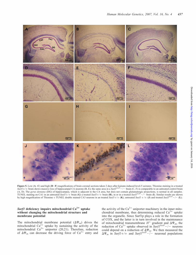
affected areas were the CA1 and CA3 regions, which stainedconsistently FJB-positive in all of the 24 control mice. Otherbrain areas were differently affected in different wt animals.However, no FJB-positive cells were ever detected in all ofthe 23 Surf1loxP2/2 brains (Fig. 4). Differential uptake ofkainic acid by Surf1loxP2/2 versus control neurons was unli-kely, since there was no appreciable difference in the amountof glutamate ionotropic receptor subunits, immunovisualizedusing specific antibodies (Supplementary Material, Fig. 3).As exemplified in Fig. 5, severe neuronal loss in glutamatergicareas was observed in kainate-treated wt brains stained withthionine, while Surf1loxP2/2 brains appeared consistentlyidentical to untreated control brains. TUNEL-positive apopto-tic nuclei were abundant in CA1–4 neurons (Fig. 5G–I) andin neurons from other glutamatergic regions of treated wtbrains (data not shown). No TUNEL-positive neurons wereever detected in the same regions of Surf1loxP2/2 brains.
Glutamate-induced cytosolic Ca21 signals and consequentneuronal cell death are reduced in Surf1loxP 2/2 neurons
Kainic acid treatment mimics glutamate-induced excitotoxi-city, which is characterized by delayed Ca2þ deregulation(DCD) and loss of mitochondrial potential (DCm) (15,16).Glutamate stimulation leads to the activation of metabotropic(mGlu) and ionotropic (AMPA, kainate and NMDA-type)plasma-membrane receptors in neurons. To further investigatethe impact of Surf1 ablation on these phenomena, we used7-day-old cortical/hippocampal primary neuronal cell culturesobtained from Surf1loxP2/2 mice and controls. Activation ofthe metabotropic receptor leads to inositol-triphosphate (IP3)-induced Ca2þ release from the endoplasmic reticulum (ER).Metabotropic Ca2þ responses were not detected in culturedneuronal cells, as confirmed by the use of the specific mGluagonist APB (data not shown). Activation of ionotropic recep-tors causes Naþ-mediated cell depolarization and subsequentCa2þ influx through voltage-dependent Ca2þ channels, aswell as direct Ca2þ influx through activation of the NMDAchannel. To measure cytosolic Ca2þ signals, we loadedneurons with the low-affinity Ca2þ dye fura-FF. Neuronswere then challenged with 10 or 100 mM glutamate for30 min, and cytosolic [Ca2þ] ([Ca2þ]c) changes weremeasured in single cells. As shown in Figure 6A and B, theincrease in [Ca2þ]c induced by 10 mM glutamate was notsignificantly different in Surf1loxP2/2 versus Surf1 þ /þ
neurons (DF/F 0.10+ 0.01 in Surf1loxP2/2 versus0.12+ 0.01 in Surf1þ/þ, n . 100 for each group,P ¼ 0.31), but it was much lower after exposure to 100 mM
glutamate (DF/F ¼ 0.26+ 0.02 in Surf1loxP2/2 versus0.45+ 0.03 in Surf1 þ /þ, n . 140 for each group,P , 1025).
Sustained stimulation with high doses of glutamate inducesderegulation of neuronal Ca2þ homeostasis, which manifestsas a secondary, delayed and irreversible [Ca2þ]c increase inthe supramicromolar range, eventually leading to cell death(17). We used fura-FF to detect glutamate toxicity bycalculating the number of cultured neurons showing thecharacteristic secondary Ca2þ increase during sustained (upto 30 min) glutamate stimulation. The percentage of neuronaldeath after stimulation with 10 mM glutamate was reduced inSurf1loxP2/2 neurons as compared to Surf1þ/þ neurons,and it remained significantly lower at 100 mM glutamatestimulation (% cell death at 10mM glutamate: Surf1þ/þ17,1+ 5,0% versus Surf1loxP2/26,7+ 2,9%; at 100 mMglutamate Surf1 þ /þ32,5+ 5,0% versus Surf1loxP2/216,9+ 2,4% P , 1022; Fig. 6C).
Reduced mitochondrial Ca21 uptake is responsible forreduced Ca21 influx in Surf1loxP2/2 neurons
The extent of mitochondrial Ca2þ uptake, strategically locatedat plasma membrane Ca2þ entry sites, has been shown toregulate Ca2þ influx through different plasma membrane chan-nels, such as capacitative or ligand-induced Ca2þ influx chan-nels (17). Ca2þ buffering in the sub-plasma membrane spacewas shown to reduce Ca2þ feedback inhibition of capacitativeCa2þ influx channels as well as of diverse subunits of NMDAchannels (18,19). In order to verify that the reduction of[Ca2þ]c following glutamate stimulation in Surf1loxP2/2neurons was correlated to a modification of mitochondrialCa2þ homeostasis, determining reduced Ca2þ influx, wemeasured mitochondrial [Ca2þ] ([Ca2þ]m) in intact andpermeabilized neurons, using a mitochondrially targeted lowaffinity aequorin probe (mitAEQmut). Neurons were trans-fected with the mitAEQmut probe and [Ca2þ]m was measuredin Surf1 þ /þ and Surf1loxP2/2 cell populations after stimu-lation with glutamate at low (10 mM) and high (100 mM)concentrations. Stimulation with 100 mM glutamate inducedvery high increase of [Ca2þ]m, leading to immediate consump-tion of the probe. However, mitochondrial Ca2þ transientscould be measured at a lower dose of glutamate (10 mM). Inthese conditions (Fig. 7A), the mitochondrial Ca2þ uptakewas drastically reduced in Surf1loxP2/2 versus Surf1þ/þcells. Maximum [Ca2þ]m was 28.95+ 2.50 mM inSurf1loxP2/2 neurons (n ¼ 24) versus 50.95+ 3.36 mM inSurf1þ/þ neurons (n ¼ 21), P , 1025.
In order to verify that the reduction of mitochondrial Ca2þ
uptake in Surf1loxP2/2 cells is due to lack of Surf1p, ratherthan to reduced cytosolic Ca2þ response consequent toreduced Ca2þ influx through the plasma membrane, neuronsexpressing the mitAEQmut probe were treated with low-dosedigitonin, which selectively permeabilizes the plasmamembrane, and endogenous cytosolic Ca2þ was washed outby perfusion with Ca2þ-free intracellular buffer. MitochondrialCa2þ uptake was then triggered by the addition of 1 mM
Table 1. Kainic acid treatment
þ/þ 2/2
Injected animals 73 72Death during crisis 13 12Total L5a mice analyzed 24 23Analyzed after 1 day 11 112 days 4 34 days 3 29 days 2 260 days 4 5
aL5, level-5 seizures (13).
Human Molecular Genetics, 2007, Vol. 16, No. 4 435
by guest on January 14, 2016http://hm
g.oxfordjournals.org/D
ownloaded from

Ca2þ to the buffer. Again, the velocity of mitochondrial Ca2þ
uptake was drastically reduced in Surf1loxP2/2 cells(Surf1loxP2/2 0.34+ 0.07 mM/s n ¼ 23 versus Surf1þ/þ1.10+ 0.12 mM/s n ¼ 26, P , 1027) (Fig. 7B). This resultindicates that the reduction of mitochondrial Ca2þ uptake isintrinsic to Surf1loxP2/2 mitochondria. Since the inhibition
of mitochondrial Ca2þ uptake has been shown to augmentthe feedback inhibition of plasma-membrane Ca2þ channelsin neurons, the impairment of mitochondrial Ca2þ bufferingin Surf1loxP2/2 neurons might ultimately prevent themfrom Ca2þ overload, which could explain their refractorinessto DCD and cell death.
Figure 4. Low, medium and high magnifications of FJB-stained brain coronal sections, taken 2 days after kainate-induced level-5 seizures. (A–C) Surf1 þ /þhippocampus; (D–F) Surf1loxP2/2 hippocampus; (G–I) Surf1þ/þ thalamic nuclei; (J–L) Surf1loxP 2/2 thalamic nuclei.
436 Human Molecular Genetics, 2007, Vol. 16, No. 4
by guest on January 14, 2016http://hm
g.oxfordjournals.org/D
ownloaded from
Surf1 deficiency impairs mitochondrial Ca21 uptakewithout changing the mitochondrial structure andmembrane potential
The mitochondrial membrane potential (DCm) drives themitochondrial Ca2þ uptake by sustaining the activity of themitochondrial Ca2þ uniporter (20,21). Therefore, reductionof DCm can decrease the driving force of Ca2þ entry and
the activity of the Ca2þ uniporter machinery in the inner mito-chondrial membrane, thus determining reduced Ca2þ uptakeinto the organelle. Since Surf1p plays a role in the formationof COX, and the latter is in turn involved in the maintenanceof mitochondrial transmembrane Hþ gradient and DCm, thereduction of Ca2þ uptake observed in Surf1loxP2/2 neuronscould depend on a reduction of DCm. We then measured theDCm in Surf1þ/þ and Surf1loxP2/2 neuronal populations
Figure 5. Low (A–C) and high (D–F) magnifications of brain coronal sections taken 2 days after kainate-induced level-5 seizures. Thionine staining in a treatedSurf1þ/þ brain shows massive loss of hippocampal CA neurons (B, E); the same area in a Surf1loxP2/2 brain (C, F) is comparable to an untreated control brain(A, D). The gyrus dentatus (DG) of hippocampus, which is adjacent to the CA area, but does not contain glutamatergic projections, is normal in all samples.TUNEL staining on CA1 in an untreated Surf1þ/þ brain (G) a treated Surf1þ/þ brain (H), in or in a treated Surf1loxP2/2 brain (I). Similar results are shownby high magnification of Thionineþ TUNEL double stained CA3 neurons in an treated Surf1þ/þ (K), untreated Surf1 þ /þ (J) and treated Surf1loxP2/2 (L).
Human Molecular Genetics, 2007, Vol. 16, No. 4 437
by guest on January 14, 2016http://hm
g.oxfordjournals.org/D
ownloaded from

by steady-state loading of neurons with the DCm-sensitive dyeteramethyl-rhodamine-methylester (TMRM), followed byfluorimetric measurement of the intensity of the dye in mito-chondria. Our results showed no difference in the steady-statedistribution of TMRM in Surf1loxP2/2 versus Surf1þ/þneurons (31.19+ 0.23 in Surf1þ/þ versus 32.12+ 0.29 inSurf1loxP2/2, fluorescence intensity in arbitrary units,Fig. 8A), demonstrating that the mild reduction observed inCOX activity in various tissues of Surf1loxP2/2 animalsfailed to result in significant changes of DCm.
We previously showed that mitochondrial fragmentation inepithelial cells leads to an average reduction of mitochondrialCa2þ load upon IP3-induced Ca2þ release (22). Therefore, wenext investigated whether Surf1 deficiency can change theshape of the mitochondrial network. Neurons were transfectedwith mitochondrially targeted DsRed probe (mtDsRed ) andthe structure of mitochondria in 300 Surf1loxP2/2 and 300Surf1 þ /þ individual cells was imaged by digital microscopy(22). As exemplified in Fig. 8B, the overall arrangement of themitochondrial network did not appear to be disturbed inSurf1loxP2/2 neurons with respect to controls.
On the basis of these results, we concluded that Surf1pdeficiency in mice does not lead to reduced DCm or alteredmitochondrial structure, suggesting that the effect of Surf1pon mitochondrial Ca2þ uptake could be independent from itsrole on COX assembly and maintenance of DCm.
To further confirm this hypothesis, we analyzed the effectsof SURF1 overexpression on global cellular and mitochondrialCa2þ signals in HeLa cells. HeLa cells were co-transfectedwith a vector expressing human SURF1 (hSURF1) (6) andwith mitAEQmut or its non-targeted cytosolic variant(cytAEQ). Cells were then challenged with histamine(100 mM), which induces IP3-dependent Ca2þ release fromthe ER. The resulting rise in [Ca2þ]c stimulates the mito-chondrial Ca2þ uptake at sites located in proximity of theER Ca2þ release sites. As shown on Figure 8C, the cytosolicCa2þ responses remained unaltered in cells overexpressingSURF1 (peak [Ca2þ]c 3.06+ 0.08 mM in controls versus2.86+ 0.09 mM in hSURF1 overexpressing cells); however,mitochondrial Ca2þ uptake was significantly increased bySURF1 overexpression (peak [Ca2þ]m 78.07+ 5.32 mM incontrols versus 94.51+ 6.04 mM in overexpressing cells,P , 0.05, Fig. 8D). This increase was not due tovariations of the DCm, as measured by TMRM uptake (datanot shown).
DISCUSSION
High embryonic lethality was a major feature of a mouseknockout model for Surf1p, an accessory assembly factor ofCOX, based on the replacement of a region of several kb in
Figure 6. Cytosolic Ca2þ response of primary cultured Surf1 þ /þ and Surf1loxP2/2 neurons to glutamate measured by fura-FF. Values are expressed in relativechange of 340/380 nm excitation ratio ( � F/F). (A) [Ca 2þ]c response shows a biphasic elevation to glutamate challenge: the immediate increase is due toNMDA channel activation, whereas the secondary delayed increase is the result of delayed cellular Ca2þ deregulation. (B) Means+ SEM values of theprimary peak response. (C) Percentage of cells undergoing Ca2þ deregulation from the total imaged cell population were calculated as an index of excitotoxicneuronal cell death.
438 Human Molecular Genetics, 2007, Vol. 16, No. 4
by guest on January 14, 2016http://hm
g.oxfordjournals.org/D
ownloaded from

the midportion of the Surf1 gene with a NEO cassette(Surf1NEO). The new mouse model reported here carries arecombinant null allele, consisting in the insertion of the35 bp loxP sequence within exon 7 of the murine Surf1 gene(Surf1loxP). The restoration of mendelian distribution of thegenotypes in newborn recombinant Surf1loxP mice indicatesthat the embryonic lethality observed in the previousSurf1NEO model was not a consequence of the ablation ofthe Surf1 gene itself, but was rather due to the presence ofthe NEO cassette or to the elimination of regulatory elementscontained in the deleted region of the Surf1 gene. Thisconclusion is further supported by the observation that theSurf1loxP-NEO-loxP allele, from which the Surf1loxP allele isderived, was again associated with 100% embryonic lethality,when present in homozygosity. Several recent reports(reviewed in 23) have shown that the maintenance of a NEOcassette in recombinant alleles can be associated with anumber of unpredictable effects, including the creation ofhypomorphic alleles, altered gene expression and embryoniclethality, mostly due to the promotion of illegitimate splicingof either the targeted gene or neighboring genes. The latterphenomenon was likely to occur in the surfeit genomicregion, which is packed with six housekeeping genes, someof which share common regulatory elements (24).
Although SURF1p is a ubiquitously expressed mitochon-drial protein, and its ablation leads to an early onset, invariablyfatal encephalopathy in humans, no clinical disease phenotypecould be observed in our Surf1loxP2/2 mice at any age. Thissituation is similar to that reported for the Surf1NEO2/2animals that survived embryonic selection (11). The absenceof neurological and extra-neurological abnormalities wasassociated with a biochemical phenotype that showed aspecific and generalized defect of COX activity, howeverless severe than that observed in SURF1 mutant patients (7).The COX defect was likely too mild to cause brain failureor impairment, but was possibly sufficient to determine themodest functional and morphological alterations found inskeletal muscle of adult Surf1loxP2/2 mice.
The mild biochemical and the virtually absent clinical phe-notypes of Surf1loxP2/2 mice suggest that, in spite of the ubi-quitous expression and high evolutionary conservation ofSurf1p, the function of this protein in COX assembly iseither ancillary or redundant, that is, it can partly be overtakenby other unknown factors. To some extent, this may well betrue also in SURF1-less mutant patients, in whom fullyassembled COX is diminished, but not absent (6). The differ-ent severity of the phenotype associated with the absence ofSurf1p may then depend on the efficiency and efficacy of com-pensatory genetic or epigenetic mechanisms acting in differentorganisms, notably fungi, mice and men.
In an attempt to determine whether the reduced COXactivity found in Surf1loxP2/2 animals could make theirbrain more sensitive to energy stress, we used an excitotoxicglutamate agonist, kainic acid, which triggers epileptic sei-zures in experimental animals. Kainic acid acts on the AMPA-kainate glutamate receptors present on the neuronal cell mem-brane (25). When activated, the Naþ-channel component ofthese receptors opens up, thus determining Naþ influx andmembrane depolarization. As a consequence, massive influxof Ca2þ occurs through the NMDA receptors and the VOCCchannels of the plasma membrane (26). In addition to deter-mining the epileptic discharge, the marked rise of [Ca2þ]ccan promote a cascade of secondary effects that may ulti-mately lead to cell death (27).
These experiments were aimed at challenging the OXPHOSreserve of neuronal cells provoked by epileptic discharge. Thesame approach has been used in the recent past to precipitatecatastrophic neurodegeneration in the MItochondrialLate-Onset Neurodegeneration (MILON) mouse, a conditionalTFAM knockout model, characterized by loss of mtDNA inneurons of the frontal cortex (28). To our surprise, however,the elicitation of level-5 seizures failed to cause any neuronaldegeneration and neuronal loss in Surf1loxP2/2 mice, whilethese lesions were consistently observed in glutamatergicareas of control brains. Since kainate-associated neurodegen-eration is largely dependent on perturbation of Ca2þ homeo-stasis, we investigated the cytosolic and mitochondrial Ca2þ
fluxes in primary neuronal cell cultures from Surf1loxP2/2and control mice. The results of these experiments can besummarized as follows.
First, the ablation of Surf1 drastically reduces theglutamate-induced increase of [Ca2þ] in both cytosolic andmitochondrial compartments.
Figure 7. Mitochondrial Ca2þ uptake of primary cultured Surf1 þ /þ andSurf1loxP 2 /2 neurons measured by the recombinant low affinity mitAEQmutprobe targeted to mitochondria. (A) Representative traces of luminescentvalues converted to [Ca2þ] (41) are shown on the left panel; mean+ SEMof [Ca2þ]m peaks are shown on the right panel. (B) The same experiment asin A was carried out on cells treated with 25 mM digitonin for 1 min. Repre-sentative traces of [Ca2þ]m values are shown on the left panel; mean+ SEMof Ca2þ uptake velocity (micromol/s) are shown on the right panel.
Human Molecular Genetics, 2007, Vol. 16, No. 4 439
by guest on January 14, 2016http://hm
g.oxfordjournals.org/D
ownloaded from

Second, the reduction of mitochondrial Ca2þ uptake isdirectly consequent to the absence of Surf1p.
Third, Surf1loxP2/2 cultured neurons are much moreresistant to glutamate toxicity than control neurons. Howcould this effect be linked to the observed reduction ofmitochondrial Ca2þ uptake? One possibility is that reducedbuffering capacity by Surf1loxP2/2 mitochondria can deter-mine the saturation of the Ca2þ microdomains in the contactsites between mitochondria and the plasma membrane or theER. This effect could in turn promote the feedback closureof the Ca2þ channels in the above structures, thus inhibitingthe [Ca2þ]c transient rise (18). Although speculative, this
hypothesis can offer a mechanistic explanation for the neuro-protection observed in vivo. A second possibility is that theablation of Surf1 may alter the expression of nuclear genesencoding proteins engaged in Ca2þ homeostasis. As a prelimi-nary result, quantitative PCR analysis failed to show differentexpression of the Ca2þ–Naþ plasma membrane exchanger(NCX1) in Surf1loxP2/2 versus wt brains (data not shown).More work is needed to expand this analysis to other Ca2þ-related genes.
Fourth and last, the reduction of the mitochondrial [Ca2þ]uptake seems not to be dependent from a decrease of theDCm, as a consequence of partial defect of COX activity
Figure 8. (A) Mitochondrial membrane potential is unchanged between Surf1loxP2/2 and þ/þ neuronal cells. A.U., arbitrary units. (B) Mitochondrial structureremains unchanged in Surf1loxP2/2 neurons. Representative images of whole neurons and somata (zoomed insets) are shown. (C and D) Analysis of cytosolic(C) and mitochondrial (D) Ca2þ homeostasis in HeLa cells overexpressing hSURF1 protein. Representative traces are shown on the left panels. Right panelsshow the mean+ SEM peak values after histamine stimulation from .10 experiments.
440 Human Molecular Genetics, 2007, Vol. 16, No. 4
by guest on January 14, 2016http://hm
g.oxfordjournals.org/D
ownloaded from

and mitochondrial respiration. This conclusion, which wasalso supported by the results of hSURF1 overexpression inHeLa cells, suggests that Surf1p could play a direct role onmitochondrial Ca2þ handling, partially or completely indepen-dent from its function as a COX assembly factor. More work isnecessary to test this hypothesis, but it is interesting to observethat an increasing number of mitochondrial proteins have beenestablished to carry out multiple functions. A well knownexample is cytochrome c, which acts as both a redox electronshuttle of the MRC, and as an apoptogenic messenger (29).Likewise, the apoptosis-inducing factor (AIF), a flavoproteinclosely associated with the mitochondrial inner membrane,has been implicated as both a cell death-promoting moleculeand a regulator of activity and protein expression of MRCcomplex I (30). Lastly, p66Shc is an electron-transfer redoxenzyme of the mitochondrial intermembrane space, whichcontrols the production of reactive oxygen species (ROS),and regulates Ca2þ transport by acting on plasmamembraneCa2þ-ATPases. These two independent activities can ulti-mately converge and synergize in mediating p66Shc-dependentapoptosis (31). Targeted disruption of p66Shc is associatedwith prolonged lifespan in mice, possibly related to its roleon the control of ROS production.
Similar to the p66Shc2/2, our Surf1loxP2/2 mouse modeldisplays significantly increased longevity. Of note, increasedlongevity was also observed in a CNS-restricted conditionalSurf1 knockdown (KD) model in Drosophila melanogaster,whereas the corresponding constitutive model was embryoniclethal (32). We do not have an explanation for this obser-vation, which is in striking contrast with the early onset,invariably fatal phenotype associated with the loss ofSURF1p function in humans. It is possible that the COXdefect in Surf1loxP2/2 mice is not severe enough to conferselective disadvantage in a ‘protected’ environment such asan animal care facility. These data are nevertheless surprising,considering that COX deficiency associated with Surf1p dis-ruption should in principle determine an increase of ROS(33), which are proposed to play a major role in the agingprocess (34). An attractive possibility is that the effect onlongevity is due to the role of Surf1p on mitochondrial Ca2þ
uptake and cellular Ca2þ homeostasis. This effect is likely tobe hidden in organisms, such as S. cerevisiae and Homosapiens, in which lack of Surf1p leads to severe impairmentof COX assembly and faulty OXPHOS, whereas it would beunmasked in other organisms, such as Mus musculus and theconditional D. melanogaster KD model, characterized byless severe impairment of COX assembly and OXPHOSphenotype.
MATERIALS AND METHODS
Creation of Surf1loxP-NEO-loxp1/2 and Surf1loxP2/2recombinant mice
The list of all primers is provided in the SupplementaryMaterial, Table 2.
For the construction of the Surf1 recombinant alleles, weused a 10 kb HindIII-EcoRI fragment containing the entireSurf1 and Surf2 genes, and part of Surf4 and Surf3 genes,cloned in BlueScript SK. A fragment of approximately
1.2 kb, composed of the gene encoding the neomycin phos-photransferase (NEO cassette) flanked by two 35-mer identicalloxP sites (loxP-NEO-loxP), was inserted in the unique AccIIIsite contained in exon 7 of the Surf1 murine gene correspond-ing to nt 670 of the Surf1 cDNA. The DNA vector was verifiedon both strands by automated sequence analysis using thebig-dye terminator kit and protocol (Applied Biosystems),on a 3100 ABI apparatus.
Gene targeting by electroporation of the HindIII-linearizedvector into AB1 ES cells, derived from 129/SvEvBr #=/Hprt-bm2 mouse substrain (a kind gift from Alan Bradley),and generation of chimeras, were performed as described(35). Three hundred colonies that survived selection with theneomycin analogue drug G-418 (200 mg/ml) were screenedfor homologous recombination by PCR and Southern blotanalyses.
For diagnostic PCR analysis, a 2 kb DNA fragment wasamplified using a forward primer corresponding to a regioninside the NEO cassette and a reverse primer correspondingto a sequence within the Surf3 gene located outside the recom-binant region. For diagnostic Southern blot analysis (Fig. 1B)on the 50 end of the recombinant region, 10 mg of ES genomicDNA was digested with EcoRI, run through a 0.8% agarosegel in 1X TE buffer, blotted on a nitrocellulose filter andhybridized with a 0.7 kb PCR fragment (probe P1 in Fig. 1)radiolabeled with [a32P]-dCTP (NEN, Boston, MA, USA)using the ‘Ready-to-go’ random priming kit (Amersham,Piscataway, NJ, USA). P1 corresponds to a region of theSurf5 gene outside but contiguous to the recombinant region.The wt Surf1 allele corresponds to a 19 kb hybridizationband, whereas the Surf1loxP-NEO-loxP recombinant allele corre-sponds to 13 and 7 kb bands due to the presence of an EcoRIsite within the NEO cassette. For diagnostic Southern blotanalysis on the 30 end, DNA was digested with EagI andHindIII, separated by electrophoresis and blotted as above.Hybridization was then carried using a 0.8 fragmentcorresponding to a sequence on the Surf4 gene outside butcontiguous to the recombinant region (probe P2 in Fig. 1).The wt allele corresponds to a hybridization band of 9.2 kb,whereas the Surf1loxP-NEO-loxP recombinant allele correspondsto a band of 7.5 kb, again due to the presence of an extra EagIsite within the NEO cassette.
Two of the five ES clones that showed homologous recom-bination were injected in B6D2F1# = C57/Bl6J blastocysts(36). Chimeric pups were identified by the presence ofagouti hair and, on maturity, mated with B6D2F1 (C57/Bl6J_DBA2) females to check for the contribution of the EScells to the germline.
For the creation of Surf1loxP animals, Surf1loxP-NEO-loxPþ /2
mice of mixed BDF1 genetic background were mated to cremice. Compound heterozygotes (Surf1loxP/þ;cre/ þ ) were ident-ified by PCR and backcrossed to each other.
For genotyping, 250 ng genomic DNA extracted from tailtips was PCR amplified in 50 ml of 1 � MgCl2-PCR buffer(Applied Biosystems), 200 mM dNTPs, 0.6 mM each dNTPsand 0.03 U/ml Taq Polymerase (Applied Biosystems), 5%DMSO. After an initial denaturation at 948C for 2 min, eachof the 35 PCR cycles was as follows: 948C for 30 s, 588Cfor 60 s, 728C for 90 s. Final extension was at 728C for5 min. For the Surf1loxP-NEO-loxP allele, we used a single
Human Molecular Genetics, 2007, Vol. 16, No. 4 441
by guest on January 14, 2016http://hm
g.oxfordjournals.org/D
ownloaded from

forward primer (LNL-FW) and two distinct reverse primers,one internal to the NEO cassette (LNL-RV1) specific to therecombinant allele, and another corresponding to a Surf1region (LNL-RV2) specific to the wt allele. The recombinantallele generates a PCR fragment of 583 bp, whereas thewt allele generates a PCR fragment of 458 bp. For theSurf1loxP allele, we used the forward primer LP-FW, corre-sponding to a region of Surf1 exon 6; and the reverse primerLP-RV, corresponding to a region of Surf1 exon 7. The PCRproduces a 305 bp wt DNA fragment and a 340 bp recombi-nant fragment). The cre transgene was detected by using theforward primers CRE-FW and the reverse primer CRE-RV(PCR fragment 590 bp).
Animal studies were approved by the animal welfare EthicsCommittee of the National Neurological Institute in accord-ance with the Institutional Animal Care and Use Committeeguidelines. Standard food and water were given ad libitum.
RT-PCR analysis
To evaluate the presence of recombinant Surf1loxP transcript,total RNA was extracted from brain, liver, muscle and heartof four Surf1loxP2/2 and four Surf1 þ /þ adult animals,using the RNeasy lipid tissue kit (QIAGEN Sciences, MD,USA) following the manufacturer’s protocol. Total RNAwas used as a template for reverse transcription, using the‘cDNA cycle’ kit and protocol (Invitrogen, Carlsbad, CA,USA). Total cDNA was purified and resuspended in a finalvolume of 20 ml and used for PCR amplification of individualcDNA fragments corresponding to GAPDH and Surf1 genes;3 ml were then used in each 50 ml PCR reaction containing1 � MgCl2-PCR buffer, 200 mM dNTPs, 5% DMSO, 0.6 mM
of each primer and 0.03 U/ml of Taq-Gold polymerase(Invitrogen, Carlsbad, CA, USA). GAPDH was detectedusing primers GAPDH-FW and GAPDH-RV. The wt andSurf1loxP transcripts were PCR-diagnosed using exonicprimers LP-FW and LP-RV (Fig. 1E).
Western blot analysis
Western blot analysis was performed on electroblotteddenaturing sodium-dodecyl sulphate polyacrylamide gelelectrophoresis (SDS–PAGE), and two-dimension bluenative electrophoresis (2D-BNE), as described previously(6). Approximately 100 mg non-collagenous protein wasused for each sample in SDS-PAGE and 20 mg of isolatedmitochondria in 2D-BNE. Chemiluminescence-based immu-nostaining (ECL kit, Amersham) was performed using thefollowing antibodies: polyclonal antibody AS182–196 (6)raised against amino acid sequence 82–96 of mouse Surf1p,which is at the N-terminal of the truncated protein predictedby the Surf1loxP allele; monoclonal antibodies against subunitsCOX I and COX IV (Molecular Probes, Eugene, OR, USA)and Surf1p (Mitosciences LLC, Eugene, OR, USA).
Morphological analysis
For light microscopy, samples from different organs werefrozen in liquid-nitrogen-cooled isopentane. Standard histo-logical and histochemical techniques for the detection of
mitochondrial alterations and muscle fiber distribution wereperformed on serial cryostat cross sections as previouslydescribed (37).
Biochemical analysis
Biochemical assays of individual respiratory complexes werecarried out on tissue homogenates (38). Specific activitiesof each complex were normalized to that of CS, an indicatorof the number of mitochondria. Blood lactate was measuredusing the ‘Lactate reagent’ kit and protocol (Sigma,St Louis, MO, USA).
Rotarod test
Motor performance tests were given to 13 wt mice and to 13Surf1loxP2/2 mice. All animals were 3 months old. The rotat-ing rod test was performed on a Rotarod apparatus for mice(Ugo Basile) (39).
Kainate-induced seizures and brain analysis
Kainic acid was dissolved in isotonic saline (pH 7) with a dropof 1 M NaOH and administered i.p. Mice were monitoredcontinuously for at least 3 h after injection to determine theonset and level of seizures according to Sperk et al., (13).For histological analysis of the brain, animals were treatedwith a lethal injection of 4% chloral hydrate before intra-cardiac perfusion with 4% paraformaldehyde in PBS. Brainswere rapidly dissected and post-fixed overnight in the samesolution. Serial rostro-caudal, 50 mm thick coronal brain sec-tions were obtained using a Vibratome (Leica) and collectedin 0.1 M PB at pH 7.2 with 0.01% NaN3. One every 12sections was labeled with FJB (Histo-Chem, Jefferson, AR)according to manufacturer’s instructions. Adjacent sectionswere stained with 0.1% thionine. Next adjacent sectionswere labeled by terminal deoxynucleotidyl transferase-mediated biotinylated UTP nick end labeling (TUNEL)(Apoptag in situ Apoptosis Detection kit; Intergen, Purchase,NY) according to the manufacturer’s instructions. Fluorescentimages were acquired on a confocal microscope (Radiance2100 confocal microscope, Bio-Rad, Hercules, CA, USA)using an FITC filter and optical photographs were acquiredusing a Nikon Eclipse E400 microscope and a Nikon DS-U1digital camera.
Cortical/hippocampal primary neuronal cell cultures
Cortical/hippocampal neurons were prepared from 1- to3-day-old newborn mice, according to Pasti et al. (40), andneurons were resuspended in NeurobasalA Medium (Gibco)with supplement B-27 (Gibco), GlutaMax (Gibco) and penicil-line/streptomycine (Gibco) rigorously at 378C and plated ontoglass coverslips, coated with poly-D-lysine (Sigma).
Dynamic in vivo [Ca21] measurements with targetedaequorin probes
The construction and use of luminescent Ca2þ sensitiveaequorin probes were previously described (41). All
442 Human Molecular Genetics, 2007, Vol. 16, No. 4
by guest on January 14, 2016http://hm
g.oxfordjournals.org/D
ownloaded from

aequorin measurements were carried out in KRB containing1 mM CaCl2 (KRB/Ca2þ, Krebs-Ringer modified Buffer:135 mM NaCl, 5 mM KCl, 1 mM MgSO4, 0.4 mM K2HPO4,1 mM CaCl2, 15 mM glucose, 20 mM HEPES, pH 7.4).For HeLa cells KRB contained 5 mM glucose. Experimentsin permeabilized neurons were performed as previouslydescribed for HeLa cells (42), except that 25 mM digitoninwas used, in order to preserve mitochondrial integrity.
Imaging procedures
Cortical cultures were loaded for 20 min with 3 mM fura-2FF/AM (or fura-2) (Teflabs, Austin, TX, USA) at 378C in thecell culture medium (Kd of fura-2FF or fura-2 for Ca2þ is 5and 55 mM, respectively; 43). Images were acquired on anepifluorescence inverted microscope Axiovert 200 (Zeiss,Germany) equipped with a 40� fluorite objective. [Ca2þ]cwas monitored in single cells using two excitation light wave-lengths, at 340 nm and 380 nm (Sutter Instrument Co., CA,USA). Emitted fluorescence light was selected by a505–530 nm filter. Images were acquired by CCD camera(Roper Scientific, USA). All imaging data were collectedand analyzed using the Metafluor 6.1 software. For the visual-ization of the mitochondrial network, Z-series of images ofneurons were 3D deconvolved and reconstructed using acustom-made software (44).
Statistical analysis
Two-tailed, unpaired, unequal variance Student’s t-test wasused for statistical analysis. Survival probability was calcu-lated using the Kaplan–Meier and log-rank tests.
SUPPLEMENTARY MATERIAL
Supplementary Material is available at HMG Online.
ACKNOWLEDGEMENTS
The authors are grateful to Barbara Geehan for revising themanuscript. They are also grateful to Chiara Falcone forassistance on statistical analysis and Mike Fried for thegenerous gift of a plasmid containing the mouse surfeit genecluster.
This study was supported by Fondazione Telethon, Italy(Grant no. GGP030039), Fondazione Pierfranco e LuisaMariani, MITOCIRCLE and EUMITOCOMBAT networkgrants from the European Union Framework Program 6. Thework of R.R. was supported by grants from Telethon,Italy, the Italian Association for Cancer Research (AIRC),the Italian University Ministry (PRIN, FIRB andlocal research grants), the Emilia-Romagna PRRIITTprogram, the Ferrara Objective 2 funds and the Italian SpaceAgency (ASI).
Conflict of Interest statement. None declared.
REFERENCES
1. Pecina, P., Gnaiger, E., Zeman, J., Pronicka, E. and Houstek, L. (2004)Decreased affinity for oxygen of cytochrome-c oxidase in Leigh syndromecaused by SURF1 mutations. Am. J. Physiol. Cell Physiol., 287,1384–1388.
2. DiMauro, S., Zeviani, M., Rizzuto, R., Lombes, A., Nakase, H., Bonilla,E., Miranda, A. and Schon, E. (1988) Molecular defects in cytochrome coxidase in mitochondrial diseases. J. Bioenerg. Biomembr., 20, 353–364.
3. Solans, A., Zambrano, A. and Barrientos, A. (2004) Cytochrome c oxidasedeficiency: from yeast to human. Preclinica, 2, 1–13.
4. Nijtmans, L.G., Taanman, J.W., Muijsers, A.O., Speijer, D. and Van denBogert, C. (1998) Assembly of cytochrome-c oxidase in cultured humancells. Eur. J. Biochem., 254, 389–394.
5. Nijtmans, L.G., Artal Sanz, M., Bucko, M., Farhoud, M.H., Feenstra, M.,Hakkaart, G.A., Zeviani, M. and Grivell, L.A. (2001) Shy1p occurs in ahigh molecular weight complex and is required for efficient assembly ofcytochrome c oxidase in yeast. FEBS Lett., 498, 46–51.
6. Tiranti, V., Galimberti, C., Nijtmans, L., Bovolenta, S., Perini, M.P. andZeviani, M. (1999) Characterization of SURF-1 expression and Surf-1pfunction in normal and disease conditions. Hum. Mol. Genet., 8, 2533–2540.
7. Munaro, M., Tiranti, V., Sandona, D., Lamantea, E., Uziel, G., Bisson, R.and Zeviani, M. (1997) A single cell complementation class is common toseveral cases of cytochrome c oxidase defective Leigh’s syndrome. Hum.
Mol. Genet., 6, 221–228.
8. Tiranti, V., Hoertnagel, K., Carrozzo, R., Galimberti, C., Munaro, M.,Granatiero, M., Zelante, L., Gasparini, P., Marzella, R., Rocchi, M. et al.(1998) Mutations of SURF1 in Leigh disease associated with cytochromec oxidase deficiency. Am. J. Hum. Genet., 63, 1609–1621.
9. Leigh, D. (1951) Subacute necrotizing encephalomyelopathy in an infant.J. Neurol. Neurosurg. Psychiatry, 14, 216–221.
10. Duhig, T., Ruhrberg, C., Mor, O. and Fried, M. (1998) The human surfeitlocus. Genomics, 52, 72–78.
11. Agostino, A., Invernizzi, F., Tiveron, C., Fagiolari, G., Prelle, A.,Lamantea, E., Giavazzi, A., Battaglia, G., Tatangelo, L., Tiranti, V. andZeviani, M. (2003) Constitutive knockout of Surf1 is associated with highembryonic lethality, mitochondrial disease and cytochrome c oxidasedeficiency in mice. Hum. Mol. Gen., 12, 399–413.
12. Harkness, J.E. and Wagner, J.E. (1995) The Biology and Medicine of
Rabbits and Rodents, 4th edn. Lea and Febiger, Philadelphia, PA.13. Sperk, G., Lassmann, H., Baran, H., Seitelberger, F. and Hornykiewicz, O.
(1985) Kainic acid-induced seizures: dose-relationship of behavioural,neurochemical and histopathological changes. Brain Res., 338, 289–295.
14. Schmued, L.C., Slikker, W., Jr. and Wang, G.J. (1998) Fluoro-Jade B: abis homolog of Fluoro-Jade with improved degenerate neuron stainingproperties. Soc. Neurosci. Ab., 24, 1064.
15. Nicholls, D.G. and Budd, S.L. (2000) Mitochondria and neuronal survival.Physiol. Rev., 80, 315–360.
16. Duchen, M.R. (2000) Mitochondria and calcium: from cell signalling tocell death. J. Physiol., 529, 57–68.
17. Bano, D., Young, K.W., Guerin, C.J., Lefeuvre, R., Rothwell, N.J.,Naldini, L., Rizzuto, R., Carafoli, E. and Nicotera, P. (2005) Cleavage ofthe plasma membrane Naþ/Ca2þ exchanger in excitotoxicity. Cell, 120,275–285.
18. Parekh, A.B. (2003) Store-operated Ca2þ entry: dynamic interplaybetween endoplasmic reticulum, mitochondria and plasma membrane.J. Physiol., 547, 333–348.
19. Sessoms-Sikes, S., Honse, Y., Lovinger, D.M. and Colbran, R.J. (2005)CaMKII-alpha enhances the desensitization of NR2B-containing NMDAreceptors by an autophosphorylation-dependent mechanism. Mol. Cell.
Neurosci., 29, 139–147.
20. Nicholls, D.G. (2005) Mitochondria and calcium signaling. Cell Calcium,38, 311–317.
21. Bernardi, P. (1999) Mitochondrial transport of cations: channels,exchangers, and permeability transition. Physiol. Rev., 79, 1127–1155.
22. Szabadkai, G., Simoni, A.M., Chami, M., Wieckowski, M.R., Youle, R.J.and Rizzuto, R. (2004) Drp-1-dependent division of the mitochondrialnetwork blocks intraorganellar Ca2þ waves and protects againstCa2þ-mediated apoptosis. Mol. Cell., 16, 59–68.
23. Lewandoski, M. (2001) Conditional control of gene expression in themouse. Nature Reviews, 2, 743–755.
Human Molecular Genetics, 2007, Vol. 16, No. 4 443
by guest on January 14, 2016http://hm
g.oxfordjournals.org/D
ownloaded from

24. Huxley, C. and Fried, M. (1990) The mouse surfeit locus contains acluster of six genes associated with four cpg-rich islands in 32 kilobases ofgenomic DNA. Mol. Cell. Biol., 10, 605–614.
25. Hunsberger, J.G., Bennett, A.H., Selvanayagam, E., Duman, R.S. andNewton, S.S. (2005) Gene profiling the response to kainic acid inducedseizures. Brain Res. Mol. Brain Res., 141, 95–112.
26. Jaskolski, F., Coussen, F. and Mulle, C. (2005) Subcellular localizationand trafficking of kainate receptors. Trends Pharmacol. Sci., 26, 20–26.
27. Zhu, X., Jin, S., Kong Ng, Y., Lee, W.L. and Wong, P.T.H. (2001)Positive and negative modulation by AMPA- and kainate-receptors ofstriatal kainate injection-induced neuronal loss in rat forebrain. BrainResearch, 922, 293–298.
28. Sorensen, L., Ekstrand, M., Silva, J.P., Lindqvist, E., Xu, B., Rustin, P.,Olson, L. and Larsson, N.G. (2001) Late-onset corticohippocampalneurodepletion attributable to catastrophic failure of oxidativephosphorylation in MILON mice. J. Neurosi., 21, 8082–8090.
29. Garrido, C. and Kroemer, G. (2004) Life’s smile, death’s grin: vitalfunctions of apoptosis-executing proteins. Curr. Opin. Cell. Biol., 16,639–646.
30. Vahsen, N., Cande, C., Triere, J.J., Benit, P., Joza, N., Larochette, N.,Mastroberardino, P.G., Pequignot, M.O., Casares, N., Lazar, V. et al.(2004) AIF deficiency compromises oxidative phosphorylation. EMBO J.,23, 4679–4689.
31. Giorgio, M., Migliaccio, E., Orsini, F., Paolucci, D., Moroni, M.,Contursi, C., Pelliccia, G., Luzi, L., Minacci, S., Marcaccio, M. et al.(2005) Electron transfer between cytochrome c and p66Shc generatesreactive oxygen species that trigger mitochondrial apoptosis. Cell, 122,221–233.
32. Zordan, M.A., Cisotto, P., Benna, C., Agostino, A., Rizzo, G., Piccin, A.,Pegoraro, M., Sandrelli, F., Perini, G., Tognon, G. et al. (2006) Post-transcriptional silencing and functional characterization of the Drosophilamelanogaster homolog of human Surf1. Genetics, 172, 229–241.
33. Lee, I., Bender, E., Arnold, S. and Kadenbach, B. (2001) New control ofmitochondrial membrane potential and ROS formation—a hypothesis.Biol. Chem., 382, 1629–1636.
34. Terman, A. and Brunk, U.T. (2006) Oxidative stress, accumulationof biological ‘garbage’, and aging. Antioxid. Redox. Signal., 8,197–204.
35. Ramirez-Solis, R., Davis, A.C. and Bradley, A. (1993) Gene targeting inembryonic stem cells. Meth. Enzymol., 225, 855–878.
36. Bradley, A. (1987) Robertson, EJ (eds), Production and Analysis of
Chimaeric Mice in Teratocarcinomas and Embryonic Stem Cells, a
Practical Approach., IRL Press, Oxford.
37. Sciacco, M. and Bonilla, E. (1996) Cytochemistry and immunocyto-chemistry of mitochondria in tissue sections. Meth. Enzymol., 264,509–521.
38. Bugiani, M., Tiranti, V., Farina, L., Uziel, G. and Zeviani, M. (2005)Novel mutations in COX15 in a long surviving Leigh syndrome patientwith cytochrome c oxidase deficiency. J. Med. Genet., 42, e28.
39. Dulioust, E., Toyama, K., Busnel, M.C., Moutier, R., Carlier, M.,Marchaland, C., Ducot, B., Roubertoux, P. and Auroux, M. (1995)Long-term effects of embryo freezing in mice. Proc. Natl Acad. Sci. USA,92, 589–593.
40. Pasti, L., Pozzan, T. and Carmignoto, G. (1995) Long-lasting changes ofcalcium oscillations in astrocytes. A new form of glutamate-mediatedplasticity. J. Biol. Chem., 270, 15203–15210.
41. Chiesa, A., Rapizzi, E., Tosello, V., Pinton, P., de Virgilio, M., Fogarty,K.E. and Rizzuto, R. (2001) Recombinant aequorin and green fluorescentprotein as valuable tools in the study of cell signalling. Biochem. J., 355,1–12.
42. Rapizzi, E., Pinton, P., Szabadkai, G., Wieckowski, M.R., Vandecasteele,G., Baird, G., Tuft, R.A., Fogarty, K.E. and Rizzuto, R. (2002)Recombinant expression of the voltage-dependent anion channel enhancesthe transfer of Ca2þ microdomains to mitochondria. J. Cell Biol., 159,613–624.
43. Xu-Friedman, M.A. and Regehr, W.G. (1999) Presynaptic strontiumdynamics and synaptic transmission. Biophys. J., 76, 2029–2042.
44. Rizzuto, R., Carrington, W. and Tuft, R.A. (1998) Digital imagingmicroscopy of living cells. Trends Cell Biol., 8, 288–292.
444 Human Molecular Genetics, 2007, Vol. 16, No. 4
by guest on January 14, 2016http://hm
g.oxfordjournals.org/D
ownloaded from