Asthma and Respiratory Foundation NZ adult asthma guidelines
INCREASED INCIDENCE OF ASTHMA IN HIV-INFECTEDCHILDREN TREATED WITH HAART IN THE NIH WOMEN ANDINFANTS TRANSMISSION STUDY
Samuel B. Foster, M.D.a, Kenneth McIntosh, M.D.b, Bruce Thompson, Ph.D.c, Ming Lu,M.S.c, Wanrong Yin, M.S.c, Kenneth C. Rich, M.D.d, Hermann Mendez, M.D.e, Leslie K.Serchuck, M.D.f, Clemente Diaz, M.D.g, Mary E. Paul, M.D.a, and William T. Shearer, M.D.,Ph.D.aaBaylor College of Medicine and Texas Children’s Hospital, Department of Pediatrics, Allergy andImmunology Section, Houston, TXbChildren’s Hospital, Division of Infectious Diseases, Harvard Medical School, Boston, MAcClinical Trials & Survey Corp., Baltimore, MDdUniversity of Illinois at Chicago, Department of Pediatrics, Chicago, ILeState University of New York, Department of Pediatrics, Brooklyn, NYfNational Institute of Child Health & Human Development, Pediatric, Adolescent & Maternal AIDSBranch, Bethesda, MDgPuerto Rico School of Medicine, Department of Pediatrics, San Juan, PR
AbstractBACKGROUND—Immunoreconstitution of HIV-infected (HIV+) patients after treatment withhighly antiretroviral therapy (HAART) appears to provoke inflammatory diseases.
OBJECTIVE—Determine whether HIV+ children on HAART (HIV+ HAART+) have a higherincidence of asthma than HIV+ children not on HAART (HIV+ HAART−).
METHODS—To investigate this possibility, 2,664 children (193 HIV+, 2,471 HIV−) born toHIV+ women were evaluated for the incidence and prevalence of asthma (i.e., asthma medicationuse), and change of CD4+ T cell percentage with time.
RESULTS—The HIV+ HAART+ children had higher CD4+ T cell percentages, lower CD8+ Tcell percentages, and lower viral burdens than the HIV+ HAART− children (P≤0.05 to P≤0.01).The cumulative incidence of asthma medication use in HIV+ HAART+ children at 13.5 year roseto 33.5% vs. 11.5% in HIV+ HAART− children (hazard ratio=3.34, P=0.01) and was equal to thatin the HIV− children. In children born prior to the HAART era, the prevalence of asthmamedication use for HIV+ HAART+ children at 11 years of age was 10.4% vs. 3.8% for HIV+HAART− children (odds ratio=3.38, P=0.02) and was equal to that of the HIV− children. The rate
© 2009 American Academy of Allergy, Asthma and Immunology. Published by Mosby, Inc. All rights reserved.Corresponding Author: William T. Shearer, M.D., Ph.D. Texas Children’s Hospital, 6621 Fannin Street (MC: FC330.01), Houston,TX 77030, Telephone: 011 (832) 824-1274/Fax: 011 (832) 825-7131, [email protected]'s Disclaimer: This is a PDF file of an unedited manuscript that has been accepted for publication. As a service to ourcustomers we are providing this early version of the manuscript. The manuscript will undergo copyediting, typesetting, and review ofthe resulting proof before it is published in its final citable form. Please note that during the production process errors may bediscovered which could affect the content, and all legal disclaimers that apply to the journal pertain.
NIH Public AccessAuthor ManuscriptJ Allergy Clin Immunol. Author manuscript; available in PMC 2011 December 27.
Published in final edited form as:J Allergy Clin Immunol. 2008 July ; 122(1): 159–165. doi:10.1016/j.jaci.2008.04.043.
NIH
-PA Author Manuscript
NIH
-PA Author Manuscript
NIH
-PA Author Manuscript
of change of CD4+ T cells (percent/year) around the time of first asthma medication for HIV+HAART+ vs. HIV+ HAART− children was 0.81 vs. −1.43 (P=0.01).
CONCLUSION—The increased incidence of asthma in HIV+ HAART+ children may be drivenby immunoreconstitution of CD4+ T cells.
CLINICAL IMPLICATIONS—This HIV model of pediatric asthma may yield clues to helpexplain the epidemic of asthma in the general pediatric population.
Keywordspediatric HIV infection; CD4+ T cell mediated induction of asthma; HAART-producedimmunoreconstitution
INTRODUCTIONAsthma or asthma-like conditions can be seen in animal models with pulmonary infectionsand immunoreconstitution.1 CD4+ T cells are essential for the development of animalmodels of asthma lose their asthma when depleted of CD4+ T cells.2, 3 B cell-, IgE-, andmast cell-deficient mice may still develop asthma, but CD4+ T cell-, STAT6-, and IL-13-deficient mice cannot.4 There are studies that document an increase in bronchialhyperresponsiveness and asthma in HIV-infected adults5–8, but other studies do not confirmthese findings.9, 10 For example, Lin and Lazarus7 made the seminal observation that arecent CD4+ T cell count of ≥200 cells/μL was significantly associated with current asthma(P=0.01). The weight of this evidence suggests that adults with HIV infection have anincreased prevalence of asthma, but most of these studies took place in the pre-HAART erawhen the immunoreconstitution inflammatory syndrome11 was unknown, and most studiesdid not stratify patients by their CD4+ T cell count.
Galli et al12 reported a reduced frequency of wheezing (not defined as asthma) in infantswith HIV infection who were followed for 2 years of life, but Foster et al13 in a single-centerretrospective study of 83 older children and young adults reported a several-fold increasedasthma prevalence of 34 percent.13 In the latter study, there was substantial evidence thatantiretroviral medications either restored the CD4+ T cell count in children inadequatelytreated, or preserved the CD4+ T cell count in children adequately treated from birth. Gutinand Secord14 reported in an abstract publication that 24 of 85 children with HIV infectionwere diagnosed with asthma. Of these 24 children, 65% developed asthma within 3 years ofbeginning HAART and 22 were immunoreconstituted or immunopreserved at the time oftheir asthma diagnosis.
To expand on these preliminary findings, investigators of the Women and InfantsTransmission Study (WITS), a large multicenter HIV+ pediatric cohort, reviewed the cohortfor the incidence and prevalence of asthma, and the results of that investigation are reportedherein.
METHODSWomen and Infants Transmission Study
WITS is a prospective natural history, non-interventional study of HIV-infected pregnantwomen and their offspring conducted by the National Institute of Child Health and HumanDevelopment, National Institute of Allergy and Infectious Diseases, National Institute onDrug Abuse, and National Institute of Mental Health from 1988 to 2006 at multiple sites inthe United States (Boston and Worchester, MA; Chicago, IL; Houston, TX; New York, NY;and San Juan, PR) with a total enrollment of 193 HIV+ and 2,471 HIV− children. Pregnant
Foster et al. Page 2
J Allergy Clin Immunol. Author manuscript; available in PMC 2011 December 27.
NIH
-PA Author Manuscript
NIH
-PA Author Manuscript
NIH
-PA Author Manuscript

women and their infants began enrollment in 1989 and CD4+ T cell and HIV culture/HIVDNA PCR assay were performed at regular intervals: birth, 1, 2, 4, 6, 9, 12 months, andevery six months thereafter for HIV+ children and every 12 months thereafter for HIV−children. Similarly, secondary diagnoses of the subjects and their medication use were anintegral part of the extensive WITS protocol that were recorded by medical personnel ateach WITS study visit.15 All WITS study data were collected in a systematic manner oncase report forms in face-to-face study visits between patient and research nurse andphysician-investigator. The institutional review board at each center approved the protocol,and written informed consent was obtained from all parents or legal guardians before studyenrollment.
In June 1998, a procedure (randomization mailers using a Bernoulli selection sequence) wasinstituted to reduce the large cohort of the WITS HIV-uninfected infants by two-thirds. Thisrandom selection of HIV− children who would stop WITS follow-up prevented a selectionbias from being introduced in the cohort of HIV patients remaining in the study. Themedical care of children was not a function of the WITS follow-up protocol visits.
Use of Antiretroviral MedicationsAll visits for each HIV+ child available for study were categorized into three antiretroviraltherapy (ART) eras, and the proportion of visits with HAART was computed for each ARTera. In ART Era I (before 02/28/94), none of the 127 HIV+ children had received HAART.In ART Era II (03/01/94 to 07/31/96), 122 HIV+ children had not received HAART; 8 had20–40% of visits with HAART. In ART Era III (after 07/31/96), 26 HIV+ children had notreceived HAART, 6 had less than 20% of visits with HAART, 17 had 20–40% of visits withHAART, 18 had 40–60% of visits with HAART, 29 had 60–80% of visits with HAART and44 had more than 80% of visits with HAART. In WITS HAART is defined as therapy withthree drugs in two of the three available classes of ART drugs: 1) nucleoside reversetranscriptase inhibitors (NRTIs), 2) non-nucleoside reverse transcriptase inhibitors(NNRTIs), and 3) protease inhibitors (PIs). Prior to the availability of PIs, 26 children hadreceived one NRTI, 29 had received 2 NRTIs, and 1 had received 1 NRTI plus 1 NNRTI.
Laboratory StudiesComplete blood counts were performed at hospital laboratories that had been certified by theCollege of American Pathologists or other nationally recognized quality assurance (QA)programs. All sites participated in the laboratory QA program of the AIDS Clinical TrialsGroup for flow cytometry and the Virology Quality Assessment for HIV culture and HIVDNA PCR assay.16 For flow cytometry, samples were prepared for staining and analysis bymeans of whole-blood lysis, as previously published.17
The diagnosis of HIV infection required two positive assays, HIV culture or HIV DNAPCR. Two-to-three negative HIV assays after 1 month of age were required for theassignment of a child to the HIV− group.
Diagnosis of AsthmaIn this prospective analysis of the WITS completed database, asthma was defined in terms ofasthma medication use rather than by parental report of wheezing, reactive airways disease,or asthma. Medication use was preferred rather than reported symptoms since most infantswith wheezing do not develop asthma18, and most pediatricians identify the response toasthma medications as necessary in establishing a diagnosis of asthma.19
All medications were recorded at each study visit from birth for all WITS patients. Short-acting and long-acting bronchodilators (e.g., albuterol), inhaled corticosteroids (e.g.,
Foster et al. Page 3
J Allergy Clin Immunol. Author manuscript; available in PMC 2011 December 27.
NIH
-PA Author Manuscript
NIH
-PA Author Manuscript
NIH
-PA Author Manuscript
fluticasone), and leukotriene antagonists (e.g., montelukast), were all considered asthmamedications in this study.
Statistical AnalysisAll statistical analyses were performed using SAS. Means, standard deviations andpercentages were estimated using Proc Means and Proc Freq, respectively.
Longitudinal data analyses were carried out using general estimating equations (GEE)20 andmixed model analysis of variance.21, 22 In some figures, data are presented using three-pointmoving averages to improve the signal to noise ratio of the curves. Both GEE and mixedmodels can account for the correlation introduced using this type of smoothing techniquethrough the use of structured correlation matrices for the regression coefficients. ProcGenmod and Proc Mixed were used to carry out these analyses. Multivariate survivalanalysis models were carried out using the Cox proportional hazards model.23 Proc Phregwas used to carry out these analyses.
The GEE, mixed model, and the survival analysis models utilized time-dependent HAART-use variables to indicate when a child was and was not taking HAART during a specifiedinterval of time. When performing survival analysis, this analysis technique allows theestimation of beta coefficients that indicate how much a patient’s risk of the event beingstudied is being changed in response to the patient’s change in HAART use. For the GEEand mixed model analyses, the beta coefficient indicates how much the expectation of theresponse variable (e.g., CD4+ T cell percentages) changes in response to the patient’schange in HAART use. For both of these models, it is possible to output from the SASanalysis a model curve corresponding to the instance in which the patient took HAART overthe entire interval. The procedure for survival analyses is presented by Kalbfleisch andPrentice.24 These types of curves are seen in our presentation of the comparison of the HIV+HAART+, HIV+ HAART−, and HIV− groups in Figure 1. The “cumulative incidences”presented in this figure corresponds to the expected incidences that would be obtained if itwere possible to segregate a group of HIV+ children into a group who took HAART for theentire interval and those who did not take HAART for the entire interval. Figure 2 presents athree point moving average of the marginal prevalence of asthma use without using modelparameterizations. Figure 3 presents a marginal presentation of the median and interquartilerange of CD4+ T cell percentages analyzed according to the child’s HIV status and HAARTtherapy at the time of CD4+ T cell evaluation. Statistical assessments comparing the curveswere done by comparing the change of CD4+ T cell percentage/time (slope) coefficientsobtained from the Proc Mixed analysis.
In some instances, we have presented pairwise comparison results from analyses thatoriginally had three different parameters. These pairwise comparisons were only generatedif the p-value associated with the three-parameter comparison was significant. Once thethree-parameter test P-value was declared significant, each pairwise test was carried outusing a 0.05 significance level of Hayter.24
All statistical tests were carried out using a 0.05 alpha level to declare statisticalsignificance.
RESULTSPatient Cohorts
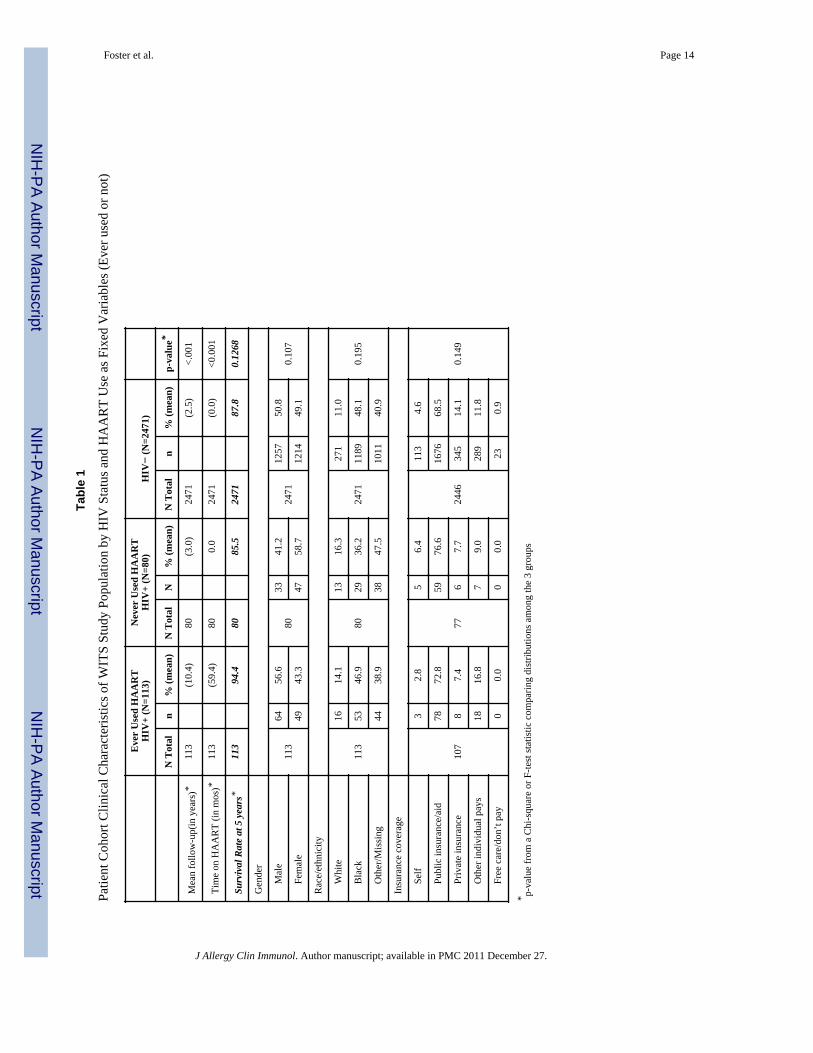
Table 1 summarizes the clinical characteristics of the study subject cohorts by HIV status(HIV+ or HIV−) and whether a HIV+ child ever used HAART (Ever Used HAART, NeverUsed HAART). The distribution of mean follow-up time (also mean age) among the three
Foster et al. Page 4
J Allergy Clin Immunol. Author manuscript; available in PMC 2011 December 27.
NIH
-PA Author Manuscript
NIH
-PA Author Manuscript
NIH
-PA Author Manuscript

groups was statistically different (P<0.001). The shorter mean follow-up time of the NeverUsed HAART group is due to the death of many patients in this group (33 out of 80)compared to the Ever Used HAART group (8 out of 113). The shorter follow-up times forthe HIV− group is due to randomization of these children out of the WITS protocol (seeMethods) (n=1,418), a parent’s refusal to allow a child to continue in the study (n=502), andloss to follow-up (n=436). Gender, race/ethnicity, and ability to pay for medical costs(socioeconomic status) did not differ among the three cohorts. All 3 cohorts of the studywere predominantly of minority race and ethnic groups and were inner-city dwellers. Thepercent survival of patients shows that the HIV+ HAART+ group were somewhat older thanthe HIV+ HAART− children (94.4% vs. 85.5%), respectively. These survival rates aresomewhat different from those in Figure 1 (see below) where time-dependent analysis wasused to compare the incidence distributions.
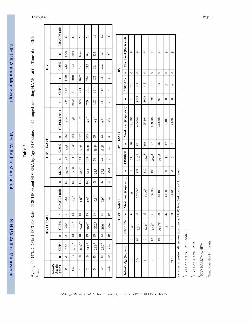
Table 2 records the average percentages of CD4+ T cells, CD8+ T cells, CD4/CD8 ratio,CD8+DR+ T cells, and HIV RNA levels at several time points with HAART serving as atime-dependent variable (i.e., children are assigned their respective group dependingwhether they were or were not taking HAART at the time of the clinic visit). In general,HIV+ children who were on HAART at the time of measurement had higher CD4+ T cell,lower CD8+ T cell, and lower CD8+DR+ T cell percentages; higher CD4/CD8 ratios; andlower viral burdens than the HIV+ children not on HAART (P≤0.05 to P≤0.01). The HIV+HAART+ and HIV+ HAART− childrens’ values at each time were also significantlydifferent from those of the HIV− children (P≤0.05 to P≤0.01). Although there were agedifferences with groups, the GEE analyses and time-dependent covariate analyses that wereperformed corrected for these age differences through the formation of the respective risksets and marginal analyses that were evaluated over time.
Survival Analysis Relating HIV Infection and HAART Use (for HIV+ Infants) to the Risk ofInitiating Asthma Medication
The time of first asthma medication use for each child was analyzed beginning at 1 month ofage and continuing up to 13.5 years of age using a time-dependent Cox Model (Fig 1). Thebeta coefficients obtained from this analysis and “cumulative incidence curves” compare theexpected incidence of asthma medication use for a model group of HIV+ infants takingHAART over the entire follow-up interval, a model group of HIV+ infants not takingHAART at any time over the entire follow-up interval, and the HIV− infants. Thecumulative incidence at 13.5 years of age for the model HIV+ HAART+ group was 33.5%and that of HIV− group was approximately 31.2%. In contrast, the cumulative incidencecurve for the model HIV+ HAART− group was 11.5%. Using the Cox model estimates toassess whether HIV infection status and HAART therapy (in HIV+ children) was related tothe risk of first asthma medication use, the exponential of the difference in the coefficientsfor HIV+ HAART+ and HIV+ HAART− yielded a hazard ratio (HR) estimate of 3.34(P=0.01) and the difference between the HIV+ HAART− and HIV− coefficients yielded aHR estimate of 0.33 (P=<0.01). The difference between the HIV+ HAART+ and HIV−coefficients was not significant. A Cox analysis model was also used to examine therelationship between asthma medication use and time-dependent treatment with NNRTIs orPIs. The results were similar to those observed in the HAART analysis (data not shown).
Analysis of the Prevalence of Asthma Medication UseFigure 2 illustrates the prevalence of asthma medication use for children born in the pre-HAART era (prior to 02/28/94) as assessed at the time of each follow-up visit (approximatechild’s age). The first asthma medication use for all groups was recorded at the 60-month (5-year) visit. Most (65%) of HIV+ HAART+ children had reported asthma medication use onat least two clinic visits. The maximum prevalence of asthma medication use for HIV+
Foster et al. Page 5
J Allergy Clin Immunol. Author manuscript; available in PMC 2011 December 27.
NIH
-PA Author Manuscript
NIH
-PA Author Manuscript
NIH
-PA Author Manuscript
children taking HAART at the same time was 10.4%; the maximum prevalence of asthmamedication use in the HIV− group was 10.5%, and the corresponding prevalence for HIV+children not taking HAART at the same time as the asthma medication was 3.8%. All ofthese maximums are achieved at 132 months (11 years) of follow-up. Using a GEE modelanalysis, the odds ratio for receiving asthma medication was 3.38 (P=0.02) comparing theHIV+ HAART+ vs. HIV+ HAART− groups; 1.40 (P=0.51) comparing the HIV+ HAART+vs. HIV− groups; and 0.41 (P=0.15) comparing the HIV+ HAART− vs. HIV− groups.
There were too few HIV+ children not taking HAART to allow the statistical analysis of theWITS children born in the ART II (after 03/01/94) and III (after 08/01/96) eras.
Effects of HAART on the Rate of Change of CD4+ T Cell Percentage at the Time of theInitiation of Asthma Medication
Mixed model analysis was used to compare CD4+ T cell percentage trajectories (percentage/year) among children ages six to ten years indexed by their initiation of asthma medicationuse (Fig 3). We have chosen to use the CD4+ T cell percent since this is a constant indicatorof CD4+ T cell sufficiency across different children’s ages. Assignment of a HIV+ child to aHAART group was made at the time of the CD4+ T cell percent evaluation (a time-dependent analysis). Data points 3 years before and 3 years after the initiation of asthmamedication were recorded at each time according to the child’s HIV status and whether ornot he or she was taking HAART at that time (HIV+ HAART+, HIV+ HAART− and HIV−). The average slopes of the trajectories were: 0.81 percent/year for HIV+ HAART+children, −1.43 percent/year for HIV+ HAART− children, and 0.15 percent/year for theHIV− children. Analysis of these slope coefficients shows significance for the comparisonof HIV+ HAART+ vs. HIV+ HAART− (P=0.010) but not for other comparisons: HIV+HAART+ vs. HIV− (P=0.41) and HIV+ HAART− vs. HIV− (P=0.08). If data points oneyear before and one year after the initiation of asthma medication were used to calculateslopes, again significant differences existed between the HIV+ HAART+ vs. HIV+ HAARTchildren (P=0.01) (data not shown).
DISCUSSIONThe present large multicenter study of infants born to HIV+ women confirms and extendsthe findings of a smaller single center study.13 The cumulative incidence (Fig 1) and theprevalence (Fig 2) of asthma, as assessed by asthma medication use, are higher in HIV+HAART+ children as compared to these HIV+ HAART− children. Our data show a muchlower cumulative incidence and prevalence of asthma in HIV+ HAART− children than HIV+ HAART+ children and a restoration of the risk of asthma when HIV+ children weretreated with HAART. This pediatric human model of asthma confirms what has been seen inanimal models with asthma1–3 and suggests that the loss of CD4+ T cells in children withuntreated HIV infection protects against asthma, and the gain of CD4+ T cells with HAARTtherapy serves as a risk factor for asthma. Our findings are in agreement with most studies ofadult patients with HIV infection5–8 but not other studies.9, 10 Most of these adult studieswere performed in the pre-HAART era6–10 and the timing of HIV infection (adults vs.infants) and immune ontogeny (developed vs. developing) most likely explains the lack ofcomplete agreement. One clear example of the difference between pediatric adult HIVpathogenesis is the rapid rise and persistent (≥3 years) elevation in HIV RNA level (hundredthousand to million range) in newborns26 compared to a much lower HIV viral set-pointobserved 6–8 weeks after infection in adults.
It is possible that the immunoreconstitution of CD4+ T cells with HAART therapy plays arole in an inflammatory response as active CD4+ T cells initially confront HIV antigens orthose of opportunistic organisms colonizing the airway27, 28 that results in a clinical state of
Foster et al. Page 6
J Allergy Clin Immunol. Author manuscript; available in PMC 2011 December 27.
NIH
-PA Author Manuscript
NIH
-PA Author Manuscript
NIH
-PA Author Manuscript
bronchial hyperresponsiveness (Fig 3). It is reasonable to speculate that inflammatorycytokines (e.g., IL-4, IL-5, IL-9, IL-13), may participate in this production of asthma.29
Activated CD8+ T cells (i.e., DR+) may be contributing to a state of pulmonaryhypersensitivity as well.30 These activated CD8+ T cells have been associated with HIVdisease progression and other serious complications of HIV infection, such asencephalopathy.31 Virus-specific CD8+ T cells can switch from IFN-γ to IL-5 production32
and may be operating in patients with HIV infection who develop asthma on HAARTtherapy. In another area of clinical immunology, Reveille and Williams33 have noted themarked change in the pattern of rheumatological complications of HIV infection in adultssince the introduction of HAART therapy with the appearance of de novo autoimmunedisorders as part of the immunoreconstitution of CD4+ T cells.
Several limitations of the present study need mention. The WITS study was not designed tolook at the incidence of asthma per se but to focus on HIV infection and its consequences.The diagnosis of asthma and reactive airways disease reported by parents and recorded byWITS medical personnel in the first 1–2 years life of the study subjects suggested anoveruse of the diagnosis of asthma as applied to wheezing infants with respiratory syncytialvirus or rhinovirus present with asthma-like symptoms (data not shown).17, 34–36 Many ofthese young infants infected with respiratory viruses, particularly those with underlyingallergies, indeed go on to have persistent asthma, but a majority have their symptoms remitin a few years. The WITS program did not provide a long-term evaluation of the persistenceof asthma symptoms with measurement of pulmonary function.
To mitigate these limitations we established a diagnosis of asthma in the WITS cohort by theuse of asthma medications, a more conservative approach than using parental recall ofwheezing in infants. We also observed a relatively high incidence of asthma in the HIV−group (roughly equal to that of the model HIV+ HAART+ group). These children mighthave been exposed to HIV antigens in utero and in the peripartum period, most likelybecoming sensitized to HIV antigens and exposed to pro-inflammatory cytokines of theHIV-infected mother. This HIV exposure perhaps renders the HIV− group a non-idealcontrol against which to compare the incidence of asthma. As the control study subjectslived in inner cities, for the most part, environmental exposures may have contributed to ahigher incidence and prevalence of asthma in all groups.37 There is some evidence in thepulmonary literature that HIV-exposed infants had a 20% lower partial forced expiratoryflow as compared to historical controls.38 Although the authors at that time discounted anHIV exposure factor as the basis for causation, in retrospect, this may well have played arole in producing this diminution of infant lung function.
There is also a possibility that a treatment-related censoring function with patients who diedcould have biased our study results. HIV+ infants had a lower risk of death when they werebeing treated with HAART. However, the nature of the finding presented here suggests thatthe direction of the bias is to elevate the model incidence curve for the HIV+ HAART−infants. This would occur because the recovery of CD4+ T cells predicts resurgence of therisk for asthma (the finding here) and the death-censoring mechanism is selectivelyeliminating from the analysis asthma information from those infants with the lowest CD4+ Tcell counts, who would be least likely to develop asthma in our hypothesis.
Despite these limitations, there is reason to continue to explore the interrelationshipsbetween HIV infection, HAART treatment, asthma, and CD4+ T cells and to expand thestudy to the evaluation of inflammatory cytokines and activated CD8+ T cells. A prospectivestudy of HIV+ patients treated with HAART is now needed that relates pulmonary functiondata to immune responses. This AIDS model of asthma may hold clues for a better
Foster et al. Page 7
J Allergy Clin Immunol. Author manuscript; available in PMC 2011 December 27.
NIH
-PA Author Manuscript
NIH
-PA Author Manuscript
NIH
-PA Author Manuscript
understanding of molecular and cellular mechanisms responsible for the epidemic of asthmain children.39
AcknowledgmentsSupported by National Institutes of Health grants and contracts HL96040, HL079533, HL72705, AI27551,AI36211, HD41983, RR0188, and AI41089; the Pediatric Research and Education Fund, Baylor College ofMedicine and the David Fund, Pediatrics AIDS Fund, and Immunology Research Fund, Texas Children’s Hospital.,and the Immunology Research Fund and Pediatric AIDS Fund of Texas Children’s Hospital.
Ms. Carolyn Jackson assisted with the preparation of the manuscript.
ABBREVIATIONS
ART antiretroviral therapy
HIV human immunodeficiency virus type 1
HIV+ HIV infected
HIV HIV uninfected
HAART highly active antiretroviral therapy
References1. Swain SD, Meissner NN, Harmsen AG. CD8 T cells modulate CD4 T-cell and eosinophil-mediated
pulmonary pathology in pneumocystis pneumonia in B-cell-deficient mice. Am J Pathol. 2006;168:466–75. [PubMed: 16436661]
2. Gavett SH, Chen X, Finkelman F, Wills-Karp M. Depletion of murine CD4+ T lymphocytesprevents antigen-induced airway hyperreactivity and pulmonary eosinophilia. Am J Respir Cell MolBiol. 1994; 10:587–93. [PubMed: 8003337]
3. Chapoval SP, Marietta EV, Smart MK, David CS. Requirements for allergen-induced airwayinflammation and hyperreactivity in CD4-deficient and CD4-sufficient HLA-DQ transgenic mice. JAllergy Clin Immunol. 2001; 108:764–71. [PubMed: 11692102]
4. Macaubas, C.; Kekruyff, RH.; Umetsu, DT. Immunology of the asthmatic response. In: Leung,DYM.; Sampson, HA.; Geha, R.; Szeffler, SJ., editors. Pediatric Allergy: Principles and Practice.St. Louis: Mosby; 2003. p. 337-349.
5. Poirier CD, Inhaber N, Lalonde RG, Ernst P. Prevalence of bronchial hyperresponsiveness amongHIV-infected men. Am J Respir Crit Care Med. 2001; 164:542–5. [PubMed: 11520712]
6. Wallace JM, Stone GS, Browdy BL, Tashkin DP, Hopewell PC, Glassroth J, et al. Nonspecificairway hyperresponsiveness in HIV disease. Pulmonary Complications of HIV Infection StudyGroup. Chest. 1997; 111:121–7. [PubMed: 8996005]
7. Lin RY, Lazarus TS. Asthma and related atopic disorders in outpatients attending an urban HIVclinic. Ann Allergy Asthma Immunol. 1995; 74:510–5. [PubMed: 7788519]
8. O’Donnell CR, Bader MB, Zibrak JD, Jensen WA, Rose RM. Abnormal airway function inindividuals with the acquired immunodeficiency syndrome. Chest. 1988; 94:945–8. [PubMed:3263260]
9. Moscato G, Maserati R, Marraccini P, Caccamo F, Dellabianca A. Bronchial reactivity tomethacholine in HIV-infected individuals without AIDS. Chest. 1993; 103:796–9. [PubMed:8449071]
10. Stover DE, White DA, Romano PA, Gellene RA, Robeson WA. Spectrum of pulmonary diseasesassociated with the acquired immune deficiency syndrome. Am J Med. 1985; 78:429–37.[PubMed: 2983548]
11. Shelburne SA, Montes M, Hamill R. Immune reconstitution inflammatory syndrome: moreanswers, more questions--authors’ response. J Antimicrob Chemother. 2006; 58:1094–5.[PubMed: 16980294]
Foster et al. Page 8
J Allergy Clin Immunol. Author manuscript; available in PMC 2011 December 27.
NIH
-PA Author Manuscript
NIH
-PA Author Manuscript
NIH
-PA Author Manuscript
12. Galli L, Sabatino G, Zappa M, Barbante E, Chiappini E, de Martino M. Reduced frequency ofwheezing respiratory illness in infants with perinatal human immunodeficiency virus-type 1infection: a model for immunologic and inflammatory mechanisms of airway obstruction? PediatrAllergy Immunol. 2003; 14:42–9. [PubMed: 12603710]
13. Foster SB, Paul ME, Kozinetz CA, Macias CG, Shearer WT. Prevalence of asthma in children andyoung adults with HIV infection. J Allergy Clin Immunol. 2007; 119:750–2. [PubMed: 17336620]
14. Gutin F, Secord E. HIV-related risk factors for asthma in children. J Allergy Clin Immunol. 2004;113:S127.
15. Sheon AR, Fox HE, Rich KC, Stratton P, Diaz C, Tuomala RE, et al. The women and infantstransmission study (WITS) of maternal-infant HIV transmission: Study design methods andbaseline data. J Women’s Health. 1996; 5:69–78.
16. Palumbo PE, Kwok S, Waters S, Wesley Y, Lewis D, McKinney N, et al. Viral measurement bypolymerase chain reaction-based assays in human immunodeficiency virus-infected infants. JPediatr. 1995; 126:592–5. [PubMed: 7699539]
17. Calvelli T, Denny TN, Paxton H, Gelman R, Kagan J. Guideline for flow cytometricimmunophenotyping: a report from the National Institute of Allergy and Infectious Diseases,Division of AIDS. Cytometry. 1993; 14:702–15. [PubMed: 8243200]
18. Taussig LM, Wright AL, Holberg CJ, Halonen M, Morgan WJ, Martinez FD. Tucson Children’sRespiratory Study: 1980 to present. J Allergy Clin Immunol. 2003; 111:661–75. [PubMed:12704342]
19. Werk LN, Steinbach S, Adams WG, Bauchner H. Beliefs about diagnosing asthma in youngchildren. Pediatrics. 2000; 105:585–90. [PubMed: 10699113]
20. Diggle, PJ.; Liang, KY.; Zeger, SL. Analysis of longitudinal data. New York: Oxford UniversityPress; 1994.
21. Laird NM, Ware JH. Random-effects models for longitudinal data. Biometrics. 1982; 38:963–74.[PubMed: 7168798]
22. McCullagh, P.; Nelder, JA. Monographs on Statistics and Applied Probability. 2. London,England: Chapman & Hall; 1989. Generalized Linear Models; p. 37
23. Cox D. Regression models and life tables. JR Stat Soc B. 1972; 34:187–220.24. Kalbfleisch, JD.; Prentice, RL. Relative risk (Cox) regression models. Estimation of the baseline
hazard or survivor function. 2. Hoboken, New Jersey: John Wiley & Sons, Inc; 2002. Thestatistical analysis of failure time data; p. 114-18.
25. Hayter AJ. The maximum familywise error rate of Fisher’s least significant difference test. J AmStati Assoc. 1986; 81:1000.
26. Shearer WT, Quinn TC, LaRussa P, Lew JF, Mofenson L, Almy S, et al. Viral load and diseaseprogression in infants infected with human immunodeficiency virus type 1. Women and InfantsTransmission Study Group. N Engl J Med. 1997; 336:1337–42. [PubMed: 9134873]
27. Bisgaard H, Hermansen MN, Buchvald F, Loland L, Halkjaer LB, Bonnelykke K, et al. Childhoodasthma after bacterial colonization of the airway in neonates. N Engl J Med. 2007; 357:1487–95.[PubMed: 17928596]
28. von Mutius E. Of attraction and rejection--asthma and the microbial world. N Engl J Med. 2007;357:1545–7. [PubMed: 17928604]
29. Corry DB, Kheradmand F. Control of allergic airway inflammation through immunomodulation. JAllergy Clin Immunol. 2007; 117:S461–S4. [PubMed: 16455347]
30. O’Sullivan S, Cormican L, Faul JL, Ichinohe S, Johnston SL, Burke CM, et al. Activated,cytotoxic CD8(+) T lymphocytes contribute to the pathology of asthma death. Am J Respir CritCare Med. 2001; 164:560–4. [PubMed: 11520715]
31. Paul ME, Mao C, Charurat M, Serchuck L, Foca M, Hayani K, et al. Predictors of immunologiclong-term nonprogression in HIV-infected children: implications for initiating therapy. J AllergyClin Immunol. 2005; 115:848–55. [PubMed: 15806009]
32. Busse, WW.; O’Bryne, PM.; Holgate, ST. Asthma pathogensis. In: Adkinson, NH., Jr; Yunginer,JW.; Busse, WW., editors. Allergy: Principles and Practice. 6. Philadelphia, Pittsburgh: Mosby;2003. p. 1175-207.
Foster et al. Page 9
J Allergy Clin Immunol. Author manuscript; available in PMC 2011 December 27.
NIH
-PA Author Manuscript
NIH
-PA Author Manuscript
NIH
-PA Author Manuscript
33. Reveille JD, Williams FM. Infection and musculoskeletal conditions: Rheumatologiccomplications of HIV infection. Best Pract Res Clin Rheumatol. 2006; 20:1159–79. [PubMed:17127202]
34. Friedlander SL, Busse WW. The role of rhinovirus in asthma exacerbations. J Allergy ClinImmunol. 2005; 116:267–73. [PubMed: 16083778]
35. Heymann PW, Carper HT, Murphy DD, Platts-Mills TA, Patrie J, McLaughlin AP, et al. Viralinfections in relation to age, atopy, and season of admission among children hospitalized forwheezing. J Allergy Clin Immunol. 2004; 114:239–47. [PubMed: 15316497]
36. Schaller M, Hogaboam CM, Lukacs N, Kunkel SL. Respiratory viral infections drive chemokineexpression and exacerbate the asthmatic response. J Allergy Clin Immunol. 2006; 118:295–302.[PubMed: 16890750]
37. Busse WW, Mitchell H. Addressing issues of asthma in inner-city children. J Allergy ClinImmunol. 2007; 119:43–9. [PubMed: 17208585]
38. Colin AA, Sunil Rao J, Chen XC, Hunter JM, Hanrahan J, Hiatt P, et al. Forced expiratory flow inuninfected infants and children born to HIV-infected mothers. Am J Respir Crit Care Med. 2001;163:865–73. [PubMed: 11282758]
39. Eder W, Ege MJ, von Mutius E. The asthma epidemic. N Engl J Med. 2006; 355:2226–35.[PubMed: 17124020]
Foster et al. Page 10
J Allergy Clin Immunol. Author manuscript; available in PMC 2011 December 27.
NIH
-PA Author Manuscript
NIH
-PA Author Manuscript
NIH
-PA Author Manuscript
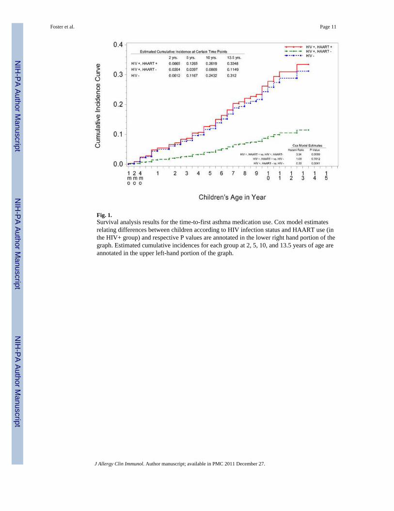
Fig. 1.Survival analysis results for the time-to-first asthma medication use. Cox model estimatesrelating differences between children according to HIV infection status and HAART use (inthe HIV+ group) and respective P values are annotated in the lower right hand portion of thegraph. Estimated cumulative incidences for each group at 2, 5, 10, and 13.5 years of age areannotated in the upper left-hand portion of the graph.
Foster et al. Page 11
J Allergy Clin Immunol. Author manuscript; available in PMC 2011 December 27.
NIH
-PA Author Manuscript
NIH
-PA Author Manuscript
NIH
-PA Author Manuscript
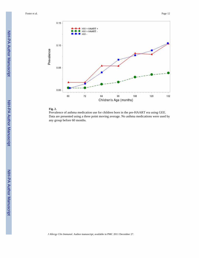
Fig. 2.Prevalence of asthma medication use for children born in the pre-HAART era using GEE.Data are presented using a three point moving average. No asthma medications were used byany group before 60 months.
Foster et al. Page 12
J Allergy Clin Immunol. Author manuscript; available in PMC 2011 December 27.
NIH
-PA Author Manuscript
NIH
-PA Author Manuscript
NIH
-PA Author Manuscript

Fig. 3.Median CD4+ T cell percentages in children aged six to ten years of age, indexed accordingto the time of first asthma medication use (Time 0). The slope estimates of CD4+ cellpercent from the mixed model analysis are annotated in the upper portion of the graph.
Foster et al. Page 13
J Allergy Clin Immunol. Author manuscript; available in PMC 2011 December 27.
NIH
-PA Author Manuscript
NIH
-PA Author Manuscript
NIH
-PA Author Manuscript
NIH
-PA Author Manuscript
NIH
-PA Author Manuscript
NIH
-PA Author Manuscript
Foster et al. Page 14
Tabl
e 1
Patie
nt C
ohor
t Clin
ical
Cha
ract
eris
tics o
f WIT
S St
udy
Popu
latio
n by
HIV
Sta
tus a
nd H
AA
RT
Use
as F
ixed
Var
iabl
es (E
ver u
sed
or n
ot)
Eve
r U
sed
HA
AR
TH
IV+
(N=1
13)
Nev
er U
sed
HA
AR
TH
IV+
(N=8
0)H
IV−
(N=2
471)
N T
otal
n%
(mea
n)N
Tot
alN
% (m
ean)
N T
otal
n%
(mea
n)p-
valu
e*
Mea
n fo
llow
-up(
in y
ears
)*11
3(1
0.4)
80(3
.0)
2471
(2.5
)<.
001
Tim
e on
HA
AR
T (in
mos
)*11
3(5
9.4)
800.
024
71(0
.0)
<0.0
01
Surv
ival
Rat
e at
5 y
ears
*11
394
.480
85.5
2471
87.8
0.12
68
Gen
der
M
ale
113
6456
.680
3341
.224
7112
5750
.80.
107
Fe
mal
e49
43.3
4758
.712
1449
.1
Rac
e/et
hnic
ity
W
hite
113
1614
.1
80
1316
.3
2471
271
11.0
0.19
5
Bla
ck53
46.9
2936
.211
8948
.1
O
ther
/Mis
sing
4438
.938
47.5
1011
40.9
Insu
ranc
e co
vera
ge
Se
lf
107
32.
8
77
56.
4
2446
113
4.6
0.14
9
Pu
blic
insu
ranc
e/ai
d78
72.8
5976
.616
7668
.5
Pr
ivat
e in
sura
nce
87.
46
7.7
345
14.1
O
ther
indi
vidu
al p
ays
1816
.87
9.0
289
11.8
Fr
ee c
are/
don’
t pay
00.
00
0.0
230.
9
* p-va
lue
from
a C
hi-s
quar
e or
F-te
st st
atis
tic c
ompa
ring
dist
ribut
ions
am
ong
the
3 gr
oups
J Allergy Clin Immunol. Author manuscript; available in PMC 2011 December 27.
NIH
-PA Author Manuscript
NIH
-PA Author Manuscript
NIH
-PA Author Manuscript
Foster et al. Page 15
Tabl
e 2
Ave
rage
CD
4%, C
D8%
, CD
4/C
D8
Rat
io, C
D8+ D
R %
and
HIV
RN
A b
y A
ge, H
IV st
atus
, and
Gro
uped
acc
ordi
ng H
AA
RT
at th
e Ti
me
of th
e C
hild
’sV
isit
HIV
+ H
AA
RT
+H
IV+
HA
AR
T−
HIV
−
Infa
nt’s
Age
(in
year
s)n
CD
4%n
CD
8%n
CD
4/C
D8
ratio
nC
D4%
nC
D8%
nC
D4/
CD
8 ra
tion
CD
4%n
CD
8%n
CD
4/C
D8
ratio
02
48.5
225
.52
2.2
124
49.6
‡12
224
.0‡
122
2.5‡
1724
54.5
1720
21.1
1720
3.0
0.5
1343
.2*
1318
.7*
132.
4*13
532
.5‡
133
28.2
‡13
31.
4‡20
5045
.920
4017
.120
403.
0
118
37.3
*†18
24.4
*†18
1.8*
†11
928
.3‡
118
32.9
‡11
71.
0*16
7942
.116
7719
.016
762.
5
219
31.1
†19
29.2
*†19
1.3*
†10
326
.1‡
103
39.4
‡10
30.
8*76
938
.876
621
.176
62.
0
535
28.9
†35
37.2
†35
0.9†
5928
.7‡
5939
.4‡
590.
8*12
238
.412
223
.412
21.
8
1053
28.7
†53
36.0
*†53
0.9*
†22
27.2
‡22
43.3
‡22
0.7*
1242
.712
20.7
122.
2
13.5
1928
.519
38.5
191.
03
26.3
345
.73
0.6
0§
0§
0§
HIV
+ H
AA
RT
+H
IV+
HA
AR
T−
HIV
−
Infa
nt’s
Age
(in
year
s)n
CD
8DR
%n
Vir
al L
oad
(Cop
ies/
ml)
nC
D8D
R%
nV
iral
Loa
d (C
opie
s/m
l)n
CD
8DR
%n
Vir
al L
oad
(Cop
ies/
ml)
00
§0
§1
14.0
7619
1,50
01
2.0
0§
0.5
108.
3*†
1226
7,00
012
714
.1‡
121
643,
600
1265
4.7
0§
19
12.2
†18
9,50
0*11
919
.2‡
108
425,
800
1028
6.8
0§
212
17.9
†19
188,
200
103
24.0
‡97
579,
100
696
7.5
0§
519
29.1
*†31
41,1
0057
21.0
‡46
443,
300
907.
40
§
100
§47
41,9
000
§17
35,1
000
§0
§
13.5
0§
1122
,700
0§
12,
600
0§
0§
Pair-
wis
e co
mpa
rison
s diff
eren
ces s
igni
fican
t at P
≤0.0
5 le
vel (
red
colo
r: P
< 0.
01 le
vel):
* HIV
+ H
AA
RT+
vs.
HIV
+ H
AA
RT−
;
† HIV
+ H
AA
RT+
vs.
HIV−
;
‡ HIV
+ H
AA
RT−
vs.
HIV−
§ Insu
ffic
ient
dat
a fo
r ana
lysi
s
J Allergy Clin Immunol. Author manuscript; available in PMC 2011 December 27.
NIH
-PA Author Manuscript
NIH
-PA Author Manuscript
NIH
-PA Author Manuscript
Foster et al. Page 16A
t eac
h tim
e po
int,
HIV
+ su
bjec
ts a
re a
ssig
ned
to a
gro
up b
y H
AA
RT
med
icat
ion,
(i.e
., H
AA
RT+
or H
AA
RT−
).
J Allergy Clin Immunol. Author manuscript; available in PMC 2011 December 27.
Copyright © 2022 FDOKUMEN