In the complex family of heat stress transcription factors, HsfA1 has a unique role as master...
13
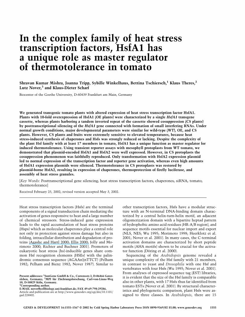
In the complex family of heat stress transcription factors, HsfA1 has a unique role as master regulator of thermotolerance in tomato Shravan Kumar Mishra, Joanna Tripp, Sybille Winkelhaus, Bettina Tschiersch, 1 Klaus Theres, 2 Lutz Nover, 3 and Klaus-Dieter Scharf Biocenter of the Goethe University, D-60439 Frankfurt am Main, Germany We generated transgenic tomato plants with altered expression of heat stress transcription factor HsfA1. Plants with 10-fold overexpression of HsfA1 (OE plants) were characterized by a single HsfA1 transgene cassette, whereas plants harboring a tandem inverted repeat of the cassette showed cosuppression (CS plants) by posttranscriptional silencing of the HsfA1 gene connected with formation of small interfering RNAs. Under normal growth conditions, major developmental parameters were similar for wild-type (WT), OE, and CS plants. However, CS plants and fruits were extremely sensitive to elevated temperatures, because heat stress-induced synthesis of chaperones and Hsfs was strongly reduced or lacking. Despite the complexity of the plant Hsf family with at least 17 members in tomato, HsfA1 has a unique function as master regulator for induced thermotolerance. Using transient reporter assays with mesophyll protoplasts from WT tomato, we demonstrated that plasmid-encoded HsfA1 and HsfA2 were well expressed. However, in CS protoplasts the cosuppression phenomenon was faithfully reproduced. Only transformation with HsfA2 expression plasmid led to normal expression of the transcription factor and reporter gene activation, whereas even high amounts of HsfA1 expression plasmids were silenced. Thermotolerance in CS protoplasts was restored by plasmid-borne HsfA2, resulting in expression of chaperones, thermoprotection of firefly luciferase, and assembly of heat stress granules. [Key Words: Posttranscriptional gene silencing; heat stress transcription factors; chaperones; siRNA; tomato; thermotolerance] Received February 25, 2002; revised version accepted May 3, 2002. Heat stress transcription factors (Hsfs) are the terminal components of a signal transduction chain mediating the activation of genes responsive to heat and a large number of chemical stressors. Stress-induced gene expression leads to the rapid accumulation of heat stress proteins (Hsps) which as molecular chaperones play a central role not only in protection against stress damage but also in folding, intracellular distribution and degradation of pro- teins (Agashe and Hartl 2000; Ellis 2000; Jolly and Mo- rimoto 2000; Richter and Buchner 2001). Promoters of eukaryotic heat stress (hs)-inducible genes share com- mon Hsf recognition elements (HSEs) with the palin- dromic consensus sequence (AGAAn)(nTTCT) (Pelham 1982; Pelham and Bienz 1982; Nover 1987). Similar to other transcription factors, Hsfs have a modular struc- ture with an N-terminal DNA-binding domain charac- terized by a central helix-turn-helix motif, an adjacent oligomerization domain with a bipartite heptad pattern of hydrophobic amino acid residues (HR-A/B region), and sequence motifs essential for nuclear import and export (NLS, NES; Wu 1995; Morimoto 1998; Heerklotz et al. 2001; Nover et al. 2001). In many cases, the C-terminal activation domains are characterized by short peptide motifs (AHA motifs) shown to be crucial for the activa- tor function (Döring et al. 2000). Sequencing of the Arabidopsis genome revealed a unique complexity of the Hsf family with 21 members, in contrast to yeast and Drosophila with one Hsf and vertebrates with four Hsfs (Wu 1995; Nover et al. 2001). From analyses of expressed sequence tag (EST) libraries, it is evident that the size of the Hsf family is comparable also in other plants, with 17 Hsfs thus far identified from tomato ESTs (Nover et al. 2001). By structural character- istics and phylogenetic comparison, plant Hsfs were as- signed to three classes. In Arabidopsis, there are 15 Present addresses: 1 SunGene GmbH & Co., Corrensstr.3, D-06466 Gater- sleben, Germany; 2 MPI für Züchtungsforschung, Carl-von-Linne-Weg 10, D-50829 Köln, Germany. 3 Corresponding author. E-MAIL [email protected]; FAX 49-69-798-29286. Article and publication are at http://www.genesdev.org/cgi/doi/10.1101/ gad.228802. GENES & DEVELOPMENT 16:1555–1567 © 2002 by Cold Spring Harbor Laboratory Press ISSN 0890-9369/02 $5.00; www.genesdev.org 1555
-
Upload
independent -
Category
Documents
-
view
2 -
download
0
Transcript of In the complex family of heat stress transcription factors, HsfA1 has a unique role as master...
In the complex family of heat stresstranscription factors, HsfA1 hasa unique role as master regulatorof thermotolerance in tomatoShravan Kumar Mishra, Joanna Tripp, Sybille Winkelhaus, Bettina Tschiersch,1 Klaus Theres,2
Lutz Nover,3 and Klaus-Dieter Scharf
Biocenter of the Goethe University, D-60439 Frankfurt am Main, Germany
We generated transgenic tomato plants with altered expression of heat stress transcription factor HsfA1.Plants with 10-fold overexpression of HsfA1 (OE plants) were characterized by a single HsfA1 transgenecassette, whereas plants harboring a tandem inverted repeat of the cassette showed cosuppression (CS plants)by posttranscriptional silencing of the HsfA1 gene connected with formation of small interfering RNAs. Undernormal growth conditions, major developmental parameters were similar for wild-type (WT), OE, and CSplants. However, CS plants and fruits were extremely sensitive to elevated temperatures, because heatstress-induced synthesis of chaperones and Hsfs was strongly reduced or lacking. Despite the complexity ofthe plant Hsf family with at least 17 members in tomato, HsfA1 has a unique function as master regulator forinduced thermotolerance. Using transient reporter assays with mesophyll protoplasts from WT tomato, wedemonstrated that plasmid-encoded HsfA1 and HsfA2 were well expressed. However, in CS protoplasts thecosuppression phenomenon was faithfully reproduced. Only transformation with HsfA2 expression plasmidled to normal expression of the transcription factor and reporter gene activation, whereas even high amountsof HsfA1 expression plasmids were silenced. Thermotolerance in CS protoplasts was restored byplasmid-borne HsfA2, resulting in expression of chaperones, thermoprotection of firefly luciferase, andassembly of heat stress granules.
[Key Words: Posttranscriptional gene silencing; heat stress transcription factors; chaperones; siRNA; tomato;thermotolerance]
Received February 25, 2002; revised version accepted May 3, 2002.
Heat stress transcription factors (Hsfs) are the terminalcomponents of a signal transduction chain mediating theactivation of genes responsive to heat and a large numberof chemical stressors. Stress-induced gene expressionleads to the rapid accumulation of heat stress proteins(Hsps) which as molecular chaperones play a central rolenot only in protection against stress damage but also infolding, intracellular distribution and degradation of pro-teins (Agashe and Hartl 2000; Ellis 2000; Jolly and Mo-rimoto 2000; Richter and Buchner 2001). Promoters ofeukaryotic heat stress (hs)-inducible genes share com-mon Hsf recognition elements (HSEs) with the palin-dromic consensus sequence (AGAAn)(nTTCT) (Pelham1982; Pelham and Bienz 1982; Nover 1987). Similar to
other transcription factors, Hsfs have a modular struc-ture with an N-terminal DNA-binding domain charac-terized by a central helix-turn-helix motif, an adjacentoligomerization domain with a bipartite heptad patternof hydrophobic amino acid residues (HR-A/B region), andsequence motifs essential for nuclear import and export(NLS, NES; Wu 1995; Morimoto 1998; Heerklotz et al.2001; Nover et al. 2001). In many cases, the C-terminalactivation domains are characterized by short peptidemotifs (AHA motifs) shown to be crucial for the activa-tor function (Döring et al. 2000).Sequencing of the Arabidopsis genome revealed a
unique complexity of the Hsf family with 21 members,in contrast to yeast and Drosophila with one Hsf andvertebrates with four Hsfs (Wu 1995; Nover et al. 2001).From analyses of expressed sequence tag (EST) libraries,it is evident that the size of the Hsf family is comparablealso in other plants, with 17 Hsfs thus far identified fromtomato ESTs (Nover et al. 2001). By structural character-istics and phylogenetic comparison, plant Hsfs were as-signed to three classes. In Arabidopsis, there are 15
Present addresses: 1SunGene GmbH&Co., Corrensstr.3, D-06466 Gater-sleben, Germany; 2MPI für Züchtungsforschung, Carl-von-Linne-Weg10, D-50829 Köln, Germany.3Corresponding author.E-MAIL [email protected]; FAX 49-69-798-29286.Article and publication are at http://www.genesdev.org/cgi/doi/10.1101/gad.228802.
GENES & DEVELOPMENT 16:1555–1567 © 2002 by Cold Spring Harbor Laboratory Press ISSN 0890-9369/02 $5.00; www.genesdev.org 1555

members in class A, five members in class B, and one inclass C. Most striking for the discrimination of the threeclasses are peculiarities of the HR-A/B regions. Similarto all nonplant Hsfs, the HR-A/B regions of class B Hsfsare compact, with seven amino acid residues separatingthe HR-A and B parts, whereas class A and class C Hsfshave extended HR-A/B regions due to an insertion of 21(class A) and seven (class C) additional amino acid resi-dues (Nover et al. 2001). The significance of these ex-tended oligomerization domains for the oligomerizationbehavior and function of Hsfs is not yet clear. Anotherplant-specific feature with probably important conse-quences for details of the stress response is the fact thatmany Hsfs are hs-inducible proteins themselves (Scharfet al. 1990, 1998; Nover et al. 2001); for example, intomato HsfA1 is constitutively expressed, whereas HsfsA2 and B1 are hs-inducible proteins.To study the role of individual Hsfs in their native
background, we generated stable genetic lines of tomatowith altered expression of HsfA1, HsfA2, or HsfB1 byAgrobacterium-mediated integration of correspondingsense and antisense transgenes. Analyses of the trans-genic plants demonstrate that HsfA1 has a unique role asmaster regulator for the synthesis of Hsfs A2 and B1 aswell as Hsps. Posttranscriptional silencing of the HsfA1gene also causes severe defects in thermotolerance andplant development at elevated temperatures.
Results
Characterization of transgenic tomato lineswith overexpression and cosuppression of HsfA1
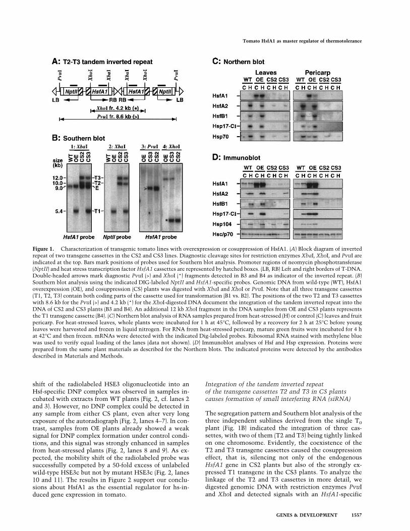
Using Agrobacterium-mediated gene transfer, tomatoleaf discs (Lycopersicon esculentum cv. Moneymaker)were transformed with Ti plasmid-derived vectors en-coding tomato heat stress transcription factor HsfA1 andneomycin phosphotransferase for selection. In the vectorused for transformation, the cDNA cassette encodingHsfA1 is positioned next to the right border (RB) of theT-DNA, whereas the NPTII cassette is linked to the leftborder (LB, see example in Fig. 1A). Since the transfer ofT-DNA occurs in a polar fashion starting at the rightborder (Becker et al. 1992; Sheng and Citovsky 1996),most regenerates showing kanamycin resistance alsoharbored the Hsf encoding cassette. Kanamycin-resistanttransgenic shoots were regenerated, rooted, and finallytransferred to the greenhouse. Seedlings derived from theT0 generation were sprayed with kanamycin solution todiscriminate between sensitive (wild-type) and resistant,that is, transgenic plants. The latter were further culti-vated, seeds were harvested (F1 generation), and the pro-cedure was repeated until stable, homozygous lines wereobtained.Starting with the F1 progenies, Kan
r plants were ana-lyzed by Southern and immunoblot analyses to monitorthe segregation of the transgene(s) and expression levelsof HsfA1. Among the 51 independent Kanr primaryplants, one was identified (A1-S17) as containing three
transgene cassettes. Progeny of this T0 plant segregatedinto three independent sublines. The results of theSouthern, Northern, and immunoblot analyses of F3progenies are shown in Figure 1B–D. One subline con-taining a single transgene cassette (T1) showed an ∼ 10-fold increased level of HsfA1 (overexpression line, OE),whereas the two other sublines containing two (T2 andT3, cosuppression line CS2) and three (T1, T2, and T3,cosuppression line CS 3) transgene cassettes in their ge-nome had no detectable HsfA1. Compared to the wildtype (WT), plants of all three transgenic lines showednormal growth and development as judged from size ofthe plants, time of flowering, fruit setting and ripening,as well as number of seeds in the fruits and seed germi-nation (data not shown).As a consequence of the altered levels of HsfA1 in the
OE and CS lines, the expression patterns of hs-induciblegenes were markedly changed. This was analyzed byNorthern blot (Fig. 1C) and immunoblot analyses (Fig.1D) using samples from young leaves and pericarp ofgreen fruits under control (lane C) and heat stress (laneH) conditions. In WT and OE plants, hs-induced syn-thesis of HsfA2, HsfB1, Hsp17-CI, and Hsp104 wasclearly detectable, and the strongly increased HsfA1 lev-els in the OE plants was connected with two- to three-fold higher levels of HsfA2, HsfB1, and Hsp17-CI in theleaves and hs-independent expression of HsfA2, Hsp17-CI, and Hsp104 in the pericarp. In contrast, hs-inducedsynthesis of HsfA2, HsfB1, and Hsp17-CI was markedlyreduced or even lacking in the CS2 and CS3 cosuppres-sion plants. The results from the Northern and immu-noblots were very similar except for the Hsc70/Hsp70complex. The antibody did not allow discrimination be-tween constitutive and heat stress-inducible isoforms ofthe protein (Fig. 1D). However, using gene-specificprobes for detection of the Hsp70 encoding mRNA, theresults of the Northern blot clearly indicated that expres-sion of Hsp70 in CS plants was also strongly reduced(Fig.1C).Also of interest are the subtle but reproducible differ-
ences in the HsfA1-dependent expression of HsfA2 andHsps observed between samples from CS2 and CS3plants. Although the silencing effect in CS3 plants isstrong enough to overcome the 10-fold increased expres-sion of HsfA1 as a result of the T1 transgene cassette,residual HsfA1 in leaves might allow some hs-inducibleexpression of HsfA2 and Hsp17-CI (Fig. 1D). This effectwas not observed in pericarp of green fruits, indicatingthat the strength of the silencing effect was dependenton the tissue investigated. This was particularly strikingfor the expression of Hsp104, which was only slightlyreduced in leaves, but totally lacking in pericarp(Fig. 1D).The expression analyses (Fig. 1C,D) indicate a unique
role of HsfA1 as a constitutively expressed member ofthe tomato Hsf family. We sought to establish whetherthe altered levels of HsfA1 in OE and CS compared toWT plants are also detectable by electrophoretic mobil-ity shift assays. Using extracts of nuclear proteins andHsf-specific oligonucleotides as probes, an hs-inducible
Mishra et al.
1556 GENES & DEVELOPMENT
shift of the radiolabeled HSE3 oligonucleotide into anHsf-specific DNP complex was observed in samples in-cubated with extracts from WT plants (Fig. 2, cf. lanes 2and 3). However, no DNP complex could be detected inany sample from either CS plant, even after very longexposure of the autoradiograph (Fig. 2, lanes 4–7). In con-trast, samples from OE plants already showed a weaksignal for DNP complex formation under control condi-tions, and this signal was strongly enhanced in samplesfrom heat-stressed plants (Fig. 2, lanes 8 and 9). As ex-pected, the mobility shift of the radiolabeled probe wassuccessfully competed by a 50-fold excess of unlabeledwild-type HSE3c but not by mutant HSE3c (Fig. 2, lanes10 and 11). The results in Figure 2 support our conclu-sions about HsfA1 as the essential regulator for hs-in-duced gene expression in tomato.
Integration of the tandem inverted repeatof the transgene cassettes T2 and T3 in CS plantscauses formation of small interfering RNA (siRNA)
The segregation pattern and Southern blot analysis of thethree independent sublines derived from the single T0plant (Fig. 1B) indicated the integration of three cas-settes, with two of them (T2 and T3) being tightly linkedon one chromosome. Evidently, the coexistence of theT2 and T3 transgene cassettes caused the cosuppressioneffect, that is, silencing not only of the endogenousHsfA1 gene in CS2 plants but also of the strongly ex-pressed T1 transgene in the CS3 plants. To analyze thelinkage of the T2 and T3 cassettes in more detail, wedigested genomic DNA with restriction enzymes PvuIand XhoI and detected signals with an HsfA1-specific
Figure 1. Characterization of transgenic tomato lines with overexpression or cosuppression of HsfA1. (A) Block diagram of invertedrepeat of two transgene cassettes in the CS2 and CS3 lines. Diagnostic cleavage sites for restriction enzymes XbaI, XhoI, and PvuI areindicated at the top. Bars mark positions of probes used for Southern blot analysis. Promoter regions of neomycin phosphotransferase(NptII) and heat stress transcription factorHsfA1 cassettes are represented by hatched boxes. (LB, RB) Left and right borders of T-DNA.Double-headed arrows mark diagnostic PvuI (>) and XhoI (*) fragments detected in B3 and B4 as indicator of the inverted repeat. (B)Southern blot analysis using the indicated DIG-labeled NptII and HsfA1-specific probes. Genomic DNA from wild-type (WT), HsfA1overexpression (OE), and cosuppression (CS) plants was digested with XbaI and XhoI or PvuI. Note that all three transgene cassettes(T1, T2, T3) contain both coding parts of the cassette used for transformation (B1 vs. B2). The positions of the two T2 and T3 cassetteswith 8.6 kb for the PvuI (>) and 4.2 kb (*) for the XhoI-digested DNA document the integration of the tandem inverted repeat into theDNA of CS2 and CS3 plants (B3 and B4). An additional 12 kb XhoI fragment in the DNA samples from OE and CS3 plants representsthe T1 transgene cassette (B4). (C) Northern blot analysis of RNA samples prepared from heat-stressed (H) or control (C) leaves and fruitpericarp. For heat-stressed leaves, whole plants were incubated for 1 h at 45°C, followed by a recovery for 2 h at 25°C before youngleaves were harvested and frozen in liquid nitrogen. For RNA from heat-stressed pericarp, mature green fruits were incubated for 4 hat 42°C and then frozen. mRNAs were detected with the indicated Dig-labeled probes. Ribosomal RNA stained with methylene bluewas used to verify equal loading of the lanes (data not shown). (D) Immunoblot analyses of Hsf and Hsp expression. Proteins wereprepared from the same plant materials as described for the Northern blots. The indicated proteins were detected by the antibodiesdescribed in Materials and Methods.
Tomato HsfA1 as master regulator of thermotolerance
GENES & DEVELOPMENT 1557

probe (Fig. 1B, panels 3 and 4). The results clearly showedthat the T2 and T3 cassettes form a tandem invertedrepeat. The 8.6 kb PvuI fragment (Fig. 1B, open arrow-head) and the 4.2 kb XhoI fragment (asterisk) corre-sponded to the expected cleavage pattern (double-headedarrows in the block diagram in Fig. 1A).Next, we wanted to determine whether formation of
small interfering RNAs (siRNAs) was responsible for thestrong cosuppression effect observed in CS2 and CS3plants containing the inverted repeat of the HsfA1 sensecassettes in their genomes. We prepared soluble RNAsfrom young leaves of WT, OE, CS2, and CS3 plants, sepa-rated them on denaturing polyacrylamide/urea gels, anddetected putative siRNA by RNA blot analysis using ra-diolabeled HsfA1-specific sense (Fig. 3A) and antisenseriboprobes (Fig. 3B). In samples of the two types of CSplants, both riboprobes detected small RNAs of about 21nucleotides which were lacking in samples fromWT andOE plants. As size marker and hybridization control, weused 23-mers of ribooligonucleotides derived from the 5�region of the DNA binding domain of HsfA1. The con-trol sense oligonucleotide gave a weaker signal with theantisense riboprobe (Fig. 3B, asterisk), because the oligo-nucleotide corresponds to the end of the ∼ 1.8 kb in vitrotranscript. But as expected, this deficit in the signal in-tensity was not observed for the siRNAs, which repre-
sent a mixture of RNAs covering the whole range of theHsfA1 mRNA.
HsfA1 cosuppression plants are highly heat-sensitive
What are the effects of the lacking synthesis of hs-induc-ible Hsfs A2 and B1 and Hsps on the growth and devel-opment of CS plants under stress conditions? To explorethis question, we exposed 6-wk-old greenhouse plants ina climate chamber to either 1-h heat treatment at 45°Cfollowed by 1 h at 25°C or to 1 h at 45°C followed by 1h at 51°C. During the hs treatment, sufficient water sup-ply was guaranteed. Control plants were maintained at25°C throughout. Photographs of the plants were takenafter another 14 d of growth in the greenhouse (Fig. 4A).The results documented that the CS3 plants were veryheat-sensitive. Even exposure for 1 h at 45°C ambienttemperature was lethal, whereas WT and OE plants werenot visibly affected in their development. The CS2 andCS3 plants gave similar results. Only the more severetreatment of 1 h at 45°C followed by 1 h at 51°C revealeda difference between WT and OE plants. The formerwere strongly damaged but slowly recovered by forminglateral shoots, whereas the latter were only slightly de-layed in their growth. Results were similar with germi-nating seedlings (data not shown).To complement these results with vegetative plants,
we harvested mature green fruits from WT, OE, and CS3plants (sample 1 in Fig. 4B) and investigated the influ-ence of a high-temperature treatment (48 h at 42°C) ontheir ripening at 25°C in the dark. For comparison, con-
Figure 2. Electrophoretic mobility shift assays with nuclearextracts from tomato leaves. Eight-week-old greenhouse plantswere heat-stressed for 1 h to 45°C (H) or kept at 25°C (C) beforeyoung leaves were harvested for preparation of nuclei (see Ma-terials and Methods). Lanes 2–9, radiolabeled HSE oligonucleo-tide incubated with the indicated nuclear extracts from WT,CS2, CS3, and OE plants. For the competition assay, the extractfrom heat-stressed OE plants was incubated with the labeledHSE3 plus a 50-fold excess of wild-type (wtHSE3c) or mutantoligonucleotide (mutHSE3c) in unlabeled form (lanes 10 and 11).Lane 1, sample without protein extract. Samples 1 to 7 and 8 to11 were run in separate gels. Film exposure was 5 d for samples1 to 7 and 1 d for samples 8 to 11.
Figure 3. Detection of siRNA in CS plants. Soluble RNA frac-tions from leaves of WT, OE, and CS plants were separated on15% denaturing polyacrylamide gels, blotted on nylon mem-branes, and hybridized with HsfA1-specific sense (A) and anti-sense riboprobes (B). For the control, 23-mers of sense and an-tisense ribooligonucleotides derived from the DNA binding do-main of HsfA1 were run on the same gels (positions indicated byasterisks). Arrows point to the siRNA bands in the CS2 and CS3samples. The inserts at the bottom of A and B represent longerexposures of the autoradiograph (only siRNA regions shown).
Mishra et al.
1558 GENES & DEVELOPMENT
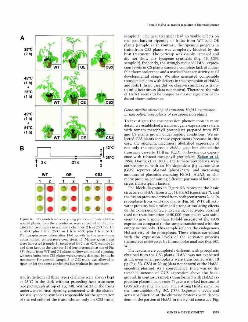
trol fruits from all three types of plants were always keptat 25°C in the dark without preceding heat treatment(see pictograph at top of Fig. 4B). Within 23 d, the fruitsunderwent normal ripening connected with the charac-teristic lycopene synthesis responsible for the generationof the red color of the fruits (shown only for CS3 fruits,
sample 3). The heat treatment had no visible effects onthe post-harvest ripening of fruits from WT and OEplants (sample 2). In contrast, the ripening program infruits from CS3 plants was completely blocked by theheat treatment. The pericarp was visibly damaged anddid not show any lycopene synthesis (Fig. 4B, CS3,sample 2). Evidently, the strongly reduced HsfA1 expres-sion levels in CS plants caused a complete lack of induc-ible thermotolerance and a marked heat sensitivity at alldevelopmental stages. We also generated comparabletransgenic plants with defects in the expression of HsfA2and HsfB1. In no case did we observe similar sensitivityto mild heat stress (data not shown). Therefore, the roleof HsfA1 seems to be unique as master regulator of in-duced thermotolerance.
Gene-specific silencing of transient HsfA1 expressionin mesophyll protoplasts of cosuppression plants
To investigate the cosuppression phenomenon in moredetail, we established a transient gene expression systemwith tomato mesophyll protoplasts prepared from WTand CS plants grown under aseptic conditions. We se-lected CS3 plants for these experiments because in thiscase, the silencing machinery abolished expression ofnot only the endogenous HsfA1 gene but also of thetransgene cassette T1 (Fig. 1C,D). Following our experi-ence with tobacco mesophyll protoplasts (Scharf et al.1998; Döring et al. 2000), the tomato protoplasts werecotransformed with an Hsf-dependent �-glucuronidase(GUS) reporter plasmid (phsp17*gus) and increasingamounts of plasmids encoding HsfA1, HsfA2, or chi-meric proteins containing different portions of both heatstress transcription factors.The block diagrams in Figure 5A represent the basic
structure of HsfA1 (construct 1), HsfA2 (construct 7), andthe fusion proteins derived from both (constructs 2–6). Inprotoplasts from wild-type plants (Fig. 5B, WT), all acti-vator proteins had similar and strong stimulating effectson the expression of GUS. Even 2 µg of activator plasmidused for transformation of 50,000 protoplasts was suffi-cient to give a more than 10-fold increase of the GUSexpression compared to the sample transformed with theempty vector only. This sample reflects the endogenousHsf activity of the protoplasts. These effects correlatedwith the expression levels of the activator proteinsthemselves as detected by immunoblot analyses (Fig. 5C,WT).The results were completely different with protoplasts
obtained from the CS3 plants. HsfA1 was not expressedat all, even when protoplasts were transformed with 10µg (Fig. 5B, CS3) or 20 µg (data not shown) of the HsfA1encoding plasmid. As a consequence, there was no de-tectable increase of GUS expression above the back-ground. In contrast, samples transformed with HsfA2 ex-pression plasmid (construct 7) gave a marked increase ofGUS activity (Fig. 5B, CS3) and a strong HsfA2 signal onthe immunoblot (Fig. 5C, CS3). Expression levels andactivator function of the chimeric proteins were depen-dent on the portion of HsfA1 in the hybrid construct (Fig.
Figure 4. Thermotolerance of young plants and fruits. (A) Six-wk-old plants from the greenhouse were subjected to the indi-cated 2-h treatments in a climate chamber: 2 h at 25°C, or 1 hat 45°C plus 1 h at 25°C, or 1 h at 45°C plus 1 h at 51°C.Photographs were taken after 14-d growth in the greenhouseunder normal temperature conditions. (B) Mature green fruitswere harvested (sample 1), incubated for 2 d at 42°C (sample 2),and then kept in the dark for 21 d (see pictograph at top of Fig.3B). Fruits from WT and OE plants underwent normal ripening,whereas fruits from CS3 plants were severely damaged by the hstreatment. For control, sample 3 of CS3 fruits was allowed toripen under the same conditions but without hs treatment.
Tomato HsfA1 as master regulator of thermotolerance
GENES & DEVELOPMENT 1559
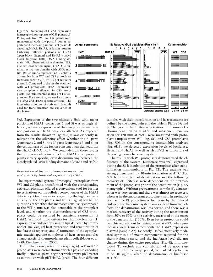
5A). Expression of the two chimeric Hsfs with majorportions of HsfA1 (constructs 2 and 3) was strongly si-lenced, whereas expression of the two proteins with mi-nor portions of HsfA1 was less affected. As expectedfrom the results shown in Figure 3, it was evidently ir-relevant for the silencing effect whether the 5� parts(constructs 2 and 5), the 3� parts (constructs 3 and 6), orthe central part of the fusion construct was derived fromthe HsfA1 cDNA (no. 4). We conclude from these resultsthat the gene-silencing effect in HsfA1 cosuppressionplants is very specific, even discriminating between theclosely related DNA binding domains ofHsfA1 andHsfA2.
Restoration of thermotolerance in mesophyllprotoplasts by transient expression of HsfA2
The experimental system of mesophyll protoplasts fromWT and CS plants transformed with the correspondingactivator plasmids offered a convenient tool for furtherinvestigations on the cellular consequences of the HsfA1deficiency. Our observations regarding the high heat sen-sitivity of the CS plants and fruits (Fig. 4) led to thequestions of whether this increased sensitivity comparedto the WT plants was also detectable at the protoplastlevel, and whether the thermotolerance of CS3 proto-plasts could be restored by transient expression ofHsfA2. We used three criteria for thermotolerance: (1)expression of endogenous small Hsps detected by immu-noblot analysis, (2) heat protection and renaturation ofluciferase as reporter, and (3) formation of the cytoplas-mic multichaperone complexes of heat stress granulescharacteristic of thermotolerant plant cells (Nover et al.1989; Kirschner et al. 2000).For the luciferase protection assay (Fig. 6), WT and CS3
protoplasts were cotransformed with a plasmid encodingfirefly luciferase (pLuc) together with empty pRT vectoras control or with pRTHsfA2 (pA2). The four different
samples with their transformation and hs treatments aredefined by the pictographs and the table in Figure 6A andB. Changes in the luciferase activities in a course of a30-min denaturation at 41°C and subsequent renatur-ation for 120 min at 25°C, were measured with proto-plast samples from WT (Fig. 6C) and CS3 protoplasts(Fig. 6D). In the corresponding immunoblot analyses(Fig. 6E,F), we detected expression levels of luciferase,HsfA1, and HsfA2 as well as Hsp17-CI as indicators ofthe endogenous chaperone system.The results with WT protoplasts demonstrated the ef-
ficiency of the system. Luciferase was well expressedduring the 23-h incubation of the protoplasts after trans-formation (immunoblots in Fig. 6E). The enzyme wasstrongly denatured by 30-min incubation at 41°C (Fig.6C), but the extent of denaturation and the followingrecovery of luciferase were dependent on the pretreat-ment of the protoplasts prior to the denaturation (Fig. 6Apictographs). Without pretreatment (sample H), denatur-ation was very strong and there was almost no recovery,whereas in thermotolerant protoplasts with hs preinduc-tion (sample P), protection of luciferase by the inducedendogenous chaperone system was evident from two ef-fects: the denaturation was less severe, and there was amarked recovery of the activity during the renaturationfrom 30% to 50% of the activity, measured at the onsetof the denaturation (100%). Even better protection couldbe achieved without hs pretreatment at 40°C when pro-toplasts were transformed with the HsfA2 expressionplasmid (sample A2). Evidently, HsfA2 effectively medi-ated synthesis of major components required for thethermotolerant state, and the luciferase level did notchange during the entire procedure (Fig. 6E, immuno-blots). To exclude any contribution of de novo syn-thesis in the recovery period, we added cyclohexi-mide (10 µg/mL) after the denaturation of luciferaseat 41°C.
Figure 5. Silencing of HsfA1 expressionin mesophyll protoplasts of CS3 plants. (A)Protoplasts from WT and CS3 plants weretransformed with the phsp17*gus as re-porter and increasing amounts of plasmidsencoding HsfA1, HsfA2, or fusion proteinsharboring different portions of HsfA1(open block diagram) and HsfA2 (shadedblock diagram). DBD, DNA binding do-main; HR, oligomerization domain, NLS,nuclear localization signal; CTAD, C-ter-minal activation domain with AHA mo-tifs. (B) Columns represent GUS activityof samples from WT and CS3 protoplaststransformed with 2, 5, or 10 µg of activatorplasmid. Compared to the results obtainedwith WT protoplasts, HsfA1 expressionwas completely silenced in CS3 proto-plasts. (C) Immunoblot analysis of Hsf ex-pression. For detection, we used a mixtureof HsfA1 and HsfA2 specific antisera. Theincreasing amounts of activator plasmidsused for transformation are explained atthe bottom.
Mishra et al.
1560 GENES & DEVELOPMENT
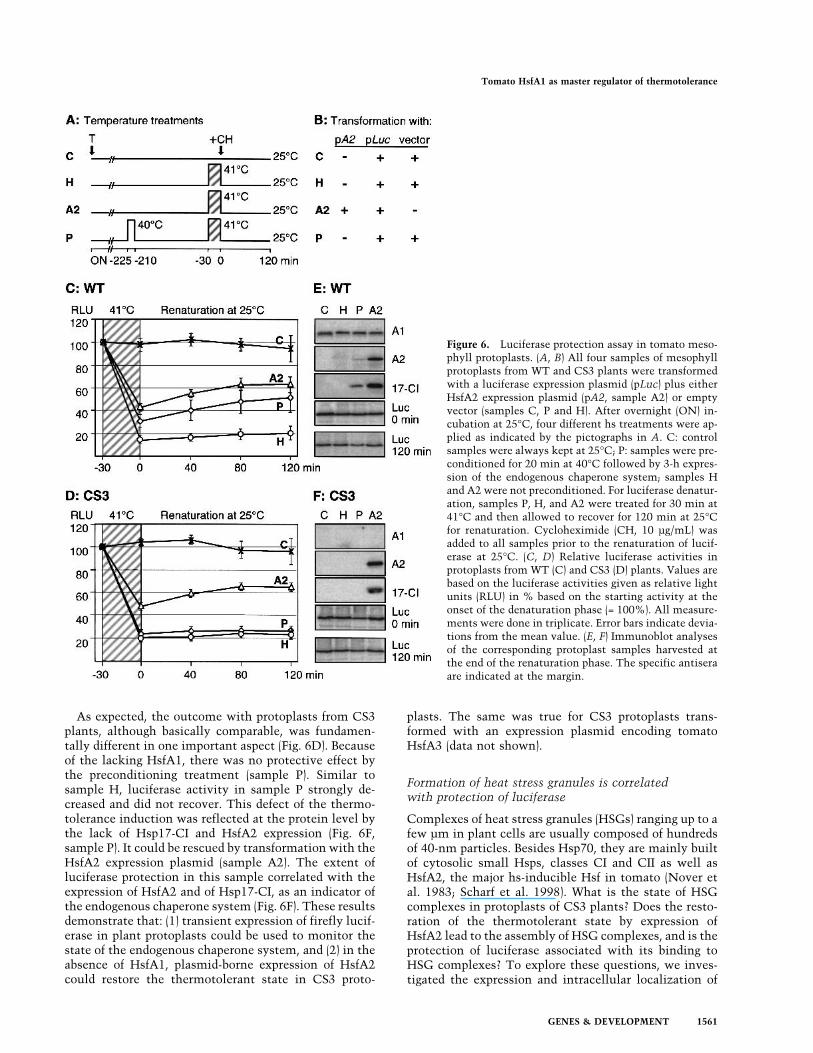
As expected, the outcome with protoplasts from CS3plants, although basically comparable, was fundamen-tally different in one important aspect (Fig. 6D). Becauseof the lacking HsfA1, there was no protective effect bythe preconditioning treatment (sample P). Similar tosample H, luciferase activity in sample P strongly de-creased and did not recover. This defect of the thermo-tolerance induction was reflected at the protein level bythe lack of Hsp17-CI and HsfA2 expression (Fig. 6F,sample P). It could be rescued by transformation with theHsfA2 expression plasmid (sample A2). The extent ofluciferase protection in this sample correlated with theexpression of HsfA2 and of Hsp17-CI, as an indicator ofthe endogenous chaperone system (Fig. 6F). These resultsdemonstrate that: (1) transient expression of firefly lucif-erase in plant protoplasts could be used to monitor thestate of the endogenous chaperone system, and (2) in theabsence of HsfA1, plasmid-borne expression of HsfA2could restore the thermotolerant state in CS3 proto-
plasts. The same was true for CS3 protoplasts trans-formed with an expression plasmid encoding tomatoHsfA3 (data not shown).
Formation of heat stress granules is correlatedwith protection of luciferase
Complexes of heat stress granules (HSGs) ranging up to afew µm in plant cells are usually composed of hundredsof 40-nm particles. Besides Hsp70, they are mainly builtof cytosolic small Hsps, classes CI and CII as well asHsfA2, the major hs-inducible Hsf in tomato (Nover etal. 1983; Scharf et al. 1998). What is the state of HSGcomplexes in protoplasts of CS3 plants? Does the resto-ration of the thermotolerant state by expression ofHsfA2 lead to the assembly of HSG complexes, and is theprotection of luciferase associated with its binding toHSG complexes? To explore these questions, we inves-tigated the expression and intracellular localization of
Figure 6. Luciferase protection assay in tomato meso-phyll protoplasts. (A, B) All four samples of mesophyllprotoplasts from WT and CS3 plants were transformedwith a luciferase expression plasmid (pLuc) plus eitherHsfA2 expression plasmid (pA2, sample A2) or emptyvector (samples C, P and H). After overnight (ON) in-cubation at 25°C, four different hs treatments were ap-plied as indicated by the pictographs in A. C: controlsamples were always kept at 25°C; P: samples were pre-conditioned for 20 min at 40°C followed by 3-h expres-sion of the endogenous chaperone system; samples Hand A2 were not preconditioned. For luciferase denatur-ation, samples P, H, and A2 were treated for 30 min at41°C and then allowed to recover for 120 min at 25°Cfor renaturation. Cycloheximide (CH, 10 µg/mL) wasadded to all samples prior to the renaturation of lucif-erase at 25°C. (C, D) Relative luciferase activities inprotoplasts from WT (C) and CS3 (D) plants. Values arebased on the luciferase activities given as relative lightunits (RLU) in % based on the starting activity at theonset of the denaturation phase (= 100%). All measure-ments were done in triplicate. Error bars indicate devia-tions from the mean value. (E, F) Immunoblot analysesof the corresponding protoplast samples harvested atthe end of the renaturation phase. The specific antiseraare indicated at the margin.
Tomato HsfA1 as master regulator of thermotolerance
GENES & DEVELOPMENT 1561
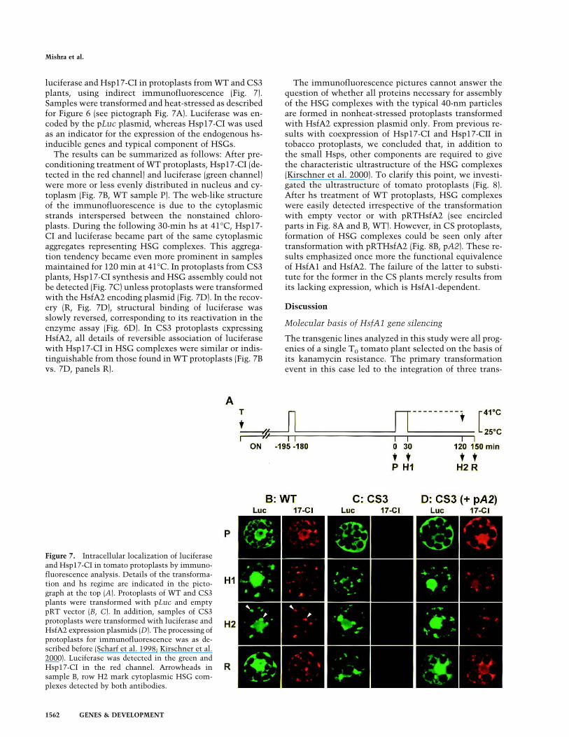
luciferase and Hsp17-CI in protoplasts fromWT and CS3plants, using indirect immunofluorescence (Fig. 7).Samples were transformed and heat-stressed as describedfor Figure 6 (see pictograph Fig. 7A). Luciferase was en-coded by the pLuc plasmid, whereas Hsp17-CI was usedas an indicator for the expression of the endogenous hs-inducible genes and typical component of HSGs.The results can be summarized as follows: After pre-
conditioning treatment of WT protoplasts, Hsp17-CI (de-tected in the red channel) and luciferase (green channel)were more or less evenly distributed in nucleus and cy-toplasm (Fig. 7B, WT sample P). The web-like structureof the immunofluorescence is due to the cytoplasmicstrands interspersed between the nonstained chloro-plasts. During the following 30-min hs at 41°C, Hsp17-CI and luciferase became part of the same cytoplasmicaggregates representing HSG complexes. This aggrega-tion tendency became even more prominent in samplesmaintained for 120 min at 41°C. In protoplasts from CS3plants, Hsp17-CI synthesis and HSG assembly could notbe detected (Fig. 7C) unless protoplasts were transformedwith the HsfA2 encoding plasmid (Fig. 7D). In the recov-ery (R, Fig. 7D), structural binding of luciferase wasslowly reversed, corresponding to its reactivation in theenzyme assay (Fig. 6D). In CS3 protoplasts expressingHsfA2, all details of reversible association of luciferasewith Hsp17-CI in HSG complexes were similar or indis-tinguishable from those found in WT protoplasts (Fig. 7Bvs. 7D, panels R).
The immunofluorescence pictures cannot answer thequestion of whether all proteins necessary for assemblyof the HSG complexes with the typical 40-nm particlesare formed in nonheat-stressed protoplasts transformedwith HsfA2 expression plasmid only. From previous re-sults with coexpression of Hsp17-CI and Hsp17-CII intobacco protoplasts, we concluded that, in addition tothe small Hsps, other components are required to givethe characteristic ultrastructure of the HSG complexes(Kirschner et al. 2000). To clarify this point, we investi-gated the ultrastructure of tomato protoplasts (Fig. 8).After hs treatment of WT protoplasts, HSG complexeswere easily detected irrespective of the transformationwith empty vector or with pRTHsfA2 (see encircledparts in Fig. 8A and B, WT). However, in CS protoplasts,formation of HSG complexes could be seen only aftertransformation with pRTHsfA2 (Fig. 8B, pA2). These re-sults emphasized once more the functional equivalenceof HsfA1 and HsfA2. The failure of the latter to substi-tute for the former in the CS plants merely results fromits lacking expression, which is HsfA1-dependent.
Discussion
Molecular basis of HsfA1 gene silencing
The transgenic lines analyzed in this study were all prog-enies of a single T0 tomato plant selected on the basis ofits kanamycin resistance. The primary transformationevent in this case led to the integration of three trans-
Figure 7. Intracellular localization of luciferaseand Hsp17-CI in tomato protoplasts by immuno-fluorescence analysis. Details of the transforma-tion and hs regime are indicated in the picto-graph at the top (A). Protoplasts of WT and CS3plants were transformed with pLuc and emptypRT vector (B, C). In addition, samples of CS3protoplasts were transformed with luciferase andHsfA2 expression plasmids (D). The processing ofprotoplasts for immunofluorescence was as de-scribed before (Scharf et al. 1998; Kirschner et al.2000). Luciferase was detected in the green andHsp17-CI in the red channel. Arrowheads insample B, row H2 mark cytoplasmic HSG com-plexes detected by both antibodies.
Mishra et al.
1562 GENES & DEVELOPMENT

gene cassettes. One of them is strongly expressed and, ifpresent alone, gives rise to HsfA1 overexpression plants.The other two cassettes are the result of integration of atandem inverted repeat causing the cosuppression effect,which is dominant over the overexpression effect.Hence, in the CS3 plants with all three transgene cas-settes, HsfA1 expression is strongly reduced, almost tothe level of CS2 plants (Fig. 1). The efficiency of thecosuppression machinery in CS3 plants is strong enoughto silence expression of HsfA1 in mesophyll protoplastseven when transformed with very high amounts of theHsfA1 expression plasmids (Fig. 5).Phenomena of gene silencing are widespread in plants
and other eukaryotes. Recent findings indicate that pe-culiarities of transgene arrangements, most frequentlyobserved with integration of direct or inverted repeats,lead to the generation of aberrant RNA and formation ofdsRNA. These RNAs can be processed to siRNAs re-sponsible for the ATP-dependent, sequence-specific deg-radation of mRNA (Wolters and Visser 2000; Ambros2001; Elbashir et al. 2001; Lipardi et al. 2001; Sharp2001). The detection of small RNAs of about 21-nt byboth sense and antisense riboprobes ofHsfA1 in CS2 andCS3 tomato and the lack of such siRNAs in WT and OEplants (Fig. 3) strongly support their decisive role for thecosuppression phenomenon. Due to the dominant epige-netic components, the extent of gene silencing in plantsis dynamic and may be influenced by the developmentalstate and growth conditions (Meins 2000). This providesa possible explanation for the differences in the strengthof the cosuppression effects in leaves versus pericarp ofCS plants (Fig. 1).
The binding of luciferase to the HSG complexes
Assembly of HSG complexes is characteristic for heat-stressed plant cells. These cytoplasmic complexes, com-prised of electron-dense 40-nm particles, are most promi-nent in cells with high capacity for Hsp synthesis, forexample meristematic cells, rapidly growing cell suspen-sion cultures, or mesophyll protoplasts (Nover et al.1983, 1989; Neumann et al. 1984). Although the domi-
nant components are the cytosolic sHsps, additional hs-inducible proteins, such as Hsp70 and HsfA2, are alsoinvolved in the assembly of these characteristic struc-tures (Neumann et al. 1987; Scharf et al. 1998; Kirschneret al. 2000).As expected from the lack of essential components,
formation of HSG complexes was not observed in heat-stressed protoplasts derived from the CS3 plants unlesstransformed with HsfA2 expression plasmids (Figs. 7, 8).Despite the lack of HsfA1, all components necessary forHSG assembly were evidently synthesized in the pres-ence of HsfA2. Under these conditions, protection of lu-ciferase from irreversible heat damage was restored, andthis was associated with the formation of the HSG com-plexes. By indirect immunofluorescence, we found a ma-jor part of luciferase bound to the HSG complexes (Fig.7). Hence, the multichaperone complexes in the cyto-plasm may indeed form a type of matrix for stabilizationof denatured proteins, as predicted earlier for small Hspsin general (Kimpel and Key 1985). Renaturation requirescooperation with ATP-dependent chaperone systems, forexample the Hsp70, Hsp90, and/or Hsp104 systems (For-reiter et al. 1997; Lee et al. 1997; Lee and Vierling 2000).It must be noted that the capability of sHsps to serve asreservoirs for intermediates of protein folding or dena-tured proteins created by hs is a general property of theseproteins and does not necessarily require assembly ofHSG complexes. It was also observed with individualmembers of the sHsp family, including those from yeastand animals, and it is associated with profound structuralrearrangements of the regular oligomers to irregular highmolecular weight aggregates incorporating the substrateproteins (Ehrnsperger et al. 1997, 1999; Löw et al. 2000).
The role of tomato HsfA1 as the master regulatorand the complexity of the Hsf family
TransgenicArabidopsis or carrot with altered expressionof hs-inducible genes have been reported. Two differentexperimental approaches were selected: (1) Expression offusion proteins of AtHsfA1a (Hsf1) with GUS in Arabi-dopsis (Lee et al. 1995) caused constitutive expression of
Figure 8. Restoration of HSG complexes in CS pro-toplasts by transformation with pRTHsfA2. Proto-plasts were transformed with empty vector or withpRTHsfA2 (pA2). After 16 h of cultivation they werepreconditioned, and after another 3-h cultivation at25°C they were subjected to a second hs for 2 h at40°C (see pictograph Fig. 7A, sample H2). Processingof protoplasts for electron microscopy was describedby Kirschner et al. (2000). Cp, chloroplast; Mi, mi-tochondria; Nu, nucleus; V vacuole. Bar represents0.5 µm. HSG complexes are circled.
Tomato HsfA1 as master regulator of thermotolerance
GENES & DEVELOPMENT 1563
sHsps and increased thermotolerance comparable to thephenotype observed with our tomato OE lines. (2) Trans-formation of Arabidopsis with antisense cassettes ofHsp70 (Lee and Schöffl 1996) or Hsp104 (Queitsch et al.2000) as well as transformation of carrot with Hsp17antisense cassettes (Malik et al. 1999) led to more or lesspronounced reductions of the chaperone expression lev-els. In all cases, the observed effects on thermotolerancesupport the view that a complex network of interactinghs-inducible proteins is necessary and that the lack ordiminished expression of any one of the major chaper-ones affects the development of thermotolerance. This isalso true for mutants of Arabidopsis with defects ofHsp104 expression (Hong and Vierling 2000, 2001). Todate, screening for thermosensitive mutants of Arabi-dopsis (Severin et al. 1995; Hong and Vierling 2000) forthe identification of a regulatory gene which might en-code a master regulator of the hs response have beenunsuccessful.Considering this situation, the effects of posttranscrip-
tional silencing of the HsfA1 gene in the CS plants oftomato were totally unexpected. The strongly reduced orundetectable expression of HsfA1 resulted in a generallack of induced Hsp synthesis and thermotolerance as-sociated with severe defects in growth and development,even under mild hs conditions, whereas the wild-typeplants and fruits were not visibly affected (Figs. 1, 4). CSplants were completely normal under standard green-house conditions. In this context, it is worth recallingthat transgenic tomato plants with strongly reduced orabsent expression of HsfA2 or HsfB1 had no comparabledefects in the hs response (data not shown). Evidently,the presence of HsfA1 is sufficient to ensure Hsp syn-thesis. The two hs-inducible Hsfs A2 and B1 certainlyinfluence details of the gene expression, but they areprobably not indispensable for the thermotolerant state.
Functional diversification of tomato Hsfs
The multiplicity of plant Hsfs leads to the question ofredundancy versus functional diversification of the indi-vidual members. Based on our results with the CS plants,we must conclude that the deficiency of HsfA1 expres-sion cannot be complemented by any of the other closelyrelated tomato Hsfs. Although our knowledge is stillvery limited, the functional analysis of the four tomatoHsfs cloned so far supports the concept of a marked di-versification (Scharf et al. 1990, 1993, 1998; Treuter et al.1993; Boscheinen et al. 1997; Lyck et al. 1997; Bharti etal. 2000; Döring et al. 2000; Heerklotz et al. 2001). Evi-dently, each of these Hsfs has its own role in a complexnetwork determined by the pattern of expression, intra-cellular localization, oligomerization behavior, activatorfunction, and interaction of Hsfs with other proteins (forreview, see Nover et al. 2001). Tomato HsfB1 is particu-larly remarkable because it has no activator function ofits own but acts as a strong synergistic coactivator ifcombined with other class A Hsfs (K. Bharti and P.Döring unpubl.).The dominant phenotype of the HsfA1 cosuppression
plants adds another example to this diversification. Part
of the explanation for the unique role of HsfA1 as masterregulator is based on the observation that in the cosup-pression plants, expression of the two hs-inducible HsfsA2 and B1 was strongly reduced or lacking, together withthe expression of other hs-inducible proteins. It is tempt-ing to speculate about a simple sequential model of hs-induced gene expression involving HsfA2 and HsfB1 asHsfA1-dependent enhancer or modifier of Hsp synthesis.In support of such a model, HsfA2 was characterized as apotent activator of reporter gene expression in general(Treuter et al. 1993; Scharf et al. 1998; Döring et al. 2000;Heerklotz et al. 2001). Moreover, in the test situation ofprotoplasts from CS plants, the defect of the hs responsecould be repaired by plasmid-borne HsfA2 expression.The efficiency of HsfA2 in these restoration assays (Figs.6–8) was not anticipated. Usually, the strong nuclear ex-port signal at the C terminus of HsfA2 precludes a sig-nificant retention in the nucleus unless it exists in het-erooligomers with HsfA1 (Scharf et al. 1998; Heerklotzet al. 2001). We assume that other class A Hsfs of tomatomay fulfill this function. However, due to the relativelylow expression levels, these unidentified coactivators arenot able to replace HsfA1 to induce thermotolerance.Unfortunately, this interesting aspect can only be clari-fied when specific antisera become available to investi-gate the expression of other members of the tomato Hsffamily in more detail. Although the existence of ESTs formore than 17 Hsfs in tomato (Nover et al. 2001) and ourpreliminary RT–PCR analyses (data not shown) demon-strate that the genes are transcribed under nonstress con-ditions, it does not necessarily indicate that the corre-sponding proteins accumulate to levels comparable tothose detected for HsfA1, HsfA2, and HsfB1 (Bharti et al.2000). This is supported by the results from the electro-phoretic mobility shift assay (Fig. 2), with no detectablesignal in the samples from CS2 and CS3 plants.Although HsfA2 is functionally equivalent to HsfA1
in the experimental situation of mesophyll protoplastsderived from CS plants (Figs. 6–8), it cannot substitutefor HsfA1 as master regulator, because its expression inwhole plants is dependent on HsfA1 (Fig. 1). These me-sophyll protoplasts of CS plants represent a valuable testsystem for our understanding of the functional diversifi-cation of other members of the Hsf family. Effects ofdifferent Hsfs and Hsf combinations can be easily inves-tigated not only with appropriate reporter constructs butabove all in the complex cellular response of Hsf-inducedthermotolerance, as exemplified by the results shown inFigures 6–8.
Materials and methods
General materials and methods
Rabbit antisera against tomato HsfA1, HsfA2, HsfB1, Hsp17class CI, and Hsp70 were described previously (Lyck et al. 1997;Kirschner et al. 2000). Luciferase antiserum was obtained fromSigma. The Hsp104 antiserum was kindly provided by AnilGrover (University South Campus, New Delhi, India). Second-ary antibodies against rabbit immunoglobulins conjugated withhorseradish peroxidase or fluorescent dyes CY2 or CY3 wereobtained from Sigma and Dianova, respectively.
Mishra et al.
1564 GENES & DEVELOPMENT
Standard procedures were used for cloning (Ausubel et al.1993; Sambrook and Russell 2001). Plant expression vectors arederivatives of pRT101 (Töpfer et al. 1988). For convenient dele-tions or combinations of functional parts of HsfA1 and HsfA2,we generated unique SalI sites in different regions of the cDNAsintroduced by site-directed mutagenesis (Treuter et al. 1993;Döring et al. 2000). The principle modular structures of thechimeric proteins of Hsfs A1 and A2 are given in the blockdiagrams in Figure 5A.For indirect immunofluorescence of protoplasts, we followed
the procedures described by Scharf et al. (1998) and Heerklotz etal. (2001). Fixation of protoplasts was done with 3.7% paraform-aldehyde in microtubule stabilizing buffer (100 mM PIPESbuffer at pH 6.8, 2 mM EGTA, 1 mM MgSO4). Preparation ofprotoplasts for electron microscopy was as described by Kirsch-ner et al. (2000).
Generation and selection of transgenic plants
For transformation of tomato (Lycopersicon esculentumcv. Moneymaker), the Agrobacterium tumefaciens strainGV3101(pMP90) was used as described (Koncz and Schell 1986).The binary vector pGPTV-KAN (Becker et al. 1992) was modi-fied by removing the �-glucuronidase (uid A) gene and insertionof the HindIII fragment containing the HsfA1 cDNA expressioncassette from the corresponding pRT vector (Scharf et al. 1990).In the construct used for transformation, the expression cassetteofHsfA1with the cauliflower mosaic virus (CaMV) 35S promoterpoints towards the right T-DNA border (RB) and therefore oppo-site to the direction of the NPTII selection marker gene, whoseexpression is under the control of the NOS promoter (Fig. 1A).Leaf disc transformation and plant regeneration were done as
described (Knapp et al. 1994). Calli were generated on Mu-rashige-Skoog nutrient medium (Duchefa) containing 100 µg/mL kanamycin and 500 µg/mL carbenicillin. For shoot induc-tion, 2 µg/mL zeatin (Duchefa) was added. After root formation,regenerated T0 plants were transferred to soil and further grownin the greenhouse under 16-h light/8-h dark at 24°C and 18°C,respectively. Segregation of the T0 lines was followed for at leastthe next three generations (F1 to F3) by kanamycin resistancetests and Southern analysis.The kanamycin resistance test was performed about 2 wk
after germination by spraying plants on 3 consecutive d with asolution containing 0.5 mg/mL kanamycin sulfate and 0.05%sapogenate as detergent (Clarion). Four days later, the youngleaves of sensitive plants turn yellow, whereas leaves of trans-genic plants are resistant to the antibiotic and remain green.
Southern analysis
Genomic DNA was extracted by the CTAB buffer method asdescribed by Ausubel et al. (1993). Ten micrograms of the DNAper sample was digested overnight, separated on 1.5% agarosegels and pressure blotted (Stratagene) to positively charged ny-lon membrane (Roche). Southern hybridization was performedfollowing the manufacturer’s protocol (Roche). Primers used forPCR generation of the DIG-labeled probes were the following:PrHsfA1 F, 5�-GCACCTGCTTAAAAGTATAAGAAGTCGG-3�;PrHsfA1 R, 5�-CCTGAAGAGTGACTCCTGAAACAC-3�; PrN-ptII F, 5�-CCTGTCATCTCACCTTGCTCCTGCC-3�; PrNptIIR, 5�-CGATACCGTAAAGCACGAGGAAGC-3�.
Northern analysis
Total RNA from leaves and fruit pericarp was prepared with theguanidinium thiocyanate procedure (Ausubel et al. 1993). Fif-teen micrograms of total RNA was separated on denaturing
1.2% agarose gels and pressure blotted (Stratagene) to positivelycharged nylon membrane (Amersham Pharmacia). Hybridiza-tion using DIG-labeled probes was performed at 55°C overnightfollowing the manufacturer’s protocol (Roche).The primers used to generate DIG-labeled probes were: PrHsfA2
F, 5�-GGATCCTGACAGATGGGAATTTGCG-3�; PrHsfA2 R,5�-CCATTTGCTGTCACAACAGAATCCG-3�; PrHsfB1 F, 5�-GGATGACATAGGTTCAAGTTCTACC-3�; PrHsfB1R, 5�-GCGAAGACATTTTCATCCAAGGACC-3�; PrHsp70 F, 5�-GGTCCTGACATGGCTGGTGG-3�; PrHsp70 R, 5�-GTACATAAGAAGATAAGTTTGGC-3�; PrHsp17-CI F, 5�-TATTGAAATCTCTGGTTAAAACTTCATT TGT-3�; PrHsp17-CI R, 5�-GCCCGGGGGATCTCAAAAGGACTAG TATTTCATATT-3�.The DIG-labeled probe for the Northern analysis of HsfA1
was the same as that used for the Southern analysis.
Electrophoretic mobility shift assay
Details of the electrophoretic mobility shift assays includingthe HSE3 oligonucleotides were described previously (Scharf etal. 1990; Boscheinen et al. 1997). For preparation of nuclei fromleaf tissue, we used the procedure of Jensen et al. (1988). Pro-teins from the nuclear preparations were extracted withNEB500 buffer (25 mM HEPES, 500 mM NaCl, 5 mM MgCl2, 1mM EDTA, 10 mMNaF, 0.2%NP-40, and a cocktail of proteaseinhibitors from Roche). One nanogram of 32P-labeled HSE3 oli-gonucleotide were incubated for 20 min at 25°C with 5 µg ofnuclear protein extracts; samples were separated on 5% nativepolyacrylamide gels, and the position of radioactivity was de-tected by 18-h exposure to x-ray films. The two oligonucleotidesused for competition were 5�-GCCAGAAGCTTCTAGAAAGC-3� (wild-type HSE3c) and 5�-GCCATAAGCTTGTACAAAGC-3� (mutant HSE3c, changed nucleotides underlined).
Heat stress and protein analysis of transgenic plantsand fruits
Four- to six-week-old plants were heat stressed for 1 h at 45°Cin a climate chamber, followed by 2-h incubation at 25°C. Up-per 2–3 young leaves were collected and stored at −80°C. Maturegreen fruits were heat stressed at 42°C for 4 h in the climatechamber. Fruit pericarp was separated and stored at −80°C. Forprotein extraction, the tissues were ground in liquid N2. Next,100–200mg of the frozen powder was transferred into Eppendorftubes and sonicated in two volumes of NEB500 buffer. Onevolume of protein extract containing about 30 µg of protein washeated with an equal volume of 2× SDS sample buffer and thenseparated on 12% SDS-polyacrylamide gels. Because of thelower protein content, the NEB500 extracts of fruit pericarpwere precipitated with 4 volumes of acetone, and proteins weredissolved in 1× SDS sample buffer. For the immunoblot analy-sis, proteins were transferred to 45 µm nitrocellulose membrane(Schleicher and Schuell) and further processed for chemilumi-nescence detection following the manufacturer’s protocol(NEN).
Isolation of small interfering RNA (siRNA) and hybridization
For the isolation of soluble RNA, large RNAs were precipitatedby LiCl. The supernatant was overlaid with 3 vol of ethanol andlarge genomic DNA was spooled out. After precipitation over-night at −20°C, the pellet of small RNA was dissolved in 200 µLwater, residual DNA was removed by treatment with DNaseI(Roche Diagnostics), followed by Proteinase K and phenol-chlo-roform treatment. After precipitation with 3 vol of ethanol,small RNAs were redissolved in 200 µL water.
Tomato HsfA1 as master regulator of thermotolerance
GENES & DEVELOPMENT 1565
Electrophoresis of siRNA on 15% polyacrylamide 7 M ureagels, transfer on positively charged nylon membrane (Amer-sham Pharmacia Biotech), and hybridization were as describedby Hamilton and Baulcombe (1999). The transfer was done by asemidry method at constant current (1 mA/cm2 of the mem-brane) for 2 h using 1× TBE buffer similar to that used duringelectrophoresis. For hybridization, [32P]UTP-labeled riboprobeswere generated by in vitro transcription (Promega) of an HsfA1cassette flanked by a T7 promoter (sense transcript) and T3promoter (antisense transcript). The riboprobes were hydro-lyzed at 60°C for 90 min using sodium carbonate buffer (pH10.2) to an average length of 50–100 nucleotides. For size markerand control of strand-specific hybridization, we used synthetic23-nt sense and antisense ribooligonucleotides corresponding tothe N terminus of theHsfA1 cassette (BioSpring). Hybridizationwas done at 37°C overnight.
Reporter constructs and assays with tomato protoplasts
The use of tomato mesophyll protoplasts for polyethylene gly-col (PEG)-mediated transformation and transient gene expres-sion is based on the published vectors and procedures for to-bacco protoplasts (Treuter et al. 1993; Scharf et al. 1998). Theonly major difference concerns the protoplasting procedure,which was done in K3M instead of K3S solution; that is, 0.4 Msucrose was replaced by 0.4 M mannitol. After protoplastingovernight, 1 vol of K3S (with a twofold concentration of sucrose)was added, and protoplasts were enriched by centrifugation at500 rpm.For the GUS reporter assay, we used the phsp17*gus vector
(Treuter et al. 1993; Heerklotz et al. 2001). For construction ofthe pLuc vector for the luciferase assay, we replaced the GFPcassette in pCRGFP vector (Reichel et al. 1996) by anNcoI/XbaIfragment of the Promega pGL3 basic vector encoding fireflyluciferase. The plant expression vector is a modified pRT vectorcontaining a duplicated CaMV35S enhancer and a tobacco etchvirus translational enhancer preceding the ATG start codon(Carrington et al. 1991).After PEG-mediated transformation with the indicated ex-
pression plasmids, protoplasts were incubated for 15 h at 25°Cunder dim light. The measurement of the �-glucuronidase(GUS) reporter activity is based on the method described byJefferson (1987) with a few modifications (Döring et al. 2000).For the luciferase reporter assay, denaturation was performed byheating protoplasts in a water bath for 30 min at 41°C. Cyclo-heximide was added immediately after the denaturation (finalconcentration 10 µg/mL), and cells were allowed to recover for2 h at 25°C. Luciferase activity was determined directly beforeand after the denaturation and at 20-min intervals during therecovery. At each timepoint, three parallel samples of the pro-toplast suspension containing 2500 protoplasts were transferredto a 96-well microtiter plate. The assay was performed using aMikrolumat LB 96P luminometer (EG&G Berthold). Followingthe injection of 100 µL of luciferase assay reagent (25 mMglycylglycine, 15 mM K-phosphate at pH 7.8, 15 mM MgCl2, 4mM EGTA, 1 mM DTT, 1 mM ATP, 40 µM luciferin), lightemission was measured for 20 sec (Forreiter et. al. 1997). Valueswere normalized to the activity measured before the denatur-ation at 41°C (equals 100%). Experiments were repeated at leastthree times.
Acknowledgments
We thank Gisela Englich, Daniela Bublak, and Christel Rülkefor excellent technical assistance, as well as Holger Schranz andGerald Kircher as members of the greenhouse team. We thankKapil Bharti, Markus Fauth, and Dirk Zielinski for many helpful
discussions during the preparation of the manuscript. This workwas supported by grants of the Bundesministerium für Bildungand Forschung to K.D.S. and of the Deutsche Forschungsge-meinschaft to L.N. as well as to S.K.M. as fellow of the Gradui-ertenkolleg.The publication costs of this article were defrayed in part by
payment of page charges. This article must therefore be herebymarked “advertisement” in accordance with 18 USC section1734 solely to indicate this fact.
References
Agashe, V.R. and Hartl, F.U. 2000. Roles of molecular chaper-ones in cytoplasmic protein folding. Semin. Cell Dev. Biol.11: 15–25.
Ambros, V. 2001. microRNAs: Tiny regulators with great po-tential. Cell 107: 823–826.
Ausubel, F.M., Brent, R., Kingston, R.E., Moore, D.D., Seidman,J.G., Smith, J.A., and Struhl, K. 1993. Current protocols inmolecular biology. John Wiley and Sons, New York, NY.
Becker, D., Kemper, E., Schele, J., and Masterson, R. 1992. Vec-tors with selectable markers located proximal to the leftT-DNA border. Plant Mol. Biol. 20: 1195–1197.
Bharti, K., Schmidt, E., Lyck, R., Bublak, D., and Scharf, K.-D.2000. Isolation and characterization of HsfA3, a new heatstress transcription factor of Lycopersicon peruvianum.Plant J. 22: 355–365.
Boscheinen, O., Lyck, R., Queitsch, C., Treuter, E., Zimarino,V., and Scharf, K.-D. 1997. Heat stress transcription factorsfrom tomato can functionally replace HSF1 in the yeast Sac-charomyces cerevisiae. Mol. Gen. Genet. 255: 322–331.
Carrington, J.C., Freed, D.D., and Leinicke, A.J. 1991. Bipartitesignal sequence mediates nuclear translocation of the plantpotyviral NIa protein. Plant Cell 3: 953–962.
Döring, P., Treuter, E., Kistner, C., Lyck, R., Chen, A., andNover, L. 2000. Role of AHA motifs for the activator func-tion of tomato heat stress transcription factors HsfA1 andHsfA2. Plant Cell 12: 265–278.
Ehrnsperger, M., Graber, S., Gaestel, M., and Buchner, J. 1997.Binding of non-native protein to Hsp25 during heat shockcreates a reservoir of folding intermediates for reactivation.EMBO J. 16: 221–229.
Ehrnsperger, M., Lilie, H., Gaestel, M., and Buchner, J. 1999.The dynamics of Hsp25 quarternary structure. J. Biol. Chem.274: 14867–14874.
Elbashir, S., Lendeckel, W., and Tuschl, T. 2001. RNA interfer-ence is mediated by 21- and 22-nucleotide RNAs. Genes &Dev. 15: 188–200.
Ellis, R.J. 2000. Chaperone substrates inside the cell. TrendsBiochem. Sci. 25: 210–212.
Forreiter, C., Kirschner, M., and Nover, L. 1997. Stable trans-formation of an Arabidopsis cell suspension culture withfirefly luciferase providing a cellular system for analysis ofchaperone activity in vivo. Plant Cell 9: 2171–2181.
Hamilton, A.J. and Baulcombe, D.C. 1999. A species of smallantisense RNA in posttranscriptional gene silencing inplants. Science 286: 950–952.
Heerklotz, D., Döring, P., Bonzelius, F., Winkelhaus, S., andNover, L. 2001. The balance of nuclear import and exportdetermines the intracellular distribution of tomato heat stresstranscription factor HsfA2. Mol. Cell. Biol. 21: 1759–1768.
Hong, S.W. and Vierling, E. 2000. Mutants of Arabidopsisthaliana defective in the acquisition of tolerance to hightemperature stress. Proc. Natl. Acad. Sci. 97: 4392–4397.
———. 2001. Hsp101 is necessary for heat tolerance but dis-
Mishra et al.
1566 GENES & DEVELOPMENT
pensable for development and germination in the absence ofstress. Plant J. 27: 25–35.
Jefferson, R.A. 1987. Assaying chimeric genes in plants: TheGUS gene fusion system. Plant Mol. Biol. Rep. 5: 387–405.
Jensen, E.O., Marcker, K.A., Schell, J., and de Bruijn, F.J. 1988.Interaction of a nodule specific, trans-acting factor with dis-tinct DNA elements in the soybean leghaemoglobin lbc3 5�
upstream region. EMBO J. 7: 1265–1271.Jolly, C. and Morimoto, R.I. 2000. Role of the heat shock re-
sponse and molecular chaperones in oncogenesis and celldeath. J. Natl. Cancer Inst. 92: 1564–1572.
Kimpel, J.A. and Key, J. 1985. Heat shock in plants. TrendsBiochem. Sci. 10: 353–357.
Kirschner, M., Winkelhaus, S., Thierfelder, J., and Nover, L.2000. Transient expression and heat stress induced aggrega-tion of endogenous and heterologous small heat stress pro-teins in tobacco protoplasts. Plant J. 24: 397–412.
Knapp, S., Larondelle, Y., Roßberg, M., Furtek, D., and Theres,K. 1994. Transgenic tomato lines containing Ds elements atdefined genomic positions as tools for targeted transposontagging. Mol. Gen. Genet. 243: 666–673.
Koncz, C. and Schell, J. 1986. The promoter of TL-DNA genecontrols the tissue-specific expression of chimaeric genescarried by a novel type of Agrobacterium binary vector.Mol.Gen. Genet. 204: 383–396.
Lee, G.J. and Vierling, E. 2000. A small heat shock protein co-operates with heat shock protein 70 systems to reactivate aheat-denatured protein. Plant Physiol. 122: 189–198.
Lee, G.J., Roseman, A.M., Saibil, H.R., and Vierling, E. 1997. Asmall heat shock protein stably binds heat-denatured modelsubstrates and can maintain a substrate in a folding-compe-tent state. EMBO J. 16: 659–671.
Lee, J.H. and Schöffl, F. 1996. An Hsp70 antisense gene affectsthe expression of HSP70/HSC70, the regulation of HSF, andthe acquisition of thermotolerance in transgenic Arabidop-sis thaliana. Mol. Gen. Genet. 252: 11–19.
Lee, J.H., Hubel, A., and Schöffl, F. 1995. Derepression of theactivity of genetically engineered heat shock factor causes con-stitutive synthesis of heat shock proteins and increased ther-motolerance in transgenic Arabidopsis. Plant J. 8: 603–612.
Lipardi, C., Wei, Q., and Paterson, B. M. 2001. RNAi as randomdegradative PCR: siRNA primers convert mRNA into dsRNAsthat are degraded to generate new siRNAs. Cell 107: 297–307.
Löw, D., Nover, L., and Forreiter, C. 2000. Cloning of cDNAsencoding cytosolic tomato Hsps Hsp17,7 class I and Hsp17,3class II and their functional characterization as molecularchaperones in vivo. Planta 211: 575–582.
Lyck, R., Harmening, U., Höhfeld, I., Treuter, E., Scharf, K.-D.,and Nover, L. 1997. Intracellular distribution and identifica-tion of the nuclear localization signals of two plant heat-stress transcription factors. Planta 202: 117–125.
Malik, M.K., Slovin, J.P., Hwang, C.H., and Zimmerman, J.L.1999. Modified expression of a carrot small heat shock pro-tein gene, hsp17.7, results in increased or decreased thermo-tolerance double dagger. Plant J. 20: 89–99.
Meins, Jr., F. 2000. RNA degradation and models for post-tran-scriptional gene silencing. Plant Mol. Biol. 43: 261–273.
Morimoto, R.I. 1998. Regulation of the heat shock transcrip-tional response: Cross talk between family of heat shockfactors, molecular chaperones, and negative regulators.Genes & Dev. 12: 3788–3796.
Neumann, D., Scharf, K.-D., and Nover, L. 1984. Heat shockinduced changes of plant cell ultrastructure and autoradio-graphic localization of heat shock proteins. Eur. J. Cell Biol.34: 254–264.
Neumann, D., zur Nieden, U., Manteuffel, R., Walter, G.,
Scharf, K.-D., and Nover, L. 1987. Intracellular localizationof heat shock proteins in tomato cells cultures. Eur. J. CellBiol. 43: 71–81.
Nover, L. 1987. Expression of heat shock genes in homologousand heterologous systems. Enzyme Microb. Technol. 9: 130–144.
Nover, L., Scharf, K.-D., and Neumann, D. 1983. Formation ofcytoplasmic heat shock granules in tomato cell cultures andleaves. Mol. Cell. Biol. 3: 1648–1655.
———. 1989. Cytoplasmic heat shock granules are formed fromprecursor particles and are associated with a specific set ofmRNAs. Mol. Cell. Biol. 9: 1298–1308.
Nover, L., Bharti, K., Döring, P., Mishra, S.K., Ganguli, A., andScharf, K.-D. 2001.Arabidopsis and the heat stress transcrip-tion factor world: How many heat stress transcription fac-tors do we need? Cell Stress Chaperones 6: 177–189.
Pelham, H.R.B. 1982. A regulatory upstream promoter elementin the Drosophila hsp70 heat-shock gene. Cell 30: 517–528.
Pelham, H.R.B. and Bienz, M. 1982. A synthetic heat-shock pro-moter element confers heat-inducibility on the Herpes sim-plex virus thymidine kinase gene. EMBO J. 1: 1473–1477.
Queitsch, C., Hong, S.-W., Vierling, E., and Lindquist, S. 2000.Heat shock protein 101 plays a crucial role in thermotoler-ance in Arabidopsis. Plant Cell 12: 479–492.
Reichel, C., Mathur, J., Eckes, P., Langenkemper, K., Koncz, C.,Schell, J., Reis, B., and Maas, C. 1996. Enhanced green fluo-rescence by the expression of an Aequorea victoria greenfluorescent protein mutant in mono- and dicotyledonousplant cells. Proc. Natl. Acad. Sci. 93: 5888–5893.
Richter, K. and Buchner, J. 2001. Hsp90: Chaperoning signaltransduction. J. Cell. Physiol. 188: 281–290.
Sambrook, J. and Russell, D.W. 2001. Molecular cloning: Alaboratory manual, 3rd ed. Cold Spring Harbor LaboratoryPress, Cold Spring Harbor, NY.
Scharf, K.-D., Rose, S., Zott, W., Schöffl, F., and Nover, L. 1990.Three tomato genes code for heat stress transcription factorswith a region of remarkable homology to the DNA-bindingdomain of the yeast HSF. EMBO J. 9: 4495–4501.
Scharf, K.-D., Rose, S., Thierfelder, J., and Nover, L. 1993. TwocDNAs for tomato heat stress transcription factors. PlantPhysiol. 102: 1355–1356.
Scharf, K.-D., Heider, H., Höhfeld, I., Lyck, R., Schmidt, E., andNover, L. 1998. The tomato Hsf system: HsfA2 needs inter-action with HsfA1 for efficient nuclear import and may belocalized in cytoplasmic heat stress granules.Mol. Cell. Biol.18: 2240–2251.
Severin, K., Wagner, A., and Schöffl, F. 1995. A heat-inducibleAdh gene as a reporter for a negative selection in transgenicArabidopsis. Transgenic Res. 4: 163–172.
Sharp, P.A. 2001. RNA interference-2001. Genes & Dev.15: 485–490.
Sheng, J. and Citovsky, V. 1996. Agrobacterium-plant cell DNAtransport: Have virulence proteins, will travel. Plant Cell8: 1699–1710.
Töpfer, R., Schell, J., and Steinbiss, H.H. 1988. Versatile cloningvectors for transient gene expression and direct gene transferin plant cells. Nucleic Acids Res. 16: 8725.
Treuter, E., Nover, L., Ohme, K., and Scharf, K.-D. 1993. Pro-moter specificity and deletion analysis of three heat stress transcription factors of tomato. Mol. Gen. Genet. 240: 113–125.
Wolters, A.-M.A. and Visser, R.G.F. 2000. Gene silencing inpotato: Allelic differences and effect of ploidy. Plant Mol.Biol. 43: 377–386.
Wu, C. 1995. Heat stress transcription factors. Annu. Rev. CellBiol. 11: 441–469.
Tomato HsfA1 as master regulator of thermotolerance
GENES & DEVELOPMENT 1567