In situ scanning probe microscopy studies of tetanus toxin-membrane interactions
10
In Situ Scanning Probe Microscopy Studies of Tetanus Toxin-Membrane Interactions Andrea L. Slade,* Joseph S. Schoeniger, y Darryl Y. Sasaki, z and Christopher M. Yip* *Department of Biochemistry, Department of Chemical Engineering and Applied Chemistry, Institute of Biomaterials and Biomedical Engineering, University of Toronto, Toronto, Ontario, Canada; y Sandia National Laboratories, Biosystems Research, Livermore, California; and z Sandia National Laboratories, Biomolecular Interfaces & Systems, Albuquerque, New Mexico ABSTRACT Despite the considerable information available with regards to the structure of the clostridial neurotoxins, and their inherent threat as biological warfare agents, the mechanisms underpinning their interactions with and translocation through the cell membrane remain poorly understood. We report herein the results of an in situ scanning probe microscopy study of the interaction of tetanus toxin C-fragment (Tet C) with supported planar lipid bilayers containing the ganglioside receptor G T1b . Our results show that Tet C preferentially binds to the surface of fluid phase domains within biphasic membranes containing G T1b and that with an extended incubation period these interactions lead to dramatic changes in the morphology of the lipid bilayer, including the formation of 40–80 nm diameter circular cavities. Combined atomic force microscopy/total internal reflection fluo- rescence microscopy experiments confirmed the presence of Tet C in the membrane after extended incubation. These mor- phological changes were found to be dependent upon the presence of G T1b and the solution pH. INTRODUCTION Recent concern regarding biological toxins, such as tetanus (TeNT) and botulinum neurotoxin (BoNT), has arisen from their potential use as biological weapons (1,2). Characterizing the dynamics of their interactions at the membrane interface may therefore provide insights into the development of new treatments, detection strategies, and prophylactic agents. The Clostridial neurotoxins are the most potent toxins known, with TeNT and BoNT responsible for the clinical symptoms of tetanus and botulism, respectively. These toxins target neuronal cells by binding to the trisialoganglioside G T1b in the presynaptic cell membrane of neuromuscular junctions (3,4), and are internalized into the cells where they ultimately inhibit neurotransmitter release (5). However, while BoNTs act primarily in the peripheral nervous system blocking ace- tylcholine release, TeNT undergoes axonal retrograde trans- port to the central nervous system where it targets inhibitory motor neurons (6,7). TeNT is synthesized by the bacterium Clostridium tetani as a single ;150 kDa polypeptide chain that must undergo posttranslational proteolytic cleavage to produce the biologically active, disulfide linked two-chain molecule that is composed of an ;50-kDa light chain (LC) and an ;100-kDa heavy chain (HC) (Fig. 1 a) (8). The bind- ing of tetanus toxin to G T1b in neuronal cell membranes is mediated by the ;50-kDa C-terminal region (H C ) of the heavy chain, known as the C fragment (Tet C) (Fig. 1 b). The ;50-kDa N-terminal region (H N ) facilitates the translocation of the LC, a zinc endopeptidase, into the cell cytosol. Once in the cytosol, the LC specifically cleaves the synaptic SNARE (soluble N-ethylmaleimide sensitive-factor attachment protein receptor) protein synaptobrevin, thereby preventing the fusion of neurotransmitter secretory vesicles to the nerve terminal membrane, and thus blocking the release of inhibitory neu- rotransmitters (9). In general, invasion of a host cell by the bacterial A n B m - toxins, which includes diphtheria toxin, cholera toxin, botu- linum A & B toxins, and tetanus toxin involves four general stages: 1), cell surface binding; 2), internalization; 3), mem- brane translocation into the cytosol of the host; and 4), modi- fication of a cytosolic target. With respect to this process of cell intoxication, the mechanisms of toxin internalization and membrane translocation are the least understood. While it is generally accepted that toxin molecules are internalized via endocytosis of the regions of the cell membrane to which they are bound, the mechanism of membrane translocation, however, appears to differ among the classes of toxin mole- cules. In the case of the TeNT, it is believed that following endocytosis, lowering of the pH inside the endosome triggers a conformational change that increases the hydrophobicity of the toxin molecule (10,11). This conformational change facilitates insertion of TeNT into the endosomal membrane with subsequent translocation of the light chain into the cy- tosol, where it exerts its catalytic activity. Indeed, it has been reported that A n B m -toxins having A 1 B 1 -stoichiometry, in- cluding the full-length tetanus toxin, form ion channels in planar lipid bilayers at acidic pH (12–18). To fully understand how toxins exert their action, it has been proposed that ‘‘trapping’’ the toxin molecules at differ- ent points along the invasion pathway from the cell surface to the cytosol, would allow for reconstruction of the entire mechanism (19). Although this approach has been applied successfully to characterize colicin Ia activity, it is extremely arduous and time intensive (20). Recently, in situ studies Submitted December 27, 2005, and accepted for publication August 22, 2006. Address reprint requests to Christopher M. Yip, IBBME, Rosebrugh Bldg., University of Toronto, 4 Taddle Creek Rd., Toronto, Ontario, Canada, M5S 3G9. Tel: 416-978-7853; Fax: 416-978-4317; E-mail: christopher.yip@ utoronto.ca. Ó 2006 by the Biophysical Society 0006-3495/06/12/4565/10 $2.00 doi: 10.1529/biophysj.105.080457 Biophysical Journal Volume 91 December 2006 4565–4574 4565
-
Upload
independent -
Category
Documents
-
view
3 -
download
0
Transcript of In situ scanning probe microscopy studies of tetanus toxin-membrane interactions

In Situ Scanning Probe Microscopy Studies of TetanusToxin-Membrane Interactions
Andrea L. Slade,* Joseph S. Schoeniger,y Darryl Y. Sasaki,z and Christopher M. Yip**Department of Biochemistry, Department of Chemical Engineering and Applied Chemistry, Institute of Biomaterials and BiomedicalEngineering, University of Toronto, Toronto, Ontario, Canada; ySandia National Laboratories, Biosystems Research, Livermore,California; and zSandia National Laboratories, Biomolecular Interfaces & Systems, Albuquerque, New Mexico
ABSTRACT Despite the considerable information available with regards to the structure of the clostridial neurotoxins, andtheir inherent threat as biological warfare agents, the mechanisms underpinning their interactions with and translocation throughthe cell membrane remain poorly understood. We report herein the results of an in situ scanning probe microscopy study of theinteraction of tetanus toxin C-fragment (Tet C) with supported planar lipid bilayers containing the ganglioside receptor GT1b. Ourresults show that Tet C preferentially binds to the surface of fluid phase domains within biphasic membranes containing GT1b
and that with an extended incubation period these interactions lead to dramatic changes in the morphology of the lipid bilayer,including the formation of 40–80 nm diameter circular cavities. Combined atomic force microscopy/total internal reflection fluo-rescence microscopy experiments confirmed the presence of Tet C in the membrane after extended incubation. These mor-phological changes were found to be dependent upon the presence of GT1b and the solution pH.
INTRODUCTION
Recent concern regarding biological toxins, such as tetanus
(TeNT) and botulinum neurotoxin (BoNT), has arisen from
their potential use as biological weapons (1,2). Characterizing
the dynamics of their interactions at the membrane interface
may therefore provide insights into the development of new
treatments, detection strategies, and prophylactic agents. The
Clostridial neurotoxins are the most potent toxins known,
with TeNT and BoNT responsible for the clinical symptoms
of tetanus and botulism, respectively. These toxins target
neuronal cells by binding to the trisialoganglioside GT1b in
the presynaptic cell membrane of neuromuscular junctions
(3,4), and are internalized into the cells where they ultimately
inhibit neurotransmitter release (5). However, while BoNTs
act primarily in the peripheral nervous system blocking ace-
tylcholine release, TeNT undergoes axonal retrograde trans-
port to the central nervous system where it targets inhibitory
motor neurons (6,7). TeNT is synthesized by the bacterium
Clostridium tetani as a single ;150 kDa polypeptide chain
that must undergo posttranslational proteolytic cleavage to
produce the biologically active, disulfide linked two-chain
molecule that is composed of an ;50-kDa light chain (LC)
and an ;100-kDa heavy chain (HC) (Fig. 1 a) (8). The bind-
ing of tetanus toxin to GT1b in neuronal cell membranes is
mediated by the ;50-kDa C-terminal region (HC) of the
heavy chain, known as the C fragment (Tet C) (Fig. 1 b). The
;50-kDa N-terminal region (HN) facilitates the translocation
of the LC, a zinc endopeptidase, into the cell cytosol. Once in
the cytosol, the LC specifically cleaves the synaptic SNARE
(soluble N-ethylmaleimide sensitive-factor attachment protein
receptor) protein synaptobrevin, thereby preventing the fusion
of neurotransmitter secretory vesicles to the nerve terminal
membrane, and thus blocking the release of inhibitory neu-
rotransmitters (9).
In general, invasion of a host cell by the bacterial AnBm-
toxins, which includes diphtheria toxin, cholera toxin, botu-
linum A & B toxins, and tetanus toxin involves four general
stages: 1), cell surface binding; 2), internalization; 3), mem-
brane translocation into the cytosol of the host; and 4), modi-
fication of a cytosolic target. With respect to this process of
cell intoxication, the mechanisms of toxin internalization and
membrane translocation are the least understood. While it is
generally accepted that toxin molecules are internalized via
endocytosis of the regions of the cell membrane to which
they are bound, the mechanism of membrane translocation,
however, appears to differ among the classes of toxin mole-
cules. In the case of the TeNT, it is believed that following
endocytosis, lowering of the pH inside the endosome triggers
a conformational change that increases the hydrophobicity of
the toxin molecule (10,11). This conformational change
facilitates insertion of TeNT into the endosomal membrane
with subsequent translocation of the light chain into the cy-
tosol, where it exerts its catalytic activity. Indeed, it has been
reported that AnBm-toxins having A1B1-stoichiometry, in-
cluding the full-length tetanus toxin, form ion channels in
planar lipid bilayers at acidic pH (12–18).
To fully understand how toxins exert their action, it has
been proposed that ‘‘trapping’’ the toxin molecules at differ-
ent points along the invasion pathway from the cell surface
to the cytosol, would allow for reconstruction of the entire
mechanism (19). Although this approach has been applied
successfully to characterize colicin Ia activity, it is extremely
arduous and time intensive (20). Recently, in situ studies
Submitted December 27, 2005, and accepted for publication August 22, 2006.
Address reprint requests to Christopher M. Yip, IBBME, Rosebrugh Bldg.,
University of Toronto, 4 Taddle Creek Rd., Toronto, Ontario, Canada, M5S
3G9. Tel: 416-978-7853; Fax: 416-978-4317; E-mail: christopher.yip@
utoronto.ca.
� 2006 by the Biophysical Society
0006-3495/06/12/4565/10 $2.00 doi: 10.1529/biophysj.105.080457
Biophysical Journal Volume 91 December 2006 4565–4574 4565

using scanning probe microscopy (SPM), and more specif-
ically, atomic force microscopy (AFM), have provided new
insights into the association and assembly of various pep-
tides and proteins, such as filipin (21), amphotericin B (22),
and melittin (23), on membrane surfaces, including their role
in inducing membrane reorganization and disruption. We
have used this technique to study the insertion and subse-
quent assembly of the amyloid-b 42 (Ab42) peptide (24),
a-synuclein (25), hemagglutinin (26), Bax protein (27), and
NAP22 (28) at membrane interfaces.
For such studies, supported planar lipid bilayers (SPBs) are
often used as membrane-mimetic surfaces. Typically prepared
in situ by free vesicle fusion onto a freshly cleaved mica
surface, SPBs retain many of the properties of free-standing
membranes, including lateral fluidity (29–31). Indeed, direct
fusion of receptor-containing lipid vesicles onto mica is a
particularly facile and attractive route of preparing model
membranes for AFM studies of ligand-receptor interactions.
We have used this approach in our earlier investigation of the
full-length transmembrane insulin receptor (IR) (32).
We report herein an in situ AFM study on the interactions
of the tetanus toxin binding domain (Tet C) with biphasic
supported planar lipid bilayers containing the ganglioside
membrane receptor GT1b. Our results revealed preferential
association of Tet C with the surface of the fluid phase re-
gions of the lipid bilayers, and that the affinity was depen-
dent upon the bilayer concentration of GT1b and solution pH.
Following an incubation period, the protein-bound regions
of the membrane became thicker and circular depressions
of 40–80 nm diameters appeared through the action of Tet C
upon the membrane. The presence of Tet C in these regions
of the bilayer was confirmed using combined atomic force
microscopy/total internal reflection fluorescence microscopy
(AFM/TIRFM) (33). From these studies, detailed insights into
the binding and activity of Tet C and subsequent restructuring
of the lipid bilayer were attained at nanoscale resolution.
MATERIALS AND METHODS
Materials
1,2-Dipalmitoleoyl-sn-glycero-3-phosphocholine (DPOPC; C16:1) was pur-
chased from Avanti Polar Lipids (Alabaster, AL). Trisialoganglioside GT1b,
tetanus toxin C fragment (Tet C), fluorescein isothiocyanate (FITC) la-
beled tetanus toxin C fragment (FITC-Tet C; 495 nm/525 nm), and DL-a-
dipalmitoyl-phosphatidylcholine (DPPC; C16:0) were purchased from
Sigma-Aldrich (Oakville, ON).
Liposome preparation
Appropriate molar quantities of lipid ((1:1) DPOPC/DPPC) and, when re-
quired, the desired amount of GT1b (10 mol % or 1 mol %) were dissolved in
chloroform. During preparation, fluorescent liposomes were protected from
light to minimize photobleaching. The solvent was removed by evaporation
under vacuum (;50�C) and the dried lipid films were rehydrated by the
addition of 5 mM (N-2-hydroxyethylpiperazine-N-2-ethanesulfonic acid)-
(2-[N-morpholino] ethanesulfonic acid)-citric acid (HEPES-MES-citric acid)
buffer (150 mM NaCl, pH 7.4) to give a final lipid concentration of 2 mM.
Unilamellar vesicles were then formed by sonication in a heated water bath
(;50�C) (Branson 200, Branson Ultrasonics, Danbury, CT) until the solu-
tion became clear or only slightly hazy. The resulting liposomes were found
to be ;145 nm in diameter by dynamic laser light scattering (Brookhaven
Instruments BI-200SM equipped with a BI-9000 AT digital correlator and
photon counter; Holtsville, NY). The fluorescent liposome solutions were
stored in the dark. All liposome solutions were stored at 4�C.
AFM imaging
Solution tapping mode AFM (TMAFM) images were acquired on a Digital
Instruments Nanoscope IIIa Multimode SPM (Santa Barbara, CA) equipped
with an ‘‘E’’ scanner (13.6 3 13.6 mm maximum lateral scan area) using
120-mm-long oxide-sharpened silicon nitride V-shaped model DNP-S canti-
levers installed in a combination contact/tapping mode liquid flow-cell
sealed against a freshly cleaved mica substrate. The typical tip oscillating
frequency was 7–9 kHz and was chosen to optimize image quality and
adjusted for the individual response characteristics of each cantilever. We
note that the nominal radius of curvature of the DNP-S tips used in these
studies was ;15 nm, and that for each experiment, a fresh tip was selected
and exposed to ultraviolet irradiation to remove any adventitious organic
contaminants prior to imaging. All AFM images were collected as 512 3
512 pixel data sets, at scan rates ranging from 1 to 2 Hz, at a scan angle of 0�to the fast scan axis, and at ambient temperature. For the Tet C studies on
mica, ;150 mL of a 5 mg/mL Tet C solution (10 mM PBS, 150 mM NaCl,
FIGURE 1 The structures of the clostridial neurotoxins: tetanus neuro-
toxin (TeNT) and botulinum neurotoxin (BoNT). All models are shown as
ribbon structures. (a) The active form of the clostridal neurotoxins is a two-
chain molecule composed of a disulfide-linked light chain (C) and heavy
chain (H). The light chain is the catalytic domain, while the C-terminal do-
main (HC) and the N-terminal domain (HN) of the heavy chain mediate
binding to neuronal cell membranes and translocation of the light chain, re-
spectively. (b) The C-terminal domain of tetanus toxin, otherwise known as
the Tet C-fragment. The last 34 residues of Tet C are involved in ganglioside
recognition (residues shown as CPK space-fill). The image is based on PDB
model 1FV2 and was prepared using the Swiss PDB Viewer molecular
graphics software package (GlaxoSmithKline).
4566 Slade et al.
Biophysical Journal 91(12) 4565–4574

pH 7.4) was applied to the mica surface and the sample sealed in the AFM
fluid cell. The fluid cell was flushed through with protein-free buffer after a
30-min incubation period. The sample remained fully hydrated during the
incubation period. Supported planar lipid bilayers were formed in situ by
injecting ;200 mL of the GT1b/DPOPC/DPPC or DPOPC/DPPC liposome
solution directly into the fluid cell and allowing it to adsorb to the mica sur-
face for ;10 min. The fluid cell was then flushed with 10 mM CaCl2 solution
to facilitate liposome fusion. Once the bilayers were formed, calcium ions
and excess lipid were removed by flushing the fluid cell with 10 mM
ethylenediaminetetraacetic acid (EDTA) solution. Before the introduction of
Tet C, reference images of the lipid bilayers were acquired in 5 mM HEPES-
MES-citric acid buffer (pH 7.4). For the Tet C-membrane interaction studies,
;300 mL of a 10 mg/mL HEPES-MES-citric acid solution (pH 7.4) of Tet C
was injected directly into the fluid cell and imaging initiated after ;1 h. In
the case of experiments carried out at pH 4.0, the Tet C-free bilayers were
formed at pH 7.4 and the imaging fluid exchanged with 5 mM HEPES-MES-
citric acid buffer (pH 4.0) before the addition of Tet C. For these studies, the
10 mg/mL Tet C solution was also made up in 5 mM HEPES-MES-citric acid
buffer (pH 4.0). All AFM imaging experiments were repeated five times.
Combined AFM/TIRFM imaging
Solution tapping mode atomic force microscopy/total internal reflection
fluorescence microscopy (TMAFM/TIRFM) images were acquired on a
Digital Instruments Nanoscope IIIa Bioscope SPM equipped with an ex-
tended z-range ‘‘J’’ scanner (116 3 116 mm maximum lateral scan area) and
mounted on an Olympus Fluoview 500 confocal microscope equipped with
an Olympus TIRFM accessory (10 mW Ar-ion (488 nm) laser (Melles Griot,
CA); Olympus 603 1.45 NA TIRFM objective). All TIRFM images were
acquired simultaneously using a cooled charge-coupled device camera
(CoolSnap HQ, Roper Scientific, Tucson, AZ) and the Image Pro Plus
software package (Media Cybernetics, Silver Springs, MD). Oxide-sharpened
silicon nitride V-shaped cantilevers (120 mm) were installed in a com-
bination contact/tapping mode liquid cell and supported planar lipid bilayers
were formed in situ by the introduction of ;200 mL of a 10 mol % GT1b/
DPOPC/DPPC liposome solution directly onto a freshly cleaved mica
surface that was sealed within a custom-built thermostated flow cell. The
flow cell was fitted with a motorized syringe pump (Harvard Apparatus,
Saint-Laurent, Quebec, Canada) to facilitate exchange of imaging solutions.
The liposomes were allowed to adsorb to a mica surface for ;10 min and
then ;200 mL of a 10 mM CaCl2 solution was then added to aid liposome
fusion. Once the bilayers had formed, the flow cell was flushed several times
sequentially with 10 mM EDTA solution and 5 mM HEPES-MES-citric acid
buffer (pH 7.4) to remove calcium ions and any excess lipid. All AFM/
TIRFM imaging was conducted in complete darkness at ambient temper-
ature in 5 mM HEPES-MES-citric acid buffer (pH 7.4). For the fluorescently
labeled Tet C studies, ;200 mL of a ;25 mg/mL HEPES-MES-citric acid
buffer solution (pH 7.4) of FITC-Tet C was added to the fluid cell. The
FITC-Tet C solution contained ;5 mg of bound FITC per milligram of Tet
C, as stated by the manufacturer. Imaging was initiated after ;1 h incu-
bation. Five replicate AFM/TIRFM experiments were performed.
AFM image analysis
Image analyses were conducted using the Digital Instruments Nanoscope
software (version 4.42r9). Height images were low-pass filtered and plane-fit
in the x-scan direction. Quantitative height measurements were determined
by section analysis.
Molecular modeling
Images were prepared using Swiss PDBViewer, Version 3.7b2 (http://
www.expasy.org/spdbv/). The Tet C and BoNT B molecular coordinates
were obtained from the Protein Data Bank (PDB) using the identification
codes 1FV2 and 1SOF, respectively.
RESULTS AND DISCUSSION
AFM imaging of Tet C adsorbed to mica
Although the three-dimensional crystal structure of the tetanus
toxin C-fragment (Tet C) complexed with a trisialoganglioside
GT1b analog receptor has been solved (34), there have been
no reported structural studies of Tet C bound to its native
receptor in a membrane or membrane-mimetic environment.
To establish a structural baseline, we first characterized Tet C
as an adsorbed species on a mica surface. After ;30 min
incubation in the presence of a ;5 mg/mL solution of Tet C,
tapping mode AFM (TMAFM) imaging of the mica surface
revealed small, randomly distributed particles with a height
of ;3.5 nm and widths ranging from ;15 to ;40 nm (Fig.
2 a). Electrostatic potential maps of the Tet C surface reveal
a large area of negative charge (red area) opposite to the
ganglioside-binding site, which is an area of concentrated
positive charge (blue area) (Fig. 2 b). As mica is negatively
charged at neutral pH, it is likely that Tet C would adsorb
in an orientation such that the area of positive charge, or the
ganglioside-binding site, would interact with the mica sur-
face. This orientation would presumably be similar to that with
which Tet C interacts with its ganglioside receptor and is
consistent with the structures observed on mica by TMAFM.
We note that although AFM tip-sample convolution does
contribute to significant overestimation of the lateral dimen-
sions of adsorbed molecules (35), image deconvolution of
these structures using a nominal tip size of ;15 nm was con-
sistent with the adsorption of Tet C, as single molecules, to the
mica surface.
AFM imaging of Tet C interactions withlipid bilayers
For our studies of Tet C-membrane interactions, SPBs were
formed by in situ fusion of an equimolar (1:1) DPOPC/
DPPC liposome solution containing 10 mol % GT1b. We
have previously shown that the fusion of ganglioside-free
DPOPC/DPPC liposomes to mica results in the formation of
a phase-separated lipid bilayer consisting of gel phase DPPC
microdomains surrounded by a fluid phase DPOPC lipid
matrix (33). In this study, solution tapping mode AFM
(TMAFM) imaging of the 10 mol % GT1b-containing
DPOPC/DPPC lipid bilayers revealed molecularly smooth
bilayers that were composed of two types of membrane struc-
tures, with tall domains extending ;1.5 nm above a shorter
continuous lipid phase (Fig. 3). The lipid bilayers themselves
were fairly continuous across the AFM imaging window,
with only the occasional defect exposing the underlying mica
surface. Cross section analysis performed at the edges of
these defect areas revealed the shorter continuous phase to
AFM Imaging of Toxin-Membrane Interactions 4567
Biophysical Journal 91(12) 4565–4574
be ;5.5 nm tall (for example, see Fig. 4 a). These results are
consistent with previous AFM studies of binary lipid systems
where the ;1.5 nm height difference has been attributed to a
decrease in the tilt angle of the acyl chains of the gel phase
lipids due to tighter packing of the hydrophobic tails (36–40).
As such, these data are in agreement with the taller domains
being rich in DPPC and the shorter regions of the membrane
being rich in DPOPC. We cannot discount the possibility of
transient bilayer defects that exist on timescales shorter than
that which can be readily resolved by the AFM.
AFM images of DPOPC/DPPC (1:1) bilayers containing
10 mol % GT1b showed no differences from those acquired
on control bilayers lacking the ganglioside (33). Even at high
resolution, no features were found that could be attributed
to aggregates of GT1b. It is known that gangliosides, as well
as other glycosphingolipids, and cholesterol are involved in
the formation of ‘‘lipid raft’’ membrane domains. These struc-
tures are thought to play a leading role in regulating mem-
brane function and have been implicated in mediating molecular
recognition and intracellular signal transduction (41–44). In
the past, evidence of such domains has often been obtained
by direct imaging of complexes between the ganglioside re-
ceptors and their corresponding ligand. In the case of
monosialoganglioside GM1, this has been accomplished through
the introduction of soluble cholera toxin B-subunits (CTX-B).
For instance, Mou et al. performed AFM studies on the CTX-
B-GM1 complex to infer that GM1 was homogeneously distrib-
uted throughout gel phase dipalmitoylphosphatidylcholine
FIGURE 2 (a) In situ TMAFM topography (height) image of Tet C ad-
sorbed on a mica surface. Cross section analysis found the Tet C molecules
to have a height of ;3.5 nm and widths of 9–32 nm. The AFM image is
height-encoded by color where lighter colors correspond to taller features.
Height scale, 20 nm; scale bar, 500 nm. (b) Electrostatic potential surface
maps of the Tet C crystal structure oriented at Tet C is believed to interact
with the mica surface. Based on these structures, Tet C is believed to adsorb
onto the mica surface as single molecules and in an orientation similar to that
with which it binds to the GT1b receptor. (red, areas of negative charge; blue,
areas of positive charge.) Images are based on PDB model 1FV2 and
prepared using the Swiss PDB Viewer molecular graphic software package
(GlaxoSmithKline).
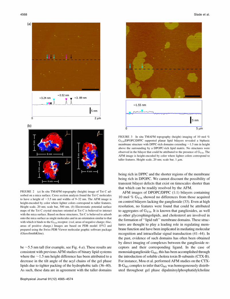
FIGURE 3 In situ TMAFM topography (height) imaging of 10 mol %
GT1b/DPOPC/DPPC supported planar lipid bilayers revealed a biphasic
membrane structure with DPPC-rich domains extending ;1.5 nm in height
above the surrounding by a DPOPC-rich lipid matrix. No structures were
observed in the bilayer that could be attributed to the presence of GT1b. The
AFM image is height-encoded by color where lighter colors correspond to
taller features. Height scale, 20 nm; scale bar, 1 mm.
4568 Slade et al.
Biophysical Journal 91(12) 4565–4574

(DPPC) bilayers (45). In yet other AFM studies, Yuan and
Johnston used CTX-B as a reporter molecule to confirm
clustering of GM1 receptors in DPPC monolayers and bi-
layers as well as in fluid phase egg-PC lipid bilayers (46,47).
To date, there has been very little work done to characterize
the distribution of GT1b in bilayers although we note that elec-
tron microscopy has been used to visualize GT1b in phosphati-
dylcholine bilayers using lectin molecules as topographical
markers (48).
Although the presence of GT1b was not confirmed by AFM
imaging, its presence was verified through binding studies
with Tet C. In the absence of GT1b, Tet C did not exhibit any
affinity for the DPOPC/DPPC bilayers with no changes ob-
served in the bilayer surface topology or structural morphol-
ogy after ;12 h exposure to a 10 mg/mL solution of Tet C
(data not shown). Under the same conditions, in situ
TMAFM revealed a structural topology of DPOPC/DPPC
bilayers containing 10% GT1b that suggests adsorption of Tet
C with selective affinity for the DPOPC-rich areas after ;2 h
incubation (Fig. 4 a). These regions of the bilayer were pop-
ulated by numerous protrusions having dimensions that are
consistent with bound Tet C molecules. The bound proteins
were homogeneously distributed throughout the fluid phase
DPOPC extending ;2 nm above the bilayer surface. The
relatively featureless surface of the areas rich in DPPC sug-
gests little adsorption of Tet C to these gel phase areas, al-
though further confirmation was attained via fluorescence
imaging (vide infra).
TMAFM imaging of the same area of the bilayer after
;12 h of exposure revealed a dramatic change in the overall
bilayer morphology (Fig. 4 b). While the DPPC-rich do-
mains remained relatively unchanged, the previously smooth
DPOPC-rich areas now had a granular texture and were
populated by numerous small, circular cavities with diam-
eters in the range of 40–80 nm (Fig. 4 c). These changes in
the overall structure of the membrane are clearly evident
in the three-dimensional image of the bilayer surface shown
in Fig. 5. Cross section analysis also revealed that after ;12 h
the DPOPC-rich lipid phase now exhibited a thickness simi-
lar to that of the DPPC domains (;7 nm). In addition to the
cavity formation and the thickening of the DPOPC lipid
phase, the Tet C molecules bound to the surface of the
FIGURE 4 In situ TMAFM topography (height) imag-
ing of the interactions between Tet C and 10 mol % GT1b/
DPOPC/DPPC supported planar lipid bilayers. (a) After
;2 h exposure, Tet C appeared to be preferentially asso-
ciated with regions of DPOPC and was distributed rela-
tively homogeneously throughout the fluid phase domains.
(b and c) After ;12 h exposure, TMAFM topography
(height) imaging of the same region of the lipid bilayer (as
indicated by the asterisk) revealed a dramatic change in the
bilayer morphology. The DPOPC-rich regions appeared to
have a granular texture and were populated by a number of
circular defects having widths of 40–80 nm. The height of
the DPOPC regions had also increased to that of the DPPC
domains, with the DPPC domains now appearing to be
encircled by line defects. The DPPC domains themselves
appeared relatively unchanged. Cross section analysis
revealed that the Tet C molecules associated with DPPC
domains extend ;2 nm above the surface of the gel phase
domains. Tet C molecules that were associated with fluid
phase DPOPC, however, extended ,1 nm above the sur-
face of the bilayer, suggesting partial insertion of Tet C
within the regions of DPOPC. The amount of Tet C
associated with the bilayer at ;12 h incubation appeared to
be much lower than at ;2 h incubation. The AFM images
are height-encoded by color where lighter colors corre-
spond to taller features. Height scale, panels a and b ¼ 20
nm, panel c ¼ 12 nm; scale bar, panels a and b ¼ 1 mm;
panel c ¼ 500 nm.
AFM Imaging of Toxin-Membrane Interactions 4569
Biophysical Journal 91(12) 4565–4574

DPOPC-rich regions were also found to have decreased in
height (,1 nm in height). Upon closer examination, the
small number of Tet C aggregates occasionally found on the
surface of the DPPC domains exhibited no change in height
nor induced any structure changes on that portion of the
membrane after ;12 h of incubation (Fig. 4 c). In control
studies performed with supported lipid bilayers of the same
composition (10 mol % GT1b/DPOPC/DPPC), no change or
obvious disruption of the bilayers was observed after ;12 h
of TMAFM imaging in the absence of Tet C (data not
shown).
These observations suggest that over the 12-h incubation
period the Tet C molecules have either become partially
embedded in the fluid DPOPC regions or the Tet C mole-
cules have dissociated from the bilayer surface. To resolve
this question, we performed a correlated TMAFM/TIRFM
imaging study on 10 mol % GT1b/DPOPC/DPPC lipid bi-
layers that had been exposed to a ;25 mg/mL solution of
FITC-labeled Tet C fragment (FITC-Tet C). After in situ
fusion of the GT1b/DPOPC/DPPC lipid vesicles on the mica
surface, TMAFM imaging revealed well-formed phase-
separated lipid bilayers with a morphology similar to those
previously described. There was no observable auto-
fluorescence from these lipid bilayers when TIRFM imaging
was performed (data not shown). After ;12 h incubation of
the bilayers with FITC-Tet C, in situ TMAFM imaging re-
vealed that the DPOPC regions were again granular in tex-
ture and populated by numerous circular cavities, whereas
the DPPC domains remained relatively unchanged (Fig. 6 a).
The corresponding TIRFM images, acquired simultaneously,
revealed a bright fluorescence emission from the DPOPC
regions, which is consistent with preferential association of
the FITC-Tet C with the fluid lipid phase (Fig. 6 b). We note
that TIRFM data acquired during intermediate time points
was consistent with the association of the FITC-Tet C with
the bilayer.
Although correlated TIRFM imaging confirms the pres-
ence of Tet C within the DPOPC regions, it is important to
note that we were unable to resolve the Tet C molecules on
FIGURE 5 Three-dimensional 1.5 3 1.5 mm TMAFM topography
(height) image of a 10 mol % GT1b/DPOPC/DPPC lipid bilayer after ;12
h incubation in the presence of the Tet C fragment. The AFM image is
height-encoded by color where lighter colors correspond to taller features.
Height scale, 15 nm.
FIGURE 6 Combined AFM/TIRFM imaging
of the interactions between FITC-labeled Tet C
and 10 mol % GT1b/DPOPC/DPPC lipid bilayers.
(a) After ;12 h incubation in the presence of
FITC-Tet C, in situ TMAFM topography (height)
imaging revealed the fluid phase DPOPC-rich
regions to have a granular texture and were
populated by a number of circular defects. The
AFM image is height-encoded by color where
lighter colors correspond to taller features. Height
scale, 18 nm; scale bar, 5 mm. (b) TIRFM imaging
of the same region of the bilayer revealed that the
bright fluorescent regions correlated with the
DPOPC regions in the TMAFM image, whereas
dark nonfluorescent regions correlated with the
DPPC domains. For better visualization, the DPPC
domains in the TMAFM image are outlined in
pink in the TIRFM image. The two bright fluo-
rescent areas in the bottom right-hand corner of
the TIRFM image correspond to the position of
two 200-nm diameter Fluosphere microspheres
(Molecular Probes) that were used to focus the
TIRFM objective on the mica surface before
introduction of the lipid vesicles into the fluid
cell. Scale bar, 5 mm. (c) Cross section profiles of
the correlated regions in the TMAFM height
image (A–A9) and the TIRFM image (B–B9). The circular defects observed in the DPOPC regions were found to have widths of 600–900 nm and a depth of
;1.5 nm. The larger size of these defects may be due to the higher concentration of FITC-Tet C used in these studies.
4570 Slade et al.
Biophysical Journal 91(12) 4565–4574

these domains using in situ TMAFM. The two bright fluo-
rescent areas in the bottom right-hand corner of the TIRFM
image correspond to the position of two 200-nm diameter
Fluosphere microspheres (Molecular Probes, Eugene, OR)
that were used to focus the TIRFM objective on the mica
surface before introduction of the lipid vesicles into the fluid
cell. We note that the TIRFM and AFM data sets are not
perfectly correlated. This is likely a consequence of a non-
uniform distribution of the GT1B receptor in the fluid phase
of the supported bilayer and hence, the distribution of the
FITC-Tet C molecules bound to GT1b (49,50). We also can-
not discount the possibility that the dynamics of the lipid
bilayer may be better resolved on TIRFM timescales rather
than on AFM imaging timescales (33).
The results of our correlated TMAFM/TIRFM study
suggest that after continuous incubation of Tet C with the
10 mol % GT1b/DPOPC/DPPC lipid bilayers, the surface-
bound Tet C undergo partial insertion into the GT1b-con-
taining DPOPC fluid phase regions of the bilayer, thus
explaining both the decrease in height and the apparent
decrease in the surface coverage of Tet C in these areas. The
absence of Tet C within the gel-phase DPPC domains may
be due to the tighter packing of the DPPC molecules, which
prevents protein insertion, or due to an absence of GT1b in the
DPPC domains. Similar studies using 10 mol % GT1b/DPPC
lipid bilayers also revealed no GT1b-induced clustering or do-
main formation nor any specific association of Tet C molecules
with these surfaces after extended incubation periods (data
not shown). Although the preferential association with the
DPOPC-rich regions observed for Tet C may reflect favor-
able partitioning of the GT1b receptors into the fluid phase
regions of the lipid bilayers, it is also possible that it may
simply reflect a greater degree of mobility of the receptors
within the fluid DPOPC matrix, and hence greater ease in
adopting the correct binding conformation. Although it
would be ideal to use combined TMAFM/TIRFM to address
the issue as to the presence/absence of GT1b within the DPPC
lipid domains, it is becoming increasingly apparent that the
behavior of a fluorescently labeled lipid is strongly influenced
by the presence of the fluorophore (51–53). The addition of
a bulky fluorophore to the acyl chain of a ganglioside or
phosphocholine molecule prevents tight packing of the lipid
tails, and thus often results in the formation of, or parti-
tioning of the fluorescent probe into, a more disordered phase
in the lipid bilayer. Similar behavior has also been observed
for headgroup labeled lipids (54,55). As such, under the
conditions of our studies, we are unable to quantitatively de-
termine the partitioning of GT1b in either lipid phase.
In situ TMAFM studies were also conducted to examine
the effect of varying the GT1b concentration and the pH of the
surrounding imaging fluid on Tet C-membrane interactions.
In the case of DPOPC/DPPC bilayers containing 1 mol %
GT1b, the Tet C molecules were also found to be preferen-
tially associated with the DPOPC-rich areas after an initial
;2-h incubation period. In contrast to our studies of the
10 mol % GT1b/DPOPC/DPPC bilayers after exposure to Tet
C (vide infra), after ;12 h of incubation with Tet C, we saw
no evidence of persistent defects in either lipid phase of the
1 mol % GT1b/DPOPC/DPPC bilayers (Fig. 7). Cross section
analysis revealed the Tet C molecules associated with the
DPOPC-rich regions after ;12 h to have a height of ;2 nm
relative to the surface of the bilayer. The occasional Tet C
molecule was also observed on the surface of the DPPC do-
mains with a height of ;2 nm.
Under acidic conditions (pH 4.0), Tet C did not appear to
associate with the 10 mol % GT1b/DPOPC/DPPC lipid bi-
layers (data not shown). This may be due to a conformational
change in Tet C, a structural change in the GT1b headgroup,
or a change in the electrostatic interaction between the gan-
glioside and the protein that prevented successful binding of
Tet C to GT1b (10,11). These data are consistent with those of
Winter et al. who reported that the full-length TeNT pref-
erentially bound to negatively charged lipids in vesicles (56).
Based on the results of their studies into the interactions of
TeNT with vesicles containing different types of lipid mol-
ecules, Winter et al. believed that successful binding of the
FIGURE 7 In situ TMAFM topography (height) imaging of the interac-
tions between Tet C and 1 mol % GT1b/DPOPC/DPPC lipid bilayers. After
;12 h incubation, Tet C was found to be preferentially associated with the
fluid phase DPOPC regions of the bilayer. The morphology of the bilayer,
however, remained relatively unchanged and no defects were observed
to have formed. Cross section analysis found the Tet C molecules to extend
;2 nm above the surface of both the DPOPC and the DPPC domains. The
AFM image is height-encoded by color where lighter colors correspond to
taller features. Height scale, 15 nm; scale bar, 1 mm.
AFM Imaging of Toxin-Membrane Interactions 4571
Biophysical Journal 91(12) 4565–4574

toxin was determined by lipid headgroup charge and con-
formation, as well as acyl chain length, with all of these
factors being best fulfilled by the ganglioside GT1b. These
observations suggest that electrostatic interactions play a
large role in the binding of TeNT to the GT1b receptor. As the
Tet C fragment is the membrane-binding domain, it is likely
that its interactions with GT1b would be sensitive to changes
in pH. This result is in contrast to work by others who have
examined the formation of pores in lipid membranes by the
full-length tetanus toxin at low pH (57–60).
The results of these studies clearly indicate that at physio-
logical pH (7.4) the tetanus toxin C-fragment binds to the
fluid phase regions of GT1b-containing membranes and after
prolonged exposure, appears to organize into structures or
assemblies that generate fairly uniform and relatively large
cavity depressions within these regions. The gel phase
domains, however, were relatively resistant to Tet C binding
and activity. Our preliminary studies also suggest a threshold
GT1b concentration is necessary to initiate Tet C association
and cavity formation, although at this time we cannot con-
firm whether the distribution of GT1b is uniform throughout
the model membranes. Studies by Winter et al. of the full-
length tetanus neurotoxin (TeNT) also found that a critical
concentration of GT1b was necessary for pore formation in
phosphatidylcholine lipid vesicles (61). Calorimetric mea-
surements indicated that each TeNT molecule required 20
bound GT1b molecules to adopt the correct conformation for
stable pore formation.
Cross section analysis performed on the larger cavities
formed in the DPOPC-rich areas revealed that they extend
;1.5 nm into the bilayer and do not span the entire SPB to
expose the underlying mica surface (Fig. 6 c). Although the
cavities themselves do not have the characteristics of trans-
membrane pores, they may instead be precursors toward the
formation of the smaller membrane pores (estimated at ;2
nm diameter for tetanus neurotoxin) (15) or perhaps the ini-
tial stages of endosome formation. Neuronal endosomes are
known to form within the size range of these observed cavi-
ties (62). The presence of a fluorescence signal in the areas
of the defects in the correlated TIRFM images indicates that
Tet C was present within the cavities, which likely contrib-
uted to their stability over extended periods of AFM imaging
(greater than hours) (Fig. 6 b). Choi and Dimitriadis per-
formed in situ AFM studies of the binding of cytochrome cto anionic lipid bilayers (61). At higher concentrations, cyto-
chrome c was found to insert into the fluid phase bilayers and
over time, formed circular defects in the bilayers. The au-
thors believed that the defects were nanodomains of pure
protein. Studies by others have suggested that the Tet C
fragment itself does not form pores in lipid bilayers (18,57–
59,63). This model is in reasonable agreement with our studies
where Tet C appears to affect only the upper leaflet of the
bilayer. In the context of this study, we cannot discount the
possibility that this effect is specific to the use of a supported
planar lipid bilayer model of a membrane.
We note that the larger size of the cavities observed in our
combined TMAFM/TIRFM studies may be attributed to the
higher concentration of FITC-labeled Tet C used. Sharpe and
London found that the size of the pores formed by the A1B1-
diphtheria toxin in lipid vesicles increased with increasing
toxin concentration (64). The authors believed that the change
in pore size was due to the oligomerization of the diphtheria
molecules, with the number of toxin subunits determining
the size of the pore formed in the membrane. In our AFM
studies, we did not observe the formation of any supramolecular
assemblies of Tet C on the surface of the lipid bilayer before
the appearance of the cavity structures.
The insertion of Tet C into the fluid phase regions of the
lipid bilayer and concomitant cavity formation may explain
the observed increase in the thickness of these areas after the
;12-h incubation period. This process would likely cause
tighter packing of the DPOPC molecules, and hence a de-
crease in the tilt angle of their acyl chains and subsequent
increase in the height of these regions (36–38). As cell mem-
branes are dynamic in nature, this change in the morphology
of the bilayer may reflect an actual physiological cellular
response. Cantor proposed a mechanism whereby changes in
the lateral pressure of a cell membrane can affect the behavior
of membrane proteins, thereby influencing cell surface recog-
nition and initiating intracellular signaling events (65,66).
In addition to the circular cavities found throughout the
fluid phase of the 10 mol % GT1b/DPOPC/DPPC lipid bi-
layers, prominent defects were found at the border between
the gel phase DPPC domains and the fluid phase DPOPC
lipid matrix (Figs. 4, b and c, and 5). These defects exist
between the intact DPPC and granular, cavity-filled DPOPC
domains, and were found on both the leading and trailing
edges, as defined by the scanning AFM tip, of various do-
mains, suggesting that these structures are not imaging arti-
facts. Indeed, Barlic et al. found that the pore-forming
equinatoxin-II preferentially binds at the boundaries between
the ordered and disordered phases in biphasic membranes
(67). They proposed that lipid packing defects and differ-
ences in the membrane thickness occurring at these inter-
faces may facilitate favorable interactions with the toxin. As
Tet C inserts into the lipid bilayer, we are unable to detect by
AFM, any accumulation of Tet C at the interface between the
DPOPC and DPPC lipid phases. Ideally, we would be able
to characterize, using TIRFM, any accumulation of Tet C at
these interfaces as an increase in the intensity of the fluo-
rescence emission in these regions. Unlike AFM, however,
TIRFM is a diffraction-limited technique. With such a large
amount of fluorescently labeled Tet C present throughout the
DPOPC fluid phase of the bilayer contributing to the overall
fluorescence emission, it was difficult to resolve whether the
intensity within the DPOPC regions differs from that at the
DPOPC-DPPC interfaces. Nevertheless, our observations of
the membrane-disruptive activity of the C-fragment of the
A1B1-tetanus toxin may have physiological relevance as it
suggests that Tet C may actually be involved or aid in the
4572 Slade et al.
Biophysical Journal 91(12) 4565–4574

formation of a ‘‘true’’ membrane pore by the full-length
TeNT.
CONCLUSIONS
This study provides the first direct nanoscale observation
of the binding and interactions between the tetanus toxin
C-fragment (Tet C) and supported lipid bilayers containing
the ganglioside receptor GT1b. As opposed to a number of ion
channel studies with tetanus toxin that have employed sus-
pended lipid membranes lacking a recognized toxin receptor,
we found that GT1b was essential for toxin binding and activ-
ity on the membrane surface (58,59). Furthermore, we observed
preferential association of Tet C with the GT1b-containing fluid
phase regions in gel-fluid biphasic membranes. Over time,
these interactions facilitated a dramatic change in the mor-
phology of the supported lipid bilayer that resulted in the
formation of 40–80 nm cavities with a concomitant increase
in the thickness of the fluid phase regions of the bilayer upon
insertion of bound Tet C. We also found that the membrane
activity of Tet C was dependent upon both the ganglioside
concentration in the membrane and solution pH.
Although TMAFM studies were unable to resolve Tet C
on the surface of the lipid bilayer after the structural change
in the fluid phase regions had occurred, combined TMAFM/
TIRFM studies were able to confirm that Tet C is indeed
present in the bilayer. This demonstrates the power of this
coupled approach for performing functional imaging of lipid
membranes and specifically ligand/protein-membrane inter-
actions. The results of these studies now provide us with the
framework necessary to perform in situ AFM studies of the
membrane interactions and subsequent pore formation by the
full-length tetanus toxin.
We gratefully acknowledge the generous support of the Canadian Institutes
of Health Research (MT-14769), the Natural Sciences and Engineering
Research Council of Canada (194435-99), the Canada Foundation for Inno-
vation, the Ontario Research and Development Challenge Fund, the Ontario
Innovation Trust, and the U.S. Department of Energy Office of Basic
Energy Sciences. C.M.Y. is a Canada Research Chair in Molecular Imaging.
A.L.S. thanks the Natural Sciences and Engineering Research Council of
Canada for fellowship support. Sandia is a multiprogram laboratory operated
by Sandia Corporation, a Lockheed Martin Company, for the U.S. Depart-
ment of Energy’s National Nuclear Security Administration under contract
DE-AC04-94AL85000.
REFERENCES
1. Henderson, D. A. 1999. The looming threat of bioterrorism. Science.283:1279–1282.
2. Bellamy, R. J., and A. R. Freedman. 2001. Bioterrorism. Q. J. Med.94:227–234.
3. Montecucco, C. 1986. How do tetanus and botulinum toxins bind toneuronal membranes? Trends Biochem. Sci. 11:314–317.
4. Louch, H. A., E. S. Buczko, M. A. Woody, R. M. Venable, andW. F. Vann. 2002. Identification of a binding site for ganglioside onthe receptor binding domain of tetanus toxin. Biochemistry. 41:13644–13652.
5. Pellizzari, R., O. Rossetto, G. Schiavo, and C. Montecucco. 1999.Tetanus and botulinum neurotoxins: mechanism of action and thera-peutic uses. Philos. Trans. R. Soc. Lond. B Biol. Sci. 354:259–268.
6. Halpern, J. L., and E. A. Neale. 1995. Neurospecific binding, internali-zation, and retrograde axonal transport. Curr. Top. Microbiol. Immunol.195:221–241.
7. Lalli, G., S. Bohnert, K. Deinhardt, C. Verastegui, and G. Schiavo.2003. The journey of tetanus and botulinum neurotoxins in neurons.Trends Microbiol. 11:431–437.
8. Turton, K., J. A. Chaddock, and K. R. Acharya. 2002. Botulinum andtetanus neurotoxins: structure, function and therapeutic utility. TrendsBiochem. Sci. 27:552–558.
9. Schiavo, G., F. Benfenati, B. Poulain, O. Rossetto, P. Polverino deLaureto, B. R. DasGupta, and C. Montecucco, 1992. Tetanus andbotulinum-B neurotoxins block neurotransmitter release by proteolyticcleavage of synaptobrevin. Nature. 359:832–835.
10. Puhar, A., E. A. Johnson, O. Rossetto, and C. Montecucco. 2004. Com-parison of the pH-induced conformational change of different clostridialneurotoxins. Biochem. Biophys. Res. Commun. 319:66–71.
11. Montecucco, C., G. Schiavo, J. Brunner, E. Duflot, Boquet, andM. Roa, 1986. Tetanus toxin is labeled with photoactivatable phos-pholipids at low pH. Biochemistry. 25:919–924.
12. Donovan, J. J., M. I. Simon, R. K. Draper, and M. Montal. 1981.Diphtheria toxin forms transmembrane channels in planar lipid bilayers.Proc. Natl. Acad. Sci. USA. 78:172–176.
13. Kagan, B. L., A. Finkelstein, and M. Colombini. 1981. Diphtheriatoxin fragment forms large pores in phospholipid bilayer membranes.Proc. Natl. Acad. Sci. USA. 78:4950–4954.
14. Deleers, M., N. Beugnier, P. Falmagne, V. Cabiaux, and J. M.Ruysschaert. 1983. Localization in diphtheria toxin fragment B of aregion that induces pore formation in planar lipid bilayers at low pH.FEBS Lett. 160:82–86.
15. Hoch, D. H., M. Romero-Mira, B. E. Ehrlich, A. Finkelstein, B. R.DasGupta, and L. L. Simpson. 1985.Channels formed by botulinum,tetanus, and diphtheria toxins in planar lipid bilayers: relevance to trans-location of proteins across membranes. Proc. Natl. Acad. Sci. USA. 82:1692–1696.
16. Donovan, J. J., and J. L. Middlebrook. 1986. Ion-conducting channelsproduced by botulinum toxin in planar lipid membranes. Biochemistry.25:2872–2876.
17. Blaustein, R. O., T. M. Koehler, R. J. Collier, and A. Finkelstein. 1989.Anthrax toxin: channel-forming activity of protective antigen in planarphospholipid bilayers. Proc. Natl. Acad. Sci. USA. 86:2209–2213.
18. Beise, J., J. Hahnen, B. Andersen-Beckh, and F. Dreyer. 1994. Poreformation by tetanus toxin, its chain and fragments in neuronal mem-branes and evaluation of the underlying motifs in the structure of thetoxin molecule. Naunyn Schmiedebergs Arch. Pharmacol. 349:66–73.
19. Cabiaux, V., C. Wolff, and J. M. Ruysschaert. 1997. Interaction with alipid membrane: a key step in bacterial toxins virulence. Int. J. Biol.Macromol. 21:285–298.
20. Qiu, X.Q., K.S. Jakes, P.K. Kienker, A. Finkelstein, A., and S. L.Slatin, 1996. Major transmembrane movement associated with colicinIa channel gating. J. Gen. Physiol. 107:313–328.
21. Santos, N. C., E. Ter-Ovanesyan, J. A. Zasadzinski, M. Prieto, andM. A. Castanho. 1998. Filipin-induced lesions in planar phospholipidbilayers imaged by atomic force microscopy. Biophys. J. 75:1869–1873.
22. Milhaud, J., V. Ponsinet, M. Takashi, and B. Michels. 2002.Interactions of the drug amphotericin B with phospholipid membranescontaining or not ergosterol: new insight into the role of ergosterol.Biochim. Biophys. Acta. 1558:95–108.
23. Steinem, C., H.-J. Galla, and A. Janshoff. 2000. Interaction of melittinwith solid supported membranes. Phys. Chem. Chem. Phys. 2:4580–4585.
24. Yip, C. M., and J. McLaurin. 2001. Amyloid-beta peptide assembly:a critical step in fibrillogenesis and membrane disruption. Biophys.J. 80:1359–1371.
AFM Imaging of Toxin-Membrane Interactions 4573
Biophysical Journal 91(12) 4565–4574

25. Jo, E., J. McLaurin, C. M. Yip, P. St George-Hyslop, and P. E. Fraser.2000. a-Synuclein membrane interactions and lipid specificity. J. Biol.Chem. 275:34328–34334.
26. Epand, R. F., C. M. Yip, V. Chernomordik, L. D. Le, Y. K. Duc,Y. K. Shin, and R. M. Epand. 2001. Self-assembly of influenza hem-agglutinin: studies of ectodomain aggregation by in situ atomic forcemicroscopy. Biochim. Biophys. Acta. 1513:167–175.
27. Epand, R. F., J. C. Martinou, S. Montessuit, R. M. Epand, andC. M. Yip. 2002. Direct evidence for membrane pore formation by theapoptotic protein Bax. Biochem. Biophys. Res. Commun. 298:744–749.
28. Epand, R. M., S. Maekawa, C. M. Yip, and R. F. Epand. 2001. Protein-induced formation of cholesterol-rich domains. Biochemistry. 40:10514–10521.
29. Bayerl, T. M., and M. Bloom. 1990. Physical properties of singlephospholipid bilayers adsorbed to micro glass beads: a new vesicularmodel system studied by 2H-nuclear magnetic resonance. Biophys. J.58:357–362.
30. Johnson, S. J., T. M. Bayerl, D. C. McDermott, G. W. Adam,A. R. Rennie, R. K. Thomas, and E. Sackmann. 1991. Structure of anadsorbed dimyristoylphosphatidylcholine bilayer measured with spec-ular reflection of neutrons. Biophys. J. 59:289–294.
31. Salafsky, J., J. T. Groves, and S. G. Boxer. 1996. Architecture andfunction of membrane proteins in planar supported bilayers: a studywith photosynthetic reaction centers. Biochemistry. 35:14773–14781.
32. Slade, A., J. Luh, S. Ho, and C. M. Yip. 2002. Single moleculeimaging of supported planar lipid bilayer-reconstituted human insulinreceptors by in situ scanning probe microscopy. J. Struct. Biol. 137:283–291.
33. Shaw, J. E., A. Slade, and C. M. Yip. 2003. Simultaneous in situ totalinternal reflectance fluorescence/atomic force microscopy studies ofDPPC/dPOPC microdomains in supported planar lipid bilayers. J. Am.Chem. Soc. 125:11838–11839.
34. Fotinou, C., P. Emsley, I. Black, H. Ando, H. Ishida, M. Kison, K. A.Sinha, N. F. Fairweather, and N. W. Isaacs. 2001. The crystal structureof tetanus toxin Hc fragment complexed with a synthetic GT1b ana-logue suggests cross-linking between ganglioside receptors and thetoxin. J. Biol. Chem. 276:32274–32281.
35. Maeda, H. 1997. An atomic force microscopy study for the assemblystructures of tobacco mosaic virus and their size evaluation. Langmuir.13:4150–4161.
36. Mou, J., J. Yang, C. Huang, and Z. Shao. 1994. Alcohol induces inter-digitated domains in unilamellar phosphatidylcholine bilayers. Bio-chemistry. 33:9981–9985.
37. Masai, J., T. Shibataseki, K. Sasaki, H. Murayama, and K. Sano. 1996.Scanning force microscopy characterization of thin lipid films on asubstrate. Thin Solid Films. 273:289–296.
38. Hollars, C. W., and R. C. Dunn. 1998. Submicron structure in L-a-dipalmitoylphosphatidylcholine monolayers and bilayers probed withconfocal, atomic force, and near-field microscopy. Biophys. J. 75:342–353.
39. McKiernan, A. E., T. V. Ratto, and M. L. Longo. 2000. Domaingrowth, shapes, and topology in cationic lipid bilayers on mica byfluorescence and atomic force microscopy. Biophys. J. 79:2605–2615.
40. Giocondi, M.-C., V. Vie, E. Lesniewska, P.-E. Milhiet, M. Zinke-Allmang, and C. Le Grimellec. 2001. Phase topology and growth ofsingle domains in lipid bilayers. Langmuir. 17:1653–1659.
41. Brown, D. A., and E. London. 2000. Structure and function ofsphingolipid- and cholesterol-rich membrane rafts. J. Biol. Chem.275:17221–17224.
42. Simons, K., and D. Toomre. 2000. Lipid rafts and signal transduction.Nat. Rev. Mol. Cell Biol. 1:31–39.
43. Simons, K., and E. Ikonen. 1997. Functional rafts in cell membranes.Nature. 387:569–572.
44. Rietveld, A., and K. Simons. 1998. The differential miscibility of lipidsas the basis for the formation of functional membrane rafts. Biochim.Biophys. Acta. 1376:467–479.
45. Mou, J., J. Yang, and Z. Shao. 1995. Atomic force microscopy ofcholera toxin B-oligomers bound to bilayers of biologically relevantlipids. J. Mol. Biol. 248:507–512.
46. Yuan, C., and L. J. Johnston. 2000. Distribution of ganglioside GM1 inL-a-dipalmitoylphosphatidylcholine/cholesterol monolayers: a modelfor lipid rafts. Biophys. J. 79:2768–2781.
47. Yuan, C., and L. J. Johnston. 2001. Atomic force microscopy studies ofganglioside GM1 domains in phosphatidylcholine and phosphatidyl-choline/cholesterol bilayers. Biophys. J. 81:1059–1069.
48. Mehlhorn, I. E., G. Parrag, K. R. Barber, and C. W. Grant. 1986.Visualization of domains in rigid ganglioside/phosphatidylcholinebilayers: Ca21 effects. Biochim. Biophys. Acta. 863:139–155.
49. Last, J. A., T. A. Waggoner, and D. Y. Sasaki. 2001. Lipid membranereorganization induced by chemical recognition. Biophys. J. 81:2737–2742.
50. Sasaki, D. Y., T. A. Waggoner, J. A. Last, J and T. M. Alam. 2002.Crown ether functionalized membranes: lead ion recognition andmolecular reorganization. Langmuir. 18:3714–3721.
51. Cruz, A., L. Vazquez, M. Velez, and J. Perez-Gil. 2005. Influence of afluorescent probe on the nanostructure of phospholipid membranes:dipalmitoylphosphatidylcholine interfacial monolayers. Langmuir. 21:5349–5355.
52. Saez-Cirion, A., S. Nir, M. Lorizate, A. Agirre, A. Cruz, J. Perez-Gil,and J. L. Nieva. 2002. Sphingomyelin and cholesterol promote HIV-1gp41 pretransmembrane sequence surface aggregation and membranerestructuring. J. Biol. Chem. 277:21776–21785.
53. Bernardino de la Serna, J., J. Perez-Gil, A. C. Simonsen, and L. A.Bagatolli. 2004.Cholesterol rules: direct observation of the coexistenceof two fluid phases in native pulmonary surfactant membranes atphysiological temperatures. J. Biol. Chem. 279:40715–40722.
54. Winter, A., W. P. Ulrich, F. Wetterich, U. Weller, and H. J. Galla.1996. Gangliosides in phospholipid bilayer membranes: Interactionwith tetanus toxin. Chem. Phys. Lipids. 81:21–34.
55. Boquet, P., and E. Duflot. 1982. Tetanus toxin fragment formschannels in lipid vesicles at low pH. Proc. Natl. Acad. Sci. USA.79:7614–7618.
56. Gambale, F., and M. Montal. 1988. Characterization of the channel prop-erties of tetanus toxin in planar lipid bilayers. Biophys. J. 53:771–783.
57. Menestrina, G., S. Forti, and F. Gambale. 1989. Interaction of tetanustoxin with lipid vesicles: effects of pH, surface charge, and transmem-brane potential on the kinetics of channel formation. Biophys. J. 55:393–405.
58. Calappi, E., M. Masserini, G. Schiavo, C. Montecucco, and G.Tettamanti. 1992. Lipid interaction of tetanus neurotoxin. A calori-metric and fluorescence spectroscopy study. FEBS Lett. 309:107–110.
59. Winter, A., W. P. Ulrich, F. Wetterich, U. Weller, and H. J. Galla.1996. Gangliosides in phospholipid bilayer membranes: interactionwith tetanus toxin. Chem. Phys. Lipids. 81:21–34.
60. Zhang, B., B. Ganetzky, H. J. Bellen, and V. N. Murthy. 1999. Tai-loring uniform coats for synaptic vesicles during endocytosis. Neuron.23:419–422.
61. Roa, M., and P. Boquet. 1985. Interaction of tetanus toxin with lipidvesicles at low pH: protection of specific polypeptides against prote-olysis. J. Biol. Chem. 260:6827–6835.
62. Sharpe, J. C., and E. London. 1999. Diphtheria toxin forms pores ofdifferent sizes depending on its concentration in membranes: probablerelationship to oligomerization. J. Membr. Biol. 171:209–221.
63. Cantor, R. S. 1997. Lateral pressures in cell membranes: a mechanismfor modulation of protein function. J. Phys. Chem. B. 101:1723–1725.
64. Cantor, R. S. 1999. Lipid composition and the lateral pressure profile inbilayers. Biophys. J. 76:2625–2639.
65. Barlic, A., I, Gutierrez-Aguirre, J. M. Caaveiro, A. Cruz, M. B. Ruiz-Arguello, J. Perez-Gil, J., and J. M. Gonzalez-Manas, 2004. Lipidphase coexistence favors membrane insertion of equinatoxin-II, a pore-forming toxin from Actinia equina. J. Biol. Chem. 279:34209–34216.
4574 Slade et al.
Biophysical Journal 91(12) 4565–4574