Improving endothelial cell adhesion to vascular graft surfaces: Clinical need and strategies
21
This article was downloaded by: [Duke University Libraries] On: 31 July 2014, At: 09:39 Publisher: Taylor & Francis Informa Ltd Registered in England and Wales Registered Number: 1072954 Registered office: Mortimer House, 37-41 Mortimer Street, London W1T 3JH, UK Journal of Biomaterials Science, Polymer Edition Publication details, including instructions for authors and subscription information: http://www.tandfonline.com/loi/tbsp20 Improving endothelial cell adhesion to vascular graft surfaces: Clinical need and strategies Vinayak D. Bhat a , Bruce Klitzman b , Kim Koger c , George A. Truskey d & William M. Reichert e a Department of Biomedical Engineering & Centre for Cellular and Biosurface Engineering, Duke University, Durham, NC 27708-0281, USA b Department of Biomedical Engineering & Centre for Cellular and Biosurface Engineering, Duke University, Durham, NC 27708-0281, USA c Department of Biomedical Engineering & Centre for Cellular and Biosurface Engineering, Duke University, Durham, NC 27708-0281, USA d Department of Biomedical Engineering & Centre for Cellular and Biosurface Engineering, Duke University, Durham, NC 27708-0281, USA e Department of Biomedical Engineering & Centre for Cellular and Biosurface Engineering, Duke University, Durham, NC 27708-0281, USA Published online: 02 Apr 2012. To cite this article: Vinayak D. Bhat , Bruce Klitzman , Kim Koger , George A. Truskey & William M. Reichert (1998) Improving endothelial cell adhesion to vascular graft surfaces: Clinical need and strategies, Journal of Biomaterials Science, Polymer Edition, 9:11, 1117-1135, DOI: 10.1163/156856298X00686 To link to this article: http://dx.doi.org/10.1163/156856298X00686 PLEASE SCROLL DOWN FOR ARTICLE Taylor & Francis makes every effort to ensure the accuracy of all the information (the “Content”) contained in the publications on our platform. However, Taylor
-
Upload
independent -
Category
Documents
-
view
0 -
download
0
Transcript of Improving endothelial cell adhesion to vascular graft surfaces: Clinical need and strategies

This article was downloaded by: [Duke University Libraries]On: 31 July 2014, At: 09:39Publisher: Taylor & FrancisInforma Ltd Registered in England and Wales Registered Number: 1072954Registered office: Mortimer House, 37-41 Mortimer Street, London W1T 3JH, UK
Journal of Biomaterials Science,Polymer EditionPublication details, including instructions for authors andsubscription information:http://www.tandfonline.com/loi/tbsp20
Improving endothelial celladhesion to vascular graftsurfaces: Clinical need andstrategiesVinayak D. Bhat a , Bruce Klitzman b , Kim Koger c ,George A. Truskey d & William M. Reichert ea Department of Biomedical Engineering & Centre forCellular and Biosurface Engineering, Duke University,Durham, NC 27708-0281, USAb Department of Biomedical Engineering & Centre forCellular and Biosurface Engineering, Duke University,Durham, NC 27708-0281, USAc Department of Biomedical Engineering & Centre forCellular and Biosurface Engineering, Duke University,Durham, NC 27708-0281, USAd Department of Biomedical Engineering & Centre forCellular and Biosurface Engineering, Duke University,Durham, NC 27708-0281, USAe Department of Biomedical Engineering & Centre forCellular and Biosurface Engineering, Duke University,Durham, NC 27708-0281, USAPublished online: 02 Apr 2012.
To cite this article: Vinayak D. Bhat , Bruce Klitzman , Kim Koger , George A. Truskey &William M. Reichert (1998) Improving endothelial cell adhesion to vascular graft surfaces:Clinical need and strategies, Journal of Biomaterials Science, Polymer Edition, 9:11,1117-1135, DOI: 10.1163/156856298X00686
To link to this article: http://dx.doi.org/10.1163/156856298X00686
PLEASE SCROLL DOWN FOR ARTICLE
Taylor & Francis makes every effort to ensure the accuracy of all the information(the “Content”) contained in the publications on our platform. However, Taylor

& Francis, our agents, and our licensors make no representations or warrantieswhatsoever as to the accuracy, completeness, or suitability for any purposeof the Content. Any opinions and views expressed in this publication are theopinions and views of the authors, and are not the views of or endorsed byTaylor & Francis. The accuracy of the Content should not be relied upon andshould be independently verified with primary sources of information. Taylor andFrancis shall not be liable for any losses, actions, claims, proceedings, demands,costs, expenses, damages, and other liabilities whatsoever or howsoever causedarising directly or indirectly in connection with, in relation to or arising out of theuse of the Content.
This article may be used for research, teaching, and private study purposes.Any substantial or systematic reproduction, redistribution, reselling, loan, sub-licensing, systematic supply, or distribution in any form to anyone is expresslyforbidden. Terms & Conditions of access and use can be found at http://www.tandfonline.com/page/terms-and-conditions
Dow
nloa
ded
by [
Duk
e U
nive
rsity
Lib
rari
es]
at 0
9:39
31
July
201
4

Improving endothelial cell adhesion to vascular graft
surfaces: Clinical need and strategies
VINAYAK D. BHAT, BRUCE KLITZMAN, KIM KOGER, GEORGE A. TRUSKEY and WILLIAM M. REICHERT *
Department of Biomedical Engineering & Centre for Cellular and Biosurface Engineering, Duke University, Durham, NC 27708-0281, USA
Received 15 October 1997; accepted 24 February 1998
Abstract-Synthetic vascular grafts do not spontaneously endothelialize in humans and require some form of anticoagulation to maintain patency. Preseeding synthetic graft materials such as
expanded polytetrafluoroethylene (ePTFE) and polyethylene terephthalate (PET) with endothelial cells (EC) has been examined in various in vitro and in vivo models. Although various studies provide encouraging results, clinical trials for EC seeding on synthetic grafts have not been equally successful. This paper provides a brief review of the various reports on EC seeding in animal and clinical studies. We discuss the inefficiencies associated with the EC seeding process and examine plasma protein treatment of the graft surfaces as a viable option for improving EC attachment, retention and spreading. As an alternative to exsisting therapies we present data on a heterogeneous ligand treatment of fibronectin (Fn) and avidin-biotin for enhanced human umbilical vein endothelial cell
(HUVEC) adhesion to ePTFE graft surfaces. Control consisted of HUVECs seeded on Fn treated ePTFE graft surfaces. Functionality of HUVECs was assessed by measuring prostacyclin production of cells on both homogeneous and heterogeneous ligand treated surfaces. Laminar flow studies with a variable width flow chamber and scanning electron microscopy were used to measure initial cell retention and observe initial cell spreading on ePTFE surfaces, respectively. HUVEC retention on heterogeneous ligand treated graft surface was significantly (p < 0.001) higher compared to
homogeneous ligand treated surfaces for shear stress in the range of 10-30 dyn cm-2. HUVEC showed more cellular spreading on the heterogeneous ligand treated surface after seeding for 1-2 h. In vivo experimentation was performed in immune deficient (nude) rats by replacing a section of both the femoral arteries with 8 mm long, 1 mm internal diameter denucleated ePTFE grafts treated with homogeneous and heterogeneous ligands respectively. Both grafts were seeded with similar cell
density for 15 min prior to implantation. EC attachment and retention was measured by staining EC with hematoxylin and counting the cells before and after flow using light microscopy. The results indicate that a heterogeneous ligand treatment of graft surfaces using avidin-biotin and Fn-integrin attachment mechanisms increase cell seeding efficiency, initial cell retention and cellular spreading.
Key words: Biomaterials; endothelialization; vascular grafts; avidin-biotin; cell adhesion; in vivo.
*To whom correspondence should be addressed.
Dow
nloa
ded
by [
Duk
e U
nive
rsity
Lib
rari
es]
at 0
9:39
31
July
201
4

1118
INTRODUCTION
Synthetic vascular grafts are used to treat manifestations of atherosclerosis, in-
cluding both occlusive and aneurysmal diseases. Large diameter vascular grafts
(> 6 mm) made of Dacron or expanded polytetrafluoroethylene (ePTFE) have been
clinically successful in terms of long term patency of the graft. This is because
they experience high blood flow rates and they are large enough such that throm-
bus formation on the inner lumen does not significantly decrease blood flow [1, 2].
However, in the case of small diameter vascular grafts (< 6 mm), which experience lower flow rates and have a higher ratio of surface area to blood volume, even a
small amount of thrombus formation will lead to occlusion of the graft and decrease
blood flow downstream. Also contributing to thrombogenecity of synthetic vascular
grafts are biomechanical and bioelectrical properties of materials, low flow condi-
tions, limited spontaneous re-endothelialization in humans, and technical errors in
surgery [3, 4]. An ideal inner lumen of a vascular graft should maintain a constant balance
between antithrombotic and prothrombotic functions. The endothelial cell (EC)
lining of all the natural blood vessels serves precisely this purpose [5, 6]. During a
response to injury, EC can release clot producing substances like platelet activating factor [7], von Willebrand's factor [8], and tissue thromboplastin [6]. Under
normal circumstances, EC limits clot formation by deactivating thrombin via the
antithrombin III and thrombomodulin systems [9]. EC also suppresses platelet activation by producing prostacyclin and can even play a role in dissolution of
clots [9]. In principle, seeding of vascular grafts with host EC prior to implantation, and exposing the blood to a layer of functional endothelium, should increase patency in small diameter synthetic vascular grafts.
In this paper, we present a brief review on the various animal and clinical studies
performed to examine EC seeding on synthetic vascular grafts and discuss the prob- lems associated with the process of EC seeding and techniques to improve them.
The latter part of the paper presents preliminary evidence that a heterogeneous lig- and system of fibronectin and avidin - biotin can improve EC attachment and spread-
ing to ePTFE graft surfaces.
Animal studies
Endothelial cell seeding was first reported by Herring et al. [10] in 1978 in which
canine venous endothelium was mechanically scraped, collected, and mixed with
the blood used in preclotting knitted Dacron grafts implanted into the infrarenal
aortas of the same dogs. The grafts were explanted after 8 weeks and the results
showed a significantly higher clot free surface on the seeded grafts (76%) as
compared to the unseeded grafts (22%). Since then, a lot of work has been
performed on EC seeding of synthetic vascular prosthesis (Table 1), yet achieving
satisfactory seeded EC coverage remains one of the problems facing cardiovascular
biomaterials [ 11-13].
Dow
nloa
ded
by [
Duk
e U
nive
rsity
Lib
rari
es]
at 0
9:39
31
July
201
4

1119
Dow
nloa
ded
by [
Duk
e U
nive
rsity
Lib
rari
es]
at 0
9:39
31
July
201
4

1120
EC seeding of vascular grafts before implantation has been studied in various
animal models 110, 14-20]. Seeded grafts exhibited reduced platelet adhesion and
improved patency as compared to unseeded grafts. EC seeding of 6 mm inner
diameter (ID) infrarenal aortic Dacron grafts in canine models improved surface
coverage and patency with increased cellular neointima and transmural capillary
ingrowth [21, 22]. Similar results were obtained with 4 mm ID PTFE grafts
interposed in carotid arteries of dogs after 2 and 5 weeks [23]. Stanley et al. [16]
implanted 4 mm ID bilateral iliofemoral Dacron grafts in dogs administered with
aspirin and dipyridamole and seeded only one limb with EC. All grafts remained
patent until medication was discontinued at 2 weeks, after which cumulative
patency rates were 73% in seeded and 27% in unseeded control grafts, which
was surprisingly close to that observed by Herring et al. [ 10 1. Seeded grafts also
exhibited a significantly higher degree of EC surface coverage and decrease in
platelet aggregation [16J.
Enzymatically harvested aortic EC mixed with blood and seeded on 10 mm
ID thoraco-abdominal bypass Dacron grafts in dogs were endothelialized with no
visible thrombus as compared to the luminal fibrin and red thrombus on unseeded
graft surface after 6 weeks [15]. Similar results were obtained with seeded and
unseeded 6 mm ID thoraco-abdominal Dacron grafts implanted in dogs for a period of 1 year [17]. At I month, 80% of seeded grafts were covered with endothelium
compared to 10% of unseeded graft surface. At greater than 4 months, seeded
grafts were completely covered with EC, while unseeded grafts, although gradually covered with endothelium at 1 year, still showed red thrombus, fibrin, and platelet
aggregation. The average thickness of inner capsule on the unseeded graft was
significantly higher than the corresponding thickness of seeded graft throughout the
time period of the study [ 17]. EC coverage of implanted prostheses have also been shown to prolong platelet
survival time [1]. Increased platelet survival time eight weeks after implantation of
10 mm ID Dacron grafts in the canine aorta suggested a decrease in graft-platelet interactions due to EC coverage. Seeded grafts also produced significantly higher amounts of prostacyclin, an antithrombotic factor, than unseeded grafts [24, 25].
Thrombogenicity of grafts, measured by radiolabelled platelet deposition, was
reduced in dog and baboon models [18, 23]. Allen et al. [26] reported a significant reduction of platelet deposition on seeded 4 mm Dacron grafts implanted in
carotid and femoral arteries of dogs over a 7-month period. The patency rates at
7 months were 96% for seeded grafts and 29% for the unseeded controls. Studies
using a combined approach of EC seeding and antiplatelet medication produced
significantly improved thrombus free surface and short term patency of vascular
grafts [27-29], although antiplatelet medication also reduced mean prostacyclin
production by seeded grafts compared with non medicated seeded grafts [28]. A factor that complicates the interpretation of these studies is that they were
all performed in various graft positions, in different animal models, with grafts of different ID and varying blood flow rates. A successful endothelialization of
Dow
nloa
ded
by [
Duk
e U
nive
rsity
Lib
rari
es]
at 0
9:39
31
July
201
4

1121
seeded grafts is impacted by all these factors [23]. Hence, these successful EC
seeding studies in animal models cannot be extrapolated to clinical situations of
coronary bypasses and other distal grafts such as femoral-popliteal and femoral-
tibial bypasses. Other EC seeding reports show no significant improvement of
seeded grafts over unseeded controls [28, 30, 31 ] and EC seeding did not improve
patency of grafts smaller than 4 mm in diameter [23].
Clinical studies
Compared to in vitro and animal studies, few clinical trials have been undertaken.
Indirect techniques such as 111In platelet scintigraphy [32] are used to evaluate
patency, thrombogenecity and neointimal hyperplasia on grafts as seeded grafts are not available for direct examination. Clinical trials of endothelial seeding of
vascular grafts have shown only a small to modest improvement over unseeded
grafts in terms of patency. The earliest report of EC seeding in humans was again by Herring et al. (1984)
where Dacron grafts were seeded with mechanically scraped EC in femoropopliteal reconstructions [33, 34]. Patency of seeded and unseeded grafts showed no
significant difference. However, patients who smoked had lower patency for
seeded grafts as compared to controls. In another study, enzymatically derived
EC incubated on preclotted PTFE implanted in femoropopliteal grafts with patients
receiving antiplatelet therapy showed increased patency from 31 to 81% [35]. Ortenwall et al. showed EC seeding reduced thrombogenicity at 1 month in
seven out of nine aortobifemoral grafts in which one limb was seeded, but there
was no significant difference between seeded and control grafts at 4 months [36]. In subsequent studies with femoropopliteal reconstruction with ePTFE grafts,
they seeded half of each implanted graft with autologous EC. Seeded segments accumulated significantly fewer platelets at 1 and 6 months after surgery [37]. In other studies, EC seeded ePTFE grafts showed no significant improvements in patency compared to unseeded grafts [35, 38]. EC seeding failed to improve
patency or thrombogenicity of ePTFE femoropopliteal graft bypass at the end of
2 or 4 months [38]. Zilla et al. found no differences in early or late platelet
deposition between seeded and unseeded fibrin glue precoated ePTFE femorodistal
grafts [39, 40].
EC seeding inefficiencie.s
The mixed results with clinical studies have led several researchers to revisit the
technique of EC seeding. Herring suggests the massive inefficiency of the EC
seeding process to be partly responsible for these results [ 11 ]. The process of
endothelial seeding of a vascular grafts involves four major steps: (1) harvesting EC from the donor; (2) attaching EC to the vascular grafts surface; (3) replication of the EC to confluency; and (4) maintaining the functionality of the EC. Each step in the seeding process is additive and improvements in one or more may result in
Dow
nloa
ded
by [
Duk
e U
nive
rsity
Lib
rari
es]
at 0
9:39
31
July
201
4

1122
significant overall increase in success of endothelialization. A successful seeded
vascular graft requires all the steps to work efficiently. Cells can be damaged by excessive intensity of harvesting, but may be left on the
donor vessel if the harvesting effort is not strong enough. Cell viability decreases
during processing, cleaning, and resuspension. Cells can fail to attach to the graft surface during seeding and the attached cells can detach upon reesrtablishment of
blood flow. Over the past years efforts have been made to optimize the methods
employed in harvesting and seeding of EC onto surfaces. In addition, enhancing the rapidly of development of confluence of the seeded cells and their resistance to
detachment due to both hemodynamic and biochemical factors have also received
attention.
Compared to the technique of mechanical scraping used by Herring et al. [10],
enzymatic harvesting [41] reduces smooth muscle cell contamination, increases
the viability of the harvested endothelium, improves cell cluster size and increases
the overall efficiency of the process. Sharefkin et al. [42] has compared different
enzymatic harvesting methods on harvests of human saphenous vein EC. However, venous EC isolated from human vessels such as pulmonory veins using enzymatic
techniques are not in high enough numbers for a successful EC seeding, especially because of the high rate of EC desquamation from the graft surface under flow
conditions.
Other approaches have been employed to overcome this limitation with the
numbers of vessel derived EC, such as culturing the vessel-derived EC in vitro to
large numbers before using them for seeding [43-45 or using an alternate source
of EC which would yield larger number of cells [46-48]. Culturing EC irz vitro
would require two separate procedures on the patient, one for removal of a large
segment of donor vessel and a second for implantation of the EC seeded graft. This technique is restricted to elective cases due to a 4-5 week delay between
cell harvest and graft implantation [5]. Microvascular endothelium harvested from
alternate sources of EC such as fat tissues yield large number of cells that have been
used in seeding vascular graft surfaces. The results with these cells have shown mixed success with a few studies recording increased patency and EC monolayer formation [49, 50] while others showed no significant differences in patency of seeded and unseeded grafts [51]. However, concerns with origin of the EC derived
from omental tissues [52, 53] and contamination of non-endothelial cells [51] have been associated with this technique. Further, because of monolayer pattern of
endothelium, no more than two fold 'extra' cells can be supplied on the graft surface
against flow associated losses [ 1 1 ].
Augmenting EC attachment
The strength of EC attachment on the vascular graft surface becomes critical
when the grafts are seeded with a limited number of primary culture autologous EC. Rosenman et al. [54] showed that only 20% of the harvested EC attached
to the ePTFE graft surface following seeding. Within 30 min of restoration
Dow
nloa
ded
by [
Duk
e U
nive
rsity
Lib
rari
es]
at 0
9:39
31
July
201
4

1123
of flow, 70% of the adherent cells detached, and by 24 h less that 4% of the
original seeded cells remained adherent on the prosthesis. Following this report, several investigations were carried out to optimize conditions on cell adhesion, cell retention and proliferation following seeding.
Vascular graft surfaces treated with plasma proteins such as Fn [14, 26, 55], vitronection [56], collagen [57] enhanced EC adhesion. These plasma proteins have an amino acid sequence Arg-Gly-Asp (RGD) which serves as the primary binding site for the transmembrane integrin receptors of the EC [58, 59]. In vitro studies have shown that EC adhesion can also be improved by directly grafting these
peptide sequences on the graft surfaces [60-64]. Fn treated ePTFE grafts as compared to untreated grafts showed increased EC
attachment following seeding (47 vs 20%) and an increased retention of adherent cells (21 vs 4%) after 24 h under flow conditions [65]. EC seeded on polyester elastomer grafts for 16-18 h under static conditions and perfused for 60 min
resulted in a 21% cell retention [66]. EC seeded on Fn treated PTFE grafts for 10 min prior to exposure of flow and experiencing a shear stress of 15 dyn cm-2 exhibited a cell retention of 25% [55]. In another study, Vohra et al. [67] studied in vitro EC retention on Fn coated ePTFE and preclotted ePTFE grafts. After 2 h of exposure to flow with maximum shear stress of 2.6 dyn cm-2, 60% of the seeded EC were retained on both Fn coated and preclotted grafts, whereas preclotted grafts exhibited better EC retention than did the Fn surface at lower shear stresses [67]. Similar results were observed by Miyata et al. on Fn treated ePTFE grafts 1681. Grafts seeded with EC for 1.5 h under static conditions and exposed to pulsatile flow retained 42% of the seeded cells.
Combinations of plasma proteins and peptide sequences have also been used to enhance EC attachment on graft surfaces. Coating graft surfaces with transglutine, a fibrin glue consisting of Fn, fibrinogen and von Willebrand factor enhanced EC adhesion on graft surfaces [69]. ePTFE grafts coated with a combination of collagen type I and either Fn or laminin showed better EC adhesion compared to Fn or laminin alone [70]. Protein based surface modifications have increased EC retention
compared to untreated graft surfaces, yet a significant number of cells detach upon restoration of flow.
Most of the plasma proteins used to improve EC attachment use the integrin mediated mechanism of adhesion. The protein-integrin bonds are low affinity ( 106 m-1) bonds [71] and do not develop into mature attachment in short periods (0.5-1 h) of seeding time [65, 66]. Furthermore, initial cell loss following restoration of flow is not significantly reduced by protein preadsorption, suggesting the detachment mechanism during this phase to be primarily due to fluid mechanical forces [65].
Recently we introduced a heterogeneous ligand system which utilizes two ad- hesion mechanisms addressing both firm initial attachment and rapid cell spread- ing [72]. The system consists of: (1) a extrinsic high-affinity receptor-ligand sys- tem for specific rapid and firm initial attachment of cell membrane to the graft sur-
Dow
nloa
ded
by [
Duk
e U
nive
rsity
Lib
rari
es]
at 0
9:39
31
July
201
4

1124
face ; and (2) an intrinsic lower affinity integrin dependent receptor-ligand system
necessary for cell spreading and actin filament assembly. The high-affinity [73] receptor-ligand system of avidin-biotin (Ka = 1 0' M-1
in solution) increased the strength of initial attachment of bovine aortic endothelial
cell (BAEC) [72]. A heterogeneous ligand system on glass surface consisting of
both avidin-biotin and Fn-integrin attachment mechanism significantly increased
the strength of initial attachment of BAEC compared to homogeneous ligand treatment with either avidin-biotin or Fn alone [74]. Cellular spreading during the
initial period of 0-2 h was also higher on the heterogeneous ligand treated surface
than either homogeneous controls. Close contact area of the EC membrane with
the substrate was measured using total internal reflection fluorescence microscopy (TIRFM) and was found to be significantly higher for the EC on heterogeneous
ligand treated glass [74]. The following describes the work using the heterogeneous ligand treatment for
improving human umbilical vein endothelial cell (HUVEC) adhesion on ePTFE
graft surfaces. An enzymeimmunoassay is used to compare the production of
prostacyclin, a vasodilator and a potent inhibitor of platelet aggregation, by EC
adhered on both homogeneous and heterogeneous ligand treated ePTFE surfaces.
Flow studies and scanning electron microscopy (SEM) were used to monitor
and compare strength of initial attachment and spreading of EC on treated and
untreated graft surface. HUVEC attachment and retention on homogeneous and
heterogeneous ligand treated ePTFE grafts implanted in nude rats were compared after 15 min of blood flow.
MATERIALS AND METHODS
HUVEC mod(fication and ePTFE treatment
Human umbilical vein endothelial cells (HUVEC) (Clonetics, Walkersville, MD,
USA) were cultured in 25 cm' flasks using Endothelial cell basement media (EBM)
(Clonetics, Walkersville, MD, USA). Cells in passages two to five were used for all
the experiments. The nonsterile ePTFE patches (unused cardiovascular patches donated by Dr. Ross
Ungerleider, Duke University Medical Center) were steam sterilized, cut to dimen-
sions of the flow chamber and tied to a perforated aluminum support plate with
7-0 Prolene suture (Ethicon). The ePTFE graft material is 70% air by volume [75] and denucleation results in increased patency of small diameter vascular grafts [76]. Hence all experiments were performed on denucleated ePTFE surfaces. Denucle-
ation was performed by immersing the plate tied with ePTFE in 100% ethanol for
15 min, followed by washes with distilled water. The ePTFE graft material was then
treated with a heterogeneous ligand mixture of Fn and b-BSA (biotinylated bovine
serum albumin) (1 ml of 25 ggml-l of Fn and 200 pl of 2 mg ml-1 of b-BSA) in PBS for 1 h. Control consisted of ePTFE treated with homogeneous ligand of
Dow
nloa
ded
by [
Duk
e U
nive
rsity
Lib
rari
es]
at 0
9:39
31
July
201
4
1125
Fn. The amount of Fn, b-BSA and avidin adsorbed on the ePTFE was quantified
by labelling with 125I by using lodo beads@ iodination reagent (Pierce, IL, USA) as
described before [74]. Flasks (25 cm2) of confluent HUVEC were washed three times with phosphate
buffered saline (PBS, pH 7.2) and were biotinylated by treating with I mM sulfo-
succinimidyl-6-(biotinamido)hexanoate (NHS-LC-biotin) solution (Pierce, Rock-
ford, IL, USA) (0.557 mg ml-1 in PBS, pH 7.2) and incubating for 30 min [77, 78]. The cells were washed with PBS and fluorescently labelled with DiL-C18 (Mole- cular Probes, WA, USA) in order to enable epifluorescence imaging for flow stud-
ies [74, 79, 80]. This was followed by washes with PBS and cells were detached
using trypsin and diluted with medium to give a cell density of 2 x 10⁵ cells ml- 1.
Biotinylated ePTFE patches were washed and treated with 0.5 mg ml-1 of avidin
in PBS for 40 min. The biotinylated HUVEC were suspended in culture media and
seeded on the treated ePTFE. Control consisted of HUVEC seeded on Fn treated
ePTFE graft surface.
Prostacyclin measurement enzyme immunoassay
Prostacyclin produced by biotinylated HUVECs on the heterogeneous treated
ePTFE surfaces and unmodified HUVECs on Fn treated ePTFE was measured
using the 6-keto-prostaglandin Fi« enzymeimmunoassay (EIA) system (Amersham Life Sciences, IL, USA). Prostacyclin produced by the cells is highly unstable
and undergoes spontaneous hydrolysis to the 6-keto-prostaglandin Flex (6-k-p Flex)'
Thus, quantitation 6-k-p Flex is accepted as a measure of prostacyclin formation.
HUVECs are seeded on heterogeneous and homogeneous treated ePTFE surfaces as
described before and placed in incubator for 8 h. The supernatant media is collected
and quantified for prostacyclin production using 6-k-p Flex EIA system. The assay is based on the competition between unlabelled 6-k-p Flex and a fixed
quantity of peroxidase labelled 6-k-p Fi« for a limited number of binding sites
on 6-k-p Flex specific antibody. With fixed amounts of antibody and peroxidase
6-k-p Flex the amount of peroxidase labelled ligand bound by the antibody is
inversly proportional to the concentration of added unlabelled ligand. The amount
of peroxidase labelled 6-k-p Flex bound to the antibody is determined by adding
tetramethylbenzidine. The reaction is stopped by adding an acid solution, and the
resultant color is read at 450 nm in a microtitre plate photometer. The concentration
of unlabelled the 6-k-p Flex in the sample media is determined by interpolating from
a standard curve.
Flow experiments
HUVECs were biotinylated and seeded on Fn and avidin treated ePTFE patches as
described above. HUVECs seeded on Fn treated ePTFE patches served as a control.
Cells were maintained in an incubator at 37 °C with an atmosphere of 5% C02 and
95% air for 15 min for both sets of experiments.
Dow
nloa
ded
by [
Duk
e U
nive
rsity
Lib
rari
es]
at 0
9:39
31
July
201
4

1126
The cell solution on the top of the ePTFE patches was decanted after 15 min
and the aluminum plate was placed in a variable width flow chamber and stud-
ies were carried out as described elsewhere [81]. Briefly, the flow chamber
consisted of a base Lucite plate with a coverslip fitted in a well so that it is
flush with the surface. The top aluminum plate had a drilled well for a smaller
aluminum support plate tied with the ePTFE to fit in exactly and the surface
of the ePTFE was at level with the top surface of the flow chamber. A sili-
cone sheeting gasket (Specialty Manufacturing, Saginaw, MI, USA), 0.051 cm
thick, was cut to the precise shape of the flow chamber calculated from Us-
ami et al. [82] and laid over the base plate and sealed with vacuum grease to pre- vent leaks. The two plates of the flow chamber were assembled and tightened with
wing nuts. Any bubbles remaining in the flow chamber were removed prior to
flow.
The shear stress along the center of the flow chamber is linearly dependent on the
distance to the entrance of the flow chamber and is given by
where p is the viscosity of the fluid, Q is the flow rate, h, WI, and L is the height, entrance width and total length of the flow chamber, respectively, and, is the
distance from the entrance at which the shear stress is to be calculated. Regions
along the length of the flow chamber experiencing shear stresses ranging from
1 to 30 dyn cm-2 were observed. The percentage cell retention was calculated
by counting the number of cells attached on the ePTFE surface before and after
flow.
Scanning electron microscopy (SEM)
ePTFE patches were cut into small pieces to fit in wells of a 16-well plate. The graft surfaces were treated with Fn, b-BSA and avidin as described above. Biotinylated HUVEC were seeded on three ePTFE graft pieces treated with Fn and b-BSA
followed by avidin and unmodified HUVEC were seeded on three Fn treated ePTFE
surfaces. The 16-well plate was incubated with culture media and samples of both
conditions were withdrawn at regular intervals of 0.5, 1, and 2 h. Upon removal, the
excess medium was removed and the samples were fixed in 2.5% glutaraldehyde solution for 0.5 h. The glutaraldehyde was aspirated and the ePTFE samples were
rinsed with a few drops of media. Drying of the samples was achieved by placing the
samples in serial dilutions of ethanol (30, 50, 70, 80, 90, and 100%) for 10 min each.
Critical point drying was achieved by adding a few drops of trimethylsilane (TMS) to each sample until it evaporated. The samples were mounted on aluminum stubs
using double-sides adhesive. Sputtering with gold was achieved using a vacuum
evaporation apparatus (Balzers, Type BAE 080T). Specimens were examined in a
Phillips SEM 515 using a tilt angle of 60 deg and electron energies of 20 kV. Cell
Dow
nloa
ded
by [
Duk
e U
nive
rsity
Lib
rari
es]
at 0
9:39
31
July
201
4

1127
growth on ePTFE was assessed qualitatively by observing the cells in the initial
stages of spreading.
Graft implantation
Care of animals used in these experiments complied with the PHS Guide for the Care and Use of Laboratory Animals, USDA regulations (9 CFR Parts 1, 2, 3) and the Federal Animal Welfare Act (7 USC 2131 et. Seq.) and the protocol was
approved by the Duke University Institutional Animal Care and Use Committee
(Registry Approval # A333-97-7). Male nude rats were anesthetized with a mixture of ketamine hydrochloride
(60 mg/kg) and xylazine (8 mg/kg, i.p.). The ventral side of the rat was shaved and a local anesthetic of lidocaine hydrochloride was administered near its neck and
groin area. A tracheotomy was performed and a polyethylene tube was inserted.
Supplemental oxygen was administered as needed in the inspired gas. The femoral arteries were surgically exposed and blood flow in a small length of the artery was blocked by placing microsurgical clamps on both sides. A small section of the artery was then excised and a 8 mm long piece of the treated graft was sewed in its place
using a Nylon 10-0 black monofilament suture (Surgical Specialities Corporation, PA). After suturing both the anastomoses, the clamps were removed and the blood flow was reestablished. The microsurgery was performed by Dr. Kim Kooger, a
plastic surgeon at the Duke University Medical Center.
Patency test
The patency of the implanted grafts was confirmed by the following tests:
Flicker test: The distal artery was gently lifted until there was a partial occlusion. With each cycle of the heart, the artery would partially engorge and empty, indicating the graft was patent.
Milking test: The proximal artery to the graft was clamped and the distal section is gently milked with forceps to push the remaining blood distally. The proximal clamp was then removed and the distal artery is observed. If it filled upwith blood, the graft was considered to be patent.
Quantification of EC attachment and retention
Explanted grafts were gently washed with Ringer's solution, then fixed with 2.5%
neutrally buffered glutaraldehyde and cut open along the vertical axis into two
pieces exposing the luminal surface of the grafts. One piece of each graft was stained with hematoxylin for photography and evaluation of retained cell density.
Light microscopy was used to compare EC density on treated grafts before and after implantation and calculate EC retention.
Dow
nloa
ded
by [
Duk
e U
nive
rsity
Lib
rari
es]
at 0
9:39
31
July
201
4

1128
RESULTS AND DISCUSSION
In our previous studies, we have demonstrated the feasibility of introducing an external high affinity receptor ligand system of avidin-biotin to enhance BAEC adhesion on glass surfaces [72]. We also showed that a combination of integrin
dependent adhesion mechanism of Fn-integrin and integrin independent mechanism
of avidin-biotin significantly increased both the strength of initial cell attachment
and cellular spreading [74]. In the current paper, we investigate the heterogeneous
ligand mechanism for improving cell adhesion using a human cell line on an ePTFE
graft surface.
The surface concentration of the heterogeneous ligand system proteins on denu-
cleated ePTFE was quantified by radiolabelling and results are presented in Ta-
ble 2. Various studies suggest a Fn concentration of 10-50 ugml-l as a solu-
tion concentration sufficient to achieve maximum surface coverage [83-85]. A
25 ttgml-l of Fn concentration in solution resulted in a ePTFE surface concen-
tration of 0.1) x 10? ? molecules per CM2 . This surface concentration is
comparable to the glass surface concentration of ( 1 ,2 * 0. I ) x 1011 molecules per cm2 which was obtained by treating glass slides with a 10 /lg ml-I Fn solution in
the previous study [74]. The functional status of the EC adherent to homogeneous and heterogeneous lig-
and treated surface was assessed by using an enzyme inmmunoassay for 6-k-p a
stable product of prostacyclin. Equal areas of homogeneous and heterogeneous lig- and treated ePTFE graft surfaces, seeded with same cell density, released
and 46.1 if= 7.7 pg ml-1 of 6-k-p in the media, respectively. These amounts of
6-k-p Flex produced by HUVECs on both graft surfaces are not significantly differ-
ent, suggesting that production of prostacyclin, an important functional property of
EC, remains unaltered on the heterogeneous treated ePTFE surface.
The initial HUVEC retention on the ligand treated ePTFE surfaces was studied by
exposing adherent cells to a range of shear stresses in a variable width flow chamber.
Three sets of flow experiments were performed for each surface treatment. Prior to
Table 2. Concentration SEM) of adsorbed Fn, and a mixture of Fn and b-BSA on ePTFE graft surfaces measured by protein radiolabelling over three experiments
Dow
nloa
ded
by [
Duk
e U
nive
rsity
Lib
rari
es]
at 0
9:39
31
July
201
4

1129
Figure 1. Epifluorescent images of DiL labelled HUVEC attached on homogeneous and hetero-
geneous ligand treated ePTFE after 15 min of seeding in serum containing media. The cells in both conditions experienced shear stress of 30 dyn cm-'. (a) and (b) HUVEC attached to Fn treated ePTFE before and after flow, respectively. (c) and (d) Biotinylated HUVEC attached to Fn, b-BSA and avidin treated ePTFE before and after flow, respectively.
exposure to flow, homogeneous and heterogeneous ligand treated ePTFE surfaces
were seeded with 2 x 10⁵ cells ml' of labelled HUVEC for 15 min in serum
containing media under static conditions. The solution with the unattached cells
was decanted and the ePTFE patches were placed in the flow chamber. The attached
cells on both surfaces prior to initiating flow were observed using a epifluorescence
microscope. Although both the surfaces were seeded with similar cell density, the
cell seeding efficiency on the heterogeneous ligand treated surface was nearly twice
compared to the homogeneous ligand treated ePTFE surface. This suggests that
the high affinity of avidin-biotin helps the biotinylated cells attach on the graft surface.
Figure 1 a-d are epifluorescence images of the seeded graft surfaces before and
after experiencing a flow shear stress of 30 dyn cm-'. Figure 2 presents the
percentage cell retention of HUVEC on homogeneous and heterogeneous ligand treated ePTFE graft surfaces over shear stresses ranging from 1 to 30 dyn cm- 2
This range was used because it represents the shear stress experienced by EC in
vessels 0.3-5 mm in diameter such as external carotid and carotid sinus [86] and
hence is applicable to the anticipated application of small diameter vascular grafts. HUVEC retention on the heterogeneous ligand treated graft surfaces is signifi-
cantly (p < 0.001 ) higher compared to homogeneous ligand treated surfaces for
Dow
nloa
ded
by [
Duk
e U
nive
rsity
Lib
rari
es]
at 0
9:39
31
July
201
4

1130
Figure 2. HUVEC retention on homogeneous and heterogeneous ligand treated ePTFE after 15 min of seeding in serum containing media (n = 3). The cells in both conditions were exposed to a shear stresses ranging from ) I to 30 dyn cm-2, which represent physiologic stresses in small diameter vascular graft. Heterogeneous ligand treated ePTFE exhibited cell retention of more than 80%
compared to 40% on homogeneous treated ePTFE surface under shear stress of 30 dyn cm-2.
shear stress in the range of 10-30 dyn cm-' (Fig. 2). Using the heterogeneous lig- and system, initial HUVEC retention in the small diameter vascular grafts can be
increased to greater than 80% compared to 40% retention rates with FN treatment
alone. Higher EC retention rates imply faster endothelialization. It is reasonable to
assume that this could in turn provide for lower platelet attachment and aggregation, less thrombus formation and thus a higher patency for the graft.
EC detachment from the Fn treated graft surface exposes the protein to blood flow.
Platelets adhere to RGD containing adhesion molecules through the glycoprotein GPIIb/IIa receptor complex. Hence, the exposed Fn can encourage platelet adhesion and aggregation at surfaces. Seeger et al. [87] have reported an increased
platelet accumulation along the entire luminal surface of Fn coated PTFE compared to an untreated surface. Secondary, Fn can also cross-link with fibrin to form a
Fn-fibrin complex creating an ideal environment for platelet aggregation [88]. The
ability of Fn to attract leukocytes may also assist in anchoring the initial thrombus
and thus accelerate its propagation. HUVEC retention would prevent exposure of
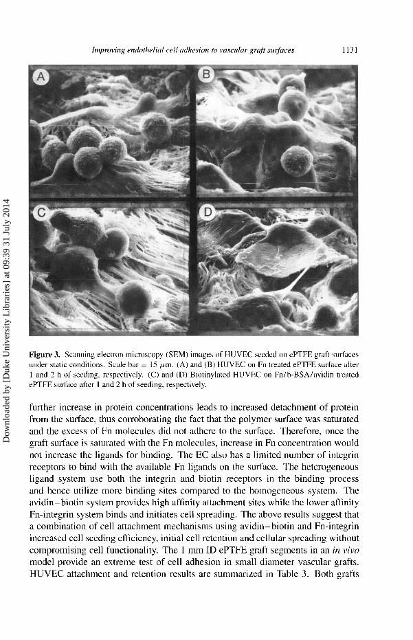
the platelets to the Fn on the graft surface and therefore is extremely important. SEM was used to observe HUVEC spreading on protein treated denucleated
ePTFE surfaces. Figure 3 shows SEM pictures of cells on which homogeneous and
heterogeneous ligand treated graft surfaces at 1 and 2 h after EC. The graft surfaces
in both cases showed HUVEC attachment and partial cellular coverage. However,
biotinylated HUVEC on heterogeneous ligand treated surface showed more cellular
spreading at both 1- and 2-h intervals. Along with a flatter hydrodynamic shape and increased area for distribution of stresses, a spread cell could also have a
higher number of adhesive bonds, increasing its overall attachment strength. Similar
results were obtained with BAEC spreading on heterogeneous ligand treated glass surfaces [74].
Honudivilla et al. [89] have demonstrated that a Fn concentration of 20 Itg ml- in
solution incubated on PTFE for 1 h provided maximum surface coverage of Fn. Any
Dow
nloa
ded
by [
Duk
e U
nive
rsity
Lib
rari
es]
at 0
9:39
31
July
201
4
1131
Figure 3. Scanning electron microscopy (SEM) images of HUVEC seeded on ePTFE graft surfaces under static conditions. Scale bar = IS 11m. (A) and (B) HUVEC on Fn treated ePTFE surface after 1 and 2 h of seeding, respectively. (C) and (D) Biotinylated HUVEC on Fn/b-BSA/avidin treated ePTFE surface after 1 and 2 h of seeding, respectively.
further increase in protein concentrations leads to increased detachment of protein from the surface, thus corroborating the fact that the polymer surface was saturated
and the excess of Fn molecules did not adhere to the surface. Therefore, once the
graft surface is saturated with the Fn molecules, increase in Fn concentration would
not increase the ligands for binding. The EC also has a limited number of integrin
receptors to bind with the available Fn ligands on the surface. The heterogeneous
ligand system use both the integrin and biotin receptors in the binding process and hence utilize more binding sites compared to the homogeneous system. The
avidin-biotin system provides high affinity attachment sites while the lower affinity
Fn-integrin system binds and initiates cell spreading. The above results suggest that
a combination of cell attachment mechanisms using avidin-biotin and Fn-integrin increased cell seeding efficiency, initial cell retention and cellular spreading without
compromising cell functionality. The 1 mm ID ePTFE graft segments in an in vivo
model provide an extreme test of cell adhesion in small diameter vascular grafts. HUVEC attachment and retention results are summarized in Table 3. Both grafts
Dow
nloa
ded
by [
Duk
e U
nive
rsity
Lib
rari
es]
at 0
9:39
31
July
201
4

1132
Table 3.
Patency rates, number of EC attached per unit graft surface area SEM), percentage of EC retained on the surface SEM) on modified denucleated ePTFE grafts in an in vivo rat model
were seeded with same cell density (2 x 10⁵ cells/ml) of EC in solution for the same
seeding time period of 15 min. However, EC arttached to the heterogeneous ligand treated grafts attached in significantly (p < 0.05) greater number as compared to the
homogeneous ligand treated grafts. The percentage of EC retained on the surface
of the heterogeneous ligand treated grafts after experiencing 15 min of blood flow
was also significantly (p < 0.05) higher than the homogeneous ligand treated grafts
(46 vs 26%). In both cases, the enhancement was nearly a factor of two, suggesting that the heterogeneous ligand treatment not only increased EC attachment but also
increased EC retention of attached cells.
Work in progress includes preparing and testing an equilibrium model to explain cell adhesion on heterogeneous ligand treated surfaces. Preliminary results suggests the two receptor-ligand complexes to function like springs in parallel and the initial
strength of cell attachment for the heterogeneous ligand system is the sum of the
forces resisted by the individual systems.
Acknowledgements
The work was supported in part by NIH grants HL-44972, HL-32132 and by NSF
grant BES 94-21425.
REFERENCES
1. L. A. Harker, S. J. Slichter and L. R. Sauvage, Ann. Surg. 186, 594 (1977). 2. J. C. Prostlethwaite, E. B. Goyle, J. A. Dormandly and J. W. Hyno, Br. J. Surg. 64, 28 (1977). 3. R. E. Clark, P. L. Gould, W. M. Swanson and G. A. M. Butterworth, Biomater. Med. Devices
Artif. Organs 2, 379 (1974). 4. M. Herring, in: Vascular Grafts: Clinical Applications and Techniques, C. B. Wright (Ed.),
p. 275. John Wright PSG, Boston (1985). 5. P. Zilla, U. von Oppell and M. Deutsch, J. Card. Surg. 8, 32 (1993). 6. M. A. Gimbrone, Jr., in: Vascular Endothelium in Hemostasis and Thrombosis, M. A. Gim-
brone, Jr. (Ed.), p. 1. Churchill Livingstone, Edinburgh (1986). 7. J. D. Pearson, in: Endothelialization of Vascular Grafts, P. Zilla, R. Fasol and M. Deutsch (Eds),
p. 71. S. Kager, Basel (1987). 8. C. L. Verweij, Heamostasis 18, 224 (1988). 9. H. P. Greisler, J. J. Klosak, J. F. McGurrin, E. D. Endean, J. Ellinger, J. D. Pozar, S. C. Henderson
and D. U. Kim, J. Cardiovasc. Surg. 31, 640 (1990).
Dow
nloa
ded
by [
Duk
e U
nive
rsity
Lib
rari
es]
at 0
9:39
31
July
201
4

1133
10. M. Herring, A. L. Gardner and J. Glover, J. Surgery 84, 498 (1978). 11. M. B. Herring, J. Vasc. Surg. 13, 731 (1991). 12. F. J. Schoen, P. Libby and P. Didishiem, Cardiovasc. Pathol. 2(S), 209 (1993). 13. R. Service, Science 270, 230 (1995). 14. A. Dekker, T. Beugiling, A. Poot, J. Spijders, J. A. van Mourik, J. Feijen, A. Bantjes and
W. G. van Aken, Clin. Mater. 13, 101 (1993). 15. J. B. Sharefkin, C. H. Latker and P. A. D'Amore, J. Surg. Res. 34, 33 (1983). 16. J. C. Stanley, W. E. Burkel and J. W. Ford, Surgery 92, 994 (1982). 17. W. E. Burkel, J. W. Ford, D. W. Vinter, R. H. Kahn, L. M. Graham and J. C. Stanley, Trans. Am.
Soc. Artif. Int. Organs 28, 178 (1982). 18. A. Shepard, J. Eldup, E. M. Keough, T. F. Foxall, K. Romberg, R. J. Conolly and A. D. Callow,
Surgery 99, 318 (1986). 19. L. Poole-Warren, P. Slowiaczek, K. Noble, A. Graham and K. Schindhelm, in: Fifth World
Biomaterials Congress. Toronto, Canada (1996). 20. N. James, K. Schindhelm, P. Slowlaczek and B. Millthorpe, in: Bioengineering Conference
(1993). 21. M. B. Herring, R. Dilley, T. Cullison, A. Gardner and J. Glover, J. Surg. Res. 28, 35 (1980). 22. J. Stanley, Trans. Am. Soc. Artif. Int. Organs 28, 613 (1982). 23. P. Ortenwall, A. Bylock, T. Kjellstrom and B. Risberg, Surgery 103, 199 (1988). 24. G. P. Clagett, W. E. Burkel, J. B. Sharefkin and J. W. Ford, Circulation 69, 632 (1984). 25. J. B. Sharefkin, C. Latker, M. Smith, D. Cruess, P. Clagett and N. M. Rich, Surgery 92, 385
( 1982). 26. B. Allen, J. Long, R. Clark et al., J. Vasc. Surg. 1 (1984). 27. S. P. Schmidt, T. J. Hunter and M. Hirko, J. Vasc. Surg. 2, 292 (1985). 28. S. P. Schmidt, T. J. Hunter, L. Falkow, M. Evancho and W. V. Sharp, ASAIO 8, 99 (1985). 29. R. Kempczinski, J. Rosenman and G. Ramalanjoana, J. Vasc. Surg. 2, 424 (1985). 30. S. Hussain, J. L. Glover, N. Augelli, P. J. Bendick, D. Maupin and M. McKain, J. Vasc. Surg. 9,
656 ( 1989). 31. W. V. Sharpe, S. P. Schmidt and D. L. Donovan, J. Vasc. Surg. 3, 256 (1986). 32. J. V. Smyth, P. D. F. Dodd and M. G. Walker, Br. J. Surg. 82, 588 (1995). 33. M. B. Herring, A. Gardner and J. L. Glover, J. Am. Soc. Artif. Int. Organs 8, 74 (1985). 34. M. B. Herring, A. Gardner and J. L. Glover, J. Vasc. Surg. 1, 279 (1984). 35. M. Herring, R. Crompton, D. LeGrand, A. Gardner, D. Madison and J. Glover, J. vasc. Surg. 6,
114 (1987). 36. P. Ortenwall, H. Wadenvik, J. Kutti and B. Risberg, J. Vasc. Surg. 6, 17 (1987). 37. P. Ortenwall, H. Wadenvik and B. Risberg, J. Vasc. Surg. 10, 374 (1989). 38. M. G. Walker, G. J. L. Thompson and J. W. Shaw, in: Endothelialization of Vascular Grafts,
P. Zilla, R. Fasol and M. Deutsch (Eds), p. 245. S. Karger, A.G., Basel (1987). 39. P. Zilla, R. Fasol, M. Deutsch, T. Fischlein, E. Minar, A. Hammerle, O. Krapicka and M. Kadletz,
J. Vasc. Surg. 6, 535 (1987). 40. R. Fasol, P. Zilla, M. Deutsch, M. Grimm and T. Fischlein, J. Vasc. Surg. 9, 432 (1989). 41. L. M. Graham, W. E. Burkel, J. W. Ford, D. W. Vinter, R. H. Kahn and J. C. Stanley, Arch. Surg.
115, 1289 ( 1980). 42. J. B. Sharefkin, H. E. van Wart, D. F. Cruess, R. A. Albus and E. M. Levine, J. Vasc. Surg. 4,
567 (1986). 43. G. Leseche, E. Bikfalvi and G. Tobelem, Surgery 105, 36 (1989). 44. S. Shindi, A. Takagi and A. D. Whittemore, J. vasc. Surg. 6, 325 (1987). 45. P. Zilla, R. Fasol and U. Dudeck, J. Vasc. Surg. 12, 180 (1990). 46. W. H. Pearce, R. B. Rutherford and T. A. Whitehill, J. Vasc. Surg. 5, 203 (1987). 47. A. V. Sterpetti, W. J. Hunter and R. D. Schultz, J. Vasc. Surg. 7, 677 (1988). 48. S. K. Williams, B. E. Jarrell and D. G. Rose, Ann. Vasc. Surg. 3, 146 (1989).
Dow
nloa
ded
by [
Duk
e U
nive
rsity
Lib
rari
es]
at 0
9:39
31
July
201
4

1134
49. P. K. Park, B. E. Jarrell and S. K. Williams, J. Vasc. Surg. 11, 468 (1990). 50. S. K. Williams, D. G. Rose and B. E. Jarrell, J. Biomed. Mat. Res. 28, 203 (1994). 51. S. O. Meerbaum, W. V. Sharp and S. P. Schmidt, in: Applied Cardiovascular Biology 1990-91,
P. Zilla, R. Fasol and A. Callow (Eds), p. 107. S. Karger, A.G., Basel (1992). 52. B. Potzsch, J. Grulich-Henn and R. Rossinger, Lab. Invest. 63, 841 (1990). 53. M. J. T. Visser, H. Bockel and G. P. van Muijen, J. Vasc. Surg. 13, 373 (1991). 54. J. E. Rosenmann, R. F. Kempczinski, W. H. Pearce and E. B. Silberstein, J. Vasc. Surg. 2, 778
(1985). 55. K. Kesler, M. Herring and M. Arnold, J. Vasc. Surg. 3, 58 (1986). 56. J. G. Steele, G. Johnson, C. MacFarland, B. A. Dalton, T. R. Gengenbatch, R. C. Chatelier,
P. A. Underwood and H. R. Griesser, J. Biomater. Sci. Polymer Edn 6, 511 (1994). 57. J. S. Randomski, B. E. Jarrell, S. K. Williams, E. A. Koolpe, D. A. Greener and R. A. Carabasi,
J. Surg. Res. 42, 133 (1987). 58. M. D. Pierschbacher and E. Ruoslahti, J. Biol. Chem. 262, 17 294 (1987). 59. E. Ruoslahti and M. D. Pierschbacher, J. Biol. Chem. 238, 491 (1987). 60. H.-B. Lin, K. B. Lewis, D. Leach-Scampavia, B. Ratner and S. L. Cooper, J. Biomater. Sci.
Polymer Edn 4, 183 (1993). 61. S. P. Massia, S. S. Rao and J. A. Hubbell, J. Biol. Chem. 268, 8053 (1993). 62. S. P. Massia and J. A. Hubbell, J. Biol. Chem. 267, 14 019 (1992). 63. S. P. Massia and J. A. Hubbell, J. Cell Biol. 114, 1089 (1991). 64. J. A. Hubbell, Bio/Technology 13, 565 (1995). 65. G. Ramalanjaona, R. F. Kempczinski, J. E. Rosenman, C. E. Douville and E. B. Silberstein,
J. Vasc. Surg. 3, 264 (1986). 66. J. B. Cambell, C. Lundgren, M. B. Herring, A. Hubbard, M. Arnold, H. M. Park, B. Mock and
J. L. Glover, ASAIO J. 8, 113 (1985). 67. R. Vohra, G. J. L. Thomson, H. Sharma, H. M. H. Carr and M. G. Walker, Eur. J. Vasc. Surg. 4,
33 ( 1990). 68. T. Miyata, M. Conte, L. Trudell, D. Mason, A. Wittemore and L. Biryini, J. Surg. Res. 50, 485
(1991). 69. J. Mazzucotelli, C. Klein-Soyer, A. Beretz, C. Brisson, G. Archipoff and J. Cazenave, Biomate-
rials 14, 482 (1991). 70. J. Kaehler, P. Zilla, R. Fasol, M. Deutsch and M. Kadletz, J. Vasc. Surg. 9, 535 (1989). 71. S. K. Akiyama and K. M. Yamada, J. Biol. Chem. 60, 4492 (1985). 72. V. D. Bhat, G. A. Truskey and W. M. Reichert, J. Biomed. Mater. Res. 40, 57 (1998). 73. N. Green, Adv. Protein Chem. 29, 85 (1975). 74. V. D. Bhat, G. A. Truskey and W. M. Reichert, J. Biomed. Mater. Res. (1998) (in press). 75. L. A. Trudell, L. Boudreau and J. M. van de Water, Trans. Am. Soc. Artif. Int. Organs 24, 320
(1978). 76. E. F. Ritter, R. D. Vann, C. Wyble, W. J. Barwick and B. Klitzman, Am. J. Physiol. 257, 1076
(1989). 77. M. Alexeyev, Bio Techniques 18, 58 (1995). 78. R. Levy-Toledano, H. L. Caro, N. Hindman and S. Toaylor, Endocrinology 133, 1803 (1993). 79. J. Burmeister, Duke University (1995). 80. J. S. Burmeister, J. Vrany, W. M. Reichert and G. A. Truskey, J. Biomed. Mater. Res. 30, 13
( 1996). 81. T. V. du Laney, G. A. Truskey and B. Klitzman, in: Fifth Annual Biomaterials Congress. Toronto,
Canada (1996). 82. S. Usami, H. H. Chen, Y. Zhao, S. Chien and R. Skalak, J. Biomed. Eng. 21, 77 (1993). 83. S. Budd, K. E. Allen, P. R. F. Bell and R. F. L. James, J. Vasc. Surg. 12, 126 (1990). 84. R. Vohra, G. J. L. Thomson, H. M. H. Carr, H. Sharma and M. G. Walker, Surgery 111, 210
(1990).
Dow
nloa
ded
by [
Duk
e U
nive
rsity
Lib
rari
es]
at 0
9:39
31
July
201
4

1135
85. B. Lindblad, S. W. Wright, R. L. Sell, W. E. Burkel, L. M. Graham and J. C. Stanley, J. Biomed. Mater. Res. 21, 1013 (1987).
86. S. M. Slack and V. T. Turitto, Cardiovasc. Pathol. 2, 11 (1993). 87. J. K. Seeger and N. Klingman, J. Surg. Res. 38, 641 (1985). 88. F. Grinnel, M. Feld and W. Snell, Cell Biol. Int. Rep. 3, 585 (1979). 89. N. G. Honduvilla, J. Bujan, M. A. Lizarbe, J. M. Bellon, N. Olmo and A. Hernando, Artificial
Organs 19, 144 (1994). 90. T. A. Belden, S. P. Schmidt, L. J. Falkow and W. V. Sharp, Trans. Am. Soc. Artif. Int. Organs 28,
173 (1982).
Dow
nloa
ded
by [
Duk
e U
nive
rsity
Lib
rari
es]
at 0
9:39
31
July
201
4