Impact of retinoic acid exposure on midfacial shape variation and manifestation of holoprosencephaly...
40
1 Impact of retinoic acid exposure on midfacial shape variation and manifestation of holoprosencephaly in Twisted gastrulation mutant mice Charles J. Billington Jr. a,b , Brian Schmidt a , Ralph S. Marcucio c , Benedikt Hallgrimsson d , Rajaram Gopalakrishnan e , Anna Petryk a.b* a Department of Pediatrics, University of Minnesota, Minneapolis, MN 55454, USA; b Department of Genetics, Cell Biology and Development, University of Minnesota, Minneapolis, MN 55454, USA c Department of Orthopedic Surgery, University of California, San Francisco, CA 94110, USA d Department of Cell Biology & Anatomy, University of Calgary, Calgary, AB T2N 4N1, Canada e Diagnostic/Biological Sciences, School of Dentistry, University of Minnesota, Minneapolis, MN 55455, USA Email addresses: Charles J. Billington Jr.: [email protected] Brian Schmidt: [email protected] Ralph Marcucio: [email protected] Benedikt Hallgrimsson: [email protected] Rajaram Gopalakrishnan: [email protected] Anna Petryk: [email protected] Running title: Retinoic acid and holoprosencephaly Keywords: Twisted gastrulation, Bone morphogenetic protein, holoprosencephaly, retinoic acid, apoptosis, oxidative stress. * To whom correspondence should be addressed: Dr. Anna Petryk University of Minnesota Children’s Hospital Pediatric Endocrinology East Building Room MB671 2450 Riverside Ave. Minneapolis, MN 55454 Phone: 612-624-5409 Fax: 612-626-5262 E-mail: [email protected] © 2014. Published by The Company of Biologists Ltd. This is an Open Access article distributed under the terms of the Creative Commons Attribution License (http://creativecommons.org/licenses/by/3.0), which permits unrestricted use, distribution and reproduction in any medium provided that the original work is properly attributed. Disease Models & Mechanisms DMM Accepted manuscript http://dmm.biologists.org/lookup/doi/10.1242/dmm.018275 Access the most recent version at DMM Advance Online Articles. Posted 2 December 2014 as doi: 10.1242/dmm.018275
-
Upload
independent -
Category
Documents
-
view
2 -
download
0
Transcript of Impact of retinoic acid exposure on midfacial shape variation and manifestation of holoprosencephaly...

1
Impact of retinoic acid exposure on midfacial shape variation and
manifestation of holoprosencephaly in Twisted gastrulation mutant mice
Charles J. Billington Jr.a,b, Brian Schmidta, Ralph S. Marcucioc, Benedikt Hallgrimssond, Rajaram Gopalakrishnane, Anna Petryka.b*
aDepartment of Pediatrics, University of Minnesota, Minneapolis, MN 55454, USA; bDepartment of Genetics, Cell Biology and Development, University of Minnesota, Minneapolis,
MN 55454, USA cDepartment of Orthopedic Surgery, University of California, San Francisco, CA 94110, USA
dDepartment of Cell Biology & Anatomy, University of Calgary, Calgary, AB T2N 4N1, Canada eDiagnostic/Biological Sciences, School of Dentistry, University of Minnesota, Minneapolis,
MN 55455, USA Email addresses:
Charles J. Billington Jr.: [email protected] Brian Schmidt: [email protected] Ralph Marcucio: [email protected] Benedikt Hallgrimsson: [email protected] Rajaram Gopalakrishnan: [email protected] Anna Petryk: [email protected]
Running title: Retinoic acid and holoprosencephaly Keywords: Twisted gastrulation, Bone morphogenetic protein, holoprosencephaly, retinoic acid, apoptosis, oxidative stress.
* To whom correspondence should be addressed: Dr. Anna Petryk University of Minnesota Children’s Hospital Pediatric Endocrinology East Building Room MB671 2450 Riverside Ave. Minneapolis, MN 55454 Phone: 612-624-5409 Fax: 612-626-5262 E-mail: [email protected]
© 2014. Published by The Company of Biologists Ltd.This is an Open Access article distributed under the terms of the Creative Commons Attribution License (http://creativecommons.org/licenses/by/3.0), which permits unrestricted use, distribution and reproduction in any medium provided that the original work is properly attributed.
Dise
ase
Mod
els &
Mec
hani
sms
D
MM
Acce
pted
man
uscr
ipt
http://dmm.biologists.org/lookup/doi/10.1242/dmm.018275Access the most recent version at DMM Advance Online Articles. Posted 2 December 2014 as doi: 10.1242/dmm.018275

2
ABSTRACT
Holoprosencephaly (HPE) is a developmental anomaly characterized by inadequate or absent
midline division of the embryonic forebrain and midline facial defects. It is believed that gene-
environment interactions play a role in the widely variable penetrance and expressivity of HPE,
although a direct investigation of such effects has been limited. The goal of this study was to
examine if mice carrying a mutation in a gene encoding a BMP antagonist Twisted gastrulation
(Twsg1) associated with a low penetrance of HPE are sensitized to retinoic acid (RA)
teratogenesis. Pregnant Twsg1+/- dams were treated by gavage with a low dose of all-trans RA
(3.75 mg/kg). Embryos were analyzed between E9.5 and E11.5 by microscopy and geometric
morphometric analysis by microCT. P19 embryonal carcinoma cells were used to examine
potential mechanisms mediating combined effects of increased BMP and retinoid signaling.
While only 7% of wild type embryos exposed to RA showed overt HPE or neural tube defects
(NTD), 100% of Twsg1 null mutants exposed to RA manifested severe HPE compared to 17%
without RA. Remarkably, up to 30% of Twsg1+/- mutants also showed HPE (23%) or NTD
(7%). The majority of shape variation among Twsg1+/- mutants was associated with narrowing
of the midface. In P19 cells, RA induced the expression of Bmp2, acted in concert with BMP to
increase p53 expression, caspase activation, and oxidative stress. This study provides direct
evidence for modifying effects of the environment in a genetic mouse model carrying a
predisposing mutation for HPE in the Twsg1 gene. Further study of the mechanisms underlying
these gene-environment interactions in vivo will contribute to better understanding of the
pathogenesis of birth defects and present an opportunity to explore potential preventive
interventions.
Dise
ase
Mod
els &
Mec
hani
sms
D
MM
Acce
pted
man
uscr
ipt

3
INTRODUCTION
Holoprosencephaly (HPE) is a malformation characterized by inadequate or absent
midline division of the embryonic forebrain. Incomplete brain septation is accompanied by
corresponding midline facial defects in about 80% of the cases (Geng and Oliver, 2009) and, less
frequently, jaw defects (Pauli et al., 1983). HPE is the most common defect of the developing
forebrain with an incidence of 1 in 250 conceptuses and about 1 in every 10,000 at term (Orioli
and Castilla, 2010; Roessler et al., 1996). An important feature of HPE is its incomplete
penetrance and expressivity. Even in families with defined mutations some individuals may have
no recognizable defects, some have mild forms (referred to as microforms, such as hypotelorism,
midfacial hypoplasia, or a single maxillary central incisor), and some are severely affected with
cyclopia or proboscis (Roessler et al., 1996). The basis of this phenotypic variability is poorly
understood.
HPE can result from widely diverse causes, including both genetic and environmental
etiologies. It has been speculated that genetic and environmental factors may have a cumulative
effect, accounting for its varied penetrance and expressivity (Ming and Muenke, 2002). The most
common genetic cause of HPE in humans are mutations in SHH (Roessler et al., 1996). Some
examples of environmental factors that have been associated with development of HPE in
humans are ethyl alcohol, poorly controlled maternal diabetes mellitus, retinoic acid (RA)
(Cohen and Shiota, 2002), and hypoxia-ischemia (Siebert, 2007). All of these exposures are
associated with elevated levels of reactive oxygen species (ROS) (Aoto et al., 2008; Davis et al.,
1990; Kay et al., 2000; Ornoy, 2007), suggesting a role for oxidative stress in mediating their
teratogenic effects.
Dise
ase
Mod
els &
Mec
hani
sms
D
MM
Acce
pted
man
uscr
ipt

4
Experimental models of HPE to study these interactions are very limited because unlike
humans, mice carrying classical HPE gene mutations do not usually show phenotypic variability.
For example, disruption of SHH pathway in mice has profound effects on embryonic
development with all Shh null embryos manifesting severe HPE (Chiang et al., 1996), while in
humans only 37% of carriers of SHH mutations develop HPE (Cohen, 1989). Other, less
classical mouse models of HPE, however, do show incomplete penetrance and phenotypic
variability, making them potentially more amenable to environmental manipulation with a
resultant shift in a phenotypic outcome. For example loss of Bone morphogenetic protein (BMP)
antagonists, such as Chordin, Noggin, or Twisted gastrulation (TWSG1) leads to reduction in
Shh expression in the ventral neural midline and recapitulates a spectrum of HPE phenotypes in
mice (Anderson et al., 2002; Lana-Elola et al., 2011; Petryk et al., 2004). As with BMPs,
exogenous RA can also lead to loss of Shh expression and HPE (Helms et al., 1997; Sulik et al.,
1995). Although it is currently unknown whether mice with disrupted BMP signaling are more
susceptible to RA teratogenic effects, there is evidence that both pathways can cooperate during
development, for example during vertebrate limb outgrowth, by inducing interdigital apoptosis
(Rodriguez-Leon et al., 1999).
The primary goals of this work were 1) to examine if a mutation in a gene encoding a
BMP binding protein TWSG1 confers susceptibility to RA exposure, and 2) whether this effect
can be quantified by microCT of the craniofacial region. We chose Twsg1 mouse model because
of a relatively low baseline incidence of HPE and an increase in apoptosis as a mechanism of
craniofacial defects in these mice (MacKenzie et al., 2009). A secondary goal was to examine
potential underlying mechanisms using P19 cells as a validated in vitro model of BMP/RA
Dise
ase
Mod
els &
Mec
hani
sms
D
MM
Acce
pted
man
uscr
ipt

5
interactions. We hypothesized that Twsg1-/- mice would be particularly sensitive to the
subteratogenic effects of RA, the midface would be most significantly affected, and the effects of
a combined treatment of P19 cells with BMP and RA would be mediated through upregulation of
apoptosis.
RESULTS
Twsg1-/- mice are sensitized to retinoic acid teratogenesis
Our first step in examining the sensitivity of TWSG1-deficient mice to RA was to
establish an appropriate treatment dosage that would cause a low but observable incidence
defects in WT mice, which could then be used to assess the sensitivity of TWSG1-deficient
mice. Since a dose of ATRA of 7.5 mg/kg has been previously shown to cause significant HPE
(Kotch et al., 1995), we also tested a lower dose of 3.75 mg/kg. Treatment with 7.5 mg/kg of
ATRA was overwhelmingly teratogenic and led to 94% of WT embryos showing defects with
about 2/3 of the embryos showing HPE and 1/3 with a neural tube defect (NTD) (Table 1).
However, treatment with 3.75 mg/kg of ATRA led to 7% of embryos affected with a vast
majority of these affected embryos showing HPE (Table 1). Therefore, we selected 3.75 mg/kg
of ATRA (referred to as low dose ATRA) as our dose for future experiments with Twsg1+/- and
Twsg1-/- mice.
While only 7% of WT embryos exposed to low dose ATRA showed overt HPE or NTD,
100% of Twsg1 null mutants manifested HPE compared to 17% without exposure to ATRA
(Table 1, Fig. 1, p=6x10-12). This rate of defects is far more than what would be expected based
solely on adding the prevalence of defects from this dose in WT mice and untreated Twsg1-/-
Dise
ase
Mod
els &
Mec
hani
sms
D
MM
Acce
pted
man
uscr
ipt
6
mice. Remarkably, even 30% of heterozygous Twsg1 mutants, which are phenotypically normal
without ATRA exposure, showed neural defects (predominantly HPE). This represents a
statistically significant increase over the incidence seen in wild type embryos with the same
dosage (p=0.01) and likewise over the 0% incidence seen in untreated heterozygotes (p=1x10⁻⁶).
Thus, TWSG1 deficiency increased the teratogenic effect of low dose ATRA in both the
homozygous and heterozygous states.
Geometric morphometric (GM) analysis of facial shape of Twsg1+/- mice demonstrates a
continuum of midfacial dysmorphology after exposure to a low dose of ATRA
While Twsg1-/- embryos exhibited severe HPE phenotypes (cyclopia or proboscis) after in
utero exposure to a low dose ATRA, Twsg1+/- showed a range of defects of variable severity. To
quantify these defects, GM was employed. The analysis included only Twsg1+/- embryos because
severe HPE phenotypes in homozygotes precluded landmark assignment. PC1, which reflects
narrowing of the midface, was the only PC that discriminated between treatment groups and
accounted for 49% of the total variance. As shown in Fig. 2, while untreated Twsg1+/- embryos
clustered with wild type (WT) embryos, those that were affected by low dose ATRA treatment
could be clearly discriminated along PC1. Thus, ATRA treatment resulted in a continuum of
midfacial narrowing in mice heterozygous for Twsg1 mutation.
ATRA induces the expression of RA-responsive genes in P19 cells
To test potential mechanisms underlying the acute sensitivity of Twsg1 mutant mice to
ATRA, we selected P19 mouse embryonal carcinoma cells as an experimental system because
they resemble embryonic cells, represent a homogenous cell population that is amenable to
Dise
ase
Mod
els &
Mec
hani
sms
D
MM
Acce
pted
man
uscr
ipt

7
quantitative assays, and have been used by others as a model for BMP/retinoid signaling
interactions (Fujita et al., 1999; Glozak and Rogers, 1996; Glozak and Rogers, 1998). P19 cells
have been previously reported to be sensitive to retinoids (Xi and Yang, 2008). We were able to
confirm this sensitivity by observing the transcriptional induction of several known RA target
genes after 1 µM ATRA treatment, including RA receptors alpha and beta (Balmer and
Blomhoff, 2002; Sucov et al., 1990), RA hydrolase Cyp26a1 (Loudig et al., 2000), Crbp1 (Xu et
al., 2001), and Hox transcription factors HoxA1 and HoxB1 (Balmer and Blomhoff, 2002;
Dekker et al., 1993; Dupe et al., 1997) (Fig. 3).
ATRA upregulates the expression of Bmp2 and its downstream targets in P19 cells
Since TWSG1’s only known mode of action is through regulation of BMP signaling, it
was essential that the P19s be competent to respond to BMPs to mimic what occurs in vivo.
Although there is some evidence that TWSG1 can promote BMP activity in some species, in
mice it appears to act mostly as a BMP antagonist (Larrain et al., 2001; Nosaka et al., 2003;
Oelgeschlager et al., 2003; Petryk et al., 2005; Ross et al., 2001; Sotillo Rodriguez et al., 2009;
Wills et al., 2006). We examined the expression of several BMP pathway genes and BMP targets
after BMP treatment alone, after ATRA treatment alone and after combined treatment (Fig. 4).
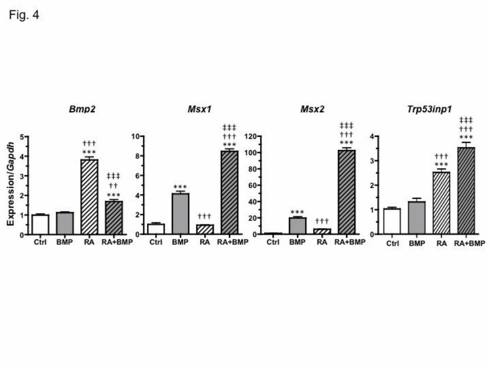
We found, consistent with previous reports (Heller et al., 1999), that Bmp2 was upregulated in
response to ATRA. The BMP targets Msx1 and Msx2 (Davidson, 1995; Liu et al., 2005; Vainio
et al., 1993) showed significant induction by BMP2 alone. With ATRA alone, Msx1 was not
induced, while Msx2 increased 6.2-fold compared to the control group, although not sufficiently
to test as statistically significant. However, when BMP and ATRA treatments were combined,
Dise
ase
Mod
els &
Mec
hani
sms
D
MM
Acce
pted
man
uscr
ipt

8
both Msx1 and Msx2 showed dramatically higher induction than with BMP alone (about 2-fold
increase for Msx1 and 5-fold increase for Msx2 compared to BMP alone).
The p53 pathway is activated in P19 cells treated with BMP and RA together
The upregulation of the p53 target Trp53inp1 (Tomasini et al., 2003) is indicative of
increased p53 transcriptional activity in the cell and activation of the p53 pathway. Following
treatment with BMP2, the expression of Trp53inp1 was not significantly changed (Fig. 4).
ATRA, however, significantly increased Trp53inp1 expression. In response to a combined
treatment with ATRA and BMP2, significantly more expression was observed beyond even that
seen with ATRA alone.
RA acts in concert with BMP to increase caspase 3/7 activation in P19 cells
Combined BMP2 and ATRA treatment has been previously shown to induce apoptosis in
P19 cells as indicated by assessment of DNA fragmentation using cell sorting or direct
electrophoresis; or by microscopic examination of cells for condensed chromatin (Fujita et al.,
1999; Glozak and Rogers, 1996; Glozak and Rogers, 1998). We have been able to corroborate
this finding by examining caspase 3/7 activation. Treatment of P19 cells with BMP2 and ATRA
resulted in a significant increase in the activity of a mediator of apoptosis caspase 3/7 compared
to control, BMP2 or ATRA alone (Fig. 5). To examine if oxidative stress can have similar
effects, P19 cells were treated with the complex III electron transport inhibitor and inducer of
oxidative stress, antimycin A (AMA) (Garcia-Ruiz et al., 1995; Turrens et al., 1985). AMA
treatment alone also significantly increased caspase activation, supporting the link between ROS
and apoptosis.
Dise
ase
Mod
els &
Mec
hani
sms
D
MM
Acce
pted
man
uscr
ipt

9
Oxidative stress is increased by retinoic acid treatment in P19 cells
The antioxidant GSH provides the main cellular defense against oxidative damage and
can be depleted and converted to the oxidized form GSSG in conditions of oxidative stress.
Hence, GSH/GSSG ratio provides a reliable indicator of the oxidative status of a cell. As
expected, treatment of P19 cells with a prooxidant AMA resulted in a significant decrease in
GSH/GSSG ratio (Fig. 6). Cells that have been treated with BMP2 alone or low dose ATRA
alone did not show any significant changes in GSH/GSSG ratio. On the other hand, a combined
BMP2/RA treatment resulted in a markedly lower GSH/GSSG ratio. This result indicates that
RA along with BMP2 can induce oxidative stress in this cellular model of early embryonic
development.
DISCUSSION
The severity of craniofacial abnormalities can vary widely between individuals, despite
similar or identical genetic risk factors and environmental exposures. HPE is a prominent
example of such phenotypic variability. Several mechanisms have been proposed to explain this
phenomenon, such as interaction of two or more HPE genes in generating the phenotype (Nanni
et al., 1999), cumulative effects of mutations in non-classical HPE genes that result in concurrent
or sequential partial defects in more than one pathway important for forebrain development
(Andersson et al., 2006; Ming et al., 2002), presence of genetic modifiers (Nadeau, 2001),
stochastic and/or epigenetic contributions (Feinberg and Irizarry, 2010) as well as non-linearities
in the properties of signaling pathways (Young et al., 2010). The multifactorial etiology of HPE
led to the “multiple hit” hypothesis (Ming et al., 2002), in which genetic predisposition puts
individuals at risk for manifesting the disease in the presence of other exposures.
Dise
ase
Mod
els &
Mec
hani
sms
D
MM
Acce
pted
man
uscr
ipt

10
This study provides direct evidence for such modifying effects of the environment in a
genetic mouse model carrying a predisposing mutation in Twsg1 gene. Importantly, even
haploinsufficiency sensitized the Twsg1 mouse embryos to teratogenic effects of retinoic acid,
resulting in HPE, similar to Shh or Gli2 haploinsufficiency predisposing to teratogenic effects of
prenatal ethanol exposure (Kietzman et al., 2014). To quantify these effects, we used a 3D
geometric morphometric analysis of craniofacial shape by microCT (Chong et al., 2012; Johnson
et al., 2006; Nagase et al., 2008). We found that the majority of shape variation in Twsg1+/-
mouse embryos with intrauterine exposure to a low dose of ATRA was associated with
narrowing of the midface as seen in the human microforms (Roessler et al., 1996), Noggin null
mice (Lana-Elola et al., 2011), and in a chick model of HPE (Marcucio et al., 2005; Young et al.,
2010). Since the type of dysmorphology, the midfacial narrowing, is similar to that observed in
untreated Twsg1-/- embryos (MacKenzie et al., 2009), we propose that RA treatment moves the
phenotypes of heterozygotes toward a mutant phenotype.
The exact underlying mechanisms of this increased sensitivity of TWSG1-deficient
embryos to a teratogen like RA are unknown. Since the only known action of TWSG1 is through
binding BMPs in the extracellular space, and we have previously shown that BMP signaling is
increased in the absence of TWSG1 (Ross et al., 2001; Sotillo Rodriguez et al., 2009), we
speculate that BMP/RA interactions contribute to the enhanced expressivity of HPE in this
mouse model. One potential mechanism of this synergy of BMP and RA lies in the observed
induction of the BMP pathway members by RA. RA induces Bmp2 expression as shown both in
this study and in previous research (Heller et al., 1999) and may in other settings also induce
Bmp4 and Bmp7 (Rodriguez-Leon et al., 1999). The upregulation of Msx genes following a BMP
Dise
ase
Mod
els &
Mec
hani
sms
D
MM
Acce
pted
man
uscr
ipt

11
and ATRA treatment is particularly interesting since the expression of Msx2 has been linked to
induction of apoptosis in the craniofacial region, including neural crest cells (Graham et al.,
1994) and optic vesicles (Wu et al., 2003).
There is evidence that both BMP and RA pathways cooperate to induce apoptosis in vivo,
for example during vertebrate limb outgrowth (Rodriguez-Leon et al., 1999), and in in vitro
systems, including P19 cells as shown in this and other studies (Glozak and Rogers, 1996; Xi and
Yang, 2008). We have previously shown that in Twsg1-/- embryos increased apoptosis correlates
with the degree of severity of craniofacial phenotypes (MacKenzie et al., 2009). Any additional
pro-apoptotic factor would be expected to enhance this dysmorphology. In fact, excessive
apoptosis is a central common pathway in various craniofacial defects due to either exposure to
external noxious agents such as alcohol (Aoto et al., 2008; Sulik, 2005), hypoxia (Smith et al.,
2013) or gene mutations (Dixon et al., 2000; Jones et al., 2008; Phelan et al., 1997). This
pathological apoptosis appears to be at least partly p53-mediated because genetic or
pharmacological inhibition of p53 activation can significantly reduce the frequency of
craniofacial defects in Twsg1-/- and other mouse models (Tcof, Pax3) of craniofacial and neural
defects (Billington et al., 2011; Jones et al., 2008; Pani et al., 2002). Importantly, teratogens such
as RA can also by themselves induce apoptosis in craniofacial primordia (Evrard et al., 2000)
and activate p53 (Hosako et al., 2007). In this study, treatment of P19 cells with BMP and ATRA
also led to a significant upregulation of the p53 target Trp53inp1. Similarly, in keratinocytes, RA
increases the expression of p53 and proapoptotic caspases and sensitizes the cells to apoptosis by
lowering their apoptotic threshold (Mrass et al., 2004). Future studies should address whether
Dise
ase
Mod
els &
Mec
hani
sms
D
MM
Acce
pted
man
uscr
ipt

12
BMP and RA pathways can act together to lower the apoptotic threshold during key stages of
midline forebrain and facial development in vivo.
Another possible intermediate mechanism underlying hyper-responsiveness of embryos
to teratogens is oxidative stress. Studies in animal models and humans have implicated ROS
generation in the pathogenesis of craniofacial and other birth defects (Chang et al., 2003; Davis
et al., 1990; Dong et al., 2008; Kay et al., 2000; Kotch et al., 1995; Loeken, 2004; Ornoy, 2007).
Three known environmental causes of HPE, gestational diabetes, fetal alcohol or RA exposure
(Cohen and Shiota, 2002), are all associated with elevated levels of ROS (Aoto et al., 2008;
Davis et al., 1990; Kay et al., 2000; Ornoy, 2007). In fact, the ability to remove ROS is thought
to be a general mechanism to neutralize environmental toxins. One of the reasons why early
embryos may be particularly sensitive to free radical damage is their limited antioxidant
capability, partly due to an inherent deficiency of scavengers of ROS, superoxide dismutase and
catalase (Davis et al., 1990). Oxidative stress is thought to promote apoptosis, which then
disrupts normal development. Supplementation with exogenous antioxidants, including N-
acetylcysteine, vitamin C, vitamin E, superoxide dismutase, or catalase in animal models has
produced promising results in terms of reducing apoptosis and dysmorphology (Aoto et al.,
2008; Kotch et al., 1995; Loeken, 2004; Siman and Eriksson, 1997; Wentzel and Eriksson,
1998). The current study shows that ATRA in combination with BMP can lower GSH to GSSG
ratios, indicating induction of oxidative stress in P19 cells. It should also be noted that other
cellular processes may also be disrupted by RA and contribute to the phenotypic heterogeneity in
Twsg1 mutant embryos, such as premature differentiation induced by increased levels of RA
(Laue et al., 2011).
Dise
ase
Mod
els &
Mec
hani
sms
D
MM
Acce
pted
man
uscr
ipt

13
Retinoic acid (an analog of vitamin A) has also been proven to cause birth defects in
humans, including central nervous system abnormalities such as HPE (Cohen and Shiota, 2002).
The association between vitamin A and birth defects comes from studies in which high doses
were used. For example, in a study of 154 human pregnancies, in utero exposure to isotretinoin
(prescribed to treat severe cystic acne) was associated with a high risk of congenital
malformations (relative risk 25.6) (Lammer et al., 1985). All women took oral isotretinoin at
some point during the first 10 weeks after conception. This has led to an increased awareness
about teratogenic effects of retinoic acid and reduced exposure, although not complete
elimination as it continues to be prescribed for the treatment of acne, sun-damaged skin,
psoriasis, prevention of nonmelanoma skin cancer, and for cancer chemotherapy (Mrass et al.,
2004). Vitamin A alone however, when used in non-teratogenic doses (as found in over the
counter vitamin supplements), has been shown to be safe in the general population.
In summary, TWSG1-deficient mice represent a genetic mouse model of a mutation with
low penetrance that sensitizes embryos to environmental influences. The mechanisms underlying
these gene-environment interactions are poorly understood. Since similar biological processes
appear to be involved in the pathogenesis of a variety of birth defects, and in response to various
teratogens, better understanding of these interactions will likely be applicable to other birth
defects beyond craniofacial malformation.
Dise
ase
Mod
els &
Mec
hani
sms
D
MM
Acce
pted
man
uscr
ipt
14
MATERIALS AND METHODS
Mice
Mice with a targeted mutation in Twsg1 (Twsg1tm1.1 Mboc) were described previously
(Petryk et al., 2004). Wild type (WT) mice were purchased from Jackson Lab. All mice were in
C57BL/6 background. Presence of a spermatic plug was counted as embryonic day 0.5 (E 0.5).
Pregnant females were treated by gavage with all-trans retinoic acid (ATRA, Sigma, St. Louis,
MO, USA) in corn oil at doses of 3.75 or 7.5 mg/kg (Kotch et al., 1995) on the morning (10 AM)
of E7.5, which is a well defined teratogenic window for the induction of HPE (Higashiyama et
al., 2007; Lipinski et al., 2010). Subsequently the pregnant females were euthanized by CO2
inhalation, embryos were isolated at E9.5 or E10.5, and assessed for external phenotypes under
the dissecting microscope, including telencephalic vesicle abnormalities consistent with HPE and
neural tube defects. For geometric morphometric shape analysis, embryos were collected at
E11.5 and fixed in 4% paraformaldehyde with gluteraldehyde (Schmidt et al., 2010). Mice were
housed in SPF conditions. Standard chow and water were provided ad libitum. All animal
procedures were approved by the University of Minnesota Institutional Animal Care and Use
Committee.
Geometric morphometric (GM) shape analysis
GM analysis of craniofacial shape was performed by microCT. Embryos were scanned at
5 micron resolution with a Scanco μCT 35 Scanner (Scanco Medical, Brüttisellen, Switzerland).
A detailed description of this technique and computation methods have been previously
published (Chong et al., 2012; Young et al., 2010; Young et al., 2007). A set of 45 landmarks
were used to define the morphology of the embryonic face and forebrain using established
Dise
ase
Mod
els &
Mec
hani
sms
D
MM
Acce
pted
man
uscr
ipt

15
protocols (Boughner et al., 2008; Parsons et al., 2011; Schmidt et al., 2010). Landmark data are
then aligned using a generalized least-squares Procrustes superimposition algorithm to remove
size and place all individuals into a common shape space (Mitteroecker and Gunz, 2009). A
series of linear combinations of variables is created (principal components or PCs) that explain
successively smaller proportions of total variance. PC1 is computed to capture the largest
proportion of variation in the original measurements.
Cell culture and treatments
P19 mouse embryonal carcinoma cells (ATCC CRL-1825) (Glozak and Rogers, 1996)
were cultured in MEM with 10% fetal bovine serum and antibiotics, maintained by splitting ten-
fold every 2 days. Cells were treated with recombinant human BMP2 (R&D systems,
Minneapolis, MN) and/or All-Trans Retinoic Acid (ATRA). BMP2 was dissolved as a stock at
100 ng/µl in 4 mM HCl, 0.1% bovine serum albumin (BSA). ATRA was dissolved in DMSO
and kept as a stock at 10-2 M. All cells in BMP and ATRA treatment experiments were adjusted
with non-solute containing vehicles to final concentrations of 0.0001% (w/v) BSA and 0.01%
(v/v) DMSO. Antimycin-A (Sigma, St. Louis, Mo, USA) was prepared in propylene glycol at
6mg/mL.
Gene expression
P19 cells were plated into 6 well plates with 20,000 cells/well and allowed to grow
overnight. Media were removed and replaced with treatment media containing test compounds.
For RA induction experiments, cells were treated either with 1 µM ATRA or with DMSO
vehicle in the same v/v dilution. For BMP/RA experiments, cells were treated with vehicle
Dise
ase
Mod
els &
Mec
hani
sms
D
MM
Acce
pted
man
uscr
ipt

16
control, 10 ng/ml BMP2 with DMSO ATRA vehicle control, 1 µM ATRA with BSA BMP2
vehicle control, or BMP2 combined with ATRA. In both sets of experiments, after 24 hours
media were removed and 1mL of Trizol (Invitrogen, Carlsbad, CA, USA) was added for RNA
isolation according to manufacturer’s instructions. cDNA was prepared from RNA samples by
reverse transcription using the Thermoscript reverse transcription sysetem (Invitrogen, Carlsbad,
CA, USA). Quantitative PCR was performed using 2x SYBR green mastermix with ROX from
SABiosciences on an MX3000p thermocycler (Stratagene/Agilent technologies, La Jolla, CA,
USA) and analyzed using the MxPro software (Stratagene). Primers used for the following
genes: Bmp2, Crbp1, Cyp26a1, Gapdh, Hoxa1, Hoxb1, Msx1, Msx2, Rara, Rarb and Trp53inp1
are shown in Supplemental Table 1.
Caspase 3/7 activity assay
Caspase 3/7 activity was measured using reagents from the Apotox-glo triplex assay kit
(Promega, Madison, WI) according to manufacturer’s protocol. Briefly, cells were plated to a
white walled clear bottom 96 well plate with 2500 cells per well then test compounds were
added, diluted in growth media. Cells were treated with vehicle control, 10 ng/ml BMP2 with
DMSO ATRA vehicle control, 1 µM ATRA with BSA BMP2 vehicle control, and BMP2
combined with ATRA. After 24 hours, Caspase activation was measured by addition of
luminogenic Caspase substrate and measured in a Centro XS³ LB 960 Microplate Luminometer
(Berthold Technologies, Bad Wildbad, Germany).
Dise
ase
Mod
els &
Mec
hani
sms
D
MM
Acce
pted
man
uscr
ipt

17
Glutathione ratio assay
P19 cells were assayed for ratio of reduced to oxidized glutathione using the GSH/GSSG-
glo kit (Promega) according to manufacturer’s instructions. Briefly, cells were plated to a white
walled clear bottom 96 well plate with 2500 cells per well then test compounds were added,
diluted in growth media. Cells were treated with BMP/RA vehicle control, 10 ng/ml BMP2 with
DMSO ATRA vehicle control, 1 µM ATRA with BSA BMP2 vehicle control, BMP2 combined
with ATRA, Propylene glycol AMA vehicle control or 70µM AMA. After 16 hours cells were
washed once with HBSS then lysed and analyzed for GSH and GSSG content using kit-provided
reagents for luminogenic reactions.
Statistical analysis
Chi-squared tests, t-tests and ANOVA combined with Tukey’s multiple comparison tests
were performed using Prism4 (GraphPad Software, San Diego, CA, USA). Significance was
accepted at alpha of 0.05. Fisher’s exact test was used to compare frequencies of embryonic
phenotypes between different genotypes and with and without retinoic acid treatments.
ACKNOWLEDGEMENTS
P19 mouse embryonal carcinoma cells were a kind gift of Dr. Li-Na Wei. This project was
supported by the National Institutes of Health [R01 DE016601 and R56 DE023530 to A.P., R01
DE019638 to R.S.M. and R01 DE021708 to B.H.], Minnesota Medical Foundation [grant #4186-
9227-14 to A.P.]. C.J.B. was supported by the Minnesota Craniofacial Research Training
Program [R90 DE023058] and the Medical Scientist Training Program [T32 GM008244] from
the National Institutes of Health.
Dise
ase
Mod
els &
Mec
hani
sms
D
MM
Acce
pted
man
uscr
ipt

18
COMPETING INTERESTS STATEMENT
The authors have no competing interests.
AUTHOR CONTRIBUTIONS
C.J.B. and A.P. designed the research, analyzed the data, and wrote the paper; C.J.B. and B.S.
executed the experiments; R.S.M. and B.H. conducted geometric morphometric analysis by
microCT; R.J. and R.S.M. participated in data interpretation and revised the manuscript critically
for important intellectual content. All authors read and approved the final manuscript.
Dise
ase
Mod
els &
Mec
hani
sms
D
MM
Acce
pted
man
uscr
ipt

19
REFERENCES
Anderson, R. M., Lawrence, A. R., Stottmann, R. W., Bachilier, D. and
Klingensmith, J. (2002). Chordin and noggin promote organizing centers of forebrain
development in the mouse. Development 129, 4975-4987.
Andersson, O., Reissmann, E., Jornvall, H. and Ibanez, C. F. (2006). Synergistic
interaction between Gdf1 and Nodal during anterior axis development. Dev Biol 293, 370-81.
Aoto, K., Shikata, Y., Higashiyama, D., Shiota, K. and Motoyama, J. (2008). Fetal
ethanol exposure activates protein kinase A and impairs Shh expression in prechordal
mesendoderm cells in the pathogenesis of holoprosencephaly. Birth Defects Res A Clin Mol
Teratol 82, 224-31.
Balmer, J. E. and Blomhoff, R. (2002). Gene expression regulation by retinoic acid. J
Lipid Res 43, 1773-808.
Billington, C. J., Jr., Ng, B., Forsman, C., Schmidt, B., Bagchi, A., Symer, D. E.,
Schotta, G., Gopalakrishnan, R., Sarver, A. L. and Petryk, A. (2011). The molecular and
cellular basis of variable craniofacial phenotypes and their genetic rescue in Twisted gastrulation
mutant mice. Dev Biol 355, 21-31.
Boughner, J. C., Wat, S., Diewert, V. M., Young, N. M., Browder, L. W. and
Hallgrimsson, B. (2008). Short-faced mice and developmental interactions between the brain
and the face. Journal of Anatomy 213, 646-62.
Chang, T. I., Horal, M., Jain, S. K., Wang, F., Patel, R. and Loeken, M. R. (2003).
Oxidant regulation of gene expression and neural tube development: Insights gained from
diabetic pregnancy on molecular causes of neural tube defects. Diabetologia 46, 538-45.
Dise
ase
Mod
els &
Mec
hani
sms
D
MM
Acce
pted
man
uscr
ipt

20
Chiang, C., Litingtung, Y., Lee, E., Young, K. E., Corden, J. L., Westphal, H. and
Beachy, P. A. (1996). Cyclopia and defective axial patterning in mice lacking Sonic hedgehog
gene function. Nature 383, 407-13.
Chong, H. J., Young, N. M., Hu, D., Jeong, J., McMahon, A. P., Hallgrimsson, B.
and Marcucio, R. S. (2012). Signaling by SHH rescues facial defects following blockade in the
brain. Dev Dyn 241, 247-56.
Cohen, M. M., Jr. (1989). Perspectives on holoprosencephaly: Part I. Epidemiology,
genetics, and syndromology. Teratology 40, 211-35.
Cohen, M. M., Jr. and Shiota, K. (2002). Teratogenesis of holoprosencephaly. Am J
Med Genet 109, 1-15.
Davidson, D. (1995). The function and evolution of Msx genes: pointers and paradoxes.
Trends Genet 11, 405-11.
Davis, W. L., Crawford, L. A., Cooper, O. J., Farmer, G. R., Thomas, D. and
Freeman, B. L. (1990). Generation of radical oxygen species by neural crest cells treated in
vitro with isotretinoin and 4-oxo-isotretinoin. J Craniofac Genet Dev Biol 10, 295-310.
Dekker, E. J., Pannese, M., Houtzager, E., Boncinelli, E. and Durston, A. (1993).
Colinearity in the Xenopus laevis Hox-2 complex. Mech Dev 40, 3-12.
Dixon, J., Brakebusch, C., Fassler, R. and Dixon, M. J. (2000). Increased levels of
apoptosis in the prefusion neural folds underlie the craniofacial disorder, Treacher Collins
syndrome. Hum Mol Genet 9, 1473-80.
Dong, J., Sulik, K. K. and Chen, S. Y. (2008). Nrf2-mediated transcriptional induction
of antioxidant response in mouse embryos exposed to ethanol in vivo: implications for the
prevention of fetal alcohol spectrum disorders. Antioxid Redox Signal 10, 2023-33.
Dise
ase
Mod
els &
Mec
hani
sms
D
MM
Acce
pted
man
uscr
ipt

21
Dupe, V., Davenne, M., Brocard, J., Dolle, P., Mark, M., Dierich, A., Chambon, P.
and Rijli, F. M. (1997). In vivo functional analysis of the Hoxa-1 3' retinoic acid response
element (3'RARE). Development 124, 399-410.
Evrard, L., Vanmuylder, N., Dourov, N., Hermans, C., Biermans, J., Werry-Huet,
A., Rooze, M. and Louryan, S. (2000). Correlation of HSP110 expression with all-trans retinoic
acid-induced apoptosis. J Craniofac Genet Dev Biol 20, 183-92.
Feinberg, A. P. and Irizarry, R. A. (2010). Evolution in health and medicine Sackler
colloquium: Stochastic epigenetic variation as a driving force of development, evolutionary
adaptation, and disease. Proc Natl Acad Sci U S A 107 Suppl 1, 1757-64.
Fujita, E., Soyama, A., Kawabata, M. and Momoi, T. (1999). BMP-4 and retinoic acid
synergistically induce activation of caspase-9 and cause apoptosis of P19 embryonal carcinoma
cells cultured as a monolayer. Cell Death Differ 6, 1109-16.
Garcia-Ruiz, C., Colell, A., Morales, A., Kaplowitz, N. and Fernandez-Checa, J. C.
(1995). Role of oxidative stress generated from the mitochondrial electron transport chain and
mitochondrial glutathione status in loss of mitochondrial function and activation of transcription
factor nuclear factor-kappa B: studies with isolated mitochondria and rat hepatocytes. Mol
Pharmacol 48, 825-34.
Geng, X. and Oliver, G. (2009). Pathogenesis of holoprosencephaly. J Clin Invest 119,
1403-13.
Glozak, M. A. and Rogers, M. B. (1996). Specific induction of apoptosis in P19
embryonal carcinoma cells by retinoic acid and BMP2 or BMP4. Dev Biol 179, 458-70.
Dise
ase
Mod
els &
Mec
hani
sms
D
MM
Acce
pted
man
uscr
ipt

22
Glozak, M. A. and Rogers, M. B. (1998). BMP4- and RA-induced apoptosis is mediated
through the activation of retinoic acid receptor alpha and gamma in P19 embryonal carcinoma
cells. Exp Cell Res 242, 165-73.
Graham, A., Francis-West, P., Brickell, P. and Lumsden, A. (1994). The signalling
molecule BMP4 mediates apoptosis in the rhombencephalic neural crest. Nature 372, 684-6.
Heller, L. C., Li, Y., Abrams, K. L. and Rogers, M. B. (1999). Transcriptional
regulation of the Bmp2 gene. Retinoic acid induction in F9 embryonal carcinoma cells and
Saccharomyces cerevisiae. J Biol Chem 274, 1394-400.
Helms, J. A., Kim, C. H., Hu, D., Minkoff, R., Thaller, C. and Eichele, G. (1997).
Sonic hedgehog participates in craniofacial morphogenesis and is down-regulated by teratogenic
doses of retinoic acid. Dev Biol 187, 25-35.
Higashiyama, D., Saitsu, H., Komada, M., Takigawa, T., Ishibashi, M. and Shiota,
K. (2007). Sequential developmental changes in holoprosencephalic mouse embryos exposed to
ethanol during the gastrulation period. Birth Defects Res A Clin Mol Teratol 79, 513-23.
Hosako, H., Little, S. A., Barrier, M. and Mirkes, P. E. (2007). Teratogen-induced
activation of p53 in early postimplantation mouse embryos. Toxicol Sci 95, 257-69.
Johnson, J. T., Hansen, M. S., Wu, I., Healy, L. J., Johnson, C. R., Jones, G. M.,
Capecchi, M. R. and Keller, C. (2006). Virtual histology of transgenic mouse embryos for
high-throughput phenotyping. PLoS Genet 2, e61.
Jones, N. C., Lynn, M. L., Gaudenz, K., Sakai, D., Aoto, K., Rey, J. P., Glynn, E. F.,
Ellington, L., Du, C., Dixon, J. et al. (2008). Prevention of the neurocristopathy Treacher
Collins syndrome through inhibition of p53 function. Nat Med 14, 125-33.
Dise
ase
Mod
els &
Mec
hani
sms
D
MM
Acce
pted
man
uscr
ipt

23
Kay, H. H., Grindle, K. M. and Magness, R. R. (2000). Ethanol exposure induces
oxidative stress and impairs nitric oxide availability in the human placental villi: a possible
mechanism of toxicity. Am J Obstet Gynecol 182, 682-8.
Kietzman, H. W., Everson, J. L., Sulik, K. K. and Lipinski, R. J. (2014). The
teratogenic effects of prenatal ethanol exposure are exacerbated by Sonic Hedgehog or GLI2
haploinsufficiency in the mouse. PLoS One 9, e89448.
Kotch, L. E., Chen, S. Y. and Sulik, K. K. (1995). Ethanol-induced teratogenesis: free
radical damage as a possible mechanism. Teratology 52, 128-36.
Lammer, E. J., Chen, D. T., Hoar, R. M., Agnish, N. D., Benke, P. J., Braun, J. T.,
Curry, C. J., Fernhoff, P. M., Grix, A. W., Jr., Lott, I. T. et al. (1985). Retinoic acid
embryopathy. N Engl J Med 313, 837-41.
Lana-Elola, E., Tylzanowski, P., Takatalo, M., Alakurtti, K., Veistinen, L.,
Mitsiadis, T. A., Graf, D., Rice, R., Luyten, F. P. and Rice, D. P. (2011). Noggin null allele
mice exhibit a microform of holoprosencephaly. Hum Mol Genet 20, 4005-15.
Larrain, J., Oelgeschlager, M., Ketpura, N. I., Reversade, B., Zakin, L. and De
Robertis, E. M. (2001). Proteolytic cleavage of Chordin as a switch for the dual activities of
Twisted gastrulation in BMP signaling. Development 128, 4439-47.
Laue, K., Pogoda, H. M., Daniel, P. B., van Haeringen, A., Alanay, Y., von Ameln,
S., Rachwalski, M., Morgan, T., Gray, M. J., Breuning, M. H. et al. (2011). Craniosynostosis
and multiple skeletal anomalies in humans and zebrafish result from a defect in the localized
degradation of retinoic acid. Am J Hum Genet 89, 595-606.
Dise
ase
Mod
els &
Mec
hani
sms
D
MM
Acce
pted
man
uscr
ipt

24
Lipinski, R. J., Godin, E. A., O'Leary-Moore, S. K., Parnell, S. E. and Sulik, K. K.
(2010). Genesis of teratogen-induced holoprosencephaly in mice. Am J Med Genet C Semin Med
Genet 154C, 29-42.
Liu, W., Selever, J., Murali, D., Sun, X., Brugger, S. M., Ma, L., Schwartz, R. J.,
Maxson, R., Furuta, Y. and Martin, J. F. (2005). Threshold-specific requirements for Bmp4 in
mandibular development. Dev Biol 283, 282-93.
Loeken, M. R. (2004). Free radicals and birth defects. J Matern Fetal Neonatal Med 15,
6-14.
Loudig, O., Babichuk, C., White, J., Abu-Abed, S., Mueller, C. and Petkovich, M.
(2000). Cytochrome P450RAI(CYP26) promoter: a distinct composite retinoic acid response
element underlies the complex regulation of retinoic acid metabolism. Mol Endocrinol 14, 1483-
97.
MacKenzie, B., Wolff, R., Lowe, N., Billington, C. J., Jr., Peterson, A., Schmidt, B.,
Graf, D., Mina, M., Gopalakrishnan, R. and Petryk, A. (2009). Twisted gastrulation limits
apoptosis in the distal region of the mandibular arch in mice. Dev Biol 328, 13-23.
Marcucio, R. S., Cordero, D. R., Hu, D. and Helms, J. A. (2005). Molecular
interactions coordinating the development of the forebrain and face. Dev Biol 284, 48-61.
Ming, J. E., Kaupas, M. E., Roessler, E., Brunner, H. G., Golabi, M., Tekin, M.,
Stratton, R. F., Sujansky, E., Bale, S. J. and Muenke, M. (2002). Mutations in PATCHED-1,
the receptor for SONIC HEDGEHOG, are associated with holoprosencephaly. Hum Genet 110,
297-301.
Ming, J. E. and Muenke, M. (2002). Multiple hits during early embryonic development:
digenic diseases and holoprosencephaly. Am J Hum Genet 71, 1017-32.
Dise
ase
Mod
els &
Mec
hani
sms
D
MM
Acce
pted
man
uscr
ipt

25
Mitteroecker, P. and Gunz, P. (2009). Advances in Geometric Morphometrics.
Evolutionary Biology 36, 235-247.
Mrass, P., Rendl, M., Mildner, M., Gruber, F., Lengauer, B., Ballaun, C., Eckhart,
L. and Tschachler, E. (2004). Retinoic acid increases the expression of p53 and proapoptotic
caspases and sensitizes keratinocytes to apoptosis: a possible explanation for tumor preventive
action of retinoids. Cancer Res 64, 6542-8.
Nadeau, J. H. (2001). Modifier genes in mice and humans. Nat Rev Genet 2, 165-74.
Nagase, T., Sasazaki, Y., Kikuchi, T. and Machida, M. (2008). Rapid 3-dimensional
imaging of embryonic craniofacial morphology using microscopic computed tomography. J
Comput Assist Tomogr 32, 816-21.
Nanni, L., Ming, J. E., Bocian, M., Steinhaus, K., Bianchi, D. W., Die-Smulders, C.,
Giannotti, A., Imaizumi, K., Jones, K. L., Campo, M. D. et al. (1999). The mutational
spectrum of the sonic hedgehog gene in holoprosencephaly: SHH mutations cause a significant
proportion of autosomal dominant holoprosencephaly. Hum Mol Genet 8, 2479-88.
Nosaka, T., Morita, S., Kitamura, H., Nakajima, H., Shibata, F., Morikawa, Y.,
Kataoka, Y., Ebihara, Y., Kawashima, T., Itoh, T. et al. (2003). Mammalian twisted
gastrulation is essential for skeleto-lymphogenesis. Mol Cell Biol 23, 2969-80.
Oelgeschlager, M., Reversade, B., Larrain, J., Little, S., Mullins, M. C. and De
Robertis, E. M. (2003). The pro-BMP activity of Twisted gastrulation is independent of BMP
binding. Development 130, 4047-56.
Orioli, I. M. and Castilla, E. E. (2010). Epidemiology of holoprosencephaly: Prevalence
and risk factors. Am J Med Genet C Semin Med Genet 154C, 13-21.
Dise
ase
Mod
els &
Mec
hani
sms
D
MM
Acce
pted
man
uscr
ipt

26
Ornoy, A. (2007). Embryonic oxidative stress as a mechanism of teratogenesis with
special emphasis on diabetic embryopathy. Reprod Toxicol 24, 31-41.
Pani, L., Horal, M. and Loeken, M. R. (2002). Rescue of neural tube defects in Pax-3-
deficient embryos by p53 loss of function: implications for Pax-3- dependent development and
tumorigenesis. Genes Dev 16, 676-80.
Parsons, T. E., Schmidt, E. J., Boughner, J. C., Jamniczky, H. A., Marcucio, R. S.
and Hallgrimsson, B. (2011). Epigenetic integration of the developing brain and face.
Developmental Dynamics 240, 2233-44.
Pauli, R. M., Pettersen, J. C., Arya, S. and Gilbert, E. F. (1983). Familial agnathia-
holoprosencephaly. Am J Med Genet 14, 677-98.
Petryk, A., Anderson, R. M., Jarcho, M. P., Leaf, I., Carlson, C. S., Klingensmith, J.,
Shawlot, W. and O'Connor, M. B. (2004). The mammalian twisted gastrulation gene functions
in foregut and craniofacial development. Dev Biol 267, 374-86.
Petryk, A., Shimmi, O., Jia, X., Carlson, A. E., Tervonen, L., Jarcho, M. P.,
O'Connor M, B. and Gopalakrishnan, R. (2005). Twisted gastrulation and chordin inhibit
differentiation and mineralization in MC3T3-E1 osteoblast-like cells. Bone 36, 617-26.
Phelan, S. A., Ito, M. and Loeken, M. R. (1997). Neural tube defects in embryos of
diabetic mice: role of the Pax-3 gene and apoptosis. Diabetes 46, 1189-97.
Rodriguez-Leon, J., Merino, R., Macias, D., Ganan, Y., Santesteban, E. and Hurle,
J. M. (1999). Retinoic acid regulates programmed cell death through BMP signalling. Nat Cell
Biol 1, 125-6.
Dise
ase
Mod
els &
Mec
hani
sms
D
MM
Acce
pted
man
uscr
ipt
27
Roessler, E., Belloni, E., Gaudenz, K., Jay, P., Berta, P., Scherer, S. W., Tsui, L. C.
and Muenke, M. (1996). Mutations in the human Sonic Hedgehog gene cause
holoprosencephaly. Nat Genet 14, 357-60.
Ross, J. J., Shimmi, O., Vilmos, P., Petryk, A., Kim, H., Gaudenz, K., Hermanson,
S., Ekker, S. C., O'Connor, M. B. and Marsh, J. L. (2001). Twisted gastrulation is a
conserved extracellular BMP antagonist. Nature 410, 479-83.
Schmidt, E. J., Parsons, T. E., Jamniczky, H. A., Gitelman, J., Trpkov, C.,
Boughner, J. C., Logan, C. C., Sensen, C. W. and Hallgrimsson, B. (2010). Micro-computed
tomography-based phenotypic approaches in embryology: procedural artifacts on assessments of
embryonic craniofacial growth and development. BMC Dev Biol 10, 18.
Siebert, J. R. (2007). Cyclopia, aprosencephaly, and acardiac twinning: Is hypoxia-
ischemia a unifying mechanism? Am J Med Genet A 143A, 3100-6.
Siman, C. M. and Eriksson, U. J. (1997). Vitamin E decreases the occurrence of
malformations in the offspring of diabetic rats. Diabetes 46, 1054-61.
Smith, F., Hu, D., Young, N. M., Lainoff, A. J., Jamniczky, H. A., Maltepe, E.,
Hallgrimsson, B. and Marcucio, R. S. (2013). The effect of hypoxia on facial shape variation
and disease phenotypes in chicken embryos. Dis Model Mech 6, 915-24.
Sotillo Rodriguez, J. E., Mansky, K. C., Jensen, E. D., Carlson, A. E., Schwarz, T.,
Pham, L., MacKenzie, B., Prasad, H., Rohrer, M. D., Petryk, A. et al. (2009). Enhanced
osteoclastogenesis causes osteopenia in twisted gastrulation-deficient mice through increased
BMP signaling. J Bone Miner Res 24, 1917-26.
Dise
ase
Mod
els &
Mec
hani
sms
D
MM
Acce
pted
man
uscr
ipt

28
Sucov, H. M., Murakami, K. K. and Evans, R. M. (1990). Characterization of an
autoregulated response element in the mouse retinoic acid receptor type beta gene. Proc Natl
Acad Sci U S A 87, 5392-6.
Sulik, K. K. (2005). Genesis of alcohol-induced craniofacial dysmorphism. Exp Biol
Med (Maywood) 230, 366-75.
Sulik, K. K., Dehart, D. B., Rogers, J. M. and Chernoff, N. (1995). Teratogenicity of
low doses of all-trans retinoic acid in presomite mouse embryos. Teratology 51, 398-403.
Tomasini, R., Samir, A. A., Carrier, A., Isnardon, D., Cecchinelli, B., Soddu, S.,
Malissen, B., Dagorn, J. C., Iovanna, J. L. and Dusetti, N. J. (2003). TP53INP1s and
homeodomain-interacting protein kinase-2 (HIPK2) are partners in regulating p53 activity. J Biol
Chem 278, 37722-9.
Turrens, J. F., Alexandre, A. and Lehninger, A. L. (1985). Ubisemiquinone is the
electron donor for superoxide formation by complex III of heart mitochondria. Arch Biochem
Biophys 237, 408-14.
Vainio, S., Karavanova, I., Jowett, A. and Thesleff, I. (1993). Identification of BMP-4
as a signal mediating secondary induction between epithelial and mesenchymal tissues during
early tooth development. Cell 75, 45-58.
Wentzel, P. and Eriksson, U. J. (1998). Antioxidants diminish developmental damage
induced by high glucose and cyclooxygenase inhibitors in rat embryos in vitro. Diabetes 47, 677-
84.
Wills, A., Harland, R. M. and Khokha, M. K. (2006). Twisted gastrulation is required
for forebrain specification and cooperates with Chordin to inhibit BMP signaling during X.
tropicalis gastrulation. Dev Biol 289, 166-78.
Dise
ase
Mod
els &
Mec
hani
sms
D
MM
Acce
pted
man
uscr
ipt

29
Wu, L. Y., Li, M., Hinton, D. R., Guo, L., Jiang, S., Wang, J. T., Zeng, A., Xie, J. B.,
Snead, M., Shuler, C. et al. (2003). Microphthalmia resulting from MSX2-induced apoptosis in
the optic vesicle. Invest Ophthalmol Vis Sci 44, 2404-12.
Xi, J. and Yang, Z. (2008). Expression of RALDHs (ALDH1As) and CYP26s in human
tissues and during the neural differentiation of P19 embryonal carcinoma stem cell. Gene Expr
Patterns 8, 438-42.
Xu, G., Bochaton-Piallat, M. L., Andreutti, D., Low, R. B., Gabbiani, G. and
Neuville, P. (2001). Regulation of alpha-smooth muscle actin and CRBP-1 expression by
retinoic acid and TGF-beta in cultured fibroblasts. J Cell Physiol 187, 315-25.
Young, N. M., Chong, H. J., Hu, D., Hallgrimsson, B. and Marcucio, R. S. (2010).
Quantitative analyses link modulation of sonic hedgehog signaling to continuous variation in
facial growth and shape. Development 137, 3405-9.
Young, N. M., Wat, S., Diewert, V. M., Browder, L. W. and Hallgrimsson, B. (2007).
Comparative morphometrics of embryonic facial morphogenesis: implications for cleft-lip
etiology. Anat Rec (Hoboken) 290, 123-39.
Dise
ase
Mod
els &
Mec
hani
sms
D
MM
Acce
pted
man
uscr
ipt

30
FIGURE LEGENDS
Fig. 1. Phenotypic analysis of Twsg1 mutants exposed in utero to a low dose (3.75 mg/kg) of
ATRA. Lateral views of E9.5 embryos: (A) WT untreated embryo; (B) normal appearance of
ATRA treated WT embryo; (C, D) Twsg1 mutants treated with ATRA showing proboscis (P)
and absence of telencephalic vesicles. Frontal views of E10.5 embryos: (E) WT untreated
embryo; (F) normal telencephalic vesicles (T; outlined by a dotted line) of ATRA treated WT
embryo; (G,H) *HPE in Twsg1 embryos treated with ATRA.
Fig. 2. GM analysis of facial shape. (A) Principal component analysis (PCA) based on GM
analysis of facial shape in WT and Twsg1+/- mouse embryos at E11.5 with or without in utero
exposure to a low dose ATRA at E7.5. PC analysis of landmark data shows that the majority of
shape variation is associated with narrowing of the midface and that PC1 discriminates between
treatment groups. PC1 distinguishes between treated embryos and both WT and heterozygous
Twsg1 mutant embryos. (B) 3D morphing showing variation along PC1; (C) Procrustes distances
between groups. P values were obtained by permutation of the Procrustes Distance.
Fig. 3. RA target gene expression in response to ATRA treatment in P19 cells by qPCR.
The P19 cell cultures were treated with either DMSO vehicle or 1 µM ATRA for 24 hours, with
transcript levels quantified by qPCR. Gene expression was normalized to the expression of
Gapdh, and shown as a ratio to the average of the DMSO vehicle treated control expression. The
induction of RA targets in P19 cells indicates the suitability of the P19 cell model for
investigating responses to RA signaling. ***p < 0.001 by Student’s t-test.
Dise
ase
Mod
els &
Mec
hani
sms
D
MM
Acce
pted
man
uscr
ipt

31
Fig. 4. Gene expression levels in response to exogenous BMP2 and ATRA by qPCR. P19
cell cultures were treated for 24 hours with vehicle control (0.00001% BSA, 0.001% DMSO), 10
ng/mL rhBMP2, 1 µM ATRA, or 10 ng/mL rhBMP2 and 1 µM ATRA. Gene expression was
normalized to the expression of Gapdh, and shown as a ratio to the mean vehicle treated control
expression, with transcript levels quantified by qPCR. ATRA induced both BMP2 and its
downstream targets in P19 cells. Combined BMP and ATRA treatment resulted in a significant
increase in Trp53inp1 expression. *Significance by Tukey’s test for difference from control
(Ctrl); † from BMP treated; ‡ from ATRA treated; 3 marks: p < 0.001; 2 marks p < 0.01.
Fig. 5. Caspase 3/7 activity in P19 cells in response to exogenous BMP2 and ATRA. P19
cells were treated for 24 hours with vehicle control (0.00001% BSA, 0.001% DMSO), 10 ng/mL
rhBMP2, 1 µM ATRA, or 10 ng/mL BMP and 1 µM ATRA, no treatment, or 70 µM Antimycin
A as a positive control. RA acted in concert with BMP to increase caspase 3/7 activation in P19
cells. * p<0.05, ** p,0.01, *** p<0.001 by Student’s t-test.
Fig. 6. Quantitation of oxidative stress in P19 cells treated with BMP2 and ATRA. Reduced
glutathione (GSH) to oxidized glutathione (GSSG) ratio was assayed in P19 cells treated for 16
hours with vehicle control (0.0001% BSA, 0.001% DMSO), 10 ng/mL rhBMP2, 1 µM ATRA,
or 10 ng/mL BMP combined with 1 µM ATRA. 70µM Antimycin A was tested as a positive
control and compared to its vehicle, propylene glycol. As expected, treatment of P19 cells with a
prooxidant AMA resulted in a significant decrease in GSH/GSSG ratio. In addition, oxidative
stress was increased in P19 cells treated with ATRA in combination with BMP2. *p < 0.05, **p
< 0.01.
Dise
ase
Mod
els &
Mec
hani
sms
D
MM
Acce
pted
man
uscr
ipt
32
TRANSLATIONAL IMPACT
Clinical issue
Holoprosencephaly (HPE) is the most common defect of the developing forebrain with an
incidence of 1 in 250 conceptuses and about 1 in every 10,000 at term. It is characterized by
inadequate or absent midline division of the embryonic forebrain and midline facial defects. A
perplexing feature of HPE as well as other craniofacial syndromes in humans is their widely
variable penetrance and expressivity even in the case of the same single gene mutation within the
same family, with some individuals having severe defects, some mild, and some being
unaffected. It is currently unknown what causes manifestation of HPE in genetically at risk
individuals, but it has been speculated that environmental factors may play a role. This work
addresses the effects of environmental exposure in a mouse model predisposed to HPE.
Results
Twsg1 mutant mice serve as a model of human HPE because of incomplete penetrance and a
range of defects among homozygotes. The authors demonstrated that Twsg1 mutants showed
increased susceptibility to teratogenic effects of relatively low doses of retinoic acid that in
control mice cause few, if any defects. The exposure to retinoic acid occurred at embryonic day
7.5, which is the most sensitive window for teratogen-induced HPE (corresponding to 3rd to 4th
week post-fertilization in humans). Remarkably, even Twsg1 haploinsufficiency exacerbated
teratogenic effects of prenatal retinoic acid exposure. The majority of shape variation among
Twsg1+/- mutants was associated with narrowing of the midface as demonstrated by microCT
analysis. Since the only known action of TWSG1 is through binding Bone morphogenetic
Dise
ase
Mod
els &
Mec
hani
sms
D
MM
Acce
pted
man
uscr
ipt
33
proteins (BMPs) in the extracellular space, the authors hypothesized that BMP/RA interactions
would contribute to the enhanced expressivity of HPE in this mouse model. P19 cells were used
as an in vitro model to begin to understand the mechanisms mediating these gene-environment
interactions. In P19 cells, RA induced the expression of Bmp2 and its downstream targets Msx1
and Msx2, and acted in concert with BMP to increase apoptosis, p53 target gene expression, and
oxidative stress, suggesting a role for these pathways in modifying the disease outcome.
Implications and future directions
This study provides direct evidence for modifying effects of the environment in a genetic mouse
model carrying a predisposing mutation for HPE. Further study of the mechanisms underlying
these gene-environment interactions in vivo will contribute to better understanding of the
pathogenesis of birth defects and present an opportunity to explore potential preventive
interventions.
Dise
ase
Mod
els &
Mec
hani
sms
D
MM
Acce
pted
man
uscr
ipt

34
Table 1. Frequency of HPE and neural tube defects in wild type and Twsg1 mutant embryos with and without in utero exposure to ATRA.
Type of defect
Wild type Twsg1+/- Twsg1-/-
-ATRA +ATRA
3.75 mg/kg
+ATRA 7.5 mg/kg -ATRA
+ATRA 3.75
mg/kg -ATRA
+ATRA 3.75
mg/kg
HPE or NTD 0%
(n=20) 7%
(n=67) 94%
(n=50) 0%
(n=100) 30%
(n=27) 17%
(n=45) 100% (n=14)
HPE 0% 5.6% 60% 0% 23% 100% 100%
NTD 0% 1.4% 34% 0% 7% 0% 0%
Dise
ase
Mod
els &
Mec
hani
sms
D
MM
Acce
pted
man
uscr
ipt