Immunosuppressant drugs - the role of therapeutic drug monitoring
13
Immunosuppressant drugs – the role of therapeutic drug monitoring Atholl Johnston & David W. Holt 1 Department of Clinical Pharmacology, St Bartholomew’s and The Royal London School of Medicine and Dentistry, Charterhouse Square, London EC1M 6BQ and 1 The Analytical Unit, Cardiological Sciences St George’s Hospital Medical School, London SW17 0RE, UK Introduction Individualizing a patient’s drug therapy to obtain the optimum balance between therapeutic efficacy and the occurrence of adverse events is the physician’s goal. However, achieving this goal is not always straight forward, being complicated by within and between patient variability in both pharmacokinetics and pharmaco- dynamics. In the early 1960s new analytical techniques became available allowing the measurement of the low drug concentrations seen in biological fluids during drug treatment. This offered the opportunity to reduce the pharmacokinetic component of variability by controlling drug therapy using concentrations in the body rather than by dose alone. This process became known as therapeutic drug monitoring (TDM) [1]. For a drug to be a suitable candidate for therapeutic drug monitoring it must satisfy the following criteria:- There should be a clear relationship between drug concentration and effect. The drug should have a narrow therapeutic index; that is, the difference in the concentrations exerting ther- apeutic benefit and those causing adverse events should be small. There should be considerable between-subject phar- macokinetic variability and therefore a poor relationship between dose and drug concentration/response. The pharmacological response of the drug should be difficult to assess or to distinguish from adverse events. The immunosuppressive drug cyclosporin satisfies all four of these criteria and yet despite over 19 years of clinical use with therapeutic drug monitoring, there is no firm consensus on the best way to use the drug and monitoring techniques are still evolving [2]. In addition, the number of available agents for use as immuno- suppressants has more than trebled in recent years and the range of diseases in which these drugs are used has also widened [3]. The purpose of this review is to examine the current strategies in use for the therapeutic drug monitoring of immunosuppressant drugs [4] and to discuss some of the factors that impinge on the monitoring of these drugs. Azathioprine, steroids, antilymphocyte globulin, and OKT3 The combination of azathioprine and prednisolone was responsible for making clinical transplantation viable [5]. With the addition of antilymphocyte globulin [6] (ALG or ATG if human thymocytes instead of human lymphocytes were used to immunize the animal host) they formed the basis of immunosuppression in the early years of transplantation and these drugs are still in widespread use today. Monitoring the blood or plasma concentration of these drugs is not considered worthwhile as they all have relatively wide therapeutic indices. The three agents are generally given in fixed doses and are not subjected to therapeutic drug monitoring. However, a case can be made for the measurement of the activity of the enzyme thiopurine methyltransferase (TPMT) as an adjunct to azathioprine therapy [7]. Azathioprine is not directly immunosuppressive, the drug must be metabolized first to 6-mercapto-purine, then by TPMT to 6-methyl-mercapto-purine and then on to the pharmacologically active 6-thioguanine nucleo- tides. The expression of the enzyme TPMT is inherited in an autosomal codominant fashion and consequently varies widely within the population [8] with 11% of the Caucasian population heterozygous and 0.3% homo- zygous with respect to TPMT deficiency [9]. Potentially fatal complications could be avoided if TPTM activity was monitored in erythrocytes [10]. The therapeutic drug monitoring of azathioprine in cancer chemotherapy is outside the scope of this article but this is reviewed separately in this supplement by Lennard [11]. OKT3 (muromonab-CD3) is a mouse-monoclonal antibody directed against the CD3 complex on T cells [12]. When complexed with its antigen, the antibody prevents the initiation of signal transduction and blocks all T cell function [13]. In a pilot study using OKT3 serum concentrations as a guide to therapy in kidney transplant patients excellent results were reported for the prevention of early graft rejection [14]. Although there is a correlation between OKT3 concentration and T cell killing the relationship is complicated by the patients’ antibody Correspondence: Dr A. Johnston, Department of Clinical Pharmacology, St Bartholomew’s and The Royal London School of Medicine and Dentistry, Charterhouse Square, London EC1M 6BQ. Tel: + 44–20–7882 3404; Fax: + 44–20–7882 3408; E-mail: [email protected] f 2001 Blackwell Science Ltd Br J Clin Pharmacol, 52, 61S–73S 61S
Transcript of Immunosuppressant drugs - the role of therapeutic drug monitoring

Immunosuppressant drugs ± the role of therapeutic drug monitoring
Atholl Johnston & David W. Holt1
Department of Clinical Pharmacology, St Bartholomew's and The Royal London School of Medicine and Dentistry, Charterhouse Square,
London EC1M 6BQ and 1The Analytical Unit, Cardiological Sciences St George's Hospital Medical School, London SW17 0RE, UK
Introduction
Individualizing a patient's drug therapy to obtain the
optimum balance between therapeutic ef®cacy and the
occurrence of adverse events is the physician's goal.
However, achieving this goal is not always straight
forward, being complicated by within and between
patient variability in both pharmacokinetics and pharmaco-
dynamics. In the early 1960s new analytical techniques
became available allowing the measurement of the low
drug concentrations seen in biological ¯uids during drug
treatment. This offered the opportunity to reduce the
pharmacokinetic component of variability by controlling
drug therapy using concentrations in the body rather than
by dose alone. This process became known as therapeutic
drug monitoring (TDM) [1].
For a drug to be a suitable candidate for therapeutic drug
monitoring it must satisfy the following criteria:-
There should be a clear relationship between drug
concentration and effect.
The drug should have a narrow therapeutic index; that
is, the difference in the concentrations exerting ther-
apeutic bene®t and those causing adverse events should
be small.
There should be considerable between-subject phar-
macokinetic variability and therefore a poor relationship
between dose and drug concentration/response.
The pharmacological response of the drug should be
dif®cult to assess or to distinguish from adverse events.
The immunosuppressive drug cyclosporin satis®es all
four of these criteria and yet despite over 19 years of
clinical use with therapeutic drug monitoring, there is no
®rm consensus on the best way to use the drug and
monitoring techniques are still evolving [2]. In addition,
the number of available agents for use as immuno-
suppressants has more than trebled in recent years and the
range of diseases in which these drugs are used has also
widened [3]. The purpose of this review is to examine
the current strategies in use for the therapeutic drug
monitoring of immunosuppressant drugs [4] and to discuss
some of the factors that impinge on the monitoring of
these drugs.
Azathioprine, steroids, antilymphocyte globulin,and OKT3
The combination of azathioprine and prednisolone was
responsible for making clinical transplantation viable [5].
With the addition of antilymphocyte globulin [6] (ALG or
ATG if human thymocytes instead of human lymphocytes
were used to immunize the animal host) they formed
the basis of immunosuppression in the early years of
transplantation and these drugs are still in widespread use
today. Monitoring the blood or plasma concentration of
these drugs is not considered worthwhile as they all have
relatively wide therapeutic indices. The three agents are
generally given in ®xed doses and are not subjected to
therapeutic drug monitoring.
However, a case can be made for the measurement of
the activity of the enzyme thiopurine methyltransferase
(TPMT) as an adjunct to azathioprine therapy [7].
Azathioprine is not directly immunosuppressive, the
drug must be metabolized ®rst to 6-mercapto-purine,
then by TPMT to 6-methyl-mercapto-purine and then on
to the pharmacologically active 6-thioguanine nucleo-
tides. The expression of the enzyme TPMT is inherited
in an autosomal codominant fashion and consequently
varies widely within the population [8] with 11% of the
Caucasian population heterozygous and 0.3% homo-
zygous with respect to TPMT de®ciency [9]. Potentially
fatal complications could be avoided if TPTM activity
was monitored in erythrocytes [10]. The therapeutic drug
monitoring of azathioprine in cancer chemotherapy is
outside the scope of this article but this is reviewed
separately in this supplement by Lennard [11].
OKT3 (muromonab-CD3) is a mouse-monoclonal
antibody directed against the CD3 complex on T cells
[12]. When complexed with its antigen, the antibody
prevents the initiation of signal transduction and blocks all
T cell function [13]. In a pilot study using OKT3 serum
concentrations as a guide to therapy in kidney transplant
patients excellent results were reported for the prevention
of early graft rejection [14]. Although there is a correlation
between OKT3 concentration and T cell killing the
relationship is complicated by the patients' antibody
Correspondence: Dr A. Johnston, Department of Clinical Pharmacology, St
Bartholomew's and The Royal London School of Medicine and Dentistry,
Charterhouse Square, London EC1M 6BQ. Tel: + 44±20±7882 3404; Fax:
+ 44±20±7882 3408; E-mail: [email protected]
f 2001 Blackwell Science Ltd Br J Clin Pharmacol, 52, 61S±73S 61S

response to murine-derived protein [15]. In another study
using ¯ow cytometry measurements to monitor OKT3
therapy the authors not only measured OKT3 concentra-
tion but also anti-OKT3 antibody concentration and the
number of CD3+ cells (the therapeutic target of OKT3)
[16]. Although the authors' conclusions were positive
about the use of ¯ow cytometry for monitoring, their over
all conclusions were that `this treatment cannot protect
against acute cellular rejection due to the presence of a
dimly positive CD3+ population'. In those centres using
muromonab-CD3, TDM for OKT3 is not in widespread
use.
Cyclosporin
In the early days of kidney transplantation Calne gave the
2 year graft survival rate for cadaveric renal transplantation
in the USA as less than 18% [17]. Five years later, 1974, the
same author reported that graft survival, at 2 years, had
risen to over 50% [18]. The change was due to better
surgical technique and improved patient management
since the main drug therapies, azathioprine and predni-
solone, had not changed. Twenty-one years later, in
those patients receiving that drug combination, the 2 year
graft survival was still only < 60% [19]. However, with
the discovery [20, 21] and use of cyclosporin for
immunosuppression the 2 year graft survival for the
majority of patients is now better than 80% [19]. Long-
term graft survival has also improved; after the ®rst
year from transplant, the estimated graft half-life in
patients on cyclosporin alone is 30 years compared with
10.5 years in those patients maintained on azathioprine
and steroids [19].
While the introduction of cyclosporin resulted in a
marked improvement in graft survival, its use was not
without problems. From the initial discovery of cyclos-
porin [22] to the present day [23], the absorption of the
drug has been problematical [24]. Variable absorption and
a narrow therapeutic index (the drug causes irreversible
kidney damage when given in too high a dose [25]) has
resulted in the measurement of cyclosporin blood
concentrations so that the dose of the drug can be tailored
to the patient in such a way as to maximize therapeutic
ef®cacy while minimizing toxicity [26]. In the past, the
utility of cyclosporin therapeutic drug monitoring has
been reduced by, choice of sample matrix for monitoring
[27, 28], lack of assay speci®city [29], inconsistent assay
performance [30], the variable absorption of the drug from
the original formulation (Sandimmun1) [31] and the poor
correlation between trough concentration and clinical
effects [32]. Over time, the majority of these problems
have been addressed [33]; blood, and not plasma or serum,
is the chosen matrix for measurement [34, 35], assays are
now more selective for the parent compound [36, 37],
most laboratories participate in pro®ciency testing [38] and
the drug has been re-formulated (Neoral1) to improve
absorption [39±41]. However, trough whole blood
concentration remains an imperfect measure of the total
exposure to cyclosporin during a dose interval [4].
Recently, it has been shown that capillary blood, obtained
by skin puncture, is suitable for monitoring cyclosporin
[42]. This may be of particular use in paediatric practice.
In the Sandimmun1 era substantial efforts were made
by Kahan and coworkers to characterize the relationship
between area under the concentration-time curve for
cyclosporin (AUC) and clinical events. They showed
improved correlation between patient outcome and
AUC [43]. Although many acknowledge the advantages
of AUC monitoring, it has failed to gain widespread
acceptance because of the practical dif®culties, both for the
patient and clinician, in implementing AUC measure-
ments [44]. The advent of the microemulsion formulation
of cyclosporin, Neoral1, with its improved pharmacoki-
netic characteristics [45], provides an opportunity to
simplify the measurement of AUC [44]. Previous studies
with Sandimmun1 have shown that the concentrations of
three blood samples, drawn at speci®c times, can be used
to derive an accurate estimate of cyclosporin AUC [46].
For Neoral1 a similar degree of accuracy in the prediction
can be achieved by two optimally timed blood samples
[47]. Prospective studies are now underway to compare
predose concentration monitoring with sparse, or limited,
sampling AUC monitoring.
However, predose, trough concentration and AUC
monitoring are not the only options for the therapeutic
monitoring of cyclosporin. For some time Cantarovich
and coworkers have been advocating the use of a single
timed sample 6 h after dosing [48]. In a prospective study
of heart transplant patients comparing predose and 6 h
cyclosporin concentration to control cyclosporin therapy,
the use of the 6 h value resulted in a 30% lower dose of the
drug with the same effectiveness in preventing rejection
and similar cardiac and renal function as that seen in those
dosed using the predose concentration [49]. These authors
have also reported a good relationship between 6 h
cyclosporin concentrations and ef®cacy in noninfectious
uveitis [50] and other auto-immune diseases [51].
The opportunities presented by new therapeutic drug
monitoring strategies to optimize cyclosporin therapy
were the subject of a consensus meeting [52]. At that
meeting another option that was promoted for monitoring
was the use of the cyclosporin blood concentration at 2 h
postdose (C2). The rationale for this comes from the
observation that was made during the clinical development
of Neoral1 when it was noted that in liver transplant
patients there was an inverse relationship between the
incidence of rejection and the maximum blood cyclos-
porin concentration (Cmax) [53±56]. The use of C2
A. Johnston & D. W. Holt
62S f 2001 Blackwell Science Ltd Br J Clin Pharmacol, 52, 61S±73S

monitoring has been tested in a small open-labelled trial in
liver transplant recipients and the results were positive [57].
Recent studies of the pharmacokinetics and pharma-
codynamics of cyclosporin have added scienti®c support to
the use of C2 monitoring. Pharmacokinetic studies have
shown for cyclosporin that the absorption phase, the ®rst
4 h postdose, is the most highly variable region of the
blood concentration pro®le between patients [58].
Pharmacodynamic studies have shown that the cyclos-
porin concentration 2 h postdose not only correlates with
the maximal calcineurin inhibition [59] but also coincides
with maximal reduction in the number of circulating
IL-2+ CD4+ peripheral T cells [60]. Evidence is
beginning to accumulate that individualizing a patient's
absorption phase for cyclosporin, so called `absorption
pro®ling', by targeting C2 concentration leads to clinical
bene®ts [61]. Trials showing positive bene®ts of absorp-
tion pro®ling have now been conducted in renal, liver
and heart transplant patients [62, 63].
Tacrolimus
Tacrolimus (previously known as FK506), like cyclos-
porin, has been shown to be an effective immuno-
suppressive for the prevention of organ rejection after
transplantation and, like cyclosporin, too much drug is
associated with toxicity and too little with rejection.
Again, like cyclosporin, whole blood concentration
measurements are used for the monitoring of tacrolimus
therapy [64]. Initial clinical trials did not include
concentration monitoring and patients experienced
neuro- and nephrotoxicity [65].
The pharmacokinetics of tacrolimus are highly variable
[66]. Since tacrolimus shares many of the pharmacokinetic
and pharmacodynamic problems associated with cyclos-
porin the rationale for therapeutic drug monitoring is
similar. An early observational study correlating concen-
tration and effect failed to show a signi®cant difference
between the blood concentration in those kidney trans-
plant patients who did not experience rejection and those
who did [67]. However other, more statistically rigorous,
studies have shown, in kidney and liver transplant patients,
signi®cant associations of low tacrolimus concentrations
with rejection and of high concentrations with nephro-
toxicity [68]. Although the feasibility of a limited sampling
scheme to predict AUC has been demonstrated [69], as
yet, trough, or predose, whole blood concentration
monitoring is still the method of choice. The use of
other timed samples [70] and AUC monitoring has been
investigated [71] but, unlike cyclosporin, there is no move
towards their use in clinical practice. This may, in part,
be due to the high correlation seen between trough
concentration and Cmax or AUC [72].
Mycophenolate mofetil (MMF)
This is the morpholinethylester of mycophenolic acid
(MPA) and acts as a pro-drug for that compound [73].
When given orally to man, MMF undergoes rapid and
complete absorption. It is hydrolysed, presystemically, to
MPA and there is no MMF measurable in plasma [74].
The drug reduces both B and T cell proliferation by
inhibition of de novo guanine nucleotide production [75].
Since, unlike cyclosporin and tacrolimus, both B and
T cells are inhibited it has been suggested that MMF may
be effective against both acute and chronic rejection [76].
In man, MPA is metabolized in the liver to 7-O-MPA-
glucuronide (MPAG), an inactive metabolite that is
present in plasma at approximately 20±100 fold higher
concentrations than MPA. The MPA glucuronide was
thought to be the only metabolite of MPA, however, it
is now known that there are at least two other metabolites.
Comparative results for the analysis of clinical samples,
using high-performance liquid chromatography and
enzyme immunoassay, showed a discrepancy between
the assay techniques [77]. Subsequently, it has been shown
that this discrepancy was due to the presence of an acyl
glucuronide metabolite, which shows in vitro pharmaco-
logical activity [78]. In addition, a 7±O-glucoside meta-
bolite of MPA has been identi®ed [79].
Although the action of the active moiety, MPA, has
been known for over 25 years [80], the amount of
published data in peer-reviewed journals, relating con-
centration to effect, is limited. Large scale multicentre,
double-blind, randomised controlled studies in renal trans-
plant patients have shown the effectiveness of MMF in
the suppression of acute, biopsy proven, rejection when
used in combination with cyclosporin and steroids
[81±83]. Logistic regression has been used to relate the
AUC and Cmax of plasma MPA to the incidence of
rejection in renal transplant patients and has demonstrated
a highly statistically signi®cant relationship [74]. The
results of the logistic regression and data from other trials
[84] suggest that low plasma MPA AUC is a signi®cant risk
factor in developing rejection [85]. These data have been
con®rmed by the results of a randomised concentration-
controlled study of MMF in renal transplant patients [86].
The link between high MPA concentrations and adverse
effects has not been characterized.
The role of TDM in MMF therapy has yet to be
established. Some authors believe that the interindividual
pharmacokinetic variability is low and therefore the utility
of TDM in the majority of patients would be limited [84],
whereas others, using the same data, believe that the
interindividual pharmacokinetic variability is high and
that TDM may have a worthwhile function in the control
of MMF therapy [87]. Support for the latter view comes
from a study of 30 de novo heart transplant patients
TDM of immunosuppressant drugs
f 2001 Blackwell Science Ltd Br J Clin Pharmacol, 52, 61S±73S 63S

receiving tacrolimus and MMF in which the dose of
MMF was adjusted to maintain the MPA trough plasma
concentration between 2.5 and 4 mg lx1 [88]. These
patients were rejection free at 6 months post-transplant
and their MMF dose ranged between 0.5 and 6 g dayx1 to
achieve trough concentrations within a target range.
Recent consensus guidelines are circumspect about
recommending TDM for the control of MMF therapy
since they were written before the clinical potential of
AUC and trough concentration monitoring was docu-
mented [89]. An update from a recent meeting gives
more information [90]. The guidelines suggest that TDM
should be used to establish that adequate MPA concentra-
tions are achieved soon after surgery and that it could
be useful in cases of adverse reaction to MMF. These
guidelines were written from the view point of MMF as
secondary immunosuppression to cyclosporin or tacroli-
mus. However, in a recent study it has been shown that
MMF can be used successfully as a primary immuno-
suppressant [91] and if MMF is to be used as monotherapy
the roà le of MPA TDM may assume more importance.
Sirolimus
Recently, this drug has been licensed in the USA for
use in combination with cyclosporin following kidney
transplantation, but is also being used in other clinical
indications and with tacrolimus [92]. The drug was also
approved recently in Europe, where the license speci®es
its use in the prophylaxis of graft rejection in adult
kidney transplant recipients, initially in combination with
cyclosporin, and with the use of blood concentration
monitoring. Therapeutic drug monitoring of sirolimus
is still in its infancy but data have accumulated from
several clinical trials, some of which were concentration-
controlled and used sirolimus as primary immunosuppres-
sive therapy [93±95]. For most Phase II clinical studies
the drug was measured using h.p.l.c. with either mass-
spectrometric or ultraviolet detection. For the pivotal
Phase III studies an investigational immunoassay was
used [96]. This immunoassay did not enter commercial
production and is no longer available. As a result, atten-
tion is now focusing on h.p.l.c. techniques for routine
monitoring of the drug [97, 98]. When sirolimus is
used in combination with cyclosporin or tacrolimus,
predose concentrations are generally targeted in the range
4±12 mg lx1.
Daclizumab and basiliximab
The binding of interleukin 2 (IL-2) to its receptor on
antigen activated T cells stimulates clonal proliferation of
the T cells that mediate organ allograft rejection [99]. The
receptor complex is made up of at least three subunits
(a, b and c) and, of these, only the a subunit is thought to
be speci®c to IL-2 [100]. This presents a potential
therapeutic target for speci®c immunosuppressive therapy
and it has been exploited by a series of monoclonal
antibodies raised against the IL-2 receptor's a subunit
(IL-2Ra). Two such antibodies are currently available.
One is a molecularly engineered human IgG1 incorporat-
ing the antigen-binding regions of a murine monoclonal
antibody (daclizumab [101]) against IL-2Ra. The other is
a murine-human chimeric antibody (basiliximab [102])
to IL-2Ra. Both have been used successfully for the
prevention of acute rejection following renal transplanta-
tion [103, 104]. The therapeutic index of these antibodies
is wide, neither antibody being associated with major
adverse effects and, since the half life of both antibodies is
long, daclizumab (tK < 6 days [105]) is given as ®ve, and
basiliximab (tK>20 days [106]) as two, weight related
intravenous doses in the ®rst 8 weeks following trans-
plantation. Thus, there appears little to be gained from
therapeutic drug monitoring for these agents [107, 108].
The `therapeutic' range
In a survey of the therapeutic ranges used by 21 transplant
centres to guide cyclosporin maintenance therapy in
kidney transplant patients there was a six fold range in
the concentrations considered effective and a three fold
range in those considered toxic [109]. There was also
considerable variation in the width of the therapeutic
`window'. Although some of this variation must come
from the different combinations of immunosuppressant
drugs used and differences in assay methodology [36] and,
perhaps, speci®city [110], much of the variation is due
to the empirical way that these ranges have been derived.
Early attempts to set target ranges to achieve ef®cacy,
while avoiding toxicity, were based on simple clinical
observations in patients who had undergone kidney-
transplantation [32]. More systematic approaches have
been made by the retrospective statistical analysis of large
volumes of patient data gathered over a period of
years [111]. However, the ranges arrived at are biased
by subjective judgements; a more objective approach
has been taken by Perna et al. [112]. These authors
used logistic regression to determine the most probable
concentrations for the occurrence of rejection and
toxicity in renal transplant patients. This approach has
also been used Nicholls [85] to determine the therapeutic
range for mycophenolic acid and is applicable to the
other immunosuppressant agents and their combinations.
Morris [113] has called the whole concept of the
therapeutic range into question. This author suggests
that the concept should be abandoned as it puts too much
emphasis on achieving the desired numbers rather than
treating the patient. He favours a single concentration
A. Johnston & D. W. Holt
64S f 2001 Blackwell Science Ltd Br J Clin Pharmacol, 52, 61S±73S

approach in which clinicians aim, initially, to dose a
patient to achieve a set target concentration. The target
is then individualized for that patient based on the number
of rejection episodes, occurrence of toxicity, concomitant
medication, etc. This has the advantage that pharmaco-
kinetic variability is controlled by the target concentration
and pharmacodynamic variability is dealt with by tailoring
the target to the patient.
Some of the problems alluded to by Morris could
be addressed if the boundaries of the therapeutic ranges
were not seen as yes-no cut-off points. To do this the
performance characteristics of the concentrations need to
be seen in terms of a diagnostic test for determining the
probability of drug-effectiveness or toxicity [114]. This,
again, requires better information to be available to the
clinician so that the blood or plasma concentrations can
be interpreted within a Bayesian statistical framework
and informed decisions can be made [115].
Assay methodology
Cyclosporin
The correct measurement of cyclosporin has been the
subject of many publications [27, 28, 116] and reviews
[38, 110]. Currently ®ve commercial companies are
producing eight different immunoassay assay systems
for the measurement of cyclosporin in whole blood,
Table 1. In addition, a number of laboratories are using
high performance liquid chromatography (h.p.l.c.) to
measure the drug. Using h.p.l.c. it is possible to separate
the parent compound from metabolites and for this reason
this technique has long been considered the `gold standard'
in cyclosporin measurement, particularly when coupled
with mass-spectrometry. However, although HPLC is
speci®c for cyclosporin, the technique can suffer from
poor precision and can give spurious results due to
interference from other sources [117].
Of the eight immunoassay variants, two are nonspeci®c
and cross-react, markedly, with the metabolites of
cyclosporin, Figure 1. The Abbott TDx1 Drug and
Table 1 Available assay for the measurement of cyclosporin and abbreviations and manufacturers.
Abbreviation Assay
H.p.l.c. High performance liquid chromatography.
TDx Fluorescence polarization immunoassay with monoclonal speci®c
antibody for the Abbott TDx1 analyser.
TDxns Fluorescence polarization immunoassay drug and metabolite with
polyclonal nonspeci®c antibody for the Abbott TDx1 analyser.
AxSYM Fluorescence polarization immunoassay with monoclonal speci®c
antibody for the Abbott AxSYM1 analyser.
R.i.a. Radioimmunoassay with monoclonal speci®c antibody, DiaSorin
Cyclo-Trac-SP.
R.i.a.ns Radioimmunoassay with monoclonal nonspeci®c antibody,
DiaSorin Cyclo-Trac-SP.
EMIT Homogeneous enzyme immunoassay, methanol extraction,
Dade Behring EMIT.
CEDIAr Homogeneous enzyme immunoassay, CEDIA Roche Diagnostics
(no results shown, See SchuÈtz et al. [121]).
CEDIA+ Homogeneous enzyme immunoassay, CEDIA CsA Plus, Microgenics
AG (Recon®gured and re-launched assay, no results shown, See SchuÈtz et al. [164]).
Cyclosporin (µg l–1)0
Met
hod
H.p.l.c.
R.i.a.ns
TDxns
200 400 600 800 1000 1200 1400 1600
Figure 1 Measurement of cyclosporin in an aliquot of pooled
blood samples from heart transplant patients receiving the drug.
The results are shown as Box and Whisker plots (the line is
drawn across the box at the median. The left of the box is at the
®rst quartile (Q1), and the right is at the third quartile (Q3)
value. The whiskers are the lines that extend from the left and
right of the box to the adjacent values within t 1.5 x
(Q3 - Q1), values outside the whiskers are plotted as circles. The
mean result is shown as a solid circle) for h.p.l.c. (11 centres),
TDx nonspeci®c (19 centres) and r.i.a. nonspeci®c. The dotted
line is at the median value, 190 mg lx1, for h.p.l.c. (data from the
Cyclosporin International Pro®ciency Testing Scheme [131]).
TDM of immunosuppressant drugs
f 2001 Blackwell Science Ltd Br J Clin Pharmacol, 52, 61S±73S 65S
Metabolite assay uses a polyclonal antibody and produces
results that are approximately 3±5 times that of h.p.l.c.
whereas the DiaSorin CYCLO-Trac NS radioimmuno-
assay uses a monoclonal nonspeci®c antibody and gives
results about 5±7 times higher than h.p.l.c. The ratio of the
nonspeci®c assays to h.p.l.c. changes with the metabolite:
parent compound ratio in the blood and therefore will
vary with transplant type and time after transplant. The
results of the nonspeci®c assays have a poor correlation
with clinical events [118].
The other six immunoassays are regarded as `speci®c' for
the parent drug but do, to a limited extent, cross-react
with some of the metabolites of the drug and therefore do
not necessarily give the same result for a given sample,
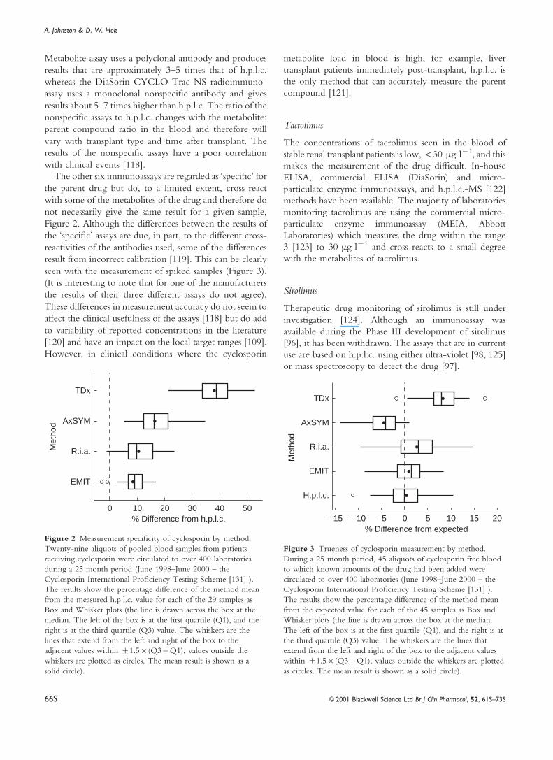
Figure 2. Although the differences between the results of
the `speci®c' assays are due, in part, to the different cross-
reactivities of the antibodies used, some of the differences
result from incorrect calibration [119]. This can be clearly
seen with the measurement of spiked samples (Figure 3).
(It is interesting to note that for one of the manufacturers
the results of their three different assays do not agree).
These differences in measurement accuracy do not seem to
affect the clinical usefulness of the assays [118] but do add
to variability of reported concentrations in the literature
[120] and have an impact on the local target ranges [109].
However, in clinical conditions where the cyclosporin
metabolite load in blood is high, for example, liver
transplant patients immediately post-transplant, h.p.l.c. is
the only method that can accurately measure the parent
compound [121].
Tacrolimus
The concentrations of tacrolimus seen in the blood of
stable renal transplant patients is low,<30 mg lx1, and this
makes the measurement of the drug dif®cult. In-house
ELISA, commercial ELISA (DiaSorin) and micro-
particulate enzyme immunoassays, and h.p.l.c.-MS [122]
methods have been available. The majority of laboratories
monitoring tacrolimus are using the commercial micro-
particulate enzyme immunoassay (MEIA, Abbott
Laboratories) which measures the drug within the range
3 [123] to 30 mg lx1 and cross-reacts to a small degree
with the metabolites of tacrolimus.
Sirolimus
Therapeutic drug monitoring of sirolimus is still under
investigation [124]. Although an immunoassay was
available during the Phase III development of sirolimus
[96], it has been withdrawn. The assays that are in current
use are based on h.p.l.c. using either ultra-violet [98, 125]
or mass spectroscopy to detect the drug [97].
% Difference from h.p.l.c.0
Met
hod
EMIT
R.i.a.
AxSYM
10 20 30 40 50
TDx
Figure 2 Measurement speci®city of cyclosporin by method.
Twenty-nine aliquots of pooled blood samples from patients
receiving cyclosporin were circulated to over 400 laboratories
during a 25 month period (June 1998±June 2000 ± the
Cyclosporin International Pro®ciency Testing Scheme [131] ).
The results show the percentage difference of the method mean
from the measured h.p.l.c. value for each of the 29 samples as
Box and Whisker plots (the line is drawn across the box at the
median. The left of the box is at the ®rst quartile (Q1), and the
right is at the third quartile (Q3) value. The whiskers are the
lines that extend from the left and right of the box to the
adjacent values within t1.5r(Q3xQ1), values outside the
whiskers are plotted as circles. The mean result is shown as a
solid circle).
% Difference from expected0
Met
hod
EMIT
R.i.a.
AxSYM
5 10 15 20
TDx
–5–10–15
H.p.l.c.
Figure 3 Trueness of cyclosporin measurement by method.
During a 25 month period, 45 aliquots of cyclosporin free blood
to which known amounts of the drug had been added were
circulated to over 400 laboratories (June 1998±June 2000 ± the
Cyclosporin International Pro®ciency Testing Scheme [131] ).
The results show the percentage difference of the method mean
from the expected value for each of the 45 samples as Box and
Whisker plots (the line is drawn across the box at the median.
The left of the box is at the ®rst quartile (Q1), and the right is at
the third quartile (Q3) value. The whiskers are the lines that
extend from the left and right of the box to the adjacent values
within t1.5r(Q3xQ1), values outside the whiskers are plotted
as circles. The mean result is shown as a solid circle).
A. Johnston & D. W. Holt
66S f 2001 Blackwell Science Ltd Br J Clin Pharmacol, 52, 61S±73S

Receptor assays
Cyclosporin, tacrolimus and sirolimus bind to a group
of widely occurring proteins, the immunophilins. The
two major immunophilins are cyclophilin, which binds
cyclosporin, and FK-binding protein 12, which binds
tacrolimus and sirolimus. Receptor assays have theoretical
advantages over current immunoassays in that binding
is associated with pharmacological activity so that only
active drug or active metabolite concentration is measured
[126]. However, this contention remains to be proved and
these assays are not in routine clinical use.
Mycophenolic acid
Compared with the other immunosuppressant drugs in
current use, the plasma concentration of MPA is much
higher (of the order 1±5 mg lx1) and this makes h.p.l.c.
measurement of the drug straightforward. Using this
technique the major metabolite MPAG can be resolved
and quanti®ed [127]. A commercial homogeneous
enzyme immunoassay (Dade Behring) is available which
is capable of accurate and precise measurement of the
drug in the concentration range 0.5±15 mg lx1 [128, 129].
On average, concentrations of MPA measured by this
immunoassay are about 20% higher than those produced
by h.p.l.c. [130], due to the cross-reactivity of the antibody
noted above.
Pro®ciency testing
The dif®culties in measuring blood cyclosporin concen-
tration lead to the development of a pro®ciency-testing
scheme for laboratories providing monitoring services for
the drug [30]. The International Cyclosporin Pro®ciency
Testing Scheme has been in existence for over 17 years
and has over 400 participating centres world-wide [131].
The Scheme circulates three blood samples per month
to each participating centre and the measurements made
on these samples enable laboratories to benchmark their
analytical performance against currently available best
practice [132]. Initially, the manufacturer of cyclosporin,
Sandoz AG (now Novartis AG), to promote measurement
accuracy of the drug, funded the Scheme. However,
the individual centres, the drug manufacturer and TDM
reagent suppliers, now fund it.
In a similar fashion Fujisawa GmbH, the makers of
tacrolimus [133], have funded a scheme for that drug and
Dade Behring, manufacturer of a kit for the measurement
of MPA, have funded a mycophenolate pro®ciency
scheme [134]. These schemes have 260 and 70 members,
respectively. A sirolimus pro®ciency testing scheme,
funded by Wyeth-Ayerst Research, was initiated in 1998
to document the pro®ciency of laboratories offering
support for clinical studies in which the drug was moni-
tored by immunoassay [135]. The scheme is continuing
as a service to laboratories measuring the drug by h.p.l.c.
The roÃle and rationale for pro®ciency testing schemes in
immunosuppressive drug monitoring has been reviewed
recently [136]. Full details of the pro®ciency testing
schemes for cyclosporin, tacrolimus, sirolimus and myco-
phenolate are available on the World Wide Web [137].
Pharmacodynamic monitoring
Blood or plasma drug concentration measurements are
only a surrogate for effect. Their use would be unnecessary
were it possible to measure the immunosuppressive action
of these drugs directly. However, de®ning the pharma-
codynamic target for monitoring is not an easy task. For
example, in a recent study investigating immunological
monitoring of azathioprine the authors examined multiple
subsets of peripheral blood lymphocytes, natural killer
activity, the serum concentrations of IgG, IgM, interferon
b (IFN b), tumour necrosis factor a (TNF a), interleukin
2 (IL-2), soluble interleukin 2 receptors (sIL-2R),
interleukin 6 (IL-6) and the soluble adhesion molecule
sICAM-1 [138]. There is a danger that one surrogate will
be replaced by another.
However, pharmacodynamic monitoring has been
used successfully to control oral prednisolone dose
[139]. Based on a previous observation that blood
eosinophil counts (BEC) rises beyond 60r106/l prior
to heart allograft rejection [140], these authors carried out
a randomised trial to compare their standard protocol for
steroid dose adjustment with a protocol guided by BEC
during the ®rst 3 months after heart transplantation.
Eighty patients were randomised to either have their
EOS reported (n=40) and used for steroid dose
adjustment or not reported. In the reporting group,
patients had their steroid dosage increased if BEC
exceeded 60r106/l. During the ®rst six postoperative
weeks, this group of patients had an 83% lower risk of
treated rejection (P<0.05) and lower median intravenous
dose of methyl-prednisolone (P<0.05) than the patients
in whom EOS was not reported. Overall, the reporting
group had less than half the risk of rejection of any grade
(P<0.001) and signi®cantly more rejection-free biopsies
than those treated by the standard protocol (P<0.01).
There was no increase the incidence of serious steroid-
related side-effects.
Cyclosporin and tacrolimus inhibit T-cell proliferation,
which is thought to result from their inhibition of cal-
cineurin [141]. This is a serine-threonine phosphatase that
plays an essential roÃle in intracellular, calcium-dependent,
signal transduction [142]. Inhibition of calcineurin in
activated T cells reduces the translocation of the cyto-
plasmic subunit of the nuclear factor to the nuclear subunit
TDM of immunosuppressant drugs
f 2001 Blackwell Science Ltd Br J Clin Pharmacol, 52, 61S±73S 67S

and, hence, impairs the transcription of genes for many
of the cytokines essential for the rejection response [143].
For this reason calcineurin has been the primary focus of
pharmacodynamic monitoring for cyclosporin. In a study
of 62 renal transplant patients the measured calcineurin
activity in leucocytes was half that of controls [144]. The
trough cyclosporin concentration was inversely related to
the calcineurin activity. Since tacrolimus acts on the same
target, calcineurin would also be applicable for monitoring
that drug. However, the measurement of calcineurin
activity is technically challenging and a much simpler
procedure would be required before it could be used in
the routine setting. In any case, further studies would be
needed to con®rm that calcineurin activity correlates better
with patient outcome than simple blood concentrations.
Mycophenolic acid exerts its immunosuppressive action
by inhibition of inosine monophosphate dehydrogenase
(IMPDH) and, thereby, blocking de novo purine biosynth-
esis in lymphocytes [145]. An assay has been developed for
the measurement of IMPDH activity in whole blood to
measure drug effect rather than concentration [146]. As yet
there are few published data to support the use of this assay,
but initial studies suggest a correlation between IMPDH
activity and clinical events [147].
Sirolimus is thought to exert its immunosuppressive
activity by blocking the phosphorylation and activation of
the P70 S6 kinase that is involved in cell signalling, this
prevents, or reduces, lymphocyte proliferation [148]. The
measurement of P70 S6 activity is therefore a prime
target for pharmacodynamic measurements. An assay has
been developed for its measurement in whole blood
and studies are ongoing to determine its utility in clinical
practice [149].
Antithymocyte globulin (ATG) has been monitored in
renal transplant patients using the effect of ATG on subsets
of T lymphocytes [150]. In this study, the author identi®ed
the CD3+ lymphocytes as a pharmacodynamic marker of
ATG response. The dose of ATG administered to patients
was titrated to maintain the patients' absolute CD3+
lymphocyte count at < 50 cells mlx1 of blood. Forty-four
patients who were treated in this way for steroid resistant
rejection had signi®cantly less serious viral infections
than 10 patients who were treated on a ®xed dose ATG
regimen, but their 1 years graft survival was not
compromised. Although not a formal pharmaco-
economic study, the author also noted that there was a
net saving of £1600 ($2500) per patient.
Bene®ts of monitoring
Evidence-based medicine is sadly lacking in the area
of drug monitoring [151]. Although the perception of
therapeutic drug monitoring is that it is bene®cial, and
aids patient management, there is little hard evidence to
support that view. Outside the ®eld of immunosuppressive
drugs there are numerous articles, detailing predominantly
retrospective studies, which suggest that TDM has a useful
and cost effective roà le in monitoring therapy [152, 153].
However, as the authors of a recent review of clinical
pharmacokinetics point out, there is little evidence to
support the effect of TDM on true patient outcomes [152].
For the immunosuppressive drugs some positive
evidence for TDM is given by the results of prospective
concentration-controlled clinical trials [154]. A concen-
tration-controlled trial is one in which patients are dosed
to achieve preassigned target concentrations and therefore
the results can be assessed in terms of drug concentration
and response rather than dose and response. This type of
trial has several bene®ts over the randomized dose-
controlled trials [155] and can provide the basis for future
TDM decisions.
In a study of tacrolimus, patients were randomly
allocated to be targeted at low, medium or high drug
blood concentrations [156]. Although there was no
difference in the incidence of rejection or toxicity in
the three groups in the ®rst 42 days post transplant, logistic
regression demonstrated a clear relationship between con-
centration and effect, Table 2. In a study of similar design
using mycophenolate mofetil, patients were randomly
allocated to a high, medium or low mycophenolic acid
AUC [157]. Again there was a clear concentration related
effect of the drug with rejection in the low, medium and
high groups of 26, 9 and 6%, respectively.
Conclusion
At the present time the majority of drug regimes in use for
transplantation are based on cyclosporin and excellent
results can be obtained in solid organ transplantation using
this drug. This is also true in the treatment of autoimmune
diseases [3]. However, cyclosporin's dominance is being
challenged as the number of immunosuppressive agents
increases and clinical experience is gained with other
drugs. We are entering an era in which combination
therapy will be the norm and clinicians will tailor
the immunosuppression to the characteristics of the
individual patient, changing dose and drugs as time
progresses and conditions change [158]. In addition,
Table 2 Incidence of toxicity and rejection by whole blood tacrolimus
concentration [156].
Tacrolimus (mg lx1)
<5 5±15 >15
Rejection % 34% 17% 5%
Toxicity % 0% 34% 54%
A. Johnston & D. W. Holt
68S f 2001 Blackwell Science Ltd Br J Clin Pharmacol, 52, 61S±73S

generic [159] formulations of cyclosporin [160] will be
available in the near future and these together with the
new drugs under clinical development, such as FTY720
[161] and everolimus (Certicam1) [162], and the
possibilities of xenotransplantation [163] present future
challenges which will add to the complexity of TDM.
References
1 Spector R, Park GD, Johnson GF, Vesell ES. Therapeutic
drug monitoring. Clin Pharmacol Ther 1988; 43: 345±353.
2 Belitsky P, Levy GA, Johnston A. Neoral absorption
pro®ling: an evolution in effectiveness. Transplant Proc 2000;
32(3A(Suppl)): 45S±52S.
3 Holt DW, Johnston A. Cyclosporin monitoring: its role
in autoimmune indications. J Autoimmunity 1992;
5(Suppl):82.
4 Holt DW, Johnston A. Monitoring new immunosuppressive
agents. Are the methods adequate? Drug Metabolism Drug
Interacts 1997; 14: 5±15.
5 Calne RY. The present position and future prospects
of organ transplantation. Ann R Coll Surg Engl 1968;
42: 283±306.
6 Starzl TE, Marchioro TL, Porter KA, Iwasaki Y, Cerilli GJ.
The use of heterologous antilymphoid agents in canine renal
and liver homotransplantation and in human renal
homotransplantation. Surg Gynecol Obstet 1967;
124: 301±308.
7 Schutz E, Gummert J, Mohr FW, Armstrong VW, Oellerich
M. Should 6-thioguanine nucleotides be monitored in heart
transplant recipients given azathioprine? Ther Drug Monit
1996; 18: 228±233.
8 Weinshilboum RM, Sladek SL. Mercaptopurine
pharmacogenetics: monogenic inheritance of erythrocyte
thiopurine methyltransferase activity. Am J Hum Genet 1980;
32: 651±662.
9 Loennechen T, Yates CR, Fessing MY, Relling MV,
Krynetski EY, Evans WE. Isolation of a human thipurine
S-methyltransferase (TPMT) complementary DNA with
a single nucleotide transition A719G (TPMT*3C) and its
association with loss of TPMT protein and catalytic activity
in humans. Clin Pharmacol Ther 1998; 64: 46±51.
10 Gummert JF, Schutz E, Oellerich M, Mohr FW, Dalichau H.
Monitoring of TPMT in heart transplant recipients under
immunosuppressive therapy with azathioprine. Artif Organs
1995; 19: 918±920.
11 Lennard L. Therapeutic drug monitoring of cytotoxic drugs.
Br J Clin Pharmacol 2001; 52(suppl. 1): 75S±87S.
12 Wilde MI, Goa KL. Muromonab CD3: a reappraisal of its
pharmacology and use as prophylaxis of solid organ transplant
rejection. Drugs 1996; 51: 865±894.
13 Smith SL. Ten years of Orthoclone OKT3
(muromonab-CD3): a review. J Transplant Coordination 1996;
6: 109±119.
14 Abramowicz D, Goldman M, Mat O, et al. OKT3 serum
levels as a guide for prophylactic therapy: a pilot study in
kidney transplant recipients. Transpl Int 1994; 7: 258±263.
15 Schena FP. New insights into therapy with monoclonal
antibodies in allograft transplantation. Nephrol Dial Transplant
1997; 12(Suppl 1): 55±58.
16 Cinti P, Cocciolo P, Evangelista B, et al. OKT3
prophylaxis in kidney transplant recipients: drug
monitoring by ¯ow cytometry. Transplantation Proc 1996;
28(6): 3214±3216.
17 Calne RY. Organ transplantation. The present position and
future prospects of organ transplantation. Trans Med Soc Lond
1969; 85: 56±67.
18 Calne RY. Immunosuppression and clinical organ
transplantation. Transplant Proc 1974; 6(4(Suppl 1): 51.
19 Opelz G. In¯uence of treatment with cyclosporine,
azathioprine and steroids on chronic allograft failure. The
Collaborative Transplant Study Kidney Int ± Supplement 1995;
52: S89±S92.
20 Borel JF, Feurer C, Gubler HU, Stahelin H. Biological effects
of cyclosporin A. a new antilymphocytic agent. Agents Actions
1976; 6: 468±475.
21 Borel JF, Kis ZL. The discovery and development of
cyclosporine (Sandimmune). Transplant Proc 1991:
23: 1867±1874.
22 Borel JF, Kis ZL, Beveridge T, Meluzzi VJ, Adams J, eds. The
Search for Anti-Inlammatory Drugs. Boston, USA. BirkhaÈuser, 2,
The History of the Discovery and Development of
Cyclosporine, pp 27±63, 1995.
23 Dunn SP, Cooney GF, Kulinsky A, Falkenstein K, Pierson A,
Meligeni J. Reduced cyclosporine absorption preceded acute
allograft rejection in a child with a liver transplant. Liver
Transplantation Surgery 1997; 3: 538±540.
24 Stahelin HF. The history of cyclosporin A (Sandimmune)
revisited: another point of view. Experientia 1996; 52: 5±13.
25 Shaw LM, Kaplan B, Kaufman D. Toxic effects of
immunosuppressive drugs: mechanisms and strategies for
controlling them. Clin Chem 1996; 42: 1316±1321.
26 Holt DW, Johnston A, Thomson A, Starzl T, eds.
Immunosuppressive Drugs: Developements in Anti-Rejection
Therapy. London: Edward Arnold; 3, Pharmacokinetics
and monitoring of cyclosporin A, pp 37±45, 1994.
27 Johnston A, Marsden JT, Holt DW. The in¯uence of
haematocrit on blood cyclosporin measurements in vivo.
Br J Clin Pharmacol 1988; 25: 509±513.
28 Johnston A, Marsden JT, Holt DW. Sample pretreatment
to minimize interference from whole blood in the
radioimmunoassay for cyclosporine. Transplantation 1987;
44: 332.
29 Holt DW, Marsden JT, Johnston A. Measurement of
cyclosporine: methodological problems. Transplant Proc 1986;
18(6(Suppl 5): 101±110.
30 Johnston A, Marsden JT, Holt DW. The United Kingdom
Cyclosporin Quality Assessment Scheme. Ther Drug Monit
1986; 8: 200±204.
31 Tsang VT, Johnston A, Heritier F, Leaver N, Hodson ME,
Yacoub M. Cyclosporin pharmacokinetics in heart-lung
transplant recipients with cystic ®brosis. Effects of pancreatic
enzymes and ranitidine. Eur J Clin Pharmacol 1994;
46: 261±265.
32 Holt DW, Marsden JT, Johnston A, Bewick M, Taube DH.
Blood cyclosporin concentrations and renal allograft
dysfunction. Br Med J 1986; 293: 1057±1059.
33 Kahan BD, Shaw LM, Holt D, Grevel J, Johnston A.
Consensus document: Hawk's Cay meeting on therapeutic
drug monitoring of cyclosporine. Clin Chem 1990;
36: 1510±1516.
TDM of immunosuppressant drugs
f 2001 Blackwell Science Ltd Br J Clin Pharmacol, 52, 61S±73S 69S

34 Shaw LM, Yatscoff RW, Bowers LD, et al. Canadian
Consensus Meeting on cyclosporine monitoring:
report of the consensus panel. Clin Chem 1990;
36: 1841±1846.
35 Sketris I, Yatscoff R, Keown P, et al. Optimizing the use of
cyclosporine in renal transplantation. Clin Biochem 1995;
28: 195±211.
36 Holt DW, Johnston A. Cyclosporin assay techniques.
Accuracy and reproducibility variables impacting on
measurements. Int J Rad Appl Instrum B 1990; 17: 733±736.
37 Holt DW, Johnston A, den Boer NC, van der Heiden C,
Leijnse B, Souverijn JHM, eds. Clinical Chemistry Plenum
Publishing Corporation; Practical applications of therapeutic
drug monitoring: The impact of technological developments,
pp 93±102, 1989.
38 Holt DW, Johnston A, Roberts NB, Tredger JM, Trull AK.
Methodological and clinical aspects of cyclosporin
monitoring: report of the Association of Clinical Biochemists
task force. Ann Clin Biochem 1994; 31: 420±446.
39 Dalrymple-Hay M, Meara M, Reynolds L, et al. Changing
stable heart transplant recipients from Sandimmune to Neoral.
Transplant Proc 1996; 28: 2285±2286.
40 Holt DW, Mueller EA, Kovarik JM, van Bree JB, Richard F,
Kutz K. Sandimmun neoral pharmacokinetics: impact of the
new oral formulation. Transplant Proc 1995; 27: 1434±1437.
41 Holt DW, Mueller EA, Kovarik JM, van Bree JB, Kutz K.
The pharmacokinetics of Sandimmun Neoral: a new oral
formulation of cyclosporine. Transplant Proc 1994;
26: 2935±2939.
42 Merton G, Jones K, Lee M, Johnston A, Holt DW. Accuracy
of cyclosporin measurements made in capillary blood samples
obtained by skin puncture. Ther Drug Monit 2000;
22: 594±598.
43 Lindholm A, Kahan BD. In¯uence of cyclosporine
pharmacokinetics, trough concentrations, and AUC
monitoring on outcome after kidney transplantation.
Clin Pharmacol Ther 1993; 54: 205±218.
44 Holt DW, Johnston A. Cyclosporin monitoring: trough or
AUC? Perspectives 1996; 2: 49±52.
45 Holt DW, Johnston A. Cyclosporin Microemulsion. A guide
to usage and monitoring. Biodrugs 1997; 7: 175±197.
46 Johnston A, Sketris I, Marsden JT, et al. A limited sampling
strategy for the measurement of cyclosporine AUC.
Transplant Proc 1990; 22: 1345±1346.
47 Johnston A, Kovarik JM, Mueller EA, Holt DW. Predicting
patients' exposure to cyclosporin. Transplant International
1996; 9: Suppl. 7.
48 Cantarovich F, Bizollon C, Cantarovich D, Lefrancois N,
Dubernard JM, Traeger J. Cyclosporine plasma levels
six hours after oral administration. A useful tool for
monitoring therapy. Transplantation 1988; 45: 389±394.
49 Cantarovich M, Besner JG, Fitchett DH, Latter DA. Ef®cacy
and side-effects of cyclosporine dose monitoring with levels
6 h after the morning dose in heart transplant patients.
Clin Transplant 1997; 11(5: Part 1): t-405.
50 Rocha G, Deschenes J, Cantarovich M. Cyclosporine
monitoring with levels 6 hours after the morning dose in
patients with noninfectious uveitis. Ophthalmology 1997;
104(2): 245±251.
51 Cantarovich M, Deschenes J. Cyclosporine dose adjustment
using levels obtained six hours after the morning dose: effect
on side effects in patients with autoimmune diseases.
Am J Nephrology 1997; 17: 450±457.
52 Keown P, Kahan BD, Johnston A, et al. Optimization of
cyclosporin therapy with new therapeutic drug monitoring
strategies: Report from the international Neoral TDM
advisory consensus meeting (Vancouver, November 1997.
Transplant Proc 1998; 30: 1645±1649.
53 Grant D, Rochon J, Levy G. Comparison of the long-term
tolerability, pharmacodynamics, and safety of Sandimmune
and Neoral in liver transplant recipients. Ontario Liver
Transplant Study Group. Transplant Proc 1996; 28: 2232±2233.
54 Grant D, Kneteman N, Tchervenkov J, et al. Peak
cyclosporine levels (Cmax) correlate with freedom from
liver graft rejection: results of a prospective, randomized
comparison of neoral and sandimmune for liver
transplantation (NOF-8).Transplantation1999;67: 1133±1137.
55 Levy GA, Grant D. Neoral in liver transplantation. Transplant
Proc 1996; 28: 1019±1021.
56 Freeman D, Grant D, Levy G, et al. Pharmacokinetics of
a new oral formulation of cyclosporine in liver transplant
recipients. Ther Drug Monit 1995; 17: 213±216.
57 Levy G. New strategies for therapeutic drug monitoring of Neoral.
Oxford, Blackwell Science. Two-hour cyclosporin
concentration (C2) as a monitoring tool for Neoral,
pp 19±22, 1998.
58 Johnston A, David OJ, Cooney GF. Pharmacokinetic
validation of neoral absorption pro®ling. Transplant Proc 2000;
32 (3A(Suppl): 53S±6S.
59 Halloran PF, Helms LM, Kung L, Noujaim J. The temporal
pro®le of calcineurin inhibition by cyclosporine in vivo.
Transplantation 1999; 68: 1356±1361.
60 Sindhi R, LaVia MF, Paulling E et al. Stimulated response
of peripheral lymphocytes may distinguish cyclosporine
effect in renal transplant recipients receiving a
cyclosporine+rapamycin regimen. Transplantation 2000;
69: 432±436.
61 Belitsky P, Dunn S, Johnston A, Levy G. Impact of absorption
pro®ling on ef®cacy and safety of cyclosporin therapy in
transplant recipients. Clin Pharmacokinet 2000; 39: 117±125.
62 Cantarovich M, Elstein E, de Varennes B, Barkun JS. Clinical
bene®t of neoral dose monitoring with cyclosporine 2-hr
post- dose levels compared with trough levels in stable heart
transplant patients. Transplantation 1999; 68: 1839±1842.
63 Cantarovich M, Quantz M, Elstein E, Ergina P, Magnan C,
de Varennes B. Neoral dose monitoring with cyclosporine
2-hour postdose levels in heart transplant patients receiving
anti-thymocyte globulin induction. Transplant Proc 2000;
32: 446±448.
64 Jusko WJ, Thomson AW, Fung J, et al. Consensus document:
therapeutic monitoring of tacrolimus (FK-506). Ther Drug
Monit 1995; 17: 606±614.
65 McMaster P, Mirza DF, Ismail T, Vennarecci G, Patapis P,
Mayer AD. Therapeutic drug monitoring of tacrolimus in
clinical transplantation. Ther Drug Monit 1995; 17: 602±605.
66 Venkataramanan R, Swaminathan A, Prasad T, et al. Clinical
pharmacokinetics of tacrolimus. Clin Pharmacokinet 1995;
29: 404±430.
67 Anonymous. Japanese study of FK 506 on kidney
transplantation: the bene®t of monitoring the whole blood
FK 506 concentration. Jap FK 506 Study Group. Transplant
Proc 1991; 23: 3085±3088.
A. Johnston & D. W. Holt
70S f 2001 Blackwell Science Ltd Br J Clin Pharmacol, 52, 61S±73S
68 Hedayat S, Kershner RP, Su G. Relationship of whole-blood
FK506 concentrations to rejection and toxicity in liver and
kidney transplants. J Biopharm Stat 1996; 6: 411±424.
69 Ku Y-M, Min DI. An abbreviated area-under-the-curve
monitoring for tacrolimus patients with liver transplants. Ther
Drug Monit 1998; 20: 219±223.
70 Cantarovich M, Fridell J, Barkun J, et al. Optimal time points
for the prediction of the area-under-the-curve in liver
transplant patients receiving tacrolimus. Transplant Proc 1998;
30: 1460±1461.
71 Wong KM, Shek CC, Chau KF, Li CS. Abbreviated
tacrolimus area-under-the-curve monitoring for renal
transplant recipients. Am J Kidney Dis 2000; 35: 660±666.
72 Ihara H, Shinkuma D, Ichikawa Y, Nojima M, Nagano S,
Ikoma F. Intra- and interindividual variation in the
pharmacokinetics of tacrolimus (FK506) in kidney transplant
recipients ± importance of trough level as a practical indicator.
Int J Urol 1995; 2: 151±155.
73 Lipsky JJ. Mycophenolate mofetil. Lancet 1996;
348: 1357±1359.
74 Bullingham RE, Nicholls A, Hale M. Pharmacokinetics of
mycophenolate mofetil (RS61443): a short review. Transplant
Proc 1996; 28: 925±929.
75 Allison AC, Eugui EM. Immunosuppressive and other effects
of mycophenolic acid and an ester prodrug, mycophenolate
mofetil. Immunol Rev 1993; 136: 5±28.
76 Gray DW. Mycophenolate mofetil for transplantation:
new drug, old problems? Lancet 1995; 346: 390.
77 Schutz E, Shipkova M, Armstrong VW, et al. Therapeutic
drug monitoring of mycophenolic acid: comparison of HPLC
and immunoassay reveals new MPA metabolites. Transplant
Proc 1998; 30: 1185±1187.
78 Schutz E, Shipkova M, Armstrong VW, Wieland E,
Oellerich M. Identi®cation of a pharmacologically active
metabolite of mycophenolic acid in plasma of transplant
recipients treated with mycophenolate mofetil. Clin Chem
1999; 45: 419±422.
79 Shipkova M, Armstrong VW, Wieland E, et al. Identi®cation
of glucoside and carboxyl-linked glucuronide conjugates
of mycophenolic acid in plasma of transplant recipients
treated with mycophenolate mofetil. Br J Pharmacol 1999;
126: 1075±1082.
80 Brewin TB, Cole MP, Jones CT, Platt DS, Todd ID.
Mycophenolic acid (NSC-129185): preliminary clinical trials.
Cancer Chemother Report 1972; 56: 83±87.
81 Placebo-controlled study of mycophenolate mofetil
combined with cyclosporin, corticosteroids for prevention
of acute rejection. European Mycophenolate Mofetil
Cooperative Study Group. Lancet 1995; 345: 1321±1325.
82 Sollinger HW. Mycophenolate mofetil for the prevention of
acute rejection in primary cadaveric renal allograft recipients.
U. S. Renal Transplant Mycophenolate Mofetil Study Group.
Transplantation 1995; 60: 225±232.
83 A blinded randomized clinical trial of mycophenolate mofetil
for the prevention of acute rejection in cadaveric renal
transplantation. The Tricontinental Mycophenolate Mofetil
Renal Transplantation Study Group. Transplantation 1996;
61: 1029±1037.
84 Bullingham RES, Nicholls AJ, Kamm BR. Clinical
pharmacokinetics of mycophenolate mofetil. Clin Pharmacokin
1998; 34: 429±455.
85 Nicholls AJ. Opportunities for therapeutic drug monitoring
of mycophenolate mofetil dose in renal transplantation
suggested by the pharmacokinetic/pharmacodynamic
relationship for mycophenolic acid and suppression of
rejection. Clin Biochem 1998; 31: 329±333.
86 van Gelder T, Hilbrands LB, Vanrenterghem Y, et al.
A randomized double-blind, multicenter plasma
concentration controlled study of the safety and ef®cacy
of oral mycophenolate mofetil for the prevention
of acute rejection after kidney transplantation.
Transplantation 1999; 68: 261±266.
87 Shaw LM, Korecka M, van Breeman R, Nowak I, Brayman
KL. Analysis, pharmacokinetics and therapeutic drug
monitoring of mycophenolic acid. Clin Biochem 1998;
31: 323±328.
88 Meiser BM, Pfeiffer M, Jagiello-Kraatz M, et al.
Mycophenolate mofetil dose adjustment based on
trough levels improves outcome after heart transplantion.
J Heart Lung Transplant 1998; 17: 85.
89 Shaw LM, Nicholls A, Hale M, et al. Therapeutic monitoring
of mycophenolic acid. A consensus panel report. Clin Biochem
1998; 31: 317±322.
90 Shaw LM, Holt DW, Keown P, Venkataramanan R, Yatscoff
RW. Current opinions on therapeutic drug monitoring of
immunosuppressive drugs. Clin Ther 1999; 21: 1632±1652.
91 Zanker B, Schneeberger H, Rothenpieler U, et al.
Mycophenolate mofetil-based, cyclosporin-free induction
and maintenance immunosuppression. Transplantation 1998;
66: 44±49.
92 McAlister VC, Gao Z, Peltekian K, Domingues J, Mahalati K,
MacDonald AS. Sirolimus-tacrolimus combination
immunosuppression [letter]. Lancet 2000; 355: 376±377.
93 Groth CG, Backman L, Morales JM, et al. Sirolimus
(rapamycin) -based therapy in human renal transplantation:
similar ef®cacy and different toxicity compared with
cyclosporine. Sirolimus European Renal Transplant Study
Group [see comments]. Transplantation 1999; 67: 1036±1042.
94 Kreis H, Cisterne JM, Land W, et al. Sirolimus in association
with mycophenolate mofetil induction for the prevention
of acute graft rejection in renal allograft recipients.
Transplantation 2000; 69: 1252±1260.
95 Kahan BD, Julian BA, Pescovitz MD, Vanrenterghem Y,
Neylan J. Sirolimus reduces the incidence of acute rejection
episodes despite lower cyclosporine doses in caucasian
recipients of mismatched primary renal allografts: a phase II
trial. Rapamune Study Group. Transplantation 1999;
68: 1526±1532.
96 Jones K, Saadat-Lajevardi S, Lee TD, et al. An immunoassay
for the measurement of sirolimus. Clin Ther 2000;
22(Suppl. B): 49±61.
97 Holt DW, Lee TD, Jones K, Johnston A. Validation of
an assay for routine monitoring of sirolimus using HPLC
with mass spectrometric detection. Clin Chem 2000;
46: 1179±1183.
98 Napoli KL. A practical guide to the analysis of sirolimus
using high-performance liquid chromatography
with ultraviolet detection. Clin Ther 2000; 22(Suppl B):
B14±B24.
99 Minami Y, Kono T, Miyazaki T, Taniguchi T. The IL-2
receptor complex: its structure, function, and target genes.
Ann Rev Immunol 1993; 11: 245±268.
TDM of immunosuppressant drugs
f 2001 Blackwell Science Ltd Br J Clin Pharmacol, 52, 61S±73S 71S

100 Taniguchi T, Minami Y. The IL-2/IL-2 receptor system:
a current overview. Cell 1993; 73: 5±8.
101 Vincenti F, Lantz M, Birnbaum J, et al. A phase I trial of
humanized anti-interleukin 2 receptor antibody in renal
transplantation. Transplantation 1997; 63: 33±38.
102 Amlot PL, Rawlings E, Fernando ON, et al. Prolonged action
of a chimeric interleukin-2 receptor (CD25) monoclonal
antibody used in cadaveric renal transplantation.
Transplantation 1995; 60: 748±756.
103 Kovarik JM, Rawlings E, Sweny P, et al. Prolonged
immunosuppressive effect and minimal immunogenicity from
chimeric (CD25) monoclonal antibody SDZ CHI 621 in
renal transplantation. Transplant Proc 1996; 28: 913±914.
104 Vincenti F, Kirkman R, Light S et al. Interleukin-2-receptor
blockade with daclizumab to prevent acute rejection in renal
transplantation. Daclizumab Triple Therapy Study Group.
N Engl J Med 1998; 338: 161±165.
105 Hakimi J, Mould D, Waldeman TA, et al. Antibody
Therapeutics. Development of Zenapax: A humanized anti±tac
antibody. CRC Press, 1997; 277±300.
106 Kovarik JM, Rawlings E, Sweny P, et al. Pharmacokinetics
and immunodynamics of chimeric IL-2 receptor monoclonal
antibody SDZ CHI 621 in renal allograft recipients. Transpl
Int 1996; 9(Suppl 1): S32±S33.
107 Kovarik J, Wolf P, Cisterne JM, et al. Disposition of
basiliximab, an interleukin-2 receptor monoclonal antibody,
in recipients of mismatched cadaver renal allografts.
Transplantation 1997; 64: 1701±1705.
108 Kovarik J, Breidenbach T, Gerbeau C, Korn A, Schmidt
A-G, Nashan B. Disposition and immunodynamics of
basiliximab in liver allograft recipients. Clin Pharmacol Ther
1998; 64: 66±72.
109 Oellerich M, Armstrong VW, Kahan B et al. Lake Louise
Consensus Conference on cyclosporin monitoring in organ
transplantation: report of the consensus panel. Therapeutic Drug
Monitoring 1995; 17(6): 642±654.
110 Holt DW, Johnston A. Cyclosporin A: analytical
methodology and factors affecting therapeutic drug
monitoring. Ther Drug Monit 1995; 17: 625±630.
111 Lindholm A. Cyclosporine A: clinical experience and
therapeutic drug monitoring. Ther Drug Monit 1995;
17: 631±637.
112 Perna A, de Gotti EBE, Perico N, Remuzzi G. A
logistic-regression model provides novel guidelines to
maximize the anti-acute rejection properties of cyclosporine
with a minimum of toxicity. J Am Soc Nephrol 1996;
7: 786±791.
113 Morris RG. Target concentration strategy for cyclosporin
monitoring. Clin Pharmacokin 1997; 32: 175±179.
114 Schumacher GE, Barr JT. Total testing process applied to
therapeutic drug monitoring: impact on patients' outcomes
and economics. Clin Chem 1998; 44: 370±374.
115 Schumacher GE, Barr JT. Bayesian and threshold probabilities
in therapeutic drug monitoring: when can serum drug
concentrations alter clinical decisions? Am J Hosp Pharm 1994;
51: 321±327.
116 Holt DW, White DJ. How to measure cyclosporin. Lancet
1984; 2(8396): 228.
117 Johnston A, Cullen G, Holt DW. Quality assurance for
cyclosporin assays in body ¯uids. Ann Acad Med Singapore
1991; 20: 3±8.
118 Lindholm A, Dahlqvist R, Groth GG, Sjoqvist F.
A prospective study of cyclosporine concentration in relation
to its therapeutic effect and toxicity after renal transplantation.
Br J Clin Pharmacol 1990; 30: 443±452.
119 Johnston A, Holt DW. Calibration of the CYCLO-Trac SP
cyclosporine radioimmunoassay. Clin Chem 1993;
39: 2532±2533.
120 McLachlan AJ, Tett SE. Effect of metabolic inhibitors on
cyclosporine pharmacokinetics using a population approach.
Ther Drug Monit 1998; 20: 390±395.
121 SchuÈtz E, Svinarov D, Shipkova M, et al. Cyclosporin whole
blood immunoassays (AxSYM, CEDIA, and Emit): a critical
overview of performance characteristics and comparison with
HPLC. Clin Chem 1998; 44: 2158±2164.
122 Taylor PJ, Hogan NS, Lynch SV, Johnson AG, Pond SM.
Improved therapeutic drug monitoring of tacrolimus (FK506)
by tandem mass spectrometry. Clin Chem 1997;
43: 2189±2190.
123 Schambeck CM, Bedel A, Keller F. Limit of quantitation
(Functional Sensitivity) of the new IMx Tacrolimus II
microparticulate immunoassay. Clin Chem 1998; 44: 2217.
124 Kahan BD, Napoli KL, Kelly PA, et al. Therapeutic drug
monitoring of sirolimus: correlations with ef®cacy and
toxicity. Clin Transplant 2000; 14: 97±109.
125 Holt DW, Lee TD, Johnston A. Measurement of sirolimus in
whole blood using high-performance liquid chromatography
with ultraviolet detection. Clin Ther 2000; 22(Suppl. B):
38-48.
126 Soldin SJ. Role of immunophilins in therapeutic drug
monitoring of immunosuppressive drugs. Clin Biochem 1998;
31: 381±384.
127 Shipkova M, Niedmann PD, Armstrong VW, et al.
Simultaneous determination of mycophenolic acid
and its glucuronide in human plasma using a simple
high-performance liquid chromatography procedure.
Clin Chem 1998; 44: 1481±1488.
128 Beal JL, Jones CE, Taylor PJ, Tett SE. Evaluation of an
immunoassay (EMIT) for mycophenolic acid in plasma from
renal transplant recipients compared with a high-performance
liquid chromatography assay. Ther Drug Monit 1998; 20:
685±690.
129 Vogl M, Weigel G, Seebacher G, Griesmacher A, Laufer G,
Muller MM. Evaluation of the EMIT Mycophenolic
Acid Assay from Dade Behring. Ther Drug Monit 1999;
21: 638±643.
130 Saadat-Lajevardi S, Jones K, Lee T, et al. The International
Mycophenolic Acid Pro®ciency Testing Scheme [Abstract].
Ther Drug Monit 1999; 21: 440.
131 Cyclosporin International Pro®ciency Testing Scheme URL.
http://www.asil.demon.co.uk.
132 Johnston A, Holt DW. External quality assessment scheme for
cyclosporin in body ¯uids. Scand J Clin Lab Invest Suppl 1993;
212: 48±53.
133 Holt DW, Johnston A. Tacrolimus quality assessment. Ther
Drug Monit 1997; 19: 243.
134 Holt DW, Jones K, Lee T, Stadler P, Johnston A. Quality
assessment issues of new immunosuppressive drugs and
experimental experience. Ther Drug Monit 1996; 18: 362±367.
135 Jones K, Johnston A, Holt DW. Pro®ciency testing
issues relating to sirolimus. Clin Ther 2000; 22(Suppl B):
122±131.
A. Johnston & D. W. Holt
72S f 2001 Blackwell Science Ltd Br J Clin Pharmacol, 52, 61S±73S

136 Holt DW, Johnston A. Monitoring immunosuppressive
drugs: The rational for pro®ciency testing. Ligand Assay 1998;
3: 2±6.
137 International Pro®ciency Testing Schemes for
Immunosuppressive Drug Measurement. URL.
http://www.asil.demon.co.uk.
138 Salmaggi A, Corsini E, La ML, et al. Immunological
monitoring of azathioprine treatment in multiple sclerosis
patients. J Neurol 1997; 244: 167±174.
139 Trull AK, Steel LA, Sharples LD, et al. Randomized trial
of blood eosinophil count monitoring as a guide to
corticosteroid dosage adjustment after heart transplantation
[In Process Citation]. Transplantation 2000; 70: 802±809.
140 Trull A, Steel L, Cornelissen J, et al. Association
between blood eosinophil counts and acute cardiac and
pulmonary allograft rejection. J Heart Lung Transplant 1998;
17: 517±524.
141 Morris R. Modes of action of FK506, cyclosporin A and
rapamycin. Transplant Proc 1994; 26: 3272±3275.
142 Schreiber SL, Crabtree GR. The mechanism of action
of cyclosporin A and FK506. Immunol Today 1992;
13: 136±142.
143 Clipstone NA, Crabtree GR. Identi®cation of calcineurin as a
key signalling enzyme in T-lymphocyte activation. Nature
1992; 357: 695±697.
144 Batiuk TD, Pazderka F, Halloran PF. Calcineurin activity is
only partially inhibited in leukocytes of cyclosporine-treated
patients. Transplantation 1995; 59: 1400±1404.
145 Eugui EM, Almquist SJ, Muller CD, Allison AC.
Lymphocyte-selective cytostatic and immunosuppressive
effects of mycophenolic acid in vitro: role of deoxyguanosine
nucleotide depletion. Scand J Immunol 1991; 33: 161±173.
146 Langman LJ, LeGatt DF, Halloran PF, Yatscoff RW.
Pharmacodynamic assessment of mycophenolic acid-induced
immunosuppression in renal transplant recipients.
Transplantation 1996; 62: 666±672.
147 Yatscoff RW, Aspeslet LJ, Gallant HL. Pharmacodynamic
monitoring of immunosuppressive drugs. Clin Chem 1998;
44: 428±432.
148 Chung J, Kuo CJ, Crabtree GR, Blenis J. Rapamycin-FKBP
speci®cally blocks growth-dependent activation of and
signaling by the 70 kd S6 protein kinases. Cell 1992;
69: 1227±1236.
149 Gallant HL, Yatscoff RW. P70 S6 kinase assay. a
pharmacodynamic monitoring strategy for rapamycin;
assay development. Transplant Proc 1996; 28: 3058±3061.
150 Clark K. Monitoring antithymocyte globulin in renal
transplantation. Ann Roy Coll Surg Engl 1996; 78: 536±540.
151 Bogaert M. [Drug policy 1996. [Dutch]. Verh K Acad
Geneeskd Belg 1997; 59: 227±236.
152 Ensom MH, Davis GA, Cropp CD, Ensom RJ. Clinical
pharmacokinetics in the 21st century. Does the evidence
support de®nitive outcomes? Clin Pharmacokin 1998;
34: 265±279.
153 Destache CJ. Use of therapeutic drug monitoring in
pharmacoeconomics. Ther Drug Monit 1993; 15: 608±610.
154 Shaw LM, Kaplan B, Brayman KL. Prospective investigations
of concentration-clinical response for immunosuppressive
drugs provide the scienti®c basis for therapeutic drug
monitoring. Clin Chem 1998; 44: 381±387.
155 Johnston A, Holt DW. Concentration-controlled trials. What
does the future hold? Clin Pharmacokin 1995; 28: 93±99.
156 Laskow DA, Vincenti F, Neylan JF, Mendez R, Matas AJ.
An open-label, concentration-ranging trial of FK506 in
primary kidney transplantation: a report of the United States
Multicenter FK506 Kidney Transplant Group. Transplantation
1996; 62: 900±905.
157 Vanrenterghen Y. The pharmacokinetic/pharmacodynamic
relationship for MPA in renal transplantation ± results of
a randomised concentration ± controlled trial (RCCT)
of MMF. Sydney, Australia.: p 1015, International
Congress of Nephrology, 1997.
158 Ponticelli C, Tarantino A. Immunosuppressive protocols
for renal transplantation. Nephrol Dial Transplant 1997;
12(Suppl 1): 45±50.
159 Johnston A, Holt DW. Generic substitution for cyclosporine:
What should we be looking for in a new formulation.
Transplant Proc 1998; 30: 1652±1653.
160 Al Meshal MA, Khidr SH, Bayomi MA, Al Angary AA. Oral
administration of liposomes containing cyclosporine: a
pharmacokinetic study. Int J Pharmaceutics 1998; 168: 163±168.
161 Troncoso P, Kahan BD. Preclinical evaluation of a new
immunosuppressive agent, FTY720. Clin Biochem 1998;
31: 369±373.
162 Schuler W, Sedrani R, Cottens S, et al. SDZ RAD, a new
rapamycin derivative: pharmacological properties in vitro and
in vivo. Transplantation 1997; 64: 36±42.
163 Bach FH. Xenotransplantation: problems and prospects. Annu
Rev Med 1998; 49: 301±310.
164 SchuÈtz E, Dias VC, Lewer M, et al. Multicenter evaluation of
the new CEDIA CsA Plus assay for whole blood cyclosporine.
Clin Chem 2000; 46: A195.
TDM of immunosuppressant drugs
f 2001 Blackwell Science Ltd Br J Clin Pharmacol, 52, 61S±73S 73S