* Loopstick transplant * Walter Cronkite RIP * Profile; the mighty KBC ...
Upload
independentCategory
view
6download
0
Research article
TheJournalofClinicalInvestigation http://www.jci.org Volume 118 Number 2 February 2008 735
Immunostimulatory Tim-1–specific antibody deprograms Tregs and prevents transplant
tolerance in miceNicolas Degauque,1 Christophe Mariat,1 James Kenny,1 Dong Zhang,1 Wenda Gao,1
Minh Diem Vu,1 Sophoclis Alexopoulos,1 Mohammed Oukka,2 Dale T. Umetsu,3 Rosemarie H. DeKruyff,3 Vijay Kuchroo,2 Xin Xiao Zheng,1 and Terry B. Strom1
1Division of Transplant Immunology and Transplant Research Center, Beth Israel Deaconess Medical Center, Harvard Medical School, Boston, Massachusetts, USA. 2Center for Neurological Diseases, Brigham and Women’s Hospital, Harvard Medical School, Boston, Massachusetts, USA.
3Division of Immunology, Children’s Hospital Boston, and Harvard Medical School, Boston, Massachusetts, USA.
TcellIgmucin(Tim)moleculesmodulateCD4+Tcellresponses.InkeepingwiththeviewthatTim-1gen-eratesastimulatorysignalforCD4+Tcellactivation,wehypothesizedthatanagonistTim-1–specificmAbwouldintensifytheCD4+Tcell–dependantallograftresponse.Unexpectedly,wedeterminedthataparticularTim-1–specificmAbexertedreciprocaleffectsuponthecommitmentofalloactivatedTcellstoregulatoryandeffectorphenotypes.CommitmenttotheTh1andTh17phenotypeswasfostered,whereascommitmenttotheTregphenotypewashindered.Moreover,ligationofTim-1invitroeffectivelydeprogrammedTregsandthusproducedTregsunabletocontrolTcellresponses.Overall,theeffectsoftheagonistTim-1–specificmAbontheallograftresponsestemmedfromenhancedexpansionandsurvivalofTeffectorcells;acapacitytodeprogramnaturalTregs;andinhibitionoftheconversionofnaiveCD4+TcellsintoTregs.ThereciprocaleffectsofagonistTim-1–specificmAbsuponeffectorTcellsandTregsservetopreventallogeneictransplanttolerance.
IntroductionT cell Ig mucin (Tim) molecules are structurally related to type I membrane glycoproteins expressed on T cells (1). Polymorphisms within the Tim gene locus are associated with susceptibility to atopy and autoimmunity, which suggests that Tim proteins mod-ulate CD4+ T cell responses (2–4). Indeed, recent reports have con-firmed the role of Tim molecules in regulating the expansion and effector function of Th1 and Th2 cells (5–8). For example, Tim-3 and Tim-2 negatively regulate Th1 and Th2 responses, respectively (9, 10), through the delivery of inhibitory or death signals into select CD4+ T cell populations.
Based on genetic linkage and epidemiologic studies, it was rea-sonable to assume that ligation of Tim-1 might preferentially skew the immune response toward a Th2 phenotype (11, 12), because anti–Tim-1 amplifies Th2-type cytokine production in a Th2-biased experimental model of airway hyperreactivity (13). Unlike ligation of Tim-3 or Tim-2, Tim-1 ligation heightens T cell activa-tion (13). Naive CD4+ T cells upregulate Tim-1 expression early after activation, and Tim-1 cell-surface expression is maintained through differentiation into the Th1 or Th2 phenotype (13, 14). Tim-4, a molecule expressed by DCs (15), is a ligand of Tim-1 (15). Cross-linking of Tim-1 on the surface of T cells in vitro by Tim-4 Ig enhances T cell proliferation and production of Th1 and Th2 cytokines. In vivo administration of Tim-4 Ig during an ongoing immune response creates similar effects (15).
In keeping with the view that Tim-1 generates a stimulatory sig-nal for T cell activation (16), we demonstrate that an agonist anti–
Tim-1 mAb (13, 17) intensifies the allograft response and prevents development of T cell tolerance. Unexpectedly, we determined that agonist anti–Tim-1 mAb exerts reciprocal effects upon the com-mitment of alloactivated T cells to regulatory and effector pheno-types. In the context of alloimmunity, we demonstrate that Tim-1 greatly enhances proinflammatory (Th1 and Th17) cell–mediated responses and hampers the development of peripheral tolerance. In addition, we now report on the capacity of Tim-1 to deprogram the CD4+Foxp3+ T cell–dependent regulatory loops and to pro-mote differentiation of Th17 cells. Collectively, our data indicate that ligation of Tim-1 reciprocally alters commitment of alloreac-tive CD4+ T cells to the CD4+Foxp3+ and CD4+Th17+ phenotypes.
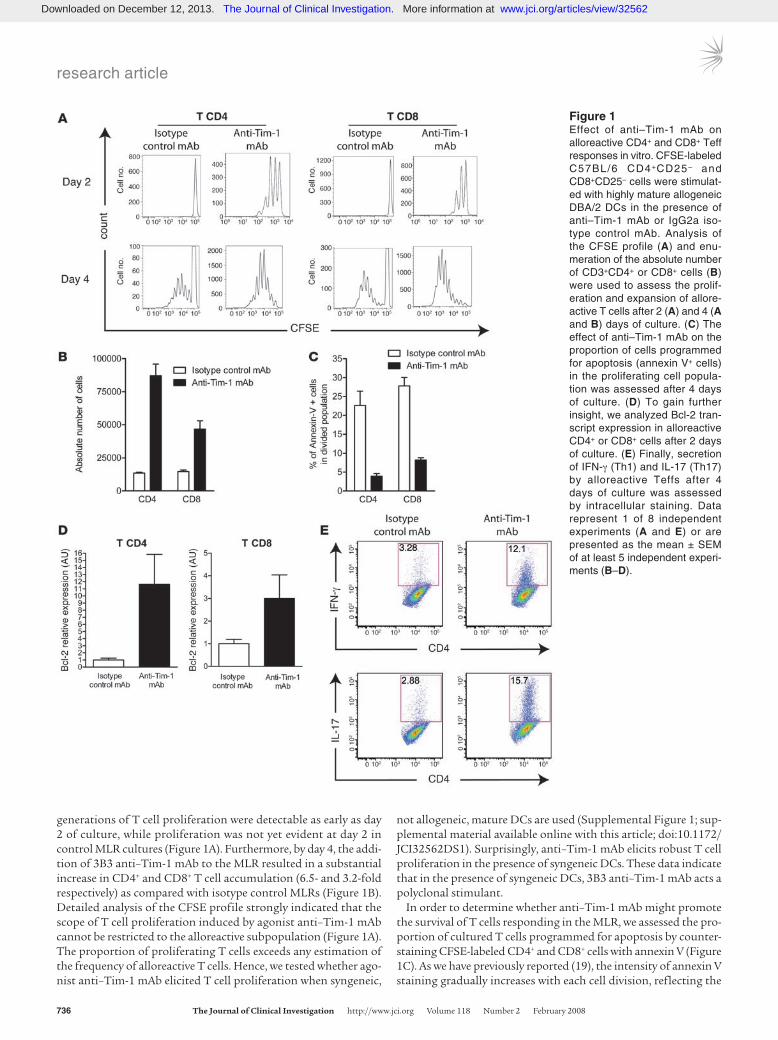
Results3B3 anti–Tim-1 mAb strengthens Th1/Th17 polarization and enhances the expansion and survival of CD4+ and CD8+ alloreactive cells in vitro. Umetsu et al. developed the 3B3 agonist type anti–Tim-1 mAb and provided the first mechanistic insights into the function of Tim-1 (13). Application of 3B3 anti–Tim-1 mAb heightens T cell activation and prevents the development of respiratory tract toler-ance in a Th2-driven model of asthma (13). We have now used 3B3 anti–Tim-1 mAb to study the role of Tim-1 in the in vivo allograft response, a prototypic Th1 effector T cell–driven (Teff-driven) process, and in vitro, e.g., the mixed lymphocyte reaction (MLR) (Figure 1). Mature allogeneic DCs (i.e., DBA/2 bone marrow–derived CD80+CD40+ DCs) were used to stimulate CFSE-labeled C57BL/6 CD4+CD25– and CD8+CD25– T cells in the presence of 3B3 anti–Tim-1 mAb or an isotype control antibody. The prolif-erative response of CFSE-stained alloreactive T cells was analyzed by flow cytometry (18) (Figure 1A). The proliferative response of CD4+CD25– and CD8+CD25– T cells in the MLR was accelerated and exaggerated with provision of 3B3 anti–Tim-1 mAb (Figure 1, A and B). In cultures containing anti–Tim-1 mAb, 3–4 discrete
Nonstandardabbreviationsused: CTLA-4, CTL-associated antigen–4; GITR, glu-cocorticoid-induced TNF receptor family–related protein; MLR, mixed lymphocyte reaction; Teff, effector T cell; TIM, T cell Ig mucin.
Conflictofinterest: The authors have declared that no conflict of interest exists.
Citationforthisarticle: J. Clin. Invest. 118:735–741 (2008). doi:10.1172/JCI32562.
Downloaded on December 12, 2013. The Journal of Clinical Investigation. More information at www.jci.org/articles/view/32562
research article
736 TheJournalofClinicalInvestigation http://www.jci.org Volume 118 Number 2 February 2008
generations of T cell proliferation were detectable as early as day 2 of culture, while proliferation was not yet evident at day 2 in control MLR cultures (Figure 1A). Furthermore, by day 4, the addi-tion of 3B3 anti–Tim-1 mAb to the MLR resulted in a substantial increase in CD4+ and CD8+ T cell accumulation (6.5- and 3.2-fold respectively) as compared with isotype control MLRs (Figure 1B). Detailed analysis of the CFSE profile strongly indicated that the scope of T cell proliferation induced by agonist anti–Tim-1 mAb cannot be restricted to the alloreactive subpopulation (Figure 1A). The proportion of proliferating T cells exceeds any estimation of the frequency of alloreactive T cells. Hence, we tested whether ago-nist anti–Tim-1 mAb elicited T cell proliferation when syngeneic,
not allogeneic, mature DCs are used (Supplemental Figure 1; sup-plemental material available online with this article; doi:10.1172/JCI32562DS1). Surprisingly, anti–Tim-1 mAb elicits robust T cell proliferation in the presence of syngeneic DCs. These data indicate that in the presence of syngeneic DCs, 3B3 anti–Tim-1 mAb acts a polyclonal stimulant.
In order to determine whether anti–Tim-1 mAb might promote the survival of T cells responding in the MLR, we assessed the pro-portion of cultured T cells programmed for apoptosis by counter-staining CFSE-labeled CD4+ and CD8+ cells with annexin V (Figure 1C). As we have previously reported (19), the intensity of annexin V staining gradually increases with each cell division, reflecting the
Figure 1Effect of anti–Tim-1 mAb on alloreactive CD4+ and CD8+ Teff responses in vitro. CFSE-labeled C57BL/6 CD4+CD25– and CD8+CD25– cells were stimulat-ed with highly mature allogeneic DBA/2 DCs in the presence of anti–Tim-1 mAb or IgG2a iso-type control mAb. Analysis of the CFSE profile (A) and enu-meration of the absolute number of CD3+CD4+ or CD8+ cells (B) were used to assess the prolif-eration and expansion of allore-active T cells after 2 (A) and 4 (A and B) days of culture. (C) The effect of anti–Tim-1 mAb on the proportion of cells programmed for apoptosis (annexin V+ cells) in the proliferating cell popula-tion was assessed after 4 days of culture. (D) To gain further insight, we analyzed Bcl-2 tran-script expression in alloreactive CD4+ or CD8+ cells after 2 days of culture. (E) Finally, secretion of IFN-γ (Th1) and IL-17 (Th17) by alloreactive Teffs after 4 days of culture was assessed by intracellular staining. Data represent 1 of 8 independent experiments (A and E) or are presented as the mean ± SEM of at least 5 independent experi-ments (B–D).
Downloaded on December 12, 2013. The Journal of Clinical Investigation. More information at www.jci.org/articles/view/32562
research article
TheJournalofClinicalInvestigation http://www.jci.org Volume 118 Number 2 February 2008 737
physiological process of activation-induced apoptotic cell death. Interestingly, the proportion of proliferating annexin V+ CD4+ and CD8+ T cells was markedly reduced in anti–Tim-1 mAb–supple-mented MLRs as compared with isotype control–treated cultures (Figure 1C). Hence, expansion of T cells induced by 3B3 anti–Tim-1 mAb in the MLR is due to accelerated and exaggerated cell division as well as a survival advantage directly conferred by Tim-1–depen-dent signals. In keeping with these observations, amplified expres-sion of the antiapoptotic Bcl-2 gene (20) was detected in MLR-activated, proliferating CD4+ and CD8+ T cells harvested from the anti–Tim-1 mAb–treated cultures (Figure 1D).
Next, we sought to characterize whether supplementation of the MLR with anti–Tim-1 mAb altered the balance of Th1/Th2/
Th17 cells. Provision of agonist anti–Tim-1 mAb to the culture enhanced the commitment of proinflammatory Th1 and Th17 T cells, as assessed by intracellular immunostaining for IFN-γ and IL-17 (Figure 1E). These profiles were further confirmed using quantitative real-time PCR (Supplemental Figure 2). In contrast, gene expression for IL-4, the prototypic Th2-type cytokine, was downregulated in cultures supplemented with agonist anti–Tim-1 mAb (Supplemental Figure 2). Thus, activation of the Tim-1 path-way promotes the commitment of naive T cells toward a Th1/Th17-biased proinflammatory-type response.
3B3 anti–Tim-1 mAb dampens expression of regulatory-associated genes and the ability of alloreactive Tregs to suppress Teff responses in vitro. A heightened Th1-type alloreactive response can result from
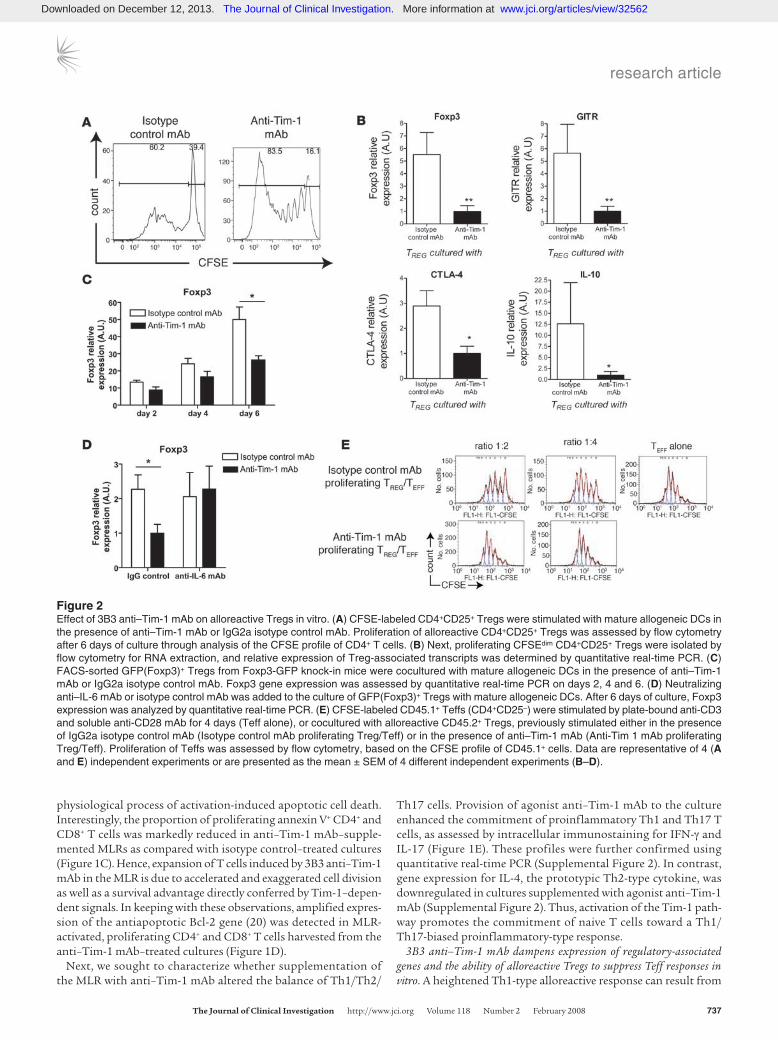
Figure 2Effect of 3B3 anti–Tim-1 mAb on alloreactive Tregs in vitro. (A) CFSE-labeled CD4+CD25+ Tregs were stimulated with mature allogeneic DCs in the presence of anti–Tim-1 mAb or IgG2a isotype control mAb. Proliferation of alloreactive CD4+CD25+ Tregs was assessed by flow cytometry after 6 days of culture through analysis of the CFSE profile of CD4+ T cells. (B) Next, proliferating CFSEdim CD4+CD25+ Tregs were isolated by flow cytometry for RNA extraction, and relative expression of Treg-associated transcripts was determined by quantitative real-time PCR. (C) FACS-sorted GFP(Foxp3)+ Tregs from Foxp3-GFP knock-in mice were cocultured with mature allogeneic DCs in the presence of anti–Tim-1 mAb or IgG2a isotype control mAb. Foxp3 gene expression was assessed by quantitative real-time PCR on days 2, 4 and 6. (D) Neutralizing anti–IL-6 mAb or isotype control mAb was added to the culture of GFP(Foxp3)+ Tregs with mature allogeneic DCs. After 6 days of culture, Foxp3 expression was analyzed by quantitative real-time PCR. (E) CFSE-labeled CD45.1+ Teffs (CD4+CD25–) were stimulated by plate-bound anti-CD3 and soluble anti-CD28 mAb for 4 days (Teff alone), or cocultured with alloreactive CD45.2+ Tregs, previously stimulated either in the presence of IgG2a isotype control mAb (Isotype control mAb proliferating Treg/Teff) or in the presence of anti–Tim-1 mAb (Anti-Tim 1 mAb proliferating Treg/Teff). Proliferation of Teffs was assessed by flow cytometry, based on the CFSE profile of CD45.1+ cells. Data are representative of 4 (A and E) independent experiments or are presented as the mean ± SEM of 4 different independent experiments (B–D).
Downloaded on December 12, 2013. The Journal of Clinical Investigation. More information at www.jci.org/articles/view/32562
research article
738 TheJournalofClinicalInvestigation http://www.jci.org Volume 118 Number 2 February 2008
weakened Treg-dependent T cell immunoregulation. Moreover, a reciprocal relationship between commitment to the Th17 and Treg phenotype has been noted in some settings (21). As 3B3 anti–Tim-1 mAb heightens Th1/Th17 type alloactivation, we sought to determine whether TIM-1–generated signals may impair Treg-dependent immunoregulation.
Interaction of Tregs with highly mature syngeneic DCs rescues Tregs from anergy in vitro (22). We and others have extended this observation to the alloimmune response and have consistently observed substantial proliferation of TCR-transgenic (23) or polyclonal (24) Tregs when MLRs utilize mature allogeneic DCs as stimulator cells. Using this system, we have examined effect of anti–Tim-1 mAb upon the in vitro expansion of alloreactive Tregs (Figure 2).
Proliferation of CD4+CD25+ C57BL/6 Tregs stimulated with mature DBA/2 DCs was markedly enhanced by provision of 3B3 anti–Tim-1 mAb (Figure 2A), suggesting at first that anti–Tim-1 mAb stimulation might amplify the proliferative response of acti-vated Tregs. Thus, we next analyzed transcription of Treg-associ-ated genes in both anti–Tim-1 mAb–treated and control MLRs, focusing specifically on the proliferating (CFSElo) Treg popula-tion. We observed that expression of Foxp3 transcription factor, the Treg master switch (25), was downregulated in the presence
of agonist anti–Tim-1 mAb, whereas Foxp3 expression remained high in the isotype control mAb Treg cultures (Figure 2B). More-over, expression of Foxp3-triggered regulatory-associated factors, including CTL-associated antigen–4 (CTLA-4) and glucocorticoid-induced TNF receptor family–related protein (GITR) (25–28), was downregulated after provision of agonist anti–Tim-1 mAb (Figure 2B). In order to exclude the possibility that the increase in cell pro-liferation resulted from a stimulatory effect of anti–Tim-1 mAb upon a minor population of contaminating CD4+ effector cells, we repeated the MLR with GFP+ (Foxp3+) Tregs that were directly iso-lated from Foxp3-GFP knock-in mice (21) using a MoFlo cell sort-er (purity >98%). A serial analysis on day 2, 4, and 6 revealed that Foxp3 gene expression increases markedly from day 2 through day 6 in the isotype control mAb cultures. In contrast, agonist anti–Tim-1 mAb in the MLR serves to powerfully blunt Foxp3 gene expression (Figure 2C). This decrease in Foxp3 gene expression was strengthened with time (from 34% decrease in gene expression on day 2 to 48% on day 6 in the agonist anti–Tim-1 mAb–supplement-ed culture vs. as isotype control mAb–supplemented culture). The negative influence of anti–Tim-1 mAb on Foxp3 expression was confirmed at the protein level (Supplemental Figure 3).
IL-6–/– mice bear a significantly elevated proportion of Foxp3+ Tregs. Apparently, an absence of IL-6 favors the generation or expansion of Tregs (29). Xu et al. have shown that provision of exogenous IL-6 induced the differentiation of Foxp3– T cells from CD4+CD25+ Foxp3+ T cells (30). To determine whether the down-regulation of Foxp3 expression induced by agonist anti–Tim-1 mAb is IL-6 dependent, neutralizing anti–IL-6 mAb was added to the culture system (Figure 2D). With provision of neutralizing anti–IL-6 mAb, agonist anti–Tim-1 mAb did not alter the magni-tude of Foxp3 gene expression.
The function of Tregs harvested from MLRs supplemented with 3B3 anti–Tim-1 mAb or IgG2a control cultures was then tested through their ability to inhibit the proliferative response of CFSE-labeled Teffs to coated anti-CD3 mAb plus soluble anti-CD28 mAb. Consistent with the decrease in Foxp3, GITR, CTLA-4, and IL-10 gene expression, the regulatory function of the anti–Tim-1– stimulated Treg population was also markedly impaired (Figure 2E). In comparison to the ability of proliferating Tregs from con-trol MLRs to blunt CD4+ proliferation of Teffs to coated anti-CD3 mAb plus soluble anti-CD28 mAb, the suppressive function of 3B3 anti–Tim-1 mAb–treated alloreactive proliferating Tregs was vastly diminished (Figure 2D). Taken together, the transcriptional pro-file and functional data suggest that anti–Tim-1 mAb stimulation
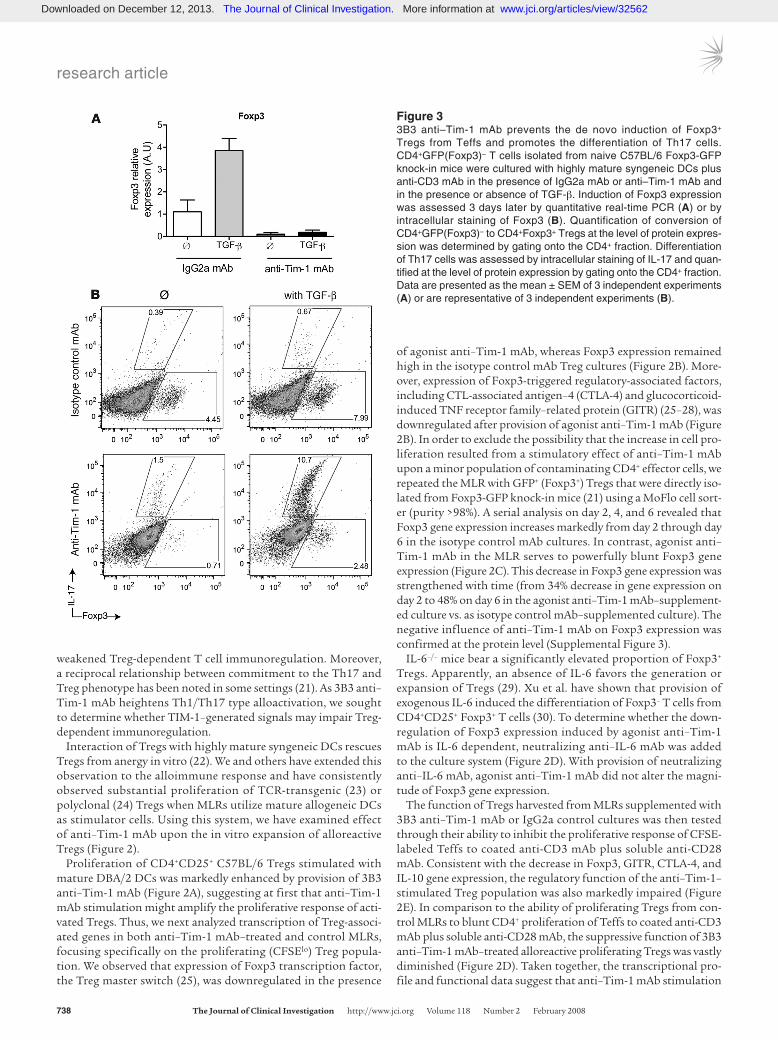
Figure 33B3 anti–Tim-1 mAb prevents the de novo induction of Foxp3+ Tregs from Teffs and promotes the differentiation of Th17 cells. CD4+GFP(Foxp3)– T cells isolated from naive C57BL/6 Foxp3-GFP knock-in mice were cultured with highly mature syngeneic DCs plus anti-CD3 mAb in the presence of IgG2a mAb or anti–Tim-1 mAb and in the presence or absence of TGF-β. Induction of Foxp3 expression was assessed 3 days later by quantitative real-time PCR (A) or by intracellular staining of Foxp3 (B). Quantification of conversion of CD4+GFP(Foxp3)– to CD4+Foxp3+ Tregs at the level of protein expres-sion was determined by gating onto the CD4+ fraction. Differentiation of Th17 cells was assessed by intracellular staining of IL-17 and quan-tified at the level of protein expression by gating onto the CD4+ fraction. Data are presented as the mean ± SEM of 3 independent experiments (A) or are representative of 3 independent experiments (B).
Downloaded on December 12, 2013. The Journal of Clinical Investigation. More information at www.jci.org/articles/view/32562

research article
TheJournalofClinicalInvestigation http://www.jci.org Volume 118 Number 2 February 2008 739
has the unique ability to deprogram Tregs and thereby weaken Treg immunoregulatory function.
Anti–Tim-1 mAb prevents the induction of CD4+Foxp3+ Tregs from CD4+Foxp3– T cells and promotes the differentiation of Th17 cells. The profound effect of anti–Tim-1 mAb on Foxp3 expression and regu-latory functions in natural Tregs prompted us to examine whether anti–Tim-1 mAb would also prevent the induction of Foxp3+ Tregs from Foxp3– Teffs. Stimulation of CD4+GFP(Foxp3)– T cells in the presence of isotype control mAb and TGF-β induced robust expression of Foxp3 transcripts (Figure 3A) and an increase in the frequency of Foxp3+CD4+ T cells (≈8%–10%) (Figure 3B). It is remarkable that conversion of such inducible Foxp3+ Tregs from naive CD4+GFP(Foxp3)– T cells was completely blocked when anti–Tim-1 mAb was added to the culture (Figure 3, A and B).
Several groups including our own have reported an intriguing reciprocal relation between the development of Tregs and the devel-opment of pathogenic effector Th17 cells (21, 31, 32). A common cytokine, TGF-β, is critical for the in vitro development of both Tregs and Th17 cells from CD4+Foxp3– T cells. The differentia-tion into these subsets is determined by the presence or absence of inflammatory cytokines such as IL-6 (21), IL-1α (32), and TNF-α (32). Interestingly, in line with the recent finding that IL-6 in con-junction with TGF-β induces pathogenic Th17 cells while inhibit-ing the generation of inducible Foxp3+ Tregs (21) and with the observation that downregulation of Foxp3 expression induced by agonist anti–Tim-1 mAb is IL-6 dependent (Figure 2D), the combi-nation of 3B3 anti–Tim-1 mAb with TGF-β promoted differentia-tion of Th17 cells as well (Figure 3B).
3B3 anti–Tim-1 mAb negates the tolerance-promoting effect of costimu-lation blockade in islet transplantation. Peripheral transplant toler-ance can be regularly achieved through therapies that curtail the expansion of donor-destructive Teffs while preserving or strength-ening donor-directed regulatory networks (33). Given the above data showing a detrimental effect of 3B3 anti–Tim-1 mAb on the balance between cytoprotective Tregs and effector cytopathic Th1/Th17 cells, we anticipated that in vivo administration of 3B3 anti–Tim-1 mAb would counteract the beneficial effects of tolerizing therapies in an allogeneic transplant model. To test this hypothesis, we chose an MHC-mismatched islet transplant model in which long-term survival of allogeneic islets can be achieved through administration of an anti-CD154 mAb (6, 34–36). Using
this anti-CD154 mAb, tolerance results from inhibitory effects upon potency of donor-directed Teffs and enhances the suppres-sive function of donor-directed Tregs (6, 34). Indeed, in this model of MHC-incompatible islet transplantation, the ability of anti-CD154 mAb treatment to prevent allograft rejection was largely negated by administration of anti–Tim-1 mAb (Figure 4; P < 0.01; mean survival time, 29 days in the anti–Tim-1 mAb group vs. indefinite graft survival in the control group).
DiscussionIn a study of the allograft response, we demonstrate that adminis-tration of 3B3 agonist anti–Tim-1 mAb prevents the induction of peripheral-type transplant tolerance by (a) enhancing the commit-ment and expansion of alloreactive T cells in the Th1/Th17 Teffs; (b) deprogramming natural Tregs at the molecular and functional levels; and (c) inhibiting the conversion of regulatory Foxp3+ cells from the naive Foxp3– T cells. In contrast, Ueno et al. show that treatment with an antagonist anti–Tim-1 mAb prolongs survival of heart allograft (37). The antagonist anti–Tim-1 mAb inhibits Teff responses and serves to enhance the potency of donor-reac-tive regulatory CD4+CD25+ T cells. Opposing effects of the same agonist and antagonist anti–Tim-1 mAbs have been also noted in an EAE model (17) in which the agonist anti–Tim-1 mAb increases the severity of EAE, whereas the antagonist anti–Tim-1 mAb inhib-its the development of EAE. Differences in the avidity of the bind-ing to the IgV domain of the Tim-1 molecule by the agonist and antagonist anti–Tim-1 mAbs may determine the consequences of Tim-1 ligation. The agonist anti–Tim-1 mAb has avidity 17-fold higher than that of the antagonist anti–Tim-1 mAb (17).
Wan and Flavell have generated a mouse in which endogenous Foxp3 gene expression is attenuated in Tregs (38). Using this strategy, they were able to compare the suppressive functions of Foxp3lo T cells and Foxp3hi T cells. They determined that reduced Foxp3 expression by natural Tregs results in diminished immuno-suppressive functions (38). We now demonstrate that activation of CD4+Foxp3+ T cells with an agonist anti–Tim-1 mAb leads to downregulation of Foxp3 expression, at both transcriptional and protein levels, as well as expression of Treg-associated molecules such as CD25, CTLA-4, and GITR (25–28). Activation of Tim-1 on CD4+Foxp3+ T cells completely abrogates the in vitro suppressive functions of Foxp3+ T cells, as these anti–Tim-1 mAb–stimulated Foxp3lo T cells fail to regulate the proliferation of Teffs. Hence, ligation of Tim-1 serves to alter the commitment of Tregs from potent Foxp3hi to ineffective Foxp3lo Tregs.
A reciprocal relationship between commitment of naive CD4+ T cells to the Th17 and Treg phenotypes has been discerned in several settings (21, 29). Depending on the presence or absence of certain proinflammatory cytokines such as IL-6 or IL-21 in the milieu of antigen recognition, TGF-β can support de novo differ-entiation of Th17 or Tregs (21, 32). We now show that engagement of the Tim-1 cell-surface receptor molecule upon T cells cultured with TGF-β skews the commitment of naive T cells to Th1 and Th17 and away from the Foxp3hi phenotype. Thus, our results lend further support to the concept of reciprocal commitment to Th17 and Treg phenotype, and we introduce a new means by which the commitment to the Teff and Treg phenotypes is regu-lated. While previous studies have centered upon the commitment of antigen-stimulated naive T cells, we also demonstrate that the Foxp3hi T cells are not terminally differentiated. Tim-1 may serve to regulate the reciprocal developmental pathway for the genera-
Figure 43B3 anti–Tim-1 mAb negates the tolerance-promoting effect of anti-CD154 therapy in vivo. Diabetic C57BL/6 mice transplanted with DBA/2 pancreatic islet were treated with an anti-CD154 mAb in combi-nation with either anti–Tim-1 mAb (circles; n = 9), or the IgG2a isotype control mAb (triangles; n = 14).
Downloaded on December 12, 2013. The Journal of Clinical Investigation. More information at www.jci.org/articles/view/32562
research article
740 TheJournalofClinicalInvestigation http://www.jci.org Volume 118 Number 2 February 2008
tion and maintenance of pathogenic and protective cells in the immune system. In the allograft response, Tim-1 ligation with an agonist anti–Tim-1 mAb promotes the commitment toward the Th17 and Th1 phenotypes and weakens commitment to the Foxp3hi phenotype. Of special importance is the fact that we have now identified a pathway that negatively regulates Foxp3 expres-sion, even in committed Foxp3hi T cells. It appears important to analyze the consequences, in physiological settings, of engagement of Tim-1 by Tim-4, the Tim-1 ligand that is expressed by selected antigen-presenting cell subsets (15). We hypothesize that interac-tion between Tim-1+ T cells and Tim-4+ DCs serves to heighten immune response by strengthening effector responses and down-regulating the regulatory networks.
In conclusion, the 3B3 agonist anti–Tim-1 mAb heightens cyto-pathic alloimmune responses and blocks the induction of trans-plant tolerance by: (a) favoring expansion and survival of Th1/Th17 cells; (b) disarming Tregs at the molecular and functional levels; and (c) inhibiting the conversion of Teffs into Tregs. In con-trast, treatment with an antagonist anti–Tim-1 mAb aids in the induction of transplant tolerance (37).
MethodsMice. C57BL/6 (H-2b/CD45.2+), B.6SJL-PtprcaPep3b/BoyJ (H-2b/CD45.1+), and DBA/2 (H-2d) mice were purchased from the Jackson Laboratory. The C57BL/6 Foxp3-GFP knock-in mice have been previously described (21). All mice were housed under standard conditions. All animal studies were approved by Institutional Review Board at Harvard Medical School.
mAbs and reagents. All mAbs were purchased from eBioscience, except the anti–Tim-1 mAb (clone 3B3), which was produced in our laboratory.
Cell preparation. CD4+CD25– or CD8+CD25– T cells were obtained using negative selection. CD4+CD25+ T cells were obtained using the mouse CD4+CD25+ Regulatory T Cell Isolation Kit (Miltenyi Biotec). The purity of the T cell subset was greater than 95%. CD4+GFP(Foxp3)+ and CD4+GFP(Foxp3)– cells were sorted using a MoFlo high-speed cell sorter (Dako-Cytomation). Highly mature GM-CSF–derived DCs were obtained as previously described (15, 23).
In vitro MLR. C57BL/6 responder T cells were labeled with CFSE (Molec-ular Probes; Invitrogen) following the manufacturer’s instructions and plated with stimulator cells in 96-well flat-bottom plates at a ratio of 4:1 in a final volume of 250 μl of complete medium. Complete medium con-sisted of 1× MEM (Gibco/BRL; Invitrogen) supplemented with 2 mM glu-tamine, 1 mM Na pyruvate, 0.1 mM nonessential amino acids, 10% FBS, 0.3% Na bicarbonate, 19 mM HEPES, 50 μg/ml gentamicin, 5.5 × 10⁻⁵ M 2-mercaptoethanol, and 100 U/ml penicillin/streptomycin. IgG2a isotype control or anti–Tim-1 mAb was added to the culture medium at a final concentration of 2.5 μg/ml. A CFSE profiling technique was used to assess the proliferation of the responder population by gating onto the CD3+ population. Cells were counterstained with annexin V–PE (BD Biosciences — Pharmingen) in order to determine the extent of cell death.
T cell suppression assay. CFSE-labeled C57BL/6 (CD45.2+) CD4+CD25+ T cells were cultured with mature DBA/2 DCs at a ratio of 4:1. After 6 days of culture, proliferating CD4+CD25+ T cells (CFSEdim), isolated by the MoFlo high-speed cell sorter, were mixed with CD45.1+CD4+CD25– T cells at vari-
ous ratios and placed into anti-CD3–coated plates in the presence of 2.5 μg/ml soluble anti-CD28 for 4 days. CFSE profiles of the CD3+CD45.1+ Teffs were analyzed by flow cytometry.
Induction of Foxp3+ Tregs from Teffs in vitro. Purified C57BL/6 CD4+GFP (Foxp3)– Teffs were stimulated with mature syngeneic DCs at a ratio of 4:1 plus anti-CD3 mAb (5 μg/ml) with IgG2a mAb or anti–Tim-1 mAb (5 μg/ml) and in the presence or absence of TGF-β (1 ng/ml; BioSource; Invitrogen) for 3 days. After intracellular Foxp3 staining, induction of Foxp3+ cells from Teffs was determined by flow cytometry by gating on the CD4+Foxp3+ cells and presented as percentage of total CD4+ cells.
Intracellular cytokine analysis. Before intracellular staining, cells were restimulated for 4 hours with 50 ng/ml PMA and 400 ng/ml ionomycin in the presence of 1 μg/ml GolgiPlug (BD Biosciences). Intracellular staining was conducted using the intracellular staining kit (eBioscience) according to the manufacturer’s protocols.
Real-time quantitative PCR. The expression of genes of interest was ana-lyzed using TaqMan primer-probe sets purchased as Assay-on-Demand from Applied Biosystems and normalized to the expression of GAPDH. The list of TaqMan primer-probe sets used is presented in Supplemental Table 1. Transcript levels were calculated according to the 2–ΔΔCt method as described by the manufacturer and expressed in arbitrary units. His-tograms represent the mean of the gene expression detected in a set of samples and error bars represent SEM.
Islet transplantation. DBA/2 (H-2d) into C57BL/6 (H-2b) islet transplanta-tion was performed as previously described (39). The tolerance-promoting protocol consisted of 250 μg hamster anti-mouse CD154 (MR1, IgG2a, ATCC HB11048) administered i.p. on days 0 and 2 after transplantation. MR1 anti-CD154 mAb treatment was coadministered with 2 doses (250 μg each) of either rat IgG2a (isotype control) or 3B3 anti–Tim-1 mAb.
Statistics. Mann-Whitney U tests were performed to compare gene-spe-cific transcript levels present in cells harvested from control (IgG2a) and anti–Tim-1 mAb–treated MLRs. Islet allograft survival was analyzed by the Kaplan-Meier test. P values of less than 0.05 were considered significant.
AcknowledgmentsThis work was supported by grants from the Société de Néphrolo-gie (to C. Mariat), the Société Francophone de Transplantation (to C. Mariat), the National Institute of Allergy and Infectious Disease (to X.X. Zheng, V. Kuchroo, D. Umetsu, and T.B. Strom), and the Juvenile Diabetes Research Foundation Center for Immunologic Tolerance at Harvard Medical School (to T.B. Strom).
Received for publication May 2, 2007, and accepted in revised form November 13, 2007.
Address correspondence to: Terry B. Strom, Transplant Research Center, Beth Israel Deaconess Medical Center, H.I.M.-1, Room 1026, 77 Avenue Louis Pasteur, Boston, Massachusetts 02115, USA. Phone: (617) 667-0850; Fax: (617) 667-0923; E-mail: [email protected].
Nicolas Degauque and Christophe Mariat are co–first authors and contributed equally to this work.
1. Kuchroo, V.K., Umetsu, D.T., DeKruyff, R.H., and Freeman, G.J. 2003. The TIM gene family: emerg-ing roles in immunity and disease. Nat. Rev. Immu-nol. 3:454–462.
2. Chae, S.C., Park, Y.R., Lee, Y.C., Lee, J.H., and Chung, H.T. 2004. The association of TIM-3 gene polymorphism with atopic disease in Korean popu-lation. Hum. Immunol. 65:1427–1431.
3. Chae, S.C., et al. 2005. The polymorphisms of Tim-1 promoter region are associated with rheumatoid arthritis in a Korean population. Immunogenetics. 56:696–701.
4. Gao, P.-S., et al. 2005. Genetic variants of the T-cell immunoglobulin mucin 1 but not the T-cell immunoglobulin mucin 3 gene are associat-ed with asthma in an African American population.
J. Allergy Clin.Immunol. 115:982–988. 5. Sabatos, C.A., et al. 2003. Interaction of Tim-3 and
Tim-3 ligand regulates T helper type 1 responses and induction of peripheral tolerance. Nat. Immu-nol. 4:1102–1110.
6. Sanchez-Fueyo, A., et al. 2003. Tim-3 inhibits T helper type 1-mediated auto- and alloimmune responses and promotes immunological tolerance.
Downloaded on December 12, 2013. The Journal of Clinical Investigation. More information at www.jci.org/articles/view/32562
research article
TheJournalofClinicalInvestigation http://www.jci.org Volume 118 Number 2 February 2008 741
Nat. Immunol. 4:1093–1101. 7. Chakravarti, S., et al. 2005. Tim-2 regulates T help-
er type 2 responses and autoimmunity. J. Exp. Med. 202:437–444.
8. Zhu, C., et al. 2005. The Tim-3 ligand galectin-9 negatively regulates T helper type 1 immunity. Nat. Immunol. 6:1245–1252.
9. Meyers, J.H., Sabatos, C.A., Chakravarti, S., and Kuchroo, V.K. 2005. The TIM gene family regulates autoimmune and allergic diseases. Trends Mol. Med. 11:362–369.
10. Mariat, C., et al. 2005. Regulation of T cell depen-dent immune responses by TIM family members. Philos. Trans. R Soc. Lond. B Biol. Sci. 360:1681–1685.
11. McIntire, J.J., Umetsu, D.T., and DeKruyff, R.H. 2004. TIM-1, a novel allergy and asthma susceptibil-ity gene. Springer Semin. Immunopathol. 25:335–348.
12. Umetsu, D.T., McIntire, J.J., and DeKruyff, R.H. 2005. TIM-1, hepatitis A virus and the hygiene theory of atopy: association of TIM-1 with atopy. J. Pediatr. Gastroenterol. Nutr. 40(Suppl. 1):S43.
13. Umetsu, S.E., et al. 2005. TIM-1 induces T cell acti-vation and inhibits the development of peripheral tolerance. Nat. Immunol. 6:447–454.
14. Khademi, M., et al. 2004. T Cell Ig- and mucin-domain-containing molecule-3 (TIM-3) and TIM-1 molecules are differentially expressed on human Th1 and Th2 cells and in cerebrospinal fluid-derived mononuclear cells in multiple sclerosis. J. Immunol. 172:7169–7176.
15. Meyers, J.H., et al. 2005. TIM-4 is the ligand for TIM-1, and the TIM-1-TIM-4 interaction regulates T cell proliferation. Nat. Immunol. 6:455–464.
16. de Souza, A.J., Oriss, T.B., O’Malley, K.J., Ray, A., and Kane, L.P. 2005. T cell Ig and mucin 1 (TIM-1) is expressed on in vivo-activated T cells and pro-vides a costimulatory signal for T cell activation. Proc. Natl. Acad. Sci. U. S. A. 102:17113–17118.
17. Xiao, S., et al. 2007. Differential engagement of Tim-1 during activation can positively or negatively costimulate T cell expansion and effector function. J. Exp. Med. 204:1691–1702.
18. Wells, A.D., Gudmundsdottir, H., and Turka, L.A. 1997. Following the fate of individual t cells throughout activation and clonal expansion. Sig-nals from T Cell receptor and CD28 differentially regulate the induction and duration of a prolifera-tive response. J. Clin. Invest. 100:3173–3183.
19. Li, X.C., et al. 2001. IL-15 and IL-2: a matter of life and death for T cells in vivo. Nat. Med. 7:114–118.
20. Petschner, F., et al. 1998. Constitutive expres-sion of Bcl-xL or Bcl-2 prevents peptide antigen-induced T cell deletion but does not influence T cell homeostasis after a viral infection. Eur. J. Immunol. 28:560–569.
21. Bettelli, E., et al. 2006. Reciprocal developmental pathways for the generation of pathogenic effector TH17 and regulatory T cells. Nature. 441:235–238.
22. Yamazaki, S., et al. 2003. Direct expansion of func-tional CD25+ CD4+ regulatory T cells by antigen-processing dendritic cells. J. Exp. Med. 198:235–247.
23. Sanchez-Fueyo, A., et al. 2006. Specificity of CD4+CD25+ regulatory T cell function in alloim-munity. J. Immunol. 176:329–334.
24. Yamazaki, S., et al. 2006. Effective expansion of alloantigen-specific Foxp3+ CD25+ CD4+ regu-latory T cells by dendritic cells during the mixed leukocyte reaction. Proc. Natl. Acad. Sci. U. S. A. 103:2758–2763.
25. Fontenot, J.D., Gavin, M.A., and Rudensky, A.Y. 2003. Foxp3 programs the development and func-tion of CD4+CD25+ regulatory T cells. Nat. Immu-nol. 4:330–336.
26. Hori, S., Nomura, T., and Sakaguchi, S. 2003. Con-trol of regulatory T cell development by the tran-scription factor Foxp3. Science. 299:1057–1061.
27. Fontenot, J.D., Rasmussen, J.P., Gavin, M.A., and Rudensky, A.Y. 2005. A function for interleukin 2 in Foxp3-expressing regulatory T cells. Nat. Immu-nol. 6:1142–1151.
28. Khattri, R., Cox, T., Yasayko, S.A., and Ramsdell, F. 2003. An essential role for Scurfin in CD4+CD25+ T regulatory cells. Nat. Immunol. 4:337–342.
29. Korn, T., et al. 2007. IL-21 initiates an alternative
pathway to induce proinflammatory T(H)17 cells. Nature. 448:484–487.
30. Xu, L., Kitani, A., Fuss, I., and Strober, W. 2007. Cut-ting edge: regulatory T cells induce CD4+CD25-Foxp3- T cells or are self-induced to become Th17 cells in the absence of exogenous TGF-beta. J. Immunol. 178:6725–6729.
31. Mangan, P.R., et al. 2006. Transforming growth factor-beta induces development of the T(H)17 lineage. Nature. 441:231–234.
32. Veldhoen, M., Hocking, R.J., Atkins, C.J., Locksley, R.M., and Stockinger, B. 2006. TGFbeta in the con-text of an inflammatory cytokine milieu supports de novo differentiation of IL-17-producing T cells. Immunity. 24:179–189.
33. Zheng, X.X., Sanchez-Fueyo, A., Domenig, C., and Strom, T.B. 2003. The balance of deletion and regulation in allograft tolerance. Immunol. Rev. 196:75–84.
34. Jarvinen, L.Z., Blazar, B.R., Adeyi, O.A., Strom, T.B., and Noelle, R.J. 2003. CD154 on the surface of CD4+CD25+ regulatory T cells contributes to skin transplant tolerance. Transplantation. 76:1375–1379.
35. Quezada, S.A., et al. 2005. Analysis of the underly-ing cellular mechanisms of anti-CD154-induced graft tolerance: the interplay of clonal anergy and immune regulation. J. Immunol. 175:771–779.
36. Parker, D.C., et al. 1995. Survival of mouse pancre-atic islet allografts in recipients treated with allo-geneic small lymphocytes and antibody to CD40 ligand. Proc. Natl. Acad. Sci. U. S. A. 92:9560–9564.
37. Ueno, T., et al. 2008. The emerging role of T cell immunoglobulin mucin 1 in alloimmune respons-es in an experimental mouse transplant model. doi:10.1172/JCI32451.
38. Wan, Y.Y., and Flavell, R.A. 2007. Regulatory T-cell functions are subverted and converted owing to attenuated Foxp3 expression. Nature. 445:766–770.
39. Sanchez-Fueyo, A., Weber, M., Domenig, C., Strom, T.B., and Zheng, X.X. 2002. Tracking the immuno-regulatory mechanisms active during allograft tol-erance. J. Immunol. 168:2274–2281.
Downloaded on December 12, 2013. The Journal of Clinical Investigation. More information at www.jci.org/articles/view/32562
Copyright © 2022 FDOKUMEN