Immunogenicity of papaya mosaic virus-like particles fused to a hepatitis C virus epitope: Evidence...
10
Immunogenicity of papaya mosaic virus-like particles fused to a hepatitis C virus epitope: Evidence for the critical function of multimerization Jérôme Denis a , Nathalie Majeau a , Elizabeth Acosta-Ramirez a,b , Christian Savard a , Marie-Claude Bedard a , Sabrina Simard a , Katia Lecours c , Marilène Bolduc a , Christine Pare a , Bernard Willems d , Naglaa Shoukry d , Philippe Tessier a , Patrick Lacasse e , Alain Lamarre e , Réjean Lapointe f , Constantino Lopez Macias b , Denis Leclerc a, ⁎ a Centre de Recherche en Infectiologie, Pavillon CHUL, Université Laval, 2705 boul. Laurier, Québec, PQ, Canada b Unidad de Investigación Médica en Inmunoquímica, 1er piso Hospital de Especialidades, Centro Médico Nacional Siglo XXI, Instituto Mexicano del Seguro Social (IMSS), México D.F., Mexico c Centre de Recherche sur la fonction, la structure et l'ingénierie des protéines CREPSIP, Department of Biochemistry and Microbiology, Laval University, Pavillon C-E Marchand, Québec, PQ, Canada d Centre de recherche du CHUM, Hôpital St-Luc, Montréal, P.Q., Canada e INRS-Institut Armand-Frappier, 531, boul. des Prairies, Laval, Québec, Canada f Research Centre, Centre Hospitalier de l’Université de Montréal, Hôpital Notre Dame, Université de Montréal and Institut du Cancer de Montréal, Montréal, Québec, Canada Received 6 September 2006; returned to author for revision 22 September 2006; accepted 12 January 2007 Available online 22 February 2007 Abstract Plant-virus-based vaccines have emerged as a promising avenue in vaccine development. This report describes the engineering of an innovative vaccine platform using the papaya mosaic virus (PapMV) capsid protein (CP) as a carrier protein and a C-terminal fused hepatitis C virus (HCV) E2 epitope as the immunogenic target. Two antigen organizations of the PapMV-based vaccines were tested: a virus-like-particle (VLP; PapMVCP-E2) and a monomeric form (PapMVCP 27–215 -E2). While the two forms of the vaccine were both shown to be actively internalized in vitro in bone-marrow-derived antigen presenting cells (APCs), immunogenicity was demonstrated to be strongly dependent on antigen organization. Indeed, C3H/HeJ mice injected twice with the multimeric VLP vaccine showed a long-lasting humoral response (more than 120 days) against both the CP and the fused HCV E2 epitope. The antibody profile (production of IgG1, IgG2a, IgG2b, IgG3) suggests a Th1/Th2 response. Immunogenicity of the PapMV vaccine platform was not observed when the monomer PapMVCP-E2 was injected. These results demonstrate for the first time the potential of the PapMV vaccine platform and the critical function of multimerization in its immunogenicity. © 2007 Elsevier Inc. All rights reserved. Keywords: Vaccination; Virus-like particles (VLPs); Chimeric plant virus; Epitope carrier Introduction In general, vaccines against viral diseases are based on attenuated or chemically inactivated live viruses (Ada, 2001). In the case of vaccine development against new emerging infectious diseases such as human immunodeficiency virus (HIV), the application of this traditional approach raises obvious safety issues due to possible reversion, recombination or mutation. Therefore, the development of novel vaccine platforms is urgently needed and is considered a priority for global health improvement over the next 5–10 years, most notably in developing countries (Daar et al., 2002). Among the numerous new approaches to vaccine develop- ment, virus-like particles (VLPs), made of viral nucleocapsids, have emerged as a promising strategy. VLPs are composed of viral structural proteins that retain the ability to self-assemble Virology 363 (2007) 59 – 68 www.elsevier.com/locate/yviro ⁎ Corresponding author. E-mail address: [email protected] (D. Leclerc). 0042-6822/$ - see front matter © 2007 Elsevier Inc. All rights reserved. doi:10.1016/j.virol.2007.01.011
-
Upload
independent -
Category
Documents
-
view
1 -
download
0
Transcript of Immunogenicity of papaya mosaic virus-like particles fused to a hepatitis C virus epitope: Evidence...
07) 59–68www.elsevier.com/locate/yviro
Virology 363 (20
Immunogenicity of papaya mosaic virus-like particles fused to a hepatitis Cvirus epitope: Evidence for the critical function of multimerization
Jérôme Denis a, Nathalie Majeau a, Elizabeth Acosta-Ramirez a,b, Christian Savard a,Marie-Claude Bedard a, Sabrina Simard a, Katia Lecours c, Marilène Bolduc a, Christine Pare a,Bernard Willems d, Naglaa Shoukry d, Philippe Tessier a, Patrick Lacasse e, Alain Lamarre e,
Réjean Lapointe f, Constantino Lopez Macias b, Denis Leclerc a,⁎
a Centre de Recherche en Infectiologie, Pavillon CHUL, Université Laval, 2705 boul. Laurier, Québec, PQ, Canadab Unidad de Investigación Médica en Inmunoquímica, 1er piso Hospital de Especialidades, Centro Médico Nacional Siglo XXI,
Instituto Mexicano del Seguro Social (IMSS), México D.F., Mexicoc Centre de Recherche sur la fonction, la structure et l'ingénierie des protéines CREPSIP, Department of Biochemistry and Microbiology,
Laval University, Pavillon C-E Marchand, Québec, PQ, Canadad Centre de recherche du CHUM, Hôpital St-Luc, Montréal, P.Q., Canada
e INRS-Institut Armand-Frappier, 531, boul. des Prairies, Laval, Québec, Canadaf Research Centre, Centre Hospitalier de l’Université de Montréal, Hôpital Notre Dame, Université de Montréal and Institut du Cancer de Montréal,
Montréal, Québec, Canada
Received 6 September 2006; returned to author for revision 22 September 2006; accepted 12 January 2007Available online 22 February 2007
Abstract
Plant-virus-based vaccines have emerged as a promising avenue in vaccine development. This report describes the engineering of aninnovative vaccine platform using the papaya mosaic virus (PapMV) capsid protein (CP) as a carrier protein and a C-terminal fused hepatitis Cvirus (HCV) E2 epitope as the immunogenic target. Two antigen organizations of the PapMV-based vaccines were tested: a virus-like-particle(VLP; PapMVCP-E2) and a monomeric form (PapMVCP27–215-E2). While the two forms of the vaccine were both shown to be activelyinternalized in vitro in bone-marrow-derived antigen presenting cells (APCs), immunogenicity was demonstrated to be strongly dependent onantigen organization. Indeed, C3H/HeJ mice injected twice with the multimeric VLP vaccine showed a long-lasting humoral response (more than120 days) against both the CP and the fused HCV E2 epitope. The antibody profile (production of IgG1, IgG2a, IgG2b, IgG3) suggests a Th1/Th2response. Immunogenicity of the PapMV vaccine platform was not observed when the monomer PapMVCP-E2 was injected. These resultsdemonstrate for the first time the potential of the PapMV vaccine platform and the critical function of multimerization in its immunogenicity.© 2007 Elsevier Inc. All rights reserved.
Keywords: Vaccination; Virus-like particles (VLPs); Chimeric plant virus; Epitope carrier
Introduction
In general, vaccines against viral diseases are based onattenuated or chemically inactivated live viruses (Ada, 2001). Inthe case of vaccine development against new emerginginfectious diseases such as human immunodeficiency virus
⁎ Corresponding author.E-mail address: [email protected] (D. Leclerc).
0042-6822/$ - see front matter © 2007 Elsevier Inc. All rights reserved.doi:10.1016/j.virol.2007.01.011
(HIV), the application of this traditional approach raisesobvious safety issues due to possible reversion, recombinationor mutation. Therefore, the development of novel vaccineplatforms is urgently needed and is considered a priority forglobal health improvement over the next 5–10 years, mostnotably in developing countries (Daar et al., 2002).
Among the numerous new approaches to vaccine develop-ment, virus-like particles (VLPs), made of viral nucleocapsids,have emerged as a promising strategy. VLPs are composed ofviral structural proteins that retain the ability to self-assemble
60 J. Denis et al. / Virology 363 (2007) 59–68
without requiring the presence of the viral genome: they havebeen shown to be highly immunogenic and avoid the above-mentioned safety issues (Noad and Roy, 2003). The ability ofVLPs to stimulate an adaptative immune response relies onseveral properties: the average diameter of VLPs (<0.05 μm),which is optimal for uptake of the VLPs by dendritic cells (Fifiset al., 2004); efficient activation of the antigen presenting cells(APCs) (Lenz et al., 2003); induction of CD8+ activation by across-priming mechanism (Ruedl et al., 2002); potent stimula-tion of a B-cell mediated response by direct cross-linking of theBCR on B cells. This latter property was first demonstrated instudies using organic polymers decorated with haptens, whichproved that 20–25 haptens spaced by 5–10 nm were enough forT-cell-independent B-cell activation (Dintzis et al., 1976),(Mond et al., 1995). These results were subsequently illustratedusing various viral models, for example by comparing theimmunogenicity of a multimeric antigen versus the immuno-
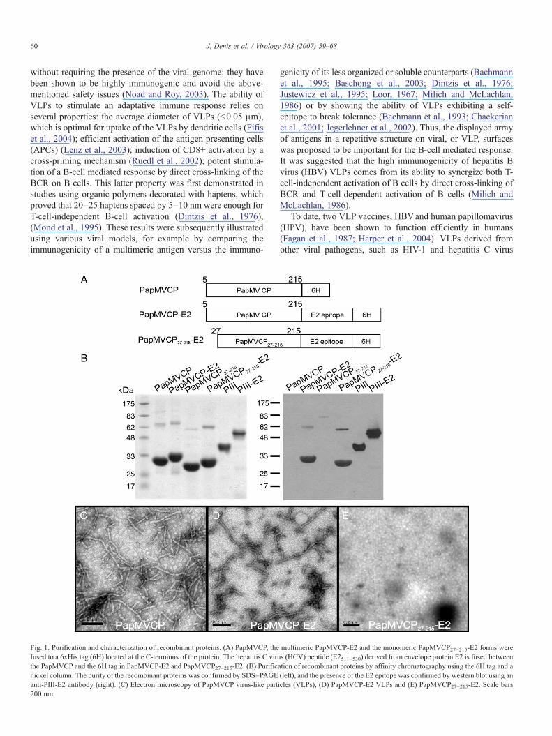
Fig. 1. Purification and characterization of recombinant proteins. (A) PapMVCP, thefused to a 6xHis tag (6H) located at the C-terminus of the protein. The hepatitis C viruthe PapMVCP and the 6H tag in PapMVCP-E2 and PapMVCP27–215-E2. (B) Purificnickel column. The purity of the recombinant proteins was confirmed by SDS–PAGEanti-PIII-E2 antibody (right). (C) Electron microscopy of PapMVCP virus-like part200 nm.
genicity of its less organized or soluble counterparts (Bachmannet al., 1995; Baschong et al., 2003; Dintzis et al., 1976;Justewicz et al., 1995; Loor, 1967; Milich and McLachlan,1986) or by showing the ability of VLPs exhibiting a self-epitope to break tolerance (Bachmann et al., 1993; Chackerianet al., 2001; Jegerlehner et al., 2002). Thus, the displayed arrayof antigens in a repetitive structure on viral, or VLP, surfaceswas proposed to be important for the B-cell mediated response.It was suggested that the high immunogenicity of hepatitis Bvirus (HBV) VLPs comes from its ability to synergize both T-cell-independent activation of B cells by direct cross-linking ofBCR and T-cell-dependent activation of B cells (Milich andMcLachlan, 1986).
To date, two VLP vaccines, HBVand human papillomavirus(HPV), have been shown to function efficiently in humans(Fagan et al., 1987; Harper et al., 2004). VLPs derived fromother viral pathogens, such as HIV-1 and hepatitis C virus
multimeric PapMVCP-E2 and the monomeric PapMVCP27–215-E2 forms weres (HCV) peptide (E2511–530) derived from envelope protein E2 is fused betweenation of recombinant proteins by affinity chromatography using the 6H tag and a(left), and the presence of the E2 epitope was confirmed by western blot using anicles (VLPs), (D) PapMVCP-E2 VLPs and (E) PapMVCP27–215-E2. Scale bars

Fig. 2. Comparison of the pro-inflammatory response of PapMVCP andlipopolysaccharides (LPS). Dorsal air pouches were raised in 10- to 12-week-oldCD1 mice. One milliliter of endotoxin-free PBS alone; PBS containing 0.05, 1or 1000 EU/ml LPS; or recombinant PapMVCP (10 μg/ml) were injected intothe air pouches. Migrating leukocytes in pouch exudates were harvested andcounted 6 h after treatment. Data represent the mean±SEM of 5 mice in eachcase. These results are representative of two identical and independentexperiments. *Significant (P<0.05) differences between cell recruitment levelsas determined by Dunn's multiple comparison test.
61J. Denis et al. / Virology 363 (2007) 59–68
(HCV), have also been generated, but their efficacy remains tobe proven in humans (Noad and Roy, 2003). Alternatively, theuse of VLPs from plant viruses as epitope presentationsystems has triggered much interest. Indeed, plant virusescombine three characteristics essential to triggering a humoralresponse and memory: (1) they are comprised mainly ofproteins that are highly immunogenic, (2) they are phylogen-etically distant from the mammalian immune system, and (3)they possess a complex and repetitive organization. Cowpeamosaic virus (CPMV), tobacco mosaic virus (TMV), alfalfamosaic virus (AlMV) and potato virus X (PVX) have beensuccessfully produced in plants as vaccine platforms present-ing peptides of interest (Canizares et al., 2005), and somevaccines have been proven to be protective in various diseasesmodels (reviewed in Streatfield and Howard, 2003). Further-more, the expression of Johnson grass mosaic virus coatprotein (CP) in E. coli (Saini and Vrati, 2003) led to theformation of VLPs that were used as a recombinantvaccination platform conferring protection after challengeagainst Japanese encephalitis virus. Following the observationthat expression of the papaya mosaic virus (PapMV) CP inE. coli also led to the self-assembly of VLPs composed ofseveral hundred CP subunits organized in a repetitive manner(Tremblay et al., 2006), the potential of PapMV as arecombinant peptidic vaccine platform was tested. An epitopefrom the HCV E2 glycoprotein was fused to the PapMV CP,resulting in the production of a new recombinant VLPplatform (PapMVCP-E2). The fused HCV peptide (511–530) belongs to a larger region (411–613) of the E2glycoprotein containing three main antibody (Ab) determi-nants that can prevent the binding of HCV to its CD81receptor on human cells (Lechner et al., 1998). Furthermore,the critical function of multimerization in triggering a humoralresponse, already demonstrated in mammalian virus models,was evaluated for the first time using a plant virus model, bycomparing the immunogenicity of a recombinant plant VLPand its monomeric counterpart. A monomeric mutant wasgenerated by deletion of the 26 first amino acids of the CP(PapMVCP27–215); this protein (referred to as PapMVCP27–215-E2 in this report) was used to express the same epitope as themultimeric protein and its performance as a vaccine platformwas compared in vivo with the PapMV VLP-based vaccineplatform. Importantly, this monomeric form of PapMV CP,which is unable to self-assemble in E. coli, has previously beenreported as having the same secondary structure as native CP(Lecours et al., 2005).
In this report, we show that the recombinant multimericPapMV vaccine platform is able to trigger a strong and long-lasting immune response against a C-terminal fused epitope.The antibody response profile directed against the epitopesuggests a balanced Th1/Th2 response. Notably, the sameplatform lost its immunogenic properties when injected as amonomeric protein, validating the finding that a repetitiveorganization is one of the key properties of molecules triggeringan immune response in the mammalian immune system. This isthe first demonstration of the critical function of a multimericstructure for a plant-virus-based platform.
Results
Production of recombinant multimeric and monomeric forms ofPapMV CP
Expression of PapMVCP in E. coli generates VLPs that aresimilar in appearance to the wild-type virus purified from theleaves of infected papaya plants (Tremblay et al., 2006). Toexamine the ability of PapMVCP fused to an epitope from theHCV E2 glycoprotein to assemble into VLPs, recombinantPapMVCP, PapMVCP-E2 and the monomeric formPapMVCP27–215-E2 (Fig. 1A) were expressed in E. coli andpurified using an easy procedure (see Materials and methods).SDS–PAGE analysis revealed that there were no significantdifferences in the yield of the various recombinant proteins (Fig.1B, left panel), and immunoblotting directed against the E2epitope indicated that no trimming of the peptide had occurred(Fig 1B, right panel). Note that the apparent molecular weightsof PapMVCP-derived proteins on SDS–PAGE are slightlyhigher than expected (Fig. 1B, left panel; observed 30–33 kDavs predicted 23–26 kDa), as previously noted (Lecours et al.,2005; Tremblay et al., 2006). As expected, an immunoblotusing antibody directed to the E2 peptide confirmed that boththe monomeric (PapMVCP27–215-E2) and the multimeric form(PapMVCP-E2) harbor the E2 fusion (Fig. 1B, right panel).Endotoxin levels were always below 0.005 EU/μg of protein.Electron microscopy (EM) confirmed that addition of the E2peptide at the C-terminus of PapMVCP did not affect the abilityof the protein to self-assemble into VLPs (Fig. 1D), although theparticles were notably shorter than VLPs formed by recombinantPapMVCP (Fig. 1C). Confirming previous results (Lecourset al., 2005), the monomeric form PapMVCP27–215-E2 wasunable to form VLPs (Fig. 1E). PapMVCP-E2 VLPs werevariable in length, with a size range of 201±80 nm. A lengthof 201 nm would represent 560 copies of the CP presentingthe E2 peptide in a repetitive fashion. The secondary structuresof the purified proteins were determined using circulardichroism spectrophotometry and were shown to be verysimilar to those previously reported (data not shown) (Lecourset al., 2005).
62 J. Denis et al. / Virology 363 (2007) 59–68
Recombinant PapMV VLPs are weak inflammatory molecules
Some commonly used adjuvants, such as alum, have beendescribed as mediating an inflammatory episode at the site ofinjection (Brewer, 2006). This characteristic can be very usefulfor the induction of a strong immune response. To test the pro-inflammatory properties of PapMVCP VLPs, we used themurine air pouch model. Injection of 10 μg of PapMVCP VLPsfailed to induce the recruitment of leukocytes into the pouch ofCD1 mice 6 h after treatment. In contrast, injection oflipopolysaccharides (LPS) at doses of 1000 and 1 EU wasvery effective in inducing the recruitment of leukocytes (Fig. 2).
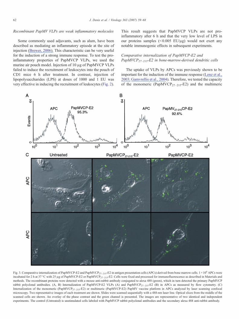
Fig. 3. Comparative internalization of PapMVCP-E2 and PapMVCP27–215-E2 in antigincubated for 2 h at 37 °C with 25 μg of PapMVCP-E2 or PapMVCP27–215-E2. Cellsmethods. The recombinant proteins were detected with a mouse anti-rabbit antibodyrabbit polyclonal antibodies. (A, B) Internalization of PapMVCP-E2 VLPs (A)Internalization of the monomeric (PapMVCP27–215-E2) or multimeric (PapMVCPmicroscopy. Two representative images of each treatment are shown. Slides were scascanned cells are shown. An overlay of the phase contrast and the green channelexperiments. The control (Untreated) is unstimulated cells labeled with PapMVCP r
This result suggests that PapMVCP VLPs are not pro-inflammatory after 6 h and that the very low level of LPS inour proteins samples (<0.005 EU/μg) would not exert anynotable immunogenic effects in subsequent experiments.
Comparative internalization of PapMVCP-E2 andPapMVCP27–215-E2 in bone-marrow-derived dendritic cells
The uptake of VLPs by APCs was previously shown to beimportant for the induction of the immune response (Lenz et al.,2003; Gamvrellis et al., 2004). Therefore, we tested the capacityof the monomeric (PapMVCP27–215-E2) and the multimeric
en presentation cells (APCs) derived from bone marrow cells. 1×10⁶ APCs werewere fixed and processed for immunofluorescence as described in Materials andconjugated to alexa 488 (green), which in turn detected the primary PapMVCPand PapMVCP27–215-E2 (B) in APCs as measured by flow cytometry. (C)-E2) PapMV vaccine platform in APCs analyzed by laser scanning confocalnned sequentially with a 488-nm laser line. Optical slices from the middle of theis presented. The images are representative of two identical and independentabbit polyclonal antibodies and the secondary alexa 488 anti-rabbit antibody.
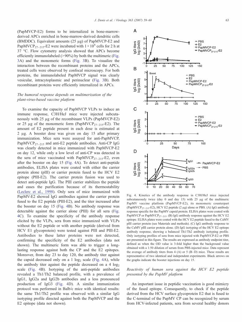
Fig. 4. Kinetics of the antibody response in C3H/HeJ mice injectedsubcutaneously twice (day 0 and day 15) with 25 μg of the multimericPapMV vaccine platform (PapMVCP-E2), its monomeric counterpart(PapMVCP27–215-E2), HCV E2 peptide (2 μg) alone or PBS. (A) IgG antibodyresponse specific for the PapMV capsid protein. ELISA plates were coated withPapMVCP or PapMVCP27–215. (B) IgG antibody response against the HCV E2epitope. ELISA plates were coated with the HCV E2 peptide fused to the CaMVpIII carrier protein (see Materials and methods). (C) IgG antibody response tothe CaMV pIII carrier protein alone. (D) IgG isotyping of the HCV E2 epitopeantibody response, showing a balanced Th1/Th2 antibody isotyping profile.Only isotyping profiles of sera from mice injected with PapMVCP-E2 or PBSare presented in this figure. The results are expressed as antibody endpoint titer,defined as when the OD value is 3-fold higher than the background valueobtained with a 1:50 dilution of serum from PBS-injected mice. Data representthe average of antibody titers from 4 (A) or 5 (B–D) mice. These results arerepresentative of two identical and independent experiments. Black arrows onthe graphs indicate the booster injections on day 15.
63J. Denis et al. / Virology 363 (2007) 59–68
(PapMVCP-E2) forms to be internalized in bone-marrow-derived APCs enriched in bone-marrow-derived dendritic cells(BMDDC). Equivalent amounts (25 μg) of PapMVCP-E2 andPapMVCP27–215-E2 were incubated with 1×10⁶ cells for 2 h at37 °C. Flow cytometry analysis showed that APCs becomeefficiently immunolabeled (>90%) by both the multimeric (Fig.3A) and the monomeric forms (Fig. 3B). To visualize theinteraction between the recombinant proteins and the APCs,treated cells were observed by confocal microscopy. For bothproteins, the immunolabeled PapMVCP signal was clearlyvesicular, intracytoplasmic and perinuclear (Fig. 3B). Bothrecombinant proteins were efficiently internalized in APCs.
The humoral response depends on multimerization of theplant-virus-based vaccine platform
To examine the capacity of PapMVCP VLPs to induce animmune response, C3H/HeJ mice were injected subcuta-neously with 25 μg of the recombinant VLPs (PapMVCP-E2)or 25 μg of the monomeric form (PapMVCP27–215-E2). Theamount of E2 peptide present in each dose is estimated at2 μg. A booster dose was given on day 15 after primaryimmunization. Mice sera were assayed for anti-PapMVCP,PapMVCP27–215 and anti-E2 peptide antibodies. Anti-CP IgGwas clearly detected in mice immunized with PapMVCP-E2on day 12, while only a low level of anti-CP was detected inthe sera of mice vaccinated with PapMVCP27–215-E2, evenafter the booster on day 15 (Fig. 4A). To detect anti-peptideantibodies, ELISA plates were coated with either the carrierprotein alone (pIII) or carrier protein fused to the HCV E2epitope (PIII-E2). The carrier protein fusion was used todetect anti-peptide IgG. The PIII carrier stabilizes the peptideand eases the purification because of its thermostability(Leclerc et al., 1998). Only sera of mice immunized withPapMV-E2 showed IgG antibodies against the carrier proteinfused to the E2 peptide (PIII-E2), and the titer increased afterthe booster on day 15 (Fig. 4B). No antibody response wasdetectable against the carrier alone (PIII) for all sera (Fig.4C). To examine the specificity of the antibody responseelicited by the VLPs, sera from mice immunized with VLPswithout the E2 peptide or with another peptide (derived fromHCV E1 glycoprotein) were tested against PIII and PIII-E2.Antibodies to these latter proteins were not detected,confirming the specificity of the E2 antibodies (data notshown). The multimeric form was able to trigger a long-lasting response against both the CP and the E2 epitopes.Moreover, from day 23 to day 120, the antibody titer againstthe capsid decreased only on a 1 log2 scale (Fig. 4A), whilethe antibody titer against the peptide decreased on a 4 log2scale (Fig. 4B). Isotyping of the anti-peptide antibodiesrevealed a Th1/Th2 balanced profile, with a prevalence ofIgG1, IgG2a and IgG2b antibodies and a less pronouncedproduction of IgG3 (Fig. 4D). A similar immunizationprotocol was performed in Balb/c mice with identical results:the same Th1/Th2 profile was observed with a similar IgGisotyping profile directed against both the PapMVCP and theE2 epitope (data not shown).
Reactivity of human sera against the HCV E2 peptidepresented by the PapMV platform
An important issue in peptidic vaccination is good mimicryof the fused epitope. Consequently, to check if the peptidederived from the HCV surface glycoprotein E2 that is fused tothe C-terminal of the PapMV CP can be recognized by serumfrom HCV-infected patients, sera from several healthy donors
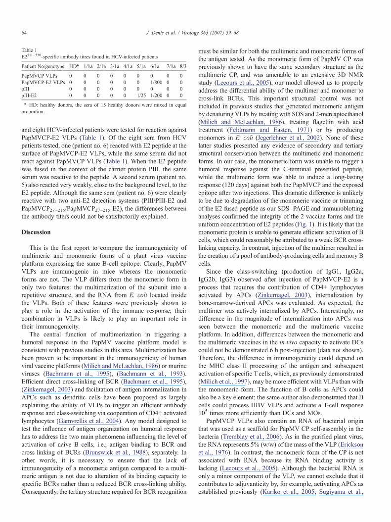
Table 1E2511–530-specific antibody titres found in HCV-infected patients
Patient No/genotype HD⁎ 1/1a 2/1a 3/1a 4/1a 5/1a 6/1a 7/1a 8/3
PapMVCP VLPs 0 0 0 0 0 0 0 0 0PapMVCP-E2 VLPs 0 0 0 0 0 0 1/800 0 0pIII 0 0 0 0 0 0 0 0 0pIII-E2 0 0 0 0 0 1/25 1/200 0 0
⁎ HD: healthy donors, the sera of 15 healthy donors were mixed in equalproportion.
64 J. Denis et al. / Virology 363 (2007) 59–68
and eight HCV-infected patients were tested for reaction againstPapMVCP-E2 VLPs (Table 1). Of the eight sera from HCVpatients tested, one (patient no. 6) reacted with E2 peptide at thesurface of PapMVCP-E2 VLPs, while the same serum did notreact against PapMVCP VLPs (Table 1). When the E2 peptidewas fused in the context of the carrier protein PIII, the sameserum was reactive to the peptide. A second serum (patient no.5) also reacted very weakly, close to the background level, to theE2 peptide. Although the same sera (patient no. 6) were clearlyreactive with two anti-E2 detection systems (PIII/PIII-E2 andPapMVCP27–215/PapMVCP27–215-E2), the differences betweenthe antibody titers could not be satisfactorily explained.
Discussion
This is the first report to compare the immunogenicity ofmultimeric and monomeric forms of a plant virus vaccineplatform expressing the same B-cell epitope. Clearly, PapMVVLPs are immunogenic in mice whereas the monomericforms are not. The VLP differs from the monomeric form inonly two features: the multimerization of the subunit into arepetitive structure, and the RNA from E. coli located insidethe VLPs. Both of these features were previously shown toplay a role in the activation of the immune response; theircombination in VLPs is likely to play an important role intheir immunogenicity.
The central function of multimerization in triggering ahumoral response in the PapMV vaccine platform model isconsistent with previous studies in this area. Multimerization hasbeen proven to be important in the immunogenicity of humanviral vaccine platforms (Milich and McLachlan, 1986) or murineviruses (Bachmann et al., 1995), (Bachmann et al., 1993).Efficient direct cross-linking of BCR (Bachmann et al., 1995),(Zinkernagel, 2003) and facilitation of antigen internalization inAPCs such as dendritic cells have been proposed as largelyexplaining the ability of VLPs to trigger an efficient antibodyresponse and class-switching via cooperation of CD4+ activatedlymphocytes (Gamvrellis et al., 2004). Any model designed totest the influence of antigen organization on humoral responsehas to address the two main phenomena influencing the level ofactivation of naive B cells, i.e., antigen binding to BCR andcross-linking of BCRs (Brunswick et al., 1988), separately. Inother words, it is necessary to ensure that the lack ofimmunogenicity of a monomeric antigen compared to a multi-meric antigen is not due to alteration of its binding capacity tospecific BCRs rather than a reduced BCR cross-linking ability.Consequently, the tertiary structure required for BCR recognition
must be similar for both the multimeric and monomeric forms ofthe antigen tested. As the monomeric form of PapMV CP waspreviously shown to have the same secondary structure as themultimeric CP, and was amenable to an extensive 3D NMRstudy (Lecours et al., 2005), our model allowed us to properlyaddress the differential ability of the multimer and monomer tocross-link BCRs. This important structural control was notincluded in previous studies that generated monomeric antigenby denaturing VLPs by treating with SDS and 2-mercaptoethanol(Milich and McLachlan, 1986), treating flagellin with acidtreatment (Feldmann and Easten, 1971) or by producingmonomers in E. coli (Jegerlehner et al., 2002). None of theselatter studies presented any evidence of secondary and tertiarystructural conservation between the multimeric and monomericforms. In our case, the monomeric form was unable to trigger ahumoral response against the C-terminal presented peptide,while the multimeric form was able to induce a long-lastingresponse (120 days) against both the PapMVCP and the exposedepitope after two injections. This dramatic difference is unlikelyto be due to degradation of the monomeric vaccine or trimmingof the E2 fused peptide as our SDS–PAGE and immunoblottinganalyses confirmed the integrity of the 2 vaccine forms and theuniform concentration of E2 peptides (Fig. 1). It is likely that themonomeric protein is unable to generate efficient activation of Bcells, which could reasonably be attributed to a weak BCR cross-linking capacity. In contrast, injection of the multimer resulted inthe creation of a pool of antibody-producing cells and memory Bcells.
Since the class-switching (production of IgG1, IgG2a,IgG2b, IgG3) observed after injection of PapMVCP-E2 is aprocess that requires the contribution of CD4+ lymphocytesactivated by APCs (Zinkernagel, 2003), internalization bybone-marrow-derived APCs was evaluated. As expected, themultimer was actively internalized by APCs. Interestingly, nodifference in the magnitude of internalization into APCs wasseen between the monomeric and the multimeric vaccineplatform. In addition, differences between the monomeric andthe multimeric vaccines in the in vivo capacity to activate DCscould not be demonstrated 6 h post-injection (data not shown).Therefore, the difference in immunogenicity could depend onthe MHC class II processing of the antigen and subsequentactivation of specific T cells, which, as previously demonstrated(Milich et al., 1997), may be more efficient with VLPs than withthe monomeric form. The function of B cells as APCs couldalso be a key element; the same author also demonstrated that Bcells could process HBV VLPs and activate a T-cell response105 times more efficiently than DCs and MOs.
PapMVCP VLPs also contain an RNA of bacterial originthat was used as a scaffold for PapMV CP self-assembly in thebacteria (Tremblay et al., 2006). As in the purified plant virus,the RNA represents 5% (w/w) of the mass of the VLP (Ericksonet al., 1976). In contrast, the monomeric form of the CP is notassociated with RNA because its RNA binding activity islacking (Lecours et al., 2005). Although the bacterial RNA isonly a minor component of the VLP, we cannot exclude that itcontributes to adjuvanticity by, for example, activating APCs asestablished previously (Kariko et al., 2005; Sugiyama et al.,

65J. Denis et al. / Virology 363 (2007) 59–68
2005). Any potential contribution to the immunogenicity ofPapMV VLPs of other bacterial components with strongadjuvanticity, such as LPS, was shown to be negligible in twodifferent ways. Firstly, immunization with PapMV VLPs inLPS-sensitive mice (Balb/c, data not shown) and LPS hypo-responsive mice C3H/HeJ (Fig. 4) led to the same humoralimmune response against both the platform and the C-fusedepitope. Secondly, PapMV VLPs with very low LPS content(<0.005 EU/μg) were unable to trigger the recruitment of innateimmune cells in the air pouch model after 6 h, contrasting withthe high recruitment capacity of LPS (Fig. 2). Taken together,our results confirm the conclusion that the immunogenic effectof recombinant PapMVCP VLPs does not derive from LPS-likepro-inflammatory properties. The only other reported case ofuse of VLPs from a plant virus produced in E. coli made nomention of determination of the LPS content of the proteinsinjected or the influence of LPS content on the observed results(Saini and Vrati, 2003).
The Th1/Th2 profile of antibodies triggered by the PapMVrecombinant protein without the use of any adjuvants suggeststhat this newly described vaccine platform potentially triggersa broader immune response than other plant-virus-basedplatforms. Indeed, antibody isotyping has previously suggestedplant virus vaccine platforms to be biased toward a Th1response (Marusic et al., 2001; McInerney et al., 1999) andcytokine secretion (Piazzolla et al., 2005). Even the presence ofa commercial adjuvant with VLPs did not modify the Th1 bias(Marusic et al., 2001). A balanced Th1/Th2 antibody profilecould increase the immune effector mechanisms of the humoralresponse such as neutralization or antibody-dependent cell-mediated cytotoxicity (ADCC) additional experiments wouldbe necessary to investigate the potential of the PapMVplatform to trigger these mechanisms in the HCV model.Moreover, the findings that the PapMV vaccine platform wasable to trigger a pool of all antibody isotypes against the HCVE2 peptide and that serum from one HCV patient was clearlyable to recognize the E2 peptide fused to the PapMV platformcould indicate that this platform is suitable for triggering, forexample, ADCC directed against hepatocytes exhibiting E2protein on their surface as described in a clinical report byNattermann et al. (2005), potentially helping to reduce theseverity of the disease. In addition, a recent report (Leclerc etal., in press) demonstrated the capacity of the PapMV platformto activate human specific CD8+ lymphocytes in vitro by cross-priming. Taken together, these results suggest that thedevelopment of a vaccination platform based on PapMVVLPs against infectious diseases such as HCV, which requiresboth a protective CD8+ and a humoral response (Houghton andAbrignani, 2005), is a promising avenue of research.
Materials and methods
Cloning and engineering of the PapMV coat protein
The PapMV CP gene was amplified by RT/PCR fromisolated viral RNA using primers 5′-AGTCCCATGGCATC-CACACCCAACATAGCCTTC-3′and 5′-GATCGGATCCT-
TACTAATGGTGATGGTGATGGTGACGCGTGGTAC-TAGTTTCGGGGGGTGGAAGGAATTGGATGGTTGG-3′and cloned as an NcoI/BamHI fragment in pET 3D (NewEngland Biolabs). To generate the PapMVCP-E2 construct,CCACCGATCGTAGCGGTGCGCCGACCTACAGCTGG-GGTGCGAACGATACGCGTCATG-3′ and 5′-CATGACGC-GTATCGTTCGCACCCCAGCTGTAGGTCGGCGCACCGC-TACGATCGGTGGTACCCACCACCACACTAGTGATC-3′were annealed together and digested with SpeI and MluI beforeligation into the SpeI/MluI-linearized PapMVCP clone. Theexpression vector for PapMVCP27–215-E2 was constructed fromthe PapMVCP-E2 plasmid as follows: two oligonucleotides(including an NcoI restriction site) designed to delete the 26first amino acids of the PapMV CP were used for PCR: forward5′-AGTCCCATGGCCGATCCAACGTCCAATCTTCTG-3′and reverse 5′-ACGTCCATGGTATATCTCCTTCTTAAAG-3′.The PCR product was then self-ligated. The expression vector forPapMVCP27–215 was derived from the PapMVCP plasmid fol-lowing the same procedure as for the construction of thePapMVCP27–215-E2 clone. The sequences of all PapMV cloneswere confirmed by DNA sequencing.
Cloning and engineering of the carrier protein (PIII)
A plasmid designed to express the cauliflower mosaic virus(CaMV) pIII protein in E. coli has been previously described(Leclerc et al., 1998). A truncated version of CaMV pIIIcomprising the 74 N-terminal amino acids was generated usingprimers 5′-AAACCCGGGGAATTCACCATGGCTAA-TCTTAATCAGATCCAAAAG 3′ and 5′-GATCGGATC-CTAACGCGTGGTACTAGTAGGTTGGGTACCTA-AGGCTTC-3′. This truncated version harbored unique SpeIand MluI sites that are convenient for cloning a peptide infusion with pIII. To generate a PIII-E2 construct, theoligonucleotides E2 5′-GATCACTAGTGTGGTGGTG-GGTACCACCGATCGTAGCGGTGCGCCGACCTA-CAGCTGGGGTGCGAACGATACGCGTCATG-3′ and5′-CATGACGCGTATCGTTCGCACCCCAGCTGTAGG-TCGGCGCACCGCTACGATCGGTGGTACCCACCACCA-CACTAGTGATC-3′ were annealed together and digested withSpeI andMluI before ligation into the PIII clone linearized withSpeI andMluI. The sequences of PIII clones were confirmed byDNA sequencing. The E. coli expression strain BL21(DE3)RIL (Stratagene) was transformed with PIII constructs, andprotein expression was induced with 1 mM IPTG for 16 h at25 °C. Cells were pelleted by centrifugation and resuspended inTris–HCl pH 7.0, 20 mM NaCl, 1 mM EDTA and 0.02 mMPMSF before lysis by sonication. The protein extracts were thenheated at 65 °C for 15 min and centrifuged at 15,000×g for20 min at 4 °C. Incubation at 65 °C was repeated a second time.The samples were incubated at 80 °C for 15 min andcentrifuged again for 20 min at 15,000×g. The chimeric pIIIprotein is resistant to heat and remains soluble in thesupernatant after this treatment. The NaCl concentration wasadjusted to 150 mM and protein extracts were separated on aSephadex G-50 column to achieve 95% purity. The PIII proteinis referred to as the carrier protein in the text.
66 J. Denis et al. / Virology 363 (2007) 59–68
Expression and purification of PapMVCP, PapMVCP-E2,PapMVCP27–215 and PapMVCP27–215-E2
Expression and purification of PapMVCP constructs wereperformed as previously described with minor modifications(Tremblay et al., 2006). Briefly, the bacteria were lysedthrough a French press and then loaded onto a Ni2+ column,washed with 10 mM Tris–HCl/50 mM Imidazole/0.5% TritonX100 (pH 8), then with 10 mM Tris–HCl/50 mM Imidazole/1% Zwittergent (pH 8) to remove endotoxin contamination.For the PapMVCP and PapMVCP-E2 proteins, the eluate wassubjected to high speed centrifugation (100,000×g) for120 min in a Beckman 50.2 TI rotor. The VLP pellet wasresuspended in endotoxin-free PBS (Sigma). Followingelution of the PapMVCP27–215 and PapMVCP27–215-E2proteins, the solutions were dialyzed against PBS using a6–8 kDa molecular weight cut-off membrane (Spectra). TheE2 peptide was synthesized by GLBiochem (Shanghai) Ltdand resuspended in endotoxin-free PBS (Sigma). Proteinsolutions were filtered using 0.45 μM filters before use. Thepurity of the proteins was determined by SDS–PAGE andconfirmed by western immunoblot analysis using mousepolyclonal antibodies generated against PIII-E2. The amountof protein was evaluated using a BCA protein kit (Pierce). Thelevel of LPS in the purified protein was evaluated with theLimulus test according to the manufacturer's instructions(Cambrex) and was below 0.005 endotoxin units (EU)/μg ofprotein.
Electron microscopy
Proteins were diluted in PBS and were absorbed for 3 min oncarbon-coated formvar grids. The grids were washed twice withdeionized water and stained with 2% uranyl acetate for 10 minat room temperature. The grids were then observed on a JeolJEM220FS transmission electron microscope. Average VLPlength was evaluated by measuring 100 VLPs using AdobePhotoshop software.
Immunization
Five 4-to 8-week-old C3H/HeJ mice (Charles RiversLaboratories) were injected subcutaneously with 25 μg ofPapMVCP-E2, PapMVCP27–215-E2 or the equivalent amountof the E2 peptide (2 μg) or endotoxin-free PBS (Sigma).Primary immunization was followed by one booster dose given2 weeks later. Blood samples were obtained at different timepoints and stored at −20 °C until analysis. All the experimentalprotocols were approved by the Laval University animalprotection committee.
ELISA quantification
Costar High Binding 96-well plates (Corning, NY, USA)were coated overnight at 4 °C with 100 μl/well of P3, P3E2,PapMVCP, PapMVCP27–215, or PapMVCP-E2 diluted to aconcentration of 1 μg/ml in 0.1 M NaHCO3 buffer pH 9.6. The
plates were blocked with PBS/0.1% Tween-20/2% BSA(150 μl/well) for 1 h at 37 °C. After washing three times withPBS/0.1% Tween-20, sera were added in 2-fold serial dilution(beginning with 1:50) and incubated for 1 h at 37 °C. Followingincubation, the plates were washed three times and incubatedwith 100 μl of peroxidase-conjugated goat anti-mouse IgG,IgG1, IgG2a, IgG2b (all from Jackson Immunoresarch) or IgG3(Rockland) at a dilution of 1:10,000 in PBS/0.1% Tween-20/2%BSA for 1 h at 37 °C. After three washes, the presence of IgGwas detected with 100 μl of TMB-S according to themanufacturer's instructions; the reaction was stopped by adding100 μl of 0.18 mM H2SO4 and the OD was read at 450 nm. Theresults are expressed as antibody endpoint titer, determinedwhen the OD value is 3-fold the background value obtainedwith a 1:50 dilution of serum from PBS-injected mice. For thedetermination of antibody levels in human sera, the sameconditions were applied, except that the peroxidase-conjugatedgoat anti-human IgG as secondary antibodies were used at adilution of 1:80,000. Sera from infected HCV patients wereprovided by B. Willems (Hopital Saint Luc, CHUM): the resultsare expressed as antibody endpoint titer, defined as the point atwhich the OD value is 3-fold the background value obtainedwith a 1:25 dilution of serum from a pool of sera from 15 non-infected patients.
Air pouches in mice
Air pouches were raised in 10- to 12-week-old CD1 mice(Charles River Laboratories). Experimental protocols wereapproved by the Laval University animal protection committee.Air pouches were raised on the dorsum by subcutaneousinjection of 3 ml of sterile air on days 0 and 3. On day 7, 1 ml ofrecombinant PapMVCP (1 to 10 μg/ml), LPS (10 μg/ml) orPBS was injected into the air pouches. Six hours after treatment,the mice were killed by asphyxiation using CO2. The airpouches were washed once with 1 ml PBS–5 mM EDTA andthen twice with 2 ml of PBS–5 mM EDTA, and the exudateswere centrifuged at 500×g for 5 min at room temperature. Cellswere counted with a hematocytometer following acetic bluestaining.
Bone marrow cell extraction and differentiation of APCs
Bone marrow progenitors cells were obtained from the femursof Balb/c mice and cultured for 6 days in dendritic cellsdifferentiation bone marrow medium (95% RPMI with 1%penicillin–streptomycin and supplemented with 5% X63-GM-CSF supernatant media culture; the X63-GM-CSF cell line wasprovided by B. Ludewig, Research Department, CantonalHospital, St. Gallen, Switzerland). Mediumwas partially replacedon days 2 and 4. On day 6, the medium was replaced by mediumwithout LX63-conditioned medium. On day 7, enrichment ofAPCs was verified by flow cytometry using FITC anti-CD11cand PE-Cy5.5 anti-CD11b surface markers (BD Biosciences).The preparation contained 25% of CD11c+ Cd11b+ cells, andmore than 80% of CD11b+ cells. We refer to this preparation asAPCs.
67J. Denis et al. / Virology 363 (2007) 59–68
Flow cytometry
To evaluate internalization of the PapMVCP-E2 orPapMVCP27–215-E2 in APCs, 1 million bone-marrow-derivedAPCs were incubated for 2 h at 37 °C with either 25 μg ofPapMV-E2 or PapMVCP27–215-E2. Briefly, cells were blockedwith PBS containing 10% FBS and anti-CD16/CD32 (1 μg/1million cells) for 15 min at 4 °C. After 2 washes with PBS, cellswere fixed with PBS/2% paraformaldehyde for 10 min at roomtemperature. After 2 washes with permeabilization buffer (PBS10%/FBS 0.2%/Triton X-100), cells were incubated for 45 minat 4 °C with rabbit polyclonal antibodies diluted 1:200 in per-meabilization buffer. After 2 washes with permeabilizationbuffer, cells were incubated for 45 min at 4 °C with the secondaryantibodies (anti-rabbit IgG alexa 488; Molecular Probes) diluted1:5000 in permeabilization buffer. After washing with PBS, cellswere immediately analyzed with an EPICS-XL cytofluorometer.Data analysis was performed using WINMDI2.8. The rabbitpolyclonal Ab used for detection was produced in our own faci-lities: rabbit preimmune serum was used as a negative control.
Confocal microscopy
APCs were grown (200,000 cells/well) in 12-well plates(Corning, NY, USA) containing sterile slides in the bottomfollowing the differentiation protocol described above. Forantigen internalization studies, 5 μg of antigen/200,000 cellswas used. The fixation, permeabilization and primary andsecondary antibodies incubation steps were as described forflow cytometry. Slides were analyzed immediately with aFluoview Fv300 confocal microscope fitted with a ×60 oilimmersion objective. Fluorescence images were acquired sequen-tially to avoid non-specific channel interference and by x–zsectioning. Pictures were then digitally processed with Image Jsoftware. Optical slices from the middle of the scanned cells areshown in the figures.
Statistical analysis
Nonparametric Kruskal–Wallis and Dunn's multiple com-parison tests were used for statistical analysis. A value ofP<0.05 was considered statistically significant. Statisticalanalyses were performed with the program PRISM 3.03.
Acknowledgments
Wewould like to thank the Natural Sciences and EngineeringResearch Council of Canada (NSERC) for funding this researchprogram, and Helen Rothnie for editing and critical reading ofthe manuscript.
References
Ada, G., 2001. Vaccines and vaccination. N. Engl. J. Med. 345 (14), 1042–1053.Bachmann, M.F., Rohrer, U.H., Kundig, T.M., Burki, K., Hengartner, H.,
Zinkernagel, R.M., 1993. The influence of antigen organization on B cellresponsiveness. Science 262 (5138), 1448–1451.
Bachmann, M.F., Hengartner, H., Zinkernagel, R.M., 1995. T helper cell-independent neutralizing B cell response against vesicular stomatitis virus:role of antigen patterns in B cell induction? Eur. J. Immunol. 25 (12),3445–3451.
Baschong, W., Hasler, L., Haner, M., Kistler, J., Aebi, U., 2003. Repetitiveversus monomeric antigen presentation: direct visualization of antibodyaffinity and specificity. J. Struct. Biol. 143 (3), 258–262.
Brewer, J.M., 2006. (How) do aluminium adjuvants work? Immunol. Lett. 102(1), 10–15.
Brunswick, M., Finkelman, F.D., Highet, P.F., Inman, J.K., Dintzis, H.M.,Mond, J.J., 1988. Picogram quantities of anti-Ig antibodies coupled todextran induce B cell proliferation. J. Immunol. 140 (10), 3364–3372.
Canizares, M.C., Nicholson, L., Lomonossoff, G.P., 2005. Use of viral vectorsfor vaccine production in plants. Immunol. Cell Biol. 83 (3), 263–270.
Chackerian, B., Lowy, D.R., Schiller, J.T., 2001. Conjugation of a self-antigen topapillomavirus-like particles allows for efficient induction of protectiveautoantibodies. J. Clin. Invest. 108 (3), 415–423.
Daar, A.S., Thorsteinsdottir, H., Martin, D.K., Smith, A.C., Nast, S., Singer,P.A., 2002. Top ten biotechnologies for improving health in developingcountries. Nat. Genet. 32 (2), 229–232.
Dintzis, H.M., Dintzis, R.Z., Vogelstein, B., 1976. Molecular determinants ofimmunogenicity: the immuno model of immune response. Proc. Natl. Acad.Sci. U.S.A. 73 (10), 3671–3675.
Erickson, J.W., Bancroft, J.B., Horne, R.W., 1976. The assembly of papayamosaic virus protein. Virology 72 (2), 514–517.
Fagan, E.A., Tolley, P., Smith, H.M., Peters, M.P., Coleman, J., Elliott, P.,Williams, R., Eddleston, A.L., 1987. Hepatitis B vaccine: immunogeni-city and follow-up including two year booster doses in high-risk healthcare personnel in a London teaching hospital. J. Med. Virol. 21 (1),49–56.
Feldmann, M., Easten, A., 1971. The relationship between antigenic structureand the requirement for thymus-derived cells in the immune response.J. Exp. Med. 134 (1), 103–119.
Fifis, T., Gamvrellis, A., Crimeen-Irwin, B., Pietersz, G.A., Li, J., Mottram, P.L.,McKenzie, I.F., Plebanski, M., 2004. Size-dependent immunogenicity:therapeutic and protective properties of nano-vaccines against tumors.J. Immunol. 173 (5), 3148–3154.
Gamvrellis, A., Leong, D., Hanley, J.C., Xiang, S.D., Mottram, P., Plebanski,M., 2004. Vaccines that facilitate antigen entry into dendritic cells. Immunol.Cell Biol. 82 (5), 506–516.
Harper, D.M., Franco, E.L., Wheeler, C., Ferris, D.G., Jenkins, D., Schuind, A.,Zahaf, T., Innis, B., Naud, P., De Carvalho, N.S., Roteli-Martins, C.M.,Teixeira, J., Blatter, M.M., Korn, A.P., Quint, W., Dubin, G., 2004. Efficacyof a bivalent L1 virus-like particle vaccine in prevention of infection withhuman papillomavirus types 16 and 18 in young women: a randomisedcontrolled trial. Lancet 364 (9447), 1757–1765.
Houghton, M., Abrignani, S., 2005. Prospects for a vaccine against the hepatitisC virus. Nature 436 (7053), 961–966.
Jegerlehner, A., Tissot, A., Lechner, F., Sebbel, P., Erdmann, I., Kundig, T.,Bachi, T., Storni, T., Jennings, G., Pumpens, P., Renner, W.A., Bachmann,M.F., 2002. A molecular assembly system that renders antigens of choicehighly repetitive for induction of protective B cell responses. Vaccine 20(25–26), 3104–3112.
Justewicz, D.M., Doherty, P.C., Webster, R.G., 1995. The B-cell response inlymphoid tissue of mice immunized with various antigenic forms of theinfluenza virus hemagglutinin. J. Virol. 69 (9), 5414–5421.
Kariko, K., Buckstein, M., Ni, H., Weissman, D., 2005. Suppression of RNArecognition by Toll-like receptors: the impact of nucleoside modification andthe evolutionary origin of RNA. Immunity 23 (2), 165–175.
Lechner, S., Rispeter, K., Meisel, H., Kraas, W., Jung, G., Roggendorf, M.,Zibert, A., 1998. Antibodies directed to envelope proteins of hepatitis Cvirus outside of hypervariable region 1. Virology 243 (2), 313–321.
Leclerc, D., Burri, L., Kajava, A.V., Mougeot, J.L., Hess, D., Lustig, A.,Kleemann, G., Hohn, T., 1998. The open reading frame III product ofcauliflower mosaic virus forms a tetramer through a N-terminal coiled-coil.J. Biol. Chem. 273 (44), 29015–29021.
Leclerc, D., Beauseigle, D., Denis, J., Morin, H., Pare, C., Lamarre, A.,Lapointe, R., in press. Proteasome-independent MHC class I cross-
68 J. Denis et al. / Virology 363 (2007) 59–68
presentation mediated by papaya mosaic virus-like particles leads to theexpansion of specific human T cells. J Virol. (in press online)
Lecours, K., Tremblay, M.H., Gagne, M.E., Gagne, S.M., Leclerc, D., 2005.Purification and biochemical characterization of a monomeric form ofpapaya mosaic potexvirus coat protein. Protein Expr. Purif.
Lenz, P., Thompson, C.D., Day, P.M., Bacot, S.M., Lowy, D.R., Schiller, J.T.,2003. Interaction of papillomavirus virus-like particles with human myeloidantigen-presenting cells. Clin. Immunol. 106 (3), 231–237.
Loor, F., 1967. Comparative immunogenicities of tobacco mosaic virus, proteinsubunits, and reaggregated protein subunits. Virology 33 (2), 215–220.
Marusic, C., Rizza, P., Lattanzi, L., Mancini, C., Spada, M., Belardelli, F.,Benvenuto, E., Capone, I., 2001. Chimeric plant virus particles asimmunogens for inducing murine and human immune responses againsthuman immunodeficiency virus type 1. J. Virol. 75 (18), 8434–8439.
McInerney, T.L., Brennan, F.R., Jones, T.D., Dimmock, N.J., 1999. Analysis ofthe ability of five adjuvants to enhance immune responses to a chimeric plantvirus displaying an HIV-1 peptide. Vaccine 17 (11–12), 1359–1368.
Milich, D.R., McLachlan, A., 1986. The nucleocapsid of hepatitis B virus isboth a T-cell-independent and a T-cell-dependent antigen. Science 234(4782), 1398–1401.
Milich, D.R., Chen, M., Schodel, F., Peterson, D.L., Jones, J.E., Hughes, J.L.,1997. Role of B cells in antigen presentation of the hepatitis B core. Proc.Natl. Acad. Sci. U.S.A. 94 (26), 14648–14653.
Mond, J.J., Lees, A., Snapper, C.M., 1995. T cell-independent antigens type 2.Annu. Rev. Immunol. 13, 655–692.
Nattermann, J., Schneiders, A.M., Leifeld, L., Langhans, B., Schulz, M.,Inchauspe, G., Matz, B., Brackmann, H.H., Houghton, M., Sauerbruch, T.,Spengler, U., 2005. Serum antibodies against the hepatitis C virus E2 protein
mediate antibody-dependent cellular cytotoxicity (ADCC). J. Hepatol. 42(4), 499–504.
Noad, R., Roy, P., 2003. Virus-like particles as immunogens. Trends Microbiol.11 (9), 438–444.
Piazzolla, G., Nuzzaci, M., Tortorella, C., Panella, E., Natilla, A., Boscia, D.,De Stradis, A., Piazzolla, P., Antonaci, S., 2005. Immunogenic propertiesof a chimeric plant virus expressing a hepatitis C virus (HCV)-derivedepitope: new prospects for an HCV vaccine. J. Clin. Immunol. 25 (2),142–152.
Ruedl, C., Storni, T., Lechner, F., Bachi, T., Bachmann, M.F., 2002. Cross-presentation of virus-like particles by skin-derived CD8(−) dendritic cells: adispensable role for TAP. Eur. J. Immunol. 32 (3), 818–825.
Saini, M., Vrati, S., 2003. A Japanese encephalitis virus peptide present onJohnson grass mosaic virus-like particles induces virus-neutralizingantibodies and protects mice against lethal challenge. J. Virol. 77 (6),3487–3494.
Streatfield, S.J., Howard, J.A., 2003. Plant-based vaccines. Int. J. Parasitol. 33(5–6), 479–493.
Sugiyama, T., Gursel, M., Takeshita, F., Coban, C., Conover, J., Kaisho, T.,Akira, S., Klinman, D.M., Ishii, K.J., 2005. CpG RNA: identification ofnovel single-stranded RNA that stimulates human CD14+CD11c+ mono-cytes. J. Immunol. 174 (4), 2273–2279.
Tremblay, M.H., Majeau, N., Gagne, M.E., Lecours, K., Morin, H., Duvignaud,J.B., Bolduc, M., Chouinard, N., Pare, C., Gagne, S., Leclerc, D., 2006.Effect of mutations K97A and E128A on RNA binding and self assembly ofpapaya mosaic potexvirus coat protein. FEBS J. 273 (1), 14–25.
Zinkernagel, R.M., 2003. On natural and artificial vaccinations. Annu. Rev.Immunol. 21, 515–546.