
IMMUNOFLUORESCENCE MICROSCOPY OF MICROTUBULES IN INTACT CELL LINEAGES OF THE MOSS, PHYSCOMITRELLA...
18
J. Cell Set. 75, 131-147 (1985) 131 Printed in Great Britain © The Company of Biologists Limited 1985 IMMUNOFLUORESCENCE MICROSCOPY OF MICROTUBULES IN INTACT CELL LINEAGES OF THE MOSS, PHYSCOMITRELLA PATENS I. NORMAL AND CIPC-TREATED TIP CELLS JOHN H. DOONAN 1 , DAVID J. COVE Department of Genetics, The University, Leeds, U.K. AND CLIVEW. LLOYD* Biosciences Division, Unilever Research Coltvorth Laboratory, Sharnbrook, Bedford, UK. SUMMARY Monoclonal antibodies to yeast tubulin have been used to visualize the distribution of microtubules in the intact filamentous protonemata of the moss Physcomitrella patens. Protonemata were prepared for immunofluorescence by fixation in formaldehyde and cells were made permeable with Driselase. Extensive cell files were preserved by 'blotting' the moss onto glutaraldehyde- derivatized coverslips. Problems due to fluorescence from chloroplasts were obviated by extraction with dimethyl sulphoxide and the non-ionic detergent, Nonidet NP40. These improvements allowed us to determine that microtubules were present throughout the cell cycle in the apical dome of caulonemal tip cells, that there was a pronounced association of microtubules with the nucleus, that 'astral' microtubules were associated with the mitotic spindle and during anaphase may be involved in reorientation of the spindle before an oblique cytokinesis in caulonemata and that the cytokinetic phragmoplast appeared identical to the structure described for higher plants. Microtubules appeared to converge at the very tip of apical caulonemal cells and this was studied further by treating cells with CIPC — a drug that is known to produce multiple microtubule- organizing centres — and which here produces multiple foci for microtubules at the tip. These observations emphasize the involvement of microtubules in tip growth, alignment of the cell plate and nuclear migration - processes that are fundamental to the morphogenesis of filamentous organisms. INTRODUCTION Indirect immunofluorescence studies of the cytoskeleton have provided great insight into the cytoplasmic organization and behaviour of animal cells but there have been few studies on plants. This can be partly attributed to the technical difficulties associated with walled cells, which do not adhere and flatten to the substratum. Consequently, the short history of the application of indirect immunofluorescence to plants represents a series of technical advances. The first study employing anti- tubulin made use of wall-less Haemanthus endosperm to stain mitotic spindles •Present address: Department of Cell Biology, John Innes Institute, Colney Lane, Norwich, U.K. Key words: microtubules, tip growth, moss, cytoskeleton.
Transcript of IMMUNOFLUORESCENCE MICROSCOPY OF MICROTUBULES IN INTACT CELL LINEAGES OF THE MOSS, PHYSCOMITRELLA...

J. Cell Set. 75, 131-147 (1985) 131Printed in Great Britain © The Company of Biologists Limited 1985
IMMUNOFLUORESCENCE MICROSCOPY OF
MICROTUBULES IN INTACT CELL LINEAGES OF THE
MOSS, PHYSCOMITRELLA PATENS
I. NORMAL AND CIPC-TREATED TIP CELLS
JOHN H. DOONAN1, DAVID J. COVE
Department of Genetics, The University, Leeds, U.K.
AND CLIVEW. LLOYD*
Biosciences Division, Unilever Research Coltvorth Laboratory, Sharnbrook,Bedford, UK.
SUMMARY
Monoclonal antibodies to yeast tubulin have been used to visualize the distribution ofmicrotubules in the intact filamentous protonemata of the moss Physcomitrella patens. Protonematawere prepared for immunofluorescence by fixation in formaldehyde and cells were made permeablewith Driselase. Extensive cell files were preserved by 'blotting' the moss onto glutaraldehyde-derivatized coverslips. Problems due to fluorescence from chloroplasts were obviated by extractionwith dimethyl sulphoxide and the non-ionic detergent, Nonidet NP40. These improvementsallowed us to determine that microtubules were present throughout the cell cycle in the apical domeof caulonemal tip cells, that there was a pronounced association of microtubules with the nucleus,that 'astral' microtubules were associated with the mitotic spindle and during anaphase may beinvolved in reorientation of the spindle before an oblique cytokinesis in caulonemata and that thecytokinetic phragmoplast appeared identical to the structure described for higher plants.
Microtubules appeared to converge at the very tip of apical caulonemal cells and this was studiedfurther by treating cells with CIPC — a drug that is known to produce multiple microtubule-organizing centres — and which here produces multiple foci for microtubules at the tip.
These observations emphasize the involvement of microtubules in tip growth, alignment of thecell plate and nuclear migration - processes that are fundamental to the morphogenesis offilamentous organisms.
INTRODUCTION
Indirect immunofluorescence studies of the cytoskeleton have provided greatinsight into the cytoplasmic organization and behaviour of animal cells but there havebeen few studies on plants. This can be partly attributed to the technical difficultiesassociated with walled cells, which do not adhere and flatten to the substratum.Consequently, the short history of the application of indirect immunofluorescence toplants represents a series of technical advances. The first study employing anti-tubulin made use of wall-less Haemanthus endosperm to stain mitotic spindles
•Present address: Department of Cell Biology, John Innes Institute, Colney Lane, Norwich,U.K.
Key words: microtubules, tip growth, moss, cytoskeleton.

132 J. H. Doonan, D. J. Cove and C. W. Lloyd
(Franke et al. 1977), Marchant (1978) observed plasma membrane-associatedmicrotubules on fragments torn from algal protoplasts and Lloyd et al. (1979) usedcellulases and detergent to permeabilize carrot suspension cells to antibodies. It is nowpossible to stain all four microtubule arrays in higher plant meristems by fixation informaldehyde and EGTA before adding cellulase and then squashing out the cells(Wicked al. 1981). However, none of these methods retains continuity between cells,which minimizes their usefulness in studying developmental lineages. During plantdevelopment, the apical meristem, by a combination of cell elongation and divisiondeposits division products in files; in tip-growing filamentous organisms, such as themoss Physcomitrella patens, this is especially clear.
During all phases of the cell cycle, the microtubular cytoskeleton is known to beinvolved in key morphogenetic events and it was the aim here to study the cyclicbehaviour of the tip cells as they extend and generate daughter cells in their wake. AsGreen (1973) has pointed out, elongation by tip growth is probably the most commonmorphogenetic process in plants but our knowledge of the microtubule cycle in thebetter-studied higher plants throws little light on the special problems associated withtip growth. For instance, how is apical morphology preserved if interphase micro-tubules re-organize or de-polymerize during mitosis? What is the organization ofmicrotubules at the tip, where new cell wall is presumably deposited? What is therelationship between the spindle equator and the alignment of the cytokineticphragmoplast since the orientation of new cross walls (different in caulonemata fromchloronemata) seems to affect, later in the life history, the orientation of sidebranches?
In this paper, methods are reported that allow these questions to be addressed.
MATERIALS AND METHODS
Fixation and extraction proceduresProtonemata were grown for 3-5 days on solid media, overlaid with cellophane as described by
Grimsley, Ashton & Cove (1977). Areas where the protonemata were well spread were located usinga dissecting microscope, and removed from the culture by cutting out the underlying cellophane andcarefully transferring it to a dish of microtubule-stabilizing buffer (MTSB): 0-1 M-PIPES-KOH(pH6-8), 1 mM-MgCl2l 5mM-EGTA, with 0-01 % (v/v) Triton X-100. As a wetting agent, thedetergent reduces the disruptive effects of surface tension. This solution was replaced immediatelyby MTSB containing 8-0% (w/v) formaldehyde and 1% (v/v) dimethyl sulphoxide (DMSO).Protonemata were fixed in this for 20-30min and then washed twice in MTSB. Freshly preparedglutaraldehyde-derivatized coverslips (Aplin & Hughes, 1981) were inverted onto the protonemataand held in place with 20g weight for 60 min. In this way, fixed, intact protonemata could beanchored to 'sticky' coverslips, thus avoiding the need to air-dry cells (Wick et al. 1981), which wehave found does not preserve intercellular continuity and cytoskeletal integrity. Coverslips werecarefully removed from the cellophane and placed in a dish containing 5 mM-EGTA, 2 % (w/v)Driselase, 50/Jg/ml leupeptin, and 5 % (w/vj mannitol. After two rinses in MTSB coverslips wereplaced in MTSB, 5 % (v/v) DMSO, 3 % (v/v) Nonidet NP40 or 1 % Triton X-100 for between 1and 4 h. Autofluorescent pigments remain unextractable after fixation unless DMSO is added to thefixative. DMSO did not introduce artefacts as judged by comparing mammalian fibroblasts andonion meristematic cells fixed in DMSO-containing buffers against cells fixed using publishedprocedures (Osborn & Weber, 1982; Wick et al. 1981). Finally, coverslips were rinsed in MTSBuntil free of detergent.

Microtubules in moss cells 133
Immunofluorescence of microtubulesThe coverslips were rinsed in Tris-buffered saline (TBS) (20mM-Tris-HCl (pH7-4), 200 mM-
NaCl) and then placed in TBS plus 0-5mg/ml NaBH4 for Smin. After rinsing three times withTBS, the coverslips were drained and 20 (̂1 (20/ig/ml) of a monoclonal antibody raised in ratsagainst yeast tubulin (clone Y34; Kilmartin, Wright &Milstein, 1981) were placed on each coverslipfor 60min in a humid chamber at room temperature. After three rinses in TBS, 50/il of 1:100dilution of fluorescein isothiocyanate (FITC)-conjugated rabbit anti-rat immunoglobulin (Miles)were applied to each coverslip for 60 min. The coverslips were washed three times in TBS, invertedonto anti-fade mountant (City University Chemistry Department, London) and examined on aZeiss Universal Microscope with fluorescence attachments, using the X40, NA1.0 Planapoobjective.
Treatment of protonemata with CIPC (isopropyl N-(3-chlorophenyl)-carbamate)CIPC (Sigma) was prepared to the final concentration by dilution of a 0-01 M stock.solution (in
DMSO) in Knops medium. Moss protonemata were then transferred using cellophane as a supportfrom normal medium to toxin-containing medium and grown under normal conditions for 12-15 h.
RESULTS
Protonemata consist of two morphologically distinct types of filament,chloronemata and caulonemata. They both grow by tip growth but differ in the degreeof cytoplasmic polarization and in the orientations of their cross-walls. Chloronematahave cross-walls perpendicular to the filament, while caulonemata have oblique cross-walls. Caulonemata also possess a highly polarized cytoplasmic organization —organelles are not symmetrically distributed.
Cytoplasmic microtubule arrays in caulonemata
These arrays contain microtubules that run in a more or less axial direction. Theyundulate throughout the cytoplasm, forming a three-dimensional meshwork thatenvelops nucleus and plastids. Towards the tip, the microtubules deviate less from thelong axis and cross over each other at steep angles (Fig. 1).
Microtubules invariably fill the apical dome of caulonemata where they appear tooriginate or terminate, since they often appear to form a focus at or near the apex of thedome (Fig. lc,D). The sharpness of the focus varies from cell to cell and not all themicrotubules appear to participate, some terminating alone on the surface of thedome. The position of the focus varies: it may be at the apex of the dome (Fig. ID) orslightly behind it (Fig. lc). In the latter case the microtubule elements fan out againand end on the surface of the dome (Fig. lc).
In some non-dividing cells microtubules can be seen to arise directly from thesurface of the nucleus (Fig. 1A,B). In most cells the nucleus is recognizable as a spacewithin the microtubule meshwork; microtubules go around it but are not conspicu-ously associated with it. This is particularly noticeable in cells following cytokinesis.
As the cell elongates and the basal vacuole enlarges the distribution of microtubulesbecomes more asymmetric (compare the short, presumably post-cytokinetic cells inFig. lc with the long cell in A). Towards the basal end of long cells the microtubulesform an irregular cortical trellis-work, not dissimilar to the cortical microtubules of

J-H. Doonan, D. J. Cove and C. W. Uoyd
the apical end (Fig. 1E). The pitch of the individual microtubule elements is moresteep in the cortical arrays than in the internal cytoplasmic microtubules, asdemonstrated by two optical sections of the same cell shown in Fig. 1D,E.
No pre-prophase band of microtubules, of the kind seen in higher plant cells, hasbeen observed in any filamentous tissue of this moss.
Fig. 1. Interphase MT arrays in fixed caulonemal tip cells of Physcomitrella (all cells areorientated with their apical ends to the right and the nucleus is indicated by an asterisk).A. A long cell that illustrates an asymmetrical distribution of MTs on each side of thenucleus and between tip and nucleus, and shows that microtubules are relatively scarcetowards the basal end of the cell. Bar, 10 /um. B. Focussing upon the nucleus of the cellshown in A demonstrates that MTs arise from it. The orientation is as in A. Bar, 10 um.C. The apical end of a tip cell showing a focus of MTs near the apex; not all MTs meet at thetip; here they cross over slightly behind the apex but still end on the surface of the apicaldome. Bars, c-E, 10/fln. D,E. Two optical sections of the same tip cell: D showing theendoplasmic MTs and the focussing of MTs at the tip; E showing the cortical MTs. Bothsets of MTs run almost parallel to the cell's long axis but in an undulating fashion, thecortical MTs being at a steeper pitch than the internal MTs.

Microtubules in moss cells
Cytoplasmic microtubule arrays in chloronemata
Optical sectioning of first and second cells of a chlpronemal filament (Fig. 2) showgthat there are transverse and obliquely arranged hiicrotubules in addition to thepredominantly axial ones seen in caulonemata. In addition, there is little or none of thepolarity that characterizes caulonemata, there being no uneven distribution offluorescence within and between cell9.
By focussing upon the nucleus of a chloronemal tip cell, it can often be seen to beenveloped by microtubules (Fig. 2A). However, the nucleus of the second cell is notdiscernible by indirect immunofluorescence except sometimes as a space within themicrotubule arrays. In sub-apical cells that are in the process of side-branching thenucleus is also closely associated with microtubules (data not shown).
During interphase, the nucleus appears as an empty sphere within the microtubulararray but, in what we interpret to be a premitotic phase, the nuclear surface growsbrightly fluorescent (Fig. 3). Further development appears to proceed in anasymmetric fashion, for cells are observed in which microtubules converge to the
Fig. 2. Different focal planes of interphase MT arrays in the first and second cell of achloronemal filament of Pkyscomitrella. Bar, 10 /im. A. This shows the central position ofthe nucleus (indicated by an asterisk) and its association with MTs in the tip cell - MTsextending to the tip. The sub-apical cell has a large central vacuole. B. Focussing at the cellperiphery shows the cortical arrays of microtubules, which form a meshwork with atendency to align longitudinally.

136 Jf. H. Doonan, D. J. Cove and C. W. Lloyd
anterior side of the nucleus (Fig. 3A,B). In Fig. 3c,D,E the focus is more distinct andmicrotubules run back across the surface of the nucleus that is totally enveloped inmicrotubules but still displaying some asymmetry. It is probably an early spindlewhere the nuclear membrane is still intact and the spindle poles are not yet discrete.Note that these arrays remain continuous with the cytoplasmic arrays.
Caulonemata also show a very similar pattern (Fig. 3H,I). Cytoplasmic micro-tubules do not completely disappear, although there are fewer to the rear of thenucleus (Fig. 3l).
Microtubules in mitosis and cytokinesis
Mitosis. The spindle is first seen as a focus of microtubules (MTs), which usuallyoccurs at the tip-ward side of the nucleus (Fig. 3). At this stage, the nuclear envelopeis still present, it can be highly fluorescent and is associated with numerous MTs (Fig.3). This description applies to chloronemata and caulonemata.
At metaphase, the spindle poles are conspicuously associated with cytoplasmicmicrotubules (Fig. 3A-D) . In caulonemata, most of these MTs are associated with theapical pole of the spindle; they extend to the cell's tip and, as far as can be seen, arecontinuous with those MTs remaining with the apical dome during mitosis. There-fore, although cytoplasmic MTs may be less abundant they never disappear duringmitosis and those near the tip remain in approximately similar numbers throughoutthe cell cycle.
In chloronemata this tip-ward asymmetry of MTs seen during caulonemal mitosisis much less marked. In this case, the cytoplasmic MTs, although still associated withthe spindle, extend from the poles in various directions and connect with the cellcortex.
As for the morphology of the spindle, it is broad in both tissues at metaphase. It isalso multipolar, for the spindle fibres form sub-groups (Fig. 4A,c). During anaphase,these poles become narrower (Fig. 4D).
In Fig. 4D (a slightly squashed anaphase spindle) the co-existence of spindle andcytoplasmic MTs is well illustrated, as are the fibres that extend from the poles to thecortical region of the long side walls. These latter MTs emerge in large numbers atanaphase and, in caulonemat, precede the re-orientation of the spindle; it adopts a
Fig. 3. MTs are to a greater or lesser extent associated with the nucleus (indicated by anasterisk). A—G. Arranged to show an increasing concentration of MTs near or over thesurface of that organelle. The cells are arranged with their apex to the right. Bars, 10 ̂ m.A. A chloronemal tip cell displaying a concentration of microtubules near the anterior sideof the nucleus. B,c. Chloronemal cells that have a focus of MTs to the anterior side of thenucleus from which MTs pass backwards across the surface of the nucleus. D,E. Opticalsections of a chloronemal cell in which the nucleus is associated with numerous MTsmainly to the anterior side. F,G. Optical sections of a chloronema cell that contains spindle-like arrays within which the nuclear envelope appears to be still intact. H,I. Caulonemalcells also display an increased association of MTs with the nuclei, once again displayingasymmetry in their distribution (i).

Microtubules in moss cells 137
Fig. 3

138 J. H. Doonan, D. J. Cove and C. W. Lloyd
more oblique orientation before the deposition of the oblique cross wall. Duringspindle reorientation the number of pole-cortex MTs continues to increase (Fig. 4E).At anaphase, the number of cytoplasmic MTs begins to increase towards interphaselevels, to both sides of the spindle.
Cytokinesis. In caulonemata, the early central phragmoplast resembles the spindlein shape in both cell types (Fig. 5A,c) and can be distinguished from it only by thepresence of the cell plate. At this stage the daughter nuclei can be seen at the 'poles' ofthe phragmoplast and microtubules extend from their surface in all directions (Fig.5c,D). The phragmoplast becomes more squat (Fig. 5E), still maintaining continuitywith the cytoplasmic microtubules. As the phragmoplast grows out centrifugally itforms a more obvious annulus, which is oblique (following spindle re-reorientation)in caulonemata (Fig. 5B) and transverse in chloronemata (Fig. 5F).
Cytoplasmic MTs (particularly cortical) continue to increase in density duringcytokinesis until they approach interphase levels. The convergence of microtubules atthe tip becomes more pronounced as cytokinesis is completed (Fig. 5B). Often,cytoplasmic MTs can be traced back to the phragmoplast (Fig. 5E).
Fig. 4. A-E. MT arrays during mitosis in caulonemal tip cells, the apex being towards theright of micrographs. A. A metaphase spindle possessing broad poles that are in continuitywith cytoplasmic MTs; a focus is still present in the apical dome. Bar, 10/im. B. Ananaphase spindle with narrow poles from which pole—cortex 'astral' MTs are developing.Bar, 10/im. c. A metaphase spindle showing broad poles composed of several 'mini-poles',which are continuous with cytoplasmic MTs. Bars, C-E, 10/tfn. D. A squashed anaphasespindle revealing the association between the poles and the cytoplasmic MTs whichcontinues throughout mitosis. E. A late anaphase spindle showing the increased number of'astral' MTs characteristic of this phase.

Microtubules in moss cells 139
Post-cytokinesis
Caulonemal daughter cells contain asymmetric distributions of both cytoplasm andmicrotubules; this is not so marked in chloronemata. They differ not only in quantitybut also in distribution (Fig. 5B) . The sub-apical cell has largely cortical, nearly axial,
Fig. 5. A,B. MT arrays during cytokinesis in caulonemata. The position of nuclei isindicated by an asterisk and all cells are positioned with their apex to the right, A. An earlyspindle-shaped phragmoplast showing cell plate (arrowheads), B. The cytoplasmic arraysduring late cytokinesis differ to either side of the phragmoplast: those to the apical sidetend to be more numerous and include both internal as well as cortical MTs, while those tothe posterior side tend to be sparse and largely cortical. C—F. MT arrays during cytokinesisin chloronemata; c,D and F are positioned with their apex to the right; E, to the top.c,D. Early spindle-shaped phragmoplasts with the two daughter nuclei and their associatedMTs. E. A middle-stage phragmoplast displaying continuity with cytoplasmic MTs. F. Alate phragmoplast appearing as an annulus around the cell, which is continuous with thenow numerous cytoplasmic microtubules. Bars, 10 /un.
140 J. H. Doonan, D.jf. Cove and C. W. Lloyd
Fig. 6. For legend see p. 142
Microtubules in moss cells 141
microtubules while the apical cell has both cortical and internal cytoplasmicmicrotubules.
The effects of CIPC
CIPC is a herbicide that affects the splitting or replication of microtubule-organizingcentres in a variety of eukaryotes(Coss&Pickett-Heaps, 1974; Oliver et al. 1978). Italsoperturbs tip growth in both ferns (Vogelmann, Bassel & Millar, 1981) and algae(Mizukami & Wada, 1983). It was used here in an attempt to perturb the organization ofthe foci of microtubules seen in apical cells and to relate this to changes in tip growth. Theeffects on growth and the microtubule arrays vary with concentration.
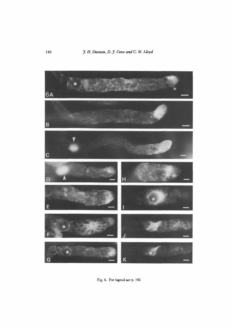
Effects on interphase arrays. At 1 pQA, CIPC produces changes in the microtubulararrays particularly within about 15/im from the apical dome (Fig. 6A-D) . In manycells, mainly caulonemata, abnormal intensely stained arrays fan out from an areaabout 15 fim back from the tip, usually towards the tip (Fig. 6B) but can also appear as
Fig. 6. For legend see p. 142

142 J. H. Doonan, D. J. Cove and C. W. Lloyd
'star-bursts'. These usually occur near to one flank of the cell, and although there maybe some degree of bending, the tip does not swell - as it may do at higherconcentrations - even after prolonged exposure to this concentration of the drug.Other microtubular arrays may contain fewer microtubules than untreated proto-nemata but are otherwise normal. Cell division occurs normally but re-orientation ofthe caulonemal spindle does not always occur. This leads to colonies with a higherproportion of chloronemal-like tissues, which grow more slowly. A variety ofabnormal microtubule arrays, at higher concentrations of CIPC, is shown in Fig. 6.
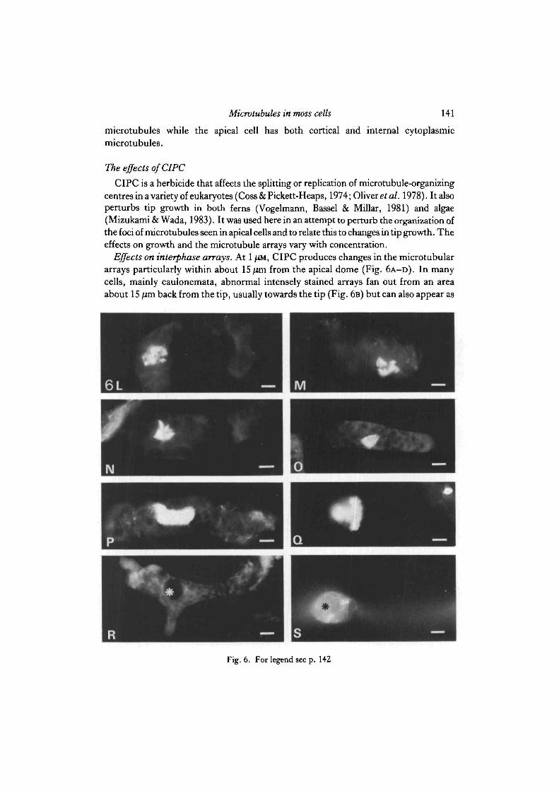
Effects on spindle and phragmoplast. Mitosis and cytokinesis are unaffected byconcentrations of CIPC up to 2-5 /iM. At or above 2-5 /IM-CIPC, spindle initiation isnormal (Fig. 6l) except that cytoplasmic microtubules are reduced in number. Laterin mitosis the spindle may be multipolar (Fig. 6L,M). NO spindle-associatedcytoplasmic microtubules are present and the spindle and phragmoplast are oftendisorientated as well as being aberrant (Fig. 6N-Q) . At 5 JUM and above, cell shape andspindle initiation become disturbed (Fig. 6R,S).
DISCUSSION
Powell, Lloyd, Slabas & Cove (1980) demonstrated the feasibility of stainingmicrotubules in moss protoplasts with anti-tubulin antibodies and we have nowdeveloped methods that permit the visualization of microtubules in fixed, walledfilaments of cells. The use of formaldehyde (Wick et al. 1981) is undoubtedlybeneficial but a combination of DMSO and non-ionic detergent is essential to reduceautofluorescence within these green plants to acceptable levels, as well as topermeabilize the cells.
Fig. 6. The microtubular arrays in tip cells of Physcomitrella protonemata treated withthe herbicide CIPC for 12-15 h. All cells are positioned with their apex to the right, and,where applicable, the nucleus is indicated by an asterisk A. At 1 im, this CIPC-treatedcaulonemal tip cell contains a 'starburst' of MTs along one flank near the apex, and fewcytoplasmic microtubules. Bar, 10/im. B. A pronounced array of microtubules fan outfrom a point about 10—15 /im back from the tip in this caulonemal cell, which was grown in1 /iM-CIPC. Bar, 10/im. C,D. At 1 /iM-CIPC mitosis may still occur normally even whenthe tip arrays were perturbed - spindles can be seen in both these cells slightly out of theplane of focus (arrowed). Bars, c-s, 10/im. E. Grown at 2-5/iM-CIPC this caulonemalapical tip cell shows a pronounced starburst at the apex with other sub-apical foci.F. Caulonemal cell grown in 2-5 /iM-CIPC. G. Interphase arrays in caulonemal cell grownin 5 /iM-CIPC. H. Interphase arrays in chloronemal cell grown in 2-5 /iM-CIPC also containmultiple starburet8 of fluorescence. I. Nucleus-associated MT constriction in caulonematagrown in 2-5 /iM-CIPC. J,K. TWO focal planes of the same cell in early mitosis, grown in5 /iM-CIPC. L,M. Fragmented, multipolar spindles in cells grown in 5 /iM-CIPC. Note thetotal lack of associated microtubules. N—Q. Aberrant phragmoplasts in cells grown in2-5 /iM-CIPC. N,O. Appear to have developed asymmetrically, and o is incorrectlyorientated within its chloronemal cell. P. Shows a phragmoplast that had started to developalong the length of the cell; and Q a tri-polar phragmoplast (grown in 2-5 /iM-CIPC). R.After growth on 5 /iM-CIPC some cells show swellings in region of the tip and the nucleus.These contain either a few fragmented microtubules or none. No normal spindles norrecognisable phragmoplasts are observed, s. At 10 /iM cells sometimes contain small asters,which may be attempts to produce spindles.

Microtubules in moss cells 143
The choice of a plant with comparatively simple organi2ation allows thevisualization of MTs in intact cell lineages; that is, in cells whose developmentalhistory is known and whose fate can be manipulated by a variety of environmentalfactors. 'Blotting' colonies upon glutaraldehyde-derivatized coverslips has been ofgreat use in retaining these developmental lineages and thus recognizing the differentcell types.
Microtubules and apical growth
The cylindrical morphology of the apical cell, the constant maintenance of thisshape throughout division cycles, the position of the nucleus and its ability to migratebefore and after mitosis, and the asymmetric distribution of organelles such aschloroplasts are fundamental features of tip-growing organisms (Schnepf, 1982), andall are reflected in the distribution of microtubules in apical cells.
Anti-tubulin fluorescence is, during interphase, most intense in the apical portionof the cell and this asymmetry reflects the polar distribution of organelles described forthe closely related moss, Funaria hygrometrica, by Schmiedel & Schnepf (1980). Thedistribution of microtubules is complicated by the presence of chloroplasts within themeshwork, which makes interpretation of the MT pattern rather difficult. Such closeassociation between MTs and the nucleus and other organelles has been seen inelectron microscopic studies of moss (Schmiedel & Schnepf, 1980; Brown &Lemmon, 1983) and these microtubules are endoplasmic as well as cortical(Schmiedel & Schnepf, 1980). In spite of this apparent complexity, it can still be seenthat the microtubules are more or less axial, as has been described for tip-growingradish root hairs (Newcomb & Bonnett, 1965; Seagull & Heath, 1980). It is believedthat the cylindrical portion of tip-growing cells does not elongate (Green, 1973) and,as a consequence, it is important to understand more about the structure within theapical dome where the shape of the extending cell is generated. Microtubules showless angular deviation at the tip than in more basal regions. At the tip, MTs cross overone another at steep angles, similar to the immunofluorescence image of radish roothairs (Lloyd & Wells, 1985).
That MTs are present at the tip of the apical cell is itself of great interest, since thishas not been reported in previous studies on root hairs (Newcomb & Bonnett, 1965;Seagull & Heath, 1980), which also grow by extension at the tip. But intra-membranous particles, presumed to be the sites of cellulose biosynthesis, occur morefrequently at the apices of tip-growing cells (Reiss, Schnepf & Herth, 1984; Wada &Staehelin, 1981) and if, as has been suggested (see Staehelin & Giddings, 1982, for areview), microtubules somehow guide their movement along the plane of themembrane, then the present results clearly indicate the presence of such anorientating assembly at the tip.
In higher plant cells, the cortical microtubules disappear completely during mitosisand cytokinesis but this does not occur in P. patens, in which the retention of apicalMTs during mitosis may be regarded as an adaptation to apical growth wherecontinuity of filamentous shape is paramount.

144 Jf.H. Doonan, D. J. Cove and C. W. Lloyd
Microtubules are seen to be focussed at the tip of the apical caulonemal cell and thisraises the question of whether MTs terminate or originate there. The fact that thenucleus and the tip are interconnected by MTs could account in part or in whole forthe more or less fixed nucleus-tip distance in a variety of tip-growing cells (see, forinstance, Jensen, 1981). In tip-growing cells there is often aSpitzenkorper- a tip bodythat contains vesicles of material presumably for tip growth. In the fungus Fusariumacuminatum, Howard & Aist (1977) report that methyl benzimidazole-2yl carbamateproduces multiple tips by affecting the Spitzenkorper. Mizukami & Wada (1983),working on the alga Bryopsis plumosa, likewise find that colchicine, vinblastine andgriseofulvin provide new growth points. One explanation is that the loss of MTsresults in the mis-direction of materials to the tip, but our observations suggest anextra, but not necessarily excluding, interpretation. Clayton & Lloyd (1984) foundthat CIPC or griseofulvin produce extra spindle poles in onion cells and these drugs,especially CIPC, produce comparable results upon diverse organisms containingapparently morphologically dissimilar MTOCs (Coss & Pickett-Heaps, 1974; Oliveret al. 1978; Hepler & Jackson, 1969). At high concentrations (5]UM) CIPC producesmulti-polar spindles and aberrant phragmoplasts, as well as distorted tips, in moss.The multiple star-bursts of anti-tubulin staining at the tips of CIPC-treated moss cellsmight therefore represent the abnormal activity of interphase MTOCs.
Nucleus and spindle-associated microtubules, and the positioning and orientation ofthe cell plate
No pre-prophase bands of the kind seen in higher plant cells (Pickett-Heaps &Northcote, 1966) or in leaves of Sphagnum (Schnepf, 1984) have been observed in theprotonemata of P. patens. Mitosis occurs without the breakdown of the interphasearrays, possibly by a redistribution of MTs as postulated in Fig. 3. This would agreewith the observations of Schmit, Vantard, De May & Lambert (1983) that the densityof microtubules at the nucleus increases at the interphase-prophase transition inHaemanthus endosperm cells. This, in turn, is consistent with Lambert's (1980)hypothesis and her observations in Mnium hornum (Lambert, 1970) that the nuclearenvelope is an MT anchorage or nucleation site. In protonemata there appears to be adistinct asymmetry in the progressive association of MTs with the nuclear envelope.The poles appear to retain at least some aspects of this initial asymmetry throughoutmitosis, especially in caulonemal cells, in which the anterior pole is associated withlarger numbers of cytoplasmic MTs than the posterior pole. During metaphase thespindle poles resemble those observed in Haemanthus endosperm cells: they are wideand composed of several discrete mini-poles (Jackson & Doyle, 1982; De Mey et al.1982). The association of spindle poles with accessory MTs is also observed inHaemanthus endosperm during anaphase (De Mey et al. 1982).
In protonemata, accessory polar MTs are present throughout mitosis and areespecially noticeable in chloronemata. But in caulonemata, during anaphase, theyincrease in numbers and this coincides with the re-orientation of the spindle from anaxial to an oblique alignment. This could represent the underlying structural basis of

Microtubules in moss cells 145
the process observed by Jensen (1981) in a time-lapse study of the caulonemata ofPhyscomitrium turbinatum, for in that moss the perpendicular metaphase platerapidly re-orientates so that it is succeeded, at cytokinesis, by a 35-40° cross-wall.When illumination is perpendicular to the filament, the upper edge of the oblique cellplate will be positioned nearer the tip but can, in advance of division, be rotated byrotating the light source. This is important for the subsequent orientation of light-harvesting tissues, which will emerge as side branches growing towards the light. Sidebranches in sub-apical cells emerge from near the most apical junction of the obliquecross-wall with the long side wall, hence the light-sensitive orientation of obliquecross-walls plays an important part in the light-gathering efficiency of the organism.'Astral' MTs in P. patens provide physical connections between nucleus and cortexand, as they increase in number just before spindle re-orientation, might reasonablybe expected to be involved in this rotation of the spindle relative to the side walls. Thisseems likely from the studies of Aist & Berns (1981) on the fungus Fusariumoxysporum, in which each spindle pole body, of a slightly oblique spindle, isconnected by astral microtubules to the adjacent cell cortex and, when these astralmicrotubules were severed with a laser micro-beam, the mechanics of chromosomeseparation were affected.
Further support for this idea is derived from drug studies for treatment with 2 • 5 /XM-CIPC preferentially disrupts the astral MTs. Concomitantly, the location andorientation of the spindle within the cell are abnormal, which implies that these MTsplay some part in maintaining and perhaps changing spindle position.
In summary, our observations on P. patens, underline the close involvement ofmicrotubules in tip growth, the polar distribution of organelles, the positioning of thenucleus in interphase and the orientation of the mitotic spindle.
We thank the following for their support: The Northern Ireland Department of Education,Unilever Research Ltd, The Royal Society, The Science and Engineering Research Council. Wealso thank Dr J. V. Kilmartin for the gift of anti-tubulin antibody, to Peter Scott for photographyand to Patricia Phillips for typing the manuscript.
REFERENCESAIST, J. R. & BERNS, M. W. (1981). Mechanics of chromosome separation during mitosis in
Fusarium (Fungi imperfeci). New evidence from ultrastructural and laser microbeamexperiments. J . Cell Biol. 91, 446-458.
APLIN, J. D. & HUGHES, R. C. (1981). Protein derivatised glass coverslips for the study of cell tosubstratum adhesion. Analyt. Biochem. 113, 144—148.
BROWN, R. C. & LEMMON, B. E. (1983). Microtubule organization and morphogenesis in youngspores of the moss Tetrapkispellucida Hedw. Protoplasma 116, 115-124.
CLAYTON, L. & LLOYD, C. W. (1984). The relationship between the division plane and spindlegeometry in Allium cells treated with CIPC and Griseofulvin: an anti-tubulin study. Eur.J. CellBiol. 34, 248-253.
Coss, R. A. & PICKETT-HEAPS, J. D. (1974). The effects of isopropyl N-phenyl carbamate on thegreen alga Oedogonium cardiacum. I. Cell division..?. Cell Biol. 63, 84—97.
D E MEY, J., LAMBERT, A. M., BAJER, A. S., MOEREMAN, M. & DEBRABANDER, M. (1982).
Visualization of microtubules in interphase and mitotic plant cells of Haemanthus endospermwith immunogold staining method. Proc. natn. Acad. Sci. U.SA. 79, 1898-1902.

146 jf.H. Doonan, D. J. Cove and C. W. Lloyd
FRANKE, W. W., SEIB, E., OSBORN, M., WEBER, K., HERTH, W. & FALK, H. (1977). Tubulin
containing structures in the anastral mitotic apparatus of endosperm cells of the plant Leucojumaestivum as revealed by immunofluorescence microscopy. Cytobiologie 15, 24—45.
GREEN, P. B. (1973). Morphogenesis of the cell and organ axis - biophysical models. BrookhavenSymp. Biol. 25, 166-190.
GRIMSLEY, N. H., ASHTON, N. W. & COVE, D. J. (1977). The production of somatic hybrids byprotoplast fusion in the mossPhyscomitrellapatens. Molec. gen. Genet. 154, 97-100.
HEPLER, P. K. & JACKSON, W. T. (1969). IsopropylTV-phenylcarbamate affects spindle microtubuleorientation in dividing endosperm cells of Haemanthus katherinae Baker. J. CellSci. 5, 727—743.
HOWARD, R. J. & AIST, J. R. (1977). Effects of MBC on hyphal tip organization, growth and mitosisof Fusarium acuminatum, and their antagonism by D2O. Protoplasma 92, 195-210.
JACKSON, W. T. & DOYLE, B. G. (1982). Membrane distribution in living endosperm cells ofHaemanthus. J. Cell Biol. 94, 637-743.
JENSEN, L. C. W. (1981). Division, growth and branch formation in moss protonema: studies ofsequential cytological changes in living cells. Protoplasma 107, 301-308.
KILMARTTN, J. V., WRIGHT, B. & MILSTETN, C. (1981). Rat monoclonal antitubulin antibodiesderived by using a new non-secreting rat cell line. J. Cell Biol. 93, 576—582.
LAMBERT, A. M. (1970). Etude de structures cin6tiques en rapport avec la rupture de la membranenucldaire, en ddbut de m6iose chex Mnium homum L. Organisation des centromeres. C.r. hebd.Seanc. Acad. Sd., Paris, ser. D, 270, 481-484.
LAMBERT, A. M. (1980). The role of chromosomes in the anaphase trigger and nuclear envelopeactivity in spindle formation. Chromosoma 76, 295-308.
LLOYD, C. W., SLABAS, A. R., POWELL, A. J., MACDONALD, G. & BADLEY, A. R. (1979).
Cytoplasmic microtubules in higher plants visualized with anti-tubulin antibodies. Nature, Land.279,239-241.
LLOYD, C. W. & WELLS, B. (1985). Microtubules are at the tips of root hairs and form patternscorresponding to inner wall fibrils. 7. Cell Sd. 75, 225-238.
MARCHANT, H. J. (1978). Microtubules associated with the plasma membrane of protoplasts of thegreen alga Mougeotia. Expl Cell Res. 115, 25-30.
MIZUKAMI, M. &WADA, S. (1983). Morphological anomalies induced by anti-microtubule agents inBryopsis plumosa. Protoplasma 114, 151-162.
NEWCOMB, E. H. & BONNETT, H. J. JR (1965). Cytoplasmic microtubule and wall microfibrilorientation in root hairs of radish. J . Cell Biol. 27, 575-589.
OLIVER, J. M., KRAWTEC, J. A. & BERLIN, R. D. (1978). A carbamate herbicide causes microtubuleand microfilament disruption and nuclear fragmentation in fibroblasts. Expl Cell Res. 116,229-237.
OSBORN, M. & WEBER, K. (1982). Immunofluorescence and immunocytochemical procedures withaffinity purified antibodies: tubulin containing structures. lnMethods in Cell Biology, vol. 24, TheCytoskeleton, part A (ed. L. Wilson). New York, London: Academic Press.
PICKETT-HEAPS, J. D. &NORTHCOTE, D. H. (1966). Organization of microtubules and endoplasmicreticulum during mitosis and cytokinesis in wheat meristems. J. Cell Sd. 1, 109-120.
POWELL, A. J., LLOYD, C. W., SLABAS, A. R. & COVE, D. J. (1980). Demonstration of the
microtubular cytoskeleton of the moss Physcomitrella patens using antibodies against braintubulin. PL Sd. Lett. 18, 401-404.
REISS, H.-D., SCHNEPF, E. & HERTH, W. (1984). The plasma membrane of theFwnan'a caulonematip cell morphology and distribution of particle rosettes and the kinetics of cellulose synthesis.Planta 160, 428-435.
SCHMTEDEL, G. & SCHNEPF, E. (1980). Polarity and growth of caulonemal tip cells of the mossFunaria hygrometrica. Planta 147, 405-413.
SCHMTT, A. C , VANTARD, M., D E MAY, J. & LAMBERT, A. M. (1983). Aster-like microtubulecentres establish spindle polarity during interphase-mitosis transition in higher plant cells. PI. CellRep. 2, 285-288.
SCHNEPF, E. (1982). Morphogenesis in moss protonema. In The Cytoskeleton in Plant Growth andDevelopment (ed. C. W. Lloyd), pp. 321-343. New York, San Francisco, London: AcademicPress.
SCHNEPF, E. (1984). Pre- and postmitotic re-orientation of microtubule arrays in young Sphagnumleaflets: transitional stages and initiation sites. Protoplasma 120, 100-112.

Microtubules in moss cells 147
SEAGULL, R. W. & HEATH, I. B. (1980). The organization of cortical microtubule arrays in theradish root hair. Protoplasma 103, 205-229.
STAEHELIN, L. A. & GIDDINGS, T. H. (1982). Membrane-mediated control of cell wallmicrofibrillar order. In Developmental Order: Its Origin and Regulation (ed. P. B. Green & S.Subtelny), pp. 133-147. New York: Alan R. Liss.
VOGELMANN, T. C , BASSEL, A. R. & MILLAR, J. H. (1981). Effects of microtubule inhibitors onnuclear migration and rhizoid differentiation in germinating fern spores (Onoclea sensibilis).Protoplasma 109, 295-316.
WADA, M. & STAEHELIN, L. A. (1981). Freeze-fracture observation on the plasma membrane, thecell wall and the cuticle of growing protonemata of Adiantum capillus-veneris L. Planta 151,462-468.
WICK, S. M., SEAGULL, R. W., OSBORN, M., WEBER, K. & GUNNING, B. E. S. (1981).
Immunofluorescence microscopy of organised microtubule arrays in structurally stabilizedmeristematic plant cells. J . Cell Biol. 89, 685-690.
(Received 25 July 1984-Accepted 28 August 1984)