IMAI district clinician manual - WHO | World Health Organization
780
VOLUME 2 IMAI District Clinician Manual: GUIDELINES FOR THE MANAGEMENT OF COMMON ILLNESSES WITH LIMITED RESOURCES Hospital Care for Adolescents and Adults I ntegrated Management of Adolescent and Adult I llness (IMAI)
-
Upload
khangminh22 -
Category
Documents
-
view
1 -
download
0
Transcript of IMAI district clinician manual - WHO | World Health Organization

VOLUME 2
IMAI District Clinician Manual:
GUIDELINES FOR THE MANAGEMENT OF COMMON ILLNESSES
WITH L IMITED RESOURCES
Hospital Care for Adolescents and Adults
Integrated Management of Adolescent and Adult Illness (IMAI)
For further information please contact:
IMAI Team Department of HIV/AIDS World Health Organisation Avenue Appia, 20CH–1211 Geneva [email protected]/hiv/capacity/en
VOLUME 2 IM
AI DISTRICT CLINICIAN MANUAL: HOSPITAL CARE FOR ADOLESCENTS AND ADULTS
WHO
FINAL Cover Vol. 2.indd 1FINAL Cover Vol. 2.indd 1 28/06/2012 17:2628/06/2012 17:26

WHO Library Cataloguing-in-Publication Data
IMAI district clinician manual: hospital care for adolescents and adults: guidelines for the management of illnesses with limited-resources.
2 v.
1.Community health services - standards. 2.Hospitals. 3.Delivery of health care - standards. 4.Clinical competence. 5.Disease management. 6.Adolescent. 7.Adult. 8.Manuals. 9.Developing countries. I.World Health Organization.
ISBN 978 92 4 154831 1 (package) (NLM classifi cation: WA 546)ISBN 978 92 4 154828 1 (vol. 1)ISBN 978 92 4 154829 0 (vol. 2)
© World Health Organization 2011
All rights reserved. Publications of the World Health Organization are available on the WHO web site (www.who.int) or can be purchased from WHO Press, World Health Organization, 20 Avenue Appia, 1211 Geneva 27, Switzerland (tel.: +41 22 791 3264; fax: +41 22 791 4857; e-mail: [email protected]).
Requests for permission to reproduce or translate WHO publications – whether for sale or for noncommercial distribution – should be addressed to WHO Press through the WHO web site (http://www.who.int/about/licensing/copyright_form/en/index.html).
The designations employed and the presentation of the material in this publication do not imply the expression of any opinion whatsoever on the part of the World Health Organization concerning the legal status of any country, territory, city or area or of its authorities, or concerning the delimitation of its frontiers or boundaries. Dotted lines on maps represent approximate border lines for which there may not yet be full agreement.
The mention of specifi c companies or of certain manufacturers’ products does not imply that they are endorsed or recommended by the World Health Organization in preference to others of a similar nature that are not mentioned. Errors and omissions excepted, the names of proprietary products are distinguished by initial capital letters.
All reasonable precautions have been taken by the World Health Organization to verify the information contained in this publication. However, the published material is being distributed without warranty of any kind, either expressed or implied. The responsibility for the interpretation and use of the material lies with the reader. In no event shall the World Health Organization be liable for damages arising from its use.
Cover images: © Bahizi Jovan (left), Petra Rohr-Rouendaal (right).
Production coordination: L’IV Com Sàrl, Villars-sous-Yens, Switzerland.
Printed in Switzerland.
FINAL Cover Vol. 2.indd 2FINAL Cover Vol. 2.indd 2 28/06/2012 17:2628/06/2012 17:26

Untitled-1 1 20/11/2009 15:07

VOLUME 2
IMAI District Clinician Manual:
Hospital Care for Adolescents and Adults
GUIDELINES FOR THE MANAGEMENT OF COMMON ILLNESSES
WITH L IMITED RESOURCES
Integrated Management of Adolescent and Adult Illness (IMAI)
FINAL Vol. 2 CH 9-21.indd iFINAL Vol. 2 CH 9-21.indd i 29/06/2012 15:1529/06/2012 15:15

ii Vol. 2 • Table of contents
Table of contents
Foreword . . . . . . . . . . . . . . . . . . . . . . . . . . . . . . . . . . . . . . . . . . . . . . . xiii
9. HIV diagnosis . . . . . . . . . . . . . . . . . . . . . . . . . . . . . . . . . . . . . . . . . . . 19.1 Provider-initiated HIV testing and counselling at the district hospital
and the role of the district clinician . . . . . . . . . . . . . . . . . . . . . . . . . . . . . . . . 39.2 Re-testing and repeat testing . . . . . . . . . . . . . . . . . . . . . . . . . . . . . . . . . . . . 7
9.2.1 Re-testing for HIV-negative individuals in the context of a generalized epidemic . . . . . . . . . . . . . . . . . . . . . . . . . . . . . . . 9
9.2.2 Re-testing for HIV-negative individuals in low level or concentrated epidemics . . . . . . . . . . . . . . . . . . . . . . . . . . . . . 11
9.2.3 Explain to patients the meaning of discordant or HIV-negative test results . . 139.3 CD4 testing . . . . . . . . . . . . . . . . . . . . . . . . . . . . . . . . . . . . . . . . . . . . . . 14
10. Acute and subacute by symptom . . . . . . . . . . . . . . . . . . . . . . . . 1510.1 Fever . . . . . . . . . . . . . . . . . . . . . . . . . . . . . . . . . . . . . . . . . . . . . . . . 19
10.1.1 Clinical approach . . . . . . . . . . . . . . . . . . . . . . . . . . . . . . . . . . 1910.1.2 Consider likely differential diagnosis using the DDx tables . . . . . . . . . . 2210.1.3 Initiate treatment(s), monitor response, and reconsider diagnosis . . . . . 2710.1.4 Management of severely ill patient with fever . . . . . . . . . . . . . . . . . 2810.1.5 Management of fever as an outpatient (not severely ill) . . . . . . . . . . . 29
10.2 Skin disorders . . . . . . . . . . . . . . . . . . . . . . . . . . . . . . . . . . . . . . . . . . 3010.2.1 Clinical approach . . . . . . . . . . . . . . . . . . . . . . . . . . . . . . . . . . 3110.2.2 Skin and soft tissue infections . . . . . . . . . . . . . . . . . . . . . . . . . . . 3410.2.3 Papular lesions . . . . . . . . . . . . . . . . . . . . . . . . . . . . . . . . . . . . 3810.2.4 Vesicular or bullous lesions . . . . . . . . . . . . . . . . . . . . . . . . . . . . 4810.2.5 Nodular lesions . . . . . . . . . . . . . . . . . . . . . . . . . . . . . . . . . . . . 4910.2.6 Maculopapular rash . . . . . . . . . . . . . . . . . . . . . . . . . . . . . . . . . 5210.2.7 Plaques . . . . . . . . . . . . . . . . . . . . . . . . . . . . . . . . . . . . . . . . 5310.2.8 Pruritus . . . . . . . . . . . . . . . . . . . . . . . . . . . . . . . . . . . . . . . . 5710.2.9 Urticaria . . . . . . . . . . . . . . . . . . . . . . . . . . . . . . . . . . . . . . . . 6010.2.10 Skin ulcers . . . . . . . . . . . . . . . . . . . . . . . . . . . . . . . . . . . . . . 61
10.3 Weight loss and malnutrition . . . . . . . . . . . . . . . . . . . . . . . . . . . . . . . . . 6910.3.1 Clinical approach . . . . . . . . . . . . . . . . . . . . . . . . . . . . . . . . . . 7010.3.2 Consider the likely cause of loss of weight . . . . . . . . . . . . . . . . . . . 7510.3.3 Treat weight loss and malnutrition and its underlying causes . . . . . . . . 7810.3.4 Prevent malnutrition . . . . . . . . . . . . . . . . . . . . . . . . . . . . . . . . . 81
10.4 Swelling of the limbs . . . . . . . . . . . . . . . . . . . . . . . . . . . . . . . . . . . . . . 8410.4.1 Clinical approach . . . . . . . . . . . . . . . . . . . . . . . . . . . . . . . . . . 8510.4.2 Differential diagnosis of oedema . . . . . . . . . . . . . . . . . . . . . . . . . 8610.4.3 Treatment of limb swelling . . . . . . . . . . . . . . . . . . . . . . . . . . . . . 8810.4.4 Symptom management . . . . . . . . . . . . . . . . . . . . . . . . . . . . . . . 8910.4.5 Manage lymphoedema . . . . . . . . . . . . . . . . . . . . . . . . . . . . . . . 89
10.5 Lymphadenopathy and lumps . . . . . . . . . . . . . . . . . . . . . . . . . . . . . . . . . 9010.5.1 Clinical approach . . . . . . . . . . . . . . . . . . . . . . . . . . . . . . . . . . 9010.5.2 Classify the lymphadenopathy and consider the differential diagnosis . . 9210.5.3 Approach to lymphadenopathy in PLHIV . . . . . . . . . . . . . . . . . . . . . 9510.5.4 Symptom management of lymphadenopathy . . . . . . . . . . . . . . . . . . 96
FINAL Vol. 2 CH 9-21.indd iiFINAL Vol. 2 CH 9-21.indd ii 29/06/2012 15:1529/06/2012 15:15

iiiVol. 2 • Table of contents
10.6 Chest symptoms: cough and shortness of breath . . . . . . . . . . . . . . . . . . . . . 9710.6.1 Clinical approach . . . . . . . . . . . . . . . . . . . . . . . . . . . . . . . . . . 9710.6.2 Differential diagnosis of chest complaints . . . . . . . . . . . . . . . . . . . 10210.6.3 Pneumonia . . . . . . . . . . . . . . . . . . . . . . . . . . . . . . . . . . . . . . 11110.6.4 Asthma . . . . . . . . . . . . . . . . . . . . . . . . . . . . . . . . . . . . . . . . . 11510.6.5 Chronic obstructive pulmonary disease (COPD) . . . . . . . . . . . . . . . . 118
10.7 Abdominal complaints . . . . . . . . . . . . . . . . . . . . . . . . . . . . . . . . . . . . . 12310.7a Abdominal pain . . . . . . . . . . . . . . . . . . . . . . . . . . . . . . . . . . . . . . . . . . 123
10.7a.1 Clinical approach . . . . . . . . . . . . . . . . . . . . . . . . . . . . . . . . . . 12310.7a.2 Differential diagnosis of abdominal pain and management
of specifi c conditions . . . . . . . . . . . . . . . . . . . . . . . . . . . . . . . 12610.7a.3 Approach to abdominal pain in PLHIV . . . . . . . . . . . . . . . . . . . . . . 134
10.7b Painful or diffi culty swallowing . . . . . . . . . . . . . . . . . . . . . . . . . . . . . . . . 13610.7b.1 Clinical approach . . . . . . . . . . . . . . . . . . . . . . . . . . . . . . . . . . 13610.7b.2 Differential diagnosis and treatment of painful or diffi cult swallowing . . . 13710.7b.3 Approach to oesophagitis in PLHIV . . . . . . . . . . . . . . . . . . . . . . . . 138
10.7c Nausea and vomiting . . . . . . . . . . . . . . . . . . . . . . . . . . . . . . . . . . . . . . 14010.7c.1 Clinical approach . . . . . . . . . . . . . . . . . . . . . . . . . . . . . . . . . . 14010.7c.2 Differential diagnosis and treatment of nausea or vomiting . . . . . . . . . 14110.7c.3 Symptom management for nausea or vomiting . . . . . . . . . . . . . . . . . 145
10.7d Diarrhoea (and constipation). . . . . . . . . . . . . . . . . . . . . . . . . . . . . . . . . . 14610.7d.1 Clinical approach . . . . . . . . . . . . . . . . . . . . . . . . . . . . . . . . . . 14610.7d.2 Classify and manage diarrhoea . . . . . . . . . . . . . . . . . . . . . . . . . . 14810.7d.3 Approach to persistent or chronic diarrhoea in PLHIV . . . . . . . . . . . . 15610.7d.4 Constipation. . . . . . . . . . . . . . . . . . . . . . . . . . . . . . . . . . . . . . 158
10.8 Jaundice . . . . . . . . . . . . . . . . . . . . . . . . . . . . . . . . . . . . . . . . . . . . . 15910.8.1 Clinical approach . . . . . . . . . . . . . . . . . . . . . . . . . . . . . . . . . . 160
10.9 Ascites . . . . . . . . . . . . . . . . . . . . . . . . . . . . . . . . . . . . . . . . . . . . . . . 16610.9.1 Clinical approach . . . . . . . . . . . . . . . . . . . . . . . . . . . . . . . . . . 16610.9.2 Classify ascites and consider the likely differential diagnosis . . . . . . . . 16910.9.3 Manage ascites according to cause . . . . . . . . . . . . . . . . . . . . . . . 171
10.10 Neurological problems . . . . . . . . . . . . . . . . . . . . . . . . . . . . . . . . . . . . . 17410.10a Neurological defi cit (without meningeal signs) . . . . . . . . . . . . . . . . . . . . . . . 175
10.10a.1 Clinical approach . . . . . . . . . . . . . . . . . . . . . . . . . . . . . . . . . . 17510.10a.2 Classify the neurological defi cit and consider the likely
differential diagnosis . . . . . . . . . . . . . . . . . . . . . . . . . . . . . . 17710.10a.3 Stroke-like syndrome . . . . . . . . . . . . . . . . . . . . . . . . . . . . . . . 17810.10a.4 Spinal cord problem (myelopathy) . . . . . . . . . . . . . . . . . . . . . . . . 18110.10a.5 Peripheral motor or sensory nervous system problem . . . . . . . . . . . . 18210.10a.6 Peripheral neuropathy . . . . . . . . . . . . . . . . . . . . . . . . . . . . . . . 18510.10a.7 Common cranial nerve palsies and their differentials . . . . . . . . . . . . 187
10.10b Headaches . . . . . . . . . . . . . . . . . . . . . . . . . . . . . . . . . . . . . . . . . . . . 18810.10b.1 Clinical approach . . . . . . . . . . . . . . . . . . . . . . . . . . . . . . . . . . 18910.10b.2 Consider the likely differential diagnosis . . . . . . . . . . . . . . . . . . . . 19310.10b.3 Treatment of specifi c conditions . . . . . . . . . . . . . . . . . . . . . . . . . 19910.10b.4 Symptom management of headache . . . . . . . . . . . . . . . . . . . . . . 202
10.10c Neurological problems: seizures (without meningism or fever) . . . . . . . . . . . . . 20310.10c.1 Clinical approach . . . . . . . . . . . . . . . . . . . . . . . . . . . . . . . . . . 20310.10c.2 Consider the likely differential diagnosis . . . . . . . . . . . . . . . . . . . . 204
10.11 Approach to patients with mental health problems . . . . . . . . . . . . . . . . . . . . 20910.11.1 Clinical approach to mental health problems . . . . . . . . . . . . . . . . . . 21010.11.2 Suicide and deliberate self-harm assessment and management . . . . . . 21510.11.3 Abnormal behaviour or thinking . . . . . . . . . . . . . . . . . . . . . . . . . . 21910.11.4 Psychosis . . . . . . . . . . . . . . . . . . . . . . . . . . . . . . . . . . . . . . . 22710.11.5 Bipolar disorder . . . . . . . . . . . . . . . . . . . . . . . . . . . . . . . . . . . 231
FINAL Vol. 2 CH 9-21.indd iiiFINAL Vol. 2 CH 9-21.indd iii 29/06/2012 15:1529/06/2012 15:15

iv Vol. 2 • Table of contents
10.11.6 Sad or low mood including depression . . . . . . . . . . . . . . . . . . . . . 23510.11.7 Anxiety . . . . . . . . . . . . . . . . . . . . . . . . . . . . . . . . . . . . . . . . . 244
10.12 Eye problems . . . . . . . . . . . . . . . . . . . . . . . . . . . . . . . . . . . . . . . . . . . 25410.12.1 Clinical approach . . . . . . . . . . . . . . . . . . . . . . . . . . . . . . . . . . 25410.12.2 Approach to red eye . . . . . . . . . . . . . . . . . . . . . . . . . . . . . . . . . 25910.12.3 Acute visual loss . . . . . . . . . . . . . . . . . . . . . . . . . . . . . . . . . . . 26510.12.4 Progressive visual loss . . . . . . . . . . . . . . . . . . . . . . . . . . . . . . . 26610.12.5 Geographically confi ned eye diseases – onchocerciasis and
trachoma . . . . . . . . . . . . . . . . . . . . . . . . . . . . . . . . . . . . . . . 26810.12.6 Eye problems in patients with HIV infection . . . . . . . . . . . . . . . . . . . 26910.12.7 Neuro-ophthalmic involvement from mass lesions, TB,
or cryptococcal meningitis . . . . . . . . . . . . . . . . . . . . . . . . . . . . 27110.13 Painful joints . . . . . . . . . . . . . . . . . . . . . . . . . . . . . . . . . . . . . . . . . . . 272
10.13.1 Clinical approach . . . . . . . . . . . . . . . . . . . . . . . . . . . . . . . . . . 27210.13.2 Diagnosis of single and multiple painful joints . . . . . . . . . . . . . . . . . 27410.13.3 Symptom management . . . . . . . . . . . . . . . . . . . . . . . . . . . . . . . 280
10.14 Female and male anorectal problems and genital ulcers . . . . . . . . . . . . . . . . 28110.14.1 Clinical approach . . . . . . . . . . . . . . . . . . . . . . . . . . . . . . . . . . 28110.14.2 Anorectal problems . . . . . . . . . . . . . . . . . . . . . . . . . . . . . . . . . 28210.14.3 Genital ulcer disease . . . . . . . . . . . . . . . . . . . . . . . . . . . . . . . . 28510.14.4 Special considerations in patients with HIV . . . . . . . . . . . . . . . . . . . 28910.14.5 Symptom management: rectal tenderness . . . . . . . . . . . . . . . . . . . 290
10.15 Female genitourinary complaints . . . . . . . . . . . . . . . . . . . . . . . . . . . . . . . 29110.15.1 Clinical approach . . . . . . . . . . . . . . . . . . . . . . . . . . . . . . . . . . 29210.15.2 Abnormal vaginal bleeding or amenorrhoea, or lower abdominal
or pelvic pain . . . . . . . . . . . . . . . . . . . . . . . . . . . . . . . . . . . . 29510.15.3 Pelvic mass . . . . . . . . . . . . . . . . . . . . . . . . . . . . . . . . . . . . . . 30210.15.4 Abnormal vaginal discharge not responding to syndromic
management . . . . . . . . . . . . . . . . . . . . . . . . . . . . . . . . . . . . 30510.15.5 Pelvic infl ammatory disease . . . . . . . . . . . . . . . . . . . . . . . . . . . . 30910.15.6 Septic abortion . . . . . . . . . . . . . . . . . . . . . . . . . . . . . . . . . . . . 31210.15.7 Approach to urinary incontinence . . . . . . . . . . . . . . . . . . . . . . . . 31310.15.8 Cervical cancer . . . . . . . . . . . . . . . . . . . . . . . . . . . . . . . . . . . 31710.15.9 Schistosomiasis of the female genitourinary tract . . . . . . . . . . . . . . . 321
10.16 Male genitourinary complaints . . . . . . . . . . . . . . . . . . . . . . . . . . . . . . . . 32210.16.1 Clinical approach . . . . . . . . . . . . . . . . . . . . . . . . . . . . . . . . . . 32310.16.2 Genital growths in men . . . . . . . . . . . . . . . . . . . . . . . . . . . . . . . 32510.16.3 Dysuria and penile discharge . . . . . . . . . . . . . . . . . . . . . . . . . . . 32510.16.4 Testicular and scrotal problems . . . . . . . . . . . . . . . . . . . . . . . . . . 32710.16.5 Foreskin problems . . . . . . . . . . . . . . . . . . . . . . . . . . . . . . . . . . 32810.16.6 Prostate problems . . . . . . . . . . . . . . . . . . . . . . . . . . . . . . . . . . 33010.16.7 Schistosomiasis of the male genitourinary tract . . . . . . . . . . . . . . . . 332
10.17 Disorders of the mouth and throat . . . . . . . . . . . . . . . . . . . . . . . . . . . . . . 33310.17.1 Clinical approach . . . . . . . . . . . . . . . . . . . . . . . . . . . . . . . . . . 33410.17.2 HIV and the mouth . . . . . . . . . . . . . . . . . . . . . . . . . . . . . . . . . . 33610.17.3 Soft tissue lesions of the mouth . . . . . . . . . . . . . . . . . . . . . . . . . . 33610.17.4 Oral cancer . . . . . . . . . . . . . . . . . . . . . . . . . . . . . . . . . . . . . . 34310.17.5 Conditions related to the hard tissue of the mouth . . . . . . . . . . . . . . . 34310.17.6 Gum disease . . . . . . . . . . . . . . . . . . . . . . . . . . . . . . . . . . . . . 34510.17.7 Noma disease . . . . . . . . . . . . . . . . . . . . . . . . . . . . . . . . . . . . 34610.17.8 Dry mouth . . . . . . . . . . . . . . . . . . . . . . . . . . . . . . . . . . . . . . . 34610.17.9 Pharyngitis . . . . . . . . . . . . . . . . . . . . . . . . . . . . . . . . . . . . . . 347
10.18 Pallor and anaemia . . . . . . . . . . . . . . . . . . . . . . . . . . . . . . . . . . . . . . . 35010.18.1 Clinical approach . . . . . . . . . . . . . . . . . . . . . . . . . . . . . . . . . . 350
FINAL Vol. 2 CH 9-21.indd ivFINAL Vol. 2 CH 9-21.indd iv 29/06/2012 15:1529/06/2012 15:15

vVol. 2 • Table of contents
10.18.2 Classifi cation of anaemia . . . . . . . . . . . . . . . . . . . . . . . . . . . . . . 35410.18.3 Management of anaemia . . . . . . . . . . . . . . . . . . . . . . . . . . . . . . 356
10.19 Abnormal bleeding and bruising . . . . . . . . . . . . . . . . . . . . . . . . . . . . . . . 36110.19.1 Clinical approach . . . . . . . . . . . . . . . . . . . . . . . . . . . . . . . . . . 36110.19.2 Diagnostic approach to active bleeding . . . . . . . . . . . . . . . . . . . . . 36610.19.3 Diagnostic approach to low platelets with no active bleeding . . . . . . . . 36810.19.4 Specifi c bleeding conditions in detail . . . . . . . . . . . . . . . . . . . . . . 370
10.20 Splenomegaly . . . . . . . . . . . . . . . . . . . . . . . . . . . . . . . . . . . . . . . . . . 37410.20 Clinical approach . . . . . . . . . . . . . . . . . . . . . . . . . . . . . . . . . . 37410.21 Use the DDx table to establish a likely differential diagnosis . . . . . . . . 37410.22 Management of splenomegaly . . . . . . . . . . . . . . . . . . . . . . . . . . 376
11. Multisystem communicable diseases, renal and HIV-related cancers (in alphabetical order) . . . . . . . . . . . . . . . . . . . . . . . . . . . . . 379
11.1 Amoebiasis . . . . . . . . . . . . . . . . . . . . . . . . . . . . . . . . . . . . . . . . . . . . . 38111.2 Bartonellosis . . . . . . . . . . . . . . . . . . . . . . . . . . . . . . . . . . . . . . . . . . . . 38311.3 Brucellosis . . . . . . . . . . . . . . . . . . . . . . . . . . . . . . . . . . . . . . . . . . . . . . 38511.4 Candida . . . . . . . . . . . . . . . . . . . . . . . . . . . . . . . . . . . . . . . . . . . . . . . . 38711.5 Cryptococcosis . . . . . . . . . . . . . . . . . . . . . . . . . . . . . . . . . . . . . . . . . . . 38911.6 Cryptosporidiosis . . . . . . . . . . . . . . . . . . . . . . . . . . . . . . . . . . . . . . . . . . 39311.7 Cysticercosis . . . . . . . . . . . . . . . . . . . . . . . . . . . . . . . . . . . . . . . . . . . . 39411.8 Cytomegalovirus . . . . . . . . . . . . . . . . . . . . . . . . . . . . . . . . . . . . . . . . . . 39611.9 Dengue fever . . . . . . . . . . . . . . . . . . . . . . . . . . . . . . . . . . . . . . . . . . . . 39911.10 Endocarditis . . . . . . . . . . . . . . . . . . . . . . . . . . . . . . . . . . . . . . . . . . . . . 40311.11 Fascioliasis . . . . . . . . . . . . . . . . . . . . . . . . . . . . . . . . . . . . . . . . . . . . . 40611.12 Filariasis, lymphatic . . . . . . . . . . . . . . . . . . . . . . . . . . . . . . . . . . . . . . . . 40911.13 Gonorrhoea . . . . . . . . . . . . . . . . . . . . . . . . . . . . . . . . . . . . . . . . . . . . . 41311.14 Hepatitis – viral . . . . . . . . . . . . . . . . . . . . . . . . . . . . . . . . . . . . . . . . . . . 41411.15 Herpes simplex virus . . . . . . . . . . . . . . . . . . . . . . . . . . . . . . . . . . . . . . . 41811.16 Histoplasmosis . . . . . . . . . . . . . . . . . . . . . . . . . . . . . . . . . . . . . . . . . . . 41911.17 Infl uenza . . . . . . . . . . . . . . . . . . . . . . . . . . . . . . . . . . . . . . . . . . . . . . . 42011.18 Isosporiasis . . . . . . . . . . . . . . . . . . . . . . . . . . . . . . . . . . . . . . . . . . . . . 42411.19 Kaposi sarcoma . . . . . . . . . . . . . . . . . . . . . . . . . . . . . . . . . . . . . . . . . . . 42511.20 Leishmaniasis . . . . . . . . . . . . . . . . . . . . . . . . . . . . . . . . . . . . . . . . . . . . 42711.21 Leprosy . . . . . . . . . . . . . . . . . . . . . . . . . . . . . . . . . . . . . . . . . . . . . . . . 43311.22 Leptospirosis . . . . . . . . . . . . . . . . . . . . . . . . . . . . . . . . . . . . . . . . . . . . 43711.23 Liver abscess . . . . . . . . . . . . . . . . . . . . . . . . . . . . . . . . . . . . . . . . . . . . 43811.24 Loaisis . . . . . . . . . . . . . . . . . . . . . . . . . . . . . . . . . . . . . . . . . . . . . . . . 43911.25 Malaria . . . . . . . . . . . . . . . . . . . . . . . . . . . . . . . . . . . . . . . . . . . . . . . . 44111.26 Microsporidiosis . . . . . . . . . . . . . . . . . . . . . . . . . . . . . . . . . . . . . . . . . . 45511.27 Mycobacterium avium complex . . . . . . . . . . . . . . . . . . . . . . . . . . . . . . . . . 45511.28 Onchocerciasis . . . . . . . . . . . . . . . . . . . . . . . . . . . . . . . . . . . . . . . . . . . 45611.29 Penicilliosis . . . . . . . . . . . . . . . . . . . . . . . . . . . . . . . . . . . . . . . . . . . . . 45811.30 Rabies and animal bites . . . . . . . . . . . . . . . . . . . . . . . . . . . . . . . . . . . . . . 45811.31 Renal problems . . . . . . . . . . . . . . . . . . . . . . . . . . . . . . . . . . . . . . . . . . . 46211.32 Rheumatic fever . . . . . . . . . . . . . . . . . . . . . . . . . . . . . . . . . . . . . . . . . . . 47711.33 Rickettsial diseases . . . . . . . . . . . . . . . . . . . . . . . . . . . . . . . . . . . . . . . . 47811.34 Schistosomiasis . . . . . . . . . . . . . . . . . . . . . . . . . . . . . . . . . . . . . . . . . . 47911.35 Sinusitis . . . . . . . . . . . . . . . . . . . . . . . . . . . . . . . . . . . . . . . . . . . . . . . 48111.36 Strongyloidiasis . . . . . . . . . . . . . . . . . . . . . . . . . . . . . . . . . . . . . . . . . . . 48311.37 Syphilis . . . . . . . . . . . . . . . . . . . . . . . . . . . . . . . . . . . . . . . . . . . . . . . . 48411.38 Taeniasis . . . . . . . . . . . . . . . . . . . . . . . . . . . . . . . . . . . . . . . . . . . . . . . 487
FINAL Vol. 2 CH 9-21.indd vFINAL Vol. 2 CH 9-21.indd v 29/06/2012 15:1529/06/2012 15:15

vi Vol. 2 • Table of contents
11.39 Tetanus . . . . . . . . . . . . . . . . . . . . . . . . . . . . . . . . . . . . . . . . . . . . . . . . 48811.40 Toxoplasmosis . . . . . . . . . . . . . . . . . . . . . . . . . . . . . . . . . . . . . . . . . . . 48911.41 Trypanosomiasis, human African . . . . . . . . . . . . . . . . . . . . . . . . . . . . . . . . 49111.42 Trypanosomiasis, American . . . . . . . . . . . . . . . . . . . . . . . . . . . . . . . . . . . 49411.43 Typhoid fever . . . . . . . . . . . . . . . . . . . . . . . . . . . . . . . . . . . . . . . . . . . . 49811.44 Urinary tract infection . . . . . . . . . . . . . . . . . . . . . . . . . . . . . . . . . . . . . . . 49911.45 Varicella/zoster . . . . . . . . . . . . . . . . . . . . . . . . . . . . . . . . . . . . . . . . . . . 50111.46 Viral haemorrhagic fever . . . . . . . . . . . . . . . . . . . . . . . . . . . . . . . . . . . . . 50311.47 Yellow fever . . . . . . . . . . . . . . . . . . . . . . . . . . . . . . . . . . . . . . . . . . . . . 504
Chronic and long-term care . . . . . . . . . . . . . . . . . . . . . . . . . . . . . 507
12. General principles of good chronic care . . . . . . . . . . . . . . . . . . 509Your role as health-care providers . . . . . . . . . . . . . . . . . . . . . . . . . . . . . . . . . . . 509For the patient in chronic or long-term care . . . . . . . . . . . . . . . . . . . . . . . . . . . . . 510
13. Chronic HIV care, antiretroviral therapy (ART), and prevention . . . . . . . . . . . . . . . . . . . . . . . . . . . . . . . . . . . . . . . . . . 511
Sequence of care after positive HIV test . . . . . . . . . . . . . . . . . . . . . . . . . . . . . . . 51213.1 Clinical approach when the district clinician is consulted
on a HIV patient in chronic HIV care . . . . . . . . . . . . . . . . . . . . . . . . . . . . . . 51413.2 Determine HIV clinical stage . . . . . . . . . . . . . . . . . . . . . . . . . . . . . . . . . . 51713.3 Prophylaxis for PLHIV . . . . . . . . . . . . . . . . . . . . . . . . . . . . . . . . . . . . . . . 523
Cotrimoxazole prophylaxis . . . . . . . . . . . . . . . . . . . . . . . . . . . . . . . . . . 523Isoniazid preventive therapy . . . . . . . . . . . . . . . . . . . . . . . . . . . . . . . . . 526
13.4 Initiate and manage patients on ART . . . . . . . . . . . . . . . . . . . . . . . . . . . . . . 529Determine ART eligibility . . . . . . . . . . . . . . . . . . . . . . . . . . . . . . . . . . . 529Determine appropriate ARV regimen . . . . . . . . . . . . . . . . . . . . . . . . . . . . 529Preferred fi rst-line ARV regimens for treatment-naive adults and adolescents . . . . . . . . . . . . . . . . . . . . . . . . . . . . . . . . . . . . . . . 531Drug interactions with fi rst-line ARV regimens . . . . . . . . . . . . . . . . . . . . . . 532
13.5 ART monitoring . . . . . . . . . . . . . . . . . . . . . . . . . . . . . . . . . . . . . . . . . . . 532Monitoring for new clinical events. . . . . . . . . . . . . . . . . . . . . . . . . . . . . . 532Monitor and support adherence . . . . . . . . . . . . . . . . . . . . . . . . . . . . . . . 533Monitoring response to ART . . . . . . . . . . . . . . . . . . . . . . . . . . . . . . . . . 533Laboratory monitoring . . . . . . . . . . . . . . . . . . . . . . . . . . . . . . . . . . . . . 533
13.6 ART initiation in complicated patients . . . . . . . . . . . . . . . . . . . . . . . . . . . . . 53513.7 Second-line ART . . . . . . . . . . . . . . . . . . . . . . . . . . . . . . . . . . . . . . . . . . 542
Defi ning ART failure . . . . . . . . . . . . . . . . . . . . . . . . . . . . . . . . . . . . . . 542Second-line ARV regimens . . . . . . . . . . . . . . . . . . . . . . . . . . . . . . . . . . 543Monitoring second-line regimens . . . . . . . . . . . . . . . . . . . . . . . . . . . . . . 543
13.8 ART toxicity and management . . . . . . . . . . . . . . . . . . . . . . . . . . . . . . . . . . 544Short- and long-term toxicities on ART . . . . . . . . . . . . . . . . . . . . . . . . . . . 544Clinical and laboratory monitoring for ART toxicity . . . . . . . . . . . . . . . . . . . . 545Side-effects of fi rst-line ARVs . . . . . . . . . . . . . . . . . . . . . . . . . . . . . . . . 545Management of ART toxicity . . . . . . . . . . . . . . . . . . . . . . . . . . . . . . . . . 546
13.9 Management of specifi c ART toxicities . . . . . . . . . . . . . . . . . . . . . . . . . . . . 546Haematological toxicity . . . . . . . . . . . . . . . . . . . . . . . . . . . . . . . . . . . . 546Hepatotoxicity . . . . . . . . . . . . . . . . . . . . . . . . . . . . . . . . . . . . . . . . . . 548Rash and hypersensitivity . . . . . . . . . . . . . . . . . . . . . . . . . . . . . . . . . . . 549Dyslipidemia . . . . . . . . . . . . . . . . . . . . . . . . . . . . . . . . . . . . . . . . . . . 552Hyperlactataemia and lactic acidosis . . . . . . . . . . . . . . . . . . . . . . . . . . . . 552Peripheral neuropathy . . . . . . . . . . . . . . . . . . . . . . . . . . . . . . . . . . . . . 554
FINAL Vol. 2 CH 9-21.indd viFINAL Vol. 2 CH 9-21.indd vi 29/06/2012 15:1529/06/2012 15:15

viiVol. 2 • Table of contents
Lipodystrophy . . . . . . . . . . . . . . . . . . . . . . . . . . . . . . . . . . . . . . . . . . 554Abdominal pain and symptoms . . . . . . . . . . . . . . . . . . . . . . . . . . . . . . . . 554Clinical toxicities grading and management . . . . . . . . . . . . . . . . . . . . . . . . 555ARVs and other HIV-related medications associated with selected toxicities . . . . . . . . . . . . . . . . . . . . . . . . . . . . . . . . . . . . . . 556IRIS . . . . . . . . . . . . . . . . . . . . . . . . . . . . . . . . . . . . . . . . . . . . . . . . 559
13.10 HIV/TB co-management . . . . . . . . . . . . . . . . . . . . . . . . . . . . . . . . . . . . . . 560Cotrimoxazole prophylaxis . . . . . . . . . . . . . . . . . . . . . . . . . . . . . . . . . . 560When to start ART in patients with TB . . . . . . . . . . . . . . . . . . . . . . . . . . . 560Recommended ART for patients with TB . . . . . . . . . . . . . . . . . . . . . . . . . . 560Women of childbearing potential (ore pregnant women) with TB and eligible for ART . . . . . . . . . . . . . . . . . . . . . . . . . . . . . . . . 560Summary of fi rst-line ART for TB patients . . . . . . . . . . . . . . . . . . . . . . . . . 561TB immune reconstitution infl ammatory syndrome (TB-IRIS) . . . . . . . . . . . . . . 561New TB in patients already receiving ART . . . . . . . . . . . . . . . . . . . . . . . . . 562ART recommendations for patients who develop TB within 6 months of starting a fi rst-line or second-line ART regimen . . . . . . . . . . . . . 562Second-line ART regimens for patients with TB . . . . . . . . . . . . . . . . . . . . . . 563
13.11 Adherence preparation, monitoring, and support. . . . . . . . . . . . . . . . . . . . . . . 563Barriers to adherence and suggestions for addressing them . . . . . . . . . . . . . . 564
13.12 Positive health, dignity, and prevention for PLHIV . . . . . . . . . . . . . . . . . . . . . . 566Preventing sexual transmission of HIV . . . . . . . . . . . . . . . . . . . . . . . . . . . 566Preventing non-sexual transmission of HIV . . . . . . . . . . . . . . . . . . . . . . . . 567Reproductive choice and family planning . . . . . . . . . . . . . . . . . . . . . . . . . 567
13.13 Positive living for PLHIV . . . . . . . . . . . . . . . . . . . . . . . . . . . . . . . . . . . . . . 568Counsel PLHIV on how to prevent other infections . . . . . . . . . . . . . . . . . . . . 568Encourage physical activity as appropriate . . . . . . . . . . . . . . . . . . . . . . . . 569Support adequate and balanced nutrition . . . . . . . . . . . . . . . . . . . . . . . . . 569Assess alcohol use . . . . . . . . . . . . . . . . . . . . . . . . . . . . . . . . . . . . . . . 569Brief interventions for patients with hazardous or harmful alcohol use . . . . . . . . . . . . . . . . . . . . . . . . . . . . . . . . . . . . . 570
13.14 Special considerations for adolescents in chronic HIV care . . . . . . . . . . . . . . . 570Psychosocial support . . . . . . . . . . . . . . . . . . . . . . . . . . . . . . . . . . . . . 570Differences among adolescents . . . . . . . . . . . . . . . . . . . . . . . . . . . . . . . 570What to do and what to avoid when communicating with adolescents . . . . . . . . . . . . . . . . . . . . . . . . . . . . . . . . . . . . . . . 572ART in adolescents . . . . . . . . . . . . . . . . . . . . . . . . . . . . . . . . . . . . . . . 573Tanner stage for female and male adolescents . . . . . . . . . . . . . . . . . . . . . . 573
14. PMTCT, HIV prevention, care, and treatment during pregnancy, and family planning . . . . . . . . . . . . . . . . . . . . . . 577
14.1 HIV prevention, care, and treatment during pregnancy . . . . . . . . . . . . . . . . . . . 57914.1.1 Recommend HIV testing and counselling, and optimize care
for HIV-positive pregnant women and their infants . . . . . . . . . . . . . . . 579Optimization of care for HIV-positive women and their infants . . . . . . . . 580
14.1.2 Identify and treat opportunistic infections (OIs) . . . . . . . . . . . . . . . . . . 58114.1.3 Offer ART or ARV prophylaxis . . . . . . . . . . . . . . . . . . . . . . . . . . . . . 582
Eligibility criteria for ART or ARV prophylaxis in HIV-positive pregnant women . . . . . . . . . . . . . . . . . . . . . . . . . . . . . . . . . . 583Considerations for choice of ART regimen . . . . . . . . . . . . . . . . . . . . 583Considerations for the choice of fi rst-line ART for pregnant women in need of treatment for their own health . . . . . . . . . . . . . . . . . . . . 585
FINAL Vol. 2 CH 9-21.indd viiFINAL Vol. 2 CH 9-21.indd vii 29/06/2012 15:1529/06/2012 15:15

viii Vol. 2 • Table of contents
Immune reconstitution infl ammatory syndrome (IRIS) . . . . . . . . . . . . . 58614.1.4 Women who become pregnant while taking ART . . . . . . . . . . . . . . . . . 58614.1.5 Monitoring antiretroviral response in pregnant women . . . . . . . . . . . . . . 58614.1.6 Use of second-line ART in pregnancy . . . . . . . . . . . . . . . . . . . . . . . . 58614.1.7 ARV prophylaxis for infants of HIV-positive women taking ART . . . . . . . . . 58714.1.8 ARV prophylaxis to prevent MTCT of HIV . . . . . . . . . . . . . . . . . . . . . . 587
Maternal ARV prophylaxis . . . . . . . . . . . . . . . . . . . . . . . . . . . . . . 587Recommended ARV-prophylaxis for pregnant women not yet eligible for ART and their infants . . . . . . . . . . . . . . . . . . . . 588Infant ARV prophylaxis . . . . . . . . . . . . . . . . . . . . . . . . . . . . . . . . 589Summary of infant ARV prophylaxis regimens . . . . . . . . . . . . . . . . . . 589Infant ARV prophylaxis dosing recommendations . . . . . . . . . . . . . . . . 589
14.1.9 Safety of antiretroviral and other medicines in pregnancy . . . . . . . . . . . . 589Safety monitoring . . . . . . . . . . . . . . . . . . . . . . . . . . . . . . . . . . . 589Anaemia in pregnancy . . . . . . . . . . . . . . . . . . . . . . . . . . . . . . . . 590NVP rash and hepatitis in pregnancy . . . . . . . . . . . . . . . . . . . . . . . 590Management of NVP-associated rash and liver toxicity . . . . . . . . . . . . 591EFV use in pregnancy . . . . . . . . . . . . . . . . . . . . . . . . . . . . . . . . . 591Lactic acidosis in pregnancy . . . . . . . . . . . . . . . . . . . . . . . . . . . . 591Rash and hypersensitivity due to abacavir (ABC) . . . . . . . . . . . . . . . . 592Safety of medicines in pregnancy . . . . . . . . . . . . . . . . . . . . . . . . . 592
14.1.10 Antiretroviral drug resistance . . . . . . . . . . . . . . . . . . . . . . . . . . . . . 592Choice of ART regimen for HIV-positive women with prior exposure to ARV prophylaxis for PMTCT . . . . . . . . . . . . . . . . . . . . 593
14.1.11 Improved care and support for HIV-positive pregnant women . . . . . . . . . 593Nausea and vomiting in pregnancy . . . . . . . . . . . . . . . . . . . . . . . . 593Antiemetic medication in pregnancy. . . . . . . . . . . . . . . . . . . . . . . . 594
14.2 Intrapartum care for HIV-positive women . . . . . . . . . . . . . . . . . . . . . . . . . . . 594Summary of ARV regimens during pregnancy, intrapartum, postpartum, and breastfeeding . . . . . . . . . . . . . . . . . . . . . . . . . . . . . . . . . . . . . . 594Safer intrapartum practices . . . . . . . . . . . . . . . . . . . . . . . . . . . . . . . . . . 595Caesarean delivery . . . . . . . . . . . . . . . . . . . . . . . . . . . . . . . . . . . . . . 595Breast care . . . . . . . . . . . . . . . . . . . . . . . . . . . . . . . . . . . . . . . . . . . 595
14.3 Infant feeding . . . . . . . . . . . . . . . . . . . . . . . . . . . . . . . . . . . . . . . . . . . . 596Breastfeeding . . . . . . . . . . . . . . . . . . . . . . . . . . . . . . . . . . . . . . . . . . 596Replacement feeding . . . . . . . . . . . . . . . . . . . . . . . . . . . . . . . . . . . . . . 597
14.4 Postpartum care for HIV-positive women . . . . . . . . . . . . . . . . . . . . . . . . . . . 599Care for HIV-exposed infants . . . . . . . . . . . . . . . . . . . . . . . . . . . . . . . . . 600
14.5 Reproductive choice and family planning . . . . . . . . . . . . . . . . . . . . . . . . . . . 60114.5.1 Dual protection . . . . . . . . . . . . . . . . . . . . . . . . . . . . . . . . . . . . . . 60114.5.2 Guidance on the use of contraceptive methods . . . . . . . . . . . . . . . . . . 602
Drug-drug interactions . . . . . . . . . . . . . . . . . . . . . . . . . . . . . . . . 602Medical eligibility criteria for contraceptive use: conditions relevant to HIV . . . . . . . . . . . . . . . . . . . . . . . . . . . . . . . . . . . . 603
14.5.3 Contraceptive method mix . . . . . . . . . . . . . . . . . . . . . . . . . . . . . . . 603Emergency contraception . . . . . . . . . . . . . . . . . . . . . . . . . . . . . . 604Indications for pregnancy testing . . . . . . . . . . . . . . . . . . . . . . . . . 605Special considerations when pregnancy is desired by a discordant couple . . . . . . . . . . . . . . . . . . . . . . . . . . . . . . . . 605
FINAL Vol. 2 CH 9-21.indd viiiFINAL Vol. 2 CH 9-21.indd viii 29/06/2012 15:1529/06/2012 15:15

ixVol. 2 • Table of contents
15. Tuberculosis . . . . . . . . . . . . . . . . . . . . . . . . . . . . . . . . . . . . . . . . . 60715.1 Suspect TB . . . . . . . . . . . . . . . . . . . . . . . . . . . . . . . . . . . . . . . . . . . . . . 609
Pulmonary TB . . . . . . . . . . . . . . . . . . . . . . . . . . . . . . . . . . . . . . . . . . 609Extrapulmonary TB . . . . . . . . . . . . . . . . . . . . . . . . . . . . . . . . . . . . . . . 610
15.2 Diagnose TB (with DDx table) . . . . . . . . . . . . . . . . . . . . . . . . . . . . . . . . . . 610Case defi nitions . . . . . . . . . . . . . . . . . . . . . . . . . . . . . . . . . . . . . . . . . 610Laboratory and radiology in TB diagnosis . . . . . . . . . . . . . . . . . . . . . . . . . 610Extrapulmonary TB . . . . . . . . . . . . . . . . . . . . . . . . . . . . . . . . . . . . . . . 613Case defi nitions for the diagnosis of TB . . . . . . . . . . . . . . . . . . . . . . . . . . 613Algorithm for the diagnosis of AFB smear-negative PTB in HIV-negative patients where Xpert MTB/RIF test is not available . . . . . . . . . . . . . . . . . . 615Algorithm for the management of ambulatory HIV-positive patients with presumptive TB where Xpert MTB/RIF test is available . . . . . . . . . . . . . 616Algorithm for the diagnosis of TB in ambulatory patients in HIV-prevalent settings where Xpert MTB/RIF test is not available . . . . . . . . . . . . . . . . . . . 617Summary of signs and symptoms of EPTB . . . . . . . . . . . . . . . . . . . . . . . . . 618Diagnosis of TB in the seriously ill patient with danger signs . . . . . . . . . . . . . . 619Algorithm for the diagnosis of TB in seriously ill patients in HIV-prevalent settings where Xpert MTB/RIF test is possible . . . . . . . . . . . . . . . . . . . . . 620Algorithm for the diagnosis of TB in seriously ill patients in HIV-prevalent settings at fi rst-level facilities where Xpert MTB/RIF test is not available . . . . . 621Differential diagnosis of PTB in PLHIV . . . . . . . . . . . . . . . . . . . . . . . . . . . 622DDx: PTB in PLHIV by stage of immunosuppression . . . . . . . . . . . . . . . . . . . 623TB in pregnant and postpartum women . . . . . . . . . . . . . . . . . . . . . . . . . . . 623
15.3 Treatment of TB . . . . . . . . . . . . . . . . . . . . . . . . . . . . . . . . . . . . . . . . . . . 624Standardized TB treatment regimens . . . . . . . . . . . . . . . . . . . . . . . . . . . . 624Standard code for TB treatment regimens . . . . . . . . . . . . . . . . . . . . . . . . . 624First-line anti-tuberculosis drugs . . . . . . . . . . . . . . . . . . . . . . . . . . . . . . . 624Standard anti-TB regimen and dosing frequency in new TB cases . . . . . . . . . . 625Dose of fi rst-line anti-tuberculosis drugs for adults . . . . . . . . . . . . . . . . . . . 625Registration group by outcome of most recent TB treatment . . . . . . . . . . . . . . 626TB treatment in special situations . . . . . . . . . . . . . . . . . . . . . . . . . . . . . . 626Anti-TB treatment regimens in special situations . . . . . . . . . . . . . . . . . . . . . 627Empirical treatment in EPTB . . . . . . . . . . . . . . . . . . . . . . . . . . . . . . . . . 627Adjuvant corticosteroids . . . . . . . . . . . . . . . . . . . . . . . . . . . . . . . . . . . 627
15.4 Monitor TB treatment . . . . . . . . . . . . . . . . . . . . . . . . . . . . . . . . . . . . . . . 628Monitor response to TB treatment . . . . . . . . . . . . . . . . . . . . . . . . . . . . . . 628Manage drug side-effects . . . . . . . . . . . . . . . . . . . . . . . . . . . . . . . . . . . 628Symptom-based approach to managing side-effects of anti-TB drugs . . . . . . . . 629Determine TB treatment outcomes . . . . . . . . . . . . . . . . . . . . . . . . . . . . . 630Standard defi nitions of TB treatment outcomes . . . . . . . . . . . . . . . . . . . . . . 630
15.5 Drug-resistant TB (with DDx table) . . . . . . . . . . . . . . . . . . . . . . . . . . . . . . . 630Summary of recommended TB treatment regimens . . . . . . . . . . . . . . . . . . . 631MDR-TB treatment regimen . . . . . . . . . . . . . . . . . . . . . . . . . . . . . . . . . . 631MDR-TB and HIV infection . . . . . . . . . . . . . . . . . . . . . . . . . . . . . . . . . . 632Manage common problems in patients with MDR-TB . . . . . . . . . . . . . . . . . . 632
FINAL Vol. 2 CH 9-21.indd ixFINAL Vol. 2 CH 9-21.indd ix 29/06/2012 15:1529/06/2012 15:15

x Vol. 2 • Table of contents
16. Assessment and therapy for alcohol use disorders . . . . . . . . 63516.1 Defi nitions of hazardous and harmful alcohol use and alcohol dependence . . . . . . 63716.2 Assessment . . . . . . . . . . . . . . . . . . . . . . . . . . . . . . . . . . . . . . . . . . . . . 638
Alcohol Use Disorders Identifi cation Test (AUDIT) . . . . . . . . . . . . . . . . . . . . 639Brief assessment using AUDIT . . . . . . . . . . . . . . . . . . . . . . . . . . . . . . . . 639Further assessment . . . . . . . . . . . . . . . . . . . . . . . . . . . . . . . . . . . . . . 641Laboratory fi ndings suggestive of harmful alcohol use or alcohol dependence . . . 642
16.3 Classify, then advise and treat the patient . . . . . . . . . . . . . . . . . . . . . . . . . . 643If the AUDIT score is 8–19 . . . . . . . . . . . . . . . . . . . . . . . . . . . . . . . . . . . 643If the AUDIT score is ≥20 . . . . . . . . . . . . . . . . . . . . . . . . . . . . . . . . . . . 643Special advice for pregnant patients . . . . . . . . . . . . . . . . . . . . . . . . . . . . 643Summary of interventions by classifi cation of alcohol use disorder . . . . . . . . . . 644
16.4 Brief interventions for those with hazardous or harmful drinking . . . . . . . . . . . . . 644FLAGS approach . . . . . . . . . . . . . . . . . . . . . . . . . . . . . . . . . . . . . . . . 645
16.5 Treatment and care for those with alcohol dependence . . . . . . . . . . . . . . . . . . 646Withdrawal management . . . . . . . . . . . . . . . . . . . . . . . . . . . . . . . . . . . 647Relapse prevention after withdrawal from alcohol . . . . . . . . . . . . . . . . . . . . 647Psychosocial support . . . . . . . . . . . . . . . . . . . . . . . . . . . . . . . . . . . . . 648
17. Substance use . . . . . . . . . . . . . . . . . . . . . . . . . . . . . . . . . . . . . . . 64917.1 General approach to substance use . . . . . . . . . . . . . . . . . . . . . . . . . . . . . . 651
Patterns of substance use . . . . . . . . . . . . . . . . . . . . . . . . . . . . . . . . . . . 651Defi nitions . . . . . . . . . . . . . . . . . . . . . . . . . . . . . . . . . . . . . . . . . . . . 651Pharmacological classes . . . . . . . . . . . . . . . . . . . . . . . . . . . . . . . . . . . 651Routes of administration . . . . . . . . . . . . . . . . . . . . . . . . . . . . . . . . . . . . 652Effects of drug use . . . . . . . . . . . . . . . . . . . . . . . . . . . . . . . . . . . . . . . 652
17.2 Assessing drug use and dependence . . . . . . . . . . . . . . . . . . . . . . . . . . . . . . 654General principles of engaging and assessing the patient . . . . . . . . . . . . . . . 654Identify the drugs used . . . . . . . . . . . . . . . . . . . . . . . . . . . . . . . . . . . . . 654Screening for substance use disorders using the ASSIST questionnaire . . . . . . . 655
17.3 General approach to managing drug use disorders . . . . . . . . . . . . . . . . . . . . . 655A pragmatic, client-centred approach . . . . . . . . . . . . . . . . . . . . . . . . . . . 655Other sources of medical care and psychosocial support . . . . . . . . . . . . . . . . 656Link with health and social welfare agencies . . . . . . . . . . . . . . . . . . . . . . . 656
17.4 Management of opioid dependence with opioid substitution treatment . . . . . . . . . 657What is opioid substitution treatment? . . . . . . . . . . . . . . . . . . . . . . . . . . . 657Overview of OST . . . . . . . . . . . . . . . . . . . . . . . . . . . . . . . . . . . . . . . . 658How to initiate OST . . . . . . . . . . . . . . . . . . . . . . . . . . . . . . . . . . . . . . . 660OST side-effects . . . . . . . . . . . . . . . . . . . . . . . . . . . . . . . . . . . . . . . . 661Psychosocial support during OST . . . . . . . . . . . . . . . . . . . . . . . . . . . . . . 661Cessation of OST . . . . . . . . . . . . . . . . . . . . . . . . . . . . . . . . . . . . . . . . 661OST dose reduction and cessation . . . . . . . . . . . . . . . . . . . . . . . . . . . . . 662
17.5 Harm reduction approaches for injecting drug users . . . . . . . . . . . . . . . . . . . . 66317.6 Antiretroviral therapy and substance use . . . . . . . . . . . . . . . . . . . . . . . . . . . 664
Initiation of antiretroviral therapy (ART) . . . . . . . . . . . . . . . . . . . . . . . . . . 664ARV-methadone interactions . . . . . . . . . . . . . . . . . . . . . . . . . . . . . . . . . 664Interactions of OST and rifampicin . . . . . . . . . . . . . . . . . . . . . . . . . . . . . 665
17.7 Pain control in people with drug use disorders . . . . . . . . . . . . . . . . . . . . . . . . 665Management of acute pain in patients with a drug use disorder . . . . . . . . . . . 666Management of acute pain in patients receiving opioid OST . . . . . . . . . . . . . . 667Management of chronic pain in patients with drug use disorders . . . . . . . . . . . 667
FINAL Vol. 2 CH 9-21.indd xFINAL Vol. 2 CH 9-21.indd x 29/06/2012 15:1529/06/2012 15:15

xiVol. 2 • Table of contents
17.8 Management of complications from injecting drug use . . . . . . . . . . . . . . . . . . . 667Complications of injection-related infections . . . . . . . . . . . . . . . . . . . . . . . 669
17.9 Involving the family . . . . . . . . . . . . . . . . . . . . . . . . . . . . . . . . . . . . . . . . 671Social support for the family . . . . . . . . . . . . . . . . . . . . . . . . . . . . . . . . . 672
17.10 Integrating alcohol and drug use management with HIV care . . . . . . . . . . . . . . . 672
18. General principles of geriatric care . . . . . . . . . . . . . . . . . . . . . . 67518.1 Helpful considerations while caring for older adults . . . . . . . . . . . . . . . . . . . . 677
Frailty . . . . . . . . . . . . . . . . . . . . . . . . . . . . . . . . . . . . . . . . . . . . . . . 677Symptoms and signs of frailty . . . . . . . . . . . . . . . . . . . . . . . . . . . . . . . . . 677
18.2 Assessment of the older adult . . . . . . . . . . . . . . . . . . . . . . . . . . . . . . . . . . 67818.3 Nutrition and hydration in older adults . . . . . . . . . . . . . . . . . . . . . . . . . . . . . 67818.4 Medicines in older adults . . . . . . . . . . . . . . . . . . . . . . . . . . . . . . . . . . . . . 67918.5 Older adults need ongoing vaccination . . . . . . . . . . . . . . . . . . . . . . . . . . . . 67918.6 Family caregivers for older adults . . . . . . . . . . . . . . . . . . . . . . . . . . . . . . . . 67918.7 Decision-making capacity and legal issues of care . . . . . . . . . . . . . . . . . . . . . 68018.8 End-of-life care . . . . . . . . . . . . . . . . . . . . . . . . . . . . . . . . . . . . . . . . . . . 680
19. Prevention services for adolescents, adults and health workers . . . . . . . . . . . . . . . . . . . . . . . . . . . . . . . . . . . . . . . . . . 68119.1 Prevention services for adolescents and adults . . . . . . . . . . . . . . . . . . . . . . . 683
All acute and chronic patients . . . . . . . . . . . . . . . . . . . . . . . . . . . . . . . . 683Special prevention for adolescents . . . . . . . . . . . . . . . . . . . . . . . . . . . . . 685
19.2 Discordant couples counselling and services . . . . . . . . . . . . . . . . . . . . . . . . 68619.3 Special considerations for MSM and transgender persons . . . . . . . . . . . . . . . . 687
MSM . . . . . . . . . . . . . . . . . . . . . . . . . . . . . . . . . . . . . . . . . . . . . . . 687Transgender persons . . . . . . . . . . . . . . . . . . . . . . . . . . . . . . . . . . . . . . 690Societal responses . . . . . . . . . . . . . . . . . . . . . . . . . . . . . . . . . . . . . . . 690
19.4 Provide prevention, care and treatment services to health workers and other staff in health facilities . . . . . . . . . . . . . . . . . . . . . . . . . . . . . . . 69119.4.1 Manage workplace exposure to HIV . . . . . . . . . . . . . . . . . . . . . . . . . 69119.4.2 Facilitate and promote HIV testing, counselling, care and ART for staff . . . . 69219.4.3 Address workplace stigma about HIV through education and advocacy . . . 69219.4.4 Provide TB prevention and care services for health service workers . . . . . 69219.4.5 Prevention of hepatitis B in health workers . . . . . . . . . . . . . . . . . . . . . 693
19.5 Urgent response to workplace HIV exposure . . . . . . . . . . . . . . . . . . . . . . . . . 694First aid in the event of possible workplace HIV exposure . . . . . . . . . . . . . . . 694How to determine if the exposure warrants PEP . . . . . . . . . . . . . . . . . . . . . 695
19.6 Prevent, recognize and manage stress and burnout in staff . . . . . . . . . . . . . . . . 696
20. Palliative care: symptom management and end-of-life care . . . . . . . . . . . . . . . . . . . . . . . . . . . . . . . . . . . . . . . . . . 697
20.1 Assess pain (acute or chronic pain) . . . . . . . . . . . . . . . . . . . . . . . . . . . . . . 700Assess the patient for pain . . . . . . . . . . . . . . . . . . . . . . . . . . . . . . . . . . 700Assessment tools . . . . . . . . . . . . . . . . . . . . . . . . . . . . . . . . . . . . . . . . 700
20.2 Manage chronic pain in lifethreatening diseases including cancer and HIV/AIDS . . 701Manage chronic pain with analgesics, other medications for special pain, and non-medical treatments . . . . . . . . . . . . . . . . . . . . . . . . 701
FINAL Vol. 2 CH 9-21.indd xiFINAL Vol. 2 CH 9-21.indd xi 29/06/2012 15:1529/06/2012 15:15

xii Vol. 2 • Table of contents
Specifi c considerations regarding the use of oral morphine in chronic pain . . . . . 705Teaching the health worker and the patient about the use of pain medications . . . 706Reduction or cessation of opioids . . . . . . . . . . . . . . . . . . . . . . . . . . . . . . 706Non-pharmacological interventions for chronic pain . . . . . . . . . . . . . . . . . . 707
20.3 Medications to control special pain problems . . . . . . . . . . . . . . . . . . . . . . . . 708The use of adjuvant analgesics . . . . . . . . . . . . . . . . . . . . . . . . . . . . . . . 708Examples of the use of adjuvant analgesics . . . . . . . . . . . . . . . . . . . . . . . . 709Nerve blocks for specifi c severe pains . . . . . . . . . . . . . . . . . . . . . . . . . . . 710
20.4 Manage acute pain in emergency or acute conditions . . . . . . . . . . . . . . . . . . . 711Manage acute severe pain . . . . . . . . . . . . . . . . . . . . . . . . . . . . . . . . . . 711Morphine IV infusion for refractory severe acute pain . . . . . . . . . . . . . . . . . . 711
20.5 Symptom management: cough or diffi culty breathing . . . . . . . . . . . . . . . . . . . . 71220.6 Symptom management: hiccups . . . . . . . . . . . . . . . . . . . . . . . . . . . . . . . . . 71320.7 Symptom management: trouble sleeping . . . . . . . . . . . . . . . . . . . . . . . . . . . . 71420.8 Manage other symptoms using other Sections of this manual . . . . . . . . . . . . . . . 71420.9 Preventive interventions for all patients . . . . . . . . . . . . . . . . . . . . . . . . . . . . 715
Oral care . . . . . . . . . . . . . . . . . . . . . . . . . . . . . . . . . . . . . . . . . . . . . 715Preventing bedsores . . . . . . . . . . . . . . . . . . . . . . . . . . . . . . . . . . . . . . 715Preventing pain, stiffness, and contractures in muscles, and moving the bedridden patient . . . . . . . . . . . . . . . . . . . . . . . . . . . . . 715
20.10 Special considerations in palliative care for PLHIV . . . . . . . . . . . . . . . . . . . . . 71620.11 Special consideration in palliative care for TB patients . . . . . . . . . . . . . . . . . . 71620.12 Special considerations in palliative care for cancer . . . . . . . . . . . . . . . . . . . . 71720.13 End-of-life care . . . . . . . . . . . . . . . . . . . . . . . . . . . . . . . . . . . . . . . . . . . 718
Bereavement care . . . . . . . . . . . . . . . . . . . . . . . . . . . . . . . . . . . . . . . 718
21. Patient monitoring and reporting including reporting outbreaks and pharmacovigilance . . . . . . . . . . . . . . . . . . . . . . . . . . 721
21.1 Longitudinal monitoring of patients in chronic or long-term care . . . . . . . . . . . . . 72321.2 Monitoring inpatient care . . . . . . . . . . . . . . . . . . . . . . . . . . . . . . . . . . . . . 725
Problem list . . . . . . . . . . . . . . . . . . . . . . . . . . . . . . . . . . . . . . . . . . . 725Regular monitoring and the monitoring chart . . . . . . . . . . . . . . . . . . . . . . . 725
21.3 Identifying and reporting notifi able diseases . . . . . . . . . . . . . . . . . . . . . . . . . 726Priority diseases, conditions and events for integrated disease surveillance and response . . . . . . . . . . . . . . . . . . . . . . . . . . . . . . . . . 726
21.4 Pharmacovigilance . . . . . . . . . . . . . . . . . . . . . . . . . . . . . . . . . . . . . . . . . 727Spontaneous reporting . . . . . . . . . . . . . . . . . . . . . . . . . . . . . . . . . . . . . 727Cohort event monitoring . . . . . . . . . . . . . . . . . . . . . . . . . . . . . . . . . . . . 727
Index . . . . . . . . . . . . . . . . . . . . . . . . . . . . . . . . . . . . . . . . . . . . . . . . . . . 729
Abbreviations and acronyms . . . . . . . . . . . . . . . . . . . . . . . . . . . . . . 742
Writers and reviewers, and process of development . . . . . . . . . . 747
FINAL Vol. 2 CH 9-21.indd xiiFINAL Vol. 2 CH 9-21.indd xii 29/06/2012 15:1529/06/2012 15:15

xiiiVol. 2 • Foreword
Foreword
IMAI District Clinician Manual: Hospital Care for Adolescents and Adults
The manual is written for clinicians working at the district hospital (fi rst-level referral care) who diagnose and manage sick adolescents and adults in resource-constrained settings. It aims to support clinical reasoning, and to provide an effective clinical approach and protocols for the management of common and serious or potentially life-threatening conditions at district hospitals. The target audience thus includes doctors, clinical offi cers, health offi cers, and senior nurse practitioners. It has been designed to be applicable in both high and low HIV prevalence settings. The manual is divided into two volumes. The fi rst covers emergency triage assessment and treatment, and acute care for a severely ill or acutely injured patient for approximately the fi rst 24 hours of care. The fi rst volume also describes the clinical procedures commonly used in emergency and acute care, and gives a summary of the drugs used and the steps necessary for infection control. This second volume provides a symptoms-based approach to clinical care for acute and subacute conditions (including mental health). It provides short summaries of the management of diseases that affect multiple systems of the body, focusing on communicable diseases. It also includes the chronic or long-term management of HIV, TB, and alcohol and substance use disorders. Future editions may incorporate the chronic management of non-communicable diseases.
The manual was developed to support clinicians in diagnosing and managing adolescent and adult patients at district hospitals with limited essential drugs, laboratory tests, and equipment. It is one component of a broader WHO second-level learning programme. It has been developed through a large collaboration of WHO Departments and their experts from many countries and regions across the world working in expert subgroups. Recommendations in the manual are predominately based on recent WHO evidence-based normative guidelines developed by several Departments and disease control programmes, including WHO HIV/AIDS, Stop TB, Global Malaria Programme, Neglected Tropical Diseases (NTD), Mental Health Gap (mhGAP), the Reproductive Health and Research (RHR) STI and cervical cancer and family planning guidelines, Integrated Management of Emergency and Essential Surgical Care (IMEESC), Integrated Management of Pregnancy and Childbirth (IMPAC) , Global Infl uenza Programme (GIP), Global Alert Response (GAR), and others. To put these normative guidelines into operation within an integrated clinical manual supports the implementation of multiple disease-control strategies.
Good clinical care is a component of most effective public health approaches. Simplifi cation and standardization of case detection and fi rst-line treatments support decentralization and expand access to care. Within a district network, the district clinician receives patients in referral who have not responded to fi rst-line treatment or who require hospitalization for severe illness. The ability to provide effective emergency care for severely ill patients, to establish a likely differential diagnosis, to provide appropriate management and then monitor the patient’s response to treatment can contribute substantially to the health of the community.
Where current WHO guidelines do not exist, selected national guidelines and evidence-based medicine sources, existing systematic reviews of evidence, and randomised clinical trials were reviewed. These evidence checks and updated sections of the manual can be accessed on the IMAI second-level EZcollab site.
FINAL Vol. 2 CH 9-21.indd xiiiFINAL Vol. 2 CH 9-21.indd xiii 29/06/2012 15:1529/06/2012 15:15

xiv Vol. 2 • Foreword
The relevant WHO normative guidelines are listed in footnotes in each Section, including an indication of when these will be revised (when available). The manual will be updated as other WHO guidelines are updated or new WHO guidelines are developed. Within three months of the revision and release of a relevant WHO normative guideline, an updated Section will be posted on the IMAI second-level EZcollab website. Each volume will be reprinted yearly. To request access to this website, or to provide comments or further queries, please send an email to [email protected]. As updates to the manual sections are frequent, readers of the manual are advised to ensure that they are using a current version of the manual. This manual is for country adaptation, to match the national essential medicine list, availability of laboratory tests, and local disease epidemiology. An evolving country Adaptation Guide will be available from the same website.
We thank the large number of people who have given valuable input, comments and feedback on this manual to date.
Drs Sandy Gove, Kirsty McHarry and Eyerusalem Negussie for the IMAI team.
FINAL Vol. 2 CH 9-21.indd xivFINAL Vol. 2 CH 9-21.indd xiv 29/06/2012 15:1529/06/2012 15:15

9. HIV diagnosis
Table of contents
9.1 Provider-initiated HIV testing and counselling at the district hospital and the role of the district clinician . . . . . . . . . . . . . . . . . . . . . . . . . . . . . . . . 3
9.2 Re-testing and repeat testing . . . . . . . . . . . . . . . . . . . . . . . . . . . . . . . . . . . . 79.2.1 Re-testing for HIV-negative individuals in the context
of a generalized epidemic . . . . . . . . . . . . . . . . . . . . . . . . . . . . . . . . 99.2.2 Re-testing for HIV-negative individuals in low level
or concentrated epidemics . . . . . . . . . . . . . . . . . . . . . . . . . . . . . . . 119.2.3 Explain to patients the meaning of discordant or HIV-negative test results . . 13
9.3 CD4 testing . . . . . . . . . . . . . . . . . . . . . . . . . . . . . . . . . . . . . . . . . . . . . . 14
FINAL Vol. 2 CH 9-21.indd 1FINAL Vol. 2 CH 9-21.indd 1 29/06/2012 15:1529/06/2012 15:15

FINAL Vol. 2 CH 9-21.indd 2FINAL Vol. 2 CH 9-21.indd 2 29/06/2012 15:1529/06/2012 15:15

3Vol. 2 • 9. HIV diagnosis: July 2011
9. HIV diagnosis
9.1 Provider-initiated HIV testing and counselling at the district hospital and the role of the district clinician1
In provider-initiated testing and counselling (PITC), health care workers recommend HIV testing and counselling to individuals attending health care facilities as a standard part of medical care. PITC enables clinical decisions to be made and services offered that would not have been possible without knowledge of a person’s HIV status. District clinicians are directly responsible for provision of PITC for patients attending hospital facilities (outpatients and inpatients) and also need to supervise implementation of PITC in fi rst-level facilities. Much of this entails supporting counsellors and other cadres of health workers to provide PITC for large numbers of patients, to encourage and offer referral for testing and counselling of partners and children, and to provide prevention services, especially for discordant couples. Discordant couples counselling and prevention services are key for preventing further HIV transmission (see Section 19 Prevention).
In PITC (in generalized epidemics) HIV testing is recommended to patients who present for medical care regardless of their initial reason for seeking care. It is especially important to highlight that HIV testing and counselling is recommended for all patients whose clinical presentation might result from underlying HIV infection, for all HIV-exposed children, for children presenting with suboptimal growth or malnutrition or who are malnourished and not responding to appropriate nutritional therapy, and for all patients prior to HIV post-exposure prophylaxis.
Throughout this manual, whenever a diagnosis makes HIV infection likely, it is marked in the differential diagnosis tables with a red ribbon.
In generalized epidemics PITC includes testing and counselling for adults, adolescents, children, and infants. Health workers should encourage and offer testing for family members and partners of HIV-positive people. HIV testing and counselling as early as possible during pregnancy enables pregnant women to benefi t from prevention, treatment, and care and to access interventions for reducing HIV transmission to their infants.
Health workers should not recommend HIV testing and counselling to all people attending all health facilities in settings with low-level or concentrated epidemics, since most people will have a low risk of exposure to HIV. In such settings the priority should be to ensure that HIV testing and counselling is recommended to all adults, adolescents, and children who present to health facilities with signs and symptoms suggestive of underlying HIV infection, including tuberculosis, and to children known to have been perinatally exposed to HIV.
1 Guidance on provider-initiated HIV testing and counselling in health facilities. WHO, 2007. Available at http://www.who.int/hiv/pub/vct/pitc/en/index.html
PITC
FINAL Vol. 2 CH 9-21.indd 3FINAL Vol. 2 CH 9-21.indd 3 29/06/2012 15:1529/06/2012 15:15

4 Vol. 2 • 9. HIV diagnosis: July 2011
When to recommend HIV testing
Low-level epidemics:• all adults and adolescents who present with signs or symptoms that could indicate HIV infection• HIV-exposed children or children born to HIV-positive women• men seeking circumcision as an HIV prevention intervention• consider for patients of:
° STI services ° services for most-at-risk populations ° antenatal, childbirth, and postpartum services ° TB settings.
Concentrated epidemics:• all adults and adolescents who present with signs or symptoms that could indicate HIV infection• HIV-exposed children or children born to HIV-positive women• men seeking circumcision as an HIV prevention intervention• consider for patients of:
° STI services ° services for most-at-risk populations ° antenatal, childbirth, and postpartum services ° TB settings.
Generalized epidemics:In generalized epidemics HIV testing and counselling should be recommended for all adults, adolescents, and children seen in all health facilities. This should include mobile or outreach medical services for targeted populations. In the case of phased implementation of PITC, the priorities for implementation, depending on local conditions, are:• medical inpatient and outpatient facilities, including TB clinics• antenatal, childbirth, and postpartum health facilities• STI services• health services for most-at-risk populations• services for children less than 10 years of age• services for adolescents• men seeking circumcision as an HIV prevention intervention• medical inpatient and outpatient facilities• surgical services• reproductive health services, including family planning.
Provider-initiated HIV testing and counselling is based on the three Cs
1. Counselling2. Consent3. Confi dentiality
Counselling: pre-testPatients require pre-test information, either individually or through group pre-test information sessions. The following information should be provided:• Reasons that HIV testing is being recommended. For example, say to
the patient, “In order to understand your health problem, it is important to know if it is related to your having HIV” or “HIV is common in this community. Therefore, in order to provide the best health care possible, it is recommended that you receive a HIV test today’’.
• Clinical and preventive benefi ts and risks of HIV testing. These could be described in this way: “There are many things we can do if we fi nd out you have HIV, including providing (or referring for) medicines that keep patients healthy for a long time” or “If you know you that have HIV, you can protect
PITC
FINAL Vol. 2 CH 9-21.indd 4FINAL Vol. 2 CH 9-21.indd 4 29/06/2012 15:1529/06/2012 15:15

5Vol. 2 • 9. HIV diagnosis: July 2011
yourself from other diseases and keep your partner and baby (if the woman is pregnant), safe”.
• Services that are available depending on whether the test result is negative or positive, including, in the latter case, ART. For example, say to patients, “We will offer drugs that fi ght HIV if you have the virus” or “If you are negative, we will treat your health problems, and we have counsellors who can help you to stay negative and to protect yourself”.
• If the health facility cannot offer some of the required services, the patient will be referred for appropriate services. In that case you can say, ‘’We will refer you to another health facility for any of the other services that you need and that we do not have at our health facility”.
• The right to decline HIV testing and that the test will be performed unless they object. Say, “As part of your visit today, we will be testing you for HIV unless you tell me that you do not want the test. Do you have any questions? If you do not want the test, please tell me” or “This test will help ensure that you receive good health care, and, unless you do not want to be tested for HIV today, I’m going to perform the test. Do you have any questions?”
• Declining HIV testing will not limit access to services. For example, say “If you refuse this test, we will still take care of you”.
• The importance of disclosure to former partners if the test is positive. Say, “If your test result is positive, it will be very important to let your former partners know that they may have been exposed to HIV. We will help you with that if you like”.
• For women of reproductive age, and especially if pregnant, include: ° The risk of transmitting HIV to the infant. Explain that HIV can be passed
from a mother to her baby. For example, say, “When a woman has HIV, the virus can be passed to her baby’’.
° PMTCT interventions that are available. For example, say, “The risk to the baby is greatly reduced if a woman fi nds out her HIV status early in her pregnancy and receives treatment” or “There are very good medicines that can protect a baby and help the mother, but we must know a mother’s HIV status to start these”.
• Benefi ts to the infant of early diagnosis of HIV. Say to patients, “If we fi nd out a baby has HIV early, we can take measures to keep the baby healthy” or “There are important things that can help a baby whose mother has HIV, but we must know early in order to help”.
PITC requires informed consent, with the patient given suffi cient information to make a rational decision and given the opportunity to decline testing. This opportunity should be given in private, in the presence of a health worker. Confi dentiality should be guaranteed, and health workers should explain to patients the procedures in place to safeguard confi dentiality. If a patient declines HIV testing, the health worker may wish to identify barriers and devise a strategy to overcome these barriers. Note also that some patient groups may be more susceptible to coercion to be tested or to adverse outcomes of disclosure of HIV status (e.g. violence, abandonment, incarceration). In these cases providing additional information beyond the minimum requirements may be appropriate to ensure that consent is voluntary and informed.
PITC
FINAL Vol. 2 CH 9-21.indd 5FINAL Vol. 2 CH 9-21.indd 5 29/06/2012 15:1529/06/2012 15:15

6 Vol. 2 • 9. HIV diagnosis: July 2011
Counselling: post-testPost-test counselling should be tailored to the test result and, in the case of a positive result, should be more extensive. As with all HIV testing, confi dentiality should be guaranteed.
Post-test counselling if the test result is negative• An explanation of the test result• Basic advice on methods to prevent HIV transmission
° Say, “Having one partner who you know is HIV-negative and who does not have other sexual partners will prevent you from getting HIV from your partner” or “If you aren’t with only one partner, or if you aren’t sure about your partner’s practices or HIV status, using a latex condom every time you have sex can prevent you from getting HIV from your partner”.
• Include some information about the window period for appearance of antibodies and possible need for re-testing (refer to the tables below). ° Say: “This test is negative. HIV antibodies weren’t found. This test will not
refl ect any contacts you have had in the last 3 months. Can we talk about that?” or “If you have had unprotected sex in the past 3 months, we will need to schedule another test in 6 weeks to be sure you didn’t get HIV from that partner”.
• Provision of male and female condoms and guidance for their usage ° Say, “Here are some latex condoms. Tell me what you have heard about
how to use them.” or “This is a female condom. You can decide when to use one; it will be your decision. Let me show you how it works” or “Since you may become pregnant, and that could mean a risk that your baby would have HIV, I’d like to schedule an appointment for you to talk to our family planning nurse about contraception. For now, it might be a good idea for you to use a condom every time you have sex”.
Post-test counselling if the test result is positive• Inform the patient of the result and give time to consider.
° Say, “The test is positive. This means we found HIV in your body” or “The results indicate that you are infected with HIV”.
• Ensure that the patient understands. ° Say, “What does what I just said mean to you?” or “If you were going to
explain to someone what I just told you, what would you say?”• Allow the patient to ask questions.
° Say, “What would you like to ask me now?” or “Is there information I can offer that will be helpful to you?”
• Help patient cope with emotions. ° Say, “This is really hard news to hear. Tell me about how you are feeling” or
“How are you feeling now that you’ve heard this result?” It may be helpful to include a message about positive living—that many people are living with HIV and living productive lives.
• Discuss immediate concerns and immediate sources of support. ° Say, “What do you plan do to in the next 24 hours?” or, “Let’s talk about
who could support you in this diffi cult time. Who do you think you can tell about this news?”
PITC
FINAL Vol. 2 CH 9-21.indd 6FINAL Vol. 2 CH 9-21.indd 6 29/06/2012 15:1529/06/2012 15:15

7Vol. 2 • 9. HIV diagnosis: July 2011
• Discuss available follow-up services. ° Say, “We have staff here providing medical treatment and support groups.
I think both of these could help you” or “There is a doctor who provides specialized HIV care, and I want you to consult with him the next time he is in the village”.
• Provide information on preventing transmission. ° Say, “Remember how HIV is transmitted. It will be important now for you
not to get your blood (or semen or vaginal secretions) in someone else’s body, or to share needles or injection equipment” or “Abstaining from sex or using condoms every time you have sex are ways to protect yourself and other people”. (If there is anyone who may inject drugs, vitamins, or traditional medicines in your target audience, risk-reduction messages about injection should be included, too.)
• Provide information on relevant preventive health measures. ° Say, “There are many things you can do to take care of yourself, which
may have a big effect on your future health. Can we talk about how healthy people stay healthy?” or “Eating right, exercising, and taking medications are 3 important ways that people with HIV keep themselves healthy”.
• Assess the risk of violence. ° Say, “What do you think people will say when you tell them your status?”
or “If you have a partner, how do you think your partner will respond if you tell him (or her) that you are HIV-positive?”
• Arrange appointments for follow-up services (e.g. counselling, family planning, STI treatment). ° Say, “I’d like to schedule an appointment for you to come back and see
the nurse this week. She will do some tests and decide the best things we can do now to keep you well” or “Since you may become pregnant, and that could mean a risk that your baby would have HIV, I’d like to schedule an appointment for you to talk to our family planning nurse about contraception. For now, it is a good idea for you to fi nd a protection method to use every time you have sex such as condoms”.
• Encourage referral for testing of children and partners ° Say, “Because HIV can be passed from a mother with HIV to her children
during pregnancy or breastfeeding, we recommend testing your children as soon as possible. I’d like us to make a plan to test them.” or “It is very important that your partner be tested. Your partner may be infected and will need care, or, if negative, we can help your partner stay that way”.
9.2 Re-testing and repeat testing2
Re-testing
Re-testing refers to a situation where additional testing is performed for an individual for specifi c reasons after a defi ned period of time. Reasons include a specifi c incident of possible HIV exposure within the past 3 months or ongoing risk of HIV exposure, such as sharing injecting equipment. Re-testing is always performed on a new specimen and may or may not use the same assays (tests) as at the initial test visit.
2 Delivering HIV test results and messages for re-testing and counselling in adults. WHO, 2010. Available at http://www.who.int/hiv/pub/vct/hiv_re_testing/en/index.html
Re-testing and repeat testing
FINAL Vol. 2 CH 9-21.indd 7FINAL Vol. 2 CH 9-21.indd 7 29/06/2012 15:1529/06/2012 15:15

8 Vol. 2 • 9. HIV diagnosis: July 2011
Early detection of HIV enhances referral to care, treatment, and prevention for people newly identifi ed as HIV-positive. The meaning of repeat testing and of the test results needs to be carefully explained to patients (see box below).
At the time of the initial test encounter, most individuals will not receive a recommendation to validate an HIV-negative result. The majority of people who test HIV-negative on their fi rst test are truly negative, and do not require another test. Avoid unnecessary re-testing for patients who have been previously tested and have learned their results. This wastes resources, causes confusion about the accuracy of HIV antibody tests, and takes time away from provision of focused post-test HIV prevention counselling that discusses risk-reduction strategies for an individual (or couple).
Repeat testing
Repeat testing refers to a situation where additional testing is performed for an individual immediately following a fi rst test, during the same testing visit. This can be due to inconclusive or discordant test results. The same assays are used and, where possible, the same specimen.
Re-testing is not recommended for people who test HIV-negative and who:• do not have a known ongoing risk for HIV infection, i.e. they are not current injecting drug users (IDU), sex
workers, or men who have sex with men (MSM); • are not at high risk or do not have a known HIV-positive partner; • do not have clinical indications for testing, such as a new STI;• cannot identify any specifi c incident of HIV exposure in the last 3 months prior to HIV testing (i.e. no
occupational exposure, no unprotected sex with a known HIV-positive person, or no sharing of injecting equipment with a known HIV-positive person).
Re-testing is recommended for:• A person who has discordant test results (when one HIV test result in an individual is reactive and another
test result using a different HIV assay in the same individual is non-reactive).2
• People who have tested HIV-negative but may be in the early stages of HIV infection. On average, HIV antibody tests are not able to identify people who have acquired HIV in the past 4 weeks, as the person may still have insuffi cient antibodies (window period). Therefore, a person may be recently infected with HIV, known as acute HIV infection, and receive a false negative test result, Often, however, errors by the tester are often the cause of a false negative or a discordant test result.
• Pregnant women in generalized epidemics who test HIV-negative in the 1st or 2nd trimester. To prevent mother-to-child HIV transmission, pregnant women should be tested as early as possible in pregnancy and again later in pregnancy. If a woman does not return for re-testing in her 3rd trimester, testing is recommended during labour, or, if that is not possible, immediately after delivery.
• People who have ongoing risks of acquiring HIV, including current IDU, MSM, sex workers and their clients, persons with a known HIV-positive partner, and people with a partner of unknown HIV status. These individuals should be tested for HIV and provided with population-appropriate risk reduction counselling at least annually.
• Patients seen for STIs or outpatients with clinical conditions suggestive of HIV infection who test HIV-negative within the context of a generalized epidemic. These individuals should receive a repeat test after 4 weeks. STI patients can also benefi t from future testing with each new STI diagnosis.
• Individuals with specifi c incidents of HIV exposure within the last 3 months (i.e. occupational exposure, sex with a known HIV-positive person, rape, sharing of injection equipment with a known HIV-positive person), When individuals test HIV-negative at the initial HIV test, a repeat HIV test in 6 weeks Is warranted to ensure they are truly HIV-negative.
Re-testing and repeat testing
FINAL Vol. 2 CH 9-21.indd 8FINAL Vol. 2 CH 9-21.indd 8 29/06/2012 15:1529/06/2012 15:15

9Vol. 2 • 9. HIV diagnosis: July 2011
Table: Re-testing for HIV-negative individuals in the context of a generalized epidemic (HIV prevalence >1%)
9.2.1 Re-testing for HIV-negative individuals in the context of a generalized epidemicIn generalized epidemics it is essential to bring people back for re-testing after an initial negative test. Explain this during post-test counselling and arrange a specifi c date for the patient to come back. The table below shows which people should return, and when, for re-testing to confi rm their HIV-negative status in different settings. Often, return rates for individuals who need a re-test are low, and active follow-up is needed.
Setting Re-testing recommended? When to re-test?
Future re-testing recommended?
Antenatal clinic Yes 3rd trimester, preferably between 28 and 36 weeks of pregnancy
Yes – with each new pregnancy
TB clinic No _ No, unless new potential exposure or if individual is in a high-risk category*
STI clinic Yes 4 weeks Yes – with each new STI or if individual is in a high-risk category*
Inpatient ward No _ No – unless new potential exposure or individual is in a high-risk category*
Outpatient clinic With clinical indication of HIV infection†
4 weeks No – unless new potential exposure or individual is in a high-risk category*
HIV test results are discordant, leading to an indeterminate HIV status
Yes Repeat the test immediately, using the same specimen and testing algorithm.
If still discordant, retest in 2 weeks. If still discordant, the patient should be referred, or a specimen sent, to a higher level facility.
Partner status unknown Yes(new individuals only)
4 weeks if initial test result is discordant6 weeks if initial test result is negative
Annually – if sexual relationship is ongoing
Known HIV-positive partner
Yes(new individuals only)
4 weeks if initial test result is discordant6 weeks if initial test result is negative
Annually – if sexual relationship is ongoing
Sex worker, male or female*
Yes(new individuals only)
4 weeks if initial test result is discordant6 weeks if initial test result is negative
At least annually
Re-testing and repeat testing
FINAL Vol. 2 CH 9-21.indd 9FINAL Vol. 2 CH 9-21.indd 9 29/06/2012 15:1529/06/2012 15:15

10 Vol. 2 • 9. HIV diagnosis: July 2011
Setting Re-testing recommended? When to re-test?
Future re-testing recommended?
Current injecting drug user*
Yes(new individuals only)
4 weeks if initial test result is discordant6 weeks if initial test result is negative
At least annually
Men who have sex with men*
Yes(new individuals only)
4 weeks if initial test result is discordant6 weeks if initial test result is negative
At least annually
Post-rape Yes, if baseline HIV test was negative or if fi rst HIV test following the encounter was negative or status was indeterminate; see WHO/ILO PEP guidelines1
4 and 12 weeks No
Occupational injury Yes, if baseline HIV test was negative or if fi rst HIV test following the encounter was negative or status was indeterminate; see WHO/ILO PEP guidelines3
4 and 12 weeks No
Negative HIV test in past 3 months
No _ No
No possible HIV exposure in past 3 months
No _ No
* Denotes high-risk category (i.e. current injecting drug users, sex workers and clients of sex workers, men who have sex with men)† Depends on HIV prevalence in the clinic setting, individual’s presenting complaint, and individual’s risk factors; to be determined by country HTC policies or programme manager
3 Post-exposure prophylaxis to prevent HIV infection: joint WHO/ILO guidelines on post-exposure prophylaxis (PEP) to prevent HIV infection. WHO, 2007. Available at http://www.who.int/hiv/pub/guidelines/PEP/en/
Re-testing and repeat testing
FINAL Vol. 2 CH 9-21.indd 10FINAL Vol. 2 CH 9-21.indd 10 29/06/2012 15:1529/06/2012 15:15

11Vol. 2 • 9. HIV diagnosis: July 2011
Table: Re-testing for HIV-negative patients in low-level or concentrated epidemics (HIV prevalence rates <1%)
9.2.2 Re-testing for HIV-negative individuals in low-level or concentrated epidemicsIn the context of a low-level epidemic (prevalence <1%), patients will not require re-testing if their risk of exposure has been low, whereas for higher risk exposures, re-testing will be required. Details are presented in the table below.
Setting Re-testing recommended? When to re-test?
Future re-testing recommended?
Antenatal clinic No – Yes – with each new pregnancy or if individual is in a high-risk category*
TB clinic No – No – unless new potential exposure or individual is in a high-risk category*
STI clinic Yes – Yes – with each new STI or if individual is in a high-risk category*
Inpatient ward No – No – unless new potential exposure or individual is in a high-risk category*
Outpatient clinic No – No – unless individual is in a high-risk category*
HIV test results are discordant, leading to an indeterminate HIV status
Yes Repeat the test immediately using the same specimen and testing algorithm.
If still discordant, re-test in 2 weeks.
Partner status unknown; low-risk partner
No – No
Partner status unknown; high-risk partner*
Yes(new patients only)
4 weeks if initial test result is discordant6 weeks if initial test result is negative
Annually – if sexual relationship is ongoing
Known HIV-positive partner*
Yes(new patients only)
4 weeks if initial test result is discordant6 weeks if initial test result is negative
Annually – if sexual relationship is ongoing
Re-testing and repeat testing
FINAL Vol. 2 CH 9-21.indd 11FINAL Vol. 2 CH 9-21.indd 11 29/06/2012 15:1529/06/2012 15:15

12 Vol. 2 • 9. HIV diagnosis: July 2011
Setting Re-testing recommended? When to re-test?
Future re-testing recommended?
Sex worker, male or female*
Yes(new patients only)
4 weeks if initial test result is discordant6 weeks if initial test result is negative
At least annually
Current injecting drug user*
Yes(new patients only)
4 weeks if initial test result is discordant6 weeks if initial test result is negative
At least annually
Men who have sex with men*
Yes(new patients only)
4 weeks if initial test result is discordant6 weeks if initial test result is negative
At least annually
Post-sexual violence or rape
4 and 12 weeks No
Occupational injury 4 and 12 weeks No
Negative HIV test in past 3 months
– No
No possible HIV exposure in past 3 months
– No
* Denotes high-risk category.
Re-testing and repeat testing
FINAL Vol. 2 CH 9-21.indd 12FINAL Vol. 2 CH 9-21.indd 12 29/06/2012 15:1529/06/2012 15:15

13Vol. 2 • 9. HIV diagnosis: July 2011
9.2.3 Explain to patients the meaning of discordant or HIV-negative test resultsPost-test services include appropriate prevention messages and supportive counselling and referrals to prevention, care, treatment, and support services. People with acute HIV infection should be counselled especially carefully, as HIV is transmitted much more effi ciently during the acute HIV stage than during later stages of HIV infection.
Setting Re-testing recommended?
A person with NO specifi c incident of HIV exposure in the last 3 months, and no ongoing HIV risk behaviours
Your HIV test result is negative. This means that you do not have HIV infection.
An individual with discordant HIV rapid test results
Upon the fi rst discordant results: Your test results are discordant. This means that one test had a positive result and another test had a negative result. This is rare, but it does happen sometimes. I would like to repeat these same tests again right now to clarify the results.
If the results are still discordant after immediate repeat testing: Your test results are still discordant. Therefore, I cannot determine your HIV status at this time. Sometimes people with very recent HIV infections have uncertain test results like these. I would like for you to come back in 4 weeks so that you can be tested again. If you are in the early stages of HIV infection, you could infect other people very easily. Therefore, please take extra precautions during these 4 weeks—do not have sex or, if you do, use condoms every time; do not share needles.
A pregnant woman in 1st or 2nd trimester of pregnancy in a generalized epidemic
Your HIV test result is negative. This means that you do not have HIV infection. I would like you to bring your partner in for HIV testing so that we can be sure you and your baby are not at risk of HIV infection before delivery. If you do become infected while you are pregnant, there is a chance that your baby could also have HIV. However, if you test positive before your 36th week of pregnancy, there may still be time to give you medication to reduce the risk of HIV transmission to your baby before you give birth. Therefore, I would like you to come back for another HIV test between your 28th and 36th weeks of pregnancy. If you are not able to come back then, we can test you at the time of labour. If you test positive at that time, there are some measures that can be taken to reduce transmission to your baby during delivery.
An individual with continuous or ongoing risk behaviours
Your HIV test result is negative. This means that you do not have HIV infection. I would like you to bring your partner in for HIV testing so that we can be sure you are not at risk of HIV infection.Also, it sounds like you have been engaging in behaviour that could put you at risk for becoming infected with HIV in the future. [Name the behaviour(s).]. If you continue with this behaviour, I recommend that you get a HIV test at least once each year. This will give you the opportunity to learn your HIV status and, if the test result remains negative, to discuss strategies to help you stay HIV-negative.
An individual with a specifi c incident of HIV exposure in the last 3 months (i.e. rape, occupational exposure)
Your HIV test result is negative. This means that you do not have HIV infection. However, these HIV tests are not able to detect a HIV infection that happened very recently. Based on your specifi c incident of HIV exposure, it is recommended that you come back in 6 weeks for another test to confi rm your HIV status. If you are in the early stages of HIV infection, you could infect other people very easily. Therefore, take extra precautions during these 6 weeks—do not have sex or, if you do, use condoms every time; do not share needles.
CD4 testing
FINAL Vol. 2 CH 9-21.indd 13FINAL Vol. 2 CH 9-21.indd 13 29/06/2012 15:1529/06/2012 15:15

14 Vol. 2 • 9. HIV diagnosis: July 2011
9.3 CD4 testing
Patients with a positive HIV test require a CD4 cell count and clinical staging (see Section 13 Chronic HIV care, ART and prevention).
Table: CD4 cell counts and correlated infections and non-infectious complications, with reference to Sections of this manual for differential diagnosis and clinical management4
CD4 cell count* Infectious complications Non-infectious† complications
>500/mm3 Acute retroviral syndrome – see Section 10.1Candida vaginitis – see sections 10.15 and 11
Persistent generalized lymphadenopathy – see Section 10.4Guillain-Barré syndrome – see Section 10.10aMyopathyAseptic meningitis – see Section 10.10b
200–500/mm3 Pneumococcal and other bacterial pneumoniaPulmonary tuberculosis – see Section 15Herpes zoster – see Sections 10.2 and 11.25Oropharyngeal candidiasis (thrush)Cryptosporidiosis, self-limitedKaposi sarcoma – see Section 11.19Oral hairy leukoplakia – see Section 10.17
Cervical and anal dysplasia – see Section 10.15Cervical and anal cancer – see Section 10.15B-cell lymphomaAnaemia – see Section 10.18Mononeuronal multiplex – see Section 10.10aIdiopathic thrombocytopenic purpura – see Section 10.19Hodgkin’s lymphomaLymphocytic interstitial pneumonitis – see Section 10.6
<200/mm3 P. jiroveci pneumonia (pneumocystis pneumonia‡) – see Section 10.6Disseminated histoplasmosis and coccidioidomycosis Miliary or extrapulmonary TB – seeSection 15Progressive multifocal leukoencephalopathy (PML)
Wasting – see Section 10.3Peripheral neuropathy – see Section 10.10aHIV-associated dementia − see Section 3.4CardiomyopathyVacuolar myelopathyProgressive polyradiculopathy – see Section 10.10aNon-Hodgkin’s lymphoma
<100/mm3 Disseminated herpes simplex – see Section 10.2Toxoplasmosis – see Section 10.10Cryptosporidiosis, chronicMicrosporidiosisCandida oesophagitis – see Section 11.4
<50/mm3 Disseminated cytomegalovirus (CMV) − see Section 11.8Disseminated Mycobacterium avium complex − see Section 11.27
Primary central nervous system lymphoma
- Most complications occur with increasing frequency at lower CD4 cell counts.† Some conditions listed as non-infectious are associated with transmissible microbes. Examples include lymphoma (Epstein-Barr virus [EBV]) and anal and cervical cancers (human papillomavirus [HPV]).‡ The preferred name is now P. jiroveci pneumonia; PCP is the accepted abbreviation.
4 Table modifi ed from John Bartlett, who derived it from Arch Intern Med 1995; 55:1537; permission obtained.
CD4 testing
FINAL Vol. 2 CH 9-21.indd 14FINAL Vol. 2 CH 9-21.indd 14 29/06/2012 15:1529/06/2012 15:15

10. Acute and subacute by symptom
Table of contents
10.1 Fever . . . . . . . . . . . . . . . . . . . . . . . . . . . . . . . . . . . . . . . . . . . . . . . . 1910.1.1 Clinical approach . . . . . . . . . . . . . . . . . . . . . . . . . . . . . . . . . . 1910.1.2 Consider likely differential diagnosis using the DDx tables . . . . . . . . . . 2210.1.3 Initiate treatment(s), monitor response, and reconsider diagnosis . . . . . 2710.1.4 Management of severely ill patient with fever . . . . . . . . . . . . . . . . . 2810.1.5 Management of fever as an outpatient (not severely ill) . . . . . . . . . . . 29
10.2 Skin disorders . . . . . . . . . . . . . . . . . . . . . . . . . . . . . . . . . . . . . . . . . . 3010.2.1 Clinical approach . . . . . . . . . . . . . . . . . . . . . . . . . . . . . . . . . . 3110.2.2 Skin and soft tissue infections . . . . . . . . . . . . . . . . . . . . . . . . . . . 3410.2.3 Papular lesions . . . . . . . . . . . . . . . . . . . . . . . . . . . . . . . . . . . . 3810.2.4 Vesicular or bullous lesions . . . . . . . . . . . . . . . . . . . . . . . . . . . . 4810.2.5 Nodular lesions . . . . . . . . . . . . . . . . . . . . . . . . . . . . . . . . . . . . 4910.2.6 Maculopapular rash . . . . . . . . . . . . . . . . . . . . . . . . . . . . . . . . . 5210.2.7 Plaques . . . . . . . . . . . . . . . . . . . . . . . . . . . . . . . . . . . . . . . . 5310.2.8 Pruritus . . . . . . . . . . . . . . . . . . . . . . . . . . . . . . . . . . . . . . . . 5710.2.9 Urticaria . . . . . . . . . . . . . . . . . . . . . . . . . . . . . . . . . . . . . . . . 6010.2.10 Skin ulcers . . . . . . . . . . . . . . . . . . . . . . . . . . . . . . . . . . . . . . 61
10.3 Weight loss and malnutrition . . . . . . . . . . . . . . . . . . . . . . . . . . . . . . . . . 6910.3.1 Clinical approach . . . . . . . . . . . . . . . . . . . . . . . . . . . . . . . . . . 7010.3.2 Consider the likely cause of loss of weight . . . . . . . . . . . . . . . . . . . 7510.3.3 Treat weight loss and malnutrition and its underlying causes . . . . . . . . 7810.3.4 Prevent malnutrition . . . . . . . . . . . . . . . . . . . . . . . . . . . . . . . . . 81
10.4 Swelling of the limbs . . . . . . . . . . . . . . . . . . . . . . . . . . . . . . . . . . . . . . 8410.4.1 Clinical approach . . . . . . . . . . . . . . . . . . . . . . . . . . . . . . . . . . 8510.4.2 Differential diagnosis of oedema . . . . . . . . . . . . . . . . . . . . . . . . . 8610.4.3 Treatment of limb swelling . . . . . . . . . . . . . . . . . . . . . . . . . . . . . 8810.4.4 Symptom management . . . . . . . . . . . . . . . . . . . . . . . . . . . . . . . 8910.4.5 Manage lymphoedema . . . . . . . . . . . . . . . . . . . . . . . . . . . . . . . 89
10.5 Lymphadenopathy and lumps . . . . . . . . . . . . . . . . . . . . . . . . . . . . . . . . . 9010.5.1 Clinical approach . . . . . . . . . . . . . . . . . . . . . . . . . . . . . . . . . . 9010.5.2 Classify the lymphadenopathy and consider the differential diagnosis . . 9210.5.3 Approach to lymphadenopathy in PLHIV . . . . . . . . . . . . . . . . . . . . . 9510.5.4 Symptom management of lymphadenopathy . . . . . . . . . . . . . . . . . . 96
10.6 Chest symptoms: cough and shortness of breath . . . . . . . . . . . . . . . . . . . . . 9710.6.1 Clinical approach . . . . . . . . . . . . . . . . . . . . . . . . . . . . . . . . . . 9710.6.2 Differential diagnosis of chest complaints . . . . . . . . . . . . . . . . . . . 10210.6.3 Pneumonia . . . . . . . . . . . . . . . . . . . . . . . . . . . . . . . . . . . . . . 11110.6.4 Asthma . . . . . . . . . . . . . . . . . . . . . . . . . . . . . . . . . . . . . . . . . 11510.6.5 Chronic obstructive pulmonary disease (COPD) . . . . . . . . . . . . . . . . 118
10.7 Abdominal complaints . . . . . . . . . . . . . . . . . . . . . . . . . . . . . . . . . . . . . 12310.7a Abdominal pain . . . . . . . . . . . . . . . . . . . . . . . . . . . . . . . . . . . . . . . . . . 123
10.7a.1 Clinical approach . . . . . . . . . . . . . . . . . . . . . . . . . . . . . . . . . . 12310.7a.2 Differential diagnosis of abdominal pain and management
of specifi c conditions . . . . . . . . . . . . . . . . . . . . . . . . . . . . . . . . 12610.7a.3 Approach to abdominal pain in PLHIV . . . . . . . . . . . . . . . . . . . . . . 134
FINAL Vol. 2 CH 9-21.indd 15FINAL Vol. 2 CH 9-21.indd 15 29/06/2012 15:1529/06/2012 15:15

10.7b Painful or diffi culty swallowing . . . . . . . . . . . . . . . . . . . . . . . . . . . . . . . . 13610.7b.1 Clinical approach . . . . . . . . . . . . . . . . . . . . . . . . . . . . . . . . . . 13610.7b.2 Differential diagnosis and treatment of painful or diffi cult swallowing . . . 13710.7b.3 Approach to oesophagitis in PLHIV . . . . . . . . . . . . . . . . . . . . . . . . 138
10.7c Nausea and vomiting . . . . . . . . . . . . . . . . . . . . . . . . . . . . . . . . . . . . . . 14010.7c.1 Clinical approach . . . . . . . . . . . . . . . . . . . . . . . . . . . . . . . . . . 14010.7c.2 Differential diagnosis and treatment of nausea or vomiting . . . . . . . . . 14110.7c.3 Symptom management for nausea or vomiting . . . . . . . . . . . . . . . . . 145
10.7d Diarrhoea (and constipation). . . . . . . . . . . . . . . . . . . . . . . . . . . . . . . . . . 14610.7d.1 Clinical approach . . . . . . . . . . . . . . . . . . . . . . . . . . . . . . . . . . 14610.7d.2 Classify and manage diarrhoea . . . . . . . . . . . . . . . . . . . . . . . . . . 14810.7d.3 Approach to persistent or chronic diarrhoea in PLHIV . . . . . . . . . . . . 15610.7d.4 Constipation. . . . . . . . . . . . . . . . . . . . . . . . . . . . . . . . . . . . . . 158
10.8 Jaundice . . . . . . . . . . . . . . . . . . . . . . . . . . . . . . . . . . . . . . . . . . . . . 15910.8.1 Clinical approach . . . . . . . . . . . . . . . . . . . . . . . . . . . . . . . . . . 160
10.9 Ascites . . . . . . . . . . . . . . . . . . . . . . . . . . . . . . . . . . . . . . . . . . . . . . . 16610.9.1 Clinical approach . . . . . . . . . . . . . . . . . . . . . . . . . . . . . . . . . . 16610.9.2 Classify ascites and consider the likely differential diagnosis . . . . . . . . 16910.9.3 Manage ascites according to cause . . . . . . . . . . . . . . . . . . . . . . . 171
10.10 Neurological problems . . . . . . . . . . . . . . . . . . . . . . . . . . . . . . . . . . . . . 17410.10a Neurological defi cit (without meningeal signs) . . . . . . . . . . . . . . . . . . . . . . . 175
10.10a.1 Clinical approach . . . . . . . . . . . . . . . . . . . . . . . . . . . . . . . . . . 17510.10a.2 Classify the neurological defi cit and consider the likely
differential diagnosis . . . . . . . . . . . . . . . . . . . . . . . . . . . . . . . . 17710.10a.3 Stroke-like syndrome . . . . . . . . . . . . . . . . . . . . . . . . . . . . . . . 17810.10a.4 Spinal cord problem (myelopathy) . . . . . . . . . . . . . . . . . . . . . . . . 18110.10a.5 Peripheral motor or sensory nervous system problem . . . . . . . . . . . . 18210.10a.6 Peripheral neuropathy . . . . . . . . . . . . . . . . . . . . . . . . . . . . . . . 18510.10a.7 Common cranial nerve palsies and their differentials . . . . . . . . . . . . 187
10.10b Headaches . . . . . . . . . . . . . . . . . . . . . . . . . . . . . . . . . . . . . . . . . . . . 18810.10b.1 Clinical approach . . . . . . . . . . . . . . . . . . . . . . . . . . . . . . . . . . 18910.10b.2 Consider the likely differential diagnosis . . . . . . . . . . . . . . . . . . . . 19310.10b.3 Treatment of specifi c conditions . . . . . . . . . . . . . . . . . . . . . . . . . 19910.10b.4 Symptom management of headache . . . . . . . . . . . . . . . . . . . . . . . 202
10.10c Neurological problems: seizures (without meningism or fever) . . . . . . . . . . . . . 20310.10c.1 Clinical approach . . . . . . . . . . . . . . . . . . . . . . . . . . . . . . . . . . 20310.10c.2 Consider the likely differential diagnosis . . . . . . . . . . . . . . . . . . . . 204
10.11 Approach to patients with mental health problems . . . . . . . . . . . . . . . . . . . . 20910.11.1 Clinical approach to mental health problems . . . . . . . . . . . . . . . . . . 21010.11.2 Suicide and deliberate self-harm assessment and management . . . . . . 21510.11.3 Abnormal behaviour or thinking . . . . . . . . . . . . . . . . . . . . . . . . . . 21910.11.4 Psychosis . . . . . . . . . . . . . . . . . . . . . . . . . . . . . . . . . . . . . . . 22710.11.5 Bipolar disorder . . . . . . . . . . . . . . . . . . . . . . . . . . . . . . . . . . . 23110.11.6 Sad or low mood including depression . . . . . . . . . . . . . . . . . . . . . 23510.11.7 Anxiety . . . . . . . . . . . . . . . . . . . . . . . . . . . . . . . . . . . . . . . . . 244
10.12 Eye problems . . . . . . . . . . . . . . . . . . . . . . . . . . . . . . . . . . . . . . . . . . . 25410.12.1 Clinical approach . . . . . . . . . . . . . . . . . . . . . . . . . . . . . . . . . . 25410.12.2 Approach to red eye . . . . . . . . . . . . . . . . . . . . . . . . . . . . . . . . . 25910.12.3 Acute visual loss . . . . . . . . . . . . . . . . . . . . . . . . . . . . . . . . . . . 26510.12.4 Progressive visual loss . . . . . . . . . . . . . . . . . . . . . . . . . . . . . . . 26610.12.5 Geographically confi ned eye diseases – onchocerciasis and
trachoma . . . . . . . . . . . . . . . . . . . . . . . . . . . . . . . . . . . . . . . 26810.12.6 Eye problems in patients with HIV infection . . . . . . . . . . . . . . . . . . . 26910.12.7 Neuro-ophthalmic involvement from mass lesions, TB,
or cryptococcal meningitis . . . . . . . . . . . . . . . . . . . . . . . . . . . . . 271
FINAL Vol. 2 CH 9-21.indd 16FINAL Vol. 2 CH 9-21.indd 16 29/06/2012 15:1529/06/2012 15:15

10.13 Painful joints . . . . . . . . . . . . . . . . . . . . . . . . . . . . . . . . . . . . . . . . . . . 27210.13.1 Clinical approach . . . . . . . . . . . . . . . . . . . . . . . . . . . . . . . . . . 27210.13.2 Diagnosis of single and multiple painful joints . . . . . . . . . . . . . . . . . 27410.13.3 Symptom management . . . . . . . . . . . . . . . . . . . . . . . . . . . . . . . 280
10.14 Female and male anorectal problems and genital ulcers . . . . . . . . . . . . . . . . 28110.14.1 Clinical approach . . . . . . . . . . . . . . . . . . . . . . . . . . . . . . . . . . 28110.14.2 Anorectal problems . . . . . . . . . . . . . . . . . . . . . . . . . . . . . . . . . 28210.14.3 Genital ulcer disease . . . . . . . . . . . . . . . . . . . . . . . . . . . . . . . . 28510.14.4 Special considerations in patients with HIV . . . . . . . . . . . . . . . . . . . 28910.14.5 Symptom management: rectal tenderness . . . . . . . . . . . . . . . . . . . 290
10.15 Female genitourinary complaints . . . . . . . . . . . . . . . . . . . . . . . . . . . . . . . 29110.15.1 Clinical approach . . . . . . . . . . . . . . . . . . . . . . . . . . . . . . . . . . 29210.15.2 Abnormal vaginal bleeding or amenorrhoea, or lower abdominal
or pelvic pain . . . . . . . . . . . . . . . . . . . . . . . . . . . . . . . . . . . . . 29510.15.3 Pelvic mass . . . . . . . . . . . . . . . . . . . . . . . . . . . . . . . . . . . . . . 30210.15.4 Abnormal vaginal discharge not responding to syndromic
management . . . . . . . . . . . . . . . . . . . . . . . . . . . . . . . . . . . . . 30510.15.5 Pelvic infl ammatory disease . . . . . . . . . . . . . . . . . . . . . . . . . . . . 30910.15.6 Septic abortion . . . . . . . . . . . . . . . . . . . . . . . . . . . . . . . . . . . . 31210.15.7 Approach to urinary incontinence . . . . . . . . . . . . . . . . . . . . . . . . 31310.15.8 Cervical cancer . . . . . . . . . . . . . . . . . . . . . . . . . . . . . . . . . . . 31710.15.9 Schistosomiasis of the female genitourinary tract . . . . . . . . . . . . . . . 321
10.16 Male genitourinary complaints . . . . . . . . . . . . . . . . . . . . . . . . . . . . . . . . 32210.16.1 Clinical approach . . . . . . . . . . . . . . . . . . . . . . . . . . . . . . . . . . 32310.16.2 Genital growths in men . . . . . . . . . . . . . . . . . . . . . . . . . . . . . . . 32510.16.3 Dysuria and penile discharge . . . . . . . . . . . . . . . . . . . . . . . . . . . 32510.16.4 Testicular and scrotal problems . . . . . . . . . . . . . . . . . . . . . . . . . . 32710.16.5 Foreskin problems . . . . . . . . . . . . . . . . . . . . . . . . . . . . . . . . . . 32810.16.6 Prostate problems . . . . . . . . . . . . . . . . . . . . . . . . . . . . . . . . . . 33010.16.7 Schistosomiasis of the male genitourinary tract . . . . . . . . . . . . . . . . 332
10.17 Disorders of the mouth and throat . . . . . . . . . . . . . . . . . . . . . . . . . . . . . . 33310.17.1 Clinical approach . . . . . . . . . . . . . . . . . . . . . . . . . . . . . . . . . . 33410.17.2 HIV and the mouth . . . . . . . . . . . . . . . . . . . . . . . . . . . . . . . . . . 33610.17.3 Soft tissue lesions of the mouth . . . . . . . . . . . . . . . . . . . . . . . . . . 33610.17.4 Oral cancer . . . . . . . . . . . . . . . . . . . . . . . . . . . . . . . . . . . . . . 34310.17.5 Conditions related to the hard tissue of the mouth . . . . . . . . . . . . . . . 34310.17.6 Gum disease . . . . . . . . . . . . . . . . . . . . . . . . . . . . . . . . . . . . . 34510.17.7 Noma disease . . . . . . . . . . . . . . . . . . . . . . . . . . . . . . . . . . . . 34610.17.8 Dry mouth . . . . . . . . . . . . . . . . . . . . . . . . . . . . . . . . . . . . . . . 34610.17.9 Pharyngitis . . . . . . . . . . . . . . . . . . . . . . . . . . . . . . . . . . . . . . 347
10.18 Pallor and anaemia . . . . . . . . . . . . . . . . . . . . . . . . . . . . . . . . . . . . . . . 35010.18.1 Clinical approach . . . . . . . . . . . . . . . . . . . . . . . . . . . . . . . . . . 35010.18.2 Classifi cation of anaemia . . . . . . . . . . . . . . . . . . . . . . . . . . . . . . 35410.18.3 Management of anaemia . . . . . . . . . . . . . . . . . . . . . . . . . . . . . . 356
10.19 Abnormal bleeding and bruising . . . . . . . . . . . . . . . . . . . . . . . . . . . . . . . 36110.19.1 Clinical approach . . . . . . . . . . . . . . . . . . . . . . . . . . . . . . . . . . 36110.19.2 Diagnostic approach to active bleeding . . . . . . . . . . . . . . . . . . . . . 36610.19.3 Diagnostic approach to low platelets with no active bleeding . . . . . . . . 36810.19.4 Specifi c bleeding conditions in detail . . . . . . . . . . . . . . . . . . . . . . 370
10.20 Splenomegaly . . . . . . . . . . . . . . . . . . . . . . . . . . . . . . . . . . . . . . . . . . 37410.20 Clinical approach . . . . . . . . . . . . . . . . . . . . . . . . . . . . . . . . . . 37410.21 Use the DDx table to establish a likely differential diagnosis . . . . . . . . 37410.22 Management of splenomegaly . . . . . . . . . . . . . . . . . . . . . . . . . . 376
FINAL Vol. 2 CH 9-21.indd 17FINAL Vol. 2 CH 9-21.indd 17 29/06/2012 15:1529/06/2012 15:15

FINAL Vol. 2 CH 9-21.indd 18FINAL Vol. 2 CH 9-21.indd 18 29/06/2012 15:1529/06/2012 15:15

19Vol. 2 • 10. Acute and subacute by symptom: July 2011
10. Acute and subacute by symptom
10.1 Fever
In this section: 10.1.1 Clinical approach to a patient with fever 10.1.2 Consider likely differential diagnosis using the DDx tables
• DDx: Fever 7 days or less without clinically obvious focus or site• DDx: Fever more than 7 days without clinically obvious focus or site
10.1.3 Initiate treatment(s), monitor response, and reconsider diagnosis• Systematic approach to reassessment and empirical treatment if unclear diagnosis
10.1.4 Management of severely ill patient with fever• Management of hyperthermia• Symptomatic management of fever in hospitalized patients
10.1.5 Management of fever as an outpatient (not severely ill)
Fever refers to a recent history of fever or an elevated body temperature of more than 38oC if measured centrally (ear, rectal, or oral) or 37.5oC axillary. The most common cause is an infection which may be localized or systemic; other causes include malignancy, allergic reaction, and infl ammatory disorders. This section deals with fever without focal signs. For fever with focal signs, please see the appropriate section.
10.1.1 Clinical approach to a patient with fever
Step 1: Perform Quick Check ° Use the Quick Check and ensure that there are no serious or life-threatening conditions. Be aware
that patients with severe febrile illness may require active fl uid management and require empirical antibiotics for possible life-threatening sepsis and antimalarials for possible severe malaria (in malaria endemic areas).
Step 2: Take a history and examine the patient.Look for signs and symptoms that may point to a focus of infection, e.g. cough, painful ear, pain on urination. Consult the specifi c Section.
Step 3: Assess HIV status
Step 4: Consider likely differential diagnosis using the DDx table(s).• If there is no obvious focus of infection, classify the fever:
° fever 7 days or less ° fever more than 7 days.
• If hyperthermia (temperature >40.5oC), see DDx and treat.• If specifi c focus of infection, see appropriate Section.
Step 5: Perform investigations.
Step 6: Initiate treatment, monitor the response, and reconsider the diagnosis.• If the cause of the fever is found, go to the relevant Sections for management.• If the diagnosis is still unclear, follow a systematic approach to reassessment and empirical
treatment.
Fever
FINAL Vol. 2 CH 9-21.indd 19FINAL Vol. 2 CH 9-21.indd 19 29/06/2012 15:1529/06/2012 15:15

20 Vol. 2 • 10. Acute and subacute by symptom: July 2011
History
Ask the patient for symptoms of infection, and then use the relevant Sections of this manual to further manage the patient, e.g.:• headache, neck stiffness, photophobia (Section 10.10b Headache)• cough, shortness of breath, chest pain (Section 10.6 Cough)• skin lesion (Section 10.2 Skin disorders)• abdominal pain (Section 10.7a Abdominal pain)
Important features of the history include:• duration of fever (less than or more than 7 days)• exposure to locally endemic diseases
° consider the local geographical distribution of diseases ° consider outbreaks of specifi c infections ° consider seasonal variation of diseases
• recent exposure history ° ask about recent travel – consider diseases that are common in the area that
was visited ° contact with animals and birds ° known TB contact ° recent unprotected sex ° intravenous drug use
• co-morbidities ° consider infections that a patient may be predisposed to as a result of co-
morbidities such as diabetes, HIV, sickle-cell anaemia; ° medical history of recent illness and the possibility of incompletely treated
disease or drug resistance, e.g. malaria, typhoid, TB; ° current medications; ° consider drug reactions if the patient has recently initiated a new
medication known to commonly cause drug reactions, e.g. cotrimoxazole, ART (especially nevirapine or abacavir), TB medication.
Examination
Examine the patient thoroughly paying attention to sites of possible infection: • general examination
° monitor temperature (might be normal at that particular moment) ° assess for confusion or decreased level of consciousness ° assess hydration, count heart rate and respiratory rate ° look for pallor, jaundice, lymphadenopathy, nail abnormalities (splinter
haemorrhages) ° skin lesions, including rash ° insect or animal bites ° nutritional status (wasting)
• head and neck ° neck pain or stiffness ° throat, tonsils, ears for infl ammation and discharge ° sinus tenderness ° mouth (Koplik’s spots, ulcers or lesions)
Fever
FINAL Vol. 2 CH 9-21.indd 20FINAL Vol. 2 CH 9-21.indd 20 29/06/2012 15:1529/06/2012 15:15

21Vol. 2 • 10. Acute and subacute by symptom: July 2011
• chest ° diffi cult breathing, fast breathing ° crackles, bronchial breathing, absent breath sounds ° new heart murmur, change in old murmur
• abdominal or genitourinary: ° enlarged liver or spleen ° abdominal tenderness or mass ° pain over kidneys (fl ank pain) ° pelvic tenderness or mass ° rectal and vaginal examination for pain, discharge, ulcers, mass
• muscles and joints ° red, hot, swollen, painful joint(s) with reduced mobility ° swollen, painful limb (deep venous thrombosis, cellulitis).
If evidence of focal infection, use the appropriate Section of the manual. If no evidence of focal infection is found, use this Section to assess the patient. Perform investigations
In all patients consider: • malaria test (RDT or microscopy), if living in1 or travelled to an endemic area • urine dipstick.
Additional tests, as indicated (see DDx tables in next Section):• full blood count with differential white cell count• blood smear for louse-borne relapsing fever (Borrellia recurrentis), in endemic
areas• liver function tests• chest X-ray• sputum for microscopy, acid fast bacilli, and sometimes culture• other urine tests – if positive fi ndings on dipstick: microscopy, culture• lumbar puncture• bone marrow, lymph node, or splenic aspirate for microscopy• serum or whole blood for rapid test• stool microscopy and culture• ultrasound• blood cultures.
1 In low malaria endemic areas, follow the national guidelines and test only patients fulfi lling the defi nition of a suspected malaria case (e.g. fever plus no other obvious cause of fever).
Fever
FINAL Vol. 2 CH 9-21.indd 21FINAL Vol. 2 CH 9-21.indd 21 29/06/2012 15:1529/06/2012 15:15

22 Vol. 2 • 10. Acute and subacute by symptom: July 2011
10.1.2 Consider likely differential diagnosis using the DDx tablesClassify the fever according to its duration and symptoms or signs found on examination and laboratory investigations:• fever with obvious focus of infection – see relevant Sections in the manual• fever 7 days or less without clinically obvious focus (use the fi rst DDx table
below)• fever more than 7 days (use the second DDx table below)• hyperthermia – temperature >40.5°C (also use third DDx table below)
For the severely ill patient – see Section 3 Approach to the severely ill patient.
DDx: Fever 7 days or less without clinically obvious focus or site
Condition In favour
Bacteraemic sepsissee Section 3.1.5
Seriously ill with no obvious apparent causeHypotensionFull blood count (FBC) – leucocytosis, leucopoenia, or thrombocytopaeniaRisk factors – HIV , injecting drug use, immunocompromisedBlood cultures – positiveAny sign of organ dysfunction – confusion, low urine output, respiratory depressionChemistries if available – acidosis, elevated creatinine
Meningococcal septicaemiasee Section 3.1.5
Maculopapular haemorrhagic petechial rashShock, hypotension
Malaria see Section 11.25
Living in, or travelled to an endemic areaPositive malaria test (RDT or microscopy)Absence of other obvious cause of fever
Typhoidsee Section 11.43
HeadacheConstipation or diarrhoea Abdominal pain and tenesmusHepato or splenomegaly“Rose spots” pink macules on abdomen
Rickettsial diseasesee Section 11.33
Headache, stupor (or other central neurological sign)Eschar Rash (sometimes petechial)Exposure to ticks, known area of endemicity
Dengue feversee Section 11.9
History of travel to endemic area or local outbreakPositive dengue RDT for NS1 or IgM Headache, pain behind the eyesBackache, arthralgia, myalgiaFine macular rash, petechiae FBC – leukopenia, thrombocytopeniaIn severe cases: • signs of plasma leakage, shock• severe bleeding, e.g. from GI or orifi ces, dark urine• organ failure
Fever
FINAL Vol. 2 CH 9-21.indd 22FINAL Vol. 2 CH 9-21.indd 22 29/06/2012 15:1529/06/2012 15:15

23Vol. 2 • 10. Acute and subacute by symptom: July 2011
Chikungunya Resembles non-severe dengue feverSevere joint pains with fever and rashNo simple test available to confi rm the diagnosis
Infl uenzasee Section 11.17
Sudden onset of fever and coughSometimes rhinitis or sore throat Frequent systemic symptoms (headache, arthralgia, or myalgia)Local epidemics, or history of travel to epidemic areasClose contact with a person with a similar illness, or contact with person from epidemic area with infl uenza
Yellow feversee Section 11.46
History of travel to endemic area or local outbreak Sudden onset of acute fever and rigorsHeadache, backache, bone painsFollowed by jaundice within 2 weeks
Primary HIV see Section 13
LymphadenopathyRash, pharyngitisHistory of unprotected sexual contact or unsafe injecting drug use in the last 3 months
IRIS see Section 13
ART usually initiated 2–12 weeks previouslyWorsening of present condition or development of new signs and symptomsMore likely if baseline CD4 <50 cells/mm3
Drug-induced feversee Section 13
New drug initiated days or weeks priorAssociated rash Patient on drugs – ART (NVP, ABC, EFV) , cotrimoxazole, dapsone, B-lactams, INH, anticonvulsants
Rheumatic feversee Section 11.32
TachycardiaArthritis, rash – erythema marginatumRecent sore throat
Acute strongyloidiasis see Section 11.36
Transient dermatitisCough, wheezing (pulmonary stage)Nausea, vomiting, diarrhoea, constipationEosinophiliaSeptic shock, acute respiratory distress syndromeSmall-bowel obstruction
Measles (in adolescents and young adults)
Conjunctivitis, coryza, and coughKoplik’s spots on buccal mucosa (“grains of salt on a red background”)Maculopapular, blanching rash LymphadenopathyComplications include:• respiratory tract infection (pneumonia, tracheobronchitis, bronchiolitis)• encephalitis (acute and chronic) • keratitis
Fever
FINAL Vol. 2 CH 9-21.indd 23FINAL Vol. 2 CH 9-21.indd 23 29/06/2012 15:1529/06/2012 15:15

24 Vol. 2 • 10. Acute and subacute by symptom: July 2011
Condition In favour
Relapsing fever (louse-borne borreliosis – Borrellia recurrentis)
Recurrent feverSpread from person-to-person among louse-infested populations (e.g. refugee camps, war, or famines with overcrowded populations with poor personal hygiene)Occurs in limited areas in Asia, east Africa and highlands of central Africa, and South America2 Rash, often petechialJaundice, impaired liver functionSpirochetes on Giemsa-stained thick or thin blood fi lm, or dark-fi eld preparation of blood, taken during a febrile periodGood response to single dose tetracyclineJarisch-Herxheimer reaction (fever, rigors, hypotension within 2 hours of antibiotic administration)
Acute schistosomiasis (Katayama fever)see Section 11.34
Exposure to fresh water in an endemic area (with sometimes skin itch just after exposure) Between 2 and 12 weeks after infectionFBC – eosinophilia
Leptospirosissee Section 11.22
Exposure to fresh water, farming or contact with rodents or dogsConjunctival suffusionAseptic meningitisJaundice, renal failure, haemorrhage (Weil’s disease)
Acute Q fever Exposure to aerosolized fl uid from birth products of farm animals (cows, goats, sheep); consumption of raw milkFlu-like illness, pneumonia, hepatitis in acute infection
Mononucleosis LymphadenopathyPharyngitisMore common in adolescents than adultsPersistent fatigue (up to 6 months)SplenomegalyFBC – >50% of WBC are lymphocytesRash following antibiotic administration
DDx: Fever more than 7 days without clinically obvious focus or site
Condition In favour
Tuberculosis see Section 15
Loss of weight, night sweats, fever, malaiseCough >2 weeks Signs of extrapulmonary disease – e.g. lymphadenopathy, pallor, abdominal painCommon complication of HIV
Typhoid see Section 11.43
See description in DDx table: Fever 7 days or less without clinically obvious focus or site
Malariasee Section 11.25
See description in DDx table: Fever 7 days or less without clinically obvious focus or site
Osteomyelitis Limb pain, often worse at nightLocal limb tenderness and swelling Risk factor may be present (IDU, sickle-cell disease)Contiguous skin infection or chronic ulcerX-ray showing periosteal reaction or bone destruction (after 2 to 4 weeks)
2 Heymann DL, ed. Control of communicable diseases manual, 19th ed. APHA and WHO, 2008.
Fever
FINAL Vol. 2 CH 9-21.indd 24FINAL Vol. 2 CH 9-21.indd 24 29/06/2012 15:1529/06/2012 15:15

25Vol. 2 • 10. Acute and subacute by symptom: July 2011
Endocarditissee Section 11.10
Low grade fever, night sweatsNew heart murmur (or change in old heart murmur)Signs of embolic disease (stroke, petechiae, splinter haemorrhage, abdominal pain)Signs of heart failure (diffi culty breathing)SplenomegalyRisk factors: known cardiac valvular disease, IDU, previous rheumatic disease
Liver abscesssee Section 11.23
Right upper quadrant pain or tendernessLiver focal lesion at ultrasound
Brucellosissee Section 11.3
Contact with farm animals (infected goats, pigs), consumption of raw milkAcute brucellosis: undulant feverSubacute localized brucellosis: lumbago due to spondylitis, mono or polyarthritis, osteomyelitis
Yellow fever see Section 11.47
See description in DDx table: Fever 7 days or less without clinically obvious focus or site
Plaguesee Sections 10.5 and 10.6
History of exposure to rodents or fl eas Unwell patient, sudden onset RigorsExtreme tirednessLarge painful, tender lymph nodes-bubo (bubonic plague)Acute dyspnoea, pleuritic chest pain (pneumonic plague)
Cryptococcosis see Section 11.5
Usually in advanced AIDS, but can rarely occur without HIVMeningo-encephalitis: headache, blindnessElevated intracranial pressure on LP, positive CSF India ink stainPneumonia: cough, opacities on chest X-rayIf available, positive serum or CSF cryptococcal antigen
Mycobacterium avium complex (MAC)
see Section 11.27
Usually in advanced AIDS, but can rarely occur without HIVLocalized MAC: tuberculosis-like pneumonia, adenopathy, osteomyelitisDisseminated MAC: generalized lymphadenopathy, diarrhoea and abdominal painAFB positive sputum, stool, or lymph node aspirate – confi rm on cultureNo response, or partial response to standard anti-tuberculous therapy
Lymphoma Weight loss, night sweatsEnlarged lymph nodes, hepatosplenomegaly
Deep fungal infections (histoplasmosis, penicilliosis, coccidiomycosis, paracoccidiomycosis) see Sections 11.16, 11.29
Usually in advanced AIDS, but can occur without HIVSkin lesionsNodular or lobar opacities on chest X-rayHepatosplenomegalyEndemic areas vary, depending on species
Cytomegalovirus(CMV) see Section 11.8
Painful swallowingDiarrhoeaVisual loss (CMV retinitis on fundoscopy)Complication of advanced AIDS
Toxoplasmosis
see Section 11.40
HeadacheFocal neurological defi citComplication of advanced AIDS
Fever
FINAL Vol. 2 CH 9-21.indd 25FINAL Vol. 2 CH 9-21.indd 25 29/06/2012 15:1529/06/2012 15:15

26 Vol. 2 • 10. Acute and subacute by symptom: July 2011
Condition In favour
African human trypanosomiasis (sleeping sickness) see Section 11.41
Endemic areas in AfricaMay occur in patients without HIVIntermittent fever, headacheGeneralized lymphadenopathy, particularly in posterior cervical triangle Disturbed sleepPoor concentration and personality changes
Acute Chagas disease(American trypanosomiasis) see Section 11.42
Endemic areas in Latin America Fever lasting for several weeksSwelling of both lids of one eye (Romaña sign), painful nodules (chagoma)Skin rash, localized enlarged lymph nodes
Visceral leishmaniasis (Kala azar) see Section 11.20
Endemic area Fever, wasting syndrome, pallor of mucous membranesGeneralized lymphadenopathySplenomegaly, darkening of skin
DDx: Hyperthermia (temperature >40.5°C)
Heat stroke Exposure to excess amount of sun Central nervous system dysfunction (e.g. anxiety, delirium, seizure, coma) Warm, red skin without sweatingSigns of end organ damage: hypotension; hypoglycemia; elevated liver or kidney enzymes; disseminated intravascular coagulation (DIC)
Intracranial injury Haemorrhage (involving the pons)Stroke (involving the hypothalamus)Status epilepticusTumor
Drug effects Toxicity from SSRIs, MAOI, anticholinergics (e.g. diphenhydramine, promethazine, amitriptyline, atropine), fl uoxetine or other SSRI – see Section 3.8Withdrawal from alcohol (delirium tremens) – see Section 3.7Malignant hyperthermia in response to halothaneNeuroleptic malignant syndrome
Endocrine conditions ThyrotoxicosisAdrenal crisisPheochromocytoma
Infectious causes SepsisBrain abscessMeningitisTyphoid feverMalaria Relapsing fever
Fever
FINAL Vol. 2 CH 9-21.indd 26FINAL Vol. 2 CH 9-21.indd 26 29/06/2012 15:1529/06/2012 15:15

27Vol. 2 • 10. Acute and subacute by symptom: July 2011
10.1.3 Initiate treatment(s), monitor response, and reconsider diagnosisIf the cause of fever is found, go to the relevant manual Section for management.
Relapsing fever (louse-borne borreliosis – Borrellia recurrentis)
Treatment
• Antibiotics, single dose orally: ° doxycycline 100 mg; OR ° erythromycin 500 mg; OR ° tetracycline 500 mg; OR ° If not able to take oral, procaine penicillin 600 000 to 800 000 IU IM once.
Prevent transmission2
• Patients, clothing, contacts,and immediate environment must be deloused. Dust or spray patient, contacts, and their clothing with 1% permethrin.
• Give antibiotic prophylaxis with single dose tetracyclines after exposure when risk of acquiring infection is high.
• Use blood and body fl uid precautions.
Systematic approach to reassessment and empirical treatment if unclear diagnosis
• Does the patient have an HIV-related condition (undiagnosed OI)? If the patient is HIV-infected, consider the following:
° TB is the most common cause of fever without localizing signs. ° Other opportunistic infections, particularly MAC, cryptococcal infection, and
CMV may not present with focal signs and symptoms. ° If ART has recently been initiated (less than 6 months), the patient may
have immune reconstitution infl ammatory syndrome (IRIS) – see Section 13 Chronic HIV care.
• Have malaria and tuberculosis been excluded? ° Consider multi-drug resistant (MDR)TB. ° Treat with antimalarials if RDT or blood smear positive (or if in endemic
area and testing is not available).• Has anything been missed?
Some often-missed sites that may cause fever include: ° dental abscesses ° sinusitis – percuss face and forehead ° endocarditis – ausculate for murmur, if possible perform blood cultures ° urinary tract infection ° prostatitis and pelvic infl ammatory disease ° intra-abdominal, retroperitoneal, or paraspinal abscess ° cholangitis, liver abscess ° deep venous thrombosis – examine for lower limb swelling ° malignancy – check for breast lumps, cervical nodes, splenomegaly,
hepatomegaly, prostate abnormalities ° connective tissue diseases (e.g. lupus, rheumatoid arthritis, small oral
aperture, thickening of skin especially on face) ° fever due to medications ° pus that cannot drain (after trauma)
Fever
FINAL Vol. 2 CH 9-21.indd 27FINAL Vol. 2 CH 9-21.indd 27 29/06/2012 15:1529/06/2012 15:15

28 Vol. 2 • 10. Acute and subacute by symptom: July 2011
• Has any new symptom or sign developed since presentation? Repeated, thorough examinations may be necessary – full body, roll over, look between the toes and behind the ears.
• Consider nosocomial infections, such as urinary tract infection from a catheter or bedsore with infection.
• Repeat important laboratory tests. Consider the possibility of an initial false negative result, especially if the clinical picture does not correlate with the laboratory result.
• Treat according to the most likely clinical diagnosis based on symptoms and signs.
10.1.4 Management of the severely ill patient with feverTreat according to suspected causes, based on clinical examination and other Sections of the manual.
If the febrile patient has a suspected infection with hypotension (systolic blood pressure <90), treat patient for septic shock (see Section 3.1.5 Manage septic shock).
Management of hyperthermia
If the patient has hyperthermia (temperature >40.5°C) after long periods of sun exposure or other causes treat patient for hyperthermia:• Use Quick Check. • Assess volume status and hydrate appropriately.• Perform rapid cooling.
° Spray naked patient with a mist of lukewarm water while a fan or cool breeze is used to blow air over the moist skin (“evaporative cooling”).
° Give IV diazepam (5 mg IV) to suppress shivering induced by cooling. ° Give an antipyretic for infectious causes. Do not use antipyretic agents for
heat stroke, intracranial injury, or drug-induced hyperthermia. ° Continuously monitor core temperature with a rectal probe to monitor for
response to cooling therapy. If a rectal probe is unavailable, frequently monitor oral temperature.
° Cooling therapy should be stopped once the temperature is 38 to 39°C to avoid excessively low body temperatures.
• Treat complications ° hypotension (see Quick Check page 19, Vol. 1 then Section 3.1) ° seizures (see Quick Check page 21, Vol. 1 then Section 3.5) ° disseminated intravascular coagulation (see Section 10.19.3) ° hypoglycaemia (see Quick Check page 41, Vol. 1)
Symptomatic management of fever in hospitalized patients
• Give antipyretics – paracetamol/aspirin/NSAIDS (except in dengue endemic areas, where aspirin/NSAID should not be used).
• Fan the patient and wipe the body with lukewarm water (tepid sponge).• Hydration (oral or IV, if patient unable to drink fl uids).
Fever
FINAL Vol. 2 CH 9-21.indd 28FINAL Vol. 2 CH 9-21.indd 28 29/06/2012 15:1529/06/2012 15:15

29Vol. 2 • 10. Acute and subacute by symptom: July 2011
10.1.5 Management of fever as an outpatient (not severely ill)• Give paracetamol or aspirin every 4 hours (no more than 4 g of paracetamol
in 24 hours). • Make sure the patient stays well hydrated. Encourage oral rehydration.• Home care:
° Encourage frequent intake of oral fl uids such as water, diluted tea, fruit juices.
° Wipe the body with a damp cloth (tepid sponge) or give a lukewarm bath. ° Encourage the patient to wear only light clothes. ° Give paracetamol, aspirin, or ibuprofen to reduce the fever.
Important: advise the patient and family to seek help or to return to the health facility if:• The fever does not improve or comes back after treatment. • The fever is accompanied by a cough, diarrhoea, severe pain, confusion, night
sweats, rigors, stiff neck or change in consciousness. • A woman has fever in pregnancy, after birth, or after abortion (spontaneous
or not).
Fever
FINAL Vol. 2 CH 9-21.indd 29FINAL Vol. 2 CH 9-21.indd 29 29/06/2012 15:1529/06/2012 15:15

30 Vol. 2 • 10. Acute and subacute by symptom: July 2011
10.2 Skin disorders
In this section:10.2.1 Clinical approach to a patient with a skin disorder10.2.2 Skin and soft tissue infections (with DDx tables)
• Impetigo• Management of skin and soft tissue infections• Abscesses, furuncles, carbuncles, cellulitis (with or without purulent drainage)• Erysipelas • Table: How to choose antibiotics for skin and soft tissue infections• Complicated skin and soft tissue infections (including necrotizing fasciitis)
10.2.3 Papular lesions (with DDx tables)• Syndromic management of itchy papular lesions in PLHIV • Chiggers• Molluscum contagiosum• Viral warts• Eosinophilic folliculitis• Pityrosporum folliculitis• Papular urticaria• Drug reactions• Scabies• Cutaneous tuberculosis• Cutaneous histoplasmosis• Cutaneous cryptococcosis• Acne
10.2.4 Vesicular or bullous lesions (with DDx table)10.2.5 Nodular lesions (with DDx table)
• Erythema nodosum• Yaws
10.2.6 Maculopapular rash (with DDx table)• Viral exanthems
10.2.7 Plaques (with DDx tables)• Eczema• Dermatophytosis (ringworm)• Pityriasis versicolor• Psoriasis
10.2.8 Pruritus (with DDx table)• Xeroderma• Pediculosis• Symptomatic management of itching
10.2.9 Urticaria 10.2.10 Skin ulcers (with DDx table)
• Diabetic ulcers• Venous ulcers• Arterial ulcers• Buruli ulcer• Pressure sores (bed sores)• Tropical ulcers• Anthrax
This Section provides an approach to the diagnosis and management of common skin and soft tissue conditions. For anorectal and genital lesions, see Sections 10.14 to 10.16.
Skin lesions may be due to primary skin conditions, or may be a sign of a multisystem disease. Most skin problems can be diagnosed based on clinical examination. If a patient is HIV-infected, skin problems are very common, but the clinical presentation may be atypical.
When using skin ointments or creams, follow the instructions in Section 8.4. Avoid use of potent topical steroids on the face.
Skin
FINAL Vol. 2 CH 9-21.indd 30FINAL Vol. 2 CH 9-21.indd 30 29/06/2012 15:1529/06/2012 15:15

31Vol. 2 • 10. Acute and subacute by symptom: July 2011
10.2.1 Clinical approach to a patient with a skin disorder
Step 1: Perform Quick CheckUse the Quick Check and ensure that there are no serious or life-threatening conditions. Exclude sepsis, anaphylaxis or severe drug reactions, bleeding disorders, and snake-bites.
Step 2: Take a history and examine the patient.Take a thorough history specifi c to the skin lesions and a general history. Perform a complete cutaneous and systemic examination.
Step 3: Assess HIV status
Step 4: Classify the skin problem and work through the DDx table(s):• red, tender, warm with pus or crusts• papular lesions• vesicular or bullous lesions• nodular lesions• maculopapular rash• plaques and patches• generalized itching• skin ulcers.
Step 5: Perform investigations if required.
Step 6: Initiate management and monitor the response.
History
A good medical history is important in the evaluation of cutaneous disorders.
Specifi c history
• What is the duration of the skin problem? • What is the mode of onset and progression of symptoms?• Site on the body – extensor versus fl exor, skin creases, exposed areas?• What are the associated symptoms: pain, itching, bleeding, discharge?• Are there any triggering, relieving factors:
° seasonal ° exposure to sun ° foods, cosmetics, drugs ° insect bites?
• Is there a past history of similar rashes or lesions?
General history
• Is there evidence of systemic disease: ° constitutional symptoms (fever, night sweats, malaise, lymphadenopathy) ° specifi c system involvement (CNS signs, cough, abdominal complaints)?
• Is there a history suggestive of co-morbidities: ° HIV status, clinical stage, or CD4 count ° autoimmune conditions, diabetes, rheumatoid conditions, any infectious
diseases?• Is there a past history of atopy, allergies, or any other skin disease?• Medications – review all prescribed medications (IV, oral, and topical)
including traditional medications.
Skin
FINAL Vol. 2 CH 9-21.indd 31FINAL Vol. 2 CH 9-21.indd 31 29/06/2012 15:1529/06/2012 15:15

32 Vol. 2 • 10. Acute and subacute by symptom: July 2011
• Substance use – injecting drug use?• Sexual history – high risk behaviour, signs or symptoms of STIs now or in the
past?• Family history – skin disorders, arthritic conditions, allergic conditions?• Travel history – the patient travels to or lives in an area endemic for relevant
infectious diseases?
Examination
Perform a full general physical examination to look for signs of multisystem disease (particularly lymphadenopathy, hepatosplenomegaly, fever, CNS, and respiratory signs).
Examine the lesionsCharacteristics:• type of lesion – macule, papule, maculopapule, vesicle, bulla, plaque, patch,
nodule, or tumour;• size;• shape – fl at-topped, umbilicated, cuniform, rough or verrucous, annular,
round, oval, linear, irregular;• number – single or multiple;• colour – hyperpigmented, hypopigmented, depigmented, erythematous,
black, blue.
Distribution (map anatomically, noting the pattern of distribution): • unilateral;• dermatome;• sun-exposed areas, dust-exposed areas, covered areas;• generalized, localized;• specifi c areas spared, e.g. palms and soles;• mucosa – oral, conjunctival, vaginal, penile;• skin appendages – hair, nails;• consistency – hard, irregular.
Assess for involvement of:• hair, nail, mucous membranes;• lymphadenopathy;• joints.
Skin
FINAL Vol. 2 CH 9-21.indd 32FINAL Vol. 2 CH 9-21.indd 32 29/06/2012 15:1529/06/2012 15:15

33Vol. 2 • 10. Acute and subacute by symptom: July 2011
Defi nitions or descriptions of skin lesions according to characteristics
Blister A collection of fl uid underneath the top layer of skin (epidermis)
Vesicle A raised lesion of <1 cm that is fi lled with clear fl uid
Bullae Bullae are circumscribed fl uid-fi lled lesions that are >1 cm in diameter
Erythema Redness or infl ammation of the skin or mucous membranes
Nodules A raised solid lesion of >1 cm, and may be in the epidermis, dermis, or subcutaneous tissue
Papule A discrete, solid, elevated lesion usually <5 mm in diameter; further classifi ed by shape, size, colour, and surface change
Macule A fl at, colour change of the skin with a size of <1 cm
Plaque A solid, raised, fl at-topped lesion >1 cm
Patch A macule >1 cm in size; may be referred to as a patch
Pustule A small elevated skin lesion containing pus
Scale Flakes or plates that represent compacted desquamated layers of the superfi cial layer of the skin
Crust Dry small plates of plasma or exudates over the skin
Ulcer Discontinuation or break in the skin or mucosa
Lichenifi cation Characterized by an increase in pigmentation, prominent skin markings and thickening of skin due to chronic scratching
Excoriation Traumatized or abraded skin caused by scratching and rubbing, usually superfi cial and linear
Investigations
The diagnosis of skin lesions usually is clinical, but if it is unclear or confi rmation is required, consider the following investigations (see Section 7).
Skin scrapings – microscopy or culture:• with KOH (potassium hydroxide) – useful for identifying causative organisms
which directly infest the skin, e.g. mite or mite parts in scabies; yeasts or fungal fi laments in fungal diseases; molluscum bodies in molluscum contagiosum;
• slit skin smears – with appropriate staining to identify AFB or microfi laria or Donovan bodies in leishmaniasis;
• Tzanck smears – to identify viral giant cells.
Skin biopsy:
• histology – to identify dysplasia or malignancy;• microscopy or culture;• Gram stain of fl uid or pus and sometimes culture.
Skin
FINAL Vol. 2 CH 9-21.indd 33FINAL Vol. 2 CH 9-21.indd 33 29/06/2012 15:1529/06/2012 15:15

34 Vol. 2 • 10. Acute and subacute by symptom: July 2011
10.2.2 Skin and soft tissue infections
DDx: Skin lesions: red, tender, warm with or without pus or crusts
Condition In favour
Impetigo (superfi cial bacterial skin infections)
Initially a vesicle or blister that ruptures to form small, superfi cial ulcersHoney-coloured crusts Little or no surrounding erythemaPruriticNo lymphadenopathy
Ecthyma a variant of impetigo
Lesions extend deeper into the dermisDry, black, tightly adherent crust commonHeals with scarringOften occur on the legs of childrenPredisposing – pruritic lesions( insect bites, scabies, or pediculosis); poor hygiene; malnutrition
Folliculitis Superfi cial, painful, yellow pustulesAssociated with hair follicleLocalizedNo lymphadenopathy
Furuncle (boil) Painful, tender, warm, erythematous noduleSurrounding infl ammationFluctuation (soft, with or without pus)
Carbuncle Large, infl amed, boggy swelling studded with pustules or cluster of furunclesWith or without central ulcerationSystemically ill
Cellulitis Fever and systemically ill Ill-defi ned diffuse swelling of the skin and subcutaneous tissues with redness, tenderness, and warmth – may ooze serous fl uidSpreads to involve signifi cant body surface area
Erysipelas Lesions are more superfi cial than cellulitisWell-defi ned, raised margin
Necrotising fasciitis Characteristics of cellulitis plus central area showing a blistering and greyish necrotic tissueBlistered area with lack of sensation under blisterIntense pain or pain out of proportion to visible skin lesionsSystemically illX-ray – gas in soft tissue (only occasionally seen)
Other conditions to consider in DDx
Deep vein thrombosis, gout, drug reactions, insect bites or strings, malignant lesions, bursitis, osteomyelitis
Infections can develop on previously healthy skin, in pre-existing lesions, or lesions from other causes (such as impetiginized scabies). Potentially fatal systemic toxaemia may occur in patients with cellulitis, erysipelas, or other soft tissue infections that remain untreated.
Skin
FINAL Vol. 2 CH 9-21.indd 34FINAL Vol. 2 CH 9-21.indd 34 29/06/2012 15:1529/06/2012 15:15

35Vol. 2 • 10. Acute and subacute by symptom: July 2011
Impetigo
Impetigo is contagious.
Treatment
• Soak and gently scrub crusts to clear collections of pus underneath. • For single lesions of impetigo or small areas of involvement, local treatment
with topical mupirocin ointment 3 times daily for 7 days may suffi ce.• For more extensive lesions, oral cotrimoxazole or doxycycline (effective
against some MRSA) or amoxicillin for 7 days.
ComplicationsGlomerulonephritis may occur as a complication of impetigo caused by nephritogenic Streptococcus pyogenes. This occurs within 1–3 weeks (average 7–10 days).
Prevention of spread
• Add a spoonful of potassium permanganate into bathwater to make it pale pink. The family should bathe in this solution 2–3 times per week.
• Clean shower and bath with bleach.• Do not share towels.
Management of skin and soft tissue infections
If a patient is systemically ill, parenteral antibiotics should be administered.
Figure: Flowchart for the management of soft-tissue infections
Soft - tissue infection(or cellulitis)
Necrotizing infection
Signs of systemic illness
Focal cellulitis
• Drain purulence• Initiate antibiotics• Consider admission
Area of fl uctuance
• Hospital admission• Parenteral antibiotics• Fluid resuscitation• Prepare for surgery
• Re-evaluate antibiotic spectrum• Reculture/review cultures• Consider imaging• Consider tissue biopsy
Initiate antibiotics
Patient responsive to therapy
Yes
Yes
Yes
Yes
No
No
No
No
Skin
FINAL Vol. 2 CH 9-21.indd 35FINAL Vol. 2 CH 9-21.indd 35 29/06/2012 15:1529/06/2012 15:15

36 Vol. 2 • 10. Acute and subacute by symptom: July 2011
Abscesses, furuncles, carbuncles, cellulitis (with or without purulent drainage)
Assess for signs of severity or risk factors:
• severe or extensive disease;• signifi cant immunosuppression including AIDS, poorly controlled DM;• very young or old age;• diffi cult area to drain, such as face, genitals, hands;• lack of response to incision and drainage alone.
Treatment
• Incision and drainage is the mainstay of treatment and is often all that is required.
• Treatment in the absence of severe sign and risk factors, for 7–10 days: ° oral cloxacillin 500 mg 4 times daily (preferred); OR ° alternatives if allergy to penicillin or high rate of methicillin-resistant
Staphylococcus aureus (MRSA): ◊ cotrimoxazole 1-2 DS tablets twice daily (if suspect MRSA, use higher dose); OR
◊ doxycycline 100 mg twice daily; OR ◊ clindamycin 300–450 mg 3 times daily.
• Give parenteral therapy such as ceftriaxone if any of these severe signs or risk factors. Switch to oral options after signifi cant improvement, for total treatment of 7–10 days.
• Patients who have diabetes or are immunocompromised require particularly careful management as carbuncles with multiple openings sometimes form as a result of invasion and necrosis of the dermis. See management of complicated skin infections below.
Erysipelas
Patients with classic signs and symptoms of erysipelas who are not systemically ill and have small lesions may be managed with oral phenoxymethyl penicillin 500 mg every 6 hours for 5 to 10 days. If there are any systemic signs (fever, malaise), treat with IV antibiotics as for cellulitis above.
Skin
FINAL Vol. 2 CH 9-21.indd 36FINAL Vol. 2 CH 9-21.indd 36 29/06/2012 15:1529/06/2012 15:15

37Vol. 2 • 10. Acute and subacute by symptom: July 2011
Table: How to choose antibiotics for skin and soft tissue infections If a patient is systemically ill, parenteral antibiotics should be administered.
Antibiotics with reliable coverage against group A Beta-haemolytic streptococci
Penicillin, ampicillin, amoxicillin, cloxacillin, fl ucloxacillin, all cephalosporins, clindamycin. Clindamycin is preferred in case of penicillin allergy.Note: Cotrimoxazole, doxycycline, and macrolides such as erythromycin have some activity, but there may be signifi cant resistance in some areas and they are not recommended as fi rst-line options.
Antibiotics with reliable coverage against Staphylococcus aureus (methicillin-susceptible strains only)
First-line: cloxacillin, fl ucloxacillin, nafcillin, oxacillin, 1st generation cephalosporins (e.g. cefazolin or cephalexin), ampicillin/sulbactam, amoxicillin/clavulanate.Second- line or penicillin allergic: clindamycin, ceftriaxone, cotrimoxazole, doxycycline.Note: Vancomycin has activity against all S. aureus, but is reserved for use against MRSA (methicillin-resistant Staphylococcus aureus).
Antibiotics with activity against many MRSA strains
Clindamycin, cotrimoxazole, doxycycline, vancomycin.
Antibiotics with activity against Gram-negative organisms occasionally encountered in skin-related infections
Note: Gram-negative organisms are not routinely associated with skin and soft tissue infections but, in some cases such as infected ulcers, necrotizing fasciitis, or penetrating injuries, Gram-negative coverage may be appropriate.Ceftriaxone, ciprofl oxacin in higher doses (also has activity against Pseudomonas), gentamicin, and amoxicillin/clavulanate, cotrimoxazole
Complicated skin and soft tissue infections (including necrotizing fasciitis)
TreatmentAntibiotic treatment should be based upon Gram stain, culture, and sensitivity where possible. Early empirical treatment should cover both aerobic and anaerobic organisms. IV antibiotics are the mainstay of therapy, there is no place for oral agents.• Treat with:
° cloxacillin 1 to 2 grams every 6 hours and clindamycin 900 mg IV every 8 hours; OR
° ampicillin 2 g IV every 6 hours and clindamycin 900 mg IV every 8 hours.• If previously hospitalized, consider adding Gram-negative coverage such as
ceftriaxone, a fl uoroquinolone, or an aminoglycoside.• If MRSA is prevalent and clindamycin is not available, consider adding MRSA
coverage with vancomycin, cotrimoxazole, or doxycycline.
Consider surgical intervention, with debridement. If necrotizing fasciitis is suspected, this is mandatory.
Skin
FINAL Vol. 2 CH 9-21.indd 37FINAL Vol. 2 CH 9-21.indd 37 29/06/2012 15:1529/06/2012 15:15

38 Vol. 2 • 10. Acute and subacute by symptom: July 2011
10.2.3 Papular lesionsAsk: • Is there an itch?• Look for mouth lesions. • Is there a fever, or is the patient systemically ill?• Are others in the home affected?• Distribution of the lesions?• HIV status?• Drug history?• Recent introduction of ART?
DDx: Papular lesions with itching
Condition In favour
Insect bite reaction(papular urticaria)(very common)
Scattered over exposed area; some insects, such as the blister beetle, cause linear rashSmall papules and central crustScratch marks mostly on exposed areas – face, arms, legsExaggerated response in HIV – vesicular or bullous lesions
Chiggers Itching occurs several hours after bites of mites found in grassy fi elds, forests, parks, and gardens and the moist areas along lakes and streams.Severe itching with red pimple-like bumpsDistribution rash on legs and waist line or parts of the body exposed to the sun; it may stop where the underwear meets the legsSecondary infection common following intense scratching
Pruritic papulareruption of HIV (PPE)
Hyperpigmented papules and nodulesOccasionally hyperkeratotic Symmetrical – affects arms, legs, lower back, and buttocks Residual hyperpigmentation after healingResolves with ART
Eosinophilicpustularfolliculitis
Extremely itchy – resembles insect bites with a tendency to groupUrticarial papules (wheal-like)Seborrhoeic areas – face, neck, scalp, chest, trunk (vs. PPE)Residual hyperpigmentationOccurs commonly as IRIS
Pityrosporum folliculitis Numerous follicular papules or pustulesMostly on upper trunk and arms; may be extensive – affecting the scalp and faceGram stain: Gram-positive budding yeast cells
ScabiesCrusted scabies Norwegian scabies
Papules and burrows – on torso, web-space of hands and feet, wrist and ankles, elbows, axilla, umbilical, and groinItching worse at nightSimilar problem in family or other contacts Clinical diagnosis or microscopy of skin scrapings – mites(KOH or mineral oil preparations)
Drug reactions Sudden onset involving the trunk, extremities, and faceHistory of starting on a new drug in recent pastMild or severe if mucosal involvement (Stevens-Johnson Syndrome)With or without fever
Skin
FINAL Vol. 2 CH 9-21.indd 38FINAL Vol. 2 CH 9-21.indd 38 29/06/2012 15:1529/06/2012 15:15

39Vol. 2 • 10. Acute and subacute by symptom: July 2011
Onchocerciasissee Section 11.28
Larger, pruritic, fl at-topped papules with lichenifi cationNodulesSymmetric – over the buttocks, waist, and shouldersSurrounding skin may be depigmented and dryEndemicClinical diagnosis or skin snip smears to demonstrate microfi laria
Myiasis (infection with a fl y larvae)
Lump will develop in subcutaneous tissue as the larva grows.Develops into pustular lesion (furuncular myiasis) Sensation of movement under skinStabbing pain May involve skin sores and open woundsMay involve genitourinary or rectal orifi ces
Syndromic management of itchy papular lesions in PLHIV
• Document distribution, duration, involvement of palms or soles or mucosal surfaces.
• Ask if anyone else at home itching?• Are there other symptoms of STI?• Obtain VDRL or RPR (if sexually active, if involvement of palms soles, or if
mucosal lesions).
It is often diffi cult to make a specifi c diagnosis. An example of an approach to empirical treatment follows..
Stage I empirical treatment:
• anti-scabetic or mite topical treatment (see scabies management below);• potent topical steroid with antifungal cream(s);• oral antipruritic agents:
° chlorphenamine 4 mg every 4 to 6 hours;• oral antibiotic (e.g., erythromycin or doxycycline).• Stage II empirical treatment if no response to Stage I:• continue Stage I regimen (including possible re-treatment for scabies or
mites);• add systemic oral antifungal (fl uconazole or griseofulvin).
Stage III Refer or biopsy.
Chiggers
Treatment
• Vigorous cleansing with soap and water may be helpful to remove the mites. • Topical antipruritics, such as menthol, calamine lotion, or topical
corticosteroids, and oral antihistamines may help relieve the itching.• The condition heals without treatment. • Treat secondary infection (see impetigo above).
Skin
FINAL Vol. 2 CH 9-21.indd 39FINAL Vol. 2 CH 9-21.indd 39 29/06/2012 15:1529/06/2012 15:15

40 Vol. 2 • 10. Acute and subacute by symptom: July 2011
DDx: Papular lesions – without itching
Condition In favour
Molluscum contagiosum Dome-shaped lumps with a dimple in the centreInvolving face, neck, armpits, hands, and genitalsLesions may be chronic and numerousIn PLHIV – may be large >1 cm and coalesce
Common warts (verruca vulgaris)
Small raised lesions with rough surface. Commonly involving the extremities Can appear anywhere May be recurrent and persistent in HIV
Genital warts Variable sizes, shapes and number – from small, fl at, to large and polypoidMay be seen on any part of the genitalia, including intra-vaginal and intra-analMay be very large, recurrent, and persistent in patients with HIV
Secondary syphilissee Section 11.37
Multiple lesions – papular or macular Generalized involving the palms and solesDusky red-copperyMucocutaneous lesions – moist erythematous patches over genital and oral mucosaGeneralized lymphadenopathyDiagnosis – blood VDRL/RPR/TPHA
Cutaneous leishmaniasissee Section 11.20
Erythematous papule (at the site of a sand-fl y bite) Enlarges to a painless ulcerated noduleRaised and well-demarcated borderSingle or multiple lesionsSkin snip or biopsy shows amastigotes
Cryptococcosis see Section 11.5
Generalized raised umbilicated, fl eshy lesions (similar to molluscum contagiosum)Develops rapidly (within days)FeverSigns and symptoms of disseminated cryptococcosis, e.g. meningitis, lung infection Serum or CSF CrAg or CSF India ink
Penicilliosis see Section 11.29
Papulo-necrotic skin lesions May be umbilicated like molluscum contagiosumUsually on the faceAssociated constitutional symptoms or systemic involvementEndemic Skin biopsy – microscopy showing organism on Wright or Gram stain
Histoplasmosis see Section 11.16
Erythematous maculopapular lesions with ulceration and purpuraOropharyngeal lesionsSystemic involvement: lung, CNS, gastrointestinal, or eyesDiagnosis – tissue biopsy or microscopy: yeast forms on haematoxylin and eosin stainingEndemicBlood or tissue culture
Kaposi sarcoma see Section 11.19
Single or multiple purple-coloured lesionsMay be papular, nodular, patches, or plaquesCan involve any part of the body including palateLymphoedema may occur if on limbs
Skin
FINAL Vol. 2 CH 9-21.indd 40FINAL Vol. 2 CH 9-21.indd 40 29/06/2012 15:1529/06/2012 15:15

41Vol. 2 • 10. Acute and subacute by symptom: July 2011
Mycobacterium avium complex (MAC) see Section 11.27
Papulo-pustular eruptions on trunk and extremitiesFever LymphadenopathyPulmonary symptoms, diarrhoea, weight loss, night sweatsAcid-fast bacilli on skin biopsy; blood cultureThe patient has advanced HIV
TB verrucosa cutis Warty papules (may be nodular or an irregular plaque)Localized to 1 area – often the hands or extremities Often mistaken for verruca vulgaris Lesions may evolve and persist for years
Papulo-necrotic tuberculides
Multiple papules with black necrotic centresScattered in 1 or 2 regions; not generalized Skin biopsy
Acne Pimples – visible lumps on the skin that may be papules, pustules, nodules, or cystsMay be old or new scarringWith or without active infl ammationOn face, neck, chest, back, and upper armsCommon during adolescence
Molluscum contagiosum
This is a common viral infection of the skin.
Key clinical features
• pearly-white, umbilicated papular lesions on the face, trunk, and genital area• frequently seen among sexually active adults, especially those infected with
HIV.
Treatment
• removal by enucleation (with forceps); OR• cautery – pierce the molluscum body with a needle and treat the base with
silver nitrate or phenol or any other mild sclerosing agent; OR• cryotherapy – freeze using liquid nitrogen.
Viral warts
Warts (verrucae) are caused by the human papillomavirus (HPV) and may regress spontaneously at any time within months or years of their fi rst appearance.
Key clinical features
• papules or nodules with a rough (verrucous) surface• occur most often on the hands and fi ngers, but may be found on any area of
the body.
Genital warts (Condyloma accuminata) are sexually transmitted infections caused by HPV.
The lesions may be seen on any part of the genitalia, including intra-vaginal and intra-anal. See Section 10.14.
Skin
FINAL Vol. 2 CH 9-21.indd 41FINAL Vol. 2 CH 9-21.indd 41 29/06/2012 15:1529/06/2012 15:15

42 Vol. 2 • 10. Acute and subacute by symptom: July 2011
TreatmentAlternative treatments include:• paints or lotions containing salicylic acid;• podophyllin resin;• cryosurgery – where available, liquid nitrogen applied with a cotton-tipped
swab or a spray can be highly effective;• electrocautery can be used when available;• trichloroacetic acid may be another option for treatment of warts.
Eosinophilic folliculitis
Key clinical features
• Extremely pruritic rash seen in patients who are HIV-infected.• Primary lesions are urticarial papules that look like insect bites.• Pustules may be present.• Because of the intense itching, excoriations and post-infl ammatory
hyperpigmentation are seen.• Lesions are most prominent on the seborrhoeic areas of the skin (scalp, face,
chest, upper back).
Treatment
• topical steroids – hydrocortisone 1%; OR • betamethasone 0.1% cream;• symptomatic relief – oral antihistamines such as chlorphenamine.
If the patient is unresponsive to the above, give:• doxycycline 100 mg twice daily for 8–12 weeks.
Pityrosporum folliculitis
Key clinical features
• numerous pruritic follicular papules or pustules;• most commonly seen on the upper trunk and arms. Treatment
• topical steroids – hydrocortisone 1%; OR • betametasone 0.1% cream;• ART if indicated for other reasons (the condition improves with immune
recovery);• symptomatic relief – antihistamines (as above).
Papular urticaria
This is an insect-related hypersensitivity reaction (Type IV).
Skin
FINAL Vol. 2 CH 9-21.indd 42FINAL Vol. 2 CH 9-21.indd 42 29/06/2012 15:1529/06/2012 15:15

43Vol. 2 • 10. Acute and subacute by symptom: July 2011
Key clinical features
• commonly seen in children in developing countries; • characterized by crops of pruritic wheals that evolve into serum-fi lled papules
and, less frequently, vesicles; • lesions seen predominantly on exposed body parts; • excoriations, due to scratching, lead to secondary bacterial infections;• spontaneous desensitization usually occurs by the age of 7 years; • in tropical climates, mosquitoes are the main cause of papular urticaria; fl ies
and bed bugs are the common causes in temperate climates.
In patients who are HIV-infected, there is an exaggerated response to insect bites.
Treatment
• topical steroids: hydrocortisone 1% OR betamethasone 0.1% cream;• symptomatic relief with antihistamines such as chlorphenamine 4 mg twice
daily;• topical antimicrobial (e.g. 2% mupirocin ointment) may be useful to treat
secondary bacterial infection;• use insect repellents, screens, and bed nets;• disinfect pets if any.
Drug reactions (see also Section 13 Chronic HIV care)
• Drug reactions may be localized to the skin or involve various organ systems.• The drugs most frequently involved include NNRTIs (NVP and EFV),
ABC, sulfa drugs (cotrimoxazole and dapsone), antiepileptics (phenytoin, phenobarbital and carbamazepine), antibiotics (quinolones, penicillins, cephalosporins), and antituberculosis drugs.
• The reaction may present 1–3 weeks after commencing the causative drug. But it may take up to 6 weeks with antituberculosis and antiepileptic medicines.
Key clinical featuresDrug reactions or eruptions may be mild or severe.
Mild drug reaction
• Mild drug reactions typically present as an itchy papular or maculopapular rash with no constitutional symptoms or systemic fi ndings.
• Erythema multiforme is a special morphological type of drug eruption. It presents as a self-limiting symmetrical skin rash with “target lesions” (a pink-red ring around a pale centre). The rash begins abruptly and heals normally in 7–14 days. It is often triggered by HSV infection. It can be a manifestation of drug allergy.
Skin
FINAL Vol. 2 CH 9-21.indd 43FINAL Vol. 2 CH 9-21.indd 43 29/06/2012 15:1529/06/2012 15:15

44 Vol. 2 • 10. Acute and subacute by symptom: July 2011
Severe drug reaction
• A severe drug reaction may develop macules that rapidly spread and coalesce, leading to epidermal blistering, necrosis, and sloughing.
• Constitutional symptoms and systemic involvement, such as an increase in liver enzymes, may be seen.
• Stevens Johnson Syndrome (SJS) refers to skin changes affecting up to 10% of the body surface area, and involvement of >1 mucosal surface (oral, conjunctival, genital).
• Toxic epidermal necrosis (TEN) refers to skin changes on large areas of the body, sometimes affecting >30% of the body surface area, and presents as sheets of erythema with blistering and skin peeling. Mucosal and systemic involvement may be seen as well.
TreatmentMild drug reaction
• Stop the suspect drugs.• Give antihistamines.• In some mild drug reactions, it may be possible to continue the drug if it is
medically necessary.
Erythema multiforme
• If the cause is HSV infection, treat the HSV.• Discontinue suspected drug if the condition is drug-induced.• Supportive management includes:
° oral antihistamines ° topical calamine lotion.
Severe drug reaction
• Stop the suspect drugs (all drugs introduced within 1 month of the reaction should be considered suspect).
• Hospitalize the patient in an intensive care or burns unit.• Referral to a tertiary dermatologic unit should be considered in toxic
epidermal necrosis.• Give topical antibiotics. • Maintain hydration and electrolyte balance. • Give analgesics. • Use mouth washes and eyes ointments.• Screen and treat empirically for septicaemia. • Give systemic corticosteroids.
Patient educationThe health worker should inform the patient about drugs that may have caused the eruption, drugs to avoid, and those drugs that can be used safely. This information should be noted in the patient’s record.
Skin
FINAL Vol. 2 CH 9-21.indd 44FINAL Vol. 2 CH 9-21.indd 44 29/06/2012 15:1529/06/2012 15:15

45Vol. 2 • 10. Acute and subacute by symptom: July 2011
Scabies
Transmission is through close contact with infested persons and occurs in crowded spaces, conditions involving poor hygiene, and through sexual contact. A more severe form of scabies (Norwegian scabies) can occur if the patient is immunocompromised.
Key clinical featuresClassical scabies• Itchy papular lesions.• Itching is typically most severe at night.• History of a similar problem in the family or contacts.• Typical lesion of scabies is the burrow of the adult mite, with excoriations
from itching.• Secondary lesions commonly seen are papules and papulovesicles.• Typical distribution: fi nger webs, palms, wrists, elbow, axillae, areola, nipple,
umbilicus, external genitalia, and feet.
Norwegian (crusted) scabies• The lesions in crusted scabies are more widespread, crusted with thick,
hyperkeratotic scales. There is infestation with a large number of mites. Little itching occurs because of immunocompromise, but this form is highly transmissible.
• It resembles psoriasis: thick crusts, scales, and dystrophic nails.• These patches and plaques are commonly seen over elbows, knees, palms,
and soles (but they can occur on almost any area of the body). Occasionally, only diffuse redness of skin is apparent.
• Bacterial superinfections are common. Investigations
• Diagnosis is usually clinical.• A burrow should be unroofed and scraped; examination under a light
microscope will reveal a mite, eggs, or faecal pellets.
TreatmentWith 1 of the following• 25% benzyl benzoate emulsion (dilute 1:1 for children; 1:3 for infants); OR• 5% permethrin cream; OR • ivermectin 0.2 mg/kg orally as a single dose, repeated in 2 weeks.
If there are crusted scabies, repeat treatments on days 1, 15, and 29, or combine treatment with oral ivermectin and a topical scabicide (benzyl benzoate or permethrin cream).
Method of application• The patient and all close contacts must be treated simultaneously, including
the entire household and sexual partners, even if they are asymptomatic.• Clothing or bed linen used by the patient should be washed and dried well (or
dry-cleaned).
Skin
FINAL Vol. 2 CH 9-21.indd 45FINAL Vol. 2 CH 9-21.indd 45 29/06/2012 15:1529/06/2012 15:15

46 Vol. 2 • 10. Acute and subacute by symptom: July 2011
• Do not bathe before applying the treatment (this increases systemic absorption and does not help).
• Apply the cream to the whole skin surface, giving particular attention to the fl exures, genitalia, intergluteal cleft, between the fi ngers and under the fi ngernails. Include the face, neck and scalp, but avoid areas near the eyes and mouth.
• The cream may irritate the skin a little, especially if there are excoriations.• Keep the cream on for the treatment period: overnight treatment for 3 days.• If any cream is washed off during the treatment period (e.g. hands), reapply
immediately.• Wash the cream off at the end of the treatment period.• Itching should start to diminish within a few days, but may persist for a
number of weeks. This does not mean that the treatment has failed. Another cream may help with the itching (calamine lotion).
Cutaneous tuberculosis
Cutaneous tuberculosis can present in many forms. Some of the common presentations include:• warty papules and plaques (tuberculosis verrucosa cutis);• well-circumscribed, dusky red, soft to fi rm papules and plaques with or
without central scarring (lupus vulgaris);• multiple sinuses with ulceration at the mouth of the sinus (scrofuloderma);• chronic ulceration at the mouth or anal region (orifi cial tuberculosis).
HIV-infected patients are more prone to developing tuberculosis and therefore are at increased risk of presenting with cutaneous hypersensitivity reactions to TB. In HIV-infected patients, these reactions occur with higher CD4 counts, are more fl orid, and several types may coexist.• Papulonecrotic tuberculid: papules, pustules, with central necrosis involving
acral sites, such as earlobes, elbows, knee, extensors, and buttocks.• Lichen scrofulosorum: grouped papules on the trunk in association with
underlying TB.• Erythema nodosum: erythematous, tender, evanescent nodules on the legs.
InvestigationsDiagnose by biopsy, histopathology, and culture.
TreatmentAs per WHO TB guidelines – see Section 15.
Onchocerciasis – see Section 11.28
Cutaneous histoplasmosis
Key clinical features
• Patients are usually ill with systemic involvement – fever, anaemia, respiratory symptoms, lymphadenopathy, hepatosplenomegaly, and skin lesions.
Skin
FINAL Vol. 2 CH 9-21.indd 46FINAL Vol. 2 CH 9-21.indd 46 29/06/2012 15:1529/06/2012 15:15

47Vol. 2 • 10. Acute and subacute by symptom: July 2011
• Skin involvement occurs in 5%–10% of patients.• Skin lesions are polymorphous: papules, nodules, plaques, abscesses, and
characteristic gingival ulcers.• This condition may often resemble molluscum contagiosum.
Investigations• Referral for biopsy and culture may be indicated (see Section 7).
Cutaneous cryptococcosis – see Section 11.5 Cryptococcosis
This occurs with advanced HIV disease.
Key clinical features
• Skin involvement occurs in 10% of patients with systemic disease. • The typical lesions are haemorrhagic, ulcerated nodules and umbilicated
papules resembling molluscum contagiosum.
Investigations
• A KOH preparation with or without India ink staining will show the characteristic round, thick-walled yeast cells of cryptococcosis.
Acne1
Key clinical features
• comedones – whiteheads or blackheads with no redness (blocked hair roots or pores with white or black tips);
• pustules – pus-fi lled pimples with no redness;• papules – pimples that appear red due to infl ammation;• nodules – pimples that affect the deeper areas of the skin that can be
disfi guring due to infl ammation (redness);• cysts – lesions formed by several nodules coming together;• scarring, which may or may not be infl amed.
Treatment
• Severe acne (nodules, cysts or scarring that is infl amed) ° Oral antibiotics taken for 3–6 months
◊ doxycycline 50 mg daily; OR ◊ tetracycline 500 mg twice daily; OR ◊ erythromycin 500 mg twice daily.
° Topical applications of: benzoyl peroxide 2.5%–5% or topical tretinoin twice daily until 2 weeks after lesions disappear.
° Wash face with mild soap twice daily (before topical application). ° In girls, if taking progesterone-only contraceptive pills or injections,
consider changing to combined oral contraceptives. ° Review in 2 months. If there is no improvement:
◊ continue oral antibiotics for up to 6 months with review every 2 months; ◊ doxycycline dose can be increased up to 100 mg or 200 mg depending on response.
1 Adolescent Job Aid. WHO, 2010. Available at www.who.int/child_adolescent_health/documents/9789241599962/en/
Skin
FINAL Vol. 2 CH 9-21.indd 47FINAL Vol. 2 CH 9-21.indd 47 29/06/2012 15:1529/06/2012 15:15

48 Vol. 2 • 10. Acute and subacute by symptom: July 2011
◊ If no improvement in a girl, consult with her about adding combined oral contraceptive pill.
◊ Refer for specialist care if very severe acne, if scarring is extensive or worsening, if acne is causing great psychological distress, or is not responding to treatment at 6 months.
• Moderate acne (papules with no nodules, cysts, or infl amed scarring) ° Apply topical antibiotics twice daily and continue until 2 weeks after lesions
disappear. ◊ clindamycin 1% gel or lotion; OR ◊ erythromycin 2% gel or lotion.
° Topical applications, face washing, and for girls, contraception change as above.
° Review in 2 months. Continue treatment if no improvement. If the condition worsens, give oral antibiotics as for severe acne.
• Mild acne (only comedones or pustules): ° Topical applications as above, face washing, and for girls, contraception
change as above. ° Review in 2 months. If no improvement, continue treatment. If acne is
worse, treat as moderate or severe acne accordingly. 10.2.4 Vesicular or bullous lesionsAssess the pattern of distribution and whether there is involvement of the mouth.
DDx: Vesicular or bullous lesions
Condition In favour
Herpes simplex see Section 11.15
Painful, grouped vesicles with surrounding mild erythemaMay evolve into a superfi cial ulcer with crustingAssociated with pain or tinglingUsually localized to lips or genital area, may involve any site With or without a history of previous episodesUsually heals in 3–5 daysPersistent ulcers for >1 month are more common in immunocompromised patients
Herpes zoster see Section 11.45
Crops of painful vesicles in dermatomal distributionDo not cross midlineIntense painIn PLHIV:• recurrent, multidermatomal or disseminated herpes zoster• severe and takes longer to heal
Chicken pox (varicella)see Section 11.45
FeverDiscrete (umbilicated) vesicles on a erythematous baseLesions in different stages of developmentGeneralized, but predominantly involving the trunk, cephalocaudal spreadOral lesionsIn PLHIV, severe disseminated infection including pneumonia
Guinea worm (dracunculiasis)
Lower extremity blister before ulcer forms when worm emerges
See 10.2.10 below
Anthrax See 10.2.10 below
Skin
FINAL Vol. 2 CH 9-21.indd 48FINAL Vol. 2 CH 9-21.indd 48 29/06/2012 15:1529/06/2012 15:15

49Vol. 2 • 10. Acute and subacute by symptom: July 2011
Pemphigus vulgaris Middle-aged individualsLong historyFragile, fl accid blisters – rupture leaves weeping, denuded skinBlisters extend readily on digital pressurePainful oral ulcers or erosions commonCommon sites: scalp, trunk, fl exures
Pemphigoid Elderly age groupTense blistersWith or without itchingOral ulcers rareCommon sites: trunk, extremities, fl exures
Severe drug reaction – Stevens Johnson syndrome (SJS) and toxic epidermal necrosis (TEN)see Section 10.2.3
History of recently starting a new drug (e.g. sulfas, NVP)Erythematous maculopapular rash with blistersConfl uent erythema with sheets of skin peeling and signifi cant oozingOral, conjunctival, genital mucosal ulceration and crustingFever Systemic illness
10.2.5 Nodular lesions
DDx: Nodular lesions
Condition In favour
Erythema nodosum Crops of painful, tender, red bumps 1–2 cm in sizeLesions appear and spontaneously heal, while new lesions appearUsually on the legsWith or without feverAssociated with tuberculosis, upper respiratory infection, leprosy, drugs, rheumatoid arthritis, malignancyDo a skin biopsy to confi rm
Kaposi sarcoma see Section 11.19
Single or multiple purple-coloured lesionsMay be papular, nodular, patches, or plaquesLesions tend to follow lines of skin cleavageCan involve any part of the body including palateLymphoedema may occur if on limbsClinical diagnosis or skin biopsy to confi rm
Bacillary angiomatosis see Section 11.2.4
Single or multiple papules, nodules, and pedunculated lesionsMay appear suddenly and may be tenderFlesh coloured, purplish, or red lesions May be large, friable, polypoid massesSigns of systemic illness: fever, lymphadenopathy, splenomegaly, or hepatomegalySkin biopsy to confi rm
Cutaneous leishmaniasissee Section 11.20
Living in an area where this is present Cytopaenia LymphadenopathyAmastigotes seen in samples of tissue or body fl uid under the microscope (Giemsa stain)
Lepromatous leprosysee Section 11.21
AsymptomaticSkin-coloured nodules, papules, and plaquesCommonly on face and extremities, but can occur at any siteOther signs of leprosy: sensory loss, peripheral nerve thickening, loss of hairSlit skin smear for AFB
Skin
FINAL Vol. 2 CH 9-21.indd 49FINAL Vol. 2 CH 9-21.indd 49 29/06/2012 15:1529/06/2012 15:15

50 Vol. 2 • 10. Acute and subacute by symptom: July 2011
Condition In favour
Mycosis fungoides Pustules, nodules, ulcers, and papules in a patient with systemic symptomsDiagnosis by biopsy and histopathology
Pruritic papular eruption (PPE)
Hyperpigmented papules and nodulesOccasionally hyperkeratotic Symmetrical – affects arms, legs, lower back, and buttocks Residual hyperpigmentation after healingResolves with ART
Cutaneous lymphoma Long history of nodules, plaques and patchesSkin coloured or slightly erythematous; annular, doughnut-shapedSystemic signs with or without lymphadenopathyHepatomegaly and splenomegalyBiopsy to confi rm
Onchocerciasissee Section 11.28
SubdermalPainlessHardRoll easily over the bones underneathDo not suppurateEndemic
Yaws Papules, or ulcerated nodular lesion with serous dischargePrimary lesion – extremitiesSecondary lesions – mucocutaneous junctionsPain, with or without swelling of small jointsHeal with scarringHyperkeratosis and fi ssuring of solesBone deformitiesResidence in endemic areaPositive syphilis test Skin biopsy
Erythema nodosum
Key clinical features
• Present as reddish, painful, tender lumps, 1–5 cm in size.• Commonly located in front of the legs below the knees.• Usually resolves spontaneously; each of the nodular lesions shrink and then
become fl at. They leave a bruised appearance and then resolve completely.• Simultaneously, as some lesions resolve, other lesions may continue to occur
elsewhere. This may go on for weeks to months.• May occur as an isolated condition or may be triggered by other conditions
– sulfa-related drugs, contraceptive pills, estrogens, streptococcal throat infection, fungal diseases, infectious mononucleosis, sarcoidosis, leprosy, and tuberculosis.
Investigations
• Diagnosis is mainly clinical. However, a skin biopsy may be needed to confi rm.
Skin
FINAL Vol. 2 CH 9-21.indd 50FINAL Vol. 2 CH 9-21.indd 50 29/06/2012 15:1529/06/2012 15:15

51Vol. 2 • 10. Acute and subacute by symptom: July 2011
TreatmentEvaluate for the underlying cause and treat accordingly.• NSAID such as ibuprofen.• Colchicine 0.5 mg 2 or 3 times daily.• Elevate feet and advise the patient to have bed rest.
Yaws
Yaws is a chronic condition caused by the spirochaete Treponaema pertenue, a subspecies of Treponaema pallidum that causes syphilis. Yaws affects skin, bone, and cartilage, and leads to disfi gurement and disability. Children and adolescents less than 15 years are most commonly affected with peak incidence 6–10 years. Yaws is: • not a sexually transmitted infection• transmitted by direct skin contact with an infected person• endemic in certain tropical areas of Africa, Asia, and Latin America• spread in conditions of overcrowding, poor hygiene, and poor sanitation.
Key clinical featuresThere are 2 stages of Yaws disease, early (infectious) stage and late (non-infectious) stage.
Early (infectious) stage
• Skin ° Primary lesions:
◊ small papule or “mother yaw”, contains large numbers of spirochaetes and is usually on the face or leg;
◊ initial papule may heal, or persist for months as a raspberry-like “framboesia” lesion, or undergo ulceration.
° Secondary lesions: ◊ crops of macules or papules anywhere on the body; ◊ papillomatous or hyperkeratotic lesions on the palms and soles are painful and disabling.
° Tender regional lymphadenopathy.• Bone
° Periostitis of the long bones (sabre shin) and fi ngers (dactylitis). ° Bone pain is usually worse at night.
Late (non-infectious) stage
• Occurs in 10–20% of untreated patients 5 or more years after infection.• Necrotic skin lesions and gumma of the bones cause disabling deformities,
such as collapse of the nasal bridge (saddle nose).• Palmar and plantar hyperkeratosis may persist.
Skin
FINAL Vol. 2 CH 9-21.indd 51FINAL Vol. 2 CH 9-21.indd 51 29/06/2012 15:1529/06/2012 15:15

52 Vol. 2 • 10. Acute and subacute by symptom: July 2011
Investigations
• Demonstration of the spirochaete by darkfi eld microscopy of exudate from lesions.
• Serological tests: ° There are no specifi c blood tests for yaws. ° Non-treponemal tests (VDRL, RPR) and treponemal tests (TPHA, FTA) that
are used for diagnosing syphilis can also be used for diagnosing yaws. They do not distinguish between yaws and syphilis.
Treatment
• Benzathine penicillin 1.2 million units as a single intramuscular injection is curative; OR ° Relapse is very rare.
• Azithromycin oral 30 mg/kg (maximum 2000 mg) single dose.
10.2.6 Maculopapular rash
DDx: Maculopapular rash
Condition In favour
Drug eruptionsee Section 10.2.3
History of recently starting a new drug (e.g. sulfas, NVP)Rash generalized or fi xed or discreteRed, itchy, maculopapular rashWith or without mucosal involvement
Viral exanthaema FeverRash is asymptomatic (non-itchy) maculopapular or papularStarts on the face, spreads later to the neck, trunk, and limbsWith or without oral lesions ( sometimes specifi c lesions, such as Koplik’s spots in measles)With or without lymphadenopathy
Secondary syphilissee Section 11.37
Maculopapular lesions are the most common
Leprosysee Section 11.21
With or without maculopapular lesions
Viral exanthaema
• Typically present with a prodrome of fever.
Treatment
• Usually asymptomatic and resolves spontaneously.• Symptom management only: paracetamol for fever.
Skin
FINAL Vol. 2 CH 9-21.indd 52FINAL Vol. 2 CH 9-21.indd 52 29/06/2012 15:1529/06/2012 15:15

53Vol. 2 • 10. Acute and subacute by symptom: July 2011
10.2.7 Plaques
DDx: Plaques and patches with itching
Condition In favour
Eczema Infl amed, scaly patches with or without excoriationPruritus and xerosisOozing, wet rash arms, legs, trunk, faceAny siteFlexures in atopic eczemaRelapses and remissionsResponds to topical corticosteroids and antihistamines
Dermatophytosis (ringworm)
Scaly patches with central clearing (ring pattern)Intense itching May involve any site – commonly groin and buttocks On scalp produces localized loss of hair With or without nail involvement ( thickening of nail plate, white discoloration of nail plate, lifting of nail plate from nail bed)KOH preparation to demonstrate fungal fi laments
Psoriasis Plaques with well-demarcated bordersSilvery white scalesCommonly involving extensors: elbow, knees, back and scalp, hairlineWith or without nail involvementWith or without joint pain or swellingChronic course In patients with HIV – sudden exacerbations and resistant to therapy
DDx: Plaques and patches without itching
Condition In favour
Pityriasis versicolor Scaly, hypopigmented and hyperpigmented macules and patches Mainly over the upper trunk, stretching skin accentuates lesionsFine, bran-like scalingKOH to confi rm
Cutaneous lymphoma Asymptomatic and of long durationNodules, plaques, and patchesSkin coloured – slightly erythematousSystemic involvement – lymphadenopathyHepatomegaly or splenomegaly
Crusted (Norwegian) scabies see Section 10.2.3
Extensive crusting (psoriasis-like lesions) with thick hyperkeratotic scales on scalp, face, back, feet, and nailsCommonly in immunocompromised personsLess itchKOH preparation to demonstrate mite – very high mite burden
Leprosysee Section 11.21
Single or multipleHypopigmented or erythematous or coppery-colouredDecreased sensation, hair growth, and sweating over patchPeripheral nerve thickeningSlit skin smear for AFB or skin biopsy
Skin
FINAL Vol. 2 CH 9-21.indd 53FINAL Vol. 2 CH 9-21.indd 53 29/06/2012 15:1529/06/2012 15:15

54 Vol. 2 • 10. Acute and subacute by symptom: July 2011
Eczema
There are several types of eczema including seborrhoeic dermatitis, contact eczema, and nummular eczema.
Seborrhoeic dermatitisKey clinical features
• Occurs on the sebum-rich areas of the scalp, face, and trunk.• The scalp is the most common site of involvement and varies from mild,
patchy, scaly areas to widespread, thick, adherent crusts.• Forehead, eyebrows, naso-labial folds, posterior part of the neck, and the
postauricular skin may also show similar greasy and scaly lesions over red, infl amed skin.
• In HIV-positive patients, seborrhoeic dermatitis tends to be more severe, to relapse frequently, and to improve on ART.
Treatment
• Aqueous cream.• Use of keratolytic shampoo containing active agents such as salicylic acid,
coal tar, zinc pyrithione, and selenium sulphide reduces both the infl ammation and scaling.
• To apply: massage into the scalp and leave for 2–3 minutes before rinsing – good foaming action is required.
• Preparations containing combinations of sulphur and salicylic acid can be applied to the scalp and other affected areas. Topical applications of corticosteroids, or azoles, such as ketoconazole, also are effective.
Mild cases:
• 2% ketoconazole shampoo for scalp (lather over hair, scalp). OR• 2% ketoconazole cream for body sites. Leave on for 5 minutes, then wash off.
Repeat daily until cleared (usually 2 weeks). After clearance, continue to use once weekly for 2–3 months to prevent recurrence.
OR• If there is no response to topical antifungals, use topical steroids: 1%
hydrocortisone cream (including face) or 0.1% betamethasone cream (not on face) twice daily (for a maximum of 3–4 weeks). Once lesions clear, continue with topical ketoconazole for 2–3 months.
Refractory cases:• Oral itraconazole 200 mg daily for a week followed by 200mg once every 2
weeks for several months. • Whenever apparent seborrhoeic dermatitis does not respond to appropriate
therapy, the diagnosis should be reconsidered.
For other eczemas such as contact eczema, nummular eczema
• Find and avoid contact.• Severe eczema: use very potent corticosteroids for 3–4 weeks (such as
clobetasol propionate); also apply emollient.
Skin
FINAL Vol. 2 CH 9-21.indd 54FINAL Vol. 2 CH 9-21.indd 54 29/06/2012 15:1529/06/2012 15:15

55Vol. 2 • 10. Acute and subacute by symptom: July 2011
• Infected eczema: treat with potassium permanganate (1:10000) compresses and oral antibiotics, followed by a combination of topical corticosteroid and antibiotic as for impetigo or antiseptic ointment.
• Mild eczema: moderately potent topical corticosteroid (such as clobetasol butyrate); also apply emollient.
• Weeping eczema: as for infected eczema, but without oral antibiotics.
Dermatophytosis (ringworm)
This presents as scaly patches or plaques with an active raised edge and central clearing, and nail infections with thickening, scaling, deformity, and discolouration.• scalp ringworm (tinea capitis) typically appears as a patch of scaling alopecia
(loss of hair), or a swollen infl ammatory area (kerion);• ringworm of the trunk (tinea corporis);
• foot ringworm (tinea pedis or athlete’s foot) – lesions most commonly and frequently fi rst appear in the fourth interdigital web;
• ringworm of the groin (tinea cruris) – limited to the groin, inner thighs, and the skin of scrotum in contact with the thigh.
In PLHIV There is a high incidence of coexisting nail infection, which has to be treated adequately to prevent recurrences of tinea infection of skin.
TreatmentTopical treatment• 1% terbinafi ne hydrochloride2 • Other creams and powders containing an imidazole, undecyclinic acid, or
tolnaftate.
Systemic treatment when topical treatment has failed:• griseofulvin 500 mg – 1 g daily until cure – for extensive and generalized
infections. Much longer courses of treatment are required when nails are affected (Note: Beware of interactions with ART. See Section 13.); OR
• fl uconazole 150–300 mg/weekly until cure (6–12 months).
Pityriasis versicolor
• Presents as scaly, hypopigmented and hyperpigmented macules and patches, mainly over the upper trunk. Fine, bran-like scaling is seen (more prominent on stretching skin).
Treatment
• sodium thiosulfate, 15%: twice daily for 4 weeks should be started;• selenium sulfi de: a thin layer of undiluted 2% detergent-based suspension
should be applied at bedtime to the trunk, groin, upper limbs, and axillae, and rinsed off after 5 to 15 minutes – treatment should be repeated after 3 and 6 days;
2 Both benzoic acid and salicylic acid (Whitfi eld’s ointment) and gentian violet solution are inexpensive fungistatic compounds. Whitfi eld’s ointment has been deleted from the WHO Essential Medicines List because it can have an irritating effect and requires lengthy treatment, but may be considered as an alternative fungistatic agent in resource-limited settings. Gentian violet has been deleted from the WHO Essential Medicines List due to reports of carcinogenicity.
Skin
FINAL Vol. 2 CH 9-21.indd 55FINAL Vol. 2 CH 9-21.indd 55 29/06/2012 15:1529/06/2012 15:15

56 Vol. 2 • 10. Acute and subacute by symptom: July 2011
• ketoconazole, 2%: applied once or twice daily for several weeks;• fl uconazole: a single oral dose of 400 mg fl uconazole is reported to be very
effective.• If there is a recurrence: use pulsed monthly fl uconazole or itraconazole for 3
months.
Psoriasis
Key clinical features
• Commonly present as erythematous plaques with profuse silvery scales.• Occurs on the scalp, extensor surfaces of the limbs and trunk.• Thickening, pitting, and discoloration of the nails are commonly seen.• Infl ammatory arthritis may occur in some patients.• Erythrodermic psoriasis involves >90% of the body surface and usually
presents as diffuse scaling.
TreatmentMany types of treatment are available. However, none have been shown to prevent relapses.
Topical treatment:• dithranol ointment (0.1% initially, higher strengths later) – for 2–4 weeks;• crude coal tar ointment, in combination with ultraviolet B therapy can be very
effective;• emollients containing low concentrations (1–2%) of salicylic acid are a useful
adjunct to treatment;• topical corticosteroids – for short term treatments for face, fl exures, hands
and feet.
Systemic treatments
• Give oral antibiotics for guttate psoriasis (amoxicillin for 7 days).• Systemic therapies are used for extensive involvement, and for psoriasis
that is not responding to topical therapies. These involve the use of immunosuppressive drugs, which are expensive, have potentially serious side-effects, necessitate close monitoring of patients, and therefore require referral to a dermatology centre.
• Systemic corticosteroids should not be used because of the risk of severe exacerbations on withdrawal.
Skin
FINAL Vol. 2 CH 9-21.indd 56FINAL Vol. 2 CH 9-21.indd 56 29/06/2012 15:1529/06/2012 15:15

57Vol. 2 • 10. Acute and subacute by symptom: July 2011
10.2.8 Pruritus
DDx: Generalized itching without primary cutaneous lesions
Condition In favour
Xeroderma Generalized dry skinWith or without scalingNo evidence of systemic causes
Scabiessee Section 10.2.3
Intense pruritusCutaneous lesions may be very few or hardly visibleHistory of similar problem in family or other contactsAssociated with overcrowding, poor hygieneMay require therapeutic trial with antiscabetics
Pediculosis (lice)
Intense pruritusWith or without excoriationsWith or without red punctae from biteLice on inner seams of clothing
Obstructive jaundicesee Section 10.8
Jaundice – liver failureLeukaemia, lymphoma, internal malignancy, iron defi ciency, anaemia and thyroid disease are all causes to be excluded
Uraemia Evidence of renal diseaseElevated urea
Xeroderma
• Xeroderma is generalized dry scaly skin that may be itchy. • Causes include: malnutrition, chronic diseases, such as chronic renal failure,
liver failure, HIV infection, and internal malignancy.
TreatmentRule out the underlying causes.• Keep the skin moist. Avoid detergents and other degreasing agents, use soap
sparingly or replace with aqueous cream or bath oils. • Avoid hot baths – rather, advise tepid water. In cold climates, use adequate
protective clothing.• Emollients: preparations such as aqueous creams, emulsifying ointments,
or urea should be applied to affected skin once or twice daily. Regular and proper application of emollients is the mainstay of therapy.
• Consider treating for scabies if there is persistent itching in an HIV-positive patient, even if there are no typical lesions.
• Use chlorphenamine or other oral antihistamines to reduce itching.• Use topical steroids for areas of skin that are very itchy due to secondary
eczema.
Skin
FINAL Vol. 2 CH 9-21.indd 57FINAL Vol. 2 CH 9-21.indd 57 29/06/2012 15:1529/06/2012 15:15

58 Vol. 2 • 10. Acute and subacute by symptom: July 2011
Pediculosis
Infection with lice is usually transmitted directly, by contact, or indirectly, via clothing and linens of infested persons.
Key clinical features
• Poor hygiene tends to increase the chance of body lice infestations. • The infection may be localized to the scalp (pediculosis capitis) or the pubic
region (pediculosis pubis).• Pediculosis is characterised by intense itching, with excoriations from
scratching. • Close inspection of the skin reveals both the characteristic red punctae from
bites, and lice.• Exclude secondary bacterial infection.
TreatmentBenzyl benzoate can be used for all types of pediculosis. Apply to affected area AND wash off 24 hours later (further applications possibly needed after 7 and 14 days). Other options follow below.
Pediculosis capitis (head lice)
• 1% permethrin preparations should be applied to damp hair and left for 10 minutes before rinsing; OR
• 0.5% malathion preparations should be massaged into the scalp and left for at least 12 hours. Do not use malathion more than once a week or for more than 3 consecutive weeks.
• Treat all household contacts, and soak all combs and brushes in any of the above preparations for at least 2 hours.
Pediculosis corporis (body lice)
• Use powdered preparations of permethrin 5%. Dust clothes, and wash in boiling water.
Pediculosis pubis
• The treatment (the medication and duration of application) is the same as for head lice and should be applied to the pubic area, thighs, axillae, trunk, and head (including eyebrows).
• Sexual partners should be treated simultaneously.
Symptomatic management of itching
Home care:
• If the affected person has dry skin, moisturize with aqueous cream or petroleum jelly mixed with water.
• Use 1 spoon of oil (bath or vegetable) in the bath water when washing.• Apply diluted chlorhexidine (0.05%) after a bath.
Skin
FINAL Vol. 2 CH 9-21.indd 58FINAL Vol. 2 CH 9-21.indd 58 29/06/2012 15:1529/06/2012 15:15

59Vol. 2 • 10. Acute and subacute by symptom: July 2011
• Rub the itchy skin with local remedies (examples: effective and safe herbs, cucumber, or wet tea bags or leaves put in a clean piece of cloth and soaked in hot water).
• Advice to the patient on care-seeking: seek help from a trained health worker for painful blisters or extensive skin infection.
Outpatient medication or clinical:
• Assess for bacterial, fungal ,or viral cause – if present, treat (see other sections in this manual).
• Consider that this may be the side-effects of medication.• Local steroid creams may be useful if infl ammation is present in the absence
of any infection (bacterial, fungal, or viral).• Chlorphenamine 4 mg twice daily, up to 4 mg every 4–6 hours (maximum
24 mg daily), or another antihistamine, may be useful for severe itching. If the itching still persists, a short-acting antihistamine and a long-acting antihistamine (from different groups) could be combined for better symptom relief.
• Consider treating for scabies if there is persistent itching in an HIV-positive patient, even if there are no typical lesions.
• If there are multiple skin infections, use a chlorhexidine (0.05%) rinse after bathing.
Specifi c management options
• Candidiasis – see Section 11.4.• Eczema or skin allergies will usually respond to topical steroids, e.g.
hydrocortisone, betamethasone, or other.
General management options
• Non-specifi c itch: ° avoid heat and hot water ° moisturise and hydrate dry skin ° apply calamine lotion ° menthol 1% in aqueous cream.
• Moisturize and hydrate the skin: ° generous use of aqueous cream as a soap substitute and bland bath oils
can restore skin hydration; ° apply an emollient (liquid paraffi n, coconut oil) immediately after a bath.
Skin
FINAL Vol. 2 CH 9-21.indd 59FINAL Vol. 2 CH 9-21.indd 59 29/06/2012 15:1529/06/2012 15:15

60 Vol. 2 • 10. Acute and subacute by symptom: July 2011
10.2.9 Urticaria
Key clinical features
• Lesions are intensely pruritic, erythematous, circumscribed plaques (often with central pallor).
• Individual lesions appear over minutes and disappear within a few hours and are often coalescent
• Sometimes accompanied by angioedema. • Often triggered by allergens (e.g., food or drug), insect stings or infection.
Evaluate the patient
• Use Quick Check to evaluate emergency signs – make sure patient is breathing normally and that circulation is not compromised.
• Quickly assess the patient, including a medical history and any history of allergies, with an aim to establishing the cause of hypersensitivity. ° Any recent exposure to something new or unusual? ° Did the patient swallow medicine bought from a local drug store? ° Carry out a thorough medication review and stop all medicines that can
potentially cause an allergic reaction.
TreatmentFor severe urticaria• Manage as an inpatient.• Ensure the airway is not compromised.• Establish IV access.• If shock, give epinephrine (see Quick Check page 19, Vol.1) and follow
management in Section 3.1.3 Anaphylactic shock.
For all urticaria• Ensure there is adequate hydration.• Give an antihistamine, such as chlorphenamine. • If rash is severe, give IV hydrocortisone 100 mg once then repeat every 6–8
hours. Parenteral steroids can be changed to oral steroids once the rash is under control.
Skin
FINAL Vol. 2 CH 9-21.indd 60FINAL Vol. 2 CH 9-21.indd 60 29/06/2012 15:1529/06/2012 15:15

61Vol. 2 • 10. Acute and subacute by symptom: July 2011
10.2.10 Skin ulcers
DDx: Skin ulcers
Condition In favour
Diabetic ulcer Diabetes, poor blood sugar controlUlcers mainly over extremities (predominately feet)May be deep, with greenish yellow slough and foul-smelling discharge Trophic changes: dry, lustreless skin, hair loss, dystrophic nailsWith or without peripheral pulsesWith or without pain
Buruli ulcer Starts as a painless nodule, an area of induration, or a diffuse swelling of the limbsDevelops into massive ulcersMost commonly on the legsUndermined marginsNo pain, no feverUlcers heal with scarringCan be diagnosed clinically or by direct smear examination, culture, or skin biopsy
Leprosy (trophic ulcers)see Section 11.21
Painless ulcers Mainly over extremitiesSensory loss over the extremityPeripheral nerve thickeningWith or without other features of leprosy
Guinea worm (dracunculiasis)
Very painful lower leg ulcer – often the footIntensely painful oedema, blister then an ulcer caused by emergence of the long worm Accompanied by intense generalized pruritusFever, nausea, vomiting, diarrhoea, urticaria may accompany or precede vesicle formationUlcers often develop secondary bacterial infectionDrinking stagnant water in 1 of the 5 endemic countries (Ethiopia, South Sudan, Chad, Ghana, and Mali reported cases in 2010)
Anthrax (cutaneous form)
Evolve from papular lesions through to vesicular lesions over 1-6 daysCan appear as a depressed eschar with accompanied oedemaLink to other suspected cases or to contaminated animal productsMay be associated with other clinical forms – gastrointestinal, pulmonary, or CNS
Chronic venous ulcers Large irregular ulcersTypically above the malleoli, medial side of legSurrounding skin hyperpigmented or eczemaVaricose veins, oedema of lower limbsWith or without pain
Arterial ulcers Intensely painful ulcers, pain at restMostly on the legs or feet, more on the lateral sideThe surrounding skin does not show the pigmentation that is usually seen in venous ulcersClean ulcers, may show areas of necrosisAbsent peripheral pulses with claudication pain
Skin
FINAL Vol. 2 CH 9-21.indd 61FINAL Vol. 2 CH 9-21.indd 61 29/06/2012 15:1529/06/2012 15:15

62 Vol. 2 • 10. Acute and subacute by symptom: July 2011
Condition In favour
Sickle-cell diseasesee Section 10.18
Ulcers most common over lateral malleoliSusceptible to secondary infection
Tropical ulcers Ulcers with raised, slightly undermined border and a yellowish necrotic baseCommon sites – legs, feetMay heal spontaneouslyMay extend, resulting in deep lesions and penetrate into muscle, tendon, boneHeal with much scar tissue
Bed sores (pressure ulcers)
Patients bedridden, underweight, malnourished, or dehydratedCommon sites: bony areas, such as the head, elbows, heels, hips, shoulders, and tailbone.
Diabetic ulcers
Peripheral arterial occlusive disease is common in diabetics. Assess for risk factors that predispose the skin to ulcer formation and infection.
• Signs or symptoms of claudication: pain occurring in the arch or forefoot at rest or during the night, absent popliteal or posterior tibial pulses, thinned or shiny skin, absence of hair on the lower leg and foot, redness of the affected area when the legs are dependent, and pallor when the foot is elevated.
• Lack of protective sensation (from sensory neuropathy).• Decreased sweating (from autonomic neuropathy) leading to dry skin and
fi ssure formation. • Foot deformities due to atrophy of intrinsic musculature are common in
diabetic patients, and lead to focal areas of high pressure.• Poor glucose control leading to impaired wound healing.• Poor footwear.• Obesity.
Investigations
• Blood glucose.• Plain-fi lm X-rays should be obtained to look for soft tissue gas and foreign
bodies, and to evaluate the ulcer for bone involvement.• The involvement of underlying structures and the presence or absence of
ischaemia or infection must be determined before an appropriate wound classifi cation can be made and a subsequent treatment plan instituted.
Treatment
• Control blood sugar (follow national guidelines for chronic management of diabetes).
• Protective footwear.• Send pus for culture and treat with appropriate empirical antibiotics.• Debride as necessary.• May require referral for specialist management.
Skin
FINAL Vol. 2 CH 9-21.indd 62FINAL Vol. 2 CH 9-21.indd 62 29/06/2012 15:1529/06/2012 15:15

63Vol. 2 • 10. Acute and subacute by symptom: July 2011
Venous ulcers
These are a common problem causing considerable morbidity due to chronic venous insuffi ciency and ulceration.• Usually shallow, less painful, with oedema, eczema, or hyperpigmentation of
the surrounding skin.• Commonly seen just above the medial malleolus.
Treatment
• Control underlying medical and metabolic disorders, e.g. diabetes, hypertension.
• Leg elevation: elevation of the legs as often as possible promotes venous return.
• Compression: apply an elastocrepe bandage from the ankle to the knee, with higher compression applied to the foot, and decreasing compression as the bandage approaches the knee (see fi gure below). Compression stockings, if available, may be an effective alternative.
• Secondary bacterial infection should be treated empirically (with broad spectrum penicillin or macrolide, or quinolone antibiotics). Treatment for 2 weeks should cover S. aureus. If no response, treatment should include MRSA and Gram-negative organisms (see table in Section 10.2.2).
• Topical antibiotics should be avoided due to the risk of increasing bacterial resistance and contact dermatitis.
• Refer for an evaluation of leg veins or ulcer in order to decide on the need for surgical intervention.
Figure: How to apply a pressure bandage
Skin
FINAL Vol. 2 CH 9-21.indd 63FINAL Vol. 2 CH 9-21.indd 63 29/06/2012 15:1529/06/2012 15:15

64 Vol. 2 • 10. Acute and subacute by symptom: July 2011
Arterial ulcers
These are commonly caused by atherosclerosis of the medium- and large-sized arteries. Other causes include diabetes, thromboangiitis, vasculitis, thalassaemia, and sickle-cell disease.• These ulcers typically occur over the toes, heels, and bony prominences of the
foot. The surrounding skin may exhibit dusky erythema and may be cool to touch, hairless, thin, and brittle, with a shiny texture. The toenails thicken and become opaque and may be lost.
• There may be gangrene of the extremities with decreased or absent pulse in the dorsalis pedis and posterior tibial arteries.
• Pain may be present when the patient is at rest, and may be alleviated by hanging the foot over the side of the bed or sleeping in a chair.
Treatment
• Stop smoking.• Control diabetes, hypertension, and hyperlipidaemia, if present.• Patients may fi nd benefi ts from sleeping in a bed raised at the head end.• Infection can cause rapid deterioration in an arterial ulcer, and treatment with
systemic antibiotics should be started. • Patients with rest pain or worsening claudication, or both, and a non-healing
ulcer should be referred to a vascular surgeon.• Opioid analgesia may be necessary during the wait for surgery.
Buruli ulcer3
This is caused by environmental mycobacterium – Mycobacterium ulcerans. The disease causes extensive destruction of skin and soft tissue. It can affect any part of the body but most commonly the limbs, and particularly the lower limbs. It is not usually associated with pain, fever, or lymph nodes. More than 50% of those infected are children and adolescents under 15 years of age.
Key clinical featuresPre-ulcerative stage:• subcutaneous nodule, papule, or plaque in the skin; OR• oedematous form:
° diffuse, extensive, non-pitting, swelling; ° fi rm, painless, ill-defi ned margins; ° involves part or all of a limb or other part of the body (e.g. face); ° may be accompanied by fever.
Ulcerative stage;• Ulcer is chronic and painless (can be massive).• Undermined edges, indurated peripherally and necrotic “cotton-wool” base.• May have multiple ulcers that communicate beneath the skin.• May involve underlying bone and joints.• May be painful if secondarily infected.• Spontaneous healing can occur after months or years.
3 Buruli ulcer. WHO. Available at: http://www.who.int/buruli/en/
Skin
FINAL Vol. 2 CH 9-21.indd 64FINAL Vol. 2 CH 9-21.indd 64 29/06/2012 15:1529/06/2012 15:15

65Vol. 2 • 10. Acute and subacute by symptom: July 2011
• Healing causes scarring that can cause contractures, restricted movement as well as cosmetic disfi gurement.
• Squamous cell carcinoma can develop in chronically active ulcers.
InvestigationsDiagnosis often made clinically by experienced health workers in endemic areas.Laboratory tests used are as follows.• Direct smear examination
° On swabs from ulcers or smears from tissue biopsies. ° Can be done at facilities where TB microscopy is done. ° Sensitivity is low because M. ulcerans bacilli are not uniformly located
within tissue and their numbers decrease over time.• Culture of M. ulcerans
° On swabs from ulcers or tissue biopsies. ° Takes 6–8 weeks or more. ° Sensitivity is about 20%–60%.
• Polymerase chain reaction (PCR)
° On swabs of ulcers or tissue biopsies. ° Results within 2 days. ° Sensitivity is about 98%. ° Not widely available. Newer dry-reagent based PCR has been developed for
use in the fi eld and may be used in some district hospital laboratories.• Histopathology
° On tissue biopsies. ° Sensitivity is about 90%. ° Useful when the results of the above methods are negative. ° Not widely available.
Treatment
• Antibiotics for all active disease. ° rifampicin 10 mg/kg daily orally for 8 weeks, PLUS streptomycin 15 mg/kg
daily IM for 8 weeks (amikacin is an alternative to streptomycin)4; OR ° rifampicin plus streptomycin for 4 weeks followed by rifampin plus
clarithromycin for 4 weeks (alternative regimen); OR ° rifampicin plus clarithromycin for 8 weeks (alternative regimen). Dose of
clarithromycin is 7.5 mg/kg twice daily (not to exceed 500 mg twice daily); ° nodules or uncomplicated cases can be treated without hospitalization.
• Surgical interventions may be needed in addition to antibiotics. ° Debridement to remove necrotic tissue. ° Skin grafting to cover skin defects. ° Correction of deformities and contractures. ° Amputation. ° Recurrence after surgery alone is 16–30%.
• Prevention of disability. ° Adequate wound care (cleaning, dressing, bandaging). ° Anti-deformity positioning. ° Control of oedema (compression, elevation). ° Minimize scarring, fi brosis and adhesions (lubricate skin, massage soft
tissue, joint stretching exercises). ° Active participation in daily activities.
4 Amikacin is an alternative to streptomycin.
Skin
FINAL Vol. 2 CH 9-21.indd 65FINAL Vol. 2 CH 9-21.indd 65 29/06/2012 15:1529/06/2012 15:15

66 Vol. 2 • 10. Acute and subacute by symptom: July 2011
Pressure sores (bed sores)
Pressure sores are blisters or breaks in the skin caused when the body’s weight stops the fl ow of blood to a certain area. They are often seen in bedridden, underweight, malnourished, and dehydrated patients over bony areas. The head, elbows, heels, hips, shoulders, and tailbone are the most common sites of involvement.
PreventionIt is important to note the following:• Pressure damage occurs more rapidly if the skin is also subject to friction
(skin damage) or lateral shearing forces (capillary damage), both of which occur if patients are pulled instead of lifted.
• All carers need to be taught the correct techniques of lifting and turning patients, paying attention to frequent repositioning in immobile patients (every 2 hours is recommended).
• Prevention of pressure sores is better than cure (which is often impossible).• Specialized dressings (very expensive) promote healing only if pressure is
relieved.• A pressure area will heal if kept clean and relieved of pressure. Adequate
pressure-relieving techniques are needed.
Symptom management: bedsoresHospital
• Routine irrigation of pressure sores with warm saline helps remove exudates.• Antiseptics can delay healing.• If sores are infected or smelly, the exudates can be removed with 10%
betadine solution, diluted to 5% with normal saline to avoid damage to new tissue.
• Avoid remedies, e.g. hypochlorites, that damage granulation tissue.• Pressure-relieving aids
° Make sure that the patient does not lie on pressure sores. ° Use foam pads or pillows or water beds to take pressure off the sore. ° Use pillows to keep the knees and ankles apart. ° When the patient is laying on the back, place a pillow under the lower
calves to lift the ankles slightly off the bed. ° Change the patient’s position at least every 2 hours. Advise health workers
accordingly if the patient is immobile.• Physical methods of treatment to promote granulation tissue formation:
° Use ice therapy (to reduce oedema in early pressure areas). ° Treat with ultraviolet light. ° Avoid massage – it can increase skin damage. ° Surgical excision of black necrotic tissue can reduce infection and smell. ° Pain in a deep pressure sore is unusual and suggests pus under a necrotic
slough.• Drugs:
° Oral zinc sulfate improves skin healing. ° Vitamin C given daily (especially if nutrition has been poor). ° Broad-spectrum antibiotics with anti-staphylococcal action (if there is
cellulitis). ° Anaerobic antibiotics, such as metronidazole, should be included if there is
a foul smell or the patient is ill. ° Barrier creams – applied generously and covered with gauze.
Skin
FINAL Vol. 2 CH 9-21.indd 66FINAL Vol. 2 CH 9-21.indd 66 29/06/2012 15:1529/06/2012 15:15

67Vol. 2 • 10. Acute and subacute by symptom: July 2011
Outpatient or primary careAll patients and health workers need advice on good skin care to avoid pressure problems:• Check for signs of infection. Exclude other skin diseases. Home careDo the following to soothe the pain of bedsores and quicken healing.• For small sores, clean gently with salty water and allow to dry.• If painful, use paracetamol or aspirin.• For deep or large sores, clean daily with diluted salt water and cover with a
clean, light dressing to encourage healing. ° Advise how to relieve pressure as in hospital care advice.
• Signs of an infected pressure sore include the following (seek help from a health worker): ° thick yellow or green pus ° a bad smell from the sore ° redness or warmth around the sore ° swelling around the sore ° tenderness around the sore.
Advice to the patient on care seekingSeek help from a trained health worker for any discoloured skin, or bedsores that are getting worse.
Tropical ulcers
Key clinical features
• necrotic painful lesions that result from a mixed bacterial infection;• occur on the lower legs or feet of children and young adults;• typically, have a raised, slightly undermined border and a yellowish necrotic
base;• can heal spontaneously, or extend into deep lesions that penetrate into
muscles, tendons, or bone;• untreated – can result in much scar tissue and disability.
Treatment
• Daily dressing with 0.01% potassium permanganate or 0.005% silver nitrate solution.
• Systemic treatment with procaine benzylpenicillin, 600 000 IU daily (25 000–50 000 IU/kg for children and adolescents under 12 years) for 2–4 weeks.
Anthrax5
Anthrax is a notifi able, infectious disease, and is transmitted from infected domestic animals or wild game to humans directly or by indirect contact (their products). See Section 21.
5 Heymann DL, ed. Control of communicable diseases manual, 19th ed. APHA and WHO, 2008.
Skin
FINAL Vol. 2 CH 9-21.indd 67FINAL Vol. 2 CH 9-21.indd 67 29/06/2012 15:1529/06/2012 15:15

68 Vol. 2 • 10. Acute and subacute by symptom: July 2011
Key clinical features
• Varies from cutaneous, gastrointestinal, or inhalational involving the respiratory tract.
• Skin lesions vary from papule to a blister to an ulcer with a black scab.• Blister or ulcer usually surrounded by much oedema.
Investigations
• Swabs from vesicular fl uid, or from the base of the ulcer.• Punch biopsy of papule.• Blood culture prior to antimicrobial treatment.• Gram stain isolating a Gram-positive rod-shaped Bacillus anthracis. TreatmentTreat localized or uncomplicated cutaneous anthrax for 7 to10 days:• ciprofl oxacin oral 500 mg twice daily (preferred); OR • doxycycline oral 100 mg twice daily; OR • if there is known antibiotic sensitivity, amoxicillin 500 mg 3 times daily; OR
phenoxymethylpenicillin 500 mg 3 times daily are an alternative.
If serious systemic illness or possible inhalation of anthrax, give multidrug IV antibiotic therapy for 10 to 14 days:• IV ciprofl oxacin 400 mg IV twice daily (preferred) OR in ciprofl oxacin-
intolerant patients, IV doxycycline 100 mg twice daily; PLUS• one or additional IV antibiotics active against B. anthracis such as rifampicin,
macrolides, aminoglycosides, vancomycin, chloramphenicol, penicillin, ampicillin, clindamycin, or clarithromycin is recommended. Use at least one antibiotic with good CNS penetration (rifampicin, vancomycin, penicillin or ampicillin) and consider clindamycin as a third agent due to its potential inhibition of toxin production.
If in shock, follow septic shock guidelines in Section 3.1.5.
Prevention
• Prolonged antibiotic prophylaxis is recommended only for persons known to have been, or are strongly suspected of having been, exposed to substantial doses of aerosolized spores in a deliberate release scenario. Report and seek expert advice if suspected bioterrorism. Treatment of bioterrorism-related cutaneous anthrax and post-exposure prophylaxis should be for 60 days, preferably with ciprofl oxacin.
• Restricted availability of vaccines for humans, reserved for persons in at-risk occupations.
• See Control of communicable diseases manual5 for prevention of naturally-occurring anthrax spread from animals, including livestock vaccination, education of persons in risk occupations, etc.
Skin
FINAL Vol. 2 CH 9-21.indd 68FINAL Vol. 2 CH 9-21.indd 68 29/06/2012 15:1529/06/2012 15:15

69Vol. 2 • 10. Acute and subacute by symptom: July 2011
10.3 Weight loss and malnutrition
In this section:10.3.1 Clinical approach to a patient with weight loss
• Assess and classify nutritional status using anthropometric measures and nutritional oedema• Assess for underlying causes of weight loss and malnutrition
10.3.2 Consider the likely cause of loss of weight • DDx Loss of weight• DDx Weight loss while on antiretroviral therapy
10.3.3 Treat weight loss and malnutrition and its underlying causes• Treat moderate malnutrition in outpatient care• Treat severe malnutrition • Hospital care of malnutrition
10.3.4 Prevent malnutrition
Malnutrition occurs when a dietary intake is not balanced with nutritional needs. This Section provides guidance on how to assess, classify, and treat patients with malnutrition as a consequence of inadequate dietary intake or disease, exhibited by thinness, weight loss, or nutritional oedema.
Micronutrient defi ciencies are also a form of malnutrition. Micronutrient defi ciencies are a consequence of reduced micronutrient intake or absorption in the body. The most common forms of micronutrient defi ciencies are related to iron, vitamin A, and iodine defi ciency. See Section10.18 for treatment of anaemia.
Signifi cant weight loss is defi ned as the loss of 5% or more of the body weight over a period of 6 months. A body mass index of <18.5 is defi ned as malnutrition, although the defi nition of malnutrition varies in different settings.1
Malnutrition can occur as a primary disorder in adolescents and adults in conditions of extreme deprivation and famine. Malnutrition can also be caused by underlying medical and psychiatric conditions, including: • infectious diseases such as HIV, TB, parasitic infections, other chronic
infections• cancers• intestinal malabsorption and liver diseases• endocrine and autoimmune diseases• psychiatric and behavioural causes leading to anorexia• alcohol and other substance dependence• medications and their side-effects• situations of dependency or insuffi cient diet, for example the elderly, and
people in prisons.
This Section provides guidance on how to approach patients presenting with signifi cant weight loss, as well as how to manage patients presenting with poor nutritional status.
1 Nutrition landscape information system interpretation guide. WHO, 2010. Available at http://whqlibdoc.who.int/publications/2010/9789241599955_eng.pdf
Weight loss and malnutrition
FINAL Vol. 2 CH 9-21.indd 69FINAL Vol. 2 CH 9-21.indd 69 29/06/2012 15:1529/06/2012 15:15

70 Vol. 2 • 10. Acute and subacute by symptom: July 2011
10.3.1 Clinical approach to a patient with weight loss
Step 1: Perform Quick Check to assess life-threatening conditions and treat urgently.
Step 2: Assess and classify nutritional status using anthropometric measures, and clinical signs of nutritional oedema.
Step 3: Assess for underlying causes of malnutrition, including manifestations of immunosuppression and opportunistic infections (such as chronic diarrhoea, fever, generalized lymphadenopathy, oral lesions, and cough), and tuberculosis.
Step 4: Assess HIV status.
Step 5: Treat symptomatic and underlying causes of malnutrition.
Step 6: Treat and monitor patients with malnutrition.
The approach to patients presenting with malnutrition includes assessing and classifying malnutrition, as well as determining and then treating the underlying cause. It is important to assess, classify, and manage malnutrition, no matter what is the underlying cause.
Current nutritional status is an important indicator of treatment outcome in many conditions. For instance, in persons with HIV, baseline malnutrition has a higher mortality even following ART initiation, and nutritional interventions support treatment retention. ART improves nutritional status, but it can also create additional issues with nutritional implications, such as dyslipidaemia and impaired glucose tolerance.
Assess the patient for life-threatening conditions and treat urgently
Use the Quick Check at the front of this manual to identify and manage emergency conditions. Patients with significant weight loss could present with severe complications of an underlying systemic disease, or severe complications of malnutrition that require urgent interventions.
Assess and classify nutritional status using anthropometric measures and clinical signs of nutritional oedema
The following anthropometric measures are essential for nutritional assessment and monitoring response to interventions:• weight in kg • height in cm• mid upper arm circumference (MUAC) in cm
Then determine:• extent of unintentional weight loss – compare with prior measurements • body mass index (BMI)• extent of malnutrition
Weight loss and malnutrition
FINAL Vol. 2 CH 9-21.indd 70FINAL Vol. 2 CH 9-21.indd 70 29/06/2012 15:1529/06/2012 15:15

71Vol. 2 • 10. Acute and subacute by symptom: July 2011
Unintentional weight lossUnintentional weight loss is calculated as the percentage of weight lost from the baseline body weight (BBW) using the following formula:
% of weight lost = [(BBW – current body weight)/BBW] x 100
Signifi cant weight loss is defi ned as the loss of 5% or more of the body weight over a period of 6 months. However, any unintentional weight loss should carefully be investigated for underlying systemic causes and treated.
Percentage of weight loss is used for WHO clinical staging of HIV disease, but is not recommended for classifi cation of malnutrition.
Body mass index (BMI)BMI is an indicator used to classify underweight, overweight, and obesity in adolescents and adults. It is defi ned as the weight in kilograms divided by the square of the height in metres.
BMI = weight in kilograms(kg/m²) height in meters²
See the table below. For adolescents, it is recommended to calculate the gender-specifi c BMI for age.2 See the table on the next page.
BMI requires the accurate measurement of both height and weight. Measurement of weight and height require equipment that must be calibrated and maintained. Basic calibration of weighing scales is included in the clinical practice sessions of IMCI, use of WHO Growth Standards and other courses. Quality assurance is required to ensure that reasonable accuracy of the measurements is maintained.
BMI can be inaccurate in several circumstances:• Oedema complicating malnutrition or other disorders. Note that patients with
malnutrition may exhibit nutritional oedema, presenting as bilateral pitting oedema.
• Pregnancy.
Thus, interpretation of these measurements must always be made within a clinical context.
2 WHO Gender-specifi c BMI for age for adolescents 5-19 years. Available at http://www.who.int/growthref/who2007_bmi_for_age/en/index.html
Weight loss and malnutrition
FINAL Vol. 2 CH 9-21.indd 71FINAL Vol. 2 CH 9-21.indd 71 29/06/2012 15:1529/06/2012 15:15

72 Vol. 2 • 10. Acute and subacute by symptom: July 2011
Table: Classifi cation of nutritional status of adults who are not pregnant or post-partum and are >18 years of age3
BMI Classifi cation
<16.0 Severe thinness
16.0 to 17.0 Moderate thinness
17.0 to 18.5 Mild thinness
<18.5 Underweight
18.5 to 24.99 Normal
25–29.9 Overweight – pre-obese
>30 Obese
For adolescents, WHO recommends the use of BMI-for-age as the best indicator of malnutrition, the cut-off value being <3rd percentile2. For adolescent patients below 18 years of age, use the BMI-for-age graph at the end of this Section to assess nutritional status.
Table: Classifi cation of nutritional status in non-pregnant, non-postpartum adolescents by body mass index-for-age <18 years of age2
Body mass index Classifi cation
<-3 SD Severe thinness
<-2 SD Thinness
-2 SD to +1 SD Normal
+1 SD to +2 SD Overweight
MUACMUAC measures the circumference of the left upper arm in centimetres (cm). It is taken at a point midway between the tip of the shoulder and the elbow. MUAC is a proxy measure of nutrient reserves in muscle and fat that are not affected by pregnancy or oedema and are independent of height. MUAC has often been used as an alternative indicator of nutritional status where the collection of height and weight measurements is diffi cult, such as during emergencies, famine, or refugee crises, or when reliable scales and height boards are not available.
Although there are no normative WHO guidelines for use of MUAC among persons older than 5 years of age, some programmes use MUAC to assess adults and adolescents. However, data are limited on thresholds to classify nutritional status among adults and adolescents based on MUAC. New evidence for its utility in assessing adults may emerge from ongoing evidence reviews.
3 WHO BMI classifi cations. Available at http://apps.who.int/bmi/index.jsp?introPage=intro_3.html
Weight loss and malnutrition
FINAL Vol. 2 CH 9-21.indd 72FINAL Vol. 2 CH 9-21.indd 72 29/06/2012 15:1529/06/2012 15:15

73Vol. 2 • 10. Acute and subacute by symptom: July 2011
In the IMAI Acute Care guidelines for the health centre level, adolescents and adults can be classifi ed as having severe malnutrition (“severe undernutrition”) and referred to therapeutic feeding if they have a MUAC <160 mm or a MUAC 161–185 mm plus one of the following4:• pitting oedema up to the knees on both sides; OR • cannot stand; OR • sunken eyes.
This has been used to identify patients for admission to therapeutic feeding.
Measuring MUAC
1. Have the patient bend her or his left arm to a 90 degree angle. Locate the top of the shoulder and the elbow bone.
2. Using a string between the top of the shoulder and elbow, fi nd the mid of the upper arm and mark with a pen.
3. With the patient’s arm relaxed and resting at her or his side, wrap MUAC tape around the arm at the pen mark. There should not be any space between the patient’s skin and the tape, but avoid wrapping the tape too tight.
4. Read the MUAC in mm from middle window exactly where the arrows point inward. Record the MUAC to the nearest 1 mm (0.1 cm).
Assess for underlying causes of weight loss and malnutrition
Look for clinical manifestations of immunosuppression and opportunistic infections (chronic diarrhoea, fever, generalized lymphadenopathy, oral lesions and cough). Assess for TB(see Section 15).
Evaluate the patient by taking a history, a thorough physical examination, and performing laboratory investigations:• to assess the signifi cance and intentionality of the weight loss • to look for underlying systemic causes of weight loss
Refer to the DDx tables Loss of weight and Weight loss while on antiretroviral therapy for the likely differential diagnosis.
4 IMAI Acute care. WHO, 2009. Available at http://www.who.int/hiv/pub/imai/primary_acute/en/index.html
Weight loss and malnutrition
FINAL Vol. 2 CH 9-21.indd 73FINAL Vol. 2 CH 9-21.indd 73 29/06/2012 15:1529/06/2012 15:15

74 Vol. 2 • 10. Acute and subacute by symptom: July 2011
History
Use the history to help identify root causes and rate of weight loss.
Ask about:
• weight change and how much, changes in belt notch, changes in the fi t of clothing
• fever and night sweats• diarrhoea• pain• cough or shortness of breath• skin changes• dietary history
° loss of appetite ° diffi culty eating, dysphasia, sore throat ° anorexia ° nausea ° change in food availability, income, or number and health of persons in
household• use of alcohol• medications• presence of pregnancy or LMP • risk factors for HIV or HIV status, if known • polyuria, polydipsia, polyphagia, nocturia, blurry vision• gynaecological symptoms (vaginal bleeding, discharge, pelvic pain)• water supply, sanitation, and hygiene.
Physical examination
Look for:
• general appearance: weak, hunched over, slowed movements, wasted appearance, distribution of fat (lipodystrophy or lipoatrophy);
• vital signs: hypotension, tachycardia, fever or hypothermia, tachypnoea;• skin: pallor, jaundice, hyperpigmentation, turgor, non-healing sores, hair loss,
lanugo;• mouth: dry mucous membranes, thrush, ulcers;• neck: lymphadenopathy, thyromegaly;• musculo-skeletal and extremities:
° muscle wasting or contraction;
Weight loss and malnutrition
FINAL Vol. 2 CH 9-21.indd 74FINAL Vol. 2 CH 9-21.indd 74 29/06/2012 15:1529/06/2012 15:15

75Vol. 2 • 10. Acute and subacute by symptom: July 2011
° oedema of extremities: examine ankles and lower legs for pitting oedema. If symmetrical, oedema is present and its cause must be determined. In adults, nutrition-associated oedema frequently presents as bilateral pitting oedema, facial oedema, and ascites. In addition to malnutrition, causes of oedema include pre-eclampsia (in pregnant women), severe proteinuria (nephrotic syndrome), nephritis, acute fi lariasis (the limb is hot and painful), heart failure, and wet beriberi. Non-nutritional causes of oedema can readily be identifi ed by the history, physical examination, and urinalysis. See Section 10.4;
• cardiac, pulmonary, abdominal exam;• rectal and vaginal examination;• neurological and psychiatric assessment.
Investigations
Essential
• HIV testing and CD4 count, if positive• stool examination (for occult blood, ova, or parasites)• haemoglobin • full blood count (FBC)• sputum AFBs, or other additional investigation, if smear-negative, or
pulmonary or extrapulmonary, TB is suspected (see Section 15) • check glucose – blood or urine to exclude diabetes mellitus• urine dipstick – protein, blood, or glucose.
Additional
• blood tests as necessary – check renal, liver, thyroid functions• ultrasound • cancer screening (for example, VIA for cervical cancer, feel for masses or
abnormal lymph nodes – see Section 10.15).
A detailed nutrition and diet history, as well as an assessment of symptoms associated with weight loss, helps in identifying any underlying diseases. The availability of adequate food and household food security should also be assessed.
10.3.2 Consider the likely cause of loss of weightUse the fi rst differential diagnosis table for all patients to identify a diagnosis or underlying cause of weight loss. In PLHIV on ARV therapy, there are additional causes for weight loss that should be considered; these appear in the second DDx table below. If the patient is a known HIV-positive individual or is taking antiretroviral treatment, also see Section 13 for the management of weight loss in people with HIV after using this Section.
Weight loss and malnutrition
FINAL Vol. 2 CH 9-21.indd 75FINAL Vol. 2 CH 9-21.indd 75 29/06/2012 15:1529/06/2012 15:15

76 Vol. 2 • 10. Acute and subacute by symptom: July 2011
DDx: Loss of weight
Consider as diagnosis or underlying cause If patient has
Poorly controlled diabetes Polyuria and polydipsiaOrthostatic hypotension DehydrationRaised blood glucose
Other chronic diseases: CHF, COPD, other chronic lung disease
Peptic ulcer disease, gastritis Chronic vomiting
Hyperemesis in pregnancy Pregnancy, especially fi rst trimester
Substance use see Section 17
Thyrotoxicosis Thyroid enlargement and proptosisFatigue Palpitations and tachycardia Heat intolerance, excessive sweating, tremor NervousnessBrisk refl exesLow TSH
Tuberculosissee Section 15
FeverNight sweats Cough (especially chronic or persistent)LymphadenopathySputum or FNA AFB positiveExudative ascites or pleural effusionChest X-ray – suggestive changesUltrasound – abdominal lymphadenopathy
HIV wasting syndrome Loss of weight >10% from baseline (WHO clinical stage 3)Chronic diarrhoea or fever longer than 1 month with unexplained cause (WHO clinical stage 3)Known HIV-positive
Chronic diarrhoea, e.g. cryptosporidiosis, isosporiasis, HIV enteropathy see Section 10.7
Diarrhoea >1 monthNot responsive to empirical therapyKnown HIV-positive Low CD4 countOther WHO clinical stage 4 defi ning condition
Other neglected tropical diseases (NTD)
Mycobacterium avium complex (MAC) see Section 11.27
FeverLymphadenopathy DiarrhoeaKnown HIV-positiveLow CD4 count
Oesophageal candidiasissee Sections 10.7b Painful or diffi cult swallowing and 11.4 Candida
Oral candidiasis Odynophagia, dysphagia, and retrosternal chest painResponsive to fl uconazole
Weight loss and malnutrition
FINAL Vol. 2 CH 9-21.indd 76FINAL Vol. 2 CH 9-21.indd 76 29/06/2012 15:1529/06/2012 15:15

77Vol. 2 • 10. Acute and subacute by symptom: July 2011
Malabsorption syndromes, e.g. celiac disease, sprue
Loss of appetiteDistended abdomen with bloating Loose fatty stools (steatorrhoea)
Malignancy Symptoms vary according to site of malignancy, e.g. cervical, Kaposi Sarcoma, liver Evidence of primary tumour, evidence of metastases
Starvation Poverty and lack of availability of food
Depressionsee Section 10.11
Sad or low moodFatigue or loss of energyLoss of appetite Sleep disturbances Previous history of depression
Eating disorders, e.g. anorexia nervosa, bulimia
Abnormal focus on eating behaviour, body shape, and weight Avoidance of foodAdequate food available, no organic cause found
Other psychiatric disorders, e.g. psychosissee Section 10.11
Delusions about food
Dementia with poor caresee Section 10.11
ForgetfulnessMisplacing thingsDiffi culty in carrying out daily routinesLack of care taker or support system
DDx: Weight loss while on antiretroviral therapy – consider all of the above, plus:
Diagnosis or underlying cause In favour
IRIS (Immune reconstitution infl ammatory syndrome) see Section 13
Usually starts within 2–3 weeks of initiating ART Fever, sweats Possibly enlarging lymph nodes CoughEvidence of pathogen or disease, e.g. chest X-ray changes
Opportunistic infection, e.g. tuberculosis
Symptoms of specifi c OI, e.g. fever, night sweats, cough, lymphadenopathy, diarrhoeaLow CD4 count
Symptomatic hyperlactataemia orlactic acidosis See Section 13
Many months on ART, good adherenceOn AZT or d4T-containing regimen (or ddI)Nausea and vomitingAbdominal pain Dyspnoea (late stage) with deep breathingDehydrationNo evidence of new OIsLow bicarbonate, high anion gapHigh lactateHigh CD4 countUndetectable viral load
Weight loss and malnutrition
FINAL Vol. 2 CH 9-21.indd 77FINAL Vol. 2 CH 9-21.indd 77 29/06/2012 15:1529/06/2012 15:15

78 Vol. 2 • 10. Acute and subacute by symptom: July 2011
Diagnosis or underlying cause In favour
Lipoatrophy (as part of lipodystrophy)
Months to years of successful ARV therapyd4T-containing ARV regimenFat wasting, particularly of face, arms, and legsNo constitutional symptomsNormal chemistriesHigh CD4 countViral load undetectable
Treatment failure and HIV disease progression
On antiretroviral drugs >6 monthsFeatures of HIV – new Stage 2, 3, 4 events Decreasing CD4 – see ART failure section High viral load (failing ARV regimen)
Lymphoma Non-tender, enlarging nodesSystemic symptoms: fever, weight loss, night sweats, malaise, itchEvidence of spread to skin, CNS, gut, lung, bone marrowLow CD4 countHistology – B or T cell proliferation
Inadequate food security Good adherence, no evidence of OIs, insuffi cient food or no stable livelihood
10.3.3 Treat weight loss and malnutrition and its underlying causes Having used the differential diagnosis tables to determine the diagnosis or underlying cause contributing to the weight loss or malnutrition, treat both the immediate symptomatic, as well as the underlying causes at all levels. For example, in the case of oral or oesophageal candidiasis, symptomatic treatment for mouth pain and odynophagia should be offered, as should treatment with antifungal agents. Stage HIV disease using clinical (WHO clinical stage 3 and 4) or immunological (CD4 count ≤350 cells/mm3) criteria, and initiate ART if eligible. See Section 13.
Refer to various Sections of this manual based on likely diagnoses.
Treat and monitor patients with poor nutritional status5
Treat moderate malnutrition in outpatient care
Moderately thin adolescents and adults require an additional 20–30% caloric intake that should be provided, in addition to their normal intake, in the form of frequent smaller amounts of locally available nutrient-rich food. Recommend diet considering those needs.
If available, enrol the person in a programme where nutritional assessment, counselling, and support including supplementary feeding are available.
5 The WHO Department of Nutrition for Health and Development (NHD) is developing evidence-based guidelines for nutritional interventions and has established the Nutrition Guidance Expert Advisory Group (NUGAG) to conduct evidence reviews and recommendations. These should be available by mid 2012. A training course in nutritional care and support for people living with HIV is available at http://www.who.int/nutrition/publications/hivaids/9789241591898/en/index.html
Weight loss and malnutrition
FINAL Vol. 2 CH 9-21.indd 78FINAL Vol. 2 CH 9-21.indd 78 29/06/2012 15:1529/06/2012 15:15

79Vol. 2 • 10. Acute and subacute by symptom: July 2011
Determine and treat the underlying cause of malnutrition. Offer nutritional counselling and information for weight gain. Encourage small and frequent meals. Treat nausea, thrush, and diarrhoea when indicated. Link or refer to community or home-based nutritional interventions or food security initiatives, if possible. Ensure follow-up visits and assessment.
Treat severe malnutrition
Adolescents and adults with severe malnutrition may be managed as outpatients in a food-by-prescription programme, providing that they are ambulatory (good Karnovsky score) and do not have a medical condition that requires hospital admission. Those with severe malnutrition should be treated as inpatients if an outpatient therapeutic feeding programme does not exist.
Hospital care of malnutrition
Initial treatment (stabilization):If able to consume food, patients with severe malnutrition require ready-to-use therapeutic foods; therapeutic foods should not be combined with additional vitamins and minerals, since they are already fortifi ed at levels considerably higher than the RDA, to correct defi ciencies and allow rebuilding of lost tissues.
The initial goal of treatment is to prevent further tissue loss. The amount of food given per kg of body weight is much less for adults than for children, and decreases with increasing age refl ecting the lower energy requirements of adults. Recommended amounts for different ages are given in the table on the next page. These amounts will meet all nutrient requirements of adolescents and adults. Nasogastric tube feeding should only be used when there is no alternative.
Adolescent and adult patients who have severe malnutrition can be given any or all of the following: ready-to-use food, fortifi ed blended fl ours, formula milks (F75, F100), as available.
Table: Dietary requirements for initial treatment of severely malnourished adolescents and adults6
Age (years) Daily energy requirementsa Volume of diet required (ml/kg per hour)
(Kcal /kg) (KJ/kg) F-75b F-100b
7–10 75 315 4.2 3.0
11–14 60 250 3.5 2.5
15–18 50 210 2.8 2.0
19–75 40 170 2.2 1.7
>75 35 150 2.0 1.5
a Individual needs may vary up to 30% from these fi gures depending on sex, activity level, infections and other factors.b F-75 and F-100 are therapeutic milk products designed to treat severe malnutrition. Ingredients include concentrated milk powder, food oil, and dextrin vitamin complexes. The designations mean that the product contains respectively 75 and 100 kcals per 100 ml.
6 Management of severe malnutrition: A manual for physicians and other senior health workers. WHO, 1999. Available at http://whqlibdoc.who.int/hq/1999/a57361.pdf
Weight loss and malnutrition
FINAL Vol. 2 CH 9-21.indd 79FINAL Vol. 2 CH 9-21.indd 79 29/06/2012 15:1529/06/2012 15:15

80 Vol. 2 • 10. Acute and subacute by symptom: July 2011
Severely malnourished adults and adolescents are also susceptible to hypothermia, hypoglycaemia, and dehydration. Treat and prevent hypothermia, hypoglycaemia (see Quick Check page 19), and dehydration (see Section 10.7).
Adolescent and adult patients who have severe malnutrition should be assessed for co-existing medical conditions and provided with appropriate treatment.
Ready-to-use food has been shown to be very effective for children with severe acute malnutrition. It has recently been used for PLHIV. Ready-to-use food has potential in this area as it is energy- and nutrient-dense, can be made with an appropriate balance of nutrients, and does not require cooking. Its high energy density means that suffi cient calories can be delivered without the patient being expected to digest large bulky meals. Adaptations and alternative formulations for adults and specifi c patient groups are underway. These are relatively expensive, and cost-effectiveness is a consideration.
Care should be taken when administering intravenous feeds and fl uids to patients with unknown cardiac status and albumin levels, as severe oedema (including pulmonary oedema) may result.
In patients with HIV, antiretroviral medication should not be stopped during re-feeding, unless there are other reasons to stop the medications. For a patient who is not yet receiving ARVs, ART should not be initiated during the initial management of severe acute malnutrition (stabilisation phase) but should start during or after the rehabilitation phase. The rehabilitation phase after which ART should be initiated is short (several days up to 2 weeks). When possible, fi nd out what might be causing the loss of weight and manage accordingly. Use the above differential diagnosis tables of Loss of weight and Weight loss while on antiretroviral therapy.
RehabilitationAn improving appetite indicates the beginning of rehabilitation. During rehabilitation, it is usual for adolescents and adults to become very hungry, sometimes refusing the specialized foods and requesting enormous amounts of other foods. When this happens, a diet should be given that is based on traditional foods, but with added oil, mineral mix, and vitamin mix. Provide a wide variety of nutrient-dense foods, and allow the patient to eat as much as she or he desires. In the rehabilitation phase, adolescent and adult patients recovering from severe malnutrition should continue to receive therapeutic foods plus traditional foods with added oil, vitamins, and minerals, as tolerated.
If possible, continue to give the formula feed with the vitamin and mineral mixes between meals and at night. If necessary, present the formula feed as a medicine.
Criteria for dischargeSeverely malnourished adolescents and adults can be discharged when: • they are eating well and gaining weight • they have a reliable source of nutritious food outside the hospital • any other health problems have been diagnosed and treatment has begun.
Weight loss and malnutrition
FINAL Vol. 2 CH 9-21.indd 80FINAL Vol. 2 CH 9-21.indd 80 29/06/2012 15:1529/06/2012 15:15

81Vol. 2 • 10. Acute and subacute by symptom: July 2011
Adults should continue to receive a supplemented diet as outpatients until their BMI is >18.5; for adolescents, their diets should be supplemented until their BMI-for-age is >5th percentile of the median NCHS/WHO reference values.2
Failure to respond to treatmentFailure to respond to treatment in adults and adolescents is usually due to an unrecognized underlying illness, a nutrient defi ciency, or refusal to follow the treatment regimen.
10.3.4 Prevent malnutrition Encourage the sick person to eat, but do not use force as the body may not be able to accept it, and the patient may vomit.• Offer smaller, attractive meals of what the sick person likes more frequently.• Let the sick person choose the foods she or he desires to eat from what is
available.• Encourage the patient to eat nutrient-dense foods that are locally available.
Monitor weight and address causes of weight loss before the patient develops malnutrition.
Advice to patient on care seeking: Seek help from a trained health worker if you notice rapid weight loss or if the sick person consistently refuses to eat any food or is not able to swallow.
Community nutrition delivery mechanisms have been shown to be effective in many programmes. Community-based feeding programmes and home-based care share many common components, including emphases on physical care, a continuum of care, health education, local capacity-building, ensured access, sustainable support, and community-based case-fi nding strategies.
If available, enrol the person in a programme where nutritional assessment, counselling, and support, including supplementary feeding, are available. Determine and treat underlying causes of malnutrition. Offer nutritional counselling and information for weight gain. Encourage small and frequent meals. Treat nausea, thrush, and diarrhoea when indicated. Link or refer to community- or home-based nutritional interventions or food security initiatives, if available.
To prevent malnutrition in PLHIV, where feasible, recommend home care for all adolescents and adults living with HIV.7
7 More detailed advice on home-based prevention of malnutrition is provided in: Living well with HIV/AIDS. FAO and WHO, 2002. Available at http://www.fao.org/DOCREP/005/Y4168E/Y4168E00.HTM
Weight loss and malnutrition
FINAL Vol. 2 CH 9-21.indd 81FINAL Vol. 2 CH 9-21.indd 81 29/06/2012 15:1529/06/2012 15:15

82 Vol. 2 • 10. Acute and subacute by symptom: July 2011Weight loss and malnutrition
FINAL Vol. 2 CH 9-21.indd 82FINAL Vol. 2 CH 9-21.indd 82 29/06/2012 15:1529/06/2012 15:15

83Vol. 2 • 10. Acute and subacute by symptom: July 2011 Weight loss and malnutrition
FINAL Vol. 2 CH 9-21.indd 83FINAL Vol. 2 CH 9-21.indd 83 29/06/2012 15:1529/06/2012 15:15

84 Vol. 2 • 10. Acute and subacute by symptom: July 2011
10.4 Swelling of the limbs
In this section:10.4.1 Clinical approach to swelling of the limbs10.4.2 Differential diagnosis of oedema (with DDx tables)
• DDx: Unilateral limb swelling • DDx: Bilateral limb swelling or generalized swelling
10.4.3 Treatment of limb swelling with pitting oedema10.4.4 Symptom management of pitting oedema10.4.5 Manage lymphoedema (non-pitting oedema)
Swelling of the limbs may be:• bilateral, usually due to:
° oedema (defi ned below)OR • unilateral, due to:
° infections (e.g. cellulitis) ° blocked veins (e.g. by blood clot or compression) ° blocked lymphatic duct (e.g. by tumour or infections) ° trauma and bleeding.
The swelling may be subtle and only found on examination, or the patient may complain of: • swollen limbs• unexplained weight gain • tightness of rings or shoes• other symptoms associated with the primary cause.
If the swelling involves the whole body the patient may complain of:• facial swelling or puffi ness.
Oedema is fl uid collecting in the interstitial spaces and is found in dependant areas, e.g. the legs (in ambulant patients) or the sacrum (in bed-bound patients).
Anasarca is when the oedema and swelling is generalized and affects the whole body and not just the legs.
Swelling of the limbs
FINAL Vol. 2 CH 9-21.indd 84FINAL Vol. 2 CH 9-21.indd 84 29/06/2012 15:1529/06/2012 15:15

85Vol. 2 • 10. Acute and subacute by symptom: July 2011
10.4.1 Clinical approach to swelling of the limbs
Step 1: Perform Quick Check. In women, consider pre-eclampsia (pregnancy with raised blood pressure). Ask about a possible snake or other animal bite, as this may require urgent attention.
Step 2: Take a history and examine the patient. Determine the extent and duration of the swelling.
Step 3: Assess HIV status.
Step 4: Classify using DDx tables and consider likely differential diagnoses.
Step 5: Perform investigations that may confi rm your diagnosis.
Step 6: Initiate treatment and monitor response.
History
General Is the limb swelling unilateral or bilateral? • Is the onset:
° acute (within the previous day) ° subacute (over the past week) ° chronic and long-standing?
• Is there associated pain or tenderness?• Is there associated lymphadenopathy?• Does the patient have fever?• Ask about the possibility of pregnancy.
For generalized oedema, ask about symptoms that may indicate an underlying cause.• cardiac:
° dyspnoea on exertion ° orthopnoea (diffi culty lying fl at) ° paroxysmal nocturnal dyspnoea (PND) (shortness of breath at night) ° known cardiac disease.
• liver: ° history of liver disease or jaundice.
• renal disease• low-protein states:
° malnutrition ° chronic diarrhoea.
Swelling of the limbs
FINAL Vol. 2 CH 9-21.indd 85FINAL Vol. 2 CH 9-21.indd 85 29/06/2012 15:1529/06/2012 15:15

86 Vol. 2 • 10. Acute and subacute by symptom: July 2011
Examination
General
• Assess the extent of the swelling: ° Is it bilateral or unilateral? ° Does it involve only the upper or lower limbs or the whole body?
• Is the swelling pitting? ° Pitting can be demonstrated by applying fi rm pressure to the swollen area
with the thumb (preferably over a bone, e.g. the anterior tibia). Pressure is applied for a few seconds, and if an indentation persists after the release of the pressure, it is referred to as pitting oedema.
Look for an underlying cause
• Blood pressure – particularly if pregnant• Look for local causes:
° tumours or nodules on the skin – e.g. Kaposi sarcoma ° any infection or infl ammation of the limb ° enlarged lymph nodes draining the site.
• Look for other causes: ° evidence of congestive cardiac failure, particularly elevated JVP ° evidence of liver disease – assess the size and consistency of the liver,
look for signs of chronic liver disease (see Sections 10.8 Jaundice and 10.9 Ascites).
• Assess other systems: ° pulmonary oedema or pleural effusion ° ascites (see Section 10.9 Ascites) ° evidence of Kaposi sarcoma elsewhere.
Assess HIV statusSee Section 9.
10.4.2 Differential diagnosis of oedemaClassify the swelling according to the: 1. extent – unilateral (confi ned to a single limb) or bilateral or generalized and 2. duration – acute or subacute in onset or long-standing.
DDx: Unilateral limb swelling
Condition In favour
Acute onset
Deep vein thrombosis(DVT)
Low grade fever <38.5°CRisk factors – immobilization or trauma to pelvis, limb, long haul bus or plane travel, pregnancy, malignancy Calf pain and tenderness Swelling of leg with pitting oedema, erythema
Cellulitissee Section 10.2
Systemically ill – fever and tachycardiaRedness and infl ammation of the skin and subcutaneous tissueOld bite or soreLab – high WCC
Local injury History of trauma or sprainBruisingPain with movement or weight-bearing
Swelling of the limbs
FINAL Vol. 2 CH 9-21.indd 86FINAL Vol. 2 CH 9-21.indd 86 29/06/2012 15:1529/06/2012 15:15

87Vol. 2 • 10. Acute and subacute by symptom: July 2011
Snake-bitesee Section 3.9
History of biteRapidly progressive swellingPain
Subacute or chronic
Kaposi sarcoma see Section 11.19
HIV-positivePurple plaques and nodules
Lymphatic obstruction History of malignancy (e.g. Kaposi sarcoma, breast, pelvic), surgery or radiation to the areaNon-pitting oedemaHyperpigmentationEnlarged lymph nodes
Lymphatic fi lariasissee 11.12
Insidious onset Non-tenderEndemic area
Venous insuffi ciencye.g. post DVT
Prior history of DVTDistended veins
DDx: Bilateral limb swelling or generalized swelling
Condition In favour
Acute onset
Cardiac failure History of heart diseaseDyspnoea, orthopnoea, paroxysmal nocturnal dyspnoeaPitting oedema of legsHepatomegalyCrackles in chest, S3 gallopElevated JVP
Renal disease – acute nephritic syndromesee Section 11.31
Hypertension, oliguria Urine dipstick: macroscopic haematuria, red cell casts, dysmorphic RBCs, leukocytes and some proteinuria
Subacute or chronic
Renal disease – nephrotic syndromesee Section 11.31
Pitting oedemaAnasarca with periorbital oedemaUrine dipstick: proteinuriaAbnormal renal function tests
Severe malnutritionsee Section 10.3
Wasted Hair lossHyperpigmentationFacial oedema Ascites may be presentLow serum albumin
Chronic liver disease, cirrhosissee Section 10.9
Jaundice, palmar erythema, spider angiomata, caput medusaeSplenomegaly, ascitesFetor hepaticusGynaecomastiaDuputren’s contractureElevated LFTs: elevated AST, ALT, alkaline phosphataseLow serum albumin levelProlonged INR
Swelling of the limbs
FINAL Vol. 2 CH 9-21.indd 87FINAL Vol. 2 CH 9-21.indd 87 29/06/2012 15:1529/06/2012 15:15

88 Vol. 2 • 10. Acute and subacute by symptom: July 2011
Condition In favour
Pretibial myxoedema – hyperthyroidism
Heat intolerance, sweating, tremor, tachycardiaWeight lossConstipationFatigueIrregular menstrual fl owNon-pitting oedema – nodular appearance above the malleoli
Portal vein obstruction Known historyAbdominal distensionSplenomegaly Normal LFTs
Perform investigations
• urine dipstick – protein, glucose• blood glucose• serum albumin, protein • renal function test (serum creatinine, BUN) • liver function tests• ultrasound of the abdomen or pelvis
• doppler ultrasound of the limbs – if DVT suspected.
10.4.3 Treatment of limb swelling with pitting oedema• It is important to identify the underlying cause in order to give specifi c
treatment. ° For bilateral pitting oedema due to interstitial fl uid (such as cardiac, renal,
or liver failure), give diuretics. ° Do not give diuretics for lymphatic obstruction or other causes of non-
pitting oedema (lymphoedema).• furosemide 40–80 mg
° diuresis should occur within 1 hour and lasts 6 hours; preferred treatment in most cases of oedema. ◊ hypotension can occur ◊ hypokalaemia can occur (see Section 5.2) ◊ avoid at night (sleep disturbance) ◊ high doses needed in renal failure (see Section 11.31)
° Infuse furosemide slowly (not more than 4 mg/minute).• amiloride 5–10 mg daily
° potassium-sparing diuretic ° given with furosemide to prevent hypokalaemia. ° Do not give if hyperkalaemia or renal failure.
• spironolactone 100–200 mg daily (can give up to 400 mg), given with furosemide ° preferred combination in cirrhosis and end-stage heart failure ° potassium sparing diuretic ° causes nausea, gynaecomastia ° contraindicated in hyperkalaemia and hyponatraemia.
Swelling of the limbs
FINAL Vol. 2 CH 9-21.indd 88FINAL Vol. 2 CH 9-21.indd 88 29/06/2012 15:1529/06/2012 15:15

89Vol. 2 • 10. Acute and subacute by symptom: July 2011
• hydrochlorothiazide 25 mg daily ° use in combination with furosemide for resistant generalized oedema but
not in cirrhosis ° avoid if the patient also has gout ° hypokalaemia can occur.
10.4.4 Symptom management of pitting oedemaIn addition to treatment of infection or diuretic treatment for pitting oedema, several interventions can reduce morbidity.• Exercise
° Flex the ankles. ° Walking reduces ankle oedema.
• Elevation of the legs
° Legs should be above horizontal. ° Support the whole limb.
• Compression stockings
• Compression pump
° Shifts soft oedema very rapidly. ° Beware of precipitating heart failure by sudden shift of fl uid. ° Keep pressure below 60 mm Hg. ° Follow up with a support stocking.
• Skin care
° Use bland non-scented products for daily cleansing and moisturizing. ° Compression and good skin care can reduce the occurrence of leakage,
lymphocele, papilloma. ° Non-adherent dressings can reduce leakage. ° Try support stockings (above the knee). Stop if uncomfortable. ° Provide good skin care to prevent cellulitis and infection (see Sections 10.2
and 20).
10.4.5 Manage lymphoedema (non-pitting oedema)• Treatment
° diuretics not indicated, and not effective ° elevation ° exercise ° bandages ° compression garments ° massage (manual lymph drainage) ° compression pump ° antibiotics if cellulitis develops – see Section 10.2.
• Prevention
° Careful skin hygiene to prevent infection – encourage the use of moisturizers and topical antibiotics after even small breaks in the skin.
° Elevate affected extremities as much as possible, even while asleep. ° Avoid tight-fi tting clothes. ° Avoid medical procedures (except IV lines or blood draws) on affected
extremities.
Swelling of the limbs
FINAL Vol. 2 CH 9-21.indd 89FINAL Vol. 2 CH 9-21.indd 89 29/06/2012 15:1529/06/2012 15:15

90 Vol. 2 • 10. Acute and subacute by symptom: July 2011
10.5 Lymphadenopathy and lumps
In this section:10.5.1 Clinical approach to lymphadenopathy and lumps10.5.2 Classify the lymphadenopathy and consider the differential diagnosis
• DDx: Localized or regional lymphadenopathy• DDx: Generalized lymphadenopathy
10.5.3 Approach to lymphadenopathy in PLHIV10.5.4 Symptom management of lymphadenopathy
This Section provides an approach to the patient with a swelling or lump that may be an abnormal enlargement of lymph nodes.
10.5.1 Clinical approach to lymphadenopathy and lumps
Step 1: Use Quick Check. Ensure that there are no serious or life-threatening conditions. Be aware that lymph nodes can cause compression of the upper airway and diffi culty breathing. Suppurating lymph nodes may be infective – separate these patients at triage.
Step 2: Take a history and examine the patient. • Confi rm that it is lymphadenopathy.• Look for underlying cause and associated conditions.
Step 3: Assess HIV status.
Step 4: Classify the lymphadenopathy and consider the likely differential diagnosis using the DDx table(s).• DDx localized or regional lymphadenopathy• DDx generalized lymphadenopathy
Step 5: Perform investigations.
Step 6: Initiate treatment and monitor the patient’s response. Always consider TB.
History
• How long has it been there, and is it changing in size?• Is it painful?• Is it draining pus or fl uid?• Prior TB or contact with TB?• Travel or occupational exposure?• Constitutional symptoms?
Examination
Confi rm lymphadenopathy
• Exclude other possible causes for a mass such as: ° hernia ° aneurysm ° lipoma ° abscess ° foreign body ° cyst
Lymphadenopathy and lumps
Lymph node groups
FINAL Vol. 2 CH 9-21.indd 90FINAL Vol. 2 CH 9-21.indd 90 29/06/2012 15:1529/06/2012 15:15

91Vol. 2 • 10. Acute and subacute by symptom: July 2011
° neoplasm ° neurofi broma.
• Position: see fi gure for sites. Remember that each group of lymph nodes drains a specifi c area and local pathology will occur in the drainage area.• quality – assess lymph nodes for:
° size: (remember that the lymph nodes grow progressively until the age of late childhood, and then undergo progressive atrophy during puberty. It is therefore normal to have palpable anterior cervical, inguinal, and axillary nodes in children).
Check for:• erythema• tenderness• warmth• consistency (Are they fi rm? Are they fl uctuant?)• mobility (Are the lymph nodes matted together? Are they fi xed to the adjacent
structures?).• pulsatile?• bowel sounds?
Classify the lymphadenopathy as localized or regional, or generalized.• Localized or regional lymphadenopathy – nodes are localized to a single
site. The cause is often apparent if the area is thoroughly examined. Look for evidence of local pathology such as: ° dental, or ear, nose, or throat disease ° STIs ° skin problems – infections, bites, dermatitis, phlebitis ° malignancy ° breast pathology.
• Generalized lymphadenopathy is the enlargement of lymph nodes at two or more sites. Look for evidence of underlying systemic disease. ° Perform general examination looking particularly for pallor, wasting, fever,
petechiae, or other rash. ° Examine the liver, spleen, and other organ systems. ° Feel for bone tenderness.
Nodes may be enlarged within the chest and abdomen. These may be seen on a chest x-ray or an abdominal ultrasound.
Assess HIV status See Section 9.
Investigations
• Full blood count: ° look for evidence of infection, disseminated disease or malignancy.
• Perform sputum examination for TB.• Chest x-ray (see Section 10.6):
° lymphadenopathy is often apparent as hilar shadows ° look for evidence of TB.
Lymphadenopathy and lumps
FINAL Vol. 2 CH 9-21.indd 91FINAL Vol. 2 CH 9-21.indd 91 29/06/2012 15:1529/06/2012 15:15

92 Vol. 2 • 10. Acute and subacute by symptom: July 2011
• Ultrasound: ° abdominal lymphadenopathy, organomegaly or free fl uid in the abdomen.
• Fine needle aspiration (FNA) of lymph node (see Section 7.2.5). Pay special attention to the choice of lymph node on which to perform FNA. ° Send sample for AFB smear.
• Additional tests may include: ° Cytology – look for presence of malignant cells (use fi xative on the slide). ° Culture – identify specifi c organisms (if enough fl uid or pus is aspirated).
DO NOT DO a FNA if the mass is pulsatile or has bowel sounds. It could be an aneurysm or a hernia.
• Lymph node biopsy (see Section 7.2.6 Procedures): ° microscopy – identifi cation of organisms (specifi c stains may be required
for certain organisms) ° culture – specifi c organisms may be isolated ° histology – characteristics of tissue architecture ° cytology – evidence of malignancy and severity of dysplasia.
Consider locally common diseases that may require specifi c investigations. See DDx tables below.
Treatment
For focal infection:• Start broad-spectrum antibiotics that include coverage for Staphylococcus
aureus and Streptococcus pyogenes. • Expect an improvement within 48 hours and a response to treatment within a
week. • If there is poor response to treatment, consider TB or malignancy. A biopsy
may be indicated.
For management of conditions requiring specifi c treatment, see Section links in the differential diagnosis tables.
10.5.2 Classify the lymphadenopathy and consider the differential diagnosis
Assess whether the lymphadenopathy is localized or regional, or generalized, and consult the relevant DDx table below to consider the likely differential diagnosis.
Lymphadenopathy and lumps
FINAL Vol. 2 CH 9-21.indd 92FINAL Vol. 2 CH 9-21.indd 92 29/06/2012 15:1529/06/2012 15:15

93Vol. 2 • 10. Acute and subacute by symptom: July 2011
DDx: Localized or regional lymphadenopathy
Condition In favour
TB lymphadenitissee Section 15
Unilateral – in neckMay be fl uctuant or discharging Single or multiple nodes – not red, painful, or infl amed; may be mattedConstitutional symptoms – night sweats, loss of weight, feverEvidence of TB elsewhere (e.g. typical chest X-ray, sputum AFB)FNA – AFB positive
Focal infection Acute onsetPainful, red, infl amedLocal source of infection
Sexually transmitted infectionsee Sections 10.14, 10.15, and 10.16
Tender inguinal lymphadenopathy Coalescence of nodes – may be fl uctuant or discharging History of genital ulcers
Kaposi sarcomasee Section 11.19
Dark purple, painless lesions or nodulesAssociated lymphoedema
Lymphoma Non-tender, enlarging nodesSystemic symptoms – fever, weight loss, night sweats, malaise, itchEvidence of spread to skin, CNS, gut, lung, bone marrowLow CD4 countHistology – B or T cell proliferation
Malignancy Non-tender, hard, irregular nodeNode fi xed to surrounding tissueEvidence of primary malignancy in the area drained by the lymph node
Leukaemia PetechiaePallorSplenomegalyWeight lossFever Sweating Extreme fatigue FBC-anaemia, thrombocytopenia, neutropaenia, lymphocytosisBlast cells in peripheral blood, bone marrow, or tissue biopsy
Immune reconstitution infl ammatory syndrome(IRIS) see Section 13
Recent initiation of ART with very low CD4 countPainful, enlarging lymph nodes – neck or axillaFever
Lymphatic fi lariasis(endemic areas)see Section 11.12
Acute: recurrent episodes of fever, tender localized lymphadenopathy, and epididymitis Chronic: lymphoedema of associated limb
African human trypanosomiasis(sleeping sickness)see Section 11.41
Generalized lymphadenopathy, particularly in posterior cervical triangle Papule or indurated nodule (site of tsetse fl y bite)Intermittent fever, headachesDisturbed sleep, poor concentration, and personality changesEpidemiological evidence (patient coming from endemic area or has travelled to endemic area)
Plague – bubonic Unwell patient with fever, extreme tirednessLarge, painful, very tender lymph gland – buboHistory of exposure to possibly infected rodents or fl eas
Lymphadenopathy and lumps
FINAL Vol. 2 CH 9-21.indd 93FINAL Vol. 2 CH 9-21.indd 93 29/06/2012 15:1529/06/2012 15:15

94 Vol. 2 • 10. Acute and subacute by symptom: July 2011
DDx: Localized or regional lymphadenopathy
Condition In favour
Viral infections Prodrome of malaise, fever, upper respiratory tract symptoms, body or joint painGeneralized rash and lymphadenopathyPossible causes – measles, rubella, EBV, CMV or this may be an HIV seroconversion illness
Miliary TBsee Section 15
Night sweats, loss of weight, feverEvidence of TB elsewhere (e.g. chest X-ray, sputum or FNA – AFB positive) May have associated hepatosplenomegaly
Mycobacterium avium complex (MAC)see Section 11.27
Unwell patientPersistent fever and night sweats, diarrhoea, with or without pulmonary symptoms May have hepatomegaly and anaemiaLymph node FNA – AFB positive (culture needed to distinguish from TB) Advanced HIV infection: CD4 <100
Persistent generalized lymphadenopathysee below
Symmetrical nodes >3 monthsEarly HIV disease and often asymptomatic, but can coexist with more advanced manifestationsOccipital and epitrochlear lymph nodes enlarged
Nocardiosis Multiple abscesses in skin and lungsRecurrent feverImmunocompromised patient CD4 <100
Fungal infectionse.g. penicilliosis, histoplasmosis, cryptococcosis see Section 11
Unwell patient – fever, malaise, skin lesions with or without lung involvement Immunocompromised patient: CD4 <100
Secondary syphilissee Section 11.37
Firm, discrete, and mildly tender nodesFever Maculo-papular rash – involving palms and solesHistory of previous chancre or residual genital chancre (25%)Syphilis test positive
Sarcoidosis Cough, chest pain, dyspnoeaFatigue, loss of weight, malaise, low-grade feverChest X-ray: hilar lymphadenopathyLN biopsy: histology shows non-caseating granulomas
African human trypanosomiasis (Sleeping sickness)see Section 11.41
Endemic areas in AfricaGeneralized lymphadenopathy, particularly in posterior cervical trianglePapule or indurated nodule (site of tsetse fl y bite)Intermittent fever, headaches Disturbed sleep, poor concentration, and personality changes
Lymphadenopathy and lumps
FINAL Vol. 2 CH 9-21.indd 94FINAL Vol. 2 CH 9-21.indd 94 29/06/2012 15:1529/06/2012 15:15
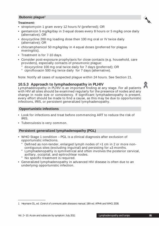
95Vol. 2 • 10. Acute and subacute by symptom: July 2011
Bubonic plague1
Treatment:
• streptomycin 1 gram every 12 hours IV (preferred); OR• gentamicin 5 mg/kg/day in 3 equal doses every 8 hours or 5 mg/kg once daily
(alternative); OR• doxycycline 200 mg loading dose then 100 mg oral or IV twice daily
(alternative); OR• chloramphenicol 50 mg/kg/day in 4 equal doses (preferred for plague
meningitis).• Treatment is for 7-10 days. • Consider post-exposure prophylaxis for close contacts (e.g. household, care
providers), especially contacts of pneumonic plague: ° doxycycline 100 mg oral twice daily for 7 days (preferred); OR ° ciprofl oxacin 500 mg twice daily for 7 days (alternative).
Note: Notify all cases of suspected plague within 24 hours. See Section 21.
10.5.3 Approach to lymphadenopathy in PLHIVLymphadenopathy in PLHIV is an important fi nding at any stage. For all patients with HIV all sites should be examined regularly for the presence of nodes and any change in node size or consistency. If signifi cant lymphadenopathy is present, every effort should be made to fi nd a cause, as this may be due to opportunistic infections, IRIS, or persistent generalized lymphadenopathy.
Opportunistic infections
• Look for infections and treat before commencing ART to reduce the risk of IRIS.
• Tuberculosis is very common.
Persistent generalized lymphadenopathy (PGL)
• WHO Stage 1 condition – PGL is a clinical diagnosis after exclusion of opportunistic infections. ° Defi ned as non-tender, enlarged lymph nodes of >1 cm in 2 or more non-
contiguous sites (excluding inguinal) and persisting for ≥3 months. ° Lymphadenopathy is symmetrical and often involves the posterior cervical,
axillary, occipital, and epitrochlear nodes. ° No specifi c treatment is required.
• Generalized lymphadenopathy in advanced HIV disease is often due to an underlying opportunistic infection.
1 Heymann DL, ed. Control of communicable diseases manual, 19th ed. APHA and WHO, 2008.
Lymphadenopathy and lumps
FINAL Vol. 2 CH 9-21.indd 95FINAL Vol. 2 CH 9-21.indd 95 29/06/2012 15:1529/06/2012 15:15

96 Vol. 2 • 10. Acute and subacute by symptom: July 2011
Immune reconstitution infl ammatory syndrome (IRIS)
IRIS commonly presents with lymphadenopathy. It may be necessary to aspirate or biopsy the lymph node for a defi nitive diagnosis.
Common IRIS conditions include:• TB• MAC • cryptococcosis• Kaposi sarcoma• lymphoma.
Treat the underlying condition and continue ART. See Section 13 Chronic HIV care for more details.
10.5.4 Symptom management of lymphadenopathy• Provide adequate analgesia.• If the swelling is severe steroid therapy may be required, but only when
specifi c treatment for the cause is also provided.• Ensure discharging lymph nodes are covered.
Lymphadenopathy and lumps
FINAL Vol. 2 CH 9-21.indd 96FINAL Vol. 2 CH 9-21.indd 96 29/06/2012 15:1529/06/2012 15:15

97Vol. 2 • 10. Acute and subacute by symptom: July 2011
10.6 Chest symptoms: cough and shortness of breath
In this section:10.6.1 Clinical approach to a patient with chest symptoms
• Interpretation of chest X-ray fi ndings10.6.2 Differential diagnosis of chest complaints
• DDx: X-ray abnormalities in patients with acute chest symptoms• DDx: Diffi cult breathing or cough – with fever• DDx: Diffi cult breathing or cough – without fever• DDx: Chest pain• Evaluation and differential diagnosis of pleural effusion
10.6.3 Pneumonia• Determining the need for hospitalization• Outpatient management of non-severe pneumonia• Pneumocystis jirovecii pneumonia• Infl uenza pneumonia• Varicella pneumonia
10.6.4 Asthma• Classify asthma severity• Table: Examples of increasing dosage and choice of medications by asthma severity
10.6.5 Chronic obstructive pulmonary disease• Classify COPD severity • Example of approach to management of COPD
This Section provides an approach to patients presenting with the most frequent chest symptoms: shortness of breath, cough, and chest pain. This approach is intended for patients who do not have conditions that require emergency management (described in Quick Check) or have been stabilized after emergency treatment and require more defi nitive diagnosis and management that can be approached more slowly. This Section should be viewed as part of a continuum of care, beginning with Quick Check, and proceeding through Section 3.2 (Severely ill patient with respiratory distress) for patients who require urgent care.
10.6.1 Clinical approach to a patient with chest symptoms
Step 1: Use Quick Check. on every patient and initiate emergency management as needed.In patients with severe diffi culty breathing, proceed with diagnosis and urgent management as described in Section 3.2. In patients with chest pain, proceed as described in Section 3.3.
Step 2: Take a history and examine the patient.
Step 3: Assess HIV status.
Step 4: Consider likely differential diagnosis using the DDx table(s).Utilize the appropriate differential diagnosis tables and establish a list of possible diagnoses ranked in order of likelihood.
Step 5: Perform investigations as required, based on the possible diagnoses.
Step 6: Initiate treatment and monitor the response.
Chest symptoms
FINAL Vol. 2 CH 9-21.indd 97FINAL Vol. 2 CH 9-21.indd 97 29/06/2012 15:1529/06/2012 15:15

98 Vol. 2 • 10. Acute and subacute by symptom: July 2011
The key features of an illness or symptom are its severity and rapidity of progression. Severe or rapidly progressive conditions should be managed as per Quick Check (emergency illness) and Section 3 (severe illness). Occasionally, conditions requiring emergency or urgent management will have been missed in triage, so all patients should have a repeated Quick Check to be certain they have been appropriately evaluated for emergency conditions and treated.
Shortness of breath, cough, and chest pain may occur alone or in combination and may be the result of either infectious or non-infectious diseases. In some patients, particularly those with compromised immune systems, multiple infectious and non-infectious processes may be present at the same time. The clinician should consider additional diagnoses, even after establishing one diagnosis.
History
Obtaining a medical history is a critical component of the diagnostic evaluation and should both help direct the physical examination and prompt more targeted questions. The history should be linked to the physical examination. While taking the history, observe the general status, e.g. whether too short of breath to speak in full sentences or answering questions inappropriately.
For all of the chest symptoms, the history should include certain standard questions:• What is the nature of the symptoms? For example, is it a single symptom
such as shortness of breath or combined with cough and chest pain?• Is fever (or feeling hot) present? • Have there been chills?• When did the symptoms begin?• Did the symptoms begin gradually or suddenly?• How severe are the symptoms?• How rapidly are the symptoms progressing (minutes, hours, days, weeks)?• What makes the symptoms worse? • What makes the symptoms better?• Are there associated non-chest symptoms, such as nausea, vomiting,
diarrhoea, muscle aches, headache, weight loss, irregular heart beat?• Does the patient have other illnesses, particularly chronic obstructive
pulmonary disease (COPD), asthma, previous lung infections, heart disease, hypertension, HIV infection?
• Are there symptoms that suggest an underlying illness?• Does the patient take any medications or traditional remedies?• Has there been exposure to persons with lung infections?• Has there been any recent trauma or bite?• Have any of the symptoms happened in the past?
If shortness of breath, also ask:• Is the diffi culty breathing only with exercise or at rest as well?• Is it affected by body position (upright or lying down or lying on one side or
the other)?
Chest symptoms
FINAL Vol. 2 CH 9-21.indd 98FINAL Vol. 2 CH 9-21.indd 98 29/06/2012 15:1529/06/2012 15:15

99Vol. 2 • 10. Acute and subacute by symptom: July 2011
• Is the breathing noisy?• Does the chest feel tight?• Is there fever or chills?• Is there cough?
If cough, also ask: • Is fever present?• Is the cough dry or is mucus produced?• If mucus is produced
° What is the colour (green, yellow, white)? ° What is the quantity (scanty, profuse)? ° Is it blood-stained?
• Is there gross blood?• When is the cough most likely to occur (especially at night or in the morning
on arising)?• Are there any aggravating factors (exertion, particular seasons, particular
environments such as the workplace, specifi c positions, exposure to dust, pollens, or other allergens or irritants such as smoke)?
• Has there been a recent upper respiratory infection or sinus infection?• Is there a prior history of similar cough?
If chest pain, also ask:• Where is the pain?• What is the quality of the pain?• Has there been any chest trauma?• Does the pain radiate anywhere? To the jaw, arm, or back?• How rapidly is it progressing (minutes, hours, days, weeks)?• What makes it worse (e.g. exercise, taking a deep breath)?• What makes it better (e.g. certain positions, non-steroidal medications)?• Does it resolve spontaneously or with antacid medications (may suggest
oesophageal spasm)?
Obtain a past medical history and social history to help identify the cause of symptoms:• history of asthma, COPD, or heart disease;• medication use currently or in recent past;• previous TB;• HIV status and latest CD4 count;• immunisation history, including pertussis, infl uenza, Streptococcus
pneumonia;• occupational history or environmental exposure (e.g. mining, exposure to
dust, fumes or strong odours, farming, animals);• known close contact with a person with TB;• smoking and exposure to second-hand tobacco smoke;
Chest symptoms
FINAL Vol. 2 CH 9-21.indd 99FINAL Vol. 2 CH 9-21.indd 99 29/06/2012 15:1529/06/2012 15:15

100 Vol. 2 • 10. Acute and subacute by symptom: July 2011
• exposure to indoor smoke from cooking or heating, open fi res using wood, grass, dung, or other fuels in poorly ventilated structures;
• sinus pain or previous sinus infections;• substance use including alcohol and inhaled cocaine.
Examination The physical examination will help to determine whether the problem is primarily from the lungs, the heart, or another organ system and will be guided by the information obtained in the history.• Vital signs. The initial vital signs serve to quantify the severity of illness and
as the baseline for monitoring the response to treatment. ° temperature (<36°C, >38°C abnormal) ° blood pressure (systolic blood pressure <90 and diastolic <60 is low) ° heart rate (>110 beats/minute is abnormally fast and <60 beats/minute
abnormally slow) ° respiratory rate (normal 12 to 16/minute; use Section 3.2 if >25/minute) ° pulse oximetry (normal: SpO2 >95%, give oxygen if <90%; SpO2 <90 is
abnormally low but may be normal at high altitude).• General examination
° Does the patient appear acutely or chronically ill? ° Is the patient too short of breath to speak in full sentences? ° Is the patient having severe pain? ° Does the patient respond to questions appropriately? ° Is there confusion or disorientation? ° Does the patient appear cyanotic (bluish discoloration around lips or under
the tongue)? ° Is the patient pale or fl ushed? ° Are there visible lymph nodes? ° Does the patient have digital clubbing?
• Examination of the respiratory system ° Does breathing appear diffi cult or easy? ° Is there nasal fl aring? ° What is the pattern of breathing (deep and sighing or rapid and shallow) ° Are accessory muscles (especially the muscles in the neck) being used? ° Is there retraction of the intercostal spaces? ° Is the trachea in the midline? ° Do both sides of the chest move evenly? ° Is any part of the chest tender to the touch? ° Is there dullness or hyper-resonance when the chest wall is percussed? ° Listen to the breathing for audible noise and by auscultation.
◊ Is there audible noise with inspiration or expiration, and is it coming from the upper or lower airways?
◊ Is there wheezing (high-pitched breath sounds) or prolonged expiratory phase?
◊ Are breath sounds equal on both sides? ◊ What is the quality of breath sounds? Are there crackles or rales or harsh breath sounds?
◊ Are the abnormal breath sounds localized or diffuse? ° Are there palpable lymph nodes?
• Examination of the heart and cardiovascular system ° Are the jugular veins elevated? (This may be diffi cult to determine in a
patient who is breathing rapidly.)
Chest symptoms
FINAL Vol. 2 CH 9-21.indd 100FINAL Vol. 2 CH 9-21.indd 100 29/06/2012 15:1529/06/2012 15:15

101Vol. 2 • 10. Acute and subacute by symptom: July 2011
° Is there swelling of one leg or both legs? ° Is the apex beat displaced from the left midclavicular line in the fi fth
intercostal space? ° Are the heart sounds decreased? ° Is there a heart murmur? ° Is there an extra heart sound (gallop or rub)? ° Is the liver enlarged? (The edge of the liver should not be felt below the rib
cage on the right side of the abdomen.) ° Is the abdomen swollen?
Investigations
• Generally, a productive cough for 2 or more weeks is an indication for obtaining sputum for molecular testing with a nationally or WHO-approved test such as Xpert MTB/RIF test where available, or for an acid-fast smear microscopy or other tests to detect mycobacteria. In HIV-positive patients or in HIV-prevalent settings, suspect TB and send sputum test in patients presenting with cough. See Section 15 Tuberculosis.
• Check Hb (for anaemia) and WBC (if elevated, suspect infection).• If suspect malaria, check malaria smear or RDT.• Pulse oximetry (SpO2).• Chest X-ray.• Peak fl ow measurements for acute management of asthma.• Spirometry (measurement of forced expiratory volume in 1 second (FEV1)
and forced vital capacity (FVC)). Spirometry is the most accurate means of quantifying the degree of airways obstruction in the chronic management of COPD and asthma. Reversibility of airfl ow obstruction is defi ned as either an increase in FEV1 ≥12 % from baseline AND ≥200 ml after inhalation of a short-acting bronchodilator such as salbutamol.
• ECG to look for evidence of cardiac disease.
Interpretation of chest X-ray fi ndings
The chest X-ray can assist in narrowing the differential diagnosis or making specifi c diagnoses. There are several books available that can guide interpretation of chest X-rays1 and also describe appropriate quality control for X-ray examination. The table in the Section 10.6.2 (DDx: Chest X-ray abnormalities in patients with acute chest symptoms) presents the X-ray patterns associated with various diagnoses.
Common terms for chest X-ray fi ndings• Infi ltrate or opacity: A generally ill-defi ned density on the X-ray fi lm.• Lucency: An area that is less dense than the surrounding tissue, thus
appearing dark compared to the surrounding area.• Focal infi ltrate or opacity: An area of increased density that is localized to one
part of the lung, usually no more than a single lobe.• Diffuse infi ltrate or opacity: Increased density that involves multiple areas of
the lungs in either a patchy or uniform distribution.• Masses and nodules: Focal densities that are solid and well-defi ned. Masses
are ≥3.0 cm in size whereas nodules are between 0.2–3.0 cm. Both may be
1 The WHO manual of diagnostic imaging: Radiographic anatomy and interpretation of the chest and pulmonarysystems. WHO, 2006. Available at http://whqlibdoc.who.int/publications/2006/9241546778_eng.pdf
Chest symptoms
FINAL Vol. 2 CH 9-21.indd 101FINAL Vol. 2 CH 9-21.indd 101 29/06/2012 15:1529/06/2012 15:15

102 Vol. 2 • 10. Acute and subacute by symptom: July 2011
single or multiple. Miliary nodules are tiny, ≤2 mm in size, and are usually associated with tuberculosis (see Section 15), but may be caused by other infections and occasionally by malignancies.
• Pleural effusion: Fluid that has accumulated in the pleural space between the chest wall and the lung. The collection may be on one or both sides of the chest.
• Pneumothorax: Air in the pleural space associated with partial or complete collapse of the lung. A tension pneumothorax will cause a shift of the heart and mediastinum to the opposite side of the chest.
• Cavity: A rim of high density containing an area of decreased density in the lung. Is usually the result of an infection that causes death of lung tissue. Some lung cancers (for example, squamous cell carcinoma) may also present as a cavity. Cavities caused by infection are usually surrounded by an area of infi ltrate.
• Lymphadenopathy: Enlargement of lymph nodes in the chest (e.g. mediastinal or hilar), may also be associated with tuberculosis (see Sections 10.5 and 15) or cancer.
10.6.2 Differential diagnosis of chest complaintsA list of possible diagnoses can be developed based on the history, physical examination, chest X-ray, and other investigations and local epidemiological factors, e.g. the prevalence of HIV infection or TB, and the season of the year (is there seasonal infl uenza circulating). The list will vary according to the patient’s age and should be roughly in order of likelihood. Generally, the fi rst diagnosis on the list will be the working diagnosis, for which empirical treatment may be necessary. Other diagnoses lower on the list may be suffi ciently likely that treatment may be indicated. However, if more than one disease or condition is treated empirically, the response to treatment cannot be used to infer a diagnosis.
DDx: Chest X-ray abnormalities in patients with acute chest symptoms
Chest X-ray fi ndingsMost likely causes (may differ in different areas according to the most common conditions)
Focal infi ltrate or opacity Figure 1a Right middle lobe infi ltrate pneumonia (PA view) and Figure 1b (lateral view)
• Pneumonia (bacterial, viral, fungal)• TB (see Section 15)
Diffuse infi ltrates or opacities Figure 2
• TB (see Section 15)• Pneumonia (particularly viruses such as infl uenza and CMV; PCP)• Fungal infections• Heart failure (pulmonary oedema)• Malignancy (including Kaposi sarcoma and lymphoma)
Multiple small nodulesFigure 3
• Infection (especially disseminated TB, see Section 15)• Metastatic malignancy (including from a primary lung cancer)
Masses and nodulesFigure 4
• Lung cancer• Metastatic malignancy (especially if multiple masses are present)• Infection (especially TB or fungal, see Sections 11 and 15)
CavityFigure 5
• TB (see Section 15)• Bacterial infection – especially caused by aspiration pneumonia
(associated with alcoholism, epilepsy, poor dentition)• Fungal infection• Malignancy, particularly if wall thickness is >1.5 cm
Chest symptoms
FINAL Vol. 2 CH 9-21.indd 102FINAL Vol. 2 CH 9-21.indd 102 29/06/2012 15:1529/06/2012 15:15

103Vol. 2 • 10. Acute and subacute by symptom: July 2011
Pleural effusionFigures 6a and 6b
• Bacterial pneumonia (including empyema)• TB, particularly if there is pleural calcifi cation (see Section 15)• Malignancy (including Kaposi sarcoma and lymphoma)• Heart failure (usually bilateral or right-sided)• Chest trauma (haemothorax)• Pulmonary embolism• Any condition associated with a low serum protein concentration
(severe liver disease, nephrotic syndrome, renal insuffi ciency, severe malnutrition – usually bilateral)
Hilar or mediastinal lymphadenopathyFigures 7a and 7b
• TB (see Section 15)• Malignancy including Kaposi sarcoma and lymphoma• Fungal infections• Metastatic cancer• Inhalational anthrax
PneumothoraxFigure 8
• Spontaneous pneumothorax (no underlying disease)• TB (see Section 15), PCP• COPD, asthma• Chest trauma
Normal Figure 9
• Asthma• Pulmonary embolism• PCP
DDx: Diffi cult breathing or cough – with fever
Disease or condition In favour
Acute bronchitis • Shortness of breath mild, if present• Cough may be productive • Acute onset • Mild or absent fever • No chest fi ndings on physical examination except wheezing in asthmatics• Normal chest X-ray
Pneumonia • Shortness of breath mild to severe• Productive cough with bacterial pneumonia and non-productive cough with non-
bacterial pneumonia (but considerable overlap)• Acute onset hours to a few days• Focal chest pain with deep breaths or coughing• Fever and chills• Fast breathing (>30 breaths/min)• If SBP <90, patient has septic shock (see Section 3.1.5) • Focal signs – bronchial breath sounds, crackles, or rales on auscultation• With pleural effusion, dullness to percussion and decreased breath sounds over
affected side• Chest X-ray may show focal or diffuse infi ltrates particularly in non-bacterial
pneumonia
Uncomplicated infl uenza (may be pandemic or seasonal)see Section 11.17
• Infl uenza known or suspected to be circulating• Fever• Cough• Sore throat• Rhinorrhoea or nasal congestion• Headache• Muscle pain or malaise• Gastrointestinal illness such as diarrhoea or vomiting• If shortness of breath, consider infl uenza with pneumonia (below)
Chest symptoms
FINAL Vol. 2 CH 9-21.indd 103FINAL Vol. 2 CH 9-21.indd 103 29/06/2012 15:1529/06/2012 15:15

104 Vol. 2 • 10. Acute and subacute by symptom: July 2011
Disease or condition In favour
Infl uenza with pneumonia see below and Section 11.17
• Infl uenza known or suspected to be circulating• History of infl uenza-like illness• Risk factors: age <2 years or ≥65 years, pregnancy (up to 2 weeks postpartum),
any chronic disease (pulmonary, cardiac, diabetes, metabolic, renal, hepatic, hematologic, or neurologic) or immunosuppression (HIV, malignancy, chemotherapy)
• Shortness of breath, pleuritic pain, cough, coloured sputum, fever • Rapid respiratory rate • Bilateral crackles, possible wheezing• Chest X-ray may show focal or diffuse infi ltrates
Note: Both uncomplicated infl uenza and infl uenza pneumonia may be complicated by secondary bacterial pneumonia with features of pneumonia as described above.
Pulmonary tuberculosissee Section 15
• Shortness of breath mild to moderate but occasionally severe• Possible history of exposure to a person with TB • Usually gradual in onset• Cough with or without bloody or blood-tinged sputum • Weight loss• Fever, night sweats, occasionally chills• Occasionally, chest pain• Xpert MTB/RIF positive (or other nationally- or WHO-approved molecular test)
where available, sputum AFB positive (possible for AFB smear to be negative in patients with smear-negative pulmonary TB) – see Section 15.
Chest X-ray in patients without immune system compromise: unilateral or bilateral upper lobe infi ltrates with or without cavitation; pleural effusion; miliary nodular pattern; other nodular opacities and fi brosis. A normal chest X-ray does not exclude TB.
Chest X-ray in patients with advanced immune defi ciency (advanced immune system compromise, advanced HIV): non-specifi c pattern, with patchy infi ltrates in the lower and mid lung zones; hilar and mediastinal lymphadenopathy may also be seen. Routine chest X-ray is not indicated for the diagnosis of TB. See Section 15.
Pneumocystis jirovecii pneumonia (PCP)
• Shortness of breath mild initially but may become severe • Subacute onset – days to weeks• Non-productive cough • Low grade to moderate fever• Fast breathing >30 breaths/minute• Nasal fl aring• Usually no fi ndings on physical examination of the chest• Chest X-ray bilateral diffuse infi ltrates without lymph node enlargement or pleural
effusion. In mild cases, X-ray may be minimally abnormal or normal.• CD4 cell count <200• Hypoxaemia (SpO2 <90) – particularly on exertion
Disseminated fungal infection (endemic fungi vary from place to place)
see Section 11.16
• Shortness of breath is mild to moderate• Subacute or chronic onset • Cough may be productive or non-productive• Generally moderate fever • Weight loss• Mild or no respiratory symptoms• Lymphadenopathy and skin lesions may be present• Enlargement of liver and spleen• Chest X-ray – various abnormalities including focal or diffuse infi ltrates, single or
multiple masses or nodules, hilar or mediastinal adenopathy, and pleural effusions
Chest symptoms
FINAL Vol. 2 CH 9-21.indd 104FINAL Vol. 2 CH 9-21.indd 104 29/06/2012 15:1529/06/2012 15:15

105Vol. 2 • 10. Acute and subacute by symptom: July 2011
Immune reconstitution infl ammatory syndrome (IRIS) see Section 13
• Shortness of breath if the lungs are involved• HIV-positive with ART initiated in past 3 months, often with CD4 <50 cells/mm3 at
initiation• Common in patients with tuberculosis• Cough, if present, is generally non-productive• Moderate fever is common• Physical examination of the chest depend on the manifestations of IRIS• Extra thoracic lymphadenopathy commonly increases • Usually an increase in CD4 cell count• Chest X-ray may show worsening of infi ltrates, pleural effusion, and increasing
intra-thoracic lymphadenopathy
Lung abscess • Shortness of breath is mild if present• Onset is subacute, generally over several weeks• Fever is moderate• Cough is productive with copious thick, yellow to brown, foul-smelling sputum• Poor dentition may be present • Chest may reveal crackles and coarse bronchial breath sounds over the involved
area• Chest X-ray – focal infi ltrate or mass with cavitation• Note: Lung abscess is generally the consequence of a necrotizing pneumonia, but
may also be secondary to endobronchial obstruction (e.g. from cancer).
Strongyloides hyperinfection see Section 11.36
• Shortness of breath moderate or severe• Risk factors – use of corticosteroids, other immunosuppressive drugs, or HIV
infection• Acute onset• Moderate fever• Cough usually non-productive but may have blood-tinged sputum • Abdominal pain, nausea, vomiting, diarrhoea may be present• Skin rash possible • Chest usually normal but wheezing may be present.• Chest X-ray – diffuse infi ltrates• Peripheral eosinophilia may be noted• Larvae on wet mount and Giemsa stain of sputum
Anthrax see Section 10.2
• History of exposure to animals• Oropharyngeal anthrax: eschar lesions in mouth, tongue, tonsils, or posterior
pharynx. Symptoms of sore throat, dysphagia, regional lymphadenopathy. Swelling of neck and anterior chest wall.
• Inhalation anthrax: fevers, chills, sweats, fatigue, cough, shortness of breath, confusion, nausea and vomiting. Chest X-ray – mediastinal lymphadenopathy plus pleural effusions and pulmonary infi ltrates.
Plague see Section 10.5
• Sudden onset of fever, chills, headache, severe malaise• Chest pain• Diffi culty breathing• Cough with blood-stained sputum or haemoptysis• Fulminant course (100% case fatality rate if not treated rapidly)• May or may not have painful swelling of lymph nodes
Varicella pneumoniasee Section 11.45
• Pneumonia may complicate chickenpox in adults, particularly pregnant women and persons who are immunocompromised
• Usually presents 1–6 days after the onset of rash • Associated with cough, dyspnoea, fever, tachypnoea, and chest tightness,
although chest signs are often minimal• The diagnosis is usually based on fi nding skin lesions characteristic of varicella• (see Sections 10.2 and 11.45)• Chest X-ray – diffuse interstitial opacities
Chest symptoms
FINAL Vol. 2 CH 9-21.indd 105FINAL Vol. 2 CH 9-21.indd 105 29/06/2012 15:1529/06/2012 15:15

106 Vol. 2 • 10. Acute and subacute by symptom: July 2011
DDx: Diffi cult breathing or cough – without fever
Disease or condition In favour
Pneumothorax
see Quick Check page 46, Vol. 1 for emergency management of tension pneumothorax
• Shortness of breath sudden, rapidly progressive and severe• Sudden onset over minutes or hours• Chest pain on affected side• May be associated with trauma or underlying lung disease such as PCP, COPD• Cyanosis may be present• If tension pneumothorax, low blood pressure and trachea shifted from mid-line to
the side opposite the pneumothorax• Hyper-resonance on percussion and diminished or absent breath sounds on the
affected side. Subcutaneous emphysema (“crunchy” feel when pressure applied to chest wall or neck)
• Chest X-ray – completely or partially collapsed lung with no lung markings between collapsed lung and chest wall. If tension – the midline structures (trachea, heart) are shifted away from the affected side
Metabolic acidosis (such as lactic acidosis or diabetic ketoacidosis)
• Patients commonly feel short of breath• Subacute onset• Always an underlying cause – diabetic crisis, renal failure, aspirin toxicity or
other poisoning (methanol, ethanol, paraldehyde), lactic acidosis (e.g. side effects of ARV drugs, commonly d4T- or ddI-containing regimens, more common in women or overweight persons)
• Cough not present• Respiratory rate rapid and respirations deep and sighing, in the absence of cough• Physical examination – chest is normal• Chest X-ray is normal
Asthma • Shortness of breath mild to severe• History of previous wheezing episodes associated with chest tightness • Identifi able triggers common (upper respiratory infection, allergen exposure,
exercise, cold air, extreme emotion) • Breathing or cough commonly worse at night• Non-productive cough is common• Chest examination can be normal between attacks• During attacks or when asthma is poorly controlled, wheezing or prolonged
expiration (compared to inspiration) throughout all lung fi elds (not focal)• In severe attacks, absent breath sounds, fast breathing, use of neck muscles • Chest X-ray may be normal, or show signs of hyperinfl ation like hyperlucent lung
fi elds and fl attened diaphragms, and thickened bronchial walls
Bronchiectasis • Shortness of breath generally mild• Often afebrile• Cough chronic and copious production of yellow/green sputum is common,
occasional blood• Often history of recurrent chest infections and worsening symptoms usually
associated with infections• Examination may reveal crackles and wheezes over the involved area(s) and
uncommonly digital clubbing (rounded deformity of the fi ngernails)• Chest X-ray may show streaky peribronchial infi ltrates and dilated bronchioles
Chest symptoms
FINAL Vol. 2 CH 9-21.indd 106FINAL Vol. 2 CH 9-21.indd 106 29/06/2012 15:1529/06/2012 15:15

107Vol. 2 • 10. Acute and subacute by symptom: July 2011
Chronic obstructive lung disease (COPD)
• Shortness of breath is chronic but worsens with exacerbations that may be caused by acute infections or heart failure
• Unlike asthma, symptoms are usually persistent, not intermittent• Chronic productive cough• Often a history of tobacco smoking, occupational exposures, or exposure to
biomass fuel smoke• In an exacerbation, fast breathing with prolonged expiratory phase and wheezing;
fever may be present (if infection)• Examination shows engorged jugular veins as evidence of right heart failure,
often hard to assess if breathing is laboured; decreased breath sounds, prolonged expiratory phase, and wheezing
• Chest X-ray – hyperinfl ation (hyperlucent lung fi elds and fl attened diaphragms)
Pulmonary Kaposi sarcoma
see Section 11.19
• Shortness of breath mild to moderate• Subacute or chronic onset over weeks• Cough is common often with bloody or blood-tinged sputum • Characteristic purple lesions or nodules of Kaposi sarcoma on the skin or oral
mucus membranes • Chest examination generally normal • Chest X-ray – perihilar patchy interstitial infi ltrates, ill-defi ned nodular densities
Lung malignancy • Shortness of breath uncommon in lung malignancies unless other disease present (COPD, pneumonia)
• Tobacco smoking is a common but not universal risk factor• Onset insidious – usually over months• Cough non-productive but there may be haemoptysis• Weight loss, anorexia are common• May have enlarged supraclavicular lymph nodes • Chest examination normal • The chest X-ray usually shows single or multiple nodules or masses often with
enlarged hilar or mediastinal lymph nodes; pleural effusion may be present
Heart failure • Shortness of breath may be severe• Onset sudden, possibly preceded by shortness of breath on exercise and at night• Possible history of an underlying condition (rheumatic heart disease,
hypertension, severe anaemia)• Chest pain may be present• Cough is common and non-productive• May have signs of right ventricular failure (elevated jugular venous pressure,
liver enlargement, pitting oedema of the both legs) or left ventricular enlargement (displaced apex beat)
• Gallop rhythm (extra heart sound) or murmur (if associated valve disease) may be present
• Chest examination shows bibasilar crackles and sometimes wheezing• Chest X-ray – an enlarged heart, bilateral pulmonary vascular congestion and
diffuse infi ltrates; pleural effusions may be present
Severe anaemiasee Section 10.18
• Shortness of breath is generally less prominent than weakness and tiredness• Gradual onset – often infl uenced by the underlying cause• Cough is absent• Examination: marked general pallor, pallor of the nail beds and oral mucus
membranes• Chest examination is normal• Chest X-ray may be normal or may show heart enlargement• Low Hb (<7 g/dl in pregnancy, <8 g/dl in non-pregnant women, <9 g/dl in men)
Panic attacksee Section 10.11
• Shortness of breath may be severe• Sudden onset• Young patients with no underlying disease and have had previous attacks• There is no cough• Physical examination is normal• Chest X-ray is normal
Chest symptoms
FINAL Vol. 2 CH 9-21.indd 107FINAL Vol. 2 CH 9-21.indd 107 29/06/2012 15:1529/06/2012 15:15

108 Vol. 2 • 10. Acute and subacute by symptom: July 2011
DDx: Chest pain
Disease or condition In favour
Pneumothoraxsee Quick Check page 46, Vol. 1
• Chest pain on affected side, sudden onset, often severe, and increased with inspiration
• Shortness of breath rapidly progressive and severe• Associated with trauma or underlying lung disease such as PCP or emphysema,
but may be primary with no known underlying disease• Cyanosis may be present• If tension pneumothorax, blood pressure will be low. This requires emergency
treatment – see Quick Check page 46, Vol. 1• Trachea shifted from mid-line away from side of pneumothorax• Hyper-resonance on percussion, diminished or absent breath sounds on affected
side • Subcutaneous emphysema (“crunchy” feel when pressure is applied to the chest
wall or neck)• Chest X-ray– radiolucent area with no lung markings between the visceral
(retracted or collapsed lung) and the parietal pleura (chest wall). In tension pneumothorax, midline structures (trachea, heart) shifted away from affected side
Pleuritis, pleurisy without pneumonia
• Localized sharp pain worse on inspiration or coughing• Acute onset• Associated with shortness of breath• Fever present, depending on cause• Chest exam demonstrates decreased expansion of the affected side• Possible pleural friction rub, crackles not heard unless underlying pneumonia• Chest X-ray may demonstrate a pleural effusion or atelectasis
Pericarditissee Section 3.3
• Pain located in anterior chest, improves with leaning forward• Acute onset• Fever present depending on cause• Examination shows rapid heart rate, pericardial friction rub may be present• Chest X-ray – may be normal or show heart enlargement• Electrocardiogram – diffuse S-T segment and T wave abnormalities• If blood pressure low, consider cardiac tamponade: urgent treatment is required
Acute coronary syndrome, myocardial infarctionsee Section 3.3
• Pain is crushing, substernal pressure, radiating to the left arm or jaw • Pain on exertion and sudden in onset• Associated with shortness of breath, sweating, and nausea • Risk factors – tobacco smoking, hypertension, diabetes, and obesity• Examination often normal• Chest X-ray is normal• Electrocardiogram localized S-T segment and T wave abnormalities
Aortic dissection or rupture
• Pain sudden and severe radiating to the back• Fainting may occur• Dissection may occur spontaneously (or rupture just distal to left subclavian
artery may occur in blunt trauma)• Hypotension possible. • Aortic insuffi ciency murmur may be heard• Chest X-ray – widened mediastinum
Costochondritis • Onset of pain – subacute, somewhat worse on inspiration• No cough or fever• Examination – tenderness on palpation of costochondral junctions• Chest X-ray is normal
Chest symptoms
FINAL Vol. 2 CH 9-21.indd 108FINAL Vol. 2 CH 9-21.indd 108 29/06/2012 15:1529/06/2012 15:15

109Vol. 2 • 10. Acute and subacute by symptom: July 2011
Oesophagitis and spasmsee Section 10.7b
• Pain sub-sternum sudden and severe, like pain of cardiac origin• Pain increased with swallowing• No shortness of breath, cough, or fever• Physical examination is normal• Chest X-ray is normal
Rib fracture associated with severe cough
• Bony crepitation (“grating” feeling ) may be palpated• Chest X-ray may be normal, show the rib fracture or demonstrate the underlying
process that caused the coughing• Pain – sudden onset, with vigorous coughing and may be severe; increases with
inspiration, coughing, or movement• Focal pain
Evaluation and differential diagnosis of pleural effusion
Many diseases and conditions may cause or be associated with pleural effusion. A presumptive cause of the effusion often can be determined based on the underlying condition or disease. For example:• bilateral effusions in a patient with heart failure are likely caused by heart
failure• an effusion in a patient with bacterial pneumonia is likely associated with the
infection.
However, if the cause of the effusion is not a clear presumptive diagnosis, the fl uid must be analysed to distinguish if the effusion is due to an infectious or a non-infectious cause.• First, detect the fl uid on a chest X-ray.• Second, perform a thoracentesis to obtain a sample of the fl uid.• Third, analyse the fl uid using the algorithm below.
Chest symptoms
FINAL Vol. 2 CH 9-21.indd 109FINAL Vol. 2 CH 9-21.indd 109 29/06/2012 15:1529/06/2012 15:15

110 Vol. 2 • 10. Acute and subacute by symptom: July 2011
Figure: Evaluation and differential diagnosis of pleural effusion
Presence of pleural effusion on chest X-rayNo evident diagnosis
Send sputum for Xpert MTB/RIF if available or AFBPerform thoracentesis (see Section 7.4.1)
Examination of fl uid• Visual inspection (Is fl uid bloody, cloudy, clear, thick, thin?)• Laboratory investigations
° protein (serum and pleural fl uid) ° glucose ° LDH (serum and pleural fl uid) ° cell count and differential white cell count ° Gram stain and AFB smear ° culture for bacteria and mycobacteria if available
Fluid is exudate if any of the following criteria are met• Pleural fl uid/serum protein concentration ratio >0.50• Pleural fl uid/serum LDH concentration ratio >0.60• Pleural fl uid protein concentration ≥30 g/litre
Fluid is LIKELY exudate if cloudy OR viscous OR WBC >5000/µl
Main differential diagnosis• Heart failure• Severe liver disease• Nephrotic syndrome• Severe malnutrition
Fluid is not an exudate (transudate)
Bloody fl uid• Trauma• Malignancy• Pulmonary embolism
Cloudy, thick fl uid• Likely to be exudate
(e.g. empyaema)
Clear, thin fl uid• Likely to be transudate
Cells are mainly lymphocytes
Main differential diagnosis• Tuberculosis
(see Section 15)• Malignancy
Cells are mainly neutrophils
Main differential diagnosis• Bacterial pneumonia
(especially if glucose <60 mg/dl)
• Pulmonary embolism (may be lymphocytic)
If diagnosis is unclear, consider empirical treatment for tuberculosis.
Chest symptoms
FINAL Vol. 2 CH 9-21.indd 110FINAL Vol. 2 CH 9-21.indd 110 29/06/2012 15:1529/06/2012 15:15

111Vol. 2 • 10. Acute and subacute by symptom: July 2011
10.6.3 PneumoniaPneumonia can be caused by many categories of infectious organisms including viruses, bacteria, fungi, certain parasites, and mycobacteria (most commonly M. tuberculosis). The relative proportion of the different categories of organisms will depend on factors such as prevalence of HIV infection and tuberculosis, and the season of the year.
Pneumonia is an infl ammatory condition that involves the air-containing sacs (alveoli) and the airways. Because the alveoli are predominately involved, most pneumonias interfere with the transfer of oxygen from inspired air into the blood and result in hypoxaemia, which can be severe. The lung infl ammation may extend to the outer membrane (pleura) of the lung and cause pleuritis, pleural effusions, and chest pain.
Key clinical featuresSymptoms can vary substantially, depending on the category of infecting organism and severity, but there is considerable overlap.• Cough and shortness of breath are common. Sharp chest pain on inspiration
or cough from pleuritis may occur.• Virtually all categories cause fever and elevated respiratory rate. However,
increased heart rate and low blood pressure should raise suspicion of severe sepsis or shock from the infection or dehydration.
• On examination of the respiratory system, chest inspection may note asymmetry of expansion. Possible fi ndings on auscultation include decreased breath sounds on one side if a pleural effusion is present, and crackles and (increased) bronchial breath sounds over the involved area. A pleural friction rub may also be heard.
Investigations
• Hypoxaemia is common and can be severe.• Chest X-ray
Determining the need for hospitalization
In the fi rst-level IMAI Acute Care guidelines, adult patients will be referred to hospital with severe pneumonia or other very severe disease based on very fast breathing (>30 breaths/minute), pulse 120 or higher, high fever, lethargy, inability to walk unaided, discomfort lying down, or severe chest pain. Second or third trimester pregnant women with signs of non-severe pneumonia (fast breathing >20 breaths/minute, night sweats, or chest pain) are also referred to hospital, rather than receiving oral antibiotics as an outpatient. The same ‘’upgrade’’ for hospital referral is recommended for PLHIV in clinical stage 4 or with a low CD4 count.
At the district hospital, Quick Check triage will identify patients who have emergency signs of airway and breathing (severe respiratory distress, cyanosis, appears obstructed), count the respiratory rate and measure SpO2. If the patient also has a fever, then empirical antibiotics for possible pneumonia will be given.
Chest symptoms
FINAL Vol. 2 CH 9-21.indd 111FINAL Vol. 2 CH 9-21.indd 111 29/06/2012 15:1529/06/2012 15:15

112 Vol. 2 • 10. Acute and subacute by symptom: July 2011
Suspect severe pneumonia if the following criteria are met and use Section 3.2.3 to guide management:• fever or suspected infection • cough• respiratory rate >30 • signs of severe respiratory distress• SpO2 <90.
There are other approaches to determining that a patient may have severe pneumonia and need hospitalization. A large multicentre study in a high-resource setting2 derived and validated the «CURB-65» prognostic score based on the following factors (each worth one point):• Confusion (altered mental status) • Urea >7 mmol/litre• Respiratory rate ≥30 breaths/minute• Blood pressure (systolic) <90 mmHg or diastolic <60 mm Hg• age ≥65 years.
Increasing CURB-65 scores are associated with increasing mortality. In some professional society guidelines, it is recommended that patients having 2 or more factors be admitted to the hospital. The utility of CURB-65 may be decreased in resource-constrained settings.
Empirical treatment is usually based on a presumptive determination of the likely category of organism causing the pneumonia and on an assessment of severity. It is important to have knowledge of the local epidemiology of community-acquired pneumonia. In high TB prevalence areas, or if HIV infection is known or suspected, TB should always be a consideration in patients presenting with respiratory infection.
If the patient has severe pneumonia, see Sections 3.2.1–3.2.3 for management.
Outpatient management of the patient with non-severe pneumonia
• Patients who have signs and symptoms of pneumonia but do not meet criteria for severe pneumonia can be managed as outpatients.
• Counsel the patient regarding the importance of adherence to the medication regimen and the need for follow-up to determine response.
• The patient should be advised to return for evaluation if there is no improvement or there is a worsening of symptoms.
• Treatment should be given for 5–7 days, assuming there is a good, prompt response.
Empirical antibiotic regimens for non-severe pneumonia
• amoxicillin 500–1000 mg 3 times daily; OR• erythromycin 500 mg 4 times daily; OR• doxycycline 100 mg 2 times daily (avoid in pregnancy).
2 Lim WS, et al. Defi ning community acquired pneumonia severity on presentation to hospital: an internationalderivation and validation. Thorax, 2003, 58:377-82.
Chest symptoms
FINAL Vol. 2 CH 9-21.indd 112FINAL Vol. 2 CH 9-21.indd 112 29/06/2012 15:1529/06/2012 15:15

113Vol. 2 • 10. Acute and subacute by symptom: July 2011
Follow national guidelines for alternative antibiotics regimens that may include:• azithromycin 500 mg once daily; OR• clarithromycin 500 mg twice daily, OR• oral respiratory quinolone (for example, levofl oxacin) – see below for
cautions.
Send sputum for Xpert MTB/RIF if available, or for AFB if TB is suspected. See Section 15.
It is important not to treat patients suspected of having TB with a respiratory quinolone as it may mask or partially treat underlying tuberculosis. Respiratory quinolones should also be avoided in high-prevalence TB settings unless TB is excluded. Safety of respiratory quinolones in pregnancy has not been established.
If not improving after 3 days and the patient has been adherent to the antibiotic regimen, review and consider switching to an IV regimen.• ceftriaxone 1–2 grams once daily PLUS erythromycin 500 mg (oral or IV) 4
times daily; OR• ampicillin 2 grams IV 4 times daily PLUS gentamicin (see Section 8.4 for
dosing) PLUS erythromycin 500 (oral or IV) 4 times daily.
If the patient has a non-anaphylactic allergy to penicillin (for example, skin rash only), then ceftriaxone can be used.
Consider other infections, including TB and, if HIV-positive, consider PCP.
Pneumocystis jirovecii pneumonia (PCP)
PCP is caused by a fungus, Pneumocystis jirovecii (formerly known as Pneumocystis carinii). The organism is present in soil, transmitted through inhalation, and distributed worldwide. Most people have been exposed to it by the age of fi ve. PCP occurs only in immunocompromised patients, particularly those who are infected with HIV and whose CD4 count is <200 cells/mm3. It is associated with high mortality in HIV patients. Patients who have recently received steroids or other immunosuppressive therapy are also at increased risk.
Key clinical features
• PCP can be associated with many of the manifestations described above for pneumonia in general.
• Shortness of breath that is slow in onset (over 1–2 weeks).• Cough that is non-productive.• Fast pulse.• Fast respiratory rate.• Cyanosis may be present and is a sign of severe hypoxaemia.• Auscultation of the chest is generally unremarkable, but some crackles may
be present.
Investigations
• SpO2 is decreased.• Elevated lactate dehydrogenase (LDH), which is a non-specifi c marker of
pulmonary infl ammation.
Chest symptoms
FINAL Vol. 2 CH 9-21.indd 113FINAL Vol. 2 CH 9-21.indd 113 29/06/2012 15:1529/06/2012 15:15

114 Vol. 2 • 10. Acute and subacute by symptom: July 2011
• Experienced microscopists with special training may be able to identify the organism in induced sputum samples with special stains: methenamine (Grocot) silver, calcofl our white, and Wright-Giemsa. The sensitivity is diminished in patients using cotrimoxazole prophylaxis.
• On chest X-ray, bilateral peri-hilar infi ltrates are common, but nodular densities, lobar consolidations, and cavitation also can occur. At fi rst presentation, in 25% of cases, the chest X-ray can be (misleadingly) normal. Pleural effusion is rare.
Treatment
• If patient has severe pneumonia, see Section 3.2.3 for additional management recommendations regarding oxygen and fl uid therapy.
• Treatment should be initiated early and empirically, based on history and clinical presentation, while awaiting defi nitive diagnosis.
• Antimicrobial treatment is most effective when started early: ° cotrimoxazole 400 mg trimethoprim/80 mg sulfamthoxazole tablets (SS):
dose based on trimethoprim (TMP) 5 mg/kg divided 4 times daily orally or IV for 21 days (preferred); OR
° clindamycin 600 mg IV or orally 4 times daily PLUS primaquine 15 mg twice daily orally for 21 days (alternative); OR
° pentamidine 4 mg/kg IV daily for 5 days, then reduce dose to 2 mg/kg daily IV to complete 21 days (alternative if unresponsive to cotrimoxazole or cotrimoxazole not tolerated).
• If the patient is hypoxaemic with SpO2 <90 on room air, give IV therapy initially and add prednisone, 40 mg twice daily for 5 days, then 40 mg daily for 5 days, then 20 mg daily for 11 days to complete 21 days of treatment.
Prophylaxis
• Primary prophylaxis is indicated for HIV-positive patients in WHO clinical stage 2, 3, or 4 irrespective of CD4 count, or with CD4 count <350/mm3 irrespective of clinical stage. See Section 13. Follow national guideline recommendations.
• Secondary prophylaxis should be given to all patients following treatment of PCP.
• Primary and secondary prophylaxis is the same: cotrimoxazole 1 double strength tablet daily.
• Patients should at least be continued on PCP prophylaxis until there is evidence of immune recovery on antiretroviral therapy (CD4 counts >350 cells/mm3 after at least 6 months of antiretroviral therapy). See Section 13.
Infl uenza pneumonia (see Section 11.17)
Key clinical features
• Infl uenza is suspected or known to be circulating.• History of infl uenza-like illness.• Progressive symptoms, such as worsening cough, purulent or bloody sputum,
shortness of breath (at rest or with exertion) or chest pain.• Signs of respiratory distress, such as fast respiratory rate, cyanosis, and
hypoxaemia (if severe) and, on chest auscultation, there may be crackles or rales or wheezing.
Chest symptoms
FINAL Vol. 2 CH 9-21.indd 114FINAL Vol. 2 CH 9-21.indd 114 29/06/2012 15:1529/06/2012 15:15

115Vol. 2 • 10. Acute and subacute by symptom: July 2011
Investigations
• Chest X-ray usually shows diffuse interstitial infi ltrates.• See Section11.17 for use of rapid diagnostic tests for an indication of the
presence of infl uenza in the community (not for routine individual patient care).
Treatment (see Section 11.17)
• Treatment should be based on clinical diagnosis and suspicion of infl uenza infection based on local epidemiology and should not be delayed for results of laboratory investigations.
• Treat with antiviral, oseltamivir 75 mg orally twice daily or zanamivir, as soon as possible.
• Co-existing pneumonia due to other pathogens may be diffi cult to exclude in a patient with suspected infl uenza infection. If you suspect community-acquired pneumonia, or another infectious pneumonia based on local epidemiology, then treat with appropriate antimicrobials. Bacterial infection by S. pneumoniae and S. aureus, both methicillin-sensitive and methicillin-resistant, are common (see Section 3.2.3).
• Occasionally, bacterial pneumonia will develop in a person who seems to be recovering from infl uenza infection. In these instances, the presentation is as described for bacterial pneumonia and empirical treatment for bacterial pneumonia should be started as described previously.
Infection prevention and control – see Section 6 for respiratory hygiene and droplet precautions.
Varicella pneumonia
Pneumonia may complicate chickenpox in adults, particularly pregnant women and immunocompromised persons. The disease can progress rapidly to fulminant respiratory failure. Varicella pneumonia usually presents 1–6 days after the onset of rash, and is associated with cough, dyspnoea, fever, fast breathing, and chest tightness, although chest signs often are minimal. The diagnosis usually is based on fi nding skin lesions characteristic of varicella (see Sections 10.2 and 11.45). A chest X-ray shows diffuse interstitial opacities.
Treat with:• IV aciclovir 10–15 mg/kg 3 times daily for 7–10 days.• Switch to oral regimen once there is evidence of clinical improvement to
complete the 10–14-day course.
10.6.4 AsthmaAsthma is an infl ammatory disease of the airways causing reversible airways obstruction and characterized by recurrent acute attacks (exacerbations). The infl ammation also causes an associated increase in the existing bronchial hyper-responsiveness to a variety of stimuli. The diagnosis is usually established from the episodic history of symptoms and by the fi nding of wheezing on auscultation of the chest.
Key clinical features
• Recurrent episodes of cough and shortness of breath often associated with noisy breathing (wheezing) and chest tightness.
Chest symptoms
FINAL Vol. 2 CH 9-21.indd 115FINAL Vol. 2 CH 9-21.indd 115 29/06/2012 15:1529/06/2012 15:15

116 Vol. 2 • 10. Acute and subacute by symptom: July 2011
• Symptoms may be worse at night and interfere with sleep.• Episodes commonly occur in response to specifi c exposures, such as pollens
or other allergens, acute respiratory infections, dust, exercise, or cold air.
Investigations
• Spirometry demonstrating reversibility is consistent with the diagnosis of asthma. However, many patients with asthma have normal spirometry when they are not having an exacerbation. Reversibility of airfl ow obstruction may be incomplete in some patients with asthma, especially those with long-standing asthma.
• Peak fl ow can also demonstrate airfl ow reversibility.
Classify asthma severity
A classifi cation of chronic asthma severity and examples of its treatment according to severity follow.
Table: Classifi cation of severity of asthma
Classifi cation Intermittent asthmaMild persistent asthma
Moderate persistent asthma
Severe persistent asthma
Episodes of breathlessness
<1 per week 1 or more per week, but <1 per day
Daily Continuous daily
Frequency of night symptoms
<2 per month >2 per month >1 per week Frequent
Peak fl ow or spirometry (FEV1), % predicted or personal best
>80% >80% Between 60–80% <60%
WHO and partners3 are working to support the availability of basic asthma medications at the primary care level. Inhaled salbutamol and beclometasone are on the WHO essential medicine list and WHO guidelines for management of asthma at the primary care level are in development .
District clinicians will often see patients with an acute exacerbation of wheezing (see Section 3.2.3) or with persistent symptoms despite use of these basic asthma medications. There are several schematic approaches (shown in the table below, Examples of increasing dosage and choice of medications by asthma severity) to increasing the intensity of treatments to control asthma. Inhaled salbutamol by metered-dose inhaler (MDI) as needed is used as a reliever medication in all categories. Inhaled steroids (for example, beclometasone) are the most important therapy to control symptoms. All patients should be counselled to stop smoking and referred for specialist care for severe persistent asthma if available.
3 For example, IUATLD has established an Asthma Drug Facility to support availability of more affordablesalbutamol and beclametasone metered-dose inhalers. Available at http://www.globaladf.org/
Chest symptoms
FINAL Vol. 2 CH 9-21.indd 116FINAL Vol. 2 CH 9-21.indd 116 29/06/2012 15:1529/06/2012 15:15

117Vol. 2 • 10. Acute and subacute by symptom: July 2011
Refer to national or other local guidelines for specifi c recommendations. It may be necessary to advise patients to purchase more effective medications that are not on the national formulary to control moderate or severe persistent asthma. Although sustained release (SR) theophylline is not on the WHO essential medicines list, it is widely available; low dose SR theophylline can be used and stopped if there is evidence of toxicity.4 Theophylline is not as effective or as safe as the other options listed. Where available, theophylline blood levels should be used to adjust dosing.
Table: Examples of increasing dosage and choice of medications by asthma severity
Asthma severityDerived from WHO model formulary 20085
Derived from IUATLD guidelines6
Derived from BTS/SIGN guidelines7
Intermittent Inhaled salbutamol as needed (100–200 mcg up to 4 times daily)
Inhaled salbutamol as needed
Inhaled salbutamol as needed
Mild persistent Inhaled salbutamol as needed
Beclometasone 100–250 mcg twice daily OR SR theophylline OR a leukotriene antagonist
Inhaled salbutamol as needed
Beclometasone 200 mcg daily
Inhaled salbutamol as needed
Beclometasone 200 mcg twice daily
Moderate persistent Inhaled salbutamol as needed
Beclometasone100–500 mcg twice daily PLUS if needed EITHER Long-acting beta-agonist or SR theophylline or leukotriene antagonist OR Beclometasone high dose >1 mg/day (divided doses)
Inhaled salbutamol as needed
Beclometasone 200 mcg twice daily
Inhaled salbutamol as needed
Beclometasone 200 mcg twice daily and a long-acting beta-agonist (must always be used with beclometasone) OR Beclometasone up to 400 mcg twice daily
Severe persistent Inhaled salbutamol as needed
Beclometasone >1 mg/day (divided doses) PLUS Long-acting beta-agonistPLUS if needed SR theophylline or leukotriene antagonist or long-acting beta-agonist or oral prednisolone in lowest dose possible, given once daily in the morning
Inhaled salbutamol as needed
Beclometasone 400 mcg twice daily and prednisolone 0.5 mg/kg/day (reassess weekly; taper when patient stable for 1 week)
Inhaled salbutamol as needed
Beclometasone 1000 mcg twice daily OR Beclometasone 1000 mcg twice daily AND 6 week trial of SR theophylline. OR Beclometasone 1000 mcg twice daily and daily prednisolone at lowest dose needed for adequate control
4 If theophylline blood levels are available, they should be used to adjust dosing. See Adaptation Guide.5 WHO model formulary. WHO, 2008. Available at http://www.who.int/selection_medicines/list/WMF2008.pdf6 IUATLD asthma guidelines available at http://www.theunion.org/index.php/en/resources/scientifi cpublications/
lung-health7 BTS/SIGN guidelines. Available at http://www.sign.ac.uk/guidelines/fulltext/101/index.html
Chest symptoms
FINAL Vol. 2 CH 9-21.indd 117FINAL Vol. 2 CH 9-21.indd 117 29/06/2012 15:1529/06/2012 15:15

118 Vol. 2 • 10. Acute and subacute by symptom: July 2011
10.6.5 Chronic obstructive pulmonary disease (COPD)Chronic obstructive pulmonary disease is characterized by airfl ow limitation that is not fully reversible. The airfl ow limitation is usually progressive and associated with an abnormal infl ammatory response to noxious particles or gases in the lung. The most common inhaled toxin is cigarette smoke, but smoke from indoor biomass fuel consumption, air pollution, and pulmonary infections also play a role.
Key clinical features
• Symptoms of chronic cough and progressive shortness of breath, with or without sputum production, in a person with an exposure history such as cigarette smoking.
• On examination, patients commonly have a fast respiratory rate, and breath sounds are usually reduced throughout all lung fi elds on chest auscultation.
• At advanced stages, patients may use accessory muscles to breathe, have clinical signs of heart failure (e.g. elevated JVP, liver enlargement and bilateral leg oedema).
Investigations
• Spirometry demonstrating fi xed airfl ow obstruction with an FEV1/FVC <70% after inhalation of short-acting bronchodilator is suggestive of COPD.
• Severity of airfl ow obstruction is based on FEV1 (% predicted).
Classify COPD severity
Severity can be based on subjective criteria (symptoms) or physiological criteria (spirometry); there is often not a good correlation between the two classifi cation schemes.
Table: Classifi cation of severity of COPD8
COPD severity FEV1 Symptoms
Mild more than 80% Short of breath when hurrying on level ground or walking up slight hill
Moderate between 50–79% Walks slower than people of same age on the level because of breathlessness, or stops for breath when walking on level ground at own pace.
Severe between 30–49% Stops for breath after 100 metres or a few minutes on level
Very severe less than 30% Too breathless to leave house or when dressing or undressing
Treatment See table below on management of COPD.
For treatment of an acute episode of severe wheezing, see Section 3.2.3.
Moderate exacerbations of COPD present with increased shortness of breath and possibly more purulent sputum (increase in the volume and change in colour).
8 Questions are from MRC questionnaire, cited in NICE COPD guideline at http://guidance.nice.org.uk/CG101. Therelationship of symptoms to COPD severity (defi ned by FEV1) is approximate.
Chest symptoms
FINAL Vol. 2 CH 9-21.indd 118FINAL Vol. 2 CH 9-21.indd 118 29/06/2012 15:1529/06/2012 15:15

119Vol. 2 • 10. Acute and subacute by symptom: July 2011
Management consists of the following:• Give higher dose salbutamol and ipratropium.• Give prednisolone 30 mg/day for 7–14 days.• Do chest X-ray to rule out pneumonia, pneumothorax, or pleural effusion.• If sputum more purulent compared to baseline, and:
° if evidence of pneumonia, treat as per above guidance. ° if no evidence of pneumonia, give amoxicillin, erythromycin,9 or
doxycycline.
Chronic COPD management is determined by the following factors:• symptoms of breathlessness or exercise limitation• frequency of exacerbations• severity of airfl ow obstruction• presence of complications.
In the management of COPD, inhaled salbutamol or ipratropium as needed are used as reliever medications in all categories. All patients should be counselled to stop smoking and avoid indoor air pollution. Give pneumococcal and annual infl uenza vaccinations. Refer for specialist care for severe or very severe COPD (for example, to consider long-term home oxygen therapy and treatment of right heart failure in very severe disease).
Commonly used inhaled medications include:• Short-acting bronchodilators, such as salbutamol metered-dose inhaler or
ipratropium MDI. • Long-acting bronchodilators, include long-acting beta-agonists (LABA, e.g.
formoterol or salmeterol) or long-acting muscarinic antagonist (LAMA, e.g. tiotropium). Regular dose ipratropium (2 puffs 4 times daily) could be substituted for LAMA.
The table that follows provides an example of an approach to the management of chronic COPD. Refer to national or local guidelines. It may be necessary to advise patients to purchase more effective medications that are not on the national formulary to control COPD. WHO guidelines for management of COPD at the primary care level are in development.
9 Follow national guidelines. Clarithromycin or azithromycin are alternatives.
Chest symptoms
FINAL Vol. 2 CH 9-21.indd 119FINAL Vol. 2 CH 9-21.indd 119 29/06/2012 15:1529/06/2012 15:15

120 Vol. 2 • 10. Acute and subacute by symptom: July 2011
10
Table: Example of approach to management of stable COPD
Severity Derived from NICE10
Mild Short-acting bronchodilator (salbutamol 100-200 mcg up to 4 times daily or ipratropium 20-40 mcg up to 4 times daily) as needed
Moderate Short-acting bronchodilator (salbutamol or ipratropium) as needed
LABA (salmeterol or formoterol) or LAMA (tiotropium)
Severe Short-acting bronchodilator (salbutamol or ipratropium) as needed
LABA (salmeterol or formoterol) and beclometasone (400 mcg twice daily) OR LAMA (tiotropium) or ipratropium 4 times daily OR LABA and LAMA or ipratropium 4 times daily
Very severe Short-acting bronchodilator (salbutamol or ipratropium) as needed
LABA and beclometasone OR LAMA or ipratropium 4 times daily AND LABA ORLABA AND (LAMA or ipratropium 4 times daily) and beclometasone
Maintenance prednisolone is not normally recommended. Some patients may require maintenance prednisolone when it cannot be stopped after an exacerbation. The dose should be kept as low as possible.
SR theophylline should only be used after a trial of short-acting bronchodilators and LABA/LAMA, or in patients who are unable to use inhaled therapy.
10 Management of chronic obstructive pulmonary disease in adults in primary and secondary care. NICE, 2010.Available at http://guidance.nice.org.uk/CG101
Chest symptoms
FINAL Vol. 2 CH 9-21.indd 120FINAL Vol. 2 CH 9-21.indd 120 29/06/2012 15:1529/06/2012 15:15

Fig 1b Lateral view
Fig 3 Multiple small nodules
Fig 5 Cavity
Fig 1a Right middle lobe infi ltrate pneumonia (PA view)
Fig 2 Diffuse infi ltrates or opacities
Fig 4 Masses and nodules
FINAL Vol. 2 CH 9-21.indd 121FINAL Vol. 2 CH 9-21.indd 121 29/06/2012 15:1529/06/2012 15:15

122 Vol. 2 • 10. Acute and subacute by symptom: July 2011
Fig 7b Hilar or mediastinal lymphadenopathy
Fig 9 Normal
Fig 6b Pleural effusionFig 6a Pleural effusion
Fig 7a Hilar or mediastinal lymphadenopathy
Fig 8 Pneumothorax
FINAL Vol. 2 CH 9-21.indd 122FINAL Vol. 2 CH 9-21.indd 122 29/06/2012 15:1529/06/2012 15:15

123Vol. 2 • 10. Acute and subacute by symptom: July 2011
10.7 Abdominal complaints
Each symptom is dealt with in its own subsection. Patients may present with several combinations of the symptoms discussed below, e.g. abdominal pain plus diarrhoea. Therefore, it is important to determine each major symptom, work through the differential diagnosis tables for each, and develop a coherent treatment plan.
Section 10.7 includes:• 10.7a Abdominal pain• 10.7b Painful or diffi cult swallowing• 10.7c Nausea and vomiting• 10.7d Diarrhoea (and constipation)
10.7a Abdominal pain
In this section:10.7a.1 Clinical approach to abdominal pain10.7a.2 Differential diagnosis of abdominal pain and management of specifi c conditions
• DDx: Generalized abdominal pain• DDx: Upper abdominal or epigastric pain• Management of specifi c conditions causing abdominal pain• Dyspepsia, gastritis, peptic ulcer disease• Pancreatitis• Cholecystitis and cholangitis• Peritonitis• Ascariasis
10.7a.3 Approach to abdominal pain in PLHIV
This Section provides an approach to the diagnosis and management of a patient with abdominal pain, either upper or epigastric, or generalized pain. Lower abdominal pain is also addressed in Section 10.15.2 Female genitourinary problems.
10.7a.1 Clinical approach to abdominal pain
Step 1: Use Quick Check. Ensure that there are no serious or life-threatening conditions. Be aware that a patient with abdominal pain could have a surgical abdomen (see below) or a gynaecological emergency that requires surgery. The patient should not eat until this diagnosis has been ruled out.
Step 2: Take a history and examine the patient.
Step 3: Assess HIV status.
Step 4: Classify the abdominal pain and use the DDx tables to work through a differential diagnosis.• DDx: Generalized abdominal pain• DDx: Upper abdominal or epigastric pain
Step 5: Perform investigations.
Step 6: Initiate treatment and monitor the patient’s response.
Painful or diffi cult swallowing
FINAL Vol. 2 CH 9-21.indd 123FINAL Vol. 2 CH 9-21.indd 123 29/06/2012 15:1529/06/2012 15:15

124 Vol. 2 • 10. Acute and subacute by symptom: July 2011
Emergency treatments
Always consider the possibility of an acute surgical abdomen or gynaecological emergency, e.g. an ectopic pregnancy. Patients should not eat or drink before this is ruled out.
A “surgical abdomen” is any abdominal condition which would result in a rapidly worsening prognosis in the absence of surgical intervention. A high level of suspicion should be maintained in immunocompromised patients when typical signs of peritoneal infl ammation may be reduced or absent.
• General signs and symptoms of a surgical abdomen: ° a very ill patient with or without fever and shock ° little pain relief from analgesics ° localized, generalized, or rebound tenderness on palpation ° distended abdomen ° diminished or absent bowel sounds ° no fl atus or bowel movements.
• X-ray fi ndings suggestive of a surgical abdomen include: ° hollow organ perforation: free intra-peritoneal air, or air under the
diaphragm on upright chest or abdominal X-ray (note that this fi nding, although uncommon, is specifi c for a surgical abdomen and should prompt immediate surgical consultation);
° hyper-infl ated bowel- consider peritonitis.• Use ultrasound to exclude other causes of an acute surgical abdomen.
If a surgical abdomen is strongly suspected, refer to the Quick Check for urgent interventions and the WHO manual Surgical care at the district hospital1 for defi nitive care.
History
The history of the presenting complaint should include:• nature of the pain – onset, duration, location, quality, and radiation;• exacerbating or relieving factors (e.g. food, antacids, exertion, defecation);• associated symptoms (e.g. fevers, chills, weight loss or gain, nausea,
vomiting, diarrhoea, constipation, blood in the stool, change in the colour of urine or stool, yellow discoloration of the eyes).
• Medical history ° previous history of similar abdominal pain ° history of peptic ulcer ° symptoms suggestive of systemic disease (e.g. cough, fever, night sweats) ° substance use including alcohol ° immune status.
• Medication history ° ART (duration of therapy is important) ° over-the-counter medications (e.g. paracetamol, aspirin, NSAIDs) ° traditional remedies ° antibiotic therapy.
1 Surgical care at the district hospital., WHO, 2003. Available at www.who.int/surgery/publications/scdh_manual/en/index.html
Painful or diffi cult swallowing
FINAL Vol. 2 CH 9-21.indd 124FINAL Vol. 2 CH 9-21.indd 124 29/06/2012 15:1529/06/2012 15:15

125Vol. 2 • 10. Acute and subacute by symptom: July 2011
• Menstrual and pregnancy history• Geographic location for diseases specifi c to certain regions (notably,
prevalence of schistosomiasis and soil-transmitted helminths) ° If the patient is from a schistosomiasis endemic area, treat empirically with
praziquantel (see Section 11.34).
Examination
• Targeted general examination ° vital signs ° jaundice, pallor, lymphadenopathy ° signs of chronic liver disease (e.g. scratch marks, palmar erythema, ascites,
spider naevi, caput medusa, bruising, oedema)• Abdominal examination
° look for abdominal distension ° palpate for masses, tenderness, peritoneal signs (rebound tenderness and
guarding) ° listen for bowel sounds ° percuss for ascites – shifting dullness ° do rectal examination and also look for blood in stool ° examine the groin and the scrota for inguinal masses and hernias.
• Pelvic examination ° in women with lower abdominal pain.
• Other organs ° look for pneumonia or heart failure – may cause abdominal pain ° shingles can cause localised abdominal pain.
Classify abdominal pain and use the differential diagnosis tables
Classify the pain based on the clinical presentation.
Refer to the respective DDx tables below to help work through a differential diagnosis.• Generalized abdominal pain (DDx table) See also lower abdominal pain in
10.15.2 Female GU problems.• Upper abdominal or epigastric pain (DDx table).
Perform investigations
• Laboratory investigations: The following are recommended investigations for all patients: ° urine analysis (dipstick and microscopy – for haematuria and S.
haematobium ova) ° pregnancy test ° U&E, FBC, glucose
• Investigations based on clinical fi ndings include: ° liver functions ° amylase ° serum lactate if the patient is taking ART. ° Stool for macroscopic and microscopic examination – look for occult blood
and various helminth infections, including Schistosoma ova. This may require repeated examinations or concentration procedures.
Painful or diffi cult swallowing
FINAL Vol. 2 CH 9-21.indd 125FINAL Vol. 2 CH 9-21.indd 125 29/06/2012 15:1529/06/2012 15:15

126 Vol. 2 • 10. Acute and subacute by symptom: July 2011
• Imaging: ° abdominal X-ray ° chest X-ray ° abdominal ultrasound is useful to identify:
◊ abdominal lymph nodes ◊ liver and spleen pathology ◊ appendix mass ◊ gall stones, cholecystitis ◊ renal stones or hydronephrosis ◊ pelvic infl ammatory disease with collections or a mass ◊ ectopic pregnancy.
Initiate treatment and monitor response
For management of specifi c conditions associated with abdominal pain, see the text after the DDx tables.
10.7a.2 Differential diagnosis of abdominal pain and management of specifi c conditionsThe differential diagnosis differs for generalized abdominal pain and for upper abdominal or epigastric pain. Use the appropriate table below. In women with lower abdominal pain, see also Section 10.15.2 Female genitourinary problems.
DDx: Generalized abdominal pain
Condition In favour
Surgical abdomen* Rebound tenderness, guarding Pain not responding to analgesicsAbdominal distensionVomitingDecrease in fl atus or stoolAbdominal X-ray – dilated loops of bowel, air fl uid levelsChest X-ray – free air under the diaphragm
*Consider bowel obstruction or perforation, appendicitis, peritonitis, mesenteric infarct, ruptured ectopic pregnancy. Manage according to Quick Check page 8. See Section 10.15 for female genitourinary problems. See the WHO Manual Surgical Care at the District Hospital.
Ectopic pregnancysee Section 10.15
Positive pregnancy test (or may be negative)Pain usually in lower abdomenAbnormal vaginal bleedingBack painCervical motion tenderness on pelvic examShock (if ruptured)History of previous ectopic pregnancy
Appendicitis Low grade feverEarly – peri-umbilical painLater – pain localized to right lower quadrantFBC – high WCC
Bowel obstruction Colicky abdominal painAbdominal distensionVomitingNo stool or fl atusBowel sounds – none, or if partial obstruction, high-pitched tinkling with rushes Hernia detected
Painful or diffi cult swallowing
FINAL Vol. 2 CH 9-21.indd 126FINAL Vol. 2 CH 9-21.indd 126 29/06/2012 15:1529/06/2012 15:15

127Vol. 2 • 10. Acute and subacute by symptom: July 2011
Abdominal TB see Section 15
Pain usually non-specifi c and chronicConstitutional symptoms – fever, weight loss, night sweatsAbdominal swelling, mass, or ascitesChest X-ray – evidence of pulmonary TBUltrasound – para-aortic lymph nodes or ascitesBiochemistry – low SAAG ascites (see Section 10.9)
Typhoidsee Section 11.43
Seriously ill without other apparent cause Prolonged high fever Abdominal tendernessRelative bradycardia compared with feverGeographic areaFBC – decreased WCC
Lactic acidosis see Section 13
Patient taking ARVs for >6 monthsPatient taking an NRTI, e.g. d4T, ddI, or AZTFemale, overweight Loss of weight, fatigue, malaiseNausea or vomitingSerum lactate >5 mmol/lArterial pH <7.3, widened anion gap >13Elevated ALT/AST, LDH, and amylase
Viral gastroenteritis Most common cause of severe gastroenteritis in the young, the elderly, and people who have suppressed immune systemsAbdominal pain, diarrhoea, nausea, vomitingChills, clammy skin, excessive sweating, fever, joint stiffnessLeakage (incontinence) of stoolMuscle painPoor feeding, vomiting blood (very rare), weight loss
Diverticulitis Abdominal pain in the left lower quadrantFeverNauseaChange in bowel habitsPeritoneal signs – guarding and rebound
Helminthic infections – ascaris
High worm burdens may cause abdominal pain and intestinal obstructionEggs on stool exam by direct wet mount or after concentration
Protozoan infections Chronic watery diarrhoeaWasting and malnutritionGeographic area Other symptoms of advanced HIV
Strongyloidiasissee Section 11.36
Diarrhoea – may have bloodCoughSerpiginous (snake-like) skin lesionsVisible parasites on stool or sputum examination
Irritable bowel syndrome (IBS)
Abdominal cramping, bloating, and a change in bowel habits between constipation and diarrhoeaMore often in women than menNo known cause of IBS
Painful or diffi cult swallowing
FINAL Vol. 2 CH 9-21.indd 127FINAL Vol. 2 CH 9-21.indd 127 29/06/2012 15:1529/06/2012 15:15

128 Vol. 2 • 10. Acute and subacute by symptom: July 2011
Condition In favour
Infl ammatory bowel disease (ulcerative colitis and Crohn’s disease)
Peak incidence in young adults (15–25 years old)Ranges from mild disease (insidious onset, non-bloody diarrhoea, poor weight gain) to severe (fulminant presentation, severe abdominal pain, bloody diarrhoea, tenesmus, and fever)Abdominal tendernessPerianal involvement (fi stulae, anal tag, or fi ssure)Extraintestinal manifestation: eye involvement (e.g., uveitis), skin involvement (e.g., rash, erythema nodosum, pyoderma gangrenosum), peripheral arthritis (involving large joints and ankylosing spondylitis), sclerosing cholangitis, thromboembolism, lung disease, renal stones, anemia, and digital clubbingPalpable abdominal mass (suggestive of a fi stula) and oral ulcerations can occur in Crohn’s diseaseDiagnosis based on characteristic history and endoscopy
Urinary tract infection,cystitissee Section 11.44
Offensive smelling urinePainful urinationFrequency of urinationUrgency of urinationCloudy urineUrine dipstick – leucocytes, nitrites, blood Urine microscopy – leucocytes, RBCs, bacteria
Acute pyelonephritissee Section 11.44
FeverRigorsFlank painPainful urinationCloudy urineCostovertebral angle tendernessUrine microscopy– leucocytes, protein, RBCs, WBC, bacteria
Renal stones Acute attacks of severe colicky back or fl ank pain, radiating to groin (loin to groin radiation)History of previous attacksTaking indinavir or sulphadiazineUrine dipstick – bloodUrine microscopy – red cells and crystalsAbdominal X-ray – stones may be visible in renal tractUltrasound – urethral dilatation, hydronephrosis, stones
Mycobacterium avium complex (MAC) see Section 11.27
Persistent feverDiarrhoeaHepatomegalyWasting FBC – severe anaemia and neutropaeniaElevated ALP and GGTCD4 <50
Drug induced History of medications known to cause abdominal side-effects
Dissecting abdominal aortic aneurysm
Sudden onset severe painShockAbdominal pain radiating to the backPulsatile abdominal mass with peritonismUltrasound – aortic dilatation, intimal fl ap dissection
Painful or diffi cult swallowing
FINAL Vol. 2 CH 9-21.indd 128FINAL Vol. 2 CH 9-21.indd 128 29/06/2012 15:1529/06/2012 15:15

129Vol. 2 • 10. Acute and subacute by symptom: July 2011
DDx: Upper abdominal or epigastric pain
Condition In favour
Gastritis, peptic ulcer disease
Burning epigastric pain and tendernessRelieved by food and antacids (more likely gastric ulcer)History of alcohol or NSAID useComplications – signs of upper GI bleed or perforation
Oesophagitis CMV see 11.8Herpes see Section 11.15Candida see Section 11.4
Upper epigastric and retrosternal painWorse on eating and swallowingSee Section 10.7.b
Gastroparesis Delayed gastric emptying, commonly occurring in diabetesHeartburn or pain in the upper abdomen with spasms in the stomach areaNausea or vomiting of undigested food – sometimes several hours after a mealEarly feeling of fullness after only a small amount of foodWeight loss due to poor absorption of nutrients or low calorie intakeAbdominal bloatingFluctuating blood glucose levels – high and lowLack of appetiteGastro-oesophageal refl ux
Viral hepatitis
see Section 11.14
JaundiceMalaiseLoss of appetiteExposure to hepatotoxic drugs – TB medications, NVP, EFV History of heavy alcohol intakeRight upper quadrant painHigh ALT, AST, bilirubin
Cholecystitis(infl ammation of the gall bladder)
Nausea, vomiting, loss of appetitePain is steady and severe – patient is often reluctant to move Pain worse after eating Right upper quadrant pain radiating to right shoulder or backGuardingJaundice if duct obstructionTender hepatomegaly or tenderness over site of gallbladderLow grade fever or chillsHigh WBC, ALP, AST/ALT, amylase, bilirubin Ultrasound –gall stones or sludge, thickened gall bladder wall, dilated common bile duct if obstruction, pericholecystic fl uid, sonographic Murphy’s sign (pain when gallbladder is pushed by transducer) Abdominal X-rays usually normalHIV patients – acalculous cholecystitis (no stones seen)
Cholangitis(infl ammation of the bile ducts)
Similar to above, but with:• high grade fever• jaundice• rigors• shock
Choledocholithiasis Abdominal pain, fever, loss of appetite, nausea, vomiting, jaundiceRisk factors – previous history of gallstones (can occur if gall bladder has been removed)
Painful or diffi cult swallowing
FINAL Vol. 2 CH 9-21.indd 129FINAL Vol. 2 CH 9-21.indd 129 29/06/2012 15:1529/06/2012 15:15

130 Vol. 2 • 10. Acute and subacute by symptom: July 2011
Condition In favour
Pancreatitis Pain radiating to the backPain is exacerbated by eating and when lying down and relieved by sitting up or leaning forward Nausea, vomitingFever, tachycardiaDehydrationMay be severely ill with shockHistory of excessive alcohol intakeExposure to NRTIs – d4T, ddI, 3TC, ritonavir Purple hue on the skin around the fl anks may signify retroperitoneal bleeding from haemorrhagic pancreatitisAmylase >3 times normal, increased lipase, increased TG, high AST, high ALT, high ALP, increased bilirubin Ultrasound – gallstones, pancreatic oedema, abdominal fl uid
Lactic acidosis see Section 13
Patient taking ARVs for >6 monthsPatient taking an NRTI, e.g. d4T, ddI, or AZTFemale, overweight Loss of weight, fatigue, malaiseNausea or vomitingSerum lactate >5 mmol/lArterial pH <7.3, widened anion gap >13Elevated ALT/AST, LDH, and amylase
Splenic abscesssee Section 10.20
Left upper quadrant painFeverPain referred to left shoulderSplenomegalyUltrasound – splenic abscessesAssociated signs of TB
Pneumoniasee Section 10.6
May present as right or left upper quadrant painChest examination reveals consolidation of the lung adjacent to site of abdominal pain
Management of specifi c conditions causing abdominal pain
Dyspepsia, gastritis, and peptic ulcer disease
Treatment
• Empirical therapy before investigations is acceptable (gastroscopic or barium studies).
• Advise the patient to discontinue NSAIDS, alcohol, caffeine, cigarettes. • Treat with proton pump inhibitors:
° omeprazole 20 mg once daily for 4–8 weeks• In refractory cases: consider diagnosis of Helicobacter pylori. It is better to
confi rm the diagnosis of H. pylori before treating. Test according to national guidelines with available tests. These may include rapid tests on blood, breath, or stool, or may require endoscopy to obtain a biopsy sample. Have a high index of suspicion for malignancy, which will require a gastroscopy and biopsy to exclude.
Painful or diffi cult swallowing
FINAL Vol. 2 CH 9-21.indd 130FINAL Vol. 2 CH 9-21.indd 130 29/06/2012 15:1529/06/2012 15:15

131Vol. 2 • 10. Acute and subacute by symptom: July 2011
• If test is positive, give triple therapy2:
amoxicillin 1 g twice daily: +
clarithromycin 500 mg twice daily for 7 days +
omeprazole 20 mg twice daily
then
omeprazole 20 mg once daily for 4–6 weeks
° For patients with allergy to amoxicillin, use metronidazole 400 mg twice daily.
° If clarithromycin is unavailable, use amoxicillin 1 g twice daily and metronidazole 400 mg twice daily.
° For second-line therapy, replace clarithromycin with doxycycline 100 mg twice daily.
• Patients who do not respond should be referred for further investigations, e.g. endoscopy.
Pancreatitis
Common causes
• alcohol• gall stones• high triglyceride levels (more than 1000 mg/dl) – protease inhibitors may
cause elevated lipid levels • toxins, such as scorpion bite or organophosphate insecticides• in patients with HIV infection:
° medications ◊ ART – ddI or d4T, especially if used in combination ◊ for OIs – cotrimoxazole, pentamidine, sulfonamides
° patients with CMV or MAC infection• other medications – valproic acid, furosemide, tetracycline, ACE inhibitors,
sulfur drugs.
Pancreatitis usually presents as an acute process, but can lead to chronic pancreatitis if the insult recurs.
2 18th expert committee on the selection and use of essential medicines. WHO, 2011. Available at http://www.who.int/selection_medicines/committees/expert/18/en/index.html
Painful or diffi cult swallowing
FINAL Vol. 2 CH 9-21.indd 131FINAL Vol. 2 CH 9-21.indd 131 29/06/2012 15:1529/06/2012 15:15

132 Vol. 2 • 10. Acute and subacute by symptom: July 2011
Calculate the severity of acute pancreatitis using Ranson’s criteria.At admission:• age in years >55 years• white blood cell count >16 000 cells/mm3
• blood glucose >10 mmol/l (more than 200 mg/dl)• serum AST >250 IU/l• serum LDH >350 IU/lIf the Ranson score is ≥3, severe pancreatitis is indicated, and rapid intervention is necessary. It indicates poor prognosis.If the score is <3, the pancreatitis is less severe and could resolve with symptomatic management.Re-evaluate after 48 hours.
Treatment
Mild acute pancreatitis
• Nothing by mouth.• Aggressive IV hydration, especially in the fi rst 24–48 hours, to replace the
large amount of fl uid lost to the third space.• Analgesia – an opioid such as morphine is usually required.• Stop toxic medications (see Section 13.8 toxicity and drug substitutions).• Re-assess regularly and re-evaluate Ranson’s criteria after 48 hours.• Close clinical monitoring to recognize complications early:
° monitor intake and output (urine output of 0.5 ml/kg/hour is desirable) ° monitor pain control ° monitor for signs of septic shock (low BP, tachycardia, poor perfusion) ° monitor for early signs of respiratory failure (tachypnoea, acidotic
breathing).
Severe acute pancreatitis
• Supportive treatment as above and referral to a higher level for surgical consultation and nutritional support.
Complications of pancreatitis
• Prolonged pancreatitis with haemodynamic instability, fever, and poor response to medical therapy should be referred for surgical opinion and management at an intensive care unit.
• A deterioration in clinical condition may indicate complications. ° Early complications (within days of onset)
◊ pancreatic necrosis ◊ acute lung injury or acute respiratory distress syndrome with hypoxaemia (see Section 3.2.3)
◊ septic shock – hypotension, tachycardia, low urine sodium concentration (see Section 3.1.5)
◊ haemorrhage ° Late complications (within 4 weeks, seen on a contrast-enhanced CT scan)
◊ pancreatic pseudocyst ◊ abscess.
Cholecystitis and cholangitis
Treatment
• Check for danger signs using Quick Check.• Administer IV fl uids.
Painful or diffi cult swallowing
FINAL Vol. 2 CH 9-21.indd 132FINAL Vol. 2 CH 9-21.indd 132 29/06/2012 15:1529/06/2012 15:15

133Vol. 2 • 10. Acute and subacute by symptom: July 2011
• Insert a nasogastric tube for drainage, or suction if vomiting.• No oral intake (except for medication).• Treat with antibiotics and expectant (conservative) management and then,
once stabilized, arrange and refer for surgical assessment.• Empirical antibiotic therapy according to local patterns of resistance,
availability, and severity of illness. For cholangitis, for 10–14 days: ° ampicillin 2 g IV every 4 hours PLUS gentamicin IV 1.5 mg/kg every 8 hours
PLUS metronidazole 500 mg IV or orally 3 times daily; OR ° ceftriaxone 1 g IV once daily PLUS metronidazole (1 g rectally or 500 mg
orally 3–4 times daily or 500 mg IV 3 times daily); OR ° ciprofl oxacin 400 mg IV (or 500 mg orally) twice daily plus metronidazole as
above. • For cholecystitis- no jaundice or rigors and able to take oral medication:
° amoxicillin-clavulanic acid 1 g orally 3 times daily (1 g orally 4 times daily if more severe); OR
° ciprofl oxacin 500 mg orally twice daily plus metronidazole 500 mg orally 3–4 times daily.
Early surgical assessment or referral should be done for those with complications or those not responding to treatment.
Complications of cholecystitis and cholangitis
• Gangrene or perforation of the gallbladder requiring surgical intervention to remove the gall bladder.
Peritonitis3
Peritonitis is an acute, life-threatening condition caused by bacterial contamination of the peritoneal cavity. The major causes of peritonitis include:• appendicitis• perforated peptic ulcer• anastomotic leak following surgery• strangulated bowel• pancreatitis• cholecystitis• intra-abdominal abscess• haematogenous spread of infective agents such as typhoid or TB• typhoid perforation• ascending infection, for example, in salpingitis and postpartum infection.
Key clinical features
• sharp pain that is worse on movement or coughing• fever• abdominal distension, tenderness, and guarding• diminished or absent bowel sounds
3 Adapted from Surgical care at the district hospital. WHO, 2003. Available at www.who.int/surgery/publications/scdh_manual/en/index.html
Painful or diffi cult swallowing
FINAL Vol. 2 CH 9-21.indd 133FINAL Vol. 2 CH 9-21.indd 133 29/06/2012 15:1529/06/2012 15:15

134 Vol. 2 • 10. Acute and subacute by symptom: July 2011
• shoulder pain (referred from diaphragm)• tenderness on rectal or vaginal examination (suggests pelvic peritonitis).
These features may be minimal in elderly patients or those who are immunosuppressed. Therefore for these patients it is important to maintain a high index of suspicion for the condition.
TreatmentThe treatment of peritonitis is the treatment of the underlying cause.• Administer IV fl uids.• Insert a nasogastric tube for drainage.• Give IV antibiotics, providing aerobic, Gram negative and anaerobic coverage,
e.g. ampicillin 2 g IV every 6 hours, PLUS gentamicin 1.5 mg/kg IV every 8 hours, PLUS metronidazole 500 mg IV every 8 hours.
• Record fl uid balance and vital signs on the bedside chart every 6 hours.
Assess or refer for surgical intervention as appropriate. The nature of the intervention will depend on the cause of the peritonitis, e.g. appendectomy for appendicitis, repair of a perforated viscus, or drainage of an abscess.
Ascariasis
Treatment
• albendazole 400 mg oral once; OR• mebendazole 500 mg oral once or 100 mg twice daily for 3 days.
Ascariasis can uncommonly cause intestinal and biliary obstruction that may require surgical intervention.
10.7a.3 Approach to abdominal pain in PLHIV The approach to patients with abdominal pain is similar in HIV-infected and uninfected patients (see fl ow chart below), except that HIV-infected patients may present atypically due to underlying immune suppression.
The common causes of abdominal pain listed above occur also in PLHIV, and these should remain high on the differential diagnosis list. However, there are some additional conditions to consider including:• MAC, CMV, fungal and protozoal infections causing diarrhoea and associated
intermittent, dull abdominal pain;• drug side-effects (e.g. pancreatitis, lactic acidosis) – take a full drug history;• HIV-related lymphomas, which can cause abdominal pain with intestinal
obstruction.
Painful or diffi cult swallowing
FINAL Vol. 2 CH 9-21.indd 134FINAL Vol. 2 CH 9-21.indd 134 29/06/2012 15:1529/06/2012 15:15

135Vol. 2 • 10. Acute and subacute by symptom: July 2011
Figure: Approach to HIV-infected patient with abdominal pain
See QuickCheck page 23,
Vol. 1
See Section 13,table on ARV
toxicities
Abdominalpain
Check for danger signs:guarding, distension,loss of bowel sounds,
high temperature, shock
Is the patienton ART?
Look for associatedsymptoms andlocation of pain
Epigastric orupper abdominalpain, +/- nausea
and vomiting
Lowerabdominal pain
Generalized+/- diarrhoea
Generalized+/- fever,
vomiting, ornausea
Check for: UTI, appendicitis, PID, pelvic abscess.
In females check gyn history and complications
(See Section 10.15)
Check for: gastritis,PUD, gallbladder
stones,hepatitis, cholecystitis,
malignancies
Check for: malaria, TB,liver or spleen abscess,UTI, typhoid fever, otherOIs (CMV, crypto, MAC)
See Section 10.7dDiarrhoea
Painful or diffi cult swallowing
FINAL Vol. 2 CH 9-21.indd 135FINAL Vol. 2 CH 9-21.indd 135 29/06/2012 15:1529/06/2012 15:15

136 Vol. 2 • 10. Acute and subacute by symptom: July 2011
10.7b Painful or diffi cult swallowing
In this section:10.7b.1 Clinical approach to painful or diffi cult swallowing10.7b.2 Differential diagnosis and treatment of painful or diffi cult swallowing (with DDx table)10.7b.3 Approach to oesophagitis in PLHIV
Painful or diffi cult swallowing may occur with liquids or solids, and patients may complain of problems at the onset of swallowing or a sensation of food getting “stuck” in their throats.
10.7b.1 Clinical approach to painful or diffi cult swallowing
Step 1: Use Quick Check. Ensure that there are no serious or life-threatening conditions. Be aware that a patient with diffi culty swallowing may present with dehydration and electrolyte imbalances as a result of poor oral intake. Refer to Quick Check for management of these patients.
Step 2: Take a history and examine the patient.
Step 3: Assess the patient’s HIV status.
Step 4: Consider likely differential diagnosis using the DDx table(s).
Step 5: Perform investigations as required.
Step 6: Initiate treatment and monitor the patient’s response.
History
• Is it painful to swallow, or is there diffi culty in swallowing?• Is it associated with liquids or solids?• What is the duration of symptoms?• Are there any associated symptoms – cough, fever, heartburn, weight loss,
nausea, vomiting, vomiting blood, breathing problems, or other?
Examination
• Check the patient’s weight and temperature. • Check the oral cavity for white plaques or ulcers.• Assess hydration and nutritional status.
Assess the patient’s HIV status HIV infection will change the possible differential diagnoses for painful or diffi cult swallowing, as a number of opportunistic infections can cause these symptoms, such as CMV and Candida oesophagitis. An HIV test and CD4 count are helpful in determining the underlying cause. See Figure below for approach to painful or diffi cult swallowing in patients with HIV.
Painful or diffi cult swallowing
FINAL Vol. 2 CH 9-21.indd 136FINAL Vol. 2 CH 9-21.indd 136 29/06/2012 15:1529/06/2012 15:15

137Vol. 2 • 10. Acute and subacute by symptom: July 2011
Investigations
• FBC, urea, and electrolytes.
10.7b.2 Differential diagnosis and treatment of painful or diffi cult swallowing
DDx: Painful or diffi cult swallowing
Condition In favour
Candida oesophagitis see Section 11.4
Painful swallowingOral thrush (Candida) Pain in chest behind the sternumResponds to fl uconazole
CMV or HSV oesophagitis (see Section 11.8 or 11.15)
FeverSevere pain on swallowingPain in chest behind the sternumPainful oral ulcersAssociated visual loss or CMV retinitis on fundoscopyCD4 <50
Foreign body in throat History of object lodging in throatVomiting or increased salivation (complete salivation)Choking (see Quick Check)
Gastric refl ux Pain or burning sensation in chest (heartburn)Bitter or sour taste in back of mouthWorse after eating or at night Chronic refl ux can lead to strictures and diffi culty in swallowing (initially solids, then liquids)
Kaposi sarcoma see Section 11.19
Purple lesions on the palate or gumsLesions can be painful or ulcerate Lesions may become infectedAssociated painless purple nodules on skin
Mouth or throat infectionsee Section 10.17
Pain on swallowingUlcer or abscess may be seenEnlarged or infl amed tonsilsFeverEnlarged lymph nodes in the neck
Oral cancer Painful or diffi cult swallowingUlcers or masses that do not healDental changesWeight lossBleeding
Oesophageal cancer Loss of appetiteWeight lossProgressive diffi culty in swallowing (initially solids, then liquids)Painful swallowingAnaemia
Painful or diffi cult swallowing
FINAL Vol. 2 CH 9-21.indd 137FINAL Vol. 2 CH 9-21.indd 137 29/06/2012 15:1529/06/2012 15:15

138 Vol. 2 • 10. Acute and subacute by symptom: July 2011
Condition In favour
Oesophageal stricture or web, or diverticula
Diffi culty swallowing Discomfort with swallowing A feeling that food gets stuck in the oesophagus Regurgitation of food Weight lossRisk factors – gastro-oesophageal refl ux (GERD) Prolonged use of a nasogastric tube Ingestion of corrosive substances Viral or bacterial infections Injuries caused by endoscopes
Achalasia Diffi culty in swallowing solids and liquidsWeight lossChronic coughHiccupsHeartburnRegurgitation
Initiate treatment and monitor the patient’s response
• Initial empirical treatment is recommended for symptoms suggestive of gastro-oesophageal refl ux (give a trial of omeprazole) or of Candida oesophagitis (give fl uconazole – see Section 11.4) until symptoms resolve.
• For treatment of specifi c conditions, see text and referenced Sections below.
10.7b.3 Approach to oesophagitis in PLHIV Key clinical features
• Common causes of oesophagitis in patients with HIV include: Candida, CMV, aphthous ulcers, HSV (see Figure below).
• Oral Candida associated with painful or diffi cult swallowing is highly suggestive of Candida oesophagitis. However, its absence does not exclude the diagnosis – especially in patients who have been using topical antifungals.
• Gastro-oesophageal refl ux disease (GERD) may present like oesophagitis, and should be considered in the differential diagnosis.
Treatment
• Empirical management as above with fl uconazole for patients with recent onset of symptoms. ° If symptoms suggest refl ux disease, give trial of antacid therapy. ° If empirical Candida therapy fails, consider treating for CMV, HSV, or refl ux
disease. ° If still failing to respond, referral for gastroscopy plus biopsy can be
considered.
Pain management
• Pain medication may be required according to analgesic ladder (see Section 20).• Crush and disperse aspirin 600 mg in a small amount of water and rinse the
mouth; gargle if throat is painful.
Diet
• Introduce soft diet to decrease discomfort.• Avoid extremely hot, cold, or spicy foods.• Increase fl uid intake when swallowing pills.
Painful or diffi cult swallowing
FINAL Vol. 2 CH 9-21.indd 138FINAL Vol. 2 CH 9-21.indd 138 29/06/2012 15:1529/06/2012 15:15

139Vol. 2 • 10. Acute and subacute by symptom: July 2011
Figure: Approach to painful or diffi cult swallowing in PLHIV
Treat presumptively
for HSV (Section 11.15)
Odynophagia or dysphagia
Treat presumptively for oesophageal candidiasis
Improved after 7 days
Continue treatment for 14 days
Commence ART
Consider treating for refl ux disease, if no improvement,
refer for oesophagoscopy
for diagnosis
Continue aciclovir for 14 daysRecurrence is likely unless
ART is commencedConsider prophylaxis with
aciclovir 400 mg twice daily
Improved after 7 days
* At any point, if symptoms are suggestive of gastro-oesophageal refl ux disease, consider treatment with acid blockers.* Consider CMV disease especially if fi ndings are suggestive of CMV in other sites (i.e. retinitis). See Section 11.8.* Kaposi sarcoma, lymphoma, and oesophageal carcinoma can cause painful and diffi cult swallowing. Further investigations including barium studies, endoscopy, and biopsy may be required for a defi nitive diagnosis.
No*
Painful or diffi cult swallowing
FINAL Vol. 2 CH 9-21.indd 139FINAL Vol. 2 CH 9-21.indd 139 29/06/2012 15:1529/06/2012 15:15

140 Vol. 2 • 10. Acute and subacute by symptom: July 2011
10.7c Nausea and vomiting
In this section:10.7c.1 Clinical approach to nausea and vomiting10.7c.2 Differential diagnosis and treatment of nausea or vomiting (with DDx table)10.7c.3 Symptom management for nausea and vomiting
This Section discusses an approach to the diagnosis and management of nausea, with or without vomiting. There are many causes for nausea and vomiting such as:• gastric causes (e.g. peptic ulcer, infection, gastro-oesophageal refl ux);• central causes (e.g. headache, motion sickness, inner ear problems, raised
intracranial pressure, offensive smells);• pregnancy (hormone changes, pressure on stomach by the uterus);• drugs and toxins; • other illnesses (e.g. hepatitis, myocardial infarction).
10.7c.1 Clinical approach to nausea and vomiting
Step 1: Use Quick Check. Ensure that there are no serious or life-threatening conditions.
Step 2: Take a history and examine the patient.
Step 3: Assess the patient’s HIV status.
Step 4: Consider likely differential diagnosis using the DDx table(s).
Step 6: Perform investigations.
Step 6: Initiate treatment and monitor the patient’s response.
History
• History of presenting complaint: ° number and timing of episodes (after food, in the morning, after certain
medications) ° contents of vomitus (food, blood, bile, or coffee grounds) ° association with changes in position or motion ° associated symptoms (abdominal pain diarrhoea, fever, headache, visual
changes, heartburn).• Exposure to toxins:
° food history (are others sick who ate the same food) ° medications and treatments (such as chemotherapy) ° drug or alcohol use.
• travel history• menstruation, contraception, pregnancy.
Nausea and vomiting
FINAL Vol. 2 CH 9-21.indd 140FINAL Vol. 2 CH 9-21.indd 140 29/06/2012 15:1529/06/2012 15:15

141Vol. 2 • 10. Acute and subacute by symptom: July 2011
Examination
• Targeted general exam: ° signs of dehydration (increased thirst, dry lips or mouth, decreased skin
turgor) ° concentrated or reduced urine ° jaundice ° weight loss ° rashes, spider naevi.
• CNS exam: ° level of consciousness ° eyes for pupil size ° neck stiffness.
• Abdominal exam: ° presence of abdominal distension or tenderness ° bowel sounds (present, reduced, or absent, high pitched or normal) ° liver for hepatomegaly.
Assess the patient’s HIV status HIV infection changes the possible differential diagnoses for nausea and vomiting, and should be considered in all patients presenting with these symptoms.
Investigations
• FBC – low Hb or low WBC• electrolytes (Na, K, Cl)• urea and creatinine• stool for macro or microscopic examination and occult blood• pregnancy test in women• liver profi le – AST, ALT, bilirubin• abdominal X-ray • ultrasound (for hepatomegaly, gallstones, thickened gallbladder wall, dilated
common bile duct).
10.7c.2 Differential diagnosis and treatment of nausea or vomitingUse the DDx table below to work through a differential diagnosis based on fi ndings.
Initiate treatment and monitor the patient’s responseTreatment will depend on the differential diagnosis. For symptom management of nausea and vomiting, see 10.7.c.2 below. For management of specifi c conditions, see referenced Sections or other guidelines.
Nausea and vomiting
FINAL Vol. 2 CH 9-21.indd 141FINAL Vol. 2 CH 9-21.indd 141 29/06/2012 15:1529/06/2012 15:15

142 Vol. 2 • 10. Acute and subacute by symptom: July 2011
DDx: Nausea or vomiting
Condition In favour
Gastrointestinal causes
Gastroenteritis Associated diarrhoeaAcute onsetFeverCramping abdominal pain
Gastritis or peptic ulcer disease
Blood in vomitus – see Quick CheckEpigastric pain, discomfort, or tendernessNauseaLoss of appetiteHistory of alcohol or NSAID use
Gastroparesis Delayed gastric emptying, commonly occurring in diabetesHeartburn or pain in the upper abdomen with spasms in the stomach areaNausea or vomiting of undigested food – sometimes several hours after a mealEarly feeling of fullness after only a small amount of foodWeight loss due to poor absorption of nutrients or low calorie intakeAbdominal bloatingFluctuating blood glucose levels – high and lowLack of appetiteGastro-oesophageal refl ux
Oesophagitis
see Section 10.7a
Painful or diffi cult swallowingPain or burning sensation in the chest (heartburn) Bitter or sour taste in back of mouthWorse at night, after eating or when lying down. Candida in the mouth (may suggest Candida oesophagitis)
Cholecystitis
see Section 10.7a
Associated right upper quadrant abdominal pain with radiation to right shoulder or backPain often severe – patient reluctant to move Pain worse after eating Tender hepatomegaly or tenderness over site of gallbladderLow grade fever or chillsJaundiceFBC – leukocytosisLaboratory results – high ALP, high AST/ALT, increased amylase, increased bilirubin Ultrasound – hepatomegaly with or without abscesses, gall stones, thickened gall bladder wall, dilated common bile duct Abdominal X-rays usually normal In HIV patients acalculous cholecystitis (without stones) occurs
Pancreatitis Pain – radiates to the back, worse after eating and when lying downFever, tachycardia, dehydrationVomiting after eatingHistory of excessive alcohol intakeMay be severely ill with shock – see Quick CheckExposure to NRTIs – d4T, ddI, 3TC, RTVPurple hue overlying skinLaboratory results – amylase more than 3 times normal, increased lipase Ultrasound – gallstones
Ileus Diffuse abdominal discomfortConstipationAbdominal distensionAbdominal X-ray: air fl uid levelsDiminished bowel sounds
Nausea and vomiting
FINAL Vol. 2 CH 9-21.indd 142FINAL Vol. 2 CH 9-21.indd 142 29/06/2012 15:1529/06/2012 15:15

143Vol. 2 • 10. Acute and subacute by symptom: July 2011
Acute hepatitis(viral, alcohol, drugs, toxins)see Section 11.14
Mild feverJaundiceMalaise, loss of appetiteExposure to drugs (TB medications, NVP, RTV, EFV, ABC), alcohol, toxinsRight upper quadrant painLaboratory results – high ALT, AST, bilirubin
Anthrax (gastrointestinal)
Gastrointestinal – initially non-specifi c nausea, vomiting, anorexia, mild diarrhoea, and abdominal pain. May progress to severe abdominal pain, haematemesis, bloody diarrhoea, massive ascites, and signs suggestive of acute abdomenFainting spells, astheniaFever and headache.
Intestinal obstruction or constipation
Crampy abdominal painAbdominal distensionVomitingNot able to pass fl atus or stool History of previous abdominal surgeryBowel sounds – high-pitched, can be decreased
Non-gastrointestinal causes
Association with general febrile illness (e.g. malaria)
Endemic areaFever, chillsSweatsHeadacheMalaise, myalgiaSplenomegaly, hepatomegaly, jaundice
Meningitis
see Section 10.10b
Headache, stiff neckFeverConfusionPetechial rash (if meningococcal)
Migrainesee Section 10.10b Headache
History of previous episodePreceding auraModerate to severe headache, photophobia, nausea
Raised intracranial pressure
see Section 10.10b Headache
Headache and visual disturbancesHypertension Unequal pupilsFocal neurologic defi citFundoscopic examination – loss of retinal vein pulsation, papilloedema
Inner ear problems(motion sickness, labyrinthitis)
History of being in a boat, plane. or carAssociated dizziness, vertigo, nystagmusPain, discomfort, or blocked ear
Hypercalcaemia
see Section 5.2
Fatigue, confusion Loss of appetite, constipationMuscle weaknessEvidence of associated malignancyPlasma calcium >2.6 mmol/l
Uraemiasee Section 11.31
Known renal diseaseHiccups, confusion, and loss of appetiteConvulsionsUraemic frost on skinLab high urea and creatinine
Nausea and vomiting
FINAL Vol. 2 CH 9-21.indd 143FINAL Vol. 2 CH 9-21.indd 143 29/06/2012 15:1529/06/2012 15:15

144 Vol. 2 • 10. Acute and subacute by symptom: July 2011
Condition In favour
Symptomatic hyperlactataemia orlactic acidosis
On ART >6 monthsOn NRTI, e.g. d4T, ddI, or AZTFemale, >40, high BMILoss of weight, fatigue, malaiseAbdominal painFast, deep breathingSerum lactate >5 mmol/lLow pH (<7.3), low bicarbonate, high anion gap (>13) Elevated ALT/AST, LDH, and amylase
Pregnancy-related vomiting and hyperemesis gravidarumsee Section 14
First 16–18 weeks pregnancyWorse in the morningsLoss of weightDehydration, ketoacidosis
Opioids
see Section 20
History of using morphineNausea, drowsiness, constipationConstricted pupils (miosis)
Other drugs AZT, ABC (hypersensitivity), protease inhibitors ; rifampicin; NSAIDs
Myocardial infarction Severe, crushing, retrosternal chest pain or discomfortRadiation of pain to left shoulder, arm, jawAssociated sweating, anxiety, nauseaHistory of smoking, hypertension, cholesterol, diabetesPrevious episodes of angina
Heat-related(heat stroke or exhaustion)
see Section 10.1.4
Exposure to heat and lack of adequate hydrationHot, fl ushed, dry skin with reduced sweating, rapid pulse, rapidly rising temperature over 40.5ºC (heat stroke)Pale, cold, clammy skin, weak pulse, low BP, high temperature (heat exhaustion)Confusion, convulsions
Psychogenicsee Section 10.11
Related to specifi c sights, smells or soundsRelated to emotional states – anxiety, fear
Nausea and vomiting
FINAL Vol. 2 CH 9-21.indd 144FINAL Vol. 2 CH 9-21.indd 144 29/06/2012 15:1529/06/2012 15:15

145Vol. 2 • 10. Acute and subacute by symptom: July 2011
10.7c.3 Symptom management for nausea or vomiting• Home care For vomiting of less than 24 hours with no associated danger signs (e.g. blood
in vomit, dehydration, headache or stiff neck, severe abdominal pain): ° drink plenty of fl uids ° eat favourite, available foods that cause less nausea ° eat frequent, small portions of food ° use effective and safe local remedies, e.g. licking ash from wood.
Advise the patient to seek help at a health facility if: ° they have been vomiting for more than 24 hours; OR ° if they have a dry tongue; OR ° are passing little urine; OR ° have abdominal pain.
• Medications ° Antiemetics:
◊ metoclopramide 10 mg IV/orally 3 times daily; OR ◊ chlorpromazine 25–50 mg IM/orally four times daily; OR ◊ ondansetron 4–8 mg IV twice daily or orally (for 24–48 hours) for moderate or severe vomiting, vomiting related to chemotherapy, or in hyperemesis gravidarum (see Section 14.1.6).
Metoclopramide should not be used in bowel obstruction or severe constipation.
Haloperidol 1.5 mg at bedtime is also an effective antiemetic if other agents are not available.
Nausea and vomiting
FINAL Vol. 2 CH 9-21.indd 145FINAL Vol. 2 CH 9-21.indd 145 29/06/2012 15:1529/06/2012 15:15

146 Vol. 2 • 10. Acute and subacute by symptom: July 2011
10.7d Diarrhoea4 (and constipation)
In this section:10.7d.1 Clinical approach to abdominal pain10.7d.2 Classify and manage diarrhoea (with DDx tables)
• Acute diarrhoea (<14 days), with no blood • Cholera• Drug-induced diarrhoea• Clostridium diffi cile colitis• Diarrhoea with blood
10.7d.3 Approach to persistent or chronic diarrhoea in PLHIV (with DDx table)• Protozoan infections• HIV enteropathy
10.7d.4 Constipation
This Section discusses an approach to managing patients with:• acute diarrhoea (<14 days) including cholera• diarrhoea with blood• chronic or persistent diarrhoea (14–30 days) in an immunocompromised
patient.
10.7d.1 Clinical approach to diarrhoea
Step 1: Use Quick Check. Use Quick Check to ensure that there are no serious or life-threatening conditions.
Step 2: Take a history and examine the patient.
Step 3: Assess the patient’s HIV status.
Step 4: Classify dehydration and diarrhoea to work through differential diagnosis.• Acute diarrhoea, no blood• Acute diarrhoea with blood• Persistent diarrhoea in immune compromised patients
Step 6: Perform investigations.
Step 6: Initiate treatment and monitor the patient’s response.
History
Ask about: • diarrhoea
° frequency of stools ° duration of diarrhoea ° blood or mucous in stool
• nausea, vomiting• abdominal pain• fever• recent antibiotic or other drug treatment• other co-morbid conditions, especially HIV status and CD4 count
4 The treatment of diarrhoea: A manual for physicians and other senior health workers. WHO, 2005. Available at www.who.int/child_adolescent_health/documents/9241593180/en/
Diarrhoea and constipation
FINAL Vol. 2 CH 9-21.indd 146FINAL Vol. 2 CH 9-21.indd 146 29/06/2012 15:1529/06/2012 15:15

147Vol. 2 • 10. Acute and subacute by symptom: July 2011
• travel history or local outbreaks of disease• food history. Examination
Assess the severity of the dehydration.• Targeted general exam:
° What is severity of dehydration? ° Is the patient lethargic? ° Does the patient have sunken eyes? ° Do the eyes appear unusually sunken in their sockets? ° Ask the family if the patient’s eyes are more sunken than usual. ° Does a skin pinch go back very slowly (more than 2 seconds)? ° Pinch the inner skin of the forearm for 1 second, then release and observe.
Does it go back very slowly, more than 2 seconds? ° Note: The inside of the forearm is suggested because it is still feasible in a
pregnant woman, and because it does not require the adult patient to get undressed.
° Is the patient not drinking, drinking poorly, or drinking eagerly? ° Are there signs of severe malnutrition or wasting? ° Are there signs of chronic illness or immune compromise?
• Abdominal exam. ° Is there tenderness or masses? ° Is there abdominal distension with increased bowel sounds? ° In a rectal examination, note characteristics of the stool, including blood.
Assess the patient’s HIV status HIV infection will change the differential diagnoses for diarrhoea, and should be considered in all patients presenting with diarrhoea. If the patient is HIV-infected, what is the CD4 count?
Investigations
• FBC – anaemia or leucocytosis• electrolytes• stool for macro and microscopic examination, and occult blood• pregnancy test in women• liver profi le – AST, ALT, bilirubin• abdominal X-ray• ultrasound – hepatomegaly, gallstones, thickened gallbladder wall, dilated
common bile duct• urinalysis – dipstick, macroscopic, and microscopic examination
(S. haematobium ova, WBCs, RBCs).
Diarrhoea and constipation
FINAL Vol. 2 CH 9-21.indd 147FINAL Vol. 2 CH 9-21.indd 147 29/06/2012 15:1529/06/2012 15:15

148 Vol. 2 • 10. Acute and subacute by symptom: July 2011
10.7d.2 Classify and manage diarrhoeaDehydration should be assessed, classifi ed, and treated in all patients with diarrhoea. Empirical treatment algorithms are suggested for the management of acute diarrhoea and diarrhoea with blood. Most patients will respond to empirical treatment, so diagnostic tests often are not necessary. If the situation is different, follow national guidelines and use clinical judgement.
Table: Classify and treat dehydration
Signs Classify as Treatments
Two of the following signs:• Lethargic or unconscious• Sunken eyes• Not able to drink or is drinking
poorly• Skin pinch goes back very slowly
SEVERE DEHYDRATON Rehydration with IV or NG – Plan CConsider causes and treat.If there is cholera in your area, give appropriate antibiotic for cholera (according to sensitivity data). Report cases (see Section 21 Recording and reporting disease outbreaks).
Two of the following signs:• Sunken eyes• Drinks eagerly, thirsty• Skin pinch goes back slowly
SOME DEHYDRATION Give fl uid and food – Plan BImmediately advise patient when to return.Follow up in 5 days if not improving.
Not enough signs to classify as severe or some dehydration
NO DEHYDRATION Treat diarrhoea at home – Plan AAdvise when to return. Follow up in 5 days if not improving
Acute diarrhoea (<14 days), with no blood
Acute diarrhoea with no blood is most commonly due to viral infections, but may be due to bacterial infections. In an outbreak, consider cholera. Patients may present with fever.
Diagnosis
• Diarrhoea generally of limited duration and does not require investigations.• Cholera should be suspected if there are cases of diarrhoea with severe
dehydration in the community (see management of cholera below).
Treatment
• Usually no antimicrobial therapy is needed.• Manage dehydration according to severity (see table above):
° Use an appropriate fl uid plan (see below) depending on the classifi cation of dehydration.
° Give adequate oral fl uids and oral rehydration salts. ° If severe dehydration, give intravenous therapy.
• In addition to fl uids, in all cases of diarrhoea it is important to continue eating.
Diarrhoea and constipation
FINAL Vol. 2 CH 9-21.indd 148FINAL Vol. 2 CH 9-21.indd 148 29/06/2012 15:1529/06/2012 15:15

149Vol. 2 • 10. Acute and subacute by symptom: July 2011
Cholera5
Key clinical featuresIt is most important to ascertain whether all patients thought to have cholera do in fact have the same disease. • Cholera may occur as an outbreak or be locally endemic.• Signs and symptoms include:
° profuse watery stools (rice-water stools) ° large volumes of fl uid are vomited ° severe dehydration ° abdominal pain not marked.
• According to the WHO case defi nition, a case of cholera should be suspected if: ° in an area where the disease is not known to be present, a patient aged
5 years or more develops severe dehydration or dies from acute watery diarrhoea; OR
° in an area where there is a cholera epidemic, a patient aged 5 years or more develops acute watery diarrhoea, with or without vomiting.
• Cholera is confi rmed if Vibrio cholerae O1 or O139 is isolated from a patient with diarrhoea. ° Stool samples should be sent for culture or a rapid dipstick test should be
performed. ° Laboratory confi rmation of the fi rst 10–20 cases is essential to determine an
outbreak of cholera. ° It is not necessary to take a sample from every patient with acute diarrhoea,
once the cholera outbreak is confi rmed.• If tests to confi rm a diagnosis are not available, empirical treatment can be
started. The clinical case defi nition allows detection and treatment of cholera.• Stool samples should be taken before giving antibiotics to the patient. See
Section 7.2.17 on taking stool samples in Cary-Blair medium for cholera.
Treatment
• Rehydration is the mainstay of treatment. ° Classify dehydration and follow fl uid plans above. ° 80% of cases can be treated with ORS.
• Provide antibiotics for patients with severe disease (according to local guidelines and sensitivities). ° doxycycline 300 mg in a single dose; OR ° tetracycline 500 mg (or 25 mg per kg) 4 times daily for 3 days; OR ° erythromycin 250 mg 4 times daily for 3 days (for areas with tetracycline-
resistant strains); OR ° ciprofl oxacin 1 g in a single dose.
Prevention
• Other important measures include: ° Ensure a safe water supply. ° Supervise careful preparation of food and drinks. ° Isolate patients in a separate ward from other patients. ° Wash hands with soap before and after taking care of the patient (see
Section 6).
5 Cholera, WHO, 2011. Available at http://www.who.int/topics/cholera/en/
Diarrhoea and constipation
FINAL Vol. 2 CH 9-21.indd 149FINAL Vol. 2 CH 9-21.indd 149 29/06/2012 15:1529/06/2012 15:15

150 Vol. 2 • 10. Acute and subacute by symptom: July 2011
° Cut nails (health worker). ° Note that the stools, vomit, and soiled clothes of patients are highly
contagious. ° Wash and disinfect latrines and patients’ buckets with chlorine. ° Provide a supply of nutritious food (small frequent meals).
Home care advice for patients with diarrhoea• Increase fl uid intake.
° Encourage the patient to drink plenty of fl uids to replace lost water. ° Ensure safe water for the patient – boiled or disinfected. ° Give the patient frequent drinks in small amounts, such as rice soup, porridge, water (with food), other
soups or oral rehydration solution (ORS), but avoid sweet drinks.• The patient should continue eating.• Advise the patient when to return to the clinic, and to seek help from a health worker if:
° The patient is vomiting and has fever. ° There is blood in the stool. ° The diarrhoea continues more than 5 days. ° The patient becomes even weaker. ° There is broken skin around the rectal area.
To prevent dehydration• The patient should drink extra fl uids frequently – see Fluid Plan A for adults.• The patient should use ORS if there is a large volume of diarrhoea or there is persistent diarrhoea.• Advise the patient to continue eating.
Fluid plans A, B and C (fl uid and food)
Plan A: Treatment of diarrhoea at home
Counsel the patient on the 3 rules of home treatment.1. Drink extra fl uid.2. Continue eating.3. Advise the patient when to return to the health facility.
1. Drink extra fl uid • Drink extra fl uid:
° as much as the patient will take ° safe fl uid that is clean or has been boiled or disinfected ° ORS or other fl uid (except fl uids with high sugar or alcohol) ° drink at least 200–300 ml after each loose stool ° continue drinking extra fl uid until the diarrhoea stops.
• It is especially important to provide ORS for use at home if the patient cannot return to the clinic if the diarrhoea worsens.
• If ORS is provided: ° teach the patient how to mix and drink ORS ° give 2 packets to take home.
• If the patient is vomiting, they should continue to take small sips. Anti-emetics are usually not necessary.2. Continue eating3. Return to the health facility when:• diarrhoea becomes worse• the patient has persistent diarrhoea or a large volume.
Diarrhoea and constipation
FINAL Vol. 2 CH 9-21.indd 150FINAL Vol. 2 CH 9-21.indd 150 29/06/2012 15:1529/06/2012 15:15

151Vol. 2 • 10. Acute and subacute by symptom: July 2011
Plan B: Treatment of patient with some dehydration using ORS
1. Determine amount of ORS to give during fi rst 4 hours. • The approximate amount of ORS required (in ml) can be calculated by multiplying the patient’s weight (in kg)
times 75.• Use the patient’s age if you do not know the weight.• If the patient wants more ORS than shown, give more.• Give the recommended amount of ORS in the clinic over a 4-hour period.• If the patient is weak or vomits:
° give frequent small sips from a cup.
After a vomit, wait 10 minutes then continue, but more slowly.
2. After 4 hours• Reassess the patient and classify for dehydration.• Select the appropriate plan to continue treatment.• Begin feeding the patient in the clinic.
3. If the patient must leave before completing treatment• Show the patient how to prepare ORS solution at home.• Show the patient how much ORS is needed to fi nish a 4-hour treatment at home.• Give enough ORS packets to complete rehydration. Give 2 packets as recommended in Plan A.
4. Explain the 3 rules of home treatment1. Drink extra fl uid.2. Continue eating.3. Return to the health facility if needed.
Age 5–14 years ≤15 years
Weight 20 to <30 kg 30 kg or more
In ml 1000–2200 2200–4000
Diarrhoea and constipation
FINAL Vol. 2 CH 9-21.indd 151FINAL Vol. 2 CH 9-21.indd 151 29/06/2012 15:1529/06/2012 15:15

152 Vol. 2 • 10. Acute and subacute by symptom: July 2011
Plan C: Treat severe dehydration quickly
Follow the arrows. If the answer is “yes” go across. If “no”, go down.
START HERE
Is IV treatmentavailable nearby(within 30 minutes)
Are you trained to usea naso-gastric (NG)tube for rehydration?
Can the patient drink?
No
Can you giveintravenous (IV) fl uidimmediately?
Refer URGENTLY tohospital for IV or NGtreatment.
No
No
No
Yes
Yes
Yes
Diarrhoea and constipation
FINAL Vol. 2 CH 9-21.indd 152FINAL Vol. 2 CH 9-21.indd 152 29/06/2012 15:1529/06/2012 15:15

153Vol. 2 • 10. Acute and subacute by symptom: July 2011
• Start IV fl uid immediately. If the patient can drink, give ORS by mouth while the drip is set up. Give 100 ml/kg Ringer’s lactate solution (or, if not available, normal saline), divided as follows:
*Repeat once if radial pulse is very weak or not detectable.
• Reassess the patient every 1–2 hours. If hydration status is not improving, give the IV drip more rapidly. Also give ORS (about 5 ml/kg/hour) as soon as the patient can drink, usually after 3– 4 hours (infant) or 1–2
hours for children, adolescents, and adults.• Reassess an infant for 6 hours and older patient after 3 hours. Classify dehydration. Then choose the
appropriate plan (A, B, or C) to continue treatment.
• Refer URGENTLY to hospital for IV treatment.• If the patient can drink, provide a relative or friend with ORS solution and show how to give frequent sips
during the trip.
• Start rehydration by tube (or mouth) with ORS solution. Give 20 ml/kg/hour for 6 hours (total of 120 ml/kg).• Reassess the patient every 1– 2 hours:
° if there is repeated vomiting or increasing abdominal distension, give the fl uid more slowly; ° if hydration status is not improving after 3 hours, send the patient for IV therapy.
• After 6 hours, reassess the patient. Classify dehydration. Then choose the appropriate plan (A, B or C) to continue treatment.
Drug-induced diarrhoea
Clinical featuresDiarrhoea is a common side-effect of many medications, especially antibiotics. In patients who have a history of antibiotic therapy or hospitalization, consider Clostridium diffi cile (see below).
Patients on antiretroviral therapy have an increased likelihood of drug-induced diarrhoea:• protease inhibitors (PI) such as LPV/ritonavir;• AZT and ddI (buffered).
TreatmentTreatment in these patients is symptomatic. In cases of severe ARV-induced diarrhoea, a switch of treatment regimen should be considered – see Section 13.8 ARV toxicity and management.
Clostridium diffi cile colitis
Clostridium diffi cile may be underestimated as a cause of diarrhoea. It may cause severe abdominal pain, fever, megacolon, and rupture. C. diffi cile colitis is also called pseudomembranous colitis.
Age First give 30 ml/kg in: Then give 70 ml/kg
Infants (under 12 months) 1 hour* 5 hours*
Older (12 months or older, including adults)
30 minutes* 2½ hours
Diarrhoea and constipation
FINAL Vol. 2 CH 9-21.indd 153FINAL Vol. 2 CH 9-21.indd 153 29/06/2012 15:1529/06/2012 15:15

154 Vol. 2 • 10. Acute and subacute by symptom: July 2011
Key clinical features
• Leucocytes and blood in stool supports the diagnosis.• Frequent hospitalization and prior exposure to antibiotics are risk factors for
C. diffi cile colitis.• Severe cases may present with toxic megacolon.• Toxin assay may be available in a referral laboratory.
Table: Antibiotics associated with C. diffi cile colitis
Frequently associated Occasionally associated Rarely associated
• Ciprofl oxacin, moxifl oxacin, levofl oxacin
• Clindamycin• Amoxicillin, ampicillin, amoxicillin-
clavulanic acid• Ceftriaxone, cefi xime
• Erythromycin, azithromycin• Trimethoprim• Sulfonamides• Cotrimoxazole• Cloxacillin
• Aminoglycosides gentamicin, streptomycin
• Tetracycline, doxycycline• Chloramphenicol• Metronidazole• Vancomycin
Treatment
• Stop any other antibiotics if possible.• Metronidazole 500 mg orally 3 times daily for 10 days. • Relapse occurs in 5% to 30% of the patients diarrhoea associated with C.
diffi cile. • In very severe cases, colonic perforation may require surgical intervention.
Prevention
• Take contact isolation precautions if the patient is hospitalized; advise strict hand hygiene for the patient and caregiver.
Diarrhoea with blood6,7
Diarrhoea with blood is most commonly due to an infection. Shigella is the most common cause. Fever may be present and is more likely associated with shigellosis than amoebiasis. The patient’s stool generally contains blood and mucous, is of relatively low volume, and may be associated with abdominal pain and tenesmus. Non-infectious infl ammatory causes of bloody diarrhoea are signifi cantly less common.
Acute presentation is more likely in:• Shigella• Other bacteria: Campylobacter, Salmonella, certain strains of E. coli• Entamoeba histolytica (amoebiasis)• Schistosoma• Balantidium coli• Clostridium diffi cile – post-antibiotic
6 Guidelines for the control of shigellosis, including epidemics due to Shigella dysenteriae type 1. WHO, 2005. Available at http://whqlibdoc.who.int/publications/2005/9241592330.pdf
7 Antibiotics in the management of shigellosis. WHO Weekly Epidemiological Record, 2004, 79:355–356. Available at http://www.who.int/wer/2004/en/wer7939.pdf
Diarrhoea and constipation
FINAL Vol. 2 CH 9-21.indd 154FINAL Vol. 2 CH 9-21.indd 154 29/06/2012 15:1529/06/2012 15:15

155Vol. 2 • 10. Acute and subacute by symptom: July 2011
• Viral haemorrhagic fevers such as Ebola• other causes.
Persistent diarrhoea is more likely and (except for infl ammatory bowel disease) more common in immunosuppressed patients. This includes:• CMV• mycobacterial infections including TB, MAC• disseminated fungal infections• Kaposi sarcoma• infl ammatory bowel disease.
Management of acute presentation of diarrhoea with blood (dysentery)
• Assess and manage fl uid status and need for hospitalization (use the appropriate Fluid Plan A, B, or C above depending on the classifi cation of dehydration).
• The use of an effective antimicrobial against shigellosis alleviates the dysenteric syndrome, fever, and abdominal cramps, reduces the duration of pathogen excretion, interrupts disease transmission, and reduces the risk of potential complications. In ideal situations, a stool or rectal swab sample should be processed for laboratory confi rmation of diagnosis and drug sensitivity testing before institution of antimicrobial therapy. However, this is rarely possible, and empirical antimicrobial therapy is instituted based on the knowledge of the antimicrobial resistance pattern of Shigella strains circulating locally.
• Initiate treatment with an antibiotic with good local activity against Shigella (adjusting for local sensitivities and national guidelines). Possible antibiotics include: ° ciprofl oxacin 500 mg orally twice daily for 5 days (the oral form can be
used for outpatients and hospitalized patients because of its excellent bioavailability).
° If the patient is unable to take oral medications: ◊ ciprofl oxacin 400 mg IV twice daily for 5 days; OR ◊ ceftriaxone 1 g IV daily for 5 days.
Note: Widespread resistance has been reported to cotrimoxazole, ampicillin, and nalidixic acid.
• Reassess the patient 2 days after they start antibiotics, but advise them to return immediately if the diarrhoea becomes worse.
• If there is no improvement: ° consider resistance to fi rst line Shigella treatment and amoebiasis. Give
second antibiotic usually effective against Shigella locally. • If there is no improvement or the diarrhoea becomes worse, consider other
causes. Obtain a stool culture and do stool microscopy for parasites and AFB. • After giving 2 different antibiotics that are usually effective against Shigella
locally, but without clinical improvement, consider treating empirically for amoebiasis. See Section 11.1. If diarrhoea persists in PLHIV, see management below.
• If the patient is severely ill and no defi nitive diagnosis is possible, start treatment for both amoebiasis and Shigella.
Diarrhoea and constipation
FINAL Vol. 2 CH 9-21.indd 155FINAL Vol. 2 CH 9-21.indd 155 29/06/2012 15:1529/06/2012 15:15

156 Vol. 2 • 10. Acute and subacute by symptom: July 2011
10.7d.3 Approach to persistent or chronic diarrhoea in PLHIV Persistent diarrhoea is defi ned as having 3 or more loose stools a day, intermittently or continuously, for 14 days or more. It is a very frequent and frustrating problem in immunocompromised patients, and has a signifi cant impact on quality of life. It is considered WHO clinical stage 3.
DDx: Persistent or chronic diarrhoea in HIV
Condition In favour
Protozoan infections Isospora bellicryptosporidiosissee sections 11.18; 11.6
• Profuse watery diarrhoea• Sudden onset of fever, abdominal pain, vomiting• Weight loss• Might present as IRIS• Diagnosis by modifi ed AFB of stool specimen
Microsporidiosis see Section 11.26
• Chronic, watery, non-bloody diarrhoea; sometimes abdominal pain and cramping, nausea, vomiting, and weight loss
• Can be associated with disseminated disease: cholecystitis and biliary tract infections, hepatitis and peritonitis, kerato-conjunctivitis; infections of the lungs, muscles, and brain
Strongyloidiasis see Section 11.36
• Diarrhoea associated with epigastric pain aggravated by food• Nausea, vomiting, constipation• GI bleeding, weight loss• Large worm loads and hyperinfection syndrome can occur in
immunocompromised patients
HIV enteropathy • Only if all investigations are inconclusive• Diagnosis of exclusion
CMV see Section 11.8
• Abdominal pain, weight loss, and (bloody or non-bloody) diarrhoea• Can be associated with other system involvement (oesophagitis, pancreatitis,
gastritis, retinitis, CNS)
Mycobacterium MTB (see Section 15), MAC see Section 11.27
• Can be associated with disseminated disease – prolonged fever and night sweats, wasting, enlarged liver and spleen, abdominal pain, symptoms of anaemia
• Or with localized disease generalized lymphadenopathy, papulo-pustular eruption on trunk and extremities
Key clinical features
• Generally watery with no blood or mucus.• Accompanied by nausea, weight loss, abdominal cramps, fever, and
dehydration.• Identifi ed infectious agent in about 50% of patients with HIV-associated
diarrhoea.• Causes in HIV patients include: Cryptosporidia, Isospora, microsporidia, and
Giardia.• In addition, mycobacteria, CMV, and disseminated fungal infections can cause
persistent diarrhoea that may or may not be bloody.• Invasive bacterial pathogens, such as Campylobacter, Shigella,
and Salmonella species can cause severe and prolonged illness in immunocompromised patients, but are not a frequent cause of persistent diarrhoea.
Diarrhoea and constipation
FINAL Vol. 2 CH 9-21.indd 156FINAL Vol. 2 CH 9-21.indd 156 29/06/2012 15:1529/06/2012 15:15

157Vol. 2 • 10. Acute and subacute by symptom: July 2011
• Intestinal tuberculosis can cause diarrhoea, especially in severely immunocompromised individuals who may also have constitutional symptoms such as fever and weight loss.
InvestigationsIt is diffi cult to distinguish the different causative agents of persistent diarrhoea without stool culture. Therefore treat empirically.• Stool microscopy and culture• If you suspect tuberculosis infection, obtain a chest X-ray and an abdominal
ultrasound.
Empirical management If there is persistent diarrhoea in immunocompromised patients, treat empirically with the 2-step approach below.
Step 1
• Give cotrimoxazole 2 double strength (800/160 mg) or 4 single strength (400/80 mg) twice daily for 14 days, followed by 1 double strength twice daily for 3 weeks, then cotrimoxazole prophylaxis with 1 double strength daily; PLUS
• Give metronidazole 500 mg 3 times daily for 7 days.
Step 2
• If no response, do stool investigations. ° stool microscopy – 3 specimens on separate days
◊ wet mount ◊ ova and parasites stain (Giemsa) ◊ modifi ed AFB smear ◊ AFB smear
° stool culture.• Give albendazole 400 mg twice daily for 5 days OR mebendazole 500 mg
twice daily for 5 days.• If appropriate investigations do not lead to the diagnosis of a specifi c cause in
patients with HIV, start the patient on ART. Most patients will improve.• Look for evidence of tuberculosis – consider empirical anti-tuberculosis
treatment (see Section 15).• Provide supportive and symptomatic care:8
° Increase fl uid intake to prevent dehydration. ° Advise on special care of the rectal area (see IMAI-IMCI Palliative Care
guideline module9). ° Advise on nutrition. ° Monitor weight.
8 If there is still debilitating, chronic, or repeated severe diarrhoea in PLHIV, consider a constipating drug (do NOT give if there is blood in stool, if the patient has fever, is a child younger than 5, or is elderly). Although these indications are not included in the WHO EML, and do not have the support of randomized trials, patient and palliative health workers’ experience support considering codeine 10 mg 3 times daily (up to 60 mg every 4 hours); or even morphine 2.5–5 mg orally every 4 hours in an ill patient, on an individual basis.
9 Palliative care: symptom management and end-of-life care. WHO, 2004. Available at http://www.who.int/hiv/capacity/modules/en/index.html
Diarrhoea and constipation
FINAL Vol. 2 CH 9-21.indd 157FINAL Vol. 2 CH 9-21.indd 157 29/06/2012 15:1529/06/2012 15:15

158 Vol. 2 • 10. Acute and subacute by symptom: July 2011
Prevention
• The patient needs to pay attention to personal hygiene (hand washing), drink boiled water, eat thoroughly cooked meat, and eat cooked or thoroughly washed fruit and vegetables.
• The patient should take cotrimoxazole prophylaxis.
Protozoan infections
Isospora belli and cryptosporidiosis are the most common protozoal infections that cause persistent diarrhoea in immunocompromised patients, and are clinically indistinguishable. However, these infections have a different response to empirical therapy. Both organisms can be detected in the stool by using a modifi ed acid-fast stain. See Section 11.18 for the management isosporiasis. ART for immune reconstitution is recommended for both and is the only effective treatment for cryptosporidiosis (see Section 11.6).
For Strongyloides stercoralis, see Section 11.37.
HIV enteropathy
Clinical featuresSymptoms may be diarrhoea and weight loss. This is a diagnosis of exclusion.
TreatmentAIDS enteropathy responds to ART.
10.7d.4 ConstipationConstipation has been defi ned as a stool frequency of <3 per week, but is often diffi cult to defi ne. Patients may complain of diffi culty passing stools or decreased stool volume. • It is important to exclude intestinal obstruction.• Consider what medicines may be contributing.• Perform a rectal examination to decide whether impacted.
Treatment
• Stop offending medicine if feasible • Give laxative, e.g. senna, initially 15 mg at night. Increase if necessary to 30
mg at night.• Give frequent oral fl uids.• Encourage high fi bre foods, such as fruits with the skin, vegetables, nuts, and
grains.• Encourage ambulation, if possible.• If impacted (a solid, immobile bulk of stool in the rectum):
° gently apply petroleum jelly or insert soapy solution into the rectum by enema.
° manual disimpaction – start with manual fragmentation if necessary. After this is accomplished, an enema with mineral oil will help to soften the stool and provide lubrication.
Diarrhoea and constipation
FINAL Vol. 2 CH 9-21.indd 158FINAL Vol. 2 CH 9-21.indd 158 29/06/2012 15:1529/06/2012 15:15

159Vol. 2 • 10. Acute and subacute by symptom: July 2011
10.8 Jaundice
In this section:10.8.1 Clinical approach to a patient with jaundice
• Table: Laboratory fi ndings used to classify jaundice • DDx pre-hepatic jaundice• DDx hepatic jaundice • DDx post-hepatic jaundice
Yellow discoloration of the skin, eyes, and mucous membranes is called jaundice or icterus. Jaundice occurs when levels of bilirubin in the blood are too high and it is deposited in the tissue. Bilirubin is a yellow pigment formed from the breakdown of haemoglobin. Jaundice can be detected clinically once bilirubin levels exceed 3 mg/dl (51.3 µmol/l).
This section discusses how to approach a patient presenting with jaundice and how to establish a differential diagnosis. Jaundice is always the result of an underlying process, and it is always important to evaluate for the underlying disease.
Bilirubin metabolism occurs in a 3-step process. Problems in any of these steps can lead to jaundice. The approach to a patient with jaundice requires an understanding of this process, as the 3 steps are used to categorize jaundice into 3 types, each with its own differential diagnosis.• Pre-hepatic – Most bilirubin is produced from the breakdown of red blood
cells. Bilirubin is then transported to the liver for conjugation. ° Problems here include red blood cell haemolysis, resulting in increased
unconjugated (indirect) bilirubin levels.• Hepatic – Unconjugated (indirect) bilirubin is metabolized to conjugated
(direct) bilirubin in the liver. Conjugation is required for the removal of bilirubin from the body. ° Problems here include direct liver injury (hepatitis) resulting in a decreased
capacity to metabolize bilirubin.• Post-hepatic – Once conjugated, bilirubin passes through the biliary ductal
system to the gall bladder or is excreted into the intestine. ° Post-hepatic problems (e.g. gall stones) may obstruct the fl ow of bile
through the common bile duct causing conjugated hyperbilirubinaemia.
Jaundice
FINAL Vol. 2 CH 9-21.indd 159FINAL Vol. 2 CH 9-21.indd 159 29/06/2012 15:1529/06/2012 15:15

160 Vol. 2 • 10. Acute and subacute by symptom: July 2011
10.8.1 Clinical approach to a patient with jaundice
Step 1: Use Quick Check to assess the patient. Make sure that the patient has no emergency or life-threatening conditions. Check for any signs or symptoms requiring urgent attention. Exclude shock and the complications of severe anaemia.If abdominal pain and fever are present, consider cholangitis. Consider referral to or consultation with a specialist if urgent surgical intervention or further investigation is needed.
Step 2: Take a history and examine the patient looking for signs and symptoms of underlying or co-morbid disease.
Step 3: Assess the patient’s HIV status.
Step 4: Classify jaundice – pre-hepatic, hepatic, or post-hepatic.(mixed pictures may occur)
Step 5: Perform investigations.
Step 6: Initiate treatment and monitor the response.
History
• Duration of jaundice• associated symptoms:
° itching of the skin, fever, pain (dull or colicky) ° dark urine and pale stool – associated with post-hepatic jaundice ° abdominal pain and vomiting.
• Contact with a jaundiced patient: ° viral hepatitis.
• Constitutional symptoms (fever, night sweats, weight loss, and loss of appetite) are indicative of TB or malignancy.
• Symptoms of underlying infection suggesting sepsis as the cause.• History indicating cardiac failure or ischaemic hepatitis• History of travel to malaria endemic area• Blood transfusions:
° malaria, HBV, and HIV can be transmitted through unsafe blood transfusion.• Tattoos and body piercing are risk factors for hepatitis C.• Medications can be a cause, especially TB medications, ART, over-the-counter
(pain) medication, bush tea, traditional remedies. Note: The time of onset of jaundice in relation to the start of the medication
can be helpful in determining whether a drug can be the cause.• Alcohol consumption• Intravenous drug use• Past surgical or medical interventions• Sexual activity• Family history of jaundice• Current or recent pregnancy.
Jaundice
FINAL Vol. 2 CH 9-21.indd 160FINAL Vol. 2 CH 9-21.indd 160 29/06/2012 15:1529/06/2012 15:15

161Vol. 2 • 10. Acute and subacute by symptom: July 2011
Examination
• Do a thorough examination of the liver: ° Check for hepatomegaly (liver span of more than 12 cm along right mid-
clavicular line or a palpable left lobe of the liver under the epigastrium). ° Feel for tenderness (may indicate the presence of hepatitis or cholangitis or
TB-IRIS of the liver). ° Feel the consistency (soft or hard) and the surface of the liver (smooth or
nodular). (Hard consistency is usually consistent with tumours and end stage cirrhosis.
° Check for palpable gall bladder (suggestive of obstruction).• Signs of chronic liver disease, including: Note: It is not uncommon to see jaundice in the early stages of chronic liver
disease. ° white nails ° clubbing ° palmar erythema ° large or small liver ° spider angiomata ° gynaecomastia ° pedal oedema, ascites.
• Signs of hepatic encephalopathy, including: ° altered mood and behaviour ° sleep disturbance ° confusion, slurred speech, restlessness, and coma ° hepatic foetor ° asterixis (hand fl ap, fl apping tremor).
• Generalized lymphadenopathy• Associated splenomegaly• Signs of sepsis• Signs of HIV infection (oral candida, oral hairy leukoplakia, lymphadenopathy) • Signs of cardiac failure: increased central venous pressure, cardiac rub,
cardiac murmurs, tachycardia (could be a sign of shock), ascites, peripheral oedema
• To establish jaundice, inspect the sclerae under natural light. In dark-skinned individuals, the mucous membrane below the tongue can show jaundice and in fair-skinned individuals the skin can be yellow-coloured.
• Signs of anaemia – pale conjunctivae• If anaemia or signs of chronic liver disease are present, consider
gastrointestinal blood loss and perform a rectal examination and test stool for blood (see 7.2.17).
Laboratory investigations
• Liver function tests (direct and indirect bilirubin, ALT, AST, ALP, GGT; albumin and INR to assess synthetic function). ° Marked elevations of transaminases are seen in viral hepatitis and toxic
liver injuries. ° ALP usually increases in obstructive jaundice.
Jaundice
FINAL Vol. 2 CH 9-21.indd 161FINAL Vol. 2 CH 9-21.indd 161 29/06/2012 15:1529/06/2012 15:15

162 Vol. 2 • 10. Acute and subacute by symptom: July 2011
• Urine dipstick, urinalysis (to check for bilirubin and urobilinogen).• FBC
° Anaemia can indicate a GI bleed; also consider stool occult blood if this is a concern.
° The platelet count can be low in patients with portal hypertension and splenomegaly, or in patients with severe sepsis and haemolysis.
• A blood smear looking for schistocytes or malaria.• Rapid test for malaria• Serologic tests for viral hepatitis (HbsAg, HbcIgM, anti-HCV antibody, anti-
HAV IgM). See Section 11.14.• Check alpha-fetoprotein (AFP) (increases in hepatocellular carcinoma and
cirrhosis).• An ultrasound, which can detect:
° hepatomegaly, gallstones, dilatation of the bile duct, mass in the liver or pancreas, ascites, obstructed hepatic or portal circulation, features of abdominal TB, e.g. lymphadenopathy and splenic lesions.
° look for normal collapsing of the inferior vena cava – absent in heart failure.
Classifi cation
A combination of liver function tests are needed to arrive at a possible diagnosis as no one of the individual tests can differentiate between the various diseases. The table below displays the results of liver function tests in jaundice. Nevertheless, it is important to remember that defi nitive diagnosis might require some additional work.
Table: Laboratory fi ndings used to classify jaundice
Pre-hepatic jaundice Hepatic jaundice Obstructive (post hepatic jaundice)
Total bilirubin normal/increased increased increased
Unconjugated (indirect) bilirubin
increased normal/increased normal
Conjugated (direct) bilirubin normal normal/decreased increased
ALT and AST levels normal increased normal
ALP level normal normal increased
Serum albumin level normal decreased normal
Urobilinogen increased normal/increased decreased/absent
Jaundice
FINAL Vol. 2 CH 9-21.indd 162FINAL Vol. 2 CH 9-21.indd 162 29/06/2012 15:1529/06/2012 15:15

163Vol. 2 • 10. Acute and subacute by symptom: July 2011
DDx: Jaundice
Pre-hepatic In favour
Severe malariasee Section 11.25
FeverPositive rapid test or blood smearLiving in or travelled to an endemic area Lab – low Hb, low platelet, high bilirubin (mostly unconjugated)
Haemolysis Evidence of haemolysis on blood smear (schistocytes)Predisposing conditions such as sickle-cell disease, G6PD, recent blood transfusionUrine – blood or urobilinogenLab – low Hb, high bilirubin (mostly unconjugated), high LDH, low haptoglobin (Hp)Inherited: sickle-cell disease (may be mixed direct or indirect), G6PD defi ciency, thalassaemiasRed blood cell destruction: valvular and splenic disordersMedications causing haemolysis: e.g. AZT dapsone, ribavirin
Inherited disorders: Gilbert’s syndrome, Crigler-Najjar syndrome
No other signsIncreased bilirubin only – no other increased liver function tests
Congestive heart failure History of right-sided heart failure, e.g. cor pulmonaleMay have massive hepatomegalyLab – mild unconjugated hyperbilirubinaemia (although may be very high if CHF acute), high ALT and AST, but no more than 2–3 times ULN*
HELLP syndrome (haemolysis, elevated liver enzymes, low platelets)
Occurs in pregnancy, mostly in women with pre-eclampsia; usually in third trimester but may occur before or postpartumAbdominal pain, nausea, vomiting, malaise; may present with severe disease (DIC, abruption)Lab – haemolysis with characteristic helmet cells (schistocytes), platelets <100 000, LDH >600, AST >70
* ULN = upper limit of normal
Hepatic jaundice In favour
Viral hepatitis (Hepatitis A, B,C, E) see Section 11.14In PLHIV consider HSV and CMV
see Sections 11.8 and 11.15 HSV, Epstein-Barr virus
FeverAnorexia preceding jaundiceTender hepatomegalyLab – high ALT and AST >10 times ULN, predominantly conjugated bilirubin, positive hepatitis serology
Drug-induced liver injury Recent initiation of new medication, e.g. NVP, EFV, anti-TB treatment, fl uconazole; paracetamol overdoseNausea, vomiting, abdominal painLab – high ALT >3 times ULN, conjugated bilirubin
Toxin-induced liver injurysee Section 3.8
Recent consumption of a potentially toxic substance including mushrooms, herbs, traditional remedies, arsenicNausea, vomiting, abdominal painLab – high ALT >10 times ULN
Alcoholic liver diseasesee Section 16
History of alcohol useStigmata of chronic liver diseaseLab – conjugated bilirubin, AST/ALT ratio >2, elevated MCV and disproportionately high GGTUltrasound may show small cirrhotic liver
Jaundice
FINAL Vol. 2 CH 9-21.indd 163FINAL Vol. 2 CH 9-21.indd 163 29/06/2012 15:1529/06/2012 15:15

164 Vol. 2 • 10. Acute and subacute by symptom: July 2011
Hepatic jaundice In favour
Non-alcoholic steatohepatitis (NASH)
History of obesity, diabetes mellitus, medications including amiodarone, glucocorticoids, tetracycline, d4TFrequently asymptomaticLab – AST/ALT ratio <1
Bacterial sepsissee Section 3.1.5
Fever, hypotension, sepsisShould rule out other causes: viral hepatitis, drug-relatedLab – high ALT and AST >1000 along with very high LDH
Hepatocellular carcinoma History of HBV, HCV, iron overload, or any other form of cirrhosis Hepatomegaly Wasting, cachexiaChest X-ray – pulmonary metastasesUltrasound – liver massLab – high AFP
Metastasesin PLHIV consider Kaposi sarcoma or lymphoma
Hard, irregular enlarged liverEvidence of primary tumour (breast lump, skin lesions)Lab – high ALP, mixed hyperbilirubinaemiaUltrasound – multiple masses
TuberculosisIn PLHIV also consider atypical mycobacteria and periportal TB. Lymphadenopathy may also cause obstructive jaundice.see Section 15
Loss of weight, night sweats, malaisePallorHepatomegalyLab – increased bilirubin, high ALP (obstructive or infi ltrative lesions)Positive TB contactUltrasound – hepatic abscesses, intra-abdominal lymphadenopathy
Ischaemic hepatitis Concurrent congestive heart failure, especially right-sidedSymptoms similar to those of acute hepatitisLab – increased ALT and AST >1000 along, with very high LDH
Portal vein thrombosis – decreased blood fl ow into the liver
History of cirrhosis, recent abdominal surgery, or hypercoagulable stateCommonly presents with oesophageal or gastric variceal haemorrhage; massive splenomegaly may also be presentLab – liver function tests may be normal, low albumin
Acute fatty liver of pregnancy Usually during second half of pregnancy, third trimester most common; many women have preeclampsiaNausea, vomiting, abdominal pain, anorexiaLab – AST and ALT may be as high as 1000, may have decreased platelets, distinguishable from HELLP by increased coagulation studies, low glucose, high ammonia
Intrahepatic cholestasis of pregnancy
Occurs in the second and third trimestersIntense pruritis; abdominal pain, and signs of liver failure are uncommonLab – high ALP, normal GGT, AST and ALT may be >1000, high serum bile acids: increased cholic acid and chenodeoxycholic acid, increased cholic acid or chenodeoxycholic acid ratioUltrasound usually normal
Benign hyperbilirubinemia secondary to sickle-cell anaemiasee Section 10.18
History of recurrent episodes that resolve spontaneouslyKnown sickle-cell diseaseVery high conjugated hyperbilirubinemiaLab – mild elevation of ALT
Jaundice
FINAL Vol. 2 CH 9-21.indd 164FINAL Vol. 2 CH 9-21.indd 164 29/06/2012 15:1529/06/2012 15:15

165Vol. 2 • 10. Acute and subacute by symptom: July 2011
Post-hepatic (obstructive) In favour
Cholangitis, cholecystitis, orpancreatitissee Section 10.10a
Right upper quadrant abdominal pain that is worse after eatingFever, chills, rigorsTender hepatomegalyLab – high WBC, increased ALP, high AST and ALT, high amylase, high conjugated bilirubinUltrasound – hepatomegaly, gall stones, thickened gall bladder wall, dilated common bile duct
Cholestasis – gallstone, head of pancreas tumour
Pruritis and dark urine Epigastric mass or palpable gall bladderLab – high conjugated bilirubin, high ALPUltrasound – dilated common bile duct, enlarged gall bladder, mass
AIDS cholangiopathy –cryptosporidium. Also CMV, microsporidium, and cyclospora.
Right upper quadrant or epigastric abdominal pain, diarrhoeaLab – CD4 <100, high ALP, high GGT, mildly increased AST and ALTUltrasound useful, but cholangiography is diagnostic
Biliary parasitosis (some may also be intrahepatic): Ascaris lumbricoides, Clonorchis sinensis, Fasciola hepatica, Echinococcus granulosus, schistosomiasis)
High index of suspicion in all patients in endemic areas presenting with biliary colicLab – eosinophiliaStool microscopy may detect eggs or parasitesAbdominal X-ray may reveal large collections of wormsUltrasound can image the biliary tree
Budd-Chiari syndrome (hepatic vein or inferior vena cava thrombosis) – decreased blood fl ow out of liver
Associated with haematological malignanciesCommonly present with ascites, hepatomegalyVariable increased AST and ALT, high ALPUltrasound with Doppler most useful
Jaundice
FINAL Vol. 2 CH 9-21.indd 165FINAL Vol. 2 CH 9-21.indd 165 29/06/2012 15:1529/06/2012 15:15

166 Vol. 2 • 10. Acute and subacute by symptom: July 2011
10.9 Ascites
In this section:10.9.1 Clinical approach to a patient with ascites10.9.2 Classify ascites and consider the likely differential diagnosis
• Classify the fl uid as a transudate or exudate and calculate the SAAG.• DDx ascites with SAAG >1.1 (portal hypertension) – transudate• DDx ascites with SAAG <1.1 (no portal hypertension)
10.9.3 Manage ascites according to cause• Manage ascites with SAAG > 1.1• Manage ascites with SAAG < 1.1• Manage cirrhosis• Spontaneous bacterial peritonitis • Schistosomiasis
Ascites is the abnormal accumulation of fluid within the peritoneal cavity, presenting with:• abdominal discomfort• increase in belt or clothing size• shortness of breath (cardiac failure, pleural effusion)• peripheral oedema.
It may be secondary to:• local causes in the peritoneum or other systemic diseases• increase in the portal venous pressure observed in cirrhosis and heart failure• direct peritoneal involvement by an infectious or neoplastic process• a low serum albumin state with decreased oncotic pressure (nephrotic
syndrome and kwashiorkor);• less frequently, end-stage renal disease or other medical conditions.
10.9.1 Clinical approach to a patient with ascites
Step 1: Perform Quick Check Use the Quick Check to assess the patient for serious and life-threatening conditions.Patients with ascites may present with: • shortness of breath or respiratory failure from pulmonary oedema secondary to CHF or a huge
abdomen that interferes with normal breathing;• shock or hypotension caused by circulatory failure; or• severe subacute bacterial peritonitis leading to sepsis; or• bleeding disorders in patients with chronic liver disease (cirrhosis).
Step 2: Take a history and perform a physical examination.
Step 3: Assess HIV status.
Step 4: Perform investigations.
Step 5: Consider the likely differential diagnosis using the DDx tables.
Step 6: Initiate treatment and monitor the patient’s response.
Ascites
FINAL Vol. 2 CH 9-21.indd 166FINAL Vol. 2 CH 9-21.indd 166 29/06/2012 15:1529/06/2012 15:15

167Vol. 2 • 10. Acute and subacute by symptom: July 2011
History
Specifi cAsk about:• abdominal discomfort and a stretching sensation of the fl anks and groin• increase in belt or clothing size• early satiety (fullness)• shortness of breath – in cardiac failure, or pleural effusion• swelling of the legs• facial or upper extremity swelling, or generalized swelling – in case of
nephrotic syndrome or end-stage renal disease• lower back pain• abdominal pain in hepatosplenic conditions, or irritation of the parietal
peritoneum by infection or cancer.
General
• co-morbid diseases – chronic hypertension with chronic heart failure, chronic liver disease (cirrhosis, cancer, hepatitis, schistosomiasis, malignancies)
• alcohol use• TB, HIV• food security or dietary history• protein-losing enteropathies – bowel disorders• chronic renal diseases – proteinuric state.
Examination
General
• Confi rm the ascites by the presence of: ° shifting dullness to differentiate ascites from other causes of abdominal
swelling ° fl uid thrill. ° Look for evidence of chronic liver disease or decompensation ° signs suggestive of cirrhosis ° jaundice; ° spider naevi ° palmar erythema ° overt encephalopathy or fl apping tremors.
• The presence of heart failure: ° distended jugular veins ° heart gallop rhythm ° pulmonary crackles (pulmonary oedema).
• Generalized oedema (anasarca): ° involving both upper and lower extremities ° most commonly associated with nephrotic syndrome and end-stage renal
disease ° can be seen in severe heart failure.
• Cachexia (wasting) and diffuse lymphadenopathy (tuberculosis or a neoplastic disease).
Ascites
FINAL Vol. 2 CH 9-21.indd 167FINAL Vol. 2 CH 9-21.indd 167 29/06/2012 15:1529/06/2012 15:15
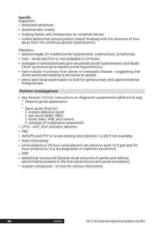
168 Vol. 2 • 10. Acute and subacute by symptom: July 2011
Specifi cInspection:• distended abdomen;• stretched skin marks;• bulging fl anks, and occasionally an umbilical hernia;• visible abdominal venous pattern (caput medusa) with the direction of fl ow
away from the umbilicus (portal hypertension).
Palpation:• splenomegaly (in marked portal hypertension, tuberculosis, lymphoma);• liver – small and fi rm or non-palpable in cirrhosis;• enlarged in schistosomiasis (pre-sinusoidal portal hypertension) and Budd-
Chiari syndrome (post-hepatic portal hypertension);• hard nodular in primary liver cancer or metastatic disease – suggesting that
direct peritoneal seeding is the cause of ascites; • pelvic and rectal examination to look for genitourinary and gastrointestinal
malignancies.
Perform investigations
• See Section 7.4.3 for instructions on diagnostic paracentesis (abdominal tap). ° Observe gross appearance. ° ° Send ascitic fl uid for:
◊ protein (albumin level) ◊ cell count (WBC, RBC) ◊ Gram stain, AFB, and culture ◊ cytology (if malignancy suspected).
• LFTs – AST, ALP; bilirubin; albumin• FBC• INR (PT) and PTT or crude clotting time (Section 7.2.18) if not available• stool microscopy• urine dipstick or 24-hour urine albumin (an albumin level <2.5 g/dl and 24-
hour proteinuria >3 g are diagnostic of nephrotic syndrome)• ESR• abdominal ultrasound (detects small amounts of ascites and defi nes
abnormalities present in the liver parenchyma and portal circulation)• doppler ultrasound – to look for venous obstruction.
Ascites
FINAL Vol. 2 CH 9-21.indd 168FINAL Vol. 2 CH 9-21.indd 168 29/06/2012 15:1529/06/2012 15:15

169Vol. 2 • 10. Acute and subacute by symptom: July 2011
10.9.2 Classify ascites and consider the likely differential diagnosis
Classify the fl uid as a transudate or exudate, and calculate the SAAG
Classify according to protein measurement of the fl uid.• transudate: protein <30 g/litre in peritoneal fl uid• exudate: protein >30 g/litre in peritoneal fl uid
Calculate the SAAG (serum-to-ascites albumin gradient)SAAG = (serum albumin) – (ascitic fl uid albumin)
• A SAAG >1.1 g/dl (11 g/litre) indicates that the patient has portal hypertension: ° cirrhosis ° heart failure ° Budd-Chiari syndrome and schistosomiasis.
• A SAAG <1.1 g/dl (less than 11 g/litre) rules out portal hypertension: ° ascites caused by infectious or neoplastic peritoneal disease ° severe acute pancreatitis ° a low albumin state (nephrotic syndrome and kwashiorkor).
Consult the relevant differential diagnosis table.
DDx: Ascites with SAAG >1.1 (portal hypertension) – transudate
Conditions In favour
Cirrhosis Known risk factor – alcoholism, history of jaundice, chronic HBV or HCV (see Section 11.14) Small nodular liver, jaundiceSigns of chronic liver disease – gynaecomastia, caput medusae, palmar erythema, spider naevi, fl apping tremorUpper gastrointestinal bleeds from varicesPeritoneal fl uid analysis (straw coloured, WBC <250 predominantly mesothelial, RBC <10 000, protein <2.5 g/dl)High bilirubin and INRLow serum albumin
Congestive heart failure Distended jugular veins, heart gallop, pulmonary crackles (pulmonary oedema), hepatomegaly, oedema of lower limbs Peritoneal fl uid analysis – straw coloured, protein >2.5 g/dl, WBC <250Abdominal ultrasound – hepatomegaly, distended IVC with minimal respiratory cycle change
Budd-Chiari syndrome Risk factors for thrombosis (haematological and other malignancies, contraceptives containing estrogen)Consistent features on ultrasound – see below
Schistosomiasis
see Section 11.34
Upper GI bleeding from varicesStool positive for ova Ultrasound – periportal fi brosis, splenomegaly, enlarged veins, collateral vessels
Ascites
FINAL Vol. 2 CH 9-21.indd 169FINAL Vol. 2 CH 9-21.indd 169 29/06/2012 15:1529/06/2012 15:15

170 Vol. 2 • 10. Acute and subacute by symptom: July 2011
DDx: Ascites with SAAG <1.1 (no portal hypertension
Condition In favour
Bacterial peritonitis
see Section 10.7a
Acute abdomen – see Section 10.7a Abdominal painSystemically illPeritoneal fl uid analysis – turbid or purulent; protein >2.5 g/dl; WBC often >1000 and predominantly PMN; RBC <10 000; positive Gram stain and cultureAbdominal X-ray – free air under diaphragm. If perforation of hollow organ suspected, titrate bile and amylase from sample.
SBP in cirrhosis Fever, abdominal pain, encephalopathyPeritoneal fl uid analysis – turbid or purulent WBC >250 Positive Gram stain and culture
Tuberculous peritonitis Constitutional symptoms – low grade fever, weight loss, night sweats, generalized lymphadenopathy, hepatosplenomegaly More common in HIV with evidence of immunosuppressionPeritoneal fl uid analysis – clear, haemorrhagic or chylous; protein >2.5 g/dl; WBC often >500 predominantly lymphocytes; RBC occasionally >10 000Positive AFB or culture (not always), High ESR Ultrasound – hepatosplenomegaly, peritoneal thickening, abdominal lymphadenopathy, micro-abscess in spleen or liver
Neoplasms Systemic symptoms – weight loss, night sweatsPeritoneal fl uid analysis – straw-coloured, haemorrhagic, mucinous or chylous; protein >2.5; WBC often >500 with variable cell types; RBC occasionally >10 000; positive cytology, high ESR Ultrasound – liver mass, abdominal mass or peritoneal thickening
Pancreatitis
see Section 10.7a
History of heavy alcohol intakeEpigastric or central abdominal painPeritoneal fl uid analysis – turbid, haemorrhagic, chylous; protein >2.5 g/dl; variable WBC and RBC counts High serum and ascites amylase levels Ultrasound – oedematous pancreas, may be normal
Nephrotic syndrome
see Section 11.31
Peritoneal fl uid analysis – straw coloured, protein <2.5 g/dl, WBC <250, RBC <10 000Serum albumin <2.5 g/dlUrine dipsticks, proteinuria, and 24-hour proteinuria >3.5 gUltrasound may show enlarged kidneys
Malnutrition
see Section 10.3
Rarely causes clinical ascites in adultsPeripheral oedema is common Peritoneal fl uid analysis – straw coloured, protein <2.5 g/dl, WBC <250, RBC <10 000Serum albumin <2.5 g/dlUrine dipstick – no protein
Visceral leishmaniasis
see Section 11.20
Slow progression of fever, malaise, and weight lossMarked cachexia, splenomegaly, hepatomegaly, jaundiceLow platelets or pancytopaeniaDemonstration of parasite by smear or culture in bone marrow or spleen
Ascites
FINAL Vol. 2 CH 9-21.indd 170FINAL Vol. 2 CH 9-21.indd 170 29/06/2012 15:1529/06/2012 15:15

171Vol. 2 • 10. Acute and subacute by symptom: July 2011
10.9.3 Manage ascites according to causeIt is important to establish the underlying cause of the ascites since the management differs in relation to different causes.
Manage ascites with SAAG >1.1 (portal hypertension) – transudate
General management
• Salt restriction <2 grams (less than half a teaspoon) per day.• Combined oral diuretics – keep the ratio between the 2 drugs constant:
° spironolactone 100 mg increased up to a maximum dose of 400 mg/day PLUS furosemide 40 mg to a maximum of 160 mg/day.
° Caution in rapid diuresis. ° Monitor daily weights and urine output. ° Therapeutic peritoneal tap if indicated – assess for ascites and see box
below. ° Intractable ascites need to be referred for further investigations. ° Refer patients who are not responding for shunt operations or possible liver
transplantation.
Therapeutic paracentesis in portal hypertension (see Section 7.4.3)The amount of fl uid taken out daily by paracentesis should be 2–3 litres.
Exception: occasionally, patients with massive ascites will develop abdominal discomfort and severe shortness of breath and will require a large volume therapeutic paracentesis (4–5 litres) for control of the symptoms.
DiuresisPatients with peripheral oedema tolerate mobilization of a high fl uid volume (2 litres/day) without developing intravascular depletion and azotaemia.
Patients with ascites but without oedema can develop hypovolaemia and acute renal failure if the rate of fl uid removal exceeds 500 ml/day.
Diuretic-resistant ascites is defi ned by a lack of response to maximum doses of spironolactone and furosemide in a patient on a low sodium diet. These patients often need serial large volume therapeutic paracenteses, not exceeding 4–5 litres to avoid intravascular volume depletion and acute renal failure.
Manage cirrhosis
Prevent complications from cirrhosis:• Look for a cause – serology for hepatitis B, C (see Section 11.14).• Advise the patient to avoid alcohol.• Determine the severity of cirrhosis using the modifi ed Child-Turcotte-Pugh
classifi cation (use Table below).• If available, refer for endoscopy to look for oesophageal varices. If varices
present, use a low-dose non-selective beta blocker (e.g. propranolol titrated to achieve a 25% reduction in the heart rate), as primary prevention for upper gastrointestinal bleeding secondary to documented oesophageal varices.
• Consider whether patient has hepatic encephalopathy (see Section 3.4.1).
Ascites
FINAL Vol. 2 CH 9-21.indd 171FINAL Vol. 2 CH 9-21.indd 171 29/06/2012 15:1529/06/2012 15:15

172 Vol. 2 • 10. Acute and subacute by symptom: July 2011
• Consider long-term lactulose, titrated to achieve 2–4 bowel movements per day, if signs of hepatic encephalopathy (see instructions in 8.4).
• Be careful about fl uid management and avoid nephrotoxic drugs (e.g. NSAIDs and aminoglycosides) to prevent hepatorenal syndrome.
• Refer patients with severe cirrhosis, hepatic encephalopathy and metabolic complications to a specialist for management of complications and for evaluation for other therapies.
Scoring to determine the severity of cirrhosis
• The score uses 5 markers of liver disease. Each measure is scored 1–3; with 3 indicating the most severe derangement.
Table: Child-Turcotte-Pugh classifi cation
Measure 1 point 2 points 3 points
Bilirubin (total) <34 µmol/l (<2 mg/dl) 34–50 µmol/l (2–3 mg/dl) >50 µmol/l (>3 mg/dl)
Serum albumin >35 g/l 28–35 g/l <28 g/l
INR <1.7 1.71–2.20 >2.20
Ascites None Suppressed with medication
Refractory
Hepatic encephalopathy
None Grade I–II (or suppressed with medication)
Grade III–IV (or refractory)
Classifi cation A B C
Total points 5–6 7–9 10–15
Manage ascites SAAG <1.1 (no portal hypertension) – transudate
Ascites caused by neoplastic or infectious processes and secondary to acute pancreatitis and a low albumin state should be managed differently. • Treat the primary disease. See Section 10.7.1.4 if peritonitis is suspected.• Avoid sodium restriction and diuresis as this can be harmful and lead to
unwanted intravascular volume depletion and acute renal failure. • May require large volume therapeutic paracentesis (4–5 litres) to manage or
alleviate symptoms of discomfort.
Spontaneous bacterial peritonitis
• Suspect in patients with cirrhosis and ascites presenting with: ° fever ° abdominal pain ° altered mental status ° hepatorenal syndrome.
Ascites
FINAL Vol. 2 CH 9-21.indd 172FINAL Vol. 2 CH 9-21.indd 172 29/06/2012 15:1529/06/2012 15:15

173Vol. 2 • 10. Acute and subacute by symptom: July 2011
Investigations
• Paracentesis (see Section 7.4.3) – WBC count ≥500, neutrophils ≥250 cells/mm3.
Treatment
• Give ceftriaxone 2 grams daily for 5–10 days.• If available, albumin 25% IV 1.5 g/kg on day 1 and 1 g/kg on day 3.
Prevention
• Antibiotic prophylaxis – for all patients with: ° a history of SBP ° a current upper gastrointestinal bleeding episode ° ascitic fl uid albumin level less than 1 g/dl.
• cotrimoxazole (1 double-strength tablet daily); OR • ciprofl oxacin 250–500 mg daily; OR • norfl oxacin 400 mg daily.
Schistosomiasis (see Section 11.34)
Schistosomiasis leads to granulomatous infl ammation and the obstruction of the blood fl ow to the liver.• Referral is usually needed if a shunt operation for portal hypertension is
available upon referral.
Ascites
FINAL Vol. 2 CH 9-21.indd 173FINAL Vol. 2 CH 9-21.indd 173 29/06/2012 15:1529/06/2012 15:15

174 Vol. 2 • 10. Acute and subacute by symptom: July 2011
10.10 Neurological problems
A patient presenting with headache, meningeal signs, change in mental status, seizures, or neurological defi cit could require urgent management. You will need to use different parts of this Section and of the manual to work through a differential diagnosis for the problem.
Use this Section 10.10
• 10.10a Neurological defi cit without meningeal signs (no headache, stiff neck, vomiting – if present, also see Section 10.10b) ° patient with a stroke-like syndrome ° patient with spinal cord problem (myelopathy) ° patient with peripheral motor or sensory nervous system problem ° patient with a cranial nerve abnormality.
• 10.10b Headache ° headache with no abnormal physical fi ndings ° headache with abnormal physical fi ndings (including meningeal signs,
fever, neurological defi cit, seizures).• 10.10c Seizures or convulsions
° seizures due to a systemic illness ° seizures due to intracranial infection or lesion ° chronic recurrent seizures.
Use Section 2 – Quick Check
• for emergency management of a convulsing or comatose patient• for patients with a history of head trauma.
Use Section 3 – Approach to the severely ill patient
• for a patient with a decreased level of consciousness, confusion, intoxication, or agitation (Section 3.4)
• for a patient who is convulsing or in status epilepticus (Section 3.5).
Use Section 10.12 – Eye problems
• for acute visual loss.
Key points• It is possible to recognize and treat common neurological problems without the use of complex diagnostic
tests. • HIV-related conditions of the central nervous system and spinal cord are common. • Patients recently started on ART may develop an immune reconstitution infl ammatory syndrome (IRIS) that
complicates the clinical picture.• Toxoplasmosis can be prevented with cotrimoxazole prophylaxis.• Secondary prophylaxis with fl uconazole is mandatory after an episode of cryptococcal meningitis.
Neurological defi cit
FINAL Vol. 2 CH 9-21.indd 174FINAL Vol. 2 CH 9-21.indd 174 29/06/2012 15:1529/06/2012 15:15

175Vol. 2 • 10. Acute and subacute by symptom: July 2011
10.10a Neurological defi cit (without meningeal signs)
In this section:10.10a.1 Clinical approach to a patient with neurological defi cit10.10a.2 Classify the neurological defi cit and consider the likely differential diagnosis10.10a.3 Stroke-like syndrome (with DDx table)
• Approach to HIV-infected patients with stroke-like syndrome10.10a.4 Spinal cord problem (myelopathy) (with DDx table)10.10a.5 Peripheral motor or sensory nervous system problem (with DDx table)10.10a.6 Peripheral neuropathy (with DDx table)10.10a.7 Common cranial nerve palsies and their differentials
This Section addresses the management of a patient who presents with a neurological defi cit and without meningeal signs. Use this Section for a patient presenting with the following symptoms:• motor defi cit (weakness, paralysis, loss of balance, diffi culty speaking)• sensory defi cit (tingling, numbness, pain)• cranial nerve defi cits (facial weakness, vertigo, double vision).
For the management of patients with headache or meningeal signs, also refer to Section 10.10b Headache.
For the management of patients with cognitive problems, see Sections 3.4 and 10.11.
10.10a.1 Clinical approach to a patient with neurological defi cit
Step 1: Use Quick Check. Ensure that there are no serious or life-threatening conditions. Use the Quick Check for management of life-threatening conditions, such as coma and convulsions.
Step 2: Take a history and perform a physical examination.If the patient has diffi culty speaking or is confused, obtain a good history from the family.
Step 3: Assess the patient’s HIV status.
Step 4: Classify the defi cit using the table Classifi cation of motor and sensory neurological defi cit.
Step 5: Use the DDx tables to work through a differential diagnosis: • DDx: Stroke-like syndrome• DDx: Spinal cord problem (myelopathy)• DDx: Peripheral motor or sensory nervous system problem• DDx: Peripheral neuropathy (distal – DSPN)• DDx: Cranial nerve abnormalities
Step 6: Perform investigations.Perform investigations according to the differential diagnosis and availability of tests. It is possible to diagnose and manage neurological problems without the use of complex diagnostic tests.
Step 7: Initiate treatment and monitor response.
Neurological defi cit
FINAL Vol. 2 CH 9-21.indd 175FINAL Vol. 2 CH 9-21.indd 175 29/06/2012 15:1529/06/2012 15:15

176 Vol. 2 • 10. Acute and subacute by symptom: July 2011
Quick Check
Ensure that there are no serious or life-threatening conditions. Use the Quick Check for patients with coma and convulsions. Ask specifi cally about any history of head injury.
History
• History of the following presenting complaints, including onset (sudden or insidious) and duration: ° motor changes (e.g. diffi culty combing hair, writing, squatting, climbing
stairs) ° sensory changes ° vision, hearing, or speech problems ° diffi culty with balance or walking ° meningeal signs – headache, stiff neck ° associated fever, constitutional symptoms ° associated nausea, vomiting, dizziness ° associated seizures ° change in consciousness, behaviour, mood ° change in memory or cognition ° bowel and bladder disturbances.
• Medical history: ° risk factors for stroke (hypertension, diabetes, heart disease, obesity,
bleeding disorders, clotting, pregnancy) ° HIV infection (CD4 count, ART) ° TB infection (current TB symptoms, previous TB, or TB contact) ° malignancy ° syphilis ° history of head injuries ° medications (e.g. aspirin, anticoagulants, oral contraceptive pill).
Examination
General signs
• vital signs• signs of immune compromise• level of consciousness• neck stiffness, meningeal signs (if present, also see Section 10.10b).
Examine the fundi
• Perform fundoscopy (see Section 10.12).• Look for loss of spontaneous venous pulsations, blurred optic disc margins,
and elevated optic head as evidence of papilloedema.
Assess higher functions
• orientation to person, place, or time• memory• speech
° diffi culty articulating words (dysarthria) ° diffi culty understanding or expressing words (aphasia)
• gait, ability to walk
Neurological defi cit
FINAL Vol. 2 CH 9-21.indd 176FINAL Vol. 2 CH 9-21.indd 176 29/06/2012 15:1529/06/2012 15:15

177Vol. 2 • 10. Acute and subacute by symptom: July 2011
• assess for cognitive defi cits (see Sections 3.4 and 10.11) ° In PLHIV, assess for possible HIV-associated cognitive disorders
Examine cranial nerves
• See Table: Common cranial nerve palsies and their differentials.
Examine motor system
• power, tone, refl exes.
Examine the sensory system
• Examine for touch, pain, temperature, and position sense.• Screen for peripheral neuropathy
° In PLHIV, assess for HIV-associated distal sensory polyneuropathy
Examine cerebellar function
• Assess for intention tremor, hypotonia, nystagmus, broad-based gait, and incoordination.
Assess HIV statusThere are a number of neurological conditions associated with immune suppression from HIV infection. The HIV status of a patient will impact on the differential diagnosis and management.
10.10a.2 Classify the neurological defi cit and consider the likely differential diagnosisNeurological defi cit can result from a number of different conditions that cause injury to the brain, spinal cord, or peripheral nerves. Classifying the defi cit will help you to work through a differential diagnosis and determine the most likely cause.
Table: Classifi cation of motor and sensory neurological defi cit
Condition Features in favour
Stroke-like syndrome(due to injury to the brain – results in upper motor neuron defi cit)
Onset acute (stroke) or subacute Unilateral Weakness or numbness in face, arm, or legDiffi culty speaking, swallowingDizziness, blurred vision, loss of balanceConfusionCerebellar signs
Spinal cord problem (myelopathy)(due to injury to the spinal cord)
Slow, subacute onset, or acute trauma to the spinal cord. May present abruptly or gradually.Bilateral or unilateralParaparesis or quadriparesisLoss of motor function and sensation (touch, temperature)Loss of bladder or bowel control
Peripheral motor or sensory nervous system problem (neuropathy) (due to injury to the peripheral nerves)
Acute or subacute onsetMotor – weakness, cramps, spasmsSensory – tingling, numbness, painAutonomic – BP instability, reduced sweating, incontinence, sexual problemsHIV positive, alcohol use, malnutrition
Cranial nerve problems Isolated or multiple cranial nerve palsiesEye and vision changes Diffi culties with balance or hearing
Neurological defi cit
FINAL Vol. 2 CH 9-21.indd 177FINAL Vol. 2 CH 9-21.indd 177 29/06/2012 15:1529/06/2012 15:15

178 Vol. 2 • 10. Acute and subacute by symptom: July 2011
Once you have classifi ed the defi cit, use the DDx tables below to fi nd a likely cause.• DDx: Stroke-like syndrome• DDx: Spinal cord problem (myelopathy)• DDx: Peripheral motor or sensory nervous system problem• DDx: Peripheral neuropathy (distal sensory peripheral neuropathy)• DDx: Cranial nerve abnormalities
Perform investigations
Perform investigations according the likely differential diagnosis.• Lumbar puncture to examine the CSF (see Section 7.4 for Procedure and
Section 10.10b for further work-up): ° perform fundoscopy to exclude papilloedema; ° this is especially important if HIV infection; ° important if you suspect meningitis or subarachnoid bleed; ° may be considered in the presence of focal signs if no signs of impending
herniation (see Section 10.10.b); ° may be considered for headache, for more information see Section 10.10b; ° CSF for opening pressure, appearance, microscopy for cells and other
microorganisms, chemistry, cryptococcal antigen, rapid syphilis test, TB.• Bloods:
° FBC ° cryptococcal Ag ° toxoplasma IgG ° rapid syphilis test.
• Spinal X-rays: ° for neurological defi cit with a spinal level.
• CT scan (if indicated and available).
Initiate treatment and monitor response
Management of patients with a neurological defi cit and no meningeal signs will depend on the availability of CT scanning. As a rule of thumb, 80–90% of patients can be managed at district level without CT scan. Very few district hospitals have CT scanners, but CT scanning may be available at a referral hospital for specifi c patients.
If there is no access to CT scanning and LP is contraindicated (see Section 10.b Headache), then empirical therapy should be started for TB and toxoplasmosis or any other diagnosed conditions, especially in the case of HIV-infected patients.
10.10a.3 Stroke-like syndromeProblems affecting the brain or central nervous system can cause stroke-like syndromes due to damage to the upper motor neurons. The differential for a patient presenting with a stroke-like syndrome is broad; however, in settings of high HIV prevalence, certain conditions occur more commonly.
Neurological defi cit
FINAL Vol. 2 CH 9-21.indd 178FINAL Vol. 2 CH 9-21.indd 178 29/06/2012 15:1529/06/2012 15:15

179Vol. 2 • 10. Acute and subacute by symptom: July 2011
Use the DDx table below to work through a differential diagnosis for a patient presenting with a stroke-like syndrome.
DDx: Stroke-like syndrome*
Condition In favour
Cerebrovascular accident
Ischaemic or haemorrhagic stroke
Sudden onsetHypertensive, diabetic, history of smokingUnilateral weakness or numbness in face, arm, or legDiffi culty speaking or swallowingDizziness or blurred visionDiffi culty walking or problem with balanceConfusionNo progression of defi cit (may have some improvement over time)
Intracranial masses
Cerebral toxoplasmosissee Section 11.40
Subacute onset over days to weeksHeadacheWith or without fever Focal signs Dull affect, impaired level of consciousnessSeizuresCT scan – multiple ring enhancing lesionsHIV infection – CD4 <100
Cryptococcoma see Section 11.15
Subacute onset over days to weeksFever, malaise, headacheSkin lesions resembling molluscum contagiosumLP – high opening pressureHIV infection – CD4 <50CSF: increased lymphocytes, low glucose, high protein, + India ink, + CRAGN.B.: CSF may be normal.
Tuberculoma Gradual onset over days to weeksFocal neurological defi citEvidence of TB elsewhereCSF – increased lymphocytes, low glucose, high protein
Bacterial brain abscess May result from contiguous spread or bacteraemia Gradual onset over weeksHeadache, neck stiffness, altered mental status, signs of increased ICPFocal defi cits beginning days after headache, seizuresLP should be avoided
Tumours (benign tumour, lymphoma, metastases)
Insidious onsetCranial nerve palsies, mental status changeFocal neurology
Other infections
Progressive multifocal leucoencephalopathy (PML)
ClumsinessVisual changes, hemiparesis, dysarthria, aphasia; seizuresProgression over 1–9 monthsHIV infection – CD4 < 100
Neurological defi cit
FINAL Vol. 2 CH 9-21.indd 179FINAL Vol. 2 CH 9-21.indd 179 29/06/2012 15:1529/06/2012 15:15

180 Vol. 2 • 10. Acute and subacute by symptom: July 2011
Condition In favour
Neurocysticercosissee Section 11.7
SeizuresHeadache, nausea, vomiting, altered mental statusEndemic areaCSF – high WBC, eosinophilsSkull X-ray – multiple calcifi ed cystsCT – multiple calcifi ed cysts and active fl uid-fi lled cysts
Neurosyphilis – meningovascularsee Section 11.37
Subacute onset of headache, dizziness, personality changes followed by stroke-like syndromeCSF – normal glucose high protein, high WBC + CSF VDRL (not RPR or FTA) VDRL or RPR
Viral encephalitis (herpes, CMV)see Sections 11.8 and 11.15
Acute or subacute onsetProdrome of high fever, headache, nausea, lethargy, myalgiaFrontal or temporal lobe focal signsSeizures, confusion, altered level of consciousness, bizarre behaviourCSF – increased lymphocytes, normal glucose, mildly elevated protein
*Other disorders with neurologic manifestations: American or African human trypanosomiasis (Sections 11.141 and 11.42), and schistosomiasis (Section 11.34).
Approach to HIV-infected patients with stroke-like syndrome
Assess patient for clinical signs and symptoms of disseminated infections such as TB, syphilis, cryptococcal infection.• Investigate according to fi ndings:
° CD4 count ° toxoplasma serology ° cryptococcal serology (serum or CSF) ° syphilis serology ° platelet count/coagulation studies ° investigations for TB if suggestive – CXR, sputum AFB, lymph node FNA.
• CD4 count and degree of immunosuppression help guide diagnosis.• Toxoplasmosis or TB are common causes:
° TB is more likely if there are signs of TB elsewhere. ° Toxoplasmosis is unlikely if CD4 >200.
Commence empirical treatment without delay
• If a CT scan is immediately available: ° Interpret CT fi ndings. ° If a lumbar puncture is safe (see Section 10.10b), do CSF investigations. ° Based on CT or CSF fi ndings, treat for the most likely cause (if uncertain,
always cover for toxoplasmosis). ° If no response to treatment – review diagnosis.
• If a CT scan is not immediately available: ° Weigh the risks of a lumbar puncture versus empirical therapy. ° Initiate empirical treatment for toxoplasmosis and TB (if evidence of TB
infection elsewhere in body); ° Review patient and revise diagnosis depending on CD4 count, results of
other investigations, subsequent CT scan, and response to treatment. ° Consider referral for CT if available and patient not responding to empirical
treatment.
Neurological defi cit
FINAL Vol. 2 CH 9-21.indd 180FINAL Vol. 2 CH 9-21.indd 180 29/06/2012 15:1529/06/2012 15:15

181Vol. 2 • 10. Acute and subacute by symptom: July 2011
Table: Neurological conditions according to immune status
HIV-negative or CD4 >500(but all can also occur with lower CD4 count)• benign and malignant brain tumours• CVA• neurosyphilis• tuberculoma or TB meningitis• neurocysticercosis• secondary malignancy or brain metastasesCD4 200–500• HIV-associated neurocognitive disorders
(HAND)
CD4 <200 • toxoplasmosis (usually CD4 <100)• tuberculoma or TB meningitis• primary CNS lymphoma (usually CD4 <50)• CVA – intracerebral haemorrhage related to
thrombocytopenia• progressive multifocal leukoencephalopathy• bacterial causes/abscesses • cryptococcal meningoencephalitis• neurocysticercosis• HIV-associated neurocognitive disorders
(HAND)
10.10a.4 Spinal cord problem (myelopathy)Problems affecting the spinal cord cause damage to the lower motor neurons and sensory nerves. The differential for myelopathy is broad; however, in settings of high HIV prevalence, certain conditions occur more commonly than others.
DDx: Spinal cord problem (myelopathy)
Condition In favour
Compressive
Spinal cord compression (herniated disc, tumour – benign, malignant, or metastatic – abscess)
Slow onset Back painNumbness or tingling in toes, fi ngers, or buttocksWeakness, unsteadiness, diffi culty walkingUrine or faecal retention or incontinenceEvidence of primary cancer (breast, lung, prostate, lymphoma)
Spondylosis(osteoarthritis of the spine)
Slow onsetPain or stiffness in neck, thoracic, or lumbar spine – radiation down arm or leg, worse in the morning, worse with movementNumbness or tingling in the arms, legs, hands, or feetWeakness, unsteadiness, diffi culty walkingLoss of bladder or bowel control
Infectious/infl ammatory
Tuberculosis of the spinesee Section 15
Onset over monthsBack pain (lasting weeks to months), muscle spasmFeverWeight lossNight sweatsSpinal deformity (kyphosis)
HIV-associated myelopathy Slow onsetProgressive weakness or stiffness in the legs (sometimes arms)Sensory loss, sphincter dysfunction, incontinenceIncreased refl exes, up-going plantar response (Babinski)Erectile dysfunction in menDiffi culty walkingAdvanced HIV diseaseAssociated HIV dementia or peripheral neuropathy
Neurological defi cit
FINAL Vol. 2 CH 9-21.indd 181FINAL Vol. 2 CH 9-21.indd 181 29/06/2012 15:1529/06/2012 15:15

182 Vol. 2 • 10. Acute and subacute by symptom: July 2011
Condition In favour
Transverse myelitis(herpes, varicella, CMV, HIV, TB, polio, rabies, measles, syphilis, hepatitis, schistosomiasis, bartonella, mycoplasma, MS, paraneoplastic, AV malformation)see Section 11 for more on specifi c diseases
Acute onsetBack pain Sensation of a tight band around the trunkNumbness or tingling below a certain spinal levelWeakness in legs (arms involved less often) Muscle spasmsBowel and bladder dysfunctionAssociated headache, fever, loss of appetiteOther symptoms depending on the cause
Tertiary syphilissee Section 11.37
Slow onsetNumbness or tingling in the hands or feetWeakness of limbs, unsteadiness, wide-based walkLoss of refl exes, incontinenceMemory loss, psychiatric problems, visual loss
Metabolic
Vitamin B12 defi ciency Subacute onsetMegaloblastic anaemiaNumbness or tingling in the hands or feetWeakness in legs, ataxia, wide-based walkAbsent ankle refl exesPoor joint position and vibration sensePeripheral neuropathyDementia, depression
10.10a.5 Peripheral motor or sensory nervous system problemProblems affecting the peripheral nervous system can cause motor or sensory disturbances, and occur as a result of damage to the peripheral nerves and nerve roots. The differential for a patient presenting with weakness, numbness, tingling and pain is broad. However, in settings of high HIV prevalence, certain conditions occur more commonly due to the effect of HIV on the nerves, as well as the effect of opportunistic infections, neoplasms, and medications.
The most common conditions are mononeuropathies (focal disorders affecting a single nerve or nerve group) and radiculopathies (disorders of the nerve roots).
Different disorders can be roughly distinguished by whether or not they are focal or multifocal, symmetrical or asymmetrical, and whether they are primarily sensory or have a motor or autonomic component.
On taking the patient’s history, the following information is important:
• onset and progression of symptoms• symmetrical or asymmetrical, distal or proximal, focal or multifocal• bladder control problems• sweating, temperature or pulse instability (autonomic involvement)• HIV infection (if positive, CD4 count)• exposure to drugs (INH, d4T, ddI) or toxins (alcohol, lead).
On examination, pay particular attention to the following:
• sensory function (pain, light touch, vibration, proprioception)• motor function (power, tone, refl exes)
Neurological defi cit
FINAL Vol. 2 CH 9-21.indd 182FINAL Vol. 2 CH 9-21.indd 182 29/06/2012 15:1529/06/2012 15:15

183Vol. 2 • 10. Acute and subacute by symptom: July 2011
• spinal examination• sphincter function• cranial nerves abnormalities.
Investigations
• CSF analysis may be helpful for distinguishing between various conditions that have involvement at the spinal cord level (see differential diagnosis table).
• In the absence of sophisticated nerve conduction tests, diagnosis is commonly based on clinical context and pattern recognition.
• Specifi c investigations according to the clinical suspicion of underlying problems.
DDx: Peripheral motor or sensory nervous system problem
Condition In favour
Focal or asymmetrical defi cits
Compression of nerve or nerve root (lymphoma, TB, carpal tunnel syndrome, bleed)
Weakness and sensory disturbance in the distribution of affected nerve and indicative of the level of the lesion
Mononeuritis multiplex (hepatitis B, hepatitis C, HIV, CMV, VZV, leprosy)
Pain, weakness, and paraesthesias in the distribution of affected nerve or nervesPain over areaWeakness of related musclesIf CD4 <50, can have severe form affecting multiple nerves of the shoulder girdle
Polyradiculopathy (commonly CMV, also VZV, lymphoma, syphilis)
Radiating back pain Loss of feeling and paraesthesias in lower limbsReduced or absent refl exes in lower limbsFlaccid paralysis with no specifi c spinal “level”Urine retention, loss of bowel functionLow CD4 count (usually <50)CSF – high polymononuclear cell, normal glucose, high proteinEvidence of CMV disease elsewhere (retinitis, GE, encephalopathy)
Symmetrical neuropathy (motor or sensory)
Distal symmetrical sensory polyneuropathy (DSPN) (HIV, medication, nutritional defi ciencies, alcohol, diabetes)
Distal, symmetrical – glove and stocking distributionPainful tingling and numbnessUsually no motor weakness Other causative factors have been excludedCSF – no cells, N glucose See below for more information
Toxic neuropathy(toxins, solvents, insecticides, alcohol, drugs including heroin and amphetamines)
Presentation depends on the agent: motor, sensory, or both Can affect cranial nervesHistory of exposure to toxinOnset related to exposure to toxin
Chronic infl ammatory demyelinating polyneuropathy (CIDP)
Gradual onset (over months)Sensory and motor defi cit with no specifi c “level”Responds to steroidsCD4 200 – 500CSF – increased mononuclear cell, normal glucose, high protein
Neurological defi cit
FINAL Vol. 2 CH 9-21.indd 183FINAL Vol. 2 CH 9-21.indd 183 29/06/2012 15:1529/06/2012 15:15

184 Vol. 2 • 10. Acute and subacute by symptom: July 2011
Condition In favour
Acute infl ammatory demyelinating polyneuropathy (AIDP)e.g. Guillain-Barré syndrome
Acute (over hours to weeks) Ascending, painless, fl accid weakness Sensory loss distallyArefl exiaCSF – normal WCC, high protein
Tetanus Recent open woundIrritabilityJaw or neck stiffness (lockjaw)Spasm of neck, back, abdominal muscles, respiratory musclesBrisk refl exesAutonomic dysfunction: hypertension, tachycardia, high temperature, sweatingNormal level of consciousness
Botulism Diffi culty swallowing or speakingFacial weaknessDouble visionTrouble breathingNausea, vomiting, and abdominal crampsParalysis
Symmetrical neuropathy (motor or sensory)
Rabies see Section 11.30
Wild animal, unimmunized domestic animal, or bat bite (bat bite may not be remembered)FeverHeadacheAltered mental state, insomnia, agitation, confusion, hallucinationsHypersensitivity or spasms in response to stimuli (noise, touch, visual) HydrophobiaExcessive salivationParalysisPain or paraesthesiaAutonomic dysfunction: big pupils, increased saliva, sweat, and tears
Acute poliomyelitis* Muscle weakness, headache, stiff neck, feverSpinal poliomyelitis: paralysis of shoulder girdle often precedes intercostal and diaphragmatic paralysis Bulbar poliomyelitis: facial weakness, dysphagia, dyspnoea, nasal voice, inability to swallow saliva, weak sternocleidomastoid and trapezius muscles. Can progress to respiratory paralysis.
Systemic causes
Hypokalaemiasee Section 5.2
Generalized motor weakness, atrial or ventricular arrhythmiasHyporefl exia ECG changes – ST depression, fl attened (or absent) T waves, U waves (positive defl ection after the T wave), prolonged P–R intervalLab: very low potassium Chronic diarrhoea
* Acute fl accid paralysis in person <15 years or acute paralytic illness at an age where polio is suspected should be reported immediately and investigated (see Section 21).
Neurological defi cit
FINAL Vol. 2 CH 9-21.indd 184FINAL Vol. 2 CH 9-21.indd 184 29/06/2012 15:1529/06/2012 15:15

185Vol. 2 • 10. Acute and subacute by symptom: July 2011
10.10a.6 Peripheral neuropathy (See also Section 13.8 – Peripheral neuropathy as an ART toxicity.)
Peripheral neuropathy is a term used to describe dysfunction in one or more of the peripheral nerves. In this section, the term "peripheral neuropathy" (PN) is used specifi cally to refer to distal symmetrical sensory polyneuropathy (DSPN) – a condition that starts at the base of the feet and progresses upwards causing numbness, paraesthesias, and pain.
There are a number of different causes of PN including: nerve compression, autoimmune or infl ammatory conditions, toxin- or drug-induced damage, and inherited conditions. It is important to look for the cause as early detection and treatment will stop and may even reverse the progression of symptoms. Pain due to PN may become irreversible if left for too long.
In settings with a high HIV and TB prevalence, PN is a common problem that can be irreversible and debilitating for patients who are affected by it. Peripheral neuropathy is the most frequent neurological complication of HIV infection. This could be either primarily due to HIV infection (after all other co-morbid causes have been excluded) or secondary to ART.
DDx: Peripheral neuropathy
Condition In favour
HIV infection Symmetrical, glove-stocking distribution:• starts in toes and balls of feet• spreads to rest of foot, ankle, and up the legs• in severe cases fi ngers or hands involved
Sensory changes (often worse at night):• numbness or increased sensitivity to touch • progresses to tingling, burning, or pain• impaired vibration sensation and temperature perception• in severe cases: super-sensitivity to touch (unable to wear shoes or lie
under bed sheets)
Motor function usually preserved:• weakness uncommon• reduced or absent ankle jerk refl exes• walking and balance may appear abnormal due to pain• if rapidly progressive weakness and high lactate, consider HIV-
associated neuromuscular weakness syndrome associated with lactic acidosis
Medicines, substance use Patient taking one or more offending medicine:• most common: INH, d4T, or ddI • other medicines: ethambutol, ethionamide, dapsone, vincristine,
thalidomide, lithium carbonate, metronidazole, high-dose vitamin B6, cisplatin
• Heroin or amphetamine use
Symptoms:• similar to peripheral neuropathy caused by HIV (see above)• reports of deep aching pain across the top of the foot
High lactate:• associated with NRTI-mediated neuropathy
Neurological defi cit
FINAL Vol. 2 CH 9-21.indd 185FINAL Vol. 2 CH 9-21.indd 185 29/06/2012 15:1529/06/2012 15:15

186 Vol. 2 • 10. Acute and subacute by symptom: July 2011
Condition In favour
Diabetes Glove-stocking distribution: • starts in toes or soles• spreads to ankles and calf
Poor glycaemic (blood sugar) control
Evidence of other diabetic complications: • peripheral vasculopathy, nephropathy, retinopathy
Sensory changes:• numbness or pain
Nutritional defi ciency Low serum vitamin B12Clinically malnourished
Alcohol History of alcohol overuseOther evidence of chronic alcohol abuse
Other infections:(VZV, CMV, hepatitis C, syphilis, Cryptococcus, leprosy)
Other evidence of the infection that is causing the neuropathy(see Section 11 for more on specifi c diseases)
Diagnosis
• The brief peripheral neuropathy screen (BPNS) should be used to assess for HIV-associated distal sensory polyneuropathy (DSPN). ° Subjective – ask for symptoms of pain, burning, pins and needles, or
numbness in the hands or feet ° Objective – assess sensation of the hands and feet, vibration sense, and
ankle tendon refl exes. Treatment
• Treat or remove the cause.• Give thiamine if known history of alcohol use.• If the patient is on TB treatment or on ART – refer to Sections 15 and 13
respectively for management and drug substitution.• After discontinuation of the offending drug, it may take a few weeks for the
pain to decrease, and in that time the pain may even worsen.• Peripheral neuropathy primarily due to HIV infection may improve once the
patient is put on effective ART: ° also give one pyridoxine 50 to 75 mg daily.
• Pain control by anticonvulsants and tricyclics for neuropathic pain: ° amitriptyline is widely used for peripheral neuropathy; dose: start at 25–75
mg at night (increase as needed if side-effects are tolerated) to a maximum dose of 300 mg daily; OR
° carbamazepine – start at 100 mg twice daily and increase to a total of up to 1600 mg daily.
° If HIV-positive and on ART with poor response to amitriptyline, then the use of gabapentin is recommended: ◊ Dose: day 1, an initial dose of 300 mg daily; day 2, 300 mg twice daily; day 3, 300 mg 3 times daily. Dose can then be titrated 100 mg increments every 3 days as needed, to a maximum of 3600 mg daily (given as either 1200 mg 3 times daily or 900 mg 4 times daily).
Neurological defi cit
FINAL Vol. 2 CH 9-21.indd 186FINAL Vol. 2 CH 9-21.indd 186 29/06/2012 15:1529/06/2012 15:15

187Vol. 2 • 10. Acute and subacute by symptom: July 2011
• Analgesics including opiates may provide some relief – see Section 17 for a stepwise approach to controlling pain. ° Low-dose opioids may be required for relief of neuropathic pain, following
trial with tricylic antidepressant agents.
Prevention
• It is desirable to give pyridoxine (vitamin B6) 10 mg daily to patients taking INH as prophylaxis or as part of TB treatment.
• Avoid the use of stavudine, see Section 13.• Educate and monitor patients taking stavudine and substitute if any toxicity.• Control diabetes and hypertension.
10.10a.7 Common cranial nerve palsies and their differentialsProblems affecting the cranial nerves can occur as a result of central or peripheral nervous system conditions. See the table below for likely signs and symptoms.
Cranial nerve palsies Presentation Common causes
Second nerve palsy(optic nerve)
Acute visual loss see Section 10.12 Eye problems
Third nerve palsy(occulomotor nerve)
Fixed, dilated pupil PtosisEye looks downwards and outwards Double vision
Ruptured aneurysmSubarachnoid haemorrhageHIV vasculopathy Diabetes mellitusSyphilis
Fourth nerve palsy(trochlear nerve)
Double visionRestricted eye movement up in adductionConcurrent third and sixth nerve palsyRecent head injury
Often idiopathicHead injuryAneurysms TumoursMultiple sclerosis
Fifth nerve palsy(trigeminal nerve)
Pain or paraesthesias in faceWeak jaw clenchingWasting of temporalis and masseter muscles
Trigeminal neuralgiaHerpes zoster
Sixth nerve palsy(abducens nerve)
Double visionUnilateral paresis of eye abduction
Raised intracranial pressureTumours or massesMeningitis
Seventh nerve palsy (facial nerve)
Facial muscle weakness Lower motor neuron (LMN) = forehead paralysed Upper motor neuron (UMN) = forehead is spared
Ramsay Hunt = facial weakness + vertigo + loss of taste
LMN:Bell's Palsy – idiopathic Ramsay Hunt – varicella zoster (treat with steroid + aciclovir)
UMN:StrokeTumour
Eighth nerve palsy(vestibulo-cochlear nerve)
Onset acute, chronic or recurrentSensorineural hearing loss Vertigo (hallucination of movement)Associated nausea, vomiting, tinnitus, Nystagmus
Brainstem strokeMenieres diseaseAminoglycoside antibioticsLabyrinthitis (acute onset)Benign paroxysmal positional vertigo (recurrent)
Neurological defi cit
FINAL Vol. 2 CH 9-21.indd 187FINAL Vol. 2 CH 9-21.indd 187 29/06/2012 15:1529/06/2012 15:15

188 Vol. 2 • 10. Acute and subacute by symptom: July 2011
10.10b Headaches
In this section:10.10b.1 Clinical approach to a patient with headache10.10b.2 Consider the likely differential diagnosis
• DDx: Primary headache• DDx: Secondary headache
10.10b.3 Treatment of specifi c conditions• Acute bacterial meningitis • Tuberculosis meningitis
10.10b.4 Symptom management of headache
This Section provides an approach to the patient with headache, with or without meningeal signs or fever. Headache is a common complaint in clinical practice, and it is important to distinguish benign headaches from those due to a serious condition. If the patient reports the headache as the “fi rst or worst” of its kind, then it is more likely to be due to a serious cause.
Headaches are most thoroughly classifi ed by the International Headache Society's International Classifi cation of Headache Disorders (ICHD) 2004. This classifi cation is accepted by WHO.
1. Primary headaches
• migraine • tension-type headache • cluster headache and other trigeminal autonomic headaches• other primary headaches (e.g. primary cough headache, exertion headache,
headache associated with sexual activity).
2. Secondary headaches
• head or neck trauma • cranial or cervical vascular disorder • non-vascular intracranial disorder • substance use or its withdrawal • infection • homeostasis • facial pain attributed to a disorder of the cranium, neck, eyes, ears, nose,
sinuses, teeth, mouth, or other facial or cranial structures • psychiatric disorders.
3. Cranial neuralgias, central and primary facial pain, and other headachesSecondary headaches may be further sub-divided, as in the differential diagnosis tables below, into:
• headache with meningeal signs, with or without fever• headache with no meningeal signs, with or without fever• headache with no meningeal signs and no fever• extracranial causes of headache.
Headache
FINAL Vol. 2 CH 9-21.indd 188FINAL Vol. 2 CH 9-21.indd 188 29/06/2012 15:1529/06/2012 15:15

189Vol. 2 • 10. Acute and subacute by symptom: July 2011
10.10b.1 Clinical approach to a patient with headache
Step 1: Perform Quick Check.Use the Quick Check and ensure that there are no serious or life-threatening conditions, such as altered consciousness or convulsions.
Step 2: Take a history and examine the patient.Determine whether the onset is acute or chronic. Look for meningeal signs, fever, rash, and agitation or confusion.
Step 3: Assess the patient’s HIV status.
Step 4: Perform investigations.If in doubt, do a lumbar puncture (LP). If the patient is HIV-positive, do an LP. There are few contraindications to an LP, and the diagnostic benefi t is signifi cant. Ask whether a CT scan will change management and whether it is available through referral.
Step 5: Work through the differential diagnosis using the DDx tables:• DDx: Headache with no abnormal physical fi ndings• DDx: Headache with abnormal physical fi ndings.
Step 6: Initiate management and monitor the patient’s response.• Flow chart 1: Approach to headache in a patient with HIV infection or unknown HIV status and
suspected CN infection.• Flow chart 2: Approach to headache in HIV-negative patient with suspected CN infection.
Take a history and examine the patientA good history and examination will help determine the cause of the headache. Headache syndromes show a typical pattern in the history and an absence of unexplained physical signs. As noted, a headache that is the fi rst of its kind or the worst of its kind is more likely to be from a serious cause.
History
• History of presenting complaint: ° onset (sudden or gradual) ° duration (hours, days, weeks, months) ° stiff neck ° associated dizziness, nausea, or vomiting ° motor or sensory abnormalities ° vision, hearing, or speech problems ° trouble with balance or walking ° associated seizures, change in level of consciousness ° change in behaviour, mood, memory, or cognition ° constitutional symptoms (fever, weight loss, night sweats).
• Medical history: ° risk factors for stroke (hypertension, diabetes, heart disease, obesity,
bleeding disorders, clotting, pregnancy) ° TB, HIV (if positive, CD4 count and ART history) ° malignancy ° syphilis ° history of head injury ° current medications.
• Exposure to infectious diseases: ° TB contact ° Living in or travelled to endemic area (malaria, dengue, rickettsia).
Headache
FINAL Vol. 2 CH 9-21.indd 189FINAL Vol. 2 CH 9-21.indd 189 29/06/2012 15:1529/06/2012 15:15

190 Vol. 2 • 10. Acute and subacute by symptom: July 2011
Examinations
General signs
• vital signs• level of consciousness, using AVPU or Glasgow Coma Scale • rash:
° Look for petechiae or purpura. It may look like bruises and can be diffi cult to see on dark skin. Check paler areas such as palms, soles, torso, conjunctiva, palate.
° Look for signs of viral infection, sinusitis, toothache, or ear infection.
Meningeal signs
• neck stiffness• Kernig’s sign (resistance to straightening of the leg while hip is fl exed)• Brudzinski’s sign (fl exing the neck causes fl exion of the hip or knee).
Fundoscopy for papilloedema See Section 10.12.
Higher functions
• orientation to person, place, or time• speech, cranial nerves, motor and sensory system, cerebellar function (as
indicated). See Section10a Neurological defi cit.• ability to walk.
Assess the patient’s HIV status. HIV infection infl uences the likely differential diagnosis for headache, as there are a number of opportunistic infections that can present with headache. Whenever possible, an HIV test should be done, and the CD4 count should be checked if a patient is HIV-positive.
Perform investigations
Bloods
• full blood count with white cell differential count to look for infection
• malaria smear if endemic area (coinfection with malaria and meningitis is common)
• ESR if temporal arteritis suspected.
Lumbar puncture (LP)For more on how to perform an LP, see Section 7.4 Procedures.
LP is recommended in the following situations:
• If blood or pus is suspected in subarachnoid space: ° sudden onset severe headache and suspected subarachnoid bleed ° recent onset headache with fever or neck stiffness or pain and meningitis is
suspected.• If a headache is the fi rst of its kind or the worst of its kind, it will need
investigation after considering a migraine.• If the patient is HIV-positive with signs and symptoms of meningitis.
LP in patients with HIV infectionLumbar puncture carries no excessive risk in patients with TB meningitis or cryptococcal meningitis, even in the presence of papilloedema, vomiting, or an altered mental state. In advanced HIV these infections are very common, and you should have a low threshold for performing an LP in these patients. See fl ow diagram below for management approach.
Headache
FINAL Vol. 2 CH 9-21.indd 190FINAL Vol. 2 CH 9-21.indd 190 29/06/2012 15:1529/06/2012 15:15

191Vol. 2 • 10. Acute and subacute by symptom: July 2011
DO NOT perform an LP if there is:
• local skin or soft tissue infection at LP site• known or suspected bleeding disorder (risk of spinal haematoma)• any sign of impending brain shift (herniation) is present
° rapidly deteriorating level of consciousness ° recent seizure (within 30 minutes) or status epilepticus ° brainstem signs
◊ unequal pupils ◊ abnormal posturing ◊ irregular respirations.
° If signs of impending brain shift are present, do the following: ◊ Refer to Quick Check for emergency management. ◊ Commence empirical antibiotics if you suspect CNS infection. ◊ Do a CT scan to look for evidence of brain shift before doing an LP.
Cerebral spinal fl uid (CSF) analysisSee the table below for characteristic CSF fi ndings for various diagnoses.• Opening pressure
° Measure routinely. This can be done with an intravenous infusion set and is useful for the diagnosis and management of cryptococcal meningitis.
° Assess the macroscopic appearance (to the naked eye). Is it clear, cloudy, purulent, straw-coloured, blood-stained?
• Microscopy ° Routine
◊ cell count ◊ glucose (compare with blood glucose for CSF: plasma glucose ratio) ◊ protein ◊ Gram stain.
° If the patient is HIV-infected or of unknown HIV status, perform the following: ◊ India ink ◊ cryptococcal latex agglutination test (CrAg) ◊ If clinical signs of syphilis are present or if diagnosis uncertain: ◊ Perform CSF rapid syphilis test (RPR/VDRL) – low sensitivity but high specifi city. A positive test confi rms the diagnosis of neurosyphilis.
° Mycobacterial microscopy is not recommended due to low sensitivity.• Culture
° Bacterial culture ◊ If limited resources, use selectively, in cases of poor response to treatment, suspected drug resistance, very ill patient with inconclusive CSF fi ndings.
◊ If readily available, use if Gram stain is positive. ° Mycobacterial culture
◊ Usefulness is limited during initial diagnosis (time delay for results). ◊ Consider using if there is suspected drug-resistant TB, previous TB, unsure of diagnosis (MAC versus TB).
° Fungal culture ◊ useful for previously treated cryptococcal meningitis in HIV-infected patients on ART to distinguish reinfection from immune reconstitution infl ammatory syndrome (IRIS).
• Cytology ° if CNS malignancy is suspected.
Headache
FINAL Vol. 2 CH 9-21.indd 191FINAL Vol. 2 CH 9-21.indd 191 29/06/2012 15:1529/06/2012 15:15

192 Vol. 2 • 10. Acute and subacute by symptom: July 2011
CT scanIf a CT scan is available, do it before an LP in the following circumstances: • Clinical fi ndings suggest a space-occupying lesion.• signs of brain shift (herniation)• coma of unknown cause.
Start empirical antibiotics while waiting for CT in all cases of suspected meningitis.
Table: Characteristic CSF fi ndings
Condition Onset
White cell count (cells/mm3)
CSF glucose: blood glucose ratio Protein Other
Bacterial meningitis
Hours to days Usually >100 PMNs
Low (<50%) High Cloudy or purulent CSFOrganisms on Gram stainPositive culture
Cryptococcal meningitis
Days to weeks Usually >501 lymphocytes
Low (<50%) Normal or high
India ink, CrAg positive High opening pressure
Cryptococcal IRIS meningitis
Days (recent initiation of ART)
Usually >100 lymphocytes
Low (<50%) High CrAg positiveIndia ink may be negative Cryptococcal culture negative
TB meningitis Days to weeks Usually >100 lymphocytes
Low to very low(<30%)
High Cloudy CSFEvidence of TB elsewhere
Tuberculoma (cerebral)
Days to weeks Raised lymphocytes
Low (<50%) Very high Focal neurologyEvidence of TB elsewhere
Syphilitic meningitis
Days to weeks Variable lymphocytes
Normal (60%) Slightly elevated
VDRL positive (FTA not used in CSF)
Neurosyphilis latent
Months to years (decades)
>5 cells Normal (60%) Slightly elevated
VDRL or RPR positive
Aseptic or viral meningitis
Hours to days Variable lymphocytes
Normal (60%) Slightly elevated
Gram stain and culture negative (consider acute HIV infection)
Neurocysticer-cosis
New onset seizures
High eosinophils
Normal (60%) Normal Active fl uid-fi lled cysts on CT
Headache
FINAL Vol. 2 CH 9-21.indd 192FINAL Vol. 2 CH 9-21.indd 192 29/06/2012 15:1529/06/2012 15:15

193Vol. 2 • 10. Acute and subacute by symptom: July 2011
Bacterial brain abscess
Days Slightly high WBC
Normal (60%) Normal Evidence of sinusitis, otitis, or abscess in other organs
Lymphomatous meningitis
Days to weeks Variable; lymphocytes
Normal to low Elevated CytologyOther evidence of systemic lymphoma
Viral encephalitis (herpes, CMV, PML)
Hours to days High lymphocytesRed cells often seen
Normal (60%) Slightly elevated
Focal neurology (frontal or temporal)Seizures
Subarachnoid haemorrhage
Sudden onset severe headache
Increased red blood cells
Normal (60%) Elevated Normal or severe neurological defi cit with or without meningeal signs
1 Unless IRIS, in which case typically >50.
10.10b.2 Consider the likely differential diagnosisUse the differential diagnosis tables to work through a likely differential diagnosis.
DDx: Primary headache
Condition In favour
Tension headache Mild to moderate dull, aching head painPressure or tightness band around the headTenderness of the scalp, neck, shouldersFatigueCan last up to a week
Migraine Intense throbbing, unilateral painHistory of previous attacksPain preceded by aura or prodromeNausea, vomitingMay have visual defectsSensitivity to light and soundLasts hours; can last days
Cluster headache Acute onset – within minutes – usually at the same time each dayHistory of previous attacks Excruciating, deep, piercing painUnilateral, pain around the eye Excessive tearing Conjunctival injection on the side of the painBlocked or runny nose on the side of the painHorner’s syndrome (droopy eyelid, constricted pupil, reduced sweating)
Headache
FINAL Vol. 2 CH 9-21.indd 193FINAL Vol. 2 CH 9-21.indd 193 29/06/2012 15:1529/06/2012 15:15

194 Vol. 2 • 10. Acute and subacute by symptom: July 2011
DDx: Secondary headache
Condition In favour
Headache with meningeal signs (with or without fever)
Bacterial meningitissee Management, below
Acute onset: hours to days FeverMeningeal signsPurpuric rash (if meningococcal)CSF – high PMNs, very low glucose, very high protein; Gram stain may be positive
TB meningitis see Section 15
Subacute onset: days to weeksFeverMeningeal signsFocal neurological defi citEvidence of TB elsewhereCSF – high lymphocytes, low glucose, higher protein
Cryptococcal meningitissee Section 11.5
Subacute onset: days to weeksHIV infection, signs of weak immune systemMeningeal signs (may be absent), fever (may be absent), malaiseSkin lesions resembling molluscum contagiosumLP – high opening pressureCSF – high lymphocytes, low glucose, high protein, positive India ink, positive CrAg (CSF may be normal in severe immunocompromise – AIDS)
Aseptic or viral meningitis Acute onset: hours to daysCan be part of acute HIV infectionMeningeal signsMaculopapular rashUsually self-limiting CSF – high lymphocytes, normal glucose, mildly elevated protein
HIV meningitis Acute onset: hours to daysAcute HIV infectionRash, pharyngitisMeningeal signs CSF – increased lymphocytes, normal glucose, mildly elevated protein
Viral encephalitis (herpes, CMV, PML)
Acute or subacute onsetProdrome of high fever, headache, nausea, lethargy, myalgiaFrontal or temporal lobe focal signsSeizures, confusion, altered level of consciousnessCSF – increased lymphocytes, normal glucose, mildly elevated protein
Headache with no meningeal signs (with or without fever)
Cerebral toxoplasmosis see Section 11.40
Subacute onset: days to weeksDull affect, altered level of consciousnessFocal signsSeizuresFeverCT scan – single or multiple ring-enhancing lesionsHIV-infected or signs of immune compromiseCD4 <100/mm3
Subarachnoid haemorrhage Very acute onset Severe headache, “worst ever”Neurological defi citMeningeal signs (may be absent)No feverCSF – very high RBCs, high protein
Headache
FINAL Vol. 2 CH 9-21.indd 194FINAL Vol. 2 CH 9-21.indd 194 29/06/2012 15:1529/06/2012 15:15

195Vol. 2 • 10. Acute and subacute by symptom: July 2011
Neurosyphilis – syphilitic meningitissee Section 11.37
Subacute onset: days to weeksMeningeal signs (may be absent)CSF – high WBC, normal glucose, high protein, positive VDRL or RPR
Bacterial brain abscess In HIV-negative patients may result from contiguous spread or bacteraemia. Gradual onset: weeksHeadache, neck stiffness, altered mental status, signs of increased ICPFocal defi cits beginning days after headache, seizuresLP should be avoided.
Cerebral tuberculomasee Section 15
Subacute onset: days to weeksFocal neurological defi citNo meningeal signsConstitutional symptoms (including fever, weight loss, night sweats) Evidence of TB elsewhere CSF – high lymphocytes, low glucose, higher protein
Anthrax Haemorrhagic leptomeningitis Neck pain with or without fl exionHeadache, changes in mental state Vomiting and high-grade feverMarkedly elevated CSF pressure and the appearance of blood in the CSF are followed rapidly by disorientation, loss of consciousness, and death.
Chagas diseasesee Section 11.42
Presents as a meningoencephalitis in HIV-positive patientsA DDx for toxoplasmosis, with a pseudo tumour located in the white matter of the brainCan progress to seizures and paralysis
Tetanussee Section 11.39
Acute onset (after injury or exposure) Muscle rigidity (jaw, neck, shoulders, back)Neck stiffnessWith or without feverAutonomic dysfunction (hypertension, tachycardia, sweating)Increased deep tendon refl exesAlert mental state
Headache with no meningeal signs and no fever
Stroke (CVA)see Section 10.10a
Sudden onsetFocal signs – hemiparesis, aphasia, unilateral facial weaknessRaised BP Normal CSF
Neurocysticercosissee Section 11.7
New onset seizuresCognitive defi cits, personality changesCSF – high WBC, eosinophilsCT – multiple fl uid-fi lled active cysts
Neurosyphilis – syphilitic meningitissee Section 11.37
Onset over days to weeksCSF – high protein, high WBC RPR or VDRL positive on CSF
Primary CNS lymphoma Insidious onsetSeizuresNeurologic defi cit – cranial nerve palsies, mental status changeImmunocompromised, CD4 <50/mm3
Brain metastases Headache – worse at nightVomiting, low-grade feverFocal signs, seizuresDeterioration in mental statusEvidence of the primary tumour, e.g. breast
Headache
FINAL Vol. 2 CH 9-21.indd 195FINAL Vol. 2 CH 9-21.indd 195 29/06/2012 15:1529/06/2012 15:15

196 Vol. 2 • 10. Acute and subacute by symptom: July 2011
Condition In favour
Human African trypanosomiasissee Section 11.41
Worsening headacheConfusion, behaviour changes, depressionSomnolenceConvulsionsSensory disturbance, poor coordination
Severe hypertension BP >180/110 mmHgConfusionFundoscopy – sclerosis, exudates, haemorrhages, papilloedemaWith or without abnormal renal function (if severe)With or without raised cholesterol
Severe pre-eclampsia or eclampsia see Quick Check page 23, Vol. 1
Pregnant 2nd or 3rd trimesterBP >140/90Oedema or anasarcaVisual disturbances, confusionUrine – proteinuriaIncreased uric acid, increased urea, increased creatinine, high ALT/AST, low platelets (HELLP syndrome)
Extra-cranial causes of headache
Malaria see Section 11.25
Acute onsetFever Positive malarial RDT or microscopyFBC – anaemia
Dengue feversee Section 11.9
Pain behind the eyesFever, joint pains, myalgiaPetechiaeTravel to or living in endemic areaPositive dengue IgM or IgG
Sinusitissee Section 11.35
Tender sinuses, pain or pressure in the faceNasal discharge, blocked nose or post-nasal dripFeverHistory of previous episodesLabs – all normal
Rickettsial diseasessee Section 11.32
Headache severeFeverRash – often involves palms and solesEschar (dark scab) at the site of the bite or history of contact with tick or fl eaHigh ALT/AST
Toothachesee Section 10.17
Pain starts in the mouthPain on chewingDental caries FeverIncreased WCC (high PMN) in blood
Tonsillitis Pain starts in the mouthPain on swallowingFeverIf severe – diffi culty breathing, meningism, headacheIncreased WCC (high PMN) on blood
Trigeminal neuralgia Sudden, severe, brief, stabbing, recurrent pain in the distribution of one or more branches of the fi fth cranial (trigeminal) nerveUsually unilateralFacial muscle spasms with severe painTriggers include touching affected area, chewing, talking, brushing teeth, cold air, smiling, or grimacing.
Headache
FINAL Vol. 2 CH 9-21.indd 196FINAL Vol. 2 CH 9-21.indd 196 29/06/2012 15:1529/06/2012 15:15

197Vol. 2 • 10. Acute and subacute by symptom: July 2011
Initiate management and monitor response
For primary headache not due to a treatable cause, refer for defi nitive diagnosis and management.
Secondary headacheEmpirical treatment:
• Empirical treatment differs depending on the patient’s immune status. ° If HIV-infected, do a CD4 count as soon as possible to guide the differential
diagnosis. Use fl ow chart 1, below, for management. ° If HIV status unknown, manage initially as HIV-infected and perform HIV test
as soon as possible. Use fl ow chart 1, below, for management. ° If HIV-negative, use fl ow chart 2, below, for management.
Flow chart 1: Approach to headache in a patient with HIV infection or unknown HIV status and suspected central nervous system infection
HIV-positive
No LP performedFocal neurological or brainstem
signs present?
Continue treatment
Do LP when safe
Reconsider LPAdd treatment for:
TB meningitis (if other evidence of TB)
cryptococcal meningitis(if serum CrAg positive)
Refer DDx table
Improvementby 48 hours?
No
LP performedCSF analysis: cell count,
glucose, protein Gram stain,India ink, CrAg
India ink or CrAg positive
Gram positive or
CSF fi ndings consistent
with bacterial meningitis
CSF fi ndings consistent
with TB meningitis
Treat:cryptococcal
meningitis (see below)
Treat:bacterial
meningitis (see below)
Treat:TB meningitis and bacterial
meningitis(see below)
Continue treatment
Repeat LPConsider wrong or dual
diagnosisRefer DDx table
Treat:bacterial meningitis
toxoplasmosisStart investigation
for TBTake serum CrAg
Treat:bacterial meningitis
toxoplasmosisRefer to empirical approach for focal neurological defi cit
Yes
Improvement by48 hours?
No Yes
No Yes
Headache
FINAL Vol. 2 CH 9-21.indd 197FINAL Vol. 2 CH 9-21.indd 197 29/06/2012 15:1529/06/2012 15:15

198 Vol. 2 • 10. Acute and subacute by symptom: July 2011
Flow chart 2: Approach to headache in HIV-negative patient with suspected central nervous system infection
Assess risk of herniation – consider LP
No LP performedFocal neurological or brainstem
signs present?
Continue treatment
Do LP when safeReconsider LP
Look for TB elsewhereIf found, treat TB meningitis
Refer DDx table
Improvementby 48 hours?
No
LP performedCSF analysis: cell count,
glucose, protein Gram stain
Gram positive or
CSF fi ndings consistent
with bacterial meningitis
CSF fi ndingsconsistent
with TB meningitis
Other
Treat:bacterial
meningitis (see below)
Clinical evidence of
TB elsewhere in body?
Refer DDx tables
Treat:TB meningitis
Repeat LP at 1 week
If no resolution consider TB Rx
Refer DDx tables
Treat:bacterial meningitis
Add steroids
Refer to focal neurological defi cit
section
Yes
Improvement by48 hours?
Yes No
Yes No
Continue treatment
Repeat LPRefer DDx table
Yes No
Headache
FINAL Vol. 2 CH 9-21.indd 198FINAL Vol. 2 CH 9-21.indd 198 29/06/2012 15:1529/06/2012 15:15

199Vol. 2 • 10. Acute and subacute by symptom: July 2011
10.10b.3 Treatment of specifi c conditions
Acute bacterial meningitis
Acute bacterial meningitis is the most common cause of meningitis with an acute onset. It is a medical emergency. If it is clinically suspected, start treatment immediately while waiting for results to confi rm the diagnosis.
On taking the patient’s history, look for:
• prodrome of non-specifi c symptoms• classic triad of
° fever ° headache ° neck stiffness
• photophobia, vomiting• confusion, seizures.
On examination look for:
• very unwell or rapidly deteriorating patient• meningeal signs
° neck stiffness ° positive Kernig's or Brudzinski's sign
• rash – petechial or purpuric (non-blanching) – meningococcal meningitis• focal neurological defi cit (may develop in later stages).
If CSF findings are consistent with bacterial meningitis, refer to the table Characteristic CSF fi ndings, above.
TreatmentIf acute bacterial meningitis is suspected, begin empirical therapy immediately without waiting for the laboratory confi rmation. Refer to national guidelines and epidemiology of local resistance patterns for individual cases and meningitis epidemics.
Empirical therapy
• ceftriaxone 2 g IV or IM twice daily (preferred): ° 5–7 days for Neisseria meningitidis and Haemophilus infl uenzae ° 10–14 days for Streptococcus pneumoniae or unknown organism.
• empirical therapy if ceftriaxone is unavailable: ° ampicillin 2 g IV every 4 hours AND cotrimoxazole 10–20 mg per kg
(based on the trimethoprim component) IV per day divided into 2–4 doses (alternative).
• modifi cations to empirical therapy ° If there is a high prevalence of pneumococcal resistance to penicillin, add
vancomycin 1 g IV twice daily, if available. ° If risk factors are present for L. monocytogenes (see below), add ampicillin
2 g IV every 4 hours. ° If patient has anaphylactic allergy to penicillin, give chloramphenicol 1 g
IV every 6 hours PLUS cotrimoxazole 10 to 20 mg per kg (based on the trimethoprim component) IV per day divided into 2–4 doses.
Headache
FINAL Vol. 2 CH 9-21.indd 199FINAL Vol. 2 CH 9-21.indd 199 29/06/2012 15:1529/06/2012 15:15

200 Vol. 2 • 10. Acute and subacute by symptom: July 2011
• oily chloramphenicol 100 mg/kg IM (maximum dose 3 g) single dose (repeat after 24–48 hours if necessary), only in meningococcal epidemics. An alternative to oily chloramphenicol is ceftriaxone 2 grams.1
• In adults with suspected bacterial meningitis, consider giving dexamethasone 10 mg IV every 6 hours for 4 days immediately prior to antibiotics. The administration of steroids should not delay the administration of antibiotics. If antibiotics are started fi rst, do not administer dexamethasone.
Therapy for confi rmed infections
• Treatment should be guided by local microbial epidemiology, resistance patterns, and results of CSF cultures, if available.
• Neisseria meningitidis, Haemophilus infl uenzae, Streptococcus pneumoniae: ° ceftriaxone: see above (can be used for patients with a non-anaphylactic
allergy to penicillin); ° ampicillin (dose above) may be used for sensitive H. infl uenzae isolates; ° benzylpenicillin (4 million units [2.4 g] IV every 4 hours) may be used for
sensitive S. pneumoniae and N. meningitidis isolates; ° If anaphylaxis to penicillin, give chloramphenicol and cotrimoxazole (see
doses above).• Listeria monocytogenes (more common in the elderly, pregnant women, and
those with impaired immunity, including HIV): ampicillin 2 g IV every 4 hours for at least 21 days PLUS gentamicin 5 mg per kg per day divided into 3 doses until patient improves (at least 1 week). ° In patients with anaphylactic allergy to penicillin, alternative is
cotrimoxazole 10 to 20 mg per kg (based on the trimethoprim component) IV per day divided into 2–4 doses.
Protection of contacts of confi rmed cases of bacterial meningitis (not in outbreak situation)2
• N. meningitidis: Prophylaxis is recommended for close contacts (household members, roommates, intimate contacts, individuals at a child-care center, young adults in dormitories, military recruits in training centers, and sitting next to an index patient for more than 8 hours on an airplane; individuals who have been exposed to oral secretions (for example, by kissing, mouth-to-mouth resuscitation, endotracheal intubation or endotracheal tube management). If a close contact develops fever, prompt evaluation is recommended. Recommended prophylaxis: ° ciprofl oxacin 500 mg orally as a single dose; OR ° ceftriaxone 250 mg IM as a single dose; OR ° rifampicin 600 mg orally 2 times daily for 2 days.
• H. infl uenzae: Prophylaxis of all household contacts (including adults) is recommended only when the index case has H. infl uenzae serotype b (Hib) AND the household has at least one child under 1 year old (excluding the index case) or has a child 1–3 years old who is not adequately immunized. Recommended prophylaxis: ° ciprofl oxacin 500 mg orally as a single dose; OR ° ceftriaxone 250 mg IM as a single dose; OR ° rifampicin 400 mg orally once daily for 4 days.
• S. pneumoniae: Prophylaxis of household contacts is not recommended.
1 Standardized treatment of bacterial meningitis in Africa in epidemic and non epidemic situations. WHO, 2007. Available at http://www.who.int/csr/resources/publications/meningitis/WHO_CDS_EPR_2007_3/en/index.html
2 Heymann DL, ed. Control of communicable diseases manual, 19th ed. APHA and WHO, 2008.
Headache
FINAL Vol. 2 CH 9-21.indd 200FINAL Vol. 2 CH 9-21.indd 200 29/06/2012 15:1529/06/2012 15:15

201Vol. 2 • 10. Acute and subacute by symptom: July 2011
Cryptococcal meningitis and IRIS (see Section 11.5)
Cryptococcus is the most common cause of meningitis in patients with advanced HIV infection.
Note: Without the results of a lumbar puncture, other concomitant infections cannot be excluded and should be covered with empirical therapy.
Tuberculosis meningitis
TB meningitis occurs more commonly in HIV-infected patients, but it may also occur in HIV-negative patients. The meningeal infl ammation is due to the spread of the TB to the meninges via the blood or rupture of a tuberculoma in the brain. TB is usually found in another part of the body, e.g. pulmonary TB.
On history look for:
• night sweats, loss of weight • gradual onset headache • low-grade fever • neck stiffness • symptoms of TB in other systems, i.e. cough • TB contact.
On examination look for:
• meningism • cranial nerve palsies • decreased conscious state • focal neurological defi cit • evidence of TB elsewhere (lymphadenopathy, chest signs).
InvestigationsFor the CSF analysis, refer to table Characteristic CSF fi ndings, above.• may appear cloudy• high protein (40–100 mg/dl)• low glucose (<20 mg/dl)• cell count 500/mm3 – lymphocytes.
The CSF fi ndings can often be ambiguous. If the fi ndings are unclear, look for evidence of TB elsewhere in the body (see Section 11 Tuberculosis).
Treatment
• for treatment guidelines refer to Section 15 Tuberculosis.
Headache
FINAL Vol. 2 CH 9-21.indd 201FINAL Vol. 2 CH 9-21.indd 201 29/06/2012 15:1529/06/2012 15:15

202 Vol. 2 • 10. Acute and subacute by symptom: July 2011
10.10b.4 Symptom management of headache
In-hospital management
• Work through differential diagnosis for headache as outlined above in this chapter.
• Treat pain using the WHO pain ladder: begin with analgesics such as paracetamol or NSAIDS and change to analgesic-opioid combinations and ultimately to strong opioids if the patient has continued pain.
• If there is raised intracranial pressure: ° If due to infl ammation or intracranial masses, give a high dose of
corticosteroids, e.g. dexamethasone 16 mg IV daily for about 5 days. ° If due to intracranial haemorrhage, do not give corticosteroids.
Outpatient or primary care management
• Ask the patient about the nature of the headache and whether he or she has had a seizure.
• Give paracetamol or ibuprofen.• Give nasal decongestants if paranasal sinus congestion is suspected.• Refer to the hospital if the patient fails to respond.
Home care
• The patient should rest.• The patient should try self-steaming, using a basin with hot water (but warn
them to be careful of being burned by the water).• The patient should take an over-the-counter pain medicine, e.g. paracetamol.• Advise patients to seek medical help if the headache is not responding to
treatment.
Headache
FINAL Vol. 2 CH 9-21.indd 202FINAL Vol. 2 CH 9-21.indd 202 29/06/2012 15:1529/06/2012 15:15

203Vol. 2 • 10. Acute and subacute by symptom: July 2011
10.10c Neurological problems: seizures (without meningism or fever)1
In this section:10.10c.1 Clinical approach to a patient with seizures10.10c.2 Consider the likely differential diagnosis (with DDx table)
• Seizures without meningism or fever
Seizures result from a number of different causes and are associated with a variety of conditions. Taking a good history is essential for all patients presenting with seizures, as it helps determine a possible cause and guides your management approach. An eyewitness account is essential for determining whether the episode was in fact a seizure.
Injury to the brain, such as trauma, infection, toxin damage, drug withdrawal, severe hypertension, and mass lesions with pressure effect can all result in seizures.
Epilepsy is defi ned as two or more seizures in a patient without a reversible condition. Epilepsy may be due to a structural brain abnormality or previous brain damage. However, in many cases, the cause is unknown (idiopathic). Acute causes of seizures must be excluded before a diagnosis of epilepsy is made.
10.10c.1 Clinical approach to a patient with seizures
Step 1: Perform Quick Check.Ensure that there are no serious or life-threatening conditions. Use Quick Check for the emergency management of convulsions remembering to protect the airway and keep the patient away from danger. DO NOT put anything into the mouth. Remember to check glucose and consider pregnancy in women.
Step 2: Take a history and examine the patient.An eyewitness account helps establish whether the patient has had a seizure. Determine the type of seizure, and whether this is the fi rst episode. Examine the patient for a possible cause. If you suspect meningitis (fever, neck stiffness), proceed to Section 10.10b Headache.
Step 3: Assess the patient’s HIV status. HIV infection alters the differential diagnosis of seizures.
Step 4: Consider a differential diagnosis using the DDx table. * See DDx: Seizures without meningism or fever, on next page.
Step 5: Perform investigations.Bloods, lumbar puncture, and CT scanning are useful in helping to fi nd a cause for the seizure. Always ask yourself whether a LP or CT scan will aid diagnosis and management.
Step 6: Initiate treatment and monitor the patient’s response.After initial emergency management of the convulsing patient, further management of seizures will depend on the differential diagnosis and the likely cause. See the management approach below.
1 mhGAP Intervention Guide for mental, neurological and substance use disorders in non-specialized health settings. WHO, 2010. Available at http://www.who.int/mental_health/evidence/mhGAP_intervention_guide/en/index.html
Seizures
FINAL Vol. 2 CH 9-21.indd 203FINAL Vol. 2 CH 9-21.indd 203 29/06/2012 15:1529/06/2012 15:15

204 Vol. 2 • 10. Acute and subacute by symptom: July 2011
History
• Determine if the patient had a seizure. ° Ask an eyewitness if someone else was present at the time of the episode.
• Obtain a description of the event: ° the type of seizure (parts of body involved, generalized or focal) ° whether there was loss of consciousness ° loss of bladder or bowel control.
• Take a medical history to determine: ° associated headache, fever, neck stiffness ° history of previous seizures and possible precipitating factors and events ° other illnesses – HIV infection, renal dysfunction, hypertension ° medications including antiepileptics, antidiabetic agents, antiretrovirals ° toxin exposure ° alcohol or drug use or withdrawal ° prescription drug overdoses ° use of traditional remedies or medicines ° other toxins (organophosphates, cleaning liquids) ° current pregnancy.
Examination
• Look for evidence that a seizure occurred (bitten tongue, soiled clothes, postictal mental state).
• Conduct a focused neurological exam to look for possible cause.• Assess the following:
° altered conscious state (AVPU/GCS) ° localizing signs ° papilloedema ° meningeal signs (see Section 10b Headache for details).
Assess the patient’s HIV statusHIV infection infl uences the likely differential for seizures. There are a number of opportunistic infections that can present with seizures, and it is important to consider HIV infection when investigating a patient with new onset seizures.
10.10c.2 Consider the likely differential diagnosis Assess whether the seizure is related to a systemic illness or condition, to an intracranial lesion or infection, or is part of an ongoing chronic seizure pattern. Consult with the relevant section of the DDx table below.
Seizures
FINAL Vol. 2 CH 9-21.indd 204FINAL Vol. 2 CH 9-21.indd 204 29/06/2012 15:1529/06/2012 15:15

205Vol. 2 • 10. Acute and subacute by symptom: July 2011
DDx: Seizures without meningism or fever*
Condition In favour criteria
Seizures as part of a systemic illness or condition
Cerebral malariasee Section 11.25
FeverPositive smear or rapid malaria testHypoglycaemiaLiving in or travelled to an endemic area
Hypoglycaemiasee Quick Check and Section 3.4
Sudden onset repeated seizuresUse of diabetic medication Unresponsive or confusedResponds to administration of glucose
Hypertensive encephalopathy BP >180/110 mmHgConfusionFundoscopy – sclerosis, exudates, haemorrhages, papilloedema
Eclampsia see Section 2
Pregnant 2nd or 3rd trimesterBP >140/90Oedema or anasarcaVisual disturbances, confusionUrine – proteinuriaLab – increased uric acid, increased urea, increased Cr, high ALT/AST, low platelets
Metabolic disturbancessee Section 5.2
Recent illness, e.g. diarrhoeaElectrolyte disturbances, e.g. hypoNa+ hypoMg++, hypoCa++, and uraemia
Overdose (drug or prescription)see Section 3.6
Known history of substance abuseHistory of cocaine, amphetamine, ecstasy, or otherTrack marks at injection sitesPinpoint or dilated pupilsOn tricyclic antidepressants (TCA)
Alcohol withdrawalsee Section 3.7
History of alcohol useTremorOther evidence of chronic liver disease
Seizures due to intracranial infection or lesion
Brain abscesssee Section 10.10a
Fever, headacheLocal spread: from sinuses, ear, teeth – discharging ear, tender mastoid, tender bridge of nose, dental caries, haematogenous spread. e.g. from lungs, heart, skin, abdomen
Meningitissee Section 10.10b
Fever HeadacheNeck stiffness
Viral encephalitis, e.g. herpes simplex
Lethargy and altered mental statusFocal neurological defi citPapilloedemaTremor
Toxoplasma encephalitissee Section 11.40
Onset over days to weeksFocal signs Impaired level of consciousnessCD4 less than 100CT scan – single or multiple ring enhancing lesions
Seizures
FINAL Vol. 2 CH 9-21.indd 205FINAL Vol. 2 CH 9-21.indd 205 29/06/2012 15:1529/06/2012 15:15

206 Vol. 2 • 10. Acute and subacute by symptom: July 2011
Condition In favour criteria
Masses (lymphoma, tuberculoma, tumours)
Insidious onsetCranial nerve palsies, mental status changeFocal neurology
Trauma History of trauma or whiplash injuryContusions or lacerations on head or faceX-ray or CT scan evidence of trauma
Chronic, recurrent seizures
Neurocysticercosissee Section 11.7
Cognitive defi citsPersonality changesCSF – high WBC, eosinophilsCT – multiple calcifi ed cysts active fl uid fi lled cysts
Epilepsy(see below)
Known history of epilepsyPrevious history of seizuresFamily history of epilepsyNo reversible causes found
* Consider other causes of seizures: schistosomiasis, Bartonellosis (cat scratch disease), African human trypanosomiasis.See Section 11 Multisystem diseases list for details on these diseases.
Perform investigations
For all convulsing patients, take:• blood glucose
° Give IV glucose D50, 25–50 ml if unable to test. See Quick Check page 41, Vol.1.
Do investigations according to the differential diagnosis:• malaria microscopy (if not immediately available, RDT can be performed
while waiting for the result of the blood smear);• electrolytes:
° Electrolyte disturbances can cause seizures (see Section 5.2.2). Measure electrolytes if available.
• CSF analysis (see Section 10.10b Headache): ° if you suspect blood or pus (i.e. subarachnoid bleed or meningitis).
Note: Do not delay antibiotic therapy if meningitis is suspected and you are unable to do a CSF analysis.
Often CT scans are not widely available. If CT is available at a referral centre, refer the patient for a CT in the following circumstances: • unexplained neurological fi ndings• intractable or worsening epilepsy (in a patient who is compliant)• onset of seizures late in life (where a cause is unclear).
TreatmentAcute seizure control – see Quick Check page 21, Vol. 1 and Section 3.5 for the management of a convulsing patient.• Identify and treat reversible causes (blood glucose, electrolytes, blood
pressure, infection, drug overdoses).
Seizures
FINAL Vol. 2 CH 9-21.indd 206FINAL Vol. 2 CH 9-21.indd 206 29/06/2012 15:1529/06/2012 15:15

207Vol. 2 • 10. Acute and subacute by symptom: July 2011
After acute seizure control, a decision needs to be made regarding ongoing management.
Management of seizures with no apparent cause depends on the history and number of episodesA patient presents having had 1 or 2 seizure episodes but now looks completely well. What should be done?• Do not treat a fi rst episode seizure.
° The risk of second seizure is less than 50%. ° Are you sure that in fact the patient really had a seizure? ° Wait to see if another or others occur.
A patient presents having had at least 2 convulsions in the last year (recurrent).• Investigate if there are unexplained neurological or physical fi ndings.• Treat any underlying cause.• Start antiepileptics.
Monotherapy with any of the standard antiepileptic drugs: • carbamazepine: start 100–200 mg once daily, maintenance at 400–1400 mg
daily• phenobarbital: start 60 mg daily, maintenance at 60–180 mg daily• phenytoin: start at 150–200 mg daily, maintenance at 200–400 mg daily• valproic acid: start at 400 mg, maintenance 400–2000 mg daily (should be
considered in children and adults with convulsive epilepsy)
However, given the costs of these drugs, phenobarbital may be considered as a fi rst option. In some settings its availability is constrained by regulatory issues. Carbamazepine might be considered with partial onset seizures.
Phenobarbital is a good antiepileptic but interacts with antiretrovirals (NNRTI, PIs) to lower drug levels via the cytochrome P450 system in the liver.
Valproic acid is a good alternative as it does not interact with antiretrovirals. However, it is expensive. It should be used in patients who are already on ART and are to be started on antiepileptics.
Valproic acid and the use of multiple drugs to control seizures should be avoided in pregnant women.
A patient with a history of seizures in the past presents after 2 years of being seizure-free.• In this circumstance, the decision to withdraw or continue antiepileptic drugs
(AEDs) should be made after consideration of several clinical, social, and personal factors, and with the involvement of the patient and the family.
• Among clinical factors, those discouraging treatment withdrawal include: ° presence of an underlying neurological condition; ° epilepsy syndrome with high potential for seizure relapse (e.g. myoclonic
epilepsy or any symptomatic epilepsy);
Seizures
FINAL Vol. 2 CH 9-21.indd 207FINAL Vol. 2 CH 9-21.indd 207 29/06/2012 15:1529/06/2012 15:15

208 Vol. 2 • 10. Acute and subacute by symptom: July 2011
° history of high seizure frequency or status epilepticus.• Social and personal factors play a role in the fi nal decision:
° patient preference; ° extremely infrequent seizures; ° occupational stigma and psychological effects of continued AED use.
Additional intervention details
• Psychological treatments such as relaxation therapy, treatments based on CBT principles, psycho-educational programmes, and family counselling may be considered as additional treatments for epilepsy.
• People with epilepsy can lead normal lives. They can marry and have children.• People with epilepsy can work in most jobs. However, they should avoid
certain jobs, such as working with or near heavy machinery.• People with epilepsy should avoid cooking on open fi res and swimming
alone.• People with epilepsy should avoid excessive alcohol and any recreational
substances, sleeping much less than usual, or going to places where there are fl ashing lights.
• National laws related to the issue of driving and epilepsy need to be observed.
Seizures
FINAL Vol. 2 CH 9-21.indd 208FINAL Vol. 2 CH 9-21.indd 208 29/06/2012 15:1529/06/2012 15:15

209Vol. 2 • 10. Acute and subacute by symptom: July 2011
10.11 Approach to patients with mental health problems
In this section:10.11.1 Clinical approach to mental health problems
• History and physical examination• Approach to good clinical practice and balanced care• Special considerations in adolescents
10.11.2 Suicide and deliberate self-harm assessment and management• Suicide risk assessment• Management of the suicidal patient• Pharmacotherapy in patients with suicide risk
10.11.3 Abnormal behaviour or thinking (with DDx table)• Assessment of abnormal behaviour• Delirium• Intellectual disability in adolescents and adults• Dementia
10.11.4 Psychosis• Defi nitions of brief and persistent psychotic disorders• Clinical management of psychosis not accompanied by mania or severe depression• Use of antipsychotic medications: some basic principles and cautions• Considerations for long-term antipsychotic therapy
10.11.5 Bipolar disorder• Psychosocial interventions in bipolar disorders• Pharmacologic management of bipolar disorders
10.11.6 Sad or low mood including depression (with DDx table)• Assessment of patients with sad or low mood or depression• Depressive symptoms due to medical conditions• Depressive symptoms due to adverse life events• Acute management of depression• Use of antidepressant medications: some basic principles and cautions• Use of antidepressant medication in the management of a depressive episode (moderate to severe) • Management of severe depression with psychotic symptoms
10.11.7 Anxiety• Assessment and diagnosis of anxiety• Management of anxiety• Psychotropic therapy for anxiety• Making a specifi c anxiety disorder diagnosis (with DDx table)• Management of specifi c anxiety disorders
Appendix 1 Psychotherapy and mental health counsellingAppendix 2 Medical conditions to consider before starting treatment for mental disorders and when patients do
not respond to initial psychiatric therapy
Importance
• Mental health problems are as disabling as physical problems, but their treatment often is overlooked or delayed.
• Mental health disorders are very common in primary care populations. • Mental health problems frequently coexist with other medical and physical
disorders. • Consider mental health problems if there are unexplained medical symptoms,
recurrent multiple complaints, or sexual dysfunction.
Mental health problems
FINAL Vol. 2 CH 9-21.indd 209FINAL Vol. 2 CH 9-21.indd 209 29/06/2012 15:1529/06/2012 15:15

210 Vol. 2 • 10. Acute and subacute by symptom: July 2011
10.11.1 Clinical approach to mental health problems
Symptoms that alert the clinician to the possibility of a mental disorderAny of the following signs or symptoms can indicate the presence of a mental disorder.• A notable change in mental status.• The development or new onset of unusual or bizarre behaviour.• A diminished level of functioning or symptoms in one or more of the
following areas: ° self-care, such as bathing, dressing, eating ° family relations – spouse, children, relatives ° attendance or performance at work or school ° doing housework or household tasks ° social activities, seeing friends ° remembering things ° subjective distress (sadness, fear, anxiety, irritability) ° agitation, outbursts of anger, potential for violence, homicidal thoughts ° suicidal thoughts or recurrent thoughts of death ° disturbed sleep and appetite ° diminished concentration, impairment in complex thinking, diffi culty in
learning new tasks ° emotional numbing or lack of a full range of emotions ° delusions and hallucinations ° confusion.
As outlined in the steps below, the clinical approach to assessing these symptoms begins by ensuring the patient’s safety and then excluding any life-threatening or signifi cant medical conditions.
Clinical approach to all patients
Step 1: Ensure the patient’s safety.See Section 10.11.2 and note the guidelines for managing suicidal, violent, or agitated patients.
Step 2: Ensure that the patient does not have any life-threatening medical conditions, especially delirium, or any other signifi cant medical conditions.Use Section 3.4 – the approach to the severely ill patient, and follow the guidelines for decreased level of consciousness or confusion or intoxication or agitation.
Step 3: Take a present history from the patient and family. • Assess symptoms of abnormal behaviour and sad or anxious states of mind.• Assess the impact on function.• Assess stressful life events and recent losses.• Ask about drug and alcohol use.• Ask for treatments received for the presenting problem and the response to it.
Step 4: Take a past history from the patient and family.• Has the person had similar symptoms in the past?• Is there a history of psychiatric hospitalizations? • Is there a history of past use of psychotropic medication? • What was the response to treatment and mental health interventions?• Is there a history of suicide attempts or violence toward others?• Is there a history of exposure to traumatic events?• Is there a history of alcohol or substance use?• Is there a childhood history of learning or developmental disabilities?
Mental health problems
FINAL Vol. 2 CH 9-21.indd 210FINAL Vol. 2 CH 9-21.indd 210 29/06/2012 15:1529/06/2012 15:15

211Vol. 2 • 10. Acute and subacute by symptom: July 2011
Step 5: Perform a mental status evaluation. Assess:• appearance and behaviour• orientation• speech• mood quality, range and appropriateness of emotions• thinking processes and clarity of content• perceptual abnormalities• suicidal ideation, intent, or plans• violent or homicidal ideation, intent, or plan• cognitive functioning• awareness of illness and need for its treatment.
Step 6: Conduct or review baseline investigations. Ask yourself:• Have I considered underlying medical conditions and side-effects of medication or home remedies?• What is the person’s HIV status?• Have I considered alcohol or substance use or withdrawal?• Have I reconsidered delirium? If you think the patient may have delirium, use Section 3.4.
Step 7: Determine whether the patient is experiencing abnormal behaviour or a sad or anxious state of mind.• If both abnormal behaviour and a sad or anxious state of mind are present, assess abnormal
behaviour fi rst.
Step 8: Based on your assessment and classifi cation, institute appropriate treatment.• Based on steps 1 and 2, consider whether hospitalization or other urgent mental health intervention
is needed.• Provide the patient and family with counselling and psycho-education (see Appendix 1).• Enlist psychosocial support (family, friends, peers).• Institute psychotherapies as available and appropriate (see Appendix 1).• Give medication as available and appropriate.• Assess for long-term care referral as available and appropriate.
Step 9: If the patient does not respond to the initial course of psychiatric treatment, then reconsider assessment for medical conditions (see Appendix 2).
Step 10: If the patient continues to have a poor response to treatment at a second level care facility, refer to specialty care for:• clarifi cation of the diagnosis• the development of an optimal treatment regimen• stabilization of the patient.Back-referral from specialty care with suggestions for further management is often the most effi cient and affordable means for continuing the patient’s treatment.
History and physical examination
Start by taking a good history Enquire about the symptoms that are common to many mental disorders and that may alert the provider to the presence of a mental health concern.
Whenever possible, and after obtaining consent to do so from capable patients, include family members and friends when obtaining this history.
Mental health problems
FINAL Vol. 2 CH 9-21.indd 211FINAL Vol. 2 CH 9-21.indd 211 29/06/2012 15:1529/06/2012 15:15

212 Vol. 2 • 10. Acute and subacute by symptom: July 2011
Important elements of a history• The time of onset of the current episode.• Type and severity of the symptoms.• Current medical problems or diagnoses.• Decrease in the level of functioning, including social isolation and decreased productivity.• Precipitating stressful life events.• Past or recent exposure to traumatic events.• Occurrence of gender-based or domestic violence.• Current medications or home remedies, particularly those that have been initiated recently.• Personal history of drug or alcohol abuse.• Family history of drug or alcohol abuse.• Personal history of prior mental disorder and treatment.• Family history of mental disorder and treatment.• Available social and family support.
Co-morbidity of mental disorders with each other• Co-morbidity is common because the presence of one mental disorder increases the risk for other mental
disorders.• Co-morbid alcohol or substance use disorders increase the complexity of treating other mental health
symptoms and disorders. Treatment for both disorders is required.• Many patients do not fi t neatly into diagnostic categories. In those cases, treatment is often targeted to
symptoms.• Use the following hierarchy when symptoms of multiple disorders are present.
° If patient is actively using alcohol or substances, reassess symptoms after patient is no longer intoxicated (see Section 17 Substance use).
° If the patient is withdrawing from alcohol or other substances, provide medical care for withdrawal, and reassess symptoms (see Section 17 Substance use).
° If the patient has abnormal behaviour and a sad or anxious state of mind, treat abnormal behaviour as the primary condition and then address the sad or low mood.
° If the patient has a sad or low mood and an anxious state of mind, treat the sad and low mood as the primary condition.
Co-morbidity of mental disorders with medical conditions• Co-morbidity is common between mental disorders and medical conditions, and each increases the risk for
the other. • When compared to the general population, people with mental health disorders have increased morbidity
and mortality from medical conditions.• As people age or acquire medical conditions, mental and medical disorders commonly are found to coexist.
Mental health problems
FINAL Vol. 2 CH 9-21.indd 212FINAL Vol. 2 CH 9-21.indd 212 29/06/2012 15:1529/06/2012 15:15

213Vol. 2 • 10. Acute and subacute by symptom: July 2011
Approach to good clinical practice and balanced care
Management of patients with mental health disorders involves the provision of a supportive, safe, and healing environment and the development of a trusting and therapeutic working relationship.
The way in which this environment is created will vary across settings depending on resources and local approaches to mental health care.
Non-specialist health-care providers should acquire appropriate knowledge and skills for, and follow good clinical practices in, their interactions with people with mental, neurological, and substance use disorders and their families. These should include the following:• attentive or active listening; • effective and cultural, language, or gender-sensitive communication, including communication with
behaviourally disturbed, anxious, and withdrawn patients;• obtaining important psychosocial information (including family, living, fi nancial and social circumstances)
from the patient and family; • assessing psychosocial stress;• being non-judgemental towards patients and families;• providing adequate privacy in interactions with patients and families;• planning treatment in consultation with the patient and family, keeping in mind their preferences;• providing appropriately detailed information and advice in a supportive manner;• communicating a realistic hope for better functioning and recovery;• responding sensitively to the disclosure of private and emotional events (such as sexual violence or suicide
attempts, especially where illegal);• providing information on the patient's health status and diagnosis in a clear, accurate, empathetic, and
culturally appropriate way, keeping in mind the patient's preferences, and in consultation with the family;• monitoring progress and encouraging self-monitoring of symptoms;• monitoring adverse effects of any treatment;• facilitating necessary follow-up and treatment continuation;• facilitating necessary specialist referral; • facilitating necessary linkages with community-based supports.
Non-specialist health-care providers should ensure the protection of and respect the rights of people with mental, neurological, and substance use disorders and their families. This should include the following:• respecting the rights of patients and families within the health-care facilities• obtaining full and informed consent for all diagnostic and treatment interventions• observing the confi dentiality of patients and promoting their participation in all aspects of their treatment.
Psychosocial support and psycho-education are helpful interventions in all mental health disorders. Psychotherapies are often effective in treating specifi c mental health symptoms and disorders. Depending on the resources and expertise that exist, psychotherapy may be delivered in individual or group settings.
Psychotropic medications, alone or combined with psychotherapies, have been shown to be effective in treating many mental health disorders.
Many mental health disorders have a chronic and recurring course and require longer term approaches to care, including psychosocial rehabilitation, to minimize disability.
Mental health problems
FINAL Vol. 2 CH 9-21.indd 213FINAL Vol. 2 CH 9-21.indd 213 29/06/2012 15:1529/06/2012 15:15

214 Vol. 2 • 10. Acute and subacute by symptom: July 2011
General principles in prescribing psychotropic medications• Weigh the effectiveness of the medication in reducing symptoms against the impact of adverse side-effects.• Keep in mind that the metabolism of psychotropic medications can vary by race and ethnicity, and, within any
given group, the metabolism of psychotropic medications can vary widely among individuals.• The elderly and medically ill often require lower doses than younger, physically healthy adults.• Ensure that medication management adheres to local principles of informed consent, including a
determination of the patient’s capacity to make treatment decisions.• Assess the patient’s acceptance of the treatment and anticipate the possibility of non-adherence.• Manage non-adherence with a non-judgemental approach.• It is best when a family member or friend can help a patient with medication management. However, it is
essential that a capable patient consents to communication with those family members or friends. • Explain the following to patients and, as appropriate, family members or friends:
° the symptoms may not immediately respond to the medication; ° medication side-effects often diminish or disappear over time; ° adherence to psychotropic medications over time is required to maintain a therapeutic response; ° medications that have been used for more than several weeks should be tapered rather than abruptly
stopped, to reduce discontinuation symptoms and relapse risk.
The categorization of mental health symptoms can be confusing and overwhelming to the busy clinician. In this Section, a simplifi ed approach is presented based on the differential diagnosis of two main categories, abnormal behaviour (which is characteristic of possible delirium, dementia, intellectual disability, or psychosis) and sad or anxious states of mind (which may indicate a mood or an anxiety disorder). It is anticipated that this will enable more effi cient assessment and diagnosis of major mental disorders by non-specialist clinicians.
The following algorithm illustrates where specifi c symptoms are addressed in these guidelines.
Refer to 10.11.6 Sad or low mood including depression
Agitation, frightening, or unusually impulsive behaviour?Unkempt appearance and odd ways of relating to others or odd mannerisms?Disorganized or strange speech, thoughts, or behaviour?Reporting or responding to hallucinations (e.g. reacting to false or imagined perceptions)?Reporting or responding to delusions (e.g. fi xed false beliefs)?
Manage suicide or deliberate self-harm (see Section 10.11.2)
Consider the patient’s symptomatic picture
Refer to 10.11.3Abnormal behaviour
Sad or low mood?Fatigue, loss of energy, tiredness?Loss of interest or pleasure?Guilt or loss of self-confi dence?Loss of sexual desire?Disturbed appetite (weight loss or gain)?Feeling tense, anxious, excessively worried, or frightened?Unexplained somatic symptoms?• aches and pains• tingling, numbness (e.g. pins and needles sensations)• shortness of breath (e.g. sense of suffocation)• palpitations• gastrointestinal distress.Reliving past traumas in thoughts, images, dreams, or acts?
Yes No
Mental health problems
FINAL Vol. 2 CH 9-21.indd 214FINAL Vol. 2 CH 9-21.indd 214 29/06/2012 15:1529/06/2012 15:15

215Vol. 2 • 10. Acute and subacute by symptom: July 2011
Special considerations in adolescents
Adolescent mental health1
Adolescence is a time of rapid physical, psychological, and social changes that can be accompanied by anxious, sad, and angry thoughts and feelings. But if symptoms persist, become strange or abnormal, or prevent a teen from being able to carry out their usual activities, including school work or healthy socialization, this may be an indication of a mental health disorder.
Points to consider regarding adolescent mental health• Adolescence is both a common time of developmental stress and a common time of onset of mental
disorders. Differentiating between the two can be challenging. Adolescents can experience any of the conditions mentioned in this Section; however, their presentation may vary from the adult presentation.
• Educating adolescents and their families about the normal development of teens and the signs of mental health problems is critical for preventing, identifying, and treating mental disorders.
• Adolescents are often reluctant to discuss substance use, sexual activity, suicidal ideation, or conduct problems with parents and adults. Health-care workers can best overcome this barrier by creating youth-friendly, trusting, and confi dential relationships with teens. Share information with family members and others only with the adolescent’s permission.
• Finding an individual identity and separating from family can cause turmoil and the rejection of parental advice. But diffi cult behaviours may also refl ect mental health problems that can put young people at higher risk for abuse and neglect, suicide, substance abuse, school failure, violence, and health-jeopardizing impulsive behaviours.
• Teens who have experienced sexual and physical abuse are at increased risk for the onset and persistence of suicidal behaviour and mental health problems. Social isolation and academic failure, bullying, and harsh or humiliating punishments may also be tied to adolescent depression and anxiety.
• Alcohol and drug use are common behaviours in adolescents. Concurrent substance misuse can worsen mental health problems and render standard mental health interventions less effective. Concurrent treatment of both substance use and other mental disorders is the ideal way to offer care.
10.11.2 Suicide and deliberate self-harm assessment and management2
• Suicide is the act of deliberately killing oneself.• Self-harm is the intentional injury or poisoning of oneself, which may or may
not have a fatal outcome. This is generally a sign of serious underlying mental disorder even when the intent is not suicide. For poisoning see Section 3.8.
Regardless of the diagnosis, an assessment for risk of suicide should always be performed. Asking about self-harm does NOT provoke acts of self-harm. It often decreases anxiety associated with thoughts or acts of self-harm and helps the person feel understood.
Attempt to establish a therapeutic working relationship with the patient and then directly ask about suicide and self-harm behaviours.• Do you feel that you would be better off dead?
1 Derived from Adolescent Job Aid. WHO, 2010. Available at http://www.who.int/child_adolescent_health/documents/9789241599962/en/index.html
2 mhGAP Intervention Guide for mental, neurological and substance use disorders in non-specialized health settings. WHO, 2010 and mhGAP Evidence Resource Centre. Available at http://www.who.int/mental_health/evidence/mhGAP_intervention_guide/en/index.html. The mhGAP Guidelines and the mhGAP-IG will be reviewed and updated in 5 years. Any revision and update before that will be made to the online version of the document.
Mental health problems
FINAL Vol. 2 CH 9-21.indd 215FINAL Vol. 2 CH 9-21.indd 215 29/06/2012 15:1529/06/2012 15:15

216 Vol. 2 • 10. Acute and subacute by symptom: July 2011
• Do you have thoughts of hurting yourself?• Have you ever attempted suicide?• Why did you want to hurt yourself?
Suicide risk assessment
It is a diffi cult task to accurately predict an individual patient’s suicide risk. Factors that suggest increased risk include:• Psychiatric disorders (generally depression, alcoholism, and personality disorders).• Physical illness (terminal, painful or debilitating illness, AIDS).• Previous suicide attempts.• Family history of suicide, alcoholism, or other psychiatric disorders.• Divorced, widowed or single status.• Living alone (socially isolated).• Unemployed or retired.• Bereavement in childhood.• If the patient is under psychiatric treatment, the risk is higher in:
° those who have recently been discharged from hospital ° those who have made previous suicide attempts.
• In addition, recent life stressors associated with increased risk of suicide include: ° marital separation ° bereavement ° family disturbances ° change in occupational or fi nancial status ° rejection by a signifi cant person ° shame and threat of being found guilty.
The suicide assessment should include the assessment of:• current suicidal thoughts and plans• characteristics of any attempt, past or present• the presence of a severe mental disorder• the presence of substance intoxication or disorder• risk factors for suicide as noted above• available family and psychosocial supports and the patient’s capacity to cope with diffi culty.
Assessing suicide risk through questions and observations1. Assess whether the person has current suicidal thoughts
• Questions ° Do you feel unhappy and hopeless? ° Do you feel unable to face each day? ° Do you feel life is a burden? ° Do you feel life is not worth living? ° Do you feel like committing suicide?
• Further questions ° Have you made any plans to end your life? ° How are you planning to do it? ° Do you have the means in your possession to carry out suicide (pills,
guns, poisonings, or other methods)? ° Have you considered when to do it?
• Ask the patient or accompanying friends or family about self-harm.
2. Assess the characteristics of any attempt was made• Was the attempt planned or was it impulsive?• Did the person let anyone know or leave a suicide note?• Were steps taken to avoid discovery?
Mental health problems
FINAL Vol. 2 CH 9-21.indd 216FINAL Vol. 2 CH 9-21.indd 216 29/06/2012 15:1529/06/2012 15:15

217Vol. 2 • 10. Acute and subacute by symptom: July 2011
• Did the person attend the hospital or clinic of their own volition?• What method was used?• How does the patient feel now about the attempt?• Look for signs of poisoning or intoxication or signs of self-injury.• Medically treat as necessary.
° Ensure that the person is closely monitored to prevent further self-harm.
° Do not leave the patient alone or unsupervised.
3. Assess whether there is an imminent risk of self-harm or suicide• Ask the patient and carer about current thoughts or plans to commit
suicide or self-harm• Ask about history of thoughts or plans of self-harm in the past month or
acts of self-harm in the past year.• Ask about access to means for following through on those thoughts or
plans.• Assess for current mental disorder, suicidal ideation, and intent.• Look for signs of emotional distress, hopelessness, agitation,
uncommunicative behaviour, social isolation.
4. Assess coping resources and activate psychosocial support• What coping mechanisms does the person have?• What are the family and social supports?• What is the service support?
Management of the suicidal patient
• If a suicide attempt has been made, observe for signs of self-injury and urgently treat associated medical complications.
• Remove means of self-harm.• If a suicide attempt has been made or a plan or threat exists for imminent
suicide: ° do not leave patient alone; ° talk gently with the patient; ° keep the patient in a secure and supportive environment in the facility – if
possible, offer a separate quiet room while waiting; ° assign a named staff member or family member for continued monitoring
to ensure patient safety; ° attend to the patient’s mental state and emotional distress.
• Management of the medical consequences of an act of self-harm may require admission to a general (non-psychiatric) hospital. In these cases, when admission for medical management is done, close monitoring is necessary to prevent subsequent self-harm in the hospital.
• Assess for underlying mental disorders and start appropriate treatment.• In situations where there is imminent risk of serious self-harm, urgent referral
to a mental health service is recommended. However, if such a service is not available, activate psychosocial support. Family, friends, concerned individuals, and other available resources should be mobilized to ensure close monitoring of the individual as long as the imminent risk persists.
• Consult a mental health specialist if available. Admit to a psychiatric hospital or mental health service (if possible) if needed to treat an underlying severe mental health disorder.
Mental health problems
FINAL Vol. 2 CH 9-21.indd 217FINAL Vol. 2 CH 9-21.indd 217 29/06/2012 15:1529/06/2012 15:15

218 Vol. 2 • 10. Acute and subacute by symptom: July 2011
• Assess for and begin treatment of co-morbid problems: ° physical (including chronic pain, epilepsy) ° mental disorders and substance use – follow this Section as well as
Sections 16 and 17 ° emotional problems.
• When the patient is well enough for conversation, follow good clinical practice – spend enough time, listen effectively, offer emotional support, and be sensitive to the patient’s distress.
• Use counselling, psycho-education and psychotherapies.• Involve carer or family support as needed and as appropriate for the patient.
° Provide psycho-education and support for carers and family. ° As caring for a suicidal patient can be extremely stressful, provide support
to the carers and family. Emphasize that they should avoid hostility or aggression, even if frustrated.
• Work to understand and lessen suicidal feelings; explore reasons to stay alive.• Focus on positive strengths and build on coping resources.• A structured problem-solving approach should be considered as a treatment
for persons with a history of acts of self-harm.• Provide information about community-based services and patient support or
self-help groups and facilitate access to these services.• Ensure careful follow-up, including the involvement of carers and community
health services. Regular contact with a non-specialized health worker is recommended. The contact should be more frequent initially and less frequent as the patient improves.
• Continue to assess suicide risk until the patient is stable.• The individual, family, and relevant others should be advised to restrict
access to the means for self-harm (e.g. pesticides and other toxic substances, medication, fi rearms) as long as the individual has thoughts or plans of self-harm. Advise the family and relevant others that asking about suicide will often lessen the anxiety of the patient and they may feel better understood.
• Follow up patient over time with regular contact and ensure continuity of care.
Pharmacotherapy in patients with suicide risk
• Treat underlying mental disorders and substance use disorders.• Special considerations in patients with suicide risk include the following:
° Medication may give access to a means of suicide or cause side-effects, such as restlessness, that may increase the risk of suicide.
° Choose drugs with fewer side-effects to ensure compliance.• Choose medications that are less dangerous in overdose (e.g. selective
serotonin re-uptake inhibitors instead of tricyclic antidepressants).• Dispense small quantities of medicines (e.g. not more than 1 week’s worth
while the patient is still suicidal).• As appropriate, involve relatives in the care and dispensing of medications.• Maintain close follow-up and monitoring of the patient.
Mental health problems
FINAL Vol. 2 CH 9-21.indd 218FINAL Vol. 2 CH 9-21.indd 218 29/06/2012 15:1529/06/2012 15:15

219Vol. 2 • 10. Acute and subacute by symptom: July 2011
10.11.3 Abnormal behaviour or thinkingUse this Section if the presenting symptoms include: • agitation, frightening or unusually impulsive behaviour;• unkempt appearance and odd ways of relating to others, or odd mannerisms;• disorganized or strange speech, thoughts, or behaviour;• distortions of thinking and perceptions;• reporting or responding to hallucinations (e.g. reacting to false or imagined
perceptions, such as talking to imaginary people as a result of hearing voices);• reporting or responding to delusions (e.g. fi xed false beliefs, such as hiding
from imaginary persecutors);• severe self-neglect; inability or disinterest in caring for self;• inappropriate or narrowed range of emotions;• impairment in cognition, memory, and attention.
Assessment of abnormal behaviour
Accurate differentiation among the possible causes of abnormal behaviour, such as delirium, dementia, intellectual disability, or developmental disorders and psychosis is essential. Although these disorders can present with similar signs and symptoms, the management is different. Delirium is a period of acute confusion that is attributable to a medical condition, and treatment involves diagnosis and management of the underlying medical condition. Dementia is a chronic degenerative disease of the brain that typically worsens over time. Progression of some causes of dementia may be partially reversed with treatment (e.g. antiretroviral treatment may halt the progression of, or partially reverse, HIV dementia). Intellectual disability or developmental disorders have a childhood onset and will benefi t from the provision of family psycho-education and community-based rehabilitation. Psychosis is a sign of a severe mental disorder in which contact with reality is lost or highly distorted, and may be found in schizophrenia, bipolar disorder, or brief reactive psychosis.
Patients presenting with abnormal behaviour and fever likely have an underlying medical condition that must be identifi ed and treated. In these patients, refer to Section 10.1.
Some patients displaying these behaviours may be diffi cult to assess. It may be necessary to get corroborative information from family, friends, or other informants.
The table that follows presents a differential diagnosis of disorders in which patients display abnormal behaviours. See also Appendix 2.
Mental health problems
FINAL Vol. 2 CH 9-21.indd 219FINAL Vol. 2 CH 9-21.indd 219 29/06/2012 15:1529/06/2012 15:15
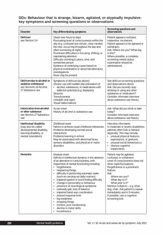
220 Vol. 2 • 10. Acute and subacute by symptom: July 2011
DDx: Behaviour that is strange, bizarre, agitated, or atypically impulsive: key symptoms and screening questions or observations
Disorder Key differentiating symptomsScreening questions and observations
Deliriumsee Section 3.4
Onset over hours to daysFluctuating level of consciousness within the day (e.g. confused one minute, clear-minded the next, occurring throughout the day and often worsening at night)Prominent diffi culties in focusing, shifting, or maintaining attentionDiffi culty orienting to place, time, and sometimes personEvidence of underlying cause based on physical examination or abnormal laboratory investigationsFever may be present
Patient appears confused, inattentive, incoherent.Patient appears to be agitated or withdrawn.Ask: Where are you? What day is this?Where possible, a complete screening mental status examination should be administered.
Delirium due to alcohol or sedative withdrawalsee Sections 16 Alcohol, 17 Substance use
Symptoms of delirium (as above)Chronic use with sudden discontinuation of:• alcohol, substances, or medications with
addiction potential (e.g. diazepam)SeizuresTremulousnessUnstable vital signsVisual hallucinations
See delirium screening questions and observations above.Ask: Did you recently stop drinking or using any other substance or medication?Consider informant interview about substance use history.
Intoxication from alcohol or other substance see Section 17 Substance use
Acute onsetHistory of alcohol or substance use
Ask: What did you drink or take today? Consider informant interview about substance use history.
Intellectual disability (may also be called developmental disability, learning disability, or mental retardation)
Childhood onsetFailure to achieve usual childhood milestonesProblems developing normal social interactionsProblems learning in schoolMay be associated with abnormal faces, sensory disabilities, and physical or motor disorders
Interview informants.In moderate or severe disabilities, patients often look or behave atypically. This may include:• unusual physical features,
expressions, or gestures.• unusual social behaviours.o• obvious cognitive
impairments.
Dementia Gradual onsetDefi cits in intellectual domains, in the absence of an alteration in consciousness, with impairment of mental functioning including:• forgetfulness• misplacing things• diffi culty in performing automatic tasks
(such as carrying out daily routines)• impaired speech or word-fi nding diffi culty • change in personality or behaviour• presence of neurological symptoms• unsteady gait, loss of balance• impaired hand–eye coordination• slowed response time• leg weakness• dropping things• tremors, poor handwriting• decline in motor skills• incontinence
Patient may be agitated, confused, or withdrawn.Level of consciousness does not show rapid fl uctuations.Forgetfulness is a prominent feature.Ask:
Where are you? What day is it? What year is it?
Mention 3 objects – e.g. shoe, dog, chair. Ask patient to repeat immediately and in 5 minutes.If possible, use a cognitive screening tool.
Mental health problems
FINAL Vol. 2 CH 9-21.indd 220FINAL Vol. 2 CH 9-21.indd 220 29/06/2012 15:1529/06/2012 15:15

221Vol. 2 • 10. Acute and subacute by symptom: July 2011
Dementia due to HIV infection (HIV-associated dementia)see Section 13
Symptoms of dementia noted abovePsychomotor slowing is often presentDementia onset at younger age than normally expectedHIV-positive
If HIV status is unknown, test for HIV infection in accordance with local law.Look for signs of HIV-related medical illnesses.Dementia is more common with advanced HIV disease.
HSV encephalitissee Sections 10.10b and 11.15
FeverFocal neurological defi citsAltered thinkingPersonality changes – hypomania, loss of emotional controlDecreased comprehension and memory
May have excessive animation and infl ated self-esteem, diminished comprehension, and hypersexuality.
Schizophrenia and other psychotic conditions not otherwise noted in this table
Hallucinations, most commonly auditory (hearing voices of people who are not there)Delusions: false fi xed beliefs that may be elaborate or bizarreDisorganized thinking or speechDisorganized behaviour, occasionally to the point of inability to care for selfNo fl uctuation of consciousnessPhysical examination and laboratory results do not suggest an underlying medical explanation of the symptomsHistory of previous episode or psychiatric hospitalizationPoor social and occupational functioningOften long-standing history, usually at least 6 months for schizophrenia
Patient may be agitated, suspicious, frightened ,or withdrawn.Conversation with patient often elicits implausible information. Ask for elaboration in a matter-of-fact manner.Ask: Do you hear voices when there is no one present or speaking to you?Ask: have you been troubled by unusual experiences lately? Have you felt threatened by events or people recently? Do you believe that people are trying to harm you? Tell me more?Mood is often restricted or out of keeping with content of conversation. Interview informants.
Bipolar disorder:Manic phaseManic episode
Distinct period ≥1 week characterized by:• abnormally elevated or irritable mood • decreased need for sleep• elevated energy • racing thoughts• rapid pressured speech• increased sense of self-importance• excessive pursuit of risk-taking behaviours• history of previous manic episode or
psychiatric hospitalization• history of depression or currently taking
antidepressant medication• family history of bipolar disorder
Patient is often irritable, loud, or excited.Patient may be speaking very rapidly.Patient may make grandiose statements, such as having supernatural powers or being extremely successful and wealthy when this is not the case.Patient may have slept few hours yet appear alert and energetic.Ask: Do you feel you are unusually happy or excessively irritable? Is your energy unusually high? Do you feel that you do not need any sleep? Do you feel your thoughts are racing?
Other psychotic disorders Brief psychotic disordersDelusional disorder
If the patient screens positive for a particular disorder, then proceed with assessment as appropriate for that specifi c disorder.
Mental health problems
FINAL Vol. 2 CH 9-21.indd 221FINAL Vol. 2 CH 9-21.indd 221 29/06/2012 15:1529/06/2012 15:15

222 Vol. 2 • 10. Acute and subacute by symptom: July 2011
In all cases, non-specialist health-care providers should ensure the protection of, and the respect for, the rights of people with mental, neurological, and substance use disorders and their families.3 This includes the following:• respecting the rights of patients and families within health-care facilities;• obtaining full and informed consent for all diagnostic and treatment
interventions;• following local mental health regulations pertaining to the assessment and
management of mental health conditions; • observing the confi dentiality of users and promoting their participation in all
aspects of their treatment.
Delirium
Delayed diagnosis and management of delirium can be fatal because it often occurs as a manifestation of an underlying severe medical condition. Risk factors and causes include:• high fever from any cause (refer to Section 10.1 Fever);• advanced stages of immunosuppression due to HIV, including associated
infections and malignancies of the CNS;• other severe medical conditions, including hypoxia and metabolic
abnormalities (see Appendix 2);• substance use or withdrawal (see Section 17 Substance use);• intoxication from any cause, including substances of misuse and ingestion of
toxins (see Section 3.8 Poisoning); • drug overdose (accidental or deliberate);• head or brain injuries;• previous episodes of delirium;• dementia, including HIV dementia;• drug interactions in patients taking multiple medications (e.g. for HIV or
tuberculosis).
Diagnosis of delirium
• The disturbance of consciousness with reduced ability to focus, sustain, or shift attention.
• A change in cognition or the development of a perceptual disturbance.• The disturbance developed over hours to days and fl uctuates during the
course of the day, and the disturbance is caused by the consequences of a general medical condition (including alcohol or drugs, medication, or acute head injury).
• Often accompanied by alterations in sleep patterns, alterations in the level of activity, including underactivity or hyperactivity with arousal and agitation, emotional instability, and poor judgement.
If delirium is suspected, refer to Section 3.4.2 Delirium.
3 Convention on the rights of persons with disabilities and optional protocol. Adopted by the United Nations General Assembly on 13 December 2006. http://www.un.org/disabilities/documents/convention/convoptprot-e.pdf
Mental health problems
FINAL Vol. 2 CH 9-21.indd 222FINAL Vol. 2 CH 9-21.indd 222 29/06/2012 15:1629/06/2012 15:16

223Vol. 2 • 10. Acute and subacute by symptom: July 2011
Delirium is frequently overlookedCommon misdiagnoses include the following:• When patients appear hypoactive, depression is a frequent misdiagnosis for
delirium.• When patients appear agitated or restless, a primary psychotic disorder is a
frequent misdiagnosis for delirium.• When patients have a history of dementia, acute changes in mental status due
to delirium is often overlooked.
Intellectual disability in adolescents and adults
Diagnosis of intellectual disabilityIntellectual disability is a lifelong condition characterized by limitations in mental functioning. A person with an intellectual disability may learn and develop more slowly and may require support to assist with full functioning. Some persons with intellectual disabilities may develop mental health problems, which further compounds their functional limitations. Typically, this presents with the onset of new behaviours that the family or community cannot manage.
WHO classifi es intellectual disability into 4 levels on the basis of functioning: mild, moderate, severe, and profound.Common signs indicating a history of delayed development beginning in childhood include:• failure to achieve usual childhood developmental milestones• problems learning self-care• problems learning in school and developing occupational skills;• inappropriate social or sexual behaviour.
Coexisting conditions and differential diagnoses include:• physical disabilities and motor disorders (e.g. cerebral palsy)• epilepsy• hypothyroidism• incontinence• depression• sensory disabilities, such as impaired vision and hearing• attention defi cit or hyperactivity disorder• autism spectrum disorders• other psychiatric and behavioural disorders• nutritional defi ciencies.
Management of intellectual disabilityWhen an adolescent or adult with intellectual disability presents with a marked change in function or a signifi cant alteration in behaviour, consider the possibility of a new onset mental health disorder and treat as indicated. Among adolescents, also consider developmental issues. In particular, growing physical strength and the increasing sexual drive that comes with the onset of puberty often present new challenges to caregivers.• Discuss prior management strategies and rehabilitation efforts.
Mental health problems
FINAL Vol. 2 CH 9-21.indd 223FINAL Vol. 2 CH 9-21.indd 223 29/06/2012 15:1629/06/2012 15:16

224 Vol. 2 • 10. Acute and subacute by symptom: July 2011
• Advise the family about handling the presenting problem, including enlisting additional support.
• Assess and treat any co-morbid medical and mental disorders.• Avoid any unnecessary psychotropic medication. If medication is required,
use caution as people with developmental disorders are often more sensitive to side-effects.
• Refer to educational, and rehabilitation services that are age-appropriate. When possible, look for work opportunities that match the person’s strengths.
• Avoid institutionalization.• Rewarding effort, not results, is the best overall approach to help the patient
and family adapt to the patient’s functional limitations.
Dementia – a decline in cognitive function including memory, thinking, and new learning
Dementia is a disorder of the brain causing a decline in cognitive function that is usually progressive and chronic. It may be classifi ed based on symptom severity and impact of impairment on functioning, as follows:• mild – independent living is possible• moderate – some assistance is needed for activities of daily living• severe – close supervision is necessary.
The symptoms associated with dementia can be wide-ranging. In the degenerative forms of dementia, forgetfulness, declining mental function, and apathy (little emotion) are the most prominent features. As the disorder progresses, patients can lose their capacity to independently manage many activities of daily living. Clinicians should be aware that dementia can be distressing to patients and families.
Patients with HIV are at risk for HIV-associated dementia. This underscores the importance of determining patients HIV status when they present with signs and symptoms suggestive of dementia. At a population level, the rates of severe HIV dementia have fallen considerably with the early introduction of ART that results in full viral load suppression. Among HIV-positive individuals, the likelihood of HIV dementia increases with the progression of systemic HIV disease and immunological decline.
Diagnosis of dementiaThe 3 most common causes of dementia are:1. Alzheimer disease2. Vascular dementia (formerly known as multi-infarct dementia)3. HIV dementia.
HIV dementia can occur at any age, whereas Alzheimer disease and vascular dementia occur primarily in the elderly.
If there is a history of progressive cognitive impairment over a 6-month period that impairs function, a diagnosis of dementia is likely. Abrupt onset of acute cognitive impairment suggests a diagnosis of delirium or a delirium superimposed on a dementia rather than dementia alone. In all cases of cognitive impairment, it is essential to correct any ongoing medical problems that may be contributing to
Mental health problems
FINAL Vol. 2 CH 9-21.indd 224FINAL Vol. 2 CH 9-21.indd 224 29/06/2012 15:1629/06/2012 15:16

225Vol. 2 • 10. Acute and subacute by symptom: July 2011
the impaired cognitive status. Common co-morbidities include hypothyroidism, cardiovascular disease, sexually transmitted infections, anaemia, poor dietary intake, malnutrition, and medication side-effects.
Non-specialist health-care providers should seek to identify possible cases of dementia in the primary health-care setting and in the community. Patient examination, key informant interview, and cognitive tests should be used to assist in confi rming these cases. For a formal dementia diagnosis, a more detailed history, medical review, and mental state examination should be carried out to exclude other common causes of cognitive impairment and decline.
Non-pharmacologic management of dementia
Convey the results of the assessment with sensitivity, and help the patient and family access support that will promote independence, mobility, and function.
Always attend to the safety of the patient fi rst. • Ensure the patient’s safety with appropriate environment and level of
supervision.• Ensure that the patient is taking medications as prescribed.• Protect wandering patients, and supervise any cigarette smoking that occurs.• Use strategies to prevent falls (e.g. reduce clutter on fl oors, re-evaluate
medications that lead to orthostatic hypotension, encourage tailored physical activity to maintain mobility).
Consider cognitive interventions applying principles of reality orientation, cognitive stimulation, or reminiscence therapy for the care of people with dementia. Health-care providers should be trained to deliver these interventions and family members should be involved in their delivery. Examples include:• reorienting the patient;• encouraging the presence of familiar objects and people;• emphasizing routines;• providing lighting that corresponds with day and night;• providing a clock and calendar in the room to help keep patients oriented to
time and to the day of the week;• using memory aids;• offering activities that keep the patient’s mind alert;• ensuring that individuals who require eyeglasses or hearing aids wear these
to help lessen confusion and disorientation.
Interventions for health-care workers should be provided as a part of the overall management of people with dementia.• Educate family members about the nature of dementia and methods for
helping patients to maintain the activities of daily living. Inform about key adaptations in the home that might foster independence and functioning.
• Information should be provided to people with dementia as well as to family members and other informal carers, keeping in mind the preferences of the patients and their families.
• Training of health-care workers involving active carer participation (e.g. role playing of behavioural problem management) may be indicated later in the course of the illness for carers who are coping with behavioural symptoms.
Mental health problems
FINAL Vol. 2 CH 9-21.indd 225FINAL Vol. 2 CH 9-21.indd 225 29/06/2012 15:1629/06/2012 15:16

226 Vol. 2 • 10. Acute and subacute by symptom: July 2011
• The psychological strain of health-care workers should be addressed with support, counselling, or cognitive-behaviour interventions.
• Depression in health-care workers should be managed according to the recommendations for depression.
• Where feasible, home-based respite care may be encouraged for carers of people with dementia.
Consider referral for psychiatric care when:• uncertainty remains about the diagnosis;• there is accompanying depression, psychosis, mania, or substance abuse;• there are behavioural disturbances;• a need exists for consultation regarding psychotropic medications,
particularly when multiple psychotropic medications may be necessary, increasing the risk of drug–drug interactions and toxicity;
• complex psychosocial strategies need to be implemented.
Pharmacologic management of dementia
• All individuals with dementia should receive regular medical review (at least every 3–6 months) and appropriate care.
• In persons with dementia presenting with behavioural symptoms, a complete physical assessment and medication review should be performed to identify any possible underlying precipitants for these symptoms. Appropriate management of these precipitants should be offered before considering the use of psychotropic medicines and non-pharmacological interventions.
• When dementia is complicated by behavioural or psychological disturbances and non-pharmacological interventions are not effective in the management of these symptoms, antipsychotic medications may be useful in treating agitation, delusions, and hallucinations.
• If behavioural symptoms persist or there is a clear and imminent risk of harm to the patient with severe and distressing symptoms, the short-term use of haloperidol or atypical antipsychotic medications can be considered, preferably with specialist inputs. To the extent possible, informed consent and agreement should be obtained from the patient or carer with regard to the balance of risk and benefi t.
• Antipsychotics have been associated with increased risk of stroke; therefore, as much as possible, they should be avoided.
• When using antipsychotic medication in patients with dementia, use the lowest possible dose and increase slowly as needed. For example, haloperidol should be initiated at 0.5 mg and titrated slowly with frequent review of impact and side-effects, particularly extrapyramidal side-effects. Thioridazine and chlorpromazine should not be used due to high potential for orthostatic hypotension and anticholinergic side-effects.
• Patients with dementia are often sensitive to medication side-effects and at a population level, antipsychotic medications appear to increase the risk of death in elderly patients with dementia.
• In people with dementia with symptoms or signs suggestive of moderate or severe depression, the use of fl uoxetine may be considered.
• Benzodiazepines have been shown to increase confusion and decrease concentration. As such, it is recommended that diazepam be avoided.
Mental health problems
FINAL Vol. 2 CH 9-21.indd 226FINAL Vol. 2 CH 9-21.indd 226 29/06/2012 15:1629/06/2012 15:16

227Vol. 2 • 10. Acute and subacute by symptom: July 2011
In the case of HIV-associated dementia, neurocognitive impairment may be partially reversible through the use of effective ART that fully suppresses viral replication. Fully suppressive ART is the treatment of choice for the prevention and treatment of HIV dementia.
10.11.4 Psychosis2
Psychosis is a state in which a person’s grasp of reality (or reality-testing) is diminished. This may be manifested by:• hallucinations (most often auditory);• delusions;• disorganized or strange speech;• bizarre behaviour or disorganized behaviour to the point that self-care is
impaired;• lack of insight (e.g. patients may not see themselves as ill or in need of
treatment, family or friends may bring them);• inappropriate or narrowed range of emotions;• social withdrawal.
Defi nitions of brief and persistent psychotic disorders
Psychotic disorders that are not attributable to an underlying medical condition can be divided into 2 categories.
1. Brief (acute) and transient psychotic disordersDisorders characterized by the acute onset of psychotic symptoms and severe disruption of ordinary behaviour. The onset of symptoms is acute, often related to a signifi cant stressor. The time interval between the fi rst appearance of any psychotic symptoms and the presentation of the fully developed disorder should not exceed 2 weeks. Usually, there is full recovery, and no evidence of an organic cause.
2. Persistent or recurrent psychotic disordersPersistent or recurrent psychotic disorders can be further broken down based on whether or not a predominant mood or affective symptoms accompany the psychosis. Schizophrenia is the most common and severe non-affective chronic psychotic illness. These disorders often begin with non-specifi c symptoms, such as social withdrawal, loss of interest in usual activities, apathy, and diminished functioning. They generally have their onset in late adolescence and young adult life. The most common examples of recurrent or persistent psychotic disorders with prominent mood symptoms include mania with psychotic features (as one phase of bipolar mood disorder) and a severe depressive episode with psychotic features.
Diagnosis of psychosisThe diagnosis of a psychotic disorder is made by the identifi cation of the abnormal behaviour suggestive of psychotic symptoms described above.
Important risk factors for the development of psychosis include:• prior psychiatric illness• family history of psychotic illness• substance misuse, including intoxication, abuse, dependence, and withdrawal• exposure to severe stressors or traumatic events.
Mental health problems
FINAL Vol. 2 CH 9-21.indd 227FINAL Vol. 2 CH 9-21.indd 227 29/06/2012 15:1629/06/2012 15:16

228 Vol. 2 • 10. Acute and subacute by symptom: July 2011
In addition, psychotic symptoms may be associated with a general medical condition or medications. Examples include patients with late-stage HIV disease who may develop psychosis more commonly than people in the general population. Examples of medications that may be associated with the development of psychotic symptoms include EFV and (anabolic) steroids.
Differential diagnosis
The differential diagnosis of a patient presenting with psychotic symptoms includes the following.• Delirium and dementia (see Section 10.11.3 above).• Other medical causes.
° Even after excluding delirium and dementia, psychotic symptoms may be caused by an underlying medical condition, such as the conditions described in Appendix 2.
° Although a past history of a chronic or recurrent psychotic disorder strongly suggests an exacerbation or a relapse of that disorder, a relapse can also be precipitated by a serious medical problem, underscoring the importance of evaluation for an underlying or coexisting medical aetiology.
• Primary psychotic disorders, including schizophrenia, mania, and depression with psychotic features. ° See the description for schizophrenia and other psychotic disorders in the
table above, DDx: Behaviour that is strange, bizarre, agitated, or atypically impulsive: key symptoms and screening questions or observations.
• Auditory hallucinations are the most common hallucinations in primary psychotic disorders. Other types of hallucinations should increase the index of suspicion that there is an underlying medical problem.
• Schizophrenia may present with “negative” symptoms, such as lack of emotion, lack of motivation, and paucity of thought. These symptoms may, however, be confused with the side-effects of antipsychotic medications.
• Patients with chronic psychotic disorders have decreased cognitive performance and may struggle with memory, concentration, complex thinking, and learning new tasks but not to the degree seen in dementia.
• People with severe and persistent mental disorders are often at higher risk of being infected with HIV and other sexually transmitted infections due to elevated rates of unsafe sexual activities, including sexual victimization, and co-morbid substance use. Even if an individual has a confi rmed primary psychotic disorder, consideration should be given to HIV status.
• Certain symptoms not typically classifi ed as psychotic can nonetheless be bizarre or involve a loss of reality testing, such as the repeated rituals seen in obsessive compulsive disorder and the fl ashbacks associated with post-traumatic stress disorder.
Investigations
A parallel history from family members should be taken whenever possible. Psychotic patients may have poor insight into their symptoms and may fail to report them, or, in some instances, they may conceal them.
Mental health problems
FINAL Vol. 2 CH 9-21.indd 228FINAL Vol. 2 CH 9-21.indd 228 29/06/2012 15:1629/06/2012 15:16

229Vol. 2 • 10. Acute and subacute by symptom: July 2011
Clinical management of psychosis not accompanied by mania or severe depression
• Assessment for the safety of the patient and others should be performed as soon as possible.
• Patients should be provided with the opportunity to seek support in making decisions about their treatment when they require it. Consult local mental health laws and guidelines.
• Initial hospitalization often is required for acute stabilization, especially for patients who are at imminent risk of violence or self-harm. Also, hospitalization may be needed for those with new-onset psychosis.
• Assessment for an underlying medical etiology and the initiation of appropriate medical treatment are essential.
• Provide psycho-education that supports the patient’s recovery. Instil realistic hope and emphasize the importance of continuing regular activities.
• Support the family and provide guidance in their interactions with the patient. • Antipsychotic medication is the mainstay of the acute management of
psychotic symptoms. Emphasize the importance of medication adherence to reduce symptoms and suffering.
• Encourage regular follow-up care, including the management of concurrent medical conditions. Patients with chronic psychotic disorders are at increased risk of co-occurring conditions, including substance use, HIV/AIDS, diabetes, heart disease, and smoking-related illnesses. These patients require a comprehensive approach to assessment and care over and above the management of psychotic symptoms.
• Negative health-care worker attitudes have been documented as interfering with the delivery of medical care It is important that health-care facilities develop training resources for health-care workers to help overcome fear and stigma associated with working with patients with psychosis.
• Facilitate rehabilitation in the community. Long-term treatment of chronic psychotic disorders such as schizophrenia often require interventions based on principles of psychosocial rehabilitation, including cognitive behavioural therapy (CBT), skills building, and family interventions. These interventions should be continued as long as needed by the user and their family, and therefore should be planned and developed in a sustainable way.
Use of antipsychotic medications: some basic principles and cautions
• Haloperidol or chlorpromazine should be routinely considered in individuals with psychotic disorders.
• The minimal effective dose of antipsychotics should be used, paying attention to minimizing adverse effects.
• Trial medication at optimum dose for 4–6 weeks before considering it ineffective. Always check for treatment adherence.
• Patients on long-term antipsychotic treatment should be given adequate information and encouraged to make a choice between oral and depot preparations, especially with the view to improve adherence. Depot antipsychotics should not be used for prompt control of acute psychotic symptoms.
Mental health problems
FINAL Vol. 2 CH 9-21.indd 229FINAL Vol. 2 CH 9-21.indd 229 29/06/2012 15:1629/06/2012 15:16

230 Vol. 2 • 10. Acute and subacute by symptom: July 2011
• Patients on antipsychotic medicines (oral and depot preparations) should be monitored regularly for symptom relief, functioning, and any adverse effects.
• Women with psychotic disorders (including schizophrenia) who are planning a pregnancy, or are pregnant or breastfeeding and require antipsychotic treatment to manage symptoms, should be treated with low-dose oral haloperidol or chlorpromazine.
• Depot antipsychotics should not be routinely prescribed to women with psychotic disorders (including schizophrenia) who are planning a pregnancy or who are pregnant or breastfeeding, because there is relatively little information on their safety.
General principles for using antipsychotic medications: haloperidol, chlorpromazine, long-acting injectable fl uphenazine
Contraindications and cautions: Do not use for alcohol withdrawal. Antipsychotic medications are associated with increased risk of death in elderly patients with dementia, and associated with increased risk for seizures and blood disorders.
Serious acute side-effects:• acute dystonic reaction or severe muscle spasm (give biperiden- see Section 8.4)• neuroleptic malignant syndrome, a potentially life-threatening disorder characterized by muscular rigidity,
elevated temperature, and high blood pressure.
Serious long-term side-effects:• involuntary muscular movements that may not be reversible (tardive dyskinesia).
Educate the patient and family:• Review medication facts as relevant.• Medications are not addictive.• Warn against the use of alcohol.
Haloperidol as antipsychotic
In healthy adults• Initiate treatment with 1.5–3 mg once daily. • Depending on symptom severity, symptom response, and tolerability, can be titrated up to 20 mg daily. • Typical effective dose: 3–20 mg daily.• Use lowest effective dose.
In elderly or medically ill patients (Including those with HIV stage 3 or 4 – these patients are very sensitive to the side-effects of haloperidol.)• Initiate treatment with 0.5–1 mg once daily.• Depending on symptom severity, symptom response, and tolerability, can be titrated up to 5 mg daily.• Higher doses may be needed, but there is a signifi cant risk of toxicity.• Use the lowest effective dose.
Chlorpromazine as antipsychotic
In healthy adults• Initiate treatment with 75 mg at night.• Depending on symptom severity, symptom response, and tolerability, can be titrated up to 300 mg daily.• Typical effective dose is 75–300 mg daily, but up to 1000 mg may be necessary in severe cases.• Use the lowest effective dose.
In elderly or medically ill patients (including those with HIV stage 3 or 4)• Haloperidol is preferred because even low doses of chlorpromazine may cause severe hypotension resulting
in falls.
Mental health problems
FINAL Vol. 2 CH 9-21.indd 230FINAL Vol. 2 CH 9-21.indd 230 29/06/2012 15:1629/06/2012 15:16

231Vol. 2 • 10. Acute and subacute by symptom: July 2011
Fluphenazine as antipsychotic – long-acting, injectable for long-term management
Other contraindications and cautions: Neuromuscular side-effects can take a long time to clear after drug discontinuation. Avoid for acute treatment because it is diffi cult to titrate the dose in accordance with symptom response and tolerability.
In healthy adults• Start with 12.5 mg deep intramuscular injection in the gluteal region.• Typical effective dose is 12.5–100 mg IM every 2 to 5 weeks.• Repeat IM injections every 2 to 5 weeks as follows:
° if the initial dose was well-tolerated and effective, continue to use that dose; ° if the initial dose was tolerated but did not control the symptoms, the dose may be increased in incre-
ments of 12.5 mg.
In elderly or medically ill patients (including those with HIV stage 3 or 4)• Oral haloperidol is preferred because severe extrapyramidal side-effects may take a long time to clear after
medication discontinuation.• If using injectable fl uphenazine, start with 6.25 mg deep intramuscular injection.• Typical effective dose is 6.25–50 mg every 2 to 5 weeks.• Repeat the IM injections every 2 to 5 weeks as follows:
° if the initial dose was well-tolerated and effective, continue to use that dose; ° if the initial dose was tolerated but did not control the symptoms, the dose may be increased in incre-
ments of 6.25 mg IM; a maximum dose of 100 mg IM may be given although such a dose is rarely required.
In all adults• The length of time between injections depends on how long the medication lasts in controlling symptoms
and how well the patient tolerates the medication.• Use the lowest effective dose.• Long-acting medication is particularly useful in patients who have diffi culty complying with treatment.• If severe side-effects occur, discontinue the injections, wait until the side-effects clear, and begin an oral
antipsychotic.
Considerations for long-term antipsychotic treatment
In patients with full and sustained remission following a fi rst psychotic episode, continue antipsychotic treatment for at least 12 months after the beginning of remission.
Any further continuation of antipsychotic treatment should be based on a clinical review, preferably by a mental health specialist, and taking into account the preferences of the patient, and in consultation with the family.
In patients with long-term or recurrent psychotic disorders (including schizophrenia) stable for several years on anti-psychotics, treatment withdrawal may be considered, keeping in mind the increased risk of relapse, the possible adverse effects of medicines, and patient preferences in consultation with the family. Preferably, this decision should be made in consultation with a mental health professional. If medicines are withdrawn, patients and family members need to be educated to detect early symptoms of relapse, and close clinical monitoring should be done.
10.11.5 Bipolar disorder2
Bipolar disorder is characterized by recurrent episodes of mood instability throughout a patient’s adult life, usually accompanied by signifi cant changes in activity and behaviour. The course of the disorder is characterized by 3 phases: (i) acute mania, involving elevations of mood; (ii) depressive phases, involving
Mental health problems
FINAL Vol. 2 CH 9-21.indd 231FINAL Vol. 2 CH 9-21.indd 231 29/06/2012 15:1629/06/2012 15:16

232 Vol. 2 • 10. Acute and subacute by symptom: July 2011
periods of low mood; and (iii) maintenance phase, where recovery and wellness is generally preserved. Pharmacologic interventions are specifi c to each phase.
Patients with bipolar disorder can present with marked changes in behaviour and it is important to consider the diagnosis of bipolar disorder in patients presenting with abnormal behaviour as well as in patients presenting with disturbance of mood.
Clinical management involves psychosocial and pharmacologic interventions.
Psychosocial interventions in bipolar disorders
• Ensure patient safety and manage concurrent medical conditions.• Provide psycho-education. Patients and families need to understand the
phases of mood disturbance and how to self-monitor for extreme mood states in order to best prevent severe relapses.
• Encourage a regular sleep cycle.• Encourage patient to avoid alcohol and psychoactive substances.• Help patients reactivate social networks and to seek support from family and
community-based resources.• Provide regular follow-up.
Pharmacologic management of bipolar disorder
(i) Acute mania (with or without psychosis) in a patient with bipolar disorderThe management of acute mania requires mood stabilizer medications and often also requires anti-psychotics. Psychiatric hospitalization often is required.
If the patient is on an antidepressant, it should quickly be tapered and discontinued, balancing the risks of withdrawal syndrome against the impact of the antidepressant worsening the mania.
The onset of effectiveness of haloperidol is more rapid than mood stabilizers and should be considered fi rst in individuals with severe symptoms of acute mania, for the management of agitation.
Lithium4 or sodium valproate should be considered in individuals with acute mania for mood stabilization. Carbamazepine may also be helpful in the treatment of acute mania. Carbamazepine may negatively interact with antiretroviral medications and is best avoided in PLHIV taking ART.
In women planning a pregnancy, pregnant or breastfeeding, lithium and sodium valproate should be avoided. In this group, low dose haloperidol should be considered with caution.
In the elderly patient, start with lower doses of medications and increase slowly. Anticipate increased risk of drug interactions.
4 See Adaptation Guide – instructions on using lithium could be added from mhGAP guidelines if lithium laboratory monitoring is available.
Mental health problems
FINAL Vol. 2 CH 9-21.indd 232FINAL Vol. 2 CH 9-21.indd 232 29/06/2012 15:1629/06/2012 15:16

233Vol. 2 • 10. Acute and subacute by symptom: July 2011
(ii) Acute depression (with or without psychosis) in a patient with bipolar disorder Treatment of the depressive phase of bipolar disorder is particularly challenging as antidepressant medication may precipitate a manic or hypomanic phase if the patient is not fi rst adequately treated with mood stabilizing medications.
Consider the diagnosis of bipolar depression if the patient presenting with a depressive episode has a known or suspected history of mania (see Table: Mood stabilizers below).
• The treatment of choice for bipolar depression is a mood stabilizer agent (sodium valproate, lithium4).
• Once the patient is adequately treated with a mood stabilizer, an antidepressant agent can be added to help further alleviate depressive symptoms. Patients should be informed of the risk of switching to mania before starting antidepressant medication.
• Antidepressant treatment should begin at a low dose and be increased gradually if necessary. Fluoxetine should be preferred to tricyclics. Patients should be monitored carefully for early symptoms or signs of mania. Antidepressant medication should be stopped soon after remission of depressive symptoms, while the mood stabilizer continued.
• Behavioural activation, lifestyle modifi cation and cognitive behaviour therapy should be used to help alleviate depressive symptoms. Use of these interventions in combination with a mood stabilizer may help the patient avoid the use of an antidepressant medication.
Table: Mood stabilizersFor Lithium see Adaptation guide
Valproic acid (sodium valproate) for mood stabilization in bipolar disorder and acute mania
Contraindications and cautions: Do not use for alcohol withdrawal.
Serious side-effects include:• hepatotoxicity (can be fatal)• pancreatitis• hyponatraemia from drinking excess fl uid• blood dyscrasias• severe allergic reaction.
Educate the patient and the family:• Review cautions and side-effects.• Mood stabilizers are not addictive.• Warn against use of alcohol.
In healthy adults• Initiate treatment with 500 mg at night.• Depending on symptom severity, response, and tolerability, increase the dose by 200 mg every 7 days.• Typical effective dose is 1000–2000 mg daily.
In elderly or medically ill patients (including those with HIV stage 3 or 4)• Initiate treatment with 200 mg in the morning and 200 mg at night.• Increase the dose by 200 mg every 7 days until there is a clinical response or the therapeutic blood level is
reached. • The maximum dose can vary; assess tolerability and the clinical response.
Mental health problems
FINAL Vol. 2 CH 9-21.indd 233FINAL Vol. 2 CH 9-21.indd 233 29/06/2012 15:1629/06/2012 15:16

234 Vol. 2 • 10. Acute and subacute by symptom: July 2011
Carbamazepine – for mood stabilization in bipolar disorder, acute mania
Contraindications and cautions: Avoid in pregnancy. Induces the metabolism of many other medications.
Serious but uncommon side-effects include:• severe hypersensitivity or allergic reactions of skin or organs• arrhythmias, AV block, heart failure• blood dyscrasias• hepatitis or hepatic failure or pancreatits• hyponatraemia from drinking excess fl uid.
Educate the patient and the family:• Review cautions and side-effects.• Mood stabilizers are not addictive.• Warn against use of alcohol.
In healthy adults• Initiate treatment with 200 mg at night.• Increase the dose by 200 mg every 3–4 days until there is a clinical response. • Give in divided doses to reduce toxicity.• Typical effective dose is 400–600 mg daily, but in severe cases may need 1000 mg.
In elderly or medically ill patients (including those with HIV stage 3 to 4)• Initiate treatment with 100 mg at night.• Increase the dose by 100 mg every 3–4 days until there is a clinical response.• Give in divided doses to reduce toxicity.• The maximum dose can vary; assess tolerability and the clinical response.• Be alert to possible virilogic failure in patients taking antiretroviral medications for HIV.
(iii) Maintenance treatment of bipolar disorder Treatment with a mood stabilizer, in combination with psychosocial interventions, is important to maintain wellness and avoid relapses of mania and depression in a patient with bipolar disorder.
Lithium4 or sodium valproate should be considered in the maintenance treatment of bipolar disorder. Lithium can only be used when laboratory monitoring of blood levels is available.
Carbamazepine can also be used but may negatively interact with antiretroviral medications and is best avoided in PLHIV taking ART.
In women planning a pregnancy, or pregnant or breastfeeding, lithium and sodium valproate should be avoided. These patients should be referred to specialist mental health care, when possible.
Maintenance treatment should continue for at least 2 years after the last episode of bipolar disorder. The decision to continue maintenance treatment after 2 years should preferably be done by a mental health specialist.
If a patient has frequent relapses of mania or depression while on mood stabilizer therapy, consider switching or adding agents. These patients should be referred to specialist mental health care, when possible.
Refer to specialty care guidelines for further information about the treatment of bipolar depression.
Mental health problems
FINAL Vol. 2 CH 9-21.indd 234FINAL Vol. 2 CH 9-21.indd 234 29/06/2012 15:1629/06/2012 15:16

235Vol. 2 • 10. Acute and subacute by symptom: July 2011
10.11.6 Sad or low mood including depression2
Sadness and anxiety can be very common feelings in everyday life. The causes of sad or low mood, anxiety, nervousness, or excessive worry often can be attributed to mood disorders or anxiety disorders. Clinical assessment seeks to correctly identify those patients who are experiencing a depressive episode or an anxiety disorder, so that adequate treatment may be instituted. However, several other physical and psychiatric disorders may manifest with symptoms of sad or low mood, and must be considered by the clinician in the differential diagnosis. These disorders can include:• delirium with low mood (see Section 10.11.3)• intellectual disabilities (see Section 10.11.3)• dementia (see Section 10.11.3)• psychosis (see Section 10.11.4)• substance use disorders (see Section 17)• depression secondary to medical illnesses and medications.
In this Section, a depressive episode refers to patients whose sadness is severe or persistent, who have a range of emotional and physical symptoms that characterize major depression, whose symptoms deviate from normal expected reactions to life stressors, traumas, and challenges, and who experience a signifi cant impairment in function or life quality due to the depressive episode.
Sad or low mood and anxious states of mind often occur together. If there is prominent low mood, treat the depression fi rst. However, temporary relief from anxiety symptoms may be needed, particularly at the outset of treatment.
At the start of treatment, always consider the possibility that medical causes may be contributing to depressive disorders. When patients do not respond to treatment for sad or anxious states of mind, see medical causes listed in Appendix 2.
The lifetime risk of developing a depressive disorder is 10%–20% in women and somewhat less in men. It is estimated that 20% of patients presenting to general medical clinics have depressive episodes. Depression often presents concomitantly with physical health problems, and patients with chronic disease complicated by severe depression have signifi cantly worse health outcomes than patients with chronic disease without severe depression. Severe depression occurs more frequently in patients with HIV than in the general population, and can greatly affect their ability to adhere to antiretroviral treatment.
Assessment of patients with sad or low mood or depression
Depressive disorders may present with both psychological and physical symptoms. Many patients with depression do not volunteer emotional complaints, presenting instead with somatic complaints, which typically include:• fatigue, which can be severe• insomnia and, less commonly, hypersomnia• somatic complaints, including changes in appetite and weight• bodily complaints, such as burning sensations in the head, unexplained
aches, and pains.
Mental health problems
FINAL Vol. 2 CH 9-21.indd 235FINAL Vol. 2 CH 9-21.indd 235 29/06/2012 15:1629/06/2012 15:16

236 Vol. 2 • 10. Acute and subacute by symptom: July 2011
Psychological or emotional complaints are often elicited on inquiry. The symptoms cause signifi cant distress and may markedly impair function and quality of life. The table below, DDx: Sad or low mood or depression, lists the defi ning characteristics of disorders associated with sad or low mood or depression.
Common presenting symptoms that should alert the clinician to a possible depressive disorder and should trigger the use of this Section include the following:
• sad or low mood• fatigue or loss of energy or tiredness• loss of interest or pleasure• guilt or loss of self confi dence• hopelessness and feelings of worthlessness• suicidal thoughts or acts or thoughts of death (Section 10.11.2 Suicide risk)• disturbed sleep• loss of libido• disturbed appetite (weight loss or gain)• feeling tense, anxious, excessively worried, or frightened• unexplained somatic symptoms
° tingling, numbness ° shortness of breath ° gastrointestinal distress ° palpitations ° aches and pains
• reliving past traumas in thoughts, images, dreams or acts• persistent preoccupation with stressful life events or stressors.
Patients presenting with these states of mind should receive an assessment for risk of suicide, self-harm, or the potential for violence or harm to others (see Section 10.11.2 Suicide risk).
Investigations
• Evaluation of the patient, including mental health history, review of medications, medical history, and mental status examination.
• Consider medical conditions and laboratory investigations as appropriate.• History from family or friends if available and if the patient consents to these
discussions.• Use the DDx tables below to identify the most suitable diagnoses to account
for the patient’s symptoms, and to guide treatment initiation.• Consider the role of the following risk factors for depressive disorders in the
patient’s presentation. ° past history of depressive episodes or family history of depressive episodes ° female gender ° major stressful life events including family confl ict, loss, or separation ° chronic medical illness, including HIV infection ° absence of positive social support ° adverse social environment (poverty, homelessness, isolation, stigma,
discrimination)
Mental health problems
FINAL Vol. 2 CH 9-21.indd 236FINAL Vol. 2 CH 9-21.indd 236 29/06/2012 15:1629/06/2012 15:16

237Vol. 2 • 10. Acute and subacute by symptom: July 2011
° indications of the progression of HIV infection (notifi cation of positive test, progression of symptoms, drop in CD4 count)
° substance misuse ° side-effects of prescribed medications ° present or past exposure to physical and sexual abuse.
DDx: Sad or low mood or depression
Psychiatric disorder Symptom constellation and treatment considerations
Depressive episode Symptoms persist for at least 2 weeks and occur on most days for most of the day.Distinct period with defi ned onset characterized by:• sad or low mood• fatigue or loss of energy or tiredness• loss of interest or pleasure• guilt or loss of self-confi dence • hopelessness and feelings of worthlessness• changes in appetite and sleep• loss of libido• suicidal thoughts or acts or thoughts of death.Antidepressants usually are helpful.
Dysthymia A more persistent and chronic (>2 years) period of sadness and low mood; fewer and less severe symptoms than in severe depression, but associated with signifi cant impairment of quality of life and function.Antidepressant medication and psychotherapy often are indicated, but response to treatment may be more limited than seen with depressive episode.
Bipolar disorder – depressive episode or depressive phase
Current symptoms of depressive episode.Lifetime history of both depressive episode and mania or, its milder form, hypomania.Antidepressant medications can induce or precipitate manic episodes in patients with personal or family histories of bipolar disorder.Mood stabilizers are the treatment of choice for bipolar disorder. In the depressive phase, treat fi rst with mood stabilizer such as lithium. Consider adding antidepressants only if mood stabilizer is insuffi cient to alleviate depressive symptoms. Use antidepressants to treat the depressive episode for the shortest possible period.
Depressive episode with psychotic features
Current symptoms of depressive episode.In addition to the symptoms of depression, the patient develops psychotic symptoms, e.g. delusions or hallucinations.Treatment necessitates the use of antipsychotic and antidepressant medications.
Depressive symptoms due to general medical condition
Medical illness or medications used to treat medical conditions may contribute to depressive symptoms.Symptoms may be due to a depressive disorder, the general medical condition, or both.Treatment may involve addressing the underlying medical disorder or instituting antidepressant medication treatment.
Depressive symptoms due to dementia
While memory loss is the hallmark symptom of dementia, many dementias, including HIV dementia, may present with symptoms of low mood, depression, and a slowing down or blunting of emotional responses.It is important to remember that many patients with dementia may also have a depressive episode. Both conditions will require targeted intervention.
Mental health problems
FINAL Vol. 2 CH 9-21.indd 237FINAL Vol. 2 CH 9-21.indd 237 29/06/2012 15:1629/06/2012 15:16

238 Vol. 2 • 10. Acute and subacute by symptom: July 2011
Psychiatric disorder Symptom constellation and treatment considerations
Uncomplicated bereavement A period of sadness following the death of a close friend or relative; the passage of time and the support of friends and family are usually suffi cient to help people through a period of bereavement. If bereavement began in the prior 2 months, do not consider antidepressants as fi rst-line therapy. Use culturally appropriate mourning and support to facilitate adjustment.Although bereavement is not a mental disorder, some people may be at risk of developing the full constellation of persistent symptoms that meet the criteria of a major depressive episode and may require consideration of targeted antidepressant therapy.
Adjustment disorder(especially with depressive features)
Time-limited mood symptoms may develop in response to a major stressful event without the full constellation of psychological and physical symptoms that are characteristic of severe depression.Treatment should focus on helping the patient to manage the adversity, by providing hope and reassurance, problem-solving, and facilitating connection with natural support systems. Physical activity should be encouraged and psychotherapies considered where available.Should the symptoms persist and their severity worsen, an adjustment disorder may develop into a depressive episode requiring antidepressant therapies.
Figure: Schematic illustration of common mood syndromes
The fi gure illustrates common mood syndromes. A patient with bipolar disorder will have distinct episodes of low mood and euphoria or high mood. A patient with depressive episodes will have episodes of low mood with a return to wellness, but episodes may be recurrent, and mild symptoms may persist between episodes. Some patients have persistently low mood; if this persists for at least 2 years without returning to wellness, they are considered to have dysthymic disorder. Severe depression and dysthymia may occur together, resulting in a baseline state of depression with periodic exacerbations.
Mental health problems
FINAL Vol. 2 CH 9-21.indd 238FINAL Vol. 2 CH 9-21.indd 238 29/06/2012 15:1629/06/2012 15:16

239Vol. 2 • 10. Acute and subacute by symptom: July 2011
Depressive symptoms due to medical conditions
Physical or somatic symptoms associated with depression, such as fatigue, weight loss, and insomnia, may be caused by physical illness alone or the medications used to treat physical illnesses. However, it is not uncommon for physical illness and depression to coexist. In these cases, there is a risk of diagnosing only one condition. When both disorders exist, it is important to identify and treat both, as each can contribute to worsening of the patient’s health status.• Physical illnesses may present with sad or low mood as a key symptom. The
low mood is often accompanied by fatigue, lethargy, and loss of motivation.• Medications used to treat physical syndromes may cause low mood, loss of
interest, and fatigue. Some medications used to treat HIV (e.g. EFV) have been associated with low-mood symptoms.
Medical conditions and medications that can present with depression can be found in Appendix 2.
Depressive symptoms related to adverse life events
Adverse life events can contribute to sad or low mood, but a depressive episode is not an expected outcome of such events. Depressive episodes often develop following an adverse or stressful life event but are distinguished from the conditions described in the table above, DDx: Sad or low mood or depression, by the symptom severity, the persistence, and pervasiveness of the mood symptoms and the functional impact. The DDx table also outlines other conditions to consider when making the differential diagnosis of sad or low mood. These conditions may be thought of as existing on a continuum of reactions to adverse life events.
In general, with the help of natural support systems and the passage of time, most people will recover from the distress of adverse life events and circumstances. More targeted interventions, including antidepressant medications and psychotherapies, will be needed when symptom burden is greater and functional impact is more severe.
Acute management of depression
When symptoms of a depressive episode are pervasive, persistent, and of moderate to severe intensity, medication treatment is usually necessary to reduce the symptoms. When symptoms are mild, psychotherapy and counselling without medications may be suffi cient to treat the episode.
Physical activity and behavioural activation should be encouraged in all patients experiencing depressive episodes. However, in moderate and severe depression, this intervention should only be used as adjunct to antidepressants or brief structured psychological treatments.
All patients should receive comprehensive clinical care including psycho-education, counselling and support, activation of social networks, structured physical activity programming, medication management, and regular follow up. Where human resources permit, referral for psychotherapy and additional psychiatric and mental health services should be considered.
Mental health problems
FINAL Vol. 2 CH 9-21.indd 239FINAL Vol. 2 CH 9-21.indd 239 29/06/2012 15:1629/06/2012 15:16

240 Vol. 2 • 10. Acute and subacute by symptom: July 2011
Health-care workers should perform the following for patients presenting with severe depression.• Assess for suicide risk (see Section 10.11.2) and ensure safety.• Initiate medication treatment.• Provide counselling.• Initiate management of any co-morbid conditions (e.g. treat substance use
disorders, medical conditions, and pain).
Use of antidepressant medications: some basic principles and cautions
• Antidepressant medications are the most effective treatment for a depressive episode of moderate to severe symptom intensity.
• Counselling and psychotherapy often are suffi cient for the treatment of a depressive episode of mild intensity. Antidepressant medications should not be offered for the initial treatment of a mild depressive episode.
• Fluoxetine5 or tricyclic antidepressants (TCAs) should be considered as treatment in individuals with moderate to severe depressive disorder.
• If fl uoxetine is available, it may be better tolerated and equally effective as TCAs and may be initiated instead of a TCA.
• If drug treatment is required in older people, fl uoxetine is preferable to tricyclics, which should be avoided due to side-effects.
• In adolescents with depressive episodes, do not consider antidepressant medication as fi rst-line treatment. Psychosocial treatments, including psychotherapy, are preferable. If no response to psychosocial treatments, consider using the lowest effective dose of fl uoxetine (but not other SSRIs or TCAs). Adolescents on fl uoxetine should be monitored closely for suicidal ideations or behaviour. For all adolescents on fl uoxetine, support and supervision from a mental health specialist should be obtained.
• Antidepressants should not be considered in the treatment of children younger than 12 years of age.
• If drug treatment is required in women with a depressive episode or disorder who are planning a pregnancy, pregnant, or breastfeeding, avoid antidepressants as much as possible. Psychosocial treatments, including psychotherapy, are preferable. If no response to psychosocial treatments, consider using the lowest effective dose of antidepressant medication.
• If antidepressant medication is required in patients with cardiovascular disease, SSRIs are the fi rst choice. Do not prescribe TCAs to people at risk of serious cardiac arrhythmias or recent myocardial infarction.
• Antidepressant medications can precipitate mania in patients with a personal or family history of mania or bipolar disorder.
5 Other SSRIs are available. See Adaptation Guide.
Mental health problems
FINAL Vol. 2 CH 9-21.indd 240FINAL Vol. 2 CH 9-21.indd 240 29/06/2012 15:1629/06/2012 15:16

241Vol. 2 • 10. Acute and subacute by symptom: July 2011
• With both SSRI and TCA medications, the early phase of acute antidepressant treatment may be a time of heightened risk for agitation, and harm to self or others.
• When starting antidepressants, monitor patient for risk of harm to self or others at every contact.
• All TCAs are highly lethal in overdose. To minimize the harm if overdose occurs, patients should have access to only a small supply at the initiation of treatment or at any points when suicide risk is high.
• If severe anxiety is present or if marked and akathisia or agitation develops on SSRIs, consider short-term use of low-dose diazepam, i.e. 5–10 mg daily for 1 week. Diazepam does not treat depression and carries a risk of dependence.
• Dosage adjustment of medication should be based on symptom response and tolerability. The treatment goal is full remission of symptoms and a full return to the level of function.
• At each visit, assess medication tolerability and check adherence.• If the patient has a poor symptom response by week 12 despite good
adherence to the maximum tolerated dose of a medication, switch to another antidepressant.
• In adult individuals with depressive disorders who have benefi ted from initial antidepressant treatment, do not consider ending the antidepressant treatment before 9–12 months after recovery. Treatment should be regularly monitored, with special attention to treatment adherence. Frequency of contact should be determined by the adherence, the severity of symptoms, and by local feasibility issues.
• When terminating antidepressant medication treatment, remind the patient about the possibility of withdrawal symptoms. Counsel to taper the dose slowly and avoid abrupt discontinuation.
• Advise patients to self-monitor for early signs of symptom relapse.
Use of antidepressant medication in the management of a depressive episode (moderate to severe)
The 2 antidepressants available in the WHO formulary are fluoxetine and amitriptyline. Doses and titration are noted below.
Mental health problems
FINAL Vol. 2 CH 9-21.indd 241FINAL Vol. 2 CH 9-21.indd 241 29/06/2012 15:1629/06/2012 15:16

242 Vol. 2 • 10. Acute and subacute by symptom: July 2011
Fluoxetine for depression
Contraindications and cautions: Watch for increased risk for agitation and suicidal ideation and behaviour (see Section 10.11.2). If there is a history of mania or bipolar disorder, use a mood stabilizer fi rst (see Section 10.11.5 on bipolar disorder for further details).
Educate the patient and family:• about side-effects• that the medication is not addictiv• to avoid the use of alcohol• that it usually takes several weeks to get a response – do not be discouraged• that the patient may feel worse initially due to side-effects; most side-effects gradually diminish.
In healthy adults• Initiate treatment with 20 mg of fl uoxetine daily.• May start at 10 mg daily to reduce the risk of side-effects that undermine adherence, and then increase to 20
mg as tolerated.• If 10 mg dosing is not available, give 20 mg every other day (fl uoxetine has a very long half-life)• If the patient has severe insomnia (caused by the psychiatric illness or fl uoxetine), consider adding diazepam
5 mg at bedtime. Gradually taper and discontinue diazepam as psychiatric symptoms improve.• If no response in 4–6 weeks or partial response at 6 weeks, the fl uoxetine dose may be increased to 40 mg.• The fl uoxetine dose may be gradually increased up to 60 mg as necessary and tolerable to achieve optimal
response.• If the patient is being treated for depression and there is an inadequate symptom response by week 12 at the
maximum tolerated dose, consider switching to amitriptyline.• When switching from fl uoxetine to amitriptyline, note that fl uoxetine may increase serum amitriptyline levels.
Therefore, when switching, initially use the amitriptyline dosing schedule suggested for elderly or medically ill patients rather than for healthy adults.
In elderly or medically ill patients or patients who cannot initially tolerate a 20 mg daily dose (including those with HIV stage 3 or 4):• Initiate treatment with 10 mg daily for 3 weeks. • If 10 mg doses are not available, give 20 mg every day (fl uoxetine has a very long half-life).• Bear in mind that fl uoxetine dosing can be increased as gradually as needed to manage uncomfortable side-
effects. Patients usually become accustomed to the side-effects in time.• If the patient has severe insomnia (caused by psychiatric illness or fl uoxetine), consider adding diazepam 2–5
mg at bedtime. Avoid diazepam in cognitively-impaired patients. Gradually taper and discontinue diazepam as psychiatric symptoms improve.
• After 3 weeks, the dose can be raised to 20 mg daily, or raised more gradually, according to patient tolerability and symptom response.
• The fl uoxetine dose may be gradually increased up to 60 mg as necessary and tolerable to achieve optimal response.
• When switching from fl uoxetine to amitriptyline, note concerns mentioned above and start with lower doses of amitriptyline.
Mental health problems
FINAL Vol. 2 CH 9-21.indd 242FINAL Vol. 2 CH 9-21.indd 242 29/06/2012 15:1629/06/2012 15:16

243Vol. 2 • 10. Acute and subacute by symptom: July 2011
Amitriptyline for depression
Contraindications and cautions: In depressed patients, watch for increased risk for agitation and suicidal ideation and behaviour (see Section 10.11.2). If there is a history of mania or bipolar disorder, give only in combination with a mood stabilizer (see Section 10.11.5 Bipolar disorder for further details). Do not give if there is a history of arrhythmia or recent heart attack. If suicide risk is a concern, give only 1 week’s supply at a time, or have the caregiver dispense the drug. It may impair the ability to perform skilled tasks such as driving – take precautions until the patient is accustomed to the drug.
Educate the patient and family:• about side-effects • that the medication is not addictive• to avoid the use of alcohol• that it usually takes several weeks to get a response in depression – do not be discouraged• that the effect on sleep or pain may be seen within 2–3 days• that the patient may feel worse initially due to the side-effects; most side-effects gradually diminish.
In healthy adults• Initiate treatment with 50 mg of amitriptyline at bedtime.• Increase by 25–50 mg every 1–2 weeks, aiming for 100–150 mg by 4–6 weeks, depending on response and
tolerability.• If no response in 4–6 weeks or partial response at 6 weeks, may increase to a maximum dose of 200 mg given
in divided doses or a single dose at night.
In elderly or medically ill patients (including those with HIV stage 3 or 4):• Initiate with 25 mg at bedtime.• Increase by 25 mg weekly, aiming for a target dose of 50–75 mg by 4–6 weeks.• If no response at 6–12 weeks or partial response at 12 weeks, may increase gradually to 100 mg in divided
doses.• Monitor carefully for orthostatic hypotension.
Management of a severe depressive episode with psychotic symptoms
• Patients with both severe depression and psychosis are at an increased risk of suicide compared to patients with severe depression alone (see Section 10.11.2).
• Begin treatment with both an antipsychotic and an antidepressant, following dosages as described in the medication boxes above. Monitor carefully for side-effects, which are more common when 2 medications are given together.
• Once stabilization has been achieved, the antipsychotic medication can be slowly tapered. The antidepressant should be continued for 9–12 months after symptom remission, as described below in the treatment of depressive episodes.
• If psychosis re-emerges, restore the antipsychotic treatment.• If specialty care is available, referral for stabilization and management is
desirable.
Mental health problems
FINAL Vol. 2 CH 9-21.indd 243FINAL Vol. 2 CH 9-21.indd 243 29/06/2012 15:1629/06/2012 15:16

244 Vol. 2 • 10. Acute and subacute by symptom: July 2011
10.11.7 Anxiety Anxiety often presents as apprehensiveness, fearfulness, nervousness, or excessive worry, accompanied by physical symptoms. Anxiety can be normal in stressful life situations. However, symptoms out of proportion to the severity of the stressful situation that persist after the stressor has gone, and that interfere with a person’s daily life, indicate an anxiety disorder. An anxiety disorder also can occur in the absence of any external stressor. Anxiety disorders are very common, often have a recurrent or chronic course, and may last for decades in the absence of effective treatment.
Assessment and diagnosis of anxiety
In addition to assessment for suicide risk (see Section 10.11.2), patients who present with anxiety should be screened as follows.• Check for the following symptoms of anxiety:
° feeling tense, anxious, apprehensive or frightened ° being excessively worried.
• Physical symptoms of anxiety: ° palpitations ° suffocation sensations ° dizziness ° trembling ° shaking ° pins and needles sensations.
• Patients who complain of sadness frequently experience feelings of anxiety.• Check for the following symptoms of depression:
° low mood or sadness ° loss of interest or pleasure ° hopelessness ° decreased energy or increased fatigue.
• If the diagnosis is positive for depression, see Section 10.11.6.• If negative for depression, follow the guidelines below for anxiety disorders.
Diagnosis of anxietyA diagnosis of anxiety disorder should be made when the patient presents with the symptoms above, especially if the symptoms are severe and the patient’s daily functioning is impaired.
Differential diagnosis for anxiety
• If symptoms of abnormal behaviour are present, assess and treat for abnormal behaviour conditions.
• If symptoms of sad or low mood are prominent, treat for depressive disorder.• If the patient does not respond to treatment for anxiety, see Appendix 2 for
medical conditions associated with anxiety.
Management of anxiety
Severe anxiety disorders should be treated. Patients with milder symptoms may benefi t from support, counselling, and relaxation training. Among people with milder symptoms and recent onset; fi rst provide support, address social issues ,and continue to monitor the symptoms. Treatment for severe symptoms often involves medication and counselling.
Mental health problems
FINAL Vol. 2 CH 9-21.indd 244FINAL Vol. 2 CH 9-21.indd 244 29/06/2012 15:1629/06/2012 15:16

245Vol. 2 • 10. Acute and subacute by symptom: July 2011
• Address underlying medical conditions that could cause the anxiety symptoms, including possible modifi cation of prescribed medication (see Appendix 2).
• Discuss health behaviours that may reduce anxiety, including eliminating caffeine, regulating sleep hygiene, and decreasing the use of alcohol and substances.
• Benzodiazepines can be used for the short-term treatment of an acute episode of severe anxiety caused by a stressful life event. Longer-term treatment (beyond 2 weeks) with benzodiazepines is not indicated as it carries a risk of dependence and the potential for addiction and abuse.
• SSRI medications are useful for the longer-term treatment of most anxiety disorders.
• Amitriptyline and clomipramine can be useful for selected anxiety disorders. Before using these medications, see below, Making a specifi c anxiety disorder diagnosis.
• Pharmacological interventions should not be offered to adolescents with anxiety disorders in non-specialist settings.
In non-specialized care, relaxation training should be considered as treatment for anxiety symptoms of at least 2 weeks duration (in the absence of depressive disorder), for patients who are in distress or have some degree of impaired functioning.
Cognitive behaviour therapy is an excellent treatment for anxiety disorders.
Psychotropic therapy for anxiety
Diazepam – for short-term treatment only for acute anxiety
Contraindications and cautions: Diazepam can be abused. Long-term treatment with diazepam can cause substance dependence. If a patient becomes dependent on diazepam, sudden discontinuation can cause withdrawal syndrome (this has similarities to alcohol withdrawal). It should not be given to patients using alcohol. Benzodiazepine use in patients with post-traumatic stress disorder may have a very high rate of development of dependence and should be used sparingly and with extreme caution.
Educate the patient and family:• Review side-effects.• Be cautious about the addictive potential.• Warn against use of alcohol.
In healthy adults• Initiate treatment with 2 mg of diazepam 1 to 3 times daily.• May increase gradually if needed to 5–10 mg twice daily.• Maximum dose is 20 mg during a 24-hour period.
In elderly or medically ill patients (including those with HIV stage 3 or 4)• Initiate treatment of 2 mg of diazepam once daily (may repeat another dose in a day).• May increase gradually to 10–20 mg daily if necessary and tolerated. However, the preferred maximum dose
in elderly patients is 10 mg daily after a gradual increase.
In all adults• Taper medication slowly after symptoms have been controlled. • Wherever possible, do not exceed a 2-week duration of treatment.• If the symptoms return, consider SSRI treatment.
Mental health problems
FINAL Vol. 2 CH 9-21.indd 245FINAL Vol. 2 CH 9-21.indd 245 29/06/2012 15:1629/06/2012 15:16

246 Vol. 2 • 10. Acute and subacute by symptom: July 2011
Fluoxetine for chronic anxiety disorders – same dosing as for depression (see Section 10.11.6 for cautions and dosing)
Making a specifi c anxiety disorder diagnosis
When there is a range of available treatments, it may be desirable to make a specifi c diagnosis of an anxiety disorder. There are several types of anxiety disorders with different symptoms, but the symptoms cluster around excessive, irrational fear and dread. The following table DDx: Specifi c anxiety disorders with screening questions and treatments, provides key features and specifi c screening questions related to each type of anxiety disorder: generalized anxiety disorder, post-traumatic stress disorder, obsessive-compulsive disorder, panic disorder, and phobias (including social phobia). This Table also lists treatments with known effectiveness for each disorder.
DDx: Specifi c anxiety disorders with screening questions and treatments
Anxiety disorder In favour Screening questions Treatments
Generalized anxiety disorder (GAD)
Prominent worryFeeling tense or nervousSense of forebodingPoor concentrationDizzinessSweatingFast or pounding heartChest pain or constrictionDry mouthStomach painsRestlessness, inability to relaxHeadaches
Have you been worrying a lot about many different things (for quite some time)?
Have you been experiencing (tension-related symptoms) headache, pounding heart, complaint of “stress”, or insomnia?
Counselling, support, relaxation training
CBT
Fluoxetine
Post-traumatic stress disorder (PTSD)
History of traumatic event.Re-experiencing symptoms, e.g. fl ashbacks, nightmares.Avoidance symptoms, e.g. avoids stimuli associated with the trauma, has sense of detachment and numbness.Hyper-arousal symptoms, e.g. insomnia, irritability, diffi culty in concentrating, hyper-vigilance, exaggerated startle response.
Do you often think or dream about something terrible that happened to you in the past?
Can these thoughts or dreams be linked to a particular traumatic event?Do you avoid things that remind you of this event?
Access to psychological fi rst aid support for acute trauma exposure
CBT including graded self-exposure
Fluoxetine
Amitriptyline
Mental health problems
FINAL Vol. 2 CH 9-21.indd 246FINAL Vol. 2 CH 9-21.indd 246 29/06/2012 15:1629/06/2012 15:16

247Vol. 2 • 10. Acute and subacute by symptom: July 2011
Obsessive-compulsive disorder (OCD)
Obsessions:Recurrent and persistent thoughts, impulses, or images causing marked anxiety or distress.The patient may consider them “silly” but cannot escape them.Recognizes and attempts to ignore or suppress such thoughts, impulses or images or to neutralize them with some other thought or action.Compulsions:Repetitive and excessive behaviour or unrealistic mental acts that the person feels driven to perform in order to reduce anxiety from obsessions. These commonly include repetitive checking, washing or cleaning, rearranging and ordering objects to prevent or reduce distress or prevent some dreaded event or situation.Patients often have both obsessions and compulsive rituals.
Do you have thoughts that disturb you but feel out of your control?
Are there certain things that you must do over and over in order to feel better?
CBT (exposure and response prevention)
Fluoxetine, often required in high doses
Clomipramine
Panic disorder Recurrent and unexpected panic attacks (spontaneous episodes of severe anxiety that start suddenly, rise rapidly, and last from a few minutes to an hour).Physical sensations, such as palpitations, chest pain, sense of choking, churning stomach, dizziness, feelings of unreality, feelings of impending disaster (losing control, going mad, sudden death, heart attack).Worry about the implications of the attack or its consequences and about having more attacks.A signifi cant change in behaviour related to the attacks (e.g. avoiding places where they have occurred).
Do you ever have periods of intense fear or anxiety with chest pain, pounding heart, shortness of breath, and sweating that occur out of the blue?
CBT
Fluoxetine
Amitriptyline
Phobias Unreasonably strong fear or avoidance of people, places, or events such as:• leaving home • being alone at home• crowds or public places• open spaces• performing in public• social events• animals, darkness, heights,
blood, or others.Person recognizes that the fear is excessive or unreasonable.
Do you avoid certain places or situations or objects because they frighten you or make you feel anxious?
Is there anything you tend to avoid or fear more than most people do?
CBT
Fluoxetine improves social phobia (severe anxiety around people) but not specifi c phobias (e.g. animals, heights).
Mental health problems
FINAL Vol. 2 CH 9-21.indd 247FINAL Vol. 2 CH 9-21.indd 247 29/06/2012 15:1629/06/2012 15:16

248 Vol. 2 • 10. Acute and subacute by symptom: July 2011
Management of specifi c anxiety disorders
When a specifi c anxiety diagnosis has been made, CBT is particularly effective for panic disorder, OCD, and social phobia.
Exposure to traumatic life events is common and may be associated with the development of PTSD. Some important principles related to psychosocial therapies for PTSD and exposure to traumatic events.• Single session psychological debriefi ng should not be used for people
exposed recently to a traumatic event as an intervention to reduce the risk of post-traumatic stress, anxiety, or depressive symptoms.
• Providing access to support, based on the principles of psychological fi rst aid, should be considered for people in acute distress exposed recently to a traumatic event.
• If it is possible to continue to follow-up the patient, graded self-exposure based on the principles of CBT should be considered in patients with PTSD symptoms.
• Psychotherapy for PTSD may be very effective, but it is essential that it is delivered by a therapist who is able to be attentive to establishing a sense of safety for the patient, who is able to guide the patient in reducing the intensity of overwhelming emotions, and who is able, over time, to help the patient to integrate the traumatic event psychologically.
In addition to CBT, medication therapies are helpful for specifi c anxiety disorders. OCD usually requires higher doses of fl uoxetine, up to 80 mg, to achieve a therapeutic response in comparison with other anxiety disorders. OCD can also be treated with clomipramine.
Mental health problems
FINAL Vol. 2 CH 9-21.indd 248FINAL Vol. 2 CH 9-21.indd 248 29/06/2012 15:1629/06/2012 15:16

249Vol. 2 • 10. Acute and subacute by symptom: July 2011
Clomipramine for treatment of obsessive compulsive disorder, depression, and anxiety.
This is a tricyclic antidepressant, like amitriptyline. See 10.11.6 for side-effects, contraindications, cautions, and patient and family education.
In healthy adults• Initiate a dose of 10–25 mg daily at bedtime.• Increase in divided doses by 25 mg every 4–7 days to a therapeutic dose of 150–200 mg in divided doses or a
single dose at night.• When switching or discontinuing, taper the dose gradually to discontinue.
In elderly or medically ill patients (including those with HIV stage 3 or 4)• Initiate a dose of 10 mg daily at bedtime.• Increase in divided doses by 10 mg every 4–7 days to 50–75 mg.• May continue to increase the dose as needed and tolerated up to the maximum dose for healthy adults.• When switching medication or discontinuing treatment, taper the dose gradually to discontinue.• Monitor carefully for hypotension.
Somatoform disorder• Although not classifi ed as an anxiety disorder, somatoform disorder can confer excessive worry, anxiety,
and low mood. Patients with somatoform disorder present with multiple physical complaints that cannot be explained by either a known medical disorder after appropriate clinical investigations or by another mental disorder, such as depression or an anxiety disorder. Complaints can include, among other symptoms: ° persistent headache ° dizziness ° chronic fatigue or tiredness ° nausea ° chronic pain ° urinary or gynaecological complaints ° gastrointestinal complaints (e.g. fl atulence).
Treatment based on CBT principles (e.g. re-attribution, graded activities) should be considered in repeat adult patients with medically unexplained somatic complaints who are in substantial distress, and who do not meet the criteria for a depressive disorder.
Where a depressive or anxiety disorder coexists, specifi c treatment interventions should be initiated.
Mental health problems
FINAL Vol. 2 CH 9-21.indd 249FINAL Vol. 2 CH 9-21.indd 249 29/06/2012 15:1629/06/2012 15:16

250 Vol. 2 • 10. Acute and subacute by symptom: July 2011
Appendix 1. Psychotherapy and mental health counselling
(i) Supportive psychotherapy and mental health counsellingPsychosocial support and psycho-education are helpful interventions in mental health disorders.• An effective counsellor can provide tremendous help to a patient with mental
health concerns, or a person facing adverse life events, by being empathetic, respectful, patient, compassionate, honest, and trustworthy.
• An effective counsellor uses supportive psychotherapy and counselling skills to support a patient’s functioning through: ° the promotion of coping skills; ° the use of problem-solving techniques; ° helping patients with the containment and management of feelings and
distress; ° the provision of support and the instillation of hope; ° reassurance and positive reinforcement; ° decreasing isolation; ° fostering connections with the patient’s natural support systems; ° focusing on short-term activities for pleasure and aiming to restore self
confi dence; ° countering misconceptions, stigma, and discrimination; ° providing psycho-education about the nature of mental health disorders
and their treatments; ° highlighting the patient’s efforts to cope, fostering hope, and recognizing
that the patient is doing the best that they can.
(ii) Cognitive behaviour therapyCognitive behaviour therapy (CBT) is based on the idea that feelings are affected by thinking and behaviour.• CBT aims to improve a patient’s sense of well-being by identifying key
patterns of thinking that are dysfunctional or distorted. These thoughts may contribute to unrealistic and overly negative appraisals of self and others, and may contribute to the patient’s mental health symptoms.
• CBT has been shown to be helpful in managing a range of mood and anxiety disorders.
Therapy approaches may include keeping a record of thought patterns and feelings related to specifi c events, examining automatic thoughts and assumptions, evaluating unhelpful or unrealistic belief systems that pertain to one’s view of self, others, or the world, graded exposure to settings or activities that may have been avoided, and trying out new ways of interacting with others.
(iii) Interpersonal psychotherapyInterpersonal psychotherapy is an effective treatment for depression, including bipolar depression.• Therapy aims to improve a patient’s sense of well-being by identifying key
areas involving interpersonal functioning, i.e. the interactions that one has with others and how depression may impact on and be impacted by these interactions.
Mental health problems
FINAL Vol. 2 CH 9-21.indd 250FINAL Vol. 2 CH 9-21.indd 250 29/06/2012 15:1629/06/2012 15:16

251Vol. 2 • 10. Acute and subacute by symptom: July 2011
Common areas of focus in interpersonal psychotherapy include:• exploration of grief and loss• life changes and social role changes• social isolation and lack of meaningful relationships• disputes and disagreements with others.
Interpersonal therapy may be delivered in individual or group settings, and has been shown to be effective when delivered in many situations including low-resource settings.
(iv) Psycho-education
• Review symptoms and give essential information: ° summarize the patient’s symptoms and explain the diagnosis or illness; ° explain the benefi ts and risks of treatment; ° establish the patient’s coping practices and encourage their use; ° counter misconceptions, stigma and discrimination; ° empathize with the patient’s distress.
• Explore ways to reduce stress with patients and family: ° provide supportive counselling (appropriate to circumstances); ° emphasize a problem-solving approach to help work towards solutions that
will bring a greater sense of well-being; ° foster connections with the patient’s natural support systems, helping the
patient to utilize family, friends, and community supports; ° explore activities that will enhance functioning and self-confi dence and give
pleasure.• Help the patient and family to maintain hope:
° highlight the patient’s efforts to cope and recognize that she or he is doing the best that she or he can.
• Remain an effective support: ° an effective counsellor can provide tremendous help to a patient by being
empathic and respectful, patient and compassionate, and honest and trustworthy.
Mental health problems
FINAL Vol. 2 CH 9-21.indd 251FINAL Vol. 2 CH 9-21.indd 251 29/06/2012 15:1629/06/2012 15:16

252 Vol. 2 • 10. Acute and subacute by symptom: July 2011
Appendix 2. Medical conditions to consider before starting treatment for mental disorders and when patients do not respond to initial psychiatric therapy
Many patients with mental health disorders have concurrent medical conditions that require treatment. Additionally, many medical conditions may present with mental health symptoms.
Medical conditions that may present with mental health symptoms
Confusion, severe agitation, or bizarre behaviour Sad or low mood Anxiety
Neurosyphilis
HIV infection
Systemic infections
Liver failure
Dehydration
Fluid and electrolyte disturbance
Renal failure
Hypoglycaemia
Diabetic ketoacidosis
Hypoxia
Prescribed medication side-effects, as well as drug-drug interactions and overlapping toxicities
Withdrawal from alcohol and substances
Endocrinopathy: hypothyroidism
Endocrinopathy: hyperthyroidism
Cushing’s syndrome
Addison’s disease
Pheochromocytoma
Neurological disorders:
Epilepsy (postictal)
Head trauma and intracranial lesion
Parkinson’s
Stroke
Mental health problems
FINAL Vol. 2 CH 9-21.indd 252FINAL Vol. 2 CH 9-21.indd 252 29/06/2012 15:1629/06/2012 15:16

253Vol. 2 • 10. Acute and subacute by symptom: July 2011
Encephalitis
Vitamin defi ciencies (e.g. endemic neuritis)
vitamin B12
vitamin B1
Under-nutrition
Anaemia
Cardiovascular conditions:
Congestive heart failure
Myocardial infarction
Arrhythmias
Pulmonary embolism
Poisoning:
Ingested poisons, such as pesticides and plants
Accidental and intentional overdose
Mental health problems
FINAL Vol. 2 CH 9-21.indd 253FINAL Vol. 2 CH 9-21.indd 253 29/06/2012 15:1629/06/2012 15:16
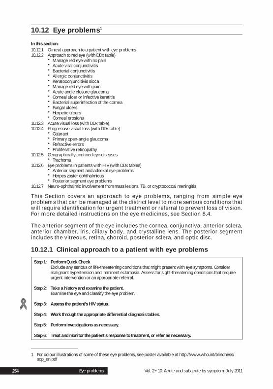
254 Vol. 2 • 10. Acute and subacute by symptom: July 2011
10.12 Eye problems1
In this section:10.12.1 Clinical approach to a patient with eye problems10.12.2 Approach to red eye (with DDx table)
• Manage red eye with no pain • Acute viral conjunctivitis• Bacterial conjunctivitis• Allergic conjunctivitis• Keratoconjuncitivis sicca• Manage red eye with pain• Acute angle closure glaucoma• Corneal ulcer or infective keratitis• Bacterial superinfection of the cornea • Fungal ulcers• Herpetic ulcers• Corneal erosions
10.12.3 Acute visual loss (with DDx table)10.12.4 Progressive visual loss (with DDx table)
• Cataract• Primary open-angle glaucoma• Refractive errors• Proliferative retinopathy
10.12.5 Geographically confi ned eye diseases• Trachoma
10.12.6 Eye problems in patients with HIV (with DDx tables)• Anterior segment and adnexal eye problems• Herpes zoster ophthalmicus• Posterior segment eye problems
10.12.7 Neuro-ophthalmic involvement from mass lesions, TB, or cryptococcal meningitis
This Section covers an approach to eye problems, ranging from simple eye problems that can be managed at the district level to more serious conditions that will require identifi cation for urgent treatment or referral to prevent loss of vision. For more detailed instructions on the eye medicines, see Section 8.4.
The anterior segment of the eye includes the cornea, conjunctiva, anterior sclera, anterior chamber, iris, ciliary body, and crystalline lens. The posterior segment includes the vitreous, retina, choroid, posterior sclera, and optic disc.
10.12.1 Clinical approach to a patient with eye problems
Step 1: Perform Quick CheckExclude any serious or life-threatening conditions that might present with eye symptoms. Consider malignant hypertension and imminent eclampsia. Assess for sight-threatening conditions that require urgent intervention or an appropriate referral.
Step 2: Take a history and examine the patient. Examine the eye and classify the eye problem.
Step 3: Assess the patient’s HIV status.
Step 4: Work through the appropriate differential diagnosis tables.
Step 5: Perform investigations as necessary.
Step 6: Treat and monitor the patient’s response to treatment, or refer as necessary.
1 For colour illustrations of some of these eye problems, see poster available at http://www.who.int/blindness/sop_en.pdf
Eye problems
FINAL Vol. 2 CH 9-21.indd 254FINAL Vol. 2 CH 9-21.indd 254 29/06/2012 15:1629/06/2012 15:16

255Vol. 2 • 10. Acute and subacute by symptom: July 2011
History
Specifi c questions
• What are the symptoms? ° pain ° light discomfort or photophobia ° red eye ° watering of the eyes ° discharge ° itching ° dryness ° sensation of foreign body.
• Is there a history of trauma?• What are the visual symptoms?
° loss of or decreased vision ° diplopia (double vision) ° halos around lights ° fl oaters (black or grey specks in the fi eld of vision) ° fl ashes of light.
• What is the duration of symptoms?• What is the progression of symptoms?• Look at the patient’s past history of similar symptoms.• Are there any locally endemic conditions in the community (e.g. trachoma,
onchocerciasis, conjunctivitis)?
General questions
• Systemic diseases – hypertension, diabetes mellitus, connective tissue disorders?
• Co-morbidities – HIV, malignancies?
Examination
Do a general physical examinationDo a specifi c eye examinationA basic ophthalmic examination may require the following:• a torch for gross examination of the eye structure and pupil response• blue light fi lter• Snellen chart or “E” chart for illiterate patients (or a LogMar chart)• fl uorescein drops or impregnated paper strips• short-acting mydriatric drops (e.g. 1% tropicamide) to dilate pupils –
remember to assess visual acuity and pupil response fi rst• direct ophthalmoscope• topical anaesthetic drops (e.g. lidocaine)• cotton buds to remove foreign body• handheld applanation tonometer (Schiotz or Puff tonometer), if available.
Eye problems
FINAL Vol. 2 CH 9-21.indd 255FINAL Vol. 2 CH 9-21.indd 255 29/06/2012 15:1629/06/2012 15:16

256 Vol. 2 • 10. Acute and subacute by symptom: July 2011
• Assess visual acuity
° Use a Snellen chart to measure distance acuity – this can be at any distance (e.g. 3 or 6 metres) as long as it is documented.
° Make sure the room is well lit. ° Have patient occlude one eye with palm (make sure patient is not peeking
and is not putting pressure on the occluded eye). ° Ask patient to read aloud each letter in the fi rst row, or indicate with the
hand what direction the E is pointing. Then proceed to the next smaller line until not able to distinguish all the letters on that line.
° Record the last line read accurately after the distance from the chart, for example 6/24 if the patient was 6 metres from the chart and the last line read was 24.
° If the patient is not able to read the top line, move forward a metre and retest up to 3 metres. If still not able to read top line, test whether the patient can count fi ngers and record this. If not able to count fi ngers, test whether the patient can detect hand motion. If not, test for light perception with a bright torch.
° Then test the other eye and record. ° Then test with glasses on. ° If acuity is less that 6/18 then assess again using pinhole and see if it
improves; if yes, then a refractive error is likely. ° Assess near vision using specifi c near vision acuity chart, or assess ability
to read newsprint or very crudely the ability to count fi ngers or detect motion.
° Assess colour vision using Ishihara plates or test for red desaturation (a good indicator of optic nerve dysfunction).
• Assess visual fi elds
° Use confrontation testing to detect gross defects (e.g. homonymous hemianopia – loss of half the visual fi eld). Confrontation testing compares the examiner's visual fi eld's with the patient’s, and may yield vital information that is often missed.
° Assessment of more discrete visual fi eld loss (e.g. from glaucoma) requires formal perimetry.
Eye problems
FINAL Vol. 2 CH 9-21.indd 256FINAL Vol. 2 CH 9-21.indd 256 29/06/2012 15:1629/06/2012 15:16

257Vol. 2 • 10. Acute and subacute by symptom: July 2011
• Examine the adnexa (attachments or structural components of the eye)
° Examine the eyebrows and eyelids. Look for ptosis (drooping), infection, congestion, hordeolum, warts, Molluscum contagiosum.
° Evert the eyelid to detect all foreign bodies. • Examine the conjunctiva
° Look for discharge, swelling, or congestion.• Examine the cornea
° Look for haziness, ulcers (herpetic or bacterial), and infl ammation. ° Test for sensory loss with a wisp of cotton wool. ° The naked eye may not detect corneal defects due to ulcerations or
abrasions. If there is trauma or a foreign body sensation in the eye, do fl uorescein staining as follows: ◊ Moisten a fl uorescein-impregnated strip with a drop of saline or artifi cial tears.
◊ Touch the strip to the inside of the lower lid. ◊ Illuminate the cornea with an ophthalmoscope with a blue fi lter if available (if not available, a strong white light may also reveal defects).
◊ Look for epithelial defects that stain bright green.• Examine both pupils
° Are they equal in size? ° Examine their reactivity to light. Shine a light alternately from one eye to
the other. ◊ A normal response is equal constriction of both pupils, regardless of which eye the light is directed at. This indicates an intact direct and consensual pupillary light refl ex.
◊ An abnormal response, or relative afferent pupillary defect (RAPD), will show that light directed in the affected eye will cause only mild constriction of both pupils (due to decreased response to light from the afferent defect), while light in the unaffected eye will cause a normal constriction of both pupils due to an intact afferent path and an intact consensual pupillary refl ex. Thus, light shone in the affected eye will produce less pupillary constriction than light shone in the unaffected eye.
• Examine the lens
° Look for any opacity (mature cataracts may be visible with torch examination.
• Perform fundoscopy to visualize the fundus, optic disc, and vessels
° Fundoscopy is an examination of the posterior segment of the eye and is done using a direct ophthalmoscope.
° It should take place in a suitably darkened room. ° The patient's right eye should be examined with the examiner's right eye,
and vice versa. ° The patient is asked to focus on an object in the distance, and the
examiner's head is kept vertical to permit this. ° This approach with the patient fi xing on a distant straight-ahead target
allows you to visualize the optic disc very easily. ° Turn to the +10DS lens in the lens wheel and observe the eye from 10 cm.
Slowly move closer to the patient and at the same time gradually reduce the power of the lens in the wheel and focus on the crystalline lens, the vitreous, and fi nally the fundus. The power of lens necessary to focus on the fundus will depend on any uncorrected refractive error in the patient or in the observer.
° Once a blood vessel on the fundus has been located, move along it and
Eye problems
FINAL Vol. 2 CH 9-21.indd 257FINAL Vol. 2 CH 9-21.indd 257 29/06/2012 15:1629/06/2012 15:16

258 Vol. 2 • 10. Acute and subacute by symptom: July 2011
locate the optic disc. While examining the disc, you will need to consider its colour, its margins, and the cup if there is one. Also, note the presence of optic atrophy and papilloedema.
° Retinal blood vessels should be examined in each quadrant after locating the disc. The veins are relatively large and dark red, while the arteries are relatively thin and brilliant red.
° Next, examine the retinal background, which is the normal retina between the blood vessels and other identifi able structures.
° To examine the macula and the fovea, instruct the patient to look into the ophthalmoscope light. ◊ This step will cause the pupil to constrict, dazzle the patient, and you will notice some troublesome corneal refl ections.
◊ These factors make the macula a diffi cult area to visualize. It may be useful to use a smaller aperture beam and slightly reduce the light intensity.
◊ The normal macula is the area between the superior and inferior temporal blood vessel arcades, and its centre is the fovea.
• Assess ocular movements
° Ask the patient follow your fi nger with their eyes without moving the head; test the 6 cardinal points of gaze in an H pattern.
° Look for failure of movement and for nystagmus (pause to check it during upward and lateral gaze).
• Assess ocular pressure
° If suspect glaucoma, press lightly on the globe with the fi ngers. Does the eye feel hard?
° If handheld tonometer is available, measure intraocular pressure.• Additional examination usually performed by specialized eye-care
professionals. ° Slit lamp examination.
Classify the problem as red eye (with or without pain) or visual loss and consult the relevant DDx table below. For HIV-infected patients, see also these specifi c sections.
Eye problems
FINAL Vol. 2 CH 9-21.indd 258FINAL Vol. 2 CH 9-21.indd 258 29/06/2012 15:1629/06/2012 15:16

259Vol. 2 • 10. Acute and subacute by symptom: July 2011
10.12.2 Approach to red eyeRed eye is commonly a sign of treatable conditions such as conjunctivitis. It is important to exclude more serious underlying conditions such as acute congestive glaucoma or iridocyclitis. Problems relating to the posterior chamber almost never present with a red eye.
Ask whether there is pain with the red eye.
DDx: Red eye
Condition In favour
Red eye with no pain
Infectious conjunctivitis• bacterial• viral
Painless red eye – little or no photophobia, no visual changesMucopurulent discharge with matting of eyelidsConjunctival congestionLid oedema if viralItchy, burning with gritty feelingLocal outbreaks in the community
Leptospirosis
see Section 11.22
History of conjunctival exposure to water contaminated with urine of infected animalsRed eyes (conjunctival suffusion) without purulent discharge involving both eyes – early phase of illness Fever and myalgias
Allergic conjunctivitis BilateralPainlessItchyWatery or mucoid (stringy and ropy) dischargeCobblestoning or raised visible bumps (papillary hypertrophia of the palpebral conjunctiva)Conjunctival infl ammation (chemosis) may occurMultiple creases in lower lidSeasonalImproves on antihistamines
Subconjunctival haemorrhage Bleeding beneath the conjunctivaPainless, harmlessAssociated with minor trauma, hypertension, increased venous pressure as in coughing, sneezing, vomiting, straining
Pinguecula or pterygium Triangular band of fi brosis next to the cornea is a pinguecula; if encroaching on the cornea, a pterygiumPresent for weeks but may become acutely infl amedPainless although may feel gritty or dryMight decrease visual acuity if advanced and encroaches on the central part of the cornea (astigmatism)
Red eye with pain
Keratoconjuncitivis sicca Burning or foreign body sensationLack of tears or occasionally excessive tearingCommon in children with HIV, Stevens Johnson syndrome, female hormonal diseases, other autoimmune diseases
Eye problems
FINAL Vol. 2 CH 9-21.indd 259FINAL Vol. 2 CH 9-21.indd 259 29/06/2012 15:1629/06/2012 15:16

260 Vol. 2 • 10. Acute and subacute by symptom: July 2011
Condition In favour
Corneal infl ammation or infection (keratitis)
Severe eye painVariable degree of decreased visual acuity (depending on location of ulcer) and photophobiaHaziness or opacifi cation of the cornea (with or without fl uorescein uptake) Pupillary constriction (secondary to ciliary spasm and iritis)Presence of pus or white cells in anterior chamber (hypopyon)
Ultraviolet kerato-conjunctivitis (welder's arc, carbon arc, sunlight)
Foreign body sensationHistory of welding work or extended sunlight exposure Pain Photophobia Tearing
Thermal or chemical keratoconjunctivitis
History of splash or exposure to heat or chemicalsBurns to eyelid or skin Singed eyelashesFlat thin haemorrhage or thicker collection of blood
Foreign body or corneal abrasion
Intense pain localized to cornea after an injuryEvidence of corneal abrasion with fl uorescein stainForeign body may be seen directly or on eyelid eversion
Corneal ulcer Round or oval sharply demarcated border with the base appearing ragged and grayPainfulBlurred vision Blue/green mucopurulent discharge with fl uorescence in UV light in favour of P. aeruginosaHerpetic ulcer: dendritic branches on fl uorescein staining Varicella zoster virus (VZV) ulcer: loss of sensation with shingles on ophthalmic branch of trigeminal nerve typical for VZVHypopyon (sterile pus in lower internal part of cornea)
Acute angle closure glaucoma(or closed angle)
Sudden loss of visionSevere headache and eye pain Nausea and vomitingHalos around lightPupils mid-dilated, vertically oval non-reactive to light (sluggish)Very high intraocular pressure >30 mmHg – eye will feel hard on digital pressure
Acute anterior uveitis (iritis or iridocyclitis)
Acute onsetUnilateral, painful red eye, blurred vision, direct and consensual photophobia, tearingCiliary (not only conjunctival) injection360 degree perilimbal fl ush
Acute posterior uveitis (choroiditis or chorioretinitis)
Blurred vision with fl oatersOccasional pain Photophobia
Eye problems
FINAL Vol. 2 CH 9-21.indd 260FINAL Vol. 2 CH 9-21.indd 260 29/06/2012 15:1629/06/2012 15:16

261Vol. 2 • 10. Acute and subacute by symptom: July 2011
Manage red eye with no pain:
Acute viral conjunctivitis
Acute viral conjunctivitis is commonly caused by adenovirus. It is highly contagious and occurs in small epidemics in households, schools and the community.
Key clinical features
• Acute onset with painless red eye – little or no photophobia, no visual changes.
• Discharge, matted eyelashes, redness and infl ammation (chemosis).• Monitor for secondary bacterial infection – which is common.• Suspect herpetic conjunctivitis if there are skin fi ndings, or if there is skin
involvement (clusters of vesicles on an erythematous base on the face, eyelids. and mucous membranes typical of herpes simplex virus; shingles along ophthalmic nerve dermatome suggestive of herpes zoster virus). Follow management specifi c for these conditions (see below and Section 10.12.6).
• Clinical diagnosis and culture or smears are not needed.
Treatment
• If suspect bacterial coinfection, use gentamicin2 or chloramphenicol eye drops or ointment (see Section 8.4).
• Use cold compresses several times a day. • Give tears substitute such as carboxymethyl cellulose 0.5% drops or 1% gel-
tears naturale.
Prevention
• Avoid contact between fi ngers and eyes.• Meticulous hand washing.• Separate face and hand towels at home for the affected person.
Bacterial conjunctivitis
Key clinical features
• Mucopurulent discharge present.• May occur as a primary infection or is secondary to a viral infection.• Gram stain of the conjunctival smear will help to identify possible organism.• Hyper-acute gonococcal infection - sudden onset of extreme infl ammation and
purulent discharge from the eyes; there may be a history of urethral discharge or other signs of a sexually transmitted infection. This is an ophthalmic emergency. Refer urgently to an ophthalmology unit in case of gonococcal infection as it can rapidly infect the cornea and lead to a severe keratitis or perforation of the cornea. Initiate treatment below in the meantime.
• Trachoma or chlamydial conjunctivitis in endemic areas – usually presents with a chronic red eye with stringy discharge. See Section 10.12.5.
Eye problems
FINAL Vol. 2 CH 9-21.indd 261FINAL Vol. 2 CH 9-21.indd 261 29/06/2012 15:1629/06/2012 15:16

262 Vol. 2 • 10. Acute and subacute by symptom: July 2011
Treatment
• Conjunctivitis due to Neisseria gonorrhoeae requires systemic antibiotics and urgent referral.
° ceftriaxone 1 gram IM OR ° spectinomycin 2 g by deep IM; OR ° ciprofl oxacin 500 mg orally.
• If suspect Chlamydia trachomatis, follow instructions in Section 10.12.5.• If neither of these, for other bacterial conjunctivitis give topical antibiotic
drops such as gentamicin eye drops 4–6 times daily.2
PreventionWhen conjunctivitis is associated with an STI, treat sexual partners to minimize recurrence and spread of disease.
Allergic conjunctivitis
Key clinical features
• Itching is a cardinal sign.• Watery with minimal discharge.• Congestion and chemosis (swelling) with papillary reaction.• May be acute and periodic in seasonal allergic conjunctivitis.• Mild and chronic in atopic or perennial allergic conjunctivitis.
Treatment
• Allergen avoidance.• Topical sodium cromoglycate drops during the allergy season is the mainstay
of treatment and is safe even if given for long periods.
Keratoconjunctivitis sicca
This is also called dry eye syndrome.
Key clinical features
• Burning, foreign body sensation, lack of tears.• Occasionally excessive tears reacting to irritation.• Common in premenopausal and menopausal women and collagen vascular
diseases.• In PLHIV,may be a consequence of previous Stevens Johnson syndrome.
Common in HIV-positive children.
Treatment
• Give tear substitute such as carboxymethyl cellulose 0.5% drops or 1% gel-tears naturale.
• Treat secondary bacterial infection, if present.
2 Gentamicin eye drops are on the WHO EML. An alternative is chloramphenicol eye drops. .
Eye problems
FINAL Vol. 2 CH 9-21.indd 262FINAL Vol. 2 CH 9-21.indd 262 29/06/2012 15:1629/06/2012 15:16

263Vol. 2 • 10. Acute and subacute by symptom: July 2011
Manage red eye with pain
Acute angle-closure glaucoma
This is due to increased intraocular pressure that leads to impairment of the functioning of the optic nerve and causes visual fi eld loss, ultimately leading to blindness. It is important to recognize and refer urgently for treatment to prevent visual loss.
Key clinical features
• More common among Asians.• Usually bilateral disease, but acute onset often unilateral.• Sudden loss of vision with halos around lights – often worse in the evenings.• Associated with severe headache, vomiting, eye pain.• Stage of primary angle closure glaucoma.• Evidence of ciliary congestion, lid oedema, hazy cornea, shallow anterior
chamber.• A mid-dilated pupil (4 to 6 mm) that reacts poorly to light.• Intraocular pressure is very high – eye feels stony hard on digital pressure.• Fundoscopy – "cupping" of the optic nerve.
Treatment
• Urgent referral to specialist facility for further treatment.• Start the following medication until the patient reaches referral centre:• pilocarpine 1 drop (2% solution) every 10 minutes for 30–60 minutes, then 1
drop every 1–3 hours until intraocular pressure subsides, then 1 drop 4 times daily; PLUS
• acetazolamide 250 mg orally stat, or IV if the patient is vomiting, then continue oral acetazolamide 500–750 mg per day.
Corneal ulcer or infective keratitis
• Common cause of painful loss of vision with red eye.• Very often follows trauma that could be trivial.• Offending organisms include bacteria, fungi, virus (such as herpes or
facilitated by HIV immunosuppression).
Treatment
• Urgent referral to specialist
• Do not use steroids for a corneal ulcer as the infection may spread resulting in corneal perforation.
• Start empirical broad spectrum antibiotic drops in all patients – chloramphenicol or gentamicin eye drops 6 times daily.
Bacterial superinfection of the cornea secondary to trauma
• Diagnosis can be made by scraping margins of the ulcer and plating for bacterial and fungal cultures. This should be done by a trained ophthalmologist, using a microscope or loupe.
Eye problems
FINAL Vol. 2 CH 9-21.indd 263FINAL Vol. 2 CH 9-21.indd 263 29/06/2012 15:1629/06/2012 15:16

264 Vol. 2 • 10. Acute and subacute by symptom: July 2011
Refer for specialist assistance.
• P. aeruginosa infection causes a ground glass appearance of the cornea, and may lead to early perforation of the cornea.
• Staphylococcal infections are less dramatic, but may cause loss of vision.
Treatment
• Urgent referral to specialist facility.• Initiate empirical antibiotic drops – chloramphenicol or gentamicin hourly or
any other available broad spectrum antibiotic drop until the patient reaches the referral centre.
• Give supportive treatment with a mydriatic (atropine eye drops) and timolol to reduce intraocular pressure.
• If there is not copious discharge, give an eye patch to rest the eye.
Fungal ulcers
• Fungal ulcers are commonly caused by fi lamentous fungi or yeasts (candida).• May occur in those who work in rural settings or those engaged in agricultural
activity and in PLHIV.
Key clinical features
• Ulcers may follow trivial trauma.• Symptoms may be mild even with large ulcers.• Ulcers are dry, elevated, with feathery margins.
Treatment
• Urgent referral to tertiary level facility.• Treat as in bacterial ulcers.
Herpetic ulcers
Herpes simplex virus (HSV) is a major cause of blindness worldwide from corneal scarring and opacity after cornea infection, infl ammation (keratitis), and ulceration. See also Section 11.15.
Key clinical features
• Painful eye with blurred vision and discharge.• Dendritic ulcers are seen on fl uorescein staining of the cornea.• May coalesce to form geographic ulcers (ameboid shape, often with dendritic
extensions at the edges).• Are usually unilateral.• Loss of corneal sensation if tested with wisp of cotton wool.
Treatment
• Refer urgently to tertiary level facility.• Treat with topical aciclovir ointment and do not use steroids.
• If topical aciclovir ointment is not available, give oral aciclovir, 400 mg 5 times daily.
• Administer supportive treatment with topical atropine.
Eye problems
FINAL Vol. 2 CH 9-21.indd 264FINAL Vol. 2 CH 9-21.indd 264 29/06/2012 15:1629/06/2012 15:16

265Vol. 2 • 10. Acute and subacute by symptom: July 2011
Corneal erosions
Key clinical features
• Areas of epithelial loss without infi ltration.• Usually follows trivial trauma – may be a history of contact lens use or even
fi ngernail trauma.• Very painful.• If history of trauma is present, rule out penetrating ocular trauma.
Treatment
• Evert eyelids to look for foreign bodies.• Treat with antibiotic ointment (tetracycline) and rest with an eye patch.• Refer to specialist care if there is non-healing or delayed healing, or recurrent
erosions.• Never use traditional medicines.
10.12.3 Acute visual loss• Acute loss of vision is usually secondary to posterior segment diseases.• All require urgent referral to a specialist facility.• These conditions may or may not be associated with pain.• They are usually not associated with “red eye”.
DDx: Acute visual loss
Condition In favour
Central retinal artery occlusion Sudden loss of visionPainless Fundoscopy – cherry red spot at fovea
Central retinal vein occlusion Visual loss less dramatic – painless loss of vision Older ageKnown hypertensive, sickle-cell disease or blood dyscrasias, or patient with high intraocular pressureFundoscopy – retinal haemorrhages
Retinal detachment Photophobia, fl ashing lights, fl oatersRecent blunt trauma or known sickle-cell diseaseHigh myopia, aphakiaPoor or partial red refl ex
Vitreous haemorrhage Visual loss mild to severeFollowing diabetic retinopathy or retinal vein occlusion
CMV retinitis
see Section 11.8
FeverSigns of meningitis or pneumonitisFundoscopy – haemorrhagic retinal necrosis (“pizza” pie appearance)Very low CD4 (<100)Visual loss may be slower in onset in early CMV disease and may not be noticed by the patients until advanced
Ocular toxoplasmosis Unifocal in HIV-negative, multifocal in HIV-positiveBlurred or hazy vision with fl oaters – erosions may look like headlights in the fog through the vitreous haze Occurs with vitritis
Eye problems
FINAL Vol. 2 CH 9-21.indd 265FINAL Vol. 2 CH 9-21.indd 265 29/06/2012 15:1629/06/2012 15:16

266 Vol. 2 • 10. Acute and subacute by symptom: July 2011
Condition In favour
Optic neuritis Females : Males = 2 : 1Unilateral rapid visual lossRetro-orbital pain with eye movementAssociated with viral illness, multiple sclerosis, systemic lupus erythematosis (SLE), post-meningitis – TB or cryptococcal
Acute congestive glaucoma Sudden loss of visionSevere headache and eye pain Nausea and vomitingHalos around lightPupils mid-dilated, vertically oval non-reactive to light (sluggish)Very high intraocular pressure >30 mmHg or eye feels hard on digital pressure
Endophthalmitis Sudden painful visual loss following intraocular surgery or penetrating trauma or corneal ulcerRed eyePus in the anterior chamber (hypopyon)Infl ammation of all inner layers of the eye
10.12.4 Progressive visual loss• Painless gradual loss of vision may be a result of various causes. • Diseases of the anterior chamber are usually amenable to treatment, while
diseases of the posterior segment require urgent referral.
DDx: Progressive visual loss
Condition In favour
Cataract Old age, history of trauma or as part of a systemic conditionMay be unilateral or bilateralOpacities seenFundoscopy – unable to see the fundus; red refl ex obscured
Refractive errors Common cause of visual loss in childrenImprovement of vision with pinhole technique confi rms refractive error
Corneal dystrophies Painless loss of vision
Chronic open angle glaucoma Late-stage visual lossCupping of optic disc on routine examinationIntraocular pressure may be raised or normalCharacteristic visual fi eld changes on manual or perimetry are diagnostic
Hereditary macular degeneration
Painless central loss of visionVisual acuity decreased
Proliferative retinopathy e.g. diabetes mellitus, sickle-cell disease
Longstanding DM poorly controlled or known sickle-cell diseaseOther risk factors: hypertensive, nephropathy, pregnancyFundoscopy: presence of microaneurysms, hard exudates, retinal oedema, proliferative retinopathy
Eye problems
FINAL Vol. 2 CH 9-21.indd 266FINAL Vol. 2 CH 9-21.indd 266 29/06/2012 15:1629/06/2012 15:16

267Vol. 2 • 10. Acute and subacute by symptom: July 2011
Cataract
Key clinical features
• Opacifi cation of the lens with decreased vision.• Usually due to advancing age, but may also occur in younger population due
to trauma or systemic disease, or may be present at birth or early years.• Clinically reduced clarity of lens, obscuring the view of the retina.
Treatment
• Referral to specialist for surgical extraction of the lens with intraocular lens implantation.
Primary open-angle glaucoma
Key clinical features
• Important cause for irreversible visual loss.• Early diagnosis of disease is important to prevent visual loss.• Visual loss in late stages.
Treatment
• Referral to specialist facility for detailed evaluation and treatment.• Start pilocarpine to lower intraocular pressure.
Refractive errors
• A common cause of gradual progressive reduction of vision.• Improvement in vision with pinhole or spectacles likely indicates refractive
error.• Regular and repeated screening, particularly in children and adolescents, to
allow timely detection and treatment.• Short-sighted vision or myopia is the diffi culty to see distant objects, while
near objects are clear.• Far-sighted vision (hyperopia and presbyopia) is the diffi culty to see near
objects. In presbyopia, this is due to loss of accommodation, often after 40 years of age.
• Uncorrected refractive errors are a common cause of headache.
Treatment ° Corrective lenses ° Repeated examinations throughout growth to prevent poor vision, if left
uncorrected.
Proliferative retinopathy
• This is a disease of the pre-capillary arteriole and venules of the retina.• In diabetics, increasing duration of diabetes is the most important risk factor.
Poor glycaemic control, concomitant HPT, nephropathy, and pregnancy are also signifi cant risk factors.
• In sickle-cell disease, a history of multiple crises will increase the likelihood of retinopathy.
Eye problems
FINAL Vol. 2 CH 9-21.indd 267FINAL Vol. 2 CH 9-21.indd 267 29/06/2012 15:1629/06/2012 15:16

268 Vol. 2 • 10. Acute and subacute by symptom: July 2011
Treatment
• Stringent glycaemic control in diabetics.• Regular eye checks - annually if possible.• Referral to specialist centre for laser treatment if available; retinal
photocoagulation can be vision-saving.
10.12.5 Geographically confi ned eye diseases – onchocerciasis and trachoma
Table: Geographically confi ned eye diseases
Condition Geographic area In favour
Onchocerciasis
see Section 11.28
Onchocerciasis is found in 36 countries in Africa as well as in Guatemala, southern Mexico, some areas of Venezuela, small areas in Brazil, Colombia, and Ecuador; and in Yemen.
Infl ammation of the eyeAssociated skin fi ndings (itching), skin nodules fi ne papular rashLacrimation, photophobia and itching in eyeEarly – night blindness, narrowed visual fi eld Late – painful iritis or glaucoma
Trachoma Occurs globally.Specifi cally in Africa: Algeria, Burkina Faso, Chad, Djibouti, Ethiopia, Gambia, Ghana, Guinea, Guinea-Bissau, Kenya, Malawi, Mali, Mauritania, Mozambique, Niger, Nigeria, Senegal, Togo, United Republic of Tanzania, Zambia.Eastern Mediterranean: Egypt, Morocco, Oman, Pakistan, Sudan, South Sudan, Yemen.South-East Asia: India, Myanmar, Nepal.Western Pacifi c: Australia, Cambodia, China, Viet Nam. Americas: Brazil, Guatemala, Mexico.
Easily visible corneal opacity over the pupil Pupil margin blurredSubstantial prevalence in the community – with severe visual loss and potentially disabling trachomatous lesions such as trichiasis and entropion (see trachoma grading system in text below).
Trachoma
Trachoma is the result of infection of the eye with Chlamydia trachomatis. Infection spreads from person to person, and is frequently passed from child to child and from child to mother, especially where there are shortages of water, many fl ies, and crowded living conditions. Infection often begins during childhood then becomes chronic. If left untreated, irreversible blindness typically occurs between 30 and 40 years of age, mainly in females.
Key clinical features
• Eversion of the eyelid shows red, swollen conjunctiva with follicles (whitish dots 0.2–2 mm) or papillae (visible red dots), scars, which can ultimately cause the in-turning of the upper eyelid (entropion trichiasis).
• Damage to the cornea resulting in corneal opacities.• Other signs: limbal follicles (follicles at the upper edge of the cornea),
herbert's pits (small round clear windows at the upper edge of the cornea), and pannus (gradual opacifi cation of the of the upper part of the cornea).
Eye problems
FINAL Vol. 2 CH 9-21.indd 268FINAL Vol. 2 CH 9-21.indd 268 29/06/2012 15:1629/06/2012 15:16

269Vol. 2 • 10. Acute and subacute by symptom: July 2011
Simplifi ed grading of trachoma disease3
• Trachomatous infl ammation (TF): 5 or more follicles (at least 0.5 mm in diameter) on the upper tarsal conjunctiva.
• Trachomatous infl ammation intense (TI) presents as infl ammatory thickening of the upper tarsal conjunctiva that obscures more than half of the normal deep tarsal vessels.
• Trachomatous scarring (TS): scarring (white fi brosis) of the tarsal conjunctiva.• Trachomatous trichiasis (TT): at least one eyelash runs on the eyeball.• Corneal opacity (CO): central corneal opacity that is so dense that at least one of the pupil margins is blurred.
TreatmentThe WHO SAFE strategy4 to eliminate trachoma and prevent blindness includes:Surgery for trichiasis – corrective lid surgeryAntibiotics to treat Chlamydia trachomatis infection in diagnosed patients Facial cleanliness and promotion of hygieneEnvironmental improvement to reduce transmission of Chlamydia trachomatis from one person to another.• The mainstay of treatment is tetracycline ointment. Apply 1% ointment
directly to both eyes twice daily for 6 weeks• Give oral azithromycin, 20 mg/kg up to1 gm once a year as a family treatment
or community intervention.• Refer patients with corneal opacity for specialist care.
10.12.6 Eye problems in patients with HIV infectionOcular manifestations of HIV infection are very common and varied. Simple infections and common eye diseases must be recognized, treated and differentiated from sight-threatening problems that may require referral or specialist management.
These conditions are not specifi c to patients with HIV, but occur more commonly, with greater severity, and often with atypical features
Anterior segment and adnexal eye problems in patients with HIV infection
DDx: Anterior segment and adnexal eye problems in patients with HIV infection
Condition In favour
Molluscum contagiosum* Pearly white papules, central umbilicationMultiple lesionsUnusually large in patients with HIV and do not resolve spontaneously
Papillomata* Skin coloured – hyperpigmented papules or warty lesionsRough surface, on lids or conjunctiva
Herpes zoster ophthalmicussee Section 11.45
Vesicular rash , dermatomal distribution – trigeminal nerveAcute pain, can involve tip of nose (Hutchinson sign)Can cause conjunctivitis, scleritis, keratitis, uveitis, glaucoma, nerve palsy
3 Trachoma simplifi ed grading card with color illustrations. Available at http://www.who.int/blindness/causes/trachoma_documents/en/index.html
4 Trachoma control: a guide for programme managers. WHO, LSHTM, and International Trachoma Initiative, 2006. Available at http://www.who.int/blindness/publications/tcm%20who_pbd_get_06_1.pdf
Eye problems
FINAL Vol. 2 CH 9-21.indd 269FINAL Vol. 2 CH 9-21.indd 269 29/06/2012 15:1629/06/2012 15:16

270 Vol. 2 • 10. Acute and subacute by symptom: July 2011
Condition In favour
Conjunctival squamous cell carcinoma*
Recent papular lesion with rapid increase in size, irregular surface and marginsGrey/white rough foamy fungating appearance in interpalpebral zoneDiffers from pterygium with smooth shiny epitheliumTends to be very aggressive with recurrence if only simple excision used
Kaposi sarcoma* Red purplish caulifl ower-like lesion on conjunctiva or eyelid – can be fl at or nodularDistinguish from subconjunctival haemorrhage
Kerato conjunctivitis sicca Burning, dry, foreign body sensationCornea lustrelessTypical stain with Rose Bengal dye
* Refer to a specialist if molluscum contagiosum, papillomata (warts), conjunctival tumours, or Kaposi sarcoma.
Herpes zoster ophthalmicus – see Section 11.45
Key clinical features
• Unilateral vesicles and blisters involving the eye• More severe cases have corneal involvement• May have an atypical presentation and involve both eyes• May cause conjunctivitis, corneal lesions, keratitis, scleritis, uveitis, papillitis,
and, in rare instances, retinal necrosis
Treatment (all of the following as appropriate)
• oral aciclovir 800 mg 5 times daily for 7 days• aciclovir 3% eye ointment applied into the eye every four hours• antibiotic eye ointment (chloramphenicol)• antibiotics for secondary skin infection if present• analgesia – paracetamol or stronger analgesics if necessary (see Section 20)• amitryptiline 25–50 mg before bed for neuropathic pain• assess for complicated disease and refer.
Posterior segment eye problems in patients with HIV infection
• Most HIV-associated posterior segment diseases of the eye are associated with visual loss.
• Loss of vision may be acute in onset and rapidly progressive.• Early recognition and treatment will help in preventing irreversible loss of
vision.• All HIV-positive patients with any visual complaints require urgent referral to
a specialist facility.
Eye problems
FINAL Vol. 2 CH 9-21.indd 270FINAL Vol. 2 CH 9-21.indd 270 29/06/2012 15:1629/06/2012 15:16

271Vol. 2 • 10. Acute and subacute by symptom: July 2011
DDx: Posterior segment eye problems in patients with HIV infection
Condition In favour
Retinal microvasculopathy Multiple cotton wool spots, haemorrhageMicroaneurysmAsymptomaticNo visual loss
CMV retinitis
see Section 11.8
Painless loss of vision, large areas of retinal necrosis yellowish white, with areas of haemorrhage or necrosis ("pizza pie" appearance)No vitreous haze – fundi are clearly seenVascular sheathing – “frosted branch” appearanceExamine the other eye if this is diagnosed in one eyeCD4 <100 – occurs less commonly if >100 (look for other cause of retinopathy)
Optic disc swelling If bilateral with normal vision and refl ex, then it is papilloedemaIf unilateral with decreased vision, then it is optic nerve disease
Immune recovery uveitis Follows CMV of the eye most commonly, but also associated with other retinal opportunistic infectionsDecreased vision of gradual onset with fl oatersRecent initiation of ARTCan result in cataract, epiretinal membrane, retinal neovascularisation that may threaten sightPoor prognosis – focus on protecting the sight of the other eye
Chorioretinitis from toxoplasmosissee Section 11.40
Posterior uveitis, may be focal, or diffuse mimicking CMVSerology for anti-CMV IgM if available to exclude CMV
10.12.7 Neuro-ophthalmic involvement from mass lesions, TB, or cryptococcal meningitis • TB and cryptococcal meningitis in HIV-infected patients can result in
papilloedema, papillitis, or ocular nerve palsies (see Section 10.10a) or may present with other neurological presentations.
• As acute or progressive loss of vision can occur with TB and cryptococcal meningitis, examine the eyes routinely after diagnosis.
• Papilloedema is not associated with early visual loss, but if long-standing, may cause progressive visual loss.
• Papillitis is associated with acute visual loss.
Eye problems
FINAL Vol. 2 CH 9-21.indd 271FINAL Vol. 2 CH 9-21.indd 271 29/06/2012 15:1629/06/2012 15:16

272 Vol. 2 • 10. Acute and subacute by symptom: July 2011
10.13 Painful joints1
In this section:10.13.1 Clinical approach to a patient with painful joints10.13.2 Diagnosis of single and multiple painful joints
• DDx: Single painful joint – monoarthritis• DDx: Multiple painful joints• Table: Characteristics of synovial fl uid by diagnosis• Septic arthritis• Crystal deposition disease (gout, pseudo-gout) • Osteoarthritis• Rheumatoid arthritis • Gonococcal arthritis
10.13.3 Symptom management
Most joint pain may be a result of chronic conditions of varying duration. However, a substantial number of patients with joint problems require immediate and ongoing care. Prompt treatment can help limit symptoms, prevent disability, and improve outcomes. Follow a stepwise approach in the evaluation and management of painful joints.
Both laboratory studies and diagnostic imaging can help to evaluate the joint and the joint pain. At the initial evaluation, and at each subsequent re-evaluation, there should be efforts to identify underlying conditions that may need disease-specifi c management.
10.13.1 Clinical approach to a patient with painful joints
Step 1: Use Quick Check to identify severe conditions that require immediate medical or surgical care. Generally, an acute onset of joint pain with a prior trauma or appearance of warmth and swelling could indicate conditions such as dislocation, fracture, or a septic joint that requires more immediate attention. Other indicators of potential musculoskeletal emergency include constitutional symptoms (fever), numbness, or weakness.
Step 2: Take a history and examine the patient.
Step 3: Assess the patient’s HIV status.
Step 4: Work through the differential diagnosis table. Step 5: Perform appropriate investigations.
Step 6: Initiate treatment and monitor the patient’s response.
History
• Is there any history of trauma or injury?• location of the pain:
° one or multiple joints ° true joint pain, pain from nearby bone, ligament, tendon, bursa, muscle, or
referred pain.• character of the pain:
° quality – burning, sharp, constant, intermittent
1 See also the manual Surgical Care at the District Hospital, Section 19. WHO, 2003. Available at http://www.who.int/surgery/publications/scdh_manual/en/
Painful joints
FINAL Vol. 2 CH 9-21.indd 272FINAL Vol. 2 CH 9-21.indd 272 29/06/2012 15:1629/06/2012 15:16

273Vol. 2 • 10. Acute and subacute by symptom: July 2011
° factors which worsen or improve pain ° diurnal rhythm – worse in the morning and eases at night, or worsens at the
end of the day, unrelenting, or nocturnal.• onset and progression, joints that were affected fi rst• symmetry of joint involvement• stiffness• swelling• limitation of motion• constitutional symptoms, such as weight loss, unexplained fevers, chills,
cough, night sweats.• associated problems:
° weakness or numbness of affected limb ° gastrointestinal, genitourinary or eye problems.
• family history of joint disease and rheumatic conditions• history of immunosuppression – HIV, immunosuppressive medicines• drug history – particularly diuretics• history of tick bite.
Examination
Specifi c examinationAssess for:• erythema (redness) or ecchymosis• warmth• evidence of any infective focus – e.g. a septic wound, an ulcer• crepitus, laxity, gross deformity, tendon, or muscle dysfunction (tested by
resisted function) • joint swelling – hypertrophy or a joint effusion• sensory changes indicate possible neurological or vascular problems• range of motion (ROM)• ability to bear weight • contractures, bone deformities• weakness• pain that is out of proportion to the injury• limping, if lower extremities are involved• palpable, bony hypertrophy.
General examination
• extra-articular features of joint disease ° cutaneous nodules ° cutaneous vasculitis lesions.
• lymphadenopathy• oedema• ocular infl ammation• urethritis• tenosynovitis (tendon sheath effusions)
Painful joints
FINAL Vol. 2 CH 9-21.indd 273FINAL Vol. 2 CH 9-21.indd 273 29/06/2012 15:1629/06/2012 15:16

274 Vol. 2 • 10. Acute and subacute by symptom: July 2011
• bursitis (swollen bursa)• diarrhoea• orogenital ulceration.
Assess the patient’s HIV statusHIV can cause either monoarthritis or polyarthritis at any CD4 count.
10.13.2 Diagnosis of single and multiple painful jointsThese tables are divided into conditions affecting single joints, and those affecting multiple joints.
DDx: Single painful joint – patient presenting with monoarthritis
Condition In favour
Trauma History of joint injury or traumaPain, swelling, ecchymosisBleeding into joint may be presentDecreased range of motion
Bursitis Pain with motion and localized tendernessHistory of acute trauma or repetitive injury to the areaSwelling over bursaOften involves knees or elbowsIf fever or recent bacterial infection, may be septic bursitis
Septic arthritis(usually Staphylococcus aureus or streptococcal species)
History of diabetes, IV drug use, sickle-cell diseaseAcute onset Large joint (knee most common, or also hip, shoulder, wrist, or ankle) May localize to joints with pre-existing arthritis or prior traumaSystemically ill with feverHot tender swollen joint with decreased mobilityMore than one joint may be involvedHigh WBC and ESRJoint fl uid WBC >50 000 (>90% neutrophils)
Gonococcal arthritissee Section 11.13
Purulent arthritis, usually knees, wrists, or ankles (see septic arthritis)May have preceding skin lesions: papules and pustules, polyarthralgia, and tenosynovitisSexually active, 25% have GU symptomsSynovial fl uid WBC >50 000 (>90% neutrophils)
Gout On diureticsDietary and alcohol overindulgencePrevious similar attacksSevere joint painRedness, hot, tender, and swollenTypically affects the base of the big toeGouty tophi
Rheumatoid arthritis AutoimmuneEarly morning stiffness lasting over 1 hourEarly stages may present as monoarthritisLater symmetrical polyarthritis: often hand and wrist involvementFixed deformities developExtra-articular manifestations: fever, anorexia, malaise, weight lossRheumatoid nodules: subcutaneous, usually extensor surfacesElevated ESR, CRP, platelet count; low Hb; positive rheumatoid factor
Painful joints
FINAL Vol. 2 CH 9-21.indd 274FINAL Vol. 2 CH 9-21.indd 274 29/06/2012 15:1629/06/2012 15:16

275Vol. 2 • 10. Acute and subacute by symptom: July 2011
Osteoarthritis (degenerative joint disease)
Gradual onsetUsual age of onset over 40 Pain on weight-bearing or activity that is eased by restStiffnessCrepitusBony enlargementTenderness to palpationDecreased range of motionSynovial fl uid – clear, WBC <2000/mm3, normal viscosity
Tuberculous arthritissee Section 15
Gradual onset joint swellingMildly infl amed, presentation may vary based on siteLow-grade feverNight sweatsSynovial fl uid: increased protein, increased WBC – predominantly lymphocytes Vertebral TB: X-ray of spine may show vertebral collapse
DDx: Multiple painful joints
Condition In favour
Gonococcal arthritissee Section 11.13
Sexually activeGenitourinary symptomsSystemically illMigratory polyarthralgia or polyarthritis (often asymmetric), tenosynovitis (asymmetric) involving fi ngers, hands, or wristsSkin lesions – papules and pustulesSeptic arthritis may follow dermatitis if untreated
Spondyloarthropathies(ankylosing spondylitis, psoriatic arthritis)
Family historyBack pain, uveitis, urethritis, gastrointestinal symptoms, and rashesLarge joints of lower extremities
Rheumatoid arthritis AutoimmuneEarly morning stiffness lasting over 1 hourSymmetrical polyarthritis: often hand and wrist involvementFixed deformities developRheumatoid nodulesHigh ESR, CRP, platelet count; low Hb; positive rheumatoid factor
Osteoarthritis (degenerative joint disease)
Gradual onsetUsual age of onset over 40 Pain on weight-bearing or activity that is eased by restStiffnessCrepitusBony enlargementTenderness to palpationDecreased range of motionFixed deformitiesSynovial fl uid: clear, WBC <2000/mm3, normal viscosity
Chikungunya Viral illness spread by infected mosquitoSevere joint painsAssociated fever and rashOccurs in outbreaks or rarely in sporadic casesDiagnosed by serum antibody titres or PCR
HIV-associated arthritis Usually of limited duration, <6 weeksMono or polyarthritisOccurs predominantly in lower extremitiesCan occur at any CD4 count
Painful joints
FINAL Vol. 2 CH 9-21.indd 275FINAL Vol. 2 CH 9-21.indd 275 29/06/2012 15:1629/06/2012 15:16

276 Vol. 2 • 10. Acute and subacute by symptom: July 2011
Perform investigations if required
Consider investigations according to the suspected underlying cause.• erythrocyte sedimentation rate (ESR)• C-reactive protein (CRP)• rheumatoid factor• anti-nuclear antibody• uric acid.
Arthrocentesis is urgently indicated when there is a warm, red joint with effusion, especially when there is no history of trauma. The aspirated synovial fl uid should be sent for cell count, chemistry, microscopy, Gram stain, culture, and crystals.
Table: Characteristics of synovial fl uid by diagnosis
Condition Appearance WBC/mm
Normal Clear <200
Infl ammatory Turbid 5000–50 000
Non-infl ammatory Clear <400
Gout Turbid 2000–5000
Pseudo-gout Turbid 5000–50 000
Septic arthritis Purulent/turbid >50 000
Diagnostic imaging
• Plain X-rays remain the choice for the diagnosis of most joint abnormalities. • Computed tomography (CT) and magnetic resonance imaging (MRI) may be
benefi cial when the diagnosis is not clear.
Septic arthritis
Common presentations and features• most dangerous cause of acute arthritis; knee the most common site• usually of bacterial origin, but can be viral, fungal, or mycobacterial• resulting from bacteraemia, soft tissue infection, bite• much more likely in patients with pre-existing joint disease, especially
rheumatoid arthritis• common organisms implicated are Staphylococcus aureus and streptococci• N. gonorrhoea in young adults• E. coli in the elderly, drug users, and very ill patients• S. aureus, M. tuberculosis, Salmonella spp. and Brucella spp. – may cause
septic spinal (vertebral) arthritis or osteomyelitis.
Painful joints
FINAL Vol. 2 CH 9-21.indd 276FINAL Vol. 2 CH 9-21.indd 276 29/06/2012 15:1629/06/2012 15:16

277Vol. 2 • 10. Acute and subacute by symptom: July 2011
Diagnosis
• commonly a single joint, associated fever• elevated WBC, ESR, CRP• synovial fl uid examination: turbid or purulent, gram stain or culture, cell count
and differential.
Treatment
• Give pain control.• Give IV antibiotics for 2–4 weeks, depending on the clinical response and host
factors: ° ceftriaxone 1–2 g once daily ° If Gram-positive cocci are seen on Gram stain or culture grows S. aureus,
may switch to cloxacillin 2 g every 6 hours. ° In areas where MRSA is prevalent or diagnosis established, vancomycin
15 mg/kg every 12 hours, or cotrimoxazole (15–20 mg/kg per day of trimethoprim) every 8 hours.
° If N. gonorrhoea is presumed or confi rmed, see below.• Aspiration and joint washout to dryness.
Crystal deposition disease (gout, pseudo-gout)
Common presentations and features• Can result from elevated levels of uric acid in the bloodstream where crystals
of monosodium urate or uric acid are deposited on the articular cartilage of joints, tendons, and surrounding tissues.
• Usually present as transient painful attacks of acute monoarticular arthritis. Eventually lead to chronic gouty arthritis, and the deposition of masses of urates in joints and other sites, creating tophi.
• Can be diffi cult to distinguish from septic arthritis.• Trauma, alcohol, and dietary overindulgence (meat and fi sh) increase the risk
of gouty attacks.• Pseudo-gout – chondrocalcinosis is a very similar disease, caused by
deposition of calcium pyrophosphate rather than uric acid.
Diagnosis
• The classic presentation of gout is podagra – sudden, unexplained swelling and pain at the base of the big toe on just one foot.
• Gouty tophi, particularly when not located in a joint.• High uric acid levels support the diagnosis, but low or normal levels do not
rule it out.• Defi nitive diagnosis is by arthrocentesis and polarized light microscopy of
synovial fl uid for intracellular crystals; the latter is diffi cult to perform and requires a trained microscopist: ° gout – negatively birefringent; needle shaped crystals ° pseudo-gout – weakly positively birefringent, linear or rhomboid shaped
crystals.• If microscopy is unavailable, a combination of a history of episodic acute
monoarticular arthritis, podagra, hyperuricaemia, and rapid relief with colchicine all strongly favour the diagnosis of gout.
Painful joints
FINAL Vol. 2 CH 9-21.indd 277FINAL Vol. 2 CH 9-21.indd 277 29/06/2012 15:1629/06/2012 15:16

278 Vol. 2 • 10. Acute and subacute by symptom: July 2011
• A combination of episodic acute arthritis (especially of the knee), chronic arthritis resembling osteoarthritis, and cartilage or joint capsule calcifi cations on X-ray favour the diagnosis of pseudo-gout.
Treatment
• Gout ° acute: NSAIDs, colchicine; may use corticosteroids if renal insuffi ciency ° chronic: urate-lowering agents, colchicine ° prevention: decreased meat and fi sh intake, weight loss, avoidance of
alcohol• Pseudo-gout
° acute: NSAIDs, colchicine; may use corticosteroids if renal insuffi ciency ° chronic: NSAIDs, colchicine
Osteoarthritis
This is a degenerative disease and is hereditary, mechanical, developmental, or metabolic.• It usually presents with joint pain, tenderness, stiffness, infl ammation,
creaking, and locking of joints. • Pain worse on weight-bearing, causing restriction in mobility resulting in
regional muscle atrophy and lax ligaments.• Osteoarthritis is the most common form of arthritis and the leading cause of
disability.
Diagnosis
• Usually a clinical diagnosis, but important to rule out other causes including rheumatoid arthritis and pseudo-gout.
• X-rays can be used to confi rm, if diagnosis unclear. Typical fi ndings include: ° joint space narrowing ° subchondral sclerosis and cysts ° marginal osteophytes.
Treatment
• Pain control: ° paracetamol ° NSAIDs are better than paracetamol for pain, but have a higher risk of GI
symptoms. ° opioids only for acute fl ares or when other treatments fail.
• physical therapy• exercise• weight loss.
R heumatoid arthritis
Rheumatoid arthritis is a chronic infl ammatory disease.• It usually begins in peripheral joints and proceeds inward.• It is more common in females.
Painful joints
FINAL Vol. 2 CH 9-21.indd 278FINAL Vol. 2 CH 9-21.indd 278 29/06/2012 15:1629/06/2012 15:16

279Vol. 2 • 10. Acute and subacute by symptom: July 2011
Diagnosis
• Diagnostic criteria (any 4 for diagnosis – American College of Rheumatology): ° morning stiffness of more than 1 hour most mornings for at least 6 weeks; ° arthritis and soft-tissue swelling of more than 3 of 14 joints or groups of
joints, present for at least 6 weeks; ° arthritis of hand joints, present for at least 6 weeks; ° symmetric arthritis, present for at least 6 weeks; ° subcutaneous nodules over bony prominences or extensor surfaces; ° rheumatoid factor at a level above the 95th percentile; ° radiological changes suggestive of joint erosion on posterior or anterior
hand or wrist X-rays.• Systemic disease may involve:
° constitutional symptoms including fatigue, low-grade fever, malaise, morning stiffness (loss of appetite and loss of weight are common systemic manifestations seen in patients with active rheumatoid arthritis).
• Skin, hepatic, renal, heart and blood vessels, eyes, anaemia, neurological.• Laboratory:
° Rheumatoid factor is diagnostic, but a negative result does not exclude rheumatoid arthritis.
° Other tests include anti-nuclear factor, ESR, CRP, FBC.• X-rays:
° no signifi cant changes in early disease – except for soft tissue swelling ° may be bony erosions and subluxation in chronic disease.
Treatment
• Pain control: ° paracetamol ° NSAIDs are better than paracetamol for pain but have a higher risk of GI
symptoms.• Patients may require referral to a specialist for disease-modifying anti-
rheumatic drugs (DMARDs). These drugs reduce the rate of damage to bone and cartilage.
• DMARDs produce symptomatic remissions or delays, or halt progression. ° NSAIDs and analgesics may also be used to improve pain and stiffness, but
they do not prevent joint damage or slow the disease progression.• physical therapy• lifestyle modifi cation.
Gonococcal arthritis
Gonococcal arthritis is associated with the sexually transmitted disease, gonorrhoea. Gonococcal arthritis can present:• as dermatitis-arthritis syndrome with arthralgia, tenosynovitis, and painless
non-pruritic dermatitis (papules, or pustules); OR• a septic arthritis.• Often there is a history of recent unprotected sexual activity or sexually
transmitted infection.
Painful joints
FINAL Vol. 2 CH 9-21.indd 279FINAL Vol. 2 CH 9-21.indd 279 29/06/2012 15:1629/06/2012 15:16

280 Vol. 2 • 10. Acute and subacute by symptom: July 2011
Diagnosis
• Gram stain of synovial fl uid will show Gram-negative diplococci.
Treatment
• Dermatitis-arthritis syndrome ° ceftriaxone 1 g IV or IM for 24 hours after symptoms resolve then cefi xime
400 mg orally twice daily to complete total 7–10 days.• Septic arthritis
° ceftriaxone IV for 24 hours after symptoms resolve then cefi xime 400 mg orally twice daily to complete total 14–21 days.
Note: add treatment for Chlamydia (doxycycline orally) and joint drainage for purulent arthritis.
See also Section 11.13 Gonorrhoea.
10.13.3 Symptom management• Analgesia:
° paracetamol with or without codeine ° aspirin.
• Anti-infl ammatory: ° NSAIDs – ibuprofen, indomethacin.
• Short-term steroid therapy can be used.• Topical analgesic:
° an ibuprofen or diclofenac-containing gel ° capsaicin also is used topically.
• Physiotherapy – hot and cold modalities can be used.
Painful joints
FINAL Vol. 2 CH 9-21.indd 280FINAL Vol. 2 CH 9-21.indd 280 29/06/2012 15:1629/06/2012 15:16

281Vol. 2 • 10. Acute and subacute by symptom: July 2011
10.14 Female and male anorectal problems and genital ulcers1
In this section:10.14.1 Clinical approach female and male anorectal problems and genital ulcers10.14.2 Anorectal problems (with DDx table)10.14.3 Genital ulcer disease (with DDx table)
• Management of genital ulcers• Chancroid• Lymphogranuloma venereum• Donovaniasis (granuloma inguinale)• Management of persistent or non-healing genital ulcers
10.14.4 Special considerations in patients with HIV10.14.5 Symptom management: rectal tenderness
This Section covers problems of the anorectal and genitourinary systems that affect both females and males. It is divided into:• 10.14.2 Anorectal problems (female and male)• 10.14.3 Genital ulcer disease (female and male)
Problems that affect only females or only males are covered in the following Sections: • 10.15 Female genitourinary complaints• 10.16 Male genitourinary complaints
Both men and women may feel uncomfortable talking about anorectal or genitourinary problems, or being examined. The health worker should reassure the patient and make him or her feel comfortable.
It is particularly important to control genital ulcers and other STIs as they facilitate the spread of HIV.
10.14.1 Clinical approach to female and male anorectal problems and genital ulcers
Step 1: Perform Quick Check.Use Quick Check to determine any life-threatening conditions.
Step 2: Take a history and examine the patient.
Step 3: Assess the patient’s HIV status.
Step 4: Consider a differential diagnosis using the DDx tables.• DDx: Anorectal problem• DDx: Genital ulcer disease
Step 5: Perform investigations.
Step 6: Initiate treatment and monitor the patient’s response.• Management of anal problems is included in the DDx table. • Use syndromic management for genital ulcer disease.• In persistent or non-responding ulcers, see management approach below. • Partner notifi cation and treatment is an essential step in treating STI-related anogenital problems.• Follow-up of patients and their partners is essential to monitor treatment success. It is particularly
important if a syndromic approach is used, as it is possible that the treatment may not have worked.
1 Guidelines for the management of sexually transmitted Infections. WHO, 2003 (updated 2011 version in process). Available at: www.who.int/hiv/pub/sti/en/STIGuidelines2003.pdf
Female & male anorectal problems
FINAL Vol. 2 CH 9-21.indd 281FINAL Vol. 2 CH 9-21.indd 281 29/06/2012 15:1629/06/2012 15:16

282 Vol. 2 • 10. Acute and subacute by symptom: July 2011
History
• History of presenting complaint – duration, associated pain, discharge, or bleeding, history of previous episodes and treatments, similar symptoms in partner, associated constipation or diarrhoea.
• Sexual history: number and sex of partners, change in sexual partner, recent unprotected sex, condom use, type of sexual activity: oral, vaginal, rectal, non-insertive.
• History of recent trauma to area.• Relevant medical history (diabetes, bleeding disorders, infl ammatory or
ulcerative bowel conditions).• Previous surgery or surgical interventions.• Medications – including ART, antibiotics, traditional medicines, over-the-
counter (OTC) medications, including creams and suppositories.• Any drug allergies. • Substance or alcohol use.
Examination
• General examination• Look for fever, lymphadenopathy, rash.• Evidence of HIV.
Specifi c examination
• Examination of the anus ° Position the patient in the left lateral jack-knife (decubitus or fetal) position
with the patient reaching behind with their right hand to lift the top buttock for visualization of the perianal area.
° An internal examination should be done digitally with a lubricated, gloved hand or with a lubricated anoscope.
• Examination of the genitals ° See Section 10.15 for details for the female genitourinary examination. ° See Section 10.16 for details for the male genitourinary examination.
• Assess size, number and characteristics of lesions (induration, exudate).• Ask whether there is tenderness.
Perform lab investigations
Laboratory tests will depend on the fi ndings of history and examination, and may include:• syphilis serology (see Section 11.37);• biopsy of an ulcer (should be considered in patients with a non-healing ulcer,
especially if they are HIV-negative, as this may indicate malignancy);• swabs or urine specimens for bacteriology assessment in cases of failed
syndromic treatment for suspected gonorrhoea and Chlamydia.
10.14.2 Anorectal problems Anorectal complaints are very common. Many people are uncomfortable talking about this part of their body and do not complain even if they are clearly experiencing problems. The stigmatization may be greatest for people who have
Female & male anorectal problems
FINAL Vol. 2 CH 9-21.indd 282FINAL Vol. 2 CH 9-21.indd 282 29/06/2012 15:1629/06/2012 15:16

283Vol. 2 • 10. Acute and subacute by symptom: July 2011
anal sex even though most anorectal problems are not related to anal sex. Look to see if the patient has diffi culty in sitting or walking, and try to put the patient at ease about discussing their symptoms.
Common complaints may include: • bleeding (from rectum, on stool, from lesion on anus)• discharge (from rectum, on stool, from lesion on anus)• pain (localized or diffuse) or during anal sex • lesions (ulcers, masses, growths)• itch (internal or external).
Figure: Clinical approach algorithm for anorectal infections2
2 Reproduced from forthcoming revision of Guidelines for the management of sexually transmitted infections, WHO, 2011 as appears in the Prevention and treatment of HIV and other sexually transmitted infections among men who have sex with men and transgender people: recommendations for a public health approach. WHO, 2011. Available at http://www.who.int/hiv/pub/guidelines/msm_guidelines2011/en/
Treat for HSV-2 plus gonorrhoea plus Chlamydia***
Patient with either passive anal sex in last 6 months oranal symptoms, or both
Take history* anddo physical
* History of gastrointestinal conditions? Suggestive of gastrointestinal emergency?
** Risk: unprotected anal intercourse last 6 months, plus• partner with STI; or• multiple partners.
*** If ulcer, consider adding treatment for syphilis. Also consider LGV treatment in MSM.
Follow up after 1 week. If symptoms persist• Treat for lymphogranuloma venereum (LGV).• Treat for herpes simplex virus (HSV).• Refer.
Yes
NoAnorectal ulcer? Anal discharge?
No
Yes
Anal or perineal pain?Yes
No
Treat for gonorrhoea and Chlamydia
Yes
Risk** assessment positive?
No
Other management?
Due to its low sensitivity, microscopy is not recommended in the management of anorectal infections.
Female & male anorectal problems
FINAL Vol. 2 CH 9-21.indd 283FINAL Vol. 2 CH 9-21.indd 283 29/06/2012 15:1629/06/2012 15:16

284 Vol. 2 • 10. Acute and subacute by symptom: July 2011
DDx: Anorectal problems
Condition In favour Management
Haemorrhoids Constipation, prolonged sitting, pregnancy, heavy lifting, diets low in fi breBright red blood on stool or tissueMucous dischargePain or itch (especially external)Visible purple swelling
Warm bath soaksGradually increase fl uid and fi bre intakeAnti-infl ammatory creamsLigation, cautery, and haemorrhoidectomy if thrombosed, large, or prolapsed
Anal fi ssuresmore common in PLHIV
History of constipation, diarrhoea, or traumaPain on defecationBlood on tissueSmall rocket-shaped superfi cial cuts, usually in the midline
Exclude syphilis, infl ammatory bowel disease, TB, carcinomaWarm baths, analgesia, stool softeners (if constipated), high-fi bre diet, drink plenty of waterGlyceryl trinitrate creams may be addedSurgical internal anal sphincterotomy may improve fi ssure healing
Perianal abscessinfection of anal crypt
Prior fi ssures or infl ammatory bowel diseaseConstant throbbing painErythemaFluctuant swellingAnal fi stula may develop
Incision and drainageExcision under anaesthesia for fi stulaeAntibiotic cover while awaiting surgery
ProctitisGonorrhoea or Chlamydia, inclusive of variants causing lympho-granuloma venereum (LGV), internal HSV, internal syphilis chancre
History of receptive anal intercourseSensation of rectal fullnessBleeding Pus on stoolConstipationPain on defecation (most severe with ulcerative proctitis like HSV or LGV) or during anal sex
Treat for gonorrhoea, HSV, Chlamydia, or syphilis
Note: Treat empirically as Gram stain diffi cult to interpret due to presence of enteric bacteria.
Proctocolitis CrampingBleedingDiarrhoeaFever
Entamoeba histolytica, Shigella, Salmonella, Campylobacter, Cryptosporidium, CMV in HIVMay be self-limiting, but treat if severe or in PLHIV
Anal ulcers
see next Section 10.14.2
Atypical presentation in PLHIV External syphilitic chancreLGV HSVConsider anal carcinoma and anal amoebiasis in MSM
Female & male anorectal problems
FINAL Vol. 2 CH 9-21.indd 284FINAL Vol. 2 CH 9-21.indd 284 29/06/2012 15:1629/06/2012 15:16

285Vol. 2 • 10. Acute and subacute by symptom: July 2011
Anal warts (HPV) External or internal warts Flat or raised fl eshy growthsItch History of HPV elsewhere (HPV can migrate from genitals or transmit through contact; anal HPV can occur without anal sex)
For external warts, use podophyllin (risk of toxicity with systemic absorption if used internally)3 or cryotherapy. For internal warts, cryotherapy – use an anoscope to visualize. Warts that are too extensive, or progress despite topical therapies, should be managed with surgical excision.Note: Flat, brownish, mucopurulent warts may be condylomalata or secondary syphilis.Patients with warts must be counselled about the high transmissibility of HPV.
Perianal itch Itch and discomfort in perianal areaAssociated dermatitis
If not caused by any of the above, then consider chronic diarrhoea, contact dermatitis, Candida infection, Staphylococcus infections, pinworms.Treat the underlying cause.
3
10.14.3 Genital ulcer disease Genital ulcer disease (GUD) is common. In females, genital ulcers can occur:• on the pubic area• on the vulva• in the vagina• on the inner thigh• in the buttock cleft.
In males, genital ulcers can occur:• on the shaft or head of the penis• at the urinary meatus• in the urethra• in the scrotum, inner thighs, or pubic area• in the buttock cleft.
3 Imiquimod ointment is an alternative that is easier to use but is costly and often not available.
Female & male anorectal problems
FINAL Vol. 2 CH 9-21.indd 285FINAL Vol. 2 CH 9-21.indd 285 29/06/2012 15:1629/06/2012 15:16

286 Vol. 2 • 10. Acute and subacute by symptom: July 2011
DDx: Genital ulcer disease
Condition In favour
Herpes simplex virus (HSV)see Section 11.15
Recurrent episodes Prodrome symptoms (tingling, itching, pain in area before lesions occur), episode begins with vesicles, which coalesce into ulcers.
In settings where HIV is more frequent, there are often more severe outbreaks, especially with more advanced immunosuppression.
Syphilissee Section 11.37
Primary syphilis – painless, indurated, single (in HIV-positive, may be multiple), clean ulcer Associated local lymphadenopathySecondary syphilis – may have rash (involvement of palms and soles of feet classic, and unusual in other conditions), hair loss, and genital lesions (condylomalata) that are raised painless fl at lesions and may be mistaken for ulcers. Syphilis ulcers may be internal in patients who engage in receptive anal intercourse.Atypical manifestations may be seen with HIV.
Chancroid Single or multiple painful ulcers Well-demarcated with ragged undermined borders, not indurated Unilateral painful adenitis (bubo) that is often fl uctuant and suppurativeUlcers start as papule, then become pustular, and fi nally become an ulcer.
Lymphogranuloma venereum (LGV)
Unilateral tender inguinal adenopathy (20% groove sign) Transient ulcerAssociated proctocolitis, rectal fi stulas, or strictures
Donovanosis(granuloma inguinale)
Painless progressive ulcer – beefy red appearanceBleeds easily on contactNo regional lymphadenopathy
Cytomegalovirus (CMV)see Section 11.8
Painful extensive ulcers(Check for CMV at other sites, e.g. perform fundoscopy for retinal disease in HIV positive patients.)
Associated with severe immunosuppression.
Tuberculosissee Section 15
Systemic symptoms – fever, night sweats, loss of weightRegional lymphadenopathyScraping may show AFB
Neoplastic Non-healing after correct management Biopsy positive
Crohn’s disease Ulcer accompanied by fever, diarrhoea, weight loss, abdominal pain Fistula formation, peri-rectal abscess
Behçet’s syndrome Oral and genital ulcersUveitis, arthritis, and vasculitis
Trauma History of traumaConsider the possibility of sexual violence
Drug reactionssee Section 10.2
Well-demarcated red or purple lesions with lighter centres (target lesions) that can ulcerate Recently started ART (NVP or EFV), antibiotics (sulfonamides or tetracyclines), analgesics, or anticonvulsants
Female & male anorectal problems
FINAL Vol. 2 CH 9-21.indd 286FINAL Vol. 2 CH 9-21.indd 286 29/06/2012 15:1629/06/2012 15:16

287Vol. 2 • 10. Acute and subacute by symptom: July 2011
Hydradenitis suppurativa Painful lesions involving the apocrine glands Foul-smelling dischargeEvolving into multiple abscesses and sinuses
Aphthous genital ulcers Deep painful ulcersAdvanced immunosuppression Coexistence with oro-oesophageal ulcersAssociated fi stulaThis is a diagnosis of exclusion and other ulcer causes must be excluded.
Frequent use of spermicides containing nonoxynol-9
Genital irritation and ulcers (educate patient about increased risk of HIV transmission).
In women
Traditional agents used for vaginal douching or dry sex
Genital schistosomiasissee Section 11.34
Offensive dischargeHistory of travel or resident in an endemic area for S. haematobium Dyspareunia, post coital bleedingVaginal and cervical lesionsPoor response to antibiotic therapy
Management of genital ulcers4
Establishing the cause of GUD is diffi cult to do clinically. There is often more than one cause, and the manifestations and response to treatment may be altered in PLHIV.
Treat all genital ulcers using national STI syndromic management protocols.
Chancroid
Treatment
• First-line: ° ciprofl oxacin 500 mg oral twice daily for 3 days; OR ° erythromycin 500 mg oral 4 times daily for 7 days; OR ° azithromycin 1 gram oral single dose.
• Second-line: ° cefriaxone 250 mg IM single dose. °
Lymphogranuloma venereum (LGV)
Treatment
• doxycycline 100 mg oral twice daily for 14 days; OR• erythromycin 500 mg oral 4 times daily for 14 days.
Donovaniasis (granuloma inguinale)
Treatment
• First-line: ° azithromycin 1 gram oral stat then 500 mg once daily until healed; OR ° doxycycline 100 mg oral twice daily until healed.
4 Guidelines for the management of sexually transmitted infections. WHO, 2011 – in process.
Female & male anorectal problems
FINAL Vol. 2 CH 9-21.indd 287FINAL Vol. 2 CH 9-21.indd 287 29/06/2012 15:1629/06/2012 15:16

288 Vol. 2 • 10. Acute and subacute by symptom: July 2011
• Second-line: ° erythromycin 500 mg oral 4 times daily; OR ° tetracycyline 500 mg oral 4 times daily; OR ° cotrimoxazole DS twice daily for 14 days minimum.
Management of persistent or non-healing genital ulcers
Most non-healing genital ulcers are due to an STI, and are persistent because of poor adherence, re-infection, incorrect management, resistance to the drugs used, or sometimes incorrect diagnosis.
In HIV patients, treatment is often needed for longer periods and in higher doses.
• Confi rm that HIV testing has been done.• Review the initial management the patient received, including drugs
prescribed, and patient adherence to treatment. • Consider syphilis serology if not previously performed or if previously non-
reactive (test may be non-reactive with early primary syphilis – reactivity takes 2–3 weeks to develop). A non-reactive test with a persistent ulcer makes a syphilis diagnosis unlikely.
• Consider that syphilitic ulcers can take some weeks to resolve.• Patients with reactive syphilis serology and neurologic signs or symptoms
require a CSF examination and may need treatment for neurosyphilis.• If not yet treated for HSV and history is suggestive, give aciclovir according to
national guidelines. • If treated for HSV with no response, or only slight response, repeat treatment
using aciclovir (400–800 mg 3–5 times per day) until the ulcers resolve. In severe cases, IV aciclovir may be needed (5–10 mg/kg every 8 hours for 5–7 days).
• If there have been frequent HSV episodes (i.e. 6 or more recurrences per year), the patient may benefi t from daily suppressive therapy – see Section 11.15.
• Consider an alternate diagnosis – longer courses of treatment are needed for possible chancroid or LGV, and an aminoglycoside should be added to treat granuloma inguinale if improvement is not seen within a few days of starting oral therapy.
• If exudate is present, consider secondary bacterial infection and treat with a broad spectrum antibiotic.
• Consider schistosomiasis if from an endemic area (see Section 11.34 Schistosomiasis).
• Continued non-healing requires a biopsy to rule out cancer and tuberculosis, and may need referral to a higher level of care.
Female & male anorectal problems
FINAL Vol. 2 CH 9-21.indd 288FINAL Vol. 2 CH 9-21.indd 288 29/06/2012 15:1629/06/2012 15:16

289Vol. 2 • 10. Acute and subacute by symptom: July 2011
10.14.4 Special considerations in patients with HIV
Table: Treatment considerations for HIV-positive patients
Condition Diagnostic and treatment considerations
HSV May require longer treatment courses and higher doses. With frequent episodes, may benefi t from chronic suppressive therapy.
Syphilis
see Section 11.37
Treatment of primary and secondary syphilis same for HIV-positive. If suspected treatment failure (titre increasing or not decreasing, clinical evidence of higher stage syphilis), re-treat with 1 dose every 3 weeks and check for neurosyphilis with CSF test. High index of suspicion for neurosyphilis if auditory, visual, or neurologic symptoms are present.If neurosyphilis diagnosed, treat as in Section 11.37 Syphilis.False-positive diagnoses are common in the context of HIV when using non-specifi c syphilis tests, e.g. RPR, and should be confi rmed with a more specifi c test, e.g. TPHA.
Chancroid Response to treatment may be reduced and risk of treatment failure is increased. Early treatment is important. Patient follow up at day 3-7 is important to assess healing. If healing is poor, treatment should be repeated or prolonged.
LGV May require longer treatment.
Granuloma inguinale Add aminoglycoside to standard treatment regimen if improvement is not seen within a few days of starting oral therapy.
Tuberculosis
See Section 15
Standard treatment as for extrapulmonary TB. This is a WHO stage 4 diagnosis and ART-naïve patients should be started on or referred for ART 2 weeks after TB treatment is instituted.
CMV
see Section 11.8
Consider with severe immunosuppression, particularly if clinical signs suggest CMV. Do fundoscopy to check for ocular CMV. See Section 11.8 for treatment.ART should be started in patients not yet on treatment.
Aphthous ulcer Consider with severe immunosuppression, particularly if associated with oral ulcers or fi stulas. Diagnosis of exclusion after treatment for other bacterial or viral causes. Treat with systemic steroids (prednisone 40–60 mg/day for 1–2 weeks, then taper). Consider TB before starting steroids.
Malignancy Squamous cell carcinoma is most common. Other possibilities: lymphoma or Kaposi sarcoma. Biopsy required for diagnosis. Refer to higher level of care for treatment.
Monitoring response to treatment and follow-up
• First follow-up should be in 1 week. Treatment must be continued if symptoms are still present (even if they have improved).
• If the ulcer is still persistent after all the above, consider referral to higher level.
• For patients with presumed syphilis (ulcer and reactive syphilis serology), follow-up at 3, 6, and 12 months for serology and syphilis titre to ensure successful treatment.
• If the ulcer is from an STI – notify and treat partners.• Counsel patients about sexual abstinence or condom use until both partners
are treated; encourage condom use.
Female & male anorectal problems
FINAL Vol. 2 CH 9-21.indd 289FINAL Vol. 2 CH 9-21.indd 289 29/06/2012 15:1629/06/2012 15:16

290 Vol. 2 • 10. Acute and subacute by symptom: July 2011
10.14.5 Symptom management: rectal tenderness
Hospital medication and clinical management
• Make a thorough assessment for possible causes of rectal pain, e.g. haemorrhoids, perianal abscess, perianal fi stula, anal fi ssure or tear, infectious proctitis, anorectal herpes simplex, anal or rectal cancer.
• If there is an abscess, incise and drain. This can be very painful for the patient, so remember to premedicate with an appropriate pain medicine.
• Use topical steroids, e.g. hydrocortisone enemas.• Give laxatives if constipated and if with anal fi ssure.• Give specifi c treatment according to suspected cause (e.g. aciclovir for HSV;
doxycycline for Chlamydia). • Control pain as per the analgesic ladder (see Section 20).
Outpatient medication and clinical management
• If there is local rectal tenderness – suggest petroleum jelly or local anaesthetic ointment.
• If the patient is incontinent – use petroleum jelly to protect perianal skin.
Home care
• After the sick person has passed a stool: ° clean with soft tissue paper ° wash the rectal area when necessary with soap and water ° apply petroleum jelly around the rectal area.
• The person should then sit in basin of water with a pinch of salt. If this is comfortable, suggest doing it twice daily.
Female & male anorectal problems
FINAL Vol. 2 CH 9-21.indd 290FINAL Vol. 2 CH 9-21.indd 290 29/06/2012 15:1629/06/2012 15:16

291Vol. 2 • 10. Acute and subacute by symptom: July 2011
10.15 Female genitourinary complaints
In this section:10.15.1 Clinical approach to female genitourinary complaints10.15.2 Abnormal vaginal bleeding or amenorrhoea, or lower abdominal or pelvic pain (with DDx tables)10.15.3 Pelvic mass (with DDx table) 10.15.4 Abnormal vaginal discharge not responding to syndromic management (with DDx table)
• Approach to management10.15.5 Pelvic infl ammatory disease 10.15.6 Septic abortion10.15.7 Approach to urinary incontinence (with DDx table)
• Symptom management: urinary incontinence10.15.8 Cervical cancer10.15.9 Schistosomiasis of female genitourinary tract
This Section provides an approach to the diagnosis and management of the most common genitourinary (GU) complaints in women. For diagnosis and management of anorectal problems and genital ulcers in women and men, see Section 10.14.
It is important to remember to have a high index of suspicion for genitourinary complaints as both men and women may be reluctant to discuss these problems. Additionally, it may be considered culturally inappropriate to discuss such topics, especially with a clinician of the opposite sex. Use direct but sensitive questions to ask about genitourinary symptoms.
Reproductive health is affected by traditional beliefs and practices, the status of women in the society, childbearing desires, family planning and reproductive choices, sexual partner behaviours and beliefs, the prevalence of domestic and sexual violence, fears, stigma, and other factors.
It is important to always consider the possibility of sexual violence. Also, it is important for the health worker to be aware of the endemicity of schistosomiasis where the patient lives, or has lived in the past (see Sections 10.15.9 and 11.34). All patients from schistosomiasis endemic areas that present with genitourinary complaints should be treated empirically with a single dose of praziquantel at 40 mg/kg.
Remember that patients may present with a combination of symptoms, e.g. pelvic pain with diarrhoea. It is important to distinguish the major symptom and to work through the approach for that symptom. It may also be necessary to work through multiple tables.
Female GU complaints
FINAL Vol. 2 CH 9-21.indd 291FINAL Vol. 2 CH 9-21.indd 291 29/06/2012 15:1629/06/2012 15:16

292 Vol. 2 • 10. Acute and subacute by symptom: July 2011
10.15.1 Clinical approach to female genitourinary complaints
Step 1: Perform Quick Check.Use the Quick Check to look for urgent and priority signs that may indicate the need for immediate intervention.
Consider pregnancy in any women of childbearing age, particularly if presenting with missed or overdue menses, other abnormal bleeding, or pelvic pain. For the management of problems during pregnancy, refer to the IMPAC guidelines.
Step 2: Take a history and examine the patient.Genitourinary complaints have a broad differential diagnosis, including primary problems in the gastrointestinal or urinary tract. Some systemic medical conditions (or therapies) may have gynaecological signs or symptoms. Vital signs (temperature, pulse, BP) are critical in determining the possible presence of an urgent or emergent problem requiring immediate intervention. Although the exam will focus on the abdomen and pelvis, the general examination is important to identify possible systemic disease or immunosuppression.
Step 3: Assess HIV status.
Step 4: Consult the relevant differential diagnosis table(s) and develop a differential diagnosis.
Step 5: Perform investigations as needed. See individual diagnosis for specifi c investigations needed.
Step 6: Initiate management and monitor response.
Step 7: In confusing cases,rconsider the possibility of underlying malignancy or opportunistic infection related to HIV infection. Maintain a high index of suspicion for sexual violence as a cause or contributor to presenting complaints.
History
Ask about the following in all women with GU complaints (the specifi cs for each presenting symptom are discussed above each table).• Reproductive tract:
° menstrual history – date of last menstrual period, missed or overdue periods, presence of abnormal bleeding (excessively heavy, bleeding between periods, irregular periods), note if postmenopausal;
° current sexual activity – number of partners or recent change in partners; ° contraceptive use – regular and reliable use, including condoms; ° pregnancy history – current gestational age, complications, history of recent
delivery, miscarriage, abortion; ° history of infertility or other chronic gynaecologic conditions; ° previous history of STI/RTI in patient and partner – if recent, what treatment
was prescribed and was it taken correctly.• Medical history – chronic or current other medical conditions:
° urinary problems – history of recurrent urinary tract infections or kidney stones, or current symptoms of dysuria, blood in urine;
° HIV status – if negative, when was the last test, if positive, last CD4 result, antiretroviral therapy status, presence of OI ;
° gastrointestinal problems – ulcers or infl ammatory conditions, current symptoms (e.g. diarrhoea, rectal bleeding, melena);
° bleeding conditions – history of easy bruising, nose or gum bleeds, anaemia;
Female GU complaints
FINAL Vol. 2 CH 9-21.indd 292FINAL Vol. 2 CH 9-21.indd 292 29/06/2012 15:1629/06/2012 15:16

293Vol. 2 • 10. Acute and subacute by symptom: July 2011
° substance or alcohol abuse.• Surgical history – any abdominal or pelvic surgical intervention in the past.• Medications:
° long term treatments including ART; ° hormonal contraception; ° recent intake of medications – including prescribed and over-the-counter
medications; ° treatment received at fi rst level, and adherence to prescriptions; ° traditional medicines.
Examination
General examination
• Ensure that vital signs are stable, paying particular attention to fi ndings from the Quick Check, including heart rate, blood pressure.
• Presence of a fever.• Perform a thorough general examination, paying particular attention to pallor,
lymphadenopathy, skin and oral lesions, and looking for signs of systemic disease, and immunosuppression.
Abdominal and pelvic examination
• Assess for abdominal tenderness with the presence of rebound tenderness and guarding: ° palpate for costovertebral angle tenderness.
• Assess for abnormal masses or hepatosplenomegaly: ° assess size, consistency, mobility, location.
• Perform a visual inspection: ° look for lesions, warts, ulcers, discharge, presence of hyperpigmentation,
erythema or induration, tenderness, evidence of trauma; ° associated inguinal or generalized lymphadenopathy.
• Perform speculum and bimanual pelvic examination (see Section 7.2.8): ° presence of abnormal bleeding or discharge; ° lesions on the cervix or vagina; ° uterine, adnexal, or cervical motion tenderness; ° size of the uterus – if it is enlarged, check the consistency, tenderness, size; ° presence of an adnexal mass – if this is present, check the size, tenderness,
consistency, mobility.
If the woman with heavy vaginal bleeding is known to be pregnant assess the duration of her pregnancy. If it is late into the pregnancy (i.e. the late second or third trimester, or if the uterus is above the umbilicus), do NOT perform digital or bimanual examination because of risk of haemorrhage if placenta previa is present. (See IMPAC MCPC. 1)
1 IMPAC Managing complications in pregnancy and childbirth: a guide for midwives and doctor. WHO, 2003. In revision. Available at http://www.who.int/making_pregnancy_safer/publications/archived_publications/mcpc.pdf
Female GU complaints
FINAL Vol. 2 CH 9-21.indd 293FINAL Vol. 2 CH 9-21.indd 293 29/06/2012 15:1629/06/2012 15:16

294 Vol. 2 • 10. Acute and subacute by symptom: July 2011
Investigations
The following investigations may be considered in the evaluation of GU complaints. Decisions about specifi c tests should be made based on the patient’s history, examination fi ndings, and differential diagnoses. Considerations with individual investigations are listed below.
Pregnancy testQualitative urine pregnancy tests currently available can generally diagnose pregnancy within 2–4 weeks after conception, and can be performed at the point of care. Pregnancy testing should be strongly considered in the following situations:• when the pregnancy (intrauterine or ectopic) may be the cause of the
presenting complaint(s), and it cannot be diagnosed on physical exam alone: ° consider in any woman with a missed or overdue period, or an abnormal
bleeding pattern; ° consider in any woman with acute pelvic pain;
• when the pregnancy will infl uence the treatment given for other conditions diagnosed or suspected.
The possibility of pregnancy should be considered even in women using contraception. Pregnancy after tubal sterilization or with the use of an IUD is more likely to be ectopic.
Wet mount with KOH preparation (see Section 7.2.15): consider this if the woman has abnormal vaginal discharge.
Urine dipstick: consider this with either lower abdominal pelvic pain or urinary symptoms (burning, blood in urine), or both. Assess for the presence of leukocytes, nitrites, and blood.
Urine microscopy (with filtration or concentration methods): check for S. haematobium ova.
Haemoglobin: consider in patients with abnormal bleeding, pelvic pain, hypotension, tachycardia, dizziness, or pallor.
Ultrasound: Consider a transabdominal ultrasound (see Section 7.2.21) for acute GU complaints in the following circumstances:• Positive pregnancy test (with acute pelvic pain or abnormal bleeding
or missed menses). The aim is to identify the location of the pregnancy (intrauterine or ectopic): ° in almost all cases, identifi cation of an intrauterine gestation excludes the
possibility of an ectopic pregnancy; ° fi ndings suggestive of ectopic pregnancy: nothing in the uterus plus
adnexal mass or free fl uid in pelvis; ° fi ndings suggestive of missed or incomplete abortion: presence of
intrauterine gestational sac but no fetal pole or fetal heart tones (after 7 weeks of pregnancy);
° in very early intrauterine pregnancy (before 5 weeks) there may be no ultrasound fi ndings.
• To characterize an intrauterine pregnancy, check: ° gestational age ° number of foetuses
Female GU complaints
FINAL Vol. 2 CH 9-21.indd 294FINAL Vol. 2 CH 9-21.indd 294 29/06/2012 15:1629/06/2012 15:16
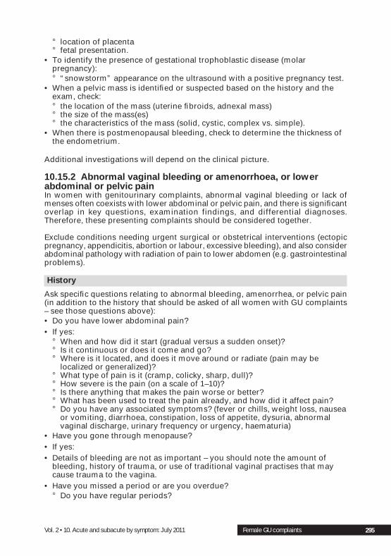
295Vol. 2 • 10. Acute and subacute by symptom: July 2011
° location of placenta ° fetal presentation.
• To identify the presence of gestational trophoblastic disease (molar pregnancy): ° “snowstorm” appearance on the ultrasound with a positive pregnancy test.
• When a pelvic mass is identifi ed or suspected based on the history and the exam, check: ° the location of the mass (uterine fi broids, adnexal mass) ° the size of the mass(es) ° the characteristics of the mass (solid, cystic, complex vs. simple).
• When there is postmenopausal bleeding, check to determine the thickness of the endometrium.
Additional investigations will depend on the clinical picture.
10.15.2 Abnormal vaginal bleeding or amenorrhoea, or lower abdominal or pelvic painIn women with genitourinary complaints, abnormal vaginal bleeding or lack of menses often coexists with lower abdominal or pelvic pain, and there is signifi cant overlap in key questions, examination findings, and differential diagnoses. Therefore, these presenting complaints should be considered together.
Exclude conditions needing urgent surgical or obstetrical interventions (ectopic pregnancy, appendicitis, abortion or labour, excessive bleeding), and also consider abdominal pathology with radiation of pain to lower abdomen (e.g. gastrointestinal problems).
History
Ask specifi c questions relating to abnormal bleeding, amenorrhea, or pelvic pain (in addition to the history that should be asked of all women with GU complaints – see those questions above): • Do you have lower abdominal pain?• If yes:
° When and how did it start (gradual versus a sudden onset)? ° Is it continuous or does it come and go? ° Where is it located, and does it move around or radiate (pain may be
localized or generalized)? ° What type of pain is it (cramp, colicky, sharp, dull)? ° How severe is the pain (on a scale of 1–10)? ° Is there anything that makes the pain worse or better? ° What has been used to treat the pain already, and how did it affect pain? ° Do you have any associated symptoms? (fever or chills, weight loss, nausea
or vomiting, diarrhoea, constipation, loss of appetite, dysuria, abnormal vaginal discharge, urinary frequency or urgency, haematuria)
• Have you gone through menopause? • If yes:• Details of bleeding are not as important – you should note the amount of
bleeding, history of trauma, or use of traditional vaginal practises that may cause trauma to the vagina.
• Have you missed a period or are you overdue? ° Do you have regular periods?
Female GU complaints
FINAL Vol. 2 CH 9-21.indd 295FINAL Vol. 2 CH 9-21.indd 295 29/06/2012 15:1629/06/2012 15:16

296 Vol. 2 • 10. Acute and subacute by symptom: July 2011
° Describe the pattern of bleeding. What is longest and shortest time between periods?
° Do you have excessively heavy periods (some measure of the amount of fl ow and the number and type of pads or cloths used in a 24-hour period, presence of clots)?
° Do you have bleeding between otherwise normal periods? ° Do you use any herbs or other agents in the vagina, and what are they?
• Have you had any trauma in the genital area (rough sex, sexual assault)?• Do you have a history of a bleeding disorder (easy bruising, nose or gum
bleeds)?
Examination
Key fi ndings on physical exam: (fi ndings in BOLD may require urgent intervention)
• Use the Quick Check for emergency signs suggesting shock: weak or fast pulse, prolonged capillary refi ll, systolic BP <90.
• Fever• Abdominal exam:
° soft or rigid ° localized vs. generalized tenderness, presence of rebound tenderness or
guarding ° presence of masses, enlarged liver or spleen ° presence of costovertebral angle tenderness.
• Pelvic exam: ° presence of abnormal discharge ° cervical motion tenderness ° uterine enlargement or tenderness ° adnexal mass or tenderness ° evidence of trauma. °
Investigations
The laboratory tests to be performed will depend on the fi ndings of the history and physical examination.
Consider the following in all patients:• a pregnancy test – in all women of childbearing age (this is not needed in
women who are clearly postmenopausal);• haemoglobin, white cell count, and differential – to look for signs of infection
and bleeding.
Differential diagnosis of pelvic pain or abnormal vaginal bleeding, divided according to the presence of pregnancy.
Female GU complaints
FINAL Vol. 2 CH 9-21.indd 296FINAL Vol. 2 CH 9-21.indd 296 29/06/2012 15:1629/06/2012 15:16

297Vol. 2 • 10. Acute and subacute by symptom: July 2011
DD
x: A
bn
orm
al vag
inal b
leed
ing
or
am
en
orr
ho
ea o
r lo
wer
ab
do
min
al
or
pelv
ic p
ain
, w
ith
a p
osit
ive
pre
gn
an
cy
te
st
resu
lt (
+)
2 Sa
fe a
borti
on: t
echn
ical
and
pol
icy
guid
ance
for h
ealth
sys
tem
s. W
HO, 2
003
(upd
ated
ver
sion
to b
e pu
blis
hed
in 2
011)
. Ava
ilabl
e at
http
://w
ww
.who
.int/r
epro
duct
iveh
ealth
/pu
blic
atio
ns/u
nsaf
e_ab
ortio
n/92
4159
0343
/en/
Cond
ition
Pelv
ic o
r low
er
abdo
min
al p
ain
Abn
orm
al b
leed
ing
or
amen
orrh
oea
Key
feat
ures
Man
agem
ent
Ecto
pic
preg
nanc
yYE
S (o
ften
unila
tera
l, bu
t may
be
mor
e ge
nera
lized
with
re
boun
d te
nder
ness
if
rupt
ured
)
YES
(usu
ally
mis
sed
perio
d an
d ab
norm
al
blee
ding
or s
potti
ng)
Uter
us n
orm
al s
ize o
r slig
htly
enl
arge
d;
cerv
ix c
lose
d; m
ay p
alpa
te a
dnex
al
mas
s. U
ltras
ound
sho
ws
adne
xal m
ass,
fre
e in
trape
riton
eal fl
uid
, em
pty
uter
us.
IV a
cces
s, s
urgi
cal t
reat
men
t. Po
ssib
le b
lood
tran
sfus
ion.
Patie
nts
with
evi
denc
e of
sho
ck o
r reb
ound
tend
erne
ss
mus
t be
eval
uate
d ur
gent
ly fo
r rup
ture
d ec
topi
c an
d ne
ed fo
r im
med
iate
sur
gery
.
Sept
ic a
bort
ion
or
post
part
um s
epsi
s2YE
S (u
sual
ly
mid
line,
but
may
be
gene
raliz
ed)
YES
(irre
gula
r, m
ay b
e he
avy)
Hist
ory
of re
cent
spo
ntan
eous
or
indu
ced
term
inat
ion
of p
regn
ancy
or
rece
nt d
eliv
ery
(pre
gnan
cy te
st m
ay s
till
be p
ositi
ve fo
r an
inde
fi nite
per
iod
of
time
afte
r ter
min
atio
n of
pre
gnan
cy o
r de
liver
y); f
ever
, ele
vate
d W
BC; u
terin
e te
nder
ness
, pos
sibl
e fo
ul o
dour
, or
puru
lent
dis
char
ge. U
ltras
ound
may
sh
ow re
tain
ed p
rodu
cts.
Refe
r to
IMPA
C M
CPC1
Star
t IV
antib
iotic
s an
d st
abili
ze (s
ee Q
uick
Che
ck a
nd
Sect
ion
3.1.
5 if
sept
ic s
hock
).12
–14
wee
ks: v
acuu
m a
spira
tion
is th
e re
com
men
ded
tech
niqu
e fo
r sur
gica
l abo
rtion
. Dila
tatio
n an
d sh
arp
cure
ttage
, whe
re it
is s
till p
ract
iced
, sho
uld
be re
plac
ed
by v
acuu
m a
spira
tion.
The
pro
cedu
re s
houl
d no
t be
rout
inel
y co
mpl
eted
by
shar
p cu
retta
ge.
Afte
r 12–
14 w
eeks
: exp
erie
nced
pro
vide
r for
dila
tatio
n an
d ev
acua
tion
unde
r son
o-gu
idan
ce. I
f not
ava
ilabl
e,
use
mis
opro
stol
.
Female GU complaints
FINAL Vol. 2 CH 9-21.indd 297FINAL Vol. 2 CH 9-21.indd 297 29/06/2012 15:1629/06/2012 15:16

298 Vol. 2 • 10. Acute and subacute by symptom: July 2011
Cond
ition
Pelv
ic o
r low
er
abdo
min
al p
ain
Abn
orm
al b
leed
ing
or
amen
orrh
oea
Key
feat
ures
Man
agem
ent
Spon
tane
ous
mis
carr
iage
or
abor
tion
YES
(usu
ally
cra
mpy
, m
idlin
e)YE
S (m
ay b
e he
avy
with
or w
ithou
t pa
ssag
e of
tiss
ue)
Enla
rged
sof
t ute
rus
Cerv
ical
os
may
be
dila
ted
Ultra
soun
d sh
ows
enla
rged
ute
rus
with
or
with
out p
rodu
cts
of c
once
ptio
n.
Sign
s of
sho
ck o
r exc
essi
ve b
leed
ing
requ
ire
urge
nt
surg
ical
inte
rven
tion.
Nee
d to
con
side
r ute
rine
perfo
ratio
n if
prev
ious
lega
l or
illeg
al p
roce
dure
or i
ncor
rect
dia
gnos
is (e
.g. e
ctop
ic).
Refe
r to
IMPA
C M
CPC.
1
Eith
er v
acuu
m a
spira
tion
or m
edic
al tr
eatm
ent c
an b
e re
com
men
ded
for w
omen
with
inco
mpl
ete
abor
tion
at
any
gest
atio
n w
hose
ute
rine
size
at t
he ti
me
of tr
eatm
ent
is 1
3 w
eeks
or l
ess.
The
reco
mm
ende
d m
edic
al tr
eatm
ent
is m
isop
rost
ol (a
dmin
iste
red
as a
sin
gle
dose
eith
er
subl
ingu
ally
400
mcg
or o
rally
600
mcg
). Th
e de
cisi
on fo
r tre
atm
ent s
houl
d be
bas
ed o
n th
e cl
inic
al c
ondi
tion
and
the
patie
nt’s
pref
eren
ces
for t
reat
men
t.Pr
ophy
lact
ic a
ntib
iotic
s: s
ingl
e do
se o
f nitr
omid
azol
es,
tetra
cycl
ines
, or p
enic
illin
s.
Rupt
ured
ute
rus
YES
(sev
ere,
may
in
itial
ly d
ecre
ase
afte
r rup
ture
take
s pl
ace)
+/- (
alth
ough
ther
e m
ay b
e so
me
vagi
nal
blee
ding
rela
ted
to
labo
ur, t
he p
rimar
y bl
eedi
ng w
ith ru
ptur
ed
uter
us is
with
in th
e pe
riton
eal c
avity
and
w
ill n
ot b
e vi
sibl
e)
Abdo
min
al d
iste
nsio
nAb
norm
al u
terin
e co
ntou
rPa
lpab
le fe
tal p
arts
Ofte
n ra
pid
puls
e, h
ypot
ensi
on
If th
is d
iagn
osis
is c
onsi
dere
d, p
atie
nt n
eeds
urg
ent
surg
ical
inte
rven
tion.
Refe
r to
IMPA
C M
CPC.
1
Plac
enta
pre
via
see
Quic
k Ch
eck
page
24
NO
YES
(pro
fuse
, of
ten
heav
y an
d co
ntin
uous
, typ
ical
ly
with
out p
ain)
Uter
us p
alpa
ble
abov
e um
bilic
us s
oft.
Feta
l hea
rt to
nes
norm
al.
Abdo
min
al u
ltras
ound
, see
Sec
tion
7.2.
21.
Refe
r to
IMPA
C M
CPC.
1
Esta
blis
h IV
acc
ess
– pa
tient
may
requ
ire b
lood
tra
nsfu
sion
.Pe
rform
ultr
asou
nd to
doc
umen
t dia
gnos
is. M
ay c
onsi
der
“dou
ble
set-u
p” in
the
oper
atin
g ro
om, w
ith p
repa
ratio
n fo
r sur
gery
and
exa
min
atio
n to
pot
entia
lly ru
le o
ut
diag
nosi
s of
pre
via.
Sh
ould
be
able
to b
e m
anag
ed a
t dis
trict
leve
l, bu
t co
nsid
er re
ferr
al u
nder
the
follo
win
g ci
rcum
stan
ces:
pa
tient
sta
ble
with
min
imal
ble
edin
g AN
D pr
evio
us
C-se
ctio
n (c
once
rn fo
r pla
cent
a ac
cret
a or
per
cret
a) O
R pr
emat
urity
with
nee
d fo
r neo
nata
l int
ensi
ve c
are
and
reso
urce
s no
t ava
ilabl
e on
site
.
Female GU complaints
FINAL Vol. 2 CH 9-21.indd 298FINAL Vol. 2 CH 9-21.indd 298 29/06/2012 15:1629/06/2012 15:16

299Vol. 2 • 10. Acute and subacute by symptom: July 2011
DD
x: A
bn
orm
al vag
inal b
leed
ing
or
am
en
orr
ho
ea,
or
low
er
ab
do
min
al
or
pelv
ic p
ain
, w
ith
a n
eg
ati
ve
pre
gn
an
cy
te
st
(-)
Cond
ition
Pelv
ic/lo
wer
ab
dom
inal
pai
n
Abn
orm
al v
agin
al
blee
ding
or
amen
orrh
oea
Key
feat
ures
Man
agem
ent
Ova
rian
tors
ion
YES
(usu
ally
un
ilate
ral;
may
giv
e hi
stor
y of
rece
nt
epis
odes
of s
imila
r pa
in)
NO
Unila
tera
l ten
dern
ess,
pos
sibl
e pa
lpab
le
adne
xal m
ass
Nau
sea
or v
omiti
ngM
ay g
ive
hist
ory
of p
ain
bein
g pr
eced
ed
by s
trenu
ous
activ
ity, s
ex. U
ltras
ound
sh
ows
adne
xal m
ass.
Requ
ires
sur
gica
l int
erve
ntio
n.
Rupt
ured
ova
rian
cy
stYE
S (u
sual
ly
unila
tera
l, m
ay
be g
ener
alize
d if
sign
ifi ca
nt
intra
perit
onea
l bl
eedi
ng o
r lea
kage
of
cyst
con
tent
s)
NO
Sudd
en o
nset
, ofte
n pr
eced
ed b
y st
renu
ous
activ
ity o
r sex
.M
ost c
omm
on in
func
tiona
l cys
ts in
late
ph
ase
of m
enst
rual
cyc
le; c
an o
ccur
w
ith c
ysts
from
end
omet
riosi
s or
a
beni
gn tu
mou
r (su
ch a
s de
rmoi
d), w
ith
resu
lting
che
mic
al p
erito
nitis
.
Usua
lly s
elf-l
imite
d if
min
imal
leak
age;
clo
se o
bser
vatio
n ne
eded
. Spe
cifi c
man
agem
ent n
ot re
quire
d un
less
ev
iden
ce o
f sig
nifi c
ant h
aem
orrh
age.
If
evid
ence
of s
hock
or i
ntra
ctab
le p
ain,
sur
gica
l in
terv
entio
n m
ay b
e re
quire
d.Fo
r pai
n m
anag
emen
t see
Sec
tion
20.
Tube
rcul
osis
see
Sect
ion
15YE
SYE
S (u
sual
ly ir
regu
lar
spot
ting
betw
een
men
ses)
Clin
ical
pre
sent
atio
n si
mila
r to
bact
eria
l PI
D; p
atie
nt g
ener
ally
has
pas
t his
tory
of
TB O
R co
ntac
t with
TB
OR e
vide
nce
of
TB in
oth
er s
yste
ms,
ofte
n pu
lmon
ary.
M
ay h
ave
asci
tes
with
abd
omin
al
dist
ensi
on w
ith fl
uid
thril
l as
wel
l as
infe
rtilit
y.
If su
spec
t, re
fer w
here
end
omet
rial s
ampl
ing
for c
ultu
re
and
stai
n is
don
e.If
treat
ed fo
r bac
teria
l PID
with
out i
mpr
ovem
ent,
cons
ider
this
dia
gnos
is.
Also
con
side
r ova
rian
canc
er, e
spec
ially
if a
scite
s or
pe
lvic
mas
s pr
esen
t. Re
com
men
d HI
V te
stin
g an
d co
unse
lling
.
Female GU complaints
FINAL Vol. 2 CH 9-21.indd 299FINAL Vol. 2 CH 9-21.indd 299 29/06/2012 15:1629/06/2012 15:16

300 Vol. 2 • 10. Acute and subacute by symptom: July 2011
Cond
ition
Pelv
ic/lo
wer
ab
dom
inal
pai
nA
bnor
mal
vag
inal
bl
eedi
ng o
r am
enor
rhoe
a
Key
feat
ures
Man
agem
ent
Ute
rine
fi br
oids
(with
or w
ithou
t de
gene
ratio
n)
YES
(if d
egen
erat
ed,
usua
lly m
idlin
e; n
o re
boun
d)
+/- (
exce
ssiv
ely
heav
y or
long
men
ses
sugg
ests
that
fi br
oids
m
ay b
e lo
cate
d in
ut
erin
e lin
ing.
Enla
rged
, irr
egul
ar u
teru
s, u
sual
ly fi
rm
Loca
lized
tend
erne
ss if
deg
ener
ated
Ul
traso
und
may
sho
w p
rese
nce
of
fi bro
ids.
Heav
y bl
eedi
ng m
ay re
quire
sta
biliz
atio
n an
d, in
som
e ca
ses,
may
requ
ire tr
ansf
usio
n or
pos
sibl
e cu
retta
ge
for i
mm
edia
te c
ontro
l of b
leed
ing.
In s
ome
case
s (e
.g.
patie
nt w
ithou
t sev
ere
anae
mia
) atte
mpt
to m
anag
e bl
eedi
ng w
ith h
orm
onal
trea
tmen
t, lo
w d
ose
COCs
or
prog
estin
-onl
y co
ntra
cept
ive
met
hods
(pill
s, in
ject
ions
, im
plan
ts, L
NG
IUDs
3 ). M
enor
rhag
ia w
ith a
naem
ia o
r sh
ock
may
requ
ire s
urgi
cal i
nter
vent
ion
(myo
mec
tom
y or
hy
ster
ecto
my)
.Pa
in w
ith d
egen
erat
ion
usua
lly s
elf-l
imite
d; fo
r pai
n m
anag
emen
t see
Sec
tion
20
Cerv
ical
can
cer
+/- (
gene
rally
with
out
pain
, exc
ept i
n la
ter
stag
es)
YES
(irre
gula
r, m
ay
be h
eavy
or l
ight
, of
ten
blee
ding
afte
r in
terc
ours
e)
Freq
uent
his
tory
of a
bnor
mal
foul
-sm
ellin
g di
scha
rge;
cer
vica
l mas
s or
ul
cer.
Mor
e co
mm
on in
HIV
+ w
omen
.
Biop
sy fo
r dia
gnos
is, i
f pos
sibl
e (s
ee S
ectio
n 10
.15.
8).
May
nee
d m
easu
res
to c
ontro
l for
per
sist
ent v
agin
al
blee
ding
.Re
fer f
or s
urgi
cal o
r rad
iatio
n tre
atm
ent.
Ute
rine
can
cer
+/-
YES
(usu
ally
in
post
men
opau
sal
wom
en, m
ay b
e lig
ht
or s
potti
ng)
Uter
us m
ay b
e en
larg
ed.
Mor
e co
mm
on in
wom
en w
ith o
besi
ty,
hype
rtens
ion,
dia
bete
s, o
r if o
n un
oppo
sed
oest
roge
n re
plac
emen
t. Ul
traso
und
show
s th
icke
ned
endo
met
rial l
inin
g.
Biop
sy fo
r dia
gnos
is, i
f pos
sibl
e.
Refe
r for
sur
gica
l or r
adia
tion
treat
men
t.
Endo
met
rios
isYE
SN
OCh
roni
c pa
in, o
ften
wor
se w
ith m
ense
s;
asso
ciat
ed w
ith in
ferti
lity.
May
hav
e te
nder
adn
exal
mas
s (e
ndom
etrio
ma)
.
May
impr
ove
with
LH
agon
ists
(rec
ent e
vide
nce
sugg
ests
gr
eate
r effi
cacy
than
con
tinuo
us o
ral c
ontra
cept
ives
). M
ay re
quire
sur
gica
l int
erve
ntio
n in
sev
ere
case
s or
with
in
ferti
lity.
Trau
ma
or ra
pe
(sim
ilar p
ictu
re
may
be
seen
with
ce
rtai
n tr
aditi
onal
va
gina
l pra
ctic
es)
YES
(loca
lized
usu
ally
to
vul
var o
r vag
inal
ar
ea)
YES
(may
be
prof
use
if te
ars,
lace
ratio
ns)
Evid
ence
of t
raum
a, s
uch
as b
ruis
ing,
te
ars,
lace
ratio
ns.
Man
agem
ent o
f tra
uma.
Psy
chos
ocia
l car
e.Pa
in c
ontro
l.Re
fer f
or c
ouns
ellin
g an
d su
ppor
tive
care
.
See
Sect
ions
4.4
and
10.
11
3 Fa
mily
pla
nnin
g: a
glo
bal h
andb
ook
for p
rovi
ders
. WHO
, (up
date
d) 2
011.
Ava
ilabl
e at
http
://w
ww
.who
.int/r
epro
duct
iveh
ealth
/pub
licat
ions
/fam
ily_p
lann
ing/
9780
9788
5630
4/en
/in
dex.
htm
l
Female GU complaints
FINAL Vol. 2 CH 9-21.indd 300FINAL Vol. 2 CH 9-21.indd 300 29/06/2012 15:1629/06/2012 15:16

301Vol. 2 • 10. Acute and subacute by symptom: July 2011
Rela
ted
to n
orm
al
men
stru
atio
nYE
SN
OCy
clic
mid
line
cram
ping
pai
n w
ith
men
ses
(dys
men
orrh
oea)
; mid
-cyc
le
unila
tera
l pai
n as
soci
ated
with
ov
ulat
ion.
Sym
ptom
atic
trea
tmen
t (N
SAID
s).
Dys
pare
unia
YES
(ass
ocia
ted
with
in
terc
ours
e)N
OM
ay h
ave
evid
ence
of c
ervi
cal i
nfec
tion
or P
ID; a
lso
seen
with
end
omet
riosi
s,
som
e pe
lvic
mas
ses.
Trea
t und
erly
ing
cond
ition
.M
ay n
eed
psyc
holo
gica
l cou
nsel
ling.
Poly
cyst
ic o
vari
an
dise
ase
NO
YES
(usu
ally
hi
stor
y of
per
iods
of
am
enor
rhoe
a fo
llow
ed b
y he
avy
and
prol
onge
d bl
eedi
ng)
Exam
gen
eral
ly n
orm
al, a
lthou
gh o
varie
s m
ay b
e so
mew
hat e
nlar
ged
on p
elvi
c ex
am o
r ultr
asou
nd.
May
hav
e hi
stor
y of
infe
rtilit
y.
Rule
out
ass
ocia
ted
anae
mia
. If p
rese
nt, t
reat
.Ru
le o
ut u
terin
e ca
ncer
or h
yper
plas
ia w
ith b
iops
y if
abno
rmal
ble
edin
g is
pro
long
ed.
Trea
t with
COC
s or
cyc
lic m
edro
xypr
oges
tero
ne a
ceta
te
10 m
g or
ally
dai
ly fo
r 10
to 1
4 da
ys p
er m
onth
, if n
ot tr
ying
to
con
ceiv
e (p
rote
ctiv
e ag
ains
t ute
rine
hype
rpla
sia
or
canc
er; C
OCs
also
pro
vide
effe
ctiv
e co
ntra
cept
ion.
)Re
fer i
f tre
atm
ent n
eede
d fo
r inf
ertil
ity.
App
endi
citis
see
Sect
ion
10.7
aYE
S (v
ague
pe
rium
bilic
al p
ain
that
loca
lizes
to ri
ght
low
er q
uadr
ant,
mor
e ge
nera
lized
if
rupt
ured
)
NO
Feve
r; rig
ht lo
wer
qua
dran
t ten
dern
ess
with
or w
ithou
t reb
ound
.FB
C –
high
WBC
.
Requ
ires
urg
ent a
rran
gem
ent o
r ref
erra
l for
sur
gica
l in
terv
entio
n.
Uri
nary
trac
t in
fect
ion,
cys
titis
. py
elon
ephr
itis
see
Sect
ion
11.4
4
YES
(if s
upra
pubi
c –
cyst
itis;
if C
VA
tend
erne
ss –
py
elon
ephr
itis)
NO
(in s
ome
case
s ha
emat
uria
may
be
mis
take
n fo
r vag
inal
bl
eedi
ng)
Pain
on
urin
atio
n.Ur
inar
y fre
quen
cy a
nd u
rgen
cy, p
ossi
ble
haem
atur
ia. C
heck
for C
VAT.
Clou
dy u
rine.
Urin
e di
pstic
k –
bloo
d, le
ucoc
ytes
, ni
trite
s.
Urin
e m
icro
scop
y: le
ukoc
ytes
, RBC
, ba
cter
ia.
Trea
t with
ant
ibio
tics.
See
Sect
ion
11.4
4.
Rena
l sto
neYE
S (s
ever
e, c
olic
ky,
may
radi
ate
into
gr
oin)
NO
Gros
s or
mic
rosc
opic
hae
mat
uria
, with
or
with
out C
VA te
nder
ness
. Ultr
asou
nd
may
sho
w d
ilata
tion
of u
rete
r.
IV h
ydra
tion,
pai
n co
ntro
l (m
orph
ine)
unt
il st
one
pass
es.
If pa
in c
ontin
ues
with
out r
elie
f, re
fer f
or p
ossi
ble
surg
ical
in
terv
entio
n.
Hyp
otha
lam
ic
amen
orrh
oea
NO
YES
(am
enor
rhoe
a fo
r var
ying
per
iods
of
tim
e)
Asso
ciat
ed w
ith c
hron
ic il
lnes
s or
si
gnifi
cant
wei
ght l
oss.
Alw
ays
rule
out
pre
gnan
cy. T
reat
und
erly
ing
cond
ition
.
Female GU complaints
FINAL Vol. 2 CH 9-21.indd 301FINAL Vol. 2 CH 9-21.indd 301 29/06/2012 15:1629/06/2012 15:16

302 Vol. 2 • 10. Acute and subacute by symptom: July 2011
10.15.3 Pelvic massPelvic masses may be due to a number of conditions. The patient may present with lower abdominal, pelvic pain, or abnormal bleeding or may have no symptoms. Serious conditions include abscesses, ectopic pregnancy, and cancers.
History
Key questions (in addition to a history that should be taken for all women with GU complaints, see above). See key questions if the woman also has lower abdominal pain, abnormal vaginal bleeding, or amenorrhoea. In a woman who complains of or is found to have a pelvic mass on examination, and is without pain or abnormal bleeding, ask:• Do you have fever or chills, weight loss or gain?• Do you have gastrointestinal symptoms (nausea or vomiting, diarrhoea,
constipation, rectal bleeding, bloating)?• Do you have urinary symptoms (dysuria, frequency, urgency, haematuria,
urinary retention)?• Do you have abnormal vaginal discharge?
Examination
Key fi ndings on physical exam:• Constitutional fi ndings: evidence of wasting, pallor, enlarged lymph nodes,
fever.• Abdominal exam: evidence of ascites, liver or spleen enlargement, tenderness
with or without rebound or guarding.• Pelvic exam: location, size, mobility, tenderness, characteristics of mass;
abnormal vaginal discharge.
In women of reproductive age, a normal ovary may be up to 5–6 cm in size. A palpable ovary in a postmenopausal woman should be considered abnormal and requires further evaluation.
Investigations
Laboratory tests to be performed will depend on the fi ndings of the history and physical examination.
Consider the following in all patients:• Pregnancy test: if premenopausal.• Laboratory tests: FBC with differential.• Pelvic ultrasound. Ultrasound can determine whether the mass is solid or
cystic, simple or complex, as well as the presence of ascites or free fl uid suggestive of blood.
• Additional evaluations involving procedures such as laparoscopy or colonoscopy require referral.
Female GU complaints
FINAL Vol. 2 CH 9-21.indd 302FINAL Vol. 2 CH 9-21.indd 302 29/06/2012 15:1629/06/2012 15:16

303Vol. 2 • 10. Acute and subacute by symptom: July 2011
DDx: Pelvic mass
Condition In favour Management
Ectopic pregnancy Missed menstrual period and abnormal bleeding or spotting; unilateral abdominal pain (may be more generalized if ruptured)Adnexal mass – tenderFBC – anaemiaPregnancy test positiveUltrasound – adnexal mass, free fl uid
IV access.Surgical treatment.If evidence of shock, see ETAT.Possible blood transfusion.
Patients with evidence of shock or rebound tenderness must be evaluated urgently for ruptured ectopic and need for immediate surgery.
Tubo-ovarian abscess or pyosalpinx
Fever, other features of PID (see above); generalized and rebound tenderness and shock suggests ruptureElevated WBCCan see similar presentation with post-surgical abscess
IV access, IV antibiotics.
Evidence of rupture with or without signs of septic shock requires urgent surgical intervention.
Hydrosalpinx Usually asymptomatic, but may have chronic pelvic pain or dyspareuniaOften history of PID WBC normalUltrasound – dilated cystic tubular structure
No treatment needed if no symptoms. May require surgical intervention if chronic pain or if increasing in size.
Fibroids Often asymptomaticHeavy or prolonged mensesUrinary frequencyNon-tender, unless degeneratedEnlarged, irregular uterus, usually fi rm; localized tenderness if degeneratedUltrasound – fi broids
May try management with COCs or progestin-only contraception. Pain with degeneration usually self-limited; pain management. Menorrhagia with anaemia may require surgical intervention (myomectomy or hysterectomy).
Endometrioma, endometrial cyst
Possible known endometriosisNon-mobile massesRupture or leaks cause sudden sharp abdominal pain and rebound tenderness.Often history of infertility and dyspareuniaUltrasound – complex cystic ovarian mass
Refer for surgical management or hormonal treatment if early signs and if desires pregnancy.
Benign or malignant ovarian tumour
Often asymptomaticWith cancer and some benign tumours, may have abdominal bloating and discomfortMay be cystic, solid, or mixedLoss of weight, presence of ascites suggests malignancyAdnexal mass with great variation in size; with a solid consistency, irregular, and fi xed suggests cancerUltrasound – multilocular complex cyst; presence of solid components, papillary projections, and ascites suggest cancer.
Refer to specialist. Surgical intervention is required.Presence of adnexal mass in postmenopausal woman should be considered cancer until proven otherwise.
Female GU complaints
FINAL Vol. 2 CH 9-21.indd 303FINAL Vol. 2 CH 9-21.indd 303 29/06/2012 15:1629/06/2012 15:16

304 Vol. 2 • 10. Acute and subacute by symptom: July 2011
Condition In favour Management
Functional ovarian cyst* Women of reproductive ageMostly asymptomatic, but may have unilateral pelvic pain If rupture, pain can be more generalized with signs of peritonitisUltrasound – unilateral cyst <5–6 cm in size
Usually self-limited. After resolution, use of hormonal contraception can prevent new cysts from formingDoes not require specifi c management unless evidence of signifi cant haemorrhage with rupture.
If shock, arrange for possible surgical intervention.
Pain management.
Ovarian torsion Unilateral tenderness, possible palpable adnexal massNausea or vomitingMay give history of pain being preceded by strenuous activity or sexUsually unilateral; may give history of recent episodes of similar painUltrasound – adnexal mass
Requires urgent surgical intervention.
Retroperitoneal lymphadenopathy
May present as pelvic mass; look for other constitutional signs (fever, weight loss, lymphadenopathy)DDx includes lymphoma, TB
Refer for appropriate diagnosis and treatment.Recommend HIV testing and counselling
Gastrointestinal mass(bowel malignancy, diverticular abscess)
Change in bowel habitsGross or occult blood in the stoolMay have weight lossSigns of infection with abscess (fever, tenderness, elevated WBC)
Refer for colonoscopy, possible surgical treatment if malignancy found.IV antibiotics for abscess.Consider Miegs syndrome.
Enlarged bladder due to urinary retention
Inability to urinate; suprapubic tenderness
Urinary catheter, search for possible causes (e.g. infection; severe genital herpes; bladder, primary or secondary tumour).
* Ovarian functional cyst: functional cysts are the most common ovarian masses found among women of reproductive age, and are related to ovulation or corpus luteal cysts, although they are not usually clinically palpable unless >4 cm. Be careful of torsion in large pedunculated cysts. In cysts <4 cm resolution occurs spontaneously in 1 to 3 months. In a thin woman of reproductive age who is not guarding, palpating an ovarian cyst at mid-cycle or shortly before she is due for her period is consistent with an ovarian functional cyst. Re-examine her shortly after her next menses (i.e. at a different time in her cycle). If persistent, then an ultrasound is needed.
An ultrasound should be considered for any adnexal mass that is thought to be >5 cm in size or with a palpable adnexal mass of any size in a postmenopausal women. In reproductive-age women with smaller cysts (5 cm or less), an alternative is to re-examine them at a different time in their menstrual cycle. Small cysts can resolve with time. Larger masses are less likely to be functional, which is why they are less likely to resolve spontaneously, and torsion is a potential risk.
Any patients with severe unexplained bleeding and anaemia, pelvic mass, fi ndings suspicious for malignancy, or bleeding that is not resolved with conservative measures should be hospitalized.
See the IMPAC MCPC.1
Female GU complaints
FINAL Vol. 2 CH 9-21.indd 304FINAL Vol. 2 CH 9-21.indd 304 29/06/2012 15:1629/06/2012 15:16

305Vol. 2 • 10. Acute and subacute by symptom: July 2011
10.15.4 Abnormal vaginal discharge not responding to syndromic managementPatients may be referred to the second level of care because of abnormal vaginal discharge that does not respond to syndromic management. The primary care guidelines in IMAI Acute Care are based on the WHO STI syndromic guidelines and assume syndromic management without laboratory tests.
History
Key questions (in addition to a history that should be taken from all women with GU complaints, see above):• How long has the discharge been present, and what does it look like (colour,
consistency)?• Have you ever had water body contact in schistosomiasis endemic area?• Do you have any associated symptoms (e.g. pruritis, malodour, sores, lower
abdominal or pelvic pain, fever, weight loss)? • Do you douche, or have you put any other substances into your vagina?• Have you taken any antibiotics recently?• How has the discharge been treated so far? Did it help?
Examination
Key fi ndings on exam:• constitutional: fever, lymphadenopathy, rash• abdomen: tender or non-tender.
Pelvic exam:
• characteristics, amount of discharge• associated signs: erythaema, oedema, cervical friability, sores • cervical motion, uterine, or adnexal tenderness.
Ensure discharge is not from abscess or fi stula.
Investigations
Laboratory tests to be performed will depend on the fi ndings of the history and physical examination. Consider the following in all patients:• Saline wet mount with or without KOH preparation (see Section 7.2.15).
Evaluate for the presence or absence of trichomonads, clue cells, white blood cells, or hyphae.
• Swab test: a swab should be collected from the cervical canal (endocervix). If the swab appears yellow when held up against white paper (positive swab test), cervical infection is likely.
• Gram stain (see Section 7.2.14) of vaginal secretions (this is helpful if saline wet mount and KOH are not available, or if they are unrevealing; highly specifi c for gonorrhoea if Gram-negative intracellular diplococci are seen).
These tests, if available, can help in the etiological diagnosis of discharge related to vaginal infections. When cervicitis is suspected based on exam and with only WBC's on wet mount, specifi c gonorrhoea and Chlamydia testing can be helpful.
Female GU complaints
FINAL Vol. 2 CH 9-21.indd 305FINAL Vol. 2 CH 9-21.indd 305 29/06/2012 15:1629/06/2012 15:16

306 Vol. 2 • 10. Acute and subacute by symptom: July 2011
DDx: Vaginal discharge using microscopic examination and KOH
Condition In favour
Bacterial vaginosis Homogenous, thin white or greyish dischargeLittle evidence of infl ammationSaline or KOH – KOH odour, clue cellspH >4.5Gram stain – decrease in lactobacilli; increase in other bacteria
Vulvovaginal candidiasis Vulvovaginal redness, oedema, swelling; excoriations; white, thick, curd-like discharge; itching, external dysuriaSaline and Gram stain – hyphae, budding yeastpH normal
Trichomoniasis Vulvar or vaginal infl ammation and irritationYellow-green, often frothy discharge Saline – motile trichomonads, WBC's infl ammationpH >4.5
Gonorrhoeasee Section 11.13
Purulent or mucopurulent discharge, cervical friability, (+) swab testGram stain – Gram-negative intracellular diplococci, WBC's infl ammationpH >4.5
Chlamydia Purulent or mucopurulent discharge, cervical friability, (+) swab testGram stain or saline – infl ammation, increased WBC, pH >4.5
Note: Above characteristics vary in sensitivity and specifi city; when possible, a combination of fi ndings and laboratory investigations should be used to optimize diagnosis.
Other possibilities to consider with vaginal discharge not responding to syndromic management include:• Atrophic vaginitis: related to oestrogen defi ciency; irritative symptoms,
vaginal dryness, and dyspareunia; the vaginal epithelium appears thin and a watery discharge may be present; treat with either topical or oral oestrogens.
• Foreign body.
• Local irritants (e.g. spermicides, vaginal medications, soap, detergent, douches, traditional medicines and herbs used in vagina).
• Fistula: foul smelling, discharge mixed with faeces or urine.• Cancer: cervical (suspect with cervical mass or ulceration).• Atypical infections (e.g. schistosomiasis, chronic endometritis, TB).
In HIV-positive patients, clinical presentation may be altered including:
• increased colonization, frequency, and persistence of candidiasis in association with immunosuppression;
• increased frequency, persistence, and severity of bacterial vaginosis in association with immunosuppression, possible improvement with use of ART;
• no change in clinical presentation of trichomoniasis, gonorrhoea, or Chlamydia;
• increased risks of cervical cancer – carefully examine the cervix (refer to 10.15.8 below regarding cervical cancer screening in the presence of persistent vaginal discharge).
Female GU complaints
FINAL Vol. 2 CH 9-21.indd 306FINAL Vol. 2 CH 9-21.indd 306 29/06/2012 15:1629/06/2012 15:16

307Vol. 2 • 10. Acute and subacute by symptom: July 2011
Approach to management of abnormal vaginal discharge not responding to syndromic management
• Review management at the fi rst level and consider possible non-adherence or reinfection.
• Consider possible diagnosis of PID. If the condition meets the criteria, treat.• If laboratory tests suggest an etiological diagnosis, treat concordantly. If
testing is negative or inconclusive, re-treat syndromically, but re-treat with a multi-day course, or consider using another drug. If yeast infection is suspected, may give single dose of fl uconazole (generally not recommended in pregnancy).
• Consider local patterns of gonorrhoeal resistance.• Recommend HIV testing and counselling, if not done. • If patient is from a schistosomiasis endemic area, treat empirically with 1 dose
of praziquantel at 40 mg/kg.
Bacterial vaginosis
Treatment
• metronidazole gel; OR • metronidazole 500 mg orally twice daily for 7 days; OR • clindamycin 2% cream, 1 full applicator (5 g) per vaginum daily for 7 days; OR• clindamycin 300 mg orally twice daily for 7 days.
Metronidazole can be used in pregnancy, but avoid it in the fi rst trimester if topical agents are available.
Treatment is the same both in HIV-positive and HIV-negative patients.
Trichomoniasis
Treatment
• metronidazole 2 g orally in a single dose; OR• metronidazole 400–500 mg orally twice daily for 7 days; OR• tinidazole 2 g orally in a single dose; OR• tinidazole 500 mg orally twice daily for 5 days.
Avoid use of alcohol during treatment.
Metronidazole can be used in pregnancy, but avoid use of tinidazole in the fi rst trimester.
Treatment is same both in HIV-positive and HIV-negative patients.
Vulvovaginal candidiasis
• Identify and manage underlying conditions: uncontrolled diabetes, corticosteroid use, topical or systemic antibiotics, spermicides (confl icting data), douching, immunosuppression, HIV-infection.
• Pregnant women are also more likely to develop vulvovaginal candidiasis; treat appropriately. See Section 11.4.
Female GU complaints
FINAL Vol. 2 CH 9-21.indd 307FINAL Vol. 2 CH 9-21.indd 307 29/06/2012 15:1629/06/2012 15:16

308 Vol. 2 • 10. Acute and subacute by symptom: July 2011
• Antifungal treatments include: ° miconazole 200 mg vaginal suppository, once daily for 3 days; OR ° clotrimazole 200 mg intravaginally daily for 4 days; OR ° clotrimazole 500 mg intravaginally in a single dose; OR ° fl uconazole 150 mg orally in a single dose (fl uconazole is not recommended
in pregnancy); ° alternative (but less effective) is nystatin 100 000 unit vaginal table daily for
14 days.
If this has already been treated topically, re-treat with oral drugs (consider more intensive dosing of fl uconazole 150 mg orally, repeated 3 days later). In severe cases (extensive vulvar erythema, oedema, excoriation, and fi ssure formation), extend topical treatment to 7–14 days, or fl uconazole 150 mg, repeated after 3 days.
Recurrent vulvovaginal candidiasis
• Usually defi ned as 4 or more symptomatic episodes per year. In some cases, this may be related to non-albicans Candida species that do not respond as well to conventional therapies.
• Consider topical therapy for 7–14 days or fl uconazole 100 mg, 150 mg or 200 mg orally every third day for a total of 3 doses.
• Maintenance therapy for 6 months should be considered: fl uconazole 100 mg, 150 mg, or 200 mg orally weekly (preferred); alternatives: topical clotrimazole 200 mg twice weekly or clotrimazole 500 mg vaginal suppositories once weekly.
• For persistent vulvovaginal candidiasis (failure or recurrence after the above regimens): consider boric acid 600 mg in a gelatine capsule inserted into vagina once daily for 2 weeks (clinical success rates of approximately 70%). Boric acid is contraindicated in pregnancy. Maintenance therapy: boric acid once daily for 2 weeks followed by twice weekly for 6 months.
• Treatment in pregnancy: use topical agents only (fl uconazole is not recommended in pregnancy), for 7 days.
• Treatment in HIV-positive patients: above treatment regimens are recommended irrespective of HIV status. In HIV-positive patients, assess for ART eligibility, and initiate if indicated. (See Section 13.) ° If the patient is referred after short-course treatment failure, and evaluation
is consistent with vulvovaginal candidiasis, re-treat for 7–14 days. Continued recurrence or persistence should be managed promptly, as for recurrent or persistent vulvovaginal candidiasis above. Given higher colonization rates, which correlate with immunosuppression, consider the prophylactic use of topical antifungals when systemic antibiotics are given.
Gonorrhoea/Chlamydia cervicitis
Treatment
• Treat for both gonorrhoea and Chlamydia if the patient was previously treated only for vaginitis, or if they were diagnosed or treated for either infection alone.
• Assess for possible PID and treat, if indicated.• Treatment for gonorrhoea (see Section 11.13):
° cefi xime 400 mg orally in a single dose; OR ° ceftriaxone 250 mg IM in a single dose (on complimentary drug list); OR
Female GU complaints
FINAL Vol. 2 CH 9-21.indd 308FINAL Vol. 2 CH 9-21.indd 308 29/06/2012 15:1629/06/2012 15:16

309Vol. 2 • 10. Acute and subacute by symptom: July 2011
° spectinomycin 2 g IM in a single dose (alternative); OR ° ciprofl oxacin 500 mg orally as a single dose is also an alternative, but use of
this drug should take into account local gonorrhoeal resistance patterns to fl uoroquinolones. Ciprofl oxacin should not be used in pregnancy.
• Treatment for Chlamydia: ° doxycycline 100 mg twice daily for 7 days; OR ° azithromycin 1 g orally in a single dose; OR ° erythromycin 500 mg orally twice daily for 7 days (alternative); OR ° tetracycline 500 mg orally 4 times daily for 7 days (alternative); OR ° ofl oxacin 300 mg twice daily for 7 days (alternative). ° Note: Treatment with alternative agents may be less effective or less well-
tolerated; if possible, treat with a single dose of azithromycin to enable directly observed therapy.
• Avoid use of quinolones and tetracyclines in pregnancy.
Sex partners should be treated with the same regimen (for both gonorrhoea and Chlamydia, if their last sexual contact was within 60 days before the diagnosis or onset of symptoms). Intercourse should be avoided until therapy is complete, and the patient and her or his partner are asymptomatic. Ensure full adherence to antibiotic treatment to decrease development of resistant strains.
If you suspect cervical cancer – see Section 10.15.8.
10.15.5 Pelvic infl ammatory disease4
Pelvic infl ammatory disease (PID) should always be considered in sexually active women presenting with lower abdominal pain. PID is an upper genital tract infection, including the uterus, fallopian tubes, and surrounding pelvic structures. It is usually polymicrobial in nature.
Infection is commonly sexually transmitted, and follows infection with either Chlamydia trachomatis or Neisseria gonorrhoeae. It can also be caused by ascending infection with normal vaginal microbial fl ora. This can occur following an abortion, postpartum, or post-operatively. The resultant infection, even when sexually transmitted, is usually polymicrobial, and thus should be treated with broad-spectrum antibiotics.
PID rarely occurs in early pregnancy. A positive pregnancy test should always raise suspicion that the diagnosis of PID is in error, and alternative diagnoses (such as ectopic pregnancy) should be considered.
Diagnosis
The most common presenting symptom of PID is lower abdominal pain. Other associated symptoms include: abnormal vaginal discharge, abnormal uterine bleeding, fever, nausea, vomiting, having pain with sexual intercourse (dyspareunia) or when passing urine. Although nausea and vomiting can occur with PID, if they are prominent symptoms with abdominal pain also consider other diagnoses, such as appendicitis.
Examination: check for lower abdominal tenderness and pain on manipulation of the cervix.
4 Guidelines for the management of sexually transmitted Infections. WHO, 2003 (2011 updated version in process). Available at http://www.who.int/hiv/pub/sti/en/STIGuidelines2003.pdf
Female GU complaints
FINAL Vol. 2 CH 9-21.indd 309FINAL Vol. 2 CH 9-21.indd 309 29/06/2012 15:1629/06/2012 15:16

310 Vol. 2 • 10. Acute and subacute by symptom: July 2011
Symptoms of PID may be virtually absent, or mild and non-specifi c. Therefore providers should maintain a high index of suspicion in women with lower abdominal pain or with other presenting genital tract complaints, including dyspareunia, postcoital bleeding, abnormal vaginal discharge, or abnormal bleeding (often between menstrual periods).
Empirical treatment of PID can be considered in sexually active young women and others at risk of STIs if one of the following are present, and no other causes for the illness can be identifi ed: • uterine tenderness; OR• adnexal tenderness; OR• cervical motion tenderness.
Additional criteria that increase the specifi city of a diagnosis of PID include:• fever >38.30C• abnormal cervical or vaginal mucopurulent discharge• increased WBCs on saline vaginal wet mount• evidence of infection with gonorrhoea or Chlamydia (e.g. culture, Gram stain)• elevated WBC count• thickened fl uid-fi lled tubes or evidence of abscess on a pelvic ultrasound.
A pregnancy test should be done. Send endo-cervical swabs for a culture if it is available, but a negative result does not exclude the diagnosis.
Treatment
Untreated PID can have serious complications and consequences, and treatment should be initiated without delay after a clinical diagnosis.
Table: Broad-spectrum antibiotics5 for PID
Oral ambulatory therapy, if uncomplicated
If on IV regimens (to be continued until at least 2 days after the patient has improved, and followed by oral therapy for up to 14 days)
ceftriaxone 250 mg IM single dose
PLUS
doxycycline** 100 mg orally twice daily for 14 days
PLUS
metronidazole 400–500 mg orally twice daily for14 days.
1. ceftriaxone 250 mg IM single dose; OR ciprofl oxacin 500 mg orally single dose*; OR spectinomycin 2 g IM PLUS doxycycline** 100 mg orally or IV twice daily PLUS metronidazole 400–500 mg orally or IV twice daily; OR chloramphenicol 500 mg 4 times daily
OR2. clindamycin 900 mg IV every 8 hours PLUS gentami-
cin 1.5 mg/kg IV every 8 hours daily; OR ciprofl oxa-cin 500 mg orally twice daily*; OR spectinomycin 1 g IM, 4 times daily PLUS metronidazole 400–500 mg orally or IV twice daily
Followed by chloramphenicol 500 mg orally 4 times daily or doxycycline 100 mg twice daily to complete 14 days.
* Use only if local data on antimicrobial sensitivity supports its use** Tetracycline can be substituted if doxycycline is not available.
5 Guidelines for the management of sexually transmitted infections. WHO, 2003 (2011 updated version in process). Available at http://www.who.int/hiv/pub/sti/en/STIGuidelines2003.pdf
Female GU complaints
FINAL Vol. 2 CH 9-21.indd 310FINAL Vol. 2 CH 9-21.indd 310 29/06/2012 15:1629/06/2012 15:16

311Vol. 2 • 10. Acute and subacute by symptom: July 2011
In the following circumstances, consider admission for inpatient treatment of PID with IV antibiotics if: • a surgical emergency, such as an ectopic pregnancy or appendicitis, cannot
be excluded• woman is pregnant• non-response to outpatient oral antibiotic therapy• the patient is unable to follow or tolerate an outpatient oral regimen• pelvic abscess is suspected• the patient is an adolescent• the patient has severe illness – high fever, nausea, or vomiting.
If PID occurs with an IUD in place, treat using appropriate antibiotics. There is no evidence that removal of the IUD provides any additional benefi t. However, if the IUD remains in place, close clinical follow-up is critical, and removal should be considered if there is no improvement. If the woman does not want to continue IUD use, it should be removed after antibiotic therapy has been commenced. Offer appropriate contraceptive counselling and services for continued method use.
Follow-upOutpatients with PID should be followed up no later than 72 hours after starting treatment (24 hours for women with fever), and admitted to the hospital if their condition fails to improve.
Within 72 hours of initiating treatment, substantial clinical improvement (absence of fever, reduction in abdominal tenderness, and reduction in uterine, adnexal, and cervical motion tenderness) should be expected. Patients who do not improve within this period may require hospitalization, additional diagnostic tests, or surgical intervention. Perform an ultrasound.
For all women diagnosed with PID, as well as their partners. • Offer counselling and testing for HIV, and counselling for risk-reduction and
condom use.• Test for syphilis.• The patient’s partner(s) should be evaluated and treated presumptively for
gonorrhoea and Chlamydia if they have had sexual contact within the 60 days preceding the onset of symptoms. Many and perhaps most of these male partners will have no symptoms, but this should not delay or discourage treatment.
• The patient and her partner(s) should abstain from sexual intercourse until the treatment has been completed.
Complications Immediate:• sepsis• abscess (may require surgical drainage).
Long-term:• chronic pain, infertility, and ectopic pregnancy due to scarring of the fallopian
tubes or other pelvic structures.
Female GU complaints
FINAL Vol. 2 CH 9-21.indd 311FINAL Vol. 2 CH 9-21.indd 311 29/06/2012 15:1629/06/2012 15:16

312 Vol. 2 • 10. Acute and subacute by symptom: July 2011
10.15.6 Septic abortion2
Septic abortion is a miscarriage or surgical abortion complicated by fever and infection of the uterus. It most commonly occurs in countries where abortion is illegal or inaccessible, and it is a common cause of maternal mortality in these countries. It is usually associated with retained products of conception and, less commonly, associated with foreign bodies (e.g. IUDs), invasive procedures (e.g. amniocentesis), maternal blood stream infection, or spontaneous abortion. The bacteria associated with septic abortion are usually caused by multiple organisms, and may include normal vaginal fl ora as well as sexually transmitted pathogens.
An illegal abortion performed by insertion of rigid foreign objects increases the risk of perforation. The use of soap solution containing cresol and phenol is associated with a risk of uterine necrosis, renal failure, or central nervous system, cardiac, and respiratory toxicity.
Key clinical features
• Consider this possibility in any woman of reproductive age who presents with vaginal bleeding or bloody discharge, lower abdominal pain, and fever.
• Women may present late and may be moribund, as they are reluctant to reveal that they have had an abortion. A pregnancy test will usually be positive in this situation.
Examination
• lower abdominal tenderness • boggy, tender uterus with a dilated cervix• possibly a foul-smelling and bloody discharge• Women may also present with a mild to moderate illness characterized by
low-grade fever, abdominal pain, or moderate vaginal bleeding. This usually occurs in women who have had either an incomplete or failed abortion.
Investigations
• A positive pregnancy test is useful if unable to confi rm a history of pregnancy and recent abortion.
• If perforation of the uterus is suspected, a plain abdominal X-ray may show the presence of free gas in the abdominal cavity.
• Blood, urine, and cervical specimens should be cultured if possible.
Treatment
Evacuation of the uterusPatient should be stabilized prior to procedure. Start antibiotics prior to procedure.• Prompt evacuation of the uterus is important. Vacuum aspiration is the
recommended technique for surgical abortion to 12–14 weeks. • Dilatation and curettage, where it is still practiced, should be replaced by
vacuum aspiration. Vacuum aspiration should not be routinely completed by sharp curettage.
• After 12–14 weeks, refer to an experienced provider for dilatation and evacuation. If not available, use misoprostol.
Female GU complaints
FINAL Vol. 2 CH 9-21.indd 312FINAL Vol. 2 CH 9-21.indd 312 29/06/2012 15:1629/06/2012 15:16

313Vol. 2 • 10. Acute and subacute by symptom: July 2011
• Vacuum aspiration can be performed under local anaesthesia with minimal sedation.
• If none of these means are available, a Foley catheter with a 50 ml balloon attached can be placed in the lower uterus. One kilogram of traction from an orthopaedic weight at the foot of the bed is then applied to the catheter, which dilates the cervix and stimulates contractions.
Indications for hospital admission for IV antibiotics include the following:
• fever >38oC• pelvic peritonitis• tachycardia.
Antibiotics
• Treat with benzyl penicillin 5 million units IV every 6 hours OR ampicillin 2–3 g IV every 6 hours, combined with gentamicin 4–6 mg/kg IV daily and clindamycin 900 mg IV every 8 hours.
• If clindamycin is not available, use metronidazole 500 mg IV every 8 hours. If gentamicin is not available or contraindicated, use ceftriaxone 1 gram IV daily as an alternative.
• When the patient is ready for discharge, change to: ° doxycycline 100 mg orally every 12 hours plus amoxicillin-clavulanic acid
500 + 125 mg orally every 12 hours for a total of 14 days of treatment (or substitute metronidazole 500 mg every 8 hours in place of amoxicillin-clavulanic acid).
• For mild post-abortal infections, oral regimens suitable for treatment of pelvic infl ammatory disease are appropriate. See Section 10.15.5 Pelvic infl ammatory disease.
MonitoringAll women managed as outpatients should be reviewed after 48 hours of treatment and hospitalized if fever and pain persist.
ComplicationsComplications include uterine perforation, which may include injury to the bowel or pelvic abscess formation. This usually requires surgical management with a laparotomy with or without hysterectomy. Uncontrolled infection (ongoing sepsis, severe infection with gas-producing clostridium species) is also an indication for a total hysterectomy.
10.15.7 Approach to urinary incontinenceUrinary incontinence generally is not life-threatening, but it causes signifi cant morbidity, and can result in social or cultural stigma that can have a major effect on quality of life. It is important to carry out a thorough assessment to establish the cause of incontinence and, whenever possible, to treat the underlying cause.
History
Specifi c questions (in addition to a history that should be taken for all women with GU complaints, see above):• Pattern of incontinence, e.g. constant leakage, associated with cough,
sneezing; associated with urgency.
Female GU complaints
FINAL Vol. 2 CH 9-21.indd 313FINAL Vol. 2 CH 9-21.indd 313 29/06/2012 15:1629/06/2012 15:16

314 Vol. 2 • 10. Acute and subacute by symptom: July 2011
• How long have you been leaking urine? Is there any event that you can link to the occurrence of this problem (e.g. childbirth, surgery, other injury)?
• Do you have any other symptoms related to urination (e.g. burning, urgency, frequency, haematuria)?
• Do you have regular and normal bowel movements?• Do you have any medical problems such as diabetes, high blood pressure, or
any psychiatric problems?• What medicines do you take (including traditional or over-the-counter)?
This question should be part of the basic history in any woman with genital tract complaints, but should be re-reviewed with a complaint of urinary incontinence.
Examination
• Constitutional: fever, cachexia or signifi cant weight loss, lymphadenopathy.• Abdominal exam: suprapubic or costovertebral angle tenderness.• Pelvic examination: prolapse of the bladder, uterus; genital sores; abnormal
vaginal discharge; evidence or suspicion of fi stula.
Investigations
Laboratory tests to be performed will depend on the fi ndings of the history and the physical examination. Consider the following in all patients:• urinalysis• BUN, creatinine, glucose, electrolytes• FBC (if suspect underlying systemic infection)• urine dipstick, urinalysis or, if available, culture to rule out infection.
DDx: Urinary incontinence
Condition In favour Management considerations
Fistula Total incontinence; recent childbirth after prolonged labour, history recent surgery; history of cervical cancer (or treatment with surgery or radiation for pelvic cancer).
Refer to surgeon with expertise in fi stula repair.At district level, a large bore urinary catheter to keep bladder dry may allow healing of a vesico-vaginal fi stula.
Urinary tract infection
see Section 11.44
Dysuria, frequency, urgency, haematuria. Clean catch urine with increase in WBCs, bacteria or positive nitrites. Urine culture and sensitivity, if available.
Treatment with antibiotics.Consider or rule out upper tract infection (e.g. pyelonephritis).Consider antibiotic resistance if no improvement and obtain C&S, if not previously obtained.
Stress urinary incontinence
Leakage associated with cough, sneeze, laugh, or other manoeuvre resulting in increased abdominal pressure; increased risk with increased parity. On exam, evidence of pelvic organ prolapse.
Pelvic fl oor muscle exercises.Possible surgical repair.
Female GU complaints
FINAL Vol. 2 CH 9-21.indd 314FINAL Vol. 2 CH 9-21.indd 314 29/06/2012 15:1629/06/2012 15:16

315Vol. 2 • 10. Acute and subacute by symptom: July 2011
Overactive bladder
More common in postmenopausal women; associated with urgency.
Frequent bladder emptying.Anticholinergic (antispasmodic) drugs to calm an overactive bladder and help with urge incontinence. Oxybutynin 5 mg orally every 8–12 hours (maximum 5 mg 4 times per day), might be of help.
Overfl ow incontinence
Bladder never feels empty, frequent need to void at night; may have inability to void, even with urge; post-void dribbling.Bladder may be palpable; catheterization shows high post-void residual.Associated with some pharmacologic treatments (see below); certain disease conditions that result in neurogenic bladder (diabetes, polio, tumours compressing sacral nerves), urinary stones, stool impaction.
Teach patient to safely perform self-intermittent catheterization; address underlying cause.Teach proper catheter care, including cleaning of catheter and irrigation with saline, when necessary.
Pharmacologic agents
On drug that is associated with urinary incontinence (see below).
Adjust drug regimen, if feasible.
Excess urinary output
Frequent urination with large amounts of urine (polyuria).Associated with diabetes, CHF, diabetes insipidus, or renal disease, polydipsia (excessive thirst), alcohol or caffeine.
Address underlying cause.
Atrophic urethritis, vaginitis
Can be seen in women who are naturally or surgically postmenopausal; atrophic changes on exam (vaginal dryness, decreased rugae). Complaints of vaginal dryness. Often presents as overactive bladder symptoms.
Topical oestrogen.See above treatment for overactive bladder.
Deliriumsee Section 10.11
Acute or chronic psychiatric illness, possibly medication-related. Decreased awareness of the need to void.
Address cause of delirium.
Immobility Unable to get to toilet as needed; may also have a neurogenic bladder with overfl ow.
Perineal care to avoid pressure sores.Address cause of immobility; assistance as needed.
Drugs that can cause urinary incontinence
See next table.
Female GU complaints
FINAL Vol. 2 CH 9-21.indd 315FINAL Vol. 2 CH 9-21.indd 315 29/06/2012 15:1629/06/2012 15:16

316 Vol. 2 • 10. Acute and subacute by symptom: July 2011
Table: Drugs that may be associated with or lead to acute urinary incontinence
Medication Effect on lower urinary tract
Diuretics (water pills) Diuresis induced by diuretics may precipitate incontinence. This is particularly relevant in older persons or in those with already impaired continence.
Sedatives (sleeping pills), hypnotics, CNS depressants
Benzodiazepines, especially long-acting agents such as fl urazepam and diazepam (valium), may build up in the bloodstream of an older person. This can cause confusion and alter the person’s ability to recognize the urge to void, leading to urinary incontinence.
Alcohol Alcohol can alter memory, impair mobility, and cause increased urine output resulting in incontinence. In addition, it has a sedative effect that may alter a person's awareness of the need to void.
Anticholinergic agents: antihistamines, antidepressants (TCA), phenothiazines, disopyramides,opiates, antispasmodics, Parkinson drugs
Prescription as well as over-the-counter drugs with anticholinergic properties are taken commonly by persons with insomnia, pruritis (itchy skin), vertigo (dizziness), and other symptoms or conditions. Side-effects include urinary retention with associated urinary frequency and overfl ow incontinence. Besides anticholinergic actions, antipsychotics, such as thioridazine and haloperidol, may cause sedation, rigidity (stiffness), and immobility.
Alpha-adrenergic agents (high blood pressure drugs),sympathomimetics (decongestants), sympatholytics (e.g. prazosin, terazosin, and doxazosin)
Alpha-adrenergic stimulation increases urethral tone and alpha-adrenergic block reduces it. Stress incontinence may become symptomatic in women treated with alpha-antagonists for antihypertensive therapy.
Calcium channel blockers (heart and blood pressure medications)
Calcium channel blockers can reduce smooth muscle contractility in the bladder and occasionally can cause urinary retention and overfl ow incontinence.
Source: http://www.seekwellness.com/incontinence/causes.htm
Symptom management: urinary incontinence
Home care
• Provide adequate support and reassurance about the situation.• Behavioural techniques and lifestyle changes work well for certain types of
urinary incontinence. They may be the only treatment you need: ° pelvic fl oor muscle exercises – help strengthen urinary sphincter and pelvic
fl oor muscles; ° scheduled toilet trips – going to the toilet according to the clock rather than
waiting for the need to go, usually every 2–4 hours.• Pads and protective garments – absorbent pads may help manage urine loss.• Adult diapers are sometimes available. Where they are found they may be
available in both disposable and reusable forms, and come in a variety of sizes.
• Some people fi nd that wearing plastic underwear over their regular underwear helps keep them dry; others opt for washable underwear and briefs with waterproof panels.
Female GU complaints
FINAL Vol. 2 CH 9-21.indd 316FINAL Vol. 2 CH 9-21.indd 316 29/06/2012 15:1629/06/2012 15:16

317Vol. 2 • 10. Acute and subacute by symptom: July 2011
10.15.8 Cervical cancer6
Cervical cancer is a preventable disease, and is curable if diagnosed and treated early. If diagnosed late, cervical cancer is very diffi cult to treat, and causes a great deal of morbidity.
Human papilloma virus (HPV) infection plays a causative role in lower genital intraepithelial and invasive neoplasia. Invasive cervical cancer generally develops slowly over 10–20 years from the earliest pre-cancerous changes, which permits early detection (screening) and treatment. A vaccine is now available (see Section 19) that protects against infection by oncogenic strains. One of the two available vaccines also protects against strains responsible for genital warts.
HIV-positive women have a higher risk of pre-cancer and invasive cervical cancer. The risk and persistence of HPV infection increases with decreasing CD4 count and increasing HIV viral load. Invasive cervical cancer is a WHO HIV Clinical Stage 4 condition.
The role of ART in the management of cervical dysplasiaThe role of ART and immune reconstitution in the management of lower genital tract pre-cancerous lesions remains unclear, and study results are mixed in terms of regression or progression of cervical dysplasia or HPV persistence. HIV-positive women should continue to be followed closely for evidence of cervical cancer, regardless of antiretroviral therapy or CD4 count and viral load results.
General principles of screening for cervical cancer
• Women should be screened according to national guidelines and all women should be offered the same cervical cancer screening options, irrespective of their HIV status.
• Women should be notifi ed when screening results are abnormal and counselled about further evaluation that is needed.
• All positive or abnormal screening results require further evaluation and adequate treatment.
• Recommendations are being reviewed as HIV-positive women may benefi t from an earlier age of fi rst screening and more frequent screening.
Before embarking on a widespread screening programme, services need to be in place to adequately manage women screened positive or newly identifi ed cancer cases. The district clinician can manage pre-cancer, while treating invasive cancer requires specialized care. Women whose disease is very advanced, or for whom treatment is impossible, need skilled palliative care, both symptom management and end-of-life care (see Section 20).
The regular use of Pap smear screening has resulted in as much as an 80% reduction in incidence of, and mortality from, cervical cancer in high-resource settings. Quality assurance is crucial. • Pap smears require transporting specimens to a cytology laboratory where
trained cryotechnicians (under the supervision of a pathologist) can read the slides. Quality assurance is crucial. Reliable transport of slides and test
6 Adapted from: Comprehensive cervical cancer control. WHO, 2006. Available at http://whqlibdoc.who.int/publications/2006/9241547006_eng.pdf
Female GU complaints
FINAL Vol. 2 CH 9-21.indd 317FINAL Vol. 2 CH 9-21.indd 317 29/06/2012 15:1629/06/2012 15:16

318 Vol. 2 • 10. Acute and subacute by symptom: July 2011
results to and from the laboratory is essential. Pap smear results are reported according to the Bethesda System (see Section 7.2.9 Procedures). Cytology programmes require adequate numbers of qualifi ed and trained professionals to interpret the specimens. Pap smear screening is therefore not easily adapted for limited-resource settings.
• Visual inspection of the cervix with acetic acid (VIA): VIA involves washing the cervix with a diluted solution (3–5%) of acetic acid, and viewing the cervix with the naked eye to look for abnormalities (see Section 7.2.10 Procedures). Pre-cancerous or cancerous lesions turn white after application of acetic acid. The supplies are inexpensive and locally available, and can be taught to health providers at different professional levels. In studies to date, VIA has sensitivity of 56–94% and specifi city of 74–94% in detection of high-grade squamous intraepithelial lesions (HSIL) or cancer. With VIA, it is often possible to treat with cryotherapy on the same visit.
VIA screen and treat approach
VIA techniques have been evaluated in several large cross-sectional and randomized studies, and are recommended where programme monitoring and quality assurance are possible. Because they have a low positive predictive value, this can result in unnecessary treatment or referral. They are less effective for use in postmenopausal women because the area at risk for dysplasia or neoplasia is more likely to be inside the cervical canal and not visible.
Response to abnormal VIA screening test
If the VIA test is positive and the lesion is eligible for cryotherapy, consider the eligibility and exclusion criteria. • Cryotherapy
a) Eligibility criteria: positive screening test for precancerous changes; lesion covered by cryoprobe with no more than 2 mm beyond its edges; lesion fully visible with no extension into endocervix or onto vagina.
b) Exclusion criteria: evidence or suspicion of invasion or glandular dysplasia; pregnancy; PID (until treated); lesion too large; menses.
Before cryotherapy, discuss with the patient that abstinence is advisable for 4 weeks after the procedure until complete healing has occurred. The treatment may dramatically increase genital tract HIV shedding and may increase the risk of sexual transmission of HIV. Condom use should be advised and condoms offered if abstinence is not possible.
If the VIA test is positive and the lesion is not suitable for cryotherapy, do colposcopy.
If there is a visible cervical lesion (a cervical mass or non-healing cervical ulceration), do colposcopy. Histological evaluation on a biopsy is indicated without regard to cytological results, even if colposcopy is not available.
Female GU complaints
FINAL Vol. 2 CH 9-21.indd 318FINAL Vol. 2 CH 9-21.indd 318 29/06/2012 15:1629/06/2012 15:16

319Vol. 2 • 10. Acute and subacute by symptom: July 2011
Rescreen at any level health centre
Treat with cryotherapy
VIA test
Positive
Suitable forcryotherapy
Negative Suspicious for cancer
Rescreen in 3 years(or as per
national policy)
Refer for colposcopy and biopsy
Not suitablefor cryotherapy
Pre-cancer NormalCancer
Treat with LEEP cold knife conization
Treat forinvasive cancer
Post-treatment follow-up
Colposcopy• Do colposcopy or refer
° Do colposcopy if a health worker has the necessary training and the equipment is available (colposcope, biopsy forceps, and an endocervical curette). This procedure can be performed at the primary care level by trained and skilled physicians, nurses, and other health care providers. More commonly, colposcopy is performed as an outpatient procedure at a second level district hospital facility. See Section 7.2.11.
° Refer for colposcopy if the equipment or trained health worker is not available. • Indications for colposcopy
° Recommended for Pap smears showing atypia or a greater abnormality, and for abnormal VIA not eligible for cryotherapy. If an abnormality is seen with colposcopy, a biopsy should be obtained, with management dependent on the results of these tests. Colposcopy can help map the abnormality to better target loop electrosurgical excision procedure (LEEP) or cryotherapy.
° Essential to treat pre-cancerous lesions, depending on cervical intraepithelial neoplasia (CIN) level. Treat CIN 2 or greater; CIN 1 lesions often resolve spontaneously, but should be followed up.
° Outpatient treatments, including cryotherapy or LEEP, are preferable to more invasive treatments, such as cervical conization, which requires anaesthesia and has a higher rate of complications.
° See Table Comparison of cryotherapy, LEEP, and cold knife conization below, and WHO Comprehensive Cervical Cancer Control4 for further details on the criteria for choice of treatment.
• Indications for referral to the third level of care ° Exam or pathology consistent with invasive cancer. ° The presence of large cervical lesions or lesions that extend beyond the cervix noted on evaluation. ° Evidence of cervical dysplasia requiring evaluation during pregnancy. ° The presence of extensive genital or cervical warts, or both.
Figure: The “screen-and-treat” approach, based on visual inspection with acetic acid
Female GU complaints
FINAL Vol. 2 CH 9-21.indd 319FINAL Vol. 2 CH 9-21.indd 319 29/06/2012 15:1629/06/2012 15:16

320 Vol. 2 • 10. Acute and subacute by symptom: July 2011
• Loop electrosurgical excision procedure (LEEP)
° Eligibility criteria: positive screening test for high-grade pre-cancerous changes; lesion extending less than 1 cm into endocervix.
° Exclusion criteria: evidence or suspicion of invasive or glandular dysplasia; lesion extending >1 cm into the endocervical canal, or whose upper extent is not visible; cervicitis or PID (until treated); pregnancy or childbirth within previous 12 weeks; bleeding disorders.
• Cold knife conization is a surgical procedure done under anaesthesia that involves excision of a portion of the cervix. ° Eligibility criteria: positive screening test for micro-invasive cancer or
endocervical glandular neoplasia; abnormal endocervical curettage; need for excision procedure and outpatient procedure (e.g. LEEP) is not feasible; no contraindications to anaesthesia.
° Exclusion criteria: untreated cervicitis or PID; pregnancy or childbirth within previous 2 weeks; obvious invasive cancer.
Table: Comparison of cryotherapy, LEEP, and cold knife conization7
Cryotherapy LEEP Cold knife conization
Advantages • high cure rate (86–95%)for small lesions
• equipment simple, relatively inexpensive
• can be performed by trained physicians andnon-physicians
• can be performed as outpatient procedure in the primary care setting
• takes only approximately 15 minutes for double-freeze method
• no anaesthesia required• no electricity required• complications and side-
effects rare
• high cure rate (91–98%)• tissue specimen – can
rule out invasive disease• few complications• can be performed as an
outpatient procedure at the secondary level
• takes only approximately 15 minutes and technically easy to perform
• diagnosis and treatment can be offered at same time
• highly effective (91–94%)• allows best assessment
of surgical margins to evaluate for complete excision
Disadvantages • less effective for larger lesions (cure rates <80% at one year)
• no tissue sample for pathologic evaluation
• needs continuous supply of CO2 or nitrous oxide
• causes profuse watery discharge
• requires intensive training• post-operative bleeding
in <2%• more sophisticated
equipment needed• requires electricity• requires local
anaesthesia
• requires operating room• requires spinal or general
anaesthesia• requires highly skilled
personnel• complications more
common: bleeding, infection, cervical stenosis, cervical incompetence
Follow-up after treatment with cryotherapy, LEEP, or cervical cold knife conization
The woman should be seen for follow-up within 6 weeks after the procedure to ensure good healing, and to give results if a histological examination was done. HIV-positive women have an increased incidence of recurrence after treatment,
7 For country adaptation.
Female GU complaints
FINAL Vol. 2 CH 9-21.indd 320FINAL Vol. 2 CH 9-21.indd 320 29/06/2012 15:1629/06/2012 15:16

321Vol. 2 • 10. Acute and subacute by symptom: July 2011
correlated with the degree of immunosuppression. This may also be related to a higher incidence of positive surgical margins with excision treatment. If invasive cancer is identifi ed on histology, referral to a tertiary centre for further management.
After cryotherapy, LEEP, or conization, women should have follow-up screening every 6 months with prompt re-treatment if recurrence occurs. If results return to normal, annual screening is recommended after the fi rst year for at least 5 years.
The most common complication after LEEP or conization is haemorrhage, which can occur up to 14 days after the procedure. There is no evidence that HIV-positive women are at increased risk for bleeding in general, but they are more likely to have platelet disorders predisposing to poor blood clotting. They are also more likely to be anaemic, making them more vulnerable to acute blood loss. Haemorrhage after the procedure is usually related to local infection, and treatment with antibiotics should be prescribed, with measures to stop the bleeding. Cervical incompetence may also be more likely in subsequent pregnancies after conization or extensive LEEP.
Treatment of invasive cancer
A hysterectomy is NOT a primary treatment for a visible cervical lesion or an abnormal Pap smear until invasive cancer is ruled out, and should not be used unless there are other indications to remove the uterus. If there is invasive cancer, a hysterectomy alone is usually not suffi cient as treatment. Treatment for early stage cancer of the cervix may require radical hysterectomy with lymph node dissection (not total hysterectomy).
The primary treatment of invasive cervical cancer is radiotherapy.
In many settings, cervical cancer is diagnosed at an advanced stage and requires radical surgery or radiotherapy, or palliative care6. If smelly vaginal discharge from advanced cervical cancer, insert metronidazole 500 mg tablet as pessary, or crush tablet and apply powder intravaginally each night for 5 nights. If available, use metronidazole vaginal gel 37.5 mg at night for 5 nights. See Section 20 Palliative care.
10.15.9 Schistosomiasis of the female genitourinary tract(See Section 11.34.)
Site Clinical manifestation
Fallopian tubes Infection can simulate PID and lead to infertility and ectopic pregnancy
Uterus Disturbed menstruation, fetal loss
Placenta Second trimester abortion
Cervix Ulceration, growths, sandy patches, cervicitis, discharge, post-coital bleeding, dyspareunia
Vagina Growths, ulcers, sandy patches, rectovaginal and vesicovaginal fi stuale
Vulva Swelling, ulceration, wart-like lesions, pruritis, clitoral hypertrophy
Female GU complaints
FINAL Vol. 2 CH 9-21.indd 321FINAL Vol. 2 CH 9-21.indd 321 29/06/2012 15:1629/06/2012 15:16

322 Vol. 2 • 10. Acute and subacute by symptom: July 2011
10.16 Male genitourinary complaints1
In this section:10.16.1 Clinical approach to male genitourinary complaints10.16.2 Genital growths in men (with DDx table)10.16.3 Dysuria and penile discharge (with DDx table)10.16.4 Testicular and scrotal problems (with DDx table)
• Epididymitis or epididymo-orchitis• Viral orchitis• Testicular cancers• Hydrocele• Inguinal hernia
10.16.5 Foreskin problems• Phimosis • Paraphimosis• Balanitis• Male circumcision
10.16.6 Prostate problems• Acute prostatitis• Chronic prostatitis• Benign prostatic hyperplasia
10.16.7 Schistosomiasis of the male genitourinary tract
This Section covers problems of the male urogenital system. See also treatment of genital ulcers in Section 10.14 Female and male anorectal problems and genital ulcers.
Some men may feel uncomfortable talking about genitourinary symptoms or being examined. The health worker needs to reassure the patient under these circumstances.
Men may present with:• genital ulcers (Section 10.14)• growths, ulcerations, itching, or skin changes (DDx: Genital growths)• burning on urination or urethral discharge (DDx: Dysuria and discharge)• pain or swelling in the testicles or scrotum (DDx: Testicular or scrotal pain and
masses)• foreskin problems• prostate problems• blood in the urine (Sections 10.16.7 and 11.34)
1 Guidelines for the management of sexually transmitted Infections, WHO, 2003 (updated 2011 version in press). Available at http://www.who.int/hiv/pub/sti/en/STIGuidelines2003.pdf
Male GU complaints
FINAL Vol. 2 CH 9-21.indd 322FINAL Vol. 2 CH 9-21.indd 322 29/06/2012 15:1629/06/2012 15:16

323Vol. 2 • 10. Acute and subacute by symptom: July 2011
10.16.1 Clinical approach to male genitourinary complaints
Step 1: Perform Quick Check.Patients may present with severe pain. Testicular pain may indicate torsion, which is a surgical emergency.
Step 2: Take a history and examine the patient.
Step 3: Assess the patient’s HIV status.
Step 4: Perform investigations.
Step 5: Consider differential diagnosis using the DDx tables and text below.• for genital ulcers see Section 10.14• DDx: Genital growths in men• DDx: Dysuria and discharge• DDx: Testicular or scrotal pain and masses• Foreskin problems (text only)• Prostate problems (text only)• Schistosomiasis of the male genitourinary tract (text only)
Step 6: Initiate management and monitor the patient’s response.
History
When taking sexual histories, do not assume men’s sexual partners are exclusively female. Many men have sex with other men, even if they are married or in a relationship with a female and identify themselves as heterosexual.
Ask about:• history of STIs in patient or partner• HIV status and status of sex partners if known• risk factors for STIs
° number and sex of sexual partners ° recent change of partner ° condom use ° type of sexual activity – oral, vaginal, anal or non-insertive
• recent trauma, sexual violence• history of catheterization• relevant medical history (including diabetes)• history of travel to or residence in an endemic area for S. haemotobium• medications – including ART, antibiotics, over-the-counter medications, and
traditional medicines• substance or alcohol use.
Male GU complaints
FINAL Vol. 2 CH 9-21.indd 323FINAL Vol. 2 CH 9-21.indd 323 29/06/2012 15:1629/06/2012 15:16

324 Vol. 2 • 10. Acute and subacute by symptom: July 2011
Examination
Do a general physical examination
• Look for signs of HIV-associated conditions.• Look for signs of systemic disease – lymphadenopathy, skin and oral lesions,
joint involvement, conjunctivitis.
Do a genito-urinary examination
• Undress the patient fully.• Inspect for retraction or elevation of testes with the patient standing up.• Make a visual inspection for rashes, ulcers, growths, discharge, and swelling.• Palpate inguinal region for hernias and lymph nodes, or undescended testes
(may need to ask the patient to retract the foreskin if not circumcised). • Palpate testicles and epididymis for masses, tenderness, or swelling• Transilluminate the testis if swollen.• Do a rectal examination if indicated (see anorectal problems in Section 10.14).
° MSM ° men over 45 years to check prostate ° symptoms of prostatitis (see below)
Note: Some men may get aroused by the physical examination. This is usually involuntary and the patient should be reassured.
Investigations
Laboratory tests:• urine dipstick• Gram stain of discharge• rapid syphilis test or laboratory-based test• FBC – look at the WBC count• urine microscopy, culture, and sensitivity• recommend HIV testing• ultrasound of scrotum, if available and indicated
Male GU complaints
FINAL Vol. 2 CH 9-21.indd 324FINAL Vol. 2 CH 9-21.indd 324 29/06/2012 15:1629/06/2012 15:16

325Vol. 2 • 10. Acute and subacute by symptom: July 2011
10.16.2 Genital growths in men
DDx: Genital growths in men
Diagnosis Symptoms and signs
Human papillomavirus (warts) see Section 10.14
Flat or raised fl eshy growths anywhere in the anogenital area including in the urinary meatus
Condyloma lata (secondary syphilis)see Section 11.37
Flat, moist, malodorous, warty-like lesionsRapid syphilis testDark-fi eld microscopy positive
Molluscum contagiosum virussee Section 10.2
Dome shaped papules with central depression on genitals, thighs, (and elsewhere)Consider HIV-related illness if extensive
Penile cancers:• squamous cell (majority) • Kaposi sarcoma• basal cell• melanoma
Non-healing penile lesionMay be red, indurated, and ulceratingRarer in men circumcised at birth Increased in patients with HIV
Fixed drug reactions(often tetracyclines or sulfonamides)
Round or oval red/purple plaques often with lighter centres (target lesions)Other lesions elsewhere on skin
Benign lesions Pearly penile papulesBlocked sebaceous glands or hair folliclesNeviSkin tags
10.16.3 Dysuria and penile discharge Dysuria and penile discharge must be treated syndromically according to national guidelines.
Ensure that the treatment was taken correctly, the partners treated, and condoms used. In the event of non-response to treatment, it may be necessary to consider testing for specifi c etiological agents.
Male GU complaints
FINAL Vol. 2 CH 9-21.indd 325FINAL Vol. 2 CH 9-21.indd 325 29/06/2012 15:1629/06/2012 15:16

326 Vol. 2 • 10. Acute and subacute by symptom: July 2011
DDx: Dysuria and penile discharge
Diagnosis In favour
Neisseria gonorrhoeae urethritissee Section 11.13 Gonorrhoea
Purulent green, yellow discharge (gently milk urethra if not evident)Burning on urinationRed, irritated meatusGram stain (after no urination for >1 hour): >5 PMNs per high power (HP) fi eld and gram-negative intracellular diplococciMay also have minimal or no symptoms
Chlamydia trachomatis urethritissee Section below
Mucopurulent, whitish, grey dischargeBurning, tingle, or itch on urination Gram stain of urine (after no urination for >1 hour): >5 PMNs/high power fi eld and no diplococcicMay also have minimal or no symptoms
Herpes simplex virus lesions in urethra or meatussee Section 11.15 Herpes simplex virus
Pain unrelated to urinationRecurrent self-limited episodesProdromal symptomsCluster of or coalesced, ulcerated vesicles or ulcers in urethral meatus or intra-urethra
Trichomonas vaginalis Men are usually asymptomaticCan be mild, transient discharge with dysuria or frequencyExamine in saline solution under microscopeTreat as a carrier if the female partner has been diagnosed
Other infectious causes:• adenovirus• Candida albicans balanitis
Milder or intermittent dysuriaNo or sterile dischargeInfl amed urinary meatus
Other non-infectious causesAnxiety over sexual activity (e.g. sex with sex workers, MSM, multiple partners)
Mild and intermittent dysuriaNo or sterile dischargeSymptoms persist despite antibioticsFrequent self-examination and worryResponds to dietary or psychological counselling, as appropriate
Male GU complaints
FINAL Vol. 2 CH 9-21.indd 326FINAL Vol. 2 CH 9-21.indd 326 29/06/2012 15:1629/06/2012 15:16

327Vol. 2 • 10. Acute and subacute by symptom: July 2011
10.16.4 Testicular and scrotal problems
DDx: Testicular or scrotal pain or swelling
Diagnosis In favour
Testicular torsionThis is an emergency! Refer urgently for surgery within 6 hours to prevent ischaemia and testicular atrophy
Sudden onset of unilateral severe pain, fever, nausea, and vomitingHistory of trauma or strenuous activity in adolescence or early adulthood Swelling that is tender to touchTestis high in scrotal sac
Epididymitis or epididymo-orchitis
see Section below
Gradual onset, unilateral pain, or dull ache With or without symptoms of urethritis or prostatitisEpididymis boggy and tender on examPain relieved by lifting testis (Prehn’s sign)Urine dipstick (after prostate massage) – leucocytes
Viral orchitis Preceded by parotid swelling – mumpsUsually resolves in 1–8 weeks, unless immunocompromised
Other infectious orchitis:TB, Brucella, fi lariasis, fungi or syphlitic granulomas
Abscess can complicate orchitisInitial presentation of HIV can be a testicular infection
Blunt trauma Haematoma and contusionUltrasoundSurgical plan or immediate referral if rupture or torsion suspected
Testicular cancer Age under 40 yearsPainless, palpable lumpHIV positiveFamily history of breast or testicular cancerHistory of childhood undescended testisMarijuana use is a risk factor
Varicocele(venous dilation in cord)
“Sac of worms” mass, usually non-tenderPalpation with patient in upright position with valsalva
Hydrocele Painless accumulation of fl uid around testesIllumination of the scrotum with visible shadowing of the testes
Inguinal hernia Palpable bulge in groin accentuated by coughing, straining, or standing Disappears when lying downUsually painless
Epididymitis or epididymo-orchitis
Age below 30: mainly due to gonorrhoea, Chlamydia, or enteric coliforms (in MSM having unprotected anal sex).
Age above 30: mainly due to enteric coliforms or Pseudomonas from obstructive urinary disease, instrumentation, systemic disease, or immunosuppression.
TreatmentIf sexual transmission is suspected: treat for uncomplicated gonorrhoea (see Section 11.13) and for Chlamydia.
Male GU complaints
FINAL Vol. 2 CH 9-21.indd 327FINAL Vol. 2 CH 9-21.indd 327 29/06/2012 15:1629/06/2012 15:16

328 Vol. 2 • 10. Acute and subacute by symptom: July 2011
If coliforms are suspected: treat with ciprofl oxacin 500 mg daily for 14 days2 or cotrimoxazole 2 SS tablets twice daily for 2 weeks.Supportive measures include: analgesia, ice, scrotal support.
Viral orchitis
• Mumps, cocksackie, rubella, varicella, and others. CMV in immunosuppressed.
Treatment
• Supportive measures: analgesia, ice, rest, scrotal support.
Testicular cancers
• Can be testicular tumour, metastases from other sites, lymphoma, or leukaemia.
• Important to screen for cancer by teaching young men testicular self-examination.
Treatment
• Consult a specialist – orchidectomy, radiation, or chemotherapy.
Hydrocele
• Secondary to orchitis, injury, cancer or lymphatic fi lariasis.• Exclude cancer and other signs of lymphatic obstruction suggestive of
fi lariasis.
Inguinal hernia
This is a protrusion of the abdominal cavity contents through the inguinal canal, and can affect 25% of men in their lives. It is usually painless. If there is pain or inability to reduce the bulge, this suggests incarceration (strangulation), which is a surgical emergency.
10.16.5 Foreskin problems3 Phimosis Phimosis is a condition in which the foreskin of the penis is so tight that it cannot retracted from the head of the penis. It can occur at any age and may be present at birth. It may be caused by an infection (balanitis), or by scar tissue as a result of injury or chronic infl ammation. A tight phimosis can interfere with urination, resulting in a thin urinary stream. In extreme cases, urine may collect between the foreskin and the glans, causing ballooning of the foreskin. In this situation an urgent circumcision is necessary, usually using the dorsal slit method.
TreatmentIf seen at a peripheral health facility, patients with phimosis should be referred to a higher level of care for proper assessment and treatment; this will usually involve circumcision.
2 Alternatives include ofl oxacin 300 mg 12 hourly or levofl oxacin 500 mg daily for 10 days. See Adaptation Guide.3 Manual for male circumcision under local anaesthesia. WHO, 2008. Available at http://www.who.int/hiv/pub/
malecircumcision/local_anaesthesia/en/index.html
Male GU complaints
FINAL Vol. 2 CH 9-21.indd 328FINAL Vol. 2 CH 9-21.indd 328 29/06/2012 15:1629/06/2012 15:16

329Vol. 2 • 10. Acute and subacute by symptom: July 2011
• If balanitis is present: ° treat according to underlying cause and national guidelines (which may
include topical antifungals or antibacterial medication)4; ° use hot compresses and gentle cleansing.
• If there are retraction problems and no balanitis: ° topical steroids may help .
• If the foreskin remains tight, circumcision is indicated.
Paraphimosis
Paraphimosis occurs when the retracted foreskin cannot be put back in place because of swelling. This usually occurs when the penis is erect and during sexual intercourse. The retracted foreskin swells and tightens around the penis, which in turn causes more swelling.
TreatmentTreatment depends on how long the paraphimosis has been present. For acute paraphimosis:• Treat promptly as it can lead to serious complication, such as skin loss,
infection, and in extreme cases loss of the penis. ° Reduce oedema by wrapping the swollen area in gauze and applying
increasing pressure on the gauze to squeeze the tissue fl uid out. This may take 10–15 minutes. Once fl uid is out, it is usually possible to replace the foreskin over the glans.
If the above is unsuccessful, surgical intervention is indicated (dorsal slit or circumcision). See Section 7.3.5 or circumcision instructions.3
Note: The least invasive treatment options should be tried fi rst unless ischaemia of the glans is present. If ischaemia is present, proceed to surgical interventions or involve an urologist for urgent management.
Balanitis
Balanitis is an infection of the glans penis and can involve the foreskin (balanoposthitis). It often occurs in men and boys who have not been circumcised. It may be associated with diabetes, recent antibiotic use, or immunosuppression. Poor hygiene can also be a contributing factor.
Key clinical features
• Itching, redness, rash, pain, superfi cial discharge, foul odour, and phimosis.• Commonly caused by Candida and other infections including bacteria, or may
be caused by drug allergy, contact dermatitis, or secondary infection that complicates genital ulceration caused by an STI.
TreatmentTreatment depends on the underlying cause and may include:• Use topical or oral antifungal therapy.• Clean frequently (avoid strong soaps or chemicals).• Specifi c treatment needed if a bacterial or other etiology is suspected.• Circumcision is usually the best treatment in patients with severe or persistent
infl ammation or diffi culty retracting the foreskin,
4 For country adaptation.
Male GU complaints
FINAL Vol. 2 CH 9-21.indd 329FINAL Vol. 2 CH 9-21.indd 329 29/06/2012 15:1629/06/2012 15:16

330 Vol. 2 • 10. Acute and subacute by symptom: July 2011
Male circumcision
Adolescent and adult male circumcision is increasingly becoming an important HIV-prevention strategy for heterosexual men as it has been shown to reduce risk of female-to-male sexual transmission of HIV by 60%. It is recommended particularly in settings with high HIV prevalence rates and low male circumcision rates. In such settings, recommending HIV testing and counselling and male circumcision to uncircumcised adolescents and adults should be supported within clinical services and in the context of broader sexual and reproductive health counselling for men. A minimum package of services should also include STI management and safer sex counselling. The effectiveness of circumcision as a prevention strategy is not as clear for MSM. However, MSM who practice exclusively penetrative anal sex (i.e. are not penetrated) and MSM who have both male and female partners may potentially benefi t.
Male circumcision has other benefi ts such as easier cleaning of the penis, reduced risk of UTIs in infants, prevention of balanitis, phimosis, and paraphimosis, reduced risk of some STIs, and reduced risk of penile cancer.
Male circumcision is generally well-tolerated. For detailed information on preparation, the procedure, and post-operative care see the WHO Manual on male circumcision under local anaesthesia.3
• Short-term complications of male circumcision could include the following: ° Pain – can usually be controlled with analgesics. ° Bleeding – usually minor; dressing can be changed. ° Haematoma formation – are generally left alone and monitored. ° Wound disruption – can be re-sutured or left to heal by secondary intention. ° Infection indicated by pain or fever with or without purulent discharge from
the wound – treat with antibiotics and do frequent dressing changes. Advise the patient to lie on his back to promote lymphatic drainage.
° Worsening infection and gangrene – rare but more likely in diabetics. Will require surgical debridement under anaesthesia.
• Late complications include: ° decreased or increased sensitivity of the glans, particularly in the fi rst few
months ° scarring and other cosmetic concerns ° adhesions ° inclusion cysts.
• Counsel patients to: ° Refrain from sex and masturbation for 4–6 weeks to prevent wound
breakdown. ° Use a condom for at least 6 months after circumcision to protect the wound
and to prevent HIV transmission through the healing wound. Continue condom use thereafter to prevent HIV, STIs, and unwanted pregnancy.
10.16.6 Prostate problemsProstatitis refers to infl ammation of the prostate gland. It can be acute or chronic.
Acute prostatitis
Acute prostatitis is caused by a refl ux of infected urine into the prostate or the migration of infection from the urinary meatus (usually during sexual intercourse). Causative organisms are usually coliform bacteria (e.g. E. coli, Klebsiela, Proteus)
Male GU complaints
FINAL Vol. 2 CH 9-21.indd 330FINAL Vol. 2 CH 9-21.indd 330 29/06/2012 15:1629/06/2012 15:16

331Vol. 2 • 10. Acute and subacute by symptom: July 2011
but may be staphyloccocal, streptococcal, Neisseria gonorrhoeae or Chlamydia trachomatis. In immunosuppressed patients, opportunistic bacterial, fungal, or viral prostatitis may occur.
Key clinical features
• fever• perineal pain• irritation on urination• tender prostate on digital examination• pain during anal sex (MSM).
Investigations
• Urine culture and Gram stain to help to narrow the diagnosis.
Treatment
• Usually give empirical treatment with ciprofl oxacin for 21 days or cotrimoxazole.
• Give supportive care (fl uids, analgesia, rest).• Avoid prostate massage as this may lead to sepsis.• Severe cases may require hospitalization and IV antibiotics.
Chronic prostatitis
This is a poorly understood syndrome that presents with recurrent, variable symptoms, and can be caused by bacterial or non-bacterial causes.
Key clinical features
• dysuria, frequency, and hesitation• dull perineal, back, or testicular pain• low-grade fevers• urine culture and Gram stain (pre- and post-prostate massage) may aid
diagnosis.
Treatment
• Treatment as for acute prostatitis above, but use lower doses for longer periods (4–6 weeks).
• Use anti-infl ammatory drugs.• Give sitz baths.• If the patient is sexually active, Ureaplasma, Mycoplasma or Chlamydia may
be the cause and a 2 week course of doxycycline can be tried.• Alpha-blockers can be added to improve symptoms and quality of life.
Benign prostatic hyperplasia
Benign prostatic hyperplasia occurs in men 50 years and older, and the incidence increases with age.
Male GU complaints
FINAL Vol. 2 CH 9-21.indd 331FINAL Vol. 2 CH 9-21.indd 331 29/06/2012 15:1629/06/2012 15:16

332 Vol. 2 • 10. Acute and subacute by symptom: July 2011
Symptoms and signs include:
• obstructive (urinary hesitancy, dribbling, inability to empty bladder)• irritative (frequency, nocturia, urgency)• enlarged, boggy prostate on digital rectal examination.
Important differential diagnoses include:
• bladder or prostate cancer• infection• prostate cancer.
Progression may lead to urinary obstruction with stones, infections, overfl ow incontinence, or kidney damage.
Treatment
• Mild cases need symptomatic relief and advice to reduce alcohol and caffeine intake, timed voiding schedule, and reduced fl uid intake before bed.
• Medications to reduce smooth muscle tone for symptom relief – not available in most centres.
• If severe, or if medical management fails, surgery (trans-urethral prostatectomy) is indicated.
Catheterization techniques – see Section 7.3.7.
10.16.7 Schistosomiasis of the male genitourinary tract – see Section 11.34
Table: Male GU clinical manifestations of schistosomiasis
Site Clinical manifestation
Semen Lumpy semen, haemospermiaAzospermia, leucocytospermia
Seminal vesiclesProstate, testes, epididymis
Calicifi cationsNon-specifi c lesions
Bladder Haematuria, calcifi cation
Penis Ulcers (very rare)
See Section 11.34 for details on diagnosis and treatment of schistosomiasis.
Male GU complaints
FINAL Vol. 2 CH 9-21.indd 332FINAL Vol. 2 CH 9-21.indd 332 29/06/2012 15:1629/06/2012 15:16

333Vol. 2 • 10. Acute and subacute by symptom: July 2011
10.17 Disorders of the mouth and throat
In this section:10.17.1 Clinical approach to disorders of the mouth and throat10.17.2 HIV and the mouth10.17.3 Soft tissue lesions of the mouth (with DDx tables)
• Oral candidiasis• Lichen planus or lichenoid drug reaction • Approach to persistent mouth ulcers
10.17.4 Oral cancer10.17.5 Conditions related to the hard tissue of the mouth
• Dental caries – tooth decay• Dental abscess• Tooth wear
10.17.6 Gum disease• Gingivitis• Periodontitis• Necrotizing conditions of the mouth
10.17.7 Noma disease10.17.8 Dry mouth10.17.9 Pharyngitis
• Acute streptococcal pharyngitis• Peritonsillar abscess (quinsy)
This Section provides an approach to recognizing, diagnosing and managing common problems of the soft and hard tissues of the mouth (oral cavity), as well as the pharyngitis.
Oral disorders may be due to disease processes localized to the mouth (teeth, gums, or mouth mucosa), or they may be signs of an underlying systemic disease, nutritional disorder, or bacterial or viral infection. An unhealthy diet rich in sugars and poor oral hygiene play major roles in localized oral diseases, affecting gums and teeth. People who use tobacco or drink alcohol in excess are particularly at risk of major oral disorders, particularly oral cancer and gum disease.
Assessment of the mouthAssessment of the mouth is quick and easy and should be part of the routine health examination. The examination should be systematic and include all parts of the mouth, including the lips and face. Early management of mouth problems is especially important for HIV-infected patients who are at increased risk of both soft tissue lesions and salivary gland disorders, as well as accelerated gum disease. Rapid detection and referral for special care is important for oral cancer.
Disorders of the mouth
FINAL Vol. 2 CH 9-21.indd 333FINAL Vol. 2 CH 9-21.indd 333 29/06/2012 15:1629/06/2012 15:16

334 Vol. 2 • 10. Acute and subacute by symptom: July 2011
10.17.1 Clinical approach to disorders of the mouth and throat
Step 1: Use Quick Check.Facial asymmetry, or swelling of the lips with mucosal ulceration should be evaluated urgently as this may be a severe drug reaction (angio-neurotic oedema) needing urgent attention.
Step 2: Take a history – both specifi c of the mouth and a general medical history.
Step 3: Examine the oral cavity and lips, and also perform a general medical examination.
Step 4: Assess the patient’s HIV status.
Step 5: Classify the mouth problem and work through the relevant differential diagnosis (see DDx tables).Look for:• tooth decay or loss of tooth tissue• gingival bleeding or dental plaque• abscess• ulcerative soft tissue lesions • soft tissue swelling (lumps and bumps)• white lesions• red lesions• dry mouth• loosening of teeth.
Step 6: Investigate if necessary.
Step 7: Initiate treatment (or refer) and monitor the response.
Note: Suspect mouth cancer if signs do not resolve with treatment and refer within 3 weeks.
History
Specifi c history of the mouth
• What is the duration of the symptoms?• Has there been a change in the condition?• Are there associated symptoms (e.g. pain, diffi culty biting, chewing or
swallowing, diffi culty speaking, dryness of the mouth, or pain referred to the ear?)
• Ask about use of dentures and appliances.• Ask about oral hygiene practices.• Discuss dietary habits (sugars, frequency of snacking).
General history
• Are there symptoms of systemic disease or co-morbidities? ° constitutional symptoms (fever, night sweats, malaise, lymphadenopathy); ° systems review for specifi c system involvement (central nervous system,
cough, abdominal complaints); ° assess the patient’s HIV status; ° ask about autoimmune conditions and immunocompromising conditions
like diabetes.• Is the patient using any medications (including those bought over-the-
counter)? Remember to ask when medications were fi rst started.
Disorders of the mouth
FINAL Vol. 2 CH 9-21.indd 334FINAL Vol. 2 CH 9-21.indd 334 29/06/2012 15:1629/06/2012 15:16

335Vol. 2 • 10. Acute and subacute by symptom: July 2011
• Look for signs and symptoms suggestive of current or prior STIs. • Ask about tobacco (smoking, chewing), alcohol, and drug use.• Ask about the patient's diet.
Physical examination
Assess the mouth (as below) and then perform a full general physical examination checking for signs of systemic disease.
Specifi c oral examinationExamination of the mouth is easy and non-invasive. Useful items include a tongue depressor or wooden spatula, gauze pads, and a light source. Use mouth mirrors if they are available.• Inspect the face and neck with the mouth closed – note skin changes,
swellings, and blisters. Inspect the lips, paying particular attention to cracks and fi ssures at the angles of the mouth. Note asymmetries of the face and neck that may indicate swollen salivary glands.
• Palpate lymph nodes including the submandibular and cervical lymph nodes (are they enlarged, movable or fi xed, tender or non-tender?). See Section 10.5 Lymphadenopathy for more details.
• Examine the oral cavity (remove dentures if present).
A systematic oral examination is important so as to not overlook parts of the mouth that are likely to be missed.• Undertake a clinical examination of inside of the cheeks, tongue, gums, fl oor
of the mouth, and palate.• Palpate any lumps visible during the clinical examination.• A two-fi nger palpation approach with one gloved fi nger inside the mouth
and another on the corresponding facial structure is most useful for noting submucosal irregularities, swelling, or enlarged structures.
• Examine the oropharyngeal area with the patient’s mouth open wide enough for the tonsillar tissue and upper pharynx to be visible. Gentle depression of the tongue may facilitate this. Remember to examine all sides of the tongue including the sides and fl oor.
• Examine particularly teeth and gums.
Investigations
• An X-ray may be useful in some mouth conditions, such as dental (apical) abscess, ulcerative gingivitis, advanced gum disease, and malignant oral cancers spreading to the jaw.
Examining the mouth is important for: 1. detection of HIV-associated oral disorders that may help to identify the disease
in the early stages and oral cancer, which, if not detected early, is lethal; 2. diagnosis and treatment of other common oral disorders, such as dental
caries, loss of tooth tissue, gum disease, candidiasis, oral ulcers, and swelling of oral cavity. A differential diagnosis of white and red patches is presented, as some potentially malignant disorders (e.g. leukoplakia and erythroplakia) present as white or red lesions.
Disorders of the mouth
FINAL Vol. 2 CH 9-21.indd 335FINAL Vol. 2 CH 9-21.indd 335 29/06/2012 15:1629/06/2012 15:16

336 Vol. 2 • 10. Acute and subacute by symptom: July 2011
10.17.2 HIV and the mouthThe majority of people with untreated HIV develop oral manifestations at some time during the illness, and about 50% of PLHIV experience oral lesions.
An examination of the oral cavity is particularly important:• when HIV is suspected;• for clinical staging of the disease;• when monitoring the response to ART;• as a marker of clinical treatment failure if HIV-associated lesions develop
while the patient is on ART.
Some conditions are particularly common in PLHIV, such as candidiasis, oral hairy leukoplakia, Kaposi sarcoma, necrotizing ulcerative gingivitis and advanced gum disease (periodontitis), atypical oral ulceration, and non-Hodgkin lymphoma. Oral candidiasis and oral hairy leukoplakia are HIV clinical stage 3 conditions.
10.17.3 Soft tissue lesions of the mouthClassify the lesion and consult the relevant differential diagnosis tables below. Sometimes, more than one differential diagnosis table may have to be used.• White lesions may be white patches on the tongue (dorsal, ventral or lateral
surfaces), inside of the lips and cheek mucosa, palate, or tonsils. Attempt to wipe off the lesion with gauze.
• Red lesions may be red patches or plaques.• Ulcerative lesions may be either small or large ulcers.• Soft tissue swellings are lumps.
Disorders of the mouth
FINAL Vol. 2 CH 9-21.indd 336FINAL Vol. 2 CH 9-21.indd 336 29/06/2012 15:1629/06/2012 15:16

337Vol. 2 • 10. Acute and subacute by symptom: July 2011
DDx: White lesions
Diagnosis In favour
Oral candidiasis(pseudomembranous)See below and Section 11.4.
Cheesy white lesion on mucosal surfaceReveals red raw surface when wiped offResponds to topical antifungal agents; nystatin or amphotericinIf recurrent, look for immunosuppressed status, e.g. HIV, diabetes, or cancer
Chronic hyperplastic candidiasis See Section 11.4.
Painless white plaques close to the angle of the mouth on the inside of cheek (i.e. buccal mucosa); rarely, dorsal tongueNo local traumaResponds to topical antifungal medication
Oral hairy leukoplakia Painless, vertical corrugations along the sides of the tongue, bilaterally – washboard-like patternCannot be wiped offResponds to ART
Lichen planus or lichenoid drug reaction
Lacy white striae or patches on the sides of the tongue or insides of the cheekGenerally asymptomaticNote in history change of medication likely to cause a lichenoid reaction
Frictional keratosis White patches on the buccal mucosa, lateral tongue, or lip due to accumulation of keratinPresence of friction or chronic irritation such as tissue chewing, broken or sharp teeth, or ill-fi tting denturesCannot be wiped off
Nicotinic stomatitis White colouration of palate with red dots mostly on the hard palateHistory of heavy smoking – particularly pipes, asymptomaticCannot be wiped offResponds to cessation of smoking
Leukoplakia Painless patches of keratosis on the mucous membranes including the tongueCannot be wiped offAssociated with tobacco use, alcohol, HPVExclude trauma as a causeConsidered potentially malignant; 4–15% of the lesions could progress to cancer
Oral candidiasis
Candidiasis is common and is often a refl ection of ill health due an underlying disorder, e.g. diabetes, anaemia, or immunodefi ciency. It is the most common oral manifestation in PLHIV. It may interfere with taste and eating, which may compromise the general status of the affected person. Either topical or systemic antifungal medication may be used for treatment.
Disorders of the mouth
FINAL Vol. 2 CH 9-21.indd 337FINAL Vol. 2 CH 9-21.indd 337 29/06/2012 15:1629/06/2012 15:16

338 Vol. 2 • 10. Acute and subacute by symptom: July 2011
Treatment
• Early treatment is warranted as candidiasis causes discomfort and may spread to the pharynx and oesophagus.
• Oral candidiasis in HIV patients usually responds to initial topical therapy (clotrimazole, nystatin, miconozole, see Section 11.4).
• Recurrences are common, and if there is no response in 1 or 2 weeks, systemic agents for both treatment and maintenance therapy may be required. This includes giving fl uconazole 200 mg daily for 14 days.
• Initiate ART.
Prevention
• Reduce tobacco consumption.• Reduce diet.• Maintain good oral hygiene.• Improve denture hygiene.
Lichen planus or lichenoid drug reaction
Oral lichen planus is a chronic autoimmune infl ammatory condition. It can result from an allergic reaction to food, food additives, fragrances, dyes, dental metals, or other substances.
Treatment
• Medical treatment focuses on symptom control and removing the offending substance if it can be identifi ed.
• Give pain control if necessary, and high-potency corticosteroid gels or ointments (triamcinolone) need to be applied to lesions.
Disorders of the mouth
FINAL Vol. 2 CH 9-21.indd 338FINAL Vol. 2 CH 9-21.indd 338 29/06/2012 15:1629/06/2012 15:16

339Vol. 2 • 10. Acute and subacute by symptom: July 2011
DDx: Red lesions in the mouth
Diagnosis Signs and symptoms
Erythematous candidiasisFor treatment see oral candidiasis above and see Section 11.4
Red patches on palate or on dorsal surface of tongueNo history of local traumaMay co-exist with pseudomembranous candidiasisUsually asymptomatic – burning sensation may occurResponds to topical antifungal medication
Angular cheilitis Painful erythaema associated with a crack or fi ssure at angle of the mouthPresence of ill-fi tting denturesDry mouthMay be due to candidiasis or Staphylococcus aureus
Kaposi sarcomasee Section 11.19
Single or multiple reddish-blue lesion on palate or gumPainless, but may progressMay coexist with skin lesionsLymphadenopathyHIV-infected
Atrophic glossitis – nutritional defi ciency or anaemia
Generalized loss of papillae on dorsal tongueBurning sensationDietary defi cienciesPallor
Contact stomatitis Diffuse erythaema and sloughingInvolvement of the gingivaPainHistory of contact with, e.g. dentures, food colouring, toothpaste. Responds to withdrawal of allergen
Geographic tongue Red lesions with yellow/white border – migratory over time or may change size and shapeLocalized absence of papillae (small hairs on the surface of the tongue) Irregularly shaped smooth, red patches to form on parts of the tongue (gives the tongue a map-like, or geographic, appearance)Asymptomatic
Erythroplakia Red patches with well-demarcated bordersFrequently found on the fl oor of the mouth, the tongue, the soft palatePrecancerous – carcinoma is found in 50% of the lesions and severe dysplasia in the rest indicating potential for cancer developmentAssociated with tobacco use
Disorders of the mouth
FINAL Vol. 2 CH 9-21.indd 339FINAL Vol. 2 CH 9-21.indd 339 29/06/2012 15:1629/06/2012 15:16

340 Vol. 2 • 10. Acute and subacute by symptom: July 2011
DDx: Ulcerative lesions in the mouth
Diagnosis Signs and symptoms
Traumatic ulcer Isolated ulcer Obvious cause of irritation or trauma Resolve within 1–2 weeks after removal of the cause
Aphthous ulcers Painful ulcer with a whitish base surrounded by discrete red border May be small, large, or multiple ulcersUsually limiting – but may be large, deep, and longer-lasting in PLHIVPrevious episodesResponds to topical steroids
Behcet’s syndrome Painful recurrent oral and genital ulcerations Eye involvement and infl ammation of other parts of the bodyUnknown cause
Herpetic stomatitis or gingivitis Painful vesicles evolving to ulcersPalate or gingivaConstitutional symptomsTender regional lymphadenopathy
Herpes labialis Starts as burning sensation or pain before onset of blister on lipTriggered by sun exposure, stress, or recent illness Previous episodesVery contagious
Herpes zoster Distribution of painful vesicles and ulcers on skin and mucosaLesions do not cross the midline but are limited to:• one side of the anterior two-thirds of the tongue• one side of the mouth or face
Cytomegalovirus (CMV) infectionsee Section 11.8
Large reddish ulcer with a white border PainfulImmunocompromisedPain on swallowing
Syphilissee Section 11.37
Primary syphilis – painless ulcer that resolves spontaneouslySecondary syphilis – painful superfi cial ulcers or erosionsTertiary syphilis – painless, punched out ulcers – gummaOther features of syphilis on history and examinationPositive syphilis serology
Tuberculosissee Section 15
Large, painful, deep ulcers – involvement of the tongueConstitutional symptoms of TBAFB presentCoinfection with HIV
Disorders of the mouth
FINAL Vol. 2 CH 9-21.indd 340FINAL Vol. 2 CH 9-21.indd 340 29/06/2012 15:1629/06/2012 15:16

341Vol. 2 • 10. Acute and subacute by symptom: July 2011
Necrotizing conditions of the mouth see Section 10.17.6 below for management of acute necrotizing ulcerative gingivitis, acute necrotizing ulcerative periodontitis necrotizing stomatitis
see also Section 10.17.7 Noma disease
Very painful ulcers – sensitive to touchSwollen, red, bleeding gums Involvement of the bone with loose teeth or bone involvement, sequestration In some populations, may very rapidly lead to noma disease with an outbreak of the necrosis onto the faceFoul mouth odourAdvanced immunosuppressionConsider drug reaction if drugs have recently been initiated – see below in HIV-associated disease
Adverse drug reaction Recent initiation of new drug or long-term use of immunosuppressantsErosions, bullous lesions, or ulcers with crusting of the lipsInvolvement of skin and other mucosal surfaces (conjunctiva, vagina)Mostly over buccal mucosa and palateSudden onset
Squamous cell carcinoma Persistent ulcer, with rolled marginsDemonstrates induration at margins or baseRapidly increases in sizeHigh risk patient- tobacco or alcohol user
Approach to persistent mouth ulcers
If mouth ulcers are not responding to empirical treatment, do not resolve in 2 weeks, and other causes are excluded consider other conditions such as:• neoplasms – squamous cell carcinoma• autoimmune conditions• infl ammatory bowel disease• neutropenia.
These conditions may need to be confi rmed by a biopsy and other appropriate investigations.
Disorders of the mouth
FINAL Vol. 2 CH 9-21.indd 341FINAL Vol. 2 CH 9-21.indd 341 29/06/2012 15:1629/06/2012 15:16

342 Vol. 2 • 10. Acute and subacute by symptom: July 2011
DDx: Soft tissue swellings in the mouth
Diagnosis Signs and symptoms
Dental abscesssee Section 10.17.5
Constant pain, history of toothacheLocalized swelling of the face or gums near a toothPointing or pus dischargePain when tapping or biting on tooth
Denture-induced (reactive) hyperplasia
Painless soft tissue swellingIll-fi tting denturesPresence of local irritation or trauma Often in locations (e.g. sulcus) where the periphery of the denture is over-extended
Mucocoele or ranula Painless cystic swelling, generally blue colourMostly on lip labial mucosa or buccal mucosa
Oral warts or condylomas Painless White or pink often multiple irregular lesionsCommon during treatment with ART
Kaposi sarcoma see Section 11.19
Reddish-blue swollen tissue on palate or gum (gingival)May accompany skin lesionsMostly asymptomatic unless ulceratingMay complicate eating, swallowing, and talking
Non-Hodgkin’s lymphoma Focal soft swelling that may be red and infl amedPainfulRapid enlargementNeeds histological confi rmation (biopsy)
Minor salivary gland tumours Slow-growing mass on palate with normal colourMostly on palate or upper lipHistological confi rmation
Epulis Fibrous lump on gum (gingiva)Firm, may be single or lobulated, painlessOverlying mucosa normal in colourIn pregnancy, infl amed and vascular (pyogenic granuloma)
Fibro epithelial polyp Common benign disorderPedunculated or sessile, over-growth of mucosaPainless
Squamous cell carcinoma Persistent lump, with central ulcerationDemonstrates induration at margins or baseRapidly increases in sizeHigh risk subject (tobacco or alcohol user)
Disorders of the mouth
FINAL Vol. 2 CH 9-21.indd 342FINAL Vol. 2 CH 9-21.indd 342 29/06/2012 15:1629/06/2012 15:16

343Vol. 2 • 10. Acute and subacute by symptom: July 2011
10.17.4 Oral cancerOral cancer appears as a growth or an ulcer that does not go away. The most common sites are on the side of the tongue, buccal mucosa and sulci, and the fl oor of the mouth. Oral cancer can be life threatening if not diagnosed and treated early, and may spread to regional lymph nodes. Oral cancer is mostly caused by tobacco (smoking or chewing), areca nut (betel quid) and excessive consumption of alcohol. HPV infection is an emerging risk factor, particularly in the young.
Common signs and symptoms of oral cancer include:• swellings, thickenings, lumps or bumps on the lips, cheeks, tongue, gums, or
other areas inside the mouth;• a persistent ulcer that does not heal within 2 weeks;• development of white, red, or speckled (white and red) patches in the mouth;• unexplained bleeding in the mouth;• unexplained numbness, loss of feeling, and pain or tenderness in any area of
the face, mouth, or neck;• soreness or a feeling that something is caught in the back of the throat;• diffi culty chewing or swallowing, speaking, or moving the jaw or tongue;• hoarseness, chronic sore throat, or changes in the voice;• ear pain;• a change in the way teeth or dentures fi t together – a change in bite;• dramatic weight loss;• lumps in the neck due to spread of the disease.
Defi nitive diagnosis requires a biopsy and referral to a specialist.
10.17.5 Conditions related to the hard tissue of the mouth
Dental caries – tooth decay
This condition is primarily caused by the combination of dietary sugars and oral bacteria in plaque, which is a sticky yellow-white coating on the tooth surface. The plaque bacteria (particularly Streptococcus mutans) ferment the sugars and produce acid, which eventually breaks down (decalcifi es) the hard tooth tissue. Over time, this results in cavitations of the tooth and may cause increasing pain. Sensitivity related to hot or cold foods or drinks is common. If the progression is not stopped the bacterial invasion will ultimately involve the pulp of the tooth (central chamber of the tooth with nerve endings and blood vessels) causing excruciating pain. The infection can spread to the jaw bones and can cause an abscess or even cellulitis.
Dental caries often impair dietary habits and affect nutritional status.
TreatmentRestorative treatment may be expensive or unavailable, and extraction of the tooth may be the only option for treatment. Where it is feasible to restore the tooth, minimally invasive techniques should be used.
Disorders of the mouth
FINAL Vol. 2 CH 9-21.indd 343FINAL Vol. 2 CH 9-21.indd 343 29/06/2012 15:1629/06/2012 15:16

344 Vol. 2 • 10. Acute and subacute by symptom: July 2011
Prevention
• Limit intake and frequency of sugars and increase intake of fruit and vegetables.
• Oral hygiene measures, such as tooth brushing, fl ossing, or use of traditional chew sticks.
• Fluoride added to toothpaste, salt, or milk has a preventive effect.
Be aware of dry mouth as a result of medication (including ART) as it increases the risk of tooth decay.
Dental abscess
Dental abscesses are usually related to the spread of infection following the progression of dental caries to the pulp, or advanced gum (periodontal) disease.
Treatment
• Drain the abscess or, if extraction of the tooth is indicated, refer urgently to an oral health professional.
• If the patient has fever, diffi culty in opening the mouth, diffi culty in breathing, or if the infection is spreading, give antibiotics: ° phenoxymethyl penicillin 250 mg 4 times a day OR amoxicillin 500 mg–1 g
3 times a day for 5–7 days PLUS metronidazole 500 mg 3 times a day for 5 days; OR
° amoxicillin-clavulanate 875 mg every 12 hours (or 500 mg 3 times daily) in adults.
• Provide adequate pain relief (analgesics).
Supportive measures
• Advise the patient on oral hygiene.• Soft diet for a few days (soup, yoghurt, jelly, boiled eggs, porridge).
Tooth wear
There are 3 types of tooth wear; attrition, erosion, and abrasion.
AttritionLoss of enamel or dentine due to excessive masticatory forces, grinding, or bruxism. Flattening of cusps of teeth or loss of incisal edges may lead to shortening of the dentition.
Erosion The irreversible progressive loss of hard tooth substance is caused by chemical factors, such as acids. Erosion is seen in persons suffering from bulimia due to the refl ux of gastric juices into the mouth, in people exposed to an unhealthy environment, e.g. workers exposed to acids, and increasingly observed among people drinking large amounts of carbonated, sugar-containing soft drinks, and among alcoholics due to vomiting of gastric acids.
Abrasion The irreversible progressive loss of hard tooth substances is caused by mechanical factors other than mastication or tooth-to-tooth contacts. Abrasion mostly is seen due to incorrect tooth brushing, which leads to notching at the junction of the crown and root of the teeth. It can be caused by environmental factors, such as exposure to quartz dust.
Disorders of the mouth
FINAL Vol. 2 CH 9-21.indd 344FINAL Vol. 2 CH 9-21.indd 344 29/06/2012 15:1629/06/2012 15:16

345Vol. 2 • 10. Acute and subacute by symptom: July 2011
Where there is tooth wear (due to any of above), the patient may complain of sensitivity of teeth to hot or cold drinks or food. This is transient but causes considerable discomfort. Use of fl uoride pastes and desensitizing toothpaste may help to control symptoms. Abraded cavities may need restoration.
10.17.6 Gum disease
Gingivitis
This is an infl ammation of the tissues (gums) surrounding the teeth. It originates from plaque (yellow-white coating on the teeth – often along the gum margin) when toxins produced by plaque bacteria cause infl ammation of the gums. Gums become infl amed and red and bleed upon touch. This common condition is reversible through oral hygiene measures, such as regular removal of plaque along the gums. If the plaque is not removed it can harden (calcify) into “calculus”.
Periodontitis
If untreated, gingival infl ammation might slowly develop into periodontitis – a breakdown of the tissues holding the teeth in place. A pocket develops between the gum and the tooth spreading toward the root of the tooth. Pain is rare but a bad-smelling breath develops, pus can accumulate, and an abscess could form. Eventually, teeth may loosen, move, and fall out.
Treatment
• Professional scaling and removing of plaque and calculus.• Adjunctive use of antiseptic mouthwash for a short period.• Extraction of mobile teeth.
Prevention
• Proper oral hygiene to remove the plaque along the gum (brushing, fl ossing, use of chew stick) on a daily basis.
Risk factors
• tobacco use• inadequate oral hygiene (presence of plaque or calculus)• systemic health conditions, such as diabetes and HIV infection.
Necrotizing conditions of the mouth
Under certain conditions, periodontitis may rapidly exacerbate into acute and severe situations.• Acute necrotizing ulcerative gingivitis (also known as ANUG): limited to the
gums.• Acute necrotizing ulcerative periodontitis (also known as ANUP): destruction
of bone supporting the teeth and oral mucosa.• Necrotizing stomatitis: destruction of cheek, sequestration of bone that can
cause patients to be systemically unwell.
These necrotizing conditions are caused by aggressive bacteria, stress, poor nutrition, and a decreased immune response, and may occur in PLHIV.
Disorders of the mouth
FINAL Vol. 2 CH 9-21.indd 345FINAL Vol. 2 CH 9-21.indd 345 29/06/2012 15:1629/06/2012 15:16

346 Vol. 2 • 10. Acute and subacute by symptom: July 2011
Symptoms
• ulcerated or necrotic gums• considerable pain• foul-smelling breath• fever and symptoms of common infection• intake of food and drink is painful.
Treatment
• In a patient with acute necrotizing ulcerative gingivitis or periodontitis, or necrotizing stomatitis, treat with metronidazole 200 mg 3 times daily for 7–10 days.
• Hydrogen peroxide as a mouth wash, diluted 1:1, for 7 days.
10.17.7 Noma diseaseIn non-HIV-infected persons, noma disease occurs mainly in young children living in poverty. Contributing factors are severe malnutrition, poor hygiene, aggressive necrotizing micro-organisms, and general infection (e.g. measles). Low immune status is also a factor, making it an HIV-related condition.
The infection results in rapid, devastating destruction of soft and bony tissues spreading from the inside of the mouth, breaking down through the tissues of the cheek, lips, and nose. If untreated, this disease results in high mortality. In survivors, it results in severe facial defects often requiring plastic surgery, which is expensive and often not available.
Treatment
• Debridement of necrotic tissue or bony sequestrate.• Acute nutritional supplementation.• Instructing the patient in oral care – showing the patient and family how to
clean the mouth with saline, peroxide, or sodium bicarbonate.• The acute stage responds readily to antibiotic treatment to control anaerobic
organisms.
PreventionNoma disease intervention should raise awareness of the disease. Prevention should focus on improved housing conditions, poverty reduction, improved nutrition, promotion of exclusive breastfeeding, optimum prenatal care, timely immunizations against the common childhood diseases, and access to clean water and sanitation facilities for optimal personal and oral hygiene.
10.17.8 Dry mouthDry mouth is common in the elderly, in those taking regular medications that contribute to xerostomia, following radiation therapy to head and neck, and in association with connective tissue disorders (e.g. rheumatoid arthritis). Dry mouth is a common symptom in HIV infection. • Review medications – dry mouth can be a side-effect of hyoscine, morphine,
atropine, amitriptyline, furosemide, and ART.• Check for signs of infection.• Breathing through the mouth can also contribute.
Disorders of the mouth
FINAL Vol. 2 CH 9-21.indd 346FINAL Vol. 2 CH 9-21.indd 346 29/06/2012 15:1629/06/2012 15:16

347Vol. 2 • 10. Acute and subacute by symptom: July 2011
• If there is a persistent problem with lack of saliva, pay close attention to preventive oral care and mouth hygiene. Intake of citrus juice or lemon may induce production of saliva.
• If Candida, treat as suggested earlier and see Section 11.4
Advise the patient to:• Moisten the mouth with regular sips of water.• Maintain good oral hygiene.• Avoid sugary snacks.• Chew sugar-free gum.
10.17.9 PharyngitisPharyngitis is common worldwide and presents as an infl ammation of the throat caused by various organisms from viruses to bacteria. Respiratory viruses causing the common cold are the most common causes, but herpes simplex virus, coxsackie virus, and Epstein-Barr virus (EBV) can cause acute pharyngitis. Cases caused by viruses usually resolve spontaneously. However, pharyngitis caused by Streptococcus pyogenes is a cause for concern. If untreated, this can progress to acute rheumatic fever or glomerulonephritis, both of which can cause signifi cant sequelae. Corynebacterium diphtheriae is a less frequent cause of bacterial pharyngitis in areas where there is insuffi cient childhood vaccination coverage. It can be life-threatening through the formation of membranes in the throat. Pharyngitis can be complicated by tonsillitis and peritonsillar abscess (quinsy).
Key clinical features• Pain on swallowing.• Red throat with enlarged tonsils.• White deposits (exudates) may be present on the pharyngeal surfaces in
bacterial and in some viral cases.• Fever, headache, swollen and painful cervical lymph nodes, myalgias, and
runny nose are associated in cases of respiratory virus infection, particularly adenovirus and infl uenza.
• Vesicles and shallow ulcerations are present in herpes or Coxsackie virus infections.
Treatment
• For viral pharyngitis, supportive management with fever reducing agents (paracetamol) is adequate.
• Antibiotic treatment should be reserved for cases of suspected streptococcal pharyngitis.
Acute streptococcal pharyngitis
Group A beta-hemolytic streptococcus (GABHS) causes only approximately 10% of adult cases of pharyngitis. Antibiotic treatment of pharyngitis benefi ts only those patients with GABHS infection.
Disorders of the mouth
FINAL Vol. 2 CH 9-21.indd 347FINAL Vol. 2 CH 9-21.indd 347 29/06/2012 15:1629/06/2012 15:16

348 Vol. 2 • 10. Acute and subacute by symptom: July 2011
Key clinical features
• high fever• very infl amed throat with many exudates • large, tender cervical lymph nodes• absence of cough.• other features of the common cold are usually absent (runny nose, sneezing).
Complications
• rheumatic fever (see Section 11.32)• post-infectious infl ammation of the kidneys (glomerulonephritis, renal failure
– see Section 11.31)• local extension: ear infection, sinusitis, pneumonia.
InvestigationsIf a streptococcal infection is suspected, use rapid antigen detection test or a culture to confi rm, if possible. A score has been developed that gives 1 point each for absence of a cough, swollen and tender cervical lymph nodes, temperature >38°C, tonsillar exudate or swelling, and age <15. A point should be subtracted if age >44. A score of 4 or more may justify empirical antibiotic treatment, although considerable overtreatment will still occur.1 Follow national guidelines.
TreatmentAntibiotic treatment should be reserved for cases of suspected streptococcal pharyngitis. This reduces the risk of rheumatic fever (Section 11.32).2
Give:• benzathine benzylpenicillin G 1.2 million units IM single dose (preferred); ORphenoxymethyl penicillin 500 mg PO twice daily for 10 days; OR • (if allergic to penicillin) erythromycin 250 mg 4 times daily for 10 days.
Peritonsillar abscess (quinsy)
Peritonsillar abscess is usually a disease of older children, adolescents, and young adults. The infection can spread to the neck and chest, including the lungs. Swollen tissues may block the airway, which is a life-threatening medical emergency (see Section 3.1).
Key clinical features
• sore throat (may be severe and is usually on one side) • throat red and swollen on one or both sides• swollen palate (roof of mouth) • deviated uvula (shifted away from swelling)• diffi culty and pain when opening the mouth• diffi culty swallowing • drooling or inability to swallow saliva
1 B.A. Choby, Diagnosis and treatment of streptococcal pharyngitis. American Family Physician. 2009,79:383–390.
2 Rheumatic fever and rheumatic heart disease. WHO, 2004. Chapter 8. Medical management of rheumatic fever. Available at http://whqlibdoc.who.int/trs/WHO_TRS_923.pdf
Disorders of the mouth
FINAL Vol. 2 CH 9-21.indd 348FINAL Vol. 2 CH 9-21.indd 348 29/06/2012 15:1629/06/2012 15:16

349Vol. 2 • 10. Acute and subacute by symptom: July 2011
• may have facial and neck swelling• fever• headache • tender lymphadenopathy of the jaw and throat.
Treatment
• Needle aspiration or incision and drainage. See instructions in the WHO manual Surgical care at the district hospital.3
• Antibiotics, for total 14 days duration: ° In areas where S. aureus remains susceptible to methicillin and patient able
to take oral: ◊ amoxicillin 500 mg to 1 g 3 times daily for 5-7 days PLUS metronidazole 500 mg 3 times a day for 5 days; OR
◊ clindamycin 300 to 450 mg every 6 hours in adults; OR ◊ amoxicillin-clavulanate 875 mg every 12 hours (or 500 mg 3 times daily) in adults.
° In areas where S. aureus remains susceptible to methicillin and patient requires IV therapy: ◊ clindamycin IV 600 mg every 6 to 8 hours.
3 Surgical care at the district hospital. WHO, 2003. Available at http://www.who.int/surgery/publications/scdh_manual/en/
Disorders of the mouth
FINAL Vol. 2 CH 9-21.indd 349FINAL Vol. 2 CH 9-21.indd 349 29/06/2012 15:1629/06/2012 15:16

350 Vol. 2 • 10. Acute and subacute by symptom: July 2011
10.18 Pallor and anaemia1
In this section:10.18. 1 Clinical approach to a patient with pallor and anaemia
• Table: Focus history and examination according to the likely cause of the anaemia• Table: Normal ranges for haemoglobin by age and gender
10.18.2 Classifi cation of anaemia• DDx: Classifi cation of anaemia based on MCV and MCHC
10.18.3 Management of anaemia• Summary of initial laboratory investigations and management of anaemia• Blood transfusion therapy• Sickle-cell disease
This Section deals with the approach to pallor and anaemia. Anaemia is present when the haemoglobin concentration in the peripheral blood is lower than normal for age, sex, pregnancy, and environmental factors.
A patient may present with symptoms of anaemia discovered during a routine examination or investigation of some other condition, or the patient may be symptomatic from the anaemia.
10.18.1 Clinical approach to a patient with pallor and anaemia
Step 1: Use Quick Check to ensure that there are no serious or life-threatening conditions.Use the Quick Check and be aware that acute blood loss (e.g. trauma or pregnancy states, acute GI bleed) can present with shock.
The chronically anaemic patient may decompensate and present with congestive cardiac failure.
Step 2: Take a history and examine the patient.Examine the patient to identify key signs:• ask about associated symptoms and look for signs that reveal the underlying cause of the anaemia• ask about and look for any complications of the anaemia.
Step 3: Assess HIV status.
Step 4: Perform investigations.• FBC and peripheral blood smear.• Malaria smear or rapid diagnostic test (RDT) for malaria.• Other special investigations as indicated.
Step 5: Classify anaemia and work through the differential diagnosis.• Classify the type of anaemia based on the shape and size of the red blood cells – mean corpuscular
volume (MCV).• Classify the anaemia based on whether one cell line is involved or multiple.• Work through the differential diagnoses found in the attached tables.• Request special investigations or diagnostic tests to confi rm the diagnosis or refer to local referral
hospital.
Step 6: Initiate treatment and monitor response to treatment. Re-evaluate as necessary.
1 The Clinical Use of Blood Handbook. WHO, 2002, (in revision). Available at http://www.who.int/bloodsafety/clinical_use/en/Handbook_EN.pdf
If a patient is found to be anaemic, always look for the cause. Anaemia is never the fi nal diagnosis.
Pallor and anaemia
FINAL Vol. 2 CH 9-21.indd 350FINAL Vol. 2 CH 9-21.indd 350 29/06/2012 15:1629/06/2012 15:16

351Vol. 2 • 10. Acute and subacute by symptom: July 2011
History
Non-specifi c symptoms of anaemia
• tiredness, fatigue, or loss of energy• dizziness, light-headedness, syncope, or fainting• shortness of breath, especially on effort• ankle swelling• headache• worsening of any pre-existing symptoms, e.g. angina.
Table: Focus history and examination according to the likely cause of the anaemia
Cause of anaemia History
Increased loss of red blood cells• acute blood loss – haemorrhage from trauma,
surgery, obstetric causes• chronic blood loss: from GI, urinary or reproductive
tracts – like menorrhagia, parasitic infestations including hookworm and whipworm, malignancy, chronic autoimmune, or infl ammatory disorders
• obstetric or gynaecological history, menorrhagia or other vaginal bleeding
• bleeding from urinary tract• bleeding gums, epistaxis, purpura• GI bleed: melena, upper GI bleed, chronic
diarrhoea, weight loss, indigestion• drug history
Decreased production of normal red blood cells• nutritional defi ciencies: iron, B12, folate,
malabsorption, malnutrition• viral infection: HIV, HBV, and HCV• bone marrow failure – aplastic anaemia,
malignancies (leukaemia)• reduced erythropoietin production – renal failure• chronic illness• poisoning of the bone marrow: lead, drugs
• history of high risk of exposure to HIV• drug history• nutritional history• socio-economic status• psychiatric disorders – anorexia, bulimia• fever, night sweats, weight loss (malignancies, TB,
HIV, and others)
Increased destruction of the red blood cells • infections – bacterial, viral, protozoal, parasitic• drugs• autoimmune disorders:• inherited disorders – sickle-cell disease,
thalassaemia, G6PD defi ciency• haemolytic disease of the newborn• others: DIC, haemolytic uraemic syndrome, TTP
• malaria episodes, travels or lives in malaria endemic area
• fever, night sweats (malaria or other infections like TB)
• family history, ethnic origins
Increased physiological demand for red blood cells• pregnancy• lactation• growth
Pallor and anaemia
FINAL Vol. 2 CH 9-21.indd 351FINAL Vol. 2 CH 9-21.indd 351 29/06/2012 15:1629/06/2012 15:16

352 Vol. 2 • 10. Acute and subacute by symptom: July 2011
Examination
Look for underlying diseases, signs of anaemia, and related complications.
Signs of anaemia with clinical decompensation
• pale mucous membranes, skin, and nail beds• rapid breathing• tachycardia• raised jugular venous pressure• heart murmurs• ankle oedema• postural hypotension – dizziness when the patient
gets up from a sitting or sleeping position• pulmonary oedema• altered mental state.
Signs of the underlying disorder
• weight loss or underweight for their height or age• angular stomatitis, koilonychia (iron defi ciency)• jaundice (haemolysis)• purpura and bruising (bone marrow failure, platelet disorders)• enlarged lymph nodes, hepatosplenomegaly, fever (infection,
lymphoproliferative disease, HIV infection)• lower leg ulcers (sickle-cell disease)• skeletal deformities (thalassaemia)• neurological signs (vitamin B12 defi ciency)• fever (malaria).
Investigations
• Haemoglobin (Hb) – confi rm the presence of anaemia. Note that normal ranges for Hb are age- and sex-dependant (see table below).
• FBC – look at the red blood cell (RBC) count, haemoglobin, haematocrit (HCT) and RBC indices, the white blood cell count, and differential leukocyte count and platelet count.
• Perform a malaria smear or RDT for malaria.• Peripheral blood smear
° Assess RBC: size, shape, colour (Hb content), polychromasia, rouleaux formation, RBC aggregation, RBC inclusion bodies.
° Morphological abnormalities of other cells – WBC and platelets – are also reviewed and may show underlying pathologies (e.g. leukaemia, aplastic or megaloblastic anaemia, infections like malaria parasites, or bone marrow failure).
° If an automated blood-cell analyser is not available, the RBC size should be evaluated on the blood smear to detect microcytosis or macrocytosis.
Remember a patient may have several causes of anaemia – nutritional, HIV, malaria, parasitic infestation, or malignancy.
Pallor and anaemia
FINAL Vol. 2 CH 9-21.indd 352FINAL Vol. 2 CH 9-21.indd 352 29/06/2012 15:1629/06/2012 15:16

353Vol. 2 • 10. Acute and subacute by symptom: July 2011
• Reticulocyte count ° Reticulocytes are newly produced red blood cells. A normal response to
anaemia is for the marrow to produce reticulocytes, and therefore increase the RBC count. Therefore, the reticulocytes count is an indicator of the marrow response to anaemia.
° The reticulocyte count results (given as a proportion of total RBC) can be calculated manually or by an automated machine. The reticulocyte count represents the percentage of total erythrocytes and does not correct for anaemia.
° The corrected reticulocyte or reticulocyte index (RI) adjusts reticulocyte count for haematocrit, and refl ects the bone marrow activity. It can be very useful when a bone marrow aspirate cannot be obtained. The formula for calculating RI is as follows:
RI = reticulocyte count x (Hct/normal Hct) A normal reticulocyte count is 0.5-1.5%. A normal reticulocyte index is 1-3%.
Reticulocytes can also be identifi ed by Wright Giemsa stains on a peripheral smear.
Red blood cells indices using an automated blood cell analyserUse MCV to classify anaemia• Normocytic anaemia: MCV within the normal range 80–100 fl .• Microcytic anaemia: MCV below 80 fl .• Macrocytic anaemia: MCV above 100 fl .
Table: Normal ranges for haemoglobin by age and gender
Age/genderNormalHb range
Anaemic ifHb (g/dl) <
Anaemic ifHct <
Severe anaemia ifHb (g/dl) <
Children 6–12 years 11.5–15.5 11.5 (Hct 34%) 4
Adult males 13.0–17.0 13 (Hct 39%) 7
Adult females: non-pregnant 12.0–15.0 12 (Hct 36%) 7
Adult females: pregnant(*)
First trimester: 0–12 weeks 11.0–14.0 11 (Hct 33%) 5
Second trimester: 13–28 weeks 10.5–14.0 10.5 (Hct 31%) 5
Third trimester: 29 weeks – term 11.0–14.0 11 (Hct 33%) 6
Additional tests
• a faecal occult blood test;• iron studies tests:
° serum ferritin, which refl ects total body iron stores and is the fi rst value to fall in iron-defi ciency anaemia;
° total iron-binding capacity (TIBC), which measures the extent to which iron-binding sites in the serum can be saturated;
° serum total iron, which measures the serum iron that is bound to transferrin;
Pallor and anaemia
FINAL Vol. 2 CH 9-21.indd 353FINAL Vol. 2 CH 9-21.indd 353 29/06/2012 15:1629/06/2012 15:16

354 Vol. 2 • 10. Acute and subacute by symptom: July 2011
° transferrin percent saturation, which measures the extent to which sites on transferrin molecules are fi lled by iron ions (serum Fe/TIBC).
• haemoglobin electrophoresis;• sickling test – for sickle-cell disease;• serum B12 level;• direct antiglobulin test for autoimmune haemolytic anaemia;• bone marrow sampling if it is indicated.
10.18.2 Classifi cation of anaemiaClassify the anaemia according to the MCV and the MCHC work through the DDx table below.
DDx: Classifi cation of anaemia based on MCV and MCHC
Blood fi lm Cause In favour of
Microcytic, hypochromic Low MCV, small RBC on smearLow MCHC
Acquired• iron defi ciency• sideroblastic anaemia• anaemia of chronic
disease
Congenital• thalassaemia• sideroblastic anaemia
Pallor of mucous membranesSmooth tongueAngular stomatitis (cracks on the sides of the mouth) Spoon-shaped nails (koilonychia)Brittle hair Pica
α-Thalassaemia and β-Thalassaemia minor:Usually asymptomaticMild pallor (low MCV)
β-Thalassaemia intermedia:Moderate compensated anaemia (Hb 7–10) which may become symptomatic leading to heart failure, pulmonary hypertensionBony expansion when the patient is between 20 and 40
β-Thalassaemia major:Severe anaemia from infancy, poor growth, progressive bone marrow expansion (thalassaemic face, skeletal deformity), jaundice, leg ulcers, cholelithiasis, splenomegaly Pathological fractures commonOften Hb <7 g/dlHigh unconjugated bilirubin
Macrocytic, normochromicIncreased MCV, large RBC on smearNormal MCHC
With megaloblastic marrow• defi ciency of vitamin
B12 or • defi ciency of folic
acid
Insidious onsetDiarrhoea – known Crohn’s disease, tapeworms, autoimmune diseaseSore, burning, red tongueSubacute combined degeneration in B12 defi ciency – a neuropathy which presents with symmetric tingling in the hands and feet, loss of sensation in the legs, feet and hands, ataxia and, on examination, absent vibration sense, absent patellar refl exes with extensor plantar refl exes Note: The severity of neurologic fi ndings does not correlate with the severity of anaemia.DiarrhoeaSmooth tongueCracks in mouth anglesDarkening of the skin and mucous membranes, Modest temperature elevation (<38.9°C) is common in patients who are folate defi cient, despite the absence of any infectionSee vitamin B12 above as the same presentationLong-term ART use – see Section 13Pallor and anaemia
FINAL Vol. 2 CH 9-21.indd 354FINAL Vol. 2 CH 9-21.indd 354 29/06/2012 15:1629/06/2012 15:16

355Vol. 2 • 10. Acute and subacute by symptom: July 2011
Macrocytic, normochromic
With normoblastic marrow• alcohol excess• myelodysplasia
MacrocyticIncreased MCV
• Haemolytic anaemia Patient may be severely ill – haemolytic crises with fever, chills, pain in the back and abdomen, prostration and shockIn severe cases, jaundice, splenomegaly, red/dark urineChronic haemolysis-pigment gallstones, normal MCV, normal MCHC High reticulocytesHigh unconjugated bilirubinHigh LDHIndirect hyperbilirubinaemiaHigh urobilinogen in urine
Normocytic, normochromicNormal MCVNormal MCHC
Chronic disorder• infection including
malaria• malignancy• autoimmune disorders• renal failure• hypothyroidism• hypopituitarism• aplastic anaemia• red cell aplasia• marrow infi ltration
In an area of malaria transmissionFever, headachePatient maybe severely ill with signs of cerebral malaria including impaired consciousness or convulsions, respiratory or renal failureGenerally mild, insidious onsetUsually few or no symptomsAssociated with HIV, chronic infl ammation or autoimmune disorder, renal failure, hypothyroidism
Leuco-erythroblastic Indices may be abnormaldue to early andnumerous forms of red and white cells
Various causes• leukaemia• metastatic cancer• myelofi brosis• severe infections
Metastatic carcinoma, lymphoma, or tuberculosis Disseminated fungal disease Symptoms of anaemia, low platelets and neutropaeniaSplenomegaly, hepatomegaly, nucleated RBC, and tear drop formsImmature WBCsHigh reticulocytes, low plateletsBizarre-shaped giant platelets Lytic and blastic lesions on skeletal X-ray
Pallor and anaemia
FINAL Vol. 2 CH 9-21.indd 355FINAL Vol. 2 CH 9-21.indd 355 29/06/2012 15:1629/06/2012 15:16

356 Vol. 2 • 10. Acute and subacute by symptom: July 2011
10.18.3 Management of anaemia
Table: Approach to management of anaemia
Type of anaemia Treatment
Iron-defi ciency anaemia • Seek bleeding site or intermittent loss, and treat• Consider bleeding from occult sites which may be due to malignancy• Stop NSAIDs or aspirin-containing drugs or other drug with anti-
coagulating properties• De-worm (consider hookworm; consider Schistosoma haematobium –
see Section 11.34) • Provide iron through iron salts (sulphate, gluconate, fumarate) 60 mg of
element iron daily for mild anaemia and 120 mg (plus folic acid 400 µg) for moderate or severe anaemia until Hb is normal and ceases to rise, then continue for 4–6 weeks (see algorithm below). Add ascorbic acid – 1 tablet daily to increase iron absorption.
Malaria-related • Consider transfusion for patients with severe malaria and Hb<7 g/dl• Test for malaria and, if the result is positive, give antimalarials according
to Section 11.25 • Watch for associated complications (dehydration, hypoglycaemia, avoid
precipitating pulmonary oedema, anticonvulsants for convulsions) – see Section 11.25
• Monitor for haemolytic crisis – see Quick Check
Hookworm-related • Albendazole 400 mg orally once; OR• Mebendazole 500 mg orally once or 100 mg orally twice a day for 3 days
AZT-related • These usually occur within 4 to 12 weeks after ART initiation, but can occur as early as 2–4 weeks
• AZT-related anaemia is either normocytic (early) or macrocytic (later)• Severe anaemia is a rare side-effect of AZT treatment• Risk of severe anaemia is enhanced by the patient having a low
haemoglobin level before starting therapy• If drop of haemoglobin below 7 g/dl or at least 25% from the baseline,
substitute another drug for AZT (see ART toxicity in Section 13 Chronic HIV care)
Macrocytic anaemia(B12 or folate defi ciency)
• Vitamin B12 as 1 mg IM 2–4 times per week until haematological abnormalities are corrected, then once a month. Continue if high demands or malabsorption.
• If neurological involvement, 1 mg IM on alternate days until no further improvement, then 1 mg every 2 months. Neurological improvement may take up to 6 months.
• Folic acid 5 mg daily for 4 months (in pregnancy, continue to term)• Because it is impossible to differentiate anaemia due to folic acid
defi ciency and B12 defi ciency based on FBC alone, always give B12 with folic acid. Folic acid given alone can precipitate neuropathy if there is underlying unrecognized vitamin B12 defi ciency.
• Try to establish cause of the macrocytic anaemia and treat.
Anaemia of chronic disease • Treat underlying cause, monitor Hb• Investigate the cause of anaemia in HIV-infected patients. Consider
particularly OIs such as TB, disseminated infections, or malignancies, nutritional defi ciencies associated with chronic diarrhoea, medications, e.g. AZT and cotrimoxazole.
• If it is secondary to the infection with HIV itself, treatment with antiretrovirals will result in improvement in the Hb and Hct. Avoid the use of AZT if the Hb is below 8 g/dl.
Pallor and anaemia
FINAL Vol. 2 CH 9-21.indd 356FINAL Vol. 2 CH 9-21.indd 356 29/06/2012 15:1629/06/2012 15:16

357Vol. 2 • 10. Acute and subacute by symptom: July 2011
Aplastic anaemia • Pure RBC aplasia: immunosuppressants like prednisone • Cyclosporine or cyclophosphamide where available• Refer for specialist care
Myelodysplasia • Transfuse as necessary
Vitamin C defi ciency • Vitamin C 500 mg daily with folic acid 1 tablet daily as this is occasionally associated with folic acid defi ciency
Haemolytic anaemia • Treat the cause of haemolysis, e.g. malaria• Iron replacement may be required because of haemoglobinuria and
haemosiderinuria• Refer for splenectomy if there is hypersplenism
Auto-immune haemolytic anaemia (AIHA)
• Drug withdrawal if drug-induced (plus corticosteroids in severe cases)• Corticosteroids • Intravenous immunoglobulin may be used when steroids are ineffective• Transfuse if there is severe anaemia• If corticosteroids fail or patient has a relapse, consider splenectomy
Sickle-cell disease • Rehydrate• Give oxygen if low SpO2• Pain relief• If indicated, treat malaria• Transfuse if there is severe anaemia or symptoms persist• See Quick Check, Section 3, and below for haemolytic crisis
Thalassaemia(major)
(at referral centres)
• Planned blood transfusion to suppress erythropoeisis with targetHb 10–12 g/dl.
• Iron chelation therapy and vitamin C 500 mg by mouth daily to promote excretion of iron the day of chelation, folic acid 5 mg per day
• Consider referral for splenectomy• Long-term prophylactic penicillin • Vaccinate against hepatitis B and pneumococcus• Endocrine replacement (for hypopituitarism, hypothyroidism, parathyroid
failure) • Anticoagulants if patient develops clots
Pallor and anaemia
FINAL Vol. 2 CH 9-21.indd 357FINAL Vol. 2 CH 9-21.indd 357 29/06/2012 15:1629/06/2012 15:16

358 Vol. 2 • 10. Acute and subacute by symptom: July 2011
Figure: Summary of initial laboratory investigations and management of anaemia
Diagnosis uncertain
Further investigationsto identify cause and type of
anaemia
Yes
Treat cause of anaemia
Anaemia
Further initial investigations• Full blood count (Hb, Hct, blood fi lm) plus
white cell count and other relevant indices• Reticulocyte count• Thick and thin blood fi lm for malaria parasites
or rapid diagnostic test• Faecal occult blood test
Provisional diagnosis:iron-defi ciency anaemia
Give course of oral iron (ferrous sulphate), if indicated
Check haemoglobin at 4–8 weeks
Patient responding.Haemoglobin rising:reticulocytosis on
blood fi lm. Diagnosisprobably correct.
Continue iron (ferrous sulphate) treatment
for at least 3 months.
Patient not responding:
review diagnosis
Is the patienttaking oral iron?
Reassess diagnosis toconfi rm or identify cause
and type of anaemia
Reinforceadvice to take oral iron.
No
Pallor and anaemia
FINAL Vol. 2 CH 9-21.indd 358FINAL Vol. 2 CH 9-21.indd 358 29/06/2012 15:1629/06/2012 15:16

359Vol. 2 • 10. Acute and subacute by symptom: July 2011
Blood transfusion therapy
• Use the proper procedure for transfusing a patient with severe anaemia and congestive heart failure, under furosemide cover. Red cell concentrates, given at slow rate, are preferred over whole blood.
Indications The only absolute indication for a blood transfusion is acute haemorrhage, leading to shock (see Quick Check). Anaemia in itself is not an indication for a transfusion. The goal of transfusion in haemorrhagic shock is to restore tissue oxygenation. There is no threshold of Hb or Hct value that indicates if transfusion is necessary and no "goal Hb" to attain. Therefore, the decision to administer blood must be based on the symptoms and functional status of the patient, such as:• clinical evidence of decompensation – heart failure, hypoxaemia (see Quick
Check)• active haemorrhage associated with shock• haemorrhage that cannot be immediately controlled• surgical procedures.
Sickle-cell disease
Sickle-cell disease (also known as sickle-cell disorder) is a genetic condition due to inheritance of abnormal haemoglobin genes from both parents. Adolescent and adult patients have long periods of well-being, with occasional acute crises.
Acute crises include vaso-occlusive crises, and lead to pain and infarction. They present as acute bone pain and joint swelling, acute chest syndrome, neurological emergencies (stroke or seizure), arterial and venous thrombotic events (pulmonary embolus), and haematologic crisis (splenic sequestration crises, aplastic crises due to infections, folate defi ciency, and rarely haemolytic crises).
Chronic complications can occur as a result of prolonged or repeated ischaemia leading to infarction. They include skeletal abnormalities, neurological loss due to stroke, hyposplenism, chronic renal failure, impotence following priapism, loss of lung function, and visual loss.
Laboratory investigations
• Hb of 5–11 g/dl (Hb usually lower than expected relative to symptoms of anaemia)
• Blood fi lm to detect sickle cells, target cells, and reticulocytosis
Additional lab tests• Sickle solubility or slide test to identify sickle cells • Haemoglobin electrophoresis to identify abnormal haemoglobin patterns • HbF quantitation to detect elevation of HbF, which may modify the severity of
the disease.
Pallor and anaemia
FINAL Vol. 2 CH 9-21.indd 359FINAL Vol. 2 CH 9-21.indd 359 29/06/2012 15:1629/06/2012 15:16

360 Vol. 2 • 10. Acute and subacute by symptom: July 2011
ManagementThe main aims are to prevent crises and minimize long-term damage when a crisis does occur.
Prevention of sickle crisis
• Avoid precipitating factors including dehydration, hypoxaemia, infection, cold, and slowed circulation.
• Give long-term prophylaxis with folic acid 1 mg daily orally.• Vaccinate with 23-valent pneumococcal polysaccharide vaccine and hepatitis
B if available (See national immunization guidelines).• Recognize and treat malaria promptly. Haemolysis due to malaria may
precipitate a sickle crisis.• Treat other infections promptly. Adolescents and adults with sickle-
cell disease are usually functionally asplenic so have a lifelong risk of pneumococcal infection.
• Consider whether regular transfusion is indicated to maintain a suffi cient proportion of normal HbA (about 30% or more) and reduce the frequency of crises and recurrent strokes. Consult with a specialist.
Treatment of sickle crisis
• Rehydrate with oral fl uids and, if necessary, intravenous normal saline.• Give oxygen if low SpO2.• Give effective pain relief: strong analgesics, including opiates (e.g. morphine)
are likely to be needed.• Treat malaria, if infected.• Treat bacterial infection with the most appropriate antibiotic in full dose.• Give transfusion for crises and severe acute anaemia (haemoglobin
concentration of <5 g/dl or >2 g/dl below the patient’s normal baseline). Aim for a haemoglobin level of 7–8 g/dl only.
Pallor and anaemia
FINAL Vol. 2 CH 9-21.indd 360FINAL Vol. 2 CH 9-21.indd 360 29/06/2012 15:1629/06/2012 15:16
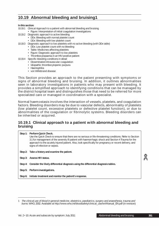
361Vol. 2 • 10. Acute and subacute by symptom: July 2011
10.19 Abnormal bleeding and bruising1
In this section:10.19.1 Clinical Approach to a patient with abnormal bleeding and bruising
• Figure: Interpretation of initial coagulation investigations10.19.2 Diagnostic approach to active bleeding
• DDx: Bleeding with normal platelet count• DDx: Bleeding with low platelet count
10.19.3 Diagnostic approach to low platelets with no active bleeding (with DDx table)• DDx: Low platelet count with no bleeding• Table: Medicines affecting platelets• Figure: Diagnostic approach to low platelets• Thrombocytopaenia in an HIV-positive patient
10.19.4 Specifi c bleeding conditions in detail• Disseminated intravascular coagulation • Idiopathic thrombocytopenic purpura • Haemophilia• von Willebrand disease
This Section provides an approach to the patient presenting with symptoms or signs of abnormal bleeding and bruising. In addition, it outlines abnormalities seen in laboratory investigations in patients who may present with bleeding. It provides a simplifi ed approach to identifying conditions that can be managed by the district hospital team and distinguishes those that need to be referred for more specialized care or managed in coordination with a specialist.
Normal haemostasis involves the interaction of vessels, platelets, and coagulation factors. Bleeding disorders may be due to vascular defects, abnormality of platelets (low platelet count, excessive platelets or defective platelet function), or due to abnormalities of the coagulation or fi brinolytic systems. Bleeding disorders can be inherited or acquired.
10.19.1 Clinical approach to a patient with abnormal bleeding and bruising
Step 1: Perform Quick Check.Use the Quick Check to ensure that there are no serious or life-threatening conditions. Refer to Section 3.1 for management of the severely ill patient with haemorrhagic shock and Section 4 Trauma for the approach to the acutely injured patient. Also, look specifi cally for pregnancy or recent delivery, and signs of infection or sepsis.
Step 2: Take a history and examine the patient.
Step 3: Assess HIV status.
Step 4: Consider the likely differential diagnosis using the differential diagnosis tables.
Step 5: Perform investigations.
Step 6: Initiate treatment and monitor the patient’s response.
1 The clinical use of blood in general medicine, obstetrics, paediatrics, surgery and anaesthesia, trauma and burns. WHO, 2002. Available at http://www.who.int/bloodsafety/clinical_use/en/Manual_EN.pdf (in revision)
Abdominal bleeding and bruising
FINAL Vol. 2 CH 9-21.indd 361FINAL Vol. 2 CH 9-21.indd 361 29/06/2012 15:1629/06/2012 15:16

362 Vol. 2 • 10. Acute and subacute by symptom: July 2011
History
Focus on the following.
• Symptoms of bleeding ° What is the duration of the bleeding? ° Is it the fi rst episode or is the bleeding recurrent? ° How many sites are involved? ° Is the bleeding spontaneous or following trauma? ° Obstetrical or gynaecological history (PPH, abnormal vaginal bleeding).
• History of recent, large volume blood transfusions >10 units, or aggressive IV fl uid therapy (may result in dilutional clotting abnormalities)
• History of exposure to drugs or chemicals ° harmful use of alcohol or illicit substances ° current or previous use of medications, e.g. warfarin, NSAIDs, aspirin, or
traditional, herbal, or alternative medications ° exposure to chemicals at work or in the home.
• Family history. ° relatives with a similar condition ° relatives with any history suggesting bleeding disorder.
Other symptoms:• weight loss• anorexia• fever and night sweats• HIV status – CD4 count and treatment history.
Symptoms suggestive of a bleeding disorder
• easy bruising, purpura, and nosebleeds• excessive bleeding after circumcision, dental extraction or other surgery, or
after giving a blood sample or blood donation• heavy menses with clots or perinatal haemorrhage• dark or bloody stools• red urine• episodes of swollen, painful joints or muscles• excessive bleeding after minor scratches• bleeding that recurs hours or days after the original trauma• poor wound healing.
The source and type of bleeding usually suggest the most likely cause. Bleeding from mucous membranes or petechial bleeds suggests a low platelet count or platelet abnormalities, von Willebrand disease (vWD), or vascular defects. Muscle and joint bleeding or bruising may be suggestive of haemophilia A or B.
Examination
Signs of bleeding or blood loss
• pale mucous membranes• petechial haemorrhages• purpura or ecchymosis (bruising)
Abdominal bleeding and bruising
FINAL Vol. 2 CH 9-21.indd 362FINAL Vol. 2 CH 9-21.indd 362 29/06/2012 15:1629/06/2012 15:16

363Vol. 2 • 10. Acute and subacute by symptom: July 2011
• bleeding from mucous membranes (conjunctiva, gums)• muscle haematomas• haemarthroses or deformed joints• blood on rectal examination• fundoscopy – retinal haemorrhages.
Note: Skin manifestations of bleeding disorders (e.g. petechial haemorrhages or ecchymoses) are sometimes diffi cult to see in dark skinne patients. Examine the mucous membranes, including the conjunctivae, oral mucosa and optic fundi, for evidence of bleeding.
Other signs that may point to underlying or co-morbid disease
• splenomegaly• hepatomegaly• jaundice• fever• tenderness• lymphadenopathy.
Investigations
For a patient with a suspected bleeding problem:• Perform FBC. • Assess whether abnormality is in a single cell line or multiple cell lines.
° single cell line (isolated low Hb, white cell count, or low platelets): ◊ low Hb – anaemia – may be as a result of bleeding, see Section 10.18 Anaemia;
◊ high WBC – sepsis or infection, leukaemia (blasts); ◊ low WBC count – leukopenia, may be result of an underlying pre-leukaemia or myelodysplastic syndrome;
◊ increased MCV – haemolysis, vitamin B12 and folate defi ciencies, alcohol misuse or myelodysplastic syndrome – see Section 10.18 Anaemia;
◊ low platelets – ensure that not a false result due to platelet clumping on peripheral blood smear. Repeat the FBC – use citrate or blue tubes if available as this might provide a more reliable result.
° multiple cell lines (pancytopenia – low Hb, low WBC, and low platelets) indicating total marrow failure or involvement – leukaemia, HIV, marrow infi ltration, myelodysplastic syndrome, drugs, aplastic anaemia.
• Peripheral blood smear ° Platelets
◊ platelet clumping – platelet count incorrectly low on FBC ◊ large platelets.
° Red cells ◊ red cell fragments – haemolysis (DIC or TTP/HUS) ◊ macrocytic (large red cells; high MCV on FBC) – vitamin B12 and folate defi ciency, chronic alcohol use – see Section 10.18 Anaemia
◊ nucleated red cells or a leucoerythroblastic fi lm – marrow infi ltration (malignancy, leukaemia or lymphoma)
◊ parasitic inclusions (malaria). ° White cells
Abdominal bleeding and bruising
FINAL Vol. 2 CH 9-21.indd 363FINAL Vol. 2 CH 9-21.indd 363 29/06/2012 15:1629/06/2012 15:16

364 Vol. 2 • 10. Acute and subacute by symptom: July 2011
◊ increased WBC – infection and sepsis – toxic granulation ◊ decrease WBC – leukopenia. aplastic anaemia or myelodysplasia ◊ blasts – leukaemia – refer for specialized care.
• PT (prothrombin time) and the INR ° PT and INR screen for abnormalities in the extrinsic and common pathways
of coagulation (factors I, II, V, VII and X). Its main use is for anticoagulant (warfarin) monitoring, and detection of acquired bleeding disorders – especially DIC, liver disease, and vitamin K defi ciency. The PT is reported as the international normalized ratio (INR), which refl ects the ratio of the patient's PT to the laboratory's control value.
° Normal range for the PT is between 10 and 13 seconds. An INR >1.5 or a PT ≥3 sec longer than a laboratory's normal control value is usually abnormal and requires further evaluation.
° Fill the citrate tube to the line for an accurate result.• aPTT (activated partial thromboplastin time)
° The aPTT screens for abnormalities in factors of the intrinsic and common pathways (II, V, VIII, IX, X, XI and XII).
° A typical normal range is 28 to 34 seconds.• Crude clotting time
° If aPTT or PT (INR) are not available, a crude clotting time will assist to exclude a coagulopathy. It is a crude screening test and not accurate. Confi rm abnormal tests with a repeat test and refer patients for further tests if the result is confi rmed abnormal.
° See Section 7.2.18 for details. ° Exclude medications, e.g. warfarin and heparin.
Additional tests that may be requested depending on the clinical fi ndings include:
• rapid malaria test or malaria blood smear• occult blood in stool• pregnancy test• HIV test and CD4 count• liver function tests• renal function (urea, creatinine, and electrolytes)• ultrasound of the abdomen and pelvis• bone marrow aspirate or biopsy• further coagulation tests, such as fi brinogen concentration, fi brin degradation
products such as d-dimers, factor assay, platelet function tests.
Abdominal bleeding and bruising
FINAL Vol. 2 CH 9-21.indd 364FINAL Vol. 2 CH 9-21.indd 364 29/06/2012 15:1629/06/2012 15:16

365Vol. 2 • 10. Acute and subacute by symptom: July 2011
Figure: Interpretation of initial coagulation investigations
Abdominal bleeding and bruising
FINAL Vol. 2 CH 9-21.indd 365FINAL Vol. 2 CH 9-21.indd 365 29/06/2012 15:1629/06/2012 15:16

366 Vol. 2 • 10. Acute and subacute by symptom: July 2011
10.19.2 Diagnostic approach to active bleeding Patients who present with active bleeding may or may not have a history of prior episodes with a confi rmed diagnosis. Obtain an FBC and classify the bleeding according to whether the platelet count is decreased or normal. Consult with the relevant DDx table below.
DDx: Bleeding with normal platelet count
Condition In favour
Primary inherited causes of bleeding
von Willebrand disease(vWD)
Family history of the disease Easy bruising, abnormal bleeding after surgical or dental procedures Normal or low platelet numbers, prolonged bleeding time
Haemophilia Bleeding into tissues after minimal trauma, e.g. haemarthroses, muscle haematoma Arthropathy may be present Elevated aPTT and normal INR and platelets
Trauma or local injury
Trauma or local injury
see Section 4
HistoryBruising or bleeding at localized siteBe aware of non-accidental injury patterns (physical abuse)
Underlying medical conditions or medication
Renal failuresee Section 11.31
History of renal diseaseDecreased urine outputUraemic fl apping tremorRaised urea and creatinine
Liver disease or liver failure
see Sections 10.9 and 11.14
History of chronic liver disease , hepatitis or excessive alcohol useWeight loss Firm liver, jaundice, hepatomegaly and right upper quadrant pain, ascitesSplenomegaly Decreased albuminProlonged INR
Coumarin-based anticoagulants (warfarin, rat poison)
see Section 3.8
History of using warfarin or ingesting rat poisonEasy bruising, epistaxis, GI bleedingResponds to vitamin K, but may need transfusion of FFP or prothrombin complex for rapid reversal Prolonged INR Normal aPTT and platelets
Heparin overdose Usually in patients on heparin infusionProlonged aPTT
Corticosteroids Long-term steroid use can increase skin and vessel fragility, leading to bruising and bleeding
Abdominal bleeding and bruising
FINAL Vol. 2 CH 9-21.indd 366FINAL Vol. 2 CH 9-21.indd 366 29/06/2012 15:1629/06/2012 15:16

367Vol. 2 • 10. Acute and subacute by symptom: July 2011
DDx: Bleeding with low platelet count
Conditions In favour
DIC (disseminated intravascular coagulation)
Severely ill patient with multi-organ failurePredisposing condition, e.g. shock, sepsis, malignancy, or obstetric complications, ABO incompatible blood transfusionBleeding from puncture sitesGastrointestinal bleedingProlonged INR and elevated aPTT, low platelet count, low fi brinogen level, presence of fi brin degradation productsPeripheral smear – may have red cell fragments
Sepsissee Section 3.1.5
Signs of infection – fever, tachycardia, dehydration, shockIdentifi able or suspected source of infection
Viral haemorrhagic fever
see Section 11.46
Endemic area or travelFever, headache, malaise, maculopapular rash Bleeding tendencies with purpura, petechiae, haematemesis, melaena, or epistaxisPatients may be severely ill with shock and organ failureProlonged INR
Thrombotic thrombocytopenic purpura (TTP)
Fever Confusion, seizures, intracranial haemorrhage, or comaHaemolytic anaemia (jaundice), renal failureVenous thromboses at unusual sitesAbdominal pain Usually younger patients <60 yearsHIV-infectedDrugs (e.g. quinine)Associated with haemolytic-uraemic syndrome (e.g. from enterohemorrhagic E. coli)FBC: Very low platelet numbers, anaemia Peripheral blood fi lm: evidence of haemolysis – RBC fragments on fi lm, high bilirubin, high LDHProteinuria, haematuria, renal impairmentNormal INR and aPTT
Aplastic anaemia
see Section 10.18
Recent viral infection, History of chemotherapy or chloramphenicol, cotrimoxazole, NSAIDs, carbamazepine, or phenytoinRecurrent infections, anaemiaHIV, hepatitis, and mycobacterial infectionsFBC: Persistent pancytopeniaLow reticulocyte count
Leukaemia (ALL, AML) and deranged clotting/DIC – medical emergency seen in acute promyelocytic leukaemia
Fatigue, fever, recurrent infectionsLymphadenopathy, anaemia with or without hepatosplenomegalyFBC: Pancytopenia; or increased WBCPeripheral blood fi lm: blasts
Abdominal bleeding and bruising
FINAL Vol. 2 CH 9-21.indd 367FINAL Vol. 2 CH 9-21.indd 367 29/06/2012 15:1629/06/2012 15:16

368 Vol. 2 • 10. Acute and subacute by symptom: July 2011
Conditions In favour
Idiopathic thrombocytopenic purpura (ITP) (a diagnosis of exclusion)
Incidental fi nding of low platelets in a well patientEasy bruising, petechiae, or nose bleedsYounger patientsWomen are more commonly affected than menNo other cause of low platelets (autoimmune, HIV or other viral infection, drugs and alcohol)No splenomegaly FBC: isolated low platelet countPeripheral blood fi lm: large platelets
Myelodysplastic syndromes (MDS)
Fatigue, anaemia, recurrent infectionsOlder patient >60 yearsPlatelet count does not respond to steroidsFBC: pancytopenia Peripheral blood fi lm: dysplastic morphological features
10.19.3 Diagnostic approach to low platelets with no active bleedingDecide if only platelets or all cell lines are affected. Exclude artefactual decrease in automated count on FBC with platelet clumping. Look at a peripheral blood fi lm for platelet clumps, large platelets, blasts, increased WBC count (infection or sepsis), and RBC fragments.
Exclude medical causes – consult the DDx table below. If a drug is suspected to be the cause of bleeding or low platelet counts, and other important causes have been excluded (HIV, infection and DIC), then the offending drug should be discontinued if possible. See table below for drugs known to affect platelets. The platelet count should return to normal within a short period of time. If recovery of the platelet count does not occur, a review of the list of other differential diagnoses must be considered.
DDx: Low platelet count with no bleeding
Conditions In favour
Malariasee Section 11.25
Fever, headacheSplenomegalyLiving in or travelled to an endemic areaMalaria test positive (microscopy or RDT)
HIV see Section 13
Other causes excluded – may present as ITPResponds to ART
Metastatic malignancy Fever Loss of weightEvidence of cancer or known diagnosis of malignancy
Alcoholsee Section 16
Long term alcohol useParotomegaly
Autoimmune conditions Known diagnosis, e.g. rheumatoid arthritis, systemic lupus erythematosisSkin lesionsArthritis
Medicines No other medical cause apparentTaking medication known to cause low platelets – see table below
Disseminated fungal infections Generalized lymphadenopathyUnwell patient: fever, malaise, skin lesions with or without lung involvement Immunocompromised patient – CD4 <100
Abdominal bleeding and bruising
FINAL Vol. 2 CH 9-21.indd 368FINAL Vol. 2 CH 9-21.indd 368 29/06/2012 15:1629/06/2012 15:16

369Vol. 2 • 10. Acute and subacute by symptom: July 2011
Table: Medicines affecting platelets
Drugs affecting platelet function (normal platelet counts)
Drugs decreasing platelet count (drug-induced thrombocytopenia)
CommonlyRarely or infrequently
• aspirin (up to 7 days);• heparin;• NSAIDs – diclofenac, ibuprofen (up
to 48 hours);• clopidogrel (up to 7 days);• penicillins and cephalosporins in high
dose; worse with renal failure or low albumin.
Recommendations for elective surgeryIn most surgery, there is no need to stop aspirin or clopidogrel if they are used as monotherapy. If used together, stop the clopidogrel. For cardiac or neurosurgery, stop aspirin 7–10 days pre-surgery; stop clopidogrel 5 days pre-surgery.
• heparin-induced thrombocytopaenia (HIT) – usually with IV heparin after 4–14 days of use if previous use;
• anti-TB drugs – rifampin, isoniazid, ethambutol;
• antibiotics – quinine and quinidine, cotrimoxazole, amphotericin B, nalidixic acid, cephalosporins (cephalothin, vancomycin);
• psychotropic medications – diazepam, haloperidol;
• analgesics and anti-infl ammatories – paracetamol (acetaminophen), diclofenac;
• cardiac drugs – methyldopa, digoxin, amiodarone, hydrochlorothiazide;
• antiretrovirals – IDV, AZT;• cimetidine and ranitidine;• minoxidil;• chlorpromazine;• carbamazepine.
• ibuprofen• phenytoin• tetracycline• glibenclamide• fl uconazole• captopril• ampicillin
Figure: Diagnostic approach to low platelets
Low plateletsInitial investigations
Look at the blood fi lm for:Platelets clumps, large
platelets, blastsIncrease WCC
(infection or sepsis)RBC fragments ?Malaria
Other infections:HIV
Hepatitis B & CEBV (monospot)
Malaria
Other diseases:Liver diseaseMalignancy (metastatic)
Autoimmune (SLE)
Nutritional:Vitamin B12, folateand iron defi ciency
Alcohol
ExcludeDrugs
Medications
Supporting evidence:Malnutrition
Gastric surgeryGI malignancy
Infl ammatory bowel diseases
Commonly:cotrimoxazole, phenytoin,
quinine, ceftriaxone, carbamazepine, etc
See table Drugs affecting platelets
Abdominal bleeding and bruising
FINAL Vol. 2 CH 9-21.indd 369FINAL Vol. 2 CH 9-21.indd 369 29/06/2012 15:1629/06/2012 15:16

370 Vol. 2 • 10. Acute and subacute by symptom: July 2011
Thrombocytopaenia in an HIV-positive patient
This may be due to HIV itself, but it is important to fi rst exclude possible medical causes and drugs, such as TB treatment, cotrimoxazole. Look for lymph nodes, chest X-ray, skin lesions, sputum.
Consider disseminated fungal infections, lymphoma, ITP, and TTP. Start ART.
10.19.4 Specifi c bleeding conditions in detail
Disseminated intravascular coagulation (DIC)
Disseminated intravascular coagulation is a pathological activation of coagulation mechanisms that occurs in response to a variety of underlying diseases. Fibrin clots form throughout the body and consume all the available coagulation factors, fi brinogen, and platelets. Normal coagulation is disrupted, leading to widespread, uncontrolled bleeding from the skin (e.g. venepuncture sites), the digestive tract, the respiratory tract, and surgical wounds. This in turn stimulates an overproduction of fi brinolytic enzymes to break down the clots formed and an increase in fi brin degradation products. The microvascular thrombi also disrupt normal blood fl ow to organs (such as the kidneys), leading to multi-organ failure. Red cell fragmentation occurs due to the passage of red cells in the narrowed and damaged microvasculature.
DIC usually is a result of a serious underlying diagnosis (see below), but can in itself become life-threatening, by haemorrhage or thrombosis.
DIC can occur in the following conditions:• infections: Gram-negative sepsis, Neisseria meningitidis, Streptococcus
pneumoniae, malaria, TB, histoplasmosis, aspergillosis, Lassa fever;• obstetric: abruptio placentae (premature separation of the placenta from the
uterus), retained dead fetus, pre-eclampsia, amniotic fl uid embolism, septic abortion or post-partum sepsis;
• massive tissue injury: trauma, crush injury, burns, massive surgery;• severe hypoxia and acidosis;• cancers of the lung, pancreas, prostate, breast and stomach, acute
myelogenous leukaemia, subtype acute promyelocytic leukaemia;• miscellaneous: liver disease, snake-bite (see Section 3.9), pancreatitis, shock,
heat stroke, ruptured aortic aneurysm, malignant hypertension, pulmonary embolism, subarachnoid haemorrhage, acute haemolytic transfusion reaction.
Key clinical features
• often acutely ill• widespread low-grade haemorrhage or oozing (commonly from the mouth,
nose, and venepuncture sites)• extensive bruising• shock• in severe cases, shock, gangrene, coma, and renal failure
Abdominal bleeding and bruising
FINAL Vol. 2 CH 9-21.indd 370FINAL Vol. 2 CH 9-21.indd 370 29/06/2012 15:1629/06/2012 15:16

371Vol. 2 • 10. Acute and subacute by symptom: July 2011
Treatment
• Manage the patient carefully. Referral to a higher facility may be needed.• The only effective treatment is the reversal of the underlying causes, e.g.
antibiotics for sepsis, removal of retained gestational products. • Give supportive treatment with fl uids to maintain renal perfusion and blood
pressure. See Section 3.1.• Monitor the haemoglobin and consider transfusion when clinically indicated.
See Section 10.18 Anaemia.• If the INR or aPTT is prolonged and the patient is bleeding, replace red cell
losses with the freshest whole blood available, as it contains fi brinogen and most other coagulation factors. Give FFP, as this contains labile coagulation factors. 1 pack/15 kg body weight(4–5 packs in adults). Repeat FFP according to the clinical response.
• If fi brinogen is low or the aPTT or thrombin time is prolonged, also give cryoprecipitate (to supply fi brinogen and factor VIII). 1 pack/6 kg (8–10 packs in adults).
• If the platelet count is less than 50 x 109/litre and the patient is bleeding, also give platelet concentrates, 4–6 packs (adult).
• If the patient is stable enough for transfer to a higher level of care, this should be arranged immediately as intensive care usually is required.
Idiopathic thrombocytopenic purpura (ITP)
ITP is the condition of having a low platelet count (thrombocytopaenia) of no known cause, i.e. idiopathic. It is a diagnosis of exclusion. Most causes appear to be related to antibodies against platelets, so ITP is known also as immune thrombocytopenic purpura or immune-mediated thrombocytopenic purpura.
ITP usually is asymptomatic, however, a very low platelet count may present with:• bruises (purpura) and petechiae, especially on the extremities;• bleeding from the nostrils and bleeding at the gums; • haematomas in the mouth or on other mucous membranes;• possible fatal complications due to an extremely low count (less than
5000 per mm3), and may include: ° subarachnoid or intracerebral haemorrhage; ° lower gastrointestinal bleeding or other internal bleeding.
TreatmentA platelet count below 20 x 109/litre is generally an indication for treatment. Platelet counts between 20 x 109/litre and 50 x 109/litre are usually evaluated on a case-by-case basis. Admit patients with very low counts, and if the patient presents with signifi cant internal or mucocutaneous bleeding. A count below 10 x 109/litre is potentially a medical emergency as the patient is vulnerable to subarachnoid or intracerebral haemorrhage as a result of a mild to moderate head trauma.
Consult with a specialized centre and consider the following treatments or refer for further management.• Steroids and intravenous immunoglobulin
Abdominal bleeding and bruising
FINAL Vol. 2 CH 9-21.indd 371FINAL Vol. 2 CH 9-21.indd 371 29/06/2012 15:1629/06/2012 15:16

372 Vol. 2 • 10. Acute and subacute by symptom: July 2011
Treatment usually is initiated with IV steroids (methylprednisolone or prednisone), intravenous immunoglobulin (IVIg) or a combination of these drugs. After the platelet count has increased to a safe level, give an oral steroid, such as prednisone (1–2 mg/kg daily). Most cases respond during the fi rst week of treatment. Gradually reduce the dose of the oral steroid therapy over several weeks. Monitor regularly as 60%–90% of patients relapse after the dose is decreased below 0.25 mg/kg daily.
• Platelet transfusion Platelets can be transfused in an emergency bleeding situation to raise the
count quickly in order to start oral steroids. Platelet transfusion alone is not recommended, as platelets usually decrease afterwards due to autoimmune destruction.
• Surgery Splenectomy may be performed although the procedure is potentially risky
due to the increased possibility of signifi cant bleeding during surgery. Splenectomy is successful in 60% –65% of cases, but less so in older patients.
Refer for specialized care if patient does not improve on steroids.
Haemophilia
Haemophilia is a group of hereditary X-linked genetic disorders that impair the body's ability to control blood clotting or coagulation. It affects men more commonly than women. Haemophilia A has a defi ciency of clotting factor VIII, and in haemophilia B, factor IX is defi cient. Refer to a specialized centre for diagnosis and management. The patient may be referred back to the district hospital with a supply of required clotting factor concentrates to be administered when needed.
TreatmentThere is no cure for haemophilia. It can be controlled with regular infusions of the defi cient clotting factor, i.e. factor VIII in haemophilia A or factor IX in haemophilia B.• Avoid antiplatelet agents such as aspirin and NSAIDs• Do not give intramuscular injections.• Initial management of haemarthrosis includes strong analgesia, ice packs,
immobilization, compression, and elevation.• Do not incise swellings and never incise a joint for haemarthrosis. • Give clotting factors before surgery, during an active bleed, or after injury,
and, in some cases, on a regular basis to prevent bleeds. • In acute bleeding episodes give coagulation factor concentrates as quickly as
possible. • Start physiotherapy as soon as possible after initial treatment to minimize
loss of joint function.• Desmopressin may also be used as it releases stored endogenous factor VIII
and von Willebrand factor, so may be useful in mild or moderate haemophilia A. It is not indicated in factor IX defi ciency.
• FFP and cryoprecipitate may be use if clotting factor concentrates are not available.
Abdominal bleeding and bruising
FINAL Vol. 2 CH 9-21.indd 372FINAL Vol. 2 CH 9-21.indd 372 29/06/2012 15:1629/06/2012 15:16

373Vol. 2 • 10. Acute and subacute by symptom: July 2011
Von Willebrand disease (vWD)
Von Willebrand disease is the most common hereditary coagulation abnormality, although it can be acquired as a result of other medical conditions. There is a qualitative or quantitative defi ciency of von Willebrand factor (vWF), required for platelet adhesion.
Key clinical features
• mucocutaneous bleeding, e.g. epistaxis, easy bruising• menorrhagia• bleeding after dental extractions• post-traumatic bleeding.
Treatment
• Patients will need to be referred to a specialized centre for diagnosis and management. They may be referred back to the district hospital for ongoing management with their specifi c medicines and blood products.
• Bleeding episodes may be managed with desmopressin (although it may become ineffectual after repeated use), factor VIII, or cryoprecipitate, which also contains vWF.
Abdominal bleeding and bruising
FINAL Vol. 2 CH 9-21.indd 373FINAL Vol. 2 CH 9-21.indd 373 29/06/2012 15:1629/06/2012 15:16

374 Vol. 2 • 10. Acute and subacute by symptom: July 2011
10.20 Splenomegaly
In this section:10.20.1 Clinical approach to a patient with splenomegaly10.20.2 Use the DDx table to establish a likely differential diagnosis
• DDx: Splenomegaly10.20.3 Management of splenomegaly
This Section provides an approach to the patient with splenomegaly. Splenomegaly is only rarely symptomatic. In those cases, the patient may present with pain or a heavy sensation in the left upper quadrant. In most cases, splenomegaly is identifi ed during either a focused or complete routine physical examination. Splenomegaly is not considered a diagnosis in and of itself. Rather, it is often a serious sign or indication of an underlying condition that requires identifi cation and treatment.
10.20.1 Clinical approach to a patient with splenomegaly
Step 1: Perform Quick Check.Ensure that there are no serious or life-threatening conditions
Step 2: Take a history and examine the patient.• confi rm that it is splenomegaly• look for underlying causes and associated conditions.
Step 3: Assess the patient’s HIV status.
Step 4: Consider the likely differential diagnosis using the DDx tables.
Step 5: Perform investigations.
Step 6: Initiate treatment and monitor the patient’s response. Always consider TB and parasitic infections.
History
• What is the age of the patient? Is there sickle-cell or thalassaemia disease in the family?
• Is the patient from a malaria endemic area or is it malaria season?• Is the area endemic for histoplasmosis, leishmaniasis, schistosomiasis, or
trypanosomiasis?• What is the nutritional status of the patient?• Is there pain or heaviness in the left upper quadrant? For how long?• What is the HIV status of the patient?• Has the patient had prior TB or recent contact with TB?• Has the patient had travel or occupational exposure to any of the above
infections?• Are there additional constitutional symptoms?• Is the patient on any hepatotoxic medications?• Does the patient drink alcohol?• Is there any shortness of breath on exertion? Ascites? Swelling of the legs?
Splenomegaly
FINAL Vol. 2 CH 9-21.indd 374FINAL Vol. 2 CH 9-21.indd 374 29/06/2012 15:1629/06/2012 15:16

375Vol. 2 • 10. Acute and subacute by symptom: July 2011
Examination
Confi rm splenomegalyExamination techniques for the spleen:• Bimanual palpation.
° Position the patient supine with bent knees. ° Place the left hand on the lower rib cage and pull the skin upward and taut. ° As the patient inspires deeply and slowly, use the right hand to stroke
upward, starting from the left lower quadrant. ° Record the number of centimetres below the costal margin at a fi xed point
(e.g. umbilicus) at which the spleen tip is fi rst felt.• Percussion (Castell’s method).
° Position the patient supine. ° Percuss the lowest intercostal space in the anterior axillary line. ° If the percussion is resonant, the spleen is normal. If it is dull, the spleen is
enlarged.
Examine the rest of the body to advance the differential diagnosis
• Skin: look for jaundice, pallor, ecchymoses, spider angiomata, caput medusae.• Neck and lymph nodes: look for the hepatojugular refl ex (refl ux)
(abdominojugular test); feel for lymphadenopathy.• Heart and lungs: look for signs of heart failure (rales) or heart murmurs.• Abdomen: check liver for tenderness, enlargement, nodularity, bruits, ascites.• Extremities: look for joint swelling or tenderness, pedal oedema.• Neurological exam: check for encephalopathy, refl exes, sensation,
proprioception.
Investigations
• FCB and smear: this may indicate infection, disseminated disease, or malignancy. Smear will show evidence of haemolysis.
• Malaria thick smear; tests for other parasites.• Liver function tests; thyroid function tests if clinically indicated.• Sputum examination and chest X-ray for TB (see Section 15).• Ultrasound to confi rm splenomegaly, and assess for discrete lesions in the
spleen.• Recommend HIV testing.
Splenomegaly
FINAL Vol. 2 CH 9-21.indd 375FINAL Vol. 2 CH 9-21.indd 375 29/06/2012 15:1629/06/2012 15:16

376 Vol. 2 • 10. Acute and subacute by symptom: July 2011
10.20.2 Use the DDx table to establish a likely differential diagnosis
DDx: Splenomegaly
Condition In favour
Splenic enlargement due to increased demand for splenic function
Sickle -cell disease, thalassaemia, or other haemoglobinopathies
see Section 10.18
Signs and symptoms of anaemia (pallor, shortness of breath on exertion, fatigue, dizziness)JaundiceFamily historyDiscoloured urine
Nutritional anaemia
see Section 10.18
Signs and symptoms of anaemia (pallor, shortness of breath on exertion, fatigue, dizziness)Malnutrition
Rheumatoid arthritis, systemic lupus erythematosus, drug reactions, thyrotoxicosis, ITP, other autoimmune and collagen vascular diseasessee Sections 10.2, 10.13, 10.19
Joint pain and swelling, rash, tachycardia, eye symptomsAbnormal bleeding or bruisingFever
Splenic enlargement due to response to infection
HIVsee Section 13
Could manifest with any symptoms
Viral hepatitissee Section 11.4
Nausea, vomiting, anorexia, feverJaundiceLarge, tender liver
Cytomegalovirussee Section 11.8
Fever, fatigue, malaisePharyngitis (exudative)Cervical lymphadenopathyGuillain-Barré syndrome (fl accid ascending paralysis)
Infectious mononucleosissee Section 10.5
Fever, fatigue, malaisePharyngitis (exudative)Cervical lymphadenopathyGuillain-Barré syndrome (fl accid ascending paralysis)Vision problemsNeuropathy
Subacute bacterial endocarditissee Section 11.10
Heart murmurFatigueFeverSigns of embolism (clotting) to distant areas, especially fi ngers and toes
Typhoid fever see Section 11.43
High feverRelative bradycardiaNon-specifi c symptoms (anorexia, cough, sore throat, constipation)Abdominal pain
Tuberculosissee Sections 10.6 and 15
CoughFeverWeight lossNight sweats
Splenomegaly
FINAL Vol. 2 CH 9-21.indd 376FINAL Vol. 2 CH 9-21.indd 376 29/06/2012 15:1629/06/2012 15:16

377Vol. 2 • 10. Acute and subacute by symptom: July 2011
Malariasee Section 11.25
Regular cyclic feverAnaemiaHypoglycaemiaJaundice, pallorPregnancyMental status changesTachypnea
Visceral leishmaniasissee Section 11.20
MalnutritionHIV-positiveIrregular and prolonged feverLymphadenopathyCoughDiarrhoea
Trypanosomiasissee Section 11.41
History of chancreFever, headache, joint painLymphadenopathyPuffy face
Histoplasmosissee Section 11.16
Enlarged liver, lymph nodesCough or wheezeWeight lossNight sweats
Splenic abscess Fever without obvious focusPain with palpation over spleenSonogram with splenic lucency
Other bacterial, viral, fungal, parasitic infectionssee sections 10.1, 10.5
FeverLymphadenopathyEosinophiliaElevated WBCDepressed WBC
Splenic enlargement due to abnormal splenic or portal blood fl ow
Non-specifi c cirrhosissee sections 10.7, 10.8, 10.9, 10.10a, 16
HepatomegalyLiver tendernessNodular liver edgeOther stigmata of liver diseaseChronic hepatitis B infectionLook for hepatomegaly History of alcohol or hepatotoxic substances or medications (e.g. bush tea,INH) or chronic hepatitis B infection
Hepatic schistosomiasissee Section 11.34
History of “swimmer’s itch”Fever, chills, cough, myalgiaBloody diarrhoea, abdominal pain
Splenic artery aneurysm or splenic vein obstruction
No hepatomegaly. If splenic vein obstruction happens acutely, spleen will be tender.
Splenic enlargement due to unknown etiology
Berylliosis Occupational exposureShortness of breath
Idiopathic splenomegaly No compelling evidence of another underlying condition
Splenomegaly
FINAL Vol. 2 CH 9-21.indd 377FINAL Vol. 2 CH 9-21.indd 377 29/06/2012 15:1629/06/2012 15:16

378 Vol. 2 • 10. Acute and subacute by symptom: July 2011
10.20.3 Management of splenomegaly
The management of splenomegaly requires the management of the underlying cause (see Sections cross-referenced in the DDx Table above). Splenectomy may be performed to correct cytopaenias when the cause is congestive destruction of blood cells. However, if the cause of cytopaenia is marrow failure, splenectomy is contraindicated as the hyperplastic spleen is the body’s only source of new blood cells.
FINAL Vol. 2 CH 9-21.indd 378FINAL Vol. 2 CH 9-21.indd 378 29/06/2012 15:1629/06/2012 15:16

11. Multisystem communicable diseases, renal and HIV-related cancers (in alphabetical order)
Table of contents
11.1 Amoebiasis . . . . . . . . . . . . . . . . . . . . . . . . . . . . . . . . . . . . . . . . . . . . . 38111.2 Bartonellosis . . . . . . . . . . . . . . . . . . . . . . . . . . . . . . . . . . . . . . . . . . . . 38311.3 Brucellosis . . . . . . . . . . . . . . . . . . . . . . . . . . . . . . . . . . . . . . . . . . . . . . 38511.4 Candida . . . . . . . . . . . . . . . . . . . . . . . . . . . . . . . . . . . . . . . . . . . . . . . . 38711.5 Cryptococcosis . . . . . . . . . . . . . . . . . . . . . . . . . . . . . . . . . . . . . . . . . . . 38911.6 Cryptosporidiosis . . . . . . . . . . . . . . . . . . . . . . . . . . . . . . . . . . . . . . . . . . 39311.7 Cysticercosis . . . . . . . . . . . . . . . . . . . . . . . . . . . . . . . . . . . . . . . . . . . . 39411.8 Cytomegalovirus . . . . . . . . . . . . . . . . . . . . . . . . . . . . . . . . . . . . . . . . . . 39611.9 Dengue fever . . . . . . . . . . . . . . . . . . . . . . . . . . . . . . . . . . . . . . . . . . . . 39911.10 Endocarditis . . . . . . . . . . . . . . . . . . . . . . . . . . . . . . . . . . . . . . . . . . . . . 40311.11 Fascioliasis . . . . . . . . . . . . . . . . . . . . . . . . . . . . . . . . . . . . . . . . . . . . . 40611.12 Filariasis, lymphatic . . . . . . . . . . . . . . . . . . . . . . . . . . . . . . . . . . . . . . . . 40911.13 Gonorrhoea . . . . . . . . . . . . . . . . . . . . . . . . . . . . . . . . . . . . . . . . . . . . . 41311.14 Hepatitis – viral . . . . . . . . . . . . . . . . . . . . . . . . . . . . . . . . . . . . . . . . . . . 41411.15 Herpes simplex virus . . . . . . . . . . . . . . . . . . . . . . . . . . . . . . . . . . . . . . . 41811.16 Histoplasmosis . . . . . . . . . . . . . . . . . . . . . . . . . . . . . . . . . . . . . . . . . . . 41911.17 Infl uenza . . . . . . . . . . . . . . . . . . . . . . . . . . . . . . . . . . . . . . . . . . . . . . . 42011.18 Isosporiasis . . . . . . . . . . . . . . . . . . . . . . . . . . . . . . . . . . . . . . . . . . . . . 42411.19 Kaposi sarcoma . . . . . . . . . . . . . . . . . . . . . . . . . . . . . . . . . . . . . . . . . . . 42511.20 Leishmaniasis . . . . . . . . . . . . . . . . . . . . . . . . . . . . . . . . . . . . . . . . . . . . 42711.21 Leprosy . . . . . . . . . . . . . . . . . . . . . . . . . . . . . . . . . . . . . . . . . . . . . . . . 43311.22 Leptospirosis . . . . . . . . . . . . . . . . . . . . . . . . . . . . . . . . . . . . . . . . . . . . 43711.23 Liver abscess . . . . . . . . . . . . . . . . . . . . . . . . . . . . . . . . . . . . . . . . . . . . 43811.24 Loaisis . . . . . . . . . . . . . . . . . . . . . . . . . . . . . . . . . . . . . . . . . . . . . . . . 43911.25 Malaria . . . . . . . . . . . . . . . . . . . . . . . . . . . . . . . . . . . . . . . . . . . . . . . . 44111.26 Microsporidiosis . . . . . . . . . . . . . . . . . . . . . . . . . . . . . . . . . . . . . . . . . . 45511.27 Mycobacterium avium complex . . . . . . . . . . . . . . . . . . . . . . . . . . . . . . . . . 45511.28 Onchocerciasis . . . . . . . . . . . . . . . . . . . . . . . . . . . . . . . . . . . . . . . . . . . 45611.29 Penicilliosis . . . . . . . . . . . . . . . . . . . . . . . . . . . . . . . . . . . . . . . . . . . . . 45811.30 Rabies and animal bites . . . . . . . . . . . . . . . . . . . . . . . . . . . . . . . . . . . . . . 45811.31 Renal problems . . . . . . . . . . . . . . . . . . . . . . . . . . . . . . . . . . . . . . . . . . . 46211.32 Rheumatic fever . . . . . . . . . . . . . . . . . . . . . . . . . . . . . . . . . . . . . . . . . . . 47711.33 Rickettsial diseases . . . . . . . . . . . . . . . . . . . . . . . . . . . . . . . . . . . . . . . . 47811.34 Schistosomiasis . . . . . . . . . . . . . . . . . . . . . . . . . . . . . . . . . . . . . . . . . . 47911.35 Sinusitis . . . . . . . . . . . . . . . . . . . . . . . . . . . . . . . . . . . . . . . . . . . . . . . 48111.36 Strongyloidiasis . . . . . . . . . . . . . . . . . . . . . . . . . . . . . . . . . . . . . . . . . . . 48311.37 Syphilis . . . . . . . . . . . . . . . . . . . . . . . . . . . . . . . . . . . . . . . . . . . . . . . . 48411.38 Taeniasis . . . . . . . . . . . . . . . . . . . . . . . . . . . . . . . . . . . . . . . . . . . . . . . 48711.39 Tetanus . . . . . . . . . . . . . . . . . . . . . . . . . . . . . . . . . . . . . . . . . . . . . . . . 48811.40 Toxoplasmosis . . . . . . . . . . . . . . . . . . . . . . . . . . . . . . . . . . . . . . . . . . . 48911.41 Trypanosomiasis, human African . . . . . . . . . . . . . . . . . . . . . . . . . . . . . . . . 49111.42 Trypanosomiasis, American . . . . . . . . . . . . . . . . . . . . . . . . . . . . . . . . . . . 49411.43 Typhoid fever . . . . . . . . . . . . . . . . . . . . . . . . . . . . . . . . . . . . . . . . . . . . 49811.44 Urinary tract infection . . . . . . . . . . . . . . . . . . . . . . . . . . . . . . . . . . . . . . . 49911.45 Varicella/zoster . . . . . . . . . . . . . . . . . . . . . . . . . . . . . . . . . . . . . . . . . . . 50111.46 Viral haemorrhagic fever . . . . . . . . . . . . . . . . . . . . . . . . . . . . . . . . . . . . . 50311.47 Yellow fever . . . . . . . . . . . . . . . . . . . . . . . . . . . . . . . . . . . . . . . . . . . . . 504
FINAL Vol. 2 CH 9-21.indd 379FINAL Vol. 2 CH 9-21.indd 379 29/06/2012 15:1629/06/2012 15:16

FINAL Vol. 2 CH 9-21.indd 380FINAL Vol. 2 CH 9-21.indd 380 29/06/2012 15:1629/06/2012 15:16

381381Vol. 2 • 11. Multisystem communicable diseases, renal problems and HIV-related cancers: July 2011
11. Multisystem diseases (in alphabetical order)
This Section includes diseases that affect multiples organs and body systems, including opportunistic infections, an HIV-related cancer (Kaposi sarcoma), neglected tropical diseases, malaria, other infectious diseases, renal problems, and other common problems such as urinary tract infection and sinusitis.
11.1 Amoebiasis
Amoebiasis results from infection with the non-invasive Entamoeba dispar or the invasive Entamoeba histolytica, and is the third most common cause of death from parasitic disease. It is most commonly contracted through ingestion of live cysts found with faecally contaminated water, food, or hands. Foodborne infection is caused by faecally contaminated soil or water used for growing vegetables.
It is endemic in most developing countries. Some patients become chronic asymptomatic carriers who can excrete up to 15 million cysts a day, thereby enabling the spread of amoeba to new hosts.
Acute amoebic colitis can be confused with bacterial diarrhoeas caused by Campylobacter, E. coli, Salmonella, Shigella, and cholera.
11.1.1 Intestinal amoebiasis
Key clinical features • Gradual development of lower abdominal pain and mild diarrhoea.• Malaise, weight loss, and diffuse lower abdominal or back pain.• If caecum is involved, signs and symptoms will mimic those of appendicitis
(right lower quadrant pain).• Full dysentery develops in some patients with passage of 10–12 stools per
day. Stools are mostly blood and mucus.
Severe gastrointestinal disease:• occurs mostly in children• characterized by high fever, profuse diarrhoea, and abdominal pain.
Complications or unusual presentations:• amoebic liver abscess• amoebic colitis can be confused with infl ammatory bowel disease• amoeboma (tender abdominal mass).
InvestigationsStool examination: • Fresh stools specimens must be examined for trophozoites typical
of E. hemolytica. Cysts of both entamoeba species are very similar therefore trophozoites that have ingested red blood cells are diagnostic of E. hemolytica.
If available, ultrasonography can establish the presence of a liver abscess (due to amoeba or bacteria).
Amoebiasis
FINAL Vol. 2 CH 9-21.indd 381FINAL Vol. 2 CH 9-21.indd 381 29/06/2012 15:1629/06/2012 15:16

382382 Vol. 2 • 11. Multisystem communicable diseases, renal problems and HIV-related cancers: July 2011
TreatmentSupportive management of dysentery, as well as oral or intravenous rehydration, are both very important. Specifi c treatment is divided into two groups:
To eradicate invasive disease:• metronidazole 750 mg 3 times daily for 7–10 days; OR• tinidazole 4 tablets (2 grams) by oral route once daily for 3 days.
To eradicate cysts: • It is recommended to follow all treatment of confi rmed amoebiasis with
eradication of cysts, although these agents may not be routinely available. If they are not available, relapse rates are high, and another course of a luminal agent may be warranted: ° diloxanide 500 mg orally 3 times daily for 5 days; OR ° iodoquinol 650 mg orally 3 times daily for 20 days; OR ° paromomycin 25–35 mg/kg/day orally divided in 3 daily doses for 7 days.
Indications for aspiration are:• the need to rule out other causes of abscess• no clinical response after 3 to 5 days• threat of imminent rupture of the abscess• prevention of left lobe abscess rupture into pericardium.This relies on the availability of an ultrasound with an expert operator.
11.1.2 Amoebic liver abscessAmoebic liver abscess is caused by an often delayed extra-intestinal infection by E. histolytica. Amoebic infection is spread by ingestion of food or water contaminated with cysts. Ten to fi fteen percent of patients with amoebic liver abscess present with only fever.
Key clinical features
• Involves clinical tenderness of the liver.• Amoebic liver abscess is not usually associated with diarrhoea (although the
source is always the colon).• In endemic areas, the course is often subacute with haepatomegaly and
weight loss.• Fever is present in 30% of cases, and may be the only presenting symptom.• May be complicated by pleuro-pulmonary involvement when the abscess
extends from the liver into the lung area.
Investigations
• An ultrasound of the liver can show abscesses.• Serology: positive serology means invasive amoebiasis and generally will
revert to negative after 6–12 months. The presence of a liver abscess usually means that the etiology is amoeba rather than bacteria.
• A negative stool examination for amoebic cysts or trophozoites does not exclude an amoebic liver abscess.
• Aspiration of the abscess with Gram staining (and culture if available) may be useful to differentiate the amoebic abscess from a pyogenic abscess. Caution should be used in aspirating cysts, as an anaphylactic reaction can occur in
Amoebiasis
FINAL Vol. 2 CH 9-21.indd 382FINAL Vol. 2 CH 9-21.indd 382 29/06/2012 15:1629/06/2012 15:16

383383Vol. 2 • 11. Multisystem communicable diseases, renal problems and HIV-related cancers: July 2011
cases of echinococcal cysts. The decision to perform a liver abscess aspiration should take into account this risk. Trophozoites are rare in liver aspirates (since they are in the capsule of the abscess and not in the aspirated necrotic centre).
• Routine haematology and chemistry tests are rarely helpful, although about 75% of patients will have WBC more than 10 000 cells/µl. Liver enzymes are often normal or only mildly elevated. Alkaline phosphatase levels are often elevated and can remain so for months.
Treatment
• metronidazole 750 mg orally or IV 3 times daily for 5–10 days; OR• single dose tinidazole 2 g (should not be used in patients with HIV infection).
Most patients will respond well to treatment with metronidazole, with a decrease in fever within 72 hours. The advantage of metronidazole is that if the etiology of the liver abscess is bacterial, this treatment will generally still work (if the bacteria is sensitive). Indications for aspiration (if possible under ultrasound guidance) are:• need to rule out other causes of abscess• no clinical response after 3–5 days• threat of imminent rupture of the abscess (superfi cial abscess)• prevention of left lobe abscess rupture into pericardium (very rare).
11.2 Bartonellosis
(Carrion’s disease, cat scratch disease, oroya fever, peliosis hepatis, trench fever, verruga peruana, bacilliary angiomatosis)Bartonella spp cause a wide variety of diseases that include trench fever, cat-scratch disease, peliosis hepatis, oroya fever, and verruga peruana. These clinical entities are not considered AIDS-defi ning, but they are seen with increased frequency in PLHIV and patients with advanced immunosuppression. Bartonellosis is diffi cult to diagnose and is largely a diagnosis of exclusion in patients with a fever of unknown origin, skin papules, lymphadenopathy, hepatosplenomegaly, maculopapular skin lesions fatigue, and malaise. (See fever table in Section 10.1)
11.2.1 Oroya fever and verruga peruanaThese entities are caused by B. bacilliformis, and are commonly encountered in the Andes mountains because the vector that transmits the disease is a sandfl y. Both entities are a spectrum of the same illness. Oroya fever presents predominantly in patients who have never encountered the disease before and verruga peruana is the manifestation of the disease in patients who have been exposed in the past. Oroya fever is associated with profound anaemia that is usually the cause of death.
11.2.2 Cat scratch diseaseCat scratch disease is a self-limited illness caused by B. henselae. It is associated with scratches from young domestic cats infested by fl eas. A majority of cases occur in children, but adults, particularly patients with immunosuppression due to AIDS or who are pregnant , are also affected. Enlargement of the lymph nodes can persist for several months, and therefore mimic a malignancy. Diagnosis is mostly clinical.
Bartonellosis
FINAL Vol. 2 CH 9-21.indd 383FINAL Vol. 2 CH 9-21.indd 383 29/06/2012 15:1629/06/2012 15:16

384384 Vol. 2 • 11. Multisystem communicable diseases, renal problems and HIV-related cancers: July 2011
Key clinical features
• localized papule at the area of the cat scratch;• tender lymph nodes in the region draining the area of the cat scratch develop
after 1 to 2 weeks;• nodes may become suppurative with bacterial superinfection common;• systemic symptoms are limited to malaise, anorexia, and weight loss.
Complications
• neurological: meningitis, encephalitis, seizures, transverse myelitis;• granulomatous hepatitis and splenitis;• endocarditis;• osteomyelitis.
Investigations
• serologic tests can be positive in 60–70% of patients;• biopsy of the affected lymph node, if available, will reveal stellate necrosis.
TreatmentThis condition is usually self-limited, but systemic symptoms may be debilitating. Patients with complicated disease should be treated:• doxycycline 100 mg twice daily for 10–14 days; OR• azithromycin 500 mg orally daily for 5 days; OR• ciprofl oxacin 500 mg daily for 10–14 days.Neurological disese and endocarditis need combined and/or longer treatment. Use caution with ciprofl oxacin, as it may partially treat undiagnosed TB.
11.2.3 Trench feverTrench fever is caused by B. quintana, and is associated with infestation with body louse. It is therefore a disease of over-crowding and poverty. It occurs throughout the world.
Key clinical features
• sudden onset of headache, meningitis• persistent or relapsing fever• bacteraemia can persist for weeks• localized fi ndings are uncommon.
Complications Endocarditis may develop with prolonged bacteraemia.
InvestigationsBlood culture may reveal the cause, but the organism grows very slowly.
TreatmentAntibiotic treatment needs to be prolonged to eradicate bacteria and to prevent relapse:• doxycycline 100 mg twice daily for 4 to 6 weeks; OR• eythromycin 2000 mg daily for 4 to 6 weeks; OR• azithromycin 500 mg daily for 4 to 6 weeks.
Bartonellosis
FINAL Vol. 2 CH 9-21.indd 384FINAL Vol. 2 CH 9-21.indd 384 29/06/2012 15:1629/06/2012 15:16

385385Vol. 2 • 11. Multisystem communicable diseases, renal problems and HIV-related cancers: July 2011
11.2.4 Bacillary angiomatosisBacillary angiomatosis can be caused by both B. henselae and B. quintana in persons who are immunocompromised. It was fi rst described in PLHIV, but has since been seen in other patients who do not seem to have a dysfunctional immune system. The disease classically involves the skin, but can spread to other areas including the liver (then it is called peliosis hepatis). Bacillary angiomatosis can easily be confused with Kaposi sarcoma, angiomas, and pyogenic granulomas.
Key clinical features
• Vascular nodules or papules or small tumours that are red or purple, and resemble Kaposi sarcoma.
Complications
• Dissemination: fever, abdominal pain, weight loss, malaise.• PLHIV can also have signs of central nervous system abnormalities such as
brain lesions or psychiatric conditions.• Skin lesions are not always evident in disseminated disease.• Peliosis hepatis is associated with pain when palpating the liver. InvestigationsDiagnosis is mostly clinical, but in cases where there may be confusion with Kaposi sarcoma, one of the following may help differentiate the two:• a blood culture may yield organisms in disseminated disease;• a defi nitive diagnosis is done by examination of a biopsy that usually reveals
tiny bacteria associated with new vessels (angiomas).
TreatmentTreatment needs to be prolonged, especially in cases where relapse has occurred. Patients with liver disease require intravenous treatment. Effective antibiotics include:• erythromycin 2000 mg per day orally for 3 weeks; OR• doxycycline 100 mg per day for 3 weeks. In patients with liver disease, intravenous antibiotics are recommended. Macrolides and tetracyclines are generally preferred to other antibiotics.
11.3 Brucellosis1,2,3
Brucellosis, also known as “Mediterranean fever” or “Malta fever”, is caused by infection with Brucella species. B. melitensis is the most virulent and invasive. Transmission to humans occurs through direct contact, through broken skin, with infected animal tissue, inhalation of infectious aerosols, or ingestion of infectious milk or dairy products. Brucellosis is predominantly an occupational disease. Sporadic cases and sometimes large outbreaks occur after consumption of raw milk and milk products. Animals involved are cows, sheep, goats, swine, and
1 Brucellosis in humans and animals. WHO, Food and Agriculture Organization of the United Nations, and World Organisation for Animal Health, 2005. Available at http://www.who.int/csr/resources/publications/Brucellosis.pdf
2 Excerpt from WHO recommended standards and strategies for surveillance, prevention and control of communicable diseases at http://www.who.int/zoonoses/diseases/Brucellosissurveillance.pdf
3 Heymann DL, ed. Control of communicable diseases manual, 19th ed. APHA and WHO, 2008.
Brucellosis
FINAL Vol. 2 CH 9-21.indd 385FINAL Vol. 2 CH 9-21.indd 385 29/06/2012 15:1629/06/2012 15:16

386386 Vol. 2 • 11. Multisystem communicable diseases, renal problems and HIV-related cancers: July 2011
occasionally dogs. Human-to-human transmission occurs rarely, through blood transfusion or sexual contact.
Brucellosis is endemic in the Mediterranean countries, North and East Africa, the Middle East, South and Central Asia, and South and Central America. Brucellosis is often unrecognized and frequently unreported.
Key clinical featuresThe patient has a history of recent exposure to known or probable source of brucellosis:• ingestion of unpasteurized milk or milk products• unprotected contact with potentially infected animal tissues, blood, or vaginal
discharge• incubation period is variable from a few days to weeks and months.
Brucellosis is a systemic disease with confi rmed intermittent or irregular fever (undulant fever of varying duration). Clinical manifestations are nonspecifi c. The term “localized” is used when symptoms related to a specifi c organ predominate. The disease has acute, subacute, and chronic presentations:• acute brucellosis: (50% of the cases);• untreated or unrecognized acute disease may become localized:
° may present as either: ◊ mild infection that resolves without treatment in 3 to 6 months; ◊ sudden onset fever, chills, sweating; ◊ gradual onset headaches, malaise, extreme fatigue with intermittent or absent fever;
° other clinical features include: ◊ myalgia and depression (common); ◊ anorexia, nausea, vomiting, diarrhoea, constipation, abdominal pain, haematuria, cough;
◊ enlarged liver spleen and lymph nodes;• subacute localized disease:
° localized disease (granulomas, vasculitis) can affect any organ: ◊ bone and joint involvement (most common) causing sacroiliitis, spondylitis, paraspinal abscesses, osteomyelitis, suppurative arthritis;
◊ genitourinary involvement – orchitis, epididymitis; ◊ CNS involvement – meningitis, encephalitis, depression; ◊ CVS involvement – endocarditis (commonest cause of death); ◊ respiratory involvement – bronchitis, lung abscess, effusions; ◊ abdominal involvement – liver granulomas; ◊ skin involvement – rashes, papules, erythema nodosum; ◊ ophthalmic involvement – uveitis, iridocyclitis (see Section 10.12).
• chronic brucellosis: ° persistence of local or systemic disease; or ° relapses of local or systemic disease.
InvestigationsTo demonstrate the presence of Brucella, the following steps are necessary:• isolation of Brucella from blood, bone marrow, pus, or other tissues:
° blood culture – requires special technique and long incubation period, and is often negative in long-standing disease;
° PCR for Brucella;
Brucellosis
FINAL Vol. 2 CH 9-21.indd 386FINAL Vol. 2 CH 9-21.indd 386 29/06/2012 15:1629/06/2012 15:16

387387Vol. 2 • 11. Multisystem communicable diseases, renal problems and HIV-related cancers: July 2011
• serological tests for Brucella antibodies in blood or other tissue: combine Rose Bengal test for agglutinating antibodies (IgM, IgG, IgA) with a test for non-agglutinating antibodies (Coombs-IgG, ELISA-IgG);
• X-rays to demonstrate joint disease (blurred joint margins, widened sacro-iliac space, destruction of vertebrae).
Do not forget to look for tuberculosis.
TreatmentUse two or more antibiotics in combination:• doxycycline 200 mg daily for 45 days PLUS streptomycin 1g IM daily (or
gentamicin 5 mg/kg daily) for 15 days (preferred); OR• doxycycline 200 mg daily for 45 days PLUS rifampicin 15 mg/kg/day (600–
900 mg) for 45 days (alternative).Longer courses may be needed for endocarditis and CNS involvement.
Surgical drainage of abscesses may be needed.
Buruli ulcer see Section 10.2 Skin problems
11.4 Candida
Candidiasis is a fungal infection most commonly caused by Candida albicans. Candida is part of the normal human fl ora and is found in the mouth, vagina, and gastrointestinal tract. Predisposing factors that can lead to infection are:• wide-spectrum antibiotic use• diabetes• HIV • pregnancy • skin maceration, or a break in the natural skin or mucosal barrier.Candidiasis can be limited to mucous membranes or can occasionally spread through the blood or be deeply invasive.
Key clinical featuresOral candida: • white deposits that adhere to the mucosa in the mouth and that can extend
into the oesophagus;• diffi culty with swallowing;• persistent oral candida is a WHO stage 3 condition indicating the need to
initiate ART. (See Section 10.17 Mouth problems).Skin infection: • red macerated skin if infection occurs in skin folds under breasts or around
the anus;• usually associated with itching.Vaginal candidiasis: • white itchy discharge;• sometimes associated with pain on urination.
Candida
FINAL Vol. 2 CH 9-21.indd 387FINAL Vol. 2 CH 9-21.indd 387 29/06/2012 15:1629/06/2012 15:16

388388 Vol. 2 • 11. Multisystem communicable diseases, renal problems and HIV-related cancers: July 2011
Oesophageal candidiasis:• can be asymptomatic;• often associated with chest pain and diffi culty swallowing; • oesophageal candidiasis (and candida of trachea, bronchi, or lungs) is a WHO
stage 4 condition, indicating the need to initiate ART. (See Section 10.7b Painful or diffi cult swallowing).
Candida can be a deeply invasive disease. Fungemia can occur mainly through infection of intravenous devices or urinary catheters and can spread throughout the whole body. There are a few organs that are very prone to be invaded after bloodstream infections. These are the eyes, causing blurred vision and showing white cotton ball lesions on fundoscopy, and the liver and spleen, particularly in patients recovering from very low WBC. These situations require specifi c treatment.
Investigations
• A wet smear can identify the fungus with pseudo-hyphae (branching structures).
• A culture of the likely source will often yield a positive result.
TreatmentApproaches to management depend on the location and the severity of the infection. Topical agents are preferred for the skin, but invasive candidiasis needs to be treated with systemic antifungals. Patients with AIDS who have recurrent episodes of candidiasis benefi t from having each episode treated separately. Medications to be used depend on where the candidiasis is found:
• Oral: ° clotrimazole troche (dissolving tablet) twice daily for 7 days; OR ° nystatin: one tablet 500 000 IU 4 times daily; tablets should be sucked and
retained in the mouth for as long as possible; therapy should be continued for at least 48 hours after symptoms have resolved; OR
° miconazole gum patch once daily for 7 days; case reports also mention its effi cacy in treating oesophageal thrush; OR
° miconazole oral gel: 60 mg 4 times daily for 7 days. ° Recurrent oral candidiasis may require fl uconazole 100 to 200 mg for 7 to 14
days.
• Skin: ° Use measures to reduce wetness or moisture and decrease friction. ° Topical application of an antifungal cream such as nystatin, cotrimazole,
terbinafi ne, or miconazole cream for 5 to 7 days.
• Vaginal (see Section 10.15)
• Oesophagus: ° fl uconazole tablet 100 mg or 200 mg daily for 14 to 21 days; OR ° itraconazole 200 mg daily, can be increased to a maximum of 400 mg daily,
for 10–14 days; the capsules should be taken with food or an acid drink (such as a cola) to increase their bioavailability; OR
° ketoconazole: 200 to 400 mg daily if fl uconazole is not available, until remission is obtained. Ketoconazole is associated with hepatotoxicity. Ketoconazole should not be co-administered with nevirapine. When
Candida
FINAL Vol. 2 CH 9-21.indd 388FINAL Vol. 2 CH 9-21.indd 388 29/06/2012 15:1629/06/2012 15:16

389389Vol. 2 • 11. Multisystem communicable diseases, renal problems and HIV-related cancers: July 2011
administered with lopinavir/r, the dose of ketoconazole should not exceed 200 mg daily.
• Disseminated disease: ° Removal of prosthetic devices including catheters is recommended for the
management of invasive candida infections. ° Give fl uconazole 400 mg daily (orally or IV if patient cannot swallow) for 14
days after the last fever.
• Stopping fl uconazole ° Long-term administration of fl uconazole may result in the development of
drug resistance. ° Patients stable on ART with immunological (CD4 count >100 cells/mm3) or
clinical evidence of immune recovery and no evidence of candidiasis should stop fl uconazole.
11.5 Cryptococcosis4
This infection is most commonly caused by Cryptococcus neoformans, particularly in HIV-positive patients with advanced immunodefi ciency (CD4 cell count generally <100/mm3). However, patients who have been on long-term steroid therapy, as well as other immunosuppressive drugs, are also at risk. Notably, other species of Cryptococcus (e.g. Cryptococcus gattii) are increasingly recognized as a cause of cryptococcosis in immunocompetent individuals. Typically, infection is acquired by inhalation of the fungus into the lungs. Cryptococcus is found in most areas with a relatively warm climate, and is not restricted to the tropics.
Cryptococcal meningitis or meningo-encephalitis is the most common presentation in HIV-positive patients, and is associated with a universal mortality without treatment. Non-meningeal presentations of cryptococcosis include pneumonia, skin lesions, and lymphadenitis.
Key clinical features
• Sub-acute meningitis: headache increasing over days to weeks, fever, photophobia, nausea, seizures, confusion, irritability, blurred vision, sixth cranial nerve palsy, papilloedema on retinal exam are common. Nuchal rigidity is often not marked.
• Coma, or a reduced level of consciousness are associated with a poor prognosis.
• Lung infections: chest pain and cough in a minority of patients, but often no fever.
• Skin lesions: disseminated disease is associated with papular lesions with an umbilicated, centrally depressed area (similar appearance to molluscum contagiosum), which can become ulcerated.
Complications
• Intracranial pressure can become raised (increasing headache, vomiting, cranial nerve palsy).
4 Rapid advice: diagnosis, prevention and management of cryptococcal disease in HIV-infectedadults, adolescents and children. WHO, 2011. Available at www.who.int/hiv/pub/cryptococcal_disease2011
Cryptococcosis
FINAL Vol. 2 CH 9-21.indd 389FINAL Vol. 2 CH 9-21.indd 389 29/06/2012 15:1629/06/2012 15:16

390390 Vol. 2 • 11. Multisystem communicable diseases, renal problems and HIV-related cancers: July 2011
• Cryptococcomas can develop in the brain, more commonly in patients who are not immunocompromised.
• Coma, cerebral oedema, and death follow if it is untreated, usually due to elevated intracranial pressure.
• Hydrocephalus, blindness, dementia, and personality change can occur as permanent sequelae.
Investigations
• A lumbar puncture and CSF examination is the most useful investigation.• Presence of encapsulated yeast forms on an India ink stain of the CSF is the
most widely available diagnostic test (sensitivity 82–85%, but may be negative if low fungal burden).
• An alternative and highly sensitive and specifi c test is the CSF cryptococcal antigen (CrAg) latex agglutination test that examines for the presence of capsular antigen (sensitivity
• 96–100%); serum CrAg may also be positive.• A positive CSF fungal culture is the defi nitive test, but is not widely available
and may take some days.• Cryptococcus can be cultured in the blood in 70% of cases.• Other features of CSF:
° elevated opening pressure (more than 20 cm H2O in 70% of patients) ° low WBC (less than 50 cells/microlitre) with mononuclear predominance ° lowered glucose level ° slightly elevated protein.
• Microscopy and culture from non-meningeal cryptococcal sites – visualization of encapsulated yeasts or positive culture from skin scraping or biopsy, sputum, urine, lymph node FNA, or biopsy.
• Chest X-ray – similar to PCP (bilateral diffuse interstitial infi ltrates).
Anti-fungal treatmentIf the patient has meningitis or pneumonia, treatment with a regimen containing amphotericin is preferred. Given potential life-threatening adverse events (e.g. severe hypokalaemia) associated with amphotericin therapy, however, amphotericin-containing regimens should not be used without adequate laboratory monitoring (i.e., electrolytes and kidney function) and the use of pre-hydration and electrolyte replacement prior to amphotericin administration (see Box below for further details of safe administration of amphotericin B).
The following therapies for cryptococcal meningitis are recommended:
Option 1 (if amphotericin, lab monitoring, pre-hydration, and fl ucytosine available): amphotericin B 0.7 to 1 mg/kg PLUS fl ucytosine 100 mg/kg for 14 days, followed by oral fl uconazole 400 mg daily for 8 weeks.
Option 2 (if amphotericin, lab monitoring, and pre-hydration available, but not fl ucytosine): amphotericin B (as above) PLUS oral fl uconazole 800 mg for 14 days followed by fl uconazole 400 mg for 8 weeks.
Option 3 (if lab monitoring unavailable): intravenous amphotericin B (as above) for 5-7 days PLUS oral fl uconazole 800 mg for 14 days followed by fl uconazole 800 mg for 8 weeks.
Cryptococcosis
FINAL Vol. 2 CH 9-21.indd 390FINAL Vol. 2 CH 9-21.indd 390 29/06/2012 15:1629/06/2012 15:16

391391Vol. 2 • 11. Multisystem communicable diseases, renal problems and HIV-related cancers: July 2011
Option 4 (if amphotericin, lab monitoring and pre-hydration NOT available): oral fl uconazole 800–1200 mg for 14 days followed by fl uconazole 400 mg for 10 weeks.
Recommendations for safe administration of amphotericin B
To reduce risk of thrombophlebitis, use large peripheral veins or a central venous catheter, changing venous access sites frequently and infusing over longer periods.
Protect against life-threatening adverse events:• Monitor renal function and electrolytes (e.g., serum potassium, serum creatinine, fl uid intake and output,
and daily weight) prior to initial treatment and then at least twice weekly.• One hour prior to administration of each amphotericin infusion, administer 1 litre of IV normal saline solution
over 2–4 hours.• Consider addition of one ampoule (20 mmol) of potassium chloride with each litre of IV saline solution plus
one to two 8 mEq KCL tablets orally twice daily. An additional one 8mEq KCL tablet twice daily may be added during the second week. If available, magnesium supplementation should also be provided (two 250 mg tablets of magnesium trisilicate twice daily).
• Use with caution in renal impairment. If creatinine increases by ≥2 fold from baseline value, switch to liposomal amphotericin (3 mg/kg/day) if available.
• If liposomal amphotericin not available, either skip an amphotericin B dose or increase pre-hydration to one litre 8 hourly. Once creatinine improved, restart at 0.7 mg/kg/day and consider alternate day amphotericin B. If creatinine remains elevated, discontinue amphotericin and continue with fl uconazole at 1200mg/day. Monitor creatinine daily.
Minimize acute infusion reactions (e.g. fever, chills, headache, hypotension):• Infuse the initial dose slowly over 3–6 hours. • Prophylactic antipyretics or hydrocortisone should only be used in patients who have previously
experienced acute infusion reactions (and in whom continued treatment with amphotericin is essential).
Control of raised intracranial pressure (ICP)Patients with increased intracranial pressure should have frequent (daily) lumbar punctures done to relieve the pressure and to avoid the development of blindness and other long-term complications.• Educate and inform the patient about the importance of baseline and follow-
up lumbar punctures (i.e., improves treatment outcomes).• Measure the intracranial pressure (see Section 7.4.2).
Treatment of raised intracranial pressure (ICP) in cryptococcal meningitis
• Raised ICP contributes to early mortality and residual morbidity.• Raised ICP is present in >50% of patients.• Repeated spinal taps lower ICP, reducing mortality and morbidity, as well as reducing severe headaches.• If initial opening pressure is normal, repeat LP in 1–2 weeks or if worsening headache, visual, or hearing
disturbances.• If initial opening pressure is >25 cm H20, tap up to 30 ml spinal fl uid to achieve pressure <20 cm H20.• Perform daily taps until the opening pressure is <25 cm H20.
Delay initiation of ARTDue to the high risk of IRIS with CNS disease, which may be life-threatening, in HIV-infected adults, adolescents and children with a recent diagnosis of cryptococcal meningitis, ART initiation should be deferred until there is evidence of a sustained clinical response to antifungal therapy, and• after 2-4 weeks of induction and consolidation treatment with amphotericin
B-containing regimens combined with fl ucytosine or fl uconazole (two weeks with non-meningeal disease); OR
Cryptococcosis
FINAL Vol. 2 CH 9-21.indd 391FINAL Vol. 2 CH 9-21.indd 391 29/06/2012 15:1629/06/2012 15:16

392392 Vol. 2 • 11. Multisystem communicable diseases, renal problems and HIV-related cancers: July 2011
• after 4-6 weeks of induction and consolidation treatment treatment with a high-dose oral fl uconazole regimen (four weeks with non-meningeal disease).
Secondary prophylaxis
• Give fl uconazole 200 mg by mouth daily until the patient is successfully on ART and the CD4 count is maintained above 200 cells/mm3 (two measurements 6 months apart).
Drug interactions
• Rifampicin and fl uconazole ° Rifampicin can reduce fl uconazole levels. ° Consider increasing fl uconazole by 50% if given with rifampicin.
• Nevirapine and fl uconazole ° Available evidence is limited for co-administering nevirapine and
fl uconazole. ° Studies have shown fl uconazole increases nevirapine levels. ° Nevirapine has not been shown to decrease fl uconazole levels signifi cantly. ° Monitor LFTs:
◊ If the baseline ALT is raised: monitor ALT at initiation, 1, 2, 3, 4, 6, and 8 weeks, and then according to the clinic’s usual standard for administering nevirapine.
◊ If the baseline ALT is normal: monitor ALT at initiation, 2 and 4 weeks, and then according to the clinic’s usual standard for administering nevirapine.
Cryptococcal IRIS
Patients with advanced immune defi ciency can develop IRIS after the initiation of ART as a result of partial immune restoration. Cryptococcal meningitis is a common infection implicated in IRIS. It occurs in patients with latent or recently treated cryptococcal meningitis who then develop an infl ammatory meningitis, headache, or focal signs. Cryptococcal IRIS can develop between 1 week and 8 months after a patient starts ART, but typically within 1–6 weeks. It is usually accompanied by a signifi cant CD4 increase and a viral load (VL) decrease.
DiagnosisIn addition to the above history, look for:• recent initiation of ART• recent treatment for cryptococcal meningitis• history of long-standing mild headache prior to ART commencement.
In addition to the above examination look for:• lymphadenopathy• increased intracranial pressure.
InvestigationsCSF results can be diffi cult to interpret, especially if the patient has recently been treated for cryptococcal meningitis. The CrAg and India ink test remain positive for many months after treatment and do not distinguish between live and dead organisms. Diagnosis will therefore often be based on the patient’s history and examination fi ndings.
Cryptococcosis
FINAL Vol. 2 CH 9-21.indd 392FINAL Vol. 2 CH 9-21.indd 392 29/06/2012 15:1629/06/2012 15:16

393393Vol. 2 • 11. Multisystem communicable diseases, renal problems and HIV-related cancers: July 2011
• CSF: ° positive cryptococcal antigen (if recently treated will still be positive – does
not indicate new infection) ° cryptococcal culture usually negative.
• VL or CD4: ° signifi cant decrease in VL ° increased CD4.
Treatment
• continue ART • cryptococcal treatment (as above) • consider steroids if meningitis is life-threatening or focal neurology exists.
11.6 Cryptosporidiosis
Diarrhoeal illness caused by Cryptosporidium parvum is acquired by ingesting oocysts from contaminated food or water. Oocysts are immediately infectious when excreted by infected persons; therefore, person-to-person transmission occurs in child care centres, medical centres, and among household contacts. The disease is self-limited in persons with normal immunity (including PLHIV with CD4 more than 200), but can be severe and debilitating in persons with immunosuppression.
Key clinical features
• watery, non-bloody diarrhoea• abdominal pain, nausea, fever, decreased appetite leading to weight loss.
Severe disease (in patients with CD4 <100):• chronic profuse diarrhoea with severe dehydration• weight loss, wasting, and severe abdominal pain• fulminant disease – loss of more than 2 kg per day.
Complications include:
• Invasion of the biliary tree, occasionally leading to cholestatic disease with symptoms of right upper quadrant pain.
InvestigationsOocysts (4–6 micrometers diameter) can be found in stools using a modifi ed acid-fast stain. Since shedding can be intermittent, at least 3 stool specimens collected on separate days should be examined before considering stool examination to be negative.
Treatment
• Start ART. In PLHIV with fulminant disease, the most important thing to do is to start ART to increase the patient’s CD4 count. With improvement of the immune system, the cryptosporidia and the symptoms will disappear.
• There are no good antiprotozoal treatment options for cryptosporidiosis. • If signs of dehydration, rehydrate. Give ORS if mild or moderate dehydration.
If severe dehydration, give intravenous fl uids. See Section 10.7d on management of persistent diarrhoea.
Cryptosporidiosis
FINAL Vol. 2 CH 9-21.indd 393FINAL Vol. 2 CH 9-21.indd 393 29/06/2012 15:1629/06/2012 15:16

394394 Vol. 2 • 11. Multisystem communicable diseases, renal problems and HIV-related cancers: July 2011
11.7 Cysticercosis see also Section 11.38 Taeniasis
Taeniasis and cysticercosis are two different diseases caused by the same organism. Taeniasis is an intestinal infection caused by the large adult tapeworms Taenia solium (pork tapeworm) and Taenia saginata (beef tapeworm). Cysticercosis is caused by the larval stage of Taenia solium that can invade any tissue and produce a variety of clinical pictures ranging from asymptomatic to fatal disease. Morbidity and relevant clinical symptoms are almost exclusively caused by neurocysticercosis and less often by ocular infection.
T. solium and T. saginata are distributed worldwide where cattle and pigs are raised for human consumption. Higher prevalence is found in areas with poor sanitation and where untreated wastewater is used in agriculture. Cysticercosis is found where T. solium is found.
Key clinical features
• Clinical manifestations vary greatly because the cysticerci can occur in any number, and in almost any organ: ° common sites: brain, muscles, subcutaneous tissue ° other sites: eye, orbit.
• Cysticerci cause varying degrees of calcifi cation, oedema, and granuloma formation.
• Neurological symptoms are the most common reason for presentation, except in Asia where subcutaneous presentation is as common.
Cysticercosis
FINAL Vol. 2 CH 9-21.indd 394FINAL Vol. 2 CH 9-21.indd 394 29/06/2012 15:1629/06/2012 15:16

395395Vol. 2 • 11. Multisystem communicable diseases, renal problems and HIV-related cancers: July 2011
• Neurocysticercosis presentations: ° can be asymptomatic ° adult onset seizures or epilepsy (most common presenting symptom) ° chronic headaches ° neurological defi cits (focal or non-focal) ° neuropsychiatric problems (altered mental status, behaviour changes) ° vomiting ° visual defects (due to neurological, meningeal, or ocular involvement) ° muscular pseudohypertrophy in massive, multicystic infection ° radiculopathy (due to spinal cysticerci) ° fever is uncommon (investigate for other causes if fever is present) ° other presentations include chronic stroke, febrile meningitis,
hydrocephalus with raised intracranial pressure.• Presentations in other organ systems:
° muscle and subcutaneous tissue cysticerci: ◊ usually asymptomatic ◊ may have nodules that can be seen or felt ◊ can have pain.
° eye: retinal and vitreal involvement may affect vision ° orbital involvement.
InvestigationsDiagnosis and staging of suspected neurocysticercosis requires CT or MRI imaging that is not available at most district hospitals. EEG is helpful for patients with seizures.• Demonstration of the parasite:
° larvae seen in biopsy of subcutaneous nodules (defi nitive diagnosis); ° fundoscopy for intraocular larvae (defi nitive diagnosis); ° stool examination is of limited use as not all patients with cysticercosis will
have an intestinal infection;• Eye exam and fundoscopy to look for papilloedema and nystagmus (signs of
neurocysticercosis);• X-ray to demonstrate muscle and subcutaneous cysts, which appear as small
cigar-shaped opacities;• Serological tests:
° ELISA on CSF for anticysticercal antibodies or cysticercal antigens (antigen is only positive in active infections). Negative serology (including antibody or antigen testing in the CSF) does NOT rule out neurocysticercosis, especially in single lesions.
TreatmentMedical and surgical treatment generally depends on the location, number, and characteristics of the cysticerci which need to be determined with advanced imaging (MRI, CT). Imaging is also necessary to establish if there is a reason to withhold cysticidal treatment, for example in cysticercotic encephalitis (heavy intraparenchymal parasite burden), and to assess for stages of the disease where cysticidal treatment does not seem to be benefi cial (calcifi ed lesions). Patients with suspected neurocysticercosis should therefore be referred to a tertiary facility for further management.
Empirical treatment of neurocysticercosis without imaging should be avoided. However, if no imaging is available at the referral hospital, the clinician has to be aware that there is a risk of exacerbating symptoms under cysticidal treatment
Cysticercosis
FINAL Vol. 2 CH 9-21.indd 395FINAL Vol. 2 CH 9-21.indd 395 29/06/2012 15:1629/06/2012 15:16

396396 Vol. 2 • 11. Multisystem communicable diseases, renal problems and HIV-related cancers: July 2011
due to increasing infl ammation, which can be life-threatening. Steroids should be given in such circumstances. This is especially the case for cysticercotic encephalitis (heavy intraparenchymal parasite burden), when parasitic treatment is controversial and treatment of symptomatic disease usually consists of only steroids.
A standard adult treatment regimen for uncomplicated cysticercosis would entail: • Antiparasitic treatment:
° albendazole 15 mg/kg daily for 8 days (preferred); OR ° praziquantel 50 mg/kg daily for 14 days (alternative second choice; is less
effective and has more interactions with steroids and antiepileptics than albendazole; is contraindicated when antiepileptic drugs are given, or in the case of ocular cysticercosis).
• Corticosteroids are usually added to antiparasitic treatment; a starting dose of dexamethasone 8–10 mg daily or prednisone 40 mg daily and titrated as infl ammation changes. Length of treatment with corticosteroids is not well-defi ned.
Also recommended for symptom management:• Analgesics for headaches;• Antiepileptics (phenytoin, carbamazepine, valproate, phenobarbitol) for
seizures (levels may be lowered when given concomitantly with praziquantel); • Mannitol may need to be given temporarily for raised intracranial pressure.
11.8 Cytomegalovirus (CMV)
CMV is in the human herpes family of viruses, and as such is characterized by its potential for latency and disseminated infection. It causes a wide spectrum of diseases in older children and adults. It is distributed worldwide and, if acquired in utero, is associated with congenital malformations. Transmission is predominantly through repeated and prolonged contact, but it is most commonly transmitted through sexual contact. In addition, the virus has been found in breast milk, saliva, faeces, and urine. After acquisition, CMV persists for life, and can be reactivated if the immune system is weakened, such as by HIV or organ transplantation. CMV is diffi cult to diagnose and requires pathological examination of specimens for defi nitive diagnosis. In areas where these examinations are not available, diagnosis relies on its typical clinical presentation (in case of CMV retinitis) that is not explained by other conditions. CMV also can cause a mononucleosis syndrome, more commonly caused by Epstein Barr virus.
There are several syndromes caused by CMV.
CMV in persons with decreased immunity
CMV causes retinitis and disseminated disease in HIV-infected patients whose CD4 counts are below 100. In the era of antiretroviral therapy, this is seen less often. However, it is important to note that CMV retinitis and colitis can occur in the fi rst few weeks after starting antiretroviral therapy, or can worsen as part of IRIS. If blindness has occurred in one eye due to CMV or other causes, the other eye should be examined carefully for signs of CMV infection before starting antiretroviral therapy. Relapses are uncommon if CD4 counts have increased due to treatment of HIV disease. Since disseminated CMV infection is only seen in patients who are
Cytomegalovirus
FINAL Vol. 2 CH 9-21.indd 396FINAL Vol. 2 CH 9-21.indd 396 29/06/2012 15:1629/06/2012 15:16

397397Vol. 2 • 11. Multisystem communicable diseases, renal problems and HIV-related cancers: July 2011
severely immune defi cient, immune reconstitution through the prompt initiation of ART in HIV-positive individuals is key to managing this disease.
CMV retinitis – see Section 10.12 Eye problems
Key clinical features
• painless loss of vision• fl oaters, visual fi eld defects, or black spots • no vitreous haze – fundi clearly seen• vascular sheathing – “frosted branch” appearance• examine the other eye if this is diagnosed in one eye• CD4 <100 – occurs less commonly if >100, then look for other cause for
retinopathy.
Immune-recovery uveitis • Follows CMV of the eye most commonly, but also associated with other
retinal opportunistic infections.• Decreased vision or gradual onset with fl oaters.• Follows recent initiation of ART.• Decreased vision, fl oaters, visual fi eld defects, or black spots.
Investigations
• Fundoscopic examination (by an ophthalmologist or trained clinician) shows typical yellow-white areas of peri-vascular infi ltrates contrasting with red areas of intraretinal haemorrhage, typical yellow-white areas of retinal necrosis, peri-vascular infi ltrates, with areas of haemorrhage or necrosis (“pizza pie” or cheese appearance).
Other investigations are not helpful in this presentation.
Complications
• retinal detachment• immune reconstitution vitreitis is a vision-threatening complication of
antiretroviral therapy initiation in the absence of effective anti-CMV treatment of active retinitis.
Note: Although long-term anti-CMV treatment is costly, a strategy of having limited amounts of CMV drugs available for use while immune reconstitution takes place with ART in patients presenting with CMV end-organ disease may limit adverse consequences of CMV immune reconstitution syndromes.
Treatment
• Antiretroviral therapy plus: ° ganciclovir 5 mg/kg IV twice daily for 3 to 4 weeks, if available; OR ° oral valganciclovir 900 mg twice daily with food for 14 days, followed by
900 mg daily maintenance therapy until CD4 count is more than 100–150 for 6 months.
• Co-administration of AZT with ganciclovir requires caution and careful monitoring because of additive bone marrow toxicity (anaemia and neutropaenia).
• If vision is threatened, intraocular ganciclovir implants may be available from a specialist clinician; anaesthesia is required for insertion.
Cytomegalovirus
FINAL Vol. 2 CH 9-21.indd 397FINAL Vol. 2 CH 9-21.indd 397 29/06/2012 15:1629/06/2012 15:16

398398 Vol. 2 • 11. Multisystem communicable diseases, renal problems and HIV-related cancers: July 2011
• Relapses should be treated with a repeat of the induction dose of the same regimen.
Gastrointestinal CMV
Suspect CMV gastrointestinal disease in patients with GI complaints that do not respond to common bacterial, antifungal therapy, and aciclovir (for suspected herpes simplex oesophagitis or proctitis), and for those who have a CD4 below 100. In case of simultaneous CMV retinitis, suspect other locations of CMV infection.
Key clinical features
• fever• oesophagitis: retrosternal pain and pain when swallowing• gastritis: substernal or burning epigastric pain• pancreatitis: epigastric pain radiating to the back• small bowel disease or colitis: abdominal pain, weight loss, and (bloody or
non-bloody) diarrhoea.
Complications
• Perforation or bleeding are possible.• Initiation of antiretroviral therapy in the setting of untreated CMV end-organ
disease can be associated with immune reconstitution syndromes that may rarely result in intestinal perforation.
Treatment
• Antiretroviral therapy PLUS ganciclovir 5 mg/kg IV twice daily for 3 to 4 weeks, if available.
• Co-administration of AZT with ganciclovir – with caution and careful monitoring because of additive bone marrow toxicity (anaemia and neutropaenia). Oral valganciclovir 900 mg twice daily may be used if not contraindicated because of suspected malabsorption, ileus, or inability to swallow.
Neurological CMV – see Section 10.10a Neurologic problems
Key clinical features
• CMV polyradiculitis: ascending polyradiculopathy (affecting several nerve roots): progressive leg weakness, then bladder and bowel dysfunction.
• CMV encephalitis: rapidly progressive delirium, cranial nerve dysfunction, nystagmus, and ataxia.
InvestigationsA lumbar puncture reveals a raised protein, low glucose, and increased WBC, which can be monocytes or neutrophils. Note that these fi ndings may be similar to those found in TB meningitis. The diagnosis should be suspected in patients with a CD4 count below 50 who present with the clinical features of encephalitis or polyradiculitis as described above. In resource-enhanced settings, the diagnosis can be confi rmed by CMV PCR of the spinal fl uid.
Treatment
• Antiretroviral therapy PLUS prompt initiation of ganciclovir, if available, 5 mg/kg IV twice daily for 3 to 6 weeks.
Cytomegalovirus
FINAL Vol. 2 CH 9-21.indd 398FINAL Vol. 2 CH 9-21.indd 398 29/06/2012 15:1629/06/2012 15:16

399399Vol. 2 • 11. Multisystem communicable diseases, renal problems and HIV-related cancers: July 2011
• Then maintenance with oral doses of valganciclovir 900 mg daily, can halt neurological progression, although established neurological defi cits are rarely reversible.
11.9 Dengue fever5
Dengue is the most rapidly spreading mosquito-borne viral disease in the world. In the last 50 years, incidence has increased 30-fold with increasing geographic expansion to new countries and, in the present decade, from urban to rural settings. Dengue virus is a small single-stranded RNA virus comprising four distinct serotypes. Infection by one serotype provides life-long immunity to that serotype, but only transient immunity to the others. Sequential infection with a different serotype increases the risk of serious disease.
Dengue is transmitted in tropical and sub-tropical regions around the world, predominantly in urban and semi-urban areas. The past 25 years have seen an increase in outbreaks of dengue fever and cases of severe dengue. Dengue fever is now endemic in countries in Africa, the Americas, the Eastern Mediterranean, South-East Asia, and the Western Pacifi c. The Americas, South-East Asia, and the Western Pacifi c are the most seriously affected.
5 Dengue: guidelines for diagnosis, treatment, prevention and control: new edition. WHO, 2009. Available at http://whqlibdoc.who.int/publications/2009/9789241547871_eng.pdf
Dengue fever
FINAL Vol. 2 CH 9-21.indd 399FINAL Vol. 2 CH 9-21.indd 399 29/06/2012 15:1629/06/2012 15:16

400400 Vol. 2 • 11. Multisystem communicable diseases, renal problems and HIV-related cancers: July 2011
Figure: Revised dengue case classifi cation by severity
Key clinical features
Dengue fever clinical case defi nition:Criteria include the sudden onset of fever lasting 2 to 7 days, living in or travel to dengue endemic area, and two of the following:• anorexia, nausea, vomiting• rash• muscle and joint pains• tourniquet test positive• leukopenia• any of the warning signs listed immediately below.Dengue is diffi cult to distinguish from other acute febrile illnesses, such as malaria or typhoid.
Warning signs include:
• abdominal pain or tenderness• persistent vomiting• clinical fl uid accumulation• mucosal bleed• lethargy, restlessness• liver enlargement more than 2 cm• laboratory: increase in haematocrit concurrent with rapid decrease in platelet
count.
Dengue fever
Probable dengueLive in or travel to dengue endemic area. Fever and two of the following criteria:• nausea, vomiting• rash• aches and pains• tourniquet test positive• leukopenia• any warning signLaboratory confi rmed dengue(Important when no sign of plasma leakage.)
Warning signs*• abdominal pain or
tenderness• persistent vomiting• clinical fl uid
accumulation• mucosal bleed• lethargy, restlessness• liver enlargement >2cmLaboratory: increase in HCT concurrent with rapid decrease in platelet count*Requiring strict observation and medical intervention
1. Severe plasma leakage• shock (DSS)• fl uid accumulation with
respiratory distress2. Severe bleeding• as evaluated by clinician3. Severe organ involvement• shock (DSS• liver: AST or ALT ≥1000• CNS: impaired
consciousness• heart and other organs
Criteria for dengue ± warning signs Criteria for severe dengue
Severe dengue
Without warning signs
Dengue ± warning signs
1. Severe plasma leakage2. Severe haemorrhage3. Severe organ impairment
With warning signs
FINAL Vol. 2 CH 9-21.indd 400FINAL Vol. 2 CH 9-21.indd 400 29/06/2012 15:1629/06/2012 15:16

401401Vol. 2 • 11. Multisystem communicable diseases, renal problems and HIV-related cancers: July 2011
Severe dengue fever: clinical case defi nitionCriteria for dengue fever above, and:• severe plasma leakage leading to dengue shock syndrome and fl uid
accumulation with respiratory distress; OR• severe bleeding as clinically evaluated; OR• severe organ involvement – liver: AST or ALT ≥1000; CNS – impaired
consciousness; or heart and other organs.
InvestigationsIn areas endemic for malaria and dengue, dengue is often the next diagnosis to consider when the malaria test is negative.
Blood investigations:• White cell count, platelets, haematocrit, other organ function tests as
necessary: ° to confi rm dengue infection (see fi gure below):
◊ detection of NS1 antigen by rapid diagnostic test from day 1 to 6 of illness;
◊ detection of IgM by rapid diagnostic test or ELISA from day 5 onwards; ◊ some rapid diagnostic tests combine the detection of NS1 and IgM/IgG, which increases the chance of confi rming the diagnosis in the early stage of illness.
Other imaging:• Chest X-ray (including right lateral decubitus) and abdominal ultrasound can
be useful to detect plasma leakage.
Figure: Laboratory confi rmation for dengue virus infection6
6 Halstead SB. Dengue. Lancet, 2007; 370:1644–52, with permission to reproduce.
Dengue fever
FINAL Vol. 2 CH 9-21.indd 401FINAL Vol. 2 CH 9-21.indd 401 29/06/2012 15:1629/06/2012 15:16

402402 Vol. 2 • 11. Multisystem communicable diseases, renal problems and HIV-related cancers: July 2011
Treatment
Group A: dengue without warning signs – may be sent home• Group criteria:
° Patients do not have warning signs.AND ° Are able:
◊ to tolerate adequate volumes of oral fl uids ◊ to pass urine at least once every 6 hours.
• Treatment: ° Advice for:
◊ adequate bed rest ◊ adequate fl uid intake ◊ paracetamol, 4 g maximum daily in adults ◊ patients with stable haematocrit can be sent home.
Group B: dengue with warning signs – refer for inpatient hospital care• Group criteria:
° Patients with any of the following features: ◊ coexisting conditions such as pregnancy, old age, diabetes mellitus, renal failure;
◊ social circumstances such as living alone, living far from hospital.• Treatment:
° Encourage oral fl uids. If not tolerated, start intravenous fl uid therapy with NS or LR at maintenance rate.
Patients with existing warning signs• Treatment:
° Obtain reference haematocrit before fl uid therapy. ° Give NS or LR. Start with 5–7 ml/kg/hour for 1 to 2 hours, then reduce to
3–5 ml/kg/hour for 2 to 4 hours, and then reduce to 2–3 ml/kg/hour or less according to clinical response.
Group C: severe dengue – require emergency treatment• Group criteria:
° Patients with any of the following features: ◊ severe plasma leakage with shock or fl uid accumulation with respiratory distress;
◊ severe bleeding; ◊ severe organ impairment.
• Treatment: ° Compensated shock:
◊ Start IV fl uid resuscitation with NS or LR at 5–10 ml/kg/hour over 1 hour. ◊ Reassess the patient’s condition. ◊ If the patient improves:• reduce IV fl uids gradually to 5–7 ml/kg/hour for 1 to 2 hours, then to
3–5 ml/kg/hour for 2 to 4 hours, then to 2–3 ml/kg/hour for 2 to 4 hours and then reduce further depending on haemodynamic status;
• IV fl uids can be maintained for up to 24–48 hours. ◊ If the patient is still unstable:• check haematocrit after fi rst bolus;• if haematocrit increases or is still high (more than 50%), repeat a
second bolus of crystalloid solution at 10–20 ml/kg/hour for 1 hour;
Dengue fever
FINAL Vol. 2 CH 9-21.indd 402FINAL Vol. 2 CH 9-21.indd 402 29/06/2012 15:1629/06/2012 15:16

403403Vol. 2 • 11. Multisystem communicable diseases, renal problems and HIV-related cancers: July 2011
• if there is improvement after second bolus, reduce rate to 7–10 ml/kg/hour for 1 to 2 hours and continue to reduce as above;
• if haematocrit decreases, this indicates bleeding and need to cross-match and transfuse blood as soon as possible.
° Hypotensive shock: ◊ Initiate IV fl uid resuscitation with NS or LR or colloid solution at 20 ml/kg as a bolus for 15 minutes.
◊ If patient improves:• give NS or LR or colloid solution of 10 ml/kg/hour for 1 hour, and then
reduce gradually as above. ◊ If patient is still unstable:• review the haematocrit taken before the fi rst bolus;• if haematocrit was low (more than 40% in females, more than 45% in
males) this indicates bleeding (see above);• if haematocrit was high compared to baseline value, change to IV
colloids at 10–20 ml/kg as a second bolus over 30 minutes to 1 hour, reassess after second bolus;
• if patient is improving, reduce the rate to 7–10 ml/kg/hour for 1 to 2 hours, then back to IV NS or LR, and reduce rates as above;
• if patient’s condition is still unstable, repeat haematocrit after second bolus;
• if haematocrit decreases, this indicates bleeding and need to cross-match and transfuse blood as soon as possible;
• if haematocrit increases or remains high (more than 50%), continue colloid infusion at 10–20 ml/kg as a third bolus over 1 hour, then reduce to 7–10 ml/kg/hour 1 to 2 hours, then change back to NS or LR and reduce rate as above.
° Haemorrhagic complications: give 5–10 ml/kg of fresh packed red cells or 10–20 ml/kg of fresh whole blood.
11.10 Endocarditis
Endocarditis is an infection of one or more of the heart valves. Risk factors include pre-existing heart valve disease that may be congenital or rheumatic heart disease. Conditions that are associated with bacteraemia, such as intravenous drug use or intravenous lines may also cause endocarditis. Endocarditis is always fatal without treatment, due to destruction of the heart valves and heart failure.
Key clinical features
• May present with a fever without a clinically evident focus.• New or changing cardiac murmur.• A precipitating event, such as a dental abscess or tooth extraction, can rarely
be identifi ed.• Can present with symptoms suggestive of embolization of the infection to
distant sites, most commonly the vertebrae (presenting with back pain), brain (presenting with focal weakness or other neurological signs). Small emboli may also be seen in the fi ngers or elsewhere on the skin. Complications of endocarditis are usually associated with a poor prognosis.
Investigations
• ECG is usually normal or shows non-specifi c fi ndings such as tachycardia. Abscesses of the aortic ring may present as heart block.
Endocarditis
FINAL Vol. 2 CH 9-21.indd 403FINAL Vol. 2 CH 9-21.indd 403 29/06/2012 15:1629/06/2012 15:16

404404 Vol. 2 • 11. Multisystem communicable diseases, renal problems and HIV-related cancers: July 2011
• Blood cultures are commonly positive. Common causative organisms include: Staphylococcus aureus and Streptococcus viridians.
• Echocardiography may show valvular regurgitation (most commonly mitral and aortic) that may be visualized. If it is large, valvular vegetations may be visible.
• Look for the possible source of bacteraemia, based on the type of bacteria isolated (e.g. dental abscess).
Treatment
• Treatment includes antimicrobial therapy, supportive care for complications (e.g. heart failure), and specialist advice, if available.
• Referral for cardiac surgical consultation for complications of a destroyed heart valve (such as intractable heart failure, shock, progressive heart block, recurrent emboli, or persistent positive blood cultures) is recommended.
• If blood culture results are available, treatment should be guided by the results of culture and antibiotic susceptibility tests.
Empirical treatment options include:
Native valve:
• benzylpenicillin 12 to 18 million units in divided doses plus gentamicin 1 mg/kg IV 3 times daily (preferred); OR
• ceftriaxone IV/IM 2 g daily plus gentamicin 1 mg/kg IV 3 times daily (alternative if mild allergy to penicillin ); OR
• vancomycin IV 30 mg/kg daily in 2 equally divided doses plus gentamicin 1 mg/kg IV 3 times daily (alternative if severe allergy to penicillin or if methicillin-resistant Staphylococcus aureus is suspected – recent admission to hospital, intravenous drug use).
Prothetic valve:
• vancomycin IV 30 mg/kg daily in 2 equally divided doses PLUS gentamicin 1 mg/kg IV 3 times daily PLUS rifampicine orally 600 mg daily.
The duration of treatment depends on antibiotic susceptibility; generally courses of 2 to 6 weeks are required.
Treat the source of bacteraemia if identifi ed (e.g. removal of an infected tooth).
If the patient has a non-anaphylactic allergy to penicillin (for example, skin rash only), then ceftriaxone can be used. Vancomycin is only recommended for patients allergic to penicillin and ceftriaxone, or for S. aureus resistant to cloxacillin (MRSA).Dosing of gentamicin and vancomycin should be guided by levels, especially in the presence of renal dysfunction. When these drugs are used in a district hospital for endocarditis, there should be collaboration with a referral hospital that provides specialist guidance and lab analysis of drug levels if possible.• For gentamicin, monitor creatinine and gentamicin levels at least once per
week. Aim for peak serum concentrations 3-4 mcg/ml and trough <1 mcg/ml when 2-3 divided doses are used; when given in a single daily dose, pre-dose (trough) levels should be <1 mcg/ml and post-dose (peak, 1 hour after injection) levels should be approximately 10-12 mcg/ml.
• For vancomycin, adjust dose to achieve peak serum concentrations (1 hour after infusion completed) of 30-45 mcg/ml and pre-dose (trough)
Endocarditis
FINAL Vol. 2 CH 9-21.indd 404FINAL Vol. 2 CH 9-21.indd 404 29/06/2012 15:1629/06/2012 15:16

405405Vol. 2 • 11. Multisystem communicable diseases, renal problems and HIV-related cancers: July 2011
concentration of 10-15 mcg/ml. The dose should not exceed 2 g per 24 hours unless levels are inappropriately low.
Treatment options for native valve endocarditis where blood cultures are available are shown in the following table. Other regimens are required when blood cultures grow highly resistant viridans streptococci, S. pneumoniae, enterococci, or fastidious Gram-negative bacilli, or when blood cultures are negative but endocarditis is highly suspected (for example, based on echocardiography).
Staphylococcus aureus
Sensitive to cloxacillin (MSSA)• cloxacillin 12 g per 24 hours IV in 4 or 6 equally divided doses for 6 weeksResistant to cloxacillin (MRSA)• vancomycin 30 mg/kg per 24 hours IV in 2 equally divided doses for 6 weeks
Table: Treatment options for viridans streptococci and S. aureus7
Penicillin-susceptible viridans streptococci (minimum inhibitory concentration (MIC) ≤0.12 mcg/ml)
4-week regimen (for patients >65 years old or with impairment of the 8th cranial nerve or renal function)• benzylpenicillin 12–18 million units (7.2-10.8 g) per 24 hours IV in 4 or 6 equally divided doses
OR• ceftriaxone 2 g per 24 hours IV/IM in 1 dose
OR• vancomycin 30 mg/kg per 24 hours IV in 2 equally divided doses
2-week regimen (for non-complicated cases; not for patients with known cardiac or extracardiac abscess or for creatinine clearance <20 ml/min or impaired 8th cranial nerve function)• benzylpenicillin 12-18 million units (7.2–10.8 g) per 24 hours IV either continuously or in 6 equally divided
doses OR ceftriaxone 2 g per 24 hours IV/IM in 1 doseAND
• gentamicin 3 mg/kg per 24 hours IV/IM in 1 dose or in 2 to 3 equally divided doses
Strains of viridans streptococci relatively resistant to penicillin G (MIC >0.12 mcg/ml and <0.5 mcg/ml) in patients with normal renal function
• benzylpenicillin 24 million units (14.4 g) per 24 hours IV in 4 or 6 equally divided doses OR ceftriaxone 2 g per 24 hours IV/IM in 1 dose for 4 weeks
AND• gentamicin 3 mg/kg per 24 hours IV/IM in 1 dose or in 2 to 3 equally divided doses for 2 weeks
OR• vancomycin (alone) 30 mg/kg per 24 hours IV in 2 equally divided doses for 4 weeks
7 Baddour LM et al. Infective endocarditis: diagnosis, antimicrobial therapy, and management of complications: a statement for healthcare professionals from the Committee on Rheumatic Fever, Endocarditis, and Kawasaki Disease, Council on Cardiovascular Disease in the Young, and the Councils on Clinical Cardiology, Stroke, and Cardiovascular Surgery and Anesthesia, American Heart Association: endorsed by the Infectious Diseases Society of America. Circulation. 2005, 111:e394-e434.
Endocarditis
FINAL Vol. 2 CH 9-21.indd 405FINAL Vol. 2 CH 9-21.indd 405 29/06/2012 15:1629/06/2012 15:16

406406 Vol. 2 • 11. Multisystem communicable diseases, renal problems and HIV-related cancers: July 2011
11.11 Fascioliasis8,9,10
Fascioliasis is caused by infection with the trematodes Fasciola hepatica and Fasciola gigantica. It is transmitted to humans through ingestion of contaminated water or food – usually uncooked vegetables to which the parasite’s larvae are attached. The lifespan of the adult worm in humans is about 9–13 years.
Fascioliasis has been reported in more than 70 countries and is a major public health problem in several areas of the world, including the Andes range, the Nile valley, the Caspian Sea basin, and the Mekong valley.
Key clinical featuresThese vary by the phase of the infection. After the symptomless incubation period, fascioliasis can be roughly divided into acute (when immature worms migrate through liver) and chronic (when mature worms are lodged in bile ducts). Chronic fascioliasis can, however, be further divided into four phases, for a total of six phases. Re-infection and new infections can result in an overlap of phases.
• Incubation phase
° Lasts from ingestion of metacercariae to appearance of the fi rst symptoms. ° Can last a few days to a few months.
• Acute or invasive phase
° Lasts 2–4 months. ° Immature worms migrate through the liver and digest hepatic tissue. ° Worms may deviate and migrate through other organs causing ectopic
fascioliasis. ° Worms cause haemorrhage and infl ammation proportionate to the number
of worms. Dying worms cause necrosis and scarring. ° Common symptoms: fever, abdominal pain, gastrointestinal disturbances,
rashes, and respiratory symptoms. ° Less common symptoms: enlarged liver or spleen, ascites, anaemia, and
jaundice. ° Complications: subcapsular haematoma, acute intra-abdominal bleeding,
pneumothorax. ° Symptoms of acute phase usually disappear when the worms reach the bile
ducts.
• Latent phase
° Lasts months to years. ° Mature worms start laying eggs during this phase. ° Many patients are asymptomatic or have non-specifi c symptoms, such as
gastrointestinal disturbance and intermittent biliary obstruction. ° Complications arise from worms living in the bile ducts for years causing
fi brosis, hyperplasia, and thickening of the duct walls.
8 Neglected tropical diseases, hidden successes, emerging opportunities. WHO, 2009. Available at http://whqlibdoc.who.int/publications/2009/9789241598705_eng.pdf
9 Report of the WHO Expert Consultation on Foodborne Trematode Infections and Taeniasis/Cysticercosis. Vientiane, Lao People’s Democratic Republic, 12-16 October 2009. WHO, 2011. Available at http://www.who.int/neglected_diseases/preventive_chemotherapy/WHO_HTM_NTD_PCT_2011.3.pdf
10 Control of foodborne trematode infections. Report of a WHO study group. WHO, 1995. (WHO Technical Report Series 849).
Fascioliasis
FINAL Vol. 2 CH 9-21.indd 406FINAL Vol. 2 CH 9-21.indd 406 29/06/2012 15:1629/06/2012 15:16

407407Vol. 2 • 11. Multisystem communicable diseases, renal problems and HIV-related cancers: July 2011
• Chronic (obstructive) phase
° Common bile duct obstruction caused by parasites, parasite fragments, or debris in the common bile duct.
° Symptoms include biliary colic (from obstruction, spasm, or distension of the common bile duct), epigastric pain, fatty food intolerance, nausea, intermittent jaundice, pruritus, right upper-quadrant abdominal pain, fever.
° Complications: gallbladder swelling, pancreatitis, jaundice, cholestatic hepatitis, and secondary bacterial infection with cholangitis and cholecystitis.
• Advanced chronic phase
° Characterized by stones in the gall bladder and common bile duct, bacteria in the bile (mainly E. coli, E. faecalis, Klebsiella pneumoniae), and chronic cholangitis and cholecystitis.
° Worms stop laying eggs during this phase.
• Post-infection phase
° Characterized by complications such as cirrhosis and growth defi ciencies. ° Stones cause chronic recurrent gallbladder obstruction, and eventually a
dilated, atonic gallbladder. ° Ectopic worms die and form calcifi cations or granulomas in ectopic sites,
such as the gastrointestinal tract (most commonly), subcutaneous tissue, heart, blood vessels, lung and pleural cavity, brain, orbit, abdominal wall, dorsal spine, appendix, pancreas, spleen, lymph nodes (mostly inguinal and cervical), skeletal muscle, epididymis, uterus, ovaries, and breasts.
° Worms are no longer present in the liver during this phase.
Note: Patients with fascioliasis often are coinfected with other parasites, and there is a signifi cant association between fascioliasis and giardiasis (Giardia intestinalis) due to the common transmission pathway (drinking of contaminated water).
Investigations
• Stool examination ° Detection of eggs or antigens in the stool
◊ detection of eggs• Kato-Katz or conic-cup sedimentation technique• specifi c but not sensitive, therefore more than one stool sample is
needed• negative during incubation phase and acute phase• easy to miss early infections• negative in ectopic fascioliasis.
◊ Detection of specifi c worm antigens in stool (e.g. FES-Ag); is more sensitive than detection of eggs. It can only be used in chronic fascioliasis.
• Serology (ELISA) ° Detects circulating antibodies to fasciola antigens (e.g. Fas2 and CL1) or
uses monoclonal antibodies to detect circulating fasciola antigens. ° Can be used in incubation, acute, and chronic phases, as well as in ectopic
fascioliasis. ° Highly sensitive and specifi c.
• Haematological ° Eosinophilia is present in the acute phase, not always in the latent phase.
Fascioliasis
FINAL Vol. 2 CH 9-21.indd 407FINAL Vol. 2 CH 9-21.indd 407 29/06/2012 15:1629/06/2012 15:16

408408 Vol. 2 • 11. Multisystem communicable diseases, renal problems and HIV-related cancers: July 2011
° Anaemia due to blood loss in the bile (haemobilia), direct blood-sucking by the worm, as well as and possibly by increased destruction and decreased production of red blood cells.
• Ultrasound ° Acute phase: migrating hypoechogenic liver foci and splenomegaly. ° Chronic phase: crescents, sludge, calculi, tender gall bladder, decreased
contractility of the gallbladder. The sensitivity for diagnosis less than 15%.• Liver function tests may be abnormal during the acute phase.
StagingStaging of the disease relies on interpretation of the clinical presentation, and the following criteria on stool examination (using Kato-Katz or conic-cup sedimentation) and serology.• stool (–) and serology (–): no infection or infection resolved• stool (–) and serology (+): acute or ectopic infection; infection resolved; biliary
obstruction; intermittence of egg shedding• stool (+) and serology (+): chronic phase• stool (+) and serology (–): long term chronic phase (negativization of the Ab
titre)
Treatment11
• triclabendazole 10 mg/kg as a single dose• in case of treatment failure (see below for criteria), re-administer
triclabendazole 10 mg/kg, followed by another dose 12–24 hours apart (giving a total dose of 20 mg/kg).
Primary criteria of treatment failureThis includes any of the following by day 60 after treatment: • detection of eggs in stools• persistence of FES-Ag in stools• crescents seen on ultrasound examination.
Additional criteria of failure
• persistence of nausea, pruritus, abdominal pain• increase of anti-fasciola antibodies• persistence of eosinophilia• persistence of IgE.
11 Report of the WHO informal meeting on use of triclabendazole in fascioliasis control. WHO, 2006. Available at http://www.who.int/neglected_diseases/preventive_chemotherapy/WHO_CDS_NTD_PCT_2007.1.pdf
Fascioliasis
FINAL Vol. 2 CH 9-21.indd 408FINAL Vol. 2 CH 9-21.indd 408 29/06/2012 15:1629/06/2012 15:16

409409Vol. 2 • 11. Multisystem communicable diseases, renal problems and HIV-related cancers: July 2011
11.12 Filariasis, lymphatic (elephantiasis)8,12
The term fi lariasis refers generally to disease caused by the lymphatic-dwelling fi larial worms Wucheria bancrofti, Brugia malayi, and Brugia timori. Wuchereria bancrofti is the most common, and Brugia malayi causes most of the remainder of infections. The infection is transmitted by mosquitoes.
There are two principal forms of the infection: one, found in most areas of the world, where microfi lariae circulate in the blood at night (the highest concentrations being between 10 pm to 2 am); the other, where microfi lariae circulate continuously in the blood (but with the highest concentrations during the day), found mainly in the Pacifi c region.
One third of people at risk live in India; one third in Africa; and the remaining third in Asia, the Pacifi c and the Americas
12 Progress report 2000-2009 and strategic plan 2010-2020 of the global programme to eliminate lymphatic fi lariasis: halfway towards eliminating lymphatic fi lariasis. WHO, 2010. Available at http://whqlibdoc.who.int/publications/2010/9789241500722_eng.pdf
Filariasis
FINAL Vol. 2 CH 9-21.indd 409FINAL Vol. 2 CH 9-21.indd 409 29/06/2012 15:1629/06/2012 15:16

410410 Vol. 2 • 11. Multisystem communicable diseases, renal problems and HIV-related cancers: July 2011
Progressive fi lariasis
In progressive fi lariasis, the clinical features depend on the clinical stage.
Key clinical features
1. Asymptomatic amicrofi lariaemic stage
• Some people living in endemic areas have no clinical signs and no detectable microfi laria in the blood despite signifi cant exposure to infective larvae. Some of these might be immune while others harbour adult parasites in their lymphatics.
• Laboratory tests are not able to determine whether such individuals are immune or recently infected, but the circulating antigen test (ICT card test) can identify those with established infections but who are amicrofi lariaemic.
2. Asymptomatic microfi lariaemic stage
• There may be no symptoms for months to years, despite circulating microfi lariae. These people are an important reservoir of infection.
• Blood surveys and other procedures will detect infection in these people. ° Blood must be taken at the correct time of day, depending on when the
maximal microfi laramia is for the prevalent species. ° Use Giemsa-stained smears or haemolysed blood in a counting chamber
for visualization of the microfi laria and species identifi cation. ° DEC provocative test (2 mg/kg). After taking DEC, microfi lariae enter the
peripheral blood within 15 minutes. ° Immuno-chromatographic test (ICT), also called the “card test”, to detect
fi larial antigen using fi nger-prick blood taken any time of day. ° Ultrasonography to visualize living adult worms in the lymphatics of
the female breast or scrotum. Adult worms show constant thrashing movements, referred to as the “fi larial dance”.
3. Stage of acute manifestationsIn the initial months and years following infection, patients may have recurrent episodes of acute infl ammation in the lymph nodes or vessels of the limb and scrotum. These are generally related to bacterial and fungal superinfections of tissue compromised by reduced lymphatic function. Clinical manifestations:• Filarial fever (ADL-DLA)
° Acute adenolymphangitis (ADL): high fever, lymphatic enlargement in the area where the adult worm resides, transient local oedema, tenderness and redness of overlying skin. Ulceration can occur.
° Dermatolymphangioadenitis (DLA): high fever, chills, muscle aches, and headache with infl ammatory skin changes in the area of infection.
• Lymphangitis• Lymphadenitis• Epididimo-orchitis
4. Stage of obstructive (chronic) lesions
• These take 5–15 years to develop. They result from permanent damage to lymph vessels by the adult worms.
• Recurrent infl ammatory reactions to the worms cause dilation of the lymph vessels, which results in oedema. The stages of lymphoedema are outlined below.
Filariasis
FINAL Vol. 2 CH 9-21.indd 410FINAL Vol. 2 CH 9-21.indd 410 29/06/2012 15:1629/06/2012 15:16

411411Vol. 2 • 11. Multisystem communicable diseases, renal problems and HIV-related cancers: July 2011
Stages of lymphoedema
Stage I• swelling reversible at night• skin folds: absent• appearance of skin: smooth, normal
Stage II• swelling not reversible at night• skin folds: absent• appearance of skin: smooth, normal
Stage III• swelling not reversible at night• skin folds: shallow• appearance of skin: smooth, normal
Stage IV• swelling not reversible at night• skin folds: shallow• appearance of skin: irregular, occasional knobs or nodules
Stage V• swelling not reversible at night• skin folds: deep• appearance of skin: smooth or irregular
Stage VI• swelling not reversible at night• skin folds: absent, shallow, deep• appearance of skin: wart-like lesions on foot or toes
Stage VII• swelling not reversible at night• skin folds: deep• appearance of skin: irregular• needs help with daily activities, dependent on family or health care system
Occult or cryptic fi lariasis, presenting as tropical pulmonary eosinophilic (TPE) syndrome
Occult fi lariasis results from hyperresponsiveness to fi larial antigens.
Key clinical features
• Classical manifestations: paroxysmal cough and wheeze, scanty sputum, occasional haemoptysis, adenopathy, chronic interstitial lung disease, recurrent low-grade fever, weight loss.
• Occurs more commonly in males.
InvestigationsIn late disease, the diagnosis is often made clinically because the clinical signs are so suggestive. In early disease, when the differential diagnosis is broad and treatment would be most useful, the following tests may be used to help make a diagnosis. • Extreme elevations of the eosinophil count.• Rarely, demonstration of microfi lariae in peripheral blood or lung biopsies.
Filariasis
FINAL Vol. 2 CH 9-21.indd 411FINAL Vol. 2 CH 9-21.indd 411 29/06/2012 15:1629/06/2012 15:16

412412 Vol. 2 • 11. Multisystem communicable diseases, renal problems and HIV-related cancers: July 2011
• Blood must be taken at the correct time of day, depending on when the maximal microfi lariaemic is for the prevalent species.
• Use Giemsa-stained smears or haemolysed blood in a counting chamber for visualization of the microfi laria and species identifi cation.
• DEC provocative test (2 mg/kg). After taking DEC, microfi lariae enter the peripheral blood within 15 minutes.
• Immunochromatographic test (ICT), also called the “card test,” to detect fi larial antigen using fi nger-prick blood taken any time of day.
• Ultrasonography to visualize living adult worms in the lymphatics of the female breast or male scrotum. Adult worms show constant thrashing movements, referred to as the “fi larial dance”.
• Chest X-ray may show interstitial thickening and diffuse nodular mottling of tropical pulmonary eosinophilia.
Treatment
• Recommended regimen for lymphatic fi lariasis in clinical settings: ° Diethylcarbamazine citrate (DEC) 6 mg/kg daily for 12 days OR ° Diethylcarbamazine citrate (DEC) 6 mg/kg plus albendazole 400 mg, as
single dose. NB: DEC should not be used in patients with onchocerciasis, due to possible
severe adverse reactions. Patients should be examined for co-infection before using DEC. In co-infected patients, the following alternative regimen should be used: ° Ivermectin 200-400 micrograms/kg plus albendazole 400 mg, as single dose
NB: ivermectin should not be used in patients with loiasis• Recommended regimen for lymphatic fi lariasis in public health interventions:
° Current public health strategies for lymphatic fi lariasis elimination rely on preventive chemotherapy with the aim of interrupting transmission of the infection. WHO currently recommends mass drug administration as an annual single dose of DEC 6 mg/kg plus albendazole 400 mg, yearly for 4-6 years in areas where onchocerciasis is not co-endemic with fi lariasis. In areas where onchocerciasis is present but loiasis is absent, an annual single dose of ivermectin 200-400 micrograms/kg plus albendazole 400 mg, yearly for 4-6 years, is recommended.
• Supportive treatment and prevention of acute ADL attacks
° hydration and rest ° antipyretics and analgesics.
• Treatment and prevention of lymphoedema
° Hygiene measures for the affected limb: ◊ wash twice daily with soap and clean water and dry well ◊ keep nails short and clean ◊ elevate the affected limb at night ◊ wear comfortable footwear ◊ prevent and treat entry lesions.
° Frequent exercise of the affected limb to promote lymph fl ow: ◊ standing on toes, fl exing and circling ankles while sitting.
° Use of antibiotic or antifungal agents: ◊ antiseptic, antibiotic, and antifungal creams for small wounds and abrasions
◊ systemic antibiotics or antifungals in severe cases ◊ surgical treatment of hydrocele.
Filariasis
FINAL Vol. 2 CH 9-21.indd 412FINAL Vol. 2 CH 9-21.indd 412 29/06/2012 15:1629/06/2012 15:16

413413Vol. 2 • 11. Multisystem communicable diseases, renal problems and HIV-related cancers: July 2011
Note: Current public health strategies for lymphatic fi lariasis control include interruption of transmission by population-based chemotherapy and vector control. WHO currently recommends mass drug administration as an annual single dose of DEC 6 mg/kg body weight, plus albendezole 400 mg, yearly for 4–6 years in areas where onchocerciasis is not co-endemic with fi lariasis. In areas where onchocerciasis is present but loiasis is absent, ivermectin plus albendazole is recommended.
11.13 Gonorrhoea13
Gonorrhoea is a sexually transmitted disease caused by Neisseria gonorrhoeae which can manifest itself in many different ways. The incidence of gonorrhoea is estimated to be very high in developing countries, and is an independent risk factor for the transmission of HIV.
Key clinical features
In men
• Acute urethritis: purulent urethral discharge with pain on urination (dysuria).• Complications: epididymitis, prostatitis, and fi stula formation.
In women
• Mucopurulent discharge from the cervix with dysuria is the most frequent presentation.
• Urethritis (infl ammation of the urethra and often the bladder) causes “internal” dysuria.
• Infection of the fallopian tubes (salpingitis), infection of the uterus (endometritis), and pelvic infl ammatory disease (PID). Fever, abdominal pain, and cervical tenderness are common.
• Pelvic peritonitis (with nausea and vomiting) and perihepatitis (jaundice and abdominal pain) are less common.
• Infertility due to scarring can occur, and can lead to an ectopic pregnancy.
In men or women
• Rectum: acute pain, diffi culty passing stools, purulent discharge. Can occur in women or men who have sex with men. See Section 10.14 Female and male anorectal problems and genital ulcers.
• Pharynx: mild symptoms with cervical lymph node enlargement.• Eyes
° In adults, this results from contamination from a genital site. Characterized by swollen eyelids, severe redness, and swelling of the conjunctiva, which can lead to corneal ulcers.
° Complications include corneal ulcers and perforation, infection of the whole eye (panophthalmitis), and blindness.
• Disseminated gonococcal infection ° This is the result of the bacteria passing into the blood and infecting distant
sites. It is more common in women, and menstruation is a risk factor for dissemination of bacteria.
13 Guidelines for the management of sexually transmitted Infections. WHO, 2003 (updated 2011 version in press). Available at: http://www.who.int/hiv/pub/sti/en/STIGuidelines2003.pdf
Gonorrhoea
FINAL Vol. 2 CH 9-21.indd 413FINAL Vol. 2 CH 9-21.indd 413 29/06/2012 15:1629/06/2012 15:16

414414 Vol. 2 • 11. Multisystem communicable diseases, renal problems and HIV-related cancers: July 2011
° Fever, chills, and painful joints. ° Skin lesions: pustules and papules, often with some blood in them, located
on the limbs. ° Septic arthritis: involves 1–2 medium joints (knees, wrists, ankles, elbows).
Investigations
• The diagnosis is usually clinical.• A Gram stain of urethral discharge in men reveals Gram-negative diplococci
in the cells.• A culture can be done, but requires a special medium for inoculation, as well
as immediate processing for a good yield.
TreatmentResistance to antibiotics has emerged in many parts of the world, particularly to the fl uoroquinolone group of antibiotics (e.g. ciprofl oxacin, ofl oxacin). Check the latest information for the region in which the infection was acquired before choosing a regimen. Gonorrhoea without dissemination can be treated with: • cefi xime 400 mg orally – 1 dose; OR • ceftriaxone 250 mg IM – 1 dose; OR • spectinomycin 2 g IM – 1 dose (alternative); OR• ciprofl oxacin 500 mg orally as a single dose is also an alternative, but use of
this drug should take into account local gonorrhoeal resistance patterns to fl uoroquinolones. Ciprofl oxacin should not be used in pregnancy.
Chlamydia is a common coinfecting agent, and additional treatment directed at Chlamydia is important in patients who receive treatment for gonorrhoea (doxycycline, or azithromycin in pregnant women). (See Section 10.14 Female and male anorectal problems and genital ulcers.)
Guinea worm see Section 10.2 Skin problems
11.14 Hepatitis – viral
Acute viral hepatitis can be caused by fi ve different viruses: hepatitis A, B, C, D, or E. All cause a wide range of symptoms, from asymptomatic infections to fulminant disease. In addition, the blood-borne viruses (B, C, and D) can cause chronic liver disease, leading to cirrhosis and liver cancer. Hepatitis A and E are the most common causes of acute hepatitis.
Differential diagnosisAbdominal trauma or aneurysm, small bowel obstruction, cholecystitis, gastritis or peptic ulcer, pancreatitis, liver abscess, autoimmune hepatitis, drug-induced hepatitis including poisoning and alcohol, other viruses (e.g. cytomegalovirus), hepatocellular or pancreatic cancer.
Hepatitis – viral
FINAL Vol. 2 CH 9-21.indd 414FINAL Vol. 2 CH 9-21.indd 414 29/06/2012 15:1629/06/2012 15:16

415415Vol. 2 • 11. Multisystem communicable diseases, renal problems and HIV-related cancers: July 2011
Transmission
Hepatitis A and E Faecally contaminated waterPerson-to-person
Hepatitis B and C Exposure to infected blood or body fl uids through sexual contact, blood transfusions, reuse of contaminated needles and syringes, and transmission from mother to child
Clinical features common to all causes of acute hepatitis
The clinical features of acute hepatitis are similar for all types of viral hepatitis:• First: nausea, anorexia, vomiting, fatigue, malaise, headaches, muscle aches, sometimes fever• Clinical jaundice develops 1–2 weeks later (yellow skin and eyes and dark urine)• Enlarged, tender liver with right upper quadrant pain and discomfort• Complete recovery can take many months
Investigations common to all causes of acute hepatitis
Liver function tests• AST and ALT are elevated (at least 3 times higher than normal). The degree of elevation does not correlate
well with the severity of disease.• Alkaline phosphatase is usually mildly elevated or remains normal.• Bilirubin levels are elevated ranging from 85–340 umol/litre.Blood count• Low WBC at fi rst, followed by an increase in lymphocytes.Coagulation studies• Elevated PT (prothrombin time). The degree of prolongation refl ects the severity of the liver damage present.
Specifi c investigations
Hepatitis A Anti-HAV IgM antibodies present within 10 days of the start of illness.
Hepatitis B Diagnosis of acute hepatitis B requires dosage of HBs Ag and anti-HBc IgM antibodiesScreening of high risk groups for hepatitis B requires dosage of HBs Ag and anti-HBs
HBs Ag (antigen)
anti-HBc (total antibodies)
anti-HBs (antibodies)
Acute infection + +/- -
Chronic infection (carriage)
+ + -
Past infection (cured)
- + +
Vaccinated - - +
Ag HBs (antigen) is a marker of carriage and thus contagiousness.Anti-HBc total antibodies is a marker of infection; specifi c dosage of IgM is helpful to distinguish between acute (IgM are positive) and chronic (IgM are negative) infection.Anti-HBs antibodies is a marker of recovery from a past infection or immunity due to vaccination.
Hepatitis C Anti-HCV antibodies can be detected within 6–8 weeks of the onset of illness.
Treatment common to all causes of acute hepatitis
Supportive care (fl uid management, and treatment of encephalopathy and coagulopathy in severe acute illness).No specifi c drug treatment is indicated.
Hepatitis – viral
FINAL Vol. 2 CH 9-21.indd 415FINAL Vol. 2 CH 9-21.indd 415 29/06/2012 15:1629/06/2012 15:16

416416 Vol. 2 • 11. Multisystem communicable diseases, renal problems and HIV-related cancers: July 2011
Specifi c treatment
Hepatitis A Nil
Hepatitis B Acute hepatitis B infectionNinety percent of cases spontaneously resolve and develop protective antibodies. It is not recommended to give specifi c antiviral therapy in acute hepatitis B. Provide supportive treatment: fl uid management, and treatment of encephalopathy and coagulopathy in severe acute illness.
Chronic active hepatitis B infectionThere is no agreed defi nition of chronic active hepatitis B in resource-limited settings. In developed country settings, the decision to treat is based on the presence of active liver infl ammation (elevated hepatic enzymes), the level of hepatitis B viral replication (HBV DNA), and evidence of histological active disease (cirrhosis on liver biopsy). The recommended treatment is tenofovir 300 mg daily.Lamivudine (3TC), 100 mg orally daily, may be the only drug available, but its use as monotherapy is associated with the rapid development of resistance.Adefovir 10 mg per day can also be used if it is available, but it is less active than tenofovir.Entecavir (see package insert for dosages) and telbivudine (600 mg/day) also are indicated for the treatment of chronic active hepatitis.In adults and adolescents with chronic active hepatitis B virus and HIV infection, triple combination antiretroviral therapy, with tenofovir plus lamivudine at a dose of 300 mg daily (or emtricitabine 200 mg daily) plus either efavirenz or nevirapine is recommended to avoid the development of HIV resistance. Adefovir, entecavir, and telbivudine should only be used in patients with HIV/HBV coinfection if combined with triple combination antiretroviral therapy for HIV infection.
Hepatic fl aresHepatic fl ares (worsening of hepatic enzymes with or without signs and symptoms of liver decompensation, which can be fatal) may occur on treatment (typically within 4–8 weeks of initiation) or if treatment is interrupted. Patient should be monitored more closely in the fi rst weeks of treatment and if treatment is stopped. Patients with HIV/HBV coinfection who fail fi rst line antiretroviral therapy and switch to second line therapy should retain hepatitis B active drugs (tenofovir and lamivudine) in the second line.
Hepatitis C14 Acute hepatitis C infectionSupportive careMost people will be asymptomatic25–30% of people will clear the infection spontaneouslyAntiviral therapy may be indicated in certain situations, if available. Improved patient outcomes have been reported with early treatment in the fi rst 6 months, if the acute phase is not resolving within 3 months.
Chronic hepatitis C infectionSee other sources for the management of chronic hepatitis C with (pegylated) interferon alpha coupled with ribavirin, if available.Treatment criteria include fi brosis on a liver biopsy or other evidence of fi brosis and detectable HCVRNA.Adults and adolescents with acute hepatitis C virus infection should be managed supportively.Patients with genotypes 2 and 3 experience the highest response, with rates in excess of 80%. In patients with genotype 1 response rates are up to 46%.
14 Management of hepatitis C and HIV coinfection. Clinical protocol for the WHO European Region. WHO, 2008. Available at http://www.euro.who.int/en/what-we-do/health-topics/diseases-and-conditions/hivaids/publications/pre-2009/hivaids-treatment-and-care2.-clinical-protocols-for-the-european-region/protocol-06.-management-of-hepatitis-c-and-hiv-coinfection
Hepatitis – viral
FINAL Vol. 2 CH 9-21.indd 416FINAL Vol. 2 CH 9-21.indd 416 29/06/2012 15:1629/06/2012 15:16

417417Vol. 2 • 11. Multisystem communicable diseases, renal problems and HIV-related cancers: July 2011
Note on hepatitis ETransmitted through drinking contaminated water (faecal-oral route) and is a signifi cant cause of acute hepatitis, particularly in East Africa and Asia. Person-to-person transmission is possible. Chronic hepatitis E is rare.
Pregnant women are at high risk of acquiring the disease, and it is also more frequently fulminant in the third trimester of pregnancy. There is a high risk of mother-to-child transmission. Pregnant women and young children are at high risk for poor outcomes.
Prevention
Hepatitis A Primary preventionActive immunization with hepatitis A vaccine for persons at risk who are older than 12 months.Three vaccinations are required with several schedule options (see package insert).
Secondary prevention (Post-exposure prophylaxis for exposed contacts.)Active immunization with hepatitis A vaccine (2 doses give life-long protection).The vaccine is safe in pregnant women and should be given in case of exposure to the disease.A combination hepatitis A and B vaccine is available.
Other preventive measuresHand washing and other enteric precautions.
Hepatitis B Primary preventionActive immunization with hepatitis B vaccine, anytime from birth.Three vaccinations required with several schedule options (see package insert).
Secondary prevention Active immunization with hepatitis B vaccine;ORPassive immunization with immunoglobulin is indicated in two specifi c clinical situations:occupational post exposure prophylaxis, or infants born to mothers who are HBsAg-positive.Active and passive immunization have equivalent effi cacy in exposed individuals. Active immunization has the added advantage of providing extended protection. The vaccine is safe in pregnant women and should be given in case of exposure to the disease.A combination hepatitis A and B vaccine is available.
Other preventive measuresCondom use for sexual contact.No sharing of injecting drug equipment.Harm reduction interventions including opioid substitution therapy.
Hepatitis C No vaccine is available.Injecting drug users are at highest risk.Men who have sex with men are at highest risk of sexual transmission.More common in areas with high prevalence of injecting drug users, tribal scarring, tattooing, and unscreened blood productsPrevention interventions include condom use, harm reduction measures, and blood product safety.
Hepatitis – viral
FINAL Vol. 2 CH 9-21.indd 417FINAL Vol. 2 CH 9-21.indd 417 29/06/2012 15:1629/06/2012 15:16

418418 Vol. 2 • 11. Multisystem communicable diseases, renal problems and HIV-related cancers: July 2011
11.15 Herpes simplex virus (HSV)
This virus causes a variety of infections of the skin and mucous membranes, as well as the central nervous system, and occasionally of internal organs. Transmission happens through close contact with infected persons, even if they do not have any obvious lesions at the time, but are shedding the virus in their mucosa. There are two subtypes of HSV: HSV-1 and HSV-2. Both of these viruses cause similar illnesses, although HSV-1 is more often found in the mouth and face, and HSV-2 is associated with genital disease. Both viruses become latent in the nerve roots and can reactivate at any time. Therefore, infection with these viruses is life-long.
Key clinical features
• First episode: fever, malaise, muscle aches, swollen regional lymph nodes. Painful small vesicles that progress to ulcers in the affected regions.
• Recurrent episodes: pain, groups of vesicles that can join to form small or large ulcers.
• Herpes simplex oesophagitis can present with pain and diffi culty swallowing. (See DDx tables in 10.7b.)
Complications or severe disease
• Eye infection: burning, blurring of vision, swelling of the conjunctiva leading to corneal scarring, and eventually blindness. (See Section 10.12.)
• Nervous system: ° encephalitis – fever with focal neurological signs and symptoms; ° facial nerve palsy (drooping of one side of the face with inability to close the
eye) can also occur during reactivation; ° autonomic dysfunction with hyperesthesia or anaesthesia of the perineal
region and urinary retention can occur during primary genital HSV infection.• Visceral infections: stomatitis, oesophagitis, pneumonitis, and hepatitis can
occur.
Investigations
• Diagnosis is most often clinical due to the typical, vesicular skin lesions involved.
• WBC may be elevated.• Light microscopy with Tzanck’s preparation of the base of a lesion shows
multinucleated cells with inclusion bodies.• In encephalitis, CSF shows predominantly lymphocytes; the red blood cell
count and protein level are often elevated. The gold standard for establishing the diagnosis is the detection of herpes simplex virus DNA in the CSF by polymerase chain reaction (PCR), if available.
TreatmentSymptomatic management with pain control and a fever-reducing agent is recommended, as well as antiviral therapy, especially in early presentations or if it is disseminated disease. Sometimes, treatment for 2 weeks or longer is necessary in disseminated disease.
Primary treatment for oral or genital herpes:• aciclovir 400 mg 3 times daily, or 200 mg 5 times daily for 7–10 days (longer
if new lesions appear during treatment or if healing incomplete; this regimen applies to fi rst presentation and recurrent disease).
Herpes simplex virus
FINAL Vol. 2 CH 9-21.indd 418FINAL Vol. 2 CH 9-21.indd 418 29/06/2012 15:1629/06/2012 15:16

419419Vol. 2 • 11. Multisystem communicable diseases, renal problems and HIV-related cancers: July 2011
Prophylaxis for recurrent herpes simplex (chronic suppression):• aciclovir 200 mg 3 to 5 times daily OR 400 mg 2 times daily• interrupt prophylaxis every 6–12 months for reassessment.
For encephalitis or hepatitis:• aciclovir IV 10 mg/kg 3 times daily for 14-21 days. Note: Infuse aciclovir slowly and with fl uid bolus to avoid crystalluria and renal failure, and adjust dosages in case of renal impairment.• Start ART
For oesophagitis:• In patients who are NOT immunocompromised: aciclovir 200 mg orally 5
times daily or 400 mg PO 3 times daily for 7 to 10 days (may be shortened if symptoms are improving).
• In patients who are immunocompromised: aciclovir 400 mg orally 5 times daily for 14–21 days.
11.16 Histoplasmosis
This is an infection caused by the fungus Histoplasma capsulatum that is found in moist surface soil containing bat or bird droppings. The infection is acquired through inhalation. Patients can become ill after acute infection, or due to reactivation of a latent infection. Histoplasmosis is endemic in many regions worldwide, and also in Africa and Asia.
Histoplasmosis generally occurs late in the course of HIV infection when the CD4 count is <100.
Key clinical features
• In the immunocompetent host: mild symptoms in a majority of cases include cough, fever, and malaise.
• In the chronic form: there is a gradual onset of productive cough, weight loss, and night sweats.
• In the disseminated form: fever, weight loss, enlarged liver, spleen, and lymph nodes, anaemia, and papular skin lesions can also occur. Pulmonary involvement is observed in 50% of cases.
It is important to differentiate histoplasmosis from PCP pneumonia (see Section 10.6.3 Pneumocystis jiroveci pneumonia), and TB (see Section 15).
Investigations
• Laboratory fi ndings are non-specifi c. • A defi nitive diagnosis is by a culture of blood or affected tissue, but takes 4–6
months for a result.• Histoplasma antigen can be tested for in urine or blood and has high
sensitivity, but the test is often unavailable in resource-limited settings. • Direct microscopy on Wright-stained smears is a cheap, easy to conduct
method, but has a low sensitivity (less than 10%). If combined with blood cultures, it has sensitivity of up to 88%.
• If there are pulmonary symptoms, the chest X-ray is abnormal in 50–70% of cases, showing mainly a diffuse interstitial image or reticulo-nodulair infi ltrates. However, other abnormalities also are possible.
Histoplasmosis
FINAL Vol. 2 CH 9-21.indd 419FINAL Vol. 2 CH 9-21.indd 419 29/06/2012 15:1629/06/2012 15:16

420420 Vol. 2 • 11. Multisystem communicable diseases, renal problems and HIV-related cancers: July 2011
Treatment
• All PLHIV with histoplasmosis should be treated with ART.
If there are mild clinical symptoms, and a single focus of disease other than the CNS:• initially, itraconazole 200 mg 3 times daily for 3 days; then itraconazole
200 mg twice daily with food for 6–12 months.• fl uconazole 800 mg daily is a safe and moderately effective induction therapy
for mild or moderately severe disseminated histoplasmosis in patients with AIDS.
In severely ill patients with or without immunosuppression, the fi rst recommended option is:• amphotericin B 0.7 mg/kg IV until clinical improvement (usually at least
14 days), and then continue with itraconazole 200 mg twice daily for 6–12 months.
• If there is CNS involvement:• same as severely ill patients, except that amphotericin B treatment should be
continued for at least 4 to 6 weeks.
Note: During treatment with amphotericin B, check the serum creatinine and electrolytes regularly. Also, be aware of a possible reaction during the infusion (fever, chills) which can be prevented by pre-treatment with paracetamol or corticosteroids. See advice on amphotericin administration in Section 11.5 and 8.4.
Secondary preventionItraconazole 200 mg daily needs to be administered for at least 1 year, and can be discontinued if the CD4 count rises above 150. If the CD4 count drops again, itraconazole therapy must be resumed.
Note: Fluconazole 400 mg daily is less effective than itraconazole 200 to 400 mg daily or amphotericin B 50 mg IV given weekly as maintenance therapy to prevent relapse.
11.17 Infl uenza see Section 10.6 Chest symptoms
Infl uenza infection is caused by one of three viruses – infl uenza A, B, or C – with infl uenza A and B being responsible for the vast majority of the 200 000 to 500 000 deaths attributed to annual infl uenza infections globally. In the temperate zones of the northern and southern hemispheres, infl uenza cases peak in the winter months, but in the tropical and subtropical zones, cases appear over much longer periods of time.
There are three kinds of outbreaks in humans:• Seasonal infl uenza is caused by strains of infl uenza that circulate continuously
in the human population. A portion of the population thus has pre-existing immunity due to prior exposure or exposure to similar infl uenza strains and is, therefore, protected from infection. Currently, there are two infl uenza A viruses (H1N1 and H3N2) and one infl uenza B virus that are responsible for annual epidemics.
Infl uenza
FINAL Vol. 2 CH 9-21.indd 420FINAL Vol. 2 CH 9-21.indd 420 29/06/2012 15:1629/06/2012 15:16

421421Vol. 2 • 11. Multisystem communicable diseases, renal problems and HIV-related cancers: July 2011
• An infl uenza pandemic occurs when an infl uenza A virus strain that is antigenically different from the seasonal virus strains enters the human population. There have been four documented pandemics in the past 100 years (1918, 1957, 1968, and 2009). The lack of pre-existing immunity to the pandemic strain in the human population leads to a substantial increase in the total number of infl uenza cases and, therefore, the number of infl uenza-related deaths, even if the newly emerged virus does not cause more severe disease than seasonal infl uenza.
• Outbreaks of infl uenza occur where limited numbers of humans in defi ned geographical areas are exposed to novel infl uenza viruses. The ongoing outbreaks of avian (H5N1) infl uenza are an example.
Key clinical featuresHuman infection with infl uenza virus can vary from asymptomatic infection to uncomplicated upper respiratory tract disease to serious complicated illness that may include exacerbation of other underlying conditions and severe viral pneumonia with multi-organ failure.
Uncomplicated infl uenza:
• Infl uenza-like illness (ILI) symptoms include: sudden onset of fever (often preceding respiratory symptoms) and cough, sometimes accompanied by sore throat, nasal congestion, or rhinorrhoea. Systemic symptoms such as headache, muscle or joint pain, and malaise may occur. Shortness of breath and dyspnoea are signs of severe infl uenza (see below).
• Gastrointestinal illness may also be present, such as diarrhoea or vomiting, especially in children. Dehydration is a sign of severe infl uenza (see below).
• Some patients may experience atypical symptoms and may not have fever (e.g. elderly or immunosuppressed patients).
Complicated or severe infl uenza
• Shortness of breath, tachypnoea, hypoxia, or chest X-ray with evidence of pneumonia; central nervous system (CNS) involvement (e.g. encephalopathy, encephalitis), severe dehydration, or presenting secondary complications such as renal failure, multi-organ failure, and septic shock. May include rhabdomyolysis and myocarditis.
• Exacerbation of underlying chronic disease, including asthma, COPD, chronic hepatic or renal insuffi ciency, diabetes, or other cardiovascular conditions (e.g. congestive cardiac failure).
• Any other condition or clinical presentation requiring hospital admission for clinical management (including bacterial pneumonia with infl uenza).
• Any of the signs and symptoms of progressive disease listed below.
Signs and symptoms of progressive diseasePatients who present initially with uncomplicated infl uenza may progress to more severe disease. Progression can be rapid (i.e. within 24 hours). The following are some of the indicators of progression that would necessitate an urgent review of patient management.• Symptoms and signs suggesting hypoxaemia (SpO2 <90%) or hypotension
(SBP <90), such as shortness of breath (with activity or at rest), diffi culty in breathing, tachypnoea, presence of cyanosis, other signs of respiratory distress, bloody or coloured sputum, chest pain.
Infl uenza
FINAL Vol. 2 CH 9-21.indd 421FINAL Vol. 2 CH 9-21.indd 421 29/06/2012 15:1629/06/2012 15:16

422422 Vol. 2 • 11. Multisystem communicable diseases, renal problems and HIV-related cancers: July 2011
• Symptoms and signs suggesting CNS complications, such as altered mental status, unconsciousness, drowsiness, or diffi culty waking, and recurring or persistent convulsions (seizures), confusion, severe weakness, or paralysis.
• Clinical evidence of sustained virus replication or invasive secondary bacterial infection based on laboratory testing or clinical signs (e.g. persistent or recurrent high fever and other symptoms beyond 3 days without signs of resolution).
• Symptoms and signs of severe dehydration, such as decreased activity, dizziness, decreased urine output, and lethargy.
Risk factors for complicated or severe diseaseCertain patients with infl uenza virus infection are recognized to be at higher risk of developing severe or complicated illness. These include the following groups:• Pregnant women.• Persons of any age with chronic pulmonary disease (e.g. asthma, COPD),
chronic cardiac disease (e.g. congestive cardiac failure), metabolic disorders (e.g. diabetes), chronic renal disease, chronic liver disease, certain neurological conditions (including neuromuscular, neurocognitive, and seizure disorders), haemoglobinopathies or immunosuppression, whether due to primary immunosuppressive conditions, such as HIV infection, or secondary conditions, such as immunosuppressive medication or malignancy.
• Persons aged 65 years and older.• A higher risk of severe complications from pandemic (H1N1) 2009
virus infection was observed in individuals who were obese and from disadvantaged populations.
Investigations
• Treatment should be based on clinical diagnosis and suspicion of infl uenza infection based on local epidemiology and should not be delayed for results of laboratory investigations.
• Point of care rapid diagnostic test results may have a high false negative rate.15
° For individual patient management, because of low sensitivity, a negative result cannot exclude pandemic or seasonal infl uenza virus infection. Other information including surveillance data on circulating infl uenza viruses; symptoms and clinical fi ndings; travel history or exposure to confi rmed or probable infl uenza cases is required to aid interpretation of a result to optimally inform patient management decisions. A true negative result is most likely when infl uenza is uncommon in the community (in the beginning and end of an outbreak) whereas as a false positive is most likely when infl uenza is common in the community (at the peak of an outbreak) or when the RIDT test is damaged.
° For outbreak management, rapid diagnostic tests can help to quickly identify infl uenza A cases in institutions, schools, or communities with reports of increasing incidence of infl uenza-like illness. They can also help to facilitate timely implementation of interventions for institutional control of outbreaks and inform public health guidance. Whenever possible, at least some of the positive specimens should be confi rmed by one of the more sensitive and specifi c methods in order to better characterize the virus and monitor viral evolution.
15 Use of infl uenza rapid diagnostic tests. WHO and TDR, 2010. Available at http://whqlibdoc.who.int/publications/2010/9789241599283_eng.pdf
Infl uenza
FINAL Vol. 2 CH 9-21.indd 422FINAL Vol. 2 CH 9-21.indd 422 29/06/2012 15:1629/06/2012 15:16

423423Vol. 2 • 11. Multisystem communicable diseases, renal problems and HIV-related cancers: July 2011
• A markedly elevated white cell count may mean that the patient has a secondary bacterial infection.
Treatment16 For the management of patients with severe respiratory distress or shock and suspected severe infl uenza infection, see Sections 3.1.4 and 3.2.1-3.2.3.
Seasonal or pandemic infl uenza infection
For patient with mild (uncomplicated) illness AND NOT from high risk groups treat symptomatically:• rest and oral hydration• paracetamol as needed • avoid aspirin in patients less than 18 years due to risk of Reye syndromeGive antiviral treatment with oseltamivir 75 mg orally twice daily as soon as possible for patients with the following indications:• patients with mild (uncomplicated illness) AND in a group known to be at
higher risk of developing severe or complicated illness;• patients who have severe or progressive clinical illness.
Other considerations• When the clinical course remains severe or progressive, despite 5 or more
days of antiviral treatment, antiviral treatment should be continued without a break until virus infection is resolved or there is satisfactory clinical improvement.
• Consider higher dosing in patients with critical illness (e.g. severe pneumonia or shock) or with severe immunocompromising conditions: oseltamivir 150 mg orally twice daily.
• If bacterial pneumonia is also suspected, give empirical antibiotic treatment as appropriate for community-acquired pneumonia as discussed in Section 10.6. Infl uenza pneumonia and bacterial pneumonia are diffi cult to distinguish, so empirical treatment of both infections is a common practice until the clinical course and diagnostic tests allow a narrowing of antimicrobial coverage.
For patients with a (H5N1) virus infection17
Treat ALL patients with infection (this includes mild infection) because unlike seasonal infl uenza, H5N1 infection progresses rapidly and has a high case fatality rate. • Give oseltamivir 75 mg orally twice daily.• Monitor vitals signs for signs of clinical deterioration.
Prevention
• Infl uenza vaccine is recommended, especially for high risk groups.• Infection prevention and control – see Section 6.
16 WHO guidelines for pharmacological management of pandemic infl uenza A(H1N1) 2009 and other Infl uenza viruses. Revised February 2010. Part I Recommendations. Available at http://www.who.int/csr/resources/publications/swinefl u/h1n1_guidelines_pharmaceutical_mngt.pdf
17 Clinical management of human infection with avian infl uenza A (H5N1) virus. WHO Updated advice, 15 August 2007. Available at http://www.who.int/infl uenza/resources/documents/ClinicalManagement07.pdf
Infl uenza
FINAL Vol. 2 CH 9-21.indd 423FINAL Vol. 2 CH 9-21.indd 423 29/06/2012 15:1629/06/2012 15:16

424424 Vol. 2 • 11. Multisystem communicable diseases, renal problems and HIV-related cancers: July 2011
Surveillance18,19
• Become familiar with the local and national infl uenza surveillance system. • If there are unusually clustered cases of disease (e.g. more cases of severe
pneumonia or ILI), disease patterns (e.g.a shift in age group of severe infl uenza, or a change in the pattern of infl uenza-associated diseases) or unexpected deaths (e.g. an increase in apparent mortality from pneumonia), then report to the designated public health offi cial.
• Reporting these events is an important part of the early warning system of surveillance and should trigger an investigation.
11.18 Isosporiasis
This is a diarrhoeal illness caused by Isospora belli. In immunocompetent hosts, it usually causes a self-limited, acute infection. In the immunocompromised hosts, it can cause severe, chronic or recurrent diarrhoea.
Key clinical features
• Sudden onset of fever, abdominal pain, vomiting.• Non-bloody, watery diarrhoea that can last for weeks or months.• Wasting if diarrhoea is persistent.
Investigations
• Large oocysts on stool examination with modifi ed acid-fast stain.
Treatment
• Fluids for dehydration (see Section 10.7 Abdominal complaints).• Symptomatic relief (also see supportive measurement under cryptosporidium
infection).• If immunosuppression, give antibiotic therapy:
° cotrimoxazole 1 DS (double strength) tablet (960 mg) orally twice daily for 10 days to 4 weeks; OR
° if contraindication to cotrimoxazole, ciprofl oxacin 500 mg twice daily for 7 days (less effi cacious than cotrimoxazole).
• Start ART.
PreventionCotrimoxazole prophylaxis reduces the risk of isosporiasis, in PLHIV, at 1 DS daily or 1 DS 3 times weekly until CD4 is higher than 200.
18 A practical guide to harmonizing virologic and epidemiological infl uenza surveillance. WHO Western Pacifi c Region, November 2008. Available at http://www.wpro.who.int/NR/rdonlyres/55E9226F-C588-4618-87FC-0D286C96DA8F/0/GuideToHarmonizingInfl uenzaSurveillancerevised2302.pdf
19 Surveillance Recommendations for Member States in the Post Pandemic Period. WHO, 12 August 2010. Available at http://www.who.int/csr/resources/publications/swinefl u/surveillance_post_pandemic.pdf
Isosporiasis
FINAL Vol. 2 CH 9-21.indd 424FINAL Vol. 2 CH 9-21.indd 424 29/06/2012 15:1629/06/2012 15:16

425425Vol. 2 • 11. Multisystem communicable diseases, renal problems and HIV-related cancers: July 2011
11.19 Kaposi sarcoma
Kaposi sarcoma (KS) is a tumour that can involve all organs, but is found mainly in skin, mucous membranes, and lymph nodes. In the case of visceral involvement, the lungs and the gastrointestinal tract are the most common locations. KS is a WHO clinical stage 4 condition. It is associated with human herpes virus 8 (HHV-8), or Kaposi sarcoma herpes virus (KSHV).
Before the introduction of ART, KS was treated with chemotherapy for palliative purposes. The use of ART has brought about a decline in the incidence of KS. This decline is due to immune restoration and immunological control of KSHV. As such, AIDS-related KS can be considered an opportunistic infection. Visceral KS can be life-threatening due to GI and pulmonary blood loss.
Key clinical featuresIn general, KS is diagnosed clinically due to the presence of typical skin lesions. • Skin lesions
° Inspect the skin for macular and papular purple lesions or nodular tumours. KS presents as dark, patchy, painless swellings or non-pruritic nodules that are located mainly on lower limbs, face (nose), oral mucosa, and genitalia.
° The skin lesions are often preceded or accompanied by lymphoedema of the face, lower limbs, or genitalia. Inspect the face, genitals, and limbs for (painful) swelling.
• Gastrointestinal KS lesions ° Can often be present without causing symptoms. ° In case of bleeding: haematemesis (vomiting of blood or coffee-ground like
material) or melena (black, tarry stools). ° In case of ulceration: pain along the GI tract. ° Check oral cavity and anal region for lesions. In case of massive GI
bleeding, a digital rectal examination can reveal blood or melena.• Pulmonary KS lesions
° Can often be present without causing symptoms. ° Dyspnoea, sometimes haemoptysis.
Note: Always enquire about steroid use in the past. Steroids are frequently used in HIV-infected patients (e.g. for immune thrombocytopenic purpura and Pneumocystis jeroveci pneumonia). Corticosteroid therapy in HIV-infected persons can result in induction of KS or exacerbation of pre-existing KS.
For differential diagnosis, see the table below and Section 10.2 Skin.
Investigations
• KS can be confi rmed by biopsy.• Perform a chest X-ray to exclude pulmonary KS lesions, as initially pulmonary
KS lesions may be asymptomatic, but they are important in staging the patient. A chest X-ray may show reticulo-nodular infi ltrates, enlargement of the mediastinal shadow, and sometimes a pleural effusion.
Staging is based on whether lesions are confi ned to the skin, or if there is mucosal and visceral involvement (T), and whether the patient has constitutional symptoms or not (S) (See Table: Staging of AIDS KS on next page). Staging helps to defi ne the patient’s prognosis and the indication for chemotherapy. A more advanced stage has a worse prognosis. A patient with T0S0 or T0S1 disease in general can be
Kaposi sarcoma
FINAL Vol. 2 CH 9-21.indd 425FINAL Vol. 2 CH 9-21.indd 425 29/06/2012 15:1629/06/2012 15:16

426426 Vol. 2 • 11. Multisystem communicable diseases, renal problems and HIV-related cancers: July 2011
treated with ART alone, while patients with T1S0 and T1S1 disease have an indication for chemotherapy.
Staging of AIDS KS
T0 = lesions confi ned to the skin or lymph nodes, or minimal oral disease (non-nodular single KS confi ned to the palate)
T1 = tumour-associated oedema or ulceration; extensive oral KS; gastrointestinal KS; KS in other non-nodal viscera
S0 = No history of OI or oral thrush; no “B” symptoms (unexplained fever, night sweats, >10% involuntary weight loss, or diarrhoea >2 weeks); performance status ≥70 (Karnofsky)
S1 = history of OI or oral thrush; “B” symptoms; performance status <70; other HIV-related illness, (e.g. neurological disease, lymphoma)
TreatmentRefer the patient with advanced disease (stage T1) to a centre with experience in KS treatment.
Types of treatment include
• ART alone: In patients who present with limited disease (T0S0), there is no indication to start chemotherapy, and they respond well to ART alone (complete remission of 80%). In patients with advanced disease, ART on its own can also be an effective treatment. It should not be withheld from patients in the absence of chemotherapy. Initiation of ART should occur regardless of the CD4 count. ART may be associated with an initial worsening of the KS lesions as part of IRIS. IRIS usually occurs in the fi rst 2 months after the introduction of ART.
• Local therapy: Local treatment modalities are useful for managing symptomatic bulky KS lesions. Radiation therapy is effective as local therapy, and is useful to treat ulcerating or bleeding lesions, or solitary symptomatic lesions that are not responding to ART alone. Other options for local therapy are surgical excision, cryotherapy with liquid nitrogen, or intralesional injections with a chemotherapeutic agent.
• Chemotherapy
° Chemotherapy must always be combined with ART. Continue prophylaxis with cotrimoxazole in patients on chemotherapy.
° Drugs with proven effi cacy include vinblastine, vincristine, dacarbazine, doxorubicin, and actinomycin D. Alkylating agents (e.g. cyclophosphamide, chlorambucil, bleomycin, doxorubicin, etoposide) also may be of value. Multiagent intravenous chemotherapy, rather than single-agent usage, is preferred for disseminated aggressive Kaposi sarcoma.
° Chemotherapy is indicated in patients with: ◊ rapidly progressive or extended cutaneous disease (more than 25 lesions) causing pain, oedema, or skin ulceration (T1 disease);
◊ visceral involvement (T1 disease); ◊ extensive disease refractory to ART alone.
Timing of treatmentThe optimal moment to start ART in patients with KS who require chemotherapy is unknown. It is probably best to start ART soon after, or concurrently with the chemotherapy, especially in patients with advanced immune defi ciency. Be aware, however, that patients may develop IRIS to the KS lesions, particularly patients with large lesions, and patients with lesions in the gastrointestinal or respiratory tract.
Kaposi sarcoma
FINAL Vol. 2 CH 9-21.indd 426FINAL Vol. 2 CH 9-21.indd 426 29/06/2012 15:1629/06/2012 15:16

427427Vol. 2 • 11. Multisystem communicable diseases, renal problems and HIV-related cancers: July 2011
Assessment of treatment responseOnce patients respond to therapy (partial or complete), the chemotherapy can be interrupted while immune restoration does the work, even in patients with pulmonary KS.
11.20 Leishmaniasis (kala-azar, black fever, dumdum fever, Aleppo boil)20
Leishmaniasis is a disease caused by infection with protozoa called Leishmania, and is transmitted to humans by infected sand fl y bites, and in some cases by contaminated blood transfusions. The two clinical types – cutaneous leishmaniasis (CL) and visceral leishmaniasis (VL) – are outlined below.
11.20.1 Cutaneous leishmaniasis (CL)
In the Eastern hemisphere CL is caused by L. infantum, L. major, L. tropica, and L. aethiopica. In the Western Hemisphere CL is caused by L. braziliensis, L. mexicana, and L. guyanensis complex. L. donovani, L. infantum, and L. chagasi usually cause visceral disease but can also cause cutaneous disease. Mucosal lesions are mainly due to L. braziliensis and L. panamensis.
20 Control of the leishmaniasis: report of a meeting of the WHO Expert Committee on the Control of Leishmaniases, Geneva, 22-26 March 2010.(WHO Technical Report Series 949). Available at http://whqlibdoc.who.int/trs/WHO_TRS_949_eng.pdf
Leishmaniasis
FINAL Vol. 2 CH 9-21.indd 427FINAL Vol. 2 CH 9-21.indd 427 29/06/2012 15:1629/06/2012 15:16

428428 Vol. 2 • 11. Multisystem communicable diseases, renal problems and HIV-related cancers: July 2011
Key clinical featuresCutaneous leishmaniasis (CL) is a disease of the skin and mucous membranes. There are different clinical forms of CL: localized CL, diffuse CL, and mucosal leishmaniasis. The typical features of each are outlined below.
Cutaneous lesions
• Occur mainly on exposed body parts (face, neck, arms, legs).• May be single or multiple with regional lymph node enlargement.• Are usually painless. • If secondarily infected, they can be painful and itchy.
Localized cutaneous leishmaniasis
• Papule at the site of the bite (like an insect bite).• If papule persists, it develops into either:
° a small nodule ° an ulcer with a fl at base and raised border ° the nodulo-ulcerative form (broad-based ulcer with crust).
• Leishmaniasis recidivans is localized CL that is characterized by a chronic solitary lesion that expands slowly and often reoccurs. The lesion can continue for many years, causing severe disfi gurement.
Diffuse cutaneous leishmaniasis
• Coalescence of papules and nodules to form plaques. • Chronic and very diffi cult to treat.
Mucosal leishmaniasis
• Is the most severe form of CL, causing severe disfi gurement and mutilation of the face.
• Nasal lesions cause discharge, bleeding, obstruction, deformity, and destruction of cartilage with collapse of the nose.
• Oropharyngeal lesions: diffi culty chewing and swallowing, bleeding gums, toothache, loose teeth, perforation of the hard palate.
• Involvement of mucosa can follow: ° primary infection (with L. major or L. donovani); OR ° dissemination of cutaneous leishmaniasis; OR ° treatment for visceral leishmaniasis (post-kala-azar dermal leishmaniasis).
InvestigationsDemonstration of the parasite:• microscopic identifi cation of intracellular amastigote in Giemsa-stained
specimens from lesions;• culture of extracellular promastigote on specialized media;• Montenegro test (leishmanin skin test):
° intradermal injection of killed amastigotes, measure skin induration after 48–72 hours (more than 5 mm is positive);
° useful in established disease;• PCR on a skin biopsy;• serology tests are of little use because of very low antibody levels in CL.
Leishmaniasis
FINAL Vol. 2 CH 9-21.indd 428FINAL Vol. 2 CH 9-21.indd 428 29/06/2012 15:1629/06/2012 15:16

429429Vol. 2 • 11. Multisystem communicable diseases, renal problems and HIV-related cancers: July 2011
Treatment
• Spontaneous healing is mainly observed in old world CL after several months (L.major: 40-70% after 3 months, 100% after 12 months; L. tropica: 1-10% after 3 months, 68% after 12 months, close to 100% after 3 years);
• The decision to treat is based on the species, the potential for dissemination, as well as the location, number, and size of the lesions, and previous treatment used if any. With the exception of L.major, CL of the old World is commonly treated with local treatment (for exceptions see below). Because of the risk of developing mucocutaneous leishmaniasis, CL of the New World is commonly treated with systemic treatment.
• Solitary lesions are common.• Small, localized lesions usually heal without treatment other than wound care.• Diffuse and mucosal lesions are usually severe and diffi cult to treat. Arrange
referral to designated leishmaniasis treatment centres if possible.
• Local treatment
This needs to be adapted according to species, national guidelines and the clinical characteristics – site, size, number of lesions, whether open or nodular, whether superinfected, and the immune status of patients. ° local infi ltration (1 to 5 intralesional injections, every few days or weekly)
with pentavalent antimonials, with or without cryotherapy (preferred); OR ° paromomycin ointment (15% paromomycin plus 12% methyl benzethonium
chloride ointment twice daily for up to 20 days); OR ° thermotherapy (1 or 2 applications of localized heat (55°C during 5 minutes)
using a thermal device), with or without cryotherapy with liquid nitrogen (-195°C) applied to the lesion once or twice weekly up to 6 weeks.
• Systemic treatment
° pentavalent antimonials (meglumine antimoniate or sodium stibogluconate) 20 mg/kg daily IV/IM for 21 days; OR
° liposomal amphotericin B 3 mg/kg for 6 doses (up to 30 mg/kg total dose if needed); OR
° pentamidine – only recommended if no other treatment available, due to severe side-effects and toxicity (except for L. guyanensis for which it is the preferred choice); OR
° miltefosine 150 mg daily (or 2.5 mg/kg daily if weight less than 25 kg) for 28 days; OR
° fl uconazole 200 mg orally daily for 6 weeks; OR ° ketoconazole 600 mg orally daily for 28 days.
Leishmaniasis
FINAL Vol. 2 CH 9-21.indd 429FINAL Vol. 2 CH 9-21.indd 429 29/06/2012 15:1629/06/2012 15:16

430430 Vol. 2 • 11. Multisystem communicable diseases, renal problems and HIV-related cancers: July 2011
11.20.2 Visceral leishmaniasis (kala-azar, VL)
VL is found in 98 countries or territories with 90% of the cases occurring in Bangladesh, Brazil, Ethiopia,India, Nepal, and Sudan.
Key clinical featuresVisceral leishmaniasis is a systemic disease, however infection is often not clinically apparent. Malnutrition and HIV infection predispose to the development of overt clinical disease (see Section 11.20.3 HIV/leishmaniasis coinfection below).
Possible clinical presentations include: • fever – irregular and prolonged (more than 2 weeks)• weight loss which can be severe with wasting• enlarged spleen (often massive) and enlarged liver• cough, diarrhoea, right upper quadrant pain• generalized lymphadenopathy• anaemia, leukopenia, thrombocytopenia• skin lesions that occur after treatment – post-kala-azar dermal leishmaniasis –
see above.
Investigations
• blood investigations: ° low platelets or pancytopenia (in advanced disease); ° low albumin;
Leishmaniasis
FINAL Vol. 2 CH 9-21.indd 430FINAL Vol. 2 CH 9-21.indd 430 29/06/2012 15:1629/06/2012 15:16

431431Vol. 2 • 11. Multisystem communicable diseases, renal problems and HIV-related cancers: July 2011
• demonstration of the parasite: ° microscopic identifi cation of parasite on Giemsa-stained smear from blood,
bone marrow, lymph nodes, or spleen (splenic puncture requires training and a blood transfusion service);
° culture of organism from aspirated or biopsied material;• serology;• rk39 rapid diagnostic tests depending on the sensitivity of the test locally;• direct agglutination test (DAT) by a trained laboratory technician; • negative serology result does not rule out leishmaniasis in HIV patients due to
low sensitivity.
TreatmentMortality in untreated patients is very high (almost certain), so effective therapy is very important. The treatment of visceral leishmaniasis depends on geographic location, the species involved, and other conditions of the patient (e.g. age, pregnancy, HIV coinfection). Preferred treatment can change rapidly based on new combination drug regimens and sensitivity of the parasite. For detailed information refer to the WHO Technical Report Series 949, Control of the leishmaniases.20
Treatment options for visceral leishmaniasis and post-kala-azar dermal leishmaniasis
Geographic area and disease form 1st preference Alternative options by rank of preference
Indian subcontinent (Bangladesh, Bhutan India, Nepal)
Anthroponotic visceral leishmaniasis caused by L. donovani
Liposomal amphotericin B: 3–5 mg/kg over 3–5 days up to a total dose of 15 mg/kg by infusion or 10 mg/kg as a single dose by infusion
1. Combinations (co-administered) • liposomal amphotericin B (5 mg/kg by infusion, single
dose) plus miltefosine (7 days, as below)• liposomal amphotericin B (5 mg/kg by infusion, single
dose) plus paromomycin (10 days, as below)• miltefosine plus paromomycin, both for 10 days, as
below• amphotericin B deoxycholate: 0.75–1.0 mg/kg per day
by infusion, daily or on alternate days for 15–20 doses 2. Miltefosine; for people aged ≥12 years and <25
kg body weight, 50 mg/day; 25–50 kg body weight, 100 mg/day; >50 kg body weight, 150 mg/day; orally for 28 days; or paromomycin: 15 mg (11 mg base) per kg body weight per day IM for 21 days
3. Pentavalent antimonials: 20 mg Sb5+/kg per day IM or IV for 30 days in areas where they remain effective: Bangladesh, Nepal, and the Indian states of Jharkhand, West Bengal, and Uttar Pradesh.
East Africa (Eritrea, Ethiopia, Kenya, Somalia, Sudan, and Uganda) and Yemen
Visceral leishmaniasis caused by L. donovani
Combination: pentavalent antimonials (20 mg Sb5+/kg per day IM or IV) plus paromomycin (15 mg (11 mg base) per kg per day IM for 17 days)
1. Pentavalent antimonials: 20 mg Sb5+/kg per day 2. IM or IV for 30 days 3. Liposomal amphotericin B: 3–5 mg/kg per day by
infusion over 6–10 days up to a total dose of 30 mg/kg4. Amphotericin B deoxycholate: 0.75–1.0 mg/kg per
day by infusion, daily or on alternate days, for 15–20 doses
5. Miltefosine orally for 28 days at dosage as above
Leishmaniasis
FINAL Vol. 2 CH 9-21.indd 431FINAL Vol. 2 CH 9-21.indd 431 29/06/2012 15:1629/06/2012 15:16

432432 Vol. 2 • 11. Multisystem communicable diseases, renal problems and HIV-related cancers: July 2011
Mediterranean basin, Middle East, Central Asia, South America Visceral leishmaniasis caused by L. infantum
Liposomal amphotericin B: 3–5 mg/kg per day in 3–6 infusions, up to a total dose of 18–21 mg/kg
Pentavalent antimonials: 20 mg Sb5+/kg per day IM or IV for 28 days Amphotericin B deoxycholate: 0.75–1.0 mg/kg per day by infusion, daily or on alternate days for 20–30 doses, for a total dose of 2–3 g
Post-kala-azar dermal leishmaniasis in East Africa
Pentavalent antimonials (meglumine antimoniate or sodium stibogluconate): 20 mg Sb5+/kg per day IM or IV for 30–60 days, when indicated
Liposomal amphotericin B: 2.5 mg/kg per day by infusion for 20 days, when indicated
Post-kala-azar dermal leishmaniasis in Bangladesh, India, and Nepal
Liposomal amphotericin B: 3–5 mg/kg per day in 3–6 infusions, up to a total dose of 18–21 mg/kg, or pentavalent antimonials: 20 mg Sb5+/kg per day IM or IV for 28 days
Amphotericin B deoxycholate: 0.75–1.0 mg/kg per day by infusion, daily or on alternate days for 20–30 doses, for a total dose of 2–3 g
11.20.3 Leishmania/HIV coinfection The HIV pandemic has modifi ed the natural history of leishmaniasis disease with a dramatic impact on its clinical course and response to treatment. Coinfection has been reported in 34 countries worldwide. Currently 2%–12% of all reported VL cases are in people coinfected with HIV.
Impact of coinfectionBoth HIV and VL target similar immune cells, causing damage to the immune system and a drop in CD4 count. Coinfected patients often present late with low CD4 counts and high HIV viral loads. Coinfection increases the risk of developing overt VL disease, and reduces response to treatment. The risk of VL relapse is increased with a low CD4 count, and VL relapse inhibits CD4 recovery in a patient on ART.
Key clinical featuresCoinfected patients often present atypically, especially those with lower CD4 counts. CL disseminates to the viscera, and VL manifests cutaneously more frequently in coinfected patients. VL causes progression to AIDS and is an AIDS-defi ning illness that warrants ART. CL can present with more than one clinical form in the same patient (polymorphism).
InvestigationsSerological or immune-based tests have limited use in coinfected patients due to low antibody levels. However, antigen detection tests are promising because of the high parasite burden in these patients. PCR on blood or bone marrow is highly sensitive, but not available in most resource-limited settings.
TreatmentAll drugs are less effective, and most patients will relapse within 6 months. With each relapse, the patient responds less to treatment until eventually no drug will work. Coinfected patients are more likely to have side-effects from treatment, and there is overlapping toxicity with drugs used for treatment of VL, HIV, and TB.
Amphotericin B deoxycholate or lipid formulations should be considered fi rst and pentavalent antimonials only in areas of no signifi cant resistance and when lipid
Leishmaniasis
FINAL Vol. 2 CH 9-21.indd 432FINAL Vol. 2 CH 9-21.indd 432 29/06/2012 15:1629/06/2012 15:16

433433Vol. 2 • 11. Multisystem communicable diseases, renal problems and HIV-related cancers: July 2011
formulations of amphotericin B are unavailable or unaffordable. Lipid formulations infused at a dose of 3–5 mg/kg daily or intermittently for 10 doses (days 1–5, 10, 17, 24, 31, and 38) up to a total dose of 40 mg/kg are recommended.
Secondary prophylaxis is given to prevent relapses and prolong disease-free intervals. There is some concern about the development of drug resistance. Secondary prophylaxis:• is for patients coinfected with HIV to prolong disease-free intervals;• can be stopped if CD4 count is more than 200 for more than 6 months;• options for secondary prophylaxis include:
° liposomal amphotericin B 3–5 mg/kg per dose once every 3 weeks; OR ° liposomal amphotericin B 3–5 mg/kg per dose once every 3 to 4 weeks; OR ° pentavalent antimonials 20 mg/kg per day every 3 to 4 weeks; OR ° amphotericin B 0.75–1.0 mg/kg per day every 3 to 4 weeks; OR ° miltefosine in repeated 28-day courses (dosing in Table: Treatment options
for visceral leishmaniasis and post-kala-azar dermal leishmaniasis) ; OR ° pentamidine 4 mg/kg per dose (300 mg) every 3 to 4 weeks.
PrognosisWithout ART, the prognosis is very poor, especially in patients with AIDS, other OIs, low CD4 counts, low platelets, or frequent relapses. The introduction of protease inhibitors has resulted in fewer symptoms, fewer relapses and improved survival.
11.21 Leprosy
Leprosy is a chronic infection caused by Mycobacterium leprae. It most commonly manifests itself in the skin, peripheral nerves, eyes, and upper respiratory tract. This disease leads to signifi cant deformity that has historically resulted in social isolation and stigmatization of patients. The most effective way of preventing disabilities in leprosy, as well as reducing further transmission of the disease, lies in early diagnosis and treatment with multidrug therapy (MDT).
Most previously highly-endemic countries have now reached elimination (defi ned as a registered prevalence rate of less than 1 case per 10 000 population). The few countries where leprosy is still a problem are very close to eliminating the disease. However, pockets of high endemicity still remain in some areas of Angola, Brazil, Central African Republic, Democratic Republic of Congo, India, Madagascar, Mozambique, Nepal, and the United Republic of Tanzania.
Transmission can occur through droplets and soil, although it is unclear which is the most common route. Skin-to-skin contact carries a very low risk of transmission; therefore, health workers caring for leprosy patients are usually not considered to be at high risk. The incubation period is very long, typically 5 to 7 years, and most individuals exposed to the bacteria do not develop the disease. Leprosy does not appear to be increased in persons living with HIV.
Key clinical featuresLeprosy presents as a spectrum of disease from lepromatous to tuberculoid states, and passes through various borderline disease states in between.
Tuberculoid leprosy (paucibacillary)There are few bacteria present in this form. The manifestations are due to an immune reaction to the bacilli.
Leprosy
FINAL Vol. 2 CH 9-21.indd 433FINAL Vol. 2 CH 9-21.indd 433 29/06/2012 15:1629/06/2012 15:16

434434 Vol. 2 • 11. Multisystem communicable diseases, renal problems and HIV-related cancers: July 2011
• Skin lesions: ° hypopigmented fl at macules or plaques with sharp edges; ° decreased sensation in the area; ° absence of hair follicles, sweat glands on the area affected.
• Nerve lesions: ° enlargement of one or more of the peripheral nerves (most commonly
ulnar, tibial, peroneal nerves); ° decreased sensation in the area innervated by the affected nerve; ° myopathy in the area affected.
Lepromatous leprosy (multibacillary)This form of leprosy is characterized by an increased number of bacteria in skin lesions, blood, and nerves.• Skin lesions:
° nodules and raised plaques with dermal infi ltration causing thickening of skin;
° diffuse lepromatosis: no visible lesions, but diffusely infi ltrated and thickened skin.
• Loss of eyebrows and eyelashes.• Symmetric enlargement of nerves with resulting neuropathy in fi ngers, toes,
and limbs.
Variants
Reactional states occur either as a result of the start of therapy or before diagnosis.
Type 1 reactions: in borderline cases.This is called a reversal reaction if it occurs after starting therapy –the presentation changes to a more tuberculoid form. Or it is called a downgrading reaction in patients who have not yet started therapy – in this case the presentation becomes more lepromatous as a result of the reaction. Type 1 reaction symptoms include:• infl ammation of macules, papules;• new skin lesions; • nerve infl ammation with painful and enlarged nerves, resulting in damage
within 24 hours if untreated (foot-drop for example);• low-grade fever.
Type 2 reactions: (Erythema nodosum leproticum – ENL) seen in patients with more lepromatous forms of leprosy:• painful red papules which resolve spontaneously;• malaise and fever;• infl ammation of nerves, lymph nodes, eyes, testes may occur;• episodes of ENL can be chronic, recurrent, and sometimes result in death.
Lucio’s phenomenonLarge ulcers and sharply demarcated plaques develop, usually in the lower limbs. Generalized eruptions can cause secondary infection by bacteria that may lead to sepsis and death.
Leprosy
FINAL Vol. 2 CH 9-21.indd 434FINAL Vol. 2 CH 9-21.indd 434 29/06/2012 15:1629/06/2012 15:16

435435Vol. 2 • 11. Multisystem communicable diseases, renal problems and HIV-related cancers: July 2011
Complications
• Limbs: neuropathy can lead to foot and wrist drop, and impairment of hand and foot function. Plantar ulcerations are also common and must be treated early.
• Nose: chronic congestion and nose bleeding, destruction of cartilage.• Eyes: corneal ulcerations, cranial nerve palsies, infl ammation of the anterior
chamber of the eye (uveitis).• Testes: invasion of the testes leads to dysfunction resulting in infertility and
impotence in many patients, particularly in those with lepromatous leprosy.• Nerve abscesses: seen mostly in patients with borderline tuberculoid leprosy:
swollen, painful nerve, with signs of infl ammation in overlying skin.
InvestigationsDiagnosis usually is made by the presence of typical clinical features.
Only for the fi rst exam – in daylight or a well-lit room:
• examine the whole body (respecting patient’s privacy)• test 1 or 2 skin patches for sensory loss• count the number of skin patches.
For all assessmentsLook for disabilities:• examine feet for ulcers, blisters on the sole or between toes, dry cracks or
fi ssures• examine hands for injury, dry cracks or fi ssures• biopsy of a skin lesion will show granuloma formation around the dermal
nerves• AFB are present in lesions of patients with lepromatous leprosy• sputum may be positive for AFB in lepromatous leprosy.
Treatment21
Five simple steps to start MDT
If in doubt about the diagnosis of leprosy, refer the patient to the nearest referral centre. Classify the type of leprosy.1. If there are 1–5 patches, this is paucibacillary (PB) leprosy.• Treatment: 6 PB monthly blister packs
If there are 6 or more patches, this is multibacillary (MB) leprosy. • Treatment: 12 MB monthly blister packs
If in doubt, classify it as MB.If a visible disability is detected, manage the disability (see below).
2. Educate the patient and anyone accompanying her or him about the disease and its treatment. Encourage them to ask questions and clear up any doubts.
3. Give the patient the fi rst dose at the health facility. Show the patient which drugs from the MDT blister pack should be taken once monthly, and which should be taken every day.
4. Give the patient enough blister packs to last until the next visit. Arrange the time and place of the visit. If it is diffi cult for the patient to come back to the hospital or to the health centre, give a the full course of treatment.
5. Fill out the patient card.
21 Guide to eliminate leprosy as a public health problem (1st ed.). Global Alliance for Elimination of Leprosy and WHO, 2000. Available at http://www.who.int/lep/resources/Guide_Int_E.pdf. The MDT regimens are available at http://www.who.int/lep/mdt/MDT_Regimens.pdf
Leprosy
FINAL Vol. 2 CH 9-21.indd 435FINAL Vol. 2 CH 9-21.indd 435 29/06/2012 15:1629/06/2012 15:16

436436 Vol. 2 • 11. Multisystem communicable diseases, renal problems and HIV-related cancers: July 2011
Paucibacillary leprosy (PB: <6 skin lesions; also called the tuberculoid form) – adult treatment.
Once monthly: day 1
• 2 capsules of rifampicin (300 mg times 2)• 1 tablet of dapsone (100 mg)Once daily: days 2–28
• 1 tablet of dapsone (100 mg)Full course: 6 months – 6 blister packs (each blister pack lasts for 1 month).
Multibacillary leprosy (MB: >6 skin lesions; also called the lepromatous form) – adult treatment.
Once monthly: day 1
• 2 capsules of rifampicin (300 mg times 2)• 3 capsules of clofazimine (100 mg times 3)• 1 tablet of dapsone (100 mg)Once daily: days 2–28
• 1 capsule of clofazimine (50 mg)• 1 tablet of dapsone (100 mg)Full course: 12 months – 12 blister packs (each blister pack lasts for 1 month).
Note: WHO supplies blister packs free of charge through national ministries of health (MOH).
If patient has a side-effect Then manage as follows
Minor side-effects
Red-coloured urine
Darkening of skin
Continue drug and
Reassure the patient that this is normal for rifampicin. This is taken once monthly – it lasts for only a few hours after taking the drug.
This is due to clofazimine. The darkening is harmless and will disappear within a few months of completing therapy. Encourage the patient to take the medicines regularly.
Minor side-effects
Severe itching or new red or dark spots on the skin
Stop drugssee Section 10.2 Skin
Response to leprosy reactionsPatients can develop reactions that are part of the natural course of the disease. Reactions are not a side-effect of MDT. They are the body’s response to leprosy, and do not mean that the disease is becoming worse, or that the treatment is not working.
Signs of reactions include:
• existing skin lesions become reddish and swollen• painful reddish nodules appear• peripheral nerves become painful, tender, and swollen• signs of nerve damage such as loss of sensation and muscle weakness
Leprosy
FINAL Vol. 2 CH 9-21.indd 436FINAL Vol. 2 CH 9-21.indd 436 29/06/2012 15:1629/06/2012 15:16

437437Vol. 2 • 11. Multisystem communicable diseases, renal problems and HIV-related cancers: July 2011
• fever and malaise• hands and feet may be swollen.
Managing reactions:Reactions require urgent treatment to prevent irreversible deformities:• give aspirin or paracetamol to reduce pain and fever• advise the patient to rest• advise that the patient must continue to take MDT during a reaction.
Give prednisolone:
• 40 mg daily for weeks 1 and 2• 30 mg daily for weeks 3 and 4• 20 mg daily for weeks 5 and 6• 15 mg daily for weeks 7 and 8• 10 mg daily for weeks 9 and 10• 5 mg daily for weeks 11 and 12.Examine the patient and reduce the dose of corticosteroids every 2 weeks. The maximum dose of prednisolone is 1 mg per kg of body weight.
If inadequate response to prednisolone or if corticosteroids are contraindicated, give clofazamine 200-300 mg daily in 2 to 3 divided doses for maximum 3 months.
11.22 Leptospirosis22,23
Leptospirosis is a cosmopolitan bacterial (spirochetal) infection due to various species and serovars of Leptospira. Severe disease is most associated with Leptospira interrogans acquired from rodents or dogs in urban and rural settings, especially in fl ood-prone areas and slums. Additionally, cattle, water buffalo, and pigs are important sources of other pathogenic Leptospira species in the agricultural setting. Leptospirosis is most commonly acquired percutaneously or by conjunctival exposure from environmental water sources contaminated with the urine of chronically infected animals. Leptospirosis is not known to be acquired by ingestion.
Many areas where leptospirosis is endemic are also endemic areas for rickettsial diseases, malaria, and zoonotic viral infections, thus clinical differentiation may be diffi cult.
Key clinical featuresLeptospirosis may present with several distinct syndromes:• non-specifi c febrile illness – fever, myalgia (especially affecting the legs),
headache;• conjunctival suffusion is a quite specifi c symptom;• aseptic meningitis – headache, fever, photophobia, neck stiffness;
22 Excerpt from WHO recommended standards and strategies for surveillance, prevention and control of communicable diseases. Available at http://www.who.int/zoonoses/diseases/Leptospirosissurveillance.pdf
23 Human leptospirosis: guidance for diagnosis, surveillance and control. WHO, 2003. Available at http://whqlibdoc.who.int/hq/2003/WHO_CDS_CSR_EPH_2002.23.pdf
Leptospirosis
FINAL Vol. 2 CH 9-21.indd 437FINAL Vol. 2 CH 9-21.indd 437 29/06/2012 15:1629/06/2012 15:16

438438 Vol. 2 • 11. Multisystem communicable diseases, renal problems and HIV-related cancers: July 2011
• Weil’s disease – complications of the non-specifi c febrile illness include jaundice, renal failure, and haemorrhage (pulmonary most common, but also gastrointestinal and cerebral).
Additional, unusual features that vary by region include myocarditis, uveitis, and biphasic illness (a second episode of fever after the fi rst, which may occur despite antibiotic therapy, characteristically not responding to antibiotics).
InvestigationsDiagnosis is primarily based on clinical features in a patient with risk factors (contact with fresh water, or farming, or contact with rodents or dogs). Reliable tests (such as the MATT serology) are rarely available outside of major cities and reference centers, and may not be sensitive early in the course of the disease. Other blood tests (urea and electrolytes, liver function tests, complete blood counts) may demonstrate organ dysfunction but are not specifi c. Direct observation of spirochetes in urine, blood, and cerebrospinal fl uid is not recommended because of frequent artifactual fi ndings.
TreatmentAntibiotics are thought to be benefi cial in severe disease, but mild disease is self-limited. Suggested regimens (oral dosing for mild disease, parenteral for moderate to severe disease) for 7 days treatment include:• doxycycline 100 mg orally, twice daily (recommended in regions where
rickettsial diseases such as scrub typhus are common); OR• benzylpenicillin 1.5 million units IV every 6 hours; OR• ceftriaxone 1 g IV daily; OR• cefotaxime 1 g IV every 6 hours; OR• ampicillin 500 mg to 1 gram IV every 6 hours.
11.23 Liver abscess
Liver abscesses are usually due to bacteria or the parasite Entamoeba histolytica (see Section 11.1). Bacterial liver abscesses are usually caused by multiple species of bacteria.
In Asia, Klebsiella is an increasingly recognized cause of liver abscesses, which may spread to other sites, including the eye.
Key clinical features
• Fever without an obvious focus. • Pain in the right upper quadrant may be present, but may only be elicited by
percussion over the lower ribs on the right.• Jaundice is uncommon unless there is obstruction of the biliary tree. Tenderness in the right upper quadrant under the costal margin on inspiration (Murphy’s sign) is more suggestive of acute cholecystitis due to gall stones.
Investigations
• Positive serology for amoeba means invasive amoebiasis, which generally will revert to negative after 6–12 months. In the presence of a liver abscess, it thus usually means that the etiology is amoeba rather than bacteria.
Liver abscess
FINAL Vol. 2 CH 9-21.indd 438FINAL Vol. 2 CH 9-21.indd 438 29/06/2012 15:1629/06/2012 15:16

439439Vol. 2 • 11. Multisystem communicable diseases, renal problems and HIV-related cancers: July 2011
• A negative stool examination for amoebic cysts or trophozoites does not exclude an amoebic liver abscess.
• Ultrasound may demonstrate an echolucent cavity in the liver. • Aspiration may be diagnostic; pink pus is suggestive of an amoebic abscess,
while purulent, offensive smelling pus may be suggestive of a bacterial infection. Pus aspirated should be sent for microscopy and culture.
TreatmentIf amoebic liver abscess is suspected: see Section 11.1.If bacterial liver abscess is suspected, use:• ampicillin 1 g IV 8 hourly plus metronidazole 400 mg orally; OR• ceftriaxone 1 g IV daily plus metronidazole 400 mg orally is another option, in
penicillin-allergic patients;• minimum duration of treatment is 2 weeks with IV antibiotics followed by
minimum of 4–6 weeks of oral antibiotics, depending on the clinical response. Patients who respond quickly can change to oral antibiotics (amoxicillin-clavulanate) and metronidazole to complete therapy after 2 weeks. Aspiration of large abscesses (more than 5 cm) may be therapeutic, or necessary if the response to IV antibiotics is poor.
11.24 Loaisis (Calabar swellings)
Loaisis is a disease caused by the fi larial worm loa loa. It is transmitted to humans by day-biting fl ies. Adult worms migrate through the body in the subcutaneous or deeper tissues releasing microfi lariae into the peripheral blood for 6 months after infection. Loaisis is found in the African rain forest of West and central Africa; the Congo river basin is most severely affected.
Key clinical features
• Diagnosis is clinical.• Appropriate travel history or exposure is essential for diagnosis.• Symptoms are primarily due to adult worms migrating near the surface of the
skin and eye and may take several years to develop.• In people living in endemic areas:
° transient swellings (lasting from a few hours to a few days): ◊ known as “calabar swellings” ◊ found anywhere on the body, most commonly the limbs ◊ painless, puffy and diffuse, located subcutaneously.
° generalized pruritis, fatigue, and joint pain ° ocular (in the conjunctiva) or skin passage of worm (lasting a few minutes) ° adult worms in the scrotum can cause hydrocoeles.
• Travellers: ° swellings are more frequent ° allergic symptoms are more common.
• Severe disease: ° severe allergic reactions with giant urticaria and fever ° meningoencephalitis with life-threatening encephalopathy following
treatment in heavily infected individuals (see below).
Loaisis
FINAL Vol. 2 CH 9-21.indd 439FINAL Vol. 2 CH 9-21.indd 439 29/06/2012 15:1629/06/2012 15:16

440440 Vol. 2 • 11. Multisystem communicable diseases, renal problems and HIV-related cancers: July 2011
Investigations
• Ocular passage of the worm is diagnostic.• Visualization of microfi laria in peripheral blood (taken during the day, if
possible around midday): ° stained thick blood smear using 2 to 3 drops of blood from a fi nger prick ° stained blood sediment after separation of red cells and haemoglobin
(laking) ° membrane fi ltration: 1–2 ml of venous blood fi ltered through a 3 µm pore
size membrane fi lter.• Haematology: eosinophils are increased, especially in travellers.
TreatmentTreat the infection with one or other of the following drugs: • diethylcarbamizine (DEC):
° do not use if onchocerciasis ° dosing:
◊ day 1: 1 mg/kg single dose (can also dose according to height) ◊ days 2 to 3: 2 mg/kg single dose ◊ days 4 to 21: 2–3 mg/kg 3 times daily
° kills microfi laria and may kill the adult worm. ° hypersensitivity reactions following DEC administration are common and
can be severe (see below); in case of high microfi laremia, treatment can be started at lower doses (6.25 mg on day 1, doubling the dose every day up to the daily total dose of 6 mg/kg) to reduce the risk of serious adverse event (see below).
• ivermectin (Mectizan®): ° 200–400 mcg/kg (can dose according to height); ° reduces microfi laria in the blood; ° serious adverse reactions occur in patients coinfected with onchocerciasis,
especially when the number of microfi laria in the blood is high; therefore, in areas endemic for both loasis and onchocerciasis, ivermectin should not be used.24
• hypersensitivity reactions with DEC or ivermectin: ° reaction to dying adult worms; ° treatment with either drug requires close medical supervision; ° those most at risk are heavily infected (microfi laria more than 2000/ml
blood); ° can result in meningoencephalitis with life-threatening encephalopathy; ° cover with corticosteroids and antihistamines from 2 days before treatment
and continue for 2 days afterwards.• If feasible, surgical removal of the adult worm from under the conjunctiva is
indicated.
24 Recommendations for the treatment of Onchocerciasis with Mectizan® in areas co-endemic for onchocerciasis and loiasis. WHO, 2004. Available at http://www.who.int/apoc/publications/englishmectccloarecs-june04.pdf
Loaisis
FINAL Vol. 2 CH 9-21.indd 440FINAL Vol. 2 CH 9-21.indd 440 29/06/2012 15:1629/06/2012 15:16

441441Vol. 2 • 11. Multisystem communicable diseases, renal problems and HIV-related cancers: July 2011
11.25 Malaria25,26
11.25.1 Suspicion of malariaThe diagnosis of malaria is fi rst suspected on the basis of clinical criteria and then confi rmed by the detection of parasites in the blood by microscopy or of malaria antigens by a rapid diagnostic test (RDT). WHO recommends diagnostic testing in all cases of suspected malaria regardless of age or setting. The defi nition of a “suspected case” is variable from one country to another and it is essential to refer to the national guidelines. In general: • in settings where the risk of malaria is high, suspicion of malaria is based
on either a recent history of fever or temperature ≥37.5°C or the presence of anaemia (Hb <8 g/dl), or both.
• in settings where the risk of malaria is low, suspicion of malaria is generally based on a recent history of fever or temperature ≥37.5°C, and an additional criteria, such as the absence of an obvious cause of fever.
In high malaria endemic areas, the differential diagnosis of an acute febrile episode should thus always include malaria, and the patient should be tested for malaria by microscopy or RDT. On the other hand, a febrile patient may have both malaria and another cause of fever. All patients, irrespective of the results of malaria testing, should therefore be fully assessed for other potential causes of fever (see Section 10.1 Fever).
In settings where malaria incidence is very low, parasitological diagnosis for all fever cases may lead to considerable expenditure to detect only a few patients who are actually suffering from malaria. In such settings, malaria testing should be restricted to patients with a higher probability of having malaria, e.g. having no obvious cause of fever or having travelled to a high endemic area.
HIV increases the risk of acquiring malaria, as well as progression to severe malaria, but it also leads to an increased incidence of febrile diseases not due to malaria, such as opportunistic diseases. This causes further diffi culties in symptom-based diagnosis of malaria, and these patients should imperatively have a parasitological test (microscopy or RDT).
11.25.2 Parasitological diagnosis of malariaIn all settings, suspicion of malaria should be confi rmed with a parasitological test. Parasitological diagnosis of malaria:• improves patient care in both parasite-positive patients (increasing certainty
of the diagnosis), and parasite-negative patients (prompting a search for the actual diagnosis);
• prevents unnecessary exposure to antimalarials, thereby reducing side-effects, drug interactions, and drug pressure selecting for resistant parasites;
• reduces cost by reducing unnecessary treatment with antimalarials;• improves malaria case detection and reporting;• confi rms treatment failures (by microscopy).
25 Guidelines for the treatment of malaria, 2nd ed. WHO, 2010. Note: The WHO Global Malaria Programme with its Technical Guideline Development Group will review the evidence on an annual basis and update these guidelines periodically. Available at http://whqlibdoc.who.int/publications/2010/9789241547925_eng.pdf
26 Parasitological confi rmation of malaria diagnosis: Report of a WHO technical consultation, Geneva, 6-8 October 2009. WHO, 2010. Available at http://whqlibdoc.who.int/publications/2010/9789241599412_eng.pdf
Malaria
FINAL Vol. 2 CH 9-21.indd 441FINAL Vol. 2 CH 9-21.indd 441 29/06/2012 15:1629/06/2012 15:16

442442 Vol. 2 • 11. Multisystem communicable diseases, renal problems and HIV-related cancers: July 2011
Parasitological diagnosis is particularly important to confi rm the diagnosis:• in settings with high HIV prevalence, because of the high incidence of febrile
disease that is not malaria in a person living with HIV. Thus, PLHIV presenting with fever should always have a parasitological test.
• in stable high-transmission settings in adults since malaria becomes progressively less likely as a cause of fever as immunity is acquired.
• in pregnant women, parasitological diagnosis is very important in order to reduce unnecessary use of antimalarials in pregnancy.
The distinction between severe and uncomplicated malaria is based on a set of clinical and non-malaria laboratory tests (such as blood glucose), and not on the basis of the parasite density. When a patient presents with a severe febrile illness (fever plus danger signs), antimalarials and antibiotics should be started immediately while waiting for the result of the malaria test.
Note: If a patient fulfi ls the criteria for a suspected case of malaria and diagnostic tests are unavailable, he or she should be treated for malaria.
11.25.3 Management of uncomplicated (non-severe) malaria in adolescents and adults, except fi rst trimester pregnant womenIn uncomplicated malaria cases (with no symptoms or signs of severity), only patients with a positive diagnostic test for malaria (microscopy or RDT) should receive antimalarial treatment. If symptoms or signs of severity are present, see Section 11.25.5. If both an RDT and microscopy are performed in parallel to assess a new episode of fever, and one of the two tests (or both) is positive, the patient should be considered as having malaria.
In addition, the patient should be assessed for other causes of fever, and specifi c treatment should be provided in addition to the antimalarial treatment if needed.
Artemisinin-based combination antimalarials
To counter the threat of resistance of P. falciparum to monotherapies, and to improve treatment outcome, artemisinin-based combination therapies (ACTs) are now recommended by WHO. The treatment schedules for uncomplicated malaria comprise 3 days of artemisinin-based combination therapy. It is very important to ensure the patient receives 2 different drugs to treat malaria, whether in 1 co-formulated tablet or in 2 separate tablets. Fixed-dose combinations are strongly preferred over blistered co-packaged or loose tablets, to promote adherence and to reduce the potential selective use of the medicines as monotherapy. Use one of the fi rst-line treatment options in the following table. Consult the national malaria guidelines.
Note: All women of childbearing age should be asked about the possibility of their being pregnant. Treatment recommendations for pregnant women are different from those for non-pregnant women (see Table: First-line antimalarial treatment options, below)
Malaria
FINAL Vol. 2 CH 9-21.indd 442FINAL Vol. 2 CH 9-21.indd 442 29/06/2012 15:1629/06/2012 15:16

443443Vol. 2 • 11. Multisystem communicable diseases, renal problems and HIV-related cancers: July 2011
Table: First-line antimalarial treatment options for P. falciparum in all adolescent and adult patients except fi rst-trimester pregnant patients*
Age and weight
Artesunate + amodiaquine daily dose, once daily for 3 days
Artemether/ lumefantrine twice daily for 3 days*
Dihydro-artemisinin/ piperaquine once daily for 3 days
Sulfadoxine/ pyrimethamine (SP) single dose in clinic + artesunate daily for 3 days**
Artesunate daily for 3 days + mefl oquine split over the 2nd and 3rd days***
Separate tablets: artesunate tablet 50 mg; amodiaquine table 153 mg base
Coformulated tablet: artemether 20 mg + lumefantrine 120 mg
Coformulated tablet:DHP 40 mg + PQP 320 mg
Separate tablets: SP tablet (sulfadoxine 500 mg + pyrimethamine 25 mg); artesunate tablet (Art) 50 mg
Separate tablets: artesunate table (Art) 50 mg; mefl oquine tablet (Mef) 250 mg base
Morning Evening Day 1 2 3 Day 1 2 3
5–7 yrs(19–24 kg) 1+1 2 2 1
SP 1 Art 1 1 1
Art 1 1 1 Mef 1
8–13 yrs or small or wasted adult (25–50 kg)
2+2 3 3 2SP 2 Art 2 2 2
Art 2 2 2 Mef 2 1
14 yrs +(>50 kg) 4+4 4 4 3
SP 3 Art 4 4 4
Art 4 4 4 Mef 4 2
The second dose on the fi rst day should be given any time between 8 and 12 hours after the fi rst dose. Dosage on the second and third days is twice daily (morning and evening). Lumefantrine absorption is enhanced by co-administration with fat. It is essential that patients or caregivers are informed of the need to take this ACT immediately after a fatty meal or drink – particularly on the second and third days of treatment. ** Do not use sulfadoxine/pyrimethamine for treatment if patient is on cotrimoxazole prophylaxis.*** In case of clinical failure after a treatment with AS+MQ, do not give AS+MQ within 60 days, because of an increased risk of neuropsychiatric reactions. Second-line treatment should rather be given.
For children under 5 years, see IMCI guidelines.
The choice of ACT in a country or region will be based on the level of resistance of the partner medicine in the combination:• In most areas of multidrug resistance (South-East Asia), artesunate plus
mefl oquine, or artemether plus lumefantrine, or dihydroartemisinin plus piperaquine are effective.27
• In most areas without multidrug resistance (mainly Africa), any of the ACTs including those containing amodiaquine or sulfadoxine-pyrimethamine are still effective.28
Consult the national malaria guidelines.
27 In limited areas, either artesunate plus mefl oquine or artemether plus lumefantrine, or both, are no longer effective.
28 In some areas, either artesunate plus amodiaquine or artesunate plus SP, or both, are no longer effective.
Malaria
FINAL Vol. 2 CH 9-21.indd 443FINAL Vol. 2 CH 9-21.indd 443 29/06/2012 15:1629/06/2012 15:16

444444 Vol. 2 • 11. Multisystem communicable diseases, renal problems and HIV-related cancers: July 2011
Antimalarial treatment in people living with HIV
• Patients with HIV infection who develop malaria should receive prompt, effective antimalarial treatment regimens as recommended above.
• Treatment or intermittent preventive treatment with sulfadoxine-pyrimethamine should not be given to HIV-infected patients receiving cotrimoxazole (trimethoprim plus sulfamethoxazole) prophylaxis.
• Treatment in HIV-infected patients on zidovudine or efavirenz should avoid, if possible, amodiaquine-containing ACT regimens.
Antimalarial treatment for uncomplicated malaria in travellers
Travellers who acquire malaria are often non-immune persons who either reside in cities with little or no transmission within endemic countries, or are visitors from non-endemic countries who travel to areas of malaria transmission. Both are at higher risk for severe malaria. If the patient has taken chemoprophylaxis, then the same medicine should not be used for treatment.
One of the oral treatments in the table below should be given.
Table: Treatment options for P. falciparum in travellers
Recommended treatment options
Formulations currently available
Treatment schedule in adolescents and adults
Treatment schedule in adolescents and adults
Artemether + lumefantrine
See Table: First-line antimalarials, above
Atovaquone + proguanil
Co-formulated tablets for adults containing 250 mg of atovaquone plus 100 mg of proguanil
1 g of atovaquone plus 400 mg of proguanil once daily for 3 days
Plasma concentration of atovaquone is reduced when the drug is co-administered with rifampicin, rifabutin, metoclopramide or tetracycline.
Artesunate + doxycycline
Separate blisters with tablets containing 50 mg of artesunate, and tablets containing 100 mg of doxycycline
200 mg of artesunate once daily for 3 days and 200 mg of doxycycline once daily for 7 days
Doxycycline is contraindicated during pregnancy and breast-feeding, and should not be given to children <8 years of age. It should be taken with plenty of water to prevent oesophageal irritation. Contraindicated with liver dysfunction.
Artesunate + clindamycin
Separate blisters with tablets containing 50 mg of artesunate and capsules containing 150 or 300 mg of clindamycin
200 mg of artesunate once daily for 3 days and 600 mg of clindamycin twice daily for 7 days
Clindamycin is contraindicated in patients with liver or kidney dysfunction, and in patients with history of colitis.
Dihydroartemisinin + piperaquine
See Table: First-line antimalarial treatment options for P. falciparum in non-pregnant patients
Malaria
FINAL Vol. 2 CH 9-21.indd 444FINAL Vol. 2 CH 9-21.indd 444 29/06/2012 15:1629/06/2012 15:16

445445Vol. 2 • 11. Multisystem communicable diseases, renal problems and HIV-related cancers: July 2011
Quinine + doxycycline
Separate blisters with tablets containing 150 or 300 mg of quinine salt, and tablets containing 100 mg of doxycycline
600 mg of quinine salt 3 times daily for 7 days and 200 mg of doxycycline once daily for 7 days
See contraindication and advice for doxycycline above. The use of antiarrhythmics, such as fl ecainide and amiodarone, should be avoided. The risk of arrhythmias can be increased with antihistamines such as terfenadine, and antipsychotic drugs such as pimozide and thioridazine. Cimetidine can increase quinine levels, while rifampicin reduces the plasma concentration of quinine leading to increased treatment failures.
Quinine + clindamycin
Separate blisters with tablets containing 150 or 300 mg of quinine salt, and capsules containing 150 or 300 mg of clindamycin base
600 mg of quinine salt given 3 times daily, every 8 hours for 7 days and 600 mg of clindamycin base 2 times daily for 7 days
See precautions with quinine and contraindications for clindamycin above.
Use chloroquine 10 mg/kg immediately and then 5 mg/kg at 6, 24, and 48 hours (total dose: 25 mg/kg over 2 days) for treatment for P. vivax, P. ovale, and P. malariae. Full anti-relapse treatment with primaquine (15 mg once daily, except for South-east Asia and Oceania where 30 mg once daily is necessary) for 14 days should be added to chloroquine in case of P. vivax and P. ovale infections, except during pregnancy and breastfeeding.
Antimalarial treatment for uncomplicated malaria if not able to tolerate oral treatment
These patients require parenteral administration for 1 to 2 days until they can swallow and retain oral medication reliably. Although such patients may never show other signs of severity and thus do not fulfi l the defi nition of severe malaria, they should receive the same initial antimalarial dose regimens as for severe malaria. Initial parenteral treatment must always be followed by a full 3-day course of an ACT.
Management of vomiting
Vomiting is common in acute malaria and may be severe. Antiemetics are widely used (see Section 20 Palliative Care). There are no studies of their effi cacy in patients with malaria and no evidence that they are harmful, although they can mask severe malaria. Patients who vomit everything, including the medicines, should be managed as severe malaria.
Use of antipyretics
Fever is a cardinal feature of malaria, and is associated with constitutional symptoms of lassitude, weakness, headache, anorexia, and often nausea. Treat with antipyretics and, if necessary, fanning and tepid sponging. Antipyretics should be used if core temperature is more than 38.5oC. Paracetamol 15 mg/kg every 4 hours is widely used; it is safe and well-tolerated, given orally or as a suppository.
Malaria
FINAL Vol. 2 CH 9-21.indd 445FINAL Vol. 2 CH 9-21.indd 445 29/06/2012 15:1629/06/2012 15:16

446446 Vol. 2 • 11. Multisystem communicable diseases, renal problems and HIV-related cancers: July 2011
Ibuprofen (5 mg/kg) has been used successfully as an alternative in malaria, although there is less experience with this compound. Aspirin (acetylsalicylic acid) should not be used in children or young adolescents less than 16 years because of risk of Reye syndrome.
Complications
• If untreated, uncomplicated malaria can progress to severe malaria.• Febrile patients without any danger signs and with a negative malaria test do
not have malaria, and thus will not progress to severe malaria. Antimalarial treatment should not be given, but a malaria test should be repeated in cases of persisting fever, and other causes of fever should be considered (see Section 10.1 Fever).
• Anaemia is the most common complication of repeated untreated episodes of malaria, especially in pregnant women.
11.25.4 Follow-up of malaria patients and suspected treatment failureNo specifi c follow-up is needed for non-severe malaria in adults. Advise them to return if still having fever or feeling ill after 2 days or immediately if worse.
Recurrence of P. falciparum malaria can be the result of a reinfection or a recrudescence (i.e. failure). In an individual patient, it is not possible to distinguish recrudescence due to treatment failure from recrudescence due to reinfection. If fever and parasitaemia fail to resolve or recur within 2 weeks of treatment, a failure of treatment is possible.
Treatment failures may result from:• poor adherence, or• inadequate drug exposure (from under-dosing, vomiting, or unusual
pharmacokinetic properties in an individual), or • substandard medicines, or • drug resistance.
It is important to determine from the patient’s history whether he or she vomited the previous treatment or did not complete a full course. Indeed, if a full course of treatment has not been ingested, a full treatment with the fi rst-line medicine should be given before considering the possibility of treatment failure. When a full course of an effi cacious antimalarial medicine has been taken, treatment failure is possible and must be confi rmed parasitologically by microscopy (as RDTs may remain positive for days or weeks due to persistence of antigens after clearance of P. falciparum infection). This may require referring the patient to a facility with microscopy.
Suspected treatment failure within 14 days
Treatment failure within 14 days of receiving an ACT is very unusual. Treatment failures within 14 days of initial full treatment should be treated with a second-line antimalarial. The following second-line treatments are recommended, in order of preference:• an alternative ACT known to be effective in the region;• artesunate plus doxycycline or clindamycin (see Table: Treatment options for
P. falciparum in travellers);
Malaria
FINAL Vol. 2 CH 9-21.indd 446FINAL Vol. 2 CH 9-21.indd 446 29/06/2012 15:1629/06/2012 15:16

447447Vol. 2 • 11. Multisystem communicable diseases, renal problems and HIV-related cancers: July 2011
• quinine plus doxycycline or clindamycin (see Table: Treatment options for P. falciparum in travellers);
• artesunate or quinine, plus tetracycline.
The alternative ACT has the advantages of simplicity, and where available, a fi xed-dose combination formulation improves adherence. The 7-day regimens using quinine are not well tolerated, and adherence is likely to be poor if treatment is not directly observed. It is essential that the patient and the caregiver understand the importance of completing the full 7-day course of treatment.
Suspected treatment failure after 14 days
Recurrence of fever and parasitaemia more than 2 weeks after treatment could result either from recrudescence (rare at the moment with ACTs) or new infection, and this distinction can only be made through parasite genotyping by PCR. Therefore, persisting or recurrent fever after 2 weeks of initial antimalarial treatment should be considered as new infections, especially in areas of high transmission, and be treated with the fi rst-line ACT. However, reuse of mefl oquine within 60 days of fi rst treatment is associated with an increased risk of neuropsychiatric reactions and, in cases where the initial treatment was AS+MQ, a combination not containing mefl oquine should be given.
11.25.5 Management of severe malariaSevere malaria due to Plasmodium falciparum is a life-threatening condition. If a patient resides in or has travelled to a malaria endemic area and is found to have P. falciparum parasitaemia, consider severe malaria if he or she has the signs and symptoms below:
Key clinical features of severe malaria
History:
• irritability• change in behavior• altered consciousness (lethargy, confusion, coma)• convulsions.
Examination – the main features are:• altered consciousness (lethargy, confusion, coma)• convulsions• shock• jaundice• marked pallor• fast and deep breathing – respiratory distress or acidosis• bi-basilar crackles and fast breathing – pulmonary oedema• abnormal bleeding• haemoglobinuria• decreased production of urine (urinary fl ow less than 400 ml/24 hours).
Malaria
FINAL Vol. 2 CH 9-21.indd 447FINAL Vol. 2 CH 9-21.indd 447 29/06/2012 15:1629/06/2012 15:16

448448 Vol. 2 • 11. Multisystem communicable diseases, renal problems and HIV-related cancers: July 2011
Investigations for severe malaria
• A blood smear to determine parasite density – all patients with suspected severe malaria should have a blood smear done as soon as possible. It may be necessary to do more than one smear to follow response to treatment when the initial blood slide is positive;
• A malaria RDT can be performed while waiting for the result of the blood slide to decide earlier on treatment.
• Blood glucose.• Full blood count (or haemoglobin, if not available).• Lumbar puncture in patients with any alteration in consciousness or
meningeal signs to exclude bacterial meningitis which, if left untreated, is invariably fatal. Cerebral malaria may present with neck retraction but is not associated with signs of meningeal irritation (neck stiffness, photophobia, or Kernig’s sign).
• Renal function – BUN, creatinine, serum bicarbonate.• Blood culture.• Platelet count and clotting studies.• Electrolytes.• Type and cross-match for possible transfusion.
Laboratory criteria of severe malaria – note that the Hb and glucose criteria to classify as severe malaria are lower than the criteria for initiating treatment in adolescents and adults:• severe anaemia – Hb less than 5 g/dl (but transfuse at 7 g/dl)• hypoglycaemia – glucose less than 2.2 mmol/l (but give glucose if 3 mmol/l or
less)• acidosis (low bicarbonate in the blood) – less than 15 mmol/l)• renal failure – high serum creatinine more than 265 µmol/l (more than 3.0 mg/
dl)• hyperparasitaemia – more than 5% or more than 250 000/µl.
Treatment of severe malaria
Severe malaria is a medical emergency. The mortality rate of untreated severe malaria is thought to approach 100%, but with antimalarial treatment, the rate falls to 15–20%.
a) Emergency measuresEmergency measures should be started within the fi rst hour. Do the Quick Check and provide emergency treatments. Repeat the Quick Check on a regular basis when caring for patients with severe malaria:• Assess airway, breathing, and circulation. • The airway should be secured in unconscious patients. • Perform a detailed clinical examination, with particular note of the level of
consciousness.• Do a blood glucose test; if no blood glucose measurement is available or is
low, give IV glucose (see Quick Check page 41, Vol. 1).• Treat convulsions with intravenous (or, if not possible, rectal) diazepam or
intramuscular paraldehyde.
Malaria
FINAL Vol. 2 CH 9-21.indd 448FINAL Vol. 2 CH 9-21.indd 448 29/06/2012 15:1629/06/2012 15:16

449449Vol. 2 • 11. Multisystem communicable diseases, renal problems and HIV-related cancers: July 2011
• Restore circulating blood volume and, if possible, treat severe anaemia (see fl uid balance disturbances and anaemia below).
• The patient should be weighed or body weight estimated so that drugs, including antimalarials and fl uids, can be given on a body weight basis.
• Start treatment with an effective parenteral antimalarial for P. falciparum.
b) Antimalarial treatment
• Intravenous artesunate should be used in preference to quinine for the treatment of severe P. falciparum malaria in adults.
• If the malaria test result is likely to be a delayed for more than 1 hour, immediately start antimalarial treatment while waiting for the test result.
• Give artesunate 2.4 mg/kg body weight IV or IM on admission (time = 0), then at 12 hours and 24 hours, then once daily. This is the recommended treatment. ° Artesunate is dispensed as a powder of artesunic acid with 60 mg/vial. ° This vial should be mixed with 1 ml of 5% sodium bicarbonate solution
(provided) and shaken for 2 to 3 minutes for better dissolution. ° For intravenous infusion, then add 5 ml of 5% dextrose or normal saline
to make the concentration of artesunate 10 mg/ml. This is administered by slow IV infusion. See Quick Check page 39, Vol. 1 for dosing table.
° For IM injection, then add 2 ml of 5% dextrose or normal saline to make the concentration of artesunate 20 mg/ml, for injection in the anterior thigh.
° The solution should be prepared freshly for each administration and should not be stored.
• If parenteral artesunate is not available, give quinine 20 mg salt/kg body weight on admission (IV infusion or divided IM injection), then 10 mg/kg body weight every 8 hours. ° Quinine should preferably be given by IV infusion in normal saline or 5%
glucose. ° Quinine dihydrochloride should be given by rate-controlled infusion with
the IV infusion rate not exceeding 5 mg salt/kg body weight per hour. Quinine must never be given by intravenous bolus injection, as lethal hypotension may occur.
° If a safe rate-controlled IV infusion is not possible, quinine can be given by intramuscular injection to the anterior thigh (not the buttock, to avoid sciatic nerve injury). The fi rst dose should be split, 10 mg/kg body weight to each thigh. Undiluted quinine dihydrochloride at a concentration of 300 mg/ml is acidic (pH 2) and painful when given by intramuscular injection, so it is best either formulated or diluted to concentrations of 60–100 mg/ml for intramuscular injection.
° Note that the fi rst administration is a double dose; this IV infusion is over a period of 4 hours.
° If the patient remains in acute renal failure or has hepatic dysfunction, then the dose should be reduced by one third after 48 hours. Dosage adjustments are not necessary if patients are receiving either haemodialysis or haemofi ltration.
• If parenteral artesunate or quinine are not available, give artemether 3.2 mg/kg IM on admission, then 1.6 mg/kg per day. ° Artemether should only be given if none of the alternatives are available, as
its absorption can be erratic. ° Parenteral artemether should be taken until the patient can take oral
medication.
Malaria
FINAL Vol. 2 CH 9-21.indd 449FINAL Vol. 2 CH 9-21.indd 449 29/06/2012 15:1629/06/2012 15:16

450450 Vol. 2 • 11. Multisystem communicable diseases, renal problems and HIV-related cancers: July 2011
° As soon as the patient can take medicines orally, complete the dose with a full dose of the recommended artemisinin-based combination therapy (ACT).
° Artemether is dispensed dissolved in oil and given IM in the anterior thigh.• Give parenteral antimalarials in the treatment of severe malaria for a
minimum of 24 hours. Then, as soon as the patient can tolerate oral medication, complete treatment by giving a complete course of: ° artemether plus lumefantrine; OR ° artesunate plus amodiaquine; OR ° dihydroartemisinin plus piperaquine; OR ° artesunate plus sulfadoxine-pyrimethamine; OR ° artesunate plus clindamycin or doxycycline; OR ° quinine plus clindamycin or doxycycline.
c) Supportive care of severe malaria
• Patients with severe malaria require intensive nursing care and monitoring. • Following the initial assessment and the start of antimalarial treatment,
clinical observations should be made as frequently as possible. These should include repeating the Quick Check assessment and measuring and recording of vital signs, with accurate assessments of respiratory rate and pattern, coma score, and urine output. Use the severely ill patient monitoring form (see Section 3.11).
• Check blood glucose every 4 hours if possible, particularly in unconscious patients and pregnant women. Hypoglycaemia should be suspected in any patient who deteriorates suddenly. For management see Table: Specifi c management of complications of severe malaria, below.
• Fluid requirements should be assessed individually. See Section 3.1.5. ° Adults with severe malaria are vulnerable to fl uid overload and there is a
thin line between under-hydration (renal impairment) and over-hydration (pulmonary oedema).
° Monitor fl uid balance including fl uid given to infuse antimalarials. ° Clinical evaluation includes careful and frequent evaluation of the jugular
venous pressure, peripheral perfusion, venous fi lling, skin turgor, and urine output.
° Monitor blood urea and creatinine.• There is also a considerable clinical overlap between septicemia, pneumonia,
and severe malaria – and these conditions may coexist. See table below and Sections 3.1.4 and 3.2.4 for specifi cs on fl uid management, use of vasopressors, monitoring. Many of the principles of caring for severely ill patients with suspected septic shock and for severe respiratory distress with pneumonia and no shock are the same as for severe malaria.
• There is increasing evidence for the benefi t of co-administration of antibiotics with antimalarials in the management of severe malaria patients, even in the absence of a positive blood culture. As soon as bacterial sepsis is suspected, antibiotics should be given.
Malaria
FINAL Vol. 2 CH 9-21.indd 450FINAL Vol. 2 CH 9-21.indd 450 29/06/2012 15:1629/06/2012 15:16

451451Vol. 2 • 11. Multisystem communicable diseases, renal problems and HIV-related cancers: July 2011
Table: Specifi c management of complications of severe malaria
Manifestation/ complication Management
Coma (cerebral malaria) Assess level of consciousness – use AVPU or the Glasgow Coma scaleMaintain airway, place patient on his or her sideIntubate if necessary (see Quick Check page 63, Vol. 1)Exclude other treatable causes of coma (e.g. hypoglycaemia, bacterial meningitis) Insert nasogastric tube to avoid aspirationTurn the patient twice hourly to prevent the development of bedsoresAvoid harmful auxiliary treatment such as corticosteroids, heparin, and epinephrine (adrenaline)
Hyperpyrexia (temperature >40.5°C) see Section 10.1.4.
Tepid sponging, fanning, cooling blanketGive paracetamol
Convulsions Maintain airwayTreat promptly with intravenous or rectal diazepam (see Quick Check page 21, Vol. 1 and Section 10.10c) or intramuscular paraldehydeExclude other treatable causes of convulsions, e.g. hypoglycaemia
Hypoglycaemia (blood glucose concentration of <3 mmol/l; <54 mg/100 ml (these are treatment thresholds; severe malaria criteria is 2.2 mmol/l)
Check blood glucose – especially in pregnant women, patients with hyperparasitaemia, and comatose patientsGive glucose immediately to correct hypoglycaemia (D50 25 to 50 ml – see Quick Check page 41, Vol. 1)Maintain with glucose-containing infusion
Severe anaemia (haemoglobin <7 g/dl or haematocrit <15%)(this is the transfusion threshold in adults; severe malaria criteria is 5 gm/dl)
HIV coinfected and pregnant patients are at particularly high riskTransfuse with screened packed cells, if available. If not, use screened fresh whole blood.
Acute pulmonary oedema/ARDS (adult respiratory distress syndrome)
see Section 3.2.5
Prop up patient at an angle of 45°Give oxygenDecrease fi lling pressures on the right side of the heart – give a dose of IV furosemide, opiates, venodilatorsStop intravenous fl uidsIntubate if not adequately ventilating (see Quick Check page 63, Vol. 1 for indications for intubation)
Aspiration pneumonia
see Sections 3.2.4 and 10.6
Treat empirically with clindamycin or penicillin + metronidazole
Acute renal failure
see Section 11.31
Exclude pre-renal causes, check fl uid balance and urinary sodiumIf in established renal failure, add haemofi ltration or haemodialysis, or if unavailable, peritoneal dialysisThe benefi ts of diuretics/dopamine in acute renal failure are not proven.
Disseminated intravascular coagulation (DIC) – spontaneous bleedingand coagulopathy
Fewer than 5% of patients with severe malaria develop clinically signifi cant DICTransfuse with screened fresh whole blood (cryoprecipitate, fresh frozen plasma, and platelets if available) Give a vitamin K injection
Malaria
FINAL Vol. 2 CH 9-21.indd 451FINAL Vol. 2 CH 9-21.indd 451 29/06/2012 15:1629/06/2012 15:16

452452 Vol. 2 • 11. Multisystem communicable diseases, renal problems and HIV-related cancers: July 2011
11.25.6 Malaria and HIV Increasing numbers of people in malaria endemic areas are living with HIV infection. In areas with stable malaria and a high prevalence of HIV infection, malaria diagnosed on clinical grounds (rather than on the basis of a parasitological test) may result in febrile illnesses caused by opportunistic infections being misdiagnosed as malaria, and thus left untreated. Confi rmatory parasitological testing for malaria should be applied with a high priority for patients at risk of HIV (in particular older children and adults). In addition, health providers should offer HIV testing and counselling.
Possible protective effect of cotrimoxazole
Daily cotrimoxazole prophylaxis reduces morbidity in PLHIV in WHO stage 2 or 3, and mortality in persons with both HIV infection and tuberculosis, and may be protective against malaria.
Cotrimoxazole prophylaxis is recommended for PLHIV in Africa who either are symptomatic or are asymptomatic with a CD4 count less than 500 (see Section 13).
Medicines used in the management of opportunistic infections in people living with HIV may also interact with antimalarials. Interactions are possible between cotrimoxazole, which is used for prophylaxis of opportunistic infections, and sulfadoxine-pyrimethamine (SP), which is used for intermittent preventive treatment of malaria in pregnant women in some parts of Africa. Sulfadoxine-pyrimethamine should not be given as malaria treatment in PLHIV receiving cotrimoxazole prophylaxis. Daily cotrimoxazole probably provides an equivalent antimalarial effect.
Possible increased risk of severe malaria and treatment failure in PLHIV
In malaria endemic areas, PLHIV are at increased risk of asymptomatic parasitaemia, clinical malaria, or severe and complicated malaria. In areas with stable malaria, HIV infection increases the risk of malaria infection and clinical malaria in PLHIV. In settings with unstable malaria, patients with AIDS are at increased risk of severe malaria and death.
There is insuffi cient information at the present time on how HIV infection modifi es the therapeutic response to antimalarials. However, increasing parasite burdens
Metabolic acidosis Metabolic acidosis presents with deep laboured breathing while the chest is clearCorrect reversible causes of acidosis, especially dehydration, severe anaemia, hypoglycaemia, hypovolaemia, and septicaemia If severe, add haemofi ltration or haemodialysis, if available
Shock
see Section 3.1.5
Hypotension Suspect septicaemia from non-malarial cause – shock seldom occurs in malaria if there is no septicaemiaTake blood for cultures – usually Gram-negative bacteria Give parenteral antimicrobials as well as antimalarialsCorrect haemodynamic disturbances – give fl uids (NS or LR) – see fl uid recommendations in Section 3.1.5Mortality is high
Hyperparasitaemia Treat with parenteral artemisinin derivates, e.g. artesunate or artemether (or, if not available, quinine)
Malaria
FINAL Vol. 2 CH 9-21.indd 452FINAL Vol. 2 CH 9-21.indd 452 29/06/2012 15:1629/06/2012 15:16

453453Vol. 2 • 11. Multisystem communicable diseases, renal problems and HIV-related cancers: July 2011
and reduced host immunity, both of which occur with HIV infection, are associated with increased treatment failure rates. Antimalarial treatment failure may thus be more common in PLHIV with low CD4 cell counts than in those not infected with HIV.
Drug interactions between antimalarials and antiretrovirals
There is limited information on drug interactions between ACTs and antiretrovirals. Based on limited studies, treatment of malaria in HIV-infected patients receiving zidovudine or efavirenz should, if possible, avoid amodiaquine-containing ACTs.
11.25.7 Malaria in pregnancy • Pregnant women with symptomatic acute malaria are a high-risk group, and
must receive prompt, effective antimalarials. • Malaria in pregnancy is associated with anaemia in the mother, low birth
weight, and, in low-transmission areas, an increased risk of severe malaria and death.
• In high-transmission settings, despite the adverse effects on fetal growth, malaria is usually asymptomatic in pregnancy or associated with only mild, non-specifi c symptoms.
• Preventive measures for malaria in pregnancy are very important.
Antimalarial treatment in pregnancy
• There is insuffi cient information on the safety and effi cacy of most antimalarials in pregnancy, particularly in the fi rst trimester, and treatment recommendations are different for the fi rst trimester from those for non-pregnant adults and second and third trimester pregnant women.
• Inadvertent exposure to ACTs in the fi rst trimester is not an indication for the termination of the pregnancy.
First trimester:• Give quinine plus clindamycin for 7 days (artesunate plus clindamycin for 7
days is indicated if this treatment fails).• An ACT is indicated only if this is the only treatment immediately available.
Second and third trimesters:• Treat as for non-pregnant women (see Section 11.25.3).• There is an increased risk of hypoglycaemia associated with quinine usage in
the second and third trimesters.
Despite these restrictions in the fi rst trimester, effective treatment must not be delayed in pregnant women. In practice, if fi rst-line treatment with an artemisinin combination is all that is immediately available for symptomatic malaria in the fi rst trimester, then it should be given.
Antimalarial treatment in lactating women
Lactating women should receive standard antimalarial treatment (including ACTs) except for primaquine and doxycycline, which should not be given during lactation.
Malaria
FINAL Vol. 2 CH 9-21.indd 453FINAL Vol. 2 CH 9-21.indd 453 29/06/2012 15:1629/06/2012 15:16

454454 Vol. 2 • 11. Multisystem communicable diseases, renal problems and HIV-related cancers: July 2011
Malaria in HIV-positive pregnant women
HIV infection impairs the ability of pregnant women to control P. falciparum infection. They are more likely to:• develop clinical and placental malaria (placental malaria is associated with
increased mother-to-child transmission of HIV in utero);• have detectable malaria parasitaemia;• have higher malaria parasite densities in peripheral blood;• have anaemia;• have preterm birth and intrauterine growth retardation.
Children born to women with dual malaria and HIV infection are at high risk of low birth weight and death during infancy.
The presence of HIV may result in a poorer response to treatment with antimalarials, and in a decreased protective effect of intermittent preventive treatment for malaria during pregnancy.
Malaria episodes in HIV-infected pregnant women who are receiving cotrimoxazole prophylaxis should be managed with non-sulfa antimalarials.
Intermittent preventive treatment is not recommended for women already on cotrimoxaxole (see above).
11.25.8 Preventive measures for malaria
Long-lasting insecticide-treated nets
The use of long-lasting insecticide-treated nets should be strongly encouraged in all people living in an endemic area, especially pregnant women.
Intermittent preventive treatment in pregnancy (IPTp)
IPTp is the administration of a full treatment dose of a drug at predetermined intervals during pregnancy. This preventive strategy protects pregnant women from malaria and reduces the related consequences, namely low birth weight and anaemia. Currently, sulfadoxine-pyrimethamine (SP) is the medicine of choice. All pregnant women in areas of stable transmission in Africa should receive at least 2 doses of treatment with sulfadoxine-pyrimethamine during the second and third trimesters (after quickening). Intermittent preventive treatments should be given at least 4 weeks apart under direct observation at visits to antenatal clinics.
Women known to be HIV-positive should receive at least 3 doses of intermittent preventive treatment. If the prevalence of HIV among pregnant women is higher than 10%, the national policy should be to deliver 3 doses of preventive treatment during pregnancy. IPTp should not be given to HIV-positive pregnant women receiving daily cotrimoxazole. Treatment with sulfadoxine-pyrimethamine is contraindicated in cases of hypersensitivity to sulphonamides.
Malaria
FINAL Vol. 2 CH 9-21.indd 454FINAL Vol. 2 CH 9-21.indd 454 29/06/2012 15:1629/06/2012 15:16

455455Vol. 2 • 11. Multisystem communicable diseases, renal problems and HIV-related cancers: July 2011
11.26 Microsporidiosis
Microsporidia are protozoa that can cause disease, most notably in immunocompromised patients. In patients with AIDS, they can cause chronic diarrhoea.
Key clinical features
• chronic, watery, non-bloody diarrhoea• sometimes abdominal pain and cramping, nausea, vomiting, and weight loss. Disseminated disease:• cholecystitis and biliary tract infections, hepatitis, and peritonitis• kerato-conjunctivitis• infections of the lungs, muscles, and brain.
InvestigationsModifi ed trichrome stain identifi es spores in stool specimens.
Treatment
• At present, there is no effective treatment for microsporidia. In patients with AIDS, starting antiretroviral therapy as soon as possible is important.
• Albendazole has been reported to decrease diarrhoea (but does not eradicate the organism) in patients with AIDS.
• Give cotrimoxazole prophylaxis.• Supportive symptomatic treatment (hydration – see Section 10.7d for
recommendations for rehydration and antidiarrhoeal drugs).
11.27 Mycobacterium avium complex (MAC)
MAC is often a life-threatening disease in PLHIV, but can also occur as a more limited, pulmonary form in HIV-negative individuals. In patients with HIV infection, MAC is primarily of concern in those with very low CD4 counts (typically less than 50), or in WHO stage 4 disease, and is usually generalized. Localized manifestations, mainly in lymph nodes, can be seen, especially in IRIS. MAC shows important regional variations in infection rates.
Key clinical featuresDisseminated disease:• prolonged fever and night sweats• wasting• enlarged liver and spleen• gastrointestinal symptoms such as diarrhoea, abdominal pain• symptoms of anaemia• localized disease• generalized lymphadenopathy, papulo-pustular eruption on trunk and
extremities.
InvestigationsThe diagnosis of MAC is often made by exclusion in a patient with symptoms
Microsporidiosis/MAC
FINAL Vol. 2 CH 9-21.indd 455FINAL Vol. 2 CH 9-21.indd 455 29/06/2012 15:1629/06/2012 15:16

456456 Vol. 2 • 11. Multisystem communicable diseases, renal problems and HIV-related cancers: July 2011
compatible with disseminated TB or MAC, who fails to respond to TB medicines, and when other causes of insuffi cient treatment response, such as poor adherence, or multi-drug resistant TB have been ruled out.• FBC – severe anaemia, leukopenia and thrombocytopenia due to bone
marrow infi ltration. • LFTs – high alkaline phosphatase (more than 2 times ULN) and gamma GT
levels (more than 3 times ULN). • Defi nitive diagnosis is made by either blood or bone marrow culture, or both,
but this may not be possible in resource-limited settings.
Treatment
• Empirical TB treatment plus a macrolide, until confi rmation that the patient does not have TB.
• Initiation of ART is the preferred treatment for MAC in many resource-constrained settings.
• Treatment of MAC may not be possible at the district level due to lack of access to the defi nitive treatment. ° At least 2 drugs should be used to avoid the emergence of resistance:
◊ ethambutol 15 mg/kg/day daily for 6 months PLUS either ◊ clarithromycin 500 mg twice daily for 6 months; OR ◊ azithromycin 600 mg daily for 6 months (this is the preferred option if the patient is on ART because of drug-drug interaction).
• Emphasis on symptomatic treatment (hydration, antidiarrhoeal drugs).
MAC and ARTIf the patient develops MAC when on ART, many experts recommend the continuation of ART, and close monitoring of the patient, including treatment with steroids if necessary.
11.28 Onchocerciasis (river blindness)
Onchocerciasis is caused by a fi larial worm and is transmitted from person to person by the bite of a black fl y. Microfi laria invades the eye, skin, and other organs. Severity depends on the number of bites and number of parasites in the skin. Infection affects multiple organ systems, but the greatest morbidity is due to cutaneous and ophthalmologic complications
Key clinical features
• Presumptive diagnosis is based on typical clinical fi ndings and a history of travel to an endemic area.
• Pruritus is the most common early symptom of infection. Itching can be severe.
• Skin involvement can have several patterns (see Section 10.2): ° Nodules: subdermal, painless, hard, rolling easily over the bones
underneath. Do not suppurate. ° Acute papular dermatitis, which is numerous small pruritic papules that
may progress to vesicles or pustules. Papules tend to develop on limbs, shoulders, face, and trunk.
° Chronic papular dermatitis – larger, pruritic, fl at-topped papules distributed
Onchocerciasis
FINAL Vol. 2 CH 9-21.indd 456FINAL Vol. 2 CH 9-21.indd 456 29/06/2012 15:1629/06/2012 15:16

457457Vol. 2 • 11. Multisystem communicable diseases, renal problems and HIV-related cancers: July 2011
symmetrically over the buttocks, waist, and shoulders. ° Lichenifi ed dermatitis is an intensely pruritic dermatitis with excoriations
and hyperpigmented and hyperkeratotic papules and plaques. ° Later, skin atrophy – the skin appears wrinkled and thin, and atrophic
changes are most often noted over the buttocks and limbs. ° Depigmentation is a common fi nding in advanced onchocerciasis. The
patchy lesions resemble vitiligo and are commonly found on the shins. • Photophobia, itching of eyes – can lead to iritis or glaucoma.• Punctuate keratitis can lead to sclerosing keratitis.• Slow progression over years.• Night blindness and narrowing visual fi elds in early stages, to irreversible
impairment in late stages.
Investigations
• Demonstration of microfi lariae on skin samples (skin snip, skin biopsy, see Section 7.2.3).
• A positive skin snip biopsy result is 100% specifi c for onchocerciasis. In early or latent disease however, low microfi larial loads produce false-negative results, thus decreasing the sensitivity of the test.
Treatment • Ivermectin should be given to patients with signs and symptoms of
onchocerciasis. Ivermectin 150 mcg/kg once daily orally as a single dose every 6 or 12 months. (Because ivermectin is a microfi laricide and does not kill adult worms, the treatment does not cure the disease.) This is the only approach in hypoendemic areas.
• Manage complications.• Serious adverse reactions occur in patients co-infected with loaisis and
onchocerciasis, especially when the number of microfi laria in the blood is high.Therefore, in areas endemic for both loaisis and onchocerciasis, ivermectin should not be used.24
Prevention
• Prevention involves using ivermectin as chemoprophylaxis against the parasite. In hyperendemic areas, ivermectin is given once or twice yearly as part of control and elimination programmes, as well as using larvicides against the vector.8 Everyone is treated with ivermectin tablets except the following: ° children who are younger than 5 years old, weigh <15 kg, or are shorter
than 95 cm tall ° pregnant women ° mothers who are breastfeeding babies younger than 1 week old ° people with disease of the CNS ° severely ill people.
PCP [Pneumocystis jiroveci pneumonia] see Section 10.6.3 Chest complaints and Section 3.2.3 Manage severe respiratory distress
Onchocerciasis
FINAL Vol. 2 CH 9-21.indd 457FINAL Vol. 2 CH 9-21.indd 457 29/06/2012 15:1629/06/2012 15:16

458458 Vol. 2 • 11. Multisystem communicable diseases, renal problems and HIV-related cancers: July 2011
11.29 Penicilliosis
This disease is caused by the fungus Penicillium marneffei, and is an important cause of opportunistic infections in PLHIV with advanced HIV disease (CD4 <100). It is more commonly found in South-East Asia, particularly in Thailand and China. Transmission is suspected to be by inhalation of spores.
Key clinical features
• fever (for longer than 4 weeks), malaise, weight loss• lymphadenopathy or hepatosplenomegaly, or both• cough, dyspnoea, and chest pain (if lung involvement)• skin lesions:
° occur in face, upper trunk, and limbs; ° papules commonly umbilicated or ulcerated (can be confused with
molluscum contagiosum, cryptococcosis, or TB); ° extension into the mouth and eyes can be seen in PLHIV.
• lung lesions – can be cavitary and may cause haemoptysis.
Investigations
• Blood count – anaemia and increased WBC may be present.• Smears, skin biopsies, or aspirates (from bone marrow or lymph nodes) can
identify the fungus on microscopic examination. • Culture – body tissues and fl uids can be cultured. • Chest X-ray – diffuse nodular pulmonary infi ltrates or cavitary lesions.
Treatment
• Give amphotericin B 0.7 mg/kg per day IV for 14 days followed by itraconazole 200 mg orally twice daily for 10 weeks.
• For mild disease, start itraconazole 400 mg/day for 8 weeks.• Start ART.
Secondary prevention
• Itraconazole 200 mg orally daily, for at least 6 months after the patient’s CD4 has increased to more than 200 on ART.
11.30 Rabies and animal bites29
11.30.1 RabiesRabies is a fatal viral disease that can affect all mammals. The virus is transmitted through inoculation of saliva, usually from the bite of an infected animal. The distribution is worldwide but the human disease is more common in developing
29 WHO guide for rabies pre and post-exposure prophylaxis in humans. WHO, June 2010. Available at http://www.who.int/rabies/PEP_prophylaxis_guidelines_June10.pdf
Human Rabies Symptoms & Pre-Exposure Immunization, WHO. Available at http://www.who.int/rabies/human/sympt_pre_exp/en/index.html
Weekly epidemiological record. Rabies vaccine: WHO position paper. WHO, August 2010. Available at http://www.who.int/wer/2010/wer8532.pdf
WHO Expert consultation on rabies: fi rst report. WHO, 2005. (WHO Technical Report Series, no. 931). Available at http://www.who.int/rabies/trs931_%2006_05.pdf
Penicilliosis/Rabies
FINAL Vol. 2 CH 9-21.indd 458FINAL Vol. 2 CH 9-21.indd 458 29/06/2012 15:1629/06/2012 15:16

459459Vol. 2 • 11. Multisystem communicable diseases, renal problems and HIV-related cancers: July 2011
countries. An estimated 55 000 people die from rabies per year in Africa and Asia. More than 95% of the deaths are due to exposure to dogs, which are the major reservoir and transmitter of rabies, but transmission by wild animals such as bats, foxes, and wolves is also possible. The incubation period is relatively long (ranging from 3 weeks to 3 months) but can be as long as several years in rare cases. The closer the inoculation site is to the central nervous system, the shorter is the incubation period. It is important to note that in cases of exposure to bats, often a bite cannot be identifi ed and the patient may be unaware or unsure whether exposure at the time had occurred.
Key clinical features
• Prodrome: ° Paresthesias (pins and needles sensation) around bite area are very
suggestive of rabies. ° Fever, headache, malaise, muscle pain, nausea, vomiting, and cough.
• Acute neurologic phase: ° Confusion, delirium, altered mentation, agitation, hallucinations. ° Excitation predominates in many cases with hypersensitivity or spasms in
response to touch, noise, visual, or olfactory stimuli. Hydrophobia (fear of water) and aerophobia (fear of air) may occur, and when they occur they are very suggestive of rabies.
° In paralytic rabies, phobic spasms occur in only half of patients. In early paralytic rabies, piloerection and myoedema may occur at percussion site on the chest, deltoid muscle, and thigh.
° Autonomic system dysfunction: enlarged pupils, increased production of saliva, tears, perspiration.
• Coma: ° Occurs after several days to 1 week. ° Hypoventilation, loss of temperature control, heart dysfunction can lead to
death.• Ascending paralysis: similar to Guillain-Barré syndrome, occurs in some cases
and makes diagnosis more diffi cult.• Atypical or non-classic rabies is increasingly being identifi ed.
InvestigationsIn the early phase, most laboratory tests are non-specifi c. Diagnosis rests on history of exposure and typical neurological fi ndings.• CSF: increased white cells (lymphocytes), mildly increased protein.• Laboratory confi rmation is usually postmortem (direct fl uorescent antibody
test (FAT); or by ELISA in clinical specimens, preferably brain tissue; or FAT after inoculation of brain tissue, saliva, or CSF in cell culture; or after intracerebral inoculation in mice; or by PCR) although FAT or PCR on clinical specimens (e.g. skin from the nape of the neck) are possible antemortem.
• Apparently healthy dogs and cats at the origin of the exposure should be kept under observation for 10 days. Dogs and cats that are suspected of being rabid, as well as wild animals, should be humanely killed and their tissues examined in the appropriate laboratory.
TreatmentThere is no effective treatment against rabies. It is almost always fatal. Supportive management is important; recovery is exceedingly rare and has only occurred in cases where intensive respiratory and cardiac support were available.
Rabies
FINAL Vol. 2 CH 9-21.indd 459FINAL Vol. 2 CH 9-21.indd 459 29/06/2012 15:1629/06/2012 15:16

460460 Vol. 2 • 11. Multisystem communicable diseases, renal problems and HIV-related cancers: July 2011
Palliative care
The short clinical course of rabies entails much suffering, whether excitation or paralysis is predominant. Patients remain conscious, are often aware of the nature of their illness, and are often very agitated, especially when excitation is predominant. Patients with rabies should receive adequate sedation and comfort with emotional and physical support, preferably in a private room. Repeated IV morphine can relieve severe agitation and phobic spasms. Sedation with barbiturates can be added. Avoid intubation and other life support measures when the diagnosis is certain.
Health worker safety
It is theoretically possible for person-to-person rabies transmission to occur since secretions may contain the virus; this has not been described. As a precaution, medical and nursing staff must wear mask, gloves, and goggles.
11.30.2 Rabies post-exposure vaccination after animal bitesAfter an exposure the following measures should be undertaken:• Wound care for any scratches, abrasions, bites, or licks on broken skin:
° immediately scrub with alkaline soap and water, and fl ush with water for 15 minutes;
° povidone-iodine or benzalkonium chloride 1–4% should be used on the wound, if available.
• Decide on post-exposure vaccination and immunoglobulin use depending on type of contact.
Categorize the type of contact with the rabid animal
The indication for post-exposure vaccination with or without rabies immunoglobulin depends on the type of contact with the rabid animal. Types of contact are:
• Category I – touching or feeding animals, licks on the skin.• Category II – nibbling of uncovered skin, minor scratches or abrasions without
bleeding.• Category III – single or multiple transdermal bites or scratches, licks on broken
skin, contamination of mucous membrane with saliva from licks; exposure to bat bites or scratches.
Treat according to category of contact:• Category I – no treatment is required• Category II – immediate vaccination• Category III – immediate vaccination and administration of rabies
immunoglobulin
Depending on vaccine type, the post-exposure schedule prescribes intramuscular doses of 1 ml or 0.5 ml given as 4 to 5 doses over 4 weeks.
As for all vaccines, appropriate staff training is needed to ensure correct storage, reconstitution, and injection technique.
Rabies
FINAL Vol. 2 CH 9-21.indd 460FINAL Vol. 2 CH 9-21.indd 460 29/06/2012 15:1629/06/2012 15:16

461461Vol. 2 • 11. Multisystem communicable diseases, renal problems and HIV-related cancers: July 2011
If no prior rabies vaccination
In category III exposure, and if available, rabies immunoglobulin should be used in addition to human rabies vaccine. In category II exposure, only vaccination is necessary. Immunoglobulin: Human rabies immune globulin 20 IU/kg or equine rabies immunoglobulin 40 IU/kg (mostly injected at the site of the bite). If any is leftover, inject IM at a distant site. This can be given up to 7 days post-exposure if not available immediately.Vaccination:Tissue-culture or purifi ed duck-embryo vaccines with a potency of at least 2.5 IU per single intramuscular immunizing dose, measured by the NIH test, should be applied according to the schedules below. Both regimens can be used in Category II and III exposures.
Intramuscular schedulesOne dose of the vaccine should be administered on days 0, 3, 7, 14, and 30. In immunocompetent people, a regimen consisting on 4 doses on days 0, 3, 7, and 14 plus immunoglobulin may also be used. All intramuscular injections must be given into the deltoid region. The vaccine should never be administered in the gluteal region.
Abbreviated multisite scheduleIn the abbreviated multisite schedule, the 2–1–1 regimen, 1 dose is given in the right arm and 1 dose in the left arm at day 0, and 1 dose applied in the deltoid muscle on days 7 and 21.
Intradermal scheduleIn order to reduce the cost of post-exposure treatment, intradermal multisite regimens using a fraction of the intramuscular volume per intradermal inoculation site have been developed. Only the cell-derived vaccines that meet the WHO requirements regarding safety, potency, and effi cacy for this application may be considered for intradermal use. This regimen can be used in Category II and III exposures.
WHO recommends the following intradermal regimen and vaccines for use by the intradermal route: 2-site intradermal method (2–2–2–0–1–1) for use with PVRV (Verorab TM, Imovax TM, Rabies vero TM, TRC Verorab TM) and PCECV (Rabipur TM).
2-site intradermal method (2–2–2–0–1–1).The volume per intradermal site is:• 0.1 ml for PVRV (Verorab TM, Imovax TM, Rabies vero TM, TRC Verorab TM)
and PCECV (Rabipur TM).
If prior pre- or post-exposure vaccination
For rabies-exposed patients who have previously undergone complete pre-exposure vaccination or post-exposure treatment with cell-derived rabies vaccines, 2 intramuscular doses of a cell-derived vaccine separated by 3 days are suffi cient. Rabies immunoglobulin treatment is not indicated in such cases. The same rules apply to persons vaccinated against rabies who have demonstrated neutralizing antibody titres of at least 0.5 IU/ml.
Rabies
FINAL Vol. 2 CH 9-21.indd 461FINAL Vol. 2 CH 9-21.indd 461 29/06/2012 15:1629/06/2012 15:16

462462 Vol. 2 • 11. Multisystem communicable diseases, renal problems and HIV-related cancers: July 2011
11.30.3 Pre-exposure vaccinationPre-exposure vaccination is recommended for those in rabies diagnostic and research laboratories and veterinarians, individuals at high risk of exposure such as stray dog handlers, park offi cials, or bat handlers.• Pre-exposure vaccination is administered as 1 full dose vaccine given 3 times,
IM or 0.1 ml intradermal, on days 0, 7, and 21 or 28. A few days variation is acceptable.
• Immunized individuals still need to get 2 post-exposure (after a bite) booster doses (see above).
• Professional groups at high risk of exposure to live virus (laboratory researchers and technicians) should have their antibody level checked regularly every 6 months and receive a booster when the level is <0.5 IU/ml.
11.31 Renal problems (kidney disease)
This Section focuses on renal problems, including acute kidney injury (AKI) (previously called acute renal failure), chronic kidney disease (CKD), haematuria, and proteinuria. Early diagnosis is imperative because focused management can improve or preserve kidney function and ultimately improve patient survival.
11.31.1 Clinical approach
Step 1: Ensure that there are no serious or life-threatening conditions.AKI can present as a result of shock. AKI and CKD can cause volume overload, seizures, or altered consciousness from uraemia or electrolyte imbalance. Use the Quick Check and emergency treatments found in Section 2 to rapidly assess and treat patients with these problems. If the patient continues to have problems that are acute complications of kidney disease, use the table below, DDx: Acute kidney injury, to provide the appropriate treatment.
Step 2: Take a history and examine the patient.Examine the patient to identify key signs:Look for clues that reveal the underlying cause of kidney disease.Check urine output.
Step 3: Assess HIV status.
Step 4: Undertake investigations.
Step 5: Determine the time course of kidney disease and work through the differential diagnosis.Request special investigations or diagnostic tests to confi rm the diagnosis; or refer to a local referral hospital.
Step 6: Initiate treatment and monitor response. Re-evaluate as necessary.AKI can present as a result of shock. AKI and CKD can cause volume.
Use the Quick Check to look for serious or life-threatening conditions and respond using the following Sections:
• Volume overload: . . . . . . . . Quick Check pages 118 and Section 3.2.5• Hyperkalaemia: . . . . . . . . . . Section 5.2• Metabolic acidosis: . . . . . . . Section 5.2• Uraemic pericarditis: . . . . . . Section 7.4.5 if pericardiocentesis needed• Seizures: . . . . . . . . . . . . . . . Section 3.5• Drug overdose: . . . . . . . . . . Section 3.8
Renal problems
FINAL Vol. 2 CH 9-21.indd 462FINAL Vol. 2 CH 9-21.indd 462 29/06/2012 15:1629/06/2012 15:16

463463Vol. 2 • 11. Multisystem communicable diseases, renal problems and HIV-related cancers: July 2011
History and examination
A good history and physical examination provides important information about the possible causes of kidney disease.
Non-specifi c symptoms and signs of kidney disease
• hypertension• oedema• shortness of breath• nausea or vomiting• decreased appetite or weight loss• pulmonary problems and arthralgias – suggest vasculitis• weakness (from anaemia).
Signs and symptoms that might hint at underlying causes
• fl ank pain – suggests kidney stone or pyelonephritis • fever, anuria – suggest shock or malaria• anuria, abdominal pain, or distended bladder – suggest obstruction• abdominal distension can occur from chronic bladder obstruction or ascites.Signs and symptoms of uraemia, severe CKD
• pericarditis• altered consciousness• neuropathy• bleeding• uraemic frost (white calcifi cations on the skin)• uraemic foetor (odour of stale urine)• uraemic fl ap.Check urine output
• oliguria – less than 400 ml urine output in 24 hours• anuria – less than 100 ml urine output in 24 hours (most commonly due to
shock or bilateral obstruction).
Assess the patient’s HIV status
HIV infection is associated with several kinds of kidney disease, including HIV-associated nephropathy (HIVAN). However, patients with HIV may also develop kidney problems due to opportunistic infections, sepsis, medications, and autoimmune causes. Moreover, HIVAN is an indication to start antiretroviral therapy irrespective of the CD4 count.
Investigations
Many symptoms often do not manifest until kidney disease is severe, making it diffi cult to diagnose. Kidney disease often is discovered as an abnormality on a routine laboratory test, including an abnormal urinalysis or elevated serum creatinine.
Renal problems
FINAL Vol. 2 CH 9-21.indd 463FINAL Vol. 2 CH 9-21.indd 463 29/06/2012 15:1629/06/2012 15:16

464464 Vol. 2 • 11. Multisystem communicable diseases, renal problems and HIV-related cancers: July 2011
If kidney disease is suspected, the following investigations can be useful in determining the cause:• urine dipsticks for haematuria, protein, glucose, nitrites, pH, bilirubin,
urobilinogen, and specifi c gravity;• urine microscopy for leucocytes more than 10/ml, red cell more than 2/ml,
casts and crystals• serum electrolytes: K, Na, HCO3, Cl;• serum creatinine, urea (BUN);• full blood count;• urine electrolytes including urine sodium and urine creatinine ;• ECG if patient has hyperkalaemia – look for peaked T-waves indicating
dangerously high levels of potassium (for more on the management of hyperkalaemia, see Section 5.2);
• Renal ultrasound: ° Large kidneys suggest hydronephrosis due to obstruction, but may
also be caused by HIV, diabetes, amyloidosis, or infi ltrative malignancy. Hydronephrosis is easily detected by ultrasound.
° Small kidneys suggest CKD (although CKD can present with large kidneys – from the causes mentioned above).
Common laboratory abnormalities may include hyperkalaemia and metabolic acidosis. If the creatinine is rising too fast in a patient with haematuria, haemoptysis, or high blood pressure, this patient may be having rapidly progressive glomerular nephritis and will need prompt referral for further management.
11.31.2 Acute kidney injury (AKI)It is important to determine the time course of kidney disease and work through the differential diagnosis. AKI is a sudden loss of kidney function as evidenced by oliguria and an increase in serum creatinine of ≥0.3 mg/dl above baseline within 48 hours, or a doubling of the serum creatinine.
Differential diagnosis of acute kidney injury
Factors that may be used to differentiate the causes of AKI include history, physical examination, and lab fi ndings including the BUN: creatinine ratio and urinalysis. Use the DDx table that follows.
Renal problems
FINAL Vol. 2 CH 9-21.indd 464FINAL Vol. 2 CH 9-21.indd 464 29/06/2012 15:1629/06/2012 15:16

465465Vol. 2 • 11. Multisystem communicable diseases, renal problems and HIV-related cancers: July 2011
Table: DDx: Acute kidney injury (AKI)
Condition In favour of
PRERENAL (decreased effective arterial volume)
Hypovolaemia, hypotension due to sepsis or any other cause
Vomiting, diarrhoea, severe burns, orthostasis, low blood pressure, tachycardia, reduced skin turgor, fever, infection
Congestive heart failure, arrhythmias Oedema, dyspnoea, increased JVP
Renal vasoconstriction due to NSAIDs, ACE inhibitors, iodinated contrast, amphotericin B
Recent use of any of these medicines
Hypercalcaemia Polyuria, kidney stones, confusion
Hepatorenal syndrome History of liver disease, hepatitis, alcohol use
INTRARENAL
Malignant hypertension Very high blood pressure, history of hypertension, poor adherence to drugs
Rhabdomyolysis Recent trauma
Acute tubular necrosis Prolonged pre-renal state; hypotension; very dark urine – malaria (blackwater fever); AKI, jaundice, bleeding – leptospirosis (Weil’s disease)
Acute glomerulonephritis: rapidly progressive, poststreptococcal
Recent streptococcal infection (scarlet fever, pharyngitis, cellulitis), haematuria, or haemoptysis
Vasculitis History of rheumatic disease (lupus, vasculitis), hepatitis B or C; arthritis
HUS, TTP Both – low platelets, anaemia, renal failureHUS – preceding episode of bloody diarrhoea (Shigella, E. coli)TTP – neurologic abnormalities, fever
Acute interstitial nephritis Recently received cephalosporins, ciprofl oxacin, NSAIDs, penicillins, or phenytoin; fever, drug rash
POSTRENAL (obstruction). Can be oliguric or non-oliguric, may have distended bladder on examination, hydronephrosis on ultrasound
Prostate hypertrophy or cancer Nocturia, hesitancy, urgency
Neurogenic bladder History of spinal trauma, stroke; urgency, incontinence
Ureters – kidney stones, tumour, retroperitoneal fi brosis
Flank pain if kidney stones, stones visible on abdominal X-ray
Renal problems
FINAL Vol. 2 CH 9-21.indd 465FINAL Vol. 2 CH 9-21.indd 465 29/06/2012 15:1629/06/2012 15:16

466466 Vol. 2 • 11. Multisystem communicable diseases, renal problems and HIV-related cancers: July 2011
Management of AKI that is prerenal
• Is usually treated with volume repletion with the goal of improving renal perfusion.
• Sepsis and volume depletion are common risk factors for AKI. Use the Quick Check to respond rapidly to signs of sepsis.
• Congestive heart failure is treated with diuresis to correct the haemodynamic imbalance leading to renal insuffi ciency. ° In patients with pulmonary edema, AKI, and oliguria, a high dose of
diuretics may be required; for example, furosemide up to 250 mg IV (1 dose); may be repeated to daily maximum of 1 g. Lack of response to furosemide 250 mg likely means that even higher doses will not be effective; consider referral for dialysis, if available.
• Appropriate non-nephrotoxic antibiotics, antifungals, and antivirals should be started as soon as possible. Avoiding nephrotoxins is imperative.
• Many drugs need dose adaptation in function of GFR (see annexes 1 and 2 at the end of this Section).
Management of AKI that is postrenal
Treatment requires removal of the obstruction, and is dependent on the site of obstruction.• Patients with a bladder outlet obstruction or neurogenic bladder should
immediately have a urinary catheter placed.• A suprapubic catheter may be required if a urinary catheter cannot be passed
through the urethra.• Post-obstructive diuresis may exceed 500–1000 ml/hour and may lead to
hypotension and hypokalaemia.• Urinary output, vital signs, and electrolytes should be closely monitored.
Some fl uid therapy is required due to initial concentration problems, but usually 75 ml/hour of normal or hypotonic saline is suffi cient.
• Referral to higher centres is necessary for obstructions in the ureters or renal pelvis. A surgical consultation for stent or nephrostomy placement may be required.
• All patients with suspected kidney stones should be treated with IV fl uids for hydration and pain medications as needed.
Management of AKI that is intrarenal
• Recognize AKI and the need for referral to higher centres for further management if kidney function does not improve after initial steps.
• Improve renal perfusion.• Remove offending drugs or agents.• Treatment of sepsis.• Manage malignant hypertension using local guidelines (avoid using
sublingual nifedipine because it lowers BP too fast).
If attempts at medical management fail, refer to a higher centre for dialysis. If not available, continue to treat and manage symptoms as they arise.
Renal problems
FINAL Vol. 2 CH 9-21.indd 466FINAL Vol. 2 CH 9-21.indd 466 29/06/2012 15:1629/06/2012 15:16

467467Vol. 2 • 11. Multisystem communicable diseases, renal problems and HIV-related cancers: July 2011
11.31.3 Chronic kidney disease (CKD)Chronic kidney disease means abnormal investigations have been present for greater than 2 months, including abnormal urea, creatinine, urine analysis, ultrasound, or GFR less than 60.
Some patients may require referral to a tertiary centre or nephrologist for renal biopsy and defi nitive diagnosis and management, which might include dialysis.
Table: DDx: Chronic kidney disease (CKD)
Category Condition In favour of
Glomerular(requires referral for diagnosis)
Diabetes mellitus Long-standing diabetes, diabetic retinopathy
HIV nephropathy Uncontrolled HIV, nephrotic syndrome, normal size kidney on ultrasound
Sickle-cell disease glomerulopathy
ProteinuriaIncreasingly severe anaemia
Glomerulonephritis(may also be acute)
Proteinuria variableUA shows RBCs, WBCs, granular casts, RBC castsUnderlying etiology: tuberculosis, streptococcal infection, hepatitis B, hepatitis C, HIV, malaria, fi lariasis, schistosomiasis
Nephrotic pattern(may also be acute)
Nephrotic range proteinuria, low albuminUA with no (or few) casts, cells
Non-glomerular
Hypertension Long-standing hypertension, hypertensive retinopathy, heaving apex beat or evidence of left ventricular hypertrophy on ECG
Interstitial nephritis Mild proteinuria, haematuria, WBCs, WBC casts, eosinophilsUnderlying etiology: visceral leishmaniasis/kala-azar, analgesics
Obstructive nephropathy
Underlying etiology: urolithiasis, BPH, schistosomiasis,nephrolithiasis, malignancy
Vasculitis Haematuria, proteinuria, RBC casts, WBCsHistory of rheumatic disease (lupus, vasculitis), hepatitis C; arthritis
Renal artery stenosis Severe hypertension, abdominal bruit, fl ash pulmonary oedema, unequal kidney sizes >2 cm on ultrasound, common in young females
Polycystic kidney disease
Family history, renal cysts on ultrasound, haematuria, fl ank pain, hypertension
Screen all patients with CKD for the major risk factors
• diabetes mellitus• hypertension• HIV and related therapies.It is important that all patients with suspected renal disease have their blood sugar measured, BP taken and their HIV status established.
If negative for the above, consider referral to a higher centre for specifi c diagnosis and management.
Renal problems
FINAL Vol. 2 CH 9-21.indd 467FINAL Vol. 2 CH 9-21.indd 467 29/06/2012 15:1629/06/2012 15:16

468468 Vol. 2 • 11. Multisystem communicable diseases, renal problems and HIV-related cancers: July 2011
Risk factors for kidney disease in PLHIV
• race: black persons of African descent• family history of kidney disease• co-morbidities: diabetes mellitus, hypertension, cardiovascular disease,
hepatitis C coinfection• low CD4 count (less than 200)• high HIV RNA levels (more than 4000 copies/ml).
Stage the patient with chronic kidney disease
If possible, the patient should be staged by determining the glomerular fi ltration rate (GFR), a measure of kidney function, by estimating the creatinine clearance. Using the Cockcroft-Gault formula is one way to estimate GFR:
Creatinine clearance = [140 - age (years)] x weight (kg) x (0.85 for females) 72 x serum creatinine (mg/dl)
Table: Stages of CKD
Stage GFR Goals
1 >90 (but persistent albuminuria)
Evaluate for any underlying conditions and complications and treat.Consider cardiovascular risk.
2 60–89 Follow and estimate progression.
3 30–59 Watch for complications and provide treatment.
4 15–29 Prepare for dialysis if available, counsel regarding prognosis and palliative care if unavailable.
5 <15 (or dialysis) Refer for dialysis if uraemic, and dialysis available. If not, supportive care and symptom control.
Management of CKD
Look for and treat reversible causes:
• infectious and autoimmune causes;• treat hypovolaemia;• stop the administration of potentially nephrotoxic drugs:
° these include NSAIDs, aminoglycoside antibiotics, and IV contrast media; ° look for and treat urinary tract obstruction; renal ultrasound may assist with
the identifi cation of the obstruction.
Intervene to slow disease progression:
• reduce sodium intake (<2.4 g/day or <100 mmol/day, which is equivalent to <6 g of salt)
• careful blood sugar control, diabetics should be treated with ACE inhibitors to slow the progression of diabetic nephropathy;
• aggressive BP control to lower than 130/80; ACE inhibitors are preferred, especially in patients with proteinuria;
Renal problems
FINAL Vol. 2 CH 9-21.indd 468FINAL Vol. 2 CH 9-21.indd 468 29/06/2012 15:1629/06/2012 15:16

469469Vol. 2 • 11. Multisystem communicable diseases, renal problems and HIV-related cancers: July 2011
• treat hyperlipidaemia with a statin;30
• encourage smoking cessation.
Volume management:
• Assuming that the patient is not hypovolemic, treat oliguria and edema (especially pulmonary edema) with furosemide (higher dose, such as 40 mg to 160 mg daily may be needed, depending on the degree of renal impairment).
Care for end-stage patients:
• Refer for peritoneal or haemodialysis to higher centres.• If dialysis is unavailable, treat and prevent complications along with symptom
control (see Section 20 Palliative care).
11.31.4 Proteinuria
Diagnosis and evaluationAlmost all kidney diseases result in proteinuria. The urine dipstick for proteinuria is an excellent screening test for kidney disease, and although it is semi-quantitative, the gradations of 1+ to 4+ refl ect increasing protein concentration.
Table: DDx: Proteinuria
Category Etiologies In favour of
Glomerular GlomerulonephritisNephrotic syndromeMedicines (heroin, captopril, lithium, NSAIDs)AllergicInfectious (bacterial, viral, parasitic, fungal)Pre-eclampsiaSickle-cell disease
May have >3 g/d proteinuria
Functional (fever, exercise, CHF)Orthostatic (positional)Idiopathic (transient or persistent)
AsymptomaticNo history of renal diseaseNormal renal function, urinalysis, and imaging
Tubular ATNAINMedicines (e.g. out-dated tetracycline)
Not apparent on urine dipstickTypically <1–2 g/d proteinuria
Overfl ow Multiple myeloma Not apparent on urine dipstick
30 See Adaptation Guide.
Renal problems
FINAL Vol. 2 CH 9-21.indd 469FINAL Vol. 2 CH 9-21.indd 469 29/06/2012 15:1629/06/2012 15:16

470470 Vol. 2 • 11. Multisystem communicable diseases, renal problems and HIV-related cancers: July 2011
Figure: Evaluation of the patient with proteinuria
Proteinuria
History and physical
Treat underlying condition, consider referral
Repeat urine dipstick
Transient proteinuria
Serum creatinineNephrotic range proteinuria
Workup:FBCtotal proteinalbuminrenal ultrasoundConsider referral
Split urine test Orthostaticproteinuria
Workup:Renal ultrasoundConsider referral
Suggestive
Normal
Negative
High creatinineProteinuria 4+
Creatinine normalProteinuria <4+
Overnighturine protein elevated
Overnightproteinuria negative
Positive
Renal problems
FINAL Vol. 2 CH 9-21.indd 470FINAL Vol. 2 CH 9-21.indd 470 29/06/2012 15:1629/06/2012 15:16

471471Vol. 2 • 11. Multisystem communicable diseases, renal problems and HIV-related cancers: July 2011
11.31.5 Haematuria
Diagnosis and evaluationHaematuria can be clearly visible, red to brown coloured urine, or may only be detectable on urinalysis. Menstruating women should be asked to cleanse the perineum prior to collection of a urine sample. Microscopic haematuria is not grossly visible, but can be detected by urine dipstick or microscopic examination of the urine. It is defi ned by the presence of more than 2 RBCs per high-powered fi eld. Urine dipsticks are sensitive enough to detect this small amount.
Consider the differential diagnosis and possible etiologies of haematuria using the following table.
Table: DDx: Haematuria
Category Etiologies In favour of
Glomerular NeoplasmTraumaVascular: renal infarct, renal vein thrombosisGlomerulonephritisSickle-cell disease
May have smoky brown appearanceDysmorphic appearing RBCsProteinuria >1 g/d or ≥2+May have cellular casts, including RBC casts (diagnostic of glomerulonephritis)
Nonglomerular/extrarenal
NephrolithiasisNeoplasm: prostate, bladderInfection: UTI, prostatitisTraumatic urinary catheter placement
May have clotsNormal appearing RBCsNo proteinuria or ≤2+
A patient with a clear glomerular etiology for haematuria will likely require referral to a nephrologist for renal biopsy.
If the history and physical are not suggestive of a particular cause:• The patient should be tested for schistosomiasis, and treated if positive.• If these tests are negative, 3 samples of urine should be sent for AFB smear,
and if positive, the patient should be treated for TB. • Sterile pyuria with haematuria should raise the index of suspicion for TB.
If the AFB smear is negative, check for other symptoms suggestive of TB (weight loss, cough, night sweats). When positive, perform a sputum exam and a chest X-ray (and an abdominal ultrasound) to evaluate for tuberculosis.
Renal problems
FINAL Vol. 2 CH 9-21.indd 471FINAL Vol. 2 CH 9-21.indd 471 29/06/2012 15:1629/06/2012 15:16

472472 Vol. 2 • 11. Multisystem communicable diseases, renal problems and HIV-related cancers: July 2011
Figure: Evaluation of the patient with haematuria
Haematuria
History and physical
Treat underlying condition Consider referral
Urine microscopy for schistosomiasis eggsSerum schistosomiasis antigen, if available
Praziquantel 40 mg/kg orally twice daily at least 6 hours apart
Suggestive
Indeterminate
Positive
Negative
Urine for 3 AFBsmears
PositiveTreat TB
Negative
Age?
Repeat UA twice, every 6 months
Any positive
All negative
Proteinuria develops
Isolated haematuria, repeat in 1 year
Consider referral
See evaluation of the patient with proteinuria
<40
Repeat UA in 1month
Positive
Negative Repeat twice, every 6 months
Consider referral>40
Renal problems
FINAL Vol. 2 CH 9-21.indd 472FINAL Vol. 2 CH 9-21.indd 472 29/06/2012 15:1629/06/2012 15:16

473473Vol. 2 • 11. Multisystem communicable diseases, renal problems and HIV-related cancers: July 2011
11.31.6 HIV-associated nephropathy (HIVAN) Renal disease in HIV-positive patients occurs commonly. It is often due to a glomerular pathology. However, there are several other causes that should be considered. Given the underlying disease, the tendency to acquire opportunistic infections, and problems associated with treatment with multiple drugs, HIV-positive patients are at risk for acute kidney injury, most often from prerenal causes or acute tubular necrosis.
HIVAN usually presents with:• nephrotic syndrome and oedema• nephrotic range proteinuria (more than 3 g/d)• no RBC casts in the urine• varying degrees of renal dysfunction• hypoalbuminaemia. Renal ultrasound will often demonstrate echogenic kidneys. Blood pressure is typically within normal limits.
Investigations
• Urinalysis, followed by creatinine and quantitation of urine protein • Additional tests should include an FBC, total protein, albumin, and renal
ultrasound.
Treatment
• The diagnosis of HIVAN is an indication to start antiretroviral therapy. • Early diagnosis of HIVAN can stabilize renal function or at least slow the
progression to end-stage renal disease. • However, if diagnosed late when the renal functions is already at CKD stage
3 or worse, rapid progression to end-stage renal disease usually occurs even with ART.
Nephrotic syndrome or nephrotic range proteinuria and HIV-infected and not on ART
• Start ART (do NOT include tenofovir) and ACE-I;• To start ACE inhibitor – see Figure: Algorithm for HIV-associated nephropathy:
ACE inhibitor, below. Give fi rst dose ACE at night and consider suspending diuretics for 1 day or 2 when starting.
• Follow random urine protein to creatinine ratio, serum electrolytes and serum creatinine: ° if worsening or not better in 3 months, refer to nephrology; ° if unable to refer to nephrology soon, and proteinuria is not better after 3
months on ART plus ACE-I or ARB, then consider adding prednisone 40 mg oral daily for 2 to 4 weeks and recheck proteinuria and serum creatinine. ◊ If better, consider continuing therapy for another 4 weeks, and recheck proteinuria. If the proteinuria resolves, taper prednisone gradually over 4 weeks.
◊ If the proteinuria is not better after 4 weeks, taper the prednisone.Note: Do not start prednisone if active tuberculosis or other uncontrolled systemic infection is present or highly suspected.
Renal problems
FINAL Vol. 2 CH 9-21.indd 473FINAL Vol. 2 CH 9-21.indd 473 29/06/2012 15:1629/06/2012 15:16

474474 Vol. 2 • 11. Multisystem communicable diseases, renal problems and HIV-related cancers: July 2011
Figure: Algorithm for HIV-associated nephropathy: proteinuria evaluation
HIV+ patient >12 years-old with new diagnosis or no
prior urine dipstickCheck urine dipstick and serum creatinine
Hospitalizepatient
No
4+ proteinuria = NEPHROTIC SYNDROME
Yes
No
Yes
No
Yes
No
1. Start ACE-inhibitor (see next Figure)2. Urine microscopy3. Kidney ultrasound4. Consult specialist5. Consider renal biopsy
Yes
No
Follow-up in 6 months
Follow-up in 1 year
NoYes
Start ART(regardless of CD4)
And
Cr >2.3 mg/dl?
Proteinuriapresent?
<4+ proteinuria
Severe oedema?
Yes
1. Urine microscopy2. Kidney ultrasound3. Refer to nephrologist (if available)
Repeat urine dipstickin 2 weeks
CD4 >500?
Proteinuriapresent?
PERSISTENT PROTEINURIA
And
Renal problems
FINAL Vol. 2 CH 9-21.indd 474FINAL Vol. 2 CH 9-21.indd 474 29/06/2012 15:1629/06/2012 15:16

475475Vol. 2 • 11. Multisystem communicable diseases, renal problems and HIV-related cancers: July 2011
Figure: Algorithm for HIV-associated nephropathy: proteinuria evaluation
How to increase enalapril dose
Current dose Dose to give
Enalapril 2.5 mg Enalapril 5 mg
Enalapril 5 mg Enalapril 10 mg
Enalapril 10 mg Enalapril 20 mg
Enalapril 20 mg Enalapril 40 mg
Enalapril 40 mg Enalapril 40 mg and hydrochlorthiazide 12.5 mg daily
Enalapril 40 mg and hydrochlorthiazide 12.5 mg daily Continue medication and consult specialist
Renal problems
FINAL Vol. 2 CH 9-21.indd 475FINAL Vol. 2 CH 9-21.indd 475 29/06/2012 15:1629/06/2012 15:16

476476 Vol. 2 • 11. Multisystem communicable diseases, renal problems and HIV-related cancers: July 2011
Annex 1: Dose adjustments of common medications in HIV infection based on kidney function
Drugs Usual dose >50 ml/min 10 to 50 ml/min <10 ml/min Comments
Ciprofl oxacin 750–1000 mg daily 100% 75% 50%
Isoniazid 300 mg daily 100% 100% 100%
Ethambutol 15 mg/kg daily 100% 15 mg/kg every 36 hours
15 mg/kg every 48 hours
Pyrazinamide 25–30 mg/kg daily 100% 100% 60 mg/kg twice a week
Rifampicin 600 mg daily 100% 100% 50–100%
Streptomycin 15 mg/kg daily 100% 15 mg/kg every 48 hours
15 mg/kg every 72 hours
Follow serum creatinine closely and stop if creatinine rises
Cotrimoxazole for treatment
1 double-strength tablet (800/160 mg) twice daily
100% 50% Full daily dose every 48 hours
Cotrimoxazole for prevention
1 double-strength tablet (800/160 mg) once daily
100% One half single-strength tablet (400/80 mg) daily
One half single-strength tablet (400/80 mg) daily
Annex 2: Antiretroviral agent dose adjustment based on kidney function
Drug Usual dose 30–59 ml/min 10–29 ml/min <10 ml/min Comments
Abacavir 600 mg daily No adjustment No adjustment No adjustment
Didanosine 400–250 mg daily (depends on weight)
60 kg–200 mg/d <60 kg–125 mg/d
>60 kg–125 mg/d<60 kg–100 mg/d
>60 kg–125 mg/d<60 kg–75 mg/d
Lamivudine 300 mg daily 150 mg/d 100 mg/d 50 mg/d
Stavudine 20–30 mg twice daily (depends on weight)
>60 kg–20 mg q12h<60 kg–15 mg q12h
>60 kg–20 mg/d<60 kg–15 mg/d
>60 kg–20 mg/d<60 kg–15 mg/d
Zidovudine 300 mg twice daily
300 mg twice daily
300 mg twice daily
300 mg/d
Tenofovir 300 mg daily 300 mg every 48 hours
300 mg twice a week
Replace TDF with another NRTI
Try to avoid if kidney function less than 50 ml/minute.FDCs like truvada should be avoided in CKD
Renal problems
FINAL Vol. 2 CH 9-21.indd 476FINAL Vol. 2 CH 9-21.indd 476 29/06/2012 15:1629/06/2012 15:16

477477Vol. 2 • 11. Multisystem communicable diseases, renal problems and HIV-related cancers: July 2011
11.32 Rheumatic fever31
Rheumatic fever (RF) occurs as a complication of group A streptococcal infection (pharyngitis). It is currently rare in industrialized countries, but is still an important cause of disease in the developing world. About 3% of patients who have acute streptococcal infection will go on to develop RF. Acute rheumatic fever usually occurs between the ages of 5 and 15, although it is also seen in adults up to the age of 40.
Key clinical featuresThe diagnosis of RF is mostly clinical, based on the Jones criteria. Jones criteria: This involves either 2 major criteria, or 1 major and 2 minor criteria, plus evidence of previous streptococcal infection.• Major criteria
° Carditis: sinus tachycardia, mitral valve disease, pericardial rub, enlarged heart.
° Migratory polyarthritis: extremely painful infl ammation of medium joints (ankles, wrists, elbows, knees) over a few days.
° Sydenham’s chorea: uncoordinated jerky movements affecting the face and limbs with loss of fi ne motor skills and problems with walking.
° Subcutaneous nodules: a rare manifestation, nodules are found over the extensor surfaces of joints, mostly in patients with long-standing disease.
° Erythema marginatum: also rare, a vanishing macular rash with rounded borders, usually found on the trunk.
• Minor criteria ° Clinical: fever, arthralgias. ° Laboratory: elevated ESR, prolonged PR interval on ECG.
Investigations
• ECG can show increased PR interval.• Echocardiography for valvular heart disease is helpful, if available.• Throat culture can yield streptococci in up to 40% of patients.• Anti-streptolysin (ASO) titres are elevated in 80% of patients.
TreatmentAntibiotics are effective in primary prevention (see Section 10.17), treatment of acute rheumatic fever, and for secondary prophylaxis. For all three situations, benzathine benzyl penicillin is recommended as the preferred fi rst-line therapy. Evidence shows that this antibiotic can reduce recurrences and that IM therapy is better than oral therapy for this outcome. However, oral phenoxymethyl penicillin is an alternative if injections are unacceptable or not possible. In patients with hypersensitivity to penicillins, erythromycin is the recommended antibiotic.• Anti-streptococcal antibiotic treatment
° benzathine benzylpenicillin G 1.2 million units single dose (preferred); ° phenoxymethyl penicillin 500 mg twice daily for 10 days; OR ° erythromycin 250 mg 4 times daily for 10 days, if allergic to penicillin.
• Symptomatic therapy ° salicylates escalating to a maximum dose of 2 grams 4 times daily for 4–6
weeks with a tapered dose at the end. Note: Gastric protection required.
31 Rheumatic fever and rheumatic heart disease. Chapter 8. In: Medical management of rheumatic fever. WHO, 2004. Available at http://whqlibdoc.who.int/trs/WHO_TRS_923.pdf
Rheumatic fever
FINAL Vol. 2 CH 9-21.indd 477FINAL Vol. 2 CH 9-21.indd 477 29/06/2012 15:1629/06/2012 15:16

478478 Vol. 2 • 11. Multisystem communicable diseases, renal problems and HIV-related cancers: July 2011
° steroids may be useful for patients with severe carditis complicated by congestive heart failure.
Secondary prophylaxis
• With carditis – prophylaxis for 10 years or until age 25: ° benzathine penicillin G 1.2 million units every 4 weeks; OR ° phenoxymethyl penicillin 250 mg orally twice daily; OR ° sulfadiazine 1 g orally once daily; OR ° erythromycin 250 mg twice daily, if penicillin allergy.
• In valvular disease, prophylaxis is given up until 40 years of age or lifelong.• Without carditis
° As above for a minimum of 5 years or until age 21.
11.33 Rickettsial diseases (scrub typhus, typhus)
Rickettsia are a group of bacteria that are intracellular, and thus are resistant to the action of many antibiotics. Individual species are mostly endemic to a specifi c region. Signifi cant species include Rickettsia africae (African tick typhus; found primarily in southern Africa), R. conorii (Mediterranean tick typhus; found around the Mediterranean sea and in subSaharan Africa), R. prowazekii (epidemic typhus; found in several regions in Africa and the Americas) and Orientia tsutsugamushi (scrub typhus; found in Asia, India, and Pakistan). Most are transmitted by ticks or mites, and an eschar may be evident at the bite site.
Key clinical features
• non-specifi c illness of fever and myalgia• maculopapular rash, sometimes petechial, may be present depending on
which species• eschar: a dark scab (also called black spot) at the site of the tick bite may be
present• central neurological signs, such as severe headache or stupor are sometimes
found• a history of tick bit is often lacking• in some rickettsial infections, non-specifi c signs of severe illness and septic
shock can occur.
InvestigationsNo laboratory test to diagnose rickettsiosis in the acute phase is available (except PCR on a biopsy of a skin lesion). This disease is thus suspected in a patient living in or having travelled to an endemic area, with fever and an eschar, or with fever and a rash, or with fever and a history of tick bite. As rickettsiosis does not respond to usual antibiotics and is potentially rapidly fatal, presumptive treatment should be given as soon as the diagnosis is suspected.
Treatment
• antibiotics with regimens active against rickettsial disease include: ° doxycycline 100 mg orally twice daily for 5 to 7 days; OR ° chloramphenicol 500 mg orally every 6 hours, in pregnancy.
Rickettsial diseases
FINAL Vol. 2 CH 9-21.indd 478FINAL Vol. 2 CH 9-21.indd 478 29/06/2012 15:1629/06/2012 15:16

479479Vol. 2 • 11. Multisystem communicable diseases, renal problems and HIV-related cancers: July 2011
Scabies see Section 10.5.2 Skin problems
11.34 Schistosomiasis (Bilharziasis)8,32,33,34
Schistosomiasis or “bilharziasis” is a disease caused by blood fl ukes. There are fi ve main species of schistosome that cause disease in humans: Schistosoma haematobium, S. mansoni, S. japonicum, S. intercalatum, and S. mekongi.Larvae (cercariae) enter the body via intact skin in contact with infested water, usually while swimming, washing, wading, or working. Adult worms develop in the liver, live in the veins around the gastrointestinal and genitourinary tracts, and produce eggs that are deposited around the body. Genitourinary disease is caused by S. haematobium and intestinal disease by S. mansoni, S. japonicum, S. intercalatum, and S. mekongi.
Schistosomiasis affects about 200 million people worldwide, and 650 million people live in endemic areas in Africa, South America, the Caribbean, the Eastern Mediterranean, South-East Asia, and the Western Pacifi c. The number of deaths, considered to be at least 20,000 per year, is likely to be underestimated due to delayed morbidity.
32 Preventive chemotherapy in human helminthiasis. WHO, 2006. Available at http://whqlibdoc.who.int/publications/2006/9241547103_eng.pdf
33 Schistosomiasis website: http://www.who.int/schistosomiasis/en/34 Schistosomiasis. Weekly epidemiological record. WHO, February 2011. Available at http://www.who.int/
wer/2011/wer8609.pdf
Schistosomiasis
FINAL Vol. 2 CH 9-21.indd 479FINAL Vol. 2 CH 9-21.indd 479 29/06/2012 15:1629/06/2012 15:16

480480 Vol. 2 • 11. Multisystem communicable diseases, renal problems and HIV-related cancers: July 2011
Key clinical features: Deposited and trapped eggs result in granulomas and scarring in the surrounding tissues. Symptoms are related to the number and location of the eggs. The clinical features of the four disease stages are:
1. Stage of invasion:
• Cercarial dermatitis (“swimmers itch”): ° an itchy maculopapular rash that occurs when cercariae invade the skin
(cercaria from non-human schistosomes may also cause a dermatitis, but do not go on to cause disease).
2. Stage of maturation:
• Acute schistosomiasis (“Katayama fever”): ° usually occurs 4–8 weeks after infection; ° more common in travellers to endemic areas; ° usually mild, self-limited but can be severe manifestations, including
neurological (CNS); ° symptoms and signs: fever, chills, cough, muscle aches, prostration,
abdominal pain, vomiting, diarrhoea, generalized lymphadenopathy, weight loss, enlarged liver and spleen, confusion, decreased level of consciousness, and spinal cord involvement (myelopathy).
3. Stage of established infection:
• It may be asymptomatic or have symptoms related to granuloma formation around deposited eggs, leading to organ dysfunction. Symptoms include: ° abdominal pain, diarrhoea with blood, tender enlarged liver, enlarged
spleen (S. mansoni); ° supra-pubic pain, urinary frequency, dysuria, terminal haematuria
(S. haematobium).• Lesions of this stage may resolve spontaneously or after anti-schistosomal
treatment.
4. Stage of late infection:
• Chronic schistosomiasis occurs if granulomas do not resolve and there is progression to fi brosis. Chemotherapy might no longer be effective at this stage. ° intestinal disease:
◊ liver: portal hypertension, periportal hepatic fi brosis, worsening liver function, liver failure, oesophageal varices with bleeding;
◊ intestine: chronic diarrhoea, polyps, protein-losing enteropathy, colorectal malignancy.
° urogenital disease: ◊ renal: kidney stones, hydronephrosis, hydroureter, recurrent infections, bladder calcifi cation and ulceration, bladder papillomas, bladder cancer, kidney failure;
◊ genital tract: pain on ejaculation, blood in semen, absent sperm (men); painful intercourse, contact bleeding, lower back pain, infertility (females), entry point lesions for sexually transmitted infections.
◊Other manifestations:• lungs: acute necrotizing arteriolitis, pulmonary hypertension, or pulmonale;• CNS: egg deposit in the brain and spinal cord can cause epilepsy and
transverse myelitis.
Schistosomiasis
FINAL Vol. 2 CH 9-21.indd 480FINAL Vol. 2 CH 9-21.indd 480 29/06/2012 15:1629/06/2012 15:16

481481Vol. 2 • 11. Multisystem communicable diseases, renal problems and HIV-related cancers: July 2011
Investigations
• inspection of urine for visible blood• urine dipstick for microscopic haematuria• markedly raised eosinophil count (rarely present in people living in endemic
areas);• demonstration of schistosoma eggs:
° in stool: direct microscopy (qualitative) or Kato-Katz thick smear (quantitative);
° in urine: microscopic examination of urine sediment (qualitative) or fi ltration through plastic or paper fi lters (quantitative);
° in biopsy samples – especially the rectum (rectal snip); ° on speculum examination – eggs appear as “sandy patches” on the cervix,
often with associated contact bleeding.
Treatment
• Acute schistosomiasis: ° Is potentially dangerous and diffi cult to diagnose (before egg laying); ° Treatment should be started if infection is clinically suspected:
◊ praziquantel 40 mg/kg as a single dose. ° If CNS involvement is suspected, refer patient to tertiary level for further
investigation and treatment.• Patients with eggs on microscopy of urine, stool, or tissue specimens:
° Antischistosomal therapy is required: ◊ praziquantel 40 mg/kg as a single dose; ◊ dose may need to be repeated if there is still evidence of infection at follow-up microscopy after 4 weeks.
Mild and transient adverse events following treatment may occur: abdominal pain or discomfort, nausea, and headache are the most frequent. To minimize their occurrence and severity, treatment should be administered between meals.
Note: Current public health strategies for the control of schistosomiasis include large-scale population-based preventive chemotherapy interventions in endemic areas where the prevalence of infection is estimated at more than 10%.
11.35 Sinusitis
Sinusitis is defi ned as infl ammation of one or more of the paranasal sinuses. Sinusitis is acute if it has lasted less than four weeks, subacute if it has lasted four to eight weeks, and chronic if it has lasted more than eight weeks.
The most common pathogens associated with acute bacteria sinusitis are Streptococcus pneumonia, Haemophilus infl uenza, and Moraxella catarrhalis. In some regions of the world, these organisms may be resistant to penicillin. Chronic sinusitis may also be associated with Pseudomonas aeruginosa and anaerobe bacteria.
Key clinical features
• Acute sinusitis: ° nasal congestion, purulent nasal discharge, facial pain, headache, postnasal
drainage, and cough; ° fever, malaise, and sore throat;
Sinusitis
FINAL Vol. 2 CH 9-21.indd 481FINAL Vol. 2 CH 9-21.indd 481 29/06/2012 15:1629/06/2012 15:16

482482 Vol. 2 • 11. Multisystem communicable diseases, renal problems and HIV-related cancers: July 2011
° oedema and erythaema of the nasal mucosa, and purulent secretions.• Chronic sinusitis:
° more subtle presentation; ° nasal congestion, postnasal drainage, slight headache, and fatigue.
Sinusitis commonly follows an upper respiratory tract infection with nasal congestion and the blocking of the sinus drainage passageway or underlying allergic rhinitis. Recurrent bacterial sinusitis in PLHIV is a WHO clinical stage 2 condition. An anatomical predisposing factor may be present, e.g. nasal polyposis or deviated nasal septum; surgery may need to be considered in these instances.
Complications
• facial or periorbital swelling;• visual changes or neurologic signs that could suggest intracranial
involvement (mainly intracranial infections).
Investigations
• The diagnosis of acute sinusitis is usually made clinically. • Sinus X-rays may have signifi cant false positives and false negatives, and
thus are not recommended.• If available, in case of chronic or complicated sinusitis a CT scan limited to the
sinuses might be useful to look for an underlying cause.
TreatmentFor acute uncomplicated sinusitis:• Normal saline irrigation (1/4 spoon of salt in a cup of water) and washout
using a syringe without needle or neti pot (frequently sterilized and using sterilized water).
• Short-term use (3–5 days) of topical decongestants (e.g. phenylephrine and oxymetazoline).
• Oral NSAID.For sinusitis lasting for more than 7 days in spite of these local measures:• amoxicillin 1000 mg 3 times daily for 7 days; OR• amoxicillin-clavulanate 875 mg twice daily for 7 days; OR• oral cephalosporin for 7 days, in particular if the patient has mild allergy to
penicillin;• if the patient is severely allergic to penicillin: cotrimoxazole double strength
tablets orally twice daily for 7 days. For chronic sinusitis or sinusitis not responding fully to a 10–14 day course of antibiotics:• check if local measures to drain sinuses are applied correctly;• nasal or oral corticosteroids might be benefi cial in chronic sinusitis, especially
in case of concomitant nasal polyps or mucosal swelling, or in sinusitis due to allergy.
Sinusitis
FINAL Vol. 2 CH 9-21.indd 482FINAL Vol. 2 CH 9-21.indd 482 29/06/2012 15:1629/06/2012 15:16

483483Vol. 2 • 11. Multisystem communicable diseases, renal problems and HIV-related cancers: July 2011
11.36 Strongyloidiasis
Strongyloidiasis is caused by the intestinal parasite Strongyloides stercoralis or Strongyloides fuelleborni (found sporadically in Africa and Papua New Guinea), which has the ability to replicate in the human body and cause overwhelming infection. The worm is acquired through contact with contaminated soil or water containing larvae. The larvae penetrate the skin or mucous membranes.
The majority of patients with strongyloidiasis have uncomplicated disease. As many as 50% of patients remain asymptomatic and can survive decades undiagnosed. Symptomatic infections typically manifest in gastrointestinal, pulmonary, and dermatologic systems. Severe symptoms may develop and death may ensue, especially in individuals who are immunocompromised.
Strongyloides hyperinfection syndrome usually occurs in immunocompromised hosts and may require hospitalization and intensive care in disseminated infection. Patients with hyperinfection syndrome often have complications of sepsis, shock, and acute respiratory distress syndrome (ARDS). Any patient suspected of disseminated disease should receive care in a facility properly equipped for intensive management.
Key clinical features
• Skin: ° transient dermatitis when larvae penetrate the skin; ° intensely itchy dermatitis (larva currens) radiating from the anus; ° stationary wheal lasting 1 to 2 days; ° migrating larvae under the skin produce a red, serpiginous rash that moves
across the trunk at several centimetres per hour. • Lungs:
° cough and wheeze as larvae migrate through the lungs.• Gastrointestinal:
° symptoms result from the adult worm in the intestinal mucosa; ° epigastric pain aggravated by food; ° nausea, vomiting, diarrhoea, constipation; ° GI bleeding, weight loss.
Complications
• Small-bowel obstruction with heavy infestation.• Strongyloides hyperinfection syndrome:
° severe wasting; ° secondary peritonitis; ° secondary Gram-negative sepsis with acute respiratory distress syndrome
(ARDS) and disseminated intravascular coagulation (DIC); ° diffuse pulmonary infi ltrates on chest X-ray; ° coma and death.
Disseminated strongyloidiasis, heavy worm loads and hyperinfection syndrome can occur in immunocompromised patients with HIV. There is also a risk of developing hyperinfection syndrome in patients taking high-dose steroids. All patients starting high-dose steroids for a period of more than 1 month should receive treatment for strongyloidiasis before starting the steroids.
Pregnant women with suggestive symptoms should be screened and treated if infected.
Strongyloidiasis
FINAL Vol. 2 CH 9-21.indd 483FINAL Vol. 2 CH 9-21.indd 483 29/06/2012 15:1629/06/2012 15:16

484484 Vol. 2 • 11. Multisystem communicable diseases, renal problems and HIV-related cancers: July 2011
Investigations
• Eosinophilia• Identifi cation of the parasite (larvae):
° stool microscopy or wet mount ° sputum examination in patients with pulmonary symptoms ° duodenal aspirate.
• Serology tests for larval stage antigens are sensitive (positive in 80–85%) but not specifi c, and may indicate past infection or cross reaction with other nematode antigens not routinely available in limited-resource settings.
Treatment
• Drug of choice: ° ivermectin 200 micrograms/kg stat or 200 micrograms/kg daily for 2 days.
• Less effective: ° thiabendazole 25 mg/kg twice daily for 3–7 days; OR ° albendazole 400 mg twice daily for 3 days.
Empirical therapy may be considered in patients with signs and symptoms suggestive of strongyloidiasis infection.
THEN:Maintenance therapy once monthly is necessary to suppress symptomatic infection (albendazole 400 mg or ivermectin 6 mg once monthly).
11.37 Syphilis35
Syphilis is caused by Treponema pallidum. Patients with HIV and syphilis may have accelerated or atypical disease.
Key clinical features
Primary syphilis
• Usually presents as a single painless chancre often with regional lymphadenopathy.
• Chancres are usually on the genitals but may be found in the anal canal or mouth.
Secondary syphilis
• Presents weeks to months after the initial infection.• Papulosquamous (papular and scaly) rash which is generalized, and involves
palms and soles.• Mucous patches.• Condylomata lata.• Constitutional symptoms of secondary syphilis include malaise, sore throat,
headache, fever, and anorexia.Tertiary syphilis
• This may progress within a year of initial infection or after latent infection for up to 30 years.
35 Guidelines for the management of sexually transmitted Infections. WHO, 2003 (updated 2011 version in press). Available at: http://www.who.int/hiv/pub/sti/en/STIGuidelines2003.pdf
Syphilis
FINAL Vol. 2 CH 9-21.indd 484FINAL Vol. 2 CH 9-21.indd 484 29/06/2012 15:1629/06/2012 15:16

485485Vol. 2 • 11. Multisystem communicable diseases, renal problems and HIV-related cancers: July 2011
• It usually involves skin (cutaneous gummas), heart (aortitis), and neurologic systems.
• Neurosyphilis can occur at any stage and is tertiary when it follows on secondary syphilis: ° early neurosyphilis is meningeal or meningovascular, and may present with
symptomatic meningitis or meningitis with stroke; ° late neurosyphilis presents with general paresis (dementia) and tabes
dorsalis (ataxia, incontinence, pain, and optic atrophy with Argyll-Robertson pupils).
Investigations
• Dark fi eld microscopy is defi nitive, but diffi cult to perform.• Serology becomes positive 2–3 weeks after the appearance of a chancre.• Non-treponemal (diagnostic) tests (RPR and VDRL) correlate with disease
activity and are used to follow response to treatment as the titre decreases and the test becomes non-reactive:
• Treponemal (confi rmatory) tests (FTA-ABS, MHA-TP, and TPHA), as well as the new rapid tests, usually remain reactive for life. Rapid treponemal tests are becoming increasingly available.
• Histology of skin may show classic fi ndings.• Neurosyphilis is diffi cult to diagnose particularly in HIV-infected patients:
° positive CSF VDRL ° increased CSF protein ° CSF pleocytosis (>5 WBC/µl and probably >20 WBC in PLHIV).
Treatment
For primary, secondary, and early latent syphilis (<2 years duration)
• Give a single dose of benzathine benzylpenicillin G, 2.4 million U IM: ° add 5 ml sterile water to a vial containing 1.2 million units = 1.2 million
units/6 ml total volume. Give 12 ml (6 ml in each buttock); OR ° azithromycin 2 g stat. Do not use azithromycin if the patient is HIV-infected.
Alternative treatment only for non-pregnant, penicillin-allergic patients:
• doxycycline 100 mg orally twice daily or tetracycline 500 mg orally 4 times daily for 14 days..
Alternative treatment only for pregnant, penicillin-allergic patients:
• 2 week course of erythromycin 500 mg orally 4 times daily. If the patient is pregnant, plan to treat the newborn.
Remember to treat the partner.
For late latent syphilis (>2 years duration), syphilis of undetermined duration, and late syphilis:
• benzathine benzyl penicillin G, 2.4 million U IM once weekly for 3 consecutive weeks; OR
• doxycycline 100 mg orally twice daily, OR tetracycline 500 mg 4 times daily for 30 days; OR
• erythromycin 500 mg orally 4 times daily for 30 days.
Syphilis
FINAL Vol. 2 CH 9-21.indd 485FINAL Vol. 2 CH 9-21.indd 485 29/06/2012 15:1629/06/2012 15:16

486486 Vol. 2 • 11. Multisystem communicable diseases, renal problems and HIV-related cancers: July 2011
For neurosyphilis:
• aqueous benzyl penicillin G, 2–4 million U IV q 4h for 14 days; OR• procaine benzyl penicillin, 2.4 million U IM once daily, PLUS probenecid
500 mg orally 4 times daily for 10–14 days.
Monitoring treatment
• VDRL titres should decline fourfold over the 6–12 months after treatment.• If titres do not decline, rule out neurosyphilis with a CSF examination.
Figure: Treatment of syphilis
Yes No
Signs, symptoms, or CSF findings consistent with
neurosyphilis
Positive syphilis screening test
Perform treponemal-specific test
Positive treponemal-specific test Negative treponemal-specific test
Establish stage of infection: obtain quantitative nontreponemal test titres
Signs or symptoms of primary or
secondary syphilis
No clinical signsor symptoms
(latent syphilis)
Signs or symptoms of tertiary (late) syphilis, or patient is HIV-positive or otherwise
immunocompromised
Primary syphilis suspected
False-positive test result
Obtain quantitative nontreponemal
test titres
Penicillin G benzathine, 2.4 million units IM
(single dose)
Consider other causes
Lumbar punctureEarly latent syphilis
Late latentsyphilis
Penicillin G benzathine, 2.4 million units IM
(single dose)
Penicillin G benzathine, 2.4 million units IM once
a week for 3 weeks (3 doses
Involve appropriate subspecialists, penicillin G benzathine, 2.4 million units IM once a week for 3 weeks
(3 doses)
No penicillin allergy
Penicillin allergy
Desensitization
Aqueous crystalline penicillin G, 3 to 4 million units IV every 4 hours for 10 to 14 days; or
Penicillin G procaine, 2.4 million units IM once daily; plus 500 mg of probenecid orally 4 times
daily for 10 to 14 days
Syphilis
FINAL Vol. 2 CH 9-21.indd 486FINAL Vol. 2 CH 9-21.indd 486 29/06/2012 15:1629/06/2012 15:16

487487Vol. 2 • 11. Multisystem communicable diseases, renal problems and HIV-related cancers: July 2011
11.38 Taeniasis
(See also Section 11.7 Cysticercosis – a different disease by same organism.)
Taeniasis and cysticercosis are two different diseases caused by the same organism. Taeniasis is an intestinal infection caused by the large adult tapeworms Taenia solium (pork tapeworm) and Taenia saginata (beef tapeworm). Cysticercosis, caused by the larval stage of Taenia solium, involves the tissues, and manifestations in the brain and eye are the main reasons for morbidity. On the other hand, taeniasis in an intestinal form plays an important role as a reservoir for direct transmission to other humans.
Humans become infected after ingesting raw beef or pork containing larvae. The larvae develop into adult worms in the gut and cause the intestinal infection (taeniasis). In T. solium infection, the eggs produced by the adult worm are passed out in the stool and faeco-oral auto-infection can occur. The eggs develop into cysts in different parts of the body (cysticercosis) including the muscles and the central nervous system (neurocysticercosis). Neurocysticercosis causes serious morbidity in endemic areas.
The diagnosis and treatment of taeniasis and cysticercosis are markedly different, and thus are considered separately.
Key clinical features
• The majority of intestinal infections with adult worms are asymptomatic.• Suggestive symptoms include:
° early morning abdominal pain, nausea, and vomiting ° history of passing proglottids (motile segments of the worm) in the stool.
Investigations
• Stool examination: ° observation of proglottids or other tapeworm fragments ° observation of eggs in stool or on anal swabs.
TreatmentTapeworm carriers, identifi ed by positive stool examination, should always be treated even when asymptomatic (epidemiologic indication). Treatment of patients living in an endemic area with suggestive symptoms or history of eating raw pork or beef should be considered even if stool examination is negative. • Both praziquantel and niclosamide are effective:
° praziquantel: ◊ contraindication: presence of ocular cysticercosis; ◊ adults more than 60 kg: 5–10 mg/kg as a single dose; ◊ patients 30–60 kg: 300 mg as a single dose;
° niclosamide: ◊ no contraindications, avoid alcohol during therapy; ◊ adults: 2 g in a single dose; ◊ children less than 35 kg: 1 g as a single dose.
Taeniasis
FINAL Vol. 2 CH 9-21.indd 487FINAL Vol. 2 CH 9-21.indd 487 29/06/2012 15:1629/06/2012 15:16

488488 Vol. 2 • 11. Multisystem communicable diseases, renal problems and HIV-related cancers: July 2011
11.39 Tetanus36
Tetanus is a neurological disease caused by a powerful toxin that is produced by Clostridium tetani growing in necrotic tissue under anaerobic conditions. This organism is found in the soil and animal dung, and is introduced through wounds or injuries. Tetanus can also occur after abortions, childbirth, surgery, injections (both medicinal and injecting drug use – see Section 17 Substance use), burns, chronic wounds and infections, ulcers, and frost-bite. The disease is preventable by adequate vaccination, and clean delivery and aseptic cord care practices, but there are still many cases reported every year, particularly in resource-limited settings where neonatal tetanus cases constitute the bulk of the cases being reported.
Key clinical features
Generalized tetanus
• The incubation period is usually 3–21 days (median 8 days) after injury.• Increased tone in muscles, often starting in the jaw (commonly called
«lockjaw»), with diffi culty swallowing, stiffness and pain in the neck, shoulder, back, or abdominal muscles.
• Painful muscle spasms; in severe tetanus, arched back (opisthotonus) and generalized spasms can cause diffi culty breathing.
• Fever is usually absent or low-grade, or may develop if frequent spasms occur.
• Mental status is preserved.• Autonomic dysfunction: hypertension or hypotension, tachycardia,
dysrhythmias, high temperature, sweating may be seen.
Local tetanus
• Occurs only in muscles around the site of the wound. • No further CNS involvement occurs and mortality rates are very low, around
1%.
Cephalic tetanus
• Local tetanus of the head, usually after a middle-ear infection or head injury. This form of tetanus is very uncommon and carries a high mortality rate.
Investigations
• Diagnosis is only clinical. There is no diagnostic test.• A high WBC may be present.
TreatmentTetanus can be fatal if not treated.• Antibiotics reduce the amount of bacteria, hence stopping toxin production.
° metronidazole 500 mg 4 times daily (every 6 hours) for 10 days; OR ° benzylpenicillin 2–4 million units every 6 hours for 10 days. ° tetracyclines, macrolides, clindamycin, cephalosporins, and
chloramphenicol are also effective.
36 Current recommendations for treatment of tetanus during humanitarian emergencies. WHO, 2010. Available at http://whqlibdoc.who.int/hq/2010/WHO_HSE_GAR_DCE_2010.2_eng.pdf
Tetanus
FINAL Vol. 2 CH 9-21.indd 488FINAL Vol. 2 CH 9-21.indd 488 29/06/2012 15:1629/06/2012 15:16

489489Vol. 2 • 11. Multisystem communicable diseases, renal problems and HIV-related cancers: July 2011
• Antitoxin lowers mortality, but only binds to the toxin that is still circulating. ° human tetanus immunoglobulin (TIG) 500 units IM; OR ° equine tetanus immunoglobulin (not preferred, can produce serum sickness
and hypersensitivity reactions).• Management of spasms:
° diazepam titrated to control spasms – large doses (up to 500 mg per day) may be required initially; OR
° phenobarbital to a maximum of 1000–1500 mg in adults; ° chlorpromazine 50–150 mg IM every 4–8 hours (adults), or magnesium
sulfate can be used alone (or with diazepam) 5 g IM or 75 mg/kg IV loading dose, then 2–3 g per hour until spasm control is achieved (monitor for toxicity). See Quick Check page 21, Vol. 1.
• For severe autonomic dysfunction: ° labetalol (alpha and beta blocker), clonidine, magnesium sulfate, or
morphine may be tried.• Intensive care
° Ventilatory support is crucial, and severe cases need to be referred for intensive care.
° With good supportive care (including nutritional support and good nursing care), patients whose spasms can be controlled usually survive with few or no long-term effects.
Prevention
• See tetanus toxoid dosing schedule in Section 19.• Ensure high coverage with the primary vaccination series with DTP in infancy.• Ensure high coverage with the booster doses at 4-7 years, in adolescence and
in pregnancy (see Section 19).• Ensure skilled attendants at birth with aseptic cord care practices.• After acute injury in non-vaccinated persons:
° wash wound with soap and water; THEN ° tetanus immune globulin 250 units IM once; AND ° initiate an age-appropriate primary vaccination series.
• All previously vaccinated persons need a booster dose every 10 years. ° For children >7 years, adolescents, and non-vaccinated adults: Td vaccine
0.5 ml IM at 0, 4–8 weeks, and 6–12 months.
11.40 Toxoplasmosis
Toxoplasma gondii is a parasite that can cause a variety of illnesses in humans. It is acquired from the ingestion of oocysts, from stools of household pets, or cysts present in undercooked meat. It can be transmitted through blood transfusions and transplacentally from an infected mother to the fetus. The parasites invade the blood stream and form cystic aggregates in tissues. Usually, the patient’s immune system can control the acute infection and dormant cysts develop mainly in the retina and CNS, but also in the heart and lungs. In PLHIV and in fetuses, the poorly functioning immune system leads to a more aggressive primary infection. Dormant cysts are reactivated when CD4 counts drop below 100. Acquired infection in a pregnant woman can be transmitted to the unborn child and cause serious disease and possibly the death of the child.
Tetanus/Toxoplasmosis
FINAL Vol. 2 CH 9-21.indd 489FINAL Vol. 2 CH 9-21.indd 489 29/06/2012 15:1629/06/2012 15:16

490490 Vol. 2 • 11. Multisystem communicable diseases, renal problems and HIV-related cancers: July 2011
Key clinical featuresIn patients who have normal immunity: • many cases do not have any symptoms• cervical lymph node enlargement may occur• fatigue, muscle pain, rash, and sore throat may be present.
In PLHIV (usually subacute)• focal fi ndings – cranial nerve defi cits, motor defi cits, visual fi eld loss, and
aphasia are common;• altered mental status, fever, seizures, headaches; • meningeal irritation is infrequent.
See Section 10.12 on chorioretinitis.
Investigations
• Serology: simultaneous presence of IgG and IgM denotes acute infection. The absence of IgG makes infection unlikely (negative predictive value 94–97%).
• Encephalitis: CSF fi ndings are usually normal; a slight increase in the white cell count or protein may be seen. Glucose remains normal.
• Multiple or single ring-enhancing lesions may be seen on computed tomography (CT) of the brain, although this form of imaging is rarely available in non-specialized centres.
In HIV-infected patients with a suggestive clinical picture and a positive toxoplasmosis serology OR normal CSF fi ndings, toxoplasmosis treatment should be started.
Treatment
Option 1Because of its wide availability in resource-limited settings, cotrimoxazole should be the fi rst option treatment for CNS toxoplasmosis. • Give cotrimoxazole 2 double-strength tablets 3 times daily for 6 weeks. • If severely ill and not able to take oral medication, cotrimoxazole IV can be
used.
Option 2Pyrimethamine and sulfadiazine: • pyrimethamine 100–200 mg loading dose once, then 50 mg once daily for 6
weeks, PLUS sulfadiazine 4 to 6 grams 4 times daily for 6 weeks, PLUS folinic acid 10-25 mg daily for 6 weeks (not folate, even though folinic acid is often not available).
Supportive managementIf intracranial pressure is elevated (papilloedema, vomiting), prednisolone (40 mg 4 times daily) or dexamethasone (4 mg 4 times daily) can be administered. Use of prednisone 40 mg 4 times daily to manage cerebral oedema can be used at the discretion of the provider, if TB is excluded.
In the case of empirical treatment, a response can be expected after 7 days of treatment in about 3/4 of patients or after 14 days in almost all patients. If no improvement occurs, reconsider the diagnosis.
Toxoplasmosis
FINAL Vol. 2 CH 9-21.indd 490FINAL Vol. 2 CH 9-21.indd 490 29/06/2012 15:1629/06/2012 15:16

491491Vol. 2 • 11. Multisystem communicable diseases, renal problems and HIV-related cancers: July 2011
Secondary prevention• cotrimoxazole, 1 double-strength tablet daily until CD4 >200 for at least 6
months.
Primary prevention• All HIV-positive patients with a CD4 <200 should receive cotrimoxazole, 1
double-strength tablet daily until their CD4 count is >200 for at least 6 months. This will protect them from both toxoplasmosis and PCP.
Trachoma see Section 10.12 Eye problems
11.41 Trypanosomiasis, human African (sleeping sickness)8
Human African trypanosomiasis or sleeping sickness is caused by infection with parasites called trypanosomes. It is transmitted to humans by tsetse fl y bites, as well as congenitally from mother to child. There are two types of African trypanosomiasis, Trypanosoma brucei gambiense and Trypanosoma brucei rhodesiense.
T. b. gambiense causes 95% of infections and is found in central and western Africa.
Trypanosomiasis, African
FINAL Vol. 2 CH 9-21.indd 491FINAL Vol. 2 CH 9-21.indd 491 29/06/2012 15:1629/06/2012 15:16

492492 Vol. 2 • 11. Multisystem communicable diseases, renal problems and HIV-related cancers: July 2011
T. b. rhodesiense causes 5% of infections and is found in eastern and southern Africa.
Key clinical featuresHuman African trypanosomiasis infection is divided into two clinical stages.
First stage: haemo-lymphatic involvement Trypanosomes multiply in subcutaneous tissue, blood and lymph. T. b. rhodesiense infection has a pronounced fi rst stage, whereas in T. b. gambiense the fi rst stage may go unnoticed.
The fi rst stage is characterized by:• A painful chancre at the site of the tsetse fl y bite
° usually develops 5 days after the bite ° heals without treatment in 2–4 weeks, leaving a hyperpigmented area ° found in about half of all T. b. rhodesiense infections, but is rare in
T. b. gambiense infection.• Fever, headache, joint pain, anaemia, patchy rash, itching, local oedema.• Lymphadenopathy in the posterior triangle of the neck (“Winterbottom’s
sign”).• Myxoedematous infi ltration of connective tissue (“puffy face syndrome”).• Cardiovascular disorders possibly resulting in death (in T. b. rhodesiense).• Endocrinological disorders leading to amenorrhoea and abortion.
Trypanosomiasis, African
FINAL Vol. 2 CH 9-21.indd 492FINAL Vol. 2 CH 9-21.indd 492 29/06/2012 15:1629/06/2012 15:16

493493Vol. 2 • 11. Multisystem communicable diseases, renal problems and HIV-related cancers: July 2011
Second stage: neurological involvementTrypanosomes cross the blood-brain barrier and infect the CNS. In T. b. rhodesiense, progression to the second stage occurs rapidly within weeks, whereas in T. b. gambiense, this usually takes months. Because of the long asymptomatic incubation period in T. b. gambiense infection, patients usually present in advanced disease with CNS involvement.
The second stage is characterized by:• Worsening headache and weakness.• Disturbance of the sleep cycle (somnolence, insomnia).• Neurological signs (convulsions, sensory, coordination, speech, gait, and tone
disturbances).• Psychiatric disorders (behaviour changes, confusion, apathy, psychotic
reactions, depression).
Without treatment the disease is always fatal.
Investigations
• Serological tests are useful in the asymptomatic fi rst stage of T. b. gambiense infection only, and therefore are used for population screening. ° Direct haemagglutination with card agglutination trypanosomiasis test
(CATT). Since CATT has a low sensitivity, a negative result does not defi nitively mean the disease is absent. Since it is not 100% sensitive, a positive result needs confi rmation with parasitological tests.
• Parasitological tests are used for confi rmation of the diagnosis and staging of disease. Parasites are usually detectable 7–10 days after the bite of the fl y. In T. b. gambiense infection, the number of parasites can be too low for the test to recognize. A negative parasitological test in the presence of a positive serological test does not necessarily indicate the absence of infection. The test may need to be repeated to confi rm the diagnosis.
• For diagnosis: ° Microscopy of body fl uids (blood, lymph node aspirate, chancre aspirate,
CSF to look for trypanosomes). Haemoconcentration of the blood (capillary tube centrifugation or mini anion exchange centrifugation technique) is usually needed to observe T. b. gambiense in blood specimens.
• For staging: ° First stage:
◊ parasites seen in the either the blood or lymph nodes, or both, but not in the CSF
◊ CSF leukocyte count ≤5/µl. ° Second stage:
◊ CSF with trypanosomes; OR ◊ CSF leukocyte count >5/µl.
Treatment 37
Treatment varies according to the stage of infection and the sub-species. Early diagnosis is essential, as the drugs used in the fi rst stage are less toxic, easier to administer, and more effective.
37 Report of the 17th expert committee on the selection and use of essential medicine. WHO, 2010. http://www.who.int/selection_medicines/committees/expert/17/WEB_TRS_DEC_2009.pdf
Trypanosomiasis, African
FINAL Vol. 2 CH 9-21.indd 493FINAL Vol. 2 CH 9-21.indd 493 29/06/2012 15:1629/06/2012 15:16

494494 Vol. 2 • 11. Multisystem communicable diseases, renal problems and HIV-related cancers: July 2011
Treatment of T. b. rhodesiense infection
First stage: • suramin IV – 5 mg/kg intravenously as a test dose, followed by 20 mg/kg on
day 3, 10, 17, 24, and 31.Second stage:• melarsoprol IV – 2.2 mg/kg per day for 10 consecutive days given in hospital
by slow IV injection to prevent thrombophlebitis and necrosis at the injection site.
Treatment of T. b. gambiense infection
First stage:• pentamidine deep IM – 4 mg/kg/day for 7 consecutive days.Second stage:• combined treatment of nifurtimox and efl ornithine:
° nifurtimox 15 mg/kg daily orally, divide into 3 doses per day for 10 days;PLUS ° efl ornithine IV – 400 mg/kg daily given as a slow infusion of 200 mg/kg
every 12 hours for 7 days;• if combination treatment is not available, efl ornithine can be used as
monotherapy: ° efl ornithine IV – 400 mg/kg daily, divided into 4 doses (infusions of 100 mg/
kg are given every 6 hours) for 14 days; given as a slow infusion over 2 hours.
• alternative treatment: ° melarsoprol IV – 2.2 mg/kg per day for 10 consecutive days given in hospital
by slow IV injection to prevent thrombophlebitis and necrosis at the injection site.
Note: Melarsoprol causes reactive encephalopathy in 5% to10% of patients, of which 50% will have a fatal outcome. Increasing rates of resistance to melarsoprol (as high as 25%) have been reported in some areas in the Democratic Republic of the Congo, southern Sudan, and northern Uganda. The drug must be administered in a hospital, in an intensive care unit if possible.
Efl ornithine needs to be administrated in slow IV every 6 hours (in monotherapy) or every 12 hours (in combination therapy). This drug may thus be diffi cult to administer in health facilities in rural Africa where human African trypanosomiasis is endemic.
11.42 Trypanosomiasis, American – Chagas disease8,38
Chagas disease (also called American trypanosomiasis) is caused by infection with the parasite Trypanosoma cruzi. T. cruzi infection is transmitted to humans by contact with the faeces of infected blood-sucking insects, called triatomine bugs (Chagas disease vector), with the insect bite, any other skin break, or mucosa membranes (mouth, eyes). Alternatively, transmission can occur through blood transfusion from an infected donor, through mother-to-child transmission, and
38 Control of Chagas disease: second report of the WHO expert committee. WHO, 2000 (WHO Technical Report Series, No. 905). Available at http://whqlibdoc.who.int/trs/WHO_TRS_905.pdf
Trypanosomiasis, American
FINAL Vol. 2 CH 9-21.indd 494FINAL Vol. 2 CH 9-21.indd 494 29/06/2012 15:1629/06/2012 15:16

495495Vol. 2 • 11. Multisystem communicable diseases, renal problems and HIV-related cancers: July 2011
through ingestion of contaminated food, typically producing oral outbreaks of acute Chagas disease. Far less frequently, transmission occurs through organ transplantation or laboratory accident.
Around 10 million people are estimated to be infected by T. cruzi in the world. For thousands of years Chagas disease was confi ned to the region of the Americas, mostly in the 21 Latin American countries, where it has been endemic. Due mainly to population mobility, in past decades it has been increasingly detected in other non-endemic countries in the region of the Americas (United States of America and Canada), some countries of the Western Pacifi c Region, and many countries of the European Region.
Key clinical featuresChagas disease has two successive phases: an acute phase and a chronic phase. Most acute cases are asymptomatic or have non-specifi c symptoms. Also, during the chronic phase most patients are symptom-free, but up to 40% may progress to clinical forms of the disease that can be life-threatening.
Initially, there is an acute phase that commonly lasts for about two months during which a high number of parasites circulate in the blood. Most acute cases are asymptomatic or show non-specifi c symptoms. Depending on where T. cruzi entered the body, the fi rst sign may be a skin lesion (chagoma) or purplish swelling of both lids of one eye (Romaña sign) accompanied by local enlarged lymph glands and fever lasting for several weeks.
Other symptoms of the acute phase may include (in order of frequency): headache, pallor, myalgia, dyspnoea, oedema of the legs or face, abdominal pain, cough, hepatomegaly, rash, painful nodules, splenomegaly, generalized oedema,
Trypanosomiasis, American
FINAL Vol. 2 CH 9-21.indd 495FINAL Vol. 2 CH 9-21.indd 495 29/06/2012 15:1629/06/2012 15:16

496496 Vol. 2 • 11. Multisystem communicable diseases, renal problems and HIV-related cancers: July 2011
diarrhoea, multiple lymphoadenopathies, myocarditis (with chest pain and even heart failure) and, less frequently, meningoencephalitis (with seizures and even paralysis).
The acute phase may occur at any age, but is frequently more severe in children aged less than fi ve years, the elderly, those who are immunosuppressed, or in individuals infected with a high number of parasites (what is supposed to occur during outbreaks of foodborne disease). Meningoencephalitis is the most frequent manifestation in people suffering from AIDS (differential diagnosis with toxoplasmosis).
The acute phase is followed by the chronic phase, during which parasites hide in target tissues, especially in the heart. Different clinical forms may be observed: • The asymptomatic or indeterminate form, the most frequent form, is typically
found immediately after the acute phase and is life-long in most patients. • The cardiac form occurs in ≤30% of patients, affecting the heart’s electrical
conduction system, and causing arrhythmia (that may lead to sudden death), cardiomyopathy, heart failure, and secondary thromboembolisms. Consequent symptoms are fatigue, dyspnoea, palpitations, oedema of the lower limbs, among others.
• The digestive form or a mixed form, that affects the heart and the digestive system, occurs in ≤10% of patients. The digestive form, generally presenting enlargement of the oesophagus (involvement varies from minor peristaltic disturbances to mega-oesophagus), or the colon (mostly in the sigmoid colon), is found South of the Amazon basin. It is normally accompanied with alterations of the autonomic nervous system. Consequent symptoms are diffi cult or painful swallowing and constipation, among others.
Investigations during the acute phase or during reactivation because of immunosuppressionDiagnosis is made by the direct detection of parasites circulating in the bloodstream by:• a blood wet smear, or • a blood concentration technique such as microhaematocrit or Strout
technique, or• a stained blood smear such as malaria fi lm, when parasitemia is high.In the Amazon basin, microscopy technicians who diagnose malaria have been trained to detect acute individual cases of Chagas disease and, through them, identify possible foodborne outbreaks.
Investigations during the chronic phaseWhen the parasites are hidden in the target tissues, diagnosis is made via the detection of antibodies against T. cruzi (serological techniques). • Some of the most frequently used techniques are: conventional or
recombinant enzyme-linked immunosorbent assay (ELISA), indirect haemagglutination assay, indirect immunofl uorescence assay, western blot, and rapid diagnostic test (such as immunochromatography).
• When available or for research purposes, the following tests may also be used: molecular tests (qualitative and quantitative polymerase chain reaction – PCR) and parasitological tests (haemoculture and xenodiagnosis – with examination of the faeces of uninfected triatomine bugs that have fed from an infected patient’s blood).
Trypanosomiasis, American
FINAL Vol. 2 CH 9-21.indd 496FINAL Vol. 2 CH 9-21.indd 496 29/06/2012 15:1629/06/2012 15:16

497497Vol. 2 • 11. Multisystem communicable diseases, renal problems and HIV-related cancers: July 2011
Diagnosis of congenital transmission is done through the direct parasite detection in the blood of the cord of the newborn or through serology after 8 months of age (because before the serology might be positive due to maternal antibodies only, and not to an infection of the child).
Clinical evaluations during the acute and chronic phase
• Physical examination.• Electrocardiography (ECG):
° ECG alterations suggestive of Chagas disease are atrioventricular blocks, right bundle branch block, left anterior hemiblock, low voltage, primary T-wave changes, and frequent multifocal ventricular extrasystoles.
° Holter ECG may record arryhthmias.• Echocardiography may detect morphological (apical aneurism, enlargement
of heart cavities) and functional alterations (hypokinesis or akinesis of the heart infero-posterior region)
• Digestive radiography ° Barium swallow to detect mega-oesophagus. ° Barium enema to detect mega-colon.
TreatmentThere are two available drugs to treat the T. cruzi infection: benznidazole and nifurtimox. They are most effective in the acute phase or at a younger age, but have also shown to be benefi cial at the early chronic phase (decrease or slowing the development of organ lesions). Patients who develop manifestations of the late chronic phase may require expensive life-long treatment and surgery.
Treatment is urgently indicated for anyone during the acute phase (including in case of laboratory accident) and for those in whom the infection has been reactivated (immunosuppression). In these situations, drug treatment is almost 100% effective, and the infection can be completely cured. However, effi cacy decreases as the duration of the infection lengthens. Different treatment responses have been found in different geographical areas. Infants are less likely to have adverse events from treatment; this risk increases with age.
Treatment is also indicated for infants with congenital infection and for patients during the early chronic phase. Adults, especially those with the indeterminate form of the disease, should be offered treatment, but its potential benefi ts in preventing or delaying the development of Chagas disease should be weighed against the frequent adverse events.
During the late chronic phase, when cardiac or digestive manifestations may occur, additional life-long medical treatment and surgery are usually indicated.
The main contraindications to treatment are pregnancy and kidney or liver failure. Nifurtimox is contraindicated in patients with a history of psychiatric or neurological disorders (such as seizures).
Trypanosomiasis, American
FINAL Vol. 2 CH 9-21.indd 497FINAL Vol. 2 CH 9-21.indd 497 29/06/2012 15:1629/06/2012 15:16

498498 Vol. 2 • 11. Multisystem communicable diseases, renal problems and HIV-related cancers: July 2011
Table: Dosage and main adverse events of benznidazole and nifurtimox
Benznidazole Nifurtimox
Adult 5 mg/kg/day, divided in 2 or 3 daily doses (PO) over 60 consecutive days. Preferably after meals.
10 mg/kg/day divided in 2 or 3 daily doses (PO) over 60 consecutive days. Preferably after meals.
Paediatric Up to 10 mg/kg/day Up to 15 mg/kg/day
Adverse events Dermatitis with cutaneous eruptions (rash and rash erythematous), generalized oedema, fever, mialgias, artralgias, gastrointestinal disorders; depression of bone marrow; polyneuropathy, paraesthesia and neuropathy peripheral, among others.
Nauseas, vomiting, diarrhoea, abdominal pain, anorexia, central nervous system alterations (sleep disturbances, excitatory states, seizures, psychotic behaviour), tremors, muscle weakness, paraesthesia, and polyneuritis, among others.
Important notes: 6. In laboratory accidents it is important to begin treatment immediately and it should be continued for 10 to 15
days.7. For benznidazole it is recommended not to exceed the 300 mg maximum daily dose. It is better to calculate the
total dose for the patient and distribute it over more than 60 days.Source: WHO/NTD/IDM/Chagas disease programme 2009.
11.43 Typhoid fever
Typhoid fever is caused by systemic infection with Salmonella typhi or paratyphi and is most commonly contracted through consumption of food or water contaminated with faeces. It is endemic in many areas, but cluster epidemics may occur from a source of contaminated food or water.
Typhoid fever is often diffi cult to diagnose because symptoms and signs are not specifi c even though the patient may be very sick, and laboratory fi ndings are not conclusive unless cultures are performed. If untreated, the disease may be self-limited. Three percent of these untreated cases become asymptomatic carriers who continue to shed Salmonella (para)typhi and may contaminate food and water sources.
History and examination fi ndings are usually non-specifi c. It is therefore important to have a high index of suspicion of typhoid in the patient who looks ill but does not have an obvious focus of infection. Also always consider malaria and disseminated TB in these cases.
Key clinical features
• (para)typhoid has an incubation period of 3 to 60 days;• prolonged fever;• a maculopapular rash lasting 2 to 3 days (in only 30% of cases and rarely seen
if the patient has dark skin);• abdominal pain and tenderness;• jaundice;• enlarged liver and spleen;• non-specifi c symptoms include anorexia, mild cough, sore throat, and
constipation.
Typhoid fever
FINAL Vol. 2 CH 9-21.indd 498FINAL Vol. 2 CH 9-21.indd 498 29/06/2012 15:1629/06/2012 15:16

499499Vol. 2 • 11. Multisystem communicable diseases, renal problems and HIV-related cancers: July 2011
In late or severe disease:• fl orid diarrhoea;• coma or decreased conscious state;• fulminant sepsis.
Complications
• focal salmonella infection in bone, meninges, heart valves, soft tissue, genitourinary tract;
• bowel perforation; • gastrointestinal haemorrhage.
Investigations
• culture of blood, urine, stool – growth of salmonella (para)typhi species occur in 24 to 48 hours and is diagnostic;
• the last generation of good quality rapid diagnostic tests have shown to be 80% sensitive on average and highly specifi c;
• in the absence of cultures, laboratory results, including the white cell count, are non-specifi c and typhoid fever is an empirical diagnosis;
• the Widal test has both low sensitivity and specifi city and is thus not helpful.
TreatmentIn countries where drug resistance to ampicillin, cotrimoxazole, or chloramphenicol among Salmonella typhi isolates is known to be a problem, follow national guidelines. In many cases, preferred treatment will include:• ciprofl oxacin 500 mg orally twice daily for 10 days; OR• ceftriaxone 1–2 g IV or IM daily for 10–14 days.If there is known antibiotic sensitivity to chloramphenicol, it can be used. Supportive care
• Depending on the severity of the illness, IV fl uids may be required.
11.44 Urinary tract infection
Urinary tract infections can be limited to the lower urinary tract causing cystitis or can spread to the upper urinary tract or kidney and cause pyelonephritis. Risk factors include female sex, sexual intercourse, diabetes, pregnancy, menopause, instrumentation, and anatomical or functional abnormalities of the urinary tract including obstruction. The most common organisms include Gram-negative bacteria (Escherichia coli, Proteus, Klebsiella) and some Gram-positive organisms.
Key clinical features
Of lower urinary tract infection:• suprapubic abdominal discomfort and tenderness• pain on urination• urinary frequency• urinary urgency• haematuria (blood in urine).
Urinary tract infection
FINAL Vol. 2 CH 9-21.indd 499FINAL Vol. 2 CH 9-21.indd 499 29/06/2012 15:1629/06/2012 15:16

500500 Vol. 2 • 11. Multisystem communicable diseases, renal problems and HIV-related cancers: July 2011
Of upper urinary tract infection:• fever >38°C• loin pain and tenderness• associated systemic symptoms: nausea, vomiting.
Investigations
• Microscopy of a clean, fresh, uncentrifuged specimen of urine. Cases of UTI will usually show more than 10 white cells per high-powered fi eld, or a dipstick will show a positive result for leucocytes or nitrites.
• If possible, obtain a midstream urine sample for culture.
Treatment
Uncomplicated lower urinary tract infections:• Treat with oral antibiotics for 3 to 5 days. Options include:
° trimethoprim 300 mg daily for 3 days, amoxicillin with clavulanic acid 500 mg plus 125 mg twice daily for 5 days; OR
° nitrofurantoin 100 mg twice daily for 5 days.• If an organism is resistant to these antibiotics on culture, then it may be
appropriate to use: ° ciprofl oxacin 500 mg twice daily for 3 days, if available.
• If treatment fails or a relapse occurs, treat as for a pyelonephritis or upper urinary tract infection.
• If the patient is pregnant, use either: ° nitrofurantoin 100 mg twice daily for 5 days; OR ° amoxicillin with clavulanic acid 500 mg 3 times daily for 3 to 7 days.
Upper urinary tract infection:• Mild: can be treated with ciprofl oxacin or trimethoprim as above for lower
tract infection, but continue treatment for 7–10 days. • Severe: as in high temperature, pain, debility, and inability to maintain oral
hydration, and in pregnancy. Options include: ° ciprofl oxacin 500 mg orally twice daily (if no vomiting); OR ° ceftriaxone 1 g daily; OR ° if these 2 options are not available: ampicillin 1 gm IV, 8 hourly plus
gentamicin 4–6 mg/kg IV daily;Avoid quinolones in pregnancy.Patients treated initially with intravenous therapy can be changed to oral antibiotics once afebrile for 24 hours and improvement has been noted. Treatment should be for 10–14 days in total.
Monitoring of upper urinary tract infection (pyelonephritis)Patients should improve within 2 to 3 days with effective treatment. Failure to improve within 48–72 hours of antibiotics may suggest a complication such as renal abscess or obstruction or infection with a resistant organism. If available, a renal ultrasound may be benefi cial to assess for complications.
Complications of upper urinary tract infection (pyelonephritis)Infection of the upper urinary tract with obstruction of the kidney (e.g. renal calculi) requires insertion of a nephrostomy to relieve the obstruction. A renal abscess may require surgical drainage.
Urinary tract infection
FINAL Vol. 2 CH 9-21.indd 500FINAL Vol. 2 CH 9-21.indd 500 29/06/2012 15:1629/06/2012 15:16

501501Vol. 2 • 11. Multisystem communicable diseases, renal problems and HIV-related cancers: July 2011
11.45 Varicella/zoster
The varicella virus causes two distinct syndromes in humans: a primary illness called chicken pox, which most often occurs in children and is relatively benign, and a second distinct syndrome called herpes zoster, which occurs in older adults and is due to reactivation of the dormant virus in the nerves. Herpes zoster causes signifi cant morbidity due to the intense and sometimes long-standing pain that it causes. It has become more signifi cant in recent years due to its propensity to affect patients with HIV infection. Herpes zoster in a young person is highly predictive of HIV infection and is a WHO clinical stage 2 condition.
11.45.1 Chickenpox
Key clinical features
• Prodrome of fever, malaise, nausea, “fl u-like” illness.• 2–5 days later a generalized, itchy rash appears.• Crops of papules-vesicles, then crusted lesions appear all over, sparing the
palms and soles.• Lesions co-exist in different stages of progression, i.e. new papules appear
when older lesions are already crusted.• Intense itching occurs.
Complications are more often seen in patients who acquire the infection as adults, and particularly in pregnant women.• Pneumonia can be severe: diffi culty breathing, low SpO2 and infi ltrates on
chest X-ray, occurs in 10% of pregnant women (see Sections 10.6 and 3.2.4).• Encephalitis is due to a necrotizing vasculitis that is seen in HIV-positive
patients.• Hepatitis with increased liver function tests can also be seen.• Hemorrhagic syndromes can also accompany varicella in adults. These range
from mild to life-threatening.
Varicella in pregnancy carries a high risk of complications.• If acquired before 28 weeks gestation, it will cause congenital abnormalities in
the child (also called congenital varicella syndrome).• If acquired around the time of birth, it can cause neonatal varicella, which
carries a high rate of pneumonia and other complications.
TreatmentAntiviral therapy depends on age, pregnancy, and immune status.• In children without immune defi ciency <12 years:
° aciclovir is not recommended.• In “high-risk” children >12 years with chronic pulmonary or cutaneous
disorders or on corticosteroid treatment: ° oral aciclovir 20 mg/kg.
• In HIV-positive children and children with disseminated disease: ° IV aciclovir 10 mg/kg 3 times daily for 7 days.
• In adults including pregnant women: ° oral aciclovir 800 mg 5 times daily for 7 days.
• In immunocompromised adults or those with disseminated disease: ° IV aciclovir 10 mg/kg 3 times daily for 7 days; OR ° high-dose oral aciclovir, if no IV available.
Varicella/zoster
FINAL Vol. 2 CH 9-21.indd 501FINAL Vol. 2 CH 9-21.indd 501 29/06/2012 15:1629/06/2012 15:16

502502 Vol. 2 • 11. Multisystem communicable diseases, renal problems and HIV-related cancers: July 2011
Treatment should be started as early as possible, ideally less than 24 hours after the start of symptoms. For oral treatment, the value of starting after 24 hours is not well established.
Follow infection control precautions – see Section 6.
11.45.2 Herpes zoster
Key clinical features
• Painful vesicular rash in a dermatomal distribution of a nerve supply that does not cross the midline.
• Pain sometimes comes before the appearance of the rash.• Vesicles form in groups and progress to crusted lesions after a few days.• Most common areas: trunk, particularly the fl anks, and forehead.• Can involve the eye and cause corneal scarring and blindness.• HIV patients have more frequent multidermal involvement, involvement of the
trigeminal nerve, presence of systemic symptoms, and have a higher risk of disseminated disease.
• Myelitis, meningitis, and encephalitis with headache, fever, neck stiffness, altered motor and sensory function.
• Guillain-Barre syndrome.
Complications
• Blindness due to corneal involvement.• Post-herpetic neuralgia: chronic pain in the area where the lesions occurred
that can last for months to years after the acute episode.
Treatment
• Local lesion care including treatment of secondary bacterial infections.• Chlorhexidine.• Calamine or topical or oral antihistamine preparations to reduce itching (no
topical steroid creams). See Section 20 Palliative care.• Good hygiene is the key, and daily bathing with soap and clean water is
recommended.• Isolation of the patient to avoid spreading the virus. Contact should be
avoided until all lesions are crusted over.• Paracetamol if there is fever.
Herpes zoster infection:• Aciclovir 800 mg 5 times daily for 7 days can be considered for all adults, and
is recommended for all HIV-positive adults. Start aciclovir within 72 hours from the onset of symptoms.
Ophthalmic involvement: • oral aciclovir 800 mg 5 times daily for 7 days and aciclovir 3% eye ointment
applied into the eye every 4 hours.
Pain management (zoster):• paracetamol or stronger analgesics if necessary;
Varicella/zoster
FINAL Vol. 2 CH 9-21.indd 502FINAL Vol. 2 CH 9-21.indd 502 29/06/2012 15:1629/06/2012 15:16

503503Vol. 2 • 11. Multisystem communicable diseases, renal problems and HIV-related cancers: July 2011
• amitriptyline 25–50 mg before bed for neuropathic pain and post-herpetic neuralgia.
Prevention or post-exposure prophylaxis:If available, varicella immune globulin should be administered to pregnant women or persons at high risk of severe disease after exposure. Varicella vaccine is available for prevention in some areas.
Yaws see Section 10.2 Skin problems
11.46 Viral haemorrhagic fevers
A viral haemorrhagic fever (VHF) is a syndrome caused by four taxonomic virus families: arenaviridae, bunyaviridae, fi loviridae, and fl aviviridae, and characterized by fever, malaise, hypotension that can lead to shock, and to coagulation defects that manifest as a tendency to bleed. Although commonly grouped together by their similar presentation, the pathogens that cause VHF syndromes are very diverse. Except for dengue fever (dealt with separately in this Section), all the agents of VHF are zoonotic pathogens. Thus, the risk of infection is strongly infl uenced by the local environment and ecology, and exposure to infected animal vectors (e.g. through occupational exposure, consumption of food, or inhalation of aerosols contaminated by infected animal excretions).
Key clinical features
• fever• headache• myalgia• abdominal discomfort, nausea, vomiting• erythematous rash (may be diffi cult to see in dark skin)• haemorrhagic manifestation (typically conjunctival haemorrhage, oozing
from puncture sites, ecchymoses, petechiae, purpura, and occasionally gastrointestinal haemorrhage)
• oedema• long-term sequelae: deafness (Lassa fever), blindness (Rift Valley fever)
Standardized case defi nitions: acute haemorrhagic fever39
Acute haemorrhagic fever syndrome
Suspected case: acute onset of fever of less than 3 weeks duration in a severely ill patient AND any 2 of the following: haemorrhagic or purpuric rash; epistaxis (nose bleed); haematemesis (blood in vomit); haemoptysis (blood in sputum); blood in stool; other haemorrhagic symptoms and no known predisposing factors for haemorrhagic manifestations.
Confi rmed case: a suspected case with laboratory confi rmation or epidemiologic link to confi rmed cases or outbreak.
Note: During an outbreak, case defi nitions may be changed to correspond to the local event.
39 Technical Guidelines for Integrated Disease Surveillance and Response in the African Region. WHO Brazzaville and Centers for Disease Control and Prevention, 2010. Available at http://www.afro.who.int/en/clusters-a-programmes/dpc/integrated-disease-surveillance.html
Viral haemorrhagic fever
FINAL Vol. 2 CH 9-21.indd 503FINAL Vol. 2 CH 9-21.indd 503 29/06/2012 15:1629/06/2012 15:16

504504 Vol. 2 • 11. Multisystem communicable diseases, renal problems and HIV-related cancers: July 2011
Laboratory investigations
• full blood count (may demonstrate lymphopaenia, thrombocytopaenia, or rise in haematocrit due to haemoconcentration);
• proteinuria;• evidence of disseminated intravascular coagulation (elevated fi brin split
products and D-dimer).
TreatmentIntravenous ribavirin may be effective against arenaviridae (the South American haemorrhagic fevers and Lassa fever) and bunyaviridae (Crimean-Congo haemorrhagic fever, hantaviruses), based on small case series. Consult with the national programme and experts on its use.
Supportive care
• Infection control ° Standard precautions should be implemented, augmented by gowns, boots,
masks, and protective eyewear. ° Respiratory precautions (e.g. HEPA mask) should be taken if risk of
exposure to aerosols (e.g. during intubation, bronchoscopy). ° Isolate infected patients from others to the extent possible. ° Minimize invasive procedures (including blood draws) to limit the risk of
occupational exposure. • Hypotension and shock must be carefully monitored and treated judiciously
with intravenous fl uid (Lactated Ringer’s Solution or normal saline). If the patient is in shock, see septic shock management in Section 3.1.5. Overhydration may precipitate pulmonary oedema.
• Pain control.• A careful search should be made for evidence (clinical or laboratory) of other
infections, such as typhoid or malaria, that could mimic the effects of a VHF. In addition, dually-infected patients have been reported during some outbreaks.
11.47 Yellow fever
Yellow fever is caused by a fl avivirus and is transmitted by mosquito bites. It is found in sub-Saharan Africa and South America. Yellow fever can be prevented by vaccination, but several hundred cases are still reported every year. Yellow fever is a classic viral haemorrhagic fever and is diffi cult to identify, particularly in the early stages when signs and symptoms are not specifi c.
Key clinical features
Acute phase: fever, malaise, backache, muscle aches, headache, nausea, and vomiting. This phase improves after 3–5 days.
Toxic phase: affects about 15% of patients. • Recrudescence of high fever.• Liver damage with jaundice, abdominal pain, and nausea with vomiting.• Bleeding can be severe: gastric bleeding (black vomit), ears, eyes, and nose
can be affected.
Yellow fever
FINAL Vol. 2 CH 9-21.indd 504FINAL Vol. 2 CH 9-21.indd 504 29/06/2012 15:1629/06/2012 15:16

505505Vol. 2 • 11. Multisystem communicable diseases, renal problems and HIV-related cancers: July 2011
• Kidney failure follows, with decreased urine production and confusion due to increased blood urea.
Severe disease: 50% of patients in the toxic phase die within 10–14 days.
Investigations
• Liver function tests: high bilirubin (conjugated) and elevated AST and ALT.• Blood leukocyte counts are decreased early on, but can be high at terminal
stages.• Urine protein levels are increased.• Blood urea nitrogen increases in severe cases with renal failure.
TreatmentSupportive treatment is essential. There is no recognized drug therapy available for yellow fever.
Patients should be managed in hospital in severe cases. If the patient is in shock, see septic shock management in Section 3.1.5. Mortality is relatively high, but those who recover do so without any residual organ damage.
Yellow fever
FINAL Vol. 2 CH 9-21.indd 505FINAL Vol. 2 CH 9-21.indd 505 29/06/2012 15:1629/06/2012 15:16

FINAL Vol. 2 CH 9-21.indd 506FINAL Vol. 2 CH 9-21.indd 506 29/06/2012 15:1629/06/2012 15:16

Chronic and long-term care
Table of contents
12. General principles of good chronic care . . . . . . . . . . . . . . . . . . . 509Your role as health-care providers . . . . . . . . . . . . . . . . . . . . . . . . . . . . . . . . . . . 509For the patient in chronic or long-term care . . . . . . . . . . . . . . . . . . . . . . . . . . . . . 510
13. Chronic HIV care, antiretroviral therapy (ART), and prevention . . . . . . . . . . . . . . . . . . . . . . . . . . . . . . . . . . . . . . . . . . . 511Sequence of care after positive HIV test . . . . . . . . . . . . . . . . . . . . . . . . . . . . . . . 51213.1 Clinical approach when the district clinician is consulted
on a HIV patient in chronic HIV care . . . . . . . . . . . . . . . . . . . . . . . . . . . . . . 51413.2 Determine HIV clinical stage . . . . . . . . . . . . . . . . . . . . . . . . . . . . . . . . . . 51713.3 Prophylaxis for PLHIV . . . . . . . . . . . . . . . . . . . . . . . . . . . . . . . . . . . . . . . 523
Cotrimoxazole prophylaxis . . . . . . . . . . . . . . . . . . . . . . . . . . . . . . . . . . 523Isoniazid preventive therapy . . . . . . . . . . . . . . . . . . . . . . . . . . . . . . . . . 526
13.4 Initiate and manage patients on ART . . . . . . . . . . . . . . . . . . . . . . . . . . . . . . 529Determine ART eligibility . . . . . . . . . . . . . . . . . . . . . . . . . . . . . . . . . . . 529Determine appropriate ARV regimen . . . . . . . . . . . . . . . . . . . . . . . . . . . . 529Preferred fi rst-line ARV regimens for treatment-naive adults and adolescents. . . . . . . . . . . . . . . . . . . . . . . . . . . . . . . . . . . . . . . . . 531Drug interactions with fi rst-line ARV regimens . . . . . . . . . . . . . . . . . . . . . . 532
13.5 ART monitoring . . . . . . . . . . . . . . . . . . . . . . . . . . . . . . . . . . . . . . . . . . . 532Monitoring for new clinical events. . . . . . . . . . . . . . . . . . . . . . . . . . . . . . 532Monitor and support adherence . . . . . . . . . . . . . . . . . . . . . . . . . . . . . . . 533Monitoring response to ART . . . . . . . . . . . . . . . . . . . . . . . . . . . . . . . . . 533Laboratory monitoring . . . . . . . . . . . . . . . . . . . . . . . . . . . . . . . . . . . . . 533
13.6 ART initiation in complicated patients . . . . . . . . . . . . . . . . . . . . . . . . . . . . . 53513.7 Second-line ART . . . . . . . . . . . . . . . . . . . . . . . . . . . . . . . . . . . . . . . . . . 542
Defi ning ART failure . . . . . . . . . . . . . . . . . . . . . . . . . . . . . . . . . . . . . . 542Second-line ARV regimens . . . . . . . . . . . . . . . . . . . . . . . . . . . . . . . . . . 543Monitoring second-line regimens . . . . . . . . . . . . . . . . . . . . . . . . . . . . . . 543
13.8 ART toxicity and management . . . . . . . . . . . . . . . . . . . . . . . . . . . . . . . . . . 544Short- and long-term toxicities on ART . . . . . . . . . . . . . . . . . . . . . . . . . . . 544Clinical and laboratory monitoring for ART toxicity . . . . . . . . . . . . . . . . . . . . 545Side-effects of fi rst-line ARVs . . . . . . . . . . . . . . . . . . . . . . . . . . . . . . . . 545Management of ART toxicity . . . . . . . . . . . . . . . . . . . . . . . . . . . . . . . . . 546
13.9 Management of specifi c ART toxicities . . . . . . . . . . . . . . . . . . . . . . . . . . . . 546Haematological toxicity . . . . . . . . . . . . . . . . . . . . . . . . . . . . . . . . . . . . 546Hepatotoxicity . . . . . . . . . . . . . . . . . . . . . . . . . . . . . . . . . . . . . . . . . . 548Rash and hypersensitivity . . . . . . . . . . . . . . . . . . . . . . . . . . . . . . . . . . . 549Dyslipidemia . . . . . . . . . . . . . . . . . . . . . . . . . . . . . . . . . . . . . . . . . . . 552Hyperlactataemia and lactic acidosis . . . . . . . . . . . . . . . . . . . . . . . . . . . . 552Peripheral neuropathy . . . . . . . . . . . . . . . . . . . . . . . . . . . . . . . . . . . . . 554Lipodystrophy . . . . . . . . . . . . . . . . . . . . . . . . . . . . . . . . . . . . . . . . . . 554Abdominal pain and symptoms . . . . . . . . . . . . . . . . . . . . . . . . . . . . . . . . 554
FINAL Vol. 2 CH 9-21.indd 507FINAL Vol. 2 CH 9-21.indd 507 29/06/2012 15:1629/06/2012 15:16

Clinical toxicities grading and management . . . . . . . . . . . . . . . . . . . . . . . . 555ARVs and other HIV-related medications associated with selected toxicities . . . . . . . . . . . . . . . . . . . . . . . . . . . . . . . . . . . . . . . 556IRIS . . . . . . . . . . . . . . . . . . . . . . . . . . . . . . . . . . . . . . . . . . . . . . . . 559
13.10 HIV/TB co-management . . . . . . . . . . . . . . . . . . . . . . . . . . . . . . . . . . . . . . 560Cotrimoxazole prophylaxis . . . . . . . . . . . . . . . . . . . . . . . . . . . . . . . . . . 560When to start ART in patients with TB . . . . . . . . . . . . . . . . . . . . . . . . . . . 560Recommended ART for patients with TB . . . . . . . . . . . . . . . . . . . . . . . . . . 560Women of childbearing potential (ore pregnant women) with TB and eligible for ART . . . . . . . . . . . . . . . . . . . . . . . . . . . . . . . . . 560Summary of fi rst-line ART for TB patients . . . . . . . . . . . . . . . . . . . . . . . . . 561TB immune reconstitution infl ammatory syndrome (TB-IRIS) . . . . . . . . . . . . . . 561New TB in patients already receiving ART . . . . . . . . . . . . . . . . . . . . . . . . . 562ART recommendations for patients who develop TB within 6 months of starting a fi rst-line or second-line ART regimen . . . . . . . . . . . . . . 562Second-line ART regimens for patients with TB . . . . . . . . . . . . . . . . . . . . . . 563
13.11 Adherence preparation, monitoring, and support. . . . . . . . . . . . . . . . . . . . . . . 563Barriers to adherence and suggestions for addressing them . . . . . . . . . . . . . . 564
13.12 Positive health, dignity, and prevention for PLHIV . . . . . . . . . . . . . . . . . . . . . . 566Preventing sexual transmission of HIV . . . . . . . . . . . . . . . . . . . . . . . . . . . 566Preventing non-sexual transmission of HIV . . . . . . . . . . . . . . . . . . . . . . . . 567Reproductive choice and family planning . . . . . . . . . . . . . . . . . . . . . . . . . 567
13.13 Positive living for PLHIV . . . . . . . . . . . . . . . . . . . . . . . . . . . . . . . . . . . . . . 568Counsel PLHIV on how to prevent other infections . . . . . . . . . . . . . . . . . . . . 568Encourage physical activity as appropriate . . . . . . . . . . . . . . . . . . . . . . . . 569Support adequate and balanced nutrition . . . . . . . . . . . . . . . . . . . . . . . . . 569Assess alcohol use . . . . . . . . . . . . . . . . . . . . . . . . . . . . . . . . . . . . . . . 569Brief interventions for patients with hazardous or harmful alcohol use. . . . . . . . . . . . . . . . . . . . . . . . . . . . . . . . . . . . . . . 570
13.14 Special considerations for adolescents in chronic HIV care . . . . . . . . . . . . . . . 570Psychosocial support . . . . . . . . . . . . . . . . . . . . . . . . . . . . . . . . . . . . . 570Differences among adolescents . . . . . . . . . . . . . . . . . . . . . . . . . . . . . . . 570What to do and what to avoid when communicating with adolescents . . . . . . . . . . . . . . . . . . . . . . . . . . . . . . . . . . . . . . . . 572ART in adolescents . . . . . . . . . . . . . . . . . . . . . . . . . . . . . . . . . . . . . . . 573Tanner stage for female and male adolescents . . . . . . . . . . . . . . . . . . . . . . 573
FINAL Vol. 2 CH 9-21.indd 508FINAL Vol. 2 CH 9-21.indd 508 29/06/2012 15:1629/06/2012 15:16

509Vol. 2 • 12. General principles of good chronic care: July 2011 509
12. General principles of good chronic care1,2
These general principles of good chronic care were derived from the WHO NMH Innovative Care for Chronic Conditions framework for assisting countries to reorganize their health care for more effective and effi cient prevention and management of chronic conditions. The Framework is centred on the idea that optimal outcomes occur when a health-care triad is formed. This triad is a partnership among patients and families, health-care teams, and community supporters that functions at its best when each member is informed, motivated, and prepared to manage their health, and communicates and collaborates with the other members of the triad. The triad is infl uenced and supported by the larger health-care organization, the broader community, and the policy environment. When the integration of the components is optimal, the patient and family become active participants in their care, supported by the community and the health-care team.
These principles are also relevant to long-term care of, for example, TB, pregnancy, family planning, and mental health problems (where care extends over months or years).
Your role as health-care providers
1. Develop a treatment partnership with your patient.
2. Focus on your patient’s concerns and priorities. Patient-centred care is health care that establishes a partnership among
practitioners, patients, and their families (when appropriate) to ensure that decisions respect the patients’ wants, needs, and preferences, and that patients have the education and support they need to make decisions and participate in their own care. Studies show that orienting health care around the preferences and needs of patients improves a range of clinical outcomes.
3. Use the 5 A’s: Assess, Advise, Agree, Assist, and Arrange. The 5 A’s approach is a proven behavioural strategy to guide clinical
interactions.
4. Support patient self-management.
5. Organize proactive follow-up.
6. Involve “expert patients”, peer educators and support staff in your health facility.
• Choose patients who: ° understand their disease well; ° are good communicators; ° are respected by other patients; and ° have time to be involved on a regular basis.
• Ensure they understand and will respect shared confi dentiality.• Ensure they do not exceed their expertise or areas of responsibility.
1 IMAI General Principles of Good Chronic Care, WHO, 2004. http://www.who.int/hiv/pub/imai/primary_general/en/index.html
2 Innovative Care for Chronic Conditions: Building Blocks for Action, WHO, 2002. http://www.who.int/chp/knowledge/publications/icccreport/en/index.html
FINAL Vol. 2 CH 9-21.indd 509FINAL Vol. 2 CH 9-21.indd 509 29/06/2012 15:1629/06/2012 15:16

510 Vol. 2 • 12. General principles of good chronic care: July 2011510
7. Link the patient to community-based resources and support.
8. Use written information – registers, treatment plan, treatment cards and written information for patients – to document, monitor and remind.
See Section 21.1 on longitudinal monitoring of patients in chronic or long-term care.
9. Work as a clinical team.
10. Assure continuity of care.
For the patient in chronic or long term care
Your proactive role as a patient:
• Develop a treatment partnership with your care providers.• Adhere to treatment.• Seek care as needed.• Ask your care provider if you have questions.• Express your concern to your care provider.• Know when to return urgently or on a scheduled visit.
FINAL Vol. 2 CH 9-21.indd 510FINAL Vol. 2 CH 9-21.indd 510 29/06/2012 15:1629/06/2012 15:16

511Vol. 2 • 13. Chronic HIV care, antiretroviral therapy (ART) and prevention at second level: July 2011 511511
13. Chronic HIV care, ART, and prevention
A team approach to chronic HIV care that highlights the potential contribution of all staff, and links district hospital services with those provided at health centres and at higher levels of care is important. In this Section, the tasks that are best done by the district clinician are highlighted, as well as those that should usually be done by other members of the clinical team. The district clinician is responsible for overseeing the clinical team working in the outpatient department of the district hospital, and clinical teams at the health centre level in the surrounding area. Therefore, this Section summarizes basic, fi rst-level HIV care (as appears in the IMAI-IMCI Chronic HIV Care with ART and Prevention guideline module1) that nurse-led clinical teams can deliver, and then relates this fi rst level of care to the specifi c services provided by the district clinician.
The interventions for chronic HIV care in this Section constitute a “continuum of care”, building on services in fi rst-level facilities that are supported by clearly defi ned referral or consultation points with the district. In addition, this Section provides an approach for the HIV-positive patient with conditions requiring second-level interventions. This may include patients:• with complicated HIV disease;• suspected of having extrapulmonary or smear negative pulmonary TB where
more recent diagnostic technologies are not available at fi rst-level facilities;• with additional medical conditions that require referral;• with serious side-effects or toxicities from ARV or other drugs (e.g.
cotrimoxazole);• suspected or with ARV regimen failure.
This Section assumes an HIV diagnosis has been made and that patients are aware they have HIV infection (see Section 9 HIV diagnosis). The management of acute opportunistic infections (OI) is addressed in Section 11 Multisystem communicable diseases. OI management is found also in the skin (10.2), chest (10.6) and eye (10.12) Sections.
1 IMAI-IMCI Chronic HIV care with ART and prevention. WHO, 2007. Available at: http://www.who.int/hiv/pub/imai/primary_arv/en/index.html
FINAL Vol. 2 CH 9-21.indd 511FINAL Vol. 2 CH 9-21.indd 511 29/06/2012 15:1629/06/2012 15:16

512 Vol. 2 • 13. Chronic HIV care, antiretroviral therapy (ART) and prevention at second level: July 2011512512
FINAL Vol. 2 CH 9-21.indd 512FINAL Vol. 2 CH 9-21.indd 512 29/06/2012 15:1629/06/2012 15:16

513Vol. 2 • 13. Chronic HIV care, antiretroviral therapy (ART) and prevention at second level: July 2011 513513
FINAL Vol. 2 CH 9-21.indd 513FINAL Vol. 2 CH 9-21.indd 513 29/06/2012 15:1629/06/2012 15:16

514 Vol. 2 • 13. Chronic HIV care, antiretroviral therapy (ART) and prevention at second level: July 2011514514
13.1 Clinical approach when the district clinician is consulted on a patient in chronic HIV care
Step 1: Perform Quick CheckUse the Quick Check at the beginning of this manual, and ensure that there are no serious or life-threatening conditions. Be aware that people with HIV may present with serious illness as a result of advanced HIV disease, opportunistic infections, drug toxicities, or HIV unrelated conditions.
Step 2: Take a history There are many problems related to HIV infection and treatment. Take a good medical history to screen for problems and to identify those patients requiring rapid treatment planning, or isolation for infection control purposes (see HIV sequence of care).
Step 3: Examine the patient Examine the patient to determine the reason for presentation.
Step 4: Perform investigations Perform investigations
Step 5: Work through a differential diagnosisPeople with HIV infection may present due to:• complicated or advanced HIV disease• tuberculosis• drug side-effects and toxicities• failure of the ART regimen and its consequences• other medical conditions (not related to HIV-infection).
Step 6: Initiate management and monitor the responseManagement will depend on the presenting problem and scenario. A symptom approach and disease-specifi c approach are presented in Sections 10 and 11 respectively. This Section covers chronic HIV care and management of problems related to ART with cross-referencing to other Sections as required.
Triage
• Establish the reason for the visit. Is this the fi rst visit, or a scheduled or unscheduled visit?
• Take an interval history and use the Quick Check (Section 2).
Identify and fast-track the following patients.• Patients with high risk of mortality or serious complications who require
urgent interventions.• Patients with risk of transmitting TB or acute respiratory diseases (ARDs).
° Separate coughing patients from the general waiting area. ° Promote cough hygiene and cough etiquette (see Section 6 Infection
prevention and control).• HIV-positive pregnant women who generally need rapid preparation and
initiation of ART or ARV prophylaxis as early as indicated during their pregnancy.
History
Targeted medical history:• main complaint, onset, and duration;• history of opportunistic infections, particularly TB;• other co-morbidities, e.g. diabetes, heart, kidney, or liver disease, depression
Clinical approach
FINAL Vol. 2 CH 9-21.indd 514FINAL Vol. 2 CH 9-21.indd 514 29/06/2012 15:1629/06/2012 15:16

515Vol. 2 • 13. Chronic HIV care, antiretroviral therapy (ART) and prevention at second level: July 2011 515515
or other psychiatric illness;• pregnancy status;• include information from accompanying persons.
Drug or treatment history
Current or prior ARV exposure What regimens?Duration of treatmentAdherence to regimensTreatment interruptionsARV exposure for PMTCT of HIVPartners with ART exposure
Other medications TB treatment OI treatmentProphylaxis – cotrimoxazole, fl uconazole, INH preventive therapyOther medications for co-morbiditiesHormonal contraceptivesTraditional medicines
Conditions that may interfere with ARV drug levels
Drug interactions (rifampicin, antiepileptics)Poor absorption from prolonged diarrhoea or vomitingLiver or renal disease
Drug allergies Previous drug-related reaction
Social history:• alcohol or drug use• HIV status of partner, children, and other family members, and disclosure.
Ask about the presence and duration of symptoms that may point to an underlying problem. Symptoms can include:• cough, chest pain, dyspnoea• refl ux, heartburn, dysphagia• tingling, painful or numb feet or legs• fever• skin rash• muscle weakness• night sweats• yellow sclera• visual disturbances• weight loss or gain• fatigue• STI symptoms• loss of appetite• mouth sores• behavioural changes• nausea, vomiting, diarrhoea• headache• cognitive problems.
Clinical approach
FINAL Vol. 2 CH 9-21.indd 515FINAL Vol. 2 CH 9-21.indd 515 29/06/2012 15:1629/06/2012 15:16

516 Vol. 2 • 13. Chronic HIV care, antiretroviral therapy (ART) and prevention at second level: July 2011516516
Full examination in patients with new signs, symptoms, or problemsBasic examinations on returning or new patients due to see the health worker include:• vital signs – temperature, pulse, blood pressure (BP), respiratory rate• weight, height, body mass index (BMI) calculation.
The physical examination based on the presenting complaint may include looking for the following signs.
General examination:• pallor• cyanosis• jaundice• lymphadenopathy – site and size• hydration status• body shape – wasting, lipo-atrophy/hypertrophy.
Skin:• rashes• pruritic papular eruption (PPE)• fungal infections• herpes zoster• manifestations of underlying disease processes
° erythema nodosum (TB), cryptococcosis, syphilis.
Eyes:• change or loss of vision• fundoscopy (See Section 10.12)• jaundice
Mouth:• Candida – tongue, palate, pharynx, gums, angular cheilitis• oral hairy leukoplakia• dental caries• Kaposi sarcoma lesions on palate or gums• oral ulcers.
Respiratory:• respiratory distress or acidotic breathing• signs of pneumonia or pleural effusion.
Cardiovascular:• pericardial effusion• cardiac failure.
Abdomen:• tenderness – site and type
Clinical approach
FINAL Vol. 2 CH 9-21.indd 516FINAL Vol. 2 CH 9-21.indd 516 29/06/2012 15:1629/06/2012 15:16

517Vol. 2 • 13. Chronic HIV care, antiretroviral therapy (ART) and prevention at second level: July 2011 517517
• organomegaly• other masses• ascites.
Genitourinary:• vaginal or penile exam to look for discharge, ulcers, or warts.
Neurology:• mental state exam• meningeal signs• cranial nerve palsies• motor system review• sensory system review
° especially if the patient is complaining of paraesthesias.
13.2 Determine HIV clinical stage
Use the Table below to determine HIV clinical stage in adolescents and adults.2
2 Antiretroviral therapy for HIV infection in adults and adolescents. Recommendations for a public health approach. WHO, 2010. Available at: http://www.who.int/hiv/pub/arv/adult2010/en/index.html
Table: WHO clinical staging for HIV in adolescents and adults
Clinical eventClinical diagnosis – no laboratory tests needed
Diagnosis by district clinician with laboratory facilities andwhere to fi nd in this manual
Clinical stage 1
Asymptomatic • No HIV-related symptoms reported • No signs on examination
• Not applicable
Persistent generalized lymphadenopathy (PGL)
• Painless enlarged lymph nodes >1 cm in 2 or more non-contiguous sites (excluding inguinal)
• Absence of known cause • Persisting for ≥3 months
• See Section 10.5 Lymphadenopathy
Clinical stage 2
Moderate unexplained weight loss (<10%)
• Reported unexplained weight loss (<10% of body weight)
• In pregnancy, failure to gain weight
• Documented weight loss <10% of body weight
• See Section 10.3 Weight loss and malnutrition
Recurrent bacterial upper respiratory tract infections (Current event plus 1 or more in last 6 months)
• Symptom complex: ° Sinusitis – unilateral face pain with nasal
discharge ° Otitis media – painful infl amed eardrum ° Pharyngotonsillitis without features of viral
infection (e.g. coryza, cough)
• Laboratory studies where available, e.g. microscopy and culture of suitable body fl uid
Determine HIV clinical stage
FINAL Vol. 2 CH 9-21.indd 517FINAL Vol. 2 CH 9-21.indd 517 29/06/2012 15:1629/06/2012 15:16

518 Vol. 2 • 13. Chronic HIV care, antiretroviral therapy (ART) and prevention at second level: July 2011518518
Clinical eventClinical diagnosis – no laboratory tests needed
Diagnosis by district clinician with laboratory facilities andwhere to fi nd in this manual
Herpes zoster • Painful vesicular rash in dermatomal distribution of a nerve supply
• Does not cross midline
• Clinical diagnosis onlySee Section 10.2 Skin
Angular cheilitis • Splits or cracks at the angle of the mouth, not due to iron or vitamin defi ciency
• Usually responds to antifungal treatment
• Clinical diagnosis onlySee Section 10.17 Mouth problems
Recurrent oral ulcerations (2 or more episodes in last 6 months)
• Aphthous ulceration: ° usually painful ° halo of infl ammation ° yellow-grey pseudomembrane
• Clinical diagnosis only See Section 10.17 Mouth problems
Papular pruritic eruption (PPE)
• Papular pruritic lesions, often with marked post-infl ammatory pigmentation
• Clinical diagnosis onlySee Section 10.2 Skin
Seborrhoeic dermatitis
• Itchy scaly skin condition• Usually affects hairy areas, e.g. scalp, axillae,
upper trunk, groin
• Clinical diagnosis onlySee Section 10.2 Skin
Fungal nail infections • Paronychia ° painful, red, swollen nail bed
• Onycholysis ° white discolouration especially on proximal
part of nail plate ° thickening and separation of nail from
nail bed
• Clinical diagnosis only
Clinical stage 3
Severe unexplained weight loss (>10%)
• Reported unexplained weight loss (>10% of body weight)
• Visible thinning of face, waist, and extremities, with obvious wasting
• Body mass index <18.5• In pregnancy weight loss may be masked
• Documented loss >10% of body weight or BMI<18.5
See Section 10.3 Malnutrition and Weight loss
Unexplained chronic diarrhoea (>1 month)
• Reported loose or watery stools, 3 or more times a day, for longer than 1 month
• No cause found
• Confi rmed if stools are observed and 2 or more stool tests show no pathogens
See Section 10.7d Diarrhoea for other causes
Prolonged fever (>1 month)
• Reports of fever or night sweats for more than 1 month
• No other cause• Lack of response to antibiotics or
antimalarials. (Malaria must be excluded in malarious areas.)
See Section 10.1 Fever
Oral candidiasis • Persistent or recurring lesions either: ° pseudomembranous type – creamy white,
curd-like plaques ° erythematous type – red patches on
tongue, palate, or lining of mouth, usually painful or tender
• Clinical diagnosis only See Sections 10.17 Mouth problems and 11.4 Candida
Determine HIV clinical stage
FINAL Vol. 2 CH 9-21.indd 518FINAL Vol. 2 CH 9-21.indd 518 29/06/2012 15:1629/06/2012 15:16

519Vol. 2 • 13. Chronic HIV care, antiretroviral therapy (ART) and prevention at second level: July 2011 519519
Clinical eventClinical diagnosis – no laboratory tests needed
Diagnosis by district clinician with laboratory facilities andwhere to fi nd in this manual
Oral hairy leukoplakia
• Fine, white, small, linear, or corrugated lesions on lateral borders of the tongue
• Do not scrape off
• Clinical diagnosis onlySee Section 10.17 Mouth problems
Pulmonary TB (PTB) current
• Symptom of cough, weight loss, fever, or night sweats with or without haemoptysis, chest pain, shortness of breath
PLUS EITHER• Positive nationally or WHO approved
molecular testing such as Xpert MTB/RIF if available or positive sputum smear for AFB
OR• Negative sputum smear for AFB, in the
absence of WHO approved molecular testing such as Xpert MTB/RIF testing
AND• Compatible chest X-ray that may include
upper lobe infi ltrates, cavitation, pulmonary fi brosis
• No evidence of extrapulmonary disease
See Section 15 Tuberculosis
Severe bacterial infection (e.g. pneumonia, meningitis, empyaema, pyomyositis, bone or joint infection, bacteraemia, pelvic infl ammatory disease)
• Fever • Specifi c symptoms or signs that localize
infection• Response to appropriate antibiotic
• Gram stain and culture if available (isolate bacteria from appropriate sites, e.g. sputum, blood, urine, CSF, joint aspirate, vaginal swab)
Acute necrotizing ulcerative gingivitis or necrotizing ulcerative periodontitis
• Severe pain• Ulcerated gingival papillae • Loosening of teeth • Spontaneous bleeding• Bad odour• Loss of bone or soft tissue
• Clinical diagnosis onlySee Section 10.17 Mouth problems
Anaemia (<8 grams/dl)
Neutropenia(<0.5 ×10⁹/litre) or >1 month Thrombocytopenia(<50 ×109/litre)
• No presumptive clinical diagnosis• Clinical suspicion if:
° petechiae ° bleeding gums ° pallor
• Full blood count and differential needed
• Not explained by other non-HIV conditions
• Not responding to standard therapy with haematinics, antimalarials or antihelminthics
See Sections 10.18 Anaemia and 10.19 Abnormal bleeding and low platelets
Determine HIV clinical stage
FINAL Vol. 2 CH 9-21.indd 519FINAL Vol. 2 CH 9-21.indd 519 29/06/2012 15:1629/06/2012 15:16

520 Vol. 2 • 13. Chronic HIV care, antiretroviral therapy (ART) and prevention at second level: July 2011520520
Clinical eventClinical diagnosis – no laboratory tests needed
Diagnosis by district clinician with laboratory facilities andwhere to fi nd in this manual
Clinical stage 4
HIV wasting syndrome
• Reported unexplained weight loss (>10% body weight)
• Obvious wasting or BMI <18.5PLUS EITHER• Unexplained chronic diarrhoea (>1 month) OR• Unexplained prolonged fever (>1 month)
• Documented weight loss >10% of body weight
PLUS• 2 or more stools negative for
pathogens (unformed)OR• Documented temperature
of >37.5oC, no other cause found, negative blood culture, malaria smear, and normal or unchanged chest X-ray
See Sections 10.1 Fever and10.3 Malnutrition and weight loss
Pneumocystis jirovecii pneumonia
• Fever, dyspnoea on exertion, non-productive cough of recent onset (<3 months), tachypnoea, bilateral crepitations
AND • Chest X-ray evidence of diffuse bilateral
interstitial infi ltrates AND • No evidence of a bacterial pneumonia
• Induced sputum, bronchoalveolar lavage, lung biopsy. Specimens sent for cytology or immunofl uorescent microscopy.
See Section 10.6 Chest
Recurrent severe bacterial pneumonia(This episode, plus 1 or more episodes in last6 months)
• Current episode plus 1 or more previous episodes in last 6 months
• Acute onset (<2 weeks) of symptoms (e.g. fever, cough, dyspnoea, chest pain)
PLUS • New consolidation on clinical examination or
chest X-ray • Response to antibiotics
• Bacterial microbiology (microscopy with Gram stain) of sputa or blood and culture if required
See Section 10.6 Chest
Chronic herpes simplex virus (HSV) infection (orolabial/anogenital for >1 month or visceral of any duration)
• Painful, progressive anogenital or orolabial ulceration caused by recurrent HSV infection
• Present for more than 1 month• History of previous episodes
• Visceral HSV requires defi nitive diagnosis
See Sections 10.2 Skin, 10.17 Mouth, and 11.15 Herpes infection
Oesophageal candidiasis
• Recent onset of retrosternal pain• Diffi culty swallowing (food and fl uids) • Oral candidiasis usually present • Responds to empirical therapy
• If not responding, then endoscopy or bronchoscopy to biopsy lesions for microscopy or histology
See Sections 10.7 Abdominal complaints and 11.4 Candida
Determine HIV clinical stage
FINAL Vol. 2 CH 9-21.indd 520FINAL Vol. 2 CH 9-21.indd 520 29/06/2012 15:1629/06/2012 15:16

521Vol. 2 • 13. Chronic HIV care, antiretroviral therapy (ART) and prevention at second level: July 2011 521521
Clinical eventClinical diagnosis – no laboratory tests needed
Diagnosis by district clinician with laboratory facilities andwhere to fi nd in this manual
Extrapulmonary TB(EPTB)
• Systemic illness (e.g. fever, night sweats, weakness, and weight loss)
• Other evidence for extrapulmonary or disseminated TB varies by site: ° pleural, pericardial, peritoneal involvement ° meningitis, mediastinal, or abdominal
lymphadenopathy ° osteitis
• Chest X-ray in miliary TB shows diffuse uniformly distributed small miliary shadows or micronodules
• Discrete cervical lymph node with M. tuberculosis infection is considered to be a less severe EPTB
• M. tuberculosis isolation or compatible histology from appropriate site, together with compatible symptoms or signs
• If culture or histology is from respiratory specimen, then must have other evidence of extrapulmonary disease
See Section 15 Tuberculosis
Kaposi sarcoma • Mostly clinical diagnosis• Typical appearance in skin or oropharynx:
° persistent, initially fl at, patches ° pink or blood-bruise colour ° develop into violaceous plaques or nodules
• Abnormal chest X-ray• Macroscopic appearance at
endoscopy or bronchoscopy • Defi nitive diagnosis: biopsy of
cutaneous lesions for histologySee Section 11.19 Kaposi sarcoma
Cytomegalovirus (CMV) disease (Retinitis or infection of other organs)
• Retinitis lesions, seen on fundoscopy: ° discrete patches of retinal whitening with
distinct borders ° spreading centrifugally along blood vessels ° associated retinal vasculitis, haemorrhage,
necrosis
• Compatible histology• CMV demonstrated in CSF by
culture See Section 11.8 Cytomegalovirus
CNS toxoplasmosis • Recent onset focal neurological abnormality OR• Reduced level of consciousness AND• Response within 10 days to specifi c therapy
• Consider sending serum toxoplasma antibody
• Neuro-imaging to look for intracranial mass lesions, usually ring-enhancing
See Sections 10.10 Neurology and 11.40 Toxoplasmosis
HIV encephalopathy • Disabling cognitive or motor dysfunction • Interference with activities of daily living• Progression over weeks or months in the
absence of a cause other than HIV • LP excludes other causes
• CT to rule out other causes• Diagnosis of exclusion (see
Sections 3.4, 10.10a.3)
Extrapulmonary cryptococcosis (including meningitis)
• Meningitis ° usually subacute onset ° fever, increasing severe headache ° meningism ° confusion, behavioural changes ° response to cryptococcal therapy
• Typical skin rash
• LP for CSF microscopy including India ink stain, Gram stain, AFB smear
• Cryptococcal antigen (CrAg) in CSF or serum
• Culture of blood, CSF, other sites
See Sections 10.10b Headache, 10.2 Skin and 11.5 Cryptococcosis
Determine HIV clinical stage
FINAL Vol. 2 CH 9-21.indd 521FINAL Vol. 2 CH 9-21.indd 521 29/06/2012 15:1629/06/2012 15:16

522 Vol. 2 • 13. Chronic HIV care, antiretroviral therapy (ART) and prevention at second level: July 2011522522
Clinical eventClinical diagnosis – no laboratory tests needed
Diagnosis by district clinician with laboratory facilities andwhere to fi nd in this manual
Disseminated non-tuberculous mycobacteria infection (MAC, MAI)
• No presumptive clinical diagnosis • Suspect if diarrhoea, fever,
hepatosplenomegaly, pallor, lymphadenopathy, CD4 <50
• Specimens from various sites (stool, blood, urine, CSF, other body fl uid or tissue except lung) for culture of atypical mycobacterium
See Section 11.27 Mycobacterium avium complex
Progressive multi-focal leukoencephalopathy (PML)
• Progressive neurological disorder (cognitive problems, gait or speech disturbances, visual loss, peripheral and cranial nerve palsies)
• No presumptive clinical diagnosis
• Hypodense white matter lesions on neuro-imaging
See Section 10.10a Neurological defi cits
Chronic cryptosporidiosis (diarrhoea lasting >1 month)
• No presumptive clinical diagnosis • Consider if
° watery diarrhoea >1 month ° no response to cotrimoxazole ° CD4 <200
• Cysts identifi ed on modifi ed ZN-stained microscopic examination of unformed stool
See Sections 10.7d Diarrhoea and 11.6 Cryptosporidiosis
Chronic isosporiasis • No presumptive clinical diagnosis• Consider if response to empirical
cotrimoxazole
• Identifi cation of Isospora on stool microscopy
See Sections 10.7d Diarrhoea and 11.18 Isosporiasis
Disseminated mycosis (coccidiomycosis, histoplasmosis)
• No presumptive clinical diagnosis • Histology, antigen detection or culture from clinical specimen or blood culture
See Section 11.16 Histoplasmosis
Recurrent septicaemia(including non-typhoid salmonella)
• No presumptive clinical diagnosis • Blood culture if available
Lymphoma (cerebral or B-cell non-Hodgkins) or other HIV-associated solid tumours
• No presumptive clinical diagnosis • Consider if rapidly enlarging lymph nodes, or
poor response to TB treatment
• Send relevant specimen for histology. May require referral of patient.
• Neuro-imaging for CNS tumours
Invasive cervical carcinoma
• No presumptive clinical diagnosis • Suspect if offensive persistent vaginal
discharge, visible cervical lesion, abnormal post-menopausal vaginal bleeding
• Send relevant specimen for histology. May require referral of patient
See Section 10.15 Female genitourinary problems
Visceral leishmaniasis
• No presumptive clinical diagnosis • Send relevant specimen for histology (amastigotes visualized) or culture
See Sections 10.1 Fever and11.20 Leishmaniasis
Determine HIV clinical stage
FINAL Vol. 2 CH 9-21.indd 522FINAL Vol. 2 CH 9-21.indd 522 29/06/2012 15:1629/06/2012 15:16

523Vol. 2 • 13. Chronic HIV care, antiretroviral therapy (ART) and prevention at second level: July 2011 523523
Clinical eventClinical diagnosis – no laboratory tests needed
Diagnosis by district clinician with laboratory facilities andwhere to fi nd in this manual
HIV-associated nephropathy (HIVAN)
• No presumptive clinical diagnosis• Consider if
° decreased renal function AND
° nephrotic range proteinuria AND
° absence of other risk factors for kidney disease
• Consistent ultrasound fi ndings • Renal biopsy for defi nitive
diagnosisSee Section 11.31.6 in Renal problems
HIV-associated cardiomyopathy
• Cardiomegaly and evidence of poor left ventricular function
• No other cause found
• Confi rmed by echocardiography
Italics assume that laboratory tests or investigations usually are not available at the district hospital, and require referral to higher level facilities.
13.3 Prophylaxis for PLHIV
Cotrimoxazole prophylaxis3
Cotrimoxazole is a broad-spectrum antimicrobial agent that acts against Pneumocystis jirovecii, Toxoplasma gondii, Plasmodium falciparum malaria, and many other pathogens. Cotrimoxazole prophylaxis is also referred to as cotrimoxazole preventive therapy (CPT).
Use the table below to determine when to start and stop cotrimoxazole prophylaxis in PLHIV. Follow national guideline recommendations.
Table: Indications for cotrimoxazole prophylaxis and when to stop
CD4 not available CD4 available
When to start primary cotrimoxazole prophylaxis
WHO clinical stages 2, 3, 4 CD4 <350 cells/mm3, irrespective of clinical stage ORWHO clinical stage 3 or 4, irrespective of CD4 count
Secondary cotrimoxazole prophylaxis
Secondary prophylaxis is recommended for all patients who have completed treatment for PCP.
Timing to start cotrimoxazole prophylaxis in relation to initiating ART
Start cotrimoxazole prophylaxis fi rst. Generally, start ART within 2 weeks if patient is tolerating cotrimoxazole and has no symptoms of allergy (e.g. rash, hepatotoxicity). A 2-week separation will assist clinical management if some of the side-effects of cotrimoxazole occur (especially if starting a nevirapine-based regimen).
Cotrimoxazole prophylaxis dose
1 double-strength tablet or 2 single-strength tablets once daily.Total daily dose is 960 mg (800 mg SMZ + 160 mg TMP).
3 Guidelines on co-trimoxazole prophylaxis for HIV-related infections in children, adolescents and adults: Recommendations for a public health approach. WHO, 2006. Available at: http://www.who.int/hiv/pub/plhiv/ctx/en/index.html
Prophylaxis for PLHIV
FINAL Vol. 2 CH 9-21.indd 523FINAL Vol. 2 CH 9-21.indd 523 29/06/2012 15:1629/06/2012 15:16

524 Vol. 2 • 13. Chronic HIV care, antiretroviral therapy (ART) and prevention at second level: July 2011524524
CD4 not available CD4 available
Cotrimoxazole in pregnant and breastfeeding women
If a pregnant woman requires cotrimoxazole prophylaxis, it should be started regardless of the stage of pregnancy. Breastfeeding women should continue to receive cotrimoxazole prophylaxis.Pregnant women in malarial areas who are taking cotrimoxazole should not be given sulfadoxine-pyrimethamine (SP) for intermittent preventive therapy for malaria. See Section 14 PMTCT, HIV care and treatment.
Patients allergic to sulfa-based medications
Give dapsone 100 mg per day, if available.Cotrimoxazole desensitization may be attempted, but not in patients with a previous severe reaction to cotrimoxazole or other sulfa-containing drugs. (See Table on management of cotrimoxazole side-effects below.)
Monitoring patients on cotrimoxazole prophylaxis
Monitor and support adherence.No specifi c laboratory testing is required in monitoring cotrimoxazole.
When to stop cotrimoxazole prophylaxis in patientson ART
Option 1Continue prophylaxis indefi nitely
Option 2Consider discontinuation in patients after 1 year on ART without WHO stages 2, 3, or 4 events, with good adherence and secure access to ART
CD4 count >350 cells/mm3, and on ART for at least 6 months.
Monitoring patients on cotrimoxazole prophylaxisOn each facility visit, assess and support adherence, and check for and manage side-effects.
Table: Management of cotrimoxazole side-effects according level of care
Side-effect First-level District-level clinician
Nausea • Continue drug and take with food • Use antiemetic drugs. • Refer if severe or persistent vomiting
Continue cotrimoxazole See Section 10.7c Nausea and vomiting
Rash • Stop drug and refer urgently to hospital if: ° generalized rash or fi xed drug
reaction ° peeling skin ° eye or mouth involvement
• Do not give cotrimoxazole again
• Stop cotrimoxazole • Grade the adverse event • If grade 4 – permanently discontinue
cotrimoxazole • If grade 3 – attempt desensitization or
commence dapsoneSee Section 10.2 Skin
Pallor (Hb <8) Bleeding gums
• Stop cotrimoxazole• Call for advice or refer
See Section 10.18 Anaemia, and toxicities in this Section
New jaundice • Stop cotrimoxazole• Call for advice or refer
• Confi rm jaundice due to drug reaction• Manage the underlying problemSee 10.8 Jaundice, and toxicities in this Section
Prophylaxis for PLHIV
FINAL Vol. 2 CH 9-21.indd 524FINAL Vol. 2 CH 9-21.indd 524 29/06/2012 15:1629/06/2012 15:16

525Vol. 2 • 13. Chronic HIV care, antiretroviral therapy (ART) and prevention at second level: July 2011 525525
Use the following Table to grade and manage cotrimoxazole toxicity.
Table: Grading skin rash due to cotrimoxazole toxicity
Toxicity Clinical description Recommendation
Grade 1 Erythema • Continue cotrimoxazole – monitor closely• Symptomatic treatment with antihistamines
Grade 2 Diffuse maculopapular rash Dry desquamation
• Continue cotrimoxazole – monitor closely• Symptomatic treatment with antihistamines
Grade 3 VesiculationMucosal ulceration
• Discontinue cotrimoxazole until the rash has resolved completely (usually 2 weeks)
• Reintroduction or desensitization can be considered (see next table on Steps for cotrimoxazole desensitization)
Grade 4 Exfoliative dermatitis, Stevens-Johnson syndrome, erythema multiforme, moist desquamation
• Cotrimoxazole should be permanently discontinued• Start dapsone for prophylaxis once patient stabilizes
Cotrimoxazole desensitization • Desensitization is usually successful and rarely causes serious reactions.• Desensitization should not be attempted in patients with history of grade 4
toxicity. • It can be attempted 2 weeks after a non-severe (grade 3 or less) cotrimoxazole
toxicity.• Initiate an antihistamine drug the day before desensitization, and continue
daily until completing the dose escalation.
On day 1, give the step 1 dose of cotrimoxazole in the table Steps for cotrimoxazole desensitization below.
This is increased by 1 step each day:• If severe reaction occurs, stop the desensitization.• If minor reaction occurs, repeat the same step for another day:
° if the reaction subsides, proceed to the next step ° if the reaction worsens, desensitization should be terminated.
Table: Steps for cotrimoxazole desensitization
Step Dose
Day 1 80 mg sulfamethoxazole + 16 mg trimethoprim (2 ml of oral suspension*)
Day 2 160 mg sulfamethoxazole + 32 mg trimethoprim (4 ml of oral suspension*)
Day 3 240 mg sulfamethoxazole + 48 mg trimethoprim (6 ml of oral suspension*)
Day 4 320 mg sulfamethoxazole + 64 mg trimethoprim (8 ml of oral suspension*)
Day 5 1 single-strength tablet (400 mg sulfamethoxazole + 80 mg trimethoprim)
Day 6 onward
2 single-strength tablets or 1 double-strength tablet (800 mg sulfamethoxazole + 160 mg trimethoprim)
* Cotrimoxazole oral suspension is 40 mg trimethoprim + 200 mg sulfamethoxazole per 5 ml.
Prophylaxis for PLHIV
FINAL Vol. 2 CH 9-21.indd 525FINAL Vol. 2 CH 9-21.indd 525 29/06/2012 15:1629/06/2012 15:16

526 Vol. 2 • 13. Chronic HIV care, antiretroviral therapy (ART) and prevention at second level: July 2011526526
Isoniazid preventive therapy4
Isoniazid preventive therapy (IPT) is the use of INH for individuals with latent infection with M. tuberculosis in order to prevent progression to TB disease. IPT is a package of HIV care to prevent TB-related morbidity and mortality in PLHIV.
HIV is the most powerful known risk factor for progression from latent infection with M. tuberculosis to active TB; therefore, preventive therapy against tuberculosis should be considered for all HIV-positive patients. HIV-positive pregnant women also are at increased risk of acquiring TB. Tuberculosis has a negative impact on the health and survival of the mother and her infant.
It is strongly recommended to follownational guidelines on IPT
Initiate and monitor IPTAll persons living with HIV, including pregnant women, should be screened routinely for active TB at each facility visit. Patients with current cough, fever, weight loss, or night sweats should be evaluated for TB and other diseases. Patients with active TB should be put on anti-TB treatment right away.
Use the algorithm below for rapid clinical screening of HIV-positive adults and adolescents to offer IPT. Patients with no current cough, fever, weight loss, or night sweats are unlikely to have active TB and should be offered IPT as part of a comprehensive package of HIV care. Tuberculin skin test (TST) is not a requirement for initiating IPT in PLHIV.
IPT is recommended irrespective of stage of HIV infection or immunosuppression, to those on ART or PreART, those who successfully completed TB treatment in the past, and pregnant women. IPT is not recommended for patients who have successfully completed MDR or XDR TB treatment.
PLHIV in congregate settings, such as prisons and centres for refugees or internally displaced persons, are at higher risk for TB. Give special attention to screening for TB and offering IPT in these groups.
Defer IPT in the presence of active hepatitis (acute or chronic), regular and heavy alcohol consumption, and symptoms of peripheral neuropathy.
Advise the patient on the benefi ts of IPT. INH 300 mg daily for at least 6 months is recommended, and up to 36 months in HIV-prevalent settings with a high prevalence and transmission of TB. It is desirable that pyridoxine 10 mg daily is given additionally to prevent peripheral neuropathy. Medication can be self-administered, or administered under the supervision of a treatment supporter. Patients should be seen by a health worker monthly.
4 Guidelines for intensifi ed tuberculosis case-fi nding and isoniazid preventive therapy for people living with HIV in resource-constrained settings. WHO, 2011. Available at: http://www.who.int/hiv/pub/tb/9789241500708/en/index.html
Prophylaxis for PLHIV
FINAL Vol. 2 CH 9-21.indd 526FINAL Vol. 2 CH 9-21.indd 526 29/06/2012 15:1629/06/2012 15:16

527Vol. 2 • 13. Chronic HIV care, antiretroviral therapy (ART) and prevention at second level: July 2011 527527
For patients on IPT, monitor and support adherence, and assess for any new signs and symptoms at each facility visit. New signs and symptoms may be due to drug side-effects, active tuberculosis, other HIV-related conditions, or non-HIV related ailments. Patients suspected of active TB and drug toxicity should be evaluated and managed accordingly.
INH is generally well-tolerated in recommended doses. Hepatitis is an important potential side-effect of INH. Patients should be educated about symptoms of hepatitis and, if symptoms occur, they should be instructed to discontinue the drug and seek advice from a health worker. Use the Table below to recognize and manage common side-effects of INH.
Table: Common side-effects of INH and their management
Side-effects of INH Recommendation
Burning, numbness or tingling sensation in the hands and feet
Give pyridoxine 50–70 mg daily. Monitor and support adherence to pyridoxine. The risk of peripheral neuropathy can be reduced if patients receive daily supplements of pyridoxine.
Drowsiness Reassure patient. Instruct to take INH at bedtime.
Anorexia, nausea, abdominal pain Give drug with small meals.If symptoms persist, stop INH immediately.
Jaundice (other causes excluded), hepatitis Stop INH.
Skin rash with or without itching (rare, but can be serious, e.g. Stevens-Johnson syndrome)
Usually during the fi rst week of therapy. Stop INH immediately. Patient may need hospital admission.
Prophylaxis for PLHIV
FINAL Vol. 2 CH 9-21.indd 527FINAL Vol. 2 CH 9-21.indd 527 29/06/2012 15:1629/06/2012 15:16

528 Vol. 2 • 13. Chronic HIV care, antiretroviral therapy (ART) and prevention at second level: July 2011528528
Figure: Algorithm for TB screening in HIV-positive adults and adolescents in HIV-prevalent and resource-constrained settings
No Yes
Adults and adolescents living with HIV a
Screen for TB if any one of the following: b Current cough Fever Weight loss Night sweats
Yes No TB Not TB Other diagnosis
Appropriate treatment and consider IPT
Follow up and consider IPT
Treat for TB Give IPT Defer IPT
Screen for TB regularly at each encounter with a health worker or visit to health facility
Assess for contraindications to IPT c Investigate for TB and other diseases d
a Every HIV-positive patient needs to be evaluated for ART eligibility, and infection control measures should be prioritized to reduce TB transmission in all settings providing care.
b Chest X-ray can be done if available, but it is not required to classify patients into TB and non-TB groups. In high HIV-prevalent settings, with a high TB prevalence among people living with HIV (e.g. greater than 10%), strong consideration must be given to adding additional sensitive investigations.
c Contraindications include: active hepatitis (acute or chronic) or regular and heavy alcohol consumption or symptoms of peripheral neuropathy. Past history of TB and current pregnancy should not be contraindications for starting IPT.
d Investigations for TB should be done in accordance with existing national guidelines.
Prophylaxis for PLHIV
FINAL Vol. 2 CH 9-21.indd 528FINAL Vol. 2 CH 9-21.indd 528 29/06/2012 15:1629/06/2012 15:16

529Vol. 2 • 13. Chronic HIV care, antiretroviral therapy (ART) and prevention at second level: July 2011 529529
13.4 Initiate and manage patients on ART
Determine ART eligibility
The optimum time to start ART is once patients are eligible for ART. Where available, CD4 count is ideal to guide the decision of when to start ART. However, where CD4 testing is not available, do not withhold ART for patients who are in WHO clinical stage 3 or 4.
Table: ART eligibility criteria in adults and adolescents
CD4 cells counta Treatment recommendationsb
≤350 Start ART irrespective of clinical stagec
>350 Do not initiate ART except for those in WHO clinical stage 3 or 4, in cases of HIV/TB co-infection, or HIV/HBV co-infection (where HBV treatment is indicated). All patients with HIV/TB coinfection, and all those with HIV/HBV coinfection who have chronic active hepatitis B should start ART irrespective of CD4 cell count.d
WHO clinical stages 3 and 4
Start ART irrespective of CD4 count
a CD4 count should be measured after stabilization of any intercurrent conditions. b CD4 count complements clinical assessment. Use in conjunction with clinical staging in decision-making. c The initiation of ART is recommended for all patients with any WHO clinical stage 3- or 4-defi ning condition.d The defi nition of chronic active hepatitis in resource-limited settings is diffi cult.
Determine the appropriate ARV regimen
• The recommended fi rst-line ARV regimens include 2 NRTIs as a backbone in combination with an NNRTI.
• In specifi c circumstances, a triple NRTI regimen may be used.
Refer to national guidelinesfor the choice of ARV regimen
Table: Classes of antiretroviral (ARV) drugs and their side-effects
Drug class Drug nameSide-effects
common and mildSide-effects severe and life-threatening
Comments, indications, contraindications
Nucleoside RTI(NRTI)
zidovudine (AZT)
Headache, nausea, fatigue, muscle pain
Bone marrow suppression – rapid onset of life-threatening anaemia, lactic acidosis, myopathy
• Determine Hb at baseline and repeat if patient develops signs or symptoms suggestive of anaemia.
• Stop or do not start AZT if Hb <7 g/dl.
• FBC, if indicated.
lamivudine (3TC)
Well tolerated – occasional nausea and diarrhoea
Pancreatitis Active against HBV.
Initiate and manage patients on ART
FINAL Vol. 2 CH 9-21.indd 529FINAL Vol. 2 CH 9-21.indd 529 29/06/2012 15:1629/06/2012 15:16

530 Vol. 2 • 13. Chronic HIV care, antiretroviral therapy (ART) and prevention at second level: July 2011530530
Drug class Drug nameSide-effects
common and mildSide-effects severe and life-threatening
Comments, indications, contraindications
Nucleoside RTI(NRTI)
emtricitabine(FTC)
Well tolerated – occasional nausea and diarrhoea
Active against HBV.
stavudine (d4T)
Lipoatrophy Lactic acidosis, pancreatitis, peripheral neuropathy, diabetes
DO NOT use with AZT or ddI.Avoid if BMI >28 (increased risk of lactic acidosis).
didanosine (ddI)
Lipoatrophy Lactic acidosis, pancreatitis, peripheral neuropathy, diabetes
Do not use with d4T.
abacavir (ABC)
Diarrhoea, nausea, vomiting
Hypersensitivity reaction (2–5%)
Safe to use after lactic acidosis.Do not use after ABC-hypersensitivity reaction.
Nucleotide RTI(NtRTI)
tenofovir (TDF)
Diarrhoea, nausea, decreased bone mineral density
Renal failure Avoid in renal impairment.Do not use with ddI (levels increased).Active against HBV.Renal monitoring is desirable if available, but its absence does not preclude the use of TDF.
Non-nucleoside RTI(NNRTI)
nevirapine (NVP)
NauseaRash 20%
Stevens-Johnson syndrome Severe hepatitisHypersensitivity reactions
Careful clinical assessment at 2 weeks (with or without ALT) before dose escalation.Avoid in pregnant women with CD4 >250 if possible, otherwise monitor for hepatitis.Increases metabolism of estrogen – use alternative contraceptive methods.
efavirenz (EFV)
Rash – usually mild and self-limitingCNS symptoms (fi rst 2–4 wks)
Avoid during fi rst trimester of pregnancy.Avoid in patients with severe psychiatric illness.Avoid taking with fatty food.Preferred for TB/HIV co-infected patients.
Protease inhibitors (PIs)
lopinavir/ritonavir(LPV/r)
DiarrhoeaHeadacheLipodystrophy
DyslipidaemiaHyperglycaemiaHepatitis
Monitor lipid profi les6 monthly.Take with meal.
atazanavir/ritonavir(ATV/r)
Diarrhoea Unconjugated hyperbilirubinaemia
Initiate and manage patients on ART
FINAL Vol. 2 CH 9-21.indd 530FINAL Vol. 2 CH 9-21.indd 530 29/06/2012 15:1629/06/2012 15:16

531Vol. 2 • 13. Chronic HIV care, antiretroviral therapy (ART) and prevention at second level: July 2011 531531
Preferred fi rst-line ARV regimens for treatment-naive adults and adolescents
Table: Preferred fi rst-line ARV regimens for treatment-naive adults and adolescents
Target population Preferred optionsa,b Comments
Adults and adolescents
AZT or TDF + 3TC + EFV or NVPOR AZT or TDF + FTC+ EFV or NVP
Use fi xed dose combination, if available.
Pregnant women AZT+ 3TC + EFV or NVP Avoid EFV during fi rst trimester.TDF is an acceptable option to AZT.In HIV-positive women with prior exposure to PMTCT regimens, see Section 13. EFV preferred in pregnant women with CD4 >250, in 2nd and 3rd trimester.EFV preferred in patients taking rifampicin.
HIV/TB coinfection AZT or TDF +3TC or FTC + EFV Initiate ART as soon as possible (within the fi rst 8 weeks) after starting TB treatment. NVP or triple NRTIs are alternative options if EFV cannot be used.
HIV/HBV coinfection TDF + 3TC or FTC + EFV or NVP Consider HBsAg screening before starting ART, especially if TDF is not a choice of fi rst-line ARV drug.
a d4T has signifi cant toxicities and its use should be phased out according to the national timeline depending on feasibility, availability of resources, and replacement drugs. Follow national guideline recommendations.
b Follow national ART guideline recommendations for choice of ARV regimens.
Box: Special considerations in starting and stopping NNRTIs
When starting nevirapinea • Lead-in NVP dose for the fi rst 2 weeks – 200 mg
daily• Escalate to full NVP dose after 2 weeks – 200 mg
twice daily;• If switching to NVP in a patient who is on EFV, no
lead-in dose needed.
When stopping either NVP- or EFV-based regimenb • Stop NVP or EFV fi rst;• Continue NRTI backbone (2 drugs only) for 7 days,
and then stop all drugs.
a The lead-in dose of 200 mg daily produces adequate drug levels in the fi rst 2 weeks after initiation; thereafter, the levels decline due to hepatic enzyme induction. Dose escalation to 200 mg twice daily is then required to maintain adequate drug levels. Starting at 200 mg twice daily results in high-serum concentrations of NVP, and an increased risk of rash and hepatotoxicity.
b This is to cover the long half-life of the NNRTI, and to reduce the risk of NNRTI resistance.
Box: Triple NRTI-based regimens in initial therapy
• Triple NRTI regimens are not preferred fi rst-line regimens, as they are inferior to NNRTI-based regimens.• AZT + 3TC + ABC may be considered in special situations where NNRTIs are contraindicated or are likely
to cause complications, including: ° pregnant women with a CD4 count of 250–350 cells/mm3
° intolerance or known resistance to NNRTIs• Use of triple NRTI regimens preserves PIs for second-line options.
Initiate and manage patients on ART
FINAL Vol. 2 CH 9-21.indd 531FINAL Vol. 2 CH 9-21.indd 531 29/06/2012 15:1629/06/2012 15:16

532 Vol. 2 • 13. Chronic HIV care, antiretroviral therapy (ART) and prevention at second level: July 2011532532
Box: PIs in initial therapy
• PIs are not preferred for use in fi rst-line regimens.• Use of PIs in fi rst-line regimens markedly limits options for second-line therapy.• PIs may be considered in fi rst-line regimens (with a standard dual NRTI backbone) in the following
situations: ° treatment of HIV-2 infection ° women with a CD4 count of 250–350 cells/mm3 who need ART and cannot take EFV ° patients with NNRTI intolerance.
Drug interactions with fi rst-line ARV regimens
Table: Drug-drug interactions with fi rst-line ARV regimens
If patient is takingDo not co-administer (obtain advice on alternative therapy) Other precautions
nevirapine (NVP)
• rifampicin• ketoconazole• St John’s wort – Hypericum perforatum
(also known as Tipton’s weed, Chase-devil, or Klamath weed)
If patient is on methadone, will need to increase dose. Monitor for withdrawal signs.
lamivudine (3TC)
• FTC
stavudine (d4T)
• zidovudine (AZT, ZDV) Higher risk of d4T neuropathy when also taking INH.
zidovudine (AZT, ZDV)
• stavudine (d4T)• ganciclovir
Higher risk of anaemia when also taking aciclovir or sulfa drugs.
efavirenz (EFV)
• diazepam (OK for convulsions in emergency)
• other benzodiazepines other than lorazepam
• phenobarbitol• phenytoin• protease inhibitor ARVs
• Do not take with high-fat meal.• If on methadone, increase dose of
methadone.• Monitor for withdrawal signs.• Do not give during fi rst trimester of
pregnancy.
tenofovir No clinically signifi cant interactions
13.5 ART monitoring
Patients on ART should be monitored carefully for:• new clinical events• adherence• response to ART, including laboratory monitoring.
Monitoring for new clinical events
Use the symptom checklist in the next box to assess for new clinical events that could indicate IRIS, drug side-effects or toxicity, new OIs, or adherence related issues.
ART monitoring
FINAL Vol. 2 CH 9-21.indd 532FINAL Vol. 2 CH 9-21.indd 532 29/06/2012 15:1629/06/2012 15:16

533Vol. 2 • 13. Chronic HIV care, antiretroviral therapy (ART) and prevention at second level: July 2011 533533
For side-effects of fi rst-line ARV drugs, see the table Section 13.8 below.
Monitor and support adherence
Adherence monitoring and support should happen at each visit. See Section 13.11 below for an approach to adherence.
Monitoring response to ART
Where CD4 and viral load testing is not available, treatment success must be monitored clinically. Where available, CD4 and viral load testing should be used to determine treatment response:• Clinical – monitor weight and conditions that may indicate progression of HIV
disease. • Immunological – monitor CD4 count.• Virological – viral suppression to <400 copies/ml within 6 months of initiation
of ART indicates treatment response.
Laboratory monitoring
Suggested laboratory tests for monitoring patients on ART are shown in the following.
Table: Routine laboratory investigations for monitoring patients on ART
Laboratory investigation
ARV regimen
Baseline/pre-ART
Every 6 months
Interval of testing Comments
CD4 cell count
All Y Y If clinically indicated
See priority table.
Haemoglobin AZT Y Y At baseline, every 3 months for high risk patients, otherwise symptom directed
See toxicity table. High risk patients for anaemia: CD4 count <200, BMI <18.5 or body weight <50 kg, or anaemia at baseline.
Box: Symptom checklist for clinical monitoring of new event
Drug toxicity or side-effectsNausea, vomitingPoor appetiteRashFeverJaundiceFatigue DiarrhoeaNumbness, tinglingPain in legs, feetAbdominal painShortness of breathWeight gain, lipohypertrophy
New illness, IRIS, OIs Cough Fever Night sweatsWeight lossDiarrhoeaThrushWeaknessHeadacheVisual problemsAbdominal painPoor appetiteNausea, vomiting Diffi culty swallowingDepression
Consider as challenges to adherence Memory problemsDrug or alcohol abuseSocial stressorsDisclosure-related issuesTraditional medicine usePatient relocation or dissatisfaction High pill burdenCo-morbiditiesDrug toxicities or side-effectsDepressionPoor patient-provider relations
ART monitoring
FINAL Vol. 2 CH 9-21.indd 533FINAL Vol. 2 CH 9-21.indd 533 29/06/2012 15:1629/06/2012 15:16

534 Vol. 2 • 13. Chronic HIV care, antiretroviral therapy (ART) and prevention at second level: July 2011534534
Laboratory investigation
ARV regimen
Baseline/pre-ART
Every 6 months
Interval of testing Comments
CD4 cell count
All Y Y If clinically indicated
See priority table.
Haemoglobin AZT Y Y At baseline, every 3 months for high risk patients, otherwise symptom directed
See toxicity table. High risk patients for anaemia: CD4 count <200, BMI <18.5 or body weight <50 kg, or anaemia at baseline.
ALT NVP Y Y Symptom directedor at week 2, 4, 8 if available
Routine monitoring is not indicated if not readily available.Routine monitoring if available, otherwise symptom-directed monitoring is desirable for women CD4 >250, HBV/HCV co-infection, alcohol abuse.Baseline ALT may be used, but its unavailability should not preclude the use of NVP.
TG/cholesterol/ glucose
PIs Y Y
Creatinine TDF Y Y At week 4, 8, 12 While creatinine monitoring is desirable, it is not a prerequisite to initiating TDF.Patients with renal diseases, diabetes, BMI <18 or body weight <50 kg, elderly patients, and patients on protease inhibitors are at high risk and need creatinine monitoring. See table ART regimens, laboratory monitoring, and treatment plan for initiating complicated patients on ART in Section 13.6 for TDF dose adjustment based on creatinine clearance.
Pregnancy test
EFV Y N If pregnancy suspected
Avoid in the fi rst-trimester of pregnancy.
Serum lactate
All NRTIs N N If clinically indicated
See Section 13.8 on ART toxicity,
Viral load All N N Routine monitoring if feasible, or targeted use to confi rm treatment failure
It is recommended to use VL to confi rm suspected treatment failure based on clinical or immunological criteria.
ART monitoring
FINAL Vol. 2 CH 9-21.indd 534FINAL Vol. 2 CH 9-21.indd 534 29/06/2012 15:1629/06/2012 15:16

535Vol. 2 • 13. Chronic HIV care, antiretroviral therapy (ART) and prevention at second level: July 2011 535535
13.6 ART initiation in complicated patients
There are a number of different problems that a patient may have prior to starting ART. The table below outlines an approach to management of patients in terms of when and which ART regimen to start and how to monitor it. Any additional information can be found in the other Sections of the manual as indicated in the last column.• For ART in TB, see Section 15 Tuberculosis and Section 13.8 below.• For ART in pregnancy, see Section 14.
ART initiation in complicated patients
FINAL Vol. 2 CH 9-21.indd 535FINAL Vol. 2 CH 9-21.indd 535 29/06/2012 15:1629/06/2012 15:16

536 Vol. 2 • 13. Chronic HIV care, antiretroviral therapy (ART) and prevention at second level: July 2011536536
Tab
le: A
RT
reg
imen
s, la
bo
rato
ry m
on
ito
rin
g, an
d t
reatm
en
t p
lan
fo
r in
itia
tin
g c
om
pli
cate
d p
ati
en
ts o
n A
RT
Com
plic
atio
nIn
itiat
ion
Reco
mm
ende
d fi r
st-l
ine
regi
men
Lab
mon
itori
ngD
rug
cons
ider
atio
ns a
nd
com
men
tsRe
fere
nces
Ana
emia
Hb
<7 g
/dl
Inve
stig
ate
caus
e an
d tre
at a
naem
ia
Do n
ot d
elay
ART
as
anae
mia
may
resp
ond
to
treat
men
t
Avoi
d AZ
T
Use
TDF
or A
BC, o
r if n
ot
avai
labl
e or
can
not b
e gi
ven,
use
d4T
Mon
itor H
b m
onth
ly fo
r at
leas
t 3 m
onth
s
Trea
t und
erly
ing
caus
es
for a
naem
ia
Depe
nds
on tr
eatm
ent o
f th
e ca
use
of th
e an
aem
iaSe
e Se
ctio
n 10
.18
– lo
ok
for t
reat
able
cau
se.
War
n pa
tient
abo
ut e
arly
sy
mpt
oms
of a
naem
ia
Thro
mbo
cyto
-pe
nia
Plat
elet
s <5
0 00
0Lo
ok fo
r und
erly
ing
caus
e an
d tre
at
Do n
ot d
elay
ART
as
the
com
plic
atio
n m
ay
resp
ond
to tr
eatm
ent
ITP
and
TTP
are
indi
catio
ns to
sta
rt AR
T
Mon
itor p
late
lets
for
resp
onse
to A
RT a
nd
othe
r the
rapy
Depe
nds
on tr
eatm
ent
War
n pa
tient
abo
ut ri
sk
of b
leed
ing
Elev
ated
tr
ansa
min
ase
Mild
<2
.5 x
ULN
Repe
at a
fter 2
wee
ks
If st
able
, sta
rt AR
T
Caut
ion
with
NVP
Leas
t hep
atot
oxic
: TDF
/EF
V/3T
C/FT
C
Mon
itor A
LT a
nd
cont
inue
clin
ical
m
onito
ring
NVP
, d4T
+ddI
are
co
ntra
indi
cate
d in
sev
ere
hepa
tic
dysf
unct
ion
Stop
all
othe
r he
pato
toxi
c dr
ugs
in
seve
re li
ver d
ysfu
nctio
n
Look
for t
reat
able
cau
se
that
may
impr
ove
liver
fu
nctio
n
Scre
en fo
r hep
atiti
s A,
B, C
Asse
ss tr
aditi
onal
m
edic
ine
and
alco
hol
use
See
Sect
ion
11.2
3 Li
ver
absc
ess
Mod
erat
e2.
5-5
x U
LNAv
oid
NVP
Leas
t hep
atot
oxic
: TDF
/EF
V/3T
C/FT
C
Seve
re>5
x U
LNDe
lay
ART
initi
atio
nLo
ok fo
r cau
se a
nd
man
age
patie
ntCo
nsul
t
Avoi
d al
l ART
On
ly s
tart
once
mild
to
mod
erat
e
Mon
itor A
LT fr
eque
ntly
to
det
ect r
esol
utio
n or
fu
lmin
ant f
ailu
re
ART initiation in complicated patients
FINAL Vol. 2 CH 9-21.indd 536FINAL Vol. 2 CH 9-21.indd 536 29/06/2012 15:1629/06/2012 15:16

537Vol. 2 • 13. Chronic HIV care, antiretroviral therapy (ART) and prevention at second level: July 2011 537537
Com
plic
atio
nIn
itiat
ion
Reco
mm
ende
d fi r
st-l
ine
regi
men
Lab
mon
itori
ngD
rug
cons
ider
atio
ns a
nd
com
men
tsRe
fere
nces
Elev
ated
tota
l bi
lirub
inM
ild
<2.5
x U
LNRe
peat
afte
r 4 w
eeks
If st
able
, sta
rt AR
T
Avoi
d N
VPLe
ast h
epat
otox
ic: T
DF/
EFV/
3TC/
FTC
Mon
itor b
iliru
bin
and
cont
inue
clin
ical
m
onito
ring
Non
eLo
ok fo
r tre
atab
le c
ause
th
at m
ay im
prov
e liv
er
func
tion
Scre
en fo
r vira
l hep
atiti
s –
see
Sect
ion
11.1
4
Live
r ultr
asou
nd if
av
aila
ble
Asse
ss tr
aditi
onal
m
edic
ine
and
alco
hol
use
See
Sect
ion
11.2
3 Li
ver
absc
ess
Mod
erat
e2.
5–5
x U
LNDe
lay
ART
initi
atio
n
Inve
stig
ate
caus
e
Avoi
d al
l ART
Only
sta
rt on
ce m
ild
Stop
all
hepa
toto
xic
drug
s
Stop
all
drug
s
Seve
re>5
x U
LNIn
vest
igat
e ca
use,
trea
t ap
prop
riate
ly
Dela
y in
itiat
ion
Avoi
d al
l ART
Only
sta
rt on
ce m
ild to
m
oder
ate
Elev
ated
cr
eatin
ine
clea
ranc
e
Mod
erat
eCr
Cl <
60 m
l/m
in(C
ockc
roft
Gau
lt fo
rmul
ase
e Se
ctio
n 11
.31)
Do n
ot d
elay
ART
in
itiat
ion
ART
is in
dica
ted
in
HIVA
N a
nd re
nal
func
tion
may
impr
ove
with
ART
Adju
st d
oses
ac
cord
ingl
y, s
ee S
ectio
n 11
.31,
Ann
ex 2
Mon
itor c
reat
inin
e m
onth
ly
Adju
st T
DF d
ose
base
d on
CrC
l
See
Sect
ion
11.3
1,
Anne
x 2
Look
for t
reat
able
cau
se
See
Sect
ion
11.3
1 Re
nal
prob
lem
s
Seve
reCr
Cl <
50 m
l/m
in
CrCl
< 1
0,
repl
ace
TDF
with
ano
ther
N
RTI
Adju
st d
oses
ac
cord
ingl
y, s
ee S
ectio
n 11
.31,
Ann
ex 2
Mon
itor c
reat
inin
e m
onth
lySe
e Se
ctio
n 11
.31
Rena
l pr
oble
ms
ART initiation in complicated patients
FINAL Vol. 2 CH 9-21.indd 537FINAL Vol. 2 CH 9-21.indd 537 29/06/2012 15:1629/06/2012 15:16

538 Vol. 2 • 13. Chronic HIV care, antiretroviral therapy (ART) and prevention at second level: July 2011538538
Com
plic
atio
nIn
itiat
ion
Reco
mm
ende
d fi r
st-l
ine
regi
men
Lab
mon
itori
ngD
rug
cons
ider
atio
ns a
nd
com
men
tsRe
fere
nces
Mal
aria
Trea
t mal
aria
fi r
st p
er n
atio
nal
reco
mm
enda
tions
Star
t ART
whe
n pa
tient
is
sta
ble
Any
stan
dard
fi rs
t-lin
e re
gim
en
Caut
ion
with
AZT
if
patie
nt is
ana
emic
Mon
itor H
b an
d re
nal
func
tion
Mon
itor c
lose
ly fo
r re
spon
se a
nd to
xici
ty
NN
RTIs
redu
ce
conc
entra
tion
of q
uini
ne
– m
onito
r clo
sely
for
effi c
acy
PIs
incr
ease
co
ncen
tratio
n of
qui
nine
–
avoi
d co
ncom
itant
use
NN
RTIs
and
PIs
redu
ce
the
conc
entra
tion
of
coar
tem
(arte
met
her-
lum
efan
trine
) – a
void
co
ncom
itant
use
No
inte
ract
ion
betw
een
ART
and
mefl
oqu
ine/
doxy
cycl
ine
See
Sect
ions
10.
1 Fe
ver
and
11.2
5 M
alar
ia
Oes
opha
geal
ca
ndid
iasi
sSu
spec
ted
on h
isto
ry o
r co
nfi rm
ed o
n en
dosc
opy
Trea
t with
fl uc
onaz
ole
Star
t ART
as
soon
as
patie
nt c
an s
wal
low
co
mfo
rtabl
y
Any
stan
dard
fi rs
t-lin
e re
gim
enN
o dr
ug in
tera
ctio
ns
with
fl uc
onaz
ole
See
Sect
ion
10.7
b Pa
infu
l or d
iffi c
ult
swal
low
ing
Late
nt s
yphi
lis
Posi
tive
VDRL
/RP
R tit
re >
1:2
and
no s
igns
of
infe
ctio
n
Trea
t syp
hilis
, not
ify a
nd
treat
par
tner
Do n
ot d
elay
initi
atio
n of
ART
Any
fi rst
-line
regi
men
Repe
at s
yphi
lis s
erol
ogy
at 3
, 6, 9
, 24
mon
ths
See
Sect
ion
11.3
7 Sy
phili
s
ART initiation in complicated patients
FINAL Vol. 2 CH 9-21.indd 538FINAL Vol. 2 CH 9-21.indd 538 29/06/2012 15:1629/06/2012 15:16

539Vol. 2 • 13. Chronic HIV care, antiretroviral therapy (ART) and prevention at second level: July 2011 539539
Com
plic
atio
nIn
itiat
ion
Reco
mm
ende
d fi r
st-l
ine
regi
men
Lab
mon
itori
ngD
rug
cons
ider
atio
ns a
nd
com
men
tsRe
fere
nces
Hep
atiti
s B
co
infe
ctio
nHe
patit
is
B su
rface
an
tigen
+ve
Or h
isto
ry o
f in
fect
ion
Do n
ot d
elay
initi
atio
n of
ART
Regi
men
sho
uld
incl
ude
3TC/
FTC
plus
TDF
as
thes
e ha
ve a
nti-H
IV a
nd
anti-
HBV
activ
ity
Avoi
d N
VP a
nd P
Is
Mon
itor A
LT a
t 1, 3
, 6
mon
ths
afte
r sta
rting
AR
T
If on
TDF
, mon
itor
crea
tinin
e at
1, 2
, 3
mon
ths
Clos
e m
onito
ring
if AR
T is
cha
nged
or s
topp
ed
Usin
g 3T
C/FT
C w
ithou
t TD
F re
sults
in H
BV
resi
stan
ce in
60–8
0% o
f pat
ient
s w
ithin
12
mon
ths
Inte
rrup
tion
of th
ese
drug
s ca
n le
ad to
life
-th
reat
enin
g he
patit
is
fl are
s an
d sh
ould
th
eref
ore
be c
ontin
ued
whe
n sw
itchi
ng to
se
cond
-line
.
Avoi
d ot
her h
epat
otox
ic
drug
s
Flar
e up
of h
epat
itis
may
oc
cur w
ithin
3 m
onth
s of
st
artin
g AR
T (h
epat
itis
B IR
IS) a
nd m
ust b
e di
stin
guis
hed
from
dru
g he
pato
toxi
city
Kapo
si
sarc
oma
Susp
ecte
d or
co
nfi rm
ed o
n bi
opsy
Star
t ART
as
soon
as
poss
ible
Any
stan
dard
fi rs
t-lin
e re
gim
enAd
ditio
nal t
hera
py
such
as
INF-α
or
chem
othe
rapy
may
be
avai
labl
e
See
sect
ion
11.1
9,
Kapo
si’s
Cryp
toco
ccal
m
enin
gitis
Confi
rmed
on
CSF
ex
amin
atio
n
Star
t ART
afte
r 2 w
eeks
of
cry
ptoc
occa
l the
rapy
to
avo
id IR
IS
Any
stan
dard
fi rs
t-lin
e re
gim
en
Avoi
d TD
F if
rena
l dy
sfun
ctio
n
Mon
itor r
enal
func
tion
if on
am
phot
eric
in B
Adju
st d
oses
of N
RTIs
if
rena
l dys
func
tion
from
am
phot
eric
in B
Wat
ch fo
r IRI
SM
ay n
eed
seria
l LPs
Nee
ds s
econ
dary
pr
ophy
laxi
s w
ith
fl uco
nazo
le
See
Sect
ion
11.5
Prev
ious
ly
trea
ted
cryp
toco
ccal
m
enin
gitis
or
toxo
plas
mos
is
of th
e br
ain,
pa
tient
now
st
able
Do n
ot d
elay
initi
atio
n of
ART
Any
stan
dard
fi rs
t-lin
e re
gim
enW
atch
for I
RIS
Wat
ch fo
r IRI
S
See
Sect
ions
11.
5 an
d 10
.10a
Foc
al
neur
olog
ical
pro
blem
s
ART initiation in complicated patients
FINAL Vol. 2 CH 9-21.indd 539FINAL Vol. 2 CH 9-21.indd 539 29/06/2012 15:1629/06/2012 15:16

540 Vol. 2 • 13. Chronic HIV care, antiretroviral therapy (ART) and prevention at second level: July 2011540540
Com
plic
atio
nIn
itiat
ion
Reco
mm
ende
d fi r
st-l
ine
regi
men
Lab
mon
itori
ngD
rug
cons
ider
atio
ns a
nd
com
men
tsRe
fere
nces
Pneu
moc
ystis
jir
ovec
i pn
eum
onia
Confi
rmed
or
susp
ecte
dSt
abili
ze p
atie
nt th
en
star
t ART
Do n
ot d
elay
initi
atio
n
Any
stan
dard
fi rs
t-lin
e re
gim
enCo
ntin
ue s
econ
dary
pr
ophy
laxi
s af
ter
treat
men
t com
plet
ed
See
Sect
ion
10.6
Che
st
sym
ptom
s
MA
CSu
spec
ted
or
confi
rmed
Do n
ot d
elay
initi
atio
n of
ART
MAC
ofte
n re
spon
ds
to im
mun
e re
stor
atio
n w
ith A
RT
Any
stan
dard
fi rs
t-lin
e re
gim
enSe
e Se
ctio
n 11
.27
Seve
re IR
IS m
ay
deve
lop,
con
side
r m
anag
ing
with
ste
roid
s
Men
tal i
llnes
s
HIV
dem
entia
/ en
ceph
alo-
path
y
Do n
ot d
elay
initi
atio
n of
ART
Adhe
renc
e m
ight
be
a ch
alle
nge
Any
stan
dard
fi rs
t-lin
e re
gim
enW
atch
out
for
benz
odia
zepi
ne
antie
pile
ptic
s, o
ther
se
dativ
es
See
Sect
ion
10.1
1 M
enta
l pro
blem
s
Hist
ory
of s
ever
e ps
ychi
atric
ill
ness
Co-m
anag
e HI
V an
d ps
ychi
atric
illn
ess
Adhe
renc
e m
ight
be
a ch
alle
nge
Avoi
d EF
VTr
eat p
sych
iatri
c ill
ness
Se
e Se
ctio
n 10
.11
Men
tal p
robl
ems
Acu
te il
lnes
s re
quir
ing
antib
iotic
s
Stab
ilize
act
ive
infe
ctio
ns p
rior t
o AR
T An
y st
anda
rd fi
rst-l
ine
regi
men
Seve
re il
lnes
sSe
e sp
ecifi
c Se
ctio
ns
Peri
pher
al
neur
opat
hyLo
ok fo
r tre
atab
le
caus
es
Do n
ot d
elay
initi
atio
n of
ART
Avoi
d D4
T or
ddI
It is
bet
ter t
o us
e AZ
T/TD
F
Bew
are
of o
ther
dru
gs
that
cau
se p
erip
hera
l ne
urop
athy
, par
ticul
arly
IN
H
Som
e pe
riphe
ral
neur
opat
hies
may
re
spon
d to
ART
See
Sect
ion
10.1
0a
Neu
rolo
gica
l defi
cit
ART initiation in complicated patients
FINAL Vol. 2 CH 9-21.indd 540FINAL Vol. 2 CH 9-21.indd 540 29/06/2012 15:1629/06/2012 15:16

541Vol. 2 • 13. Chronic HIV care, antiretroviral therapy (ART) and prevention at second level: July 2011 541541
Com
plic
atio
nIn
itiat
ion
Reco
mm
ende
d fi r
st-l
ine
regi
men
Lab
mon
itori
ngD
rug
cons
ider
atio
ns a
nd
com
men
tsRe
fere
nces
On
met
hado
neM
etha
done
und
ergo
es
met
abol
ism
in th
e liv
er
by p
athw
ays
that
are
co
mm
on to
som
e AR
Ts
and
anti-
TB d
rugs
It is
pre
fera
ble
to u
se
AZT+
3TC+
ABC
ORAZ
T+3T
C+TD
F
Refe
r to
tabl
e ab
ove,
Dr
ug-d
rug
inte
ract
ions
w
ith fi
rst-l
ine
ARV
regi
men
s
NVP
and
EFV
redu
ce
met
hado
ne le
vels
and
m
ay le
ad to
met
hado
ne
with
draw
al
The
dose
of m
etha
done
m
ay n
eed
to b
e in
crea
sed
by 5
0-10
0%
Sudd
en c
essa
tion
of N
NRT
Is w
ithou
t co
rres
pond
ing
redu
ctio
n in
the
met
hado
ne d
ose
can
also
resu
lt in
a
sudd
en in
crea
se in
m
etha
done
leve
l and
op
ioid
ove
rdos
e
See
Sect
ion
17.6
ART
an
d su
bsta
nce
use
Dia
bete
s Do
not
del
ay in
itiat
ion
of A
RTAv
oid
TDF
Mon
itor b
lood
glu
cose
, lip
ids,
TG
Met
form
in a
nd N
RTIs
ca
n bo
th c
ause
lact
ic
acid
osis
Epile
psy
Stab
ilize
pat
ient
then
st
art A
RT
Refe
r to
tabl
e ab
ove,
Dr
ug-d
rug
inte
ract
ions
w
ith fi
rst-l
ine
ARV
regi
men
s
Child
bear
ing
pote
ntia
lId
eally
, giv
e pr
egna
ncy
test
prio
r to
initi
atio
n of
EFV
Do n
ot u
se E
FV in
wom
en
of c
hild
bear
ing
pote
ntia
l un
less
they
are
usi
ng
cons
iste
nt a
nd e
ffect
ive
cont
race
ptio
n
Leve
ls o
f hor
mon
al
cont
race
ptiv
es a
re
affe
cted
by
som
e AR
Vs.
See
Sect
ion
14.5
for
deta
ils.
See
NVP
and
EFV
abo
ve
See
Sect
ion
10.1
5 Fe
mal
e ge
nito
urin
ary
prob
lem
s
Dru
g re
actio
nDo
not
sta
rt AR
T du
ring
drug
reac
tion
as it
mig
ht
not b
e di
stin
guis
habl
e fro
m a
n AR
V dr
ug
reac
tion
See
Sect
ion
10.2
Ski
n
ART initiation in complicated patients
FINAL Vol. 2 CH 9-21.indd 541FINAL Vol. 2 CH 9-21.indd 541 29/06/2012 15:1629/06/2012 15:16

542 Vol. 2 • 13. Chronic HIV care, antiretroviral therapy (ART) and prevention at second level: July 2011542542
ART for patients with prior ARV exposure
Patients who have been exposed to ARV drugs before should be managed depending on what drugs were used and the duration of exposure and adherence, e.g. exposure for PMTCT, previous incorrect dual therapy, or previous ART with interrupted therapy due to adherence, toxicity, or intolerance. See Section 14 for PMTCT.
13.7 Second-line ART
Second-line ART is the next regimen to be used after the fi rst-line regimen has failed (see below for defi nition of treatment failure). Second-line ART does not include regimen changes due to toxicity, drug interactions, or drug intolerances.
Changing to second-line ART is a major treatment decision. The drugs used in second-line are not as well-tolerated, are more expensive, and the decision to switch should be based on accurate diagnosis of failure of fi rst-line regimen, and made in consultation with the patient, the treatment supporter, and the clinical team. Input from an ART expert is recommended.
Adherence support for patients starting second-line ART is especially critical (see Section 13.11 on adherence) as in most settings there are no or few options for subsequent regimens (salvage therapy) if second-line therapy fails.
If a second-line regimen is not available, patients can continue the failing regimen, but should be referred to the next level for expert advice. This decision should not be made alone. The patient must understand the rationale behind any decision as well as the potential risks associated with each option.
The ART expert should act as clinical mentor to the district clinician by providing decision-making support and ongoing education around treatment failure and second-line therapy.
Defi ning ART failure
Treatment failure can be defi ned on a clinical, immunological, or virological basis in a patient on ART for more than 6 months. Where viral load testing is available, this allows for early detection of treatment failure; otherwise clinical or immunological criteria can be used.
Second-line ART
FINAL Vol. 2 CH 9-21.indd 542FINAL Vol. 2 CH 9-21.indd 542 29/06/2012 15:1629/06/2012 15:16

543Vol. 2 • 13. Chronic HIV care, antiretroviral therapy (ART) and prevention at second level: July 2011 543543
Table: Defi nitions of treatment failure for patients on fi rst-line antiretroviral regimen
Clinical failurea A new or recurrent WHO stage 4 conditionb,c
Immunological failured • Fall of CD4 count to pre-therapy baseline (or below)• 50% fall from the on-treatment peak value (if known)• Persistent CD4 levels below 100 cells/mm3e
Virological failure Plasma viral load above 5000 copies/mlf
a Current event must be differentiated from IRIS.b Certain WHO clinical stage 3 conditions (e.g. pulmonary TB, severe bacterial infections) may be an indication
of treatment failure, and thus require consideration of second-line therapy.c Some WHO clinical stage 4 conditions (lymph node TB, uncomplicated TB pleural disease, oesophageal
candidiasis, recurrent bacterial pneumonia) may not indicate treatment failure and thus do not require consideration of second-line therapy.
d Without concomitant infection to cause transient CD4 cell decrease.e Some experts consider that patients with persistent CD4 cell counts below 50/mm3 after 12 months on ART may
be more appropriate.f The optimal viral load value at which ART should be switched has not been defi ned. However, values of more
than 5000 copies/ml have been associated with subsequent clinical progression and signifi cant CD4 cell count decline.
Second-line ARV regimens
Second-line regimens consist of a boosted PI supported by 2 NRTIs, one of which is new and was not used in the fi rst-line regimen.
Based on effi cacy, simplicity, toxicity, population coverage, potential for low cost, and compatibility with paediatric formulations, the WHO-preferred PIs are lopinavir/r or atazanavir/r. If thymidine analogues (AZT, d4T) are used in the fi rst-line regimen, the preferred second-line NRTI backbone is TDF/3TC. The priority option for patients who fail a TDF fi rst-line regimen is AZT/3TC. 3TC is maintained in all second-line regimens, even if used in the fi rst-line regimen, as the M184V mutation to 3TC sensitizes the virus to AZT and TDF, and reduces its capacity for viral replication.
Table: Choice of second-line ART based on fi rst-line NRTIs used2
NRTI used in fi rst-line Preferred second-line NRTIs Preferred PIs
(AZT or d4T) + 3TCa TDF + 3TCa LPV/rb
orATV/rTDF + 3TCa AZT + 3TCa
a FTC is interchangeable with 3TCb LPV/r is currently the only fi xed-dose combination of PI + RTV available. LPV/r is available as a heat-stable
tablet.
Monitoring second-line regimens
PIs adversely affect lipid and glucose metabolism. If available, routine monitoring of lipids (triglycerides and cholesterol) and glucose is recommended. ATV/r can cause unconjugated hyperbilirubinaemia, and there have been a few cases of kidney stones reported in patients taking this drug.
Second-line ART
FINAL Vol. 2 CH 9-21.indd 543FINAL Vol. 2 CH 9-21.indd 543 29/06/2012 15:1629/06/2012 15:16

544 Vol. 2 • 13. Chronic HIV care, antiretroviral therapy (ART) and prevention at second level: July 2011544544
13.8 ART toxicity and management
At the clinic, you will most likely see patients with one or more ART-related adverse event during the course of their treatment. These adverse events may be drug toxicities, drug intolerances, or IRIS. Drug intolerance is the inability of a patient to continue taking a drug due to unacceptable side-effects, for example dizziness or vivid dreams from EFV, and diarrhoea from PIs. On the other hand, drug toxicity refers to an allergic reaction to a drug. These reactions range from mild disturbances to life-threatening events, for example NVP-associated rash or hepatotoxicity.
Patients starting ART should be well-informed about potential side-effects, both before and after starting treatment. Most mild toxicities can be managed symptomatically or by substituting the offending drug in the regimen. Life-threatening episodes may necessitate stopping the treatment temporarily and stabilizing the patient before re-introduction of a different regimen.
ART toxicity must be distinguished from other causes of symptoms in PLHIV, for example:• toxicity from concurrently administered medications other than ART• drug interactions causing increased drug levels with resultant toxicity• OIs that develop after initiation of treatment IRIS• other diseases (e.g. infectious hepatitis, gastroenteritis).
ART toxicities can be divided into short- and long-term, depending on when they are likely to occur after treatment initiation. The following table provides guidance on short- and long-term toxicities of ART.
Table: Short- and long-term toxicities on ART
Short-term toxicities Long-term toxicities
Time from initiation of new ARV
• Days to weeks • Months to years
Duration or course • Typically improve spontaneously within a few weeks
• Some may persist longer
• Less likely to resolve quickly upon discontinuation of the offending agent
Examples • GI disturbance (AZT, PIs)• Rash (NNRTIs, ABC)• Hepatotoxicity (NNRTIs, PIs)• Drowsiness, dizziness, vivid dreams
(EFV)
• Lipodystrophy (d4T, ddI, AZT, PIs)• Dyslipidaemia (d4T, PIs)• Peripheral neuropathy (d4T, ddI)• Lactic acidosis (d4T, ddI, AZT)
Treatment • Often managed symptomatically without interrupting or changing the drug
• Some require interruption of treatment and intensive management until the toxicity resolves, e.g. Stevens-Johnson syndrome, hepatotoxicity
• Substitution of the offending drug
Resolution • Removal of offending drug typically leads to prompt resolution
• Best to prevent by using drugs with lowest risk for toxicity, if possible
ART toxicity and management
FINAL Vol. 2 CH 9-21.indd 544FINAL Vol. 2 CH 9-21.indd 544 29/06/2012 15:1629/06/2012 15:16

545Vol. 2 • 13. Chronic HIV care, antiretroviral therapy (ART) and prevention at second level: July 2011 545545
Clinical and laboratory monitoring for ART toxicity
Signs and symptoms that suggest toxicity warrant prompt investigation and management. Monitoring of certain blood parameters (haematology, chemistry, liver function) is indicated for patients on ART in order to screen for toxicities. However, the absence of laboratory resources should not be a barrier to providing ART to patients who otherwise qualify for treatment.
General recommendations for baseline and follow-up monitoring are discussed in Section 13.5 above. Most drug-related toxicities appear within the fi rst few months of starting treatment; therefore, it is reasonable to decrease the frequency of monitoring in patients who initially display no evidence of toxicity.
Table: Side-effects of fi rst-line ARVs
Common associated toxicity
Drugs probably responsible Suggested substitute
Lactic acidosis AZT or d4T TDF
Lipoatrophya d4T TDF
Neuropsychiatric toxicityb EFV NVP or TDF (or any PId)
Renal toxicity (renal tubular dysfunction) TDF AZT or d4T
Severe or life-threatening rash (Stevens-Johnson syndrome)c NVP TDF (or any PId)
Severe anaemia or neutropaenia AZT TDF or d4T
Peripheral neuropathy d4T AZT or TDF
Hypersensitivity reaction ABCNVP
AZT or TDF or d4TTDF (or any PId)
Severe gastrointestinal intolerance AZT TDF or d4T
Potential teratogenicity (during fi rst trimester or in women not on adequate contraception)
EFV NVP or TDF (or any PId)
Hepatitis NVP EFV or TDF (or any PId)
a Substitution of d4T may not reverse lipoatrophy. b For example, persistent hallucinations or psychosis. c Severe rash is defi ned as extensive rash with desquamation, angioedema, or a reaction resembling serum
sickness; or a rash with constitutional fi ndings such as fever, oral lesions, blistering, facial oedema, or conjunctivitis. Stevens-Johnson syndrome can be life-threatening. For a life-threatening rash, substitution with EFV is not recommended.
d PI class should ideally be reserved for second-line therapy. Consider stopping all drugs in severe reactions. Consult with an expert HIV clinician when a regimen change is needed.
ART toxicity and management
FINAL Vol. 2 CH 9-21.indd 545FINAL Vol. 2 CH 9-21.indd 545 29/06/2012 15:1629/06/2012 15:16

546 Vol. 2 • 13. Chronic HIV care, antiretroviral therapy (ART) and prevention at second level: July 2011546546
Management of ART toxicity
Table: ART toxicity classifi cation and general management principles5
Toxicity grade Description General management principles
Grade 1: Mild
Transient or mild discomfort No limitation in activity No medical intervention required
Continue current ART regimen Counsel patient regarding importance of adherence despite toxicity
Grade 2: Moderate
Limitation of activity – some assistance may be neededMinimal medical intervention required
Consider continuation of ART If the patient does not improve on symptomatic therapy, consider single-drug substitutions
Grade 3: Severe
Marked limitation of activity – some assistance usually required Medical intervention required Hospitalization possible
Substitute the offending drug without stopping ART
Grade 4: Severe or life-threatening
Extreme limitation in activity Signifi cant assistance required Signifi cant medical intervention required Hospitalization or hospice care
Discontinue all ARVs immediatelyGive symptomatic and supportive therapyReintroduce ARVs once patient is stabilized, using modifi ed regimen (substitute the offending drug)
See Section 13.9 following for further details of specifi c ARV toxicity management.
13.9 Management of specifi c ART toxicities
Recommendations for the management of several common toxicities, including management tables and fl owcharts, are presented below. Annex C lists ARVs and other HIV-related medications that can cause or contribute to several common toxicities.
Haematological toxicity
• Drugs that cause haematological toxicity include AZT, d4T, and cotrimoxazole.• AZT causes anaemia and neutropenia (platelet count often increases with
AZT). • Screen for anaemia with Hb at baseline, and at 3 and 6 months for high-risk
patients (see Table: Routine laboratory investigations for monitoring patients on ART). Otherwise, check Hb if patient has symptoms of anaemia.
• The onset is usually within 6 weeks of starting AZT. • It is unlikely to cause toxicity after 6 months on AZT (late-onset toxicity can
occur).• Bone marrow toxicity from cotrimoxazole is usually associated with high
doses used to treat opportunistic infections. However, it has been seen in patients on low-dose prophylaxis.
5 Adapted from Antiretroviral therapy for HIV infection in adults and adolescents. Recommendations for a public health approach (2006 revision). WHO, 2006. Available at: http://www.who.int/hiv/pub/guidelines/adult/en/
Management of specifi c ART toxicities
FINAL Vol. 2 CH 9-21.indd 546FINAL Vol. 2 CH 9-21.indd 546 29/06/2012 15:1629/06/2012 15:16

547Vol. 2 • 13. Chronic HIV care, antiretroviral therapy (ART) and prevention at second level: July 2011 547547
Table: Guidelines for managing haematological toxicity (Management is written in italics)
Haematology Grade 1 Grade 2 Grade 3 Grade 4
Haemoglobin 8.0–9.4 g/dl or 80–94 g/litre
7.0–7.9 g/dl or 70–79 g/litre
6.5–6.9 g/dl or65–69 g/litre
<6.5 g/dl or <65 g/litre
Monitor clinically Repeat in 4 weeks, orChange to another drug
Repeat in 2 weeks
Change to another drug
Stop AZT
Change to another drug with minimal or no bone marrow toxicity (e.g. TDF or d4T), once stable
Consider transfusion if indicated
Absolute neutrophil count
1000–1500/mm3
or 1.0–1.5/g/litre750–999/mm3 or 0.75–0.99/g/litre
500–749/mm3 or0.5–0.749/g/litre
<500/mm3 or <0.5/g/litre
Repeat in 4 weeks Repeat in 2 weeks Repeat in 2 weeks
Consider stopping drug
Stop drug
Figure: Algorithm for management of AZT-related anaemia
Suspected AZT-induced anaemia
Seek and correct other causes: iron deficiency, malaria, other drugs that
may suppress the bone marrow
Recheck Hb level
Grade 1 and 2 Grade 3 and 4
Substitute TDF for AZT; recheck Hb in 4 weeks
d4T can be used as back-up drug if TDF is not available
Consider transfusion if indicated
Close monitoring of Hb (recheck in 1 week)
Management of specifi c ART toxicities
FINAL Vol. 2 CH 9-21.indd 547FINAL Vol. 2 CH 9-21.indd 547 29/06/2012 15:1629/06/2012 15:16

548 Vol. 2 • 13. Chronic HIV care, antiretroviral therapy (ART) and prevention at second level: July 2011548548
Hepatotoxicity
• All ARVs may cause hepatotoxicity, but NVP is the most common. • Long-term use of NRTIs can cause fatty infi ltration of the liver. Other
hepatotoxic drugs are TB drugs and azoles. • Transient, mild elevation of ALT occurs commonly with many drugs and does
not require specifi c management.• With severe abnormalities, stop all hepatotoxic drugs immediately and do not
attempt to restart the drugs.• Any associated systemic illness should be regarded as a severe reaction, and
treatment should be stopped immediately.• Isolated, mild, unconjugated hyperbilirubinaemia is associated with some PIs
(atazanavir).
Hepatic fl ares
• Typically present as an unexpected increase in ALT/AST with symptoms of clinical hepatitis (fatigue, nausea, abdominal pain, jaundice) within 6–12 weeks of commencing ART.
• Hepatic fl ares may occur following: ° initiation of ART, as part of IRIS ° stopping of 3TC or tenofovir, due to their activity against HBV.
• Flares may be diffi cult to distinguish from ART-induced hepatic toxicity. • Drugs active against HBV/HCV should be continued during a suspected fl are. • If it is not possible to distinguish a hepatitis B or C fl are from grade 4 drug
toxicity, all ART should be temporarily stopped until the patient stabilizes. An EFV-based regimen is preferred when ART is restarted.
Table: Guidelines for managing hepatotoxicity (Management is written in italics)
LFT Grade 1 Grade 2 Grade 3 Grade 4
ALT/AST 1.25 – 2.5 X ULN >2.5 – 5.0 X ULN >5.0 – 10.0 X ULN >10.0 X ULN
Monitor Repeat in 1 week Discontinue relevant drugs
Discontinue all drugs
Management of specifi c ART toxicities
FINAL Vol. 2 CH 9-21.indd 548FINAL Vol. 2 CH 9-21.indd 548 29/06/2012 15:1629/06/2012 15:16

549Vol. 2 • 13. Chronic HIV care, antiretroviral therapy (ART) and prevention at second level: July 2011 549549
Rash and hypersensitivity
Rash is a common side-effect of NNRTIs (NVP and EFV). NNRTI-associated rash usually presents in the few weeks after initiation of therapy.
Management depends on clinical grading of the severity:
Grade 1 Grade 2 Grade 3 Grade 4
Cutaneous reaction – rash
Localized macular rash
Diffuse maculopapularORmorbilliform rash OR target lesions
Diffuse maculopapular OR morbilliform rash with vesicles or limited number of bulla OR superfi cial ulcerations of mucous membranes limited to 1 site
Extensive or generalized bullous lesions OR Stevens-Johnson syndrome ORulceration or mucous membranes involving 2 or more distinct mucosal sites ORtoxic epidermal necrolysis
Management of specifi c ART toxicities
FINAL Vol. 2 CH 9-21.indd 549FINAL Vol. 2 CH 9-21.indd 549 29/06/2012 15:1629/06/2012 15:16

550 Vol. 2 • 13. Chronic HIV care, antiretroviral therapy (ART) and prevention at second level: July 2011550550
NVP rash occurs in 15–30% of patients ART interruption is required in 6–8% of patients
Figure: NVP hypersensitivity management
NVP 200 mg once daily
Rash on dose increase
Grade 1 Grade 2 Grade 3 Grade 4
Continue NVP 200 under medical supervision
for not more than 2 weeks Antihistamines for symptom
relief
Stop NVP (tail regimen for 7 days)
Check rash after 2 weeks
Stop ARV until rash is Grade 1 or
gone
PI
EFV 600 mg under careful supervision
If giving PI, no tail regimen needed
Grade 3
Grade 4 Grade 1 or 2
Increase to NVP 200 mg twice daily if on lead-in dose
Continue NVP 200 mg twice daily if on full dose
Rash develops
Management of specifi c ART toxicities
FINAL Vol. 2 CH 9-21.indd 550FINAL Vol. 2 CH 9-21.indd 550 29/06/2012 15:1629/06/2012 15:16
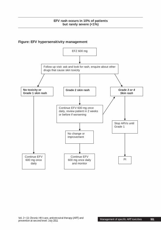
551Vol. 2 • 13. Chronic HIV care, antiretroviral therapy (ART) and prevention at second level: July 2011 551551
EFV rash occurs in 10% of patientsbut rarely severe (<1%)
Figure: EFV hypersensitivity management
EFZ 600 mg
Follow up visit: ask and look for rash, enquire about other drugs that cause skin toxicity
Grade 3 or 4 Skin rash
Continue EFV 600 mg once daily, review patient in 2 weeks or before if worsening
Stop ARVs until Grade 1
PI
No change or improvement
Continue EFV 600 mg once daily
and monitor
Continue EFV 600 mg once
daily
Grade 2 skin rash No toxicity or Grade 1 skin rash
Management of specifi c ART toxicities
FINAL Vol. 2 CH 9-21.indd 551FINAL Vol. 2 CH 9-21.indd 551 29/06/2012 15:1629/06/2012 15:16

552 Vol. 2 • 13. Chronic HIV care, antiretroviral therapy (ART) and prevention at second level: July 2011552552
ABC hypersensitivity This is a systemic reaction that usually occurs within the fi rst 8 weeks of therapy in about 3% of patients. Deaths have been reported after reintroducing ABC; therefore, ABC should never be reintroduced after discontinuation due to hypersensitivity. Signs of ABC hypersensitivity include:• rash• fever• fatigue• abdominal or respiratory symptoms.
See Table: ARVs and other HIV-related medications associated with selected toxicities below for grading clinical toxicities.
Dyslipidaemia
• PIs can cause elevated triglycerides and elevated LDL cholesterol.• d4T can cause elevated triglycerides, and EFV can cause elevated total
cholesterol.• Markedly elevated triglycerides can cause pancreatitis.• Solve by substituting for the offending drugs, e.g. substitute d4t with AZT,
LPV/r with atazanavir.• Fibrates are the treatment of choice for treating hypertriglyceridaemia.• Simvastatin interacts with PIs and should not be used. It creates toxic
concentrations of statins in the blood.• Suggest lifestyle changes to the patient, such as a reduced fat diet, exercise,
weight reduction, smoking cessation, and control of hypertension and diabetes.
Hyperlactataemia and lactic acidosis
• All NRTIs, particularly d4T and ddI, can cause mitochondrial toxicity, which results in elevated levels of serum lactate and potentially fatal lactic acidosis.
• 3TC, ABC, and TDF are the least likely to cause this side-effect. • Symptoms are non-specifi c and include abdominal pain, malaise, lethargy,
weight loss, and respiratory distress, and may be associated with other symptoms, such as peripheral neuropathy.
• The mortality rate with lactic acidosis is 30–60%; maintain a high level of suspicion.
• If available, lactate levels will confi rm the diagnosis.
Management of specifi c ART toxicities
FINAL Vol. 2 CH 9-21.indd 552FINAL Vol. 2 CH 9-21.indd 552 29/06/2012 15:1629/06/2012 15:16

553Vol. 2 • 13. Chronic HIV care, antiretroviral therapy (ART) and prevention at second level: July 2011 553553
Figure: Management of suspected hyperlactataemia or lactic acidosis
Change to new NRTI regimen:
3TC+AZT/ABC/TDF Repeat lactate
weekly until normal Stop ART if:
symptoms or lactate worsening
Approach to hyperlactataemia or lactic acidosis
Risk factors On d4T or ddI or AZT Good adherence to treatment Good response to treatment Overweight (BMI >28) or rapid
weight gain after starting ART On ART >2 months (usually
>6 months) Associated leg cramps or
weakness
Suspect lactic acidosis
YES
NO
Stop all ARVs for 1 month (no tail regimen).
Supportive treatment: hospitalization, IV fluid, respiratory support if needed
Lactate <2.5
Lactate 5–10
Lactate >10
Lactate 2.5–5
Suggestive symptoms Fatigue Anorexia Nausea and vomiting Unexplained weight loss Abdominal pain Tachypnoea (not respiratory) Tachycardia Peripheral oedema
Lactate measurement available?
Once symptoms improved, plan to start new regimen
Consider other causes of weight loss/abdominal pain OI or IRIS (check for TB) Chronic diarrhoea or malabsorption
Pancreatitis or hepatitis Diabetes GI intolerance of drugs
STOP all ART, admit to hospital for high-level care
Supportive therapy: thiamine 100 mg IV every 12 hours, IV fluids
If very acidiotic: Broad spectrum antibiotics and IV bicarbonate
Note: It may take many weeks for serum lactate to normalize.
NEW REGIMEN OPTIONS 3TC + TDF + NNRTI
or
TDF +NNRTI + PI/b
or NNRTI + PI/b
Start new regimen once lactate <2.5 and
symptoms improved
STOP all ARVs Supportive
therapy: Hospitalization, oral fluids, thiamine
Consider other causes of raised lactate
Sepsis Severe dehydration Severe anaemia Kidney or liver disease Pancreatitis Heart failure, DKA
Lactic acidosis
excluded, consider
other causes of symptoms
Management of specifi c ART toxicities
FINAL Vol. 2 CH 9-21.indd 553FINAL Vol. 2 CH 9-21.indd 553 29/06/2012 15:1629/06/2012 15:16

554 Vol. 2 • 13. Chronic HIV care, antiretroviral therapy (ART) and prevention at second level: July 2011554554
Peripheral neuropathy
Distal symmetric peripheral neuropathy (DSPN), also known as “peripheral neuropathy,» is a common adverse effect associated with long-term use of NRTIs, especially d4T and ddI, and occasionally AZT. DSPN can also result from other medications, such as isoniazid, and from medical conditions such as diabetes mellitus, vitamin B6 defi ciency, CMV, and HIV infection itself. See Sections 10.10a and 10.10a.6.
DSPN typically presents as tingling, burning, numbness or paraesthesia that begins in the soles of the feet (usually the toes), and advances up the foot and leg in a stocking distribution. Involvement of the arms in a glove distribution starting at the fi ngers is seen in more advanced cases. Decreased ankle jerk refl exes and decreased vibratory sensation are important signs to look for during physical examination.
ART-associated DSPN may reverse on discontinuation of the offending drug, although recovery is often slow, and the condition may continue to worsen even after discontinuation. For this reason, it is important to respond quickly to worsening symptoms to prevent or minimize its progression.
See Table: Clinical toxicities grading and management below. As part of plans to phase out d4T, it is recommended to target those with any established d4T-related toxicity for priority replacement of d4T with either AZT or TDF.
Lipodystrophy
Long-term use of NRTIs (especially d4T, ddI, AZT), as well as PIs, is associated with changes in body fat distribution known as lipodystrophy. These changes include fat accumulation (central obesity, buffalo-hump, breast enlargement, lipomas) and lipoatrophy (fat loss from face, buttocks, and limbs). Lipodystrophy may be unacceptable to some patients, and may therefore have an impact on adherence if it is not properly addressed.
There is currently no specifi c therapy, but a low-fat diet and aerobic exercise may be partly effective. Substituting d4T/ddI/AZT for another NRTI such as TDF/ABC may be of help. The recovery is usually slow and incomplete, so consider changing early to avoid ongoing lipodystrophy.
Central obesity is associated with an increased risk of insulin resistance and dyslipidaemias.
Abdominal pain and symptoms
Abdominal pain is a common side-effect of ART. Many ARVs can cause abdominal pain through a variety of mechanisms such as pancreatitis, hepatitis, steatohepatitis, lactic acidosis, IRIS with abdominal TB, and GI intolerance. Establishing a probable etiology is critically important because some of these underlying conditions (e.g. pancreatitis, lactic acidosis) may be fatal if not diagnosed and managed in a timely fashion. See Section 10.7a.2.
Management of specifi c ART toxicities
FINAL Vol. 2 CH 9-21.indd 554FINAL Vol. 2 CH 9-21.indd 554 29/06/2012 15:1629/06/2012 15:16

555Vol. 2 • 13. Chronic HIV care, antiretroviral therapy (ART) and prevention at second level: July 2011 555555
Table: Clinical toxicities grading and management
Grade 1 Grade 2 Grade 3 Grade 4
Diarrhoea Mild or transient3 to 4 loose stools per day OR Diarrhoea for <1 week
Moderate or persistent 5–7 loose stools per dayOR Diarrhoea for ≥1 week
Bloody diarrhoea OROrthostatic hypotension OR IV Rx required OR>7 loose stools per day
Hypotensive shock OR Hospitalization required
Allergic reaction
Pruritus without rash Localized urticaria Generalized urticaria angioedema
Anaphylaxis
Rash orhyper-sensitivity
ErythemaPruritus
Diffuse maculopapular rash ORDry desquamation
Vesiculation ORUlceration ORMoist desquamation
Mucous membrane involvement ORStevens-Johnson syndrome or toxic epidermal necrolysis ORErythema multiforme ORExfoliative dermatitis
Fatigue Normal activity reduced <25%
Normal activity reduced 25–50%
Normal activity reduced >50%, cannot work
Unable to care for self
Nausea andVomiting
Mild or transient (lasting <1 week)Reasonable intake maintained
Moderate or persistentIntake decreasedfor <3 days Vomiting for≥1 week
Severe discomfort minimal intake for≥3 days ORSevere vomiting of all food and fl uids in 24 hours ORIV fl uids required OROrthostatic hypotension
Hypotensive shock OR Hospitalization for IV fl uids required
Paraesthesia Mild discomfort Moderate discomfortNon-narcotic analgesia required
Severe discomfortNarcotic analgesia required
IncapacitatingNot responding to narcotic analgesia
Neurosensory Mild impairment of sensationFocal, symmetrical
Moderate impairment of sensation ORMild, asymmetrical
Severe impairment –loss of sensation to the knee ORModerate in multiple body sites
Sensation loss involving limbs or trunk
Management of specifi c ART toxicities
FINAL Vol. 2 CH 9-21.indd 555FINAL Vol. 2 CH 9-21.indd 555 29/06/2012 15:1629/06/2012 15:16

556 Vol. 2 • 13. Chronic HIV care, antiretroviral therapy (ART) and prevention at second level: July 2011556556
Tab
le: A
RV
s a
nd
oth
er
HIV
-rela
ted
med
icati
on
s a
sso
cia
ted
wit
h s
ele
cte
d t
oxic
itie
s
ARV
toxi
city
Key
feat
ures
Ass
ocia
ted
ARV
sO
ther
pos
sibl
e ae
tiolo
gies
Gen
eral
man
agem
ent r
ecom
men
datio
ns
Acu
te p
ancr
eatit
isAb
dom
inal
pai
n, e
ither
el
evat
ed li
pase
or a
myl
ase,
or
bot
h
d4T,
ddI
Alco
hol a
buse
, gal
lsto
ne
panc
reat
itis,
oth
er
med
icat
ions
(rar
e)
Disc
ontin
ue A
RT. G
ive
supp
ortiv
e tre
atm
ent a
nd d
o la
bora
tory
m
onito
ring.
Res
ume
ART
with
an
NRT
I with
low
pan
crea
tic
toxi
city
risk
. AZT
, ABC
, TDF
, and
3TC
are
less
like
ly to
cau
se
this
type
of t
oxic
ity.
See
Sect
ion
10.7
a Ab
dom
inal
pai
n fo
r sym
ptom
atic
m
anag
emen
t.
Dia
rrho
eaFr
eque
nt lo
ose
or w
ater
y bo
wel
mov
emen
ts (w
ithou
t bl
ood
or m
ucou
s)
Syst
emic
sym
ptom
s ar
e ra
re
ddI,
LPV/
r,SQV
/rIn
fect
ious
gas
troen
terit
is
(par
asiti
c; b
acte
rial;
vira
l), H
IV e
nter
opat
hy,
othe
r med
icat
ions
Usua
lly s
elf-l
imite
d, w
ithou
t nee
d to
dis
cont
inue
ART
. Sy
mpt
omat
ic tr
eatm
ent s
houl
d be
offe
red,
e.g
. ant
i-dia
rrho
eal
agen
t or d
ieta
ry m
odifi
catio
n.
See1
0.7d
.2 a
nd 1
0.7d
.3 fo
r sug
gest
ed m
anag
emen
t of
diar
rhoe
a.
Dru
g ra
shes
: m
ild to
mod
erat
e (e
xclu
ding
Ste
vens
-Joh
nson
sy
ndro
me
or to
xic
epid
erm
al
necr
olys
is)
Red
or h
yper
pigm
ente
d m
acul
opap
ular
rash
, pr
edom
inan
tly o
n to
rso,
th
ighs
With
or w
ithou
t Pr
uritu
sN
o m
ucos
al in
volv
emen
tN
o sy
stem
ic s
igns
or
sym
ptom
s
NVP
, EFV
Othe
r med
icat
ions
, e.
g. c
otrim
oxaz
ole,
su
lfado
xine
-py
rimet
ham
ine
(SP,
Fa
nsid
ar);
vira
l exa
nthe
m
In m
ild c
ases
ther
e m
ay b
e re
gres
sion
with
out t
he n
eed
to
chan
ge A
RV d
rug.
Giv
e an
tihis
tam
ines
or N
SAID
S an
d m
onito
r cl
osel
y, in
clud
ing
AST
if av
aila
ble.
If m
oder
ate
rash
, non
-pr
ogre
ssin
g an
d w
ithou
t muc
osal
invo
lvem
ent o
r sys
tem
ic
sign
s, c
onsi
der a
sin
gle
NN
RTI s
ubst
itutio
n (i.
e. fr
om N
VP to
EF
V).
See
Figu
res
for N
VP a
nd E
FV a
bove
for s
ugge
sted
m
anag
emen
t alg
orith
ms.
Dru
g ra
shes
seve
re
(incl
udin
g St
even
s-Jo
hnso
n sy
ndro
me
or to
xic
epid
erm
al
necr
olys
is)
Diffu
se, b
liste
ring
rash
invo
lvin
g m
ucou
s m
embr
anes
(con
junc
tival
, ur
ethr
al, n
ares
, or
anor
ecta
l); w
ith o
r with
out
feve
r
NVP
, EFV
(e
spec
ially
NVP
)Ot
her m
edic
atio
ns, e
.g.
cotri
mox
azol
e (T
MP-
SMX)
, sul
fado
xine
-py
rimet
ham
ine
(SP)
;
Disc
ontin
ue a
ll AR
Vs a
nd g
ive
supp
ortiv
e tre
atm
ent;
thes
e pa
tient
s ar
e m
anag
ed in
tens
ivel
y, s
imila
r to
burn
vic
tims.
M
onito
r AST
and
ALT
, as
co-m
orbi
d he
patit
is is
com
mon
. Af
ter r
esol
utio
n, re
sum
e AR
T w
ith 3
NRT
Is o
r 2 N
RTIs
+ P
I; ho
wev
er, u
se o
f EFV
follo
win
g N
VP-in
duce
d se
vere
rash
is
cont
rove
rsia
l.6
See
Figu
res
for N
VP a
nd E
FV a
bove
for s
ugge
sted
m
anag
emen
t alg
orith
ms.
6 Se
vere
rash
is d
efi n
ed a
s ex
tens
ive
rash
with
des
quam
atio
n, a
ngio
edem
a, o
r a re
actio
n re
sem
blin
g se
rum
sic
knes
s; o
r a ra
sh w
ith c
onst
itutio
nal fi
ndi
ngs,
suc
h as
feve
r, or
al
lesi
ons,
blis
terin
g, fa
cial
oed
ema,
or c
onju
nctiv
itis;
Ste
vens
-Joh
nson
syn
drom
e ca
n be
life
-thre
aten
ing.
For
life
-thre
aten
ing
rash
, sub
stitu
tion
with
EFV
is n
ot re
com
men
ded,
al
thou
gh th
is a
ppro
ach
has
been
repo
rted
in a
sm
all n
umbe
r of p
atie
nts
in T
haila
nd w
ithou
t rec
urre
nce
of ra
sh.
Management of specifi c ART toxicities
FINAL Vol. 2 CH 9-21.indd 556FINAL Vol. 2 CH 9-21.indd 556 29/06/2012 15:1629/06/2012 15:16

557Vol. 2 • 13. Chronic HIV care, antiretroviral therapy (ART) and prevention at second level: July 2011 557557
ARV
toxi
city
Key
feat
ures
Ass
ocia
ted
ARV
sO
ther
pos
sibl
e ae
tiolo
gies
Gen
eral
man
agem
ent r
ecom
men
datio
ns
Dys
lipid
emia
Elev
ated
trig
lyce
rides
or
LDL;
low
HDL
PIs,
EFV
, d4T
(e
spec
ially
PIs
, ex
cept
ATV
)
Dysl
ipid
aem
ia
inde
pend
ent o
f ART
, ot
her m
edic
atio
ns
Diet
ary
chan
ges
and
phys
ical
exe
rcis
e. L
ipid
-low
erin
g ag
ents
(e
.g. p
rava
stat
in, fi
bra
tes
– av
oid
sim
vast
atin
). Co
nsid
er
repl
acin
g of
fend
ing
ARV
with
a d
iffer
ent a
gent
with
low
er
risk
of th
is to
xici
ty (N
VP, A
TV, a
nd m
ost N
RTIs
hav
e lo
w ri
sk
of in
duci
ng d
yslip
idae
mia
). Do
not
giv
e si
mva
stat
in a
nd P
Is
toge
ther
.
Insu
lin re
sist
ance
, dia
bete
sEl
evat
ed b
lood
glu
cose
PIs
(exc
ept A
TV,
APV,
fos-
APV)
; EF
V, d
4T
Obes
ity o
r gen
etic
pr
edis
posi
tion,
oth
er
med
icat
ions
Repl
acem
ent o
f offe
ndin
g AR
V w
ith a
diff
eren
t age
nt. D
ieta
ry
chan
ges
and
incr
ease
d ph
ysic
al e
xerc
ise.
Hyp
ogly
caem
ic
agen
ts fo
r est
ablis
hed
diab
etes
.
Gen
eral
gas
tro-
inte
stin
al
into
lera
nce
Tast
e ch
ange
s, n
ause
a,
vom
iting
, abd
omin
al p
ain
or
diar
rhoe
a
All A
RVs
(less
fre
quen
t with
d4T
, 3T
C, F
TC, a
nd
ABC)
Infe
ctio
us g
astro
ente
ritis
(p
aras
itic;
bac
teria
l; vi
ral),
HIV
ent
erop
athy
, ot
her m
edic
atio
ns
Usua
lly s
elf-l
imite
d, w
ithou
t nee
d to
dis
cont
inue
ART
. Sy
mpt
omat
ic tr
eatm
ent s
houl
d be
offe
red,
e.g
. ant
idia
rrho
eal
agen
ts, d
ieta
ry c
hang
es, a
ntie
met
ics.
Con
side
r sin
gle
ARV
subs
titut
ion
if co
nditi
on is
per
sist
ent a
nd s
ever
e en
ough
to
disr
upt n
utrit
ion
or a
dher
ence
. Se
e Se
ctio
n 10
.7a.
1 fo
r sug
gest
ed m
anag
emen
t of a
bdom
inal
pa
in a
nd S
ectio
n 10
.7d.
2 an
d 10
.7d.
3 fo
r sug
gest
ed
man
agem
ent o
f dia
rrho
ea.
Hae
mat
olog
ical
toxi
citie
s (p
artic
ular
ly a
naem
ia a
nd
leuk
open
ia)
Low
Hb,
low
WBC
, low
ne
utro
phil
coun
tAZ
TOt
her m
edic
atio
ns (e
.g.
CTX)
; inf
ectio
us d
isea
ses
(mal
aria
, hoo
kwor
m,
othe
r par
asite
s); H
IV
or O
I-ind
uced
mar
row
su
ppre
ssio
n; ir
on
or o
ther
nut
ritio
nal
defi c
ienc
ies
If no
t sev
ere,
mon
itor c
lose
ly a
nd c
onsi
der r
epla
cing
AZT
w
ith a
diff
eren
t NRT
I with
min
imal
or n
o bo
ne m
arro
w to
xici
ty
(e.g
. d4T
or T
DF).
If se
vere
(Hg
<6.5
g%
or a
bsol
ute
neut
roph
il co
unt <
500
cells
/mm
3 ), re
plac
e w
ith a
diff
eren
t NRT
I and
co
nsid
er b
lood
tran
sfus
ion.
See
Figu
re: A
lgor
ithm
for m
anag
emen
t of A
ZT-r
elat
ed
anae
mia
abo
ve fo
r sug
gest
ed m
anag
emen
t.
Hep
atiti
sEl
evat
ions
of l
iver
func
tion
test
s, e
spec
ially
AST
/ALT
; w
ith o
r with
out r
ight
upp
er
quad
rant
abd
omin
al p
ain,
na
usea
or v
omiti
ng, f
ever
, m
alai
se, j
aund
ice
All A
RVs
(esp
ecia
lly N
VP
and
riton
avir-
bo
oste
d PI
s)
Infe
ctio
us h
epat
itis;
ot
her m
edic
atio
ns (e
.g.
anti-
mic
robi
als)
, dru
g-dr
ug in
tera
ctio
ns
Sign
ifi ca
nt e
leva
tions
of A
LT a
ssoc
iate
d w
ith c
linic
al fe
atur
es
are
mos
t ofte
n de
scrib
ed w
ith N
VP. H
owev
er, c
hang
es (o
f va
ryin
g de
gree
) may
be
obse
rved
with
all
ARVs
.If
ALT
>5-fo
ld th
e ba
sal l
evel
, dis
cont
inue
ART
and
mon
itor.
Afte
r res
olut
ion,
repl
ace
the
drug
mos
t lik
ely
asso
ciat
ed.
Management of specifi c ART toxicities
FINAL Vol. 2 CH 9-21.indd 557FINAL Vol. 2 CH 9-21.indd 557 29/06/2012 15:1629/06/2012 15:16

558 Vol. 2 • 13. Chronic HIV care, antiretroviral therapy (ART) and prevention at second level: July 2011558558
ARV
toxi
city
Key
feat
ures
Ass
ocia
ted
ARV
sO
ther
pos
sibl
e ae
tiolo
gies
Gen
eral
man
agem
ent r
ecom
men
datio
ns
Hyp
erbi
lirub
inae
mia
(in
dire
ct)
Elev
ated
unc
onju
gate
d (in
dire
ct) b
iliru
bin;
with
or
with
out j
aund
ice,
icte
rus
ATV
Chol
esta
tic d
isea
se;
hepa
titis
(if o
ther
live
r en
zym
e ab
norm
aliti
es
pres
ent);
gen
etic
pre
-di
spos
ition
(e.g
. Gilb
ert’s
sy
ndro
me)
Gene
rally
asy
mpt
omat
ic, w
ithou
t ind
icat
ion
for c
hang
e in
th
erap
y. H
owev
er, f
or p
ersi
sten
t jau
ndic
e yo
u m
ay re
plac
e AT
V w
ith a
diff
eren
t PI o
r an
NN
RTI.
AB
C hy
pers
ensi
tivity
re
actio
nM
ulti-
orga
n sy
stem
sy
ndro
me
that
may
incl
ude
rash
, fev
er, d
yspn
oea,
ab
dom
inal
dis
com
fort,
AS
T/AL
T el
evat
ion,
or
mal
aise
ABC
Othe
r ARV
or m
edic
atio
n to
xici
ties;
acu
te v
iral,
bact
eria
l, or
par
asiti
c in
fect
ious
pro
cess
; IRI
S
Disc
ontin
ue A
BC a
nd n
ever
re-c
halle
nge.
Re-e
xpos
ure
to A
BC m
ay le
ad to
a s
ever
e an
d po
tent
ially
life
-th
reat
enin
g al
lerg
ic (a
naph
ylac
tic) r
eact
ion.
Hyp
erla
ctat
aem
ia o
r lac
tic
acid
osis
Anio
n ga
p ac
idos
is w
ith
low
ser
um p
H, m
alai
se,
fatig
ue, a
bdom
inal
pai
n,
with
or w
ithou
t res
pira
tory
di
stre
ss
All N
RTIs
(e
spec
ially
d4T
an
d dd
I)
Othe
r cau
ses
of a
nion
ga
p ac
idos
is: s
alic
ylat
e ov
erdo
se, e
than
ol,
ison
iazid
toxi
city
, ur
aem
ia, d
iabe
tic
keto
acid
osis
Disc
ontin
ue A
RT a
nd g
ive
supp
ortiv
e tre
atm
ent.
Afte
r clin
ical
re
solu
tion,
resu
me
ART,
repl
acin
g th
e of
fend
ing
NRT
Is. A
BC,
TDF,
and
3TC
are
less
like
ly to
cau
se th
is ty
pe o
f tox
icity
. Se
e Fi
gure
: Man
agem
ent o
f sus
pect
ed h
yper
lact
atae
mia
or
lact
ic a
cido
sis
abov
e fo
r sug
gest
ed m
anag
emen
t.
Lipo
atro
phy
Subc
utan
eous
fat w
astin
g,
mos
t vis
ible
in fa
ce,
butto
cks,
ext
rem
ities
All N
RTIs
(e
spec
ially
d4T
)Ge
nera
lized
was
ting
Early
repl
acem
ent o
f the
sus
pect
ed A
RV d
rug
(e.g
. d4T
) with
an
age
nt le
ss li
kely
to c
ause
this
effe
ct (e
.g. T
DF o
r ABC
). Ve
ry
grad
ual i
mpr
ovem
ent o
ften
follo
ws.
Cos
met
ic tr
eatm
ents
are
ef
fect
ive,
but
exp
ensi
ve a
nd n
ot w
idel
y av
aila
ble.
Management of specifi c ART toxicities
FINAL Vol. 2 CH 9-21.indd 558FINAL Vol. 2 CH 9-21.indd 558 29/06/2012 15:1629/06/2012 15:16

559Vol. 2 • 13. Chronic HIV care, antiretroviral therapy (ART) and prevention at second level: July 2011 559559
Figure: Management of suspected hyperlactataemia or lactic acidosis
Suspect IRIS
Unexpected deterioration in clinical condition with signs and symptoms of inflammation or infection soon after
commencing ART (typically 2–12 weeks)
Management principles 1. Continue ART.
2. Treat the specific pathogen in order to decrease the antigen load.
3. Consider corticosteroids in moderate to severe cases of IRIS (prednisolone (or prednisone) at 0.5–1.0 mg/kg/day orally or IV for 5–10 days or longer depending on the severity of the inflammation.
4. Aspirate and drain infected lymph nodes and abscesses (may need to be repeated several times).
5. Perform emergency surgical decompression in cases of tracheal or intestinal obstruction.
Reported IRIS events Flare of hepatitis B or C Visceral leishmaniasis TB abscess
Reported IRIS events Disseminated TB Invasive fungal disease MAC CMV
Reported IRIS events Pulmonary TB Invasive fungal pneumonia PCP
Hepato-biliary
symptoms
CNS symptoms
Feverwithout
localizing signs
Respiratory
symptoms with worsening
changes on chest X-ray
Muco- cutaneous conditions
Reported IRIS events Herpes zoster and simplex HPV infection (warts) Molluscum contagiosum Kaposi sarcoma Psoriasis Eczema, folliculitis, PPE Leprosy Leishmaniasis Cutaneous fungal infections
Focal adenopathy
Reported IRIS events Extra pulmonary TB MAC Kaposi sarcoma Histoplasmosis
Auto- immune diseases
Reported IRIS events Cryptococcal meningitis TB meningitis or abscess Toxoplasmosis PML CMV Lymphoma
Reported IRIS events Sarcoidosis Graves disease Guillain-Barré syndrome Reiter’s syndrome
IRIS
Management of specifi c ART toxicities
FINAL Vol. 2 CH 9-21.indd 559FINAL Vol. 2 CH 9-21.indd 559 29/06/2012 15:1629/06/2012 15:16

560 Vol. 2 • 13. Chronic HIV care, antiretroviral therapy (ART) and prevention at second level: July 2011560560
13.10 HIV/TB co-management
TB is a common cause of morbidity and death among people living with HIV. HIV-positive TB patients require ART in addition to TB treatment. TB also is a signifi cant cause of IRIS in patients who are started on ART. Early diagnosis and treatment of TB in HIV-positive patients, and the provision of ART, signifi cantly reduce the mortality of HIV-positive patients from TB.
See Section 15 for diagnosis and treatment of TB.
Cotrimoxazole prophylaxis
Provide cotrimoxazole prophylaxis for all HIV-positive TB patients throughout the course of anti-TB treatment. Support and monitor adherence to cotrimoxazole and to all the treatments.
Refer to Section 13.3 for cotrimoxazole prophylaxis dosage, side-effects and their management, and monitoring.
When to start ART in patients with TB
All HIV-positive TB patients are eligible for ART, irrespective of their CD4 count. Assess or review the WHO clinical stage, whether they are already on ART, and their CD4 count.
ART in patients who are taking TB treatment deserves special consideration. This is because HIV and TB co-management can be complicated by pill burden, drug-to-drug interaction, IRIS, overlapping drug toxicities, and adherence issues.
Guide to determine when to initiate fi rst-line ART in TB patients
• Start ART in all HIV-positive individuals with active TB irrespective of their CD4 cell count.
• Start TB treatment fi rst, followed by ART as soon as possible afterwards – usually within 8 weeks of starting TB treatment.
• Use an EFV-based regimen in patients starting ART while on TB treatment.
Recommended ART for patients with TB
Patients who are on TB/HIV co-treatment are more likely to experience pill burden, drug side-effects and drug-to-drug interactions. Therefore, patients should be adequately prepared for adherence. See Table: Summary of recommended fi rst-line ART for TB patients below.
Women of childbearing potential (or pregnant women) with TB and eligible for ART
An EFV-containing regimen should not be used during the fi rst-trimester of pregnancy. Provide effective contraception for women of childbearing potential. For pregnant women in the second or third trimester, use EFV-containing regimen. An alternate is a 3 NRTI regimen, e.g. AZT + 3TC + ABC.
HIV/TB co-management
FINAL Vol. 2 CH 9-21.indd 560FINAL Vol. 2 CH 9-21.indd 560 29/06/2012 15:1629/06/2012 15:16

561Vol. 2 • 13. Chronic HIV care, antiretroviral therapy (ART) and prevention at second level: July 2011 561561
Table: Summary of recommended fi rst-line ART for TB patients
Recommendation ARV regimen Comments
Preferred fi rst-line regimen
TDF or AZT + 3TC + EFV EFV is preferred because the interaction with rifampicin is easier to manage. But EFV is contraindicated in pregnant women in the fi rst trimester. Provide adequate contraception to women of childbearing age who are on EFV.
AZT may cause anaemia, and Hb monitoring is necessary. AZT is preferred over d4T because of d4T toxicity.
Alternative fi rst-line regimens
TDF or AZT + 3TC + NVP NVP blood level is decreased in the presence of rifampicin. Close clinical and laboratory monitoring of liver enzymes at 4, 8, and 12 weeks is advised for all patients receiving NVP and rifampicin.
There is risk of symptomatic or fatal hepatitis in women with CD4 counts between 250 and 350 cells/mm3. An NVP-containing regimen should only be considered when no alternative is available for women on rifampin-containing regimens whose CD4 count is in the range of 250–350 cells/mm3.
Other options AZT + 3TC + TDF If available, TDF can be used safely with rifampicin, and can be used in patients with higher CD4 counts. Limited data on antiviral potency and hypersensitivity reactions for patients with TB.
TDF or AZT+ 3TC + ABC ABC is registered in many countries, but the cost is high. ABC can be used safely with rifampicin, and can be used in patients with higher CD4 counts. Limited data on antiviral potency and hypersensitivity reactions for patients with TB.
FTC FTC can be substituted for 3TC in any fi rst-line regimen.FTC/TDF co-formulation is available.
Note: All HIV-positive patients receiving an anti-TB drug regimen containing isoniazid should also receive pyridoxine 10 mg daily to prevent peripheral neuropathy.
TB immune reconstitution infl ammatory syndrome (TB-IRIS)
IRIS may present as a worsening of a clinical condition after initial improvement. It may occur in up to one third of persons with TB who initiate ART. IRIS typically presents within 3 months of the initiation of ART, but can occur as early as 5 days after initiation.
TB-IRIS is a recognized complication of ART. Paradoxical TB-IRIS presents as worsening of the clinical condition of a patient on anti-TB treatment after a period of improvement following initiation of ART. Paradoxical forms of TB-IRIS have been reported in 8%–43% of TB patients starting on ART. It most commonly presents with fever and a worsening of pre-existing lymphadenopathy or respiratory conditions. It is similar to, but more frequent than, the paradoxical reactions seen in immunocompetent patients on anti-TB treatment. In addition, subclinical or undiagnosed TB often presents within the fi rst 6 months after the initiation of ART, frequently as part of IRIS.
HIV/TB co-management
FINAL Vol. 2 CH 9-21.indd 561FINAL Vol. 2 CH 9-21.indd 561 29/06/2012 15:1629/06/2012 15:16

562 Vol. 2 • 13. Chronic HIV care, antiretroviral therapy (ART) and prevention at second level: July 2011562562
Most cases of IRIS are self-limited and resolve without any intervention, and ART can safely be continued. Serious reactions may occur, such as tracheal compression caused by massive adenopathy or respiratory diffi culty. Therapy may require the use of corticosteroids in such patients. However, mortality due to TB-IRIS remains limited, and the risk of TB-IRIS must be weighed against the benefi t of early initiation of ART in patients with advanced immunosuppression.
New TB in patients already receiving ART
There are 2 issues to consider in patients who are diagnosed with TB while on ART. The fi rst issue is whether ARV drug substitution is required. This may be recommended for patients who develop TB within 6 months of initiating fi rst-line or second-line ART, which is most likely TB-IRIS. The ART recommendation in these patients is summarized in the following Table.
Table: ART recommendations for patients who develop TB within 6 months of starting a fi rst-line or second-line ART regimen
First-line or second-line ARTART regimen at the time active TB occurs Options
First-line ART regimen 2 NRTIs + EFV Continue with the same regimen
2 NRTIs + NVP Substitute to EFVa,b ORSubstitute to triple NRTI regimena ORContinue with 2 NRTIs + NVPc
Triple NRTI regimen Continue triple NRTI regimen
Second-line ART2 NRTIs + PI Substitute to or continue (if already being taken)
LPV/r- or SQV/r-containing regimen and adjust dose of RTVa
a Substituting back to the original regimens once the rifampicin-containing regimen is completed can be considered. When switching back from EFV to NVP, no lead-in dose is required.
b The use of EFV-containing regimens is not recommended during the fi rst trimester of pregnancy. Provide adequate contraception in women of childbearing potential.
c Careful clinical and laboratory monitoring (ALT) is advised when NVP or boosted PIs are administered concurrently with rifampicin.
The second issue to consider in patients who are diagnosed with TB while on ART is whether the diagnosis of TB constitutes treatment failure.
ART signifi cantly decreases the risks of TB in treated patients, but rates of TB nevertheless remain persistently higher than among HIV-negative individuals. An episode of TB can occur across a wide range of CD4 cell counts, and does not necessarily indicate ART failure and the need to switch to second-line regimens. Therefore, if an episode of TB occurs during the fi rst 6 months following the initiation of ART, this should not be considered a treatment failure, and the ART regimen should be adjusted for TB-ART co-treatment.
If an episode of TB develops more than 6 months after initiation of ART, the decision about whether the TB diagnosis represents ART failure is based on the CD4 cell count and, if available, the viral load. If a CD4 cell count is not available, assess the patient for signs and symptoms of progression of HIV-infection. This includes whether the TB is pulmonary or extrapulmonary (extrapulmonary TB is
HIV/TB co-management
FINAL Vol. 2 CH 9-21.indd 562FINAL Vol. 2 CH 9-21.indd 562 29/06/2012 15:1629/06/2012 15:16

563Vol. 2 • 13. Chronic HIV care, antiretroviral therapy (ART) and prevention at second level: July 2011 563563
WHO stage 4), and whether there are other non-TB WHO stage 3 or 4 conditions. While waiting for CD4 count, the development of an episode of pulmonary TB after 6 months of ART, without other clinical and immunological evidence of disease progression, should not be regarded as ART failure.
Extrapulmonary TB should be considered as an indication of ART failure, although simple TB lymphadenitis or uncomplicated pleural disease may be less signifi cant than disseminated TB.
If there is a good response to anti-TB treatment, the decision to switch to a second-line regimen can be delayed until short-course TB therapy has been completed.
Second-line ART regimen for patients with TB
An episode of TB in patients receiving fi rst-line ART may indicate that the fi rst-line regimen probably has failed. The effectiveness of second-line therapy for patients in whom an NNRTI regimen has failed depends on the introduction of PIs in the new regimen. However, there are signifi cant drug interactions with the PIs and rifampicin. Unboosted PIs cannot be used with rifampicin-containing regimens because the PI levels are sub-therapeutic.
Option 1
• Rifabutin 150 mg 3 times per week with boosted PI (LPV/r or ATV/r) at normal standard dose.
• Rifabutin can cause uveitis.
Option 2
• Rifampicin-containing TB therapy plus super-boosted LPV/r (400 mg/400 mg BID or SQV/r 400 mg/400 mg BID) under close clinical and laboratory monitoring to detect hepatotoxicity.
The recommendations and precautions for the use of PI-based regimens in combination with rifampicin in women of childbearing potential, and in pregnant women, are the same as for other TB patients.
Good clinical practice:Follow national TB and HIV guidelines and
co-treatment recommendations
13.11 Adherence preparation, monitoring and support
Adherence support should not just focus on medical issues; there are many other issues that affect adherence. Take steps to educate patients, their families, and community members on adherence principles. If the patient agrees, home visits can be useful.
Partially or fully observed therapy for a defi ned period of time can be helpful, especially for some patients such as IDUs. Poor adherence may signify an
Adherence, monitoring, support
FINAL Vol. 2 CH 9-21.indd 563FINAL Vol. 2 CH 9-21.indd 563 29/06/2012 15:1629/06/2012 15:16

564 Vol. 2 • 13. Chronic HIV care, antiretroviral therapy (ART) and prevention at second level: July 2011564564
underlying unstable social situation, heavy alcohol dependence, or serious psychiatric illness. These barriers to adherence need to be addressed.
Adherence may improve with the development of a trusting relationship between the patient and the health workers. Health workers need to develop listening skills, and look into issues that may be affecting good adherence. Uncoordinated or chaotic clinical circumstances may hamper adherence.
Table: Barriers to adherence and suggestions to address them
Reasons for poor adherence Possible barriers Problem solving
Forgot to take pills Patient forgot because of:• travelling• alcohol or active drug use• depression or psychiatric
illness• living alone and sick• no family support, e.g.
not disclosed, homeless, prisoner, displaced person
• Plan before travel, take extra pills.• Use reminder cues.• Address alcohol and drugs use.• Enlist family support.• Treat depression or any other psychiatric
illness.• Use PLHIV support groups.• Develop links with local community-based
organizations and support groups.• Discuss and encourage disclosure.• Consider other, e.g. nutrition support.
Pills do not help
Felt better, so did not continue
Inadequate knowledgeIncorrect beliefs and attitudes
• Improve counselling (review ART and adherence).
• Take time and use all encounters to educate the patient and explain the goals of therapy and the reasons for adherence. Provide information and examples.
• Ask family and friends to support the treatment plan.
• Use PLHIV support groups, particularly those taking ART.
Family said no to medications
Inadequate knowledgeIncorrect beliefs and attitudes
• Provide family counselling and support, information, and examples on antiretroviral therapy and adherence.
• Link with family and community-based support groups.
Instructions were not clear
Did not understand how to take medications
Co-treatment, pill burden Literacy levels – sequence of dosing might be confusing, or the regimen might be complex Poor instructions and insuffi cient time to counselDepression or other psychiatric illnessAlcohol or active drug use
• Use literacy materials and individually tailored information.
• Negotiate a treatment plan that the patient understands and to which the patient is committed.
• If possible, reduce dose frequency and number of pills, e.g. by using FDCs.
• Use demonstration pills and repeat instructions. Ask patient to repeat instructions.
• Ask family and friends to support treatment plan.
• Consider the impact of co-morbidities on adherence and co-manage: treat depression, address alcohol and drug use.
Adherence, monitoring, support
FINAL Vol. 2 CH 9-21.indd 564FINAL Vol. 2 CH 9-21.indd 564 29/06/2012 15:1629/06/2012 15:16

565Vol. 2 • 13. Chronic HIV care, antiretroviral therapy (ART) and prevention at second level: July 2011 565565
Reasons for poor adherence Possible barriers Problem solving
Unable to care for self Living aloneNo employmentAIDS dementia or mental illness
• Use PLHIV support groups.• Link with community and home-based
care programmes and NGOs support groups.
• Consider nutrition and other support. • Locate family and support.• Identify friends and peers who could help.
Did not want others to see patient taking medications
Stigma at place of workNon-disclosure in the family or at the workplace
• Provide counselling.• Give support to help with disclosure.• Identify a friend or family members who
could help.
Fear of toxicity Insuffi cient preparationInadequate knowledge
• Inform patient, anticipate and manage side-effects.
• Avoid or minimize adverse drug interactions, if possible.
• Provide accurate information on what to expect and what to do.
• Counsel on the dangers of non-adherence.
Poor patient-health worker relationship
Over-burdened clinical teamInsuffi cient time to counsel and provide information Being judgemental Stigma in the health sector related to HIV, MSM, IDU, sex work
• Establish trust with all patients and their families.
• Use a team approach to care. Share tasks among members of clinical team; involve trained peer educators, lay counsellors, and expert patients in the clinical team.
• Avoid being judgemental – in attitude, language, or action; anticipate and educate staff.
• Provide accurate information and support at each encounter and for all patients; be resourceful.
• Prevent, identify, and manage burnout among staff.
Long waiting time at the facility
Uncoordinated clinic visits and care
• Consider and anticipate the impact of more than one family member needing life-long HIV care and multiple visits on adherence: ° proactively coordinate care ° provide family-focused care ° triage patient fl ow ° reduce waiting time by organizing ser-
vice delivery, including lab, pharmacy and clinical services.
Adherence, monitoring, support
FINAL Vol. 2 CH 9-21.indd 565FINAL Vol. 2 CH 9-21.indd 565 29/06/2012 15:1629/06/2012 15:16

566 Vol. 2 • 13. Chronic HIV care, antiretroviral therapy (ART) and prevention at second level: July 2011566566
13.12 Positive health, dignity, and prevention for PLHIV1
Preventing sexual transmission of HIV
Counsel on safer sex and reducing risk of transmissionBe sure to include the following topics. Encourage questions throughout. Answer accurately, clearly, and honestly.• For a patient on ART, emphasize that he or she can still transmit HIV even
though ART signifi cantly reduces the risk of transmission of HIV.• Counsel on ways to reduce risk of transmission:
° mutual faithfulness; ° limiting the number of partners; ° consistent and correct condom use; ° engaging in only sexual activities that do not allow semen, fl uid from the
vagina or blood to enter the anus, vagina, or mouth of the partner; ° not having sex;
◊ for adult men, emphasize that they should not have sex with teenagers or girls (or boys);
• Help the patient assess his or her current risk of transmission and make an individual risk reduction plan.
• Emphasize that sexual activity can continue if desired, with the above-stated precautions. Respond to any concerns about sexual function.
• Dispel any prevailing myths on the cleansing of HIV infection through sexual intercourse with young people, virgins, or others. Discuss any other locally common myths that may hinder HIV prevention, e.g. the belief that condoms transmit HIV.
Counsel on the consistent and correct use of condoms during every sexual encounter (most risk-reduction plans will include condom use)
• Educate the patient that it is essential to consistently use condoms, even when they are already infected with HIV. The patient should use condoms for all acts of vaginal, anal, and oral intercourse.
• Point out that condoms help prevent pregnancy and infection with other STIs. For better pregnancy prevention, an additional family planning method can be used along with condoms.
• Provide male or female condoms, as the patient prefers, and discuss where and how to keep them at home and where to get them.
• Demonstrate how to use condoms. ° Use a model to demonstrate correct use. ° Advise the patient to put a condom on or in before penetrative sex, and not
to wait until just before ejaculation. ° Request that the patient demonstrate the correct use of condoms using
a model. Give advice and gently correct mistakes. (See the reproductive health (RH) counselling fl ipchart, for basic instructions on condom use.)7
• Discuss facts about male and female condoms (see RH counselling fl ipchart7).
7 Reproductive choice and family planning for people living with HIV. Counselling tool. WHO, 2007. Available at: http://www.who.int/reproductivehealth/publications/family_planning/9241595132/en/index.html
Positive health for PLHIV
FINAL Vol. 2 CH 9-21.indd 566FINAL Vol. 2 CH 9-21.indd 566 29/06/2012 15:1629/06/2012 15:16

567Vol. 2 • 13. Chronic HIV care, antiretroviral therapy (ART) and prevention at second level: July 2011 567567
• For male (latex) condoms, advise patients to use only water-based lubricants and to avoid oil-based lubricants, which damage the latex.
• Discuss and advise that condoms and lubricants should be used together. For anal sex, using lubricants in the absence of condoms may increase both anal mucosal infl ammation and HIV risk due to the damage to epithelial cells from the hyperosmolar nature of most lubricants. (See Section 19.3)
• Discuss potential barriers to consistent and correct use of condoms: ° Explore barriers that the patient foresees and ways to overcome them. ° Discuss techniques and skills for negotiating condom use, and help the
patient decide on an appropriate approach. ° Role-play condom negotiation with the patient.
Discuss disclosure and encourage partner testing
• Explore the benefi ts of disclosure and discuss barriers to it.• Develop a strategy for disclosure if the client is ready. • Refer to PLHIV support groups or others for additional support, if required.• Encourage and facilitate partner testing. Explain that it is possible for partners
of PLHIV to be HIV-negative. (Record the result in the patient’s medical record.)
• Discuss and offer HIV testing of children.• Provide ongoing counselling to discordant couples.
Important information, particularly for discordant couples
• In a discordant couple, the HIV-negative partner faces very high risk of infection through penetrative sexual contact (anal, vaginal, or oral).
• Effective risk-reduction options are: ° condoms ° abstinence
See Section 19.2 Discordant couples counselling and services.
Preventing non-sexual transmission of HIV
Explain how sharing needles, syringes, razor blades, or other sharp instruments can infect others.• Educate patients to cover any open sores or cuts.• Educate health workers to wear appropriate personal protective equipment
when indicated, and to use gloves and disinfectants when cleaning up any blood or body fl uids. Explain that this is normal procedure. See Section 6.
• See Section 14 for preventing mother-to-child transmission of HIV.
Reproductive choice and family planning
(See Section 14.5.)
Counselling on reproductive choice and family planning can and should be conducted routinely at the fi rst level of care. Likewise, most services related to reproductive choice and family planning can and should be provided at the fi rst level in MCH/PMTCT/RH service settings (see Section 14.5). Health workers at the fi rst level can provide a number of family planning methods, such as condoms, pills, and injectables. Methods requiring procedures – IUDs, implants, female
Positive health for PLHIV
FINAL Vol. 2 CH 9-21.indd 567FINAL Vol. 2 CH 9-21.indd 567 29/06/2012 15:1629/06/2012 15:16

568 Vol. 2 • 13. Chronic HIV care, antiretroviral therapy (ART) and prevention at second level: July 2011568568
sterilization, and vasectomy – usually require referral to a clinician with further training in family planning. Follow country guideline recommendations.
Note: The information and guidance in Section 14.5 is consistent with WHO reproductive health guidance. It comes largely from the RH counselling fl ipchart.7 In counselling, this tool helps guide and inform patients’ reproductive choices and practices.
The information on reproductive choice and family planning in this manual can help the district team meet the reproductive health needs of patients referred to them for other reasons, if those needs are not being met at the fi rst level. It is not expected that most reproductive health needs or services themselves will require referral to the second level of HIV care.
13.13 Positive living for PLHIV1
PLHIV can live full and healthy lives if they take care of themselves and obtain treatment.
Counsel PLHIV on how to prevent other infections
• Use condoms to prevent STIs and re-infection with HIV.• Avoid other people with infections (e.g. fl u, boils, impetigo, herpes zoster,
chickenpox, pulmonary TB).• Use safe drinking water – chlorinate water or drink boiled water or tea when
possible. Store the water in a container that prevents contamination. • Eat well-cooked food.• Practice hand hygiene, especially after going to the toilet. Caregivers and
patients should wash their hands often: after using the toilet, before preparing food, or before touching any blood, semen, vaginal fl uid, or faeces. (See Section 6.)
• Have a local antiseptic (such as gentian violet or chlorhexidine) at home to apply to minor wounds after washing them.
• Sleep under an insecticide-treated bednet to prevent malaria.• Get vaccinations to prevent illness:8
° Hepatitis B: Where serological testing for hepatitis B virus is available, WHO recommends 3 doses of standard- or double-strength hepatitis B vaccine for adults with HIV who are susceptible (i.e. antibody to hepatitis B core antigen-negative) and have not been vaccinated previously. Vaccine response (titre of hepatitis B surface antibody after 3 doses of hepatitis B vaccine) can be measured and, if suboptimal, revaccination may be considered. In settings where serologic testing is not available, and hepatitis B prevalence is substantial, programme managers may choose to offer 3 doses of hepatitis B vaccine to all adults with HIV. Follow national guideline recommendations.
° Hepatitis A: Prior exposure to hepatitis A in childhood and early adulthood is common in many resource-limited settings and insuffi cient data exist on the effectiveness of hepatitis A vaccinations in adults with HIV.
8 Priority Interventions: HIV/AIDS prevention, treatment and care in the health sector. WHO, 2010. Available at: http://whqlibdoc.who.int/publications/2010/9789241500234_eng.pdf
Positive living for PLHIV
FINAL Vol. 2 CH 9-21.indd 568FINAL Vol. 2 CH 9-21.indd 568 29/06/2012 15:1629/06/2012 15:16

569Vol. 2 • 13. Chronic HIV care, antiretroviral therapy (ART) and prevention at second level: July 2011 569569
° Infl uenza: Where available and feasible, annual infl uenza vaccination with the inactivated subunit infl uenza vaccine should be offered to adults with HIV. If infl uenza vaccine is indicated in the context of a large epidemic or pandemic, adults with HIV should receive inactivated infl uenza vaccine.
° Yellow fever: In general, yellow fever vaccine is not recommended for PLHIV.
° Pneumococcal and HPV: Pneumococcal conjugate vaccine and human papilloma (HPV) vaccine are not recommended for PLHIV due to insuffi cient data.
Encourage physical activity as appropriate
• Help the patient develop his or her own activity programme.• Exercise can make the person feel better and maintain muscle tone.• Physical activity is important to prevent weight loss because it:
° stimulates appetite ° reduces nausea ° improves functioning of the digestive system ° strengthens muscles.
Note: Avoid ineffective or expensive therapies and supplements.
Support adequate and balanced nutrition
Advise the patient on adequate nutrition, and encourage a diet with adequate energy, protein, and micronutrients.• Nutrient-dense foods include (adapt to national guidelines):
° Examples include: soya products, meat, fi sh, nuts and seeds, beans, lentils, potatoes, rice, barley, wheat, maize.
• Eat frequent, small meals each day to increase intake.• Avoid excessive alcohol and drugs.• Arrange nutritional support for patient and family if it is available and needed
(requires local adaptation). Address food security.• Give priority to patients with moderate or severe malnutrition. • Manage with appropriate feeding regimens as available (requires local
adaptation).• Have a peer demonstrate preparation of nutritious foods.
See Section 10.3.
Assess alcohol use
• Classify the patient’s risk level for alcohol abuse and provide brief interventions (see Section 16 Alcohol use disorders).
• Assess how alcohol use is affecting adoption of safer sex practices.• Ask if alcohol misuse by a partner is undermining safer sex practices. • Provide advice on how to help the partner and encourage treatment.• Assess whether alcohol use is affecting adherence to ARVs or the safety of
these and other drugs. ° Alcohol may worsen liver problems in people with hepatitis B or C.
Positive living for PLHIV
FINAL Vol. 2 CH 9-21.indd 569FINAL Vol. 2 CH 9-21.indd 569 29/06/2012 15:1629/06/2012 15:16

570 Vol. 2 • 13. Chronic HIV care, antiretroviral therapy (ART) and prevention at second level: July 2011570570
• Misuse of alcohol is associated with several factors: ° Poor nutrition, social problems, and other health problems that may worsen
HIV disease. ° The safety of sexual behaviours is lowered by impaired decision-making,
poorer condom negotiation skills, and increased condom accidents during heavy drinking episodes. This places PLHIV at risk for acquiring STIs and others at risk for acquiring HIV.
Brief interventions for patients with hazardous or harmful alcohol use (see Section 16)
• Use brief interventions to raise the person’s awareness of the risks associated with alcohol use, and to identify patients who require further intervention. This involves 3 steps: 1. Assess and classify the patient’s alcohol use.2. Advise the patient on the health risks associated with alcohol use and
recommend reducing consumption (using the FLAGS approach. See Section 16.5).
3. Provide more intensive or specialized services for those who require it. • Encourage patients with hazardous or harmful alcohol use to adopt a low-risk
approach or to stop alcohol altogether. • Ensure that people with alcohol dependency understand that the goal of
treatment is abstinence from alcohol.
13.14 Special consideration for adolescents in chronic HIV care
Psychosocial support
Adolescents living with HIV are an important group that requires special psychosocial support for adherence, for dealing with discrimination and disclosure, and for preventing high-risk behaviours. The special needs of adolescents differ between those who were perinatally infected and those infected during adolescence, although the majority of this latter group are unlikely to require treatment during adolescence.
It is important to understand that adolescents are different from adults in terms of their psychological development; they may not respond to information in the same way as adults. Information needs to be available in a format that adolescents can understand and relate to. They need to have links to peer support (other PLHIV) and they need guidance on taking medicines.
Adolescents may be sexually active, or may be intending to become sexually active, and may have MANY questions and concerns. They need information to which that they can relate, they need opportunities to talk, and they need access to properly informed peers. They also need support with disclosure and dealing with stigma or rejection.
Differences among adolescents
It is important to realize that all adolescents are not the same; there are various stages of adolescence. This realization has important implications for the health worker, especially in regard to effective communication with this population.
Adolescents in chronic HIV care
FINAL Vol. 2 CH 9-21.indd 570FINAL Vol. 2 CH 9-21.indd 570 29/06/2012 15:1629/06/2012 15:16

571Vol. 2 • 13. Chronic HIV care, antiretroviral therapy (ART) and prevention at second level: July 2011 571571
• Age: if the patient is a minor (e.g. parental or guardian consent may be needed to provide treatment, and there may be issues of confi dentiality); if the patient is a younger or older adolescent (sexually active or not – they may need appropriate prevention information).
• Stage of development and maturity, physical, and cognitive growth (e.g. sexually active, psychosocial and family support, importance of peer group, ability to understand information, understanding consequences of action, adherence to medication).
• Gender differences: different social and cultural infl uences on boys and girls that effect how they view themselves and relate to others (e.g. sexuality, contraception, condom use, social acceptance of and tolerance for being sexually active).
• Married or unmarried (e.g. couples counselling, fertility, consent of partner, other sexual partner).
• Home situation: living alone, living with parents, guardians, or other relatives, living on the street, orphan, in- or out-of-school (e.g. available support and care, referral to peer support).
• Education level: (e.g. how to explain health issues, literacy level, future prospects).
• Level of information and knowledge on risk factors for STI, HIV, IDU (e.g. able to understand risks of behaviour, well- or poorly-informed peers).
• Disposable income (e.g. whether the adolescent patient has money for health care, basic needs, transport costs to health services).
• HIV transmission pattern: acquired HIV perinatally or as an adolescent (e.g. how long they have known (or suspected) that they are HIV-positive, implications for mother, clinical status, timing for entering care, new diagnosis, health-risk behaviour).
• Who else knows they are HIV-positive and whether they can control who knows: issues of disclosure and confi dentiality (e.g. support, prevention, coping with stigma).
• Health and stage of HIV disease (may be asymptomatic for many years, symptomatic, needs ART).
• Personal and family experience of stigma and discrimination (disclosure, support, fear).
Adolescents in chronic HIV care
FINAL Vol. 2 CH 9-21.indd 571FINAL Vol. 2 CH 9-21.indd 571 29/06/2012 15:1629/06/2012 15:16

572 Vol. 2 • 13. Chronic HIV care, antiretroviral therapy (ART) and prevention at second level: July 2011572572
Box: What to do and what to avoid when communicating with adolescents
Do Avoid
Be truthful about what you know and what you do not know.
Telling a patient lies (to scare them or to “make them behave”).
Be professional and technically competent. Threatening to break confi dentiality “for their own good”. Giving inaccurate information to “make them behave”.
Use words and concepts that they can understand and to which they can relate. Assess if they understand.Use pictures and the Flipchart for Patient Education referred to above to explain.
Giving them only the information that you think they will understand.Using medical terms they will not understand.
Treat them with respect and use respectful words. Talking down to them, shouting, getting angry, blaming.
Give all the information and choices and then let them decide what to do. Encourage them to develop life skills (e.g. problem-solving, decision-making).
Telling them what to do because you know best and they “are young”.
Treat all equally.Be respectful even if you do not approve of their behaviour.
Being judgemental about their behaviour, showing disapproval, imposing your own values.
Accept that they may choose to show their individuality in dress or language.
Being critical of their appearance or behaviour (unless it relates to their health or well-being).
Adolescents in chronic HIV care
FINAL Vol. 2 CH 9-21.indd 572FINAL Vol. 2 CH 9-21.indd 572 29/06/2012 15:1629/06/2012 15:16

573Vol. 2 • 13. Chronic HIV care, antiretroviral therapy (ART) and prevention at second level: July 2011 573573
ART in adolescents
Determine the Tanner Stage to decide whether to use a paediatric or adult dose of ART.
9 Tanner JM. Growth at Adolescence. 2nd ed. New York, NY: Appleton-Century-Crofts, 1966.
Box: Tanner stage for female and male adolescents9
Female breast
Scale 1: no breast tissue with fl at areola
Scale 2: breast budding with widening of the areola
Scale 3: larger and more elevated breast extending beyond the areola
Scale 4: larger and even more elevated breast – areola and nipple projecting from the breast contours
Scale 5: adult size with nipple projecting above areola
Adolescents in chronic HIV care
FINAL Vol. 2 CH 9-21.indd 573FINAL Vol. 2 CH 9-21.indd 573 29/06/2012 15:1629/06/2012 15:16

574 Vol. 2 • 13. Chronic HIV care, antiretroviral therapy (ART) and prevention at second level: July 2011574574
Male and female pubic hair
Scale 1: none
Scale 2: small amount of long hair at base of male scrotum or female labia majora
Scale 3: moderate amount of curly and coarser hair extending outwards
Scale 4: resembles adult hair but does not extend to inner surface of thigh
Scale 5: adult type and quantity extending to the medial thigh surface
Male genitalia
Scale 1: testes small in size with childlike penis
Scale 2: testes reddened, thinner and larger (1.6–6.0 cc) with childlike penis
Scale 3: testes larger (6–12 cc), scrotum enlarging, increase in penile length
Scale 4: testes larger (12–20 cc) with greater enlargement and darkening of the scrotum; increase in length and circumference of penis
Scale 5: testes over 20 cc with adult scrotum and penis
Adolescents in chronic HIV care
FINAL Vol. 2 CH 9-21.indd 574FINAL Vol. 2 CH 9-21.indd 574 29/06/2012 15:1629/06/2012 15:16

575Vol. 2 • 13. Chronic HIV care, antiretroviral therapy (ART) and prevention at second level: July 2011 575575
• Tanner Stage I, II, and III are pre-pubertal adolescents – use paediatric dosing. These patients require careful monitoring because hormonal changes associated with the growth spurt are occurring.
• Tanner Stage IV and V are post-pubertal adolescents – use adult ARV dose.
Adherence is a challenge during adolescence for patients with any chronic disease. Giving adequate attention to the transition from paediatric to adolescent/adult care is important for the clinical team.
Simplicity and anticipated long-term adherence are additional important criteria for selecting an appropriate fi rst-line regimen for adolescents. With adolescents, the health worker should be especially attentive to:• readiness for ARV therapy• adherence preparation• mental health• family and other support.
If an adolescent patient is non-adherent, it is very important to assess the reasons why. Adolescents may have many reasons why they do not take their medication (as with many chronic conditions) and need relevant support to encourage adherence. In particular, when perinatally infected adolescents make the transition of care from the child to adolescent/adult level, they need to take more responsibility for their own treatment and care (rather than continuing to rely on their parents to handle it).
Health workers should provide streamlined regimens with a low pill burden and fewer doses per day to maximize adherence. This may be particularly important for adolescents.
Adolescents in chronic HIV care
FINAL Vol. 2 CH 9-21.indd 575FINAL Vol. 2 CH 9-21.indd 575 29/06/2012 15:1629/06/2012 15:16

FINAL Vol. 2 CH 9-21.indd 576FINAL Vol. 2 CH 9-21.indd 576 29/06/2012 15:1629/06/2012 15:16

14. PMTCT, HIV prevention, care, and treatment during pregnancy, and family planning
Table of contents
14.1 HIV prevention, care, and treatment during pregnancy . . . . . . . . . . . . . . . . . . . 57914.1.1 Recommend HIV testing and counselling, and optimize care
for HIV-positive pregnant women and their infants . . . . . . . . . . . . . . . . 579Optimization of care for HIV-positive women and their infants . . . . . . . . 580
14.1.2 Identify and treat opportunistic infections (OIs) . . . . . . . . . . . . . . . . . . 58114.1.3 Offer ART or ARV prophylaxis . . . . . . . . . . . . . . . . . . . . . . . . . . . . . 582
Eligibility criteria for ART or ARV prophylaxis in HIV-positivepregnant women . . . . . . . . . . . . . . . . . . . . . . . . . . . . . . . . . . . 583Considerations for choice of ART regimen . . . . . . . . . . . . . . . . . . . . 583Considerations for the choice of fi rst-line ART for pregnant womenin need of treatment for their own health . . . . . . . . . . . . . . . . . . . . . 585Immune reconstitution infl ammatory syndrome (IRIS) . . . . . . . . . . . . . 586
14.1.4 Women who become pregnant while taking ART . . . . . . . . . . . . . . . . . 58614.1.5 Monitoring antiretroviral response in pregnant women . . . . . . . . . . . . . . 58614.1.6 Use of second-line ART in pregnancy . . . . . . . . . . . . . . . . . . . . . . . . 58614.1.7 ARV prophylaxis for infants of HIV-positive women taking ART . . . . . . . . . 58714.1.8 ARV prophylaxis to prevent MTCT of HIV . . . . . . . . . . . . . . . . . . . . . . 587
Maternal ARV prophylaxis . . . . . . . . . . . . . . . . . . . . . . . . . . . . . . 587Recommended ARV-prophylaxis for pregnant women not yet eligible for ART and their infants . . . . . . . . . . . . . . . . . . . . . 588Infant ARV prophylaxis . . . . . . . . . . . . . . . . . . . . . . . . . . . . . . . . 589Summary of infant ARV prophylaxis regimens . . . . . . . . . . . . . . . . . . 589Infant ARV prophylaxis dosing recommendations . . . . . . . . . . . . . . . . 589
14.1.9 Safety of antiretroviral and other medicines in pregnancy . . . . . . . . . . . . 589Safety monitoring . . . . . . . . . . . . . . . . . . . . . . . . . . . . . . . . . . . 589Anaemia in pregnancy . . . . . . . . . . . . . . . . . . . . . . . . . . . . . . . . 590NVP rash and hepatitis in pregnancy . . . . . . . . . . . . . . . . . . . . . . . 590Management of NVP-associated rash and liver toxicity . . . . . . . . . . . . 591EFV use in pregnancy . . . . . . . . . . . . . . . . . . . . . . . . . . . . . . . . . 591Lactic acidosis in pregnancy . . . . . . . . . . . . . . . . . . . . . . . . . . . . 591Rash and hypersensitivity due to abacavir (ABC) . . . . . . . . . . . . . . . . 592Safety of medicines in pregnancy . . . . . . . . . . . . . . . . . . . . . . . . . 592
14.1.10 Antiretroviral drug resistance . . . . . . . . . . . . . . . . . . . . . . . . . . . . . 592Choice of ART regimen for HIV-positive women with prior exposure to ARV prophylaxis for PMTCT . . . . . . . . . . . . . . . . . . . . . 593
14.1.11 Improved care and support for HIV-positive pregnant women . . . . . . . . . 593Nausea and vomiting in pregnancy . . . . . . . . . . . . . . . . . . . . . . . . 593Antiemetic medication in pregnancy. . . . . . . . . . . . . . . . . . . . . . . . 594
14.2 Intrapartum care for HIV-positive women . . . . . . . . . . . . . . . . . . . . . . . . . . . 594Summary of ARV regimens during pregnancy, intrapartum, postpartum, and breastfeeding . . . . . . . . . . . . . . . . . . . . . . . . . . . . . . . . . . . . . . . 594Safer intrapartum practices . . . . . . . . . . . . . . . . . . . . . . . . . . . . . . . . . . 595Caesarean delivery . . . . . . . . . . . . . . . . . . . . . . . . . . . . . . . . . . . . . . 595Breast care . . . . . . . . . . . . . . . . . . . . . . . . . . . . . . . . . . . . . . . . . . . 595
FINAL Vol. 2 CH 9-21.indd 577FINAL Vol. 2 CH 9-21.indd 577 29/06/2012 15:1629/06/2012 15:16

14.3 Infant feeding . . . . . . . . . . . . . . . . . . . . . . . . . . . . . . . . . . . . . . . . . . . . 596Breastfeeding . . . . . . . . . . . . . . . . . . . . . . . . . . . . . . . . . . . . . . . . . . 596Replacement feeding . . . . . . . . . . . . . . . . . . . . . . . . . . . . . . . . . . . . . . 597
14.4 Postpartum care for HIV-positive women . . . . . . . . . . . . . . . . . . . . . . . . . . . 599Care for HIV-exposed infants . . . . . . . . . . . . . . . . . . . . . . . . . . . . . . . . . 600
14.5 Reproductive choice and family planning . . . . . . . . . . . . . . . . . . . . . . . . . . . 60114.5.1 Dual protection . . . . . . . . . . . . . . . . . . . . . . . . . . . . . . . . . . . . . . 60114.5.2 Guidance on the use of contraceptive methods . . . . . . . . . . . . . . . . . . 602
Drug-drug interactions . . . . . . . . . . . . . . . . . . . . . . . . . . . . . . . . 602Medical eligibility criteria for contraceptive use: conditions relevant to HIV . . . . . . . . . . . . . . . . . . . . . . . . . . . . . . . . . . . . . 603
14.5.3 Contraceptive method mix . . . . . . . . . . . . . . . . . . . . . . . . . . . . . . . 603Emergency contraception . . . . . . . . . . . . . . . . . . . . . . . . . . . . . . 604Indications for pregnancy testing . . . . . . . . . . . . . . . . . . . . . . . . . 605Special considerations when pregnancy is desired by a discordant couple . . . . . . . . . . . . . . . . . . . . . . . . . . . . . . . . 605
FINAL Vol. 2 CH 9-21.indd 578FINAL Vol. 2 CH 9-21.indd 578 29/06/2012 15:1629/06/2012 15:16

579Vol. 2 • 14. PMTCT, HIV prevention, care and treatment during pregnancy, and family planning: July 2011 579
14. PMTCT, HIV prevention, care, and treatment during pregnancy, and family planning
This Section summarizes services for prevention of mother-to-child transmission (PMTCT) of HIV at fi rst-level facilities and discusses the HIV care required at the district level for positive pregnant women and postpartum women with complications.
14.1 HIV prevention, care, and treatment during pregnancy
Antenatal care (ANC) is an important entry point for pregnant women and their partners to learn about their HIV status, and to receive HIV prevention, care and treatment services. Interventions for prevention of mother-to-child transmission (PMTCT) of HIV need to be integrated into maternal, newborn and child health (MNCH) services. They are an important intervention for HIV prevention (including for HIV-negative pregnant women and partners) and are an entry point for HIV treatment, care and support of HIV-positive women, their children, and partners. Such services also should be able to provide the essential package of antenatal and postnatal services.1
HIV-positive pregnant women need initiation of ART or highly effi cacious ARV prophylaxis as early as indicated during their pregnancy. Given the poor acceptance of referral to a second clinic during pregnancy, services close to the client’s home are necessary. This includes primary and outpatient services for initiation and monitoring of ART and ARV prophylaxis, ongoing HIV care, and MNCH care as well as other priority HIV, TB, and malaria services. It has been shown in several resource-limited settings that most services can and must be provided by adequately prepared teams led by nurses or clinical offi cers, with district clinicians who are equipped to mentor and provide support to these teams, including management of complications.
14.1.1 Recommend HIV testing and counselling, and optimize care for HIV-positive pregnant women and their infants 2
Early identifi cation of HIV infection through testing and counselling plays a central role in providing HIV services during pregnancy, childbirth, and breastfeeding. It will enable women and their partners to gain maximum benefi t for their own health and for preventing HIV transmission to their infants. In settings with a generalized epidemic, health workers should recommend HIV testing and counselling to all pregnant women at their fi rst ANC visit, unless the woman is already documented to be HIV-positive.
The following interventions support early identifi cation of HIV-positive pregnant women and initiation of ARVs (ART or ARV prophylaxis): • Rapid HIV test technology (see Section 9).• HIV testing and counselling:
1 IMPAC Pregnancy, childbirth, postpartum and newborn care: A guide for essential care. WHO, 2006. Available at http://www.who.int/making_pregnancy_safer/documents/924159084x/en/index.html
2 IMAI-IMCI Chronic HIV Care with ART and prevention guideline module. WHO, 2007. Available at http://www.who.int/hiv/pub/imai/primary_arv/en/index.html
Intrapartum care
FINAL Vol. 2 CH 9-21.indd 579FINAL Vol. 2 CH 9-21.indd 579 29/06/2012 15:1629/06/2012 15:16

580 Vol. 2 • 14. PMTCT, HIV prevention, care and treatment during pregnancy, and family planning: July 2011580
° at fi rst ANC visit (provider-initiated testing and counsellin g)3
° offer male partner testing and counselling ° offer voluntary c ouples HIV testing and counselling with mutual disclosure.4
• In settings with a generalizaed epidemic, recommend re-testing in the third trimester to pregnant women who tested negative in their fi rst or second trimesters, preferably between the 28th and 36th weeks of gestation. 5
• Provide HIV test result on same day as testing.• Determine CD4 count on the same day as HIV testing (for those testing
positive). Prioritize HIV-positive pregnant women for CD4 count, particularly those in WHO clinical stage 1 or 2.
• Health workers, including laboratory and pharmacy personnel, prioritize timely PMTCT interventions in pregnant women.
• Rapid adherence preparation, e.g. arranging facility visits within short intervals and coordinating care across services.
• Support for HIV-positive pregnant women, throughout their pregnancy, childbirth, breastfeeding and thereafter, as much as possible, e.g. involve their partners in PMTCT services, facilitate peer support from other HIV-positive pregnant women who are already receiving care, as pertinent to each woman.
It is important that pregnant women receive their HIV test result on same day, and those who test positive receive HIV prevention, care, and treatment services. It is also very important that HIV-negative pregnant women receive services, including partner testing, to prevent HIV infection. Seroconversion during pregnancy and the postpartum period is a problem in some settings and recent infection has a high risk of MTCT of HIV.4 (See Section 19 for HIV prevention.)
Optimize care for HIV-positive women and their infants
To maximize PMTCT and to improve maternal and infant health and survival, it is critical that care of both HIV-positive mothers and their infants is optimized as follows.
During antenatal visits, HIV-positive pregnant women should be offered and counselled on: • routine antenatal care and birth preparedness;• additional counselling and support to encourage partner and couple testing
and disclosure, adoption of HIV and STI risk reduction and safer sex practices, including correct and consistent use of condoms during pregnancy;
• ART or ARV prophylaxis as indicated;• infant feeding including ARV interventions to prevent MTCT of HIV during
breastfeeding; • family planning;• adherence to care and treatment; • adequate nutrition and self-care;
3 Guidance on provider-initiated HIV testing and counselling in health facilities. WHO, 2007. Available at http://www.who.int/hiv/pub/vct/pitc/en/index.html
4 Interim guidance on couples HIV testing and counselling and antiretroviral therapy for treatment and prevention in serodiscordant couples. Work in progress at WHO, 2011.
5 Delivering HIV test results and messages for re-testing and counselling in adults. WHO, 2010. Available at http://www.who.int/hiv/pub/vct/hiv_re_testing/en/index.html
Intrapartum care
FINAL Vol. 2 CH 9-21.indd 580FINAL Vol. 2 CH 9-21.indd 580 29/06/2012 15:1629/06/2012 15:16

581Vol. 2 • 14. PMTCT, HIV prevention, care and treatment during pregnancy, and family planning: July 2011 581
• importance of adequate rest;• stopping smoking; • avoiding alcohol and drugs (other than medicines prescribed by their health
care provider); • TB screening and treatment when indicated;• INH preventive therapy (IPT) and cotrimoxazole prophylaxis when
indicated (see Section 13 for cotrimoxazole prophylaxis, IPT and TB/HIV co-management, Section 15 for TB diagnosis and treatment);
• other prevention interventions, e.g. safe drinking water, malaria prevention.
Syphilis screening and treatment is an essential component of antenatal care.6 All pregnant women should be screened for syphilis at the fi rst antenatal care visit, ideally within the fi rst trimester and again in late pregnancy. At delivery, women who for some reason do not have test results should be tested or retested. Women testing positive should be treated (at least 2.4 million IU benzathine benzylpenicillin intramuscularly as a single dose). Their partners should also be treated and plans should be made to treat their infants at birth.7 (See Section 11.37 Syphilis.)
Anaemia among pregnant women is common, particularly in HIV-positive pregnant women in resource-limited settings. HIV infection, in combination with other factors such as malaria, worm infections and nutritional defi ciencies, can exacerbate anaemia in pregnant women. Severe anaemia in turn increases the risk of adverse maternal outcomes. Routine prevention, screening, and treatment of anaemia and its contributory factors are important components of essential antenatal care for all pregnant women, including HIV-positive pregnant women. (See Section 10.18.)
Give preventive iron and folic acid supplementation for pregnant women (1 tablet, 100 mg iron and 400 mcg folic acid, daily throughout pregnancy)1.Give mebendazole (500 mg every 6 months) once, in the second or third trimester (whenever the woman is fi rst seen). Do not give mebendazole in the fi rst trimester. 8In malaria areas, offer intermittent preventive treatment (IPTp) for P. falciparum malaria. Give sulfadoxine-pyrimethamine in the second and third trimesters, if the HIV-positive pregnant woman is not on cotrimoxazole prophylaxis. 9 Encourage women to sleep under an insecticide-treated bed net. Treat malaria in pregnant women promptly with an effective antimalarial regimen in accordance with national guidelines. Treatment of malaria in HIV-positive patients who are receiving ZDV or EFV, if possible, should avoid amodiaquine-containing artemisinin-based combination therapy (ACT) regimens. (See Section 11.25.)
14.1.2 Identify and treat opportunistic infections (OIs)Most HIV-positive pregnant women in ANC settings are in their early stage of HIV infection and are less likely to have OIs. However, routine assessment should include screening for TB, clinical staging, and identifying and treating OIs. This is
6 Guideline for the management of sexually transmitted infections. WHO, 2003 (updated 2011 version in press). Available at http://www.who.int/reproductivehealth/publications/rtis/9241546263/en/index.html
7 Standards for maternal and newborn care: Standard for prevention of mother-to-child transmission of syphilis. WHO, 2002. Available at http://www.who.int/reproductivehealth/publications/maternal_perinatal_health/prevention_mtct_syphilis.pdf.
8 WHO model list of essential medicines. WHO, 2011. WHO model formulary. WHO, 2008. Available at http://www.who.int/selection_medicines/list/en/
9 Guidelines for the treatment of malaria (second edition). WHO, 2010. Available at http://www.who.int/malaria/publications/atoz/9789241547925/en/index.html
Intrapartum care
FINAL Vol. 2 CH 9-21.indd 581FINAL Vol. 2 CH 9-21.indd 581 29/06/2012 15:1629/06/2012 15:16

582 Vol. 2 • 14. PMTCT, HIV prevention, care and treatment during pregnancy, and family planning: July 2011582
important to maintain the health of the woman and her baby. During pregnancy, it is important to consider the need to rapidly start ARVs for the health of the woman and for PMTCT of HIV. It may sometimes be necessary to initiate ART or ARV prophylaxis once the woman has been stabilized, even though the OI has not fully resolved. See Section 13.
Symptoms and signs in an HIV-positive pregnant woman may be due to:• obstetric causes• HIV-related illness including opportunistic infections• drug side-effects• other intercurrent illnesses.
Severe pre-eclampsia or eclampsia should be included in the differential diagnosis of an HIV-positive pregnant woman who has severe headache, changes in mental status or convulsions. HIV-related illness or adverse effects of drugs can cause nausea and vomiting, which may also be due to morning sickness. In any HIV-positive pregnant woman with fever, rule out obstetrical causes such as chorioamnionitis or sepsis, as well as HIV-related opportunistic infections, TB, and other conditions, such as malaria or urinary tract infection (UTI). (See Sections 11.25 Malaria and 11.44 Urinary tract infection.)
Assess all HIV-positive pregnant women for tuberculosis during each visit. Offer isoniazid preventive therapy if there is no current cough, fever, weight loss (or poor weight gain during pregnancy), or night sweats. Those with a cough, fever, weight loss (or poor weight gain during pregnancy), or night sweats should be assessed for TB. HIV-positive pregnant women with active tuberculosis should fi rst start anti-TB treatment, and initiate ART irrespective of their CD4 cell count, as soon as clinically possible (often within 8 weeks after the start of anti-TB treatment). (See Sections 13.10 TB-HIV co-management and 15 Tuberculosis.)
14.1.3 Offer ART or ARV prophylaxis 10
ART is recommended to all eligible pregnant and postpartum women for their own health, and is a highly effective intervention for PMTCT of HIV. Once started, ART is a lifelong treatment. For women who do not yet need ART for their own health, ARV prophylaxis is indicated for PMTCT of HIV. ARV prophylaxis is not a lifelong intervention, and its primary purpose is prevention of the transmission of HIV from the mother to her infant during pregnancy, childbirth, and breastfeeding.
Pregnant women who need treatment should start ART irrespective of gestational age, following adequate and rapid preparation. Once ART is started, it should continue throughout pregnancy, childbirth, breastfeeding, and thereafter.
The criteria for when to initiate ART in pregnant women are the same as for non-pregnant adults. WHO recommends initiation of ART for:• all with a CD4 count ≤350 cells/mm3, irrespective of WHO clinical stage;• all in WHO clinical stage 3 or 4, irrespective of the CD4 cell count (see
Table below to determine ART or ARV prophylaxis eligibility in HIV-positive pregnant women). (See Section 13 for WHO clinical staging.)
10 Antiretroviral drugs for treating pregnant women and preventing HIV infections in infants. WHO, 2010. Available at http://www.who.int/hiv/pub/mtct/antiretroviral2010/en/index.html
Intrapartum care
FINAL Vol. 2 CH 9-21.indd 582FINAL Vol. 2 CH 9-21.indd 582 29/06/2012 15:1629/06/2012 15:16

583Vol. 2 • 14. PMTCT, HIV prevention, care and treatment during pregnancy, and family planning: July 2011 583
Follow national guideline recommendations on ART eligibility criteria for pregnant women.
The timing of ART initiation for HIV-positive pregnant women is the same as for non-pregnant women, i.e. as soon as eligible. It is important to consider the risk-benefi t of any drugs offered in pregnancy with the severity of the mother’s condition, the risk to the fetus, and alternative treatment options.
Whenever possible, identify all pregnant women who require ART by using CD4 cell count. Send the specimen and assure prompt receipt of the result and action. If there is limited laboratory capacity to do CD4 counts, prioritize pregnant women in WHO clinical stages 1 and 2. Do not delay ART for pregnant women in WHO clinical stage 3 or 4 by waiting for CD4 count result. Women in stages 3 and 4 are eligible for ART irrespective of CD4 count.
All HIV-positive pregnant women who are not yet eligible for ART require effi cacious ARV prophylaxis to prevent MTCT of HIV during pregnancy, childbirth, and breastfeeding.
Table: Eligibility criteria for ART or ARV prophylaxis in HIV-positive pregnant women
WHO clinical stage
CD4 cell count
CD4 cell count not available
CD4 cell count available
CD4 ≤350 cells/mm3 CD4 >350 cells/mm3
Stage 1 ARV prophylaxis ART ARV prophylaxis
Stage 2 ARV prophylaxis ART ARV prophylaxis
Stage 3 ART ART ART
Stage 4 ART ART ART
From Antiretroviral drugs for treating pregnant women and preventing HIV infections in infants. WHO, 2010. Available at http://www.who.int/hiv/pub/mtct/antiretroviral2010/en/index.html
Considerations for choice of ART regimen
The preferred fi rst-line ART regimens for HIV-positive pregnant women are the same as for non-pregnant women, and for adults in general (see Section 13) and depends on: • potential adverse effects to the woman or the fetus• presence of co-morbidities, e.g. TB, hepatitis• ease of formulation and dosing, e.g. fi xed-dose combination• packaging and availability of the regimen.
The preferred fi rst-line ART regimens in pregnancy is:AZT+3TC+NVP OR AZT+3TC+EFV(See Table: Considerations for the choice of fi rst-line ART for pregnant women in need of treatment for their own health.)
Intrapartum care
FINAL Vol. 2 CH 9-21.indd 583FINAL Vol. 2 CH 9-21.indd 583 29/06/2012 15:1629/06/2012 15:16

584 Vol. 2 • 14. PMTCT, HIV prevention, care and treatment during pregnancy, and family planning: July 2011584
Alternative regimens are:TDF+3TC+NVP OR TDF+ FTC+NVP OR TDF+3TC+EFV OR TDF+FTC+EFV
• Whenever possible, recommend fi xed-dose combinations or co-packaged formulations as these reduce pill burden and support treatment adherence.
• EFV is not recommended during the fi rst trimester. EFV can be used during the second and third trimesters.
• Avoid AZT in women with severe anaemia (Hb <7 g/dl). Treat the anaemia and its contributing factors and consider using a TDF-based regimen rather than AZT. Use a d4T-based regimen only if AZT and TDF are contraindicated or not available.
• Women with baseline CD4 cell counts between 250−350 cells/mm are potentially at increased risk for drug-related hepatitis when receiving a NVP-based regimen. For women in their second or third trimester who have CD4 counts between 250–350 cells/mm3, consider using an EFV-based regimen. During the fi rst trimester, for women who require ART, the benefi ts of using a NVP-based regimen outweigh the risks of not initiating ART. In such cases, close clinical monitoring (with laboratory, when available) during the fi rst 12 weeks of therapy is recommended (see Section 13).
• For HIV-positive pregnant women with active TB: ° Initiate standard anti-TB treatment in accordance with national guideline
recommendations. (See Sections 15 TB and 13.10 TB-HIV co-management). ° In HIV-positive TB patients, ART is indicated irrespective of CD4 cell count
and should be started as soon as clinically possible – often within 8 weeks after starting anti-TB treatment.
° An EFV-based regimen is preferred (after the fi rst trimester) for pregnant women who are on anti-TB treatment.
° A NVP-based regimen or a triple NNRTI regimen (e.g. AZT+3TC+ABC or AZT+3TC+TDF) are alternative options for those who do not tolerate EFV. In the presence of rifampicin, a lead-in dose of NVP is not required.
It is important to follow national ART and PMTCT guideline recommendations onchoice of treatment regimens
Intrapartum care
FINAL Vol. 2 CH 9-21.indd 584FINAL Vol. 2 CH 9-21.indd 584 29/06/2012 15:1629/06/2012 15:16

585Vol. 2 • 14. PMTCT, HIV prevention, care and treatment during pregnancy, and family planning: July 2011 585
Table: Considerations for the choice of fi rst-line ART for pregnant women in need of treatment for their own health10
Recommended regimens Dosing
Feasibility and operational considerations Safety considerations
AZT+3TC+NVP AZT 300 mg twice daily3TC 150 mg twice dailyNVP 200 mg twice daily
• Regimen could potentially be provided as a fi xed-dose combination
• Extensive experience with AZT+3TC in pregnancy
• Hb assessment is recommended (but not necessary) before use of AZT
• NVP dose escalation from once-daily in the fi rst 2 weeks to twice-daily regimen after 2 weeks
• Risk of anaemia with prolonged use of AZT
• Risk of hepatotoxicity and hypersensitivity with use of NVP that needs close monitoring for fi rst 12 weeks
• Not recommended in pregnant women with CD4 >350
AZT+3TC+EFV AZT 300 mg twice daily3TC 150 mg twice dailyEFV 600 mg once daily
• Extensive experience with AZT+3TC in pregnancy
• Hb assessment is recommended (but not necessary) before use of AZT
• Effective contraception after delivery is required to prevent (subsequent) pregnancy when using EFV
• EFV is recommended for women with TB
• Risk of anaemia with prolonged use of AZT
• Potential risk (likely <1%) of neural tube defect with use of EFV in fi rst month of pregnancy. Avoid using EFV during the fi rst trimester.
TDF+3TC+EFV
OR
TDF+FTC+EFV
TDF 300 mg once daily3TC 150 mg twice dailyEFV 600 mg once daily
OR
TDF 300 mg once dailyFTC 200 mg once dailyEFV 600 mg once daily
• Could be given as once daily regimen in a fi xed-dose combination
• Effective contraception after delivery is required to prevent (subsequent) pregnancy with use of EFV
• EFV use is recommended for women with TB
• TDF+3TC (or FTC) use is recommended for women with HBV infection requiring treatment
• Risk of nephrotoxicity with use of TDF
• Limited data available on potential maternal and infant bone toxicity with use of TDF
• Potential risk (likely <1%) of neural tube defect with use of EFV in fi rst month of pregnancy. Avoid using EFV during the fi rst trimester.
TDF+3TC+NVP
OR
TDF+FTC+NVP
TDF 300 mg once daily3TC 150 mg twice dailyNVP 200 mg twice daily
OR
TDF 300 mg once dailyFTC 200 mg once dailyNVP 200 mg twice daily
• NVP dose escalation from once-daily in the fi rst 2 weeks to twice-daily regimen after 2 weeks
• TDF+3TC (or FTC) use is recommended for women with HBV infection requiring treatment
• Risk of nephrotoxicity with use of TDF
• Limited data available on potential maternal and infant bone toxicity with use of TDF
• Risk of hepatotoxicity and hypersensitivity with use of NVP resulting in need for close clinical observation for fi rst 12 weeks
• Not recommended in pregnant women with CD4 >350
As with all adults, give NVP 200 mg once daily for the fi rst 2 weeks then 200 mg twice daily (see Section 13).
Intrapartum care
FINAL Vol. 2 CH 9-21.indd 585FINAL Vol. 2 CH 9-21.indd 585 29/06/2012 15:1629/06/2012 15:16

586 Vol. 2 • 14. PMTCT, HIV prevention, care and treatment during pregnancy, and family planning: July 2011586
Immune reconstitution infl ammatory syndrome (IRIS)
Transient worsening of clinical condition due to IRIS may occur in up to a third of patients who initiate ART. It typically presents within 3 months of ART initiation, but can occur as early as 5 days. (See Section 13.)
14.1.4 Women who become pregnant while taking ART10
Discuss and review any plans for pregnancy with all women enrolled in chronic HIV care during each facility visit. For women planning pregnancy or who become pregnant while receiving ART, the choice of ARV regimen may be based on: • gestational age of the pregnancy; and• the clinical and laboratory fi ndings.
With all women enrolled in HIV care, discuss the benefi ts and potential risks of ARV use during pregnancy (particularly during the fi rst trimester) and during breastfeeding. Do not interrupt ART before or during pregnancy, as this has been associated with rebound rise in the viral load and decline in CD4 cell count increasing the risk of MTCT and HIV disease progression.
Women who are planning to become pregnant should not use an EFV-based regimen. For these women, offer a NVP-based regimen for at least the periconception period. If a woman receiving EFV is recognized as pregnant before 28 days gestation, EFV should be stopped and substituted with NVP (immediately start at NVP 200 mg twice a day) or a protease inhibitor (PI). If a woman is on an EFV-based ART regimen and is diagnosed as pregnant after 28 days gestation, EFV should be continued.
14.1.5 Monitoring antiretroviral response in pregnant women Treatment success, as well as effective prevention of MTCT, is dependent on antiretroviral drug adherence. Assess and support adherence at each visit.
Monitor the therapeutic response to ART clinically and immunologically, as for all adults (see Section 13). HIV disease stage and potential disease progression can be monitored through assessment of WHO clinical stage. It can be diffi cult to use weight in monitoring treatment response during pregnancy. When defi ning the clinical stage of a pregnant woman, take into consideration her expected weight gain in relation to the gestational age of the pregnancy and her potential weight loss from HIV.
Monitoring of immunological status through measurement of CD4 cell count is not essential for patients on ART, but can be used to confi rm clinically suspected treatment failure.
A decrease in absolute CD4 count in a pregnant woman should be interpreted with caution. Due to pregnancy-related haemodilution, absolute CD4 cell count decreases during pregnancy; after delivery, body fl uid changes normalize to the non-pregnant state, and CD4 levels may rise by 50–100 cells/mm3.
14.1.6 Use of second-line ART in pregnancy10 When fi rst-line ART failure occurs, a second-line regimen is indicated (see Section 13). Consider the following as differential diagnosis to ART failure:• IRIS• untreated intercurrent OIs
Intrapartum care
FINAL Vol. 2 CH 9-21.indd 586FINAL Vol. 2 CH 9-21.indd 586 29/06/2012 15:1629/06/2012 15:16
587Vol. 2 • 14. PMTCT, HIV prevention, care and treatment during pregnancy, and family planning: July 2011 587
• intercurrent infections causing transient decrease in CD4 count (repeat CD4 to confi rm immunological failure)
• poor treatment adherence• inadequate drug dosing• drug-to-drug interactions resulting in reduced blood levels of ARV drugs• poor absorption of drugs due to adverse effects such as vomiting.
When fi rst-line treatment failure occurs, the entire regimen should be changed to a regimen containing a boosted PI plus two NRTIs, one of which is new and not taken in the fi rst-line regimen (see Section 13). There are some concerns that levels of PI drugs may be lowered in pregnancy, but standard second-line regimens can be used in pregnant women, as with other adults.
14.1.7 ARV prophylaxis for infants of HIV-positive pregnant women taking ART10
All infants, whether breastfeeding or receiving replacement feeding only, who are born to HIV-positive women receiving ART should be given:NVP daily; OR AZT twice daily from birth or as soon as feasible thereafter until 4−6 weeks of age. See Table: Summary of infant ARV prophylaxis regimen and Table: Infant ARV prophylaxis dosing recommendations.
14.1.8 ARV prophylaxis to prevent MTCT of HIV10
Maternal ARV prophylaxis
Pregnant women who do not need ART for their own health should be started on ARV prophylaxis from as early as 14 weeks of gestation (second trimester). Women who present later in pregnancy, labour, postpartum, or during breastfeeding should be started on ARV prophylaxis as soon as possible. A choice of 1 or 2 equally effi cacious ARV prophylaxis options is recommended (see table below). Both recommended ARV prophylaxis options provide a signifi cant reduction in the risk of MTCT of HIV. There are advantages and disadvantages of both options in terms of feasibility, acceptability and safety for mothers and infants, as well as cost. Follow national PMTCT guideline recommendations.
Intrapartum care
FINAL Vol. 2 CH 9-21.indd 587FINAL Vol. 2 CH 9-21.indd 587 29/06/2012 15:1629/06/2012 15:16

588 Vol. 2 • 14. PMTCT, HIV prevention, care and treatment during pregnancy, and family planning: July 2011588
Table: Recommended ARV-prophylaxis for pregnant women not yet eligible for ART and their infants
Maternal AZT + infant ARV prophylaxis(Option A)
Maternal triple ARV prophylaxis(Option B)
Maternal AZT prophylaxisAZT 300 mg twice daily starting from as early as 14 weeks of gestation. At onset of labour, single-dose NVP (sd-NVP) 200 mg + AZT 300 mg twice daily + 3TC 150 mg twice daily for 7 days postpartum.
(Note: If maternal AZT was provided for more than 4 weeks during pregnancy, omission of the sd-NVP and AZT+3TC “tail” can be considered. In this case, continue maternal AZT during labour until delivery.)
Maternal triple ARV prophylaxisAZT+3TC+LPV/r; OR AZT+3TC+ABC; OR AZT+3TC+EFV; OR TDF+3TC (or FTC)+EFV
Starting from as early as 14 weeks gestation and continued until delivery, or if breastfeeding, continued until 1 week after all infant exposure to breast milk has ended.
For breastfeeding infantsDaily NVP from birth (within 6–12 hours) until 1 week after all exposure to breast milk has ended or, if breastfeeding stops prior to age 6 weeks, for a minimum of 4−6 weeks following birth.
For infantsDaily NVP or twice daily AZT from birth until 4−6 weeks of age (irrespective of mode of infant feeding).Infants receiving replacement feeding only
Daily NVP or sd-NVP+ twice daily AZT from birth until 4−6 weeks of age (irrespective of mode of infant feeding).
For women not in need of ART for their own health with clinically signifi cant or severe anaemia (Hb <7 g/dl), a non-AZT-containing regimen should be considered (e.g. TDF+3TC (or FTC)+ EFV – option B). Alternatively, an AZT-based prophylaxis (Option A) could be initiated after the severe anaemia has been corrected.
For all women on ARV prophylaxis, monitoring of immunological status through measurement of CD4 cell count should be done every 6 months in order to determine the possible need for treatment. Clinical assessment and CD4 cell count should be done to assist the decision to stop triple ARV prophylaxis.
Intrapartum care
FINAL Vol. 2 CH 9-21.indd 588FINAL Vol. 2 CH 9-21.indd 588 29/06/2012 15:1629/06/2012 15:16

589Vol. 2 • 14. PMTCT, HIV prevention, care and treatment during pregnancy, and family planning: July 2011 589
Infant ARV prophylaxis10
Table: Summary of infant ARV prophylaxis regimens
Maternal ARV regimen
Infant feeding practice
Breastfeeding infants Replacement feeding only
Maternal AZT prophylaxis (Option A)
Daily NVP from birth (within 6–12 hours) until 1 week after all exposure to breast milk has ended or, if breastfeeding stops prior to age 6 weeks, for a minimum of 4−6 weeks following birth.
Daily NVP or sd-NVP+AZT twice daily from birth until 4−6 weeks of age.
Maternal triple ARV prophylaxis (Option B) OR Maternal ART
Daily NVP or twice daily AZT from birth until 4−6 weeks of age (irrespective of mode of infant feeding).
Table: Infant ARV prophylaxis dosing recommendations
ARV drug Infant age Dose
NVPliquid formulation 10 mg/ml
Birth* to 6 weeks Birth weight 2000–2500 g: 10 mg (1 ml) once daily Birth weight >2500 g: 15 mg (1.5 ml) once dailyIf infant weight is not available, administer 10 mg (1 ml) liquid and follow national dosing recommendations thereafter.
>6 weeks to 6 months 20 mg (2 ml) once daily
>6 months to 9 months 30 mg (3 ml) once daily
>9 months to 1 week after end of BF
40 mg (4 ml) once daily
Give the fi rst dose of NVP for the infant as early as possible after delivery, preferably within the fi rst 6 hours. Shake well before use; store at room temperature. A bottle of liquid should be used within 6 months of opening.11
AZTliquid formulation 10 mg/ml
Birth** to 6 weeks Birth weight 2000–2500 g: 10 mg (1 ml) twice daily Birth weight >2500 g: 15 mg (1.5 ml) twice daily
Stable at room temperature, but needs storage in a glass jar and is light sensitive.10
* Low birth weight infants should receive NVP 2 mg/kg once daily as a starting dose. Therapeutic drug monitoring is recommended.** Low birth weight infants should receive mg/kg dosing.
14.1.9 Safety of antiretroviral and other medicines in pregnancy10
Safety monitoring
Clinical and laboratory monitoring of pregnancy in HIV-positive women should be the same as is recommended for non-HIV-infected pregnant women and be part of a package of antenatal care interventions. Assess the woman carefully, looking for signs and symptoms, especially for pallor, jaundice, mucosal sores, and rash.
11 Antiretroviral therapy for HIV infection in infants and children: Towards universal access. WHO, 2010. Available at http://www.who.int/hiv/pub/paediatric/infants2010/en/index.html
Intrapartum care
FINAL Vol. 2 CH 9-21.indd 589FINAL Vol. 2 CH 9-21.indd 589 29/06/2012 15:1629/06/2012 15:16

590 Vol. 2 • 14. PMTCT, HIV prevention, care and treatment during pregnancy, and family planning: July 2011590
Clinical and laboratory monitoring of HIV-positive women includes monitoring of HIV and related drugs, in addition to routine monitoring of pregnancy.
Consider the following in HIV-positive pregnant and postpartum women with new signs and symptoms: • medication adverse effects or toxicity, e.g. ARV drugs• pregnancy or childbirth-related problems or complications• HIV disease, e.g. the natural progression of HIV if a woman is not yet taking
ART including opportunistic infections• an infection or condition that is not directly related to HIV, e.g. malaria• IRIS in a patient recently started on ART• failure of an ART regimen.
In general, adverse effects and toxicity of ARVs seen in pregnancy are similar to those in non-pregnant adults. The development of a new or recurrent OI soon after starting ART is not necessarily evidence of regimen failure, and could be due to IRIS (see Section 13).
Anaemia in pregnancy
Assess for signs of clinically signifi cant anaemia in HIV-positive pregnant women. The major toxicity of AZT is haematological, including risk of anaemia and neutropaenia. Thus, in pregnant women with clinically signifi cant anaemia or documented severe anaemia (haemoglobin <7 g/dl), the use of alternative drugs instead of AZT (e.g. TDF or d4T) or delayed initiation until after the anaemia is corrected should be considered. However, in pregnancy, there has been extensive experience with the use of AZT and 3TC and these have been shown to be well-tolerated. Therefore, the combination of AZT+3TC is preferred in pregnancy for women receiving ART or triple ARV prophylaxis (Option B).
NVP rash and hepatitis in pregnancy
Women initiating ART, particularly those with CD4 count >250 cells/mm3, have an increased risk of developing symptomatic, often rash-associated liver toxicity with NVP. Pregnant women may be at increased risk. This complication is most likely to occur during the fi rst 12 weeks after an NVP-based regimen has been started. When possible, monitor ALT and AST levels at 2, 4, 8, and 12 weeks after starting an NVP-based regimen if the woman’s CD4 count is >250 cells/mm3.
Intrapartum care
FINAL Vol. 2 CH 9-21.indd 590FINAL Vol. 2 CH 9-21.indd 590 29/06/2012 15:1629/06/2012 15:16
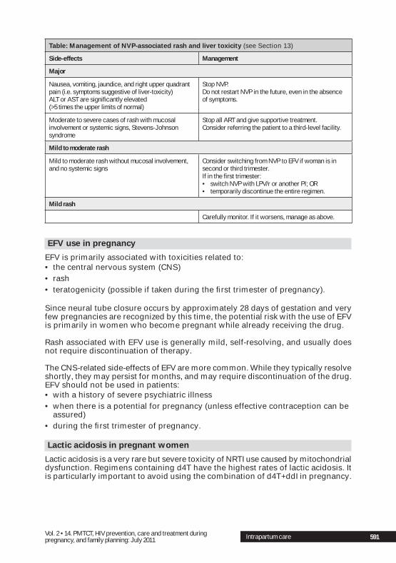
591Vol. 2 • 14. PMTCT, HIV prevention, care and treatment during pregnancy, and family planning: July 2011 591
Table: Management of NVP-associated rash and liver toxicity (see Section 13)
Side-effects Management
Major
Nausea, vomiting, jaundice, and right upper quadrant pain (i.e. symptoms suggestive of liver-toxicity)ALT or AST are signifi cantly elevated (>5 times the upper limits of normal)
Stop NVP. Do not restart NVP in the future, even in the absence of symptoms.
Moderate to severe cases of rash with mucosal involvement or systemic signs, Stevens-Johnson syndrome
Stop all ART and give supportive treatment.Consider referring the patient to a third-level facility.
Mild to moderate rash
Mild to moderate rash without mucosal involvement, and no systemic signs
Consider switching from NVP to EFV if woman is in second or third trimester.If in the fi rst trimester: • switch NVP with LPV/r or another PI; OR • temporarily discontinue the entire regimen.
Mild rash
Carefully monitor. If it worsens, manage as above.
EFV use in pregnancy
EFV is primarily associated with toxicities related to:• the central nervous system (CNS)• rash• teratogenicity (possible if taken during the fi rst trimester of pregnancy).
Since neural tube closure occurs by approximately 28 days of gestation and very few pregnancies are recognized by this time, the potential risk with the use of EFV is primarily in women who become pregnant while already receiving the drug.
Rash associated with EFV use is generally mild, self-resolving, and usually does not require discontinuation of therapy.
The CNS-related side-effects of EFV are more common. While they typically resolve shortly, they may persist for months, and may require discontinuation of the drug. EFV should not be used in patients: • with a history of severe psychiatric illness • when there is a potential for pregnancy (unless effective contraception can be
assured)• during the fi rst trimester of pregnancy.
Lactic acidosis in pregnant women
Lactic acidosis is a very rare but severe toxicity of NRTI use caused by mitochondrial dysfunction. Regimens containing d4T have the highest rates of lactic acidosis. It is particularly important to avoid using the combination of d4T+ddI in pregnancy.
Intrapartum care
FINAL Vol. 2 CH 9-21.indd 591FINAL Vol. 2 CH 9-21.indd 591 29/06/2012 15:1629/06/2012 15:16

592 Vol. 2 • 14. PMTCT, HIV prevention, care and treatment during pregnancy, and family planning: July 2011592
Lactic acidosis often develops slowly and is characterized by several non-specifi c symptoms, including shortness of breath or hyperventilation, abdominal pain, nausea, fatigue, and weight loss. The symptoms can be vague, therefore a high degree of suspicion is needed. Many of these symptoms could occur as part of a normal pregnancy, and extra vigilance for lactic acidosis among pregnant women using NRTIs is required. Lactic acidosis can sometimes be confused with obstetrical complications (such as pre-eclampsia or HELLP syndrome) or with an HIV-related illness. (See Section 13.)
Rash and hypersensitivity due to abacavir (ABC)
Fever, fatigue, rash, sore throat, or shortness of breath may indicate ABC hypersensitivity syndrome (see Section 13 Toxicity and drug substitutions).
Safety of medicines in pregnancy
Refer to Section 8 Medicines and the WHO Model Formulary8 for each medication.
14.1.10 Antiretroviral drug resistanceARV drug resistance, in the context of PMTCT interventions, most commonly occurs with poor adherence and with the use of single-dose NVP in pregnancy. NVP has a prolonged half-life, with signifi cant drug levels measurable for up to 3 weeks after a single dose. Further, a single viral point mutation can cause high levels of resistance to NVP. This may lower the effectiveness of later ART containing an NNRTI drug. The following strategies can minimize these effects.• Identify all women who are eligible for ART as early as possible in order to
initiate combination ART while ensuring good adherence.• If ART is not yet indicated, offer AZT prophylaxis (Option A) or triple ARV
prophylaxis (Option B). In Option A, a single-dose of NVP is given in combination with AZT+3TC intrapartum, and for 7 days postpartum, to reduce the potential for resistance.
• For women receiving single-dose NVP who are in false labour, a repeat NVP dose should not be given during established labour. After delivery, the infant should receive the recommended ARV prophylaxis.
For women previously exposed to an ARV prophylaxis regimen for PMTCT in an earlier pregnancy and who are not in need of ART for their own health, the recommendations for ARV prophylaxis in a subsequent pregnancy are the same as for women not previously exposed. For women who initiate ART within 12 months after sd-NVP exposure, a non-NNRTI-based regimen is recommended (see the table below).
Intrapartum care
FINAL Vol. 2 CH 9-21.indd 592FINAL Vol. 2 CH 9-21.indd 592 29/06/2012 15:1629/06/2012 15:16

593Vol. 2 • 14. PMTCT, HIV prevention, care and treatment during pregnancy, and family planning: July 2011 593
Table: Choice of ART regimen for HIV-positive women with prior exposure to ARV prophylaxis for PMTCT10
Characteristics of previous PMTCT ARV exposure Recommendation
sd-NVP (+/- short-course AZT) with no NRTI tail within the last 12 months
• Initiate a non-NNRTI regimen• 2 NRTIs + PI preferred over 3 NRTIs
sd-NVP (+/- short-course AZT) with an NRTI tail within the last 12 months
• Initiate an NNRTI regimen• If available, check viral load at 6 months and if
>5000 copies/ml, switch to second-line ART with PI
sd-NVP (+/- short-course AZT) with or without an NRTI tail more than 12 months before
• Initiate an NNRTI regimen• If available, check viral load at 6 months and if
>5000 copies/ml, switch to second-line ART with PI
All triple ARV regimens, irrespective of duration of exposure and time since exposure
• Initiate NNRTI regimen• If earlier triple ARV regimen was NNRTI-based and
was stopped without administration of an NRTI tail, check viral load at 6 months, and if >5000 copies/ml, switch to second-line ART with PI
See Section 13 for the different classes of antiretroviral (ARV) drugs.
14.1.11 Improved care and support for HIV-positive pregnant women
Nausea and vomiting in pregnancy
Nausea and vomiting are common problems in the fi rst trimester especially in fi rst pregnancies, affecting up to 80% and 50% of all pregnant women, respectively. About 1% of pregnant women will have severe nausea and vomiting, known as hyperemesis gravidarum. A district clinician should assess women who have severe nausea and vomiting, who are unable to tolerate oral fl uids, or who are severely dehydrated. The goal of treatment is to manage symptoms and minimize risks to the mother and the fetus, and may vary depending on the severity.
Assess the hydration status carefully (see Section 10.7d Classify dehydration). A woman may require hospitalization for rehydration with intravenous fl uids if she is unable to tolerate fl uids by mouth. • Encourage the woman to eat smaller portions of food more frequently and
to drink lots of fl uids. Bland and low fat food may be preferable, as fatty foods may delay gastric emptying. Serve cold food, if the smell of hot food is noxious.
• Ginger drinks and pyridoxine (vitamin B6) are helpful in reducing nausea and vomiting in pregnancy. Promethazine may also be used, as indicated (see table below, Antiemetic medication in pregnancy for dose).
Some antiemetics can be given intramuscularly or as rectal suppositories if the woman is unable to tolerate oral medications. See Section 10.7c and Section 20. If vomiting persists for longer than 2 weeks, give thiamine (vitamin B1) replacement.
Nausea and vomiting can severely affect adherence to ART or ARV prophylaxis. Manage nausea and vomiting so that ART or ARV prophylaxis can be continued. Encourage the woman to continue taking her ARV drugs, and to repeat the dose if she vomits whole tablets.
Intrapartum care
FINAL Vol. 2 CH 9-21.indd 593FINAL Vol. 2 CH 9-21.indd 593 29/06/2012 15:1629/06/2012 15:16

594 Vol. 2 • 14. PMTCT, HIV prevention, care and treatment during pregnancy, and family planning: July 2011594
Table: Antiemetic medication in pregnancy (see specifi cs in Section 8.4 Medicines)
Antiemetic Dose
Pyridoxine (vitamin B6) 25 mg PO, up to 3–4 times daily
Promethazine 10–25 mg PO, IM, or IV, up to 4 times daily
Metoclopramide 10 mg PO up to 3–4 times daily; 5–10 mg IM, up to 3–4 times daily
Ondansetron 4 to 8 mg PO every 12 hours
14.2 Intrapartum care for HIV-positive women1,2,5
Recommend HIV testing during labour if not already done. In settings with a generalized HIV epidemic, pregnant women who tested negative in their fi rst or second trimesters of pregnancy should be recommended to have HIV testing and counselling in the third trimester. If this is not done, HIV testing should be recommended during labour or immediately after delivery. A new diagnosis of HIV infection can be made in labour (i.e. positive rapid test during labour). Give ARV prophylaxis while waiting for a confi rmatory test result. For women identifi ed HIV-positive during labour or immediately postpartum, it may be better to give Option A, which provides daily NVP prophylaxis for the infant. Assess the mother for ART eligibility immediately postpartum. If she is eligible, initiate ART. The infant should continue daily NVP until the mother has received at least 6 weeks of ART before the infant prophylaxis is discontinued. Follow national PMTCT guideline recommendations.
If the woman is already on ARV prophylaxis or ART, continue the same regimen throughout labour and delivery. (See table below.)
Table: Summary of ARV regimens during pregnancy, intrapartum, postpartum, and breastfeeding
ARV regimen during pregnancy
Intrapartum ARV regimen
Postpartum ARV regimen
ARV regimen if
Breastfeeding Replacement feeding only
Maternal ART ART ART Mother: ARTInfant: AZT or NVP prophylaxis from birth until 4−6 weeks of age
Mother: ARTInfant: AZT or NVP prophylaxis from birth until 4−6 weeks of age
Maternal AZT prophylaxis (Option A)
At onset of labour, AZT+ sd-NVP + 3TC
AZT+3TC for 7 days
Mother: AZT+3TC for 7 days onlyInfant: NVP prophylaxis from birth until 1 week after all breast milk exposure ends
Mother: AZT+3TC for 7 daysInfant: AZT or NVP prophylaxis from birth until 4−6 weeks of age
Assess the woman regularly; start ART as soon as eligible.
Maternal triple ARV prophylaxis (Option B)
Maternal triple ARV prophylaxis
Maternal triple ARV prophylaxis
Mother: Triple ARV prophylaxis until 1 week after all breast milk exposure endsInfant: AZT or NVP prophylaxis from birth until 4−6 weeks of age
Mother: NONEInfant: AZT or NVP prophylaxis from birth until 4−6 weeks of age
Assess the woman regularly; start ART as soon as eligible.
Intrapartum care
FINAL Vol. 2 CH 9-21.indd 594FINAL Vol. 2 CH 9-21.indd 594 29/06/2012 15:1629/06/2012 15:16

595Vol. 2 • 14. PMTCT, HIV prevention, care and treatment during pregnancy, and family planning: July 2011 595
Safer intrapartum practices
Use safer intrapartum practices for both the mother and the newborn to minimize the risks of MTCT.• Use standard precautions to prevent infection (see Section 6).• Minimize the number of cervical exams.• Avoid artifi cial membrane rupture.• Avoid invasive obstetrical procedures, such as episiotomy, invasive fetal
monitoring, or nasal suction of newborns.• Use the partograph and active management of labour to reduce the duration
of labour.• Avoid the use of vacuum or forceps, except in selected cases when one
of these techniques is required because of fetal distress, or to shorten the duration of labour or membrane rupture (e.g. cases of maternal exhaustion).
• Vaginal cleansing with 0.25% chlorhexidine solution does not decrease the overall risk of MTCT, but may decrease MTCT in a subgroup of women with prolonged membrane rupture, and may decrease early maternal and neonatal infectious morbidity.
• Dry the infant as soon as possible after delivery and remove any cloths with blood and maternal secretions.
Caesarean delivery
Elective caesarean delivery decreases MTCT of HIV in women not receiving ARV prophylaxis. There is no evidence that caesarean delivery reduces risk of MTCT if performed after the onset of labour or membrane rupture, or that it offers any additional benefi t to women already taking effective ART with optimal viral load suppression.
In limited-resource settings, caesarean delivery should only be performed for obstetrical indications. Concerns about routine caesarean deliveries in such settings include: • increased risk of maternal mortality and morbidity• anaesthesia availability• availability of safe blood supply should transfusion be needed• the need for antibiotic prophylaxis• lack of human and material resources.
Breast care
Prior to discharge from the maternity ward, mothers should be educated about appropriate breast care and assisted with proper attachment. They should be informed about signs and symptoms of breast problems and the potential role of breast infections (abscess, mastitis) or nipple trauma in MTCT. They should be encouraged to seek care promptly if these occur.
Intrapartum care
FINAL Vol. 2 CH 9-21.indd 595FINAL Vol. 2 CH 9-21.indd 595 29/06/2012 15:1629/06/2012 15:16

596 Vol. 2 • 14. PMTCT, HIV prevention, care and treatment during pregnancy, and family planning: July 2011596
14.3 Infant feeding12
Mothers known to be HIV-infected (and whose infants are HIV-uninfected or of unknown HIV status) should follow national policy and recommendations on infant feeding.
National m inistries of health should decide which infant feeding practice will be promoted and supported in clinics and hospitals of their individual countries. The decision should be guided by which feeding practice will give infants the best chance of HIV-free survival, i.e. remaining HIV-uninfected, as well as protecting infants from other serious illnesses, such as diarrhoea or pneumonia, that are common causes of infant mortality. In making this decision, the national ministry should consider:• the prevalence of HIV and the type of HIV epidemic in the region or country;• the socio-economic conditions of most families attending public health
facilities;• the quality of water and sanitation;• the distribution and quality of MCH services including availability of HIV and
PMTCT interventions;• the main causes of infant and child mortality;• the prevalence of infant and child under-nutrition.
It is expected that ministries of health will recommend one of two infant feeding practices for HIV-positive mothers and their infants: either that HIV-positive mothers breastfeed and be provided with appropriate ARVs or that HIV-positive mothers avoid breastfeeding and give replacement feeds only. This decision and how it should be implemented should be clearly communicated to health workers.
Breastfeeding
In settings where breastfeeding is recommended, HIV-positive mothers should exclusively breastfeed their infants for the fi rst 6 months of life, introducing appropriate complementary foods thereafter, and then continue breastfeeding for the fi rst 12 months of life.
Health workers need to assist the mother to initiate breastfeeding within 30 minutes of birth and support exclusive breastfeeding over the fi rst 6 months of life. This includes advising and encouraging the mother about correct positioning and attachment of the baby to the breast. Mothers should also be informed about growth spurts in the infant and how she will need to increase breastfeeding to meet the infant’s needs. These practices are the same as for the HIV-negative mothers and their infants.
In places where diarrhoea, pneumonia, and malnutrition are common, continuing to breastfeed over the fi rst 12 months protects the infant from death (during this time and afterwards) caused by these serious illnesses.
At the same time, antiretroviral drugs should be given to reduce the risk of HIV transmission.
12 Guidelines on HIV and infant feeding. WHO, 2010. Available at http://www.who.int/child_adolescent_health/documents/9789241599535/en/
Infant feeding
FINAL Vol. 2 CH 9-21.indd 596FINAL Vol. 2 CH 9-21.indd 596 29/06/2012 15:1629/06/2012 15:16

597Vol. 2 • 14. PMTCT, HIV prevention, care and treatment during pregnancy, and family planning: July 2011 597
HIV-positive pregnant women and mothers who are eligible for lifelong treatment should be started on antiretroviral therapy. ART eligibility criteria for breastfeeding women are the same as for pregnant and non-pregnant adults. (See above Table: Eligibility criteria for ART or ARV prophylaxis in HIV-positive pregnant women, and Table: Considerations for the choice of fi rst-line ART for pregnant women in need of treatment for their own health).
HIV-positive mothers who are not yet eligible to start ART should be provided with appropriate ARVs, that either the mother gives to the infant (Option A) or takes herself (Option B) to prevent HIV transmission through breastfeeding. (See above Table: Recommended ARV-prophylaxis for pregnant women not yet eligible for ART; see above Table: Summary of infant ARV prophylaxis regimens; and see above Table: Infant ARV prophylaxis dosing recommendations).
Option A: Daily NVP to the infant while breastfeeding and until 1 week after all breast milk exposure ends.
Option B: Triple ARVs to the mother while breastfeeding and until 1 week after all breast milk exposure ends.
Note: These are continuations of ARVs provided during pregnancy and labour.
All children need complementary foods from 6 months of age. Details of how much food should be given in addition to milk are provided in other guidance documents.13
In general, HIV-positive mothers should stop breastfeeding after 12 months and provide a nutritionally adequate and safe diet without breast milk. If, however, she is unable to do this, the mother may need to continue to breastfeed while using ARVs to prevent HIV transmission. Health workers should counsel the mother at around 12 months in order to help her make this decision, to consider how she will manage the change to no breastfeeding, how she will comfort the infant when crying, and how she will feed her infant afterwards. When stopping breastfeeding, mothers should do so gradually over about 1 month.
Health workers need to provide information to the mother about ARVs and how they reduce HIV transmission during breastfeeding. Mothers need to know: • where they will get the ARVs, how often and from whom;• where to go if her ARV supplies are running low;• what to do when she plans to stop breastfeeding, and when to stop the ARVs;• when to go to the clinic for immunization, cotrimoxazole prophylaxis, and HIV
testing of her infant.
Replacement feeding
In settings where ministries of health recommend that HIV-positive mothers give replacement feeds, health workers should confi rm that HIV-positive mothers (or other carers) have the following.• Assured safe water and sanitation at the household level and in the
community.
13 Guiding principles for feeding non-breastfed children 6–24 months of age. WHO, 2005. Available at http://www.who.int/child_adolescent_health/documents/9241593431/en/index.html
Infant feeding
FINAL Vol. 2 CH 9-21.indd 597FINAL Vol. 2 CH 9-21.indd 597 29/06/2012 15:1629/06/2012 15:16

598 Vol. 2 • 14. PMTCT, HIV prevention, care and treatment during pregnancy, and family planning: July 2011598
• Reliably provide suffi cient infant formula milk to support normal growth and development.
• Prepare infant formula milk cleanly so that it is safe and carries a low risk for diarrhoea and frequently enough to avoid malnutrition.
• In the fi rst 6 months, exclusively give infant formula milk.• The family is supportive of giving infant formula milk.• Access to health care that offers comprehensive child health services.
If these conditions are met, health workers should demonstrate to mothers why and how to safely prepare replacement feeds, how to sterilize cups and bottles, and what to do if the infant becomes ill with diarrhoea or develops oral thrush. Health workers should continue regularly to see mothers and infants, to check hygienic feeding practices (especially preparation of feeds), monitor weight gain, and to detect complications. Mothers should be advised about when to attend health facilities with her infant for fi nal HIV testing.
When infants are not or no longer breastfed (either from birth or after a period of breastfeeding), the mother should provide a nutritional, adequate, and safe diet.
When infants are <6 months of age, mothers should give either of the following:• Commercial infant formula milk (as long as the conditions listed above are
fulfi lled); or• Expressed, heat-treated breast milk. This can be considered as an interim
feeding strategy in the following circumstances: ° when the infant is born with low birth weight or is otherwise ill in the
neonatal period and unable to breastfeed; ° when the mother is unwell and temporarily unable to breastfeed or has a
temporary breast health problem, such as mastitis; ° to assist mothers to stop breastfeeding; ° if ARV drugs are temporarily not available.
When infants are >6 months of age mothers can give either of the following:• Commercial infant formula milk (as long as the conditions listed above are
fulfi lled).• Animal milk (boiled for infants <12 months), as part of a diet providing
adequate micronutrient intake. Meals, including milk-only feeds, other foods, and combinations of milk feeds and other foods, should be provided 4 or 5 times per day.
When infants become HIV-infected during pregnancy, labour, or delivery, and test HIV-positive at 6 weeks of age (i.e. virological test) or any other time, mothers are strongly encouraged to exclusively breastfeed for 6 months and to continue to breastfeed for 2 years (the same as infants of mothers who are HIV-negative or who do not know their HIV status). If an infant is already infected with HIV, there is no reason to avoid breastfeeding. Rather, HIV-positive infants will benefi t from breastfeeding and the protection that it provides against other infectious diseases.
Facility managers and district health teams should monitor feeding practices over time. Health workers can ascertain feeding practices at specifi c points in time, such as at 3 months, as a proxy of practices at other times. If breastfeeding with ARVs is the recommended practice, then monitoring of ARV adherence among breastfeeding mothers is important.
Infant feeding
FINAL Vol. 2 CH 9-21.indd 598FINAL Vol. 2 CH 9-21.indd 598 29/06/2012 15:1629/06/2012 15:16

599Vol. 2 • 14. PMTCT, HIV prevention, care and treatment during pregnancy, and family planning: July 2011 599
Whatever the recommended feeding practice, health workers in clinics and hospitals need to communicate to the broader community the infant feeding practice that is promoted and supported by local health facilities, and the rationale for the chosen public health approach. Given the history and confusion concerning infant feeding in the context of HIV, health-care workers need to convey the new opportunities for HIV-positive mothers to breastfeed while giving ARVs to prevent transmission and to improve HIV-free survival of HIV-exposed infants. Health services should make particular efforts to reach and gain the support of the fathers of infants to support optimal feeding practices. This provides a major opportunity to better link HIV-specifi c interventions with general maternal, newborn, and child health services that also promote breastfeeding to improve child survival.
14.4 Postpartum care for HIV-positive women
If the postpartum woman presents to the facility for the fi rst time after childbirth, recommend HIV testing and counselling if not already done. For those testing positive, assess WHO clinical stage and CD4 cell count to determine eligibility for ART. Clinical assessment of the HIV-positive woman should be performed each time she presents for postpartum care and for child health visits. Review TB status at each visit. Offer INH preventive therapy if the woman has no cough, fever, weight loss, or night sweats.
If the woman is already receiving ART or triple ARV prophylaxis, monitor and support adherence. If the woman is on triple ARV prophylaxis and breastfeeding, she should continue with same regimen until 1 week after all breast milk exposure of the infant ends. If she is on ART, she should continue the treatment for life. See above Table: Summary of ARV regimens during pregnancy, intrapartum, postpartum and breastfeeding for the ARV regimens to follow during postpartum and breastfeeding period.
Postpartum follow up of HIV-infected mothers and their infants is a critical part of PMTCT
interventions and clinical management
Ensure that HIV-positive women, their infants and their partners continue to receive HIV prevention, care, and treatment services postpartum.
• Support HIV-positive breastfeeding women as much as possible, e.g. involve their partners in PMTCT services, and facilitate peer support from other HIV-infected women who are already receiving care, as appropriate for each woman.
• Coordinate facility visits for the woman, her infant, partner, and family. • Integrate and support single point of care as much as possible. Facilitate
linkages across services, e.g. MNCH, family planning, HIV care and treatment.
• Provide family-focused care.
Counsel women on the advantages of birth spacing and planning future pregnancies, and how ARVs can protect women during breastfeeding and prevent transmission of HIV to their infants. Review a woman’s family planning and fertility
Postpartum care
FINAL Vol. 2 CH 9-21.indd 599FINAL Vol. 2 CH 9-21.indd 599 29/06/2012 15:1629/06/2012 15:16

600 Vol. 2 • 14. PMTCT, HIV prevention, care and treatment during pregnancy, and family planning: July 2011600
desires. Emphasize the importance of dual protection, including condom use (dual methods or dual protection using only condoms). See Section 14.5 Reproductive choice and family planning.
Care for HIV-exposed infants
See IMCI Chart Booklet for High HIV Prevalence Settings14 and Child Pocket Book for Hospital Care.15
• Ensure follow up care of HIV-exposed infants. The following are essential care for HIV-exposed infants. ° Routine newborn and infant care, including immunization, growth
monitoring, and nutrition. ° Completion of ARV prophylaxis. Support and monitor adherence, and give
adherence counselling to caregivers. ° Continued infant feeding counselling and support, and nutritional support
throughout the fi rst year of life. ° Early HIV diagnostic testing and diagnosis of HIV-related conditions. Early
infant diagnosis is critical to provide ART for those who are infected. Recommend virological testing at 4–6 weeks of age or at fi rst presentation after 4–6 weeks of age.16 If HIV-infected, the infant is eligible for ART. If the test is negative the mother or her breastfeeding infant should continue with the ARV regimen. See above Table: Summary of ARV regimens during pregnancy, intrapartum, postpartum, and breastfeeding for reference.
° Cotrimoxazole prophylaxis starting at 4–6 weeks of age and continuing until HIV infection has been excluded and there is no risk of HIV exposure. It is recommended for all HIV-exposed infants until HIV infection is excluded.
° INH preventive therapy when indicated. ° Malaria prevention and treatment when indicated. ° Diagnosis and management of common childhood infections and
conditions, TB, and integrated management of childhood illnesses (IMCI). ° Establishing fi nal diagnosis of HIV-exposed infants at 15–18 months of age.
Early diagnosis of HIV-infection in infants is criticalto reduce HIV-related morbidity and mortality
in this age group
14 IMCI chart booklet for high HIV settings. WHO, 2008. Available at http://www.who.int/child_adolescent_health/documents/9789241597388/en/index.html
15 Manual on paediatric HIV care and treatment for district hospitals. WHO, 2011. Available at http://www.who.int/child_adolescent_health/documents/9789241501026/en/index.html
16 WHO recommendations on the diagnosis of HIV infection in infants and children. WHO, 2010. Available at http://www.who.int/hiv/pub/paediatric/diagnosis/en/index.html
Postpartum care
FINAL Vol. 2 CH 9-21.indd 600FINAL Vol. 2 CH 9-21.indd 600 29/06/2012 15:1629/06/2012 15:16

601Vol. 2 • 14. PMTCT, HIV prevention, care and treatment during pregnancy, and family planning: July 2011 601
14.5 Reproductive choice and family planning17
Discuss fertility intentions and provide family planning counselling with each woman. The goals of counselling and discussion are to prevent unintended pregnancy, and to help achieve a safe pregnancy, for both mother and child, when pregnancy is desired. The discussion should never be coercive; the woman has the right to make her own decisions about reproduction. She should be provided with the available options. Family planning services and counselling are especially important at the following times:• after HIV diagnosis, and at regular intervals during ongoing care;• when there is a new marriage or relationship;• when a desire is expressed not to become pregnant;• when the woman is not using effective or consistent contraception;• during pregnancy or the postpartum period (to reinforce the value of birth
spacing);• following the death of a child; • when a woman is using ART, which may be associated with resumption of
sexual activity and fertility, and therefore an increased risk for unintended pregnancy;
• when a woman takes ART that includes EFV. She should use an effective method of contraception due to the possible risk of teratogenicity from EFV use during early pregnancy exposure.
14.5.1 Dual protectionProvision of family planning counselling and of contraception services should occur within the context of HIV clinical care. It is recommended that family planning services need to integrate into and support HIV clinical services. If this is not possible, the woman should be referred to a nearby family planning service.
The condom is the only contraceptive method that protects against STIs, including HIV, as well as against pregnancy. Male latex condoms are the most popular and protect against both female-to-male and male-to-female transmission of HIV, as shown in studies of discordant couples. Female condoms are still an underused method of dual protection against both pregnancy and HIV. When a signifi cant risk of STI or HIV transmission exists, dual protection against both pregnancy and STI and HIV transmission should be strongly recommended. Consider these 5 dual protection strategies, each of which might be appropriate in different situations and at different times for an individual. The best strategy is the one that a person is able to practice effectively in the situation she or he is facing.
Strategy 1: Use a male or female condom correctly with every act of sex.Strategy 2: Use condoms consistently and correctly, plus another family planning
method. This may be a good choice for women who want to confi dently avoid pregnancy, but who cannot always count on their partners to use condoms.
Strategy 3: If both partners know they are not HIV-infected, use any family planning method to prevent pregnancy and stay in a mutually faithful relationship. This strategy depends on communication and trust between partners.
17 Family planning: a global handbook for providers (updated). WHO, 2011. Available at http://www.who.int/reproductivehealth/publications/family_planning/9780978856304/en/index.html
Reproductive choice
FINAL Vol. 2 CH 9-21.indd 601FINAL Vol. 2 CH 9-21.indd 601 29/06/2012 15:1629/06/2012 15:16

602 Vol. 2 • 14. PMTCT, HIV prevention, care and treatment during pregnancy, and family planning: July 2011602
Strategy 4: Engage only in safe sexual intimacy that avoids intercourse, and otherwise prevents semen and vaginal fl uids from coming in contact with each other’s genitals. This depends on communication, trust, and self-control.
Strategy 5: Delay or avoid sexual activity (either avoiding sex at any time that it might be risky, or abstaining for a longer time).
The latter two strategies do not involve contraceptives. Many patients need help and guidance to ensure their specifi c dual protection strategy will succeed. Seroconcordant couples may be less likely to use condoms consistently, and require a more effective contraceptive method if pregnancy is not desired.
14.5.2 Guidance on the use of contraceptive methods There are guidance references that policy makers and programme managers can use to support the safe and effective provision and use of family planning methods. These evidence-based guidance documents dispel any lack of information or misinformation about using various methods. The WHO Medical eligibility criteria for contraceptive use (4th edition, 2010) provides guidance on whether people with certain medical conditions can safely and effectively use specifi c contraceptive methods.18 There is a section on recommendations for persons living with HIV. Drug interactions and other effects of hormonal methods need to be considered in making recommendations (see below Table: Medical eligibility criteria for contraceptive use: conditions relevant to HIV. Use this table to guide the selection of a contraceptive method.)
Category 1: Use this method in any circumstances; use the method with limited clinical judgement.
Category 2: With clinical judgment, generally use the method; with limited clinical judgment, use the method.
Category 3: With clinical judgment, use of the method not generally recommended unless more appropriate methods are not available or acceptable. With limited clinical judgment, do not use the method.
Category 4: Method not to be used.
Drug-drug interactions
There are drug-drug interactions between hormonal contraceptives and many PIs and NNRTIs. The interactions may either increase or decrease contraceptive steroid levels, which may potentially either decrease contraceptive effectiveness when co-administered, or increase hormonal side-effects or toxicity when steroid levels are increased. The medical eligibility categories assigned to ART in the table below take into account these potential interactions. • Women taking ritonavir-boosted PIs generally should not use combined
hormonal methods (combined pills, monthly injectables, patch, and vaginal ring) or progestogen-only pills.
• Progestogen-only injectables and implants generally can be used with any ART.
Selected Practice Recommendations for Contraceptive Use answers specifi c questions about how to use various contraceptive methods.19
18 Medical eligibility criteria for contraceptive use. 4th ed. WHO, 2009. Available at http://www.who.int/reproductivehealth/publications/family_planning/9789241563888/en/index.html
19 Selected Practice Recommendations for Contraceptive Use. 2nd ed. WHO, 2004. (With selected 2008 update). Available at http://www.who.int/reproductivehealth/publications/family_planning/9241562846index/en/index.html
Reproductive choice
FINAL Vol. 2 CH 9-21.indd 602FINAL Vol. 2 CH 9-21.indd 602 29/06/2012 15:1629/06/2012 15:16

603Vol. 2 • 14. PMTCT, HIV prevention, care and treatment during pregnancy, and family planning: July 2011 603
Table: Medical eligibility criteria for contraceptive use: conditions relevant to HIV
Condition COC
CIC
P/R
POP
DM
PA/
NET
-EN
LNG
impl
ant*
Copp
er
IUD
LNG
IU
D
Sper
mic
ide
Dia
phra
gm(w
ith s
perm
icid
e)
Cerv
ical
cap
HIV risk 1 1 1 1 1 1 2 2 4 4
HIV 1 1 1 1 1 1 2 2 3 3
AIDS 1 1 1 1 1 1 I=3C=2
I=3C=2 3 3
Drug interactions
NRTIs 1 1 1 1 DMPA=1NET-EN=1 1 I=2/3
C=2I=2/3C=2 3 3
NNRTIs 2 2 2 2 DMPA=1NET-EN=2 2 I=2/3
C=2I=2/3C=2 3 3
Ritonavir-boosted PIs 3 3 3 3 DMPA=1
NET-EN=2 2 I=2/3C=2
I=2/3C=2 3 3
Rifampicin 3 2 3 3 DMPA=1NET-EN=2 2 1 1 1 1
*Also includes etonogestrel implant.COC=combined oral contraceptives with ≤35 mcg ethinyl estradiol; CIC=combined injectable contraceptives; P=patch; R=ring; POP=progestogen only pills; DMPA=depot medroxyprogesterone acetate, NET-EN=norethisterone enantate; LNG-implant=levonorgestrel implant; I=initiation; C=continuation
14.5.2 Contraceptive method mix
Hormonal methods (combined pills and injectables, progestogen-only pills, injectables, implants and IUDs: As discussed above and shown in the table, some hormonal methods may have interactions with some antiretroviral drugs. However, the hormonal methods have non-contraceptive benefi ts that may have particular relevance for HIV-infected women. Proven health benefi ts of combined oral contraceptives include: decreased iron-defi ciency anaemia, increased menstrual regularity, decreased dysmenorrhoea (menstrual cramps), decreased risk of pelvic infl ammatory disease (PID), and decreased incidence of endometrial, ovarian, and possibly colorectal cancer. Proven health benefi ts of depot medroxyprogesterone acetate (DMPA) include: decreased iron-defi ciency anaemia, decreased risk of PID, decreased uterine fi broids, reduced symptoms of endometriosis, and decreased incidence of endometrial cancer.
Intrauterine devices (Copper-bearing IUD, LNG IUD): A woman with HIV can have an IUD inserted. A woman with AIDS should not have an IUD inserted unless she is clinically well on ART. A woman who develops AIDS while using an IUD can safely continue using the IUD, and initiate ART. A woman using an IUD is not at increased risk of HIV transmission to sexual partners.
Condoms: Male and female condoms are the only contraceptive methods that also provide protection against HIV transmission to uninfected partners and against STI acquisition.
Reproductive choice
FINAL Vol. 2 CH 9-21.indd 603FINAL Vol. 2 CH 9-21.indd 603 29/06/2012 15:1629/06/2012 15:16

604 Vol. 2 • 14. PMTCT, HIV prevention, care and treatment during pregnancy, and family planning: July 2011604
Spermicides: Repeated and high-dose use of the spermicide nonoxynol-9 was associated with increased risk of genital lesions in women, which may increase the risk of acquiring HIV infection. Therefore, for women at high risk of HIV, the use of spermicides, or diaphragms or cervical caps with spermicides are a category 4 (do not use). For women who are HIV-positive, the use of spermicides, or diaphragms or cervical caps with spermicides are a category 3 (generally do not use) due to concerns about increased risk of transmission to uninfected partners.
Lactational amenorrhoea (LAM): In circumstances in which a woman with HIV decides to breastfeed, LAM can be used for family planning purposes. For adequate protection from an unplanned pregnancy, she must have the main requirements for LAM: amenorrhoea, exclusive breastfeeding, and <6 months postpartum. See the previous subsection on infant feeding.
Female sterilization (tubal ligation): Women, who are infected with HIV, have AIDS, or who are on ART can safely undergo female sterilization. Special care must be taken to ensure that the woman makes a voluntary, informed choice of method. Health workers should ensure that women are not pressured or coerced to undergo the procedure, and that the decision is not made in a moment of crisis. Standard infection prevention practices should be followed. Special arrangements are needed to perform female sterilization on a woman with AIDS. The procedure should be delayed if the woman currently has an AIDS-related illness. Female sterilization does not protect against acquiring and transmitting HIV. Condom use should be emphasized post-procedure.
Male sterilization (vasectomy): Men, who are infected with HIV, have AIDS, or who are on ART can safely undergo male sterilization. Special care must be taken to ensure that the man makes a voluntary, informed choice of method. Health workers should ensure that men are not pressured or coerced to undergo the procedure, and that the decision is not made in a moment of crisis. Good infection prevention practices should be followed. Special arrangements are needed to perform male sterilization on a man with AIDS. The procedure should be delayed if the man currently has an AIDS-related illness. Male sterilization does not protect against acquiring and transmitting HIV. Condom use should be emphasized post-procedure.
Emergency contraception18
Women with HIV or on ART can safely use emergency contraception. It can be used when a woman has made a mistake using contraception, when a condom breaks, in the case of rape, or when the woman was not protected by a reliable method of contraception. It is not recommended as a routine method of contraception as it is not as effective as non-emergency methods. It does not provide protection against STIs or HIV.
• Oral contraceptive pills for emergency contraception should be used within 120 hours (5 days) after unprotected intercourse: ° Use LNG 0.75 mg at once followed by same dose 12 hours later OR
alternatively 1.5 mg at once, or ° LNG 0.25 mg + ethinyl estradiol 50 mcg (less well-tolerated and less
effective).• An alternative is a copper IUD inserted within 120 hours (5 days) of
unprotected intercourse. This is the most effective form of emergency contraception and appropriate for a woman who wants an IUD as her
Reproductive choice
FINAL Vol. 2 CH 9-21.indd 604FINAL Vol. 2 CH 9-21.indd 604 29/06/2012 15:1629/06/2012 15:16

605Vol. 2 • 14. PMTCT, HIV prevention, care and treatment during pregnancy, and family planning: July 2011 605
continuing method. However, avoid use if the woman is at high risk for gonorrhoea or Chlamydia or has AIDS and is not clinically well.
Indications for pregnancy testing
Women who have been sexually active and are experiencing any of the following should take a pregnancy test.• missed menses, unless on progestogen-only injectable or implant (can cause
amenorrhoea)• irregular bleeding, unless on progestogen-only injectable or implant• new onset of irregular bleeding after prolonged amenorrhoea on
progestogen-only injectable or implant• new onset of pelvic pain• enlarged uterus or adnexal mass• prior to the initiation of an EFV-based ARV regimen.
Special considerations when pregnancy is desired by a discordant couple
When, after preconception counselling, a discordant couple (where one partner is HIV-positive and the other partner is HIV-negative or of unknown status) wishes to become pregnant, they should limit unprotected sexual exposure. This can be done by having unprotected sex only at times of greatest fertility, as determined with fertility awareness methods. During other times they may engage in sex using a condom, to ensure that the uninfected partner remains uninfected. • PLHIV in a serodiscordant couple who have been started on ART for their
own health should be informed that ART also is recommended to reduce HIV transmission to their HIV-uninfected partners.
• See Sections 13 and 19 for prevention.
Reproductive choice
FINAL Vol. 2 CH 9-21.indd 605FINAL Vol. 2 CH 9-21.indd 605 29/06/2012 15:1629/06/2012 15:16

FINAL Vol. 2 CH 9-21.indd 606FINAL Vol. 2 CH 9-21.indd 606 29/06/2012 15:1629/06/2012 15:16

15. Tuberculosis
Table of contents
15.1 Suspect TB . . . . . . . . . . . . . . . . . . . . . . . . . . . . . . . . . . . . . . . . . . . . . . 609Pulmonary TB . . . . . . . . . . . . . . . . . . . . . . . . . . . . . . . . . . . . . . . . . . 609Extrapulmonary TB . . . . . . . . . . . . . . . . . . . . . . . . . . . . . . . . . . . . . . . 610
15.2 Diagnose TB (with DDx table) . . . . . . . . . . . . . . . . . . . . . . . . . . . . . . . . . . 610Case defi nitions . . . . . . . . . . . . . . . . . . . . . . . . . . . . . . . . . . . . . . . . . 610Laboratory and radiology in TB diagnosis . . . . . . . . . . . . . . . . . . . . . . . . . 610Extrapulmonary TB . . . . . . . . . . . . . . . . . . . . . . . . . . . . . . . . . . . . . . . 613Case defi nitions for the diagnosis of TB . . . . . . . . . . . . . . . . . . . . . . . . . . 613Algorithm for the diagnosis of AFB smear-negative PTB in HIV-negative patients where Xpert MTB/RIF test is not available . . . . . . . . . . . . . . . . . . 615Algorithm for the management of ambulatory HIV-positive patients with presumptive TB where Xpert MTB/RIF test is available . . . . . . . . . . . . . 616Algorithm for the diagnosis of TB in ambulatory patients in HIV-prevalent settings where Xpert MTB/RIF test is not available . . . . . . . . . . . . . . . . . . . 617Summary of signs and symptoms of EPTB . . . . . . . . . . . . . . . . . . . . . . . . . 618Diagnosis of TB in the seriously ill patient with danger signs . . . . . . . . . . . . . . 619Algorithm for the diagnosis of TB in seriously ill patients in HIV-prevalent settings where Xpert MTB/RIF test is possible . . . . . . . . . . . . . . . . . . . . . 620Algorithm for the diagnosis of TB in seriously ill patients in HIV-prevalent settings at fi rst-level facilities where Xpert MTB/RIF test is not available . . . . . 621Differential diagnosis of PTB in PLHIV . . . . . . . . . . . . . . . . . . . . . . . . . . . 622DDx: PTB in PLHIV by stage of immunosuppression . . . . . . . . . . . . . . . . . . . 623TB in pregnant and postpartum women . . . . . . . . . . . . . . . . . . . . . . . . . . . 623
15.3 Treatment of TB . . . . . . . . . . . . . . . . . . . . . . . . . . . . . . . . . . . . . . . . . . . 624Standardized TB treatment regimens . . . . . . . . . . . . . . . . . . . . . . . . . . . . 624Standard code for TB treatment regimens . . . . . . . . . . . . . . . . . . . . . . . . . 624First-line anti-tuberculosis drugs . . . . . . . . . . . . . . . . . . . . . . . . . . . . . . . 624Standard anti-TB regimen and dosing frequency in new TB cases . . . . . . . . . . 625Dose of fi rst-line anti-tuberculosis drugs for adults . . . . . . . . . . . . . . . . . . . 625Registration group by outcome of most recent TB treatment . . . . . . . . . . . . . . 626TB treatment in special situations . . . . . . . . . . . . . . . . . . . . . . . . . . . . . . 626Anti-TB treatment regimens in special situations . . . . . . . . . . . . . . . . . . . . . 627Empirical treatment in EPTB . . . . . . . . . . . . . . . . . . . . . . . . . . . . . . . . . 627Adjuvant corticosteroids . . . . . . . . . . . . . . . . . . . . . . . . . . . . . . . . . . . 627
15.4 Monitor TB treatment . . . . . . . . . . . . . . . . . . . . . . . . . . . . . . . . . . . . . . . 628Monitor response to TB treatment . . . . . . . . . . . . . . . . . . . . . . . . . . . . . . 628Manage drug side-effects . . . . . . . . . . . . . . . . . . . . . . . . . . . . . . . . . . . 628Symptom-based approach to managing side-effects of anti-TB drugs . . . . . . . . 629Determine TB treatment outcomes . . . . . . . . . . . . . . . . . . . . . . . . . . . . . 630Standard defi nitions of TB treatment outcomes . . . . . . . . . . . . . . . . . . . . . . 630
15.5 Drug-resistant TB (with DDx table) . . . . . . . . . . . . . . . . . . . . . . . . . . . . . . . 630Summary of recommended TB treatment regimens . . . . . . . . . . . . . . . . . . . 631MDR-TB treatment regimen . . . . . . . . . . . . . . . . . . . . . . . . . . . . . . . . . . 631MDR-TB and HIV infection . . . . . . . . . . . . . . . . . . . . . . . . . . . . . . . . . . 632Manage common problems in patients with MDR-TB . . . . . . . . . . . . . . . . . . 632
FINAL Vol. 2 CH 9-21.indd 607FINAL Vol. 2 CH 9-21.indd 607 29/06/2012 15:1629/06/2012 15:16

FINAL Vol. 2 CH 9-21.indd 608FINAL Vol. 2 CH 9-21.indd 608 29/06/2012 15:1629/06/2012 15:16

609Vol. 2 • 15. Tuberculosis: July 2011 609609
15. Tuberculosis
Tuberculosis (TB) is caused by Mycobacterium tuberculosis (M. tuberculosis), which is transmitted from a patient with pulmonary TB who is often sputum smear positive. Coughing and sneezing produce tiny infectious droplets that can remain suspended in the air for long periods of time. Poorly ventilated settings increase the risk of TB transmission.
It is very important that clinicians follow the recommendations of country specifi c national
TB and HIV treatment guidelines
Despite infection with M. tuberculosis, only 5–10% of people with normal immune systems develop TB.
HIV infection signifi cantly increases the risk of contracting TB. HIV-positive patients are more likely to acquire TB compared to HIV-negative patients. Early diagnosis and prompt treatment of TB and HIV is essential to reduce morbidity and death among persons living with HIV.
The diagnosis of TB refers to the recognition of an active case, i.e. a patient with current disease due to M. tuberculosis. Defi ning its anatomical site is important for recording and reporting purposes, and to identify the more infectious patients. In addition, the diagnosis of extrapulmonary TB in a HIV-positive patient signifi es an advanced stage of HIV infection (WHO clinical stage 4). In general, the recommended TB treatment regimens are the same irrespective of anatomical site.• Pulmonary tuberculosis (PTB) refers to a case of TB involving the lung
parenchyma. Globally, PTB is the most common presentation of TB patients. • Extrapulmonary tuberculosis (EPTB) refers to a case of TB involving organs
other than the lungs, e.g. pleura, lymph nodes, abdomen, genitourinary tract, skin, joints and bones, meninges.
15.1 Suspect TB
Pulmonary TB
Generally, the most common symptom of PTB is a productive cough for more than 2 weeks, which may be accompanied by other respiratory symptoms, such as shortness of breath, chest pain, or haemoptysis, with or without constitutional symptoms, such as fever, night sweats, weight loss, loss of appetite, and fatigue1.
In HIV-positive patients or in HIV-prevalent settings, suspect PTB and EPTB in patients who present with any one of these symptoms – cough, fever, weight loss, or night sweats. The presentation of TB may vary in HIV-positive patients, particularly in those with advanced immunosuppression. They are more likely
1 Treatment of tuberculosis: guidelines. 4th edition. WHO, 2010. Available at http://whqlibdoc.who.int/publications/2010/9789241547833_eng.pdf
Suspect TB
FINAL Vol. 2 CH 9-21.indd 609FINAL Vol. 2 CH 9-21.indd 609 29/06/2012 15:1629/06/2012 15:16

610 Vol. 2 • 15. Tuberculosis: July 2011610610
to present with EPTB or sputum smear-negative TB compared to HIV-negative patients, especially as their immunosuppression advances. HIV-positive patients also are more likely to be very ill when they present with possible TB.
Extrapulmonary TB
Generally, suspect EPTB in patients with:• cough, unintentional weight loss with night sweats, or temperature more
than> 37.5°C or feels feverish;• breathlessness (effusion or pericarditis)• enlarged glands in neck or armpit • chest X-ray (see Section 10.6) with
° miliary or diffuse shadowing ° large heart (especially if symmetrical and rounded) ° suggestive of pleural effusion ° enlarged lymph nodes inside the chest
• chronic headache or altered mental state.
Good clinical practice
• HIV-positive patients should be screened for TB at each clinic visit. • Recommend HIV testing and counselling for patients suspected of having
TB and TB patients, if not already done. • Encourage disclosure and partner testing, and provide HIV prevention, care,
and treatment services.
These are good clinical practices because diagnosis and management of both TB and HIV is critical for good treatment outcome in a HIV-positive TB patient.
Note: HIV-positive patients can develop TB at any time, before initiation of ART or while on fi rst-line or second-line ART.
15.2 Diagnose TB
Case defi nitions
Case defi nitions used for the diagnosis of TB, both in HIV-positive and HIV-negative patients, are summarized in the Table: Case defi nitions for the diagnosis of TB below.
Laboratory and radiology in TB diagnosis
Sputum microscopy for acid-fast bacilli (AFB) (Ziehl-Neelsen stain)Patients suspected of having PTB (i.e. those with suggestive signs and symptoms or with chest X-ray fi ndings suggestive of TB) should have at least 2 sputum specimens submitted for microscopic examination to a quality-assured laboratory, if the WHO-approved newer method for the diagnosis of TB is not available.
Treatment of TB
FINAL Vol. 2 CH 9-21.indd 610FINAL Vol. 2 CH 9-21.indd 610 29/06/2012 15:1629/06/2012 15:16

611Vol. 2 • 15. Tuberculosis: July 2011 611611
At least one early morning specimen should be obtained, as this sputum collection time has the highest yield. A patient with one positive AFB sputum smear is considered a defi nite case of smear-positive PTB.2
Good clinical practice Assure rapid diagnosis of TB and initiation of anti-TB treatment. This helps to reduce the risk of further transmission of TB, and minimizes treatment delay, which may be lifesaving particularly for HIV-positive TB patients.
Microscopy of other specimens for AFBMicroscopy for AFB can also be done on specimens obtained from extrapulmonary sites including:• pleural fl uid• cerebrospinal fl uid (CSF)• fi ne needle aspiration of lymph nodes.
Newer methods recommended by WHO such as Xpert MTB/RIF test3
Xpert MTB/RIF test, where available, can assure rapid di agnosis of TB. In such settings, one sputum specimen should be tested for diagnosis of TB from the following selected patients:• HIV-positive individuals or patients with unknown HIV status in high HIV
prevalence settings who are suspected of having TB.• Seriously ill patients who are suspected of having TB, regardless of their HIV
status. • Individuals at risk of multidrug-resistant TB (MDR-TB) who are either
diagnosed with TB or suspected of having TB.• If resources permit, HIV-negative individuals or those with unknown HIV
status (not seriously ill and in low HIV prevalence settings) not at risk of MDR-TB with either: ° abnormal X-ray or ° suspected of having TB with sputum AFB smear-negative result.
Where resources are limited, the national programme might prioritize specifi c groups of patients for the WHO-approved newer method for the diagnosis of TB. Molecular tests, including Xpert MTB/RIF, are not suitable for patient monitoring as these tests also detect DNA from non-viable bacilli.
Instruct and support patients in the collection of a good quality sputum specimen.
Follow national guideline recommendat ions on the newer WHO-approved diagnostic testing method
such as Xpert MTB/RIF
2 External quality assessment for AFB smear microscopy. CDC, WHO, 2007. Available at http://wwwn.cdc.gov/dls/ila/documents/eqa_afb.pdf
3 Rapid implementation of Xpert MTB/RIF diagnostic test: Technical and operational “How-to” practical considerations. WHO, 2011. Available at http://whqlibdoc.who.int/publications/2011/9789241501569_eng.pdf
Treatment of TB
FINAL Vol. 2 CH 9-21.indd 611FINAL Vol. 2 CH 9-21.indd 611 29/06/2012 15:1629/06/2012 15:16

612 Vol. 2 • 15. Tuberculosis: July 2011612612
RadiographyA chest X-ray is not necessary in most cases of smear-positive PTB. It can support diagnosis of:• smear-negative PTB, in the absence of the WHO-approved newer method for
the diagnosis of TB. In such settings, a chest X-ray is necessary to support diagnosis and to prevent unnecessary delay of the diagnosis of smear-negative PTB, while waiting for culture result;
• complications of PTB, e.g. pneumothorax;• complications of EPTB, e.g. pericardial effusion;• other causes of frequent or severe haemoptysis (e.g. bronchiectasis,
aspergilloma, or TB sequelae).A chest X-ray is suggestive of active TB if there is:• upper lobe infi ltrate, cavitation, pleural effusion, miliary pattern; or• non-specifi c patchy infi ltrates with or without hilar lymphadenopathy in HIV-
positive patients.
A normal chest X-ray does not exclude TB.
In a HIV-positive patient with TB, the chest X-ray fi ndings may largely depend upon the degree of immunodefi ciency. In mild immunodefi ciency, the appearance of the chest X-ray may not be different compared with a HIV-negative patient.
Refer to Section 10.6 for differential diagnosis of chest X-ray fi ndings.
Sputum culture for M. tuberculosisSputum culture is the “gold standard” for the diagnosis of PTB, but it may not be readily available in most settings and results may take longer. • Sputum culture is recommended for patients suspected of smear-negative
PTB in settings with a HIV prevalence of more than 1% in pregnant women, or a HIV prevalence of ≥5% in TB patients to confi rm the diagnosis of TB if newer diagnostic methods recommended by WHO such as Xpert MTB/RIF test are not available.
• Sputum culture and drug susceptibility testing (DST) are recommended for patients with previous TB treatment, those suspected of having MDR-TB, or patients with Xpert MTB+/RIF+ test result.
Other than sputum, cerebrospinal fl uid (CSF) and lymph node aspirate may be cultured in patients suspected of having TB meningitis or TB lymphadenitis respectively.
Ultrasound (see Section 7)Features of abdominal TB on ultrasound include:• lymphadenopathy;
° multiple, discrete, enlarged (more than 1.5 cm) para-aortic or mesenteric nodes, matted nodes with central necrosis;
• hepatosplenomegaly with or without multiple hypoechogenic nodules or abscesses;
• ascites (free or loculated).
Lymph node biopsy (see Section 7.2)fi ne needle aspiration with cytology, AFB smear, and culture;open biopsy with pathology, AFB smear, and culture.
Treatment of TB
FINAL Vol. 2 CH 9-21.indd 612FINAL Vol. 2 CH 9-21.indd 612 29/06/2012 15:1629/06/2012 15:16

613Vol. 2 • 15. Tuberculosis: July 2011 613613
HIV-testingRecommend HIV testing and counselling to all TB patients and patients suspected of having TB.4
Ext rapulmonary TB
TB lymphadenitis, pleural TB (usually unilateral), and disseminated (miliary) TB are the most common forms of EPTB. Pericardial TB and meningeal TB are less frequent.
Suspect disseminated TB in HIV-positive patients with rapid or marked weight loss,
fever and night sweats
EPTB is more common in HIV-positive patients compared with HIV-negative patients. In HIV-positive patients, the presenting signs and symptoms might overlap with other HIV-related conditions, especially in patients with an advanced stage of HIV-infection.
The Table: Summary of signs and symptoms of EPTB below summarizes when to suspect EPTB.
Table: Case defi nitions for the diagnosis of TB1,3
Cases of TB Defi nition used for diagnosis
TB case • A patient with M. tuberculosis complex identifi ed from a clinical specimen either by conventional culture or by WHO-approved newer methods such as line probe assay or Xpert MTB/RIF* OR
• Where there is lack of laboratory capacity to routinely identify M. tuberculosis complex, a patient with one or more initial sputum smear examinations positive for AFB, provided that there is a functional external quality assurance (EQA) system OR
• A case in which a health worker (clinician or other medical practitioner) has diagnosed TB and has decided to treat the patient with a full course TB treatment.
Smear-positive pulmonary tuberculosis (PTB+)
Where newer WHO-approved tests are not available
Where capacity to routinely identify M. tuberculosis complex is not available, a patient with one or more initial sputum smear examinations positive for acid-fast bacilli (AFB), provided that there is a functional external quality assurance (EQA) system with blind rechecking.
In the absence of a functional quality-assured laboratory:• two or more initial sputum smear
examinations positive for AFB OR• one sputum smear examination
positive for AFB plus radiographic abnormalities consistent with active pulmonary tuberculosis as determined by a clinician
OR• one sputum smear positive for AFB
plus sputum culture positive for M. tuberculosis.
4 Guidance on provider-initiated HIV testing and counselling in health facilities. WHO, 2007. Available at http://www.who.int/hiv/pub/vct/pitc/en/index.html
Treatment of TB
FINAL Vol. 2 CH 9-21.indd 613FINAL Vol. 2 CH 9-21.indd 613 29/06/2012 15:1629/06/2012 15:16

614 Vol. 2 • 15. Tuberculosis: July 2011614614
Cases of TB Defi nition used for diagnosis
Where newer WHO-approved tests are not available
Smear-negative pulmonary tuberculosis (PTB –)
At least 2 sputum specimens at the start of treatment are negative for AFB2 and culture-positive for M. tuberculosis. (If HIV prevalence is more than 1 % in pregnant women or ≥5% in TB patients, sputum culture for M. tuberculosis should be performed in patients who are sputum smear-negative to confi rm the diagnosis of TB.)ORAt least 2 sputum specimens negative for AFB plus radiographic abnormalities consistent with active tuberculosis, and a decision by a clinician to treat with a full course of anti-TB treatment, and a laboratory confi rmation or strong clinical evidence of HIV infection or, if HIV-negative (or unknown HIV status in low prevalence settings), no improvement in response to a course of broad spectrum antibiotics (excluding anti-TB drugs and fl uoroquinolones and aminoglycosides).
Extrapulmonary tuberculosis (EPTB)
One specimen from an extrapulmonary site smear-positive for AFB or a culture positive for M. tuberculosis ORHistological or strong clinical evidence consistent with active EPTB and laboratory confi rmation, or strong clinical evidence of HIV infection, and a decision by a clinician to treat with a full course of antituberculosis antibiotics.
Case of MDR-TB A patient with M. tuberculosis confi rmed to have resistance to both rifampicin and isoniazid identifi ed from a clinical specimen, either by conventional DST or by a newer method such as line probe assay.
* All TB cases diagnosed with Xpert MTB/RIF and rifampicin-susceptible, irrespective of smear result, should be registered as Xpert MTB/RIF-positive TB cases. All TB cases diagnosed with Xpert MTB/RIF and rifampicin-resistant should be registered as Xpert MTB/RIF-positive rifampicin resistance. If isoniazid resistance is confi rmed by conventional or molecular techniques, the case should be registered as MDR-TB.
The algorithm below provides guidance for the diagnosis of smear-negative PTB in HIV-negative patients where Xpert MTB/RIF test or other WHO-approved new tests are not available.
Treatment of TB
FINAL Vol. 2 CH 9-21.indd 614FINAL Vol. 2 CH 9-21.indd 614 29/06/2012 15:1629/06/2012 15:16

615Vol. 2 • 15. Tuberculosis: July 2011 615615
Figure: Algorithm for the diagnosis of AFB smear-negative PTB in HIV-negative patients where Xpert MTB/RIF test is not available5
Source: Modified from WHO, 2007
Sputum microscopy for AFB Recommend HIV testing and counselling
2 negative sputum smears for AFB HIV-negative
Broad-spectrum antimicrobials (excluding anti-TB drugs and
fluoroquinolones)
No improvement Improved
All smears negative for AFB
Chest radiograph and physician’s judgment
TB No TB
Repeat sputum microscopy
All patients suspected of having PTB
1 or more positive smears
5 Improving the diagnosis and treatment of smear-negative pulmonary tuberculosis among adults and adolescents: Recommendations for HIV-prevalent and resource-constrained settings. WHO, 2007. Available at http://whqlibdoc.who.int/hq/2007/WHO_HTM_TB_2007.379_eng.pdf
Treatment of TB
FINAL Vol. 2 CH 9-21.indd 615FINAL Vol. 2 CH 9-21.indd 615 29/06/2012 15:1629/06/2012 15:16

616 Vol. 2 • 15. Tuberculosis: July 2011616616
Figure: Algorithm for the management of ambulatory HIV-positive patients with presumptive TB where Xpert MTB/RIF test is available3
EPTB unlikely
Treat for TB ARTe
CPTd
Response
3rd visit
EPTB likely
No or partial response
Clinical assessment for EPTB or other diseases with chest X-rayg
Ambulatory TB suspecta HIV-positiveb
No danger signsc
- Treat for MDR-TB - CPTd
- ARTe
- DST FLD+SLDf
Xpert MTB+/RIF-
- Treat for TB - CPTd
- ARTe
1stvisit
2nd visit
Xpert MTB/RIF
Xpert MTB+/RIF+
Xpert MTB-/RIF- PTB unlikely
Treat for PCPj
ART assessmenti
CPTd
Treat for bacterial infectionh
ART assessmenti
CPTd
Reassess for TB Repeat Xpert MTB/RIF
a Among adults and adolescents living with HIV, a TB suspect is defi ned as a person who reports any one of current cough, fever, weight loss or night sweats.
b In all persons with unknown HIV status, HIV testing should be recommended according to national guidelines. In patients who are HIV-negative or remain HIV unknown (e.g. decline testing), a TB suspect is defi ned according to national case defi nitions. A person with unknown HIV status can still be classifi ed as HIV-positive if there is strong clinical evidence of HIV infection.
c The danger signs include any one of: respiratory rate >30/minute, temperature >39°C, pulse >120/minute, and inability to walk unaided.
d CPT = cotrimoxazole prophylaxis. See Section 13. e ART = antiretroviral therapy. All TB patients living with HIV are eligible for ART irrespective of CD4 count.
See Section 13.f DST FLD+SLD = drug susceptibility test, fi rst-line drugs + second-line drugs. In low MDR-TB prevalence settings,
a confi rmatory test for rifampicin resistance should be performed. g A chest X-ray can assist with the diagnosis of extra-pulmonary TB (e.g. pleural, pericardial) and help assess for
other etiologies of respiratory illness. It should only be performed in those settings where the quality of the fi lm and its interpretation are assured.
h Antibiotics (except fl uoroquinolones) to cover both typical and atypical bacteria should be considered. i A HIV treatment assessment includes WHO clinical staging and/or CD4 count to assess eligibility for
antiretroviral therapy. See Section 13.j PCP = Pneumocystis jirovecii pneumonia.
Treatment of TB
FINAL Vol. 2 CH 9-21.indd 616FINAL Vol. 2 CH 9-21.indd 616 29/06/2012 15:1629/06/2012 15:16

617Vol. 2 • 15. Tuberculosis: July 2011 617617
Figure: Algorithm for the diagnosis of TB in ambulatory patients in HIV-prevalent settings where Xpert MTB/RIF test is not available
Treat for TB Cotrimoxazole prophylaxis
HIV assessmente
ART
TB likely Chest X-rayd, sputum AFB and cultured HIV assessmente
TB unlikely
Responseh Responseh No or partial response
Reassess for TB
Treat for bacterial infectiong HIV assessmente
Cotrimoxazole prophylaxis
Treat for PCPf
HIV assessmente
Ambulatory patient with cough and no danger signsa
HIV-positive or status unknownb
Sputum for AFB Recommend HIV testing and counselling
AFB-positivec AFB-negativec
4th V
ISIT
3rd
VIS
IT
2nd
VIS
IT
1st VIS
IT
a The danger signs include any one of respiratory rate more than 30/minute, fever more than 39oC, pulse more than 120/minute or unable to walk unaided.
b In the absence of a HIV test, classifying HIV status unknown into HIV-positive depends on clinical assessment, or national or local policy guidance.
c AFB-positive is defi ned as one or more sputum smears test positive. AFB-negative is defi ned as two or more sputum smears test negative.
d These investigations should be done at the same time in order to decrease the number of visits and speed up the diagnosis. Additional investigations for extrapulmonary TB may include lymph node aspirations for AFB microscopy and culture, and abdominal ultrasound.
e HIV assessment includes HIV clinical staging, CD4 count if available. See Section 13. f PCP = Pneumocystis jirovecii pneumonia.g Antibiotics (except fl uoroquinolones) to cover both typical and atypical bacteria should be considered.h Advise to return for reassessment if symptoms recur.
Treatment of TB
FINAL Vol. 2 CH 9-21.indd 617FINAL Vol. 2 CH 9-21.indd 617 29/06/2012 15:1629/06/2012 15:16

618 Vol. 2 • 15. Tuberculosis: July 2011618618
Table: Summary of signs and symptoms of EPTB
Suspect When What to doDifferential diagnosis
Lymphnode TB (peripheral)
Enlarged lymph node: • painless swelling• 2 cm or more in size• asymmetrical and localized• fi rm, fl uctuant, or fi stulated• cervical location commonWeight loss, night sweats, fever
Recommend HIV testing and counselling Use WHO-approved newer testing method such as Xpert MTB/RIF test, otherwise send sputum for AFB if coughingFine needle aspirate (see Section 7.2.5 for procedure) for AFB Excision biopsy if aspirate non-diagnostic
See Section 10.5
Pleural effusion
High suspicion of TB if:• unilateral effusion• weight loss, night sweats, fever • evidence of TB elsewhere• aspirated fl uid is clear and
straw coloured, and clots on standing in a tube without anticoagulants
Recommend HIV testing and counsellingUse WHO-approved newer testing method such as Xpert MTB/RIF test, otherwise send sputum for AFB if coughingAspirate and inspect* (Section 7.2.5)Start anti-TB treatment immediately if suggestive of TB, or if the only unusual feature is failure of aspirate to clot, or no other diagnosis by 7 days
See Section 10.6
Pericardial effusion
High suspicion of TB if:• lung fi elds clear (but may have
bilateral pleural effusion)• weight loss, night sweats, fever,
evidence of TB elsewhere
Recommend HIV testing and counsellingChest X-rayUse WHO-approved newer testing method such as Xpert MTB/RIF test, otherwise send sputum for AFB if coughingCardiac ultrasound ECG if ultrasound not availableUrgent aspiration if breathless(see Section 7.2.5 for procedure)
Abdominal TB
Abdominal distension, ascites Recommend HIV testing and counsellingAbdominal tap Abdominal ultrasound might show lymphadenopathy
See Section 10.9
Tuberculous meningitis
Neck stiffness, fever, confusion, abnormal neurological fi ndings
Recommend HIV testing and counsellingCSF analysisUse WHO-approved newer testing method such as Xpert MTB/RIF test, or send sputum for AFB if coughing
See Section 10.10b
Tuberculoma Abnormal neurologic fi ndings, fever, confusion, coma
Recommend HIV testing and counsellingCT scan if available Offer empirical TB treatment
See Section 10.10a
Treatment of TB
FINAL Vol. 2 CH 9-21.indd 618FINAL Vol. 2 CH 9-21.indd 618 29/06/2012 15:1629/06/2012 15:16

619Vol. 2 • 15. Tuberculosis: July 2011 619619
Bone TB TB can affect any bones but is most common in vertebraeSigns might vary based on involved bones In vertebral bone TB, back pain or signs of neurological defi cit, e.g. weakness, paraesthesia, paralysis, bowel or bladder dysfunction
Recommend HIV testing and counsellingBone radiologyVertebral TB: X-ray of spine with vertebral collapse
Disseminated TB
High suspicion of TB if:• weight loss, fever, and cough• abnormal chest X-ray (which
can include miliary pattern)• enlarged spleen/liver• night sweats• anaemia
Recommend HIV testing and counsellingChest X-rayMalaria blood fi lmUse WHO-approved newer testing method such as Xpert MTB/RIF test, or send sputum for AFB if coughingSuspect disseminated TB in febrile HIV-positive patients with wasting syndrome
See Section 10.6 for differential diagnosis of cough and chest X-ray fi ndings
* The aspirate should be put into a plain tube (with no anticoagulant) in order to observe its appearance and clotting. A second aliquot should be placed into an anticoagulated tube so that differential WBC count and protein determination can be requested if there are any fi ndings to suggest a non-TB diagnosis.
Diagnosis of TB in the seriously ill patient with danger signs
HIV-positive patients with PTB and EPTB are at high risk of rapid clinical deterioration and death.
At the primary facility level, use the following two algorithms for the diagnosis of TB in HIV-positive (or status unknown) patients or patients in HIV-prevalent settings who are suspected of having TB and present with danger signs. The fi rst algorithm is suitable where Xpert MTB/RIF testing is available, and use the second algorithm where laboratory capacity lacks Xpert MTB/RIF testing.
Danger signs include:• respiratory rate >30 per minute• fever >39°C• pulse rate >120 per minute• inability to walk unaided.
Treatment of TB
FINAL Vol. 2 CH 9-21.indd 619FINAL Vol. 2 CH 9-21.indd 619 29/06/2012 15:1629/06/2012 15:16
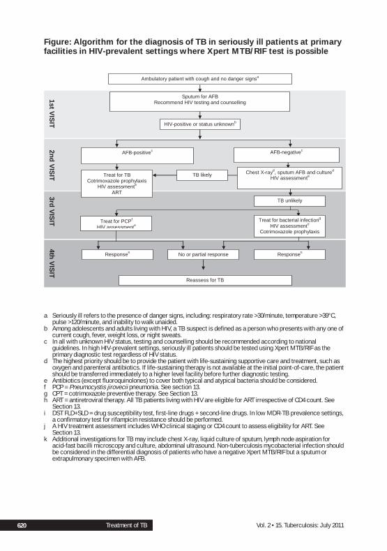
620 Vol. 2 • 15. Tuberculosis: July 2011620620
Figure: Algorithm for the diagnosis of TB in seriously ill patients at primary facilities in HIV-prevalent settings where Xpert MTB/RIF test is possible
Treat for TB Cotrimoxazole prophylaxis
HIV assessmente
ART
TB likely Chest X-rayd, sputum AFB and cultured HIV assessmente
TB unlikely
Responseh Responseh No or partial response
Reassess for TB
Treat for bacterial infectiong HIV assessmente
Cotrimoxazole prophylaxis
Treat for PCPf
HIV assessmente
Ambulatory patient with cough and no danger signsa
HIV-positive or status unknownb
Sputum for AFB Recommend HIV testing and counselling
AFB-positivec AFB-negativec
4th V
ISIT
3rd
VIS
IT
2nd
VIS
IT
1st VIS
IT
a Seriously ill refers to the presence of danger signs, including: respiratory rate >30/minute, temperature >39°C, pulse >120/minute, and inability to walk unaided.
b Among adolescents and adults living with HIV, a TB suspect is defi ned as a person who presents with any one of current cough, fever, weight loss, or night sweats.
c In all with unknown HIV status, testing and counselling should be recommended according to national guidelines. In high HIV-prevalent settings, seriously ill patients should be tested using Xpert MTB/RIF as the primary diagnostic test regardless of HIV status.
d The highest priority should be to provide the patient with life-sustaining supportive care and treatment, such as oxygen and parenteral antibiotics. If life-sustaining therapy is not available at the initial point-of-care, the patient should be transferred immediately to a higher level facility before further diagnostic testing.
e Antibiotics (except fl uoroquinolones) to cover both typical and atypical bacteria should be considered.f PCP = Pneumocystis jirovecii pneumonia. See section 13. g CPT = cotrimoxazole preventive therapy. See Section 13. h ART = antiretroviral therapy. All TB patients living with HIV are eligible for ART irrespective of CD4 count. See
Section 13. i DST FLD+SLD = drug susceptibility test, fi rst-line drugs + second-line drugs. In low MDR-TB prevalence settings,
a confi rmatory test for rifampicin resistance should be performed. j A HIV treatment assessment includes WHO clinical staging or CD4 count to assess eligibility for ART. See
Section 13. k Additional investigations for TB may include chest X-ray, liquid culture of sputum, lymph node aspiration for
acid-fast bacilli microscopy and culture, abdominal ultrasound. Non-tuberculosis mycobacterial infection should be considered in the differential diagnosis of patients who have a negative Xpert MTB/RIF but a sputum or extrapulmonary specimen with AFB.
Treatment of TB
FINAL Vol. 2 CH 9-21.indd 620FINAL Vol. 2 CH 9-21.indd 620 29/06/2012 15:1629/06/2012 15:16

621Vol. 2 • 15. Tuberculosis: July 2011 621621
Figure: Algorithm for the diagnosis of TB in seriously ill patients in HIV-prevalent settings at fi rst-level facilities where Xpert MTB/RIF test is not available
Referral to higher level facility
Reassess for other HIV-
related disease
Start TB treatment and cotrimoxazole prophylaxis
Complete antibiotics ART
Reassess for TBe
Start TB treatment Complete antibiotics
Refer for HIV and TB care
Immediate referral not possible
HIV-positive (or status unknowna) patient with danger signs and suspected of having TBb
No TB Sputum positive, culture positive, suggestive X-ray
or TB likely
One or more smear AFB positive
2 smears AFB negative
TB unlikely
Improvement after 3–5 days
No improvement after 3–5 days
Parenteral antibiotic treatmentc Sputum for AFB and culture
Chest X-ray
Parenteral antibiotic treatmentc Sputum for AFB and culture
Recommend HIV testing if not done Consider treatment for PCP d
a When HIV testing is not possible, classifying a patient as HIV-infected depends on clinical assessment in accordance with national guideline recommendations.
b Danger signs include respiratory rate more than 30/minute, fever more than 39°C, pulse more than 120/minute, and inability to walk unaided.
c Antibiotics (except fl uoroquinolones) to cover both typical and atypical bacteria should be considered.d Pneumocystis jirovecii pneumonia. See Section 13. e Assessment includes HIV clinical staging, CD4 count if available, ART, and HIV care.
Treatment of TB
FINAL Vol. 2 CH 9-21.indd 621FINAL Vol. 2 CH 9-21.indd 621 29/06/2012 15:1629/06/2012 15:16

622 Vol. 2 • 15. Tuberculosis: July 2011622622
At the district hospital, patients presenting with severe illness will be identifi ed during Quick Check as having emergency signs of airway, breathing, circulation, and consciousness, and emergency treatments should be given (see Quick Check). After emergency interventions, urgent treatments will be given based on the most likely or most life-threatening diagnosis (see Section 3.0). For patients with severe respiratory distress, shock, or altered consciousness who also have fever, treatment will be empirical antibiotics for pneumonia, septic shock, or meningitis, respectively.• If a PLHIV (or status unknown in high HIV-prevalent settings) has suspected
septic shock, TB must be considered, especially if there is malnutrition and weight loss. See Section 3.1.5 for guidance on management of suspected septic shock. Perform all appropriate TB investigations and HIV testing in severely ill patients (see Section 15.2). Consider early empirical TB treatment in critically ill PLHIV if there is a high suspicion for disseminated disease causing shock, based on suggestive chest X-ray or clinical judgment. This may mean simultaneous treatment for TB and bacterial infection.
• If a PLHIV (or status unknown in high HIV prevalent settings) has suspected severe pneumonia, then TB must be considered. See Section 3.2.3 for guidance on management of suspected severe pneumonia. Perform all appropriate TB investigations and recommend HIV testing and counselling (see Section 15.2). Consider early empirical treatment for TB in critically ill PLHIV with severe pneumonia, based on suggestive X-ray or clinical judgement. This may mean simultaneous treatment for bacterial pneumonia, PCP, and TB.
• Consult senior clinician.
Differential diagnosis of PTB in PLHIV
In a HIV-positive patient consider the following possible diagnoses:• Acute bacterial pneumonia: Common in HIV-positive patients and often has a
short history of onset. • PCP is very diffi cult to distinguish from PTB in a severely ill patient with
advanced HIV infection.
Treatment of TB
FINAL Vol. 2 CH 9-21.indd 622FINAL Vol. 2 CH 9-21.indd 622 29/06/2012 15:1629/06/2012 15:16

623Vol. 2 • 15. Tuberculosis: July 2011 623623
DDx: PTB in PLHIV by stage of immunosuppression
Stage of HIV-infection
HIV clinical stage (1–2)CD4 >200
HIV clinical stage (3–4)CD4 <200
Pulmonary TB
Clinical picture Cough Productive sputumWeight loss, with or without fever, night sweats, breathlessness, chest pain
Dry cough Weight loss and feverTB should be excluded in any patient presenting with cough, fever, weight loss, or night sweats
X-ray appearance Upper lobe infi ltrates Cavitation Nodular or patchy shadowsMight not have abnormal fi ndings
Lower lobe infi ltrates, no cavitationOften mediastinal lymphadenopathy or pleural effusionSometimes miliary or interstitial pneumoniaMight not have abnormal fi ndings
Sputum smear for AFBor newer WHO-approved method such as Xpert MTB/RIF test
AFB often positive
Sputum positive for Xpert testing
AFB often negative
Sputum positive for Xpert testing
Pneumocystis pneumonia
Clinical picture Unlikely FeverDry coughDyspnoea (progressive and exertional)Subacute and insidious onset
X-ray appearance Bilateral diffuse infi ltrates (ground glass appearance)May be normalMay have fi ndings of spontaneous pneumothorax
Laboratory Not useful
Acute bacterial pneumonia
Clinical picture Productive coughHigh feverPatient appears illShort history of onset
Productive coughHigh feverPatient appears illShort history of onset
X-ray appearance Lobar infi ltrate but may be less apparentMight be negative
Lobar infi ltrateMight be negative
Sputum smear or newer WHO-approved method such as Xpert MTB/RIF test
Gram stain may be positive May also be AFB-positiveXpert MTB/RIF testing negative
Xpert MTB/RIF testing negative
TB in pregnant and postpartum women
• Pregnant and postpartum women are at risk of developing TB.• The symptoms of TB are the same in pregnant and postpartum women
compared with non-pregnant women. • All HIV-positive pregnant women should be screened for TB at each visit.
Treatment of TB
FINAL Vol. 2 CH 9-21.indd 623FINAL Vol. 2 CH 9-21.indd 623 29/06/2012 15:1629/06/2012 15:16

624 Vol. 2 • 15. Tuberculosis: July 2011624624
15.3 Treatment of TB
Standardized TB treatment regimens
Standardized treatment means that all patients in a defi ned group receive the same treatment regimen. Standard regimens have the advantage of reducing the risk of emergence of drug resistance. These regimens facilitate estimates of drug needs, procurement, distribution and monitoring, support regular drug supply when patients move from one area to another, and make outcome evaluation convenient and comparable across different facilities. It is important that clinicians follow the national standardized TB treatment regimens.
Good clinical practice
• TB treatment regimen should be based on recommendations of national TB guidelines.
• Once started, a full course of TB treatment is indicated for all defi nite cases of TB and those started based on a clinician’s decision.
Standard code for TB treatment regimens
A standard code for TB treatment regimens includes a number before a phase that is the duration of that phase in months. Letters in parenthesis indicate fi xed-dose combinations (FDCs) of those drugs. A subscript number (e.g. 3) after a letter or letters in parentheses indicates the number of doses of that drug per week. If there is no subscript number, treatment is daily (or for 6 days per week, excluding, for instance, Sunday).
For example, 2(HRZE)/4(HR): this regimen uses two FDCs, each in separate parenthesis. In the intensive phase of 2 months (i.e. the number before the fi rst parenthesis), each day the patient would take an FDC tablet of isoniazid, rifampicin, pyrazinamide, and ethambutol (i.e. the drugs in the fi rst parentheses). In the continuation phase, the patient would take FDCs of isoniazid and rifampicin (i.e. the drug in the second parentheses) daily for 4 months (i.e. the number before the second parenthesis).
First-line anti-tuberculosis drugs
A TB treatment regimen consists of 2 phases – an intensive phase and a continuation phase. Treatment for PTB should be a regimen containing 6 months of rifampicin.
Whenever feasible, patients with PTB should receive treatment daily throughout the course of therapy. The alternative recommendations can be used as long as the patient is receiving directly observed therapy of every dose, and is NOT living with HIV or living in a HIV-prevalent setting. See the Table: Standard anti-TB regimen and dosing frequency in new TB cases on the next page.
Treatment of TB
FINAL Vol. 2 CH 9-21.indd 624FINAL Vol. 2 CH 9-21.indd 624 29/06/2012 15:1629/06/2012 15:16

625Vol. 2 • 15. Tuberculosis: July 2011 625625
Table: Standard anti-TB regimen and dosing frequency in new TB cases1
Regimen and dosing frequency for new TB patients: 2HRZE/4HR
Comment
Intensive phase2 months of HRZE
Continuation phase4 months of HR
Daily Daily Optimal
Daily 3 times a week Acceptable alternative for any new TB patient receiving directly observed therapy.
3 times a week 3 times a week Acceptable alternative if the patient is receiving directly observed therapy and is NOT living with HIV or from a HIV-prevalent setting.
H= isoniazid R = rifampicin Z = pyrazinamide E = ethambutol S = streptomycinWHO no longer recommends omission of ethambutol during the intensive phase of treatment for patients with non-cavitary, smear-negative PTB or EPTB who are HIV-negative.
Table: Dose of fi rst-line anti-tuberculosis drugs for adults1
Drug
Recommended dose
Daily 3 times per week
Dose and range(mg/kg body weight) Maximum (mg)
Dose and range(mg/kg body weight) Daily maximum (mg)
isoniazid 5 (4–6) 300 10 (8–12) 900
rifampicin 10 (8–12) 600 10 (8–12) 600
pyrazinamide 25 (20–30) — 35 (30–40) —
ethambutol 15 (15–20) — 30 (25–35) —
streptomycin* 15 (12–18) — 15 (12–18) 1000
* Patients over 60 years of age may not be able to tolerate more than 500–750 mg daily, so some guidelines recommend reduction of the dose to 10 mg/kg/day in patients in this age group. Patients weighing less than 50 kg may not tolerate doses above 500–750 mg daily.6
6 Essential Medicines List and WHO Model Formulary. WHO, 2008. Available at http://www.who.int/selection_medicines/list/en/
Treatment of TB
FINAL Vol. 2 CH 9-21.indd 625FINAL Vol. 2 CH 9-21.indd 625 29/06/2012 15:1629/06/2012 15:16

626 Vol. 2 • 15. Tuberculosis: July 2011626626
Table: Registration group by outcome of most recent TB treatment1
Registration group(any anatomical site) Bacteriology Outcome of most recent prior treatment
New + or -
Previously treatedRelapse +
Cured
Treatment completed
Failure + Treatment failed
Default + Defaulted
Transfer in: A patient who has been transferred from another district to continue treatment.
+ or - Still on treatment
Other + or - All cases that do not fi t the above defi nitions: • not known whether or not previously treated• previously treated but outcome is unknown• returned to treatment with AFB smear-
negative PTB or bacteriologically negative EPTB
+ bacteriology indicates positive AFB smear, culture, or other newer means of identifying M. tuberculosis.- bacteriology indicates that any specimens tested were negative.
Category of patient for registrationIn order to identify patients at increased risk of acquired drug resistance, and to recommend appropriate treatment, defi ne a case based on whether or not the patient has previously received TB treatment.
TB treatment in special situations
The next table summarizes special situations that might require consideration during TB treatment.
Rifampicin is a strong inducer of the cytochrome P450 (a liver enzyme), and lowers the plasma concentrations of boosted protease inhibitors (bPIs). All bPIs, at standard doses, are contraindicated with rifampicin. See Section 13.10 for TB/HIV co-management.
Treatment of TB
FINAL Vol. 2 CH 9-21.indd 626FINAL Vol. 2 CH 9-21.indd 626 29/06/2012 15:1629/06/2012 15:16

627Vol. 2 • 15. Tuberculosis: July 2011 627627
Table: Anti-TB treatment regimens in special situations
Conditions Special considerations
Pregnancy Avoid streptomycin during pregnancy. Streptomycin is ototoxic to the fetus.
Assess women of childbearing age for current or planned pregnancy before starting TB treatment. Advise pregnant women with TB that successful treatment is important for their own health and the health of their baby. Support and monitor adherence to treatment.
Breastfeeding Give a full course of TB treatment to the mother. Timely and proper TB treatment of the mother is important to reduce risks of TB transmission to the infant.
For the infant, offer INH prophylaxis for at least 3 months beyond the time the mother is considered non-infectious. BCG vaccination should be given at completion of course of isoniazid preventive therapy.
Women on oral contraceptives
Rifampicin can decrease effi cacy of oral contraceptives.
Advise her to use another contraceptive while taking rifampicin..
Liver disorder Give the usual regimen for patients with chronic viral hepatitis, past acute hepatitis, and excessive alcohol consumption.
Monitor for any hepatotoxic reactions.
Advanced liver disease. Obtain liver function test (LFT) at the start of treatment, if possible. If ALT level is more than 3 times the normal level, consider alternative TB treatment regimens per your national guideline recommendations.
Renal failure and severe renal insuffi ciency
Avoid streptomycin. Adjust dosage of ethambutol and pyrazinamide.
Give pyridoxine for patients with severe renal insuffi ciency.
Empirical treatment in EPTB
Culture, biopsy or other investigations (such as ultrasound, CT scan) may not readily be available in most resource-limited settings. With the exception of lymph node TB, which usually can be confi rmed through fi ne needle aspiration, most patients with EPTB are managed without bacteriological or histological confi rmation, based on the decision by a clinician to treat with a full course of TB treatment.
TB treatment should be started as soon as other common conditions have been excluded.
Do not delay treatment until culture or other investigations are available, especially in patients suspected of TB pericarditis, TB meningitis, or disseminated TB, and in HIV-positive patients.
Adjuvant corticosteroids
Corticosteroids are recommended for HIV-negative patients with TB meningitis. Dose: 1 mg/kg/day, to be slowly tapered down over 4–6 weeks.
Treatment of TB
FINAL Vol. 2 CH 9-21.indd 627FINAL Vol. 2 CH 9-21.indd 627 29/06/2012 15:1629/06/2012 15:16

628 Vol. 2 • 15. Tuberculosis: July 2011628628
15.4 Monitor TB treatment
Monitor response to TB treatment
Monitor all patients on TB treatment to assess their response to treatment. Link with existing community DOTS mechanisms – confi rm that the medication is being taken as instructed, by direct observation of each dose.• Monitor the patient’s weight. • Clinical improvement should be seen within 1 month of initiation of TB
treatment. If this is not attained, review the case, check adherence and reconsider the diagnosis.
• For smear-positive pulmonary TB patients, monitor sputum smear microscopy based on national guideline recommendations. Molecular tests, including Xpert MTB/RIF, are not suitable for patient monitoring as these tests also detect DNA from non-viable bacilli.
• There is a higher recurrence rate of TB in HIV-positive than in HIV-negative TB patients. Monitor treatment response in all patients on TB treatment.
• In patients with EPTB, response to treatment can only be monitored clinically. As in cases of smear-negative PTB, the weight of the patient is important to monitor response to treatment.
• Monitor and support treatment adherence.
Manage drug side-effects
Few patients on TB treatment experience adverse drug effects. Monitor patients clinically so that adverse effects can be detected early and managed promptly. Routine laboratory monitoring is not necessary. Inform patients about possible side-effects, and what to do if they experience them.
Isoniazid-induced peripheral neuropathy usually presents as numbness, tingling or burning sensation of the feet. It more commonly occurs in pregnant women and in persons with one of the following: • HIV infection• alcohol abuse• malnutrition• diabetes• chronic liver disease• renal failure.
Give pyridoxine 10 mg daily, with anti-TB drugs, or as per national TB guideline recommendations for these groups of patients.
Monitor TB treatment
FINAL Vol. 2 CH 9-21.indd 628FINAL Vol. 2 CH 9-21.indd 628 29/06/2012 15:1629/06/2012 15:16

629Vol. 2 • 15. Tuberculosis: July 2011 629629
Table: Symptom-based approach to managing side-effects of anti-TB drugs
Side-effects Drug(s) probably responsible
Management
Major Stop responsible drug(s), manage the condition
Skin rash with or without itching
S, H, R, Z Stop anti-TB drugs. If itching without rash, try antihistamine, avoid drying of skin, observe the patient closely and continue anti-TB treatment. If patient has itching with rash, stop all anti-TB drugs and consider to re-introduce one by one per your country TB guideline recommendations.
Deafness (no wax on otoscopy)
streptomycin Stop streptomycin
Dizziness (vertigo and nystagmus)
streptomycin Stop streptomycin
Jaundice (other causes excluded), hepatitis
H, Z, R Stop anti-TB drugs
Confusion (suspect drug-induced acute liver failure if jaundice present)
Most anti-TB drugs Stop anti-TB drugs
Visual impairment (other causes excluded)
ethambutol Stop ethambutol
Shock, purpura, acute renal failure
rifampicin Stop rifampicin
Decreased urine output streptomycin Stop streptomycin
Minor Continue anti-TB drugs; check drug doses
Anorexia, nausea, abdominal pain
pyrazinamide, rifampicin, isoniazid
Give drugs with small meals or just before bedtime. Advise patient to swallow pills slowly with small sips of water. If symptoms persist or worsen, or there is protracted vomiting or signs of bleeding, consider the side-effect to be major and refer to a clinician urgently.
Joint pains pyrazinamide Aspirin or nonsteroidal anti-infl ammatory drug, or paracetamol
Burning, numbness or tingling sensation in the hands or feet
isoniazid Pyridoxine 50–75 mg daily
Drowsiness isoniazid Reassurance. Give drugs before bedtime.
Orange/red urine rifampicin Reassurance. Inform the patient this may happen when starting treatment and is normal.
Flu syndrome (fever, chills, malaise, headache, bone pain)
Intermittent dosing of rifampicin
Change from intermittent to daily rifampicin.
Monitor TB treatment
FINAL Vol. 2 CH 9-21.indd 629FINAL Vol. 2 CH 9-21.indd 629 29/06/2012 15:1629/06/2012 15:16

630 Vol. 2 • 15. Tuberculosis: July 2011630630
Determine TB treatment outcomes
At the end of the treatment course for each patient with sputum smear-positive PTB, the treatment outcome should be recorded in the district TB register. The following table illustrates the standard defi nitions of TB treatment outcomes. Refer to your national TB guidelines.
Table: Standard defi nitions of TB treatment outcomes
Outcomea Defi nitiona
Cure Sputum smear or culture positive at the beginning of TB treatment, but which was smear or culture negative in the last month of treatment and on at least 1 previous occasion.
Treatment completed
Completed treatment but does not have a negative sputum smear or culture result in the last month of treatment and on at least 1 previous occasion.b
Treatment failure Sputum smear or culture is positive at 5 months or later during treatment. Also included are MDR patients at any point of time during the treatment, whether they are smear negative or positive.
Died Dies for any reason during the course of treatment.
Default Treatment was interrupted for 2 consecutive months or more.
Transfer out Transferred to another unit and for whom the treatment outcome is not known.
Treatment successc A sum of cured and completed treatment.c
a Applies to pulmonary smear-positive and smear-negative patients, and to patients with EPTB.b The sputum examination may not have been done, or the results may not be available. c For smear or culture positive patients only.
Good clinical practice Follow national TB guideline recommendations.
15.5 Drug-resistant TB (DR-TB)7
DR-TB is diagnosed if sputum culture shows in vitro growth of M. tuberculosis in the presence of one or more anti-TB drugs. Prompt diagnosis and effective treatment of DR-TB is important to reduce the risks of further spread and for a good treatment outcome. Multiple factors related both to the health-care delivery system and to the patient play a role in the emergence of a DR-TB strain, of which previous TB treatment is a strong determinant.
MDR-TB is diagnosed in a patient with M. tuberculosis who is confi rmed to have resistance to both rifampicin and isoniazid identifi ed from a clinical specimen, either by conventional DST or by a newer diagnostic method recommended by WHO, such as line probe assay. Rifampicin resistance is a reliable proxy for MDR-TB in high burden settings and its detection by Xpert MTB/RIF is usually enough to start a patient on a second-line TB regimen. These patients should provide an additional sputum specimen for conventional culture and DST against other fi rst- and second-line anti-TB drugs, and their treatment should be adjusted based on
7 Guidelines for the programmatic management of drug-resistant tuberculosis. WHO, 2011. Available at http://whqlibdoc.who.int/publications/2011/9789241501583_eng.pdf
Drug-resistant TB
FINAL Vol. 2 CH 9-21.indd 630FINAL Vol. 2 CH 9-21.indd 630 29/06/2012 15:1629/06/2012 15:16

631Vol. 2 • 15. Tuberculosis: July 2011 631631
the results of further DST results. However, in low MDR-TB-prevalence settings, testing new TB cases not at risk for MDR-TB will result in a low positive predictive value for rifampicin resistance, and will require that rifampicin resistance detected by Xpert MTB/RIF be confi rmed fi rst by conventional DST or line probe assay.
• Rapid DST of isoniazid and rifampicin or of rifampicin alone is recommended at the time of diagnosis where resources permit.
• Where resources are limited, the following patients can be prioritized for rapid DST for isoniazid and rifampicin or rifampicin alone at the time of diagnosis: ° those who fail re-treatment; ° those exposed to a known DR-TB case; ° those who fail antituberculosis treatment in the private sector; ° those who remain sputum smear-positive at month 2 or 3; ° those exposed in institutions that have DR-TB outbreaks or a high DR-TB
prevalence; ° those resident in areas with high DR-TB prevalence; ° those with a history of using anti-TB drugs of poor or unknown quality; ° those on treatment in programmes that operate poorly (especially
programmes with recent or frequent drug stock-outs); ° those with certain co-morbidities, e.g. malabsorption, substance
dependency disorders.
If MDR-TB is highly prevalent in PLHIV, they should be screened for MDR-TB with a DST if diagnosed with active TB.
Patients at high risk of MDR-TB are those:• with probable treatment failure or whose fi rst-line treatment has failed;• who developed TB after contact with a documented MDR-TB case; • who are relapsing or returning from their second or subsequent course of
treatment.
Patients returning after defaulting or relapsing from their fi rst course of treatment have medium risk of MDR-TB.
Table: Summary of recommended TB treatment regimens
Standard treatment
New patients 2HRZE/4HR
Retreatment with fi rst-line drugs 2HRZES/1HRZE/5HRE
MDR- drug-susceptibility testingTB Follow national recommendation for treatment of MDR-TB
MDR-TB treatment regimen
Before or at the start of treatment, all previously treated TB patients and patients suspected of having MDR-TB should have sputum, as well as appropriate specimens for EPTB, obtained for culture and DST. Referral of the patients or transportation of specimens might be needed if culture and DST are not available in the hospital.
Rapid, molecular-based DST results are obtainable in 1 to 2 days, and will guide choice of treatment. This minimizes delay before effective treatment. Where rapid
Drug-resistant TB
FINAL Vol. 2 CH 9-21.indd 631FINAL Vol. 2 CH 9-21.indd 631 29/06/2012 15:1629/06/2012 15:16

632 Vol. 2 • 15. Tuberculosis: July 2011632632
molecular-based DST is not available, the choice of regimen should be guided by conventional specimen culture. This necessitates initiation of empirical treatment while waiting for the result.
Refer to national guideline recommendations for second-line anti-TB regimens.
Once the diagnosis of MDR-TB is confi rmed (through culture and DST), patients can be treated with a standardized or individualized regimen, depending on the TB and MDR-TB treatment guideline recommendations of the country.
MDR-TB and HIV infection
There is a high rate of mortality in patients coinfected with MDR-TB and HIV. Early diagnosis of MDR-TB and HIV, prompt treatment with adequate regimens, good patient support, and strong infection control measures are all essential components in the management of MDR-TB in HIV-positive patients.
Good clinical practice
• MDR-TB contact investigation should be given high priority• Close contacts of MDR-TB patients should receive careful clinical follow-up
Manage common problems in patients with MDR-TB8
Common problems
Cough ordiffi cult breathing
Cough due to TB may not improve for several months after starting MDR-TB therapy.Patient may wheeze as lungs scar during healing.Exclude superimposed bronchitis or pneumonia, consider PCP if patient is HIV-positive.• If patient has recently started ART, consider
IRIS.
See Section 10.6 for differential diagnosis.
Haemoptysis Dangerous if large (patients may die of asphyxiation, not blood loss) or if TB has not been treated.• No special treatment for haemoptysis except
for TB treatment. Cough suppressant (e.g. codeine) may decrease the frequency of episodes.
• May continue for months after starting MDR-TB therapy. Chronic haemoptysis with small amounts of blood is not dangerous, and will resolve slowly as the lungs heal.
Persistent fever Fever due to TB may not improve for several months after starting MDR-TB therapy. Consider common causes of fever if patient has recently started ART. New fevers may be a sign of IRIS.
Prevent dehydration: increase fl uid intake.
Paracetamol, but avoid excessive doses.Tepid sponging if patient likes it.
8 Management of MDR-TB: A fi eld guide. WHO and Partners in Health, 2009. Available at http://whqlibdoc.who.int/publications/2009/9789241547765_eng.pdf
Drug-resistant TB
FINAL Vol. 2 CH 9-21.indd 632FINAL Vol. 2 CH 9-21.indd 632 29/06/2012 15:1629/06/2012 15:16

633Vol. 2 • 15. Tuberculosis: July 2011 633633
Persistent nausea or vomiting
Usually caused by ethionamide or para-aminosalicylic acid (PAS).Nausea due to AZT is usually self-limiting.Ethionamide has a direct toxic effect on the stomach lining and nausea is usually immediate. PAS toxicity is usually more delayed in onset: both have a “dose-dependent” effect.
See Section 10.7 for differential diagnosis and management of nausea and vomiting.
Persistent diarrhoea
May be caused by PAS.If HIV-positive, consider chronic infectious diarrhoea and treat empirically.
Increase fl uid intake to prevent dehydration. Give ORS if large volume diarrhoea.Give constipating drug unless blood in stool or fever or elderly.Advise on special care of the rectal area.Advise on a supportive diet for patients with diarrhoea.
Peripheral neuropathy
Many TB and HIV drugs can cause burning or tingling sensations.
See Section 10.10a for differential diagnosis.
Severe depression, anxiety or psychosis
Usually due to terizidone or cycloserine.If due to EFV, consider switching to NVP.Symptoms usually improve when the dose of cycloserine is decreased.Stop cycloserine immediately if patient is suicidal or psychotic.Decreasing the dose of cycloserine may increase the risk of treatment failure.
See Section 10.11 for differential diagnosis.
Hypokalaemia (low potassium)
The injectable drugs, particularly capreomycin, can cause hypokalaemia. This is due to their direct effect on the kidneys. The kidneys start to excrete large amounts of electrolytes, the most important of which is potassium.
See Section 5.2 for differential diagnosis.
Drug-resistant TB
FINAL Vol. 2 CH 9-21.indd 633FINAL Vol. 2 CH 9-21.indd 633 29/06/2012 15:1629/06/2012 15:16

FINAL Vol. 2 CH 9-21.indd 634FINAL Vol. 2 CH 9-21.indd 634 29/06/2012 15:1629/06/2012 15:16

16. Assessment and therapy for alcohol use disorders
Table of contents
16.1 Defi nitions of hazardous and harmful alcohol use and alcohol dependence . . . . . . 63716.2 Assessment . . . . . . . . . . . . . . . . . . . . . . . . . . . . . . . . . . . . . . . . . . . . . 638
Alcohol Use Disorders Identifi cation Test (AUDIT) . . . . . . . . . . . . . . . . . . . . 639Brief assessment using AUDIT . . . . . . . . . . . . . . . . . . . . . . . . . . . . . . . . 639Further assessment . . . . . . . . . . . . . . . . . . . . . . . . . . . . . . . . . . . . . . 641Laboratory fi ndings suggestive of harmful alcohol use or alcohol dependence . . . 642
16.3 Classify, then advise and treat the patient . . . . . . . . . . . . . . . . . . . . . . . . . . 643If the AUDIT score is 8–19 . . . . . . . . . . . . . . . . . . . . . . . . . . . . . . . . . . . 643If the AUDIT score is ≥20 . . . . . . . . . . . . . . . . . . . . . . . . . . . . . . . . . . . 643Special advice for pregnant patients . . . . . . . . . . . . . . . . . . . . . . . . . . . . 643Summary of interventions by classifi cation of alcohol use disorder . . . . . . . . . . 644
16.4 Brief interventions for those with hazardous or harmful drinking . . . . . . . . . . . . . 644FLAGS approach . . . . . . . . . . . . . . . . . . . . . . . . . . . . . . . . . . . . . . . . 645
16.5 Treatment and care for those with alcohol dependence . . . . . . . . . . . . . . . . . . 646Withdrawal management . . . . . . . . . . . . . . . . . . . . . . . . . . . . . . . . . . . 647Relapse prevention after withdrawal from alcohol . . . . . . . . . . . . . . . . . . . . 647Psychosocial support . . . . . . . . . . . . . . . . . . . . . . . . . . . . . . . . . . . . . 648
FINAL Vol. 2 CH 9-21.indd 635FINAL Vol. 2 CH 9-21.indd 635 29/06/2012 15:1629/06/2012 15:16

FINAL Vol. 2 CH 9-21.indd 636FINAL Vol. 2 CH 9-21.indd 636 29/06/2012 15:1629/06/2012 15:16

637Vol. 2 • 16. Assessment and therapy for alcohol use disorders: July 2011 637
16. Assessment and therapy for alcohol use disorders 1
This Section outlines how district-level clinicians (and their teams) can provide services to reduce the harm caused by alcohol. Most of these services can be provided at the primary-care level and by health workers who are supervised by the district clinician. For most patients management consists of a brief assessment and intervention. A 5-minute intervention session, described below, can lead to a substantial reduction in a person’s alcohol use. More intensive services may be required for alcohol-dependent patients.
Alcohol use varies widely from country to country and from person to person. In some countries alcohol is prohibited or is rarely consumed; in others a high proportion of the adult population drinks alcohol to some extent. There is no level of alcohol consumption that is without any health risk. Some patterns and levels of alcohol consumption are associated with higher health risks than others.
Alcohol use disorders are a major cause of poor health and social problems. They cause or contribute to a wide range of acute and chronic physical and mental disorders. In many countries a high proportion of people attending primary and district hospitals and clinics use alcohol hazardously or harmfully; and yet alcohol use disorders often are not identifi ed or treated. Patients may even be unaware of the role of alcohol in their problems. Screening for alcohol use disorders and offering advice and help accordingly can assist patients to reduce or cease alcohol consumption and reduce alcohol related harm.
Management of acute alcohol withdrawal and alcohol intoxication are described in Section 3.7.
16.1 Defi nitions of hazardous and harmful alcohol use and alcohol dependence
Hazardous alcohol consumption is a pattern of alcohol use that puts a person at risk of harmful consequences, such as injuries, mental disorders, liver and cardiovascular diseases, problems in relationships and at work, and other problems.
Hazardous alcohol use includes but is not limited to:• having on a daily basis more than 4 drinks (men) or 2 drinks (women);• having more than 5 drinks on a single occasion;• drinking on more than 4 days per week• using alcohol when
° driving or operating machinery ° pregnant or breastfeeding ° taking medications that react with alcohol ° there are medical conditions present that are made worse by alcohol.
1 mhGAP Intervention Guide for Mental, Neurological and Substance Use Disorders in Non-specialized Health Settings. WHO and mhGAP Evidence Resource Centre, 2010. Available at http://www.who.int/mental_health/evidence/mhGAP_intervention_guide/en/index.html. The mhGAP Guidelines and the mhGAP-IG will be reviewed and updated in 5 years. Any revision and update before that will be made to the online version of the document.
Assessment
FINAL Vol. 2 CH 9-21.indd 637FINAL Vol. 2 CH 9-21.indd 637 29/06/2012 15:1629/06/2012 15:16

638 Vol. 2 • 16. Assessment and therapy for alcohol use disorders: July 2011638
Harmful alcohol use is a pattern of alcohol consumption that is causing damage to health. The degree of hazard and the degree of harm vary not only according to the amount of alcohol consumed but also according to the pattern of consumption and individual factors such as the age, gender, body weight, and medical status of the individual.
Alcohol intoxication is associated with impaired thinking and reactions, which put that person at risk of injuries and other harms. Alcohol has disinhibiting effects that can affect decisions about having sex as well as affecting skills for negotiating condom use and their correct use. Together, these impairments can result in unsafe sex and condom use accidents, with resulting transmission of HIV and other sexually-transmitted illnesses (STIs). Acute problems, such as trauma or unsafe sex, may occur after a session of alcohol consumption. Chronic medical and social problems may be induced or exacerbated by repeated alcohol use over a period of time.
Even in amounts that do not cause acute intoxication, alcohol use can still cause long-term harm to health. In the long term, harmful and hazardous alcohol use can result in a range of cancers, injuries, and infections and cause serious diseases in nearly every organ system in the body (e.g. liver cirrhosis, pancreatitis and neurological diseases) as well as social problems such as domestic violence, unemployment ,and occupational diffi culties.
Harmful use of alcohol is a pattern of alcohol consumption that is causing actual physical or mental problems but has not reached the point of being considered alcohol dependence. When harmful use of alcohol results in alcohol dependence, the latter diagnosis is made in preference.
Alcohol dependence is a disorder with a psychological and physiological drive to consume alcohol, even in the face of serious alcohol-related harm. The diagnosis of alcohol dependence is based on several key symptoms, which include a strong (often overpowering) desire to drink, diffi culties in controlling drinking behaviour, progressive neglect of alternative pleasures or interests, evidence of tolerance to alcohol, and withdrawal features when stopping or trying to reduce drinking. People with alcohol dependence have a repetitive pattern of drinking large amounts of alcohol and fi nd it diffi cult to limit their consumption. Alcohol withdrawal can be a medical emergency (see Section 3.7 Acute alcohol withdrawal).
16.2 Assessment
The earlier that hazardous or harmful drinking is identifi ed or a diagnosis of alcohol dependence is made, the better the response to intervention. A systematic assessment enables detection of hazardous alcohol use, harmful alcohol use, and alcohol dependence syndrome.
In an assessment of substance use, it is important to address the patient in an empathic and non-judgemental way and to be sensitive to the patient’s cultural background and situation. This encourages the patient to be open and to report their substance use accurately. The ability to elicit this information will be diminished if the health worker’s personal views about substance use are communicated in the interaction with a patient, Review of the patient’s records for previous evidence of hazardous drinking or alcohol use disorders can provide valuable information.
Assessment
FINAL Vol. 2 CH 9-21.indd 638FINAL Vol. 2 CH 9-21.indd 638 29/06/2012 15:1629/06/2012 15:16

639Vol. 2 • 16. Assessment and therapy for alcohol use disorders: July 2011 639
Alcohol Use Disorders Identifi cation Test (AUDIT)2
The AUDIT was developed as a simple method of screening for excessive drinking and to assist brief assessment. It can help identify excessive drinking as the cause of the presenting illness. It provides a framework for intervention to help risky drinkers reduce or cease alcohol consumption and thereby avoid the harmful consequences of drinking. The AUDIT also helps to identify alcohol dependence and some specifi c consequences of harmful drinking. Of utmost importance for screening is the fact that people who are not dependent on alcohol may stop or reduce their alcohol consumption with appropriate assistance and effort. The AUDIT is particularly designed for health workers to use in a range of health settings.
Brief assessment using AUDIT (see Figure AUDIT below)
1. Be alert to possible alcohol misuse. Alcohol problems often are missed and untreated. Many common symptoms prompt suspicion of alcohol misuse. These include:• appearing to be under the infl uence of alcohol (e.g. smells of alcohol or
looks intoxicated), tiredness, dyspepsia, anorexia, vomiting, diarrhoea, and headaches;
• accidents at work or home and diffi culties carrying out usual work, school, domestic, or social activities;
• psychological symptoms, such as anxiety and depression and repeated requests for medical certifi cates of absence from work; these may have alcohol as the underlying cause.
Think about alcohol use in every patient.
2. Ask each patient about alcohol use: “How often do you have a drink containing alcohol?” (see AUDIT question 1). If the patient indicates that they do not drink alcohol, then just ask questions 9 and 10 to see if they have had a problem in the past (see AUDIT question 9).
3. If patients drink alcohol, determine the amount and pattern of alcohol use. Ask, “How many drinks containing alcohol do you have on a typical day when you are drinking?” (AUDIT question 2) and “How often do you have 6 or more drinks on one occasion?”(AUDIT question 3). Ask about both commercial (i.e. purchased) alcohol and domestic or illicit alcohol (i.e. home-produced alcohol).
4. If the score for the fi rst 3 AUDIT questions is 3 or more (or if there is any suspicion of alcohol misuse (symptoms or signs possibly due to alcohol), complete the full AUDIT questionnaire. Add up the AUDIT scores. An AUDIT score of 8–12 indicates hazardous or harmful drinking. A score of ≥13 suggests alcohol dependence.
This information is sufficient to decide whether to offer the patient a brief intervention for hazardous or harmful alcohol use.
2 The Alcohol Use Disorders Identifi cation Test: Guidelines for Use in Primary Care, 2nd ed. WHO, 2001. Available at http://whqlibdoc.who.int/hq/2001/WHO_MSD_MSB_01.6a.pdf
Assessment
FINAL Vol. 2 CH 9-21.indd 639FINAL Vol. 2 CH 9-21.indd 639 29/06/2012 15:1629/06/2012 15:16

640 Vol. 2 • 16. Assessment and therapy for alcohol use disorders: July 2011640
Figure: AUDIT − Alcohol Use Disorders Identifi cation Test questionnaire
Please select an answer for each of the 10 questions below and total the scores.
1. How often do you have a drink containing alcohol?
[0] Never [1] Monthly or less
[2] 2–4 timesa month
[3] 2–3 timesa week
[4] 4 or more timesa week
2. How many drinks containing alcohol do you have on a typical day when you are drinking?
[0] 0, 1 or 2 [1] 3 or 4 [2] 5 or 6 [3] 7 to 9 [4] 10 or more
3. How often do you have 6 or more drinks on one occasion?
[0] Never [1] Less than monthly
[2] Monthly [3] Weekly [4] Daily or almost daily
4. How often during the last year have you found it diffi cult to get the thought of alcohol out of your mind?
[0] Never [1] Less than monthly
[2] Monthly [3] Weekly [4] Daily or almostdaily
5. How often during the last year have you found that you were not able to stop drinking once you had started?
[0] Never [1] Less than monthly
[2] Monthly [3] Weekly [4] Daily or almostdaily
6. How often during the last year have you been unable to remember what happened the night before because you were drinking?
[0] Never [1] Less than monthly
[2] Monthly [3] Weekly [4] Daily or almostdaily
7. How often during the last year have you needed a drink fi rst thing in the morning to get yourself over a heavy drinking session?
[0] Never [1] Less than monthly
[2] Monthly [3] Weekly [4] Daily or almostdaily
8. How often during the last year have you had a feeling of guilt or remorse after drinking?
[0] Never [1] Less than monthly
[2] Monthly [3] Weekly [4] Daily or almostdaily
9. Have you or someone else been injured as a result of your drinking?
[0] No [2] Yes, but not in the last year
[4] Yes, during thelast year
10. Has a friend, or relative, or doctor, or other health worker been concerned about your drinking or suggested that you cut down?
[0] No [2] Yes, but not in the last year
[4] Yes, during thelast year
Total score: [ ]Scores from 8–10 suggest hazardous or harmful drinking.Scores of 13 or more suggest the likelihood of alcohol dependence.
Use this AUDIT version in face-to-face interviews. The questionnaire can be self-administered, in which case the score for each response should be omitted.
Assessment
FINAL Vol. 2 CH 9-21.indd 640FINAL Vol. 2 CH 9-21.indd 640 29/06/2012 15:1629/06/2012 15:16

641Vol. 2 • 16. Assessment and therapy for alcohol use disorders: July 2011 641
Further assessment
A more comprehensive assessment of alcohol use and its complications may be necessary in some patients, especially those with suspected alcohol dependence.
Ask the patient about the following:
1. Alcohol use
• the amount of alcohol (in grams, standard drinks or bottles, cans, or other containers) that the person consumes in a session of drinking, or during a day;
• the frequency of drinking, e.g. the number of days per week or month when alcohol is consumed;
• the pattern of drinking;• the duration of drinking overall – in months or years;• the time of their last drink;• the typical drinking context, e.g. at home, in a local gathering, in a club or
licensed premises, in a public place.
2. Alcohol dependence
• diffi culty in controlling the amount of alcohol consumed in an episode of drinking;
• giving up activities or responsibilities in favour of drinking;• increased tolerance to alcohol;• withdrawal symptoms when not drinking;• previous attempts to stop.
3. Alcohol problems
• physical health problems, such as liver disease, neuropathy;• anxiety, depression, or suicide attempts;• social and legal problems from alcohol.
What is a standard drink?1 unit (10 gm alcohol) of alcohol. Show pictures of
common local drinks (for local adaptation)
Other aspects of the assessment
4. Co-existing mental disorder (see Section 10.11 Mental problems)
5. Use of other substances such as tobacco, opiates, or benzodiazepines. Enquire about the use of prescribed medication and adherence to these.
6. Use of alcohol by their partner or family members.
7. Signs of alcohol use disorders and organ damage.
Assessment
FINAL Vol. 2 CH 9-21.indd 641FINAL Vol. 2 CH 9-21.indd 641 29/06/2012 15:1629/06/2012 15:16

642 Vol. 2 • 16. Assessment and therapy for alcohol use disorders: July 2011642
Signs are more common in people with alcohol dependence; there may be no obvious signs in early stages of alcohol misuse. Signs may include:• tremor of the hands (which typically indicates either alcohol withdrawal or
cerebellum injury);• a reddish blush of the face, abnormal appearance of blood vessels in the face
and neck, and changes in the mucous membranes (e.g. conjunctivitis) or oral cavity (e.g. glossitis);
• both the size (number of fi nger breadths below the costal margin) and consistency (normal, fi rm, hard, or very hard) of the liver (assess the liver);
• mental state − examination and mini mental state tests may reveal confusion or short-term memory loss.
Laboratory tests, such as red blood cell macrocytosis and liver function tests (e.g. GGT), may be helpful, especially in the assessment of patients with harmful alcohol consumption or dependence (see below, Table: Laboratory fi ndings suggestive of harmful alcohol use or alcohol dependence).
Table: Laboratory fi ndings suggestive of harmful alcohol use or alcohol dependence
Investigations Results Interpretation of results
Full blood count AnaemiaLeucocytosisLeucopoeniaMacrocytosisThrombocytopenia
GI or other bleedingInfectionsReduced immunityHarmful alcohol use − folate defi ciency or B12 defi ciencyHypersplenism or bone marrow toxicity
Liver function tests Elevated GGTAST/ALT >2Isolated rise in ALT
Harmful alcohol useLikely alcoholic liver diseaseOther forms of liver disease including hepatitis C
Electrolyte tests HypokalaemiaHyponatraemia
Vomiting, diarrhoea, excessive sweating
Serum uric acid Raised Harmful alcohol usePossible gout
Serum magnesium Lowered Harmful alcohol use
Serum amylase/lipase Raised Alcoholic pancreatitis
Cholesterol, triglyceride Raised Harmful alcohol use
Blood alcohol concentration or breathalyser
>0.05 g% Recent alcohol consumptionPerson is at increased risk of a motor vehicle accident
Assessment
FINAL Vol. 2 CH 9-21.indd 642FINAL Vol. 2 CH 9-21.indd 642 29/06/2012 15:1629/06/2012 15:16

643Vol. 2 • 16. Assessment and therapy for alcohol use disorders: July 2011 643
16.3 Classify, then advise and treat the patient
Based on the assessment, decide if the patient has hazardous or harmful alcohol consumption or has alcohol dependence. List any harmful experiences or incidents the person has experienced so far.
If the AUDIT score is 8–19
• The diagnosis is likely to be hazardous or harmful alcohol consumption.• The treatment is an early intervention using the FLAGS (see below) approach. • The goal of treatment is reduced or controlled drinking to reduce risk of
alcohol-related problems.
If the AUDIT score is ≥20
• The diagnosis is likely to be alcohol dependence; the higher the score, the greater the likelihood.
• The next step is to confi rm the diagnosis. Look for evidence of impaired control of alcohol consumption, alcohol being a central part of the person’s life, withdrawal symptoms, and continued drinking despite harm.
• The treatment is withdrawal from alcohol, treatment with an alcohol pharmacotherapy, and counselling to prevent relapse or else referral to a specialist facility where available.
• The goal of treatment is abstinence from alcohol.
Special advice for pregnant patients
Pregnant women and women who are at risk of pregnancy should be strongly advised not to use alcohol. Encourage patients who drink alcohol to limit themselves to low-risk alcohol use or to stop alcohol altogether. If time constraints do not allow defi nitive treatment during a consultation, still provide some advice, even if limited, and arrange a follow-up visit.
Classify then advise and treat
FINAL Vol. 2 CH 9-21.indd 643FINAL Vol. 2 CH 9-21.indd 643 29/06/2012 15:1629/06/2012 15:16

644 Vol. 2 • 16. Assessment and therapy for alcohol use disorders: July 2011644
Table: Summary of interventions by classifi cation of alcohol use disorder
Diagnosis Main effects of problem Intervention required Goal of therapy
Low-risk alcohol consumption
In general, none In general, none Women who are (or may become) pregnant are best advised to abstain from alcohol.
Hazardous alcohol consumption
Risk of harm in the future Brief intervention to reduce alcohol consumptionAdvise women who are (or may become) pregnant to abstain from alcohol.
Reduced or controlled drinking
Harmful alcohol consumption
Physical, mental, and social harms are present.
Brief interventionReduce the effects of harm
Reduced drinking or abstinence from alcohol
Alcohol dependence syndrome
Disabilities and complications:• physical• psychological• social
Detoxifi cationPharmacotherapy Psychological therapies and support groups
Abstinence from alcohol
Alcohol withdrawal syndrome
Hyperactivity after cessation or reduction of drinking; may have seizures
Management of withdrawal Stabilize the patient, relieve symptoms then treat the alcohol dependence.
16.4 Brief interventions for hazardous or harmful drinking
Much can be done at primary care and district level to help people adopt low-risk drinking practices or stop alcohol altogether. A 5-minute session of brief therapy is outlined here. This is particularly useful for people with hazardous or harmful drinking, but the intervention can be adapted for providing initial treatment for people with alcohol dependence.
Brief interventionsBrief interventions are used for people with hazardous or harmful alcohol consumption. This ranges from those who have high-risk drinking patterns to drinkers already experiencing harmful effects of excessive alcohol and to those who would develop dependence without early intervention. Women who engage in low-risk drinking but are pregnant or taking drugs that interact with alcohol also require brief interventions.
In the brief intervention give simple structured advice. Use the FLAGS approach, which is based on an intervention developed by WHO and evaluated in many countries. It has 5 components: Feedback, Listening, Advice, Goals, and Strategies (see below, Figure FLAGS approach). The goal is reduced or controlled drinking. The intervention can be provided in 3−4 minutes, although it may be extended to 10 or even 30 minutes if time allows. This simple, brief therapy can be very effective, and on average results in a 30% reduction in drinking. Always offer the patient the opportunity to receive the therapy on a later occasion or in repeated sessions.
Brief interventions
FINAL Vol. 2 CH 9-21.indd 644FINAL Vol. 2 CH 9-21.indd 644 29/06/2012 15:1629/06/2012 15:16

645Vol. 2 • 16. Assessment and therapy for alcohol use disorders: July 2011 645
Many different members of the district health team can assist in providing these services, with supervision from the district clinician. Good communication skills are necessary for successful implementation of brief interventions. Therefore, make use of health workers who have developed these specifi c skills.
Figure: FLAGS approach
Feedback Provide the AUDIT score and levels of alcohol consumption. Give feedback on problems or harm experienced or likely to be experienced.
If willing to change,
Advise to reduce drinking. Be unequivocal. Highlight the benefits of reducing hazardous or harmful drinking. Use a visual aid. Assist patient to appreciate benefits.
Strategies Suggest strategies and practical approaches to achieve the goal of reduced drinking; offer self help pamphlets and drink diaries.
If not willing to change,
Advise with motivational interviewing techniques.
Listen Listen to patient’s response and what they like or dislike about their drinking. Gauge their readiness to change.
Goals Help to set goals for reduced drinking. Be specific; i.e. advise no more than 2 standard drinks a day with at least 2 alcohol-free days per week, or advise to stop drinking completely.
Brief interventions
FINAL Vol. 2 CH 9-21.indd 645FINAL Vol. 2 CH 9-21.indd 645 29/06/2012 15:1629/06/2012 15:16

646 Vol. 2 • 16. Assessment and therapy for alcohol use disorders: July 2011646
Arrange follow-up visits to assess whether the patient has been able to reduce or stop drinking, to review progress, to renegotiate goals and strategies, and to reinforce, support, and advise. Arrange support through peer and self-help groups.
Alcohol interacts with many medications, and many medications can increase the toxic effects of other medications or of alcohol. Alcohol can change the metabolism of some medications and make them ineffective. Review the medications that a patient is receiving and whether they interact with alcohol. Alcohol misuse lowers adherence to medications. Family members and health workers have an important role in supporting adherence in people with hazardous or harmful drinking.
For those with more established alcohol-related harm (or alcohol dependence), the FLAGS intervention offers an important initial approach that often is successful. People with patterns of harmful alcohol use who are unable to reduce or stop their drinking are showing signs that they may be alcohol dependent and require more intensive treatments. These are described next.
16.5 Treatment and care for those with alcohol dependence
In patients with alcohol dependence, recovery usually will require total abstinence from alcohol. Often, this will alleviate the symptoms and signs of the disease. For this reason, patients with alcohol dependence are best advised to abstain completely from alcohol. Every effort must be made to support and encourage the patient to abstain. Primary health care workers play an important role in this, together with family members. In some cases it may be necessary to refer a patient for residential treatment or for brief periods of hospitalization. These options provide a much greater guarantee of abstinence than the patient’s usual setting.
Patients with more severe alcohol dependence and related harm should be encouraged to seek specialist treatment services. The aim of treatment is to stop drinking completely, join support groups, and obtain the support of family members or other persons in helping them work on their long-term recovery. For many alcohol-related disorders, especially the various forms of brain damage, correction of vitamin and nutritional defi ciencies is vital. Give thiamine and multivitamins to all patients with alcohol dependence or with any physical damage due to alcohol. Maintain patient records and document carefully the physical, mental, and social consequences of the patient’s condition, as well as any coexisting medical or mental problems.
Alcohol dependence is a serious disorder, which often takes a progressive or chronic relapsing course. Many patients with alcohol dependence will get withdrawal symptoms when they stop drinking alcohol, such as anxiety, nausea, headache, hand tremors, sweating, and confusion. Severe alcohol withdrawal can be life-threatening, so patients with alcohol withdrawal need careful management of alcohol cessation (see below, Withdrawal management).
The initial aim of treatment is to encourage the person to stop drinking alcohol. If the person is not ready or willing to take this step, it might be possible to engage services that are available locally, including counselling, family counselling, and psychosocial support matched to specifi c needs. Involvement of self-help and peer groups can help patients considerably. Support from family and friends, a sense that underlying issues can be addressed, and the understanding of other people with substance dependence can help prepare people to stop drinking.
Treatment and care
FINAL Vol. 2 CH 9-21.indd 646FINAL Vol. 2 CH 9-21.indd 646 29/06/2012 15:1629/06/2012 15:16

647Vol. 2 • 16. Assessment and therapy for alcohol use disorders: July 2011 647
Specialist services for alcohol dependence treatment are not readily available in many settings. Therefore, most people with alcohol dependence seen at district level will have to be treated with available resources. Find out what services are available and how to refer to them. Some services still can be offered even where specialist services are unavailable.
Withdrawal management
Withdrawal management is a planned process that allows patients to cease drinking safely and as comfortably as possible. It can be undertaken at home, in specialized centres, or in district hospitals. It is the fi rst step towards a goal of abstinence and recovery. The emphasis is on preventing a withdrawal syndrome from occurring, or, if one does occur, to minimize its effects. Detoxifi cation may require medication, depending on the severity of alcohol withdrawal (see Section 3.7 Alcohol withdrawal). People with severe alcohol dependence (a previous history of alcohol withdrawal, seizures, or delirium) or who have concurrent medical disorders are best managed in a specialist unit or hospital.
Withdrawal from alcohol can be undertaken in a planned (elective) way in the person’s home. In this situation diazepam is typically administered at a dose of 10 mg 4 times daily, (i.e. up to 40 mg/day) for up to 5 days. It is best for the patient to be reviewed daily by medical staff and to be supported by a family member or friend. If patients become sleepy after the medication, then the dose should be reduced and the next dose withheld until withdrawal symptomsre-emerge.
If higher doses are required, it may be necessary to admit the patient to hospital, where higher doses (i.e. up to 100 mg diazepam daily), or sometimes more can be administered in safety. For severe alcohol withdrawal, monitor the patient with the alcohol withdrawal scale (see Section 3.7) and give 10-20 mg diazepam every 1-2 hours until the patient’s withdrawal severity score is low or the patient is lightly sedated. If a patient is requiring 120 mg diazepam or more in a 24 hour period it might be wise to consult with a specialist, if one is available. If patients are known to experience severe alcohol withdrawal, commence diazepam before the patient experiences signifi cant alcohol withdrawal symptoms, aiming for light sedation in the fi rst 24 hours.
Relapse prevention after withdrawal from alcohol
There are several medications useful in the treatment of alcohol dependence that increase the likelihood of the patient maintaining abstinence from alcohol. The principal medications are naltrexone, acamprosate, or disulfi ram.3
Naltrexone suppresses the urge to drink alcohol by blocking opioid receptors. It can be started after withdrawal from alcohol or when the patient is still drinking some alcohol. It is given at a dose of 50 mg/day and then maintained at 50−100 mg/day, usually for 3−12 months. Patients must not have taken any opioid drugs for the previous 5 days. They must be warned that naltrexone will block opioid drugs, in case the patient is likely to need opioid analgesia. Naltrexone should be avoided in pregnant or breastfeeding women and in patients with severe liver disease. In patients with liver disease, and when using the higher dose, monitor liver function tests.
3 To add or substitute disulfi ram for acamprosate or naltrexone, see mhGAP Intervention Guide for Mental, Neurological and Substance Use Disorders in Non-specialized Health Settings1.
Treatment and care
FINAL Vol. 2 CH 9-21.indd 647FINAL Vol. 2 CH 9-21.indd 647 29/06/2012 15:1629/06/2012 15:16

648 Vol. 2 • 16. Assessment and therapy for alcohol use disorders: July 2011648
Acamprosate suppresses the urge to drink alcohol in the alcohol-dependent patient. It is best started immediately after withdrawal from alcohol has been achieved, as a relapse preventive approach. It is given at a dose of 2 tablets (each containing 333 mg acamprosate) 3 times daily (total of 1.998 g), except in people with a body weight of less than 55 kg, when the dose is reduced to 2 tablets twice daily. It has no signifi cant interactions with other drugs. Occasionally, it causes diarrhoea, in which case treatment should be stopped for a few days. It should not be used in patients who have renal impairment. Treatment is usually for 12–18 months.
Naltrexone and acamprosate are not diffi cult to use in primary care. Alcohol dependence is a relapsing condition, and on average these medicines double the time between each relapse. Any relapse should not necessarily be seen as a sign that the treatment has not worked, and the medication should still be considered for use again, although it might be worth trying alternative medications (switching from acamprosate or naltrexone or vice versa) to see if they are more effective. Where a specialist centre is available, the patient can be referred there for use of relapse-prevention medications and other treatments.
Psychosocial support
Supportive outpatient alcohol treatment involves one-to-one counselling. Involvement of family members in recovery can markedly enhance treatment outcomes. The goal of abstinence requires signifi cant changes in many daily activities and way of life. The longer-term recovery of someone from alcohol dependence is based on good initial treatment, support, and self-help. Following that, recovery is based on a gradual process of restoring and establishing supportive relationships and securing a social and work environment that is conducive to maintenance of abstinence from alcohol. Together with continued involvement with a self-help group, these enhance the sense of reward attained through abstinence and the achievement of personal goals.
Treatment and care
FINAL Vol. 2 CH 9-21.indd 648FINAL Vol. 2 CH 9-21.indd 648 29/06/2012 15:1629/06/2012 15:16

17. Substance use
Table of contents
17.1 General approach to substance use . . . . . . . . . . . . . . . . . . . . . . . . . . . . . . 651Patterns of substance use . . . . . . . . . . . . . . . . . . . . . . . . . . . . . . . . . . . 651Defi nitions . . . . . . . . . . . . . . . . . . . . . . . . . . . . . . . . . . . . . . . . . . . . 651Pharmacological classes . . . . . . . . . . . . . . . . . . . . . . . . . . . . . . . . . . . 651Routes of administration . . . . . . . . . . . . . . . . . . . . . . . . . . . . . . . . . . . . 652Effects of drug use . . . . . . . . . . . . . . . . . . . . . . . . . . . . . . . . . . . . . . . 652
17.2 Assessing drug use and dependence . . . . . . . . . . . . . . . . . . . . . . . . . . . . . . 654General principles of engaging and assessing the patient . . . . . . . . . . . . . . . 654Identify the drugs used . . . . . . . . . . . . . . . . . . . . . . . . . . . . . . . . . . . . . 654Screening for substance use disorders using the ASSIST questionnaire . . . . . . . 655
17.3 General approach to managing drug use disorders . . . . . . . . . . . . . . . . . . . . . 655A pragmatic, client-centred approach . . . . . . . . . . . . . . . . . . . . . . . . . . . 655Other sources of medical care and psychosocial support . . . . . . . . . . . . . . . . 656Link with health and social welfare agencies . . . . . . . . . . . . . . . . . . . . . . . 656
17.4 Management of opioid dependence with opioid substitution treatment . . . . . . . . . 657What is opioid substitution treatment? . . . . . . . . . . . . . . . . . . . . . . . . . . . 657Overview of OST . . . . . . . . . . . . . . . . . . . . . . . . . . . . . . . . . . . . . . . . 658How to initiate OST . . . . . . . . . . . . . . . . . . . . . . . . . . . . . . . . . . . . . . . 660OST side-effects . . . . . . . . . . . . . . . . . . . . . . . . . . . . . . . . . . . . . . . . 661Psychosocial support during OST . . . . . . . . . . . . . . . . . . . . . . . . . . . . . . 661Cessation of OST . . . . . . . . . . . . . . . . . . . . . . . . . . . . . . . . . . . . . . . . 661OST dose reduction and cessation . . . . . . . . . . . . . . . . . . . . . . . . . . . . . 662
17.5 Harm reduction approaches for injecting drug users . . . . . . . . . . . . . . . . . . . . 66317.6 Antiretroviral therapy and substance use . . . . . . . . . . . . . . . . . . . . . . . . . . . 664
Initiation of antiretroviral therapy (ART) . . . . . . . . . . . . . . . . . . . . . . . . . . 664ARV-methadone interactions . . . . . . . . . . . . . . . . . . . . . . . . . . . . . . . . . 664Interactions of OST and rifampicin . . . . . . . . . . . . . . . . . . . . . . . . . . . . . 665
17.7 Pain control in people with drug use disorders . . . . . . . . . . . . . . . . . . . . . . . . 665Management of acute pain in patients with a drug use disorder . . . . . . . . . . . 666Management of acute pain in patients receiving opioid OST . . . . . . . . . . . . . . 667Management of chronic pain in patients with drug use disorders . . . . . . . . . . . 667
17.8 Management of complications from injecting drug use . . . . . . . . . . . . . . . . . . . 667Complications of injection-related infections . . . . . . . . . . . . . . . . . . . . . . . 669
17.9 Involving the family . . . . . . . . . . . . . . . . . . . . . . . . . . . . . . . . . . . . . . . . 671Social support for the family . . . . . . . . . . . . . . . . . . . . . . . . . . . . . . . . . 672
17.10 Integrating alcohol and drug use management with HIV care . . . . . . . . . . . . . . . 672
FINAL Vol. 2 CH 9-21.indd 649FINAL Vol. 2 CH 9-21.indd 649 29/06/2012 15:1629/06/2012 15:16

FINAL Vol. 2 CH 9-21.indd 650FINAL Vol. 2 CH 9-21.indd 650 29/06/2012 15:1629/06/2012 15:16

651Vol. 2 • 17. Substance use: July 2011 651
17. Substance use1
17.1 General approach to substance use
Patterns of substance use
Patterns of psychoactive substance use differ from country to country, between areas within a country (rural versus urban, province to province), and even within the same local area. Individuals may take different substances over time and by different routes, and they may also often switch among routes of administration (e.g. between smoking and injecting a drug). The reasons that people use psychoactive substances vary as well – to alter or enhance their mood, to feel better, to alleviate real or imagined pain, to satisfy their desire for pleasure, for entertainment, or to increase sexual enjoyment.
Defi nitions
Defi nitions of psychoactive substance use can be found in the Lexicon of alcohol and drug terms2 published by WHO. In this manual the term psychoactive substance is taken to include (i) alcohol, (ii) prescribed and proprietary medications that have psychoactive effects, and (iii) a range of drugs (often illicit) that are taken primarily for their psychic effects. The unqualifi ed term “drug” denotes these substances, with the exceptions of alcohol (the subject of Section 16) and tobacco or nicotine.
Pharmacological classes
Psychoactive substances are classified according to their pharmacological properties into 3 main groups:
1. Sedatives
• alcohol (wine, beer, spirits, home-brew);• sedative-hypnotics (benzodiazepines, z-drugs, methaqualone, barbiturates,
chloral hydrate);• opioids (heroin, morphine, opium, codeine, hydroxymorphone,
buprenorphine, methadone, pethidine, and compound preparations containing codeine or other opioids);
• cannabis3 (marijuana/ganja/bhang/pot/grass, hashish);• volatile solvents (petrol/gasoline, glue, paint thinners, aerosol sprays, butane
gas, nitrites, solvents, felt-tip marker fl uid);• gamma-hydroxybutyric acid (GHB);• kava.
1 mhGAP Intervention Guide for mental, neurological and substance use disorders in non-specialized health settings. WHO and mhGAP Evidence Resource Centre, 2010. Available at www.who.int/mental_health/evidence/mhGAP_intervention_guide/en/index.html
The mhGAP Guidelines and the mhGAP-IG will be reviewed and updated in fi ve years. Any revision and update before that will be made to the online version of the document.
2 Lexicon of Alcohol and Drug Terms. WHO, 1994.3 Cannabis also has hallucinogenic properties.
General approach to substance use
FINAL Vol. 2 CH 9-21.indd 651FINAL Vol. 2 CH 9-21.indd 651 29/06/2012 15:1629/06/2012 15:16

652 Vol. 2 • 17. Substance use: July 2011652
2. Stimulants
• nicotine (cigarettes, cigars, pipes, chewing tobacco, snuff);• cocaine (crack, crystal, coca products); • amphetamines (methamphetamine [“crystal”, or “ice”], methylene-dioxy-
methamphetamine [MDMA or “ecstasy”], and other amphetamine-type stimulants (ATS);
• caffeine (coffee, tea, certain soft drinks); • betel nut;• khat.
3. Hallucinogens
• lysergic acid diethylamide (LSD);• mescaline;• psilocybin;• peyote.
Routes of administration
Psychoactive substances can be administered in many ways. They can be injected, chewed, dissolved slowly in the mouth, or swallowed; smoked or inhaled; injected; rubbed into the skin, placed under the eyelid, or inserted in the anus or vagina. Some of the health risks of substance use (e.g. local or general infection, HIV transmission, hepatitis B and C transmission, nasal sepsis, cancer of the airways, etc.) are related directly to the route of administration. Not all drugs can be taken by all routes.
Table: Example of routes of substance administration
Substances that are commonly...
smoked or inhaled Tobacco, marijuana, opium, heroin, amphetamine-type stimulants, volatile solvents
ingested or swallowed (as in drinking) Alcohol, opium, sedatives (e.g. diazepam), compound analgesics, amphetamine-type stimulants, MDMA
injected Heroin, sedatives, ATS, cocaine, buprenorphine
inhaled into the nostrils (“snorted”) Cocaine, tobacco (as snuff)
Effects of drug use
When people initially take drugs, it may be for a variety of reasons – curiosity, to experience temporary euphoria (a “high”), to attempt to relieve pain, tension, anxiety, or other unpleasant emotions, or due to social pressure. In many people drug use becomes a repeated activity, and they move into a pattern of regular use. The brain adapts to repeated exposure and can become tolerant to the effects of the drug, thus requiring increasing quantities for the same effect. Drug use tends to become the central focus of that person’s life. Maintaining access to drugs can lead to antisocial behaviours such as stealing and other criminal activities. Drugs become the emotional and social focus at the expense of other interests and activities.
General approach to substance use
FINAL Vol. 2 CH 9-21.indd 652FINAL Vol. 2 CH 9-21.indd 652 29/06/2012 15:1629/06/2012 15:16

653Vol. 2 • 17. Substance use: July 2011 653
Drug use often can result in a decline in initiative and drive and a lack of interest in other activities in life, which gradually may cause social, emotional, and physical problems. Loss of control and the breakdown of close relationships may bring about feelings of self-doubt, poor self-esteem, guilt, anxiety, and sadness, which may lead to further drug use as a temporary escape.
The spectrum of drug use
There is a wide spectrum of drug use. It ranges from once-only use or occasional use to repeated use, which may have harmful consequences, to drug dependence, when the person’s life revolves around the drug use. The correct diagnosis will capture the various stages of drug use and is the basis of management.
Hazardous drug use is a pattern of drug use that puts a person at risk of harmful consequences, such as injuries, medical disorders (including bloodborne virus and bacterial infections), mental disorders, problems in relationships, and diffi culties at work. Certain drugs have disinhibiting effects, with unsafe sex (being unable to negotiate condom use and to use a condom correctly) being a common consequence. Injecting drug use and unsafe sex can result in the transmission of HIV and other sexually transmitted illnesses (STIs). Hazardous drug use is not a diagnostic term in International Classifi cation of Diseases (ICD)-10.4
A drug use disorder is a term used to include both harmful drug use and drug dependence.
Harmful drug use is a pattern of drug consumption that is causing damage to health. The damage may be physical (as in cases of infections related to drug use) or mental (e.g. episodes of depressive disorder) and is often associated with damage to social functioning (e.g. family problems, legal problems, or work-related problems).
Drug dependence (sometimes also known as addiction) is a cluster of physiological, behavioural, and cognitive phenomena in which drug use takes on a much higher priority for an individual than other behaviours that once had greater value. The central features of drug dependence, as defi ned in ICD-10 are:• a strong desire or sense of compulsion to take the drug• diffi culties in controlling drug use (e.g. its onset, termination, or levels of use)• experience of withdrawal symptoms or avoidance of them by taking the drug• tolerance – increased doses are required to achieve the desired effect• persisting with substance use despite harm• neglect of alternative pleasures or interests because of substance use.
Drug withdrawal syndrome. A drug dependent person who ceases use or tries to reduce the level of use will likely suffer from a withdrawal syndrome, as a consequence of neuroadaptive changes. The withdrawal syndrome may range from mild discomfort to a severe syndrome (including delirium and convulsions), depending on the type of drug used, the amount of drug being taken, the duration of use, and the presence of any physical disorders. Symptoms of drug withdrawal are usually the opposite of the effects produced by the presence of the drug in the body. For example, opioid intake causes constipation, while one of its withdrawal symptoms is diarrhoea. In addition, the fear of withdrawal can dominate the thoughts of the drug-dependent person. Although initially drugs are often taken to experience particular sensations, later they are typically used to stave off the unpleasant effects of withdrawal.
The features of the drug withdrawal syndrome are relatively specifi c to the pharmacological class of the drug. Thus, the withdrawal syndrome from a psychostimulant is very different from that of a sedative-hypnotic or of an opioid.
4 International Classifi cation of Diseases, 10th rev. WHO, 2007. Available at http://apps.who.int/classifi cations/apps/icd/icd10online/
General approach to substance use
FINAL Vol. 2 CH 9-21.indd 653FINAL Vol. 2 CH 9-21.indd 653 29/06/2012 15:1629/06/2012 15:16

654 Vol. 2 • 17. Substance use: July 2011654
17.2 Assessing drug use and dependence
Drug use is commonly under-reported and can be missed if the patient is not specifi cally asked about it. Gathering the information needed for a complete drug use history requires overcoming a patient’s reluctance to talk about drug use.
General principles of engaging and assessing the patient
It is important to establish a good rapport with the patient. Some patients are reluctant to discuss their substance use problems for fear of prejudice or legal consequences. An understanding and non-judgemental approach, demonstrating sensitivity to the patient’s current situation and cultural background, will increase the accuracy of the history.
Assessment should:• effectively engage the patient• establish trust and confi dentiality• acknowledge to the patient that drug use can be diffi cult to talk about• obtain the patient’s permission to obtain a drug use history• deal with the presenting issue appropriately and professionally in a fl exible
and non-judgemental manner• use an appropriate questioning style and content• be non-confrontational• explain that questions on drug use are standard• explain that asking about drug use is important to get the full background to
the patient’s current health concerns.
Identify the drugs used
People who use drugs commonly use, or have used, more than one drug. It is important to identify all drugs, both legal and illicit, that a patient has used. This includes drugs that he/she uses currently and any used in the past. Ask specifi cally about the drugs commonly used in your local area. See lists above. If a patient has used any other substance not listed above, ask him/her to specify what it is.
Having identifi ed the main drugs used, enquire about the following:
1. The pattern of useBecause people use drugs differently over time, it is important to gain an understanding of a patient’s pattern of drug use for each drug. Ask questions such as: • How often have you used [name of drug] in the last 30 days – and in the last
week?• How much did you use on a typical occasion?• When did you last use [name of drug]?• How long have you used [name of drug] like this?• How old were you when you fi rst used [name of drug]?
2. The route of administrationInjecting drug use carries the risk of bloodborne virus transmission, and dependence is more likely than with other routes of administration. A patient may have taken a
Management of complications from IDU
FINAL Vol. 2 CH 9-21.indd 654FINAL Vol. 2 CH 9-21.indd 654 29/06/2012 15:1629/06/2012 15:16

655Vol. 2 • 17. Substance use: July 2011 655
particular drug by a number of different routes. Ask, “What are the different ways you have taken [name of drug]?” (e.g. oral, nasal, smoking, injecting (IV and non-IV), rectal). If the patient is not reporting injection, ask specifi cally, “Have you ever injected [name of drug]?”
3. DependenceDrug dependence is a disorder characterized by a psychological and physiological drive to use a drug (or a group of drugs, or a range of drugs), even in the face of serious consequences. The diagnosis of drug dependence is based on 6 criteria (see box above), which include a strong (often overpowering) desire to use the drug, diffi culties in controlling use, progressive neglect of alternative pleasures or interests, evidence of drug tolerance, and withdrawal symptoms when they stop or try to reduce use. Drug dependence is diagnosed on the basis of at least 3 of the 6 criteria being met at some time during the previous year.
4. ProblemsThe problems associated with drug use can be physiological, psychological, and social. They include:physical health problems, due to the drug’s toxic effects or to the mode of administration (e.g. injecting), such as liver disease, septicaemia, endocarditis;anxiety, depression, aggression, violence, suicidal ideation;social and legal problems.
Screening for substance use disorders using the ASSIST questionnaire5
In settings with a high prevalence of drug use, one approach that can be effi cient is to screen patients for alcohol, tobacco, and drug use disorders with the WHO Alcohol, Smoking and Substance Involvement Screening Test (ASSIST). ASSIST was developed for WHO by an international group of substance abuse researchers to detect and manage substance use and related problems in different health care settings.
17.3 General approach to managing drug use disorders
Patients with drug use disorders may need medical and psychosocial interventions. This involves linkages with other services outside the district hospital (see below, Figure: Link with health and social welfare agencies).
A pragmatic, client-centred approach
It is important to develop a pragmatic, client-centred approach to drug use disorders. Drug use disorders vary in severity and complexity. In more severe cases the patient may use drugs seemingly compulsively, showing marked craving and drug-seeking behaviour. Drug use can persist in the face of extreme consequences. Drug use, particularly at the stage of dependence, can take over a person’s life, creating a wide-range of abnormal behaviours that interfere with normal functioning in the family, the workplace, and the broader community.
No single treatment is appropriate for all individuals. Persons who use drugs need a pragmatic approach. This begins by addressing the patient’s immediate needs and then establishing the person’s view of their drug use. It is important to respond
5 The ASSIST Project – Alcohol, Smoking and Substance Involvement Screening Test. WHO, 2011. Available at http://www.who.int/substance_abuse/activities/assist/en/index.html
Management of complications from IDU
FINAL Vol. 2 CH 9-21.indd 655FINAL Vol. 2 CH 9-21.indd 655 29/06/2012 15:1629/06/2012 15:16

656 Vol. 2 • 17. Substance use: July 2011656
to tangible needs, such as shelter, clothing, food, transportation, or childcare. This improves the relationship between the patient and the health worker and is more likely to improve understanding of, and compliance with, treatment. Entering treatment may not be an immediate priority for the patient. However, treatment needs should be addressed as soon as possible, even if the patient is uncertain about entering treatment.
Effective treatment needs to:• address the patient’s immediate needs;• balance the importance of changing the patient’s drug use (such as cessation,
maintenance, less risky use) with the patient’s physical and health needs (such as food, housing, treatment of bloodborne viral infection);
• establish an empathic relationship with the patient;• create rapport as the basis of future working together; patients who are
experiencing diffi culties need to feel confi dent and comfortable in expressing their concerns to staff;
• address the patient’s immediate needs;• inform patients about the available services and community resources;• evaluate the patient’s strengths and needs and help the patient identify those
behaviours that may create barriers to obtaining goals;• work toward achievable goals that are observable and time-limited;• be realistic – do not overwhelm the patient with too many things at once;• help the patient to develop a sense of responsibility for the drug use and for
treatment.
Other sources of medical care and psychosocial support
A wide variety of services can contribute to recovery from drug use disorders. People with these disorders need different kinds of services at different times; thus, a wide range of services may need to be accessed. No single centre will be able to offer all the most appropriate facilities and provide for all the rehabilitation needs of all patients. Whenever necessary and possible, patients should be referred also to other helping units or services. Links need to be established with both health and social welfare agencies, as described below.
Link with health and social welfare agencies
In addition to knowing the health services provided by hospitals, health centres, and health posts, contact key individuals in government and non-governmental organizations (NGOs, FBOs, CBOs) to learn where the services are, what their eligibility criteria are, whom to contact, and their current telephone numbers.
Services may include: • opioid agonist maintenance treatment (methadone and buprenorphine)• detoxifi cation centres• self-help groups• social welfare centres• religious and spiritual organizations• legal assistance services• probation service or similar• trade schools or vocational training institutions• job-fi nding institutions or placement agencies• mutual help groups such as Narcotics Anonymous (NA).
Management of complications from IDU
FINAL Vol. 2 CH 9-21.indd 656FINAL Vol. 2 CH 9-21.indd 656 29/06/2012 15:1629/06/2012 15:16

657Vol. 2 • 17. Substance use: July 2011 657
17.4 Management of opioid dependence with opioid substitution treatment
The single most effective and valuable treatment for people with opioid dependence is opioid substitution treatment (OST), also known as opioid agonist maintenance treatment (OAMT) or opioid agonist pharmacotherapy.
What is opioid substitution treatment?
OST involves the prescription of long-acting opioids, such as methadone and buprenorphine, generally on a daily, supervised basis, in combination with psychosocial support. The opioid effect produced by these medications maintains the tolerance to opioids in the patient. In stable doses, however, the substituted opioid produces neither signifi cant opioid intoxication nor withdrawal, enabling the patient to function normally and to cease illicit opioid use without withdrawal symptoms. The increased tolerance to opioids that is induced (in the case of methadone) or occupancy of opioid receptors (in the case of buprenorphine) diminishes the opioid effects of any additional opioid use. This reduces the incentive to use additional opioids and lowers the risk of death from opioid overdose.
In addition to keeping the patient alive, the goals of treatment are to eliminate, reduce, or modify the use of a particular substance, especially if it is illegal, and in so doing reduce the harms associated with its use (e.g. from needle sharing). The ultimate goal of treatment for most people is to stop using drugs completely. Opioid agonist treatment should be accompanied by the offer of psychological and social support.
OST allows people with opioid dependence to:
• Reduce or stop their harmful use of illicit or non-prescribed drugs in order to: ° reduce the duration of episodes of harmful drug use ° reduce the risk of overdose or intoxication ° reduce the chance of future relapse to harmful drug use.
• Reduce their high-risk behaviours and their consequences in order to: ° reduce the dangers associated with harmful drug use, particularly the risk
of HIV, hepatitis B and C, and other bloodborne infections that may be transmitted by injecting and sharing injecting equipment
° reduce mortality associated with drug use ° improve and stabilize their health ° reduce the need for criminal activity to fi nance drug use ° reduce the social consequences of harmful drug use ° improve overall personal, social, and family functioning ° focus on normal life activities without the need to obtain and use drugs ° break connections with criminal activity and facilitate changes in lifestyle.
There is strong evidence that methadone and buprenorphine maintenance treatment effectively reduce illicit drug use, mortality, the spread of HIV, and criminality and also improve physical and mental health and social functioning.
Higher doses of methadone and buprenorphine generally are associated with greater reductions in the use of heroin and other opioids.
Management of complications from IDU
FINAL Vol. 2 CH 9-21.indd 657FINAL Vol. 2 CH 9-21.indd 657 29/06/2012 15:1629/06/2012 15:16

658 Vol. 2 • 17. Substance use: July 2011658
Overview of OST
OST is a long-term treatment approach (usually lasting a number of years), which, by controlling drug craving and opioid use, provides the opportunity for patients to distance themselves from a drug-using lifestyle and to re-enter society. The combination of medication with psychosocial services can help to heal the psychological damage and problems with socialization caused by years of illicit drug use and exclusion from mainstream culture.
Methadone
• the most commonly prescribed and effective substitute medication• synthetic opioid drug with a long duration of action• taken orally once daily• daily dosing under supervision is easy and minimizes diversion and
inappropriate use• some take-home doses usually can be given to suitable patients as
determined on an individual basis• in adequate doses methadone reduces the desire to use heroin and other
opiates, eliminates opioid withdrawal, and blocks the euphoric effects of the other opioid drugs
• daily dosing with methadone prevents withdrawal symptoms for approximately 24 hours.
Buprenorphine
• partial agonist that also blocks other opioids• opioid-like effects (but less risk of depressed respiration than with methadone)
but can lead to death if combined with other sedatives• long duration of action• given sublingually (allowed to dissolve under the tongue)• daily dosing under supervision minimizes diversion and inappropriate use• some take-home doses usually can be given to suitable patients as
determined on an individual basis• alternate-day dosing may be possible when the patient is stabilized.
The choice between methadone and buprenorphine depends upon:
• national guidelines and availability• logistics of participating in treatment• response to treatment• individual variation in absorption, metabolism, and clearance of medication• side-effects• ease of withdrawal from medication• lack of or inadequate response to one of the drugs• patient (and health worker) expectations• interactions with concomitant medications (e.g. ARVs)• fi nancial issues (e.g. cost).
Management of complications from IDU
FINAL Vol. 2 CH 9-21.indd 658FINAL Vol. 2 CH 9-21.indd 658 29/06/2012 15:1629/06/2012 15:16

659Vol. 2 • 17. Substance use: July 2011 659
Recommended eligibility for OSTThe minimum requirements to enter OST are that the patient should be opioid-dependent, give informed consent, and satisfy any local legal requirements. Usually, there are local requirements for training before health workers can dispense methadone or buprenorphine. In addition, there are often additional criteria for patients, such as a minimum age limit or minimum period of opioid use or dependency or parental consent for adolescents.
Persons for whom treatment has the greatest public health impact include:• HIV-positive heroin users requiring ART• pregnant heroin users• patients with active TB requiring DOTS.
Giving preference to patients with these conditions can create problems, however. Ideally, treatment would be available to all patients in need.
There are no absolute exclusion criteria, apart from not wanting OST. Some co-morbid conditions require extra care when commencing substitution treatment (particularly with methadone). These patients include:• high-risk polydrug users • heroin users with a low level of neuroadaptation or a short duration of use• those below the legal age of consent for treatment• people with severe psychiatric conditions• those who have acute or severe medical conditions (e.g. severe hepatic
disease, respiratory illness, or head injury)• patients with chronic pain disorders.
Most of these conditions respond well to methadone treatment in particular, but they require additional expertise at assessment, on initiation, and during maintenance.
AssessmentAccurate assessment of the patient is essential to planning treatment. No prescription should be given until a full assessment has been completed. However, acute withdrawal symptoms can be treated without unnecessary delay. Issues to assess include:• treating the emergency or acute problem, including withdrawal symptoms• confi rming that the patient is dependent on opioids (history, examination, and
urine analysis);• severity of opioid dependence• history of previous treatment• identifying complications of drug use and assessing risk behaviour• identifying other medical, social, and mental health problems• the patient’s motivations and goals• access to sterile needles and syringes• HIV status and, if seropositive, CD4 count and need for ART (see Section 17.6)• testing for hepatitis B and C and HIV and immunization against hepatitis A
and B (see Section 11.14)• need for referral to a dentist or provision of STI, TB, or obstetrical services.
Management of complications from IDU
FINAL Vol. 2 CH 9-21.indd 659FINAL Vol. 2 CH 9-21.indd 659 29/06/2012 15:1629/06/2012 15:16

660 Vol. 2 • 17. Substance use: July 2011660
It is important that a proper physical examination takes place with a focus on medical issues and injection sites.
Ideally, a sample of urine should be collected to confi rm the history of opioid use, if the results can be available in 2−3 days. If available, an on-the-spot test can be conducted. If the urine sample indicates no recent opioid use, then the use of methadone and buprenorphine should be reconsidered, as there is a risk of over-sedation when starting these treatments (particularly with methadone).
How to initiate OST
Safe initiation of OST requires achieving a balance between relieving opioid withdrawal symptoms and avoiding opioid toxicity. The latter is particularly important because methadone and buprenorphine accumulate with time.
Methadone is started at a low dose, with the purpose of avoiding excessive blood levels that could result in overdose and death. It is important to identify the contributing risk factors at starting (e.g. other drug use, chaotic heroin use, medical or psychiatric conditions) and to assess the level of neuroadaptation (decide whether there is high, medium, or low dependence).
Buprenorphine also is started at a low dose to avoid a precipitated withdrawal as a consequence of its displacing any full-potency agonist remaining in the brain.
It is vital to explain to the patient the OST programme features, including:• the rationale for the low starting doses and for slow increase of the
methadone dose• the risk factors when starting and the importance of minimising them• the factors contributing to risk at starting• the cumulative effect of methadone over a number of days.
There should be the opportunity to discuss the side-effects of methadone and buprenorphine, and other questions. Also, if available, written information about OST should be provided.
Initiating methadoneIn the case of methadone, the initial dose is generally 20 mg. In cases where tolerance is low or uncertain, an initial dose of 10 mg is more appropriate. Monitor whether the dose is enough to prevent withdrawal symptoms. An additional 10 mg can be given if necessary after 4 hours.
In the fi rst week patients should be seen daily in order to monitor withdrawal symptoms and craving and to establish a stabilization dose. The maximum daily increase in dose is 5 mg to 10 mg, and the maximum daily dose at the end of the fi rst week should be 40 mg. In general, the optimal dose for patients on methadone maintenance is between 60 mg and 120 mg. The time needed to properly stabilize someone on methadone treatment can be 6 weeks or more.
Initiating buprenorphineEnsure that the patient either has objective signs of opioid withdrawal or has not used an opioid in the last several hours. The minimum period before commencement of buprenorphine from the last use of a short-acting drug such as heroin or morphine is 6 hours. The minimum period when the person has used a long-acting opioid is 24 hours. The usual starting dose of buprenorphine is 4 mg.
Management of complications from IDU
FINAL Vol. 2 CH 9-21.indd 660FINAL Vol. 2 CH 9-21.indd 660 29/06/2012 15:1629/06/2012 15:16
661Vol. 2 • 17. Substance use: July 2011 661
On the following day the dose usually can be increased to 8 mg. On subsequent days, depending on the degree of use and the severity of dependence, it can be further increased to 8–16 mg daily. In general, the optimal dose is 12–24 mg daily, with a recommended maximum of 32 mg daily.
OST side-effects
It should be noted that tolerance develops to many initial side-effects.• More persistent side-effects include headache, constipation, increased
sweating, sleep disturbance, reduced libido, amenorrhoea, reduced concentration, potential for weight gain, and dental problems.
• Successful management of these side-effects during maintenance OST can make a dramatic impact on adherence to the programme.
Psychosocial support during OST
Psychosocial support while patients are receiving OST is primarily directed to the practical issues that patients are facing. Useful interventions include:• education and in particular the attainment of basic levels of literacy and
numeracy• vocational training• occupational assistance• legal advice• parenting interventions• encouragement to attend an appropriate mutual help group.
Cessation of OST
Sudden cessation of OST is associated with a high risk of relapse, with its associated morbidity and mortality. In general, the longer patients remain in treatment, the better their outcomes. This is probably because there is a high relapse rate to dependent heroin use for patients who prematurely stop methadone or who have not stopped heroin use for a substantial time when they try to stop methadone. Factors predicting successful completion of OST include:• employment• abstinence from opioids and other drugs use during treatment• changes in the person’s social environment supportive of recovery• stable employment, stable accommodation, and a stable, supportive intimate
relationship• stability of dose and good adherence• good relationships with health care workers and the clinic.
It is important that these factors be seen as markers of recovery rather than a list of things to do. The answer to one of the most commonly asked questions in OST. “When can I stop the treatment?” is to be answered by, “When you are well on the way to recovery.”
Patients and their families should receive good counselling and information about the cessation of OST. It is not possible to keep patients involuntarily on methadone or buprenorphine treatment. Most patients will experience frustration with the OST programme at some stage and attempt at least one premature dose reduction.
Management of complications from IDU
FINAL Vol. 2 CH 9-21.indd 661FINAL Vol. 2 CH 9-21.indd 661 29/06/2012 15:1629/06/2012 15:16

662 Vol. 2 • 17. Substance use: July 2011662
OST dose reduction and cessation
To avoid losing the gains from recovery, it is necessary to decrease OST very gradually.
Methadone reduction should generally be no faster than 5 mg/month. Many patients reach a dose level (often between 10 mg and 30 mg) at which dysphoria or discomfort increases; if heroin use resumes, re-stabilization on a higher dose will be required.
Buprenorphine reduction also should be undertaken gradually. Patients may experience some withdrawal symptoms after cessation of even very low doses (for example, <2 mg per day).
Contraindications to OST dose reduction and cessationThere are a number of physical, social, and behavioural contraindications to OST reduction and a variety of likely adverse outcomes if withdrawal is not postponed or avoided in these instances.• ongoing heroin use, with a predictable increase in heroin consumption and
deterioration in lifestyle control in the presence of decreasing OST support;• pregnancy − severe adverse outcomes for the fetus, including spontaneous
abortion, intrauterine fetal death, premature labour, and stillbirth if opioid withdrawal is forced;
• chronic pain or depression – expect worsening of symptoms with OST reduction;
• critical social events, such as school or university examinations, new employment, or a new relationship, are likely to be disrupted by the dysphoria of opioid withdrawal;
• other unstable drug use.
If possible, in the presence of these factors, opioid agonist reduction should be postponed until recovery has reached a more stable and resilient stage.
Interventions prior to and after withdrawal of OSTPsychosocial interventions prior to and after cessation of OST should focus on:• relapse prevention• problem-solving approaches• continuation of educational and occupational assistance• attendance at mutual help groups.
Family support groups can be helpful to the patient and can help propagate accurate information about OST in the community.
Management of complications from IDU
FINAL Vol. 2 CH 9-21.indd 662FINAL Vol. 2 CH 9-21.indd 662 29/06/2012 15:1629/06/2012 15:16

663Vol. 2 • 17. Substance use: July 2011 663
17.5 Harm reduction approaches for injecting drug users
Harm reduction approaches are widely employed in the management of injecting drug use. The aim of these approaches is to prevent avoidable death and major complications, such as overdose and the acquisition of bloodborne viral infections (e.g. HIV).
Often, patients with drug-use disorders take time to recognize that cessation of drug use is necessary. Harm reduction can prevent the worst adverse effects of injecting drugs pending engagement in treatment. Harm reduction initiatives can engage patients so that they eventually take up the offer of treatment.
Advise
• On the risks of injecting drugs: ° HIV, hepatitis B and C can be transmitted via all injecting equipment – needles, syringes, spoons. ° overdose of heroin or other opioids when injected ° other co-morbidities related to IDU or other drug use, including infections, mental health problems, and
liver and kidney problems ° the risk of dependence developing or progressing ° interference with the ability to function in society.
• On ways to minimize risks: ° the use of sterile equipment (needles, syringes, and diluting solution) ° using own equipment (not others’) if sterile equipment is not available (especially syringes) ° using bleach to clean equipment if sterile equipment is not available ° avoiding sharing of injecting equipment, blades, and tattoo equipment ° ceasing or reducing the use of injection drugs.
• On how to inject safely and how to protect veins: ° disinfecting skin prior to injecting, which reduces the risk of developing deep skin infections that can
affect the veins ° regularly changing veins used for injection ° the use of new needles and syringes (used needles can be more damaging to veins) ° reducing the number of injections per day or per week.
• On encouraging OST: ° establishing relationships of trust and offering OST when appropriate ° informing patients that there are programmes that can help them to stop drugs ° educating patients that this is the best option, if OST is available ° considering detoxifi cation as an entry point to drug rehabilitation and treatment ° counselling and promoting consistent condom use to prevent the sexual transmission of HIV, viral
hepatitis and STIs ° educating the patient on special nutrition needs.
Assist• Refer the patient to needle and syringe programmes offering sterile equipment (needles, syringes, and
diluting solution) and safe injection information, or to outlets to purchase sterile equipment • Provide clean new needles and syringes.
Arrange• Follow-up visits: you need to establish a long-term relationship of trust before offering most interventions
to IDU patients. This might take several visits. Do not lose hope and do not have unreal expectations. High rates of drug use relapse and low rates of patient retention in programmes are common.
• Establish links and know-how to track the IDU client in the community.• Schedule appointments with needle and syringe programmes.• Detoxifi cation and opioid substitution treatment• Hepatitis B vaccinations• If patient not yet tested, recommend HIV testing and counselling for clients and partners.
Management of complications from IDU
FINAL Vol. 2 CH 9-21.indd 663FINAL Vol. 2 CH 9-21.indd 663 29/06/2012 15:1629/06/2012 15:16

664 Vol. 2 • 17. Substance use: July 2011664
17.6 Antiretroviral therapy and substance use
Initiation of antiretroviral therapy (ART)
Initiation of ART in HIV-infected IDUs should follow the current recommendations for initiation of ART in HIV-infected patients generally. Initiation of ART is rarely an emergency, and IDU patients should be well-informed, motivated, and have had potential barriers to adherence addressed prior to starting ART. It is extremely important to take time to assess and prepare the patient for ART.
Laying the groundwork for adherence to treatment begins before ART is started. ART is best initiated when the patient:• has emotional and practical support• fi ts the treatment regimen into a daily routine• understands that non-adherence leads to resistance to ARV drugs • recognizes that all doses must be taken• feels comfortable taking treatment drugs in front of others• keeps clinic appointments• knows alarm signs and when to see a doctor about them.
Usually, the opioid-dependent patient should be stabilized through OST prior to starting ART. However, in some cases, OST may not be available or accessible, or the patient may not want it. In these cases – if ART is indicated and if the patient has proved (like any non-IDU candidate to ART) likely to adhere to treatment – there is no reason to postpone ART.
A service that is able to provide and monitor both ART and OST can be very effective in the monitoring of adherence and other outcomes and in management of side-effects and interactions. Once-daily directly administered ART, in conjunction with methadone or buprenorphine maintenance, is recommended because it: • results in signifi cant numbers of patients achieving maximum viral
suppression;• achieves higher levels of viral suppression than among those injecting drug
users receiving either standard care or treatment adherence support; • minimizes the impact of ART on the daily routine of the injecting drug user.
Consideration should be given to the treatment of hepatitis C infection prior to ART if the former is available.
Note: Patients with liver disease (especially when severe or when liver failure is present) will likely require consultation and referral to an appropriate specialist service.
ARV-methadone interactions
Methadone undergoes metabolism in the liver by pathways that are common to some ARTs and anti-TB drugs but is highly variable due to genetic factors. Management of these interactions requires close collaboration and clear communication between the drug dependence and the HIV treatment clinicians/services, if the treatments are not being provided by the same clinicians/services.
Management of complications from IDU
FINAL Vol. 2 CH 9-21.indd 664FINAL Vol. 2 CH 9-21.indd 664 29/06/2012 15:1629/06/2012 15:16

665Vol. 2 • 17. Substance use: July 2011 665
If available and affordable it may be preferable to use ARV combinations (AZT-3TC-ABC or AZT-3TC-TDF) that have minimal interactions with methadone. NNRTIs and boosted PIs in particular should be used with appropriate caution.
Methadone and NRTIs Methadone can inhibit the elimination of AZT from the body and increases AZT concentrations. The clinical signifi cance of this is variable and unclear, and no AZT dose reduction currently is recommended. Patients should nevertheless be monitored closely for signs of AZT toxicity, such as anaemia.
Methadone levels are not affected by ddI but methadone can decrease ddI concentrations and consequent incomplete viral suppression. An increase in ddI dosage (specifi cally for buffered tablet formulation) may be indicated for patients on methadone.
Methadone and NNRTIsNNRTIs (EFV and NVP) can decrease steady state methadone concentrations through the induction of drug metabolism in the liver. They can produce clinically signifi cant opioid withdrawal. In patients receiving an NNRTI, the patient requires titration and monitoring of the dose of methadone which may need to be increased by 50-100%. Sudden cessation of NNRTIs without a corresponding reduction in the methadone dose can result in a sudden increase in methadone levels and methadone toxicity. If NNRTIs are to be used, EFV appears to have less impact on methadone levels than NVP. The appropriate management is to monitor closely for the fi rst week when starting NNRTIs in stable methadone patients and to adjust the methadone dose upwards, signifi cantly and frequently as needed. Caution, close monitoring and signifi cant and frequent reductions in the methadone dose should be used if NNRTIs are ceased for any reason, recognising that there may be a delay in the impact of the medication cessation (
Methadone and PIs As with NNRTIs, boosted protease inhibitors also increase the clearance of methadone by enzyme induction, and clinical symptoms of opioid withdrawal are unusual but well documented. If PIs and methadone are co-administered, use the same considerations on titration and monitoring of the methadone dose as for with NNRTIs.
Interactions of OST and rifampicin
Rifampicin is a potent inducer of liver drug metabolism and increases the elimination of methadone, lowering its blood levels. When effective clinical alternatives exist (eg rifabutin), rifampicin should not be administered to patients receiving methadone. If rifampicin and methadone are co-administered, use the same considerations on titration and monitoring of methadone dose as for NNRTIs.
17.7 Pain control in people with drug use disorders
Pain control is a common and often challenging issue when a patient has a history of harmful drug use, in particular harmful opioid use. However, there are certain principles that help the development of a rational plan for pain relief and reduce its potential complications.
People engaged in harmful drug use are at a higher risk than the general population for trauma and hospitalization and therefore more often in need of pain management. In particular, several painful medical disorders, such as
Management of complications from IDU
FINAL Vol. 2 CH 9-21.indd 665FINAL Vol. 2 CH 9-21.indd 665 29/06/2012 15:1629/06/2012 15:16

666 Vol. 2 • 17. Substance use: July 2011666
abscesses, cellulitis, septic arthritis, osteomyelitis, and HIV-related disorders, such as neuropathy, are more common.
Drug users, especially those with opioid dependence, often have signifi cant tolerance to most analgesics and sedatives. A key diffi culty in pain management is the emergence of drug-seeking behaviour in which the patient’s demand for drugs is out of proportion to the demonstrable disease or symptoms.
Consultation with an expert in pain management and drug dependence can be very helpful in the management of the more complex situations.
Management of acute pain in patients with a drug use disorder
Management requires:• assessment of pain, ensuring that the drug use and related symptoms are not
masking severe illness, such as a perforated peptic ulcer or bowel obstruction;• correct classifi cation of pain, such as nociceptive versus neuropathic pain (see
Section 20 Palliative care);• simultaneous management of underlying medical conditions, of the drug
dependence, and of acute pain related to the medical condition;• ensuring adequate analgesia for the acute pain;• managing the risks associated with prescribing opioids;• avoiding withdrawal symptoms.
Use the WHO pain ladder in Section 20 Palliative care.
As with all patients:• Give oral analgesics in preference to IV or IM drugs.• Give preference to non-opioids if these provide adequate analgesia.
F or moderate to severe acute pain, the starting dose and frequency may need to be increased due to opioid tolerance (for example, oral morphine 30 mg every 3–4 hours; codeine 60 mg every 3−4 hours).6
Consider adding other local approaches to analgesia such as regional anaesthesia or nerve block (in the case of a limb fracture), intercostal blocks for fractured rib(s), anti-infl ammatory gel or gargling with anaesthetic solution for dental pain.
Decisions on the duration of opioid analgesia should be guided by the duration of analgesia usually required for the particular disorder. It may be necessary to switch to a longer-acting opioid prior to terminating analgesia. Consideration should be given to starting the patient on OST or referral to a treatment service with this in mind.
6 Ja cox A et al. Management of Cancer Pain. AHCPR, U.S. DHHS, Public Health Service, 1994.
Management of complications from IDU
FINAL Vol. 2 CH 9-21.indd 666FINAL Vol. 2 CH 9-21.indd 666 29/06/2012 15:1629/06/2012 15:16

667Vol. 2 • 17. Substance use: July 2011 667
Management of acute pain in patients receiving opioid OST
• Continue methadone dose and add other opioids for analgesia;• If on buprenorphine, stop it and prescribe other opioids; then switch back to
buprenorphine when the pain has resolved; or • Continue the buprenorphine dose and use non-opioids such as ketamine,
clonidine, and benzodiazepines; or• Continue buprenorphine and use an opioid with a higher potency and/or
affi nity for opioid receptors, such as fentanyl.
On discharge from hospital, the need for ongoing analgesia should be balanced against the risks of unsupervised opioid administration. For patients on methadone, the methadone dose can be increased temporarily to provide additional analgesia.
Management of chronic pain in patients with drug use disorders
In the treatment of non-malignant chronic pain in patients dependent on opioids:• Non-opioids should be used in preference to opioids.• If opioids are thought necessary for adequate pain control, supervised
doses of long-acting opioids (i.e. once daily, if possible) should be used in preference to unsupervised short acting opioids.
• Objectively monitor the problem.• Continue to consider the possibility of a new medical problem or the
deterioration of an existing problem as the cause of pain.• Continue to implement other treatments to manage chronic pain, such as
physiotherapy. (Review past experience with such treatments, to avoid rejecting modalities due to a single bad experience.)
17.8 Management of complications from injecting drug use
“Dirty hit” is slang for an injection that makes the person sick or results in an infection (e.g. an abscess). It can be caused by:• contaminants in the water used to dissolve drugs• bacteria, fungi, or other microbes• chemicals in a cigarette fi lter that was used to fi lter a shot• adulterants or contaminants in the drugs themselves• not properly cleaning the skin prior to injection.
A dirty hit can result in a rapid, intense reaction, or it might take days or weeks to produce an effect. Symptoms often include sweating, headache, fever, and trembling. The effects of a dirty hit may pass by themselves, but medical attention is recommended if they are particularly strong or persistent.
Venous injury and bruising are common complications of injecting drug use. Repeated injecting at the same site can cause occlusion, local infections, and scarring of the vein. Bruising occurs when blood leaks out from the vein under the skin during the process of injecting. Bruising at the injection site can refl ect poor injection techniques. To prevent bruising, advise patients who inject drugs to: • use a soft, fl exible, easy to open tourniquet and remove it before injecting
Management of complications from IDU
FINAL Vol. 2 CH 9-21.indd 667FINAL Vol. 2 CH 9-21.indd 667 29/06/2012 15:1629/06/2012 15:16

668 Vol. 2 • 17. Substance use: July 2011668
• apply adequate pressure for suffi cient time after injecting• use a new needle for each injection• alternate and rotate injecting sites• use the thinnest needle possible, to make the smallest puncture wound• always inject in the direction of the body’s blood fl ow (toward the heart).
Scarring (track marks) is caused by repeated injecting into the same injection site. To prevent track marks, advise patients who inject drugs to: • alternate and rotate the injecting sites• inject at least 2−3 cm from previous injection sites (veins need to recover
between injections)• use a sharp, sterile needle for each injection (using blunt needles causes
trauma to veins and surrounding tissue).
There is no specifi c treatment for scarring. If there is severe scarring or keloid formation, the patient may be referred to a skin specialist.
Venous thrombosis (clot formation in veins) can result from injecting drug use. Chronic venous damage or infection of skin tissues or veins is a risk factor for deep vein thrombosis (DVT).
The repeated trauma of venipuncture, local infections and the irritating qualities of injected substances are the main causes of superfi cial and deep venous thrombosis. Septic thrombosis is responsible for bacteraemia and can lead to other complications, such as infective endocarditis. High risk locations for complicating embolization include iliofemoral and upper limb deep venous thrombosis.
Arterial injury results from inadvertent injection into the artery. This is more common when a vein is located close to an artery such as in the groin, or on the medial side of the cubital fossa. Aneurysms are weaknesses in the wall of the blood vessel resulting in a bulging of the wall. Aneurysms of an artery are a common complication of intra-arterial injections. These can become infected and result in haemorrhage. The inadvertent injection of drugs into the arterial circulation can also result in vascular spasm, with the death of tissues due to ischaemia. This may be complicated by infections (gas gangrene or tetanus) or muscle swelling (compartment syndrome), which may lead to renal failure.
To prevent arterial injury, people who inject drugs should be advised:• not to inject into any pulsating blood vessel• to apply adequate pressure for at least 15 minutes if intra-arterial injection
occurs.
Any problems following injection into an artery should be assessed and treated immediately. Aneurysms may require surgical repair. Pallor of the limb due to arterial spasm or occlusion needs immediate treatment. Consult with a surgical team immediately.
Management of complications from IDU
FINAL Vol. 2 CH 9-21.indd 668FINAL Vol. 2 CH 9-21.indd 668 29/06/2012 15:1629/06/2012 15:16

669Vol. 2 • 17. Substance use: July 2011 669
Figure: Complications of injection-related infections
Injection-related skin
infections
Bacteraemia
Septicaemia
Bacteria lodge in small vessels in any organ
Embolization BRAIN
Brain abscess/ meningitis
HEART VALVES Endocarditis BONE
Osteomyelitis
J OINTS Septic arthritis
Septicaemia (“blood poisoning”) is a bacterial infection of the blood that can be caused by injecting bacteria inadvertently – by using non-sterile equipment, contaminated water, or failing to clean the skin prior to injection.
Septicaemia is an established blood infection resulting from bacteraemia. Bacteraemia is the presence of bacteria in the bloodstream. It is due to the insertion of skin fl ora into the vascular system.
The risk of septicaemia is heightened by poverty, malnutrition, dental caries, leg ulcers, and abscesses. See Section 3.1.5 for the management of septic shock.
Endocarditis is an infection of the heart valves that is caused by bacteria, fungi, and other microorganisms. It is a severe illness, which if untreated has an 85% mortality rate. Risk factors for it (as well as injecting drug use) include a previous history of infective endocarditis, rheumatic heart disease, and HIV infection. See Section 11.10 Endocarditis.
Tetanus is a bacterial infection that occurs when tetanus spores enter a wound. If the needle, syringe, or other injection equipment is contaminated with tetanus spores due to dirt or rust, infection can occur. People who inject drugs into the skin (“skin-popping”) or muscle are particularly susceptible to tetanus infection; fresh, sterile equipment always should be used. See Section 11.39 Tetanus for clinical features and management. Tetanus vaccination, or boosters for those vaccinated in the past, are very effective in preventing tetanus.
Necrotizing fasciitis is a bacterial infection commonly known as “fl esh-eating disease”. Symptoms of necrotizing fasciitis include increasing redness and
Management of complications from IDU
FINAL Vol. 2 CH 9-21.indd 669FINAL Vol. 2 CH 9-21.indd 669 29/06/2012 15:1629/06/2012 15:16

670 Vol. 2 • 17. Substance use: July 2011670
swelling and extreme pain at the wound or injection site accompanied by a fever. The fl esh around the site of infection begins to decay and looks as if it has been eaten away. Since this infection often is fatal, early treatment with antibiotics is crucial to survival, although even appropriate therapy does not prevent death in all cases. Wounds must be kept impeccably clean. See Section 10.2.2 Skin and soft tissue infections.
Hepatitis is an infl ammation of the liver that can be caused by alcohol, certain medications, and some illicit drugs (iatrogenic or chemically-induced hepatitis) or is the result of infection with a hepatitis virus (i.e. viral hepatitis; see Section 11.14 Hepatitis). While there are numerous types of hepatitis viruses, hepatitis B and C are the two that most frequently affect IDUs.
Hepatitis B is spread through blood-to-blood contact – as when drug-injecting equipment is shared. It is also transmitted through contact with infected body fl uids such as semen, blood, urine, saliva, and mucous, and from mother to infant at birth. Hepatitis B infection can be a short-term or a long-term illness. Chronic hepatitis B can cause serious liver damage, including cirrhosis, liver cancer, and death from liver failure. It results in premature death in about 15−25% of individuals affected. Hepatitis B is much more infectious than HIV. It is one of the most important reasons that people who inject drugs should never share injecting equipment and also why they should be immunized against hepatitis B.
Hepatitis C is spread mainly through blood-to-blood contact. It is highly infectious. It is readily diagnosed, but there is a window of approximately 3 months after the infection is contracted before antibodies can be detected. There is as yet no vaccine for hepatitis C, and antibodies are not protective. For IDUs prevention consists of using sterile injecting equipment and not sharing injecting equipment. Safer sex also can reduce the risk of acquiring hepatitis C.
Hepatitis C can be either chronic but asymptomatic or chronic-active, which means the disease will develop over a long period of time, several years, or perhaps even decades. People with active hepatitis C may have elevated liver function tests (LFTs), fatigue, and jaundice. The active disease can result in cirrhosis, liver cancer, and ultimately liver failure.
Embolism can occur when air, fat, or particles are injected that occlude a blood vessel. It also occurs when part of a thrombus detaches. Emboli can be extremely serious, particularly if they lodge in the major vessels supplying the lungs or occlude arterioles supplying the brain or eyes. To prevent embolism:• Avoid injecting material from tablet preparations, no matter how crushed or
pulverized it appears to be.• Make sure the injected drug is adequately dissolved.• use a fi lter to catch larger particles during injection preparation.• Ensure any venous clots are treated promptly.
Cellulitis and abscesses can result from dirty or missed hits (injecting into the tissue surrounding the vein), injecting a particulate solution, failing to clean the injection site prior to injecting, or using non-sterile injecting equipment. (See Section 10.2.2 Skin and soft tissue infections.)
Risk factors include poor injection technique, injecting tablets, injecting “cocktails” (for example, mixtures of diazepam, heroin, or antihistamines), repeatedly fl ushing and pulling back during injection, being HIV−positive, and poor nutrition.
Management of complications from IDU
FINAL Vol. 2 CH 9-21.indd 670FINAL Vol. 2 CH 9-21.indd 670 29/06/2012 15:1629/06/2012 15:16

671Vol. 2 • 17. Substance use: July 2011 671
Complications include the spread of infection into the adjacent tissue, gangrene, and the spread of infection through the bloodstream, causing: • endocarditis• osteomyelitis• multiple new abscesses (“seeding” of infection) • abscess formation on the joints, pleura, or other locations.
Thrombophlebitis is infection of the vein wall. It can be an extension of cellulitis or result from the infection of a clot within the vein. For IUDs prevention consists of adopting sterile injecting techniques.
17.9 Involving the family
The family can be very important in a drug-dependent person’s life, even though many drug users are estranged from them. Family members are often at a disadvantage, however, because they do not understand the origins and effects of harmful drug use and, therefore, lack the skills necessary to cope with the behaviour of the drug-dependent person. Also, they may lack understanding of how their own behaviour can contribute to someone’s harmful drug use or how they can facilitate or retard the drug user’s engagement in and response to treatment.
Family counselling aims to help the family understand and cope with the situation and to enlist family members’ support in achieving the recovery goals of the drug-dependent person. A major challenge to family counselling is to convince family members (who may or may not be using drugs or alcohol themselves) to attend counselling sessions. Even if all the members of a family do not turn up for counselling sessions, it is possible to make headway with those who do.
Common family reactions when confronted with their relative’s drug misuse
Denial – an unconscious process of blocking out reality, which is usually manifested as failing to recognize the extent or severity of the problem, the connection between alcohol or drug use, and the problems it has caused, and failing to understanding that the drug user needs help to deal with the problem. The family also can deny their own part in the dependence. The longer denial goes on, the longer it takes before drug-using individuals change their behaviour.
Co-dependence – Family members may unwittingly develop a pattern of co-dependence with their drug dependent relative. Family members may deny that the relative has a drug problem even when faced with clear evidence. They may cover up for the dependent person, do work that the dependent person does not complete, pay the bills not paid, rescue the person from various kinds of problems, and generally take up the responsibilities that the drug-dependent person has abandoned.
Shame – Whether the substance is socially acceptable or is illicitly obtained, people in the family often feel ashamed.
Self-blame – Some families often feel they are to blame for the situation and reproach themselves; parents may feel they have failed.
Anger – Apportioning blame is another common family response. People may also blame other family members.
Confusion – Family members often feel at the mercy of confl icting emotions. While they strive to protect the user from harm or censure, they feel furious that he or she has been “so stupid”.
Management of complications from IDU
FINAL Vol. 2 CH 9-21.indd 671FINAL Vol. 2 CH 9-21.indd 671 29/06/2012 15:1629/06/2012 15:16

672 Vol. 2 • 17. Substance use: July 2011672
Social support for the family
Many families need support when a drug-using relative is being treated. They often discuss the individual’s problem with other relatives, and this can be a source of resentment for the person with the drug problem. The patient with a drug use disorder should be encouraged to invite family members to discuss how they might help. Family members can help to ensure that the patient’s support system is strengthened. Other signifi cant people in the support system (e.g. friends, co-workers) must also encourage the person’s attempts to be drug-free and, if they are willing, can be involved in the process.
Family participation during active treatment can be very important. However, family members are frequently extremely distressed and have often exhausted all their resources. They may need a great deal of assistance and encouragement to be able to support the person in treatment.
A variety of strategies exist for the health worker to help family members of drug users:• listening in a non-judgemental way to the family’s concerns• providing basic information about the drug use disorder• providing individual support and counselling to family members as
appropriate• explaining what is helpful and what is unhelpful in interacting with the
recovering dependent person.
In particular, it is important to discuss with the family what they should stop doing, or not do, if they want to help the drug user. It is unhelpful to:• become isolated from the person with a drug use disorder• be judgemental• give money to the drug dependent person or pay off their debts• habitually compare the dependent person with others who are healthy or
successful.
Family members should be encouraged to:• maintain contact and care for the person with a drug use disorder• be understanding toward that person• recognize the drug use disorder as an illness• remain confi dent and hopeful.
17.10 Integrating alcohol and drug use management with HIV care
In the sequence of care of HIV-infected patients, there is considerable opportunity to integrate alcohol and drug use management (see Section 13 Chronic HIV care and Chronic HIV Care with ART and Prevention7 guideline module – IDU modifi cation). This is not just an opportunity but a necessity. In fact, denying the problem of
7 Chronic HIV Care with ART and Prevention. WHO, 2007. Available at http://www.who.int/3by5/capacity/chronic_care_3_may_06.pdf
Management of complications from IDU
FINAL Vol. 2 CH 9-21.indd 672FINAL Vol. 2 CH 9-21.indd 672 29/06/2012 15:1629/06/2012 15:16

673Vol. 2 • 17. Substance use: July 2011 673
alcohol or drug use or not properly addressing it may put at risk the HIV/AIDS management of an HIV-infected alcohol or drug user.
The integration process can occur at different steps of the sequence of care, for example:• Prevention of HIV transmission: Harm reduction education can be part of
a broader education programme (see Section 17.5 above) that includes promotion of safe sexual practices, such as condom use.
• Education and support: Special adherence support can be offered, not only for ART, but also for any other HIV-related intervention.
• OST: The same health facility can provide HIV/AIDS care, including ART, and dispense methadone or buprenorphine.
• Other substance abuse treatment: Detoxifi cation and psychosocial care can be offered.
• Routine clinical assessment of patients can include regular functioning, signs and symptoms of drug intoxication and withdrawal; in addition, the presence of morbidities associated with drug or alcohol use can be assessed and managed (including injection-related complications).
• Immunization: Hepatitis B vaccination can be offered.• Referral and linkages: Patients can be enrolled in the network of institutions
and organizations taking care of drug and alcohol use and HIV/AIDS, and referred in case of need. This includes linking with existing community and social services to solve socioeconomic problems and with peer support groups to solve problems related to HIV (such as HIV status disclosure, stigma, or discrimination) and ART challenges.
Co-location of clinics providing HIV care and ART and those providing OST can be very advantageous in ensuring coordinated and effective treatment.
Management of complications from IDU
FINAL Vol. 2 CH 9-21.indd 673FINAL Vol. 2 CH 9-21.indd 673 29/06/2012 15:1629/06/2012 15:16

FINAL Vol. 2 CH 9-21.indd 674FINAL Vol. 2 CH 9-21.indd 674 29/06/2012 15:1629/06/2012 15:16

18. General principles of geriatric care
Table of contents
18.1 Helpful considerations while caring for older adults . . . . . . . . . . . . . . . . . . . . 677Frailty . . . . . . . . . . . . . . . . . . . . . . . . . . . . . . . . . . . . . . . . . . . . . . . 677Symptoms and signs of frailty . . . . . . . . . . . . . . . . . . . . . . . . . . . . . . . . . 677
18.2 Assessment of the older adult . . . . . . . . . . . . . . . . . . . . . . . . . . . . . . . . . . 67818.3 Nutrition and hydration in older adults . . . . . . . . . . . . . . . . . . . . . . . . . . . . . 67818.4 Medicines in older adults . . . . . . . . . . . . . . . . . . . . . . . . . . . . . . . . . . . . . 67918.5 Older adults need ongoing vaccination . . . . . . . . . . . . . . . . . . . . . . . . . . . . 67918.6 Family caregivers for older adults . . . . . . . . . . . . . . . . . . . . . . . . . . . . . . . . 67918.7 Decision-making capacity and legal issues of care . . . . . . . . . . . . . . . . . . . . . 68018.8 End-of-life care . . . . . . . . . . . . . . . . . . . . . . . . . . . . . . . . . . . . . . . . . . . 680
FINAL Vol. 2 CH 9-21.indd 675FINAL Vol. 2 CH 9-21.indd 675 29/06/2012 15:1629/06/2012 15:16

FINAL Vol. 2 CH 9-21.indd 676FINAL Vol. 2 CH 9-21.indd 676 29/06/2012 15:1629/06/2012 15:16

677Vol. 2 • 18. General principles of geriatric care: July 2011
18. General principles of geriatric care
Good management of older adults requires a holistic approach, with attention to overall health, daily physical functioning, and the mental and social well-being of the individual. Ageing is not a disease but instead refl ects altered physiology, sometimes with multiple, coexisting pathologies. These can be challenging to a clinician, both to diagnose and to manage appropriately. Whatever the age, the older adult should be helped to keep his or her autonomy, e.g. the ability to take decisions concerning his or her own life and wishes.
18.1 Helpful considerations while caring for older adults
• Ageing is a normal process, which does not require evaluation and treatment except when specifi c problems arise.
• Diseases often present atypically. Metabolic diseases and depression are often misdiagnosed because their clinical presentations can be unusual.
• Many disorders observed in older adults are multi-factorial in origin (and may result from adverse effects of medicines).
• In cases of confusion, temporo-spatial disorientation, wandering, memory loss, impaired reasoning, or recent onset of shaking, consider cognitive disturbances in relation to dementia, Alzheimer disease, cerebro-vascular disturbances, or Parkinson disease (see Sections 3.4 and 10.11). If these symptoms are acute, also consider dehydration, infections, or drug reaction.
• Daily functioning, control of pain and symptoms, and wellness all contribute to quality of life, which may be appreciated differently by older adults, their siblings, and caregivers. Quality of life is a multidimensional perception, depending also on spirituality, culture, life history, and project.
Frailty
Frailty is a state of age-related physiological vulnerability resulting from impaired homeostatic reserve and reduced capacity of the organism to withstand stress. Frailty should be considered a pre-disability stage.
Frailty can result in:• repeated falls, multiple fractures, and various traumas• disability• loss of independence • functional decline• hospitalizations• nosocomial infections• dependence• personal suffering• caregiver burden• institutionalization• death.
FINAL Vol. 2 CH 9-21.indd 677FINAL Vol. 2 CH 9-21.indd 677 29/06/2012 15:1629/06/2012 15:16

678 Vol. 2 • 18. General principles of geriatric care: July 2011
Table: Symptoms and signs of frailty
Symptoms Signs or conditions
• weight loss• weakness• fatigue or exhaustion• anorexia• inactivity
• undernutrition• sarcopaenia (progressive and generalised loss of skeletal muscle mass and
strength or function)• osteoporosis (bone mass decrease and altered bone micro-architecture, leading
to increased risk of fractures)• slow gait or walking speed• balance abnormalities• deconditioning
18.2 Assessment of the older adult
Assessment of the older adult should include:• all systems as indicated• pain (assess physical causes of acute and chronic pain; see Section 20)• physical function, including activities of daily living• psychological function, including cognition, memory, mood• oral health• weight (if weight loss, is it voluntary?)• nutritional status and dietary needs• bowel habits and urinary and faecal continence• skin integrity• foot care• renal function if drugs need to be prescribed• history of any loss of function (transient or lasting), especially hearing and
vision• sleep patterns• incidence of falls• smoking, alcohol, and other substance use• medications, including chronic therapy and over-the-counter (OTC) self-
medication.
18.3 Nutrition and hydration in older adults
• Regular weighing is necessary. • Investigate involuntary weight loss because often it is linked to diseases with
bad prognoses. • Check oral hygiene and mouth health. Diffi culty chewing and swallowing due
to oral health problems often are an unrecognized cause of malnutrition (see Sections 10.17 and 20).
• Water and food security are essential. Poor access to food can be due to the cost of food or to social diffi culties.
FINAL Vol. 2 CH 9-21.indd 678FINAL Vol. 2 CH 9-21.indd 678 29/06/2012 15:1629/06/2012 15:16

679Vol. 2 • 18. General principles of geriatric care: July 2011
18.4 Medicines in older adults
Complex medication regimens and polypharmacy need to be regularly checked because of possible interactions, side-effects, inappropriate dependence, and treatment adherence problems. Inappropriate medication prescribing as well as drug abuse (e.g. with benzodiazepines) should be avoided.
At the same time, medicine omissions also can be a problem (e.g. not recognizing and treating depression in older persons).
Consider carefully these principles of geriatric pharmacology.• For some medications, start low and go slow. (However, give full doses of
antimicrobial and antiretroviral drugs.) The central nervous system (CNS) is a particularly vulnerable drug target in the elderly. For example, there is increased susceptibility to benzodiazepines with long half-lives (diazepam) even at low dose levels. They tend to have serious hangover effects, including drowsiness, unsteady gait, slurred speech, and confusion.
• Expect the unexpected, such as unusual side-effects and drug interactions. Confusion is often the presenting symptom (caused by almost any of the commonly used drugs). Other common manifestations are constipation (with antimuscarinics and many tranquillizers) and postural hypotension and falls (with diuretics and many psychotropics). The elderly are more prone to tendon damage with the use of quinolones and GI bleeding with NSAIDs.
• There is a need for dynamic monitoring. Medications or dosages may need to be adjusted with changes in weight, renal function, or concurrent illness. Renal function is known to deteriorate with age, but there is large variability among individuals. Examples of medicines that should be used with caution are digoxin and lithium.
• Avoid polypharmacy where possible, as it may lead to side-effects.• Some common medications should be used only if there is no alternative.
These include analgesics containing codeine.• Consider the possibility of a drug-drug and drug-disease interaction prior to
adding any new medicine. For example, NSAIDs can precipitate acute renal failure when used in a patient with concomitant chronic renal disease or can exacerbate high blood pressure in a hypertensive patient.
18.5 Older adults need ongoing vaccination
Follow national guidelines for the vaccination requirements of older adults. Tetanus, influenza, and pneumococcal pneumonia are vaccine-preventable diseases that continue to kill more older persons than children.
18.6 Family caregivers for older adults
• Respect the importance and needs of family caregivers in the life of the older patient.
• Recognize their role, train them to give adequate care to their family member, and support them. Prevent their burn-out, which can lead to mistreatment or hospital admission of the old patient.
FINAL Vol. 2 CH 9-21.indd 679FINAL Vol. 2 CH 9-21.indd 679 29/06/2012 15:1629/06/2012 15:16

680 Vol. 2 • 18. General principles of geriatric care: July 2011
• Pay attention to continuity of care from the home (with informal care by family and non-professionals) to the emergency ward, acute hospitalization, rehabilitation, and then return home.
• Ageist attitudes of caregivers or health-care workers can be harmful.• Recognize and prevent mistreatment of the elder by family, caregivers, or
guardians.
18.7 Decision-making capacity and legal issues of care
Mental capacity and comprehension often fl uctuate during ageing and with some diseases. • Clarity of communication and of understanding needs special attention.
° Ask the older adult to reformulate messages to be sure of their understanding.
• Regularly check autonomy (see above). ° A legal guardian should be considered if there is loss of autonomy.
• Check that there is a last will and testament and advanced care directives; the latter may need regular updating to remain valid.
18.8 End-of-life care
Preserve the health and autonomy of older persons while recognizing that decline, disability, and death are inevitable developmental stages of ageing. For control of pain, other symptom management, and end-of-life care, see Section 20. It is important to assure comfort and to respect human dignity.
FINAL Vol. 2 CH 9-21.indd 680FINAL Vol. 2 CH 9-21.indd 680 29/06/2012 15:1629/06/2012 15:16

19. Prevention services for adolescents, adults and health workers
Table of contents
19.1 Prevention services for adolescents and adults . . . . . . . . . . . . . . . . . . . . . . . 683All acute and chronic patients . . . . . . . . . . . . . . . . . . . . . . . . . . . . . . . . 683Special prevention for adolescents . . . . . . . . . . . . . . . . . . . . . . . . . . . . . 685
19.2 Discordant couples counselling and services . . . . . . . . . . . . . . . . . . . . . . . . 68619.3 Special considerations for MSM and transgender persons . . . . . . . . . . . . . . . . 687
MSM . . . . . . . . . . . . . . . . . . . . . . . . . . . . . . . . . . . . . . . . . . . . . . . 687Transgender persons . . . . . . . . . . . . . . . . . . . . . . . . . . . . . . . . . . . . . . 690Societal responses . . . . . . . . . . . . . . . . . . . . . . . . . . . . . . . . . . . . . . . 690
19.4 Provide prevention, care and treatment services to health workers and other staff in health facilities . . . . . . . . . . . . . . . . . . . . . . . . . . . . . . . . 69119.4.1 Manage workplace exposure to HIV . . . . . . . . . . . . . . . . . . . . . . . . . 69119.4.2 Facilitate and promote HIV testing, counselling, care and ART for staff . . . . 69219.4.3 Address workplace stigma about HIV through education and advocacy . . . 69219.4.4 Provide TB prevention and care services for health service workers . . . . . 69219.4.5 Prevention of hepatitis B in health workers . . . . . . . . . . . . . . . . . . . . . 693
19.5 Urgent response to workplace HIV exposure . . . . . . . . . . . . . . . . . . . . . . . . . 694First aid in the event of possible workplace HIV exposure . . . . . . . . . . . . . . . 694How to determine if the exposure warrants PEP . . . . . . . . . . . . . . . . . . . . . 695
19.6 Prevent, recognize and manage stress and burnout in staff . . . . . . . . . . . . . . . . 696
FINAL Vol. 2 CH 9-21.indd 681FINAL Vol. 2 CH 9-21.indd 681 29/06/2012 15:1629/06/2012 15:16

FINAL Vol. 2 CH 9-21.indd 682FINAL Vol. 2 CH 9-21.indd 682 29/06/2012 15:1629/06/2012 15:16

683Vol. 2 • 19. Prevention services for adolescents, adults, and health workers: July 2011
19. Prevention services for adolescents, adults, and health workers
19.1 Prevention services for adolescents and adults
Check status of routine screening, prophylaxis, and treatment in all acute and chronic patients.
Table: All acute and chronic patients
Assess Treat and advise
• Ask whether the patient and family are sleeping under a bednet.
• If yes, has it been dipped in insecticide?
• Encourage use of insecticide-treated bednets.1
• Is patient sexually active? (For adolescent: have you started having sex yet? See next page.)
• Determine if the patient is at risk of HIV infection.• Is patient’s HIV status known?
• Counsel on safer sex. See next page for adolescents.
• Screen and treat for STIs (see Sections 10.14-16).• Offer family planning.• If unknown HIV status,
° recommend HIV testing and explain its advantages, and
° counsel after HIV testing (see Section 9 HIV diagnosis).
• Does patient smoke?• If adolescent, “Do you feel pressure to smoke?”
• If yes, counsel to stop smoking.2
• If adolescent is smoking, educate on hazards, help to say no. If not, provide positive reinforcement.
• Does patient drink alcohol? If yes, calculate drinks per week over last 3 months.
• “Have you had 5 or more drinks on 1 occasion in the last year?”
• If more than 21 drinks per week for men, 14 for women, or 5 drinks at once, assess further and counsel to reduce or quit. See Section 16.
• If adolescent is drinking, educate on hazards, help to say no. If not, provide positive reinforcement.
• Has patient been screened for hypertension and for cardiovascular risk (heart attack and stroke )?
• Measure blood pressure. • Assess cardiovascular risk.• Follow national guidelines for treatment of
hypertension, aspirin, antiplatelet medication, physical activity, diet, weight control.3
• Occupation with back strain or history of back pain?
• Describe exercises to stretch and strengthen abdomen and back.
• Advise on correct lifting.
1 Insecticide-treated mosquito nets: a position statement. WHO, 2007. Available at http://www.who.int/malaria/publications/atoz/itnspospaperfi nal/en/index.html
2 mpower: A policy package to reverse the tobacco epidemic. WHO, 2008. Available at http://www.who.int/tobacco/mpower/mpower_english.pdf
3 Prevention of cardiovascular disease: Guideline for assessment and management of cardiovascular risk. WHO, 2007. Use the appropriate version for subregion and adapt in country. Available at http://www.who.int/cardiovascular_diseases/resources/publications/en/index.html
Prevention services
FINAL Vol. 2 CH 9-21.indd 683FINAL Vol. 2 CH 9-21.indd 683 29/06/2012 15:1629/06/2012 15:16

684 Vol. 2 • 19. Prevention services for adolescents, adults, and health workers: July 2011
Assess Treat and advise
In adolescent girls and women of childbearing age
Check when last dose of mebendazole
Check tetanus toxoid (TT) immunization status:• When was TT last given?• Which dose of TT was
this?
Check HPV vaccination status4
Give mebendazole if due.5
If TT is due:• give 0.5 ml IM, upper arm• advise her when next dose is due• record on her card.
5 TETANUS TOXOID (TT or Td)6 schedule (if no primary series)At fi rst contact with woman of childbearing age or at fi rst antenatal care visit, as early as possible during pregnancy.• At least 4 weeks after TT1 TT2• At least 6 months after TT2 TT3• At least 1 year after TT3 TT4• At least 1 year after TT4 TT5
Offer HPV vaccine. Priority should be the vaccination of girls before their fi rst sexual intercourse (between 9 and 10 through to 13 years). Follow manufacturer’s schedule: 3 doses over 6 months.
In women of childbearing age• Is she pregnant?
• If pregnant, discuss her plans, follow antenatal care guidelines, and advise against alcohol use and smoking.
• If not pregnant, offer family planning.
In adolescent boys and men • Give sexual and reproductive health counselling.• If not circumcised, explain what circumcision is, the benefi ts of circumcision
in protecting against HIV infection, and where circumcision services are available.7
• Explain that male circumcision does not protect completely against HIV infection; it only reduces the risk of becoming infected. It is very important to continue using other ways of reducing the risk of infection – using condoms correctly and consistently, reducing the number of sexual partners, delaying the start of sexual relations, avoiding penetrative sexual intercourse, and avoiding unsafe injections.
• Tetanus toxoid ° If received primary series (3 DTP, then booster TT at 4–7 years), give TT
booster at 12–15 years. ° Give TT booster to adults. ° If no primary series, give 5 TT as above.
See Section 13 for immunization for PLHIV.
4 Child and adolescent health and development: Which health problems affect adolescents and what can be done to prevent and respond to them? WHO, 2011. Available at http://www.who.int/child_adolescent_health/topics/prevention_care/adolescent/dev/en/index.html
5 Working to overcome the global impact of neglected tropical diseases. WHO, 2010. Available at http://whqlibdoc.who.int/publications/2010/9789241564090_eng.pdf
6 Maternal immunization against tetanus. WHO, 2006. Available at http://www.who.int/reproductivehealth/publications/maternal_perinatal_health/immunization_tetanus.pdf
7 For country adaptation.
Prevention services
FINAL Vol. 2 CH 9-21.indd 684FINAL Vol. 2 CH 9-21.indd 684 29/06/2012 15:1629/06/2012 15:16

685Vol. 2 • 19. Prevention services for adolescents, adults, and health workers: July 2011
Table: Special prevention for adolescents (see Adolescent Job Aid8)
Assess Treat and advise
• Is patient sexually active?
• If yes – sexually active, also ask: ° Do you use condoms? ° Were you forced to have sex? ° Do you consider yourself to be at risk of HIV,
other STIs, or pregnancy? ° Do you know your HIV status?
Young people may know very little about HIV and how it is transmitted. Check their understanding, especially about how to protect themselves.
If no, encourage the patient to delay initiation of penetrative vaginal, anal, or oral sexual intercourse and to avoid anything that brings her or him into contact with a partner’s semen or vaginal secretions.• Advise to explore sexual pleasure in other forms
of intimacy.• Find non-sexual activities that you and your partner
enjoy.
If yes – sexually active, provide information and counselling about the prevention of HIV, STIs, and pregnancy, emphasizing that condoms are dual protection against pregnancy and against STIs and HIV.• Advise the patient to reduce the number of partners
or, better yet, be faithful to one.• Advise the patient to use condoms correctly and
consistently every time he or she has sexual intercourse.
Demonstrate how to use a condom.• Discuss appropriate ways of saying no to unwanted
sex and negotiating condom use. Reinforce skills to say no (refer to an appropriate organization or group if the patient does not have the skills). Make sure girls understand that they cannot tell by looking at someone if the person is infected with HIV and that HIV risk increases with the age of the man.
• Recommend HIV testing and counselling (see Section 9).
If there was unprotected sexual intercourse, advise on emergency contraception within 120 hours (5 days) and on the prevention and treatment of STIs.If patient has been forced to have sex or was raped, see Section 4.4.
8 Adolescent job aid. A handy desk reference tool for primary level health workers. WHO, 2010. Available at http://www.who.int/child_adolescent_health/documents/9789241599962/en/index.html
Prevention services
FINAL Vol. 2 CH 9-21.indd 685FINAL Vol. 2 CH 9-21.indd 685 29/06/2012 15:1629/06/2012 15:16

686 Vol. 2 • 19. Prevention services for adolescents, adults, and health workers: July 2011
19.2 Discordant couples counselling and services9
It is estimated that as many as 50% of PLHIV who have a partner are in a discordant couple (where one partner is HIV-negative and the other is HIV-positive). Therefore, a large proportion of HIV transmission occurs within HIV-discordant couples. Many people, however, have the misconception that discordance is not possible or is not common. In fact, HIV-negative partners in discordant couples are at high risk of HIV infection and are a very important group for HIV prevention efforts. Counselling and the provision of condoms for discordant couples is effective in preventing HIV transmission. The district clinician has a key role to play in ensuring that health care providers and patients are aware how frequent discordance is, especially in populations with high HIV prevalence.
Couples counselling, where two people in a relationship come for HIV testing and counselling as a couple, can identify serodiscordant couples and empower them to prevent HIV transmission. A major benefi t of couples counselling is that couples can be supported to share their HIV test results with each other with the guidance of a counsellor. This can be important whether or not the couple’s status is discordant, but it can be particularly helpful when a couple has serodiscordant results. Concordant positive partners can be assisted to support each other and to make decisions together about HIV care, ART, and PMTCT.
The district clinician and team may need to provide detailed oversight and specifi c inputs to encourage and facilitate couples counselling where possible and appropriate. The couples counselling strategy complements other testing models well and plays its part in increasing the overall coverage of testing. Be fl exible in choosing the sites for testing and counselling of couples, e.g. by offering these services in community settings as well as in the clinic.
More broadly, all people diagnosed as HIV-positive must be encouraged to disclose their HIV status to those who need to know and to propose that these people obtain HIV testing and counselling—not only sexual partners but also, as the case may be, drug-injecting partners, children, and other immediate family members.
Support and other services for couplesAfter HIV testing and counselling as a couple, people require ongoing services, especially couples who are concordant positive or discordant. This includes ongoing behavioural counselling and psychosocial support through couples counselling and support groups. The counselling covers topics such as reducing HIV transmission risk, reproductive health and family planning counselling, planning for pregnancy (see Sections 13 and 14), couples communication, and condom provision. See Section 12.4 Positive prevention for further details. Specifi c measures may be needed to support and counsel individuals with multiple concurrent partnerships.
The counsellor has a crucial opportunity to help discordant couples deal with their test results, and to take steps to reduce the risk of transmission. Key functions of counselling for discordant couples are to:• facilitate understanding and acceptance of results• dispel myths about HIV transmission
9 Interim guidance on couples HIV testing and counselling and antiretroviral therapy for treatment and prevention in serodiscordant couples. Work in progress at WHO, 2011.Guidance on cRelease of the
Discordant couples persons
FINAL Vol. 2 CH 9-21.indd 686FINAL Vol. 2 CH 9-21.indd 686 29/06/2012 15:1629/06/2012 15:16

687Vol. 2 • 19. Prevention services for adolescents, adults, and health workers: July 2011
• provide clear and accurate prevention messages• empower the couple to commit to risk reduction.
19.3 Special considerations for MSM and transgender persons10
MSM
The term “men who have sex with men” (MSM) is a broad term describing a diverse group of sexual identities, including men who self-identify as gay, bisexual, or transgender, as well as those who identify as heterosexuals but who have sex with other men for pleasure or economic exchange. Despite some claims to the contrary, sex between men occurs in every culture and society throughout the world, although its acknowledgement and social and legal acceptance varies greatly. Especially in countries where stigma is widespread or the behaviours are criminalized, MSM often are married to women, and individuals are not open about their behaviour and practice, placing additional HIV risk on them and their otherfemale partners who are often not aware of their risk. Other special contexts include MSM who sell sex, sexual experimentation in youth, sexual violence, including rape, and male-only environments, such as prisons, the military, and some communities of migrant workers.
As unprotected sex between men, especially anal sex, carries signifi cant risk of HIV transmission, MSM remain a key group in the global HIV epidemic; UNAIDS estimates that globally 5%–10% of infections are in MSM. As MSM are the predominant risk group in many countries, and high transmission rates among MSM also occur in countries with generalized epidemics, these men require targeted prevention, care, support, and treatment.
Stigma, discrimination, and punitive laws, and homophobic attitudes of health workers, increase vulnerability to HIV by discouraging access to prevention and treatment services, and HIV testing, and health care in general. In particular, lack of screening and treatment for STIs promotes HIV transmission. Further, stigmatization of MSM can fuel depression and substance use, which further raises vulnerability to HIV.
Providing health care to MSMEssential to good patient care is the health worker’s cultural competence, defi ned as the ability to interact effectively with individuals of different cultures. For most clinicians in several settings, MSM represent, in effect, a “foreign culture”, with different practices and world views. Health workers need to develop skills to better understand and communicate with patients from different social cultures such as MSM.
Because of stigma and discrimination, many MSM need reassurance that it is safe to speak openly to health workers and that public services are available to them as to anyone else. Safety can be communicated through attitudes, language, and confi dentiality. Creating inclusive health services requires strategies to sensitize and educate health workers.
10 Prevention and treatment of HIV and other sexually transmitted infections among men who have sex with men and transgender people. WHO, 2011. Available at http://whqlibdoc.who.int/publications/2011/9789241501750_eng.pdf
MSM and transgender persons
FINAL Vol. 2 CH 9-21.indd 687FINAL Vol. 2 CH 9-21.indd 687 29/06/2012 15:1629/06/2012 15:16

688 Vol. 2 • 19. Prevention services for adolescents, adults, and health workers: July 2011
• Although attitudes can be deeply rooted, health workers should adopt a non-judgmental, empathic approach to all patients, including MSM. Being aware of one’s reactions to people who are different is the fi rst step.
• Health workers’ language should be free of assumptions about patients’ sexual identity and behaviour. Non-assumptive questions around sexual preference include “Are your sexual partners female, male, or both?” or “Some men, although they are married or have a girl friend, also have sex with men. Do you ever have sex with men?” It may be helpful for the health worker to know the words that the patient uses to describe himself and his behaviour, and to use those words.
• The assurance of privacy and confi dentiality is a fundamental aspect of health care.
Before enquiring about a sexual history, the health worker can explain why it is required. This can be reassuring for the patient, e.g. “Sometimes it can be uncomfortable to talk about sex, but in order for me to help you, I need some information that may be very personal to you. Would you mind if I ask you a few questions?”
For some patients it may take a while to build trust; others, out of fear, may never disclose. Although not ideal, it may be possible to provide appropriate care to MSM without patients formally acknowledging their sexual practices. Especially in environments where there is criminalization of the behaviour of MSM, the health worker must ensure the confi dentiality of shared information and health records.
Safer sex and MSMSafer sex is any choice made or behaviours adopted that reduce the risk of transmitting HIV and other STIs. HIV testing and counselling is one component of making sex safer, and health workers should recommend testing to MSM (see Section 9 HIV diagnosis). For MSM unprotected anal sex holds the highest risk for transmitting HIV, whether the person is the insertive partner or receptive partner, but especially for the receptive partner. The best way to lower this risk is through consistent condom use. Insertion without a condom and then withdrawing before ejaculation is not an effective strategy. In anal sex a water- or silicone-based lubricant should be used to avoid breakage of the condom and micro-tears and bleeding in the rectum. Oil-based lubricants should be avoided because they can damage the latex of the condom. Hand lotions and cooking oils are examples of oil-based lubricant substitutes that should be avoided.
Oral sex without a condom holds a much lower risk of HIV transmission than anal sex, but it still may transmit the virus, especially if there are lesions, cuts, or infections in the mouth. PLHIV need to practice safer sex consistently to avoid passing HIV to others and to prevent getting infected with other STIs.
Health prevention messaging for MSM must stress that condoms and lubricants should be used together. Using lubricants in the absence of condoms for anal sex may increase HIV risk. Lubricants can cause anal mucosal infl ammation and thus increase HIV risk due to the hyperosmolar nature of most lubricants, which damages epithelial cells. It is not currently known if using lubricant alone is better or worse than dry anal sex, which also physically damages anal epithelium; further studies are required.
Safer sex counselling for MSM should aim to normalize, and not moralize about, consensual sex between men. Informing MSM about the potential infectivity of
MSM and transgender persons
FINAL Vol. 2 CH 9-21.indd 688FINAL Vol. 2 CH 9-21.indd 688 29/06/2012 15:1629/06/2012 15:16

689Vol. 2 • 19. Prevention services for adolescents, adults, and health workers: July 2011
various body fl uids and how they might be transferred from one sex partner to another will provide information that allows MSM to determine their specifi c risk according to their own sexual behaviours and to use this information to develop a risk-reduction strategy. This counselling strategy is likely to be effective in reducing HIV risk and should also inform to protect against STIs that do not require transfer of semen or blood to cause a new infection, e.g. gonorrhoea and chlamydia.
Prevention and treatment of sexually transmitted infectionsSTIs increase HIV transmission through open sores and infl ammation of the urethra, glans, rectum, and oral pharynx. The site where STIs occur in MSM, and thus the screening and diagnosis of STIs, depends on sexual practices. More details on treatment of STIs are in Sections 10.14 Anogenitourinary problems (female and male), 10.16 Male genitourinary problems, 11.13 Gonorrhoea, and 11.37 Syphilis. • Gonorrhoea and Chlamydia infections can be present in the pharynx, rectum,
and urethra and are often asymptomatic. Standard treatment usually is suffi cient in HIV-positive men, even if immunocompromised.
• Syphilis chancres may also develop in the pharynx, rectum, and urethra. Any sexually active MSM should have regular syphilis screening and receive adequate treatment as needed (see Section 11.37). Clinicians should be alert for ocular, auditory, or neurological symptoms in a sexually active HIV-positive MSM as this may indicate neurosyphilis.
• Herpes simplex virus (HSV) may be present in the mouth, anus, or penis and may cause a very painful proctitis. Treatment is the same regardless of HIV serostatus, although those with low CD4 cell counts may have frequent recurrences and benefi t from aciclovir prophylaxis. See Section 11.15.
• Condyloma accuminata (HPV) is less common in the mouth but very common in the penile and perianal regions. Anally, HPV can be present externally or internally up until the dentate line (anal papillae). Treatment of internal anal warts requires anoscopy for visualization and cryotherapy, surgery, or topical acid therapy. Podophylin should not be used internally due to the risk of systemic absorption. There is growing concern that individuals with a history of internal anal HPV may be at increased risk for anal cancers. HIV-positive patients with low CD4 cell counts can be slow to respond to HPV treatment. Improvements in CD4 cell counts with ART will often improve the response.
• Hepatitis viruses are very prevalent in some populations of MSM. Hepatitis A is acquired through oral-faecal contact including during sexual practice; hepatitis B, mainly through sexual contact; and hepatitis C is transmitted through sexual contact or intravenous drug use. Immunization for hepatitis A and B is recommended in sexually active MSM (see Section 11.14). MSM should be included as targets of catch-up HBV immunization strategies in settings where infant immunization has not reached full coverage.
• In addition to the infectious proctitis and perianal lesions described above, MSM can present with protocolitis or enteritis usually associated with oral-faecal contact. Pathogens include Entamoeba histolytica, Giardia lamblia, Shigella, Yersinia, and Camplylobacter. See Section 10.7.
HIV-positive MSMAlthough recommendations for ART in MSM are consistent with those for other adults, MSM may require different approaches for care and support. Some individuals may view HIV as a punishment for their sexual behaviour and will need counselling to help reframe their self-perceptions. In providing care, the health worker should take into account the relationship and home life of the patient, which may include a male partner. This is especially important in end-of-life care
MSM and transgender persons
FINAL Vol. 2 CH 9-21.indd 689FINAL Vol. 2 CH 9-21.indd 689 29/06/2012 15:1629/06/2012 15:16

690 Vol. 2 • 19. Prevention services for adolescents, adults, and health workers: July 2011
(see Section 20 Palliative care). It is the health worker’s responsibility to ensure that the patient does not experience discrimination from other staff or patients. Where possible, it is often useful to work with MSM community organizations to help provide support and care for HIV-positive MSM. Health care workers should be wary of disclosing the HIV status of individual MSM to MSM community-based organizations, as it opens up the possibility of HIV stigma (even from MSM “friendly” organizations). This should be discussed with the HIV-positive MSM before any referrals are made.
Other MSM health issues MSM may have higher rates of depression, suicidal ideation, anxiety, and substance disorders than the general public, especially in adolescence. This is likely due to internalization of stigma and marginalization, potentially compounded by family rejection and lack of social support. These problems increase vulnerability of MSM by interfering with their ability to practice safer sex and to access health services. For more details on treatment, see Section 10.11 Mental health and Section 17 Substance use.
Transgender persons
Transgender individuals often are included in discussions of HIV and MSM even if they do not identify themselves as men or are passing as men although biologically female. As with MSM, there is a wide spectrum of gender identity and expression. A number of cultures have special identities for individuals who adopt gender roles different from their natal, anatomical gender, but even in these cultures they can be highly stigmatized. In addition to the health care issues described above for MSM, health problems may stem from treatments to change gender, such as hormone injections, implants, or gender reassignment surgery.
Societal responses
Adequate care and HIV prevention among MSM and transgender individuals will require widespread legal and human rights reform in all countries. Development of community organizations run by and for MSM and transgender persons, in combination with legal aid services, will complement the formal health care system and help advocacy for legal and human rights reform. Finally, anti-homophobia campaigns in the general population should be a part of HIV awareness campaigns. Ultimately, it will require widespread acknowledgement that there is a diversity of human sexual experience in all cultures and societies and that everyone deserves non-judgmental prevention, care, and treatment by the health and social services systems and by society at large.
MSM and transgender persons
FINAL Vol. 2 CH 9-21.indd 690FINAL Vol. 2 CH 9-21.indd 690 29/06/2012 15:1629/06/2012 15:16

691Vol. 2 • 19. Prevention services for adolescents, adults, and health workers: July 2011
19.4 Provide prevention, care, and treatment services to health workers and other staff in health facilities
19.4.1 Manage workplace exposure to HIV11
Provide free and timely post-exposure prophylaxis (PEP) to all health workers and other staff, for both occupational and non-occupational exposures. During employee training, workplace procedures that deal with exposure to HIV should be included with other workplace safety guidelines. Pre-specifi ed monitoring plans should assure that these procedures are implemented. Although prevention measures can reduce workplace exposure to HIV, health workers should be prepared for accidental exposures.
Here are some steps to manage HIV exposure in the workplace.
A. Identify a contact person to deal with workplace HIV exposure. This person could also be responsible for infection control in general for the facility.
Choose someone who is:• responsible• trained• respects confi dentiality• trusted and agreed upon by staff• willing and motivated to be available during all
working hours (or else the hospital needs to assign more than 1 contact person).
Responsibilities:• explain procedures and PEP • coordinate blood test results• arrange and facilitate confi dential HIV testing
and counselling for the staff• remind the staff when follow-up blood tests are
due• complete the necessary forms and reports, and
ensure confi dential storage of all documentation• complete the incident report (for occupational
health and safety review and for possible compensation) and include in the health centre log book.
B. Set up a system to urgently respond to workplace HIV exposure and make HIV PEP available 24 hours a day, 7 days a week (including during holidays and weekends).
C. Post the PEP procedures on the clinic wall, on staff notice boards, and in staff rooms, and make PEP clinical guidelines easy for all staff members to obtain at all timesof .
D. Keep starter packs – or initial doses of PEP – in the hospital emergency cupboard and ensure that they are accessible 24 hours a day, including during holidays and weekends. Staff should have the option to obtain services away from the worksite for greater privacy and confi dentiality.
E. Strongly encourage staff to report incidents of exposure to HIV in the workplace. Use the HIV PEP procedures for ALL staff exposed to HIV in the workplace. (This means all categories of health personnel, including public and private employees and auxiliary staff.)
F. Routinely educate and inform staff about HIV PEP. This includes how and
11 The joint WHO ILO UNAIDS policy guidelines for improving health workers’ access to HIV and TB prevention., treatment, care and support services. WHO, 2010. Available at http://www.who.int/occupational_health/publications/hiv_tb_guidelines/en/
Health workers
FINAL Vol. 2 CH 9-21.indd 691FINAL Vol. 2 CH 9-21.indd 691 29/06/2012 15:1629/06/2012 15:16

692 Vol. 2 • 19. Prevention services for adolescents, adults, and health workers: July 2011
where to obtain information and services, and reporting procedures during working hours.
G. Support staff access to confi dential HIV testing and counselling services. Ongoing counselling is extremely important in the context of PEP, as the exposed staff member usually is very anxious. If not managed, this high level of stress and anxiety may increase perceived ART side-effects and lead to premature discontinuation of PEP.
19.4.2 Facilitate and promote HIV testing, counselling, care, and ART for staff12
• Encourage every member of staff, including auxiliary staff, to be tested for HIV.
• Place information in staff rooms about convenient locations of HIV testing, counselling, care, and treatment services.
• Health workers should understand the importance of early HIV diagnosis and receiving HIV care and treatment, including early ART.
• Make sure health workers understand that unprotected sex remains the most common route of HIV transmission; emphasize that safer sex practices are very important.
• Support the formation of HIV-positive staff support groups.• Respect the choice of staff members to access HIV services in health facilities
other than where they are working.
19.4.3 Address workplace stigma about HIV through education and advocacy • Members of staff often are afraid to be tested or to receive treatment for fear
of breach of confi dentiality and being rejected or stigmatized by others at work.
• Understand that stigma can manifest itself in the form of derogatory words, attitudes, and practices.
• Develop an HIV/AIDS workplace policy (the district health offi ce can assist).• Hang posters with messages that combat stigma.• Communicate with fellow health workers about having positive attitudes
towards people living with HIV.• Invite a PLHIV community group to visit the health centre during a regular
staff meeting to discuss their experiences with HIV-related stigma and how being HIV-positive affects a person’s life.
19.4.4 Provide TB prevention and care services for health workersAll health workers should know the symptoms of TB and undergo TB screening if they have signs and symptoms (see Section 15). PLHIV should be offered a package of prevention, treatment, and care that includes regular screening for active TB, isoniazid preventive therapy (IPT),and antiretroviral therapy (ART).
12 Chronic HIV care with ARV therapy and prevention. WHO, 2007. Available at http://www.who.int/hiv/pub/imai/primary_arv/en/index.html
Health workers
FINAL Vol. 2 CH 9-21.indd 692FINAL Vol. 2 CH 9-21.indd 692 29/06/2012 15:1629/06/2012 15:16

693Vol. 2 • 19. Prevention services for adolescents, adults, and health workers: July 2011
If a staff member is diagnosed with active TB, provide the full regimen of anti-TB treatment.
HIV-positive health workers should not be working with patients with known or suspected TB, MDR-TB, or XDR-TB, and they should be relocated from positions where exposure to untreated TB is high to a lower risk area.
Particulate respirators provide additional protection against TB.• Make them available and train staff in their proper use.• Prioritize their use (if supply is limited) for:
° care for infectious TB patients, particularly MDR-TB and XDR-TB, ° when caring for patients with ARD caused by a novel pathogen (mode of transmission not known) or with
diseases such as measles and Varicella zoster (chicken pox) in their infectious phase.
19.4.5 Prevention of hepatitis B in health workers13
Routine immunization of health workers against infection with HBV is recommended. All health workers who are exposed to the risk of bloodborne pathogens, including waste disposal workers and emergency and safety workers, should be immunized either before training or as soon as possible after commencing work, unless they are already immunized.• Pre-vaccination serological testing is unnecessary. • A common schedule is 3 doses at 0, 1, and 6 months. Many different
schedules are available.• The usual adult dose is 1.0 ml IM (twice the monovalent paediatric dose of 0.5
ml).• Perform serological testing 2–6 months after the third dose of HBV vaccine to
demonstrate whether an antibody response has developed against hepatitis B surface antigen.
13 WHO best practices for injections and related procedures toolkit. WHO, 2010. Available at http://whqlibdoc.who.int/publications/2010/9789241599252_eng.pdf
Health workers
FINAL Vol. 2 CH 9-21.indd 693FINAL Vol. 2 CH 9-21.indd 693 29/06/2012 15:1629/06/2012 15:16

694 Vol. 2 • 19. Prevention services for adolescents, adults, and health workers: July 2011
19.5 Urgent response to workplace HIV exposure
When a health worker is exposed to HIV through a needle-stick or splash of blood or body fl uid, the following steps should be taken to ensure their safety.
Step 1: Give fi rst aid
Table: First aid in the event of possible workplace HIV exposure
Needle-stick exposure or other laceration
After a splash contacts unbroken skin
After a splash contacts the eye After a splash contacts the mouth
Attend to the injury immediately.
Wash the area immediately.
Wash the exposed eye immediately with water or NS.
Spit the fl uid out immediately.
Do not squeeze the injury site.
Sit in a chair, tilt the head back, and have a colleague gently pour water or NS over the eye, pulling eyelids up and down to make sure the eye is cleaned thoroughly.
Rinse the mouth thoroughly, using water or saline, and spit again.
Wash site immediately using soap and water or a mild disinfectant Solution (such as chlorhexidine)
If contact lenses are worn, leave them in place while irrigating the eye, as they form a barrier over the eye and will help protect it.
Repeat this process several times.
If running water is not available, clean site with a gel or hand-rub solution.
Once the eye has been cleaned, remove the contact lenses and clean them in the normal manner. This will make them safe to wear again.
Do not use any strong solutions, such as bleach or iodine, as these may irritate the wound or skin and make the injury worse.
Do not use soap or disinfectant on the eye or in the mouth.
Step 2: Contact your health centre contact person designated for workplace HIV exposure. See example contact form below:
Name Mobile phone Hours available
Step 3: Determine whether PEP is needed14
PEP is needed urgently if all 3 conditions below are present:
1. Workplace exposure occurred within 72 hours.
2. The exposed person is HIV-negative. If their status is unknown, advise them to take an HIV test.
14 Post-exposure prophylaxis to prevent HIV infection. WHO/ILO, 2007. Available at http://www.who.int/hiv/pub/prophylaxis/guidelines/en/
Stress and burnout
FINAL Vol. 2 CH 9-21.indd 694FINAL Vol. 2 CH 9-21.indd 694 29/06/2012 15:1629/06/2012 15:16

695Vol. 2 • 19. Prevention services for adolescents, adults, and health workers: July 2011
3. You have determined that there is a high- or medium-risk exposure to blood, body tissues, blood-stained fl uid, or other bodily fl uids (see chart below).
PEP should be available in the facility 24 hours a day, 7 days a week.
Table: How to determine if the exposure warrants PEP
Signs Classifi ed as Management
Puncture or cut with:• Large-bore hollow needle• Needle used in source
patient’s artery or vein• Deep puncture wound• Visible blood on instrument
HIGH-RISK EXPOSURE • Offer PEP regimen: ° preferred regimen: 28 days AZT+3TC ° alternative regimen: 28 days d4T+3TC or
TDF+3TC*• Before starting PEP, strongly recommend
HIV testing and counselling. • If the exposed person is HIV-positive, stop
PEP and refer for chronic HIV care.
Puncture or cut with:• Small-bore or solid needle• Superfi cial scratch• Splash onto broken skin or
mucus membranes
MEDIUM-RISK EXPOSURE
• Offer PEP regimen: ° preferred regimen: 28 days AZT+3TC ° alternative regimen: 28 days d4T+3TC or
TDF+3TC*• Before starting PEP, strongly recommend
HIV testing and counselling. • If the exposed person is HIV-positive, stop
PEP and refer for chronic HIV care.
Splash onto intact skin VERY LOW RISK • PEP not recommended
*See IMAI Chronic HIV Care with ART and Prevention guideline module and your national PEP guidelines for details.
A regimen comprising two nucleoside-analogue reverse-transcriptase inhibitors plus a boosted protease inhibitor can be considered if: • the source person is HIV positive, taking antiretroviral therapy and is known to
have signs of, personal history of or proven antiretroviral therapy resistance;or
• the source person’s HIV status is unknown;and
• the background prevalence of resistance to antiretroviral therapy in the community exceeds 15% (where this is known).
If drug resistance is suspected and a third drug is considered necessary, it should be a boostedprotease inhibitor, not a non-nucleoside reverse-transcriptase inhibitor. Follow your national guideline on the specifi c preferred regimen in such situations. If the resistance profi le of the source person is known, the selection of PEP medicines should take account of that profi le.
Nevirapine is not recommended for PEP due to the risk of toxicity and efavirenz should not be given to women who are pregnant and in their fi rst trimester or are of childbearing age because it is teratogenic.
Stress and burnout
FINAL Vol. 2 CH 9-21.indd 695FINAL Vol. 2 CH 9-21.indd 695 29/06/2012 15:1629/06/2012 15:16

696 Vol. 2 • 19. Prevention services for adolescents, adults, and health workers: July 2011
Step 4: HIV testing and counselling – confi dential and with informed consent
• Recommend testing to the source of the exposure if HIV status is unknown.
• Recommend testing to the exposed person as soon as possible and before starting PEP.
° The exposed person should not receive PEP if already HIV-positive as it can cause ARV resistance, which can limit future treatment options. HIV-positive exposed persons should instead be referred for HIV care and ART if eligible.
Step 5: Administer PEP as soon as possibleGive a fi rst dose at the health centre, dispense remaining pills, and arrange follow-up according to national PEP guidelines.
Step 6: Record the workplace exposure incident in the health centre log bookContact your district health offi ce for this log book if you do not have one.
19.6 Prevent, recognize, and manage stress and burn-out in staff
• Be confi dent that you have the skills and resources to care for the patients and their families.
• Defi ne for yourself what is meaningful and valued in care giving.• Be aware of what causes stress, and proactively manage it, or avoid it if
possible.• Use strategies that focus on problems rather than emotions.• Change your approach to -caregiving: divide tasks into manageable parts
(small acts of care); learn how to adjust the pace of -caregiving; ask others to help; encourage self-care by the patient.
• Share or delegate tasks to your colleagues as appropriate. • Be aware that you cannot do everything and that you also need help.• Take care of your life outside of your workplace (ensure you have other
interests, family, friends). Take care of your own health.• Use relaxation techniques regularly.• Develop your own psychosocial support network (such as -caregiver support
groups, counsellors’ group, etc).• Discuss problems with someone else.• Share problems with your colleagues; consider forming a staff support group.• Take time off on a regular basis.• Include in your week a time to discuss patients with other staff (at staff
meetings, case reviews, with your mentor, etc.).• Organize or participate in social events (staff birthdays, marriages, or
graduations).
Recognize burn-out! Symptoms include irritability, anger, poor sleep patterns, inadequate concentration, avoidance of patients and problems, withdrawal from others, fatigue, and emotional numbing, including lack of pleasure; resorting to alcohol or drugs; and (in survivors of multiple loss) fear of grieving.
Stress and burnout
FINAL Vol. 2 CH 9-21.indd 696FINAL Vol. 2 CH 9-21.indd 696 29/06/2012 15:1629/06/2012 15:16

20. Palliative care: symptom management and end-of-life care
Table of contents
20.1 Assess pain (acute or chronic pain) . . . . . . . . . . . . . . . . . . . . . . . . . . . . . . 700Assess the patient for pain . . . . . . . . . . . . . . . . . . . . . . . . . . . . . . . . . . 700Assessment tools . . . . . . . . . . . . . . . . . . . . . . . . . . . . . . . . . . . . . . . . 700
20.2 Manage chronic pain in lifethreatening diseases including cancer and HIV/AIDS . . 701Manage chronic pain with analgesics, other medications for special pain, and non-medical treatments . . . . . . . . . . . . . . . . . . . . . . . . 701Specifi c considerations regarding the use of oral morphine in chronic pain . . . . . 705Teaching the health worker and the patient about the use of pain medications . . . 706Reduction or cessation of opioids . . . . . . . . . . . . . . . . . . . . . . . . . . . . . . 706Non-pharmacological interventions for chronic pain . . . . . . . . . . . . . . . . . . 707
20.3 Medications to control special pain problems . . . . . . . . . . . . . . . . . . . . . . . . 708The use of adjuvant analgesics . . . . . . . . . . . . . . . . . . . . . . . . . . . . . . . 708Examples of the use of adjuvant analgesics . . . . . . . . . . . . . . . . . . . . . . . . 709Nerve blocks for specifi c severe pains . . . . . . . . . . . . . . . . . . . . . . . . . . . 710
20.4 Manage acute pain in emergency or acute conditions . . . . . . . . . . . . . . . . . . . 711Manage acute severe pain . . . . . . . . . . . . . . . . . . . . . . . . . . . . . . . . . . 711Morphine IV infusion for refractory severe acute pain . . . . . . . . . . . . . . . . . . 711
20.5 Symptom management: cough or diffi culty breathing . . . . . . . . . . . . . . . . . . . . 71220.6 Symptom management: hiccups . . . . . . . . . . . . . . . . . . . . . . . . . . . . . . . . . 71320.7 Symptom management: trouble sleeping . . . . . . . . . . . . . . . . . . . . . . . . . . . . 71420.8 Manage other symptoms using other Sections of this manual . . . . . . . . . . . . . . . 71420.9 Preventive interventions for all patients . . . . . . . . . . . . . . . . . . . . . . . . . . . . 715
Oral care . . . . . . . . . . . . . . . . . . . . . . . . . . . . . . . . . . . . . . . . . . . . . 715Preventing bedsores . . . . . . . . . . . . . . . . . . . . . . . . . . . . . . . . . . . . . . 715Preventing pain, stiffness, and contractures in muscles, and moving the bedridden patient . . . . . . . . . . . . . . . . . . . . . . . . . . . . . 715
20.10 Special considerations in palliative care for PLHIV . . . . . . . . . . . . . . . . . . . . . 71620.11 Special consideration in palliative care for TB patients . . . . . . . . . . . . . . . . . . 71620.12 Special considerations in palliative care for cancer . . . . . . . . . . . . . . . . . . . . 71720.13 End-of-life care . . . . . . . . . . . . . . . . . . . . . . . . . . . . . . . . . . . . . . . . . . . 718
Bereavement care . . . . . . . . . . . . . . . . . . . . . . . . . . . . . . . . . . . . . . . 718
FINAL Vol. 2 CH 9-21.indd 697FINAL Vol. 2 CH 9-21.indd 697 29/06/2012 15:1629/06/2012 15:16

FINAL Vol. 2 CH 9-21.indd 698FINAL Vol. 2 CH 9-21.indd 698 29/06/2012 15:1629/06/2012 15:16

699Vol. 2 • 20. Palliative care: symptom management and end-of-life care: July 2011
20. Palliative care: symptom management and end-of-life care
Palliative care includes symptom management during both acute and chronic illness and end-of-life (terminal) care. This includes management of pain but also other symptoms such as nausea, vomiting, itching, etc. This fi ts with Figure B, rather than the original dichotomous version shown in Figure A, where patients receive curative-restorative care until this fails and then receive palliative care.
Figure A1
Figure B1
When providing palliative care, both specifi c treatment for the illness and treatment to relieve symptoms are needed in hospital and as part of home-based care. There are certain interventions for symptom control that may be possible only in a hospital setting.
In addition to using these guidelines for symptom management of patients in hospital, fi rst-level facility health workers may need to consult with the district clinician for morphine prescriptions, a decision that the patient is terminal, the use of steroids in end-of-life care, and other medical issues.
1 Reprinted with permission of the American Thoracic Society. An offi cial American Thoracic Society clinical policy statement: palliative care for patients with respiratory diseases and critical illnesses. American Journal of Respiratory and Critical Care Medicine, 2008, 177:912–927. (© 2011 American Thoracic Society)
FINAL Vol. 2 CH 9-21.indd 699FINAL Vol. 2 CH 9-21.indd 699 29/06/2012 15:1629/06/2012 15:16

700 Vol. 2 • 20. Palliative care: symptom management and end-of-life care: July 2011
20.1 Assess pain (acute or chronic pain)
Pain is defi ned as an unpleasant sensory and emotional experience associated with actual or potential tissue damage. There are two types of pain.• Nocioceptive pain (common pain), which is due to the stimulation of pain
receptors in the tissues (there is no damage to the nerve). This pain is often described as sharp, dull, aching, throbbing, or spasmodic.
• Neuropathic pain (special pain), which is caused by damage to the central or peripheral nervous system. Such pain is often described as burning, tingling, stinging, or “pins and needles”.
Chronic pain is associated with a chronic pathological process:• It may have a gradual or ill-defi ned onset.• Can continue relentlessly for months or years.• Often, chronic pain is not accompanied by clinical signs, so the patients may
not appear to be in pain.• May be accompanied by depression or withdrawal.
Assess the patient for pain
Assessment is vital for successful pain management.• Remember that “pain is what the patient says it is”. Pain is always subjective;
it is important to believe the patient. (For special considerations in people who use drugs, see Section 17 Substance use.)
• Determine the cause of the pain by history and examination (for new pain and any change in pain). Ask the following or similar questions: ° Where is the pain? ° What makes it better or worse? ° Describe it, e.g. sharp, dull, shooting, tingling, stinging, etc. (to ascertain the
type of pain). ° What are you taking now for the pain?
• Use other Sections in this manual to determine if there is an infection or other problem needing specifi c treatment. Prompt diagnosis and treatment of infection are important for pain control.
• Is there a psychological or spiritual component?
Assessment tools
Assessment is an ongoing process and should be carried out regularly to ascertain the pattern of the individual’s pain and the effectiveness of any treatment.
There are a variety of assessment tools that can be used. The simplest and easiest to use are body diagrams, pain intensity scales, and the pain faces scale.• Body diagrams
° The body diagrams are used to document the site of pain. ° Patients mark the site of their pain or pains on the body diagrams.
• Pain intensity scales ° Pain rating scales are simple scales that help to follow the course of the
patient’s pain and the effect of any treatment given. ° Among the most commonly used scales are the numeric pain intensity
scale and the Wong-Baker FACES pain rating scale.
FINAL Vol. 2 CH 9-21.indd 700FINAL Vol. 2 CH 9-21.indd 700 29/06/2012 15:1629/06/2012 15:16

701Vol. 2 • 20. Palliative care: symptom management and end-of-life care: July 2011
The patient is asked to rate the pain along the pain intensity scale (shown above) or to grade the pain with the FACES scale (especially for children) or with your hand (with no fi ngers being no pain, 1 fi nger very mild pain, and 5 fi ngers the worst possible pain) (shown below). It is important to record the ratings given by the patient, noting the intensity of the pain, the time, and the date.
20.2 Manage chronic pain in lifethreatening diseases including cancer and HIV/AIDS
Manage chronic pain with analgesics, other medications for special pain, and non-medical treatments
• The aim is to relieve pain as quickly as possible and to prevent it from coming back.
• Pain can be managed in several ways: ° with analgesics, according to the analgesic ladder ° with medications to control special pain problems, as appropriate ° with non-pharmacological treatments.
• It is important to regularly reassess the need for pain medication and other interventions. Regular grading of the pain, using the above assessment tools, will help to see whether the pain is being managed or not.
• Any new problems should be investigated as appropriate.• Use the ABCDE steps for pain assessment and management:
° A – ask about pain regularly. ° B – believe the patient and family in their reports of pain and what relieves
it. ° C – choose pain control options appropriate for the patient, family, and
setting. ° D – deliver interventions in a timely, logical, and coordinated fashion.
0–10 Numeric Pain Intensity Scale
0 1 2 3 4 5 6 7 8 9 10
Nopain
Moderatepain
Worstpossible
pain
FINAL Vol. 2 CH 9-21.indd 701FINAL Vol. 2 CH 9-21.indd 701 29/06/2012 15:1629/06/2012 15:16

702 Vol. 2 • 20. Palliative care: symptom management and end-of-life care: July 2011
° E – educate and empower patients and their families; enable them to control their course to the greatest possible extent.
• Use the WHO guidelines for pain guidelines for Cancer Pain Relief2, i.e. by mouth, by the clock, by the individual, and by the ladder.1. By mouth
• If possible, give by mouth, as it is the easiest route.• Rectally is an alternative.• Consider continuous IV infusion or intermittent or continuous sub-
cutaneous infusion in a hospital or home setting, under supervision.• Avoid IM, as is shows no added benefi t.
2. By the clock• Give pain killers at fi xed time intervals (by clock, radio, or sun).• Give pain killers regularly to prevent the pain from returning; do not
give analgesics for chronic pain on a PRN (as required) basis.• The next dose should be given before the effect of the previous dose
wears off.• For breakthrough pain, give an extra “rescue” dose (same dosing as the
four-hourly dose) in addition to the regular schedule.• Start with a small dose, and then titrate the dose against the patient’s
pain until the patient is comfortable.3. By the individual
• Link the fi rst and the last dose of the day with waking and sleeping times.
• Write out the drug regimen in full or present it in a drawing.• Teach patients and their families how and when to take the prescribed
medication.• Check to be sure that the patient and family or assistant at home
understands the regimen.• Ensure that pain does not return and that the patient is as alert as
possible.4. By the analgesic ladder – for adults
• The choice of which analgesic to take should be guided by the WHO 3-step analgesic ladder for cancer pain.
• The 3 steps of the ladder represent mild, moderate, and severe pain.• It is usual to start an individual on an analgesic from the fi rst step and
then progress up the steps as needed. However, sometimes patients present with severe pain that requires going straight to a step 3 analgesic.
• It is important to note that, if pain is not controlled by a step 2 analgesic, do not change to another stage 2 analgesic but instead move up to a step 3 analgesic.
• Step 2 and step 3 opioids should not be used at the same time.• Patients should be given only 1 drug from the opioid group and 1
drug from the non-opioid group at a time. (Exception: If no codeine is available, aspirin every 4 hours can be combined with paracetamol
2 Cancer Pain Treatment, Including a guide to opioid availability (second edition). WHO, 1996. Geneva, Switzerland. Available at: http://whqlibdoc.who.int/publications/9241544821.pdf
FINAL Vol. 2 CH 9-21.indd 702FINAL Vol. 2 CH 9-21.indd 702 29/06/2012 15:1629/06/2012 15:16

703Vol. 2 • 20. Palliative care: symptom management and end-of-life care: July 2011
every 4 hours. They should be overlapped so that one is given every 2 hours.)
• Other medications that are helpful for pain can be combined with these drugs (see next sub-section).
* Avoid aspirin in the presence of bleeding or if patient is less than 16 years old.
General issues to consider:• The correct dose of analgesics is the dose that gives the best sustainable
balance between symptom relief, function and adverse effects.• Although there is a maximum dose for most analgesics, this is not the case
for morphine. It has no maximum dose or ceiling. From a low initial dose, it can be increased slowly until pain relief is obtained.
• The choice of analgesic is determined by the severity, site, and type of pain.• The aim is to improve quality of life, balancing pain with physical and mental
functioning. • Consider the concept of “total pain”, i.e. that pain can be physical,
psychological, social, spiritual, or cultural. These are all overlapping components that result in the “total” pain experienced by the individual.
• Treat the underlying disease where possible.• If treatment of the underlying condition reduces pain or improves prognosis,
consider reducing or ceasing opioids.
FINAL Vol. 2 CH 9-21.indd 703FINAL Vol. 2 CH 9-21.indd 703 29/06/2012 15:1629/06/2012 15:16

704 Vol. 2 • 20. Palliative care: symptom management and end-of-life care: July 2011
The following is a guide to the use of various analgesics.
Analgesics Starting dose in adults Range Side-effects and cautions
STEP
1
Non-opioid
Paracetamol(also lowers fever)
1 gram every 4–6 hours, but no more than 4 grams in 24 hours
Only 1 tablet may be required in elderly or the very ill, or when combined with an opioid. Mild pain might be controlled with doses every 6 hours.
Do not exceed 4 grams in 24 hours (more can cause serious liver toxicity).
Aspirin(acetylsalicylic acid) (also anti-infl ammatory and lowers fever)
600 mg (2 tablets of 300 mg) every 4 hours
300–900 mg (1–3 tablets of 300 mg) every 4–6 hoursMaximum 4 grams daily
Avoid use if gastric problems. Stop if epigastric pain, indigestion, black stools, petechiae, or bleeding. Avoid if any bleeding or renal impairment.Do not give to children under 16 years.
Ibuprofen(also anti-infl ammatory, lowers fever)
200–400 mg3–4 times daily
Maximum daily dose of 2.4 grams
With or after food
STEP
2
Opioid for mild to moderate pain (give in addition to aspirin or paracetamol)
Codeine (if not available, consider alternating aspirin and paracetamol)
Codeine phosphate 30 mg every 4 hours
Codeine phosphate30–60 mg every 4 hours.Maximum daily dose for pain 240 mg. Consider switch to morphine when a dose of 180 mg is reached.
Unless diarrhoea, give laxative to avoid constipation.
STEP
3
Opioid for moderate to severe pain
Oral morphine:5 mg/5 ml or 50 mg/5 ml or tablets.Give by mouth. If necessary, can be given IV or IM or subcutaneously.
Initially, morphine sulphate 2.5–10 mg every 4 hours, increased by 30–50% if pain persists.
According to pain
There is NO ceiling dose.
Unless diarrhoea, give laxative to avoid constipation. Excessive dosage can reduce respiratory rate.
FINAL Vol. 2 CH 9-21.indd 704FINAL Vol. 2 CH 9-21.indd 704 29/06/2012 15:1629/06/2012 15:16

705Vol. 2 • 20. Palliative care: symptom management and end-of-life care: July 2011
Specifi c considerations regarding the use of oral morphine in chronic pain
• Either immediate-release oral morphine solution or tablets can be used, as available.
• Start with a low dose, e.g. 5–10 mg every 4 hours.• Increase the dose of oral morphine solution if pain persists, e.g. 5 mg 10 mg
15 mg 20 mg as doses every 4 hours. Increase in 30–50% dose increments; anything less is not effective.
• Ensure that a breakthrough dose is prescribed initially, in case of pain before the next dose is available. The breakthrough dose can be 50–100% of the full dose in addition to the regular dose.
• Explain to the individual that there are side-effects to morphine, which include constipation, nausea, and drowsiness.
• Respond to the side-effects of morphine as appropriate (see below).• Always prescribe a laxative at the same time as the oral morphine, unless the
individual has persistent diarrhoea.
If patient has a morphine side-effect: Then manage as follows:
Constipation • Increase fl uids.• Give stool softener at time of prescribing plus stimulant
(senna).• Prevent by prophylaxis (unless diarrhoea, TB on PAS, or HIV).
Nausea or vomiting • Give an antiemetic: ° metoclopramide 10 mg IV/orally three times daily; OR ° chlorpromazine 25–50 mg IM/orally four times daily; OR ° ondansetron 4–8 mg twice daily orally or IV (see also
Section 10.7c). • Usually resolves in several days. • May need around-the-clock dosing.
Respiratory depression (rare when oral morphine is increased step-by-step for pain)
• If severe, consider withholding next opioid dose; then halve the dose.
Confusion or drowsiness (if due to opioid)Decreased alertnessTrouble making decisions
• Usually occurs at start of treatment or when dose is increased. Usually resolves within a few days.
• Can occur at end-of-life with renal failure. • Halve dose or increase time between doses. • Provide time with less analgesia when patient wants to be
more fully alert to make decisions.
Myoclonus (twitching), if severe or bothers patient during waking hours
• If on high dose, consider reducing dose or changing opioids (consult or refer).
• Reassess the pain and its treatment.
Somnolence (excessively sleepy) • Extended sleep can be from exhaustion due to pain. If it persists more than 2 days after starting treatment, reduce the dose by half.
Itching • May occur with normal dose. If present for more than a few days and hard to tolerate, give chlorphenamine.
Urinary retention • Pass urinary catheter, if trained to do so – in and out since it usually does not recur.
FINAL Vol. 2 CH 9-21.indd 705FINAL Vol. 2 CH 9-21.indd 705 29/06/2012 15:1629/06/2012 15:16

706 Vol. 2 • 20. Palliative care: symptom management and end-of-life care: July 2011
Misconceptions surrounding morphine have often limited its use in palliative care and led to unnecessary suffering. If necessary, reassure the individual that when used appropriately, morphine is a safe and effective analgesic and is the drug of choice. Although morphine has addictive potential, this is hardly ever a problem when a patient is terminally ill. A high level of tolerance develops, but this is easily overcome by steadily increasing the dose, and this does not usually lead to drowsiness or respiratory depression; neither does it hasten death. There is no ceiling dose.
Reduce analgesic, e.g. morphine, when cause of pain is controlled (common in HIV complications)
• If used only for a short time, stop or rapidly reduce.• If used for weeks, reduce gradually to avoid withdrawal symptoms.
Teaching the patient and family about the use of pain medications
• The health worker must teach patients how to take their medications properly.• This should include discussion of the dosing frequency, with emphasis on
adherence. Make clear that pain control must be balanced with side-effects.• If prescribing an oral morphine solution, make clear to the patient and the
family how to take the correct amount. • Explain breakthrough analgesia and the importance of recording how much
the patient is taking. Explain how this information is used to titrate the dose of the patient’s pain medicine.
• Discuss potential side-effects and how these would be managed.
See the IMAI-IMCI Palliative care: symptom management and end-of-life guideline module3 on instructing the patient and family to give pain medications, including oral morphine, and on how to use additional methods of pain control.
Reduction or cessation of opioids
The above guidelines assume to a certain extent that treatment of pain goes in one direction, i.e. towards stronger medication, in higher doses, and that due to the life-threatening nature of the underlying condition, continued opioid treatment for the rest of the patient’s life does not pose any problem.
There are a number of situations in which it might be necessary or desirable to reduce or even cease opioids, moving back down the pain ladder:• There is a reduction in pain severity due to treatment of the underlying
condition.• Treatment of the underlying condition leads to cure or an improvement in
prognosis.• Adverse effects of opioids are affecting quality of life.• There is evidence of abuse or diversion of opioids.• There is evidence of opioid-induced hyper-algesia.
3 IMAI-IMCI Palliative care: symptom management and end-of-life care. WHO, 2004. Available at: www.who.int/hiv/pub/imai/genericpalliativecare082004.pdf
FINAL Vol. 2 CH 9-21.indd 706FINAL Vol. 2 CH 9-21.indd 706 29/06/2012 15:1629/06/2012 15:16

707Vol. 2 • 20. Palliative care: symptom management and end-of-life care: July 2011
Reduction or cessation of chronic opioid treatment should be conducted gradually. Sudden cessation of opioids will result in a worsening of pain and an opioid withdrawal syndrome, consisting of nausea, vomiting, diarrhoea, anxiety, insomnia, sweating, as well as muscle and joint aches and pains (see section on opioid withdrawal). A general rule of thumb is to start the reduction at not more than 10% per week and adjust as tolerated.
If there is evidence of abuse or diversion of opioids then consider if the person has developed opioid dependence (see Section 17). In this case, is may be necessary to provide supervised doses of opioids, preferably with once daily opioids such as methadone. If this is not feasible, then opioid reduction or cessation may be required.
Non-pharmacological interventions for chronic pain
• In light of the concept of total pain, it is important to remember that it is not only pharmacological interventions that can help relieve pain; there is also a role for non pharmacological interventions as an alternative to pharmacological interventions or in addition to them.
• Psychological, social, spiritual. and cultural factors can play an important part in the perception and relief of pain.
• Psychological factors: ° Psychological aspects are often seen in terms of anxiety or depression but
may include anger, frustration, withdrawal, etc. ° Individuals struggle with chronic pain, as it is hard to accept and
understand. ° Support can help the patient to adapt to and cope with the situation. ° Psychosocial support can help relieve pain. ° Examples of interventions include:
◊ psychosocial support and counselling ◊ support groups ◊ relaxation therapy ◊ meditation ◊ distraction, e.g. listening to the radio.
• Spiritual factors ° Spiritual distress is an important part of suffering and may manifest itself in
physical symptoms. ° An important part of pain control ° Examples of interventions include:
◊ spiritual support or counselling ◊ support groups ◊ prayer (respect the patient’s practise).
• Social factors ° Social problems can contribute to pain; ° Examples of interventions include:
◊ information ◊ supportive counselling ◊ practical assistance ◊ accessing community resources ◊ food support ◊ transport issues ◊ care of the children ◊ provision for a will ◊ care of the corpse.
FINAL Vol. 2 CH 9-21.indd 707FINAL Vol. 2 CH 9-21.indd 707 29/06/2012 15:1629/06/2012 15:16

708 Vol. 2 • 20. Palliative care: symptom management and end-of-life care: July 2011
• Cultural factors ° Individuals from different cultural backgrounds respond to their pain
differently. ° Health workers need to be non-judgmental in their response to an
individual’s pain and ◊ Overcome language barriers if possible. ◊ Be sensitive to culture, ethnicity, gender, sexuality, etc.
• Other non-pharmacological interventions that help reduce pain ° regular limb exercises (can reduce contractures and therefore pain) ° massage ° acupuncture ° trans-cutaneous electrical nerve stimulation (TENS) ° heat and cold packs ° deep breathing ° music ° yoga ° traditional practises that are helpful and not harmful; it is important to get
to know what can help in the local setting.
20.3 Medications to control special pain problems
There are nerve injury pains and pains from special conditions that can be relieved by specifi c medication (see table below). Provide specifi c treatment in combination with drugs from the analgesic ladder.
The use of adjuvant analgesics
• Adjuvant analgesics are medications whose primary purpose is not as an analgesic but that may contribute signifi cantly to pain relief.
• They can be used on their own or in combination with steps 1, 2, and 3 analgesics.
• They are particularly useful in neuropathic pain, bone pain, pain related to smooth or skeletal muscle spasm, and pain related to anxiety.
FINAL Vol. 2 CH 9-21.indd 708FINAL Vol. 2 CH 9-21.indd 708 29/06/2012 15:1629/06/2012 15:16

709Vol. 2 • 20. Palliative care: symptom management and end-of-life care: July 2011
Examples of adjuvant analgesics are as follows:• antidepressants, e.g. amitriptyline for nerve pain• anticonvulsants, e.g. carbamazepine for nerve pain – used if amitriptyline
does not work or is not available• muscle relaxants, benzodiazepines, e.g. diazepam – used for muscle spasm• corticosteroids, dexamethasone – used for bone pain, neuropathic pain, raised
intracranial pressure, and pain associated with oedema and infl ammation.
Table: Examples of the use of adjuvant analgesics
Adjuvant analgesic Example Dose Use
Antidepressants amitriptyline 25–150 mg at night. Start with a low dose and slowly increase if needed.
• Neuropathic pain
Anticonvulsants carbamazepine Start at 100 mg twice a day; can be increased to 800 mg twice a day.
• Neuropathic pain
Muscle relaxants or anxiolytics
diazepam 5 mg orally 2–3 times daily • Skeletal muscle spasm• Anxiety-related pain
Corticosteroids dexamethasone 2–4 mg per day for all situations apart from raised intracranial pressure.For raised intracranial pressure, start at 24 mg daily and reduce by 2 mg daily to lowest effective maintenance dose.
• Bone pain• Neuropathic pain• Headache due to raised
intracranial pressure• Pain associated with
oedema and infl ammation• Caution in TB and HIV
FINAL Vol. 2 CH 9-21.indd 709FINAL Vol. 2 CH 9-21.indd 709 29/06/2012 15:1629/06/2012 15:16

710 Vol. 2 • 20. Palliative care: symptom management and end-of-life care: July 2011
• Non-steroidal anti-infl ammatory drugs (NSAIDs) such as ibuprofen can be helpful for bone pain.
• Neuropathic pain is due to damage to the peripheral or central nervous system. It can often be complex and hard to treat, requiring a range of medications, e.g. ° steps 1, 2 or 3 analgesia ° antidepressant or anticonvulsant ° NSAIDs.
Special pain problem: Medication – adolescent/adult:
For burning pains; abnormal sensation pains; severe, shooting pains with relatively little pain in between; pins and needles
Low-dose amitriptyline (25 mg at night or 12.5 mg twice daily; it is possible to start at 12.5 mg daily) allowing 5 days for response, and increase gradually to 50 mg at night or 25 mg twice daily. Carbamazepine can be added or substituted if pain persists.Substitute another antiretroviral for d4T (or ddI) (see Section 13 Chronic HIV Care).
For muscle spasms and anxiety as in end-of-life care or paralysed patient
Diazepam 5 mg orally or rectally 2–3 times daily.
Herpes zoster (or the shooting pain following it)
• See Section 11.45.• Low-dose amitriptyline (see above)• Early eruption: Give aciclovir; apply antiseptic if ruptured
vesicles. • Consider locally available remedies that have been shown to
be safe and effective.
Abdominal pain from colic, only after exclusion of intestinal obstruction (vomiting, no stool, and gas passing, visible bowel movements)
Codeine 30 mg every 4 hours
Bone pain or renal colic or dysmenorrhoea Ibuprofen 400 mg every 6–8 hours (or other NSAID).
If pain from:• Swelling around tumour• Nerve or spinal cord compression• Persistent severe headache is likely
from increased intracranial pressure
In end-of-life care consider giving steroids under clinical supervision
Nerve blocks for specifi c severe pains
Nerve blocks involve the injection of anaesthetic around a nerve to alleviate pain in the distribution of that nerve. Nerve blocks can be technically diffi cult procedures that should be done by experienced clinicians.• The most common sites for nerve block are:
° brachial plexus – interscalene block for shoulder surgeries and procedures ° sciatic nerve – sciatic nerve block for procedures below the knee ° femoral nerve – femoral nerve block for femoral shaft fractures, knee
procedures ° most somatic nerves may be targeted depending on the location of the
pain.• Refractory neuropathic pain resulting from herpes zoster may be treated with
a block of the relevant thoracic (or other affected) nerve.
FINAL Vol. 2 CH 9-21.indd 710FINAL Vol. 2 CH 9-21.indd 710 29/06/2012 15:1629/06/2012 15:16

711Vol. 2 • 20. Palliative care: symptom management and end-of-life care: July 2011
20.4 Manage acute pain in emergency or acute conditions
Acute pain is usually due to an acute injury or illness or a complicating infection or problem during a chronic illness. Acute pain often is associated with other symptoms, such as sweating, tachycardia, tachypnoea, pupil dilation, and anxiety. • When administering strong analgesics, use a stepwise approach. • Direct appropriate treatment at the acute injury or illness while managing
associated symptoms such as pain.• Regular analgesics need to be provided in both acute and chronic pain and
may need to be supplemented with PRN pain medications. For acute pain, patients should be assessed at regular intervals, to evaluate their response and whether repeat doses are required. If pain is treated early, it will be managed more effectively and often with lower doses of medication.
• Morphine remains the medicine of choice in the management of moderate-to-severe acute pain.
• IM or IV administration of analgesics is more often required in acute pain.
Manage acute severe pain
• First use Quick Check and stabilize the circulation.• Treat the reasons for the pain: for example, immobilize a broken leg. • Morphine will lower a patient’s blood pressure and so should be given
cautiously in patients in shock. These patients should get small, titrated doses of pain medication and be regularly reassessed.
• Morphine dosing is weight-based. The usual dose of morphine is 0.1 mg/kg IV, or carefully titrated doses. Maximum single dose should be 8–10 mg IV.
• Patients will develop tolerance to opiates and may require higher dosing.• Naloxone reverses the effects of morphine. Morphine can depress the
respiratory rate. When administering morphine, it is important to have naloxone available in case the patient is over-sedated.
• Monitor responsiveness, airway, respiratory rate, BP, and pulse every 15 minutes when administering morphine IV in an acute situation. Record in notes: ° If systolic BP <100 or falls more than 20 mm Hg from previous reading,
SpO2 <90, or respiratory rate <10/minute, do Quick Check, determine if patient needs more fl uids, and give naloxone.
° If continuous monitoring is not possible, give bolus dose, with frequent regular monitoring. IM and subcutaneous dosing are reasonable alternatives for administration, but the patient must be observed for delayed absorption.
• Assess and regularly monitor acute pain, using the pain scale (see above). With treatment of the underlying cause, the level of pain and the need for analgesia can rapidly diminish.
Morphine IV infusion for refractory severe acute pain
• For patients continuing to experience severe pain in the hospital, morphine can be given by carefully monitored IV drip. A strictly controlled rate, with
FINAL Vol. 2 CH 9-21.indd 711FINAL Vol. 2 CH 9-21.indd 711 29/06/2012 15:1629/06/2012 15:16

712 Vol. 2 • 20. Palliative care: symptom management and end-of-life care: July 2011
a metal gate-clamp in the IV rather than a roller device (which can become loose), must be guaranteed and closely monitored.
• A bolus dose of 2 mg is given initially, followed by a continuous infusion at 1 mg/hour. ° The infusion may be titrated hourly as needed for pain control, usually
between 5–35 mg/hour. ° The infusion should be slowed if the patient begins to show signs of
somnolence and stopped if there is respiratory depression (RR <16) or hypotension (SBP <90).
• Patients with respiratory depression unresponsive to cessation of the morphine infusion may require naloxone for reversal of opioid overdose. ° If systolic BP <90 or falls more than 20 mm Hg, SpO2 <90, or respiratory rate
<10/minute, do Quick Check, determine if the patient needs more fl uids, and give naloxone.
° Naloxone should be given with great caution, as it will reverse analgesia. ° See Section 3.6.1 Opioid intoxication or overdose for more on the
management of opioid overdose.
Clinical examples of acute pain and initial analgesiaSevere “undifferentiated pain” may require morphine for initial management, e.g:• renal colic from kidney stone• acute nerve compression• acute gangrene• trauma • perforated peptic ulcer• sickle cell crisis• burns• biliary colic• pancreatitis.
Pain from the following is usually moderate. Give paracetamol with codeine or a NSAID – the latter for both pain control and its anti-infl ammatory effects.• sprained ankle• broken rib.
Pain from the following is usually mild. Give paracetamol. A NSAID is an option for treatment of mild pain. Do not include aspirin for treatment of mild pain due to viral syndrome or infl uenza- like illness.• infl uenza• simple headache.
20.5 Symptom management: cough or diffi culty breathing
Use Section 10.6 fi rst to decide if the patient has pneumonia or tuberculosis. In addition to specifi c antimicrobial management (antibiotics for pneumonia; sputum examination if suspect TB and TB treatment as indicated; see Section 15), do the following:• Control bronchospasm
FINAL Vol. 2 CH 9-21.indd 712FINAL Vol. 2 CH 9-21.indd 712 29/06/2012 15:1629/06/2012 15:16

713Vol. 2 • 20. Palliative care: symptom management and end-of-life care: July 2011
° Give bronchodilators by a metered-dose inhaler with spacer or mask, or by nebulizer. In terminal care, stop the use of bronchodilators when the patient is not able to use them anymore or has very shallow or laboured breathing.
° Give steroids (see Section 3).• Relieve excessive sputum
° If there is a cough with thick sputum, give steam inhalations. ° If more than 30 ml/day, try forced expiratory technique (“huffi ng”) with
postural drainage.• For a bothersome dry cough, give codeine tablets 5–10 mg 4 times daily. • If there is hypoxaemia (SpO2 <90), O2 can be given continuously in hospital
and, depending on availability and affordability of concentrators or O2 cylinders, at home with training of the patient and family members.
If a patient is terminal and is dying from COPD, lung cancer, drug-resistant tuberculosis, or any other terminal pulmonary problem (but NOT acute pneumonia that can be treated with antibiotics), there are additional measures to relieve dyspnoea.• For a bothersome cough not responding to codeine, give oral morphine
2.5–5 mg.• In end-of-life care a small dose of morphine can reduce dyspnoea. Monitor
closely but do not let fears of respiratory depression prevent trying this drug. Titrate the dose of the opioid to its effect in relieving dyspnoea using a dyspnoea scale or physical signs of dyspnoea. ° For a patient not on morphine for pain, start with 2.5 mg 4–6 hourly. ° For a patient already on morphine, increase the dose by 25%. If this does
not work, increase by another 25%.• If there is heart failure or excess fl uid with pitting oedema, give furosemide
40–80 mg orally. Refer if no improvement.• For anxiety or terminal agitation, consider giving small doses of diazepam
2.5–5 mg.• Patients on treatment for tuberculosis should continue treatment even if
terminal. The decision to continue or stop treatment must be a team decision, with the patient having full information on toxicity, resources to continue treatment, and the quality-of-life issues. The concept of treatment failure should not be a consideration for stopping treatment, as patients still do convert after years of treatment. See Section 20.9.
• Infection control precautions to protect the family, health workers, and other patients are important (see Section 6).
For home-care instructions see the IMAI-IMCI Palliative care: symptom management and end-of-life care guideline module.3
20.6 Symptom management: hiccups
Investigation: Assess patient for possible cause of hiccups, such as extensive oro-esophageal candidiasis, ascites, organomegaly, or metabolic disorders.Treatment
• metoclopromide 10 mg every 8 hours in combination with an antacid and antifl atulant, e.g. aluminium hydroxide
• haloperidol 2.5–5 mg for non-responding hiccups• If ascites, carry out abdominal paracentesis (see Section 7.4.5).
FINAL Vol. 2 CH 9-21.indd 713FINAL Vol. 2 CH 9-21.indd 713 29/06/2012 15:1629/06/2012 15:16

714 Vol. 2 • 20. Palliative care: symptom management and end-of-life care: July 2011
20.7 Symptom management: trouble sleeping
Consider the following reasons:• pain, anxiety, depression, drug withdrawal, nocturia.Treatment
• Address the cause of insomnia. • If patient is getting up to urinate at night, consider the cause of the nocturia
and treat it. • Reduce noise where possible.• Avoid caffeine-containing beverages at night.• Diazepam 5 mg can be used but only in the short-term (less than 4 weeks).
20.8 Manage other symptoms using other Sections of this manual
Fever Section 10.1 Fever
Peripheral oedema, swelling of limbs
Section 10.4 Swelling of the limbs
Depression Section 10.11.6 Sad or low mood (depression)
Anxiety and agitation Section 10.11.7 Anxiety and Section 10.11.3 Abnormal behaviour
Pain on swallowing Section 10.7b Painful or diffi culty swallowing
Mouth ulcers Section 10.17 Mouth problems
Dementia or delirium Section 3.4 Altered consciousness and Section 10.11.3 Abnormal behaviour
Diarrhoea Section 10.7d Diarrhoea and constipation
Incontinence of stool and urine Section 10.15 and IMAI-IMCI Palliative care guideline module
Constipation Section 10.7d Diarrhoea and constipation
Nausea and vomiting Section 10.7c Nausea and vomiting
Vaginal discharge from cervical cancer
Section 10.15 Female genitourinary problems
Itching Section 10.2 Skin
Bedsores Section 10.2 Skin
FINAL Vol. 2 CH 9-21.indd 714FINAL Vol. 2 CH 9-21.indd 714 29/06/2012 15:1629/06/2012 15:16

715Vol. 2 • 20. Palliative care: symptom management and end-of-life care: July 2011
20.9 Preventive interventions for all patients
See the IMAI-IMCI Palliative care: symptom management and end-of-life guideline module3 for more information on the following.
Oral care
Instruct all patients in oral care.• Use a soft toothbrush to gently brush teeth, tongue, palate, and gums to
remove debris.• Use diluted sodium bicarbonate (baking soda) or toothpaste.• Rinse mouth with diluted salt water after eating and at bedtime (usually 3–4
times daily).• Petroleum jelly can be used to moisten the lips when dry.
Preventing bedsores (see also Section 10.2 Skin problems).
Remember that prevention of bedsores is better than cure.• Help the bedridden patient to sit in a chair from time to time.• Lift the patient up in the bed (do not drag, as this can break the skin).• Encourage patients to move their bodies in bed if able.• Change the patient’s position on the bed often, if possible every 1 or 2 hours.
Use pillows or cushions to keep the position.• Keep the bedding clean and dry.• Look for damaged skin (change of colour) on the back, shoulders, and hips
every day.• Put extra soft material, such as a soft cotton towel, under the sick person.
Preventing pain, stiffness, and contractures in muscles, and moving the bedridden patient3
• Check range of motion (ROM); move limbs gently.• Give diazepam if spasms or very spastic.• Check ROM in the key seven joints on both sides (wrist, knee, elbow, ankle,
shoulder, hip, and neck).• Encourage mobilization.• If patient is immobile, do simple range-of-motion exercises.
° Exercise limbs and joints on both sides at least twice daily. ° Protect the joint by holding the limb above and below it and supporting as
much as possible. ° Bend, straighten, and move joints as far as they normally go; be gentle and
move slowly, without causing pain. ° Stretch joints by holding as described but applying fi rm, steady pressure. ° Let the patient do it as far as they can and help the rest of the way. ° Massage.
FINAL Vol. 2 CH 9-21.indd 715FINAL Vol. 2 CH 9-21.indd 715 29/06/2012 15:1629/06/2012 15:16

716 Vol. 2 • 20. Palliative care: symptom management and end-of-life care: July 2011
20.10 Special considerations in palliative care for PLHIV
PLHIV have physical, spiritual, social, and emotional care needs. These needs change at different stages of the disease process and are substantially altered on effective ART. The need for good palliative care, both symptom management and end-of-life care, remain.
Pain continues to be a problem in people with HIV infection, compromising overall quality of life, both physically and psychologically. PLHIV experience pain for many different reasons: OIs, malignancies, direct effects of the virus such as distal sensory polyneuropathy and HIV-related myopathy; medication side-effects, IRIS, nonspecifi c manifestations of late-stage illness, and other, non-HIV-related causes. Identify and treat the underlying cause where possible while at the same time controlling the pain.
Illness in PLHIV and symptoms can be unpredictable. The course of the illness can change. Treatment of infection often can improve the patient’s condition and result in reduction of pain and other symptoms.
Effective symptom management of ARV therapy side-effects is important to support adherence and respond to serious adverse reactions. See also Section 13 Chronic HIV care.
20.11 Special consideration in palliative care for TB patients
Drug-resistant TB (DR-TB) treatment requires adequate support measures to achieve a high level of adherence. These measures include disease education, DOT, socioeconomic support, emotional support, and effective management of adverse effects. The long duration of complicated treatment and the often diffi cult adverse effects of second-line anti-TB drugs, particularly when combined with ART treatment, require close attention to management and place demands on the health worker’s skill. It is rarely necessary to suspend antituberculosis drugs completely. Ancillary drugs for the management of adverse effects should be available to the patient, without charge for patients without resources. See specifi c guidance on managing these adverse effects (Section 15 TB). The clinical team must communicate closely with the DR-TB treatment supporter about adverse effects.
Cough, fever, diffi culty breathing, and chronic haemoptysis caused by TB may not improve for several months after starting MDR-TB therapy. Symptomatic management is summarized in Section 15 and other Sections of this manual, including the use of salbutamol (see Quick Check page 17 and Section 10.6).
In some patients, after exhausting all options, treatment of DR-TB fails, and a decision is made to suspend therapy and provide only supportive care. Usually, the process to stop therapy involves a number of visits and is made over several weeks, with discussions with the patient. It is not recommended to suspend therapy before the patient understands and accepts the reasons to do so and agrees with the supportive care offered.
Stopping DR-TB treatment is often done because the drugs used in DR-TB treatment have significant adverse effects, and continuing them while the treatment is failing may cause additional suffering. When treatment is stopped,
FINAL Vol. 2 CH 9-21.indd 716FINAL Vol. 2 CH 9-21.indd 716 29/06/2012 15:1629/06/2012 15:16

717Vol. 2 • 20. Palliative care: symptom management and end-of-life care: July 2011
the palliative measures described in this Section, with special attention to Section 20.5 on cough and diffi culty breathing, are needed.
A second reason to stop therapy that is not working is the public health concern: Continuing a treatment that is failing can amplify resistance in the patient’s strain, resulting in highly resistant strains, such as XDR-TB, that may subsequently infect others.
The patient who is taken off treatment because of failure often remains infectious for long periods of time. Infection control measures should be continued, including both environmental controls and personal protection. Health workers and family members at high risk who are providing close patient care should use N95 particulate respirators (N95 masks) over nose and mouth and other infection control interventions when caring for infectious TB patients, particularly in the presence of MDR-TB and XDR-TB (see Section 6).
Make sure the mask fi ts:• The nose clip is properly bent over the bridge of the nose.• Both elastic bands are in place.• There are no obvious gaps around the nose or cheeks.Check the fi t of the N95 mask before entering a high-risk area. Cover the fi ltration material with a hand and inhale. If there is a good face seal, the front of the mask will suck up against the face.
20.12 Special considerations in palliative care for cancer
While the overall 5-year rate of survival from cancer is over 50% in high income countries, cancer patients need symptom management during treatment and, sooner or later, end-of-life care, regardless of the cause of death. In limited-resource countries the proportion of cancer patients receiving only palliative care is much higher because of late presentation and poor access to cancer treatment services. Because of demographic changes and changes in life style, the incidence and mortality from cancer will increase in the next 20 years. HIV infection, since it predisposes to several cancers, is contributing to this increase in cancer incidence. This manual addresses only the management of Kaposi sarcoma (see Section 11.19) and cervical cancer (Section 10.15). Subsequent editions will address additional cancers where diagnosis and management are feasible at the district hospital or at regional or central referral hospitals.
For millions of people with cancer who lack access to cancer treatment services, access to palliative care will be their core essential need. Patients with cancer suffer with problems similar to those commonly encountered in other chronic illnesses including AIDS. Pain, dyspnoea, wasting, confusional states, psychosocial distress, and other devastating symptoms commonly affl ict both AIDS patients and those with cancer. As symptom etiology (AIDS wasting is a possible exception) and the roots of suffering are often common across diseases, the principles of palliative care and specifi c interventions can serve patients with a wide range of chronic, potentially fatal disorders.
FINAL Vol. 2 CH 9-21.indd 717FINAL Vol. 2 CH 9-21.indd 717 29/06/2012 15:1629/06/2012 15:16

718 Vol. 2 • 20. Palliative care: symptom management and end-of-life care: July 2011
20.13 End-of-life care
End-of-life care refers to care when death is imminent. Its duration usually varies from a few hours to several weeks and perhaps to one month or more in exceptional cases. In some cases, it is obvious to the health workers that the patient is in this phase, but in other cases even experienced clinicians may be uncertain whether the end of life is truly close at hand.
Often, the patient wants to die at home. This is not always possible, however. So end-of-life care may be provided in the hospital as well as in the home. Where possible, respect for the wishes of the patient and family is crucial in providing high-quality end-of-life care. Having a patient die at home can be diffi cult for the family. Through caregivers and continued support from the clinical team, hospice-like care should be offered to families who want to keep the patient at home. See IMAI-IMCI Palliative care: symptom management and end-of-life guideline module.3
Follow the recommendations in this Section.• Manage pain and other symptoms. • Manage cough or diffi cult breathing based on Section 20.4. Morphine also
provides signifi cant relief from respiratory insuffi ciency and should be offered if available.
• Provide nutritional support. Small and frequent meals are often best for a person at the end of life. Assisting patients to eat often is necessary. It should be accepted that the intake will reduce as the patient’s condition deteriorates and during end-of-life care, but end-of-life patients should not die of malnutrition. Nausea and vomiting, and any other conditions that interfere with nutritional support, should be treated. It may be decided together with the patient or patient’s family that feeding tubes would not add to the overall quality of the patient’s life. However, in some cases, feeding tubes may be needed, especially in patients not determined to be terminal.
• Continue regular medical visits.• Continue ancillary medicines, such as those for depression and anxiety.• Treat fever if the patient is uncomfortable. Fever treatment is not always
necessary at the end of life.• Continue preventive measures; oral care, prevention of bedsores, bathing,
and prevention of muscle contractures are indicated in all patients. Regular, scheduled movement of the bedridden patient is very important.
• Continue infection control measures. The patient who is taken off antituberculosis treatment because of failure often remains infectious for long periods of time. Infection control measures are very important and should be continued.
Bereavement care
The support of people who are bereaved is an important aspect of palliative care. This support may be provided at the district level, but it is more likely that the support will be provided at the community level.• It is important that the health worker recognizes the need for bereavement
support and can refer people for support where necessary.• After a person’s death, try to support the family with regards to their wishes
for disposition of the body and funeral rites.
FINAL Vol. 2 CH 9-21.indd 718FINAL Vol. 2 CH 9-21.indd 718 29/06/2012 15:1629/06/2012 15:16

719Vol. 2 • 20. Palliative care: symptom management and end-of-life care: July 2011
• Bereavement support is not only needed for the family but also for the patient prior to their death. The caregiver needs to give them the opportunity to talk about what is happening to them if they want to; do not force this, however. The patient may need some practical help and support, e.g. helping them to make a will, planning for the care of their children, etc. Where possible, respect the patient’s wishes.
• It is important to recognize that the health worker may come into contact with many people who are dying and are bereaved. It is important for the health worker to care for themselves and grieve as appropriate, while not letting their grief get in the way of patient or family care.
For psychosocial and spiritual support and more detail on bereavement counselling, see also the IMAI-IMCI Palliative care: symptom management and end-of-life guideline module.3
FINAL Vol. 2 CH 9-21.indd 719FINAL Vol. 2 CH 9-21.indd 719 29/06/2012 15:1629/06/2012 15:16

FINAL Vol. 2 CH 9-21.indd 720FINAL Vol. 2 CH 9-21.indd 720 29/06/2012 15:1629/06/2012 15:16

21. Patient monitoring and reporting including reporting outbreaks and pharmacovigilance
Table of contents
21.1 Longitudinal monitoring of patients in chronic or long-term care . . . . . . . . . . . . . 72321.2 Monitoring inpatient care . . . . . . . . . . . . . . . . . . . . . . . . . . . . . . . . . . . . . 725
Problem list . . . . . . . . . . . . . . . . . . . . . . . . . . . . . . . . . . . . . . . . . . . 725Regular monitoring and the monitoring chart . . . . . . . . . . . . . . . . . . . . . . . 725
21.3 Identifying and reporting notifi able diseases . . . . . . . . . . . . . . . . . . . . . . . . . 726Priority diseases, conditions and events for integrated disease surveillance and response . . . . . . . . . . . . . . . . . . . . . . . . . . . . . . . . . . 726
21.4 Pharmacovigilance . . . . . . . . . . . . . . . . . . . . . . . . . . . . . . . . . . . . . . . . . 727Spontaneous reporting . . . . . . . . . . . . . . . . . . . . . . . . . . . . . . . . . . . . . 727Cohort event monitoring . . . . . . . . . . . . . . . . . . . . . . . . . . . . . . . . . . . . 727
FINAL Vol. 2 CH 9-21.indd 721FINAL Vol. 2 CH 9-21.indd 721 29/06/2012 15:1629/06/2012 15:16

FINAL Vol. 2 CH 9-21.indd 722FINAL Vol. 2 CH 9-21.indd 722 29/06/2012 15:1629/06/2012 15:16

723Vol. 2 • 21. Patient monitoring and reporting, including reporting outbreaks and pharmacovigilance: July 2011
21. Patient monitoring and reporting, including reporting outbreaks and pharmacovigilance
Patient monitoring is an important part of high-quality patient care. Monitoring involves documenting all patient encounters by keeping regular, standardized, and accurate records of key aspects of the care and treatment offered.
Many district hospitals rely on disease-specifi c treatment and reporting protocols, but these present problems since neglected tropical diseases, coinfections, and co-morbidities are less likely to be considered when vertical programmes (HIV, malaria, and TB) predominate. Clinicians are trained to look for one problem rather than to consider the whole patient and the order of importance of their various problems.
21.1 Longitudinal monitoring of patients in chronic or long-term care
For chronic care of HIV and other conditions and for long-term care of TB and pregnancy, longitudinal patient monitoring makes it possible to capture the history of a patient or of a group of patients over time and across different clinical sites as well as to collect data for reporting on and evaluating patient care at regular intervals. Information collected through patient or pregnancy registers is critical to better evaluate antiretroviral and patient safety – data that are often missing in low- and middle-income countries.
In addition to longitudinal monitoring, cohort event monitoring may be used in some countries to investigate specifi c adverse events linked to ARVs. (See the description of cohort event monitoring below in Section 21.4 Pharmacovigilance.)
In the context of facility-based HIV, TB, and MCH care, monitoring offers major benefi ts at 3 levels.1. It provides essential information for individual case management, particularly
to assure continuity of care.2. It provides key information for managing the health facility (e.g. for ordering
drugs and supplies and for making quality improvements).3. It provides information for operating and improving the HIV/AIDS, TB, and
MCH programmes at district, national, and international levels.
All health facilities, both health centres and the outpatient and inpatient facilities of the district hospital, should use a nationally adapted, simple, paper-based patient monitoring system that collects a standard minimum data set. Facilities with high patient volume and resources may use an electronic register system and other electronic systems to facilitate care, reporting, and data transfer; these should mirror the paper forms.
The three interlinked longitudinal patient monitoring systems for HIV care/ART, MCH/PMTCT, and TB/HIV include the following:1
1 Three interlinked patient monitoring systems for HIV care/ART, MCH/PMTCT (including malaria prevention during pregnancy), and TB/HIV. WHO, 2010. Available at http://www.who.int/hiv/pub/imai/three_patient_monitor/en/index.html
Pharmacovigilance
FINAL Vol. 2 CH 9-21.indd 723FINAL Vol. 2 CH 9-21.indd 723 29/06/2012 15:1629/06/2012 15:16

724 Vol. 2 • 21. Patient monitoring and reporting, including reporting outbreaks and pharmacovigilance: July 2011
1. For HIV prevention, care, and treatment services
• patient-held card, if applicable• facility-held HIV care/ART patient card• pre-ART (HIV care) register• ART register• cross-sectional (e.g. quarterly) reporting form (HIV care/ART, MCH/PMTCT,
TB/HIV)• cohort reporting form (ART only)• appointment book• transfer or referral form.
2. For MCH/PMTCT services
• patient-held maternal health card (with HIV fi elds added)• patient-held child health card (with HIV fi elds added)• ANC register (with HIV fi elds added)• labour and delivery register (with HIV fi elds added)• labour record/partograph, postpartum record (with HIV fi elds added)• summary forms• HIV-exposed infant register.
3. TB services (with HIV fi elds added)
• facility-held TB treatment card• TB suspects register• TB laboratory register• TB basic management unit (BMU) register• quarterly report on TB case registration• quarterly report on TB treatment outcome and TB/HIV activities.
The roles of the district clinician within the longitudinal patient monitoring systems
• completing clinical sections of the patient card during individual patient management;
• using the patient card and registers to provide assistance and guidance to the clinical team to facilitate individual patient management, both in the outpatient clinic and when mentoring clinical teams at the health centre level;
• reviewing uncommon or unexplained side-effects and OIs;• consulting concerning unusual or serious patient outcomes and
corresponding codes on patient cards;• internally reviewing and analysing patient monitoring data;• ensuring quality through regular review of pre-ART and ART registers,
selected patient cards, and aggregated quarterly cross-sectional and cohort analysis reports.
Pharmacovigilance
FINAL Vol. 2 CH 9-21.indd 724FINAL Vol. 2 CH 9-21.indd 724 29/06/2012 15:1629/06/2012 15:16

725Vol. 2 • 21. Patient monitoring and reporting, including reporting outbreaks and pharmacovigilance: July 2011
21.2 Monitoring inpatient care
To ensure effective monitoring of a patient admitted to a hospital, the health worker needs to know:• the correct administration of the treatment• the expected progress of the patient• the possible adverse effects of the treatment and the management of adverse
effects• the complications that may arise and how these can be identifi ed• the possible alternative diagnoses in a patient not responding to treatment.
Problem list
For a patient hospitalized with an acute illness, it is important to consider the whole patient and to develop a problem list that orders the priority of the various problems. The status and nature of the list might change as test results become available or as the patient’s status changes, as shown in the example that follows. It is important to update the status of each problem regularly.
Day 1 problem list Day 2 problem list (for same patient)
1. Breathlessness 2. Fever with negative malaria fi lm 3. Chronic cough awaiting sputum results
1. Microcytic anaemia2. Sputum positive TB 3. Confi rmed HIV-positive
Regular monitoring and the monitoring chart
All patients treated in hospital require regular monitoring so that any deterioration in their condition, as well as complications, adverse effects of treatment, or errors in the administration of treatment, can be identifi ed promptly. The frequency of monitoring depends on the severity and nature of the illness.
A monitoring chart should include the following items:• the patient’s details• level of consciousness (AVPU or Glasgow Coma Scale (GCS))• vital signs (temperature, respiratory rate, pulse rate, and weight)• fl uid balance• presence of clinical signs, complications, and positive investigation fi ndings;
record any new signs or complications and signs of improvement• treatments ordered, whether medications are taken as ordered, and
medication side-effects• feeding and nutrition.
Note: A monitoring form for severely ill patients can be found in Section 3.11.
Pharmacovigilance
FINAL Vol. 2 CH 9-21.indd 725FINAL Vol. 2 CH 9-21.indd 725 29/06/2012 15:1629/06/2012 15:16

726 Vol. 2 • 21. Patient monitoring and reporting, including reporting outbreaks and pharmacovigilance: July 2011
21.3 Identifying and reporting notifi able diseases2
Immediate notifi cation of suspicion or diagnosis of epidemic-prone disease, followed by weekly reporting during the epidemic period, is very important. The district clinician should diagnose and manage the patient, use available data to initiate action at the local level, and immediately notify regional and national authorities. Nurse-led clinical teams at the health centre level, following the guidance in IMAI Acute Care, may call the district clinician to report suspicious cases that require further investigation and reporting. The differential diagnosis tables throughout this manual present case defi nitions that can help to identify reportable diseases, as well as a reference to this Section. Notifi able diseases are marked with a trumpet:
The following list is from the comprehensive public health surveillance and response systems in African countries, Integrated disease surveillance and response (second edition 2010, updated with the 2005 International Health Regulations).2 Added to the list for each disease is the Section where it can be found in this manual.
Priority diseases, conditions and events for integrated disease surveillance and response – 2010Diseases, conditions, or events requiring immediate reporting are in italics. Numbers of relevant Sections in this manual appear in [brackets].
Epidemic-prone diseasesDiseases targeted for eradication or elimination
Other major diseases, events, or conditions of public health importance included in this manual
• acute haemorrhagic fever syndrome* [11.46]
• anthrax [10.2, 10.6, 10.10]• chikungunya [10.1, 10.13]• cholera [10.7]• dengue [11.9]• diarrhoea with blood (Shigella)
[10.7d]• measles [10.1]• meningococcal meningitis
[10.10b]• plague [10.5, 10.6]• SARI** (cluster of SARI) [3.2]• typhoid fever [10.7, 11.43]• yellow fever [11.47]
*Ebola, Marburg, Rift Valley, Lassa, Crimean Congo, West Nile fever.**National programmes may wish to add Infl uenza-like illnesses to their priority disease list.
• Buruli ulcer [10.2.10]• dracunculiasis [10.2.10]• Leprosy [10.2.10]• lymphatic fi lariasis [10.4, 11.12]• Neonatal tetanus• noma [10.17.7]• onchocerciasis [11.28]• poliomyelitis† (report acute
fl accid paralysis) [10.10]
• acute viral hepatitis [11.14]• HIV/AIDS (new cases) [9, 13]• injuries (road traffi c accidents)
[2]• malaria [3.1, 11.25]• maternal deaths• mental health (epilepsy) [10.11]• rabies (confi rmed cases) [11.30]• STIs [10.14, 10.15, 10.16]• trachoma [10.12]• trypanosomiasis [11.41, 11.42]• tuberculosis [15]
Diseases or events of international concern
• human infl uenza due to a new subtype† [11.17], • SARS† [3.2]. • smallpox†
• Any public health event of international or national concern (infectious, zoonotic, food-borne, chemical, radio nuclear, or due to an unknown condition).
† Diseases specifi ed by International Health Regulations (2005) for immediate notifi cation (http://www.who.int/ihr/en/)
2 Technical guidelines for integrated disease surveillance and response in the African Region. WHO and Centers for Disease Control and Prevention, 2010. Available at http://www.afro.who.int/en/clusters-a-programmes/dpc/integrated-disease-surveillance/ids-publications.html
Pharmacovigilance
FINAL Vol. 2 CH 9-21.indd 726FINAL Vol. 2 CH 9-21.indd 726 29/06/2012 15:1629/06/2012 15:16

727Vol. 2 • 21. Patient monitoring and reporting, including reporting outbreaks and pharmacovigilance: July 2011
The above list is meant for country adaptation of notifi able diseases and their case defi nitions. (Countries have their own lists according to national priorities and the epidemiological situation.)diseases where a single suspected case should be reported urgently (and to whom);diseases where a cluster of cases should be reported (and to whom);how reports are sent, e.g. telephone, radio, automated mobile phone system, messenger, paper, web site.
21.4 Pharmacovigilance
Pharmacovigilance (PV) is defi ned as the science and activities relating to the detection, assessment, understanding, and prevention of adverse effects or any other drug-related problem.3
Pharmacovigilance should be standard of care for all patient management. Due to the emergency response to get as many people as possible onto ART and the rapid scale-up of ARVs, PV of ARVs has lagged behind. As HIV-related treatment programmes head into a phase of more sustained response, PV methodologies appropriate to lifelong chronic care within public health programmes need to be developed.
Spontaneous reporting
Spontaneous reporting is voluntary, unsolicited reporting of an adverse event (AE) by a clinician providing routine care. It is often part of a national PV programme that may also include active sentinel surveillance and research studies. Reports may also be made by consumers of a drug.
Advantages of spontaneous reporting
• to help detect adverse drug reactions (ADR) not previously observed in pre-clinical or clinical studies;
• to improve understanding of the potential risks, including reactions resulting from drug interactions or drug effects in particular populations;
• to help provide a basis for effective drug regulation, education, and consequent changes in practices by prescribers and consumers.
Spontaneous reporting is responsible for more than 90% of all ADR reports globally and is the minimum standard for HIV public health programmes wishing to include PV as standard of care.
HIV programmes need to strengthen links to the national PV centre in their country and to encourage treatment providers to complete their national ADR form when an adverse event occurs. These are generic forms capable of capturing ADR information for any disease, including HIV. A separate system does not need to be set up for PV of ART.
According to WHO criteria, the following basic information is required before a report is acceptable:
3 A practical handbook on the pharmacovigilance of antiretroviral medicines. WHO, 2009. Available at http://www.who.int/medicines/areas/quality_safety/safety_effi cacy/HIVhandbook.pdf
Pharmacovigilance
FINAL Vol. 2 CH 9-21.indd 727FINAL Vol. 2 CH 9-21.indd 727 29/06/2012 15:1629/06/2012 15:16

728 Vol. 2 • 21. Patient monitoring and reporting, including reporting outbreaks and pharmacovigilance: July 2011
• an identifi able source of the information or reporter • an identifi able patient• names of the suspected products• a description of the suspected reactions• the patient details.
For spontaneous reporting, national programmes may use the Council of International Organizations of Medical Sciences (CIOMS) pharmacovigilance form (http://www.cioms.ch/form/frame_form.htm) and special forms for antiretroviral drugs. These forms should be sent to the national pharmacovigilance centre.
Cohort event monitoring
The HIV care/ART clinics in some district hospitals may be recruited to carry out cohort event monitoring. This involves the prospective collection of all events occurring both before and after a patient commences ART, with the objective of detecting known ARV-related side-effects and early warning signals of new or as yet unrecognized adverse events.
Cohort event monitoring is more resource-intensive than spontaneous reporting, the more common form of PV. Cohort event monitoring is less prone to missing data and to bias, but spontaneous reporting is more feasible in settings with limited resources.
Both spontaneous reporting and cohort event monitoring provide the opportunity to systematically collect drug safety data in populations where little is known, e.g. on the use of TDF in resource-limited settings.
Pharmacovigilance
FINAL Vol. 2 CH 9-21.indd 728FINAL Vol. 2 CH 9-21.indd 728 29/06/2012 15:1629/06/2012 15:16

729Vol. 2 • Index, Volumes 1 and 2: July 2011
Abbreviations/acronyms . . . . . End of Vol. 1 & 2ABC emergency signs . . . . . . . . . QC2-3-4-5Abdominal complaints . . . . . . . . . . . . 10.7 Cholangitis . . . . . . . . . . . . . . . . 10.7a.2 Cholecystitis . . . . . . . . . . . . . . . 10.7a.2 Constipation . . . . . . . . . . . . . . . 10.7d.4 Diarrhoea . . . . . . . . . . . . . . . 10.7d.1-3 Pain . . . . . . . . . . . . . QC8, 10.7a, 10.15.2 Pancreatitis . . . . . . . . . . . . . . . 10.7a.2 Peptic ulcer disease . . . . . . . 10.7a.2, 10.7c.2 Trauma with abdominal pain . . . . . . QC9, 4.2Abdominal tap (paracentesis) . . . . . . . . 7.4.3Abnormal behaviour . . . . . . . . . . . . 10.11.3Abnormal vaginal bleeding . . . . QC24-25, 10.15.2Abnormal bleeding or bruising . . . . . . . . 10.19Abortion Septic . . . . . . . . . . . 3.1.5, 10.15.2, 10.15.6 Spontaneous . . . . . . . . . . . . . . . 10.15.2Abscess Amoebic liver . . . . . . . . . . . . . . 11.1.2 Dental . . . . . . . . . . . . . . . . . . 10.17.5 Liver . . . . . . . . . . . . . . . . . . . . 11.23 Lung . . . . . . . . . . . . . . . . . . . . 10.6 Perianal . . . . . . . . . . . . . . . . . 10.14.2 Peritonsillar . . . . . . . . . . . . . . . 10.17.9 Skin . . . . . . . . . . . . . . . . . . . 10.2.2 Splenic . . . . . . . . . . . . . . . 10.7a.2, 10.20 Tubo-ovarian . . . . . . . . . . . . . . . 10.15.3Abuse . . . . . . . . . . . . . . . . . . . . . 4.4Acidosis Lactic acidosis . . . . . . . . . . . 10.7a.2, 13.9 Diabetic ketoacidosis . . . . . . . . . . . 3.4.3 In poisoning . . . . . . . . . . . . . . . . . 3.8Achalasia . . . . . . . . . . . . . . . . . 10.7b.2Acne . . . . . . . . . . . . . . . . . . . . 10.2.3Acute infl ammatory demyelinating
polyneuropathy (AIDP) . . . . . . . . . 10.10a.3Acute lung injury . . . . . . . . . . . . . . . 3.2.3Acute poliomyelitis . . . . . . . . . . . . 10.10a.3Addison’s disease . . . . . . . . . . . . . . 3.4.3Adjustment disorder . . . . . . . . . . . . 10.11.6
Adherence . . . . . . . . . . . . . . . . . . 13.11Adolescents and HIV . . . . . . . . . . . . . 13.14Adrenal insuffi ciency . . . . . . . . . . . . . 3.4.3Aerosol precautions . . . . . . . . . . . . . . 6.3AFB . . . . . . . . . . . . . . . . . . . 7.2.20, 15African human trypanosomiasis . . . . . . . 11.41Agitated patient management . . . . . . QC29, 3.4AIDS, see HIV . . . . . . . . . . . . . . . . . 13Airway Advanced airway management . . . QC31 to 36 Assessment . . . . . . . . . . . . . . . . QC2 Management . . . . . . . . . . . . . . QC12-13 Obstruction . . . . . . . . . . . . . QC12, 3.2.1Alcohol Assessment . . . . . . . . . . . . . . . . 16.3 Dependence . . . . . . . . . . . . . . . . 16.2 Disorders . . . . . . . . . . . . . . . . . . 16 Harmful use . . . . . . . . . . . . . . . . 16.2 Hazardous use . . . . . . . . . . . . . . . 16.2 Intoxication . . . . . . . . . . . . . . . 3.7,16 Withdrawal . . . . . . . . . . 3.7, 16.6, 10.10cAltered level consciousness/
convulsions . . . . . . . . . . . QC6-7, 3.4, 3.5American trypanosomiasis (Chagas) . . . . . 11.42Amoebiasis . . . . . . . . . . . . . . . . . . 11.1Amphetamine-type stimulants . . . . . . . . 3.6.3Anaemia . . 3.2.1, 10.18, 13.2, 13.3, 13.4,13.9, 10.6.2Anaesthetic . . . . . . . . . . . . . . . . . 7.1.2Anal fi ssures . . . . . . . . . . . . . . . . 10.14.2Anal ulcer . . . . . . . . . . . . . . . . . 10.14.2Analgesia Approach to pain control . . . . . . . . 20.2, 20.4Medicines . . . . . . . . . . . . . . . . . . . 8.1 Ladder (approach to pain) . . . . . . . . . . 8.2Anaphylaxis . . . . . . . . . . . . QC 2,4,11, 3.1.1 Anaphylactic shock . . . . . . . . . . . . 3.1.1 Epinephrine (adrenaline) . . . . . . . . . . QC11Angular cheilitis . . . . . . . . . . . . . . 10.17.3Anogenital problems–male and female . . . . 10.14Anthrax Cutaneous . . . . . . . . . . . . . . . . 10.2.10
Index to syndromes, diseases, conditions (in both Volumes 1 and 2)
To fi nd the indications and Section locations of specifi c medicines, as well as dosing, adverse effects, use in pregnancy/breastfeeding, contraindications, cautions, administration details, and patient counselling, see Section 8.4.
The Quick Check and Emergency Treatments (Section 2) is referenced as QC followed by the page number. With this exception, the subsections are provided below.
FINAL Vol. 2 CH 9-21.indd 729FINAL Vol. 2 CH 9-21.indd 729 29/06/2012 15:1629/06/2012 15:16

730 Vol. 2 • Index, Volumes 1 and 2: July 2011
Gastrointestinal . . . . . . . . . . . . . 10.7c.2 Pulmonary . . . . . . . . . . . . . . . . 10.6.2Antibiotics (see individual medicines in 8 and
recommendations in Sections by likely disease) Empirical therapy IV/IM . . . . . . QC19
Antiepileptics (see individual medicines in 8) . . . . . . . . . . . . 3.5, 10.10c
Antimalarials (see individual medicines in 8) . . . . . . . . . . . QC20, 11.25
Antiretroviral therapy (ART) . . . . . . . . . 13.4 Adherence preparation, monitoring
and support . . . . . . . . . . . . . . . 13.11 And substance abuse . . . . . . . . . . . 17.6 ARV drugs (see individual medicines in 8.4 and 13)
Eligibility . . . . . . . . . . . . . . . 13.4,14.1.3 Initiation in complicated patients . . . . . . 13.6 Monitoring . . . . . . . . . . . . . . . . . 13.5 Monitoring in pregnancy . . . . . . . . . 14.1.5 Patients with prior ART exposure . . . . . 13.6 Pregnancy eligibility . . . . . . . . . 14.1, 14.1.4 Prevention of mother-to-child
transmission . . . . . . . . . . . 14.1.3, 14.1.4 Second-line therapy . . . . . . . . . . . . 13.7 Second-line therapy in pregnancy . . . . 14.1.2 Side-effects, toxicity and
management . . . . . . . . . . . . . 13.8, 13.9 Tuberculosis co-management with HIV . . . 13.10 Women who become pregnant on ART . . 14.1.4Antituberculosis therapy . . . . . . . . . . . . 15Anxiety . . . . . . . . . . . . . . . . . . . 10.11.7 Aphthous ulcers Genital . . . . . . . . . . . . . . . . . . 10.14.3 Mouth . . . . . . . . . . . . . . . . . . 10.17.5Aplastic anaemia . . . . . . . . . . . 10.18.3, 10.19Appendicitis . . . . . . . . . . . . 10.7a.2, 10.15.2Arthritis Septic . . . . . . . . . . . . . . . . . . 10.13.1 Gonococcal . . . . . . . . . . . . . . . 10.13.1 Rheumatoid . . . . . . . . . . . . . . . 10.13.1 Tuberculous . . . . . . . . . . . . . . . 10.13.1 HIV-associated. . . . . . . . . . . . . . 10.13.1Arthrocentesis (joint aspiration) . . . . . . . 7.4.4Ascaris . . . . . . . . . . . . . . . . . . . 10.7a.2Ascites . . . . . . . . . . . . . . . . . . . 10.9Aspiration Fine-needle . . . . . . . . . . . . . . . . 7.2.5 Joint (arthrocentesis) . . . . . . . . . . . 7.4.4Asthma . . . . . . . . . . . . . QC17, 3.2.4,10.6.8Atrophic urethritis, vaginitis . . . . . . . . 10.15.7Atrophic glossitis . . . . . . . . . . . . . . 10.17.3Bacillary angiomatosis . . . . . . . . 10.2.6, 11.2.4Bacterial vaginosis . . . . . . . . . . . . . 10.15.4Bag valve mask . . . . . . . . . . . QC13, QC 35Balanitis . . . . . . . . . . . . . . . . . . 10.16.4Bartonellosis . . . . . . . . . . . . . . . . . 11.2
Bacillary . . . . . . . . . . . . . . . . . 11.2.4 Cat scratch disease . . . . . . . . . . . 11.2.2 Oroya fever . . . . . . . . . . . . . . . 11.2.1 Trench fever . . . . . . . . . . . . . . . 11.2.3Behcet’s syndrome . . . . . . . . . . . . . 10.14.3Benign prostatic hyperplasia . . . . . . . . 10.16.5Bereavement Uncomplicated . . . . . . . . . . . 10.11.6, 20.13Biliary parasitosis . . . . . . . . . . . . . . 10.8Bimanual exam . . . . . . . . . . . . . . . . 7.2.8Biopsy Bone marrow . . . . . . . . . . . . . . . 7.2.7 Cervical . . . . . . . . . . . . . . . . . 7.2.11 Endometrial . . . . . . . . . . . . . . . 7.2.13 Fine-needle . . . . . . . . . . . . . . . . 7.2.5 Lymph node . . . . . . . . . . . . . . . . 7.2.6 Skin . . . . . . . . . . . 7.2.1, 7.2.2, 7.2.3, 7.2.4Bipolar disorder . . . . . . . . . . . . . . 10.11.5Bleeding Abnormal bleeding, bruising . . . . . . . . 10.19 Haemoptysis . . . . . . . . . . . . . . . . QC23 Haemothorax . . . . . . . . . . . . . . . QC22 How to stop (compression) . . . . . . . . . QC22 Nosebleed (epistaxis) . . . . . . . . . . . QC23 Pelvic binder . . . . . . . . . . . . . . . . QC22 Vaginal . . . . . . . . . . QC24-25-26, 10.15.2 Upper gastrointestinal . . . . . . . . . . . QC23Blood transfusion . . . . . . . . . . . 4.2, 10.19.3Body mass index . . . . . . . . . . . . . . 10.3.1Bone marrow aspiration and biopsy . . . . . 7.2.7Blood smear- malaria Indications . . . . . . . . . . . . . QC2, 11.25.2 Thick and thin smears . . . . . . . . . . 7.2.19Borreliosis . . . . . . . . . . . . . . . . . . 10.1Botulism . . . . . . . . . . . . . . . . . 10.10a.3Bowel obstruction . . . . . . . . . . . . . 10.7a.2Brain abscess Bacterial . . . . . . . . . . . . . . . . 10.10a.2 Differential diagnosis . . . . . . . . . 10.10a.2 Toxoplasmosis . . . . . . . . . . . . . . . 11.40Breast examination . . . . . . . . . . . . . 7.2.12Breathing diffi culty . . . . . . . QC2, 3.2, 10.6, 20.5Bronchiectasis . . . . . . . . . . . . . . . 10.6.2Bronchitis . . . . . . . . . . . . . . . . . 10.6.2Bronchodilators (see individual medicines in 8) Sequential therapy . . . . . . QC17, 3.2.4, 10.6.4Bronchospasm . . . . . . . . . QC17, 3.2.4, 10.6.4Brucellosis . . . . . . . . . . . . . . . . . . 11.3Bruising, abnormal . . . . . . . . . . . . . . 10.19Bullous or vesicular lesion . . . . . . . . . 10.2.4Budd-Chiari syndrome . . . . . . . . . . . . 10.8Burn Classifi cation severity . . . . . . . . . . 3.10.2 Chemical . . . . . . . . . . . . . . . 3.8.3, 3.10 Inhalation . . . . . . . . . . . . . . 3.2.1, 3.10
FINAL Vol. 2 CH 9-21.indd 730FINAL Vol. 2 CH 9-21.indd 730 29/06/2012 15:1629/06/2012 15:16

731Vol. 2 • Index, Volumes 1 and 2: July 2011
Treatment . . . . . . . . . . . . . . . . 3.10.3Bursitis . . . . . . . . . . . . . . . . . . . 10.13.1Buruli ulcer . . . . . . . . . . . . . . . . . 10.2.9Cancer Cervical . . . . . . . . . . . . . . . . . 10.15.8 Kaposi sarcoma . . . . . . . . . . . . . . 11.19 Oesophageal . . . . . . . . . . . . . . . 10.7b.2 Oral . . . . . . . . . . . . . . . 10.17.3, 10.17.4 Uterine . . . . . . . . . . . . . . . . . . 10.15.2Candida . . . . . . . . . . . . . . . . . . . 11.4 Candidemia . . . . . . . . . . . . . . . . 11.4 Oesophageal . . . . . . . . 11.4, 10.7b.2, 10.7b.3 Oral . . . . . . . . . . . . . . . . . . . 10.17.3 Vaginal . . . . . . . . . . . . . . . 11.4, 10.15.4Carbon monoxide poisoning . . . . . . . . . 3.8.2Cardiogenic shock . . . . . . . . . . . . . . 3.1.4Cardiac tamponade . . . . . . . . . . . 3.1, 3.2.1Cataract . . . . . . . . . . . . . . . . . . 10.12.4Cat scratch disease . . . . . . . . . . . . 11.2.2Catheter, nasogastric- insertion . . . . . . . 7.3.8Catheter, urinary (see urinary catheter insertion)CD4 testing . . . . . . . . . . . . . . . . . . . 9.3Cellulitis . . . . . . . . . . . . . 10.2.2, 10.4, 17.8Central retinal artery occlusion . . . . . . . 10.12.3Central retinal vein occlusion . . . . . . . . 10.12.3Cerebral spinal fl uid . . . . . . . . . . . . 10.10bCervical cancer . . . . . . . . . . 10.15.2, 10.15.8 Colposcopy . . . . . . . . . . . . . . . 7.2.11 Pap smear . . . . . . . . . . . . . . . . . 7.2.9 Screen and treat, visual
inspection . . . . . . . . . . . 7.2.10, 10.15.8Chagas disease (American
Trypanosomiasis) . . . . . . . . . . . . . 11.42Chancroid . . . . . . . . . . . . . . . . . 10.14.4Chest pain . . . . . . . . . . . . . QC8, 3.3, 10.6.2Chest tap (thoracentesis) . . . . . . . . . . . 7.4.1Chest tube (intercostal chest drain) . . QC22, 7.3.1Chest X-ray . . . . . . . . . . . . . . . . . 10.6.2Chicken pox . . . . . . . . . . . . . 10.2.4, 11.45Chiggers . . . . . . . . . . . . . . . . . . 10.2.3Chikungunya Fever . . . . . . . . . . . . . . . . . . . . 10.1 Painful joints . . . . . . . . . . . . . . . 10.13.2Children, HIV-exposed . . . . . . . . . . . . 14.4Child-Turcotte-Pugh classifi cation . . . . . . 10.9Chlamydia . . . . . . . . . . . . . . . . . 10.15.4Choking . . . . . . . . . . . . . . . . . . . QC11Cholangitis . . . . . . . . . . . . . . . . . 10.7a.2Cholangiopathy . . . . . . . . . . . . . . . . 10.8Cholecystitis . . . . . . . . . . . . . . . . 10.7a.2Choledocholithiasis . . . . . . . . . . . . 10.7a.2Cholestasis . . . . . . . . . . . . . . . . . . 10.8Cholera . . . . . . . . . . . . . . . . . . . 10.7d.2Chorioretinitis . . . . . . . . . . . . . . . 10.12.6Chronic care- principles . . . . . . . . . . . . 12
Chronic HIV care . . . . . . . . . . . . . . . . 13Chronic infl ammatory demyelinating
polyneuropathy (CIDP) . . . . . . . . . 10.10a.3Chronic liver disease . . . . . . . . 10.4,10.9,11.14Chronic obstructive pulmonary
disease . . . . . . . . . . . . . . . 3.2.4,10.6.5Circulation, assessment Shock/heavy bleeding . . . . . . . . . . . QC4Circumcision, male . . . . . . . . . . . . . 10.16.5Cirrhosis . . . . . . . . . . . . . . . . . . . 10.9Clinical reasoning . . . . . . . . . . . . . 1.6, 5.1Clostridium diffi cile colitis . . . . . . . . . 10.7d.2Cobra spit . . . . . . . . . . . . . . . . . . . 3.9Cocaine . . . . . . . . . . . . . . . . . . 3.6, 17Coccidiomycosis . . . . . . . . . . . . . . . 10.1Cockroft-Gault formula . . . . . . . . . . . . 11.31Colposcopy . . . . . . . . . . . . . 7.2.11, 10.15.8Coma . . . . . . . . . . . . . . . . . . QC6, 3.4.1Communicable diseases – multisystem . . . . . 11Confusion . . . . . . . . . . . . QC7, 3.4, 10.11.3Conjunctivitis Allergic . . . . . . . . . . . . . . . . . 10.12.2 Infectious . . . . . . . . . . . . . . . . 10.12.2 Bacterial . . . . . . . . . . . . . . . . . 10.12.2 Viral . . . . . . . . . . . . . . . . . . . 10.12.2Contact stomatitis . . . . . . . . . . . . . 10.17.3Contraception, emergency . . . . . . . . . 14.5.3Consciousness altered . . . . . . . . . . QC6, 3.4Constipation . . . . . . . . . . . . . . . . 10.7d.4Convulsions . . . . . . . . . QC6, 3.4, 3.5, 10.10c In pregnancy – eclampsia . . . . . . . . . QC28COPD (chronic obstructive lung
disease) . . . . . . . . . . . . . . 3.2.4, 10.6.5Corneal problems Dystrophies . . . . . . . . . . . . . . . 10.12.4 Erosion . . . . . . . . . . . . . . . . . . 10.12.4 Foreign body . . . . . . . . . . . . . . . 10.12.2 Fungal . . . . . . . . . . . . . . . . . . 10.12.4 Herpetic . . . . . . . . . . . . . . . . . 10.12.4 Keratitis, keratoconjunctivitis . . . . . . 10.12.2 Ulceration . . . . . . . . . . . . . . . . 10.12.2Costochondritis . . . . . . . . . . . . . . 10.6.2Cough . . . . . . . . . . . . . . . . . . . . 10.6Counselling, HIV . . . . . . . . . . . . . . . . 9.1Cranial nerve palsies . . . . . . . . . . . 10.10a.7Creatinine clearance . . . . . . . . . . . . . 11.31Cricothyroidotomy . . . . . . . . . . . . . . QC36Crohn’s disease . . . . . . . . . . 10.7a.2,10.14.3Crude clotting time . . . . . . . . 3.9, 7.2.18, 10.19Cryptococcosis Meningitis . . . . . . . . . . . . . . . 11.5, 10.1 Cutaneous . . . . . . . . . . . . . . . . 10.2.3 Cryptococcoma . . . . . . . . . . . . . 10.10aCryptosporidiosis . . . . . . . . 11.6, 10.7d.3, 10.3.2Cutaneous leishmaniasis . . . . . . . . . . . 11.20
FINAL Vol. 2 CH 9-21.indd 731FINAL Vol. 2 CH 9-21.indd 731 29/06/2012 15:1629/06/2012 15:16

732 Vol. 2 • Index, Volumes 1 and 2: July 2011
Cyanide poisoning . . . . . . . . . . . . . . 3.8.2Cyanosis . . . . . . . . . . . . . . . . . . . QC2Cysticercosis . . . . . . . . . . . . . . . . . 11.7Cytomegalovirus (CMV) Retinitis . . . . . . . . . . . . . . 11.8, 10.12.3 Gastrointestinal . . . . . . . . . . . . . . 11.8 Neurological . . . . . . . . . . . . . . . . 11.8Deep vein thrombosis . . . . . . . . . . . . 10.4Dehydration . . . . . . . . . . . . . . . . . 10.7d Assessment . . . . . . . . . . . . . . 10.7d.1-2 Severe with shock . . . . . . . . . 3.1.2, 10.7d.2 Treatment . . . . . . . . . . . . . . . . 10.7d.2Delirium . . . . . . . . . . . . . . . 3.4.1, 10.11.3Dementia . . . . . . . . . . . . . . . . . . 10.11.3Dengue Fever . . . . . . . . . . . . . . . . . . 10.1,11.9 Hemorrhagic complications . . . . . . . . 11.9 Shock syndrome . . . . . . . . . . . 3.1.5, 11.9Dental infections Dental caries . . . . . . . . . . . . . . . 10.17.5 Dental abscess . . . . . . . . . . . . . 10.17.5Denture-induced hyperplasia . . . . . . . . 10.17.3Depression . . . . . . . . . . . . . . . . . 10.11.6Dermatophytosis See ring worm . . . . . . . . . . . . . . 10.2.7Diabetes Chronic kidney disease . . . . . . . . . 11.31.3 Ketoacidosis . . . . . . . . . . . . . . . . 3.4.1 Low blood glucose (hypoglycaemia) . QC19, 3.4.2 Retinopathy . . . . . . . . . . . . . . . 10.12.4 Skin ulcers . . . . . . . . . . . . . . . . 10.2.10Diarrhoea . . . . . . . . . . . . . QC5, 3.1, 10.7d Acute . . . . . . . . . . . . . . . . . . 10.7d.2 Clostridium diffi cile colitis . . . . . . . . 10.7d.2 Cholera . . . . . . . . . . . . . . . . . 10.7d.2 Cryptosporidiosis . . . . . . . . . . . . . 11.6 Isosporiasis . . . . . . . . . . . . . . . . 11.18 Persistent diarrhoea in PLHIV . . . . . . 10.7d.3Diffi cult breathing . . . . . . . . . . QC2, 3.2, 10.6Discordant couples services . . . . . . . . . 19.2Dissecting abdominal aortic aneurysm . . . 10.7a.2Disseminated intravascular
coagulation (DIC) . . . . . . . . . . . . 10.19.3Distal symmetrical sensory
polyneuropathy . . . . . . . . . . . . 10.10a.3Diuretics – see individual medicines
in 8 . . . . . . . . . . . . . . . 3.2.5, 10.9, 11.31Diverticulitis . . . . . . . . . . . . . . . . 10.7a.2Donovanosis . . . . . . . . . . . . . . . . 10.14.3Dracunculiasis- see Guinea worm . . . . . 10.2.10Droplet precautions . . . . . . . . . . . . . . 6.3DR-TB (drug-resistant TB) . . . . . . . . . . 15.5Drug dosages . . . . . . . . . . . . . . . . . 8.4Drug eruption . . . . . . . . . . . . . . . 10.2.3Drug interactions – antiretrovirals . . . . . . 13.4
Drug overdose . . . . . . . . . . . . . . . 3.6, 3.8Drug side-effects – see individual
medicines in 8 . . . . . . . . . . . . . . . . 8.4Drug-resistant TB (DR-TB) . . . . . . . . . . 15.5Drugs (medicines)- adolescents/adults,
summary . . . . . . . . . . . . . . . . . . . 8.4Drugs/therapies- see medicines/therapies . . . 8.4Dry mouth . . . . . . . . . . . . . . . . . 10.17.8Dysentery . . . . . . . . . . . . . . . . . 10.7d.2Dyspareunia . . . . . . . . . . . . . . . . 10.15.2Dysthymia (persistent sad mood) . . . . . . 10.11.6Dysphagia . . . . . . . . . . . . . . . . . . 10.7bDysuria . . . . . . . . 10.15.1, 10.15.4, 10.16.3, 11.44Eclampsia . . . . . . . . . . . . . . QC28, 10.10cEctopic pregnancy . . . . . . . . . 10.15.2, 10.15.3Ecthyma . . . . . . . . . . . . . . . . . . 10.2.2Eczema Contact . . . . . . . . . . . . . . . . . 10.2.7 Nummular . . . . . . . . . . . . . . . . 10.2.7 Seborrhoeic dermatitis . . . . . . . . . . 10.2.7Electrolytes Disorder management . . . . . . . . . . . . 5.2 Imbalances . . . . . . . . . . . . . . . . . 5.2 In diabetic ketoacidosis . . . . . . . . . . 3.4.1 In poisoning . . . . . . . . . . . . . . . . 3.8.1Emergency Triage, assessment and treatment . . . . . . QC Trolley . . . . . . . . . . . . . . . . . . . QC38Emergency contraception . . . . . . . . . 14.5.3Encephalopathy HIV . . . . . . . . . . . . . . . . . . 3.4.1, 13.2 HSV . . . . . . . . . . . . . . . . . . . . 11.15 Hypertensive . . . . . . . . . . . . . . . 10.10cEncephalitis . . . . . . . . . . . . . 3.4.1,10.10aEndocarditis . . . . . . . . . . . . . . 11.10, 17.8Endometrial biopsy . . . . . . . . . . . . . 7.2.13Endometrioma . . . . . . . . . . . . . . . 10.15.3Endometriosis . . . . . . . . . . . . . . . 10.15.2Endophthalmitis . . . . . . . . . . . . . . 10.12.5Endotracheal tube placement . . . . . . QC31-34Epididymitis or epididymo-orchitis . . . . . 10.16.4Epilepsy . . . . . . . . . . . . . . . . . . 10.10cEpistaxis . . . . . . . . . . . . . . . . . . . QC23Epulis . . . . . . . . . . . . . . . . . . . 10.17.3Erythroplakia . . . . . . . . . . . . . . . . 10.17.3Erysipelas . . . . . . . . . . . . . . . . . 10.2.2Erythema nodosum . . . . . . . . . . . . . 10.2.5Exanthaema, viral . . . . . . . . . . . . . 10.2.6Eye problems . . . . . . . . . . . . . . . . . 10.12 Cataract . . . . . . . . . . . . . . . . . 10.12.4 Conjunctivitis . . . . . . . . . . . . . . 10.12.2 Corneal ulcers . . . . . . . . . . . . . . 10.12.2 Exam . . . . . . . . . . . . . . . . . . . 10.12.1 Glaucoma . . . . . . . . . . . . 10.12.2,10.12.4 Red eye . . . . . . . . . . . . . . . . . 10.12.2
FINAL Vol. 2 CH 9-21.indd 732FINAL Vol. 2 CH 9-21.indd 732 29/06/2012 15:1629/06/2012 15:16

733Vol. 2 • Index, Volumes 1 and 2: July 2011
Visual loss . . . . . . . . . . . . 10.12.3, 10.2.4Family planning and HIV infection . . . . . . 14.5Fasciitis, necrotising . . . . . . . . . . . . 10.2.2Fascioliasis . . . . . . . . . . . . . . . . . . 11.11Fever . . . . . . . . . . . . . . . . . 10.1, 11.25.1Filariasis lymphatic . . . . . . . . . . . . . . 11.12 Fine-needle aspiration . . . . . . . . . . . . 7.2.5Fistula . . . . . . . . . . . . . . . . . . . 10.15.7Fluid management . . . . . . . . . . QC18, 3.1, 4.1Fluid overload . . . . . . . . . . . . . . . . 3.2.5Folliculitis . . . . . . . . . . . . . . . . . 10.2.3 Eosinophilic pustular . . . . . . . . . . . 10.2.3 Pityrosporum . . . . . . . . . . . . . . . 10.2.3Foreign body inhalation . . . . . . . . . . . . QC11Fractures . . . . . . . . . . . . . . . . . . . 4.5.2Frailty . . . . . . . . . . . . . . . . . . . . 18.1Frictional keratosis . . . . . . . . . . . . . 10.17.3Fungal infections, disseminated . 10.1.2, 11.4, 11.16Furuncle . . . . . . . . . . . . . . . . . . 10.2.2Gastric lavage . . . . . . . . . . . . . . 3.8, 7.3.9Gastric refl ux . . . . . . . . . . . . . . . . 10.7b.2Gastritis . . . . . . 10.3.2,10.7a.2, 10.7c.2, 10.7d.3Gastroenteritis, viral . . . . 10.7a.2, 10.7c.2, 10.7d.4Gastroparesis . . . . . . . . . . . . . . . 10.7c.2Genital examination, external female
(speculum) . . . . . . . . . . . . . . . . . 7.2.8Genitourinary problems . . . . . . . 10.14, 11.44 Female . . . . . . . . . . . . . . . . . . . 10.15 Male . . . . . . . . . . . . . . . . . . . . 10.16Genital ulcer . . . . . . . . . . . . . . . . 10.14.3Geographic tongue . . . . . . . . . . . . . 10.17.3Geriatric care . . . . . . . . . . . . . . . . . 18GFR (glomerular fi ltration rate) . . . . . . . . 11.31Gingivitis . . . . . . . . . . . . . . . . . . 10.17.6Glasgow coma scale . . . . . . . . . . . . . . 4.2Glaucoma Acute angle closure . . . . . . . . . . . 10.12.2 Chronic open angle . . . . . . . . . . . 10.12.4Glomerular fi ltration rate (GFR) . . . . . . . . 11.31Glomerulonephritis . . . . . . . . . . . . . 11.31.2Glucose Hyperglycaemia, DKA . . . . . . . . . . . 3.4.1 Hypoglycaemia . . . . . . . . . . . QC19, 3.4.2Gonorrhoea . . . . . 11.13, 10.14.2, 10.15.4, 10.16.3Gout . . . . . . . . . . . . . . . . . . . . 10.13.1Gram stain . . . . . . . . . . . . . . . . . 7.2.14Guinea worm- see dracunculiasis . . . . . 10.2.10Haematuria . . . . . . . . . . . . . . . . . 11.31.5Haemolysis . . . . . . . . . . . . . . . . . 10.19.2Haemolytic-uraemic syndrome . . . . . . . 10.19.2Haemophilia . . . . . . . . . . . . . . . . 10.19.5Haemophilus infl uenza type b . . . . . . . . 11.17Haemoptysis . . . . . . . . . . . . . . . . . QC23Haemorrhage Abnormal bleeding, bruising . . . . . . . . 10.19
Haemoptysis . . . . . . . . . . . . . . . . QC23 Haemothorax . . . . . . . . . . . . . . . QC22 How to stop (compression) . . . . . . . . . QC22 Nosebleed (epistaxis) . . . . . . . . . . . QC23 Pelvic binder . . . . . . . . . . . . . . . . QC22 Vaginal . . . . . . . . . . . QC24-25-26, 10.15.2 Upper gastrointestinal . . . . . . . . . . . QC23Haemorrhagic fever . . . . . . . . . 11.46,10.19.9Haemorrhoids . . . . . . . . . . . . . . . 10.14.2Haemothorax . . . . . . . . . . . . . . . . . QC22Hand washing . . . . . . . . . . . . . . . . . 6.2Harm reduction for injecting-drug users . 17.5, 4.2Head injury . . . . . . . . . . . . . . . . . . QC21Headache . . . . . . . . . . . . . . . . . 10.10b Cluster . . . . . . . . . . . . . . . . . . 10.10b Meningitis . . . . . . . . . . . . . 10.10b, 11.5 Migraine . . . . . . . . . . . . . . . . . 10.10b Tension . . . . . . . . . . . . . . . . . 10.10bHeart failure . . . . . . . . . . . . . . . . . 3.2.5Heat stroke . . . . . . . . . . . . . 10.1.4, 10.7c.2Heimlich manoeuvre . . . . . . . . . . . . . QC11Helicobacter pylori . . . . . . . . . . . . . 10.7a.2HELLP syndrome . . . . . . . . . . . . . . . 10.8Helminthic infection Abdominal pain . . . . . . . . . . . . . 10.7a.2 Prevention . . . . . . . . . . . . . . . . . 19.1Hepatic encephalopathy . . . . . . . . . 3.4.1,10.9Hepatitis A, B, C, D . . . . . . . . . . . . . . . . . . 11.14 Prevention hepatitis B in health workers . 19.4.5 Viral . . . . . . . . . . . . . . 11.14, 10.7.3, 10.8 Ischaemic . . . . . . . . . . . . . . . . . 10.8Hepatosplenomegaly . . . . . . . . . . 10.8, 10.20Hepatocellular carcinoma . . . . . . . . . . 10.8Hernia . . . . . . . . . . . . . . . . . . . 10.16.3Herpes Encephalitis . . . . . . . . . 3.4.1,10.11.3, 11.15 Genital . . . . . . . . . . . . . . . 10.14, 11.15 Keratitis . . . . . . . . . . . . . . . . . 10.12.2 Labialis . . . . . . . . . . . . . . . . . 10.17.3 Simplex . . . . . . . . . . 10.2.5, 11.15, 10.7b.3 Stomatitis . . . . . . . . . . . . . . . . 10.17.3 Zoster/varicella . . . . . . 10.2.5, 10.12.2, 11.45 Zoster ophthalmicus . . . . . . . . . . . 10.12.6Hiccups . . . . . . . . . . . . . . . . . . . 20.6Histoplasmosis . . . . . . . . . . . . . . . . 11.16 Cutaneous . . . . . . . . . . . . . . . . 10.2.3HIV, PLHIV . . . . . . . . . . . . . . . . . . . 13 Abdominal pain . . . . . . . . . . . . . 10.7a.3 Adolescent considerations . . . . . . . . . 13.12 Anorectal problems . . . . . . . . . . . 10.14.3 Antiretroviral therapy . . . . . . . . . . . 13.4 ART monitoring . . . . . . . . . . . . . . 13.4 CD4 testing . . . . . . . . . . . . . . . . . . 9.3 Cholangiopathy . . . . . . . . . . . . . . 10.8
FINAL Vol. 2 CH 9-21.indd 733FINAL Vol. 2 CH 9-21.indd 733 29/06/2012 15:1629/06/2012 15:16

734 Vol. 2 • Index, Volumes 1 and 2: July 2011
Chronic HIV care and prevention . . . . . . . 13 Clinical staging . . . . . . . . . . . . . . . 13.1 Cotrimoxazole prophylaxis . . . . . 13.2.1, 13.8.1 Counselling, provider-initiated . . . . . . . . 9.1 Diagnosis . . . . . . . . . . . . . . . . . . . 9 Diarrhoea . . . . . . . . . . . . . . . . 10.7d.3 Drug resistance . . . . . . . . . . . . . . . ?? Enteropathy . . . . . . . . . . . . . . . 10.7d.3 Eye problems . . . . . . . . . . . . . . 10.12.6 Family planning . . . . . . . . . . . . . . 14.5 Health worker prevention . . . . . . . 6.4, 19.4 Infant feeding . . . . . . . . . . . . . . . 14.4 Isoniazid preventive therapy (IPT) . . . . 13.2.2 Lymphadenopathy . . . . . . . . . . . . 10.5.1 Malaria and HIV . . . . . . . . . . . . . 11.25.5 Malnutrition . . . . . . . . . . . . . . . 10.3.2 Medicines – see individual medicines in 8.4 . 8.4 Mouth problems . . . . . . . . . . . . . 10.17.2 Nephropathy (HIVAN) . . . . . . . . . . 11.31.5 Neurological defi cit . . . . . . . . . . . 10.10a Palliative care, special considerations . . . 20.10 Persistent diarrhoea . . . . . . . . . . . 10.7d.3 Positive living . . . . . . . . . . . . . . . 13.11 Positive prevention . . . . . . . . . . . . . 13.10 Postpartum services for HIV-infected
and HIV-exposed children . . . . . . . . 14.4 Post-exposure prophylaxis . . . . . . 19.4.1, 19.5 Pregnancy and HIV . . . . . . . . . . . . . 14 Prevention . . . . . . . . . 6.4, 13.10, 19.4, 14.1 Prevention MTCT . . . . . . . . . . . . . . ?? Prophylaxis for positive patients . . . . . . 13.3 Related conditions, management –
see opportunistic infections . . . . . . . 13.1 Repeat testing . . . . . . . . . . . . . . . . 9.2 Reproductive choice . . . . . . . . . . . . 14.12 Seizures . . . . . . . . . . . . . . . . . 10.10c Special considerations . . . . . . . . . . . 13.2 Stigma in workplace . . . . . . . . . . . 19.4.3 Transmission . . . . . . . . . . 6.4, 13.10, 14, 19 Testing and counselling . . . . . . . . . . . . 9 Treatment . . . . . . . . . . . . . . . . . 13.12 Tuberculosis . . . . . . . . . . . . . . 13.7, 13.8 Virological testingv13.5, 14.4 Wasting syndrome . . . . . . . . . . . . 10.3.3HIVAN . . . . . . . . . . . . . . . . . . . 11.31.5HIV/TB coinfection and co-management . . . 13.10 INH preventive therapy . . . . . . . . . . 13.3Hookworm . . . . . . . . . . . . . 10.18.1, 10.18.3 Hydradenitis suppurativa . . . . . . . . . . 10.14.3Hydrocele . . . . . . . . . . . . . . . . . 10.16.3Hydrosalpinx . . . . . . . . . . . . . . . . 10.15.3Hyperemesis gravidarum . . . . . . 10.7c.3, 14.1.11Hypercalcaemia . . . . . . . . . . . . 5.2.3, 10.7bHyperkalaemia . . . . . . . . . . . . . . . . 5.2.2Hypernatraemia . . . . . . . . . . . . . . . 5.2.1
Hypersensitivity, antiretroviral . . . . . . . . 13.7Hyperthermia . . . . . . . . . . . . . . . . 10.1.4Hyperthyroid . . . . . . . . . . . . . . . . . 10.4Hypocalcaemia . . . . . . . . . . . . . . . . 5.2.3Hypoglycaemia . . . . . . . . . QC19, 3.4.2, 10.10cHypokalaemia . . . . . . . . . . . . . . . . 5.2.2Hyponatraemia . . . . . . . . . . . . . . . . 5.2.1Hypothalamic amenorrhoea . . . . . . . . 10.15.2Hypoxaemia . . . . . . . . . . . . . . . . . 3.2.2Idiopathic thrombocytopenia purpura . . . 10.19.2Ileus . . . . . . . . . . . . . . . . . . . . 10.7c.2Immobilize spine . . . . . . . . . . . . . . . QC21Immune reconstitution infl ammatory
syndrome (IRIS) . . . . . . . . . . . . . . 13.7Immunization Adolescents and adults . . . . . . . . . . 19.1 In older adults . . . . . . . . . . . . . . . 18.5 In PLHIV . . . . . . . . . . . . . . . . . . 13.13Impetigo . . . . . . . . . . . . . . . . . . 10.2.2Incontinence . . . . . . . . . . . . . . . . 10.15.7Infection Health care provider, prevention . . . . . . 19.4 Prevention and control . . . . . . . . . QC39, 6.4Infection prevention and control Acute respiratory diseases – epidemic
and pandemic prone . . . . . . . . . . . 6.11 Facility-level activities . . . . . . . . . . 6.1, 6.3 Hand hygiene . . . . . . . . . . . . . . . . 6.2 Isolation precautions . . . . . . . . . . . . . 6.3 PPE . . . . . . . . . . . . . . . . . . . . . 6.3 Standard precautions . . . . . . . . . . . . 6.2 Filovirus haemorrhagic fever . . . . . . . . 6.13 Respiratory hygiene . . . . . . . . . . . . . 6.4 Transmission-based precautions . . . . . . . 6.4 Tuberculosis . . . . . . . . . . . . . 6.12, 19.4.4Infl uenza . . . . . . . . . . . . . . . 11.17, 10.6.3Inguinal hernia . . . . . . . . . . . . . . . 10.16.3Inhalation burn . . . . . . . . . . . . . . 3.2, 3.10Inhalers (see individual medicines in 8) . QC17, 10.6Injection Safe techniques . . . . . . . . . . . . . . . 6.2Injecting drug users Harm reduction . . . . . . . . . . . . . . 17.5 Management of complications . . . . . . . 17.8 Opioid substitution therapy . . . . . . . . . 17.4Insect bite reaction . . . . . . . . . . . . . 10.2.3Intellectual disability . . . . . . . . . . . . 10.11.3Intercostal chest drain (chest tube) . . . . . 7.3.1Intoxication Alcohol . . . . . . . . . . . . . . . . . 3.7, 16 Opioid . . . . . . . . . . . . . . . . . . 3.6, 17Intra partum services for HIV-infected
women . . . . . . . . . . . . . . . . . . . 14.10Intrauterine device (IUD) Placement . . . . . . . . . . . . . . . . . 7.3.4
FINAL Vol. 2 CH 9-21.indd 734FINAL Vol. 2 CH 9-21.indd 734 29/06/2012 15:1629/06/2012 15:16

735Vol. 2 • Index, Volumes 1 and 2: July 2011
IUD and HIV . . . . . . . . . . . . . . . 14.5.2Intravenous fl uids . . . . . . . . . . . . QC18, 3.1Intubation Indications, technique . . . . . . QC31-32-33-34Post-care . . . . . . . . . . . . . . . . . . QC34IRIS (immune reconstitution infl ammatory
syndrome) . . . . . . . . . . . . 13.7, 10.1,10.5Irritable bowel syndrome (IBS) . . . . . . . 10.7a.2Isolation precautions . . . . . . . . . . . . . . 6.3Isoniazid preventive therapy . . . . . . . . . 13.3Isosporiasis . . . . . . . . . . . . . 11.18, 10.7d.3IUD (intrauterine device) placement . . . . . 7.3.4IV insertion . . . . . . . . . . . . . . . . . . QC18Jaundice . . . . . . . . . . . . . . . . . . . 10.8Joints Arthritis . . . . . . . . . . . . . . . . . 10.13.1 Gout . . . . . . . . . . . . . . . . . . . 10.13.2 Osteoarthritis . . . . . . . . . . . . . . 10.13.3 Pain . . . . . . . . . . . . . . . . . . . . 10.13 Rheumatoid arthritis . . . . . . . . . . . 10.13.4Kaposi sarcoma . . 11.19, 10.2.4, 10.2.5, 10.4, 10.5,
10.12.6, 10.17.3Keratitis . . . . . . . . . . . . . . . . . . 10.12.2Keratoconjuncitivis Sicca . . . . . . . . . . . . . . 10.12.2, 10.12.6 Thermal or chemical . . . . . . . . . . . 10.12.2 Ultraviolet . . . . . . . . . . . . . . . . 10.12.2Kidney problems (see Renal problems)Laboratory Blood counts . . . . . . . . . . . . . . . 10.19.2 Electrolyte abnormalities . . . . . . . . . . 5.2.1 Essential tests – health centre,
district hospital . . . . . . . . . . . . . . . 1.2 Result interpretation . . . . . . . . . . . . . 5.1Labyrinthitis . . . . . . . . . . . . . . . . 10.7c.2Lactic acidosis . . . . . . . . . . . . 13.9, 10.7a.2 In pregnant women on ART . . . . . . . 14.1.9Leishmaniasis . . . . . . . . . . . . . . 11.20, 8.4 Cutaneous . . . . . . . . . . . . 11.20.1, 10.2.3 Visceral . . . . . . . . . . . . . . . . . 11.20.2 Coinfection with HIV . . . . . . . . . . . 11.20.3Leprosy . . . . . . . . . . . . . . . . . . . 11.21 Lepromatous . . . . . . . . . . . . . . . 10.2.5Leptospirosis . . . . . . . . . . . . . . . . . 11.22Lethargy . . . . . . . . . . . . . . . . . QC6, 3.4Leukaemia . . . . . . . . . . . . . 10.5.2, 10.19.2Leukoplakia . . . . . . . . . . . . . . . . 10.17.3LGV . . . . . . . . . . . . . . . . . . . . 10.14.3Lichen planus . . . . . . . . . . . . . . . 10.17.3Lipoatrophy . . . . . . . . . . . . . . . . . 13.7Lipodystrophy . . . . . . . . . . . . . . . . 13.7Liver abscess Amoebic . . . . . . . . . . . . . . . . . 11.1.2 Bacterial . . . . . . . . . . . . . . . . . . 11.23Liver disease/injury
Alcoholic . . . . . . . . . . . . . . 10.8, 10.9, 16 Ascites . . . . . . . . . . . . . . . . . . . 10.9 Drug-induced . . . . . . . . . . . . . . . 10.8 Hepatitis, viral . . . . . . . . . . . . . . . 11.14 Non-alcoholic steatohepatitis . . . . . . . 10.8 Schistosomiasis . . . . . . . . . . . . . . 11.34 Toxin-induced . . . . . . . . . . . . . 3.8, 10.8Loaisis . . . . . . . . . . . . . . . . . . . . 11.24Lumbar puncture . . . . . . . . . . . 7.4.2, 10.10bLymphatic fi liariasis . . . . . . . . . . 10.4, 11.13Lymphatic obstruction . . . . . . . . . . . . 10.4Lymph node biopsy . . . . . . . . . . . . . . 7.2.6Lymphadenopathy and lumps . . . . . . . . . 10.5 Retroperitoneal lymphadenopathy . . . . 10.15.3Lymphoedema . . . . . . . . . . . . . . . . 10.4Lymphoma in DDx . . . . . . . . . . . . 10.1, 10.5 Cutaneous . . . . . . . . . . . . . . . . 10.2.5 Primary CNS . . . . . . . . . . . . . . . 10.10bMycobacterium avium infection (MAC) . 10.1, 11.27Macular degeneration . . . . . . . . . . . 10.12.6Maculopapular rash . . . . . . . . . . . . 10.2.6Malaria Diagnosis . . . . . . . . . . . . 11.25.1, 11.25.2 Malaria and HIV . . . . . . . . . . . . . 11.25.5 Prevention . . . . . . . . . . . . . . . . 11.25.7 Risk . . . . . . . . . . . . . . . . . . . 11.25.2 Severe . . . . . . . . . . QC20, 3.1.5, 3.2.3, 11.25 Treatment . . . . . . . . . . . . . QC20, 11.25 Uncomplicated . . . . . . . . . . . . . . 11.25.3Malnutrition . . . . . . . . . . . . . . . 10.3, 10.9Manual ventilation . . . . . . . . . . . . . . QC35MAO-I toxicity . . . . . . . . . . . . . . . . 3.8.1Marsupialisation . . . . . . . . . . . . . . . 7.3.3Massage, uterus . . . . . . . . . . . . . . . QC26MDR-TB (multidrug-resistant TB) . . . . . . . 15.5Measles . . . . . . . . . . . . . . . . . . . 10.1Medicines/therapies Adolescent and adult medicines . . . . . . . 8.4 Analgesics . . . . . . . . . . . . . 8.1, 20.2-20.4 Corticosteroid equivalents . . . . . . . . . . 8.2 Side-effects – see individual medicines . . . 8.4 Summary of medicines/therapies . . . . . . 8.4Meningitis . . . . . . . . . . . . . . . . . 10.10b Aseptic . . . . . . . . . . . . . . . . 10.10b.2 Bacterial . . . . . . . . . . . . . . . . 10.10b.3 Cryptococcal . . . . . . . . . . . . . . . . 11.5 Meningococcal . . . . . . . . . . . . 10.10b.3 Tuberculous . . . . . . . . . . . . 10.10b.2, 15.3Meningococcal infection . . . . . . . 10.1, 10.10b.3Mental health problems . . . . . . . . . . . 10.11 Abnormal behaviour . . . . . . . . . . . 10.11.3 Anxiety . . . . . . . . . . . . . . . . . . 10.11.7 Bipolar disorder . . . . . . . . . . . . . 10.11.5 Delerium . . . . . . . . . . . . . . . 3.4, 10.11.3 Dementia . . . . . . . . . . . . . . . . 10.11.3
FINAL Vol. 2 CH 9-21.indd 735FINAL Vol. 2 CH 9-21.indd 735 29/06/2012 15:1629/06/2012 15:16

736 Vol. 2 • Index, Volumes 1 and 2: July 2011
Depression . . . . . . . . . . . . . . . . 10.11.6 Intellectual disability . . . . . . . . . . . 10.11.3 Panic attacks . . . . . . . . . . . . . . 10.11.7 Psychosis . . . . . . . . . . . . . . . . 10.11.4 Sad mood . . . . . . . . . . . . . . . . 10.11.6 Schizophrenia . . . . . . . . . . . . . . 10.11.4 Suicide risk/self-harm . . . . . . . QC30,10.11.2Metabolic acidosis . . . . . . . . . . . 3.2.2, 10.6 In poisoning . . . . . . . . . . . . . . . . 3.8.1Methadone . . . . . . . . . . . . . . . . . . 17.4Microsporidiosis . . . . . . . . . . . 11.26,10.7d3Mid upper arm circumference (MUAC) . . . 10.3.1Migraine . . . . . . . . . . . . . . . . . . 10.10bMolluscum contagiosum . . . . . . . . . . 10.2.3Monitoring Fluid intake . . . . . . . . . . . . . . . . . QC18 Forms and charts . . . . . . . . . . . . . 3.11 Longitudinal monitoring of patients . . . . . 21.1 Inpatient care . . . . . . . . . . . . . . . 21.2 Disease outbreak . . . . . . . . . . . . . 21.3Mononeuritis multiplex . . . . . . . . . . 10.10a.3Mononucleosis . . . . . . . . . . . . . . . . 10.1Motion sickness . . . . . . . . . . . . . . . 10.7bMouth problems . . . . . . . . . . . . . . . 10.17 Dental caries . . . . . . . . . . . . . . . 10.17.5 Candidiasis . . . . . . . . . . . . 10.17.3, 11.4 Gingivitis . . . . . . . . . . . . . . . . . 10.17.6 Noma . . . . . . . . . . . . . . . . . . 10.17.7 Oral cancer . . . . . . . . . . . . . . . 10.17.4MSM (men who have sex with men) . . . . . 19.3Multidrug-resistant TB (MDR-TB) . . . . . . 15.5Mycobacterium avium complex . . . 10.2.3, 11.27Myelodysplastic syndromes . . . . . . . . . 10.19Myelopathy . . . . . . . . . . . . . . . 10.10a.4 HIV-associated. . . . . . . . . . . . . 10.10a.4Myiasis . . . . . . . . . . . . . . . . . . . 10.2.3Myocardial infarction . . . . . . . . . . QC8, 3.3Mycosis fungoides . . . . . . . . . . . . . 10.2.6Nausea and vomiting . . . . . . . . . . . . . 10.7bNasogastric tube . . . . . . . . . . . . . . . 7.3.8Necrotizing fasciitis . . . . . . . . . . 10.2.2, 17.8Nephropathy, HIV-associated (HIVAN) . . . 11.31.5Nephritic syndrome . . . . . 11.31.2, 11.31.3, 10.4Neurocysticercosis . . . . . . . . . 10.10a, 11.7Nephrotic syndrome . . . . . . . 10.4, 10.9, 11.31Neurosyphilis Meningovascular . . . . . . . . . 11.37, 10.10aNeurological problems Defi cit . . . . . . . . . . . . . . . . . . 10.10a Headache . . . . . . . . . . . . . . . . 10.10b Seizures . . . . . . . . . . . . . . . . . 10.10cNeuropathy- peripheral . . . . . . . 10.10a.6, 13.8Nicotinic stomatitis . . . . . . . . . . . . . 10.17.3Nocardiosis . . . . . . . . . . . . . . . . 10.5.2Noma . . . . . . . . . . . . . . . . . . . 10.17.7
Nosebleed (epistaxis) . . . . . . . . . . . . QC23Nutrition In sepsis . . . . . . . . . . . . . . . . . . . 3.0 In older adults . . . . . . . . . . . . . . . 18.3 Infant feeding . . . . . . . . . . . . . . . 14.3 In PLHIV . . . . . . . . . . . . . . . . . . 13.13 Prevention malnutrition . . . . . . . . . 10.3.4 Weight loss, malnutrition . . . . . . . . . 10.3.4Obsessive-compulsive disorder . . . . . . 10.11.7Oedema Limbs . . . . . . . . . . . . . . . . . 10.4, 20.5 Pulmonary . . . . . . . . . . . . . . . . . 3.2.5Oesophageal stricture Web, diverticula . . . . . . . . . . . . . . 10.7bOesophagitis DDx . . . . . . . . . . . . . . . . . . . 10.7b.2 Approach in PLHIV . . . . . . . . . . . . 10.7b.3 Onchocerciasis . . . . . . . . . . . 11.28, 10.2.3Opioids Analgesic, step 3 pain ladder . . . . . . 20.2, 8.1 Dependence . . . . . . . . . . . . . . . . 17.4 Dyspnoea, diffi cult breathing . . . . . . . . 20.4 Overdose, use of naloxone . . . . . . QC18, 3.6.1 Substitution therapy (OST) . . . . . . . . . 17.4 Withdrawal . . . . . . . . . . . . . . . . 3.6.2Opportunistic infections . . . . . . . . . . . 10.5,
multiple sections in 11, 13 Optic disk swelling . . . . . . . . . . . . . 10.12.6Optic neuritis . . . . . . . . . . . . . . . . 10.12.3Oral hairy leukoplakia . . . . . . . . . . . 10.17.3Oral problems – see mouth problems . . . . . 10.17Oral rehydration solution (ORS) . . . . . . . 10.7d2Orchitis- viral . . . . . . . . . . . . . . . . 10.16.3Organophosphate intoxication . . . . . . . . 3.8.1Osteoarthritis . . . . . . . . . . . . . . . . 10.13.1Osteomyelitis . . . . . . . . . . . . . . . . . 10.1Overdose Alcohol . . . . . . . . . . . . . . . . . . . 3.7 Opioids . . . . . . . . . . . . . . . . . . . 3.6.1 Medicines, poisons . . . . . . . . . . . . 3.8.1 Stimulants . . . . . . . . . . . . . . . . . 3.6.3Ovarian cyst Functional . . . . . . . . . . . . . . . . 10.15.3 Ruptured . . . . . . . . . . . . . . . . . 10.15.2Ovarian torsion . . . . . . . . . . . 10.15.2, 10.15.3Overactive bladder . . . . . . . . . . . . . 10.15.7Oxygen Equipment . . . . . . . . . . . . . QC14, QC16 Therapy . . . . . . . . . . . . . . . . QC14, 3.0Pain Abdominal . . . . . . . . . . . . . . QC8, 10.7b Acute . . . . . . . . . . . . . . . . . . . 20.4 Assessment . . . . . . . . . . . . . . . . 20.1 Chronic, in life-threatening conditions . . . 20.2 Control, analgesia . . . . . . . . 20.2, 20.3, 20.4
FINAL Vol. 2 CH 9-21.indd 736FINAL Vol. 2 CH 9-21.indd 736 29/06/2012 15:1629/06/2012 15:16

737Vol. 2 • Index, Volumes 1 and 2: July 2011
Headache . . . . . . . . . . . . . . . . 10.10b Injecting drug users . . . . . . . . . . . . 17.7 Life-threatening causes . . . . . . . . . QC8-9 Neuropathic . . . . . . . . . . . . 10.10a, 20.3 Pregnancy . . . . . . . . . . . . . . . . . QC8Palliative care . . . . . . . . . . . . . . . . . 20 Breathing diffi culty . . . . . . . . . . . . . 20.5 Cancer- special considerations . . . . . . 20.12 Hiccups . . . . . . . . . . . . . . . . . . 20.6 Pain – see Pain PLHIV- special considerations . . . . . . . 20.10 Preventive interventions . . . . . . . . . . 20.9 TB patients- special considerations . . . . 20.11 End-of-life care . . . . . . . . . . . . . . 20.13Pallor . . . . . . . . . . . . . . . . . . . . 10.18.1Pancreatitis . . . . . . . . . . . . . . . . 10.7a.2Panic attack . . . . . . . . . . . . . . . . 10.11.7PAP smear . . . . . . . . . . . . . . . . . . 7.2.9Papillomata . . . . . . . . . . . . . . . . 10.12.6Papular lesions . . . . . . . . . . . . . . . 10.2.3Papular urticaria . . . . . . . . . . . . . . 10.2.3Paracentesis (abdominal tap) . . . . . . . . 7.4.3Paraphimosis, reduction . . . . . . . 7.3.5, 10.16.4Parenteral fl uids . . . . . . . . . . . . . . . QC18Patient consent . . . . . . . . . . . . . . . . 7.1Patient monitoring and reporting . . . . . 3.11, 21PCP (Pneumocystis jirovecii)
pneumonia . . . . . . . . . . . . . 3.2.4, 10.6.3Pediculosis- capitis, corporis, pubis . . . . 10.2.8Pelvic binder . . . . . . . . . . . . . . . . . QC22Pelvic mass . . . . . . . . . . . . . . . . 10.15.3Pelvic examination . . . . . . . . . . . . . . 7.2.8Pelvic infl ammatory disease (PID) . . . . . 10.15.5Pemphigoid . . . . . . . . . . . . . . . . 10.2.4Pemphigus vulgaris . . . . . . . . . . . . 10.2.4Penicilliosis . . . . . . . . . . . . . . . . . 11.29 Cutaneous . . . . . . . . . . . . . . . . 10.2.3 Lymphadenopathy . . . . . . . . . . . . 10.5.2Penile discharge . . . . . . . . . . . . . . 10.16.2PEP (post exposure prophylaxis- HIV) . . . . 19.4Peptic ulcer disease (PUD) . . . . . . . . . 10.7a.2Periodontitis . . . . . . . . . . . . . . . . 10.17.6Perianal itch . . . . . . . . . . . . . . . . 10.14.2Pericardiocentesis . . . . . . . . . . . . . . 7.4.5Pericarditis . . . . . . . . . . . . . 3.3, 10.6, 10.7.3Peripheral neuropathy . . . . . . . . 13.7,10.10a.6Peritonitis . . . . . . . . . . . . . . . . . . 10.7b Spontaneous bacterial, in cirrhosis . . . . 10.9Persistent generalized
lymphadenopathy (PGL) . . . . . . . 10.5.2, 13.2Personal protective equipment (PPE) . . . . . . 6.3Pharyngeal abscess . . . . . . . . . . . . 10.17.9Pharyngitis . . . . . . . . . . . . . . . . . 10.17.9Pharmacovigilance . . . . . . . . . . . . . . 21.4Phimosis . . . . . . . . . . . . . . . . . . 10.16.5
Phobias . . . . . . . . . . . . . . . . . . 10.11.7PITC (provider-initiated testing
and counselling) . . . . . . . . . . . . . . . 9.1Pityriasis versicolor . . . . . . . . . . . . 10.2.7Placenta, manual removal . . . . . . . . . . QC27Placenta previa . . . . . . . . . . . QC24,10.15.2Plague Bubonic . . . . . . . . . . . . . . . . . 10.5.2 Pneumonia . . . . . . . . . . . . . . . . 10.6.2Plaques . . . . . . . . . . . . . . . . . . 10.2.7 Plasmodium falciparum malaria . . . . . . . 11.25Pleural effusion . . . . . . . . . . 3.2.1, 10.6, 15.1Pleuritis, pleurisy . . . . . . . . . . . . . . 10.6.2PMTCT (prevention of mother-to-child
transmission) . . . . . . . . . . . . . . . . 14Pneumocystis jirovecii pneumonia
(PCP) . . . . . . . . . . . . . . . . 3.2.3, 10.6.3Pneumonia . . . . . . . . . . . . . . . 3.2, 10.6.3 Aspiration . . . . . . . . . . . . . . . 10.6.2, 16 Infl uenza . . . . . . . . . . . . . . 10.6.3, 11.17 Lobar . . . . . . . . . . . . . . . . . . 10.6.3 Non-severe . . . . . . . . . . . . . . . 10.6.3 Pneumocystis, P. jirovecii, PCP . . . . . . 10.6.7 Severe . . . . . . . . . . . . . . . . 3.2.3, 10.6 Staphylococcal . . . . . . . . . . . . . . 3.2.3 Varicella . . . . . . . . . . . . . . 10.6.3, 11.45Pneumothorax Tension . . . . . . . . . . . QC22, 3.2.1, 10.6.2Poisoning . . . . . . . . . . . . . . . . . . . 3.8 Alumimun or zinc phosphide . . . . . . . . 3.8.1 Aspirin . . . . . . . . . . . . . . . . . . . 3.8.1 Antidiabetic agents . . . . . . . . . . . . 3.8.1 Assessment . . . . . . . . . . . . . . . . 3.8.1 Beta-blockers . . . . . . . . . . . . . . . 3.8.1 Calcium-channel blockers . . . . . . . . . 3.8.1 Carbamazepine . . . . . . . . . . . . . . 3.8.1 Chemical . . . . . . . . . . . . . . . . . . 3.8.3 Chloroquine . . . . . . . . . . . . . . . . 3.8.1 Chlorphenoxy herbicides . . . . . . . . . . 3.8.1 Cyanide . . . . . . . . . . . . . . . . . . 3.8.1 Digoxin . . . . . . . . . . . . . . . . . . . 3.8.1 Ethylene glycol . . . . . . . . . . . . . . . 3.8.1 Inhaled . . . . . . . . . . . . . . . . . . . 3.8.2 Iron . . . . . . . . . . . . . . . . . . . . 3.8.1 Lithium . . . . . . . . . . . . . . . . . . . 3.8.1 Management . . . . . . . . . . . . . . . . . 3.8 MAOI drugs . . . . . . . . . . . . . . . . 3.8.1 Methanol . . . . . . . . . . . . . . . . . 3.8.1 Opioids . . . . . . . . . . . . . . QC18, 3.6, 3.8.1 Organophosphates . . . . . . . . . . . . . 3.8.1 Paracetamol (acetaminophen) . . . . . . . 3.8.1 Paraquat . . . . . . . . . . . . . . . . . . 3.8.1 Petrol, kerosene . . . . . . . . . . . . . . 3.8.1 Phenobarbital . . . . . . . . . . . . . . . 3.8.1 Propanil . . . . . . . . . . . . . . . . . . 3.8.1
FINAL Vol. 2 CH 9-21.indd 737FINAL Vol. 2 CH 9-21.indd 737 29/06/2012 15:1629/06/2012 15:16

738 Vol. 2 • Index, Volumes 1 and 2: July 2011
Quinine . . . . . . . . . . . . . . . . . . 3.8.1 SSRI medicines (fl uoxetine, others) . . . . 3.8.1 Symptoms . . . . . . . . . . . . . . . . . . 3.8 Theophylline . . . . . . . . . . . . . . . . 3.8.1 Tricyclic antidepressants . . . . . . . . . . 3.8.1 Warfarin, anticoagulant rodenticides . . . . 3.8.1Polycystic ovarian disease . . . . . . . . . 10.15.2Polyradiculopathy . . . . . . . . . . . . 10.10a.3Portal hypertension . . . . . . . . . . . . . 10.9Portal vein obstruction . . . . . . . . . . . . 10.4Portal vein thrombosis . . . . . . . . . . . . 10.8Post exposure prophylaxis (PEP)- HIV . 19.4.1, 19.5Postpartum Bleeding . . . . . . . . . . . . . . . . . . QC25 Services for HIV-infected, HIV-exposed
children . . . . . . . . . . . . . . . . . 14.12 Services for mother . . . . . . . . . . . . 14.4Post-traumatic stress disorder (PTSD) . . . 10.11.7PPE (personal protective equipment) . . . . . . 6.2Precautions Health-care worker . . . . . . . . . . 6.2, 19.4 Standard . . . . . . . . . . . . . . . . . 6.2-6.9Pre-eclampsia . . . . . . . . . . . . . . . . QC28Pregnancy – see IMPAC tools for management Acute fatty liver . . . . . . . . . . . . . . 10.8 Antiemetic medication . . . . . . . . . . 14.1.6 Ectopic . . . . . . . . . . 10.7a.2, 10.15.2, 10.15.3 Headache . . . . . . . . . . . . . QC8, 10.10b HIV . . . . . . . . . . . . . . . . . . . . . . 14 Hypertensive crisis . . . . . . . . . . . . . 3.2.5 Incomplete abortion . . . . . . . . . . . 10.15.2 Intrahepatic cholestasis . . . . . . . . . . 10.8 Pain . . . . . . . . . . . . . . . . . . . . QC8 Placenta previa . . . . . . . . . . . . . 10.15.2 Placental removal- manual . . . . . . . . . QC27 Postpartum . . . . . . . . . . . . QC25, 14.12 Safety of drugs- see individual medicines . . 8.4 Vaginal bleeding . . . . . . . . . . . . QC24-25Pretibial myxoedema . . . . . . . . . . . . . 10.4Prevention of mother-to-child transmission
HIV (PMTCT) . . . . . . . . . . . . . . . . . 14Prevention of HIV with positives . . . . . . . 13.10Prevention For adolescents and adults . . . . . . . . . 19.1 For health workers . . . . . . . . . . . . . 19.4Priority signs and symptoms . . . . . . . . . QC10Procedures Abdominal tap (paracentesis) . . . . . . . 7.4.3 Arthrocentesis (joint aspiration) . . . . . . 7.4.4 Chest tap (thoracentesis) . . . . . . . . . 7.4.1 Chest tube (intercostal chest drain) . . . . 7.3.1 Colposcopy . . . . . . . . . . . . . . . 7.2.11 Crude clotting time . . . . . . . . . . . . 7.2.18 Gastric lavage . . . . . . . . . . . . . . . 7.3.9 Gram stain . . . . . . . . . . . . . . . . 7.2.14
Intercostal chest drain . . . . . . . . . . . 7.3.1 Intubation . . . . . . . . . . . . QC31-23-33-34 IUD placement . . . . . . . . . . . . . . . 7.3.4 Lumbar puncture . . . . . . . . . . . . . . 7.4.2 Manual removal placenta . . . . . . . . . QC27 Marsupialisation . . . . . . . . . . . . . . 7.3.3 Nasogasatric tube placement . . . . . . . 7.3.8 Paracentesis (abdominal tap) . . . . . . . 7.4.3 Paraphimosis, reduction . . . . . . . . . . 7.3.3 Pap smear . . . . . . . . . . . . . . . . . 7.2.9 Pelvic exam . . . . . . . . . . . . . . . . 7.2.8 Pericardiocentesis . . . . . . . . . . . . . 7.4.5 Safety considerations . . . . . . . . . . . 7.1.2 Skin biopsy, snip . . . . . . . . 7.2.1, 7.2.2, 7.2.3 Stool samples . . . . . . . . . . . . . . 7.2.17 Suprapubic catheter insertion . . . . . . . 7.3.7 Thoracentesis (chest tap) . . . . . . . . . 7.4.1 Ultrasound . . . . . . . . . . . . . . . . 7.2.21 Urinalysis . . . . . . . . . . . . . . . . 7.2.16 Urinary catheter insertion – female . . . . 7.3.2 Urinary catheter insertion – male . . . . . 7.3.6 Urinary catheter insertion – suprapubic . . 7.3.7 Wet mount . . . . . . . . . . . . . . . . 7.2.15Progressive multifocal
leucoencephalopathy . . . . . . . . . 10.10a.2Proteinuria . . . . . . . . . . . . . . . . . 11.31.3Proctitis . . . . . . . . . . . . . . . . . . 10.14.2Proctocolitis . . . . . . . . . . . . . . . . 10.14.2Prostatitis – acute, chronic . . . . . . . . . 10.16.5Protozoan infections . . . . . . . . . . . . 10.7.1Provider-initiated testing and
counselling (PITC) . . . . . . . . . . . . . . 9.xPruritis Generalized . . . . . . . . . . . . . . . 10.2.8 Pruritic papular eruption of HIV . . . 10.2.3, 13.2Psoriasis . . . . . . . . . . . . . . . . . . 10.2.7Psychiatric problems – see Mental
health problems . . . . . . . . . . . . . . 10.11Psychotherapy . . . . . . . . . 10.11- Appendix 1Psychosis . . . . . . . . . . . . . . . . . 10.11.4Pterygium or pinguecula . . . . . . . . . . 10.12.2Pulmonary oedema . . . . . . . . . . . 3.2.1, 3.2.5Pulse oximeter . . . . . . . . . . . . . . . . QC14Pupils, pinpoint Opioid intoxication . . . . . . . . . . . QC18, 3.6 Organophosphate intoxication . . . . . . . 3.8.1Pyelonephritis . . . . . . . . . . . . 10.7.1, 11.44Pyomyositis . . . . . . . . . . . . . . 17.8, 10.2.2Pyrexia . . . . . . . . . . . . . . . . . . . . 10.1Q fever . . . . . . . . . . . . . . . . . . . . 10.1Quick check and emergency treatments . . . . . 2Rabies Disease management, palliative care . . 11.30.1 Rabies vaccine . . . . . . . . . . . . . . 11.30.2Rape and abuse . . . . . . . . . . . . 4.4, 10.15.2
FINAL Vol. 2 CH 9-21.indd 738FINAL Vol. 2 CH 9-21.indd 738 29/06/2012 15:1629/06/2012 15:16

739Vol. 2 • Index, Volumes 1 and 2: July 2011
Recovery position . . . . . . . . . . . . . . QC19Red eye . . . . . . . . . . . . . . . . . . 10.12.2Refractive errors . . . . . . . . . . . . . . 10.12.4Referral and transport of ill patient . . . . . . QC37Remove placentavQC27Renal problems . . . . . . . . . . . . . . . . 11.31Renal Acute kidney injury . . . . . . . . . . . . 11.31.1 Chronic kidney disease . . . . . . . . . 11.31.2 Hematuria . . . . . . . . . . . . . . . . 11.31.4 Proteinuria . . . . . . . . . . . . . . . . 11.31.3 Stones . . . . . . . . . . . . . . . . . . 10.7a.2Reproductive choice and HIV . . . . . . . . . 14.12Retroperitoneal lymphadenopathy . . . . . 10.15.3Retinal detachment . . . . . . . . . . . . . 10.12.3Retinitis- CMV . . . . . . . . . . . . 10.12.3, 11.8Retinopathy Diabetic . . . . . . . . . . . . . . . . . 10.12.6 Sickle-cell disease . . . . . . . . . . . . 10.12.6Retinal microvasculopathy . . . . . . . . . 10.12.8Respiratory distress . . . . . . . . . QC2, 3.2, 10.6Respiratory infections . . . . . . . . . . . . 10.6Rheumatic fever . . . . . . . . . . . . . . . 11.32Rib fracture . . . . . . . . . . . . . . 10.6.2, 4.2Rickettsial diseases . . . . . . . . . . 11.33, 10.1Ringworm (dermatophytosis) . . . . . . . . 10.2.7Rodenticide or warfarin toxicity . . . . . . . 3.8.1Ruptured uterus . . . . . . . . . . . . . . 10.15.2SAAG (serum-to-ascites gradient) . . . . . 10.9.2Safe injection techniques . . . . . . . . . . . 6.2Sarcoidosis . . . . . . . . . . . . . . . . 10.5.2Scabies . . . . . . . . . . . . . . . . . . 10.2.3Scalp infections . . . . . . . . . . . . . . 10.2.7Schistosomiasis . . . . . . . . . . . . . . . 11.34 Fever . . . . . . . . . . . . . . . . . . . . 10.1 Liver . . . . . . . . . . . . . . . . . . . . 11.34 Genital . . . . . . . . . . . . . . 10.15.9, 10.16.6Schizophrenia . . . . . . . . . . . . . . . 10.11.4Seborrhoeic dermatitis . . . . . . . . . . . 10.2.7Second-line antiretroviral therapy . . . . . . 13.6Sedation For violent or very agitated patients . . . . QC29 Intubation . . . . . . . . . . . . . QC 31, QC34 Ketamine for procedures . . . . . . . . . . QC28Seizures . . . . . . . . . . . . . . . . 3.5, 10.10cSeptic shock/sepsis . . . . . . . . . . . . . 3.1.5 Abortion . . . . . . . . . . . . . . 3.1.5, 10.15.6 Amnionitis . . . . . . . . . . . . . . . . . 3.1.5 Bacterial . . . . . . . . . . . . . . . . . . 3.1.5 Dengue . . . . . . . . . . . . . . . 3.1.5, 11.9 During pregnancy . . . . . . . . . . . . . 3.1.5 Severe infl uenza . . . . . . . . . . . 3.1.5, 11.17 Postpartum sepsis . . . . . . . . . 3.1.5, 10.15.6Severely ill patients . . . . . . . . . . 3.0 to 3.11Sexually transmitted
infections (STI) . . . . . . . . 11.14, 11.15, 11.16 Gonorrhoea . . . . . . . . . . . . . . . . 11.13 Pelvic infl ammatory disease . . . . . . . 10.15.5 Syphilis . . . . . . . . . . . . . . . . . . 11.37 Vaginal discharge . . . . . . . . . . . . 10.15.4Shigella . . . . . . . . . . . . . . . . . . 10.7d.2Shingles . . . . . . . . . . . . . . . . . . . 11.45Shock . . . . . . . . . . . . . . . . . . QC4, 3.1 Anaphylaxis . . . . . . . . . . . . . . . . 3.1.3 Haemorrhagic . . . . . . . . . . . . . . . 3.1.1 Hypovolaemic . . . . . . . . . . . . . . . 3.1.2 Differential diagnosis, categories . . . . . . 3.1 Management . . . . . . . . . . . . . . . . . 3.1 Septic . . . . . . . . . . . . . . . . . . . 3.1.5Sickle-cell disease Anaemia . . . . . . . . . . . . . . . . . 10.18.3 Chest pain . . . . . . . . . . . . . . . . . . 3.3 Jaundice . . . . . . . . . . . . . . . . . . 10.8 Painful crisis . . . . . . . . . . . . QC10, 10.18.3 Renal problems . . . . . . . . . . . . . . 11.31 Retinal detachment . . . . . . . . . . . 10.12.3 Retinopathy . . . . . . . . . . . . . . . 10.12.4 Stroke . . . . . . . . . . . . . . . . . . 10.10a Ulcer, leg . . . . . . . . . . . . . . . . . 10.2.10 Visual loss . . . . . . . . . . . . . . . . 10.12.4Sinusitis . . . . . . . . . . . . . . . . . . . 11.35Skin biopsy Scraping, punch, and excision . 7.2.1, 7.2.2, 7.2.4Skin snip . . . . . . . . . . . . . . . . . . . 7.2.3Skin problems . . . . . . . . . . . . . . . . 10.2 Cellulitis . . . . . . . . . . . . . . . . . 10.2.2 Cutaneous TB . . . . . . . . . . . . . . 10.2.3 Eczema . . . . . . . . . . . . . . . . . 10.2.7 Folliculitis . . . . . . . . . . . . . . . . 10.2.2 Herpes . . . . . . . . . . . . . . . . . . 10.2.4 Infection . . . . . . . . . . . . . . . . . 10.2.2 Nodular lesions . . . . . . . . . . . . . 10.2.5 Onchocerciasis . . . . . . . . . . . . . 10.2.4 Plaques . . . . . . . . . . . . . . . . . 10.2.7 Psoriasis . . . . . . . . . . . . . . . . . 10.2.7 Scabies . . . . . . . . . . . . . . . . . 10.2.3 Ulcers . . . . . . . . . . . . . . . . . . 10.2.10 Viral warts . . . . . . . . . . . . . . . . 10.2.3 Yaws . . . . . . . . . . . . . . . . . . . 10.2.5Snake-bite . . . . . . . . . . . . . . . . 3.9, 10.4Sodium disorders . . . . . . . . . . . . . . . . 5.2Somatoform disorder . . . . . . . . . . . . . 10.11Speculum exam . . . . . . . . . . . . . . . . 7.2Splenomegaly . . . . . . . . . . . . . . . . 10.20Spinal cord compression . . . . . . . . . 10.10a.4Spine immobilization . . . . . . . . . . . . . QC21Splints . . . . . . . . . . . . . . . . . . . . . 4.5SpO2 . . . . . . . . . . . . . . . . . . . QC14, 3.2Spondyloarthropathies . . . . . . . . . . . 10.13.1Spondylosis . . . . . . . . . . . . . . . 10.10a.3
FINAL Vol. 2 CH 9-21.indd 739FINAL Vol. 2 CH 9-21.indd 739 29/06/2012 15:1629/06/2012 15:16

740 Vol. 2 • Index, Volumes 1 and 2: July 2011
Staging, WHO for HIV . . . . . . . . . . . . 13.1Standard precautions . . . . . . . . . . . . . 6.2Staphylococcal pneumonia . . . . . . . . . 3.2.3Status epilepticus . . . . . . . . . . . . . 3.5, 8.4Stevens-Johnson syndrome . . . . . 10.2.3, 10.2.4Stiff neck, in meningitis . . . . . . . . . . 10.10bStimulant Intoxication/overdose . . . . . . . . . . . 3.6.3 Withdrawal . . . . . . . . . . . . . . . . 3.6.4Stool samples . . . . . . . . . . . . . . . 7.2.17Stop bleeding . . . . . . . . . . . . . . . . QC22Streptococcal pharyngitis . . . . . . . . . 10.17.9Stridor . . . . . . . . . . . . . . . . . . . . . 3.2Strongyloidiasis . . . . . . . . . . . 10.7a.2, 11.36Strongyloides . . . . . . . . . . 11.36, 10.6, 10.7dStroke-like syndrome . . . . . . . . . . . . 10.10a Ischaemic or haemorrhagic stroke . . . . 10.10aSubarachnoid haemorrhage . . . . . . . . 10.10bSubconjunctival haemorrhage . . . . . . . 10.12.2Substance use . . . . . . . . . . . . . . 17, 10.1 Alcohol use . . . . . . . . . . . . . . . . 17.10 Antiretroviral therapy . . . . . . . . . . . 17.6 Family role . . . . . . . . . . . . . . . . . 17.9 Injecting-drug use . . . . . . . . . . . . . 17.8 Opioid dependence . . . . . . . . . . . . 17.4 Pain control . . . . . . . . . . . . . . . . 17.7 Substance dependence . . . . . . . . . . 17.2Sucking chest wound . . . . . . . . . . . . QC22Suicide, self-harm . . . . . . . . . . QC30, 10.11.2Suprapubic catheter insertion . . . . . . . . 7.3.7Surgical abdomen . . . . . . . 4.2, 10.7a.2, 10.15.2Surgical problems See Trauma (pre-operative only)Swallowing, painful or diffi cult . . . . . . . . 10.7bSwelling of limbs . . . . . . . . . . . . . . . 10.4Syphilis . . . . . . . . . . . . . . . . . . . 11.37 Secondary . . . . . . . . . . 10.2.3, 10.2.6, 11.37 Tertiary . . . . . . . . . . . . . . 11.37,10.10a.3Tachycardia (fast pulse) . . . . . . . . QC4, 3.1.0Taeniasis . . . . . . . . . . . . . . . . . . 11.38Testicular problems . . . . . . . . . . . . 10.16.3Testicular torsion . . . . . . . . . . . . . . 10.16.3Tetanus . . . . . . . . . . . . . . . . . . . 11.39Disease management . . . . . . . . . . . . 11.39Prevention . . . . . . . . . . . . . . . . . . 19.1Tetanus spasms . . . . . . . . . . . . . . . 11.39Thoracentesis (chest tap) . . . . . . . . . . 7.4.1Thrombocytopenia . . . . . . . . . . . . . . 10.19Thrombocytopenia, idiopathic purpura . . . 10.19.6Thrombotic thrombocytopenic
purpura (TTP) . . . . . . . . . . . . . . 10.19.2Thyroid . . . . . . . . . . . . . . . . . . . 10.11.3Tonsillitis . . . . . . . . . . . . . . . . . . 10.17.9Toothache . . . . . . . . . . . . . . . . . 10.17.5Tooth wear . . . . . . . . . . . . . . . . . 10.17.5
Toxic epidermal necrosis . . . . . . . 10.2.3, 10.2.4Toxicity of antiretroviral drugs . . . . . . . . 13.7Toxoplasmosis . . . . . . . . . . . 11.40,10.10a.3Tracheal intubation . . . . . . . . . QC31-32-33-34Trachoma . . . . . . . . . . . . . . . . . 10.12.5Transaminases (elevated) . . . . . . . . . . 13.5Transgender persons . . . . . . . . . . . . . 19.3Transporting ill patients . . . . . . . . . . . QC37Transverse myelitis . . . . . . . . . . . . 10.10a.4Trauma . . . . . . . . . . . . . . . . . . . . . . 4 Emergency triage, assessment,
treatment . . . . . . . . . . . . . . . . QC, 4.2 General principles . . . . . . . . . . . . . . . 4 Fractures . . . . . . . . . . . . . . . . . 4.5.2 Managing rape and abuse . . . . . . . . . . 4.4 Suturing . . . . . . . . . . . . . . . . . . 4.5.1 Violence and injury prevention . . . . . . . . 4.3 Wounds . . . . . . . . . . . . . . . . . . 4.5.1Trichomoniasis . . . . . . . . . . . . . . . 10.15.4Trigeminal neuralgia . . . . . . . . . . . . 10.10bTrolley, emergency . . . . . . . . . . . . . . QC38Tropical ulcer . . . . . . . . . . . . . . . 10.2.10Trypanosomiasis, human African . 11.41, 10.1, 10.5Trypanosomiasis, American . . . . . . . . . 11.42Tube placement Endotracheal . . . . . . . . . . QC31-32-33-34 Nasogastric . . . . . . . . . . . . . . . . 7.3.8Tuberculosis . . . . . . . . . . . . . . . . . . 15 Abdominal or pelvic pain . . . . . 10.7a.3, 10.15.2 Adrenal . . . . . . . . . . . . . . . . . . 3.4.5 Anaemia . . . . . . . . . . . . . . . . . 10.18.2 Arthritis . . . . . . . . . . . . . . . . . 10.13.2 Chest X-ray abnormalities . . . . . . . . 10.6.2 Combination therapy . . . . . . . . . . 15.3, 8.4 Cutaneous . . . . . . . . . . . . . . . . 10.2.3 Diagnosis . . . . . . . . . . . . . . . . . 15.2 Dosing, fi rst-line . . . . . . . . . . . . . . 15.3 Drug resistant . . . . . . . . . . . . . . . 15.5 Extrapulmonary (EPTB) . . . . . . . . . 15.1,15.2 Eye problems . . . . . . . . . . . . . . 10.12.7 Focal neurological defi cit, tuberculoma,
stroke-like syndrome . . . . . . . . . . 10.10a Genital ulcer . . . . . . . . . . . . . . . 10.14.3 Hepatic jaundice . . . . . . . . . . . . . . 10.8 HIV coinfection . . . . . . . . . . . . 15.2, 15.5 HIV testing, retesting . . . . . . . . . . . 9.1, 9.2 HIV–TB co-management . . . . . . . . . . 13.10 Infection prevention and control . . 6.1, 6.12, 19.4 IRIS . . . . . . . . . . . . . . . . . . . . . 13 Isoniazid preventive therapy (IPT) . . . . . 13.3 Lymphadenitis . . . . . . . . . . . . . . 10.5.2 Malnutrition . . . . . . . . . . . . . . . . 10.3 Management . . . . . . . . . . . . . . . . 15.3 Meningitis . . . . . . . . . . . . . . . . 10.10b Miliary, disseminated . . . . . . . 3.1.5, 10.5.2
FINAL Vol. 2 CH 9-21.indd 740FINAL Vol. 2 CH 9-21.indd 740 29/06/2012 15:1629/06/2012 15:16

741Vol. 2 • Index, Volumes 1 and 2: July 2011
Monitoring treatment . . . . . . . . . . . 15.4 Peritonitis . . . . . . . . . . . . . . . . . 10.9Pericardial effusion or tamponade . . . . . 3.1, 3.3 Persistent diarrhoea in PLHIV . . . . . . . 10.7d Pleural effusion . . . . . . . . . . . . . . 10.6 Prevention in health workers . . . . . . . 19.4.4 Pulmonary TB (PTB) . . . . . . . . . . 10.6, 15.1 Recommended regimens . . . . . . . . . . 15.5 Regimen codes . . . . . . . . . . . . . . 15.3 Resistance . . . . . . . . . . . . . . . . . 15.5 Septic shock . . . . . . . . . . . . . . . . 3.1.5 Severe respiratory distress/pneumonia . . 3.2.3 Smear-positive, smear-negative . . . 3.2.3, 15.2 Treatment . . . . . . . . . . . . . . . . . 15.3 Seizures . . . . . . . . . . . . . . . . . . . 3.5 Spinal . . . . . . . . . . . . . . . . . 10.10a.3 Splenomegaly . . . . . . . . . . . . . . . 10.20Tubo-ovarian abscess Pyosalpinx . . . . . . . . . . . . . . . . 10.15.3Typhoid fever . . . . . . . . . . . . 10.7a.2, 11.43Typhus . . . . . . . . . . . . . . . . . . . . 11.33Ulcer Skin . . . . . . . . . . . . . . . . . . . 10.2.10 Diabetic . . . . . . . . . . . . . . . . . 10.2.10 Buruli . . . . . . . . . . . . . . . . . . 10.2.10 Trophic . . . . . . . . . . . . . . . . . . 10.2.10 Chronic venous . . . . . . . . . . . . . 10.2.10 Arterial . . . . . . . . . . . . . . . . . . 10.2.10 Tropical . . . . . . . . . . . . . . . . . 10.2.10 Pressure . . . . . . . . . . . . . . . . . 10.2.10Ultrasound . . . . . . . . . . . . . . . . . 7.2.21Unconscious patient . . . . . . . . . . . QC6, 3.4Uraemia . . . . . . . . . . . . . . . . . . 10.7.3Upper gastrointestinal bleeding . . . . . . . QC23Urinalysis . . . . . . . . . . . . . . . . . 7.2.16Urinary catheter insertion Female . . . . . . . . . . . . . . . . . . . 7.3.2 Male . . . . . . . . . . . . . . . . . . . . 7.3.6Urinary incontinence . . . . . . . . . . . . 10.15.7 Drugs associated with . . . . . . . . . . 10.15.7 Stress . . . . . . . . . . . . . . . . . . 10.15.7 Overfl ow . . . . . . . . . . . . . . . . . 10.15.7Urinary tract infection . . . . 11.44, 10.7a.2, 10.15.2Urogenital trichomoniasis . . . . . . . . . 10.15.4Urticaria . . . . . . . . . . . . . . . . . . 10.2.9Uterine fi broids . . . . . . . . . . . 10.15.2, 10.15.3Uterus, massage for PPH . . . . . . . . . . . QC26Uveitis . . . . . . . . . . . . . . . . . . . 10.12.2
Vaginal bleeding . . . . . . . . . QC5, QC24-25-26Vaginal candidiasis . . . . . . . . . . 10.15.4, 11.4Vaginal discharge . . . . . . . . . . . . . 10.15.4Varicella . . . . . . . . . . . . . . . . . . . 11.45 Chicken pox . . . . . . . . . . . . . . . 11.45.1 Herpes zoster . . . . . . . . . . . . . . 11.45.2Varicocele . . . . . . . . . . . . . . . . . 10.16.3Venous cut down . . . . . . . . . . . . . . 7.3.10Ventilation Assess . . . . . . . . . . . . . . QC2, QC12, 3.2 Assist . . . . . . . . . . . . . . . . . QC12-13 Manual (bagging) . . . . . . . . . . . . . QC35Verruca vulgaris See warts, common . . . . . . . . . . . 10.2.3Vesicular or bullous lesions . . . . . . . . 10.2.4Violence prevention . . . . . . . . . . . QC10, 4.3Violent patient management . . . . . . . QC29, 3.4Viral gastroenteritis . . . . 10.7a.2,10.7c.2,10.7d.4Viral haemorrhagic fever . . . . . . . . . . . 11.46Virological testing . . . . . . . . . . . . 13.5, 14.4Visceral leishmaniasis . . . . . . . . 10.1, 11.20.2Vitamin B12 defi ciency . . . 10.10a, 10.18.2, 10.18.3Vitreous haemorrhage . . . . . . . . . . . 10.12.5Vomiting . . . . . . . . . . . . . . . . . . . 10.7c Blood . . . . . . . . . . . . . . . . . . . QC23 In pregnancy . . . . . . . . . . . . . . . 14.1.11Von Willebrand disease . . . . . . . . . . 10.19.6Warfarin overdose . . . . . . . . . . 3.8.1,10.19.7Wart Common . . . . . . . . . . . . . . . . . 10.2.3 Genital . . . . . . . . . . . . . . . . . . 10.14.2 Anal . . . . . . . . . . . . . . . . . . . 10.14.2Waste management . . . . . . . . . . . . . . . 6 Containers, colour-coded . . . . . . . . . . 6.9 Disposal . . . . . . . . . . . . . . . . . . . 6.8 Linens . . . . . . . . . . . . . . . . . . . . 6.7 Patient care equipment . . . . . . . . . . . 6.8 Sharps . . . . . . . . . . . . . . . . . . . . 6.8Weight loss . . . . . . . . . . . . . . . . . 10.3Wet mount . . . . . . . . . . . . . . . . . 7.2.15Wheezing . . . . . . . . . . . . . . . QC17, 3.2.4Wounds . . . . . . . . . . . . . . QC5, QC22, 4.5Xeroderma . . . . . . . . . . . . . . . . . 10.2.8Yaws . . . . . . . . . . . . . . . . . . . . 10.2.5Yellow fever . . . . . . . . . . . . . . 11.47, 10.1
FINAL Vol. 2 CH 9-21.indd 741FINAL Vol. 2 CH 9-21.indd 741 29/06/2012 15:1629/06/2012 15:16

742 Vol. 2 • Abbreviations and acronyms, Volumes 1 and 2: July 2011
Abbreviations, acronyms for both Volumes 1 and 2
/r boosted with ritonavir
3TC lamivudine
ABC abacavir
ACE angiotensin-converting enzyme
ACT artemisinin-based combination therapy
AFB acid-fast bacillus
AIDS acquired immune defi ciency syndrome
AKI acute kidney injury
ALI acute lung injury
ALT alanine aminotransferase
ANC antenatal care
ARD acute respiratory diseases
ART antiretroviral therapy
ARV antiretroviral
AST aspartate aminotransferase
ATS amphetamine-type stimulants
ATV atazanavir
AVPU alert, voice, pain, unresponsive
AZT azidothymidine (zidovudine)
BMI body mass index
BP blood pressure
BPM beats per minute (pulse)
BUN blood urea nitrogen
BVM bag valve mask
C&S culture and sensitivity
Ca Calcium
CBT cognitive behavioural therapy
CD4 count of the lymphocytes with a CD4 surface marker per cubic millimetre of blood (mm3)
CHF congestive heart failure
CIN cervical intraepithelial neoplasia
CKD chronic kidney disease
CMV cytomegalovirus
CNS central nervous system
COPD chronic obstructive pulmonary disease
CPK creatine phosphokinase
CPT cotrimoxazole prophylaxis (cotrimoxazole preventive therapy)
CrAg cryptococcal antigen
CrCl creatinine clearance
CRP C-reactive protein
CSF cerebral spinal fl uid
CT computed tomography
CVA cerebrovascular accident
d4T stavudine
DBS dried blood spot
ddI didanosine
DDx differential diagnosis
DIC disseminated intravascular coagulation
DKA diabetic ketoacidosis
DOTS directly observed therapy short course
DR TB drug-resistant tuberculosis
DS double strength
DST drug-susceptibility testing
DTP diphtheria-tetanus-pertussis vaccine
DVT deep vein thrombosis
E ethambutol
EBV Epstein-Barr virus
ECG electrocardiogram
EEG electroencephalogram
EFV efavirenz
FINAL Vol. 2 CH 9-21.indd 742FINAL Vol. 2 CH 9-21.indd 742 29/06/2012 15:1629/06/2012 15:16

743Vol. 2 • Abbreviations and acronyms, Volumes 1 and 2: July 2011
ELISA enzyme-linked immunosorbent assay
EPTB extrapulmonary tuberculosis
ESR erythrocyte sedimentation rate
ETAT emergency triage assessment and treatment
Eto ethionamide
FAST focused assessment of sonography in trauma (ultrasound exam)
FBC full blood count (also known as CBC)
FDC fi xed dose combination
FEV1 forced expiratory volume in one second
FFP fresh frozen plasma
FNA fi ne needle aspiration
FTA-ABS fl uorescent treponemal antibody absorption test
FTC emitricatabine
FVC forced vital capacity
G6PD glucose 6 phosphate dehydrogenase
GCS Glasgow coma scale
GERD gastroesophogeal refl ux disease
GFR glomerular fi ltration rate
GI gastrointestinal
GU genitourinary (system or urogenital system)
H isoniazid
Hb haemoglobin
HBsAg hepatitis B surface antigen
HBV hepatitis B virus
Hct haematocrit
HCV hepatitis C virus
HDL high density lipoprotein
HELLP haemolysis, elevated liver enzymes & low platelets
HIV human immunodefi ciency virus
HIVAN HIV-associated nephropathy
HONK hyperosmolar non-ketotic coma
HPV human papillomavirus
HR heart rate
HSV herpes simplex virus
HTC HIV testing and counselling
HZ herpes zoster
IC infection control
IDU injecting drug user
IDV idinavir
IgA immunoglobulin A
IgG immunoglobulin G
IgM immunoglobulin M
IM intramuscular
IMAI Integrated Management of Adolescent and Adult Illness
IMCI Integrated Management of Childhood Illness
IMEESC Integrated Management of Emergency and Essential Surgical Care
IMPAC Integrated Management of Pregnancy and Childbirth
INH isoniazid
INR international normalized ratio (to express prothrombin time)
IPC infection prevention and control
IPT isoniazid preventive therapy
IPTp intermittent preventive therapy (for malaria in pregnant women)
IRIS immune reconstitution infl ammatory syndrome
ITP idiopathic thrombocytopenic purpura
IU international unit
IUD intrauterine device
IV intravenous
FINAL Vol. 2 CH 9-21.indd 743FINAL Vol. 2 CH 9-21.indd 743 29/06/2012 15:1629/06/2012 15:16

744 Vol. 2 • Abbreviations and acronyms, Volumes 1 and 2: July 2011
JVP jugular venous pressure
K potassium
Kcal kilocalorie
KCL potassium chloride
KJ kilojoule
KOH potassium hydroxide
LAM lactational amenorrhea
LDH lactate dehydrogenase
LDL low density lipoprotein
LEEP loop electrosurgical excision procedure
LFT liver function tests
LGV lymphogranuloma venereum
LMN lower motor neuron
LMP last menstrual period
LP lumbar puncture
LPV/r lopinavir boosted with ritonavir
LR lactated ringers solution
MAC Mycobacterium avium complex
MCH maternal and child health
MCPC Managing Complications in Pregnancy and Childbirth
MDI metered-dose inhaler
MDR TB multi-drug resistant tuberculosis
MDT multiple drug therapy
mEq milliequivalents
Mg magnesium
MNCH maternal, newborn, and child health
MRI magnetic resonance imaging
MRSA methicillin-resistant Staphylococcus aureus
MSM men who have sex with men
MTCT mother-to-child transmission
MUAC mid upper arm circumference
Na sodium
NaCI sodium chloride
NFV nelfi navir
NG nasogastric
NNRTI non-nucleoside reverse transcriptase inhibitor
NPO Nil per os (nothing through the mouth or nil by mouth)
NRTI nucleoside reverse transcriptase inhibitor
NS normal saline
NSAID nonsteroidal anti-infl ammatory drug
NTD neglected tropical diseases
NtRTI nucleotide reverse transcriptase inhibitor
NVP nevirapine
OI opportunistic infection
ORS oral rehydration salts
OST opioid substitution treatment
PAS para-aminosalycilic acid(4-aminosalycilic acid)
PBS peripheral blood smear
PCP Pneumocystis jirovecii pneumonia
PCPNC Pregnancy, childbirth, postpartum, and newborn care
PCR polymerase chain reaction
PEFR peak expiratory fl ow rate
PEP post exposure prophylaxis
PI protease inhibitor
PID pelvic infl ammatory disease
PITC provider-initiated testing and counselling
PLHIV people living with HIV
PML progressive multifocal leukoencephalopathy
PMN polymorphonuclear neutrophils
FINAL Vol. 2 CH 9-21.indd 744FINAL Vol. 2 CH 9-21.indd 744 29/06/2012 15:1629/06/2012 15:16

745Vol. 2 • Abbreviations and acronyms, Volumes 1 and 2: July 2011
PMTCT prevention of mother-to-child transmission
PO per os (by mouth)
PPE personal protection equipment
PPH post-partum haemorrhage
PR per rectum
PRBC packed red blood cells
PT prothrombin time
PTB pulmonary tuberculosis
PTSD post- traumatic stress disorder
PTT partial thromboplastin time
PUD peptic ulcer disease
PV per vaginal
QC Quick Check (Section 2)
R rifampicin
RAPD relative afferent pupillary defect
RBC red blood cells
RDT rapid diagnostic test
RPR rapid plasma reagin (a syphilis test)
RR respiratory rate
RTV ritonivir
Rx treatment
S streptomycin
SAAG serum-to-ascites albumin gradient
SARS severe acute respiratory syndrome
SBP spontaneous bacterial peritonitis
SC subcutaneous
SCJ squamocolumnar junction
sd-NVP single-dose nevirapine
SIADH syndrome of inappropriate ADH (antidiuretic hormone) secretion
SJS Stevens-Johnson syndrome
SLE systemic lupus erythematosis
SMX sulfamethoxazole
SP sulphadoxine-pyrimethamine
SpO2 oxygen saturation
spp species
SQV saquinavir
SS single strength
SSRI selective serotonin reuptake inhibitors
STB Stop TB
STI sexually transmitted infection
T temperature
TB tuberculosis
TBSA total body surface area
TCA tricylic anti-depressants
Td tetanus-diphtheria toxoid adult vaccine
TDF tenofi vir
TEN toxic epidermal necrosis
TIG tetanus immune globulin
TMP trimethoprim
TMP-SMX
trimethoprim- sulfamethoxazole (cotrimoxazole)
TPHA treponema pallidum haemagglutanation assay
TSH thyroid stimulating hormone
TST tuberculin skin test
TT tetanus toxoid
TTP thrombotic thrombocytopenic purpura
UMN upper motor neuron
UO urinary output
UTI urinary tract infection
VDRL venereal disease research laboratory- a syphilis test
VIA visual inspection with ascetic acid
VL viral load
FINAL Vol. 2 CH 9-21.indd 745FINAL Vol. 2 CH 9-21.indd 745 29/06/2012 15:1629/06/2012 15:16

746 Vol. 2 • Abbreviations and acronyms, Volumes 1 and 2: July 2011
VLDL very low density lipoproteins
VT ventricular tachycardia
VZV varicella zoster virus
WBC white blood cell count
WHO World Health Organization
W/W weight of solute/weight of solution
XDR TB extensively drug resistant tuberculosis
Z pyrazinamide
ZDV zidovudine (also azidothymidine - AZT)
FINAL Vol. 2 CH 9-21.indd 746FINAL Vol. 2 CH 9-21.indd 746 29/06/2012 15:1629/06/2012 15:16

747Vol. 2 • Writers and reviewers, Volume 2, and process of development: July 2011
Writers and reviewers, Volume 2, and process of development
Overall clinical editing and writing of Volume 2 of the IMAI District Clinician Manual
Sandy Gove (WHO IMAI team leader), Kirsty McHarry (WHO HIV, now consultant, KwaZulu-Natal, South Africa), Neeri Moodley (WHO HIV, now U KwaZulu-Natal Centre for Rural Health, South Africa), Chris Duncombe (WHO HIV, now Gates Foundation), Matthew Chersich (Centre for Health Policy, University of Witwatersrand, South Africa) and Shevin Jacob (University of Washington)
Editors: Emily Tuthill, Sarah Johnson, John Liddy, Sandra Woods, Ward Rinehart
The process of development, the evidence review, fi eld-testing, and fi nal external review are as described in the process statement at the end of Volume 1.
Final guideline panels: The Palliative Care expert panel met on the 17th of November 2010 in Geneva. It was co-sponsored by MDR TB team in the Stop TB Department. All other guideline panels met by teleconference. Other writers contributing to specifi c sections are indicated in bold in lists below. Members of the fi nal guideline panels are included in the related expert groups below with a superscript designation.
Declarations of interest were received from all contributors to the fi nal guideline panels for Volume 2 of this manual and from participants in the fi nal external review meeting, held on 20–22 June 2011. Potential confl icts of interest were declared by the following individuals participating in the fi nal guideline panels: Philip Peters and Barbara Marston, on the hepatitis panel, both working for CDC, USA, declared working for an organization that has an interest in hepatitis. Their working for a public health institution without proprietary interest in the outcome of the discussions was considered an insignifi cant confl ict of interest. Justin Ortiz, on the pulmonary treatment panel, reported having received an award from the American Thoracic Society for lifetime achievement. He was considered unconfl icted. Natalya Dinat consulted with Pfi zer Inc. on their trial drug pregabalin for neuropathy in HIV until August 2010. As this drug is not included or considered in the proposed guideline, she was considered substantively unconfl icted.
FINAL Vol. 2 CH 9-21.indd 747FINAL Vol. 2 CH 9-21.indd 747 29/06/2012 15:1629/06/2012 15:16

748 Vol. 2 • Writers and reviewers, Volume 2, and process of development: July 2011
HIV care/OI (OI within Sections 11 and 13)
Chris Behrens I-Tech, University of Washington, USA
Robert ColebundersOlivier KooleMaria ZolfoLut LynenSarah Van RompaeyBateganya Moses
Institute of Tropical Medicine, Belgium
Martin Dedicoat14 The Africa Centre and Hlabisa Hospital, KwaZulu-Natal, South Africa
Corrado Cancedda Partners In Health, Rwanda
Ndella Diakhate Centre Hospitalier National Universitaire de Fann, Senegal
Mulamba Diese inistry of ealthEngenderHealth, New York, USA
Chris Duncombe11,18
Marco VitoriaEyerusalem Negussie
WHO HIV, Switzerland
Jane FergusonBruce DickRobin FlamLloyd Mulenga
WHO CAH, Switzerland
Columbia University International Center for AIDS Care and Treatment Programs (ICAP), USA
Eric Van Praag Family Health International (Fhi360), Tanzania
Pru Ive Consultant, South Africa
Nzali KancheyiSteward Reid11,14
Center for Infectious Disease Research in Zambia (CIDRZ)
Louise Ivers Partners In Health
Chris Matthews11,14 University of California San Diego (UCSD) Medical Center, USA
Gisela Schneider Infectious Disease Institute, Uganda
Chikholal Thangsing AIDS Health, Thailand
Fikre Woldeamlak Consultant, Ethiopia
Robert Kalyesubula11,14 Makerere University, Uganda
Papa Salif Sow11,14 Centre Hospitalier National Universitaire de Fann, Senegal
Jean Nachega11,14 University of Stellenbosch, South Africa
Expert writers and reviewers for Volume 2
Superscript numbers refer to involvement in fi nal guideline panels as shown below. Writers’ names appear in boldface.
11 Opportunistic infection 12 Eye panel13 Oral health14 Internists15 Mental health
16 Female GU17 Palliative care18 Hepatitis19 Renal20 Skin
22 Acute pain23 Generalist24 Weight loss25 MSM
FINAL Vol. 2 CH 9-21.indd 748FINAL Vol. 2 CH 9-21.indd 748 29/06/2012 15:1629/06/2012 15:16

749Vol. 2 • Writers and reviewers, Volume 2, and process of development: July 2011
Lena MatataMaureen Mutinda
Kilifi , Kenya
Alice Maida Ministry of Health, Malawi
Barbara Marston Centers for Disease Control and Prevention (CDC), USA
Fareed Ramzi Asfour Formerly WHO, now Consultant, USA
JH Mukendi Kazadi KITSO AIDS Training Program, Botswana
Pulmonary expert group (10.6)
Phil Hopewell2
Janet DiazClement YehLuke DavisAdithya CattmanchiAnh Innes
University of California San Francisco (UCSF)/San Francisco General Hospital (SFGH), USA
Alvaro Cruz Universidade Federal da Bahia, Brazil
Leonard HudsonShevin Jacob
Pulmonary and Critical Care Management, University of Washington, USA
Stephen GordonJamie Rylance
Liverpool School of Tropical Medicine, UK
Patrick Lee Partners In Health
Salah Ottmani WHO StopTB, Switzerland
Allen Cheng School of Health Research, Menzies, Australia
Jeremy Farrar Oxford University Clinical Research Unit, Viet Nam
Julian Bion1 University Dept of Anaesthesia & Intensive Care Medicine, Birmingham, UK
Satish Bhagwanjee1 Anaesthesiology, University of Witwatersrand, South Africa
Alain Vuylsteke Cambridge University Teaching Hospital, UK
Paul Torzillo2 University of Sydney, Australia
Anthony HarriesChen Yuen Chaing
International Union Against Tuberculosis and Lung Disease (IUATLD),, Paris
Neill Adhikari1,2 Sunnybrook Health Sciences Centre, Toronto, Canada
Kwonjune Seung Partners In Health and Harvard Brigham and Women’s Hospital, USA
Natalie Van Meerbeeck Médicins Sans Frontières, Belgium
Patrick Banura Masaka Regional Hospital, Uganda
FINAL Vol. 2 CH 9-21.indd 749FINAL Vol. 2 CH 9-21.indd 749 29/06/2012 15:1629/06/2012 15:16

750 Vol. 2 • Writers and reviewers, Volume 2, and process of development: July 2011
Christopher Moore Department of Medicine, University of Virginia, USA
Internists
Martin Dedicoat14 Africa Centre, Hlabisa, South Africa
Valerie Asselman GF Jooste Hospital, South Africa
Graeme Meintjies University of Cape Town, South Africa
Ed Waki UCSF, USA
Stewart Reid14 CIDRZ, Zambia
Fareed Ramzi Asfour14 Consultant, USA
Robert Kalyesubula14 Makerere University, Uganda
Chris Mathews14 UCSD Medical Center, USA
David Meya14 Infectious Disease Institute, Uganda
Papa Salif Sow Centre Hospitalier National Universitaire de Fann, Senegal
Paula Brentlinger I-Tech, University of Washington, USA
Robin Flam ICAP, USA
Eric Goosby CEO of Pangaea and Professor of Clinical Medicine, UCSF, USA
Samuel Habte WHO Ethiopia
Melanie Little Northern Territory Health, Australia
Omer Nemeri Omdurman Teaching Hospital, Sudan
Regina Osih Reproductive Health Research Unit, South Africa
Gilles Raguin Ensemble pour une solidarité thérapeutic hospitalièr en réseau (ESTHER), France
Theo Smart NAM HATIP, South Africa
John Stephen St John’s Medical College, Bangalore, India
Miriam Taegtmeyer Liverpool School of Tropical Medicine, UK
Rudi Wabitsch Deutsche Gesellschaft für Internationale Zusammenarbeit, Tanzania
Frank Lule WHO AFRO
Chinkholal Thangsing AIDS Health, Thailand
Diana Silimperi Management Sciences for Health, USA
Eric Van Praag Family Health International (Fhi360), Tanzania
Zenebe Melaku Yirsaw Columbia University ICAP, Ethiopia
Brian Allwood University of Cape Town, South Africa
Mary Lyn Field-Nguer Basics Project, USA
FINAL Vol. 2 CH 9-21.indd 750FINAL Vol. 2 CH 9-21.indd 750 29/06/2012 15:1629/06/2012 15:16

751Vol. 2 • Writers and reviewers, Volume 2, and process of development: July 2011
Anaemia and bleeding disorders (10.18, 10.19)
Imelda Bates Liverpool School of Tropical Medicine, UK
Mulamba Diese Health Action and Social Intervention, USA
Fikre Woldeamlak Gondar College, Ethiopia
Ronwyn Tilley Consultant, UK
Dora Mbanya University of Yaoundé, Cameroon
Noryati AminNeelam Dhingra
WHO BTS, Switzerland
Hepatitis (11.14)
Phillip Peters18 Barbara J. Marston18
CDC, USA
Nick Walsh18 Consultant, Cambodia
Chris Duncombe18
Annette VersterWHO HIV, Switzerland
Stephen Wiersma WHO HEA, Switzerland
Skin
John Stephen20 St John’s Medical College, Bangalore, India
Toby Maurer UCSF, USA
Leopold Blanc WHO Stop TB, Switzerland
Anisa MosamNcoza Dlova
University of KwaZulu-Natal, South Africa
Chris Matthews20 UCSD, USA
Melanie Little Consultant, Australia
Samuel Habte WHO Ethiopia
Robert Colebunders Institute of Tropical Medicine, Belgium
Sian Hartshorne Wits University, South Africa
Ramzi Asfour20 Consultant, USA
Dagnachew Shibeshi20 Consultant, Ethiopia
Malaria (11.25)
Andrew Brent7 KEMRI–Wellcome Trust Research Programme, Kenya
Nick White7 Mahidol University, Thailand
Malcolm Molyneux7 College of Medicine, University of Malawi
Jamie Eliades Columbia University, USA
Valerie D’Acremont Swiss Tropical Institute
Peter OlumeseMarian WarsameAndrea Bosman
WHO GMP, Switzerland
FINAL Vol. 2 CH 9-21.indd 751FINAL Vol. 2 CH 9-21.indd 751 29/06/2012 15:1629/06/2012 15:16

752 Vol. 2 • Writers and reviewers, Volume 2, and process of development: July 2011
HIV testing and counselling (9)
Mimi Sabin WHO HIV, now UNAIDS, Switzerland
Elizabeth MarumAlison Schilsky
CDC, USA
Vincent Wong WHO HIV, now USAID, USA
Donna Higgins WHO HIV, now consultant, USA
Rachel Baggaley WHO HIV, Switzerland
TB/HIV (15)
Rolando Cedillos El Salvador National Hospital
Laura Ciaffi Médicins Sans Frontière, Switzerland, Consultant
Anthony D Harries IUATLD, UK
Louise IversKwonjune Seung
Partners in Health
Annette Ravaud Médecins Sans Frontières
Jan Van den Hombergh Pharmaccess, Tanzania
Haileyesus GetahunDelphine SculierChristian Gunneberg
WHO STB, Switzerland
Reuben Granich WHO HIV, Switzerland
Phil Hopewell UCSF/SFGH, USA
Lisa Nelson CDC, USA
Amy Bloom USAID, USA
Eyerusalem Negussie WHO HIV, Switzerland
Neurology (Quick Check, 3.4, 10.10)
Corrado Barbui15 University of Verona, Italy
Charles Newton Kenya Medical Research Institute, Kenya
Gretchen Birbeck Chikankata Hospital, Zambia
Zenebe Melaku Yirsaw ICAP, Ethiopia
Chris Mathews UCSD, USA
Elijah Chaila The Adelaide and Meath Hospital, Trinity College Dublin Ireland
Penny Lewthwaite University of Liverpool, UK
Theo Smart HIV and AIDS Treatment in Practice, South Africa
Martin Dedicoat Africa Centre and Hlabisa Hospital, South Africa
Kevin Robertson University of North Carolina, USA
Miriam Taegtmaeyer Liverpool School of Tropical Medicine, UK
FINAL Vol. 2 CH 9-21.indd 752FINAL Vol. 2 CH 9-21.indd 752 29/06/2012 15:1629/06/2012 15:16

753Vol. 2 • Writers and reviewers, Volume 2, and process of development: July 2011
Tarun Dua WHO MSD, Switzerland
Ross Carne University of Melbourne, Australia
David Simpson Mount Sinai School of Medicine, USA
Timothy Steiner Imperial Hospital, Division of Neurosciences and Mental Health, UK
Eloise Malan Consultant, South Africa
Chris Behrens I-Tech, University of Washington, USA
Chris Duncombe WHO HIV, Switzerland
Mental health (Quick Check, 3.4, 10.11)
Francine Cournos15 Columbia University, USA
Mark Halman15
Julie MaggiSaint Michael’s Hospital, University of Toronto, Canada
Joseph K. Mbatia Ministry of Health and Social Welfare, Tanzania
Helen McColl Pattison Centre, UK
John Palen USAID, USA
Melvyn Freeman Department of Health, South Africa
Zoe Rush Johns Hopkins University, USA
Rita Thom University of Witwatersrand, South Africa
MaryAnn Vitello International Training and Education Centre on HIV (I-Tech)
Vikram Patel London School of Hygiene and Tropical Medicine, UK
Jose Manuel Bertolote WHO Mental Health, retired, Switzerland
Jose Catalan Imperial College, UK
Pamela CollinsMilton Wainberg
Columbia University, USA
Shekhar SaxenaTarun Dua Alexandra FleischmannMohammed Yasamy
WHO MSD, Switzerland
Corrado Barbui15 France
Eye (10.12)
Suneetha Nithyanadam12 St John’s Medical School, Bangalore, India
Ramachandran Pararajasegaram12
Consultant
Sophia Pathai12 International Centre for Eye Health, UK
Millicent Muthoni12 Cochrane eye group, Kenya
Silvio Paulo MariottiSimona MinchiottiIvo Kokur
WHO PBL, Switzerland
FINAL Vol. 2 CH 9-21.indd 753FINAL Vol. 2 CH 9-21.indd 753 29/06/2012 15:1629/06/2012 15:16

754 Vol. 2 • Writers and reviewers, Volume 2, and process of development: July 2011
Men’s health (10.14, 10.16, 19)
Edmond Coleman University of Minnesota, USA
Evan Collins Global Network for People Living with HIV
Don Kilby University of Ottawa, Canada
Sarah Hawkes UCL Centre for International Health and Development, Institute of Child Health and Hygiene, UK
Kevin Moody Global Network of People Living with HIV, Netherlands
Rafael Mazin Reynosa22 WHO PAHO
Eduard Sanders Wellcome Trust Research Laboratory, Kenya
Jeffrey Stanton University of Connecticut, USA
Jamie Uhrig United Nations Development Programme (UNDP)
Andrew DoupeKevin O’Reilly
WHO HIV, Switzerland
Kevin Rebe22 ANOVA Health Institute, South Africa
Glenn de Swardt22
Tudor Kovacs22 Population Services International, Romania
Zoryan Kis22 Ukraine
Gaston Djomand22 CDC, USA
Steave Nemande22 Cameroon
Stefan Baral22 Johns Hopkins University, USA
Weight loss and malnutrition (10.3)
Chris Duncombe24 WHO HIV, Switzerland
Carmen Casanovas24 WHO Nutrition, Switzerland
Pamela Fergusson24 Food and Nutrition Technical Assistance, USA
Eyerusalem NegussieTim Quick
WHO HIV- IMAI, SwitzerlandUSAID,USA
FINAL Vol. 2 CH 9-21.indd 754FINAL Vol. 2 CH 9-21.indd 754 29/06/2012 15:1629/06/2012 15:16

755Vol. 2 • Writers and reviewers, Volume 2, and process of development: July 2011
Neglected Tropical Diseases
Albis Gabrielle Jorge Alvar Bampoe Kingsley Lester Chitsulo Denis Paul Jacques Silvio Paulo Mariotti Francoise Xavier Pedro Jose Ramon Simarro Pere Perez Hugh Taylor Mike NathanAndre Meheus John Carlos Pinto Dias Kingsley AsieduOlaf HorstickRaman Velayudhan Daniel Argaw Dagne Lester Chitsulo Albis Francesco GabrielliI Pamela Mbabazi Pierre Cattand Mas Coma Dennis Nordeen Pat Lamy Frank Richards Uche Amazigo Marco Albonico
WHO NTD, Switzerland
Eloise Meindl Consultant
John Vorhies WHO Intern, Switzerland
Renal (11.31)
Robert Kalyesubula14 Makerere University, Uganda
June Fabian University of Witwatersrand, South Africa
Lut LynenEric Gheuens
Institute of Tropical Medicine, Belgium
Alfredo Tiu UCSD, USA
Pat Lee Partners In Health, Rwanda
Martin Dedicoat14 Africa Centre, South Africa
Ramzi Asfour14 Consultant, USA
Kiran Joshi Consultant, USA
Oral health (10.17)
Sudeshni Naidoo13 University of Western Cape, South Africa
Peter Berthold University of Minnesota, USA
Morten Schiodt University of Copenhagen, Denmark
Poul Erik Petersen WHO CHP, Switzerland
FINAL Vol. 2 CH 9-21.indd 755FINAL Vol. 2 CH 9-21.indd 755 29/06/2012 15:1629/06/2012 15:16

756 Vol. 2 • Writers and reviewers, Volume 2, and process of development: July 2011
Saman Warnakalsuriya13 King’s College London, UK
Crispian Scully13 Eastman, Uni, UK
David Reznik CDC, USA
Ferbronia Kahabuka13 Muhimbili Centre for Health Sciences, University of Dar es Salaam, Tanzania
Alcohol (3.7, 16)
Parvis Afshar Ministry of Health, Iran
Rajat Ray All India Institute of Medical Sciences, India
Alex Wodak Alcohol and Drug Service, St Vincent’s Hospital, Sydney, Australia
Emanuele Pontali Genoa Prison, Italy
Mahmud Mazlan Addiction Medicine Substance Abuse Center,, Malaysia
Mike Farrell University of New South Wales, Australia
Lubomir Okruhlica Centre for Treatment of Drug Dependencies, Slovakia
Fred Owiti Arrow Medical Centre, Kenya
John SaundersNoeline Latt
Sydney Medical School, University of Sydney, Australia
Afarin Rahimi-Movaghar Arrow Medical Centre, Kenya
Robert Ali University of Adelaide, Australia
Rick Rawson UCLA, USA
Annette Verster WHO HIV, Switzerland
Jumana Hermez WHO EMRO, Switzerland
Nico ClarkVladimir Poznyak
WHO MSD, Switzerland
Vivek Benegal National Institute of Mental Health and Neuro Sciences, India
Jonathan Chick Spire Murrayfi eld Hospital Edinburgh and Spire Shawfair Park Hospital, UK
Mats Berglund Lund University, Sweden
Mark Willenbring Treatment and Recovery Research Division of the National Institute on Alcohol Abuse and Alcoholism, National Institutes of Health, USA
Sawitiri Assanangkornchai Prince of Songkla University, Thailand
Maria Lucia Formigoni UDED – Drug Dependence Unit, Federal University of Sao Paulo, Brazil
Sisumru Higuchi International Society for Biomedical Research on Alcoholism, USA
Isodore Obot Nigeria
Acute pain
Hillary Cohen22 Maimonides Medical Center, USA
Ayman Yassa22
May Choi22Columbia University, USA
Rockafeller Oteng22 Komfo Anokye Teaching Hospital, Ghana
FINAL Vol. 2 CH 9-21.indd 756FINAL Vol. 2 CH 9-21.indd 756 29/06/2012 15:1629/06/2012 15:16

757Vol. 2 • Writers and reviewers, Volume 2, and process of development: July 2011
Palliative care (20)
Ray Barfi eld Duke Divinity School, USA
Kevin Bezanson Mount Sinai Hospital, Canada
Julia Downing17 African Palliative Care Association, Uganda
K.M. Foley Death In America Project, USA; Open Society Institute
Liz GwytherKath Defi lippi17
Palliative Care Association, South Africa
Carla Horne17 Family Health International (Fhi360), USA
Emmanuel Luyirika Mildmay Uganda
M R Rajagopal SUT Academy of Medical Sciences, India
Nick Pahl Help the Hospices, UK
John Palen USAID, USA
Willem ScholtenBarbara Milani17
WHO EMP, Switzerland
Lameck Thambo Palliative Care Association of Malawi (PACAM)
Theo Smart NAM HATIP, South Africa
Salem Barghout17 WHO EMRO
Aage Schulz17 Cancer Care, Denmark
Norbert Ndjeka17 Department of Health, South Africa
Ugochukwu Amanyeiwe17 USAID
Hindi Satti17
Michael Rich17Partners In Health
Andreas Ullrich17
Cecilia Sepulveda17WHO CHP, Switzerland
Case Gordon17 World Care Council
Olivia Dix Palliative Care Initiative, UK
Jorge Eisenchlas Udaondo Hospital, Argentina
Cynthia Goh Lien Centre for Palliative Care, Singapore
Akiiki Bitalabeho WHO HIV IMAI
Rose Kiwanuka Palliative Care Association of Uganda
Catherine Mears British Red Cross
Zipporah Merdin – Ali Hospice, Kenya
Lydia Mpanga Sebuyira Infectious Disease Institute, Uganda
Sunli Pant Hospice, Nepal
Georges Reena Christian Medical College, India
Olaitan Soyannwe Society for Study of Pain, Nigeria
Francis Tsikai Island Hospice, Zimbabwe
FINAL Vol. 2 CH 9-21.indd 757FINAL Vol. 2 CH 9-21.indd 757 29/06/2012 15:1629/06/2012 15:16

758 Vol. 2 • Writers and reviewers, Volume 2, and process of development: July 2011
Natalya Dinat17 University of Witwatersrand, South Africa
Aamir Khan17 Centre for Community Development, Pakistan
Ernesto Jaramillo17 WHO Stop TB, Switzerland
Kiran Joshi Consultant
Maternal/female GU
Jean Anderson16
Adriana AndradeMeg Doherty
John Hopkins University, USA
Francis NdowaNathalie Broutet
WHO RHR, Switzerland
Kelly CurranJeffrey Smith16
Jhpiego, USA
Luc de Bernis UNFPA, USA
Isaac MalonzaThomas Marwa
Jhpiego, Kenya
Groesbeck Parham University of Alabama, Birmingham, USA
Zahida Qureshi16 University of Nairobi, Kenya
Richard Hughes Jhpiego, Madagascar
Michael Gravett16 University of Washington, USA
Kathy Shapiro Consultant, USA
Ehounou René Ekpini WHO HIV, now UNICEF
Fatu Forna CDC, now private clinical practice, USA
Nii Hammond University of Zimbabwe
Ruby Ayorsey
Matthews Mathai16 WHO MPS, Switzerland
Yusuf Ahmed16 University Teaching Hospital, Zambia
PMTCT, family planning (14)
Eyerusalem Negussie,Nathan Shaffer
WHO HIV, Switzerland
Mario Festin WHO RHR, Switzerland
Nigel Rollins WHO CAH, Switzerland
Matthews Mathai WHO MPS, Switzerland
Stanley Luchters Ghent University, Belgium
Sarah Johnson, Ward Rinehart Jura Editorial Services, France
FINAL Vol. 2 CH 9-21.indd 758FINAL Vol. 2 CH 9-21.indd 758 29/06/2012 15:1629/06/2012 15:16

759Vol. 2 • Writers and reviewers, Volume 2, and process of development: July 2011
IDU
Parvis AfsharFarzad Kasraee
Ministry of Health, Iran
Thomas Kresina Substance Abuse and Mental Health Services Administration, USA
Emanuele Pontali Prisons of Genoa, Italy
Robert Ali University of Adelaide, Australia
Lubomir Okruhlica Centre for Treatment of Drug Dependencies, Slovakia
Annette Verster WHO HIV, Switzerland
Jumana Hermez WHO EMRO
Tarak Gawad ISAM, Egypt
Afarin Rahimi-Movaghar Iranian Research Center for HIV/AIDS
Alex Wodak St Vincent’s Hospital, Australia
Rick Rawson UCLA, USA
Nico ClarkVladimir Poznyak
WHO MSD, Switzerland
Penny Miller Family Health International, (Fhi360), Thailand
Michael Farrell University of New South Wales, Australia
Mahmud Maazlan Yale University of Medicine, USA
Rajat Ray All India Institute of Medical Sciences, India
Fred Owiti Arrow Medical Centre, Kenya
Geriatric care
Jean-Pierre Michel Hôpital Cantonal, Geneva, Switzerland
John Beard WHO ALC, Switzerland
Medicines/therapeutics: Mona Shah, WHO Consultant, Florida, USA; Kristin Lunghi, UCSF; Julia Lord, The Alfred, Australia; Rohini Fernandopulle.
Illustrations: Robert Thatcher
X-rays: Harry Shulman, Sunnybrook Health Sciences Centre, Canada; Thienkhai Vu, UCSF/SFGH, USA; Jan van den Hombergh, Pharmaccess, Tanzania; Ruzica Maksimovic, WHO, Switzerland
Evidence check: The following contributed to the evidence check for Volume 2, under the direction of Matthew Chersich and Janet Diaz: Laura Diamondstone, Lorna Jenkin, Araya Giday, Helene van Gorsel, Senbeta Guteta, Kiran Joshi, Rudzani Muloiwa, and Priya Shete.
FINAL Vol. 2 CH 9-21.indd 759FINAL Vol. 2 CH 9-21.indd 759 29/06/2012 15:1629/06/2012 15:16

760 Vol. 2 • Writers and reviewers, Volume 2, and process of development: July 2011
WHO acknowledges the specific funding support from USAID towards the development of this manual. This development has also benefi ted from the active collaboration of HIV/AIDS and other WHO departments. We would like to thank the donors supporting these departments and making this possible.
AbbreviationsCDC Centers for Disease Control and Prevention, Atlanta, USACIDRZ Center for Infectious Disease Research in ZambiaGIP Global Infl uenza Programme, WHO ICAP International Center for AIDS Care and Treatment Programs, Columbia UniversityIDF International Diabetes FederationIMAI Integrated Management of Adolescent and Adult Illness, WHOIUATLD International Union Against TB and Lung DiseasePAHO Pan American Health OrganizationSFGH San Francisco General HospitalUCLA University of California Los AngelesUCSD University of California San DiegoUCSF University of California San FranciscoUNAIDS Joint United Nations Programme on AIDSUNICEF United Nations Children’s FundUNFPA United Nations Population FundUSAID United States Agency for International DevelopmentWHO ALCWHO AFRO WHO Regional Offi ce for AfricaWHO BTS WHO CAP WHO EMRO WHO Regional Offi ce for the Eastern MediterraneanWHO GMP WHO HEA WHO HIV WHO Department of HIV/AIDSWHO MPS WHO Department of Making Pregnancy SaferWHO MSD WHO NTD WHO Department of Neglected Tropical DiseasesWHO PBL WHO RHR WHO Department of Reproductive Health and ResearchWHO STB WHO Department of Stop TB
FINAL Vol. 2 CH 9-21.indd 760FINAL Vol. 2 CH 9-21.indd 760 29/06/2012 15:1629/06/2012 15:16

Notes
. . . . . . . . . . . . . . . . . . . . . . . . . . . . . . . . . . . . . . . . . . . . . . . . . . . . . . . . . .
. . . . . . . . . . . . . . . . . . . . . . . . . . . . . . . . . . . . . . . . . . . . . . . . . . . . . . . . . .
. . . . . . . . . . . . . . . . . . . . . . . . . . . . . . . . . . . . . . . . . . . . . . . . . . . . . . . . . .
. . . . . . . . . . . . . . . . . . . . . . . . . . . . . . . . . . . . . . . . . . . . . . . . . . . . . . . . . .
. . . . . . . . . . . . . . . . . . . . . . . . . . . . . . . . . . . . . . . . . . . . . . . . . . . . . . . . . .
. . . . . . . . . . . . . . . . . . . . . . . . . . . . . . . . . . . . . . . . . . . . . . . . . . . . . . . . . .
. . . . . . . . . . . . . . . . . . . . . . . . . . . . . . . . . . . . . . . . . . . . . . . . . . . . . . . . . .
. . . . . . . . . . . . . . . . . . . . . . . . . . . . . . . . . . . . . . . . . . . . . . . . . . . . . . . . . .
. . . . . . . . . . . . . . . . . . . . . . . . . . . . . . . . . . . . . . . . . . . . . . . . . . . . . . . . . .
. . . . . . . . . . . . . . . . . . . . . . . . . . . . . . . . . . . . . . . . . . . . . . . . . . . . . . . . . .
. . . . . . . . . . . . . . . . . . . . . . . . . . . . . . . . . . . . . . . . . . . . . . . . . . . . . . . . . .
. . . . . . . . . . . . . . . . . . . . . . . . . . . . . . . . . . . . . . . . . . . . . . . . . . . . . . . . . .
. . . . . . . . . . . . . . . . . . . . . . . . . . . . . . . . . . . . . . . . . . . . . . . . . . . . . . . . . .
. . . . . . . . . . . . . . . . . . . . . . . . . . . . . . . . . . . . . . . . . . . . . . . . . . . . . . . . . .
. . . . . . . . . . . . . . . . . . . . . . . . . . . . . . . . . . . . . . . . . . . . . . . . . . . . . . . . . .
. . . . . . . . . . . . . . . . . . . . . . . . . . . . . . . . . . . . . . . . . . . . . . . . . . . . . . . . . .
. . . . . . . . . . . . . . . . . . . . . . . . . . . . . . . . . . . . . . . . . . . . . . . . . . . . . . . . . .
. . . . . . . . . . . . . . . . . . . . . . . . . . . . . . . . . . . . . . . . . . . . . . . . . . . . . . . . . .
. . . . . . . . . . . . . . . . . . . . . . . . . . . . . . . . . . . . . . . . . . . . . . . . . . . . . . . . . .
. . . . . . . . . . . . . . . . . . . . . . . . . . . . . . . . . . . . . . . . . . . . . . . . . . . . . . . . . .
. . . . . . . . . . . . . . . . . . . . . . . . . . . . . . . . . . . . . . . . . . . . . . . . . . . . . . . . . .
. . . . . . . . . . . . . . . . . . . . . . . . . . . . . . . . . . . . . . . . . . . . . . . . . . . . . . . . . .
. . . . . . . . . . . . . . . . . . . . . . . . . . . . . . . . . . . . . . . . . . . . . . . . . . . . . . . . . .
. . . . . . . . . . . . . . . . . . . . . . . . . . . . . . . . . . . . . . . . . . . . . . . . . . . . . . . . . .
. . . . . . . . . . . . . . . . . . . . . . . . . . . . . . . . . . . . . . . . . . . . . . . . . . . . . . . . . .
. . . . . . . . . . . . . . . . . . . . . . . . . . . . . . . . . . . . . . . . . . . . . . . . . . . . . . . . . .
. . . . . . . . . . . . . . . . . . . . . . . . . . . . . . . . . . . . . . . . . . . . . . . . . . . . . . . . . .
. . . . . . . . . . . . . . . . . . . . . . . . . . . . . . . . . . . . . . . . . . . . . . . . . . . . . . . . . .
. . . . . . . . . . . . . . . . . . . . . . . . . . . . . . . . . . . . . . . . . . . . . . . . . . . . . . . . . .
. . . . . . . . . . . . . . . . . . . . . . . . . . . . . . . . . . . . . . . . . . . . . . . . . . . . . . . . . .
. . . . . . . . . . . . . . . . . . . . . . . . . . . . . . . . . . . . . . . . . . . . . . . . . . . . . . . . . .
. . . . . . . . . . . . . . . . . . . . . . . . . . . . . . . . . . . . . . . . . . . . . . . . . . . . . . . . . .
FINAL Vol. 2 CH 9-21.indd 761FINAL Vol. 2 CH 9-21.indd 761 29/06/2012 15:1629/06/2012 15:16

Notes
. . . . . . . . . . . . . . . . . . . . . . . . . . . . . . . . . . . . . . . . . . . . . . . . . . . . . . . . . .
. . . . . . . . . . . . . . . . . . . . . . . . . . . . . . . . . . . . . . . . . . . . . . . . . . . . . . . . . .
. . . . . . . . . . . . . . . . . . . . . . . . . . . . . . . . . . . . . . . . . . . . . . . . . . . . . . . . . .
. . . . . . . . . . . . . . . . . . . . . . . . . . . . . . . . . . . . . . . . . . . . . . . . . . . . . . . . . .
. . . . . . . . . . . . . . . . . . . . . . . . . . . . . . . . . . . . . . . . . . . . . . . . . . . . . . . . . .
. . . . . . . . . . . . . . . . . . . . . . . . . . . . . . . . . . . . . . . . . . . . . . . . . . . . . . . . . .
. . . . . . . . . . . . . . . . . . . . . . . . . . . . . . . . . . . . . . . . . . . . . . . . . . . . . . . . . .
. . . . . . . . . . . . . . . . . . . . . . . . . . . . . . . . . . . . . . . . . . . . . . . . . . . . . . . . . .
. . . . . . . . . . . . . . . . . . . . . . . . . . . . . . . . . . . . . . . . . . . . . . . . . . . . . . . . . .
. . . . . . . . . . . . . . . . . . . . . . . . . . . . . . . . . . . . . . . . . . . . . . . . . . . . . . . . . .
. . . . . . . . . . . . . . . . . . . . . . . . . . . . . . . . . . . . . . . . . . . . . . . . . . . . . . . . . .
. . . . . . . . . . . . . . . . . . . . . . . . . . . . . . . . . . . . . . . . . . . . . . . . . . . . . . . . . .
. . . . . . . . . . . . . . . . . . . . . . . . . . . . . . . . . . . . . . . . . . . . . . . . . . . . . . . . . .
. . . . . . . . . . . . . . . . . . . . . . . . . . . . . . . . . . . . . . . . . . . . . . . . . . . . . . . . . .
. . . . . . . . . . . . . . . . . . . . . . . . . . . . . . . . . . . . . . . . . . . . . . . . . . . . . . . . . .
. . . . . . . . . . . . . . . . . . . . . . . . . . . . . . . . . . . . . . . . . . . . . . . . . . . . . . . . . .
. . . . . . . . . . . . . . . . . . . . . . . . . . . . . . . . . . . . . . . . . . . . . . . . . . . . . . . . . .
. . . . . . . . . . . . . . . . . . . . . . . . . . . . . . . . . . . . . . . . . . . . . . . . . . . . . . . . . .
. . . . . . . . . . . . . . . . . . . . . . . . . . . . . . . . . . . . . . . . . . . . . . . . . . . . . . . . . .
. . . . . . . . . . . . . . . . . . . . . . . . . . . . . . . . . . . . . . . . . . . . . . . . . . . . . . . . . .
. . . . . . . . . . . . . . . . . . . . . . . . . . . . . . . . . . . . . . . . . . . . . . . . . . . . . . . . . .
. . . . . . . . . . . . . . . . . . . . . . . . . . . . . . . . . . . . . . . . . . . . . . . . . . . . . . . . . .
. . . . . . . . . . . . . . . . . . . . . . . . . . . . . . . . . . . . . . . . . . . . . . . . . . . . . . . . . .
. . . . . . . . . . . . . . . . . . . . . . . . . . . . . . . . . . . . . . . . . . . . . . . . . . . . . . . . . .
. . . . . . . . . . . . . . . . . . . . . . . . . . . . . . . . . . . . . . . . . . . . . . . . . . . . . . . . . .
. . . . . . . . . . . . . . . . . . . . . . . . . . . . . . . . . . . . . . . . . . . . . . . . . . . . . . . . . .
. . . . . . . . . . . . . . . . . . . . . . . . . . . . . . . . . . . . . . . . . . . . . . . . . . . . . . . . . .
. . . . . . . . . . . . . . . . . . . . . . . . . . . . . . . . . . . . . . . . . . . . . . . . . . . . . . . . . .
. . . . . . . . . . . . . . . . . . . . . . . . . . . . . . . . . . . . . . . . . . . . . . . . . . . . . . . . . .
. . . . . . . . . . . . . . . . . . . . . . . . . . . . . . . . . . . . . . . . . . . . . . . . . . . . . . . . . .
. . . . . . . . . . . . . . . . . . . . . . . . . . . . . . . . . . . . . . . . . . . . . . . . . . . . . . . . . .
. . . . . . . . . . . . . . . . . . . . . . . . . . . . . . . . . . . . . . . . . . . . . . . . . . . . . . . . . .
FINAL Vol. 2 CH 9-21.indd 762FINAL Vol. 2 CH 9-21.indd 762 29/06/2012 15:1629/06/2012 15:16

VOLUME 2
IMAI District Clinician Manual:
GUIDELINES FOR THE MANAGEMENT OF COMMON ILLNESSES
WITH L IMITED RESOURCES
Hospital Care for Adolescents and Adults
Integrated Management of Adolescent and Adult Illness (IMAI)
For further information please contact:
IMAI Team Department of HIV/AIDS World Health Organisation Avenue Appia, 20CH–1211 Geneva [email protected]/hiv/capacity/en
VOLUME 2 IM
AI DISTRICT CLINICIAN MANUAL: HOSPITAL CARE FOR ADOLESCENTS AND ADULTS
WHO
FINAL Cover Vol. 2.indd 1FINAL Cover Vol. 2.indd 1 28/06/2012 17:2628/06/2012 17:26