
Negotiating the “Relevant” in Culturally Relevant Mathematics
Upload
independentCategory
view
0download
0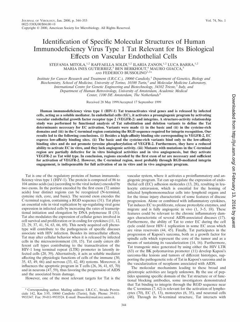
JOURNAL OF VIROLOGY,0022-538X/00/$04.0010
Jan. 2000, p. 344–353 Vol. 74, No. 1
Copyright © 2000, American Society for Microbiology. All Rights Reserved.
Identification of Specific Molecular Structures of HumanImmunodeficiency Virus Type 1 Tat Relevant for Its Biological
Effects on Vascular Endothelial CellsSTEFANIA MITOLA,1,2 RAFFAELLA SOLDI,1,2 ILARIA ZANON,1,2 LUCA BARRA,1,2
MARIA INES GUTIERREZ,3 BEN BERKHOUT,4 MAURO GIACCA,3
AND FEDERICO BUSSOLINO1,2*
Institute for Cancer Research and Treatment (I.R.C.C.), 10060 Candiolo,1 Department of Genetics, Biology andBiochemistry, School of Medicine, University of Torino, 10100 Turin,2 and Molecular Medicine Laboratory,
International Centre for Genetic Engineering and Biotechnology, 34102 Trieste,3 Italy, andDepartment of Human Retrovirology, University of Amsterdam, Academic Medical
Center, 1100 DE Amsterdam, The Netherlands4
Received 28 May 1999/Accepted 17 September 1999
Human immunodeficiency virus type 1 (HIV-1) Tat transactivates viral genes and is released by infectedcells, acting as a soluble mediator. In endothelial cells (EC), it activates a proangiogenic program by activatingvascular endothelial growth factor receptor type 2 (VEGFR-2) and integrins. A structure-activity relationshipstudy was performed by functional analysis of Tat substitution and deletion variants to define the Tatdeterminants necessary for EC activation. Variants were made (i) in the basic and (ii) in the cysteine-richdomains and (iii) in the C-terminal region containing the RGD sequence required for integrin recognition. Ourresults led to the following conclusions. (i) Besides a high-affinity binding site corresponding to VEGFR-2, ECexpress low-affinity binding sites. (ii) The basic and the cysteine-rich variants bind only to the low-affinitybinding sites and do not promote tyrosine phosphorylation of VEGFR-2. Furthermore, they have a reducedability to activate EC in vitro, and they lack angiogenic activity. (iii) Mutants with mutations in the C-terminalregion are partially defective for in vitro biological activities and in vivo angiogenesis, but they activateVEGFR-2 as Tat wild type. In conclusion, regions encoded by the first exon of tat are necessary and sufficientfor activation of VEGFR-2. However, the C-terminal region, most probably through RGD-mediated integrinengagement, is indispensable for full activation of an in vitro and in vivo angiogenic program.
Tat is one of the regulatory proteins of human immunode-ficiency virus type 1 (HIV-1). The protein is composed of 86 to104 amino acids (aa) (according to the viral isolate) encoded bytwo exons. In the portion encoded by the first exon (72 aminoacids) four distinct regions can be recognized (N-terminal,cysteine rich, core, and basic). The second exon encodes theC-terminal region, containing a RGD sequence (31). Tat playsan essential role in viral replication by up-regulating viral geneexpression in infected cells by increasing the rates of transcrip-tional initiation and elongation by DNA polymerase II (31).Tat also modulates the expression of cellular genes involved incell survival and proliferation or in coding for cytokines (11, 12,23, 29, 37, 41, 54, 63, 66, 69). This newly acquired cell pheno-type will contribute to the pathogenesis of specific diseasesassociate with HIV infection. Besides its intracellular effects,Tat may alter cellular behavior when it is released by infectedcells in the microenvironment (10, 15). Tat easily enters dif-ferent cell types contributing to the transactivation of theHIV-1 long terminal repeat (LTR) promoter in latently in-fected cells (20, 39). Alternatively, it acts as soluble mediatoraffecting the physiologic functions of cells of the immune (30,35, 43, 49, 66) and nervous (33, 42, 48) systems. Moreover, itinfluences the apoptotic program in T cells (26, 36, 41, 65, 70)and in neurons (47, 59), thus favoring the progression of AIDSand the associated brain damage.
However, one of the most relevant targets for Tat is the
vascular system, where it activates a proinflammatory and an-giogenic program. Tat can up-regulate the expression of endo-thelial cell (EC) adhesion molecules (13, 28), resulting in leu-kocyte extravasion, which is essential for the homing ofinfected lymphomononuclear cells into lymphoid organs andfor the tissue injury characteristic of some features of diseaseprogression. Alone or combined with inflammatory cytokines,Tat induces EC to proliferate, release proteolytic enzymes, andmigrate and is fully angiogenic in vivo (1, 3–5, 18). Thesefeatures could be relevant to the chronic inflammatory dam-ages characteristic of several AIDS-associated diseases (17).Furthermore, the ability of Tat to enter EC during the cellcycle could favor HIV-1 replication in some EC areas whichare virus reservoirs (44, 45). Finally, Tat participates in theprogression of Kaposi’s sarcoma, both as a growth factor forspindle cells which represent the core of the tumor and as ameans of sustaining its vascularization (14, 16). Furthermore,Tat transgenic mice generated by using either the HIV LTR(63) or the BK polyomavirus promoter (11) develop Kaposi’ssarcoma-like lesions and tumors of different histotypes, sup-porting the pathogenetic role of Tat in Kaposi’s sarcoma and inthe vascularization of neoplasms associated with AIDS.
The molecular mechanisms leading to these broad andpleiotropic activities are largely unknown. By the use of pep-tides spanning specific domain of the Tat structure or of func-tional blocking antibodies, some investigators demonstratedthat Tat binding to integrin through the RGD sequence nearthe C terminus (7, 62) is relevant for the activation of lympho-cytes (70), EC (5, 13), monocytes (6, 35), and neuronal cells(48). Through its N-terminal structure, Tat interacts with
* Corresponding author. Mailing address: I.R.C.C., Strada Provin-ciale 142, Km 3.95, 10060 Candiolo (Turin), Italy. Phone: 39-011-9933347. Fax: 39-011-9933524. E-mail: [email protected].
344
on February 13, 2016 by guest
http://jvi.asm.org/
Dow
nloaded from
dipeptidyl peptidase IV, located on the T cell surface, andsuppresses antigen-induced cell activation (26, 68). Addition-ally, Tat binds to and activates the tyrosine kinase receptorsencoded by the KDR and Flt-1 genes in EC and Kaposi’ssarcoma cells (4, 21) and monocytes (43), respectively.
We initiated a study of the structure-activity relationship ofTat to identify functionally important domains that are respon-sible for the activation of the angiogenic program in vascularEC. We report here that cysteine-rich and basic domains arerelevant for functional activation of VEGFR-2 whereas theC-terminal region does not directly participate in the receptoractivation but is required for full cell activation.
MATERIALS AND METHODS
Cells. Human EC from umbilical cord veins, prepared and characterized aspreviously described (9), were grown in M199 (Gibco, Grand Island, N.Y.)supplemented with 20% fetal calf serum (FCS) (Irvine, Santa Ana, Calif.), ECgrowth factor (100 mg/ml) (Sigma Chemical Co., St. Louis, Mo.), and porcineheparin (100 mg/ml) (Sigma). They were used at passage II and grown on aplastic surface coated with porcine gelatin (Sigma), unless specified.
Tat molecules. Recombinant wild-type HIV-1 Tat of 86 (Tat86) and 101(Tat101) amino acids were expressed in Escherichia coli as maltose-binding pro-tein (MBP) or glutathione S-transferase (GST) fusion proteins. They are re-ferred to below as Tat (MBP-Tat) and *Tat (GST-Tat), respectively. Mutantconstructs were obtained by a recombinant PCR procedure with overlappingoligonucleotides corresponding to the mutated sequences, and the specific mu-tations were verified by DNA sequencing. To obtain Tat mutants, a PstI-BamHIcDNA fragment of Tat86 containing the coding region of both exons was sub-cloned in the pALTER-Ex1 vector (Promega, Madison, Wis.). Site-directedmutagenesis was carried out with the Altered Sites mutagenesis kit (Promega) byusing mutant oligonucleotides to introduce specific mutations (Tat D80E, 59CCC GAG GGG AAC CGA CAG GCC 39; Tat R78K/D80E, 59 CCC GAGGGG AAC CGA CAG CC 39 and 59 ACC TCC CAA TCC AAA GGG GAACCG AC 39; Tat R49G/K50I, 59 CTC CTA TGG CGG GAT CAA GCG GAGAC 39; Tat R49G/K50I/R52L/R53I, 59 CTC CTA TGG CGG GAT CAA GCGGAG AC and 59 CGG GAT CAA GCT AAT ACA GCG ACG AAG 39). Themutated cDNAs (317-bp EcoRI-BamHI fragments) were subcloned into thepMAL-c2 vector (New England Biolabs, Beverly, Mass.) and expressed as spec-ified by the manufacturer. Tat86 and its mutants were purified to homogeneityfrom bacterial cell lysates by affinity chromatography on amylose resin, as spec-ified by New England Biolabs, and used as fusion proteins.
*Tat86 and its mutated derivatives including the product of the first exon,*Tat72, a nonconservative mutant with mutations in basic [*TatR(49,52,53,55,56,57)A] and cysteine-rich [*Tat C(22,25,27)A] regions, were pro-duced as previously described (40), as well as *Tat101 (61). Recombinant fusionproteins were purified by glutathione-Sepharose affinity chromatography (Sig-ma) (53). The purified MBP and GST fusion proteins gave a unique band aftersodium dodecyl sulfate-polyacrylamide gel electrophoresis (SDS-PAGE) (10%polyacrylamide) and silver staining. Tat86, *Tat86, and *Tat101 and their mutantswere lipopolysaccharide free, as assessed by the Limulus assay (Sigma). Tat and*Tat were able to induce transcriptional activation of the HIV-1 LTR in HL3T1cells that contain the bacterial gene of chloramphenicol acetyltransferase (CAT)directed by the HIV-1 LTR (67). Tat86 and *Tat101 dose dependently stimulatedCAT expression. Briefly, Tat molecules were added to confluent HL3T1 cells ina 100-mm-diameter dish containing prewarmed phosphate-buffered saline(PBS). Cells were immediately scraped from the plastic surface, resuspended infresh medium, and centrifuged. The cells were again plated in a CO2 incubator,and a CAT assay was performed after 6 h as described previously (67). Under ourexperimental conditions, just scraping of the cells in absence of Tat moleculeshad no effect on LTR-directed CAT gene expression (125 6 56 cpm of [3H]acety-lated chloramphenicol [n 5 3]). However, CAT gene expression was markedlyelevated in cells that received Tat86 at 0.5 mg/ml (423 6 86 cpm of [3H]acetylatedchloramphenicol), 1 mg/ml (2,345 6 167 cpm of [3H]acetylated chlorampheni-col), or 5 mg/ml (6,543 6 567 cpm of [3H]acetylated chloramphenicol) or *Tat86at 0.5 mg/ml (512 6 34 cpm of [3H]acetylated chloramphenicol), 1 mg/ml(3,145 6 343 cpm of [3H]acetylated chloramphenicol), or 5 mg/ml (7,009 6 671cpm of [3H]acetylated chloramphenicol).
Tat molecules were stored at 280°C in aliquots of 5 mg/10 ml of PBS contain-ing 0.1% human serum albumin (HSA; Farma Biagini, Lucca, Italy) and 1 mMdithiothreitol (Sigma).
Iodination of Tat molecules and binding studies. MBP, GST, Tat86, *Tat86,Tat R78K/D80E, Tat R49G/K50I/R52L/R53I, or *Tat C(22,25,27)A (2-mg sam-ples) were dissolved in 200 ml of 20 mM sodium phosphate buffer (pH 7.4)without dithiothreitol and transferred in iodogen-coated tubes (50 mg/ml)(Pierce Europe B.V., Oud Beijerland, The Netherlands), where proteins wereiodinated (5 min at 4°C) with 0.2 mCi of 125I (Amersham Pharmacia Biotech,Little Chalfont, United Kingdom). A 20-ml volume of 20 mM phosphate buffer(pH 7.2) containing 1% HSA, 0.4 M NaCl, and 0.1% 3-[(3-cholamidopropyl)-
dimethylammonio]-1-propanesulfonate (CHAPS; Pierce) was added, and thereaction products were separated on Sephadex-G10. The specific activities of thetracers were as follows: MBP, 1 mCi/118 fmol; GST, 1 mCi/134 fmol; Tat86, 1mCi/108 fmol; *Tat86, 1 mCi/100 fmol; Tat R78K/D80E, 1 mCi/128 fmol; TatR49G/K50I/R52L/R53I, 1 mCi/112 fmol; and *Tat C(22,25,27)A, 1 mCi/103 fmol.[125I]Tat86 and [125I]*Tat86 retained their biological activity on Kaposi’s sarcomacells (8).
For binding studies, adherent EC on 24-well plates were incubated for 90 minat 22°C in 200 ml of M199 containing 20 mM HEPES (pH 7.4), 0.1% HSA, 0.2U of heparin, 100 mg of soybean trypsin inhibitor per ml, bacitracin (bindingbuffer), and increasing concentrations of iodinated Tat molecules or controlproteins in the presence of 100-fold excess of unlabeled proteins. After washes inbuffer binding, the cells were extracted with 2% SDS in PBS. Specific binding(calculated by subtracting from the total the cpm bound after incubation with a100-excess of unlabelled ligand) was approximately 80%. Curve displacementbinding was obtained by incubating cells with 0.05 nM [125I]Tat and processed asdescribed above. Kinetic parameters were estimated with the Ligand program(Elsevier-Biosoft, Cambridge, United Kingdom).
Immunoprecipitation and immunoblotting. Confluent EC (107 cells/150-cm2
dish) were made quiescent by 20 h of starvation in M199 containing 0.5% FCSand 0.1% HSA, preincubated for 15 min at 37°C with 1 mM Na3VO4, and thenstimulated as detailed in Results in the presence of heparin (1 U/ml). The cellswere lysed in a 50 mM Tris-HCl buffer (pH 7.4) containing 150 mM NaCl, 1%Triton X-100, and protease and phosphatase inhibitors (pepstatin, 50 mg/ml;leupeptin, 50 mg/ml; aprotinin, 10 mg/ml; phenylmethylsulfonyl fluoride, 1 mM;soybean trypsin inhibitor, 500 mg/ml; ZnCl2, 100 mM, Na3VO4, 1 mM [Sigma]).After centrifugation (20 min at 10,000 3 g), supernatants were precleared byincubation for 1 h with protein A-Sepharose or with anti-mouse immunoglobu-lin-agarose (Sigma). Samples (1 mg of protein) were incubated with rabbitanti-VEGFR-2 polyclonal antibody (no. C-1158; Santa Cruz Biotechnology, Inc.,Santa Cruz, Calif.) or antiphosphotyrosine monoclonal antibody (MAb) (cloneG410; Upstate Biotechnology Inc., Lake Placid, N.Y.) (5 to 10 mg/ml) for 1 h at4°C, and immune complexes were recovered on protein A-Sepharose or anti-mouse Ig-agarose. Immunoprecipitates were washed four times with lysis buffer,twice with the same buffer without Triton X-100, and once with Tris-bufferedsaline. Proteins were solubilized under reducing conditions, separated by SDS-PAGE (8 or 10% polyacrylamide), transferred to Immobilon-P sheets (Millipore,Bedford, Mass.), and probed with antiphosphotyrosine MAb or with anti-VEGFR-2 antibody. The enhanced chemiluminescence technique (AmershamPharmacia Biotech) was used for detection.
PI 3-kinase assay. A phosphoinositide 3-kinase (PI 3-kinase) assay was per-formed directly on antiphosphotyrosine immunoprecipitates exactly as describedpreviously (58). Briefly, immunoprecipitates were incubated with 40 mM ATP, 50to 100 mCi of [g-32P]ATP (Amersham), and a presonicated mixture of phospha-tidylinositol-4,5-bisphosphate and phosphatidylserine (final concentration ofboth lipids, 50 mg/ml [Sigma]) in 25 mM HEPES (pH 7.4)–and 1 mM EGTA.The reaction was stopped after a 10-min incubation at room temperature by theaddition of 1 volume of 1 M HCl and 2 volumes of chloroform-methanol (1:1).The lipids in the organic phase were separated by thin-layer chromatography(Silica Gel 60; Merck, Darmstadt, Germany) in 1-propanol–2 M acetic acid(65:35, vol/vol) and visualized by autoradiography.
Migration, proliferation, and adhesion assays. EC motility was studied byusing a modified Boyden chamber technique exactly as previously described (9).
To evaluate EC proliferation, 2 3 103 cells were plated in M199 containing20% FCS in 96-well plates (Falcon, Becton Dickinson Labware, Bedford, Mass.)coated with gelatin. After 24 h, the medium was removed and replaced withM199 containing 2.5% FCS. Tat molecules were added on days 0, 2, and 4, andthe cell number was estimated on day 6 as previously described (9).
To study EC adhesion to immobilized Tat molecules, 96-well polystereneplates (Falcon) were coated overnight at 4°C with Tat molecules or controlproteins (10 mg/well), washed, and then incubated for an additional 2 h at roomtemperature with 1% HSA in PBS. Cells were detached in cold PBS containing2 mM EGTA, washed twice in M199 containing 1% FCS, and plated (5 3 104/0.1ml) in the adhesion assay. After a 1-h incubation at 37°C, the plates wereextensively washed in M199 containing 1% FCS, fixed, and stained with crystalviolet (9). The absorbance was read at 540 nm in a microtiter plate spectropho-tometer (EL340; Bio-Tek Instruments, Highland Park, Vt.).
In vivo angiogenesis. Matrigel (Becton Dickinson) supplemented with 10 U ofheparin per ml was mixed with Tat molecules or control proteins and injected(0.7 ml) into the subcutaneous tissue of BALB/c male mice (Charles River,Conago, Italy) along the peritoneal midline. After 5 days, the mice were killedand gels were processed for histology, morphometric analysis, and hemoglobincontent determination measured with a Drabkin reagent kit (Sigma) as previ-ously described (8).
Statistical methods. One-way analysis of variance (ANOVA) and the Student-Neuman-Keuls test were used to test the difference within the experimentalblocks of each biological assay. Statistical analyses were performed with STA-TISTICA for Windows, version 4.5 (StatSoft, Tulsa, Okla.).
VOL. 74, 2000 ENDOTHELIAL CELL ACTIVATION BY HIV-1 Tat 345
on February 13, 2016 by guest
http://jvi.asm.org/
Dow
nloaded from
RESULTSEffect of Tat and Tat mutants on biological functions of EC.
It has been reported that soluble Tat induces the activation ofvascular endothelium and of Kaposi’s sarcoma cells throughthe engagement of VEGFR-2 (4, 21) and the integrin system(5, 13, 16). Furthermore, Tat mimics the extracellular matrixprotein and favors cell adhesion through the amino acid se-quence RGD (7) or the basic domain (62, 64). To examine theTat regions involved in EC activation, a set of Tat mutants wasconstructed (Fig. 1), expressed as fusion proteins in E. coli, andstudied for their ability to induce cell migration, proliferation,and adhesion.
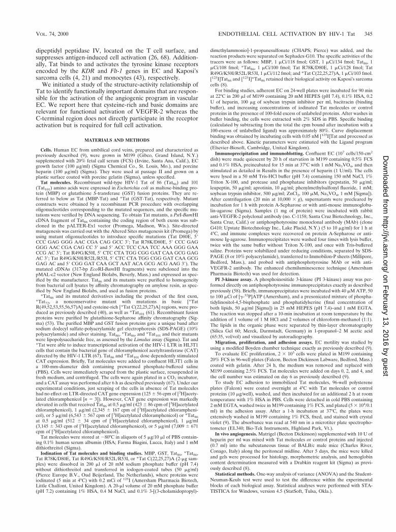
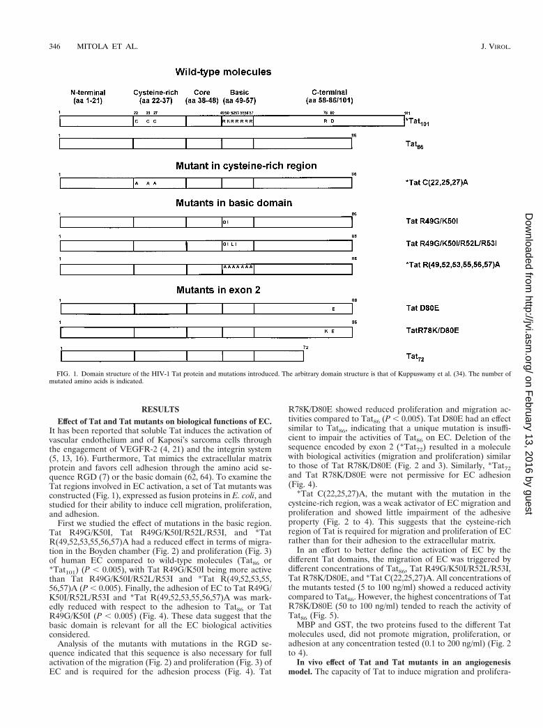
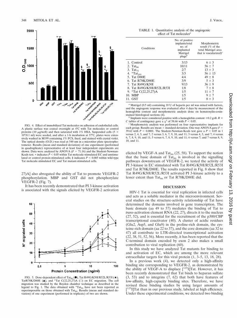
First we studied the effect of mutations in the basic region.Tat R49G/K50I, Tat R49G/K50I/R52L/R53I, and *TatR(49,52,53,55,56,57)A had a reduced effect in terms of migra-tion in the Boyden chamber (Fig. 2) and proliferation (Fig. 3)of human EC compared to wild-type molecules (Tat86 or*Tat101) (P , 0.005), with Tat R49G/K50I being more activethan Tat R49G/K50I/R52L/R53I and *Tat R(49,52,53,55,56,57)A (P , 0.005). Finally, the adhesion of EC to Tat R49G/K50I/R52L/R53I and *Tat R(49,52,53,55,56,57)A was mark-edly reduced with respect to the adhesion to Tat86 or TatR49G/K50I (P , 0.005) (Fig. 4). These data suggest that thebasic domain is relevant for all the EC biological activitiesconsidered.
Analysis of the mutants with mutations in the RGD se-quence indicated that this sequence is also necessary for fullactivation of the migration (Fig. 2) and proliferation (Fig. 3) ofEC and is required for the adhesion process (Fig. 4). Tat
R78K/D80E showed reduced proliferation and migration ac-tivities compared to Tat86 (P , 0.005). Tat D80E had an effectsimilar to Tat86, indicating that a unique mutation is insuffi-cient to impair the activities of Tat86 on EC. Deletion of thesequence encoded by exon 2 (*Tat72) resulted in a moleculewith biological activities (migration and proliferation) similarto those of Tat R78K/D80E (Fig. 2 and 3). Similarly, *Tat72and Tat R78K/D80E were not permissive for EC adhesion(Fig. 4).
*Tat C(22,25,27)A, the mutant with the mutation in thecysteine-rich region, was a weak activator of EC migration andproliferation and showed little impairment of the adhesiveproperty (Fig. 2 to 4). This suggests that the cysteine-richregion of Tat is required for migration and proliferation of ECrather than for their adhesion to the extracellular matrix.
In an effort to better define the activation of EC by thedifferent Tat domains, the migration of EC was triggered bydifferent concentrations of Tat86, Tat R49G/K50I/R52L/R53I,Tat R78K/D80E, and *Tat C(22,25,27)A. All concentrations ofthe mutants tested (5 to 100 ng/ml) showed a reduced activitycompared to Tat86. However, the highest concentrations of TatR78K/D80E (50 to 100 ng/ml) tended to reach the activity ofTat86 (Fig. 5).
MBP and GST, the two proteins fused to the different Tatmolecules used, did not promote migration, proliferation, oradhesion at any concentration tested (0.1 to 200 ng/ml) (Fig. 2to 4).
In vivo effect of Tat and Tat mutants in an angiogenesismodel. The capacity of Tat to induce migration and prolifera-
FIG. 1. Domain structure of the HIV-1 Tat protein and mutations introduced. The arbitrary domain structure is that of Kuppuswamy et al. (34). The number ofmutated amino acids is indicated.
346 MITOLA ET AL. J. VIROL.
on February 13, 2016 by guest
http://jvi.asm.org/
Dow
nloaded from
tion of EC has as in vivo counterpart, i.e., the formation of newblood vessels in rabbit and mouse models (3, 4). Analysis of theeffects of Tat mutants in a murine angiogenesis assay is re-ported in Table 1. Progressive mutations in the basic region orin the RGD sequence were associated with an increasing lossof angiogenic activity compared to the effect of wild-type mol-ecules (Tat86 or *Tat101). The deletion of the amino acidsencoded by exon 2 of tat led to negligible angiogenic activity.Also, the mutation in the cysteine-rich region did not promoteangiogenesis. Overall, the in vivo data clearly indicate that theangiogenic program elicited by Tat is dependent on the mo-lecular integrity of its basic and cysteine-rich domains as well asthat of the exon 2 product.
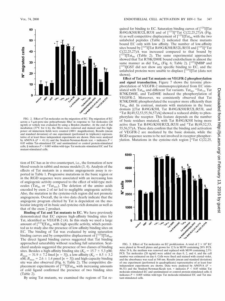
Binding of Tat and Tat mutants to EC. We have previouslydemonstrated that EC express high-affinity binding sites forTat, identified as VEGFR-2 (4). In this study we used a largeamount of [125I]Tat86 with high specific activity, which permit-ted us to study also the presence of low-affinity binding sites onEC. The binding of Tat was evaluated by using saturationbinding curves and by competitive displacement of [125I]Tat86.The direct ligand binding curves suggested that Tat bindingapproached saturability without reaching full saturation. Scat-chard analysis suggested the presence of two classes of bindingsites. Besides a high-affinity binding site (Kd 5 15.7 6 5.3 pM;Bmax 5 31.9 6 7.2 fmol [n 5 3]), a low-affinity (Kd 5 8.5 6 3.2nM; Bmax 5 2.6 6 1.4 pmol [n 5 3]) and high-capacity bindingsite was also observed (Fig. 6; Table 2). The competitive dis-placement experiments of [125I]Tat86 with increasing amountsof cold ligand confirmed the presence of two binding sites(Table 2).
By using Tat mutants, we examined the regions of Tat re-
quired for binding to EC. Saturation binding curves of [125I]TatR49G/K50I/R52L/R53I and of [125I]*Tat C(22,25,27)A (Fig.6) as well competitive displacement of [125I]Tat86 with the twounlabeled peptides (Table 2) indicated that these mutantsbound EC only with low affinity. The number of low-affinitysites bound by [125I]Tat R49G/K50I/R52L/R53I and [125I]*TatC(22,25,27)A was increased compared to that bound by[125I]Tat86 (Table 2). The same experimental approachesshowed that Tat R78K/D80E bound endothelium in almost thesame manner as did Tat86 (Fig. 6; Table 2). [125I]MBP and[125I]GST did not show any specific binding to EC, and theunlabeled proteins were unable to displace [125I]Tat (data notshown).
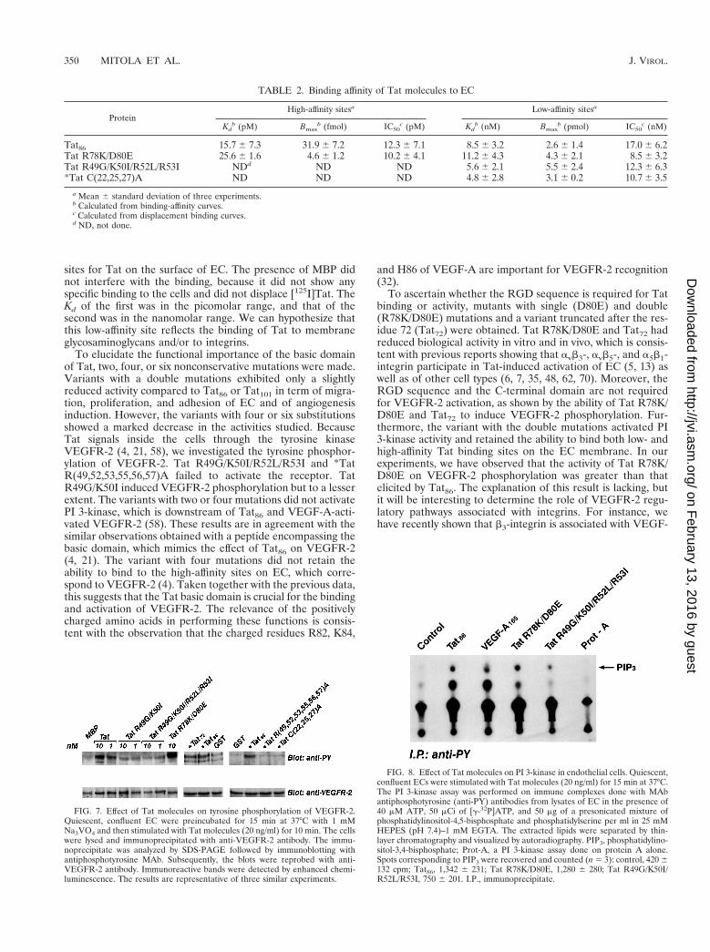
Effect of Tat and Tat mutants on VEGFR-2 phosphorylationand signal transduction. Figure 7 shows the tyrosine phos-phorylation of VEGFR-2 immunoprecipitated from EC stim-ulated with Tat86 and different Tat variants. Tat86, *Tat72, TatR78K/D80E, and TatD80E induced the phosphorylation ofVEGFR-2. Moreover, we consistently observed that TatR78K/D80E phosphorylated the receptor more efficiently thanTat86 did. In contrast, mutants with mutations in the basicdomain [(Tat R49G/K50I, Tat R49G/K50I/R52L/R53I, and*Tat R(49,52,53,55,56,57)A] showed a reduced ability to phos-phorylate the receptor. This feature depends on the numberof basic residues mutated, with Tat R49G/K50I being moreactive than Tat R49G/K50I/R52L/R53I and *Tat R(49,52,53,55,56,57)A. These data confirm that the binding and activationof VEGFR-2 are mediated by the basic domain, while theRGD sequence seems to be not involved in receptor phosphor-ylation. Mutations in the cysteine-rich region [*Tat C(22,25,
FIG. 2. Effect of Tat molecules on the migration of EC. The migration of ECacross a 5-mm-pore-size polycarbonate filter in response to Tat molecules (20ng/ml) or vehicle was evaluated by using a Boyden chamber. At the end of theincubation (37°C for 6 h), the filters were removed and stained and five high-power oil immersion fields were counted (4003 magnification). Results (meanand standard deviation) of one experiment (performed in triplicate) represen-tative of at least three independent experiments are shown. Data were analyzedby ANOVA (F 5 41.11) and the Student-Newman-Keuls test. p indicates P ,0.05 within Tat-stimulated EC and unstimulated or control protein-stimulatedcells; § indicates P , 0.005 within wild-type Tat molecule-stimulated EC and Tatmutant-stimulated cells.
FIG. 3. Effect of Tat molecules on EC proliferation. A total of 2 3 103 ECwere plated in 96-well plates and grown for 12 h in M199 containing 20% FCS.After 24 h, the medium was removed and replaced with M199 containing 2.5%FCS. Tat molecules (20 ng/ml) were added on days 0, 2, and 4, and the cellnumber was estimated on day 6. Cells were fixed and stained with crystal violet,and the absorbance was read at 540 nm. Results (mean and standard deviation)of one experiment (performed in quadruplicate) representative of at least fourindependent experiments are shown. Data were analyzed by ANOVA (F 586.31) and the Student-Newman-Keuls test. p indicates P , 0.05 within Tatmolecule-stimulated EC and unstimulated or control protein-stimulated cells; §indicates P , 0.005 within wild-type Tat molecule-stimulated EC and Tat mu-tant-stimulated cells.
VOL. 74, 2000 ENDOTHELIAL CELL ACTIVATION BY HIV-1 Tat 347
on February 13, 2016 by guest
http://jvi.asm.org/
Dow
nloaded from
27)A] also abrogated the ability of Tat to promote VEGFR-2phosphorylation. MBP and GST did not phosphorylateVEGFR-2 (Fig. 7).
It has been recently demonstrated that PI 3-kinase activationis associated with the signals elicited by VEGFR-2 activation
elicited by VEGF-A and Tat86 (25, 58). To support the notionthat the basic domain of Tat86 is involved in the signallingpathways downstream of VEGFR-2, we tested the activity ofPI 3-kinase in EC stimulated with Tat R49G/K50I/R52L/R53Iand Tat R78K/D80E. The results reported in Fig. 8 show thatTat R49G/K50I/R52L/R53I activated PI 3-kinase activity to alesser extent than Tat86 or Tat R78K/D80E did.
DISCUSSIONHIV-1 Tat is essential for viral replication in infected cells
and acts as a soluble mediator in the microenvironment. Sev-eral studies on the structure-activity relationship of Tat havedetermined the domains involved in gene transcription. Thebasic domain (aa 49 to 57) mediates the binding of Tat totrans-activation element RNA (22, 27), directs it to the nucleus(27, 52), and is essential for the recruitment of the p300/CBPtranscriptional coactivator (40). A cluster of acidic residues(Glu2, Asp5, and Glu9) in the proline-rich domain, the cys-teine-rich domain (aa 22 to 37), and the core domains (aa 32 to47) all contribute to LTR-directed transcriptional activation(22, 38, 51, 52, 56). More recently, it has been reported that theC-terminal domain encoded by exon 2 also makes a smallcontribution to viral replication (60).
In this study we have analyzed Tat mutants for binding toand activation of EC, which are among the more relevantextracellular targets for this viral protein (1, 3–5, 13, 18, 28).
In a previous work (4), we detected only a high-affinitybinding site corresponding to VEGFR-2, as demonstrated bythe ability of VEGF-A to displace [125I]Tat. However, it hasbeen recently demonstrated that Tat binds to heparan sulfate(3, 53) and to integrins (7, 62) that both have features oflow-affinity, high-capacity binding sites. Therefore, we haverevised these binding studies by using larger amounts of[125I]Tat than in our previous study, labeled at high efficiency.Under these experimental conditions, we detected two binding
FIG. 4. Effect of immobilized Tat molecules on adhesion of endothelial cells.A plastic surface was coated overnight at 4°C with Tat molecules or controlproteins (10 mg/well) and then saturated with 1% HSA. Suspended cells (5 3104/0.1 ml) were seeded, and after a 1-h incubation at 37°C, plates were exten-sively washed in M199 containing 1% FCS, fixed, and stained with crystal violet.The optical density (O.D.) was read at 540 nm in a microtiter plate spectropho-tometer. Results (mean and standard deviation) of one experiment (performedin quadruplicate) representative of at least four independent experiments areshown. Data were analyzed by ANOVA (F 5 71.16) and the Student-Newman-Keuls test. p indicates P , 0.05 within Tat molecule-stimulated EC and unstimu-lated or control protein-stimulated cells; § indicates P , 0.005 within wild-typeTat molecule-stimulated EC and Tat mutant-stimulated cells.
FIG. 5. Dose-dependent effect of Tat86 (F), Tat R49G/K50I/R52L/R53I (.),TatR78K/D80E (Œ), and *Tat C(22,25,27)A (E) on EC migration. The cellmigration was studied by the Boyden chamber technique as described in thelegend to Fig. 1. The data obtained with *Tat86 have not been reported assuperimposable on those obtained with Tat86. Results (mean and standard de-viation) of one experiment (performed in triplicate) of two are shown.
TABLE 1. Quantitative analysis of the angiogeniceffect of Tat moleculesa
Conditions
No. of positiveimplants/total
no. ofimplantedMatrigel
plugsb
Morphometricresult (% of the
total Matrigel areathat is vascularized)c
1. Control 3/13 6 6 32. Tat86 10/11 56 6 73. Tat72 2/7 13 6 34. *Tat101 3/3 56 6 135. Tat D80E 4/4 49 6 86. Tat R78K/D80E 3/9 5 6 37. Tat R49G/K50I 9/13 36 6 98. Tat R49G/K50I/R52L/R53I 1/8 7 6 89. *Tat C(22,25,27)A 1/5 11 6 7
10. MBP 1/5 9 6 711. GST 1/5 8 6 3
a Matrigel (0.5 ml) containing 10 U of heparin per ml was mixed with factors,and the angiogenic response was evaluated after 4 days by measurement of thehemoglobin content and morphometric analysis done on hematoxylin-eosin-stained histological sections (4).
b Implants were considered positive with a hemoglobin content $0.2 g/dl. R 3C tables of contingency gave a x2 of 58.66 with P , 0.05.
c Morphometric analysis was performed on four representative implants foreach group. Results are mean 6 standard deviation. One-way ANOVA gave F 539.62 with P , 0.0001. The Student-Newman-Keuls test gave a P , 0.05 in 1versus 2, 4, 5, and 7; 2 versus 3, 6, 7, 9, 10, and 11; 3 versus 4, 5, and 7; 4 versus6, 7, 8, 9, 10, and 11; 5 versus 6, 7, 8, 9, 10, and 11; 6 versus 7; and 7 versus 8, 9,10, and 11.
348 MITOLA ET AL. J. VIROL.
on February 13, 2016 by guest
http://jvi.asm.org/
Dow
nloaded from
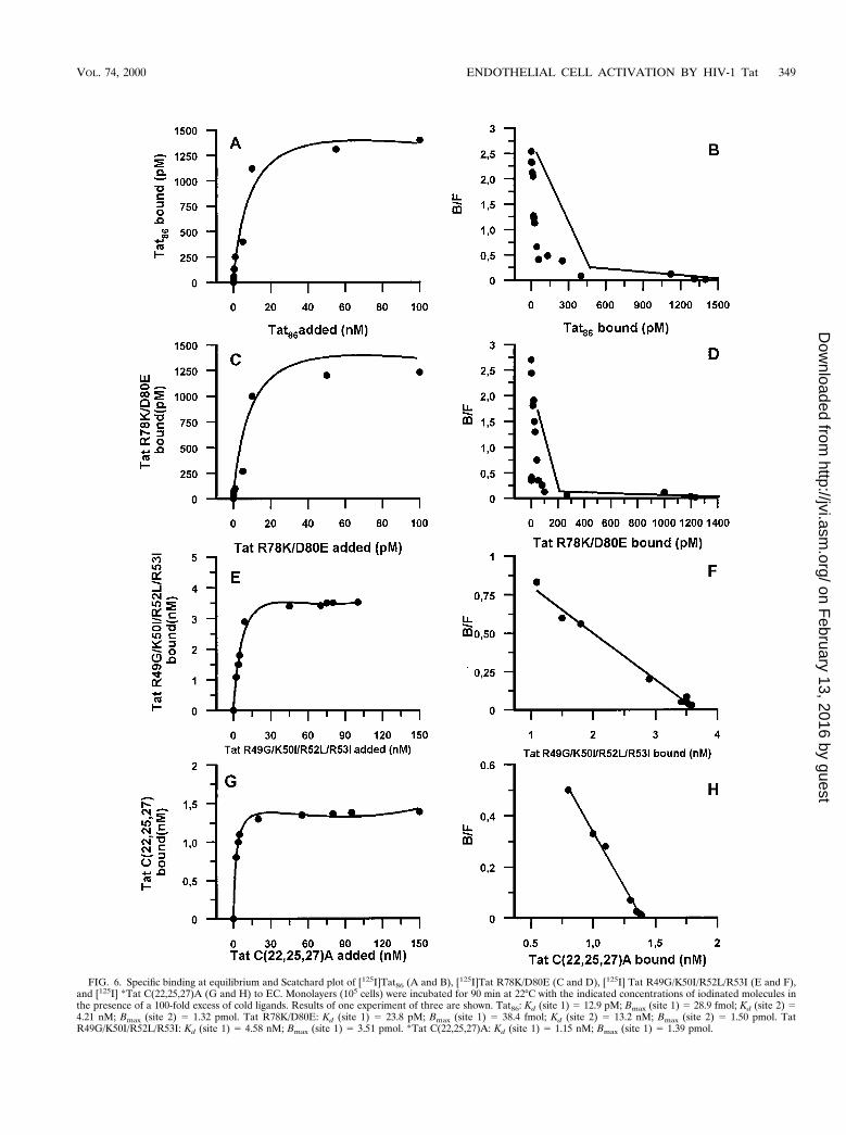
FIG. 6. Specific binding at equilibrium and Scatchard plot of [125I]Tat86 (A and B), [125I]Tat R78K/D80E (C and D), [125I] Tat R49G/K50I/R52L/R53I (E and F),and [125I] *Tat C(22,25,27)A (G and H) to EC. Monolayers (105 cells) were incubated for 90 min at 22°C with the indicated concentrations of iodinated molecules inthe presence of a 100-fold excess of cold ligands. Results of one experiment of three are shown. Tat86: Kd (site 1) 5 12.9 pM; Bmax (site 1) 5 28.9 fmol; Kd (site 2) 54.21 nM; Bmax (site 2) 5 1.32 pmol. Tat R78K/D80E: Kd (site 1) 5 23.8 pM; Bmax (site 1) 5 38.4 fmol; Kd (site 2) 5 13.2 nM; Bmax (site 2) 5 1.50 pmol. TatR49G/K50I/R52L/R53I: Kd (site 1) 5 4.58 nM; Bmax (site 1) 5 3.51 pmol. *Tat C(22,25,27)A: Kd (site 1) 5 1.15 nM; Bmax (site 1) 5 1.39 pmol.
VOL. 74, 2000 ENDOTHELIAL CELL ACTIVATION BY HIV-1 Tat 349
on February 13, 2016 by guest
http://jvi.asm.org/
Dow
nloaded from
sites for Tat on the surface of EC. The presence of MBP didnot interfere with the binding, because it did not show anyspecific binding to the cells and did not displace [125I]Tat. TheKd of the first was in the picomolar range, and that of thesecond was in the nanomolar range. We can hypothesize thatthis low-affinity site reflects the binding of Tat to membraneglycosaminoglycans and/or to integrins.
To elucidate the functional importance of the basic domainof Tat, two, four, or six nonconservative mutations were made.Variants with a double mutations exhibited only a slightlyreduced activity compared to Tat86 or Tat101 in term of migra-tion, proliferation, and adhesion of EC and of angiogenesisinduction. However, the variants with four or six substitutionsshowed a marked decrease in the activities studied. BecauseTat signals inside the cells through the tyrosine kinaseVEGFR-2 (4, 21, 58), we investigated the tyrosine phosphor-ylation of VEGFR-2. Tat R49G/K50I/R52L/R53I and *TatR(49,52,53,55,56,57)A failed to activate the receptor. TatR49G/K50I induced VEGFR-2 phosphorylation but to a lesserextent. The variants with two or four mutations did not activatePI 3-kinase, which is downstream of Tat86 and VEGF-A-acti-vated VEGFR-2 (58). These results are in agreement with thesimilar observations obtained with a peptide encompassing thebasic domain, which mimics the effect of Tat86 on VEGFR-2(4, 21). The variant with four mutations did not retain theability to bind to the high-affinity sites on EC, which corre-spond to VEGFR-2 (4). Taken together with the previous data,this suggests that the Tat basic domain is crucial for the bindingand activation of VEGFR-2. The relevance of the positivelycharged amino acids in performing these functions is consis-tent with the observation that the charged residues R82, K84,
and H86 of VEGF-A are important for VEGFR-2 recognition(32).
To ascertain whether the RGD sequence is required for Tatbinding or activity, mutants with single (D80E) and double(R78K/D80E) mutations and a variant truncated after the res-idue 72 (Tat72) were obtained. Tat R78K/D80E and Tat72 hadreduced biological activity in vitro and in vivo, which is consis-tent with previous reports showing that avb3-, avb5-, and a5b1-integrin participate in Tat-induced activation of EC (5, 13) aswell as of other cell types (6, 7, 35, 48, 62, 70). Moreover, theRGD sequence and the C-terminal domain are not requiredfor VEGFR-2 activation, as shown by the ability of Tat R78K/D80E and Tat72 to induce VEGFR-2 phosphorylation. Fur-thermore, the variant with the double mutations activated PI3-kinase activity and retained the ability to bind both low- andhigh-affinity Tat binding sites on the EC membrane. In ourexperiments, we have observed that the activity of Tat R78K/D80E on VEGFR-2 phosphorylation was greater than thatelicited by Tat86. The explanation of this result is lacking, butit will be interesting to determine the role of VEGFR-2 regu-latory pathways associated with integrins. For instance, wehave recently shown that b3-integrin is associated with VEGF-
FIG. 7. Effect of Tat molecules on tyrosine phosphorylation of VEGFR-2.Quiescent, confluent EC were preincubated for 15 min at 37°C with 1 mMNa3VO4 and then stimulated with Tat molecules (20 ng/ml) for 10 min. The cellswere lysed and immunoprecipitated with anti-VEGFR-2 antibody. The immu-noprecipitate was analyzed by SDS-PAGE followed by immunoblotting withantiphosphotyrosine MAb. Subsequently, the blots were reprobed with anti-VEGFR-2 antibody. Immunoreactive bands were detected by enhanced chemi-luminescence. The results are representative of three similar experiments.
FIG. 8. Effect of Tat molecules on PI 3-kinase in endothelial cells. Quiescent,confluent ECs were stimulated with Tat molecules (20 ng/ml) for 15 min at 37°C.The PI 3-kinase assay was performed on immune complexes done with MAbantiphosphotyrosine (anti-PY) antibodies from lysates of EC in the presence of40 mM ATP, 50 mCi of [g-32P]ATP, and 50 mg of a presonicated mixture ofphosphatidylinositol-4,5-bisphosphate and phosphatidylserine per ml in 25 mMHEPES (pH 7.4)–1 mM EGTA. The extracted lipids were separated by thin-layer chromatography and visualized by autoradiography. PIP3, phosphatidylino-sitol-3,4-bisphosphate; Prot-A, a PI 3-kinase assay done on protein A alone.Spots corresponding to PIP3 were recovered and counted (n 5 3): control, 420 6132 cpm; Tat86, 1,342 6 231; Tat R78K/D80E, 1,280 6 280; Tat R49G/K50I/R52L/R53I, 750 6 201. I.P., immunoprecipitate.
TABLE 2. Binding affinity of Tat molecules to EC
ProteinHigh-affinity sitesa Low-affinity sitesa
Kdb (pM) Bmax
b (fmol) IC50c (pM) Kd
b (nM) Bmaxb (pmol) IC50
c (nM)
Tat86 15.7 6 7.3 31.9 6 7.2 12.3 6 7.1 8.5 6 3.2 2.6 6 1.4 17.0 6 6.2Tat R78K/D80E 25.6 6 1.6 4.6 6 1.2 10.2 6 4.1 11.2 6 4.3 4.3 6 2.1 8.5 6 3.2Tat R49G/K50I/R52L/R53I NDd ND ND 5.6 6 2.1 5.5 6 2.4 12.3 6 6.3*Tat C(22,25,27)A ND ND ND 4.8 6 2.8 3.1 6 0.2 10.7 6 3.5
a Mean 6 standard deviation of three experiments.b Calculated from binding-affinity curves.c Calculated from displacement binding curves.d ND, not done.
350 MITOLA ET AL. J. VIROL.
on February 13, 2016 by guest
http://jvi.asm.org/
Dow
nloaded from
A-stimulated VEGFR-2 and that a MAb against this integrininhibits the activation of the receptor (58). Alternatively, thisvariant can assume a spatial conformation more favorable forreceptor activation, independent of the interaction with inte-grins. Therefore, the activation of the biological activities ofEC related to angiogenesis most probably requires the engage-ment of VEGFR-2 as well as integrins by two specific molec-ular determinants of Tat: the basic domain and the product ofexon 2 containing the RGD motif. Many of the integrin-in-duced signalling pathways are also normally activated by bind-ing of soluble growth factors to their receptors, which suggeststhe existence of coordinate mechanisms between integrins andgrowth factors in the control of cell functions, and there isincreasing evidence that growth factors can induce an appro-priate cellular response only when the target cells express de-fined sets of integrins (24, 55). Normally, growth factors do notdirectly bind to and activate integrin. The data reported hereand those showing that anti-avb3 or anti-VEGFR-2 neutraliz-ing antibodies (4, 5, 58) inhibit the EC response to Tat suggestthat this viral protein is a unique example of a molecule whichturns on intracellular signals by direct activation of both re-ceptor and integrin systems. Furthermore, besides playing adirect role in integrin activation, the C terminus region of Tatcould regulate VEGFR-2 through the presence of other de-terminants relevant for the engagement of neuropilin-1, a co-receptor of this tyrosine kinase receptor (57).
*Tat C(22,25,27)A was used to study the role of the cysteine-rich region of Tat in EC activation. This variant was less potentthan Tat86 or Tat101 in terms of migration, proliferation, ad-hesion, and in vivo angiogenesis, bound only the low-affinitybinding site, and showed low efficiency in VEGFR-2 phosphor-ylation. Because it has been suggested that Tat forms a dimerbridging cysteine-rich regions from each monomer (19) andthat two cysteine residues are pivotal for the VEGF-A dimer-ization and the subsequent binding to and activation of endo-thelium (46, 50), it could be hypothesized that the active formof Tat on the endothelium has a dimeric structure. The rele-vance of this domain has also been emphasized by Albini et al.,who demonstrated its role in the migration of monocytes (2).
Except for Tat D80E, all mutants studied showed a reducedability to activate EC functions, but their effects are only par-tially overlapping. For example, mutants with mutations in theRGD sequence have reduced biological activities but fully ac-tivated VEGFR-2 phosphorylation. Tat R49G/K50I promotedcell adhesion as Tat86, but its migratory and proliferative ca-pacities were consistently lower than those of Tat86. Mutantswith mutations in basic and in cysteine-rich domains had sim-ilar kinetic binding parameters, which differed from those ofTatR78K/E80D. Furthermore, Tat E80D was similar to Tat86or *Tat101 in all assays performed. Taken together, these ob-servations exclude the possibility that the effects of the Tatmutants are caused merely by an unfolded protein, as alsoreported for other studies performed with Tat mutants (22, 27,38, 40, 51, 52, 56, 60).
In conclusion, this study demonstrates that the product ofthe first exon of tat is crucial for the activation of VEGFR-2and for induction of the activation of an angiogenic program inEC. However, to obtain full activation of this program, Tat alsorequires the C-terminal region containing the RGD sequence.The C-terminal region could influence the VEGFR-2 responseby interacting with integrin subunits or with neuropilin-1 (57),which modulate the functions of this tyrosine kinase receptor,or with other unidentified coreceptors. Further studies of thehypothesized role of Tat dimerization by intermolecular cys-teine bridge formation should make it possible to define thereceptor-ligand interaction more precisely.
ACKNOWLEDGMENTS
This study was supported by grants from the European Community(Biomed-2 Project BMHL-CT96-0669), Italian Association for CancerResearch (A.I.R.C.), Istituto Superiore di Sanita (Programma nazio-nale sull’AIDS-Patogenesi, immunita e vaccino per l’AIDS; Programon Tumor Therapy), Centro Nazionale delle Ricerche (Progetto Fi-nalizzato Biotecnologie), and Ministero dell’ Universita e dellaRicerca Scientifica e Tecnologica (60% and Programmi di Ricerca diRilevante Interesse Nazionale-1998). S.M. and I.Z. are supported bygrants from FIRC and from the “Gigi Ghirotti” foundation, respec-tively.
ADDENDUM IN PROOF
Boykins et al. (R. A. Boykins, R. Mahieux, U. T. Shanka-varam, Y. S. Gho, S. F. Lee, I. K. Hewlett, L. M. Wahl, H. K.Kleiman, J. N. Brady, K. M. Yamada, and S. Dhawan, J. Im-munol. 163:15–20, 1999) recently reported that a peptide con-taining six cysteine residues (from amino acids 21 to 40 of theTat molecule) is angiogenic in chicken chorioallantoic mem-branes.
REFERENCES1. Albini, A., G. Barillari, R. Benelli, R. C. Gallo, and B. Ensoli. 1995. Angio-
genic properties of human immunodeficiency virus type 1 Tat protein. Proc.Natl. Acad. Sci. USA 92:4838–4842.
2. Albini, A., R. Benelli, D. Giunciuglio, T. Cai, G. Mariani, S. Ferrini, andD. M. Noonan. 1998. Identification of a novel domain of HIV tat involved inmonocyte chemotaxis. J. Biol. Chem. 273:15895–15900.
3. Albini, A., R. Benelli, M. Presta, M. Rusnati, M. Ziche, A. Rubartelli, G.Paglialunga, F. Bussolino, and D. Noonan. 1996. HIV-tat protein is a hep-arin-binding angiogenic growth factor. Oncogene 12:289–297.
4. Albini, A., R. Soldi, D. Giunciuglio, E. Giraudo, R. Benelli, L. Primo, D.Noonan, M. Salio, G. Camussi, W. Rockl, and F. Bussolino. 1996. Theangiogenesis induced by HIV-1 tat protein is mediated by the Flk-1/KDRreceptor on vascular endothelial cells. Nat. Med. 2:1371–1375.
5. Barillari, G., C. Sgadari, V. Fiorelli, F. Samaniego, S. Colombini, V. Man-zari, A. Modesti, B. C. Nair, A. Cafaro, M. Sturzl, and B. Ensoli. 1999. TheTat protein of human immunodeficiency virus type-1 promotes vascular cellgrowth and locomotion by engaging the a5b1 and avb3 integrins and bymobilizing sequestered basic fibroblast growth factor. Blood 94:663–672.
6. Benelli, R., R. Mortarini, A. Anichini, D. Giunciuglio, D. M. Noona, S.Montalti, C. Tacchetti, and A. Albini. 1998. Monocyte-derived dendritic cellsand monocytes migrate to HIV-Tat RGD and basic peptides. AIDS 12:261–268.
7. Brake, D. A., C. Debouk, and G. Biesecker. 1990. Identification of an Arg-Gly-Asp (RGD) cell adhesion site in human immunodeficiency virus type itransactivation Tat protein. J. Cell Biol. 111:1275–1281.
8. Bussolino, F., M. Arese, G. Montrucchio, L. Barra, L. Primo, R. Benelli, F.Sanavio, M. Aglietta, D. Ghigo, M. R. Rola-Pleszczynski, A. Albini, and G.Camussi. 1995. Platelet activating factor produced in vitro by Kaposi’s sar-coma cells induces and sustains in vivo angiogenesis. J. Clin. Investig. 96:940–952.
9. Bussolino, F., M. F. Di Renzo, M. Ziche, E. Bocchietto, M. Olivero, L.Naldini, G. Gaudino, L. Tamagnone, A. Coffer, and P. M. Comoglio. 1992.Hepatocyte growth factor is a potent angiogenic factor which stimulatesendothelial cell motility and growth. J. Cell Biol. 119:629–641.
10. Chang, H. C., F. Samaniengo, B. C. Nair, L. Buonaguro, and B. Ensoli. 1997.HIV-1 Tat protein exits from cells via a leaderless secretory pathway andbinds to extracellular matrix-associated heparan sulphate proteoglycansthrough its basic region. AIDS 11:1421–1431.
11. Corallini, A., G. Altavilla, L. Pozzi, F. Bignozzi, M. Negrini, P. Rimessi, F.Gualandi, and G. Barbanti-Brodano. 1993. Systemic expression of HIV-1 tatgene in transgenic mice induces endothelial proliferation and tumours ofdifferent histotypes. Cancer Res. 53:5569–5575.
12. Cupp, C., J. P. Taylor, K. Khalili, and S. Amini. 1993. Evidence for stimu-lation of the transforming growth factor beta 1 promoter by HIV-1 Tat incells derived from CNS. Oncogene 8:2231–2236.
13. Dhawan, S., R. K. Puri, A. Kumar, H. Duplan, J. Masson, and B. B. Aggar-wal. 1997. Human immunodeficiency virus-1-Tat protein induces the cellsurface expression of endothelial leukocyte adhesion molecule-1, vascularcell adhesion molecule-1, and intercellular adhesion molecule-1 in humanendothelial cells. J. Immunol. 90:1535–1544.
14. Ensoli, B., G. Barillari, S. Z. Salahuddin, R. C. Gallo, and F. Wong-Staal.1990. Tat protein of HIV-1 stimulates growth of cells derived from Kaposi’ssarcoma lesions of AIDS patients. Nature 344:84–86.
15. Ensoli, B., L. Buonaguro, G. Barillari, V. Fiorelli, R. Gendelman, R. A.Morgan, P. Wingfield, and R. C. Gallo. 1993. Release, uptake, and effects of
VOL. 74, 2000 ENDOTHELIAL CELL ACTIVATION BY HIV-1 Tat 351
on February 13, 2016 by guest
http://jvi.asm.org/
Dow
nloaded from
extracellular human immunodeficiency virus type 1 Tat protein on cellgrowth and viral transactivation. J. Virol. 67:277–287.
16. Ensoli, B., R. Gendelman, P. Markham, V. Fiorelli, S. Colombini, M.Raffeld, A. Cafaro, H. K. Chang, J. N. Brady, and R. C. Gallo. 1994. Synergybetween basic fibroblast growth factor and HIV-1 Tat protein in induction ofKaposi’s sarcoma. Nature 371:674–680.
17. Fauci, A. S. 1987. AIDS: immunopathogenic mechanism and research strat-egies. Clin. Res. 35:503–510.
18. Fiorelli, V., R. Gendelman, F. Samaniego, P. D. Markham, and B. Ensoli.1995. Cytokines from activated T cells induce normal endothelial cells toacquire the phenotypic and functional features of AIDS-Kaposi’s sarcomaspindle cells. J. Clin. Investig. 95:1723–1734.
19. Frankel, A. D., D. S. Bredt, and C. O. Pabo. 1988. Tat protein from humanimmunodeficiency virus forms a metal-linked dimer. Science 240:70–73.
20. Frankel, A. D., and C. O. Pabo. 1988. Cellular uptake of the Tat protein fromhuman immunodeficiency virus. Cell 55:1189–1193.
21. Ganju, R. K., N. Munshi, B. C. Nair, L. Zy, P. Gill, and J. E. Groopman.1998. Human immunodeficiency virus Tat modulates the Flk-1/KDR recep-tor, mitogen-activated protein kinases, and components of focal adhesion inKaposi’s sarcoma cells. J. Virol. 72:6131–6137.
22. Garcia, J. A., D. Harrich, E. Soultanakis, F. Wu, R. Mitsuyasu, and R. B.Gaynon. 1989. Human immunodeficiency virus type 1 LTR TATA and TARregion sequences required for transcriptional regulation. EMBO J. 8:765–778.
23. Garza, H. H., O. Prakash, and D. J. Carr. 1996. Aberrant regulation ofcytokines in HIV-1 TAT72-transgenic mice. J. Immunol. 156:3631–3637.
24. Giancotti, F. G. 1997. Integrin signaling: specificity and control of cell sur-vival and cell cycle progression. Curr. Opin. Cell Biol. 9:691–700.
25. Guo, D., Q. Jia, H. Y. Song, R. S. Warren, and D. B. Donner. 1995. Vascularendothelial cell growth factor promotes tyrosine phosphorylation of media-tors of signal transduction that contain SH2 domains. Association with en-dothelial cell proliferation. J. Biol. Chem. 270:6729–6733.
26. Gutheil, W. G., M. Subramanyam, G. R. Flentke, D. G. Sanford, E. Munoz,B. T. Huber, and W. W. Bachovchin. 1994. Human immunodeficiency virus1 Tat binds to dipeptidyl aminopeptidase IV (CD26): a possible mechanismfor Tat’s immunosuppressive activity. Proc. Natl. Acad. Sci. USA 91:6594–6598.
27. Hauber, J., H. M. Malim, and B. R. Cullen. 1989. Mutational analysis of theconserved basic domain of human immunodeficiency virus tat protein. J. Vi-rol. 63:1181–1187.
28. Hofman, F. M., A. D. Wright, M. M. Dohadwala, F. Wong-Staal, and S. M.Walker. 1993. Exogenous tat protein activates human endothelial cells.Blood 82:2774–2780.
29. Howcroft, T. K., K. Strebel, M. A. Martin, and D. S. Singer. 1993. Repressionof MHC class I gene promoter activity by two-exon Tat of HIV. Science260:1320–1322.
30. Huang, L., C. J. Li, and A. B. Pardee. 1997. Human immunodeficiency virustype 1 TAT protein activates B lymphocytes. Biochem. Biophys. Res. Com-mun. 237:461–464.
31. Jones, K. A., and M. B. Peterlin. 1994. Control of RNA initiation andelongation at the HIV-1 promoter. Annu. Rev. Biochem. 63:717–743.
32. Keyt, B. A., H. V. Nguyen, L. T. Berleau, C. M. Duarte, J. Park, H. Chen, andN. Ferrara. 1996. Identification of vascular endothelial growth factor deter-minants for binding KDR and FLT-1 receptors. Generation of receptor-selective VEGF variants by site-directed mutagenesis. J. Biol. Chem. 271:5638–5646.
33. Kolson, D. L., J. Buchhalter, R. Collman, B. Hellmig, C. F. Farrell, C.Debouck, and F. Gonzalez-Scarano. 1993. HIV-1 Tat alters normal organi-zation of neurons and astrocytes in primary rodent brain cell cultures: RGDsequence dependence. AIDS Res. Human Retroviruses 9:677–685.
34. Kuppuswamy, M., T. Subramanian, A. Srinivasan, and G. Chinnadurai.1989. Multiple functional domains of Tat, the trans-activator of HIV-1,defined by mutational analysis. Nucleic Acids Res. 17:3551–3561.
35. Lafrenie, R. M., L. M. Wahl, J. S. Epstein, I. K. Hewlett, K. M. Yamada, andS. Dhawan. 1996. HIV-1-Tat protein promotes chemotaxis and invasivebehavior by monocytes. J. Immunol. 157:974–977.
36. Li, C. J., D. J. Friedman, C. Wang, V. Metelev, and A. B. Pardee. 1995.Induction of apoptosis in uninfected lymphocytes by HIV-1 Tat protein.Science 268:429–431.
37. Li, C. J., C. Wang, D. J. Friedman, and A. B. Pardee. 1995. Reciprocalmodulations between p53 and Tat of human immunodeficiency virus type 1.Proc. Natl. Acad. Sci. USA 92:5461–5464.
38. Luo, Y., and B. M. Peterlin. 1993. Juxtaposition between activation and basicdomains of human immunodeficiency virus type 1 Tat is required for optimalinteractions between Tat and TAR. J. Virol. 67:3441–3445.
39. Marcuzzi, A., J. Weinberger, and O. K. Weinberger. 1992. Transcellularactivation of the human immunodeficiency virus type 1 long terminal repeatin cocultured lymphocytes. J. Virol. 66:4228–4232.
40. Marzio, G., M. Tyagi, M. I. Gutierrez, and M. Giacca. 1998. HIV-1 tattransactivator recruits p300 and CREB-binding protein histone acetyltrans-ferases to the viral promoter. Proc. Natl. Acad. Sci. USA 95:13519–13524.
41. McCloskey, T. W., M. Ott, E. Tribble, S. A. Khan, S. Teichberg, M. O. Paul,
S. Pahwa, E. Verdin, and N. Chirmule. 1997. Dual role of HIV Tat inregulation of apoptosis in T cells. J. Immunol. 158:1014–1019.
42. Milani, D., M. Mazzoni, P. Borgatti, G. Zauli, L. Cantley, and S. Capitani.1996. Extracellular human immunodeficiency virus type-1 Tat protein acti-vates phosphatidylinositol 3-kinase in PC12 neuronal cells. J. Biol. Chem.271:22961–22964.
43. Mitola, S., S. Sozzani, W. Luini, M. Arese, A. Borstatti, H. A. Weich, and F.Bussolino. 1997. Tat-HIV-1 induces human monocyte chemotaxis by activa-tion of vascular endothelial growth factor receptor-1. Blood 90:1365–1372.
44. Moses, A., F. E. Bllom, C. D. Pauza, and J. A. Nelson. 1993. Human immu-nodeficiency virus infection of human brain capillary endothelial cells occursvia a CD4/galactosylceramide-independent mechanism. Proc. Natl. Acad.Sci. USA 90:10474–10478.
45. Moses, A. S., S. G. Stenglein, J. G. Strussenberg, K. Wehrly, B. Chesebro,and J. A. Nelson. 1996. Sequences regulating trophism of human immuno-deficiency virus type 1 for brain capillary endothelial cells map to a uniqueregion on the viral genome. J. Virol. 70:3401–3406.
46. Muller, Y. A., B. Li, H. W. Christinger, B. C. Cunningham, and A. M. de Vos.1997. Vascular endothelial growth factor: crystal structure and functionalmapping of the kinase domain receptor binding site. Proc. Natl. Acad. Sci.USA 94:7192–7197.
47. New, D. R., S. B. Maggirwar, L. G. Epstein, S. Dewhurst, and H. A. Gelbard.1998. HIV-1 Tat induces neuronal death via tumor necrosis factor-alpha andactivation of non-N-methyl-D-aspartate receptors by a NFkappaB-indepen-dent mechanism. J. Biol. Chem. 273:17852–17858.
48. Orsini, M. J., C. M. Debouck, C. L. Webb, and P. G. Lysko. 1996. Extracel-lular human immunodeficiency virus type 1 Tat protein promotes aggrega-tion and adhesion of cerebellar neurons. J. Neurosci. 16:2546–2552.
49. Ott, M., J. L. Lovett, L. Mueller, and E. Verdin. 1998. Superinduction of IL-8in T cells by HIV-1 Tat protein is mediated through NF-kappaB factors.J. Immunol. 160:2872–2880.
50. Potgens, A. J., N. H. Lubsen, M. C. van Altena, R. Vermeulen, A. Bakker,J. G. Schoenmakers, D. J. Ruiter, and R. M. de Waal. 1994. Covalentdimerization of vascular permeability factor/vascular endothelial growth fac-tor is essential for its biological activity. Evidence from Cys to Ser mutations.J. Biol. Chem. 269:32879–32885.
51. Rice, A. P., and F. Carlotti. 1990. Structural analysis of wild-type and mutanthuman immunodeficiency virus type 1 Tat proteins. J. Virol. 64:6018–6026.
52. Ruben, S., A. Perkins, R. Purcell, K. Joung, R. Sia, R. Burghoff, W. A.Haseltine, and C. A. Rosen. 1989. Structural and functional characterizationof human immunodeficiency virus tat protein. J. Virol. 63:1–8.
53. Rusnati, M., G. Tulipano, C. Urbinati, E. Tanghetti, R. Giuliani, M. Giacca,M. Ciomei, A. Corallini, and M. Presta. 1998. The basic domain in HIV-1Tat protein as a target for polysulfonated huparin-mimicking extracellularTat antagonists. J. Biol. Chem. 273:16027–16037.
54. Scala, G., M. R. Ruocco, C. Ambrosino, M. Mallardo, V. Giordano, F.Baldassarre, E. Dragonetti, I. Quinto, and S. Venuta. 1994. The expressionof the interleukin 6 gene is induced by the human immunodeficiency virus 1TAT protein. J. Exp. Med. 179:961–971.
55. Schwartz, M. A. 1997. Integrins, oncogenes, and anchorage independence.J. Cell Biol. 139:575–578.
56. Siderovski, O. P., T. Matsuyama, E. Frigerio, S. Chui, X. Min, H. Erfle, M.Sumner-Smith, R. W. Barnett, and T. W. Mak. 1992. Random mutagenesisof the human immunodeficiency virus type-1 trans-activator of transcription(HIV-1 Tat). Nucleic Acids Res. 20:5311–5320.
57. Soker, S., S. Takashima, H. Q. Miao, G. Neufeld, and M. Klagsbrun. 1998.Neuropilin-1 is expressed by endothelial and tumor cells as an isoform-specific receptor for vascular endothelial growth factor. Cell 92:735–745.
58. Soldi, R., S. Mitola, S. Strasly, P. Defilippi, G. Tarone, and F. Bussolino.1999. Role of avb3 integrin in the activation of vascular endothelial growthfactor receptor-2. EMBO J. 18:734–740.
59. Taylor, J. P., C. Cupp, A. Diaz, M. Chowdhury, K. Khalili, S. A. Jimenez, andS. Amini. 1992. Activation of expression of genes coding for extracellularmatrix proteins in Tat-producing glioblastoma cells. Proc. Natl. Acad. Sci.USA 89:9617–9621.
60. Verhoef, K., M. Bauer, A. Meyerhans, and B. Berkhout. 1998. On the role ofthe second coding exon of the HIV-1 Tat protein in virus replication andMHC class I downregulation. AIDS Res. Hum. Retroviruses 14:1553–1559.
61. Verhoef, K., S. E. C. Koken, and B. Berkhout. 1993. Electroporation of theHIV Tat trans-activator protein into cells. Anal. Biochem. 210:210–214.
62. Vogel, B. E., S. J. Lee, A. Hildebrand, W. Craig, M. D. Pierschbacher, F.Wong-Staal, and E. Ruoslahti. 1993. A novel integrin specificity exemplifiedby binding of the alpha v beta 5 integrin to the basic domain of the HIV Tatprotein and vitronectin. J. Cell Biol. 121:461–468.
63. Vogel, J., S. H. Hinrichs, K. R. Reynolds, P. A. Luciw, and G. Jay. 1988. TheHIV tat gene induces dermal lesions resembling Kaposi’s sarcoma in trans-genic mice. Nature 335:606–611.
64. Weeks, B. S., K. Desai, P. M. Loewenstein, M. E. Klotman, P. E. Klotman,M. Green, and H. K. Kleinman. 1993. Identification of a novel cell attach-ment domain in the HIV-1 Tat protein and its 90-kDa cell surface bindingprotein. J. Biol. Chem. 268:5279–5284.
65. Westendorp, M. O., R. Frank, C. Ochsenbauer, K. Stricker, J. Dhein, H.
352 MITOLA ET AL. J. VIROL.
on February 13, 2016 by guest
http://jvi.asm.org/
Dow
nloaded from
Walczak, K. M. Debatin, and P. H. Krammer. 1995. Sensitization of T cellsto CD95-mediated apoptosis by HIV-1 Tat and gp120. Nature 375:497–500.
66. Westendorp, M. O., V. A. Shatrov, K. Schulze-Osthoff, R. Frank, M. Kraft,M. Los, P. H. Krammer, W. Droge, and V. Lehmann. 1995. HIV-1 Tatpotentiates TNF-induced NF-kappa B activation and cytotoxicity by alteringthe cellular redox state. EMBO J. 14:546–554.
67. Wright, C. M., B. K. Felber, H. Paskalis, and G. N. Pavlakis. 1986. Expres-sion and characterization of the trans activator of HTLV-III/LAV virus.Science 234:988–992.
68. Wrenger, S., T. Hoffmann, J. Faust, C. Mrestani-Klauss, W. Brandt, K.Neubert, M. Kraft, S. Olek, R. Frank, S. Ansorge, and D. Reinhold. 1997.
The N-terminal structure of HIV-1 Tat is required for suppression of CD26-dependent T cell growth. J. Biol. Chem. 272:30283–30288.
69. Zauli, G., D. Gibellini, A. Caputo, A. Bassini, M. Negrini, M. Monne, M.Mazzoni, and S. Capitani. 1995. The human immunodeficiency virus type-1Tat protein upregulates Bcl-2 gene expression in Jurkat T-cell lines andprimary peripheral blood mononuclear cells. Blood 86:3823–3834.
70. Zauli, G., D. Gibellini, C. Celeghini, C. Mischiati, A. Bassini, M. La Placa,and S. Capitani. 1996. Pleiotropic effects of immobilized versus solublerecombinant HIV-1 Tat protein on CD3-mediated activation, induction ofapoptosis, and HIV-1 long terminal repeat transactivation in purified CD41
T lymphocytes. J. Immunol. 157:2216–2224.
VOL. 74, 2000 ENDOTHELIAL CELL ACTIVATION BY HIV-1 Tat 353
on February 13, 2016 by guest
http://jvi.asm.org/
Dow
nloaded from
Copyright © 2022 FDOKUMEN