Identification of potential cellular targets of aloisine A by affinity chromatography
11
Identification of potential cellular targets of aloisine A by affinity chromatography Caroline Corbel a , Rose Haddoub b , Damien Guiffant a , Olivier Lozach a , David Gueyrard b , Jérôme Lemoine c, * , Morgane Ratin a , Laurent Meijer a , Stéphane Bach a, * , Peter Goekjian b, * a CNRS USR-3151, Protein Phosphorylation and Human Disease, Station Biologique, B.P.74, F-29682 Roscoff Cedex, Bretagne, France b Université de Lyon, CNRS UMR 5246 ICBMS, Laboratoire de Chimie Organique 2—Glycochimie, Université Claude Bernard Lyon 1, CPE-Lyon, Bâtiment 308, 43 Boulevard du 11 Novembre 1918, Villeurbanne F-69622, France c Université de Lyon, CNRS UMR 5180, Université Claude Bernard Lyon 1, Bâtiment 308, 43 Boulevard du 11 Novembre 1918, Villeurbanne F-69622, France article info Article history: Received 6 February 2009 Revised 5 June 2009 Accepted 14 June 2009 Available online 18 June 2009 Keywords: Aloisine Immobilized protein kinase inhibitor Mass spectrometry Click chemistry Pyrrolo[2,3-b]pyrazine Cyclin-dependent kinase Pyridoxal kinase Affinity chromatography GSK-3 Fuse binding protein 1 Glycogen-debranching enzyme ERK1 abstract Affinity chromatography was used to identify potential cellular targets of aloisine A (7-n-butyl-6-(4 0 - hydroxyphenyl)-5H-pyrrolo[2,3b]pyrazine), a potent inhibitor of cyclin-dependent kinases. This tech- nique is based on the immobilization of the drug on a solid matrix, followed by identification of specif- ically bound proteins. To this end, both aloisine A and the protein-kinase inactive control N-methyl aloisine, bearing extended linker chains have been synthesized. We present the preparation of such ana- logues having the triethylene glycol chain at different positions of the molecule, as well as their immo- bilization on an agarose-based matrix. Affinity chromatography of various biological extracts on the aloisine matrices allowed the identification of both protein kinases and non-kinase proteins as potential cellular targets of aloisine. Ó 2009 Elsevier Ltd. All rights reserved. 1. Introduction Cyclin-dependent kinases (CDKs) are low-molecular-weight serine-threonine kinases that require association to a regulatory subunit, a cyclin, in order to be activated. CDKs play important roles in the cell division cycle, apoptosis, differentiation, neuronal functions, and transcription. 1–3 Abnormalities and dysregulations of CDK activities have been associated with many diseases includ- ing cancers, viral infections, ischemia, and neurodegenerative dis- orders such as Alzheimer’s and Parkinson’s disease. 4 Recent data have notably implicated CDK5 in aberrant tau phosphorylation and the association of the latter with microtubules. Hyperphosph- orylation, aggregation, and formation of the filamentous form of tau protein (paired helical filaments or PHF-Tau) are all observed in Alzheimer’s disease but also in other diseases referred to as neu- rodegenerative tauopathies. 5 Furthermore, cyclin-dependent ki- nases are conserved across species during evolution, providing new insights and new pharmacological opportunities for the treat- ment of infectious diseases. These observations have encouraged an intensive search for potent and selective pharmacological inhib- itors of CDKs. A large number of small molecular weight inhibitors have been characterized to date, all of which appear to act by direct competition with ATP. 6–8 The selectivity of kinase inhibitors is usually addressed by test- ing the inhibitor against a panel of purified kinases. 9 But even for the largest panels, these only reflect a fraction of the 518 kinases that constitute the human kinome. In addition, the chemical com- pound might interact with some of the other 1500 ATP utilizing enzymes and numerous other nucleotide-binding proteins present in the human proteome. As a consequence many of the potential targets are not evaluated using this method which, in addition, re- quires time- and budget- consuming expression, purification and assay set up for each individual kinase. Finally, in vitro assay against recombinant enzymes may not adequately reflect the distribution or the phosphorylation states found in vivo. This 0968-0896/$ - see front matter Ó 2009 Elsevier Ltd. All rights reserved. doi:10.1016/j.bmc.2009.06.024 * Corresponding authors. Fax: +33 472448319 (J.L.); fax: +33 298292526 (S.B.); fax: +33 478898914 (P.G.). E-mail addresses: [email protected] (J. Lemoine), [email protected] (S. Bach), [email protected] (P. Goekjian). Bioorganic & Medicinal Chemistry 17 (2009) 5572–5582 Contents lists available at ScienceDirect Bioorganic & Medicinal Chemistry journal homepage: www.elsevier.com/locate/bmc
-
Upload
univ-lyon1 -
Category
Documents
-
view
5 -
download
0
Transcript of Identification of potential cellular targets of aloisine A by affinity chromatography
Bioorganic & Medicinal Chemistry 17 (2009) 5572–5582
Contents lists available at ScienceDirect
Bioorganic & Medicinal Chemistry
journal homepage: www.elsevier .com/locate /bmc
Identification of potential cellular targets of aloisine A byaffinity chromatography
Caroline Corbel a, Rose Haddoub b, Damien Guiffant a, Olivier Lozach a, David Gueyrard b, Jérôme Lemoine c,*,Morgane Ratin a, Laurent Meijer a, Stéphane Bach a,*, Peter Goekjian b,*
a CNRS USR-3151, Protein Phosphorylation and Human Disease, Station Biologique, B.P.74, F-29682 Roscoff Cedex, Bretagne, Franceb Université de Lyon, CNRS UMR 5246 ICBMS, Laboratoire de Chimie Organique 2—Glycochimie, Université Claude Bernard Lyon 1, CPE-Lyon, Bâtiment 308,43 Boulevard du 11 Novembre 1918, Villeurbanne F-69622, Francec Université de Lyon, CNRS UMR 5180, Université Claude Bernard Lyon 1, Bâtiment 308, 43 Boulevard du 11 Novembre 1918, Villeurbanne F-69622, France
a r t i c l e i n f o a b s t r a c t
0
Article history:Received 6 February 2009Revised 5 June 2009Accepted 14 June 2009Available online 18 June 2009Keywords:AloisineImmobilized protein kinase inhibitorMass spectrometryClick chemistryPyrrolo[2,3-b]pyrazineCyclin-dependent kinasePyridoxal kinaseAffinity chromatographyGSK-3Fuse binding protein 1Glycogen-debranching enzymeERK1
0968-0896/$ - see front matter � 2009 Elsevier Ltd. Adoi:10.1016/j.bmc.2009.06.024
* Corresponding authors. Fax: +33 472448319 (J.L.fax: +33 478898914 (P.G.).
E-mail addresses: [email protected] (J.(S. Bach), [email protected] (P. Goekjian).
Affinity chromatography was used to identify potential cellular targets of aloisine A (7-n-butyl-6-(4 -hydroxyphenyl)-5H-pyrrolo[2,3b]pyrazine), a potent inhibitor of cyclin-dependent kinases. This tech-nique is based on the immobilization of the drug on a solid matrix, followed by identification of specif-ically bound proteins. To this end, both aloisine A and the protein-kinase inactive control N-methylaloisine, bearing extended linker chains have been synthesized. We present the preparation of such ana-logues having the triethylene glycol chain at different positions of the molecule, as well as their immo-bilization on an agarose-based matrix. Affinity chromatography of various biological extracts on thealoisine matrices allowed the identification of both protein kinases and non-kinase proteins as potentialcellular targets of aloisine.
� 2009 Elsevier Ltd. All rights reserved.
5
1. IntroductionCyclin-dependent kinases (CDKs) are low-molecular-weightserine-threonine kinases that require association to a regulatorysubunit, a cyclin, in order to be activated. CDKs play importantroles in the cell division cycle, apoptosis, differentiation, neuronalfunctions, and transcription.1–3 Abnormalities and dysregulationsof CDK activities have been associated with many diseases includ-ing cancers, viral infections, ischemia, and neurodegenerative dis-orders such as Alzheimer’s and Parkinson’s disease.4 Recent datahave notably implicated CDK5 in aberrant tau phosphorylationand the association of the latter with microtubules. Hyperphosph-orylation, aggregation, and formation of the filamentous form oftau protein (paired helical filaments or PHF-Tau) are all observedin Alzheimer’s disease but also in other diseases referred to as neu-
ll rights reserved.
); fax: +33 298292526 (S.B.);
Lemoine), [email protected]
rodegenerative tauopathies. Furthermore, cyclin-dependent ki-nases are conserved across species during evolution, providingnew insights and new pharmacological opportunities for the treat-ment of infectious diseases. These observations have encouragedan intensive search for potent and selective pharmacological inhib-itors of CDKs. A large number of small molecular weight inhibitorshave been characterized to date, all of which appear to act by directcompetition with ATP.6–8
The selectivity of kinase inhibitors is usually addressed by test-ing the inhibitor against a panel of purified kinases.9 But even forthe largest panels, these only reflect a fraction of the 518 kinasesthat constitute the human kinome. In addition, the chemical com-pound might interact with some of the other �1500 ATP utilizingenzymes and numerous other nucleotide-binding proteins presentin the human proteome. As a consequence many of the potentialtargets are not evaluated using this method which, in addition, re-quires time- and budget- consuming expression, purification andassay set up for each individual kinase. Finally, in vitro assayagainst recombinant enzymes may not adequately reflect thedistribution or the phosphorylation states found in vivo. This
N
N
NO
R
2
3
7
6 4'
3'
H
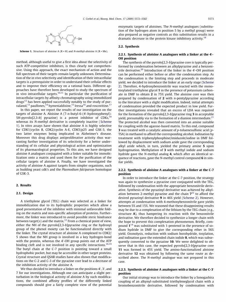
Scheme 1. Structure of aloisine A (R = H) and N-methyl-aloisine A (R = Me).
C. Corbel et al. / Bioorg. Med. Chem. 17 (2009) 5572–5582 5573
method, although useful to give a first idea about the selectivity ofsuch ATP-competitive inhibitors, is thus clearly not comprehen-sive. Using this approach, the exact mechanism of action and thefull spectrum of their targets remain largely unknown. Determina-tion of the in vivo selectivity and identification of their intracellulartargets is a prerequisite in order to understand their cellular effectsand to improve their efficiency on a rational basis. Different ap-proaches have therefore been developed to study the spectrum ofin vivo intracellular targets.10,11 In particular the purification ofintracellular targets by affinity chromatography using immobilizeddrugs12 has been applied successfully notably to the study of pur-valanol,13 paullones,14 hymenialdisine,15 Iressa16 and roscovitine.17
In this paper, we report the results of our investigation on thetargets of aloisine A. Aloisine A (7-n-butyl-6-(40-hydroxyphenyl)-5H-pyrrolo[2,3-b] pyrazine) is a potent inhibitor of CDKs,18
whereas its N-methyl derivative is completely inactive (Scheme1). In vitro assays have shown that aloisine A is highly selectivefor CDK1/cyclin B, CDK2/cyclin A-E, CDK5/p25 and GSK-3, thetwo latter enzymes being implicated in Alzheimer’s disease.Moreover this drug displays antiproliferative activity thus war-ranting further investigation of its selectivity for a better under-standing of its cellular and physiological action and optimizationof its pharmacological properties. To this aim, we have designedaloisine A analogues conjugated with a linker suitable for immobi-lization onto a matrix and used them for the purification of thecellular targets of aloisine A. Finally, we have investigated theactivity of aloisine A against targets from simpler organisms, suchas budding yeast cdk’s and the Plasmodium falciparum homologueof GSK-3.
2. Results
2.1. Design
A triethylene glycol (TEG) chain was selected as a linker forimmobilization due to its hydrophilic properties which allow agood presentation of the inhibitor with minimal unfavorable fold-ing on the matrix and non-specific adsorption of proteins. Further-more, the linker was introduced to avoid possible steric hindrancebetween target(s) and the matrix.12 From a chemical point of view,either the NH of the pyrrolo[2,3-b]pyrazine ring or the hydroxylgroup of the phenol moiety can be functionalized directly withthe linker. The crystal structure of aloisine A complexed to CDK2/5 shows that the NH group is involved in a key hydrogen-bondwith the protein, whereas the 40-OH group points out of the ATPbinding cleft and is not involved in any specific interactions.18,19
The butyl chain at the C-7 position is pointing toward a smallhydrophobic pocket, but may be accessible in other target proteins.Crystal structure and QSAR studies have also shown that modifica-tions on the C-2 and C-3 of the pyrazine core lead to a decrease ofthe inhibition activity of the aloisine A.
We thus decided to introduce a linker on the positions 40, 30, and7 for our investigations. Although one can anticipate a slight per-turbation in the biological activity of aloisine A by such modifica-tions, the combined affinity profiles of the differently linkedcompounds should give a fairly complete view of the potential
enzymatic targets of aloisines. The N-methyl analogues (substitu-tion of the hydrogen atom in position 5 by a methyl group) werealso prepared as negative controls as this substitution results in adramatic decrease in the protein kinase inhibitory activity.18
2.2. Synthesis
2.2.1. Synthesis of aloisine A analogues with a linker at the 40-OH position
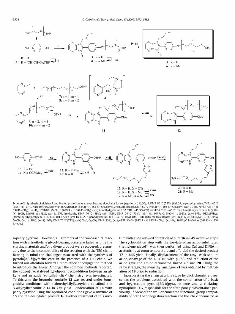
The synthesis of the pyrrolo[2,3-b]pyrazine core is typically per-formed by condensation between an alkylpyrazine and a benzoni-trile derivative.20 Introduction of the linker in the 40-OH positioncan be performed either before or after the condensation step. Asthe condensation is the limiting step and proceeds in moderateyield, we decided to introduce the linker at an early stage (Scheme2). Therefore, 4-hydroxybenzonitrile was reacted with the mono-tosylated triethylene glycol 1 in the presence of potassium carbon-ate in DMF to obtain 2 in 75% yield. The aloisine core was thenprepared by condensation of 2 with n-pentylpyrazine accordingto the literature with a slight modification. Indeed, initial attemptsof condensation provided the expected product in low yield. Fur-ther investigations revealed that an excess of LDA was requiredfor the formation of the pyrrolo[2,3-b]pyrazine ring 3 in acceptableyield, presumably via to the formation of a dianion intermediate.21
The protected alcohol was then converted into an amine suitablefor coupling with the agarose-based matrix. To this end, compound3 was treated with a catalytic amount of p-toluenesulfonic acid (p-TSA) in methanol to afford the corresponding alcohol. Iodination bytreatment with triphenylphosphine/imidazole/iodine in DMF fol-lowed by displacement with sodium azide gave the correspondingalkyl azide which, in turn, yielded the primary amine 5 uponhydrogenation. Methylation of 3 with methyl iodide and sodiumhydride gave the N-methyl analog 4, which after an identical se-quence of reactions, gave the N-methyl control compound 6 in sim-ilar yields.
2.2.2. Synthesis of aloisine A analogues with a linker at the C-7position
In order to introduce the linker at the C-7 position, the strategywas again to synthesize a pyrazine core conjugated with the TEG,followed by condensation with the appropriate benzonitrile deriv-ative. Synthesis of the pyrazinyl derivative was achieved by alkyl-ation between 2-methyl pyrazine and the chain 722 to afford thedesired pyrazinyl derivative 9 in 46% yield (Scheme 2). However,attempts at condensation with 4-methoxybenzonitrile gave yieldsbetween 5% and 15%. We reasoned that these disappointing resultsmay be due to a complexation of the lithium by the TEG chain (e.g.,structure A), thus hampering its reaction with the benzonitrilederivative. We therefore decided to synthesize a longer chain withthe hope to prevent this complexation phenomenon. Briefly, com-pound 114 was substituted with 1-pentenol in the presence of so-dium hydride in DMF to give the corresponding ether in 56%yield. Ozonolysis, reduction with sodium borohydride, tosylation,and iodination gave the extended chain iodide 8, which was subse-quently converted to the pyrazine 10. We were delighted to ob-serve that in this case, the expected pyrrolo[2,3-b]pyrazine core11 was formed in 45% yield. The amino-functionalized aloisinederivative 12 was obtained by following the same route as de-scribed above. The N-methyl analogue was not prepared in thiscase.
2.2.3. Synthesis of aloisine A analogues with a linker at the C-30
positionOur initial strategy was to introduce the linker by a Sonogashira
coupling of an alkynyl-substituted triethyleneglycol chain with abromobenzonitrile derivative, followed by condensation with
NC OR
TsOO
THP
N
N
NO
OTHP
R
N
N
NO
O
R
NH2
1
i 5 : R = H6 : R = Me
3 : R = H4 : R = Me
ii
iii
iv-vii
2 :
3 2
3
R = HR = (CH2CH2O)3THP
N
N
N
N
OO
OTHP
IO
OOTHP
A
O
ORO
N
NLi
N
O OTHP
OMe
N NH
N
ONH2
OMe
N NH
7: n = 1, m = 18: n = 3, m = 2
+
viii
ix
n m
9: n = 2, m = 110: n = 4, m = 2
n m 3x-xiii
3
1211
OTHPNC
XN
N
NH
OTHP
R
N
N
NR
OTHP
NN
N
O
OX
N
N
NR
OH
NN
N
O
OH2N
17: R = H, X = OTs18: R = H, X = N319: R = Me, X = N3
xvii
13: X = Br14: X = CCSiMe3
15: R = SiMe316: R = H
xiv
xv
xvi
20: R = H21: R = Me
xx-xxi
xviiixix
Scheme 2. Synthesis of aloisine A and N-methyl aloisine A analogs bearing sidechains for conjugation. (i) K2CO3, 1, DMF, 60 �C (75%); (ii) LDA, n-pentylpyrazine, THF, �40 �C(55%); (iii) CH3I, NaH, DMF (67%); (iv) p-TSA, MeOH, rt (83% R = H, 86% R = CH3); (v) I2, PPh3, imidazole, DMF, 60 �C (40% R = H, 39% R = CH3); (vi) NaN3, DMF, 70 �C (70% R = H,69% R = CH3); (vii) H2, 10%Pd/C, MeOH, rt (62% R = H, 66% R = CH3); (viii) 2-methylpyrazine, LDA, THF, �45 �C (46%); (ix) LDA, THF, �45 �C, then 4-methoxybenzonitrile (45%);(x) TsOH, MeOH, rt (83%); (xi) I2, TPP, imidazole, DMF, 70 �C (39%); (xii) NaN3, DMF, 70 �C (73%); (xiii) H2, 10%Pd/C, MeOH, rt (52%); (xiv) PPh3, PdCl2(PPh3)2,(trimethylsilyl)acetylene, TEA, CuI, THF (77%); (xv) 14, LDA, n-pentylpyrazine, THF, �40 �C; (xvi) TBAF, THF (84% for two steps); (xvii) N3CH2(CH2OCH2)2CH2OTs, DIPEA,MeCN, CuI, rt (86%); (xviii) NaN3, DMF, 70 �C (77%); (xix) CH3I, Cs2CO3, DMF (85%); (xx) p-TSA, MeOH (84% R = H, 83% R = CH3); (xxi) H2, 10%Pd/C, MeOH, rt (64% R = H, 73%R = CH3).
5574 C. Corbel et al. / Bioorg. Med. Chem. 17 (2009) 5572–5582
n-pentylpyrazine. However, all attempts at the Sonogashira reac-tion with a triethylene glycol-bearing acetylene failed as only thestarting materials and/or a diyne product were recovered, presum-ably due to the incompatibility of the reaction with the TEG chain.Bearing in mind the challenges associated with the synthesis ofpyrrolo[2,3-b]pyrazine core in the presence of a TEG chain, weturned our attention toward a more efficient conjugation methodto introduce the linker. Amongst the common methods reported,the copper(II)-catalyzed 1,3-dipolar cycloaddition between an al-kyne and an azide (so-called ‘click’ chemistry) was investigated.To this aim, the bromobenzonitrile 13 was reacted under Sono-gashira conditions with (trimethylsilyl)acetylene to afford the3-alkynylbenzonitrile 14 in 77% yield. Condensation of 14 withpentylpyrazine using the optimized conditions gave a mixture of15 and the desilylated product 16. Further treatment of this mix-
ture with TBAF allowed obtention of pure 16 in 84% over two steps.The cycloaddition step with the tosylate of an azido-substitutedtriethylene glycol23 was then performed using CuI and DIPEA inacetonitrile at room temperature and afforded the desired product17 in 86% yield. Finally, displacement of the tosyl with sodiumazide, cleavage of the 40-OTHP with p-TSA, and reduction of theazide gave the amino-terminated linked aloisine 20. Using thesame strategy, the N-methyl analogue 21 was obtained by methyl-ation of 18 prior to reduction.
Incorporating the chain at a late stage by click chemistry over-comes the problems associated with the combination of a basicand hygroscopic pyrrolo[2,3-b]pyrazine core and a chelating,hydrophilic TEG, responsible for the often poor yields obtained pre-viously. In view of the well-documented functional group compat-ibility of both the Sonogashira reaction and the ‘click’ chemistry, as
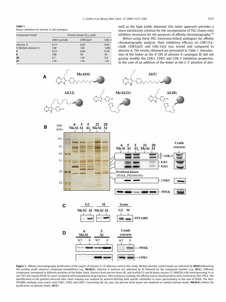
Table 1Kinase inhibition by aloisine A and analogues
Compound tested Protein kinase IC50 (lM)
CDK1/cyclinB CDK5/p25 GSK-3
Aloisine A 0.15 0.20 0.65N-Methyl-aloisine A 140 160 >2005 0.23 0.66 0.346 100 50 4520 6.7 >10 5.821 >10 >10 >10
MW(kDa)
191 -
97 -
64 -
51 -
39 -
28 -
NN
N
OO
O
NN
N
OH
N
NN
O
A
B MeAl Al Ald MeAl Al
M
MeAl(6)
CAl AlMeAl MeAl
MG2
P(P
D MeAl AlWT + WT +
M
6 5 21 205
6 5
NHN
N
OH
O
O
O
Al(12)
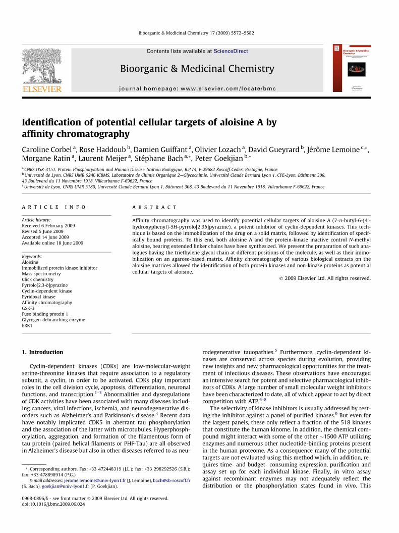
Figure 1. Affinity chromatography purification of the targets of aloisine A. (A) Matrices uthe number small chemical compound immobilized (e.g., MeAl(6)). Aloisine A matriccompounds correspond to different positions of the linker chain. Extracts from porcine brnot (WT) the human PDXK (D) were incubated with immobilized-drug matrices. After extidentification of the proteins detected after silver staining was realized by western blotPSTAIRE antibody cross-reacts with CDK1, CDK2 and CDK3. Concerning the Ald lane, thepurification on aloisine beads (Al(5)).
C. Corbel et al. / Bioorg. Med. Chem. 17 (2009) 5572–5582 5575
well as the high yields obtained, this latter approach provides amore satisfactory solution for the incorporation of TEG chains ontoinhibitor structures for the purposes of affinity chromatography.24
Before using these PEG extension-linked analogues for affinitychromatography analysis, their inhibitory efficacy on CDK1/Cy-clinB, CDK5/p25 and GSK-3a/b was tested and compared toaloisine A. The results obtained are presented in Table 1. Introduc-tion of the linker at the 40-OH of aloisine A (analogue 5) did notgreatly modify the CDK1, CDK5 and GSK-3 inhibition properties.In the case of an addition of the linker at the C-30 position of aloi-
α
NHN
N
OO
O
O
NHN
N
OH
N
NN
OO
GSK-3β2β
Erk1Erk2
PDXK
CDK5
eAl Al Ald
Al(5)
Crudeextracts
lysate
G2 M
PSTAIRE
yridoxal Kinase DXK_PIG/046560)
PDXK
CDK1
Crudeextracts
WT +
eAl(21) Al(20)
MeAl Al6 5 21 205
sed in this study. Methyl-aloisine control beads are indicated by MeAl followed byes are indicated by Al followed by the compound number (e.g., Al(5)). Differentain (B), and starfish G2 and M phase oocytes (C) HEK293 cells overexpressing (+) orensive washing, the affinity matrix-bound proteins were resolved by SDS–PAGE. Theting with specific antibodies or mass spectrometry in the case of PDXK. The Anti-porcine brain lysate was depleted on methyl-aloisine beads (MeAl(6)) before the
5576 C. Corbel et al. / Bioorg. Med. Chem. 17 (2009) 5572–5582
sine A (analogue 20), the inhibition properties are significantly de-creased. This modification is probably the consequence of sterichindrance inside the ATP binding pocket of the selected kinases.As already known,18 the substitution of the hydrogen atom in posi-tion 5 by a methyl group results in a dramatic decrease in theinhibitory activity of the analogues 6 and 21. The aloisine A ana-logues 5 and 6 were thus chosen as the best tools for the initialselectivity analysis by affinity chromatography. However, theremaining analogues were used for identifying cellular targets thatbind in a different manner than the CDK’s.
2.2.4. Preparation of affinity chromatography matricesIn order to identify the cellular targets of aloisine A, the chem-
ical analogues substituted with a linker at the different positions(compounds 5 and 6, 12, 20 and 21 in Scheme 2) were coupledto sepharose beads to provide the matrices Al(5), MeAl(6),Al(12), Al(20), and MeAl(21) (Fig. 1A). Coupling of the compoundsto the matrix were realized according to the recommendations ofthe manufacturer (Sigma–Aldrich) and is described in the experi-mental procedures section. Sepharose beads conjugated with eth-anolamine were prepared as controls.
2.3. Identification of potential cellular targets of aloisine A
2.3.1. Purification of potential aloisine A cellular targets byaffinity chromatography
The immobilized small-molecule ligand matrices were firstincubated with porcine brain extracts. Unbound proteins were
MeAl(6)Al(5)d
MW(kDa)
191 -
97 -
64 -
51 -
39 -
28 -
MeAl(6) bead
a. Clathrin heavy chain (CLH1_Human) and GlycogDebranching Enzyme (GDE_CANFA)
b. Exportin 1 (XP01_HUM
Exportin 7 (XP07_PONPY
Oxoglutarate dehydroge(ODO1_Human)
c. Exportin 2 (XP02_HUM
d. Glycogen phosphoryl(PYGM_BOVIN)
e. Fuse binding protein (FUBP1_MOUSE)
f. Tubulin beta chain (TB
g. Glutamine synthase (G
h. Glutamine synthase (GLNA_PIG) and beta Act(ACTB_CERPY)
i. Glial Fibrillary acidic(GFQP_BOVIN)
j. Pyridoxal kinase (PDX
abcde
fghij
p
mn
l
k
o
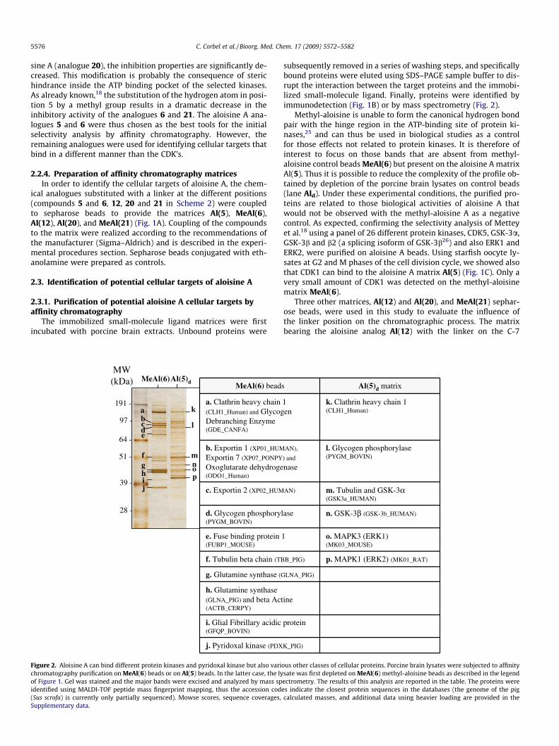
Figure 2. Aloisine A can bind different protein kinases and pyridoxal kinase but also varichromatography purification on MeAl(6) beads or on Al(5) beads. In the latter case, the lyof Figure 1. Gel was stained and the major bands were excised and analyzed by mass spidentified using MALDI-TOF peptide mass fingerprint mapping, thus the accession cod(Sus scrofa) is currently only partially sequenced). Mowse scores, sequence coverages,Supplementary data.
subsequently removed in a series of washing steps, and specificallybound proteins were eluted using SDS–PAGE sample buffer to dis-rupt the interaction between the target proteins and the immobi-lized small-molecule ligand. Finally, proteins were identified byimmunodetection (Fig. 1B) or by mass spectrometry (Fig. 2).
Methyl-aloisine is unable to form the canonical hydrogen bondpair with the hinge region in the ATP-binding site of protein ki-nases,25 and can thus be used in biological studies as a controlfor those effects not related to protein kinases. It is therefore ofinterest to focus on those bands that are absent from methyl-aloisine control beads MeAl(6) but present on the aloisine A matrixAl(5). Thus it is possible to reduce the complexity of the profile ob-tained by depletion of the porcine brain lysates on control beads(lane Ald). Under these experimental conditions, the purified pro-teins are related to those biological activities of aloisine A thatwould not be observed with the methyl-aloisine A as a negativecontrol. As expected, confirming the selectivity analysis of Metteyet al.18 using a panel of 26 different protein kinases, CDK5, GSK-3a,GSK-3b and b2 (a splicing isoform of GSK-3b26) and also ERK1 andERK2, were purified on aloisine A beads. Using starfish oocyte ly-sates at G2 and M phases of the cell division cycle, we showed alsothat CDK1 can bind to the aloisine A matrix Al(5) (Fig. 1C). Only avery small amount of CDK1 was detected on the methyl-aloisinematrix MeAl(6).
Three other matrices, Al(12) and Al(20), and MeAl(21) sephar-ose beads, were used in this study to evaluate the influence ofthe linker position on the chromatographic process. The matrixbearing the aloisine analog Al(12) with the linker on the C-7
s Al(5)d matrix
1 en
k. Clathrin heavy chain 1 (CLH1_Human)
AN),
) and
nase
l. Glycogen phosphorylase (PYGM_BOVIN)
AN) m. Tubulin and GSK-3α (GSK3a_HUMAN)
ase n. GSK-3β (GSK-3b_HUMAN)
1 o. MAPK3 (ERK1) (MK03_MOUSE)
B_PIG) p. MAPK1 (ERK2) (MK01_RAT)
LNA_PIG)
ine
protein
K_PIG)
ous other classes of cellular proteins. Porcine brain lysates were subjected to affinitysate was first depleted on MeAl(6) methyl-aloisine beads as described in the legendectrometry. The results of this analysis are reported in the table. The proteins werees indicate the closest protein sequences in the databases (the genome of the pigcalculated masses, and additional data using heavier loading are provided in the
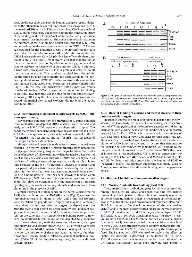
Eth 1 2.5 5 10 0 0.1 0
ATP (mM) on Al(5) beads
CDK5
GSK-3
PDXK
Erk1Erk2
0 15 0 7.5 15 7.5 10
MeAl(6) beads Al(5) beads
ATP (mM) Pyridoxal (mM) Pyridoxal (mM)
A
B
αβ2β
CDK5
GSK-3
PDXK
Erk1Erk2
αβ2β
Figure 3. Analysis of the mode of interaction between cellular components andaffinity matrices by competition assays. Porcine brain extracts were supplementedwith increasing concentrations of ATP (A and B) or pyridoxal (B), prior to loading onthe indicated matrices (Al(5) or MeAl(6)).
C. Corbel et al. / Bioorg. Med. Chem. 17 (2009) 5572–5582 5577
position did not show any specific binding and gave results identi-cal to the ethanolamine control (not shown). As shown in Figure 1,the matrix Al(20) with a C-40 linker cannot bind CDK5 but can bindGSK-3. This is most likely due to steric hindrance. Indeed, the studyof the binding mode of CDKs/GSK-3 inhibitors by co-crystallizationexperiments have indicated that the major difference is located inthe entrance of the ATP-binding pocket and thus that GSK-3 canaccommodate bulkier compounds compared to CDK5.27,28 The re-sult obtained for the inhibition of GSK-3 by 20 confirms this data(see Table 1). Indeed compound 20 is still able to inhibit theGSK-3 kinase activity (IC50 = 5.8 lM) but less efficiently than com-pound 5 (IC50 = 0.34 lM). This indicates also that modification ofthe structure at this position by addition of bulky group could beused to increase the selectivity of aloisines for GSK-3. In addition,a band that corresponds to a �37 kDa protein is detected on allthe matrices evaluated. This band was excised from the gel foridentification by mass spectrometry and corresponds to the por-cine pyridoxal kinase, PDXK. We further confirmed this interactionwith human PDXK stably over-expressed in human HEK293 cells(Fig. 1D). In this case, the high level of PDXK expression resultsin reduced binding of CDK1, suggesting a competition for bindingto aloisine. PDXK may thus act as a sink for aloisine and cells over-expressing PDXK may thus lose some sensitivity to aloisine. As ex-pected, the methyl-aloisine gel MeAl(21) did not bind GSK-3, butdoes bind PDXK.
2.3.2. Identification of potential cellular targets by MALDI-TOFmass spectrometry
Eleven bands obtained from the MeAl(6) and 12 bands obtainedfrom methylaloisine-depleted Al(5) matrices were excised andanalyzed by MALDI-TOF peptide mass fingerprint mapping. Thebands that yielded conclusive identifications are reported in Figure2. All the mass spectrometry data obtained are reported in the S1(for MeAl(6) matrix) and S2 and S3 (for Al(5) matrix) tables ofthe Supplementary data section.
Methyl-aloisine A interacts with several classes of non-kinaseproteins. The methyl-aloisine A matrix MeAl(6) binds notably to:(i) glycogen debranching enzyme that helps facilitate the break-down of glycogen;29 (ii) oxoglutarate dehydrogenase, a key controlpoint in the citric acid cycle that uses NADP+ and coenzyme A asco-factors;30 (iii) glycogen phosphorylase, catalyzes phosphoro-lytic cleavage of the a(1?4) glycosidic linkages of glycogen anduses pyridoxal phosphate for acid/base catalysis by the enzyme,which furthermore has a well characterized indole-binding site;31
iv) fuse binding protein 1 that has been shown to function as anATP-dependent DNA helicase;32 (v) glutamine synthase, an en-zyme that plays an essential role in the metabolism of nitrogenby catalyzing the condensation of glutamate and ammonia to formglutamine in the presence of ATP.33
Similar analysis of protein bands on the parent aloisine matrixAl(5) after depletion on the MeAl(6) matrix (Fig. 2) confirm theimmunoblot results in Figure 1: both GSK-3 and Erk isoformswere identified by peptide mass fingerprint mapping. Removingmore abundant and less selective targets by depletion on theMeAl(6) matrix will permit to better identify less abundant tar-gets such as protein kinases, and to focus on those targets thatrely on the canonical ATP-competitive H-bonding pattern. How-ever, no additional targets bound on the depleted Al(5) inhibitormatrix were identified, with the exception of tubulin, glycogenphosphorylase and clathrin heavy chain 1, all of which were alsoidentified on the MeAl(6) matrix.34 Heavier loading of the matrixin order to study some of the minor bands led only to the iden-tification of weakly binding, relatively abundant metabolic pro-teins (Table S3 of the Supplementary data), but no additionalprotein kinases.
2.3.3. Mode of binding of aloisine and methyl-aloisine to theirputative cellular targets
In order to analyse the mode of binding of aloisine and methyl-aloisine, we have analyzed the effect of increasing the concentra-tions of ATP and pyridoxal in the brain extract, prior to and duringincubation with aloisine beads, on the binding of several proteintargets (Fig. 3). First, ATP is able to compete for the binding ofGSK-3 isoforms, ERK1/2, PDXK and CDK5 to Al(5) beads (Fig. 3A).These results are consistent with kinetic studies, as well as the res-olution of a CDK2-aloisine co-crystal structure, that demonstratethat aloisine acts by competitive inhibition of ATP binding to thecatalytic subunit of protein kinase.18 In the case of PDXK the modeof interaction is less predictable. ATP is able to compete for thebinding of PDXK to both Al(5) beads and MeAl(6) beads (Fig. 3Aand B). Pyridoxal can only compete for the binding of PDXK tothe MeAl(6) matrix (Fig. 3B) beads suggesting that methyl-aloisineA and aloisine A have two different binding sites on pyridoxalkinase.
2.4. Aloisine A inhibition of non-mammalian targets
2.4.1. Aloisine A inhibits two budding yeast CDKsThere are six CDKs in the budding yeast Saccharomyces cerevisiae.
Among these CDKs are Cdc28 and Pho85 that bind two distinctclasses of cyclins to perform different functions namely regulationof the cell cycle transitions (Cdc28) or regulation of the cellular re-sponse to nutrient levels and environmental conditions (Pho85).35
Pho85 is the yeast functional homologue of the mammalianCDK536 and is the most closely related kinase in the yeast kinometo Cdc28 (for review see37). Cdc28 is the yeast homologue of CDK1and regulates each cell cycle transition in yeast.38 As shown in Fig-ure 4A, both Pho85 and Cdc28 can be purified on aloisine matrixfrom yeast cell lysates. As expected, methyl-aloisine cannot bindto these CDKs. To confirm these targets, we have analyzed the inhi-bition of Pho85 and Cdc28. An in vivo assay using the transcriptionfactor Pho4 tagged with GFP was used to analyse the effect onPho85 (the principle is described in the legend of Fig. 4). A250 lM aloisine treatment induces a nuclear localization of theGFP-tagged transcription factor Pho4 showing that Pho85 is
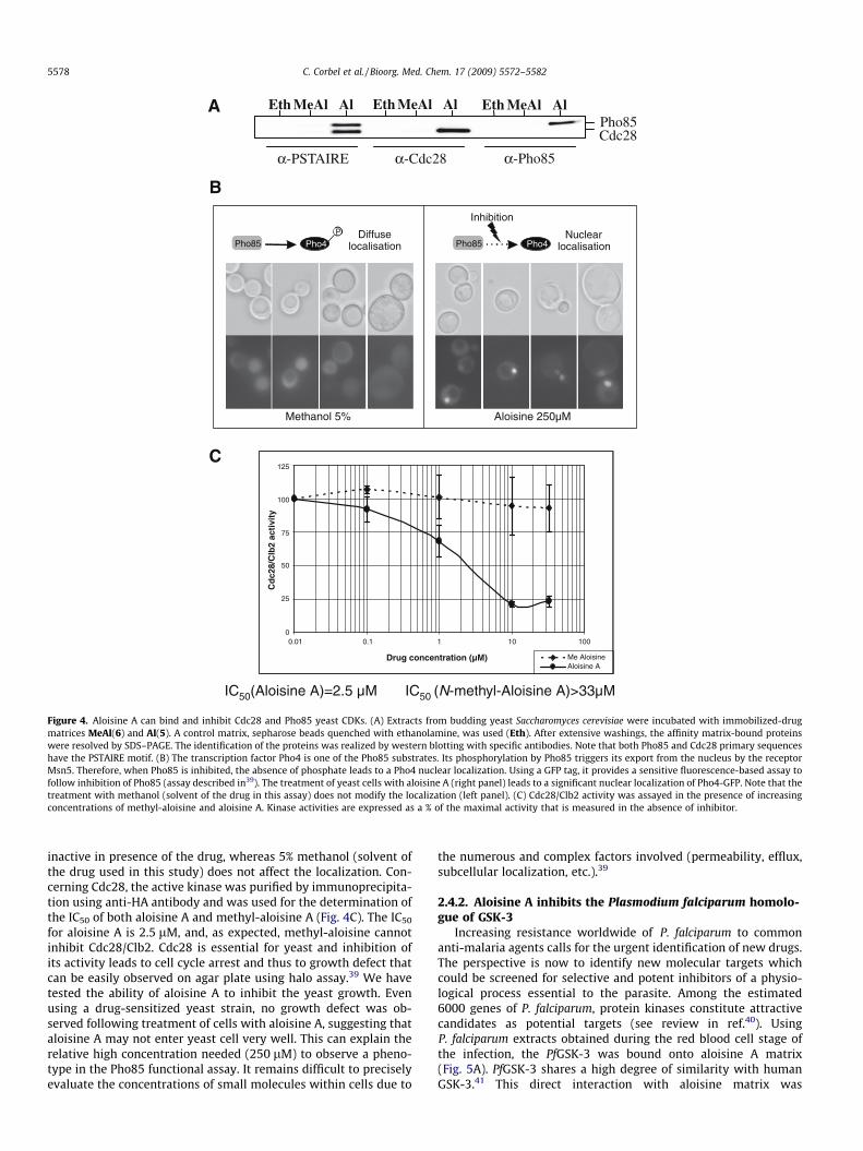
A
B
MeAl AlEthPho85Cdc28
α-PSTAIRE α-Cdc28 α-Pho85
MeAl AlEth MeAl AlEth
C
0
25
50
75
100
125
1001010.10.01
Drug concentration (µM)
Cd
c28/
Clb
2 ac
tivi
ty
Me AloisineAloisine A
Pho85 Pho4P Diffuse
localisation Pho85 Pho4Nuclear
localisation
Inhibition
Aloisine 250µMMethanol 5%
IC50(Aloisine A)=2.5 µM IC50 (N-methyl-Aloisine A)>33µM
Figure 4. Aloisine A can bind and inhibit Cdc28 and Pho85 yeast CDKs. (A) Extracts from budding yeast Saccharomyces cerevisiae were incubated with immobilized-drugmatrices MeAl(6) and Al(5). A control matrix, sepharose beads quenched with ethanolamine, was used (Eth). After extensive washings, the affinity matrix-bound proteinswere resolved by SDS–PAGE. The identification of the proteins was realized by western blotting with specific antibodies. Note that both Pho85 and Cdc28 primary sequenceshave the PSTAIRE motif. (B) The transcription factor Pho4 is one of the Pho85 substrates. Its phosphorylation by Pho85 triggers its export from the nucleus by the receptorMsn5. Therefore, when Pho85 is inhibited, the absence of phosphate leads to a Pho4 nuclear localization. Using a GFP tag, it provides a sensitive fluorescence-based assay tofollow inhibition of Pho85 (assay described in39). The treatment of yeast cells with aloisine A (right panel) leads to a significant nuclear localization of Pho4-GFP. Note that thetreatment with methanol (solvent of the drug in this assay) does not modify the localization (left panel). (C) Cdc28/Clb2 activity was assayed in the presence of increasingconcentrations of methyl-aloisine and aloisine A. Kinase activities are expressed as a % of the maximal activity that is measured in the absence of inhibitor.
5578 C. Corbel et al. / Bioorg. Med. Chem. 17 (2009) 5572–5582
inactive in presence of the drug, whereas 5% methanol (solvent ofthe drug used in this study) does not affect the localization. Con-cerning Cdc28, the active kinase was purified by immunoprecipita-tion using anti-HA antibody and was used for the determination ofthe IC50 of both aloisine A and methyl-aloisine A (Fig. 4C). The IC50
for aloisine A is 2.5 lM, and, as expected, methyl-aloisine cannotinhibit Cdc28/Clb2. Cdc28 is essential for yeast and inhibition ofits activity leads to cell cycle arrest and thus to growth defect thatcan be easily observed on agar plate using halo assay.39 We havetested the ability of aloisine A to inhibit the yeast growth. Evenusing a drug-sensitized yeast strain, no growth defect was ob-served following treatment of cells with aloisine A, suggesting thataloisine A may not enter yeast cell very well. This can explain therelative high concentration needed (250 lM) to observe a pheno-type in the Pho85 functional assay. It remains difficult to preciselyevaluate the concentrations of small molecules within cells due to
the numerous and complex factors involved (permeability, efflux,subcellular localization, etc.).39
2.4.2. Aloisine A inhibits the Plasmodium falciparum homolo-gue of GSK-3
Increasing resistance worldwide of P. falciparum to commonanti-malaria agents calls for the urgent identification of new drugs.The perspective is now to identify new molecular targets whichcould be screened for selective and potent inhibitors of a physio-logical process essential to the parasite. Among the estimated6000 genes of P. falciparum, protein kinases constitute attractivecandidates as potential targets (see review in ref.40). UsingP. falciparum extracts obtained during the red blood cell stage ofthe infection, the PfGSK-3 was bound onto aloisine A matrix(Fig. 5A). PfGSK-3 shares a high degree of similarity with humanGSK-3.41 This direct interaction with aloisine matrix was
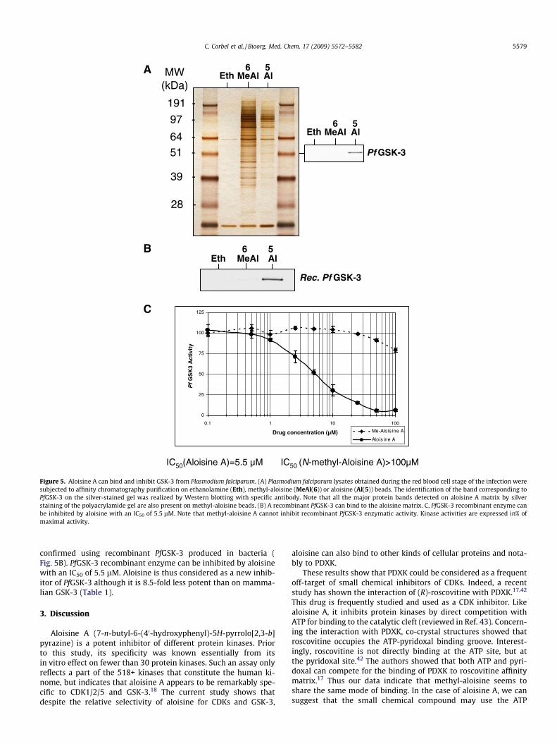
Eth MeAl Al
MeAl Al
MW(kDa)
191 -
97 -
64 -51 -
39 -
28 -
Pf GSK-3
B
A
C
Eth
MeAl Al
Rec. Pf GSK-3
Eth
0
25
50
75
100
125
1001010.1
Drug concentration (µM)
Pf
GS
K3
Act
ivit
y
Me-Alois ine A
Alois ine A
IC50(Aloisine A)=5.5 µM IC50 (N-methyl-Aloisine A)>100µM
6 5
6 5
6 5
Figure 5. Aloisine A can bind and inhibit GSK-3 from Plasmodium falciparum. (A) Plasmodium falciparum lysates obtained during the red blood cell stage of the infection weresubjected to affinity chromatography purification on ethanolamine (Eth), methyl-aloisine (MeAl(6)) or aloisine (Al(5)) beads. The identification of the band corresponding toPfGSK-3 on the silver-stained gel was realized by Western blotting with specific antibody. Note that all the major protein bands detected on aloisine A matrix by silverstaining of the polyacrylamide gel are also present on methyl-aloisine beads. (B) A recombinant PfGSK-3 can bind to the aloisine matrix. C, PfGSK-3 recombinant enzyme canbe inhibited by aloisine with an IC50 of 5.5 lM. Note that methyl-aloisine A cannot inhibit recombinant PfGSK-3 enzymatic activity. Kinase activities are expressed in% ofmaximal activity.
C. Corbel et al. / Bioorg. Med. Chem. 17 (2009) 5572–5582 5579
confirmed using recombinant PfGSK-3 produced in bacteria (Fig. 5B). PfGSK-3 recombinant enzyme can be inhibited by aloisinewith an IC50 of 5.5 lM. Aloisine is thus considered as a new inhib-itor of PfGSK-3 although it is 8.5-fold less potent than on mamma-lian GSK-3 (Table 1).
3. Discussion
Aloisine A (7-n-butyl-6-(40-hydroxyphenyl)-5H-pyrrolo[2,3-b]pyrazine) is a potent inhibitor of different protein kinases. Priorto this study, its specificity was known essentially from itsin vitro effect on fewer than 30 protein kinases. Such an assay onlyreflects a part of the 518+ kinases that constitute the human ki-nome, but indicates that aloisine A appears to be remarkably spe-cific to CDK1/2/5 and GSK-3.18 The current study shows thatdespite the relative selectivity of aloisine for CDKs and GSK-3,
aloisine can also bind to other kinds of cellular proteins and nota-bly to PDXK.
These results show that PDXK could be considered as a frequentoff-target of small chemical inhibitors of CDKs. Indeed, a recentstudy has shown the interaction of (R)-roscovitine with PDXK.17,42
This drug is frequently studied and used as a CDK inhibitor. Likealoisine A, it inhibits protein kinases by direct competition withATP for binding to the catalytic cleft (reviewed in Ref. 43). Concern-ing the interaction with PDXK, co-crystal structures showed thatroscovitine occupies the ATP-pyridoxal binding groove. Interest-ingly, roscovitine is not directly binding at the ATP site, but atthe pyridoxal site.42 The authors showed that both ATP and pyri-doxal can compete for the binding of PDXK to roscovitine affinitymatrix.17 Thus our data indicate that methyl-aloisine seems toshare the same mode of binding. In the case of aloisine A, we cansuggest that the small chemical compound may use the ATP
5580 C. Corbel et al. / Bioorg. Med. Chem. 17 (2009) 5572–5582
binding site. A co-crystal structure of PDXK-aloisine A wouldclearly shed light on the position of atoms involved in the interac-tion. These data would be helpful to obtain derived structures ofaloisine that cannot bind to PDXK but still to protein kinases toavoid potential side effects during preclinical/clinical studies. In-deed, PDXK catalyses the phosphorylation of the three forms ofvitamin B6 (pyridoxine, pyridoxal and pyridoxamine) in the pres-ence of ATP and Zn2+. This phosphorylation constitutes an essentialstep in the synthesis of pyridoxal 50-phosphate, the intracellularactive form of vitamine B6, a key factor for at least 140 enzymes,such as aminotransferases and decarboxylases. But, in the case ofthe roscovitine, despite the interaction of this drug with PDXK,its effects on the catalytic activity of PDXK are rather limited.Experimental data indicate that only 100 lM of ATP is sufficientto compete roscovitine out, therefore resulting in a weak effecton PDXK activity.42
This study of putative cellular targets of aloisine and methyl-aloisine using an affinity chromatography method has furthershown that these compounds can also interact with non kinaseproteins: for instance glycogen phosphorylase, glycogen-debran-ching enzyme, oxoglutarate dehydrogenase, glutamine synthase,and Fuse binding protein 1. We feel that these findings illustratethe advantages of this method for identifying cellular targets of agiven family of compounds. Indeed, these results were unexpectedbefore this study and can only be obtained using such a compre-hensive approach. However, these results must still be analyzedcarefully in order to evaluate the contribution of the binding ofaloisine A to these proteins to the pharmacological properties ofthe drug, such as the inhibition of cell proliferation as shown byMettey et al.18 The use of methyl-aloisine as a negative control insuch studies, however, can help restrict the origin of the observedeffect to the protein kinases that were identified after depletion onthe MeAl(6) matrix (Ald), namely the CDK, ERK and GSK isoforms.
Using various models, from unicellular eukaryote to mamma-lian cells and tissues, we have also shown the conservation of tar-gets of aloisine A through evolution, as illustrated by the inhibitionof two budding yeast CDKs (Pho85 and Cdc28), and also the GSK-3counterpart of P. falciparum.
The simultaneous evaluation of all classes of proteins expressedin one tissue (from dehydrogenases to protein kinases) is one of thestrengths of this method. But as such, it also raises the question ofthe relative expression of the putative targets. Indeed, this methodreflects the distribution of the inhibitor within target proteins inthe cell, meaning that highly abundant detected targets can maskless abundant, though perhaps relevant candidates. In our case,we chose to focus on those targets that do not bind the methyl-aloisine negative control by depleting the cell lysates on themethyl-aloisine matrix. For the most part these were limited tothe expected CDK, GSK-3 and ERK isoforms. It is particularly inter-esting to note that the off-target activities of the aloisines are notdue to the typical ATP-competitive binding mode, as these targetsall bind equally well to methyl-aloisine.
It is also worth pointing out here that while these matricescould be used to provide a comprehensive determination of all cel-lular targets (preferably using human tissues), it is not the objec-tive of the current study. Larger amounts of proteins and matrixassociated with separation of the putative targets using two-dimensional gel electrophoresis have been used to increase thesensitivity of the analysis in the study of chemical inhibitors.44,45
Instead, we aim here to provide only those new targets that wereidentified conclusively, and an insight into the distribution of thecompound among its principal cellular targets. Furthermore, inconjunction with other such studies,13–17,46 we hope to identifythose recurring targets, such as PDXK and NADH-dependentenzymes, that should be included systematically in the panel ofenzymes used for the study of protein kinase inhibitor selectivity.
4. Conclusion
Potential applications of CDK/GSK-3 inhibitors are currentlyevaluated against cancers, neurodegenerative disorders such asAlzheimer’s disease, proliferation of protozoan parasites, and viralinfections (HIV, cytomegalovirus, and herpes virus). Using affinitychromatography on immobilized drug, we show here the charac-terization of the putative cellular targets of aloisine A. This com-pound was initially selected for its ability to be a potent inhibitorof CDK1/2/5 and GSK-3. This study indicates that despite the rela-tive selectivity of aloisine for CDKs and GSK-3, aloisine can alsobind to other kind of cellular proteins and notably PDXK. Analysisof various biological models indicates that aloisine selectivity pro-file is well conserved from the yeast S. cerevisiae to human. Each ofthese interactions must be carefully investigated in order to im-prove the biological activity of the drug and also to evaluate andavoid the possible side effects preliminary to further assays on ani-mal models of human diseases involving CDK5 and/or GSK-3. How-ever, our results indicate that methyl-aloisine can be usedeffectively as a control for many of these non-kinase activities.
5. Experimental procedures
5.1. Synthesis
Compounds 2–21 were synthesised by following the pathwaydescribed in Scheme 2. Detailed synthetic procedures and spectro-scopic data are available in the Supplementary data.
5.2. Affinity chromatography on immobilized drugs
Homogenization buffer: 60 mM b-glycerophosphate, 15 mM p-nitrophenylphosphate, 25 mM Mops (pH 7.2), 15 mM EGTA,15 mM MgCl2, 1 mM DTT, 1 mM sodium vanadate, 1 mM NaF,1 mM phenylphosphate, 10 lg leupeptin/ml, 10 lg aprotinin/ml,10 lg soybean trypsin inhibitor/ml and 100 lM benzamidine.
Bead buffer: 50 mM Tris pH 7.4, 5 mM NaF, 250 mM NaCl, 5 mMEDTA, 5 mM EGTA, 0.1% NP-40, 10 lg/ml of leupeptin, aprotininand soybean trypsin inhibitor and 100 lM benzamidine.
5.2.1. Immobilization of drugs on sepharose matrixThe drug attached to the linker (e.g., 5 for active aloisine; 6 for
negative control N-methyl analogue) are coupled in 0.1 M NaHCO3,0.2 M NaCl, pH 8.3 on CNBr-Activated Sepharose 4B (Sigma–Al-drich, #C9142) under agitation at room temperature for 8 h. Thefinal calculated loading was 3 lmol/ml of resin. After the couplingprocess, the remaining active groups are blocked in pH 8.0 buffercontaining 1 M ethanolamine, under agitation for 3 h at 20 �C.The beads are then washed three times with bead buffer and storedin bead buffer (supplemented with 0.05% NaN3 to avoid microbialgrowth) at 4 �C until use.
5.2.2. Preparation of extractsPork brains were obtained from a local slaughterhouse and di-
rectly homogenized and processed for affinity chromatography orstored at �80 �C prior to use. Tissues were weighed, homogenizedand sonicated in homogenization buffer (2 ml per g of material).Homogenates were centrifuged for 10 min at 14,000g at 4 �C. Thesupernatant was recovered, assayed for protein content (Bio-Radprotein assay) and immediately loaded batch wise on the affinitymatrix. Yeast S. cerevisiae extracts were prepared using a ‘One Shot’cell disrupter (Constant Systems LTD, Daventry, England).
5.2.3. Affinity chromatography of aloisine-interacting proteinsJust before use, 10 ll of packed aloisine beads were washed
with 1 ml of bead buffer and resuspended in 600 ll of this buffer.

C. Corbel et al. / Bioorg. Med. Chem. 17 (2009) 5572–5582 5581
The cell or tissue extract supernatant (3 mg total protein) or puri-fied protein was then added; the tubes were rotated at 4 �C for30 min. After a brief spin at 10,000g and removal of the superna-tant, the beads were washed 4 times with bead buffer beforeaddition of 60 ll 2X Laemmli sample buffer. Following heat dena-turation for 3 min, the bound proteins were analyzed by SDS–PAGEand western blotting or silver staining as described below.
5.2.4. Electrophoresis, western blotting, and silver stainingFollowing heat denaturation for 3 min, the proteins bound to
the aloisine matrices were separated by 10% SDS–PAGE (1 mmthick gels) followed by immunoblotting analysis or silver stainingusing a GE Healthcare SDS–PAGE silver staining kit. For immuno-blotting, proteins were transferred to 0.45 lm nitrocellulose filters(Schleicher and Schuell). These were blocked with 5% low fat milkin Tris-Buffered Saline—Tween-20, incubated for 1 h with antibod-ies and analyzed by Enhanced Chemiluminescence (ECL, GEHealthcare). The following antibodies were used: anti-GSK-3(1:1000, Stressgen), anti-CDK5 (1:500; Santa Cruz), anti-ERK1/2(1:4000; Sigma), anti-p35/p25 (1:500; Santa Cruz), anti-PSTAIRE(1:3000, Sigma), anti-PDXK (1:500;17), anti-Pho85 and anti-Cdc28(respectively given by E. O’Shea and C. Mann).
5.2.5. Protein identification by mass spectrometrySilver stained protein bands were excised from the correspond-
ing 1D gel. In-gel digestion was carried out using porcine trypsinfrom Promega (Madison, WI, USA): Gel fragments were washedwith 2 � 200 ll of 25 mM ammonium hydrogen carbonate, driedwith CH3CN, and dehydrated under vacuum. Then, 10 ll of trypsinsolution (12.5 ng/ll in 25 mM ammonium hydrogen carbonate)was added and the digestion was carried out overnight at 37 �C.After separation of the supernatant, gel fragments were washedtwice with 50 ll of H2O/ACN (v/v) containing 0.5% formic acid.The fractions were concentrated under vacuum and stocked at�20 �C. Prior to analysis on an Ultraflex MALDI-TOF-TOF instru-ment (Bruker Daltonics, Bremen), the peptides were diluted in20 ll of H2O/ACN (v/v) containing 0.5% formic acid. One microliterof the peptide solution was mixed directly onto the target with 1 llof DHB matrix (10 mg/mL of DHB in H2O/CH3OH (v/v)). All peptidemass fingerprint spectra were internally calibrated with the trypticauto-digestion ions. Identification of proteins was achieved usingthe Mascot algorithm. Only those bands yielding a correlation fac-tor above 60 and a genomic predicted mass that is consistent withthe mass observed on the gel are shown in Figure 2. Mowse scores,sequence coverages, calculated masses, and additional data using aheavier loading are provided in the Supplementary data.
5.3. Protein kinase assays
The description of protein kinase preparations and assays(CDK1/cyclin B, CDK5/p25, GSK-3 and PfGSK-3) is provided in theSupplementary data section.
5.4. Biochemical and genetic experiments using the yeast S.cerevisiae
5.4.1. Strains and plasmidsMNY629 is a gift from Marie-Noëlle Simon (IGC, Marseille,
France) and allows the expression of CLB2 fused to HA-tag. YRP1is a gift from Karl Kuchler (Vienna University, Austria) and corre-sponds to a drug-sensitized yeast strain (snq2Dpdr5Derg6D).EB0347 is a gift from Erin O’Shea (Howard Hughes Medical Insti-tute, Harvard University, Cambridge, MA, USA). It contains PHO4promoter and the entire PHO4 ORF fused in-frame to N terminusof GFP on pRS316 plasmid.
5.4.2. Preparation of extractsYeast S. cerevisiae were grown to OD600nm � 2 (exponential
phase). A yeast pellet is then obtained by centrifugation(3000 rpm, 15 min) and diluted in breaking buffer (25 mM TrispH 7.4, 100 mM NaCl, 0.2% Triton X100, 15 mM p-nitrophenyl-phosphate, 0.1 mM Na-Vanadate, 1 mM PMSF and protease inhib-itor cocktail (CompleteTM, Roche)). Cells were broken using a ‘OneShot’ cell disrupter (Constant Systems LTD, Daventry, England)with a 2.5 kbar pressure. Extracts were clarified by centrifugation(13,000 rpm, 20 min) before protein quantification using a Brad-ford assay.
5.4.3. ImmunoprecipitationProtein A/G PLUS-agarose (Santa Cruz, SC-2003) and monoclo-
nal anti-HA (Covance, MMS-101R) were used for immunoprecipi-tation of the complex Cdc28/Clb2-HA before kinase assay.
5.4.4. Kinase assaysPurified Cdc28/Clb2-HA complex was incubated for 30 min at
30 �C in a 30 ll reaction mix containing 5 ll of Histone H1(1 mg/ml), 6 ll of [c-33P]ATP (15 lmol/l) and varying concentra-tions of drugs in buffer C (60 mmol/l b-glycerophosphate,15 mmol/l p-nitrophenylphosphate, 25 mmol/l MOPS (pH, 7.2),5 mmol/l EGTA, 15 mmol/l MgCl2, 1 mmol/l DTT, 1 mmol/l sodiumvanadate, 1 mmol/l phenylphosphate). After 30 min incubation at30 �C, 20 ll of supernatant were spotted onto pieces of WhatmanP81 phosphocellulose paper. The filters were washed three times(for at least 5 min each time) in a 1% phosphoric acid solution.The wet filters were counted in the presence of 1 ml ACS (GEHealthcare) scintillation fluid.
5.4.5. Pho4-GFP assayYRP1 carrying EB0347 plasmid were grown over-night under
selection (w/o uracil) then diluted at OD600nm = 0.5. The culture istreated with 250 lM aloisine A. Samples are observed at 3 h andanalyzed with fluorescence microscopy: GFP-tagged proteins werevisualized using a Chroma GFP4 filter (excitation 455–495 nm) onan Olympus BX61 microscope and photographed with a DiagnosticInstruments Spot RT cooled CCD camera.
Acknowledgments
We thank Dr. E. O’Shea for Pho4-GFP expressing vector andPho85 antibody, Dr. K. Kuchler for yeast highly permeable strainYRP1 and Dr. M.N. Simon for yeast strains expressing Clb2-HA pro-tein. Special thanks to Dr. C. Mann for Cdc28 antibody and also forhelpful suggestions. C.C. and D.G. are doctoral research fellowssupported by the ‘Région Bretagne—CNRS Département des Sci-ences du Vivant’ and ‘Ligue contre le Cancer, Comité des Côtes d’Ar-mor—Association pour la Recherche contre le Cancer’, respectively.S.B. is supported by ‘Association pour la Recherche contre le Can-cer’ (Contract ARC No. 3889). This research was also supportedby grants from the EEC (FP6-LSHB-CT-2004-503467, PRO-KINASEResearch (R.H., L.M., P.G.) and FP6-LSH-2003-2.3.0_2, ANTIMAL(L.M.)) and the Canceropole Grand-Ouest (L.M.).
Supplementary data
Supplementary data (The synthesis and characterization data ofcompounds 2–21) associated with this article can be found, in theonline version, at doi:10.1016/j.bmc.2009.06.024.
References and notes
1. Dhavan, R.; Tsai, L. H. Nat. Rev. Mol. Cell Biol. 2001, 2, 749.2. Malumbres, M.; Ortega, S.; Barbacid, M. Biol. Chem. 2000, 381, 827.
5582 C. Corbel et al. / Bioorg. Med. Chem. 17 (2009) 5572–5582
3. Morgan, D. O. Annu. Rev. Cell Dev. Biol. 1997, 13, 261.4. Knockaert, M.; Greengard, P.; Meijer, L. Trends Pharmacol. Sci. 2002, 23, 417.5. Mazanetz, M. P.; Fischer, P. M. Nat. Rev. Drug Discovery 2007, 6, 464.6. Cohen, P. Nat. Rev. Drug Discovery 2002, 1, 309.7. Fischer, P. M.; Lane, D. P. Curr. Med. Chem. 2000, 7, 1213.8. Gray, N.; Detivaud, L.; Doerig, C.; Meijer, L. Curr. Med. Chem. 1999, 6, 859.9. Davies, S. P.; Reddy, H.; Caivano, M.; Cohen, P. Biochem. J. 2000, 351, 95.
10. Bach, S.; Blondel, M.; Meijer, L.. In Monographs on Enzyme Inhibitors; CRC Press:Boca Raton, FL, 2006; Vol. 2, p 103.
11. Terstappen, G. C.; Schlupen, C.; Raggiaschi, R.; Gaviraghi, G. Nat. Rev. DrugDiscovery 2007, 6, 891.
12. Guiffant, D.; Tribouillard, D.; Gug, F.; Galons, H.; Meijer, L.; Blondel, M.; Bach, S.Biotechnol. J. 2007, 2, 68.
13. Knockaert, M.; Gray, N.; Damiens, E.; Chang, Y. T.; Grellier, P.; Grant, K.;Fergusson, D.; Mottram, J.; Soete, M.; Dubremetz, J. F.; Le Roch, K.; Doerig, C.;Schultz, P.; Meijer, L. Chem. Biol. 2000, 7, 411.
14. Knockaert, M.; Wieking, K.; Schmitt, S.; Leost, M.; Grant, K. M.; Mottram, J. C.;Kunick, C.; Meijer, L. J. Biol. Chem. 2002, 277, 25493.
15. Wan, Y.; Hur, W.; Cho, C. Y.; Liu, Y.; Adrian, F. J.; Lozach, O.; Bach, S.; Mayer, T.;Fabbro, D.; Meijer, L.; Gray, N. S. Chem. Biol. 2004, 11, 247.
16. Brehmer, D.; Greff, Z.; Godl, K.; Blencke, S.; Kurtenbach, A.; Weber, M.; Muller,S.; Klebl, B.; Cotten, M.; Keri, G.; Wissing, J.; Daub, H. Cancer Res. 2005, 65, 379.
17. Bach, S.; Knockaert, M.; Reinhardt, J.; Lozach, O.; Schmitt, S.; Baratte, B.; Koken,M.; Coburn, S. P.; Tang, L.; Jiang, T.; Liang, D. C.; Galons, H.; Dierick, J. F.; Pinna,L. A.; Meggio, F.; Totzke, F.; Schachtele, C.; Lerman, A. S.; Carnero, A.; Wan, Y.;Gray, N.; Meijer, L. J. Biol. Chem. 2005, 280, 31208.
18. Mettey, Y.; Gompel, M.; Thomas, V.; Garnier, M.; Leost, M.; Ceballos-Picot, I.;Noble, M.; Endicott, J.; Vierfond, J. M.; Meijer, L. J. Med. Chem. 2003, 46, 222.
19. Mapelli, M.; Massimiliano, L.; Crovace, C.; Seeliger, M. A.; Tsai, L. H.; Meijer, L.;Musacchio, A. J. Med. Chem. 2005, 48, 671.
20. Vierfond, J. M.; Mettey, Y.; Mascrier-Demagny, L.; Miocque, M. Tetrahedron Lett.1981, 22, 1219.
21. Davis, M.; Wakefield, B.; Wardell, J. Tetrahedron 1992, 48, 939.22. Fuchter, M.; Beall, L.; Baum, S.; Montalban, A.; Sakellariou, E.; Mani, N.; Miller,
T.; Vesper, B.; White, A.; Williams, D.; Barrett, A.; Hoffman, B. Tetrahedron2005, 61, 6115.
23. Eduardo, F. M.; Correa, J.; Irene, R. M.; Ricardo, R. Macromolecules 2006, 39,2113.
24. Haddoub, R.; Gueyrard, D.; Goekjian, P. Tetrahedron Lett. 2009, 50, 741.25. Traxler, P.; Furet, P. Pharmacol. Therapeut. 1999, 82, 195.26. Mukai, F.; Ishiguro, K.; Sano, Y.; Fujita, S. C. J. Neurochem. 2002, 81, 1073.
27. Fischer, P. M. Chem. Biol. 2003, 10, 1144.28. Meijer, L.; Skaltsounis, A. L.; Magiatis, P.; Polychronopoulos, P.; Knockaert, M.;
Leost, M.; Ryan, X. P.; Vonica, C. A.; Brivanlou, A.; Dajani, R.; Crovace, C.;Tarricone, C.; Musacchio, A.; Roe, S. M.; Pearl, L.; Greengard, P. Chem. Biol. 2003,10, 1255.
29. Watanabe, Y.; Makino, Y.; Omichi, K. J. Biochem. 2008, 143, 435.30. Araujo, W. L.; Nunes-Nesi, A.; Trenkamp, S.; Bunik, V. I.; Fernie, A. R. Plant
Physiol. 2008, 148, 1782.31. Lukacs, C. M.; Oikonomakos, N. G.; Crowther, R. L.; Hong, L.-N.; Kammlott, R. U.;
Levin, W.; Li, S.; Liu, C. M.; McGady, D. L.; Pietranico, S.; Reik, L. Proteins:Structure, Function and Bioinformatics 2006, 63, 1123.
32. Chung, H. J.; Liu, J.; Dundr, M.; Nie, Z.; Sanford, S.; Levens, D. Mol. Cell. Biol.2006, 26, 6584.
33. Berlicki, L. Mini.-Rev. Med. Chem. 2008, 8, 869.34. Reprocessing of the data with a recent version of the database led to the
possible identification of MEK-1 (Table S2 of the Supplementary data).However, Mettey et al. have shown previously that this kinase is not a targetof aloisine (Ref. 18).
35. Wu, D.; Dou, X.; Hashmi, S. B.; Osmani, S. A. J. Biol. Chem. 2004, 279,37693.
36. Huang, D.; Patrick, G.; Moffat, J.; Tsai, L. H.; Andrews, B. Proc. Natl. Acad. Sci.U.S.A. 1999, 96, 14445.
37. Huang, D.; Friesen, H.; Andrews, B. Mol. Microbiol. 2007, 66, 303.38. Ceccarelli, E.; Mann, C. J. Biol. Chem. 2001, 276, 41725.39. Kung, C.; Kenski, D. M.; Krukenberg, K.; Madhani, H. D.; Shokat, K. M. Chem.
Biol. 2006, 13, 399.40. Doerig, C.; Meijer, L.; Mottram, J. C. Trends Parasitol. 2002, 18, 366.41. Droucheau, E.; Primot, A.; Thomas, V.; Mattei, D.; Knockaert, M.; Richardson,
C.; Sallicandro, P.; Alano, P.; Jafarshad, A.; Baratte, B.; Kunick, C.; Parzy, D.;Pearl, L.; Doerig, C.; Meijer, L. Biochim. Biophys. Acta 2004, 1697, 181.
42. Tang, L.; Li, M. H.; Cao, P.; Wang, F.; Chang, W. R.; Bach, S.; Reinhardt, J.;Ferandin, Y.; Galons, H.; Wan, Y.; Gray, N.; Meijer, L.; Jiang, T.; Liang, D. C. J. Biol.Chem. 2005, 280, 31220.
43. Meijer, L.; Raymond, E. Acc. Chem. Res. 2003, 36, 417.44. Godl, K.; Wissing, J.; Kurtenbach, A.; Habenberger, P.; Blencke, S.; Gutbrod, H.;
Salassidis, K.; Stein-Gerlach, M.; Missio, A.; Cotten, M.; Daub, H. Proc. Natl. Acad.Sci. U.S.A. 2003, 100, 15434.
45. Brehmer, D.; Godl, K.; Zech, B.; Wissing, J.; Daub, H. Mol. Cell. Proteomics 2004,3, 490.
46. Trapp, J.; Jochum, A.; Meier, R.; Saunders, L.; Marshall, B.; Kunick, C.; Verdin, E.;Goekjian, P.; Sippl, W.; Jung, M. J. Med. Chem. 2006, 49, 7307.