
Identification of Human Gene Products Containing Pro-Pro-x ...
14
Original Paper Cell Physiol Biochem 2010;25:293-306 Accepted: November 19, 2009 Cellular Physiology Cellular Physiology Cellular Physiology Cellular Physiology Cellular Physiology and Biochemistr and Biochemistr and Biochemistr and Biochemistr and Biochemistry Copyright © 2010 S. Karger AG, Basel Fax +41 61 306 12 34 E-Mail [email protected] www.karger.com © 2010 S. Karger AG, Basel 1015-8987/10/0253-0293$26.00/0 Accessible online at: www.karger.com/cpb Identification of Human Gene Products Containing Pro-Pro-x-Tyr (PY) Motifs that Enhance Glutathione and Endocytotic Marker Uptake in Yeast Shujie Shi 1, *, Sylvia Notenboom 1, *, Mark E. Dumont 2 and Nazzareno Ballatori 1 Ned Ballatori, Ph.D. Box EHSC, 601 Elmwood Ave, Rochester, NY 14642 (USA) Tel. +1 585-275-0262, Fax +1 585-256-2591 E-Mail [email protected] Departments of Environmental Medicine 1 , and Biochemistry and Biophysics 2 , University of Rochester School of Medicine, Rochester, *Contributed equally to the results of this article Key Words PY-motif • Yeast • Membrane • Transport • Endocytosis • Glutathione Abstract In an attempt to identify genes involved in glutathione (GSH) transport, a human mammary gland cDNA library was screened for clones capable of complementing a defect in GSH uptake in yeast cells that lack Hgt1p, the primary yeast GSH uptake transporter. Five genes capable of rescuing growth on sulfur-deficient GSH-containing medium were identified: prostate transmembrane protein, androgen induced 1 (PMEPA1); lysosomal-associated protein transmembrane 4 alpha (LAPTM4α); solute carrier family 25, member 1 (SLC25A1); lipopolysaccharide- induced TNF factor (LITAF); and cysteine/tyrosine- rich-1 (CYYR1). All of these genes encode small integral membrane proteins of unknown function, although none appear to encode prototypical GSH transporters. Nevertheless, they all increased both intracellular glutathione levels and [ 3 H]GSH uptake rates. [ 3 H]GSH uptake was uniformly inhibited by high concentrations of unlabeled GSH, GSSG, and ophthalmic acid. Interestingly, each protein is predicted to contain Pro-Pro-x-Tyr (PY) motifs, which are thought to be important for regulating protein cell surface expression. Uptake of the endocytotic markers lucifer yellow and FM4-64 was also enhanced by each of the five genes. Mutations of the PY motifs in LITAF largely abolished all of its effects. In summary, although the results do not reveal novel GSH transporters, they identify five PY-containing human gene products that may influence plasma membrane transport activity. Introduction Glutathione (GSH) is a major intracellular thiol, and is present in virtually all eukaryotic and most prokaryotic cells at concentrations ranging from 0.5 to 10 mM. Maintenance of GSH levels is required for many critical cell processes, and disturbances in GSH homeostasis have been implicated in the etiology and/or progression of a number of human diseases, including cancer, diseases of aging, and cardiovascular, inflammatory, immune, liver and
-
Upload
khangminh22 -
Category
Documents
-
view
3 -
download
0
Transcript of Identification of Human Gene Products Containing Pro-Pro-x ...

293
Original Paper
Cell Physiol Biochem 2010;25:293-306 Accepted: November 19, 2009Cellular PhysiologyCellular PhysiologyCellular PhysiologyCellular PhysiologyCellular Physiologyand Biochemistrand Biochemistrand Biochemistrand Biochemistrand Biochemistryyyyy
Copyright © 2010 S. Karger AG, Basel
Fax +41 61 306 12 34E-Mail [email protected]
© 2010 S. Karger AG, Basel1015-8987/10/0253-0293$26.00/0
Accessible online at:www.karger.com/cpb
Identification of Human Gene Products ContainingPro-Pro-x-Tyr (PY) Motifs that EnhanceGlutathione and Endocytotic Marker Uptake inYeastShujie Shi1,*, Sylvia Notenboom1,*, Mark E. Dumont2 and NazzarenoBallatori1
Ned Ballatori, Ph.D.Box EHSC, 601 Elmwood Ave, Rochester, NY 14642 (USA)Tel. +1 585-275-0262, Fax +1 585-256-2591E-Mail [email protected]
Departments of Environmental Medicine1, and Biochemistry and Biophysics2, University of RochesterSchool of Medicine, Rochester, *Contributed equally to the results of this article
Key WordsPY-motif • Yeast • Membrane • Transport • Endocytosis• Glutathione
AbstractIn an attempt to identify genes involved in glutathione(GSH) transport, a human mammary gland cDNAlibrary was screened for clones capable ofcomplementing a defect in GSH uptake in yeast cellsthat lack Hgt1p, the primary yeast GSH uptaketransporter. Five genes capable of rescuing growthon sulfur-deficient GSH-containing medium wereidentified: prostate transmembrane protein, androgeninduced 1 (PMEPA1); lysosomal-associated proteintransmembrane 4 alpha (LAPTM4α); solute carrierfamily 25, member 1 (SLC25A1); lipopolysaccharide-induced TNF factor (LITAF); and cysteine/tyrosine-rich-1 (CYYR1). All of these genes encode smallintegral membrane proteins of unknown function,although none appear to encode prototypical GSHtransporters. Nevertheless, they all increased bothintracellular glutathione levels and [3H]GSH uptakerates. [3H]GSH uptake was uniformly inhibited by highconcentrations of unlabeled GSH, GSSG, andophthalmic acid. Interestingly, each protein is
predicted to contain Pro-Pro-x-Tyr (PY) motifs, whichare thought to be important for regulating protein cellsurface expression. Uptake of the endocytotic markerslucifer yellow and FM4-64 was also enhanced by eachof the five genes. Mutations of the PY motifs in LITAFlargely abolished all of its effects. In summary,although the results do not reveal novel GSHtransporters, they identify five PY-containing humangene products that may influence plasma membranetransport activity.
Introduction
Glutathione (GSH) is a major intracellular thiol, andis present in virtually all eukaryotic and most prokaryoticcells at concentrations ranging from 0.5 to 10 mM.Maintenance of GSH levels is required for many criticalcell processes, and disturbances in GSH homeostasis havebeen implicated in the etiology and/or progression of anumber of human diseases, including cancer, diseases ofaging, and cardiovascular, inflammatory, immune, liver and

294
neurodegenerative diseases [1]. Intracellular andextracellular GSH concentrations are normally tightlyregulated to maintain normal physiological functions, andthe two major mechanisms involved in this regulation areGSH biosynthesis and GSH efflux from cells [2]. Despitethe increasing evidence that some of the multidrugresistance-associated proteins (MRP/ABCC) mediateGSH export, it is unknown whether the MRPs are thesole or even the major GSH exporters in human cells.Given the important functions of GSH in multiple cellularprocesses, it is possible that additional GSH transportersare present in the human genome, but have not yet beenidentified at molecular level [3, 4].
With the aim of identifying additional GSHtransporters, we screened a human mammary glandcDNA library using an expression cloning strategy inyeast. Unlike mammalian cells, S. cerevisiae are able totake up GSH from the extracellular medium, and thisuptake is mediated primarily by a high affinity GSH uptaketransporter, Hgt1p [5]. In addition to Hgt1p, a low-affinityGSH uptake mechanism is present in yeast cells; however,the gene or genes responsible for this alternate GSH uptakeprocess have not yet been identified [5, 6]. Orthologuesof HGT1 are not present in higher eukaryotic organisms.The deletion of the HGT1 gene in S. cerevisiae leads to amarked impairment in the ability to grow in media in whichGSH is the only sulfur source [5]. The present study tookadvantage of the growth deficiency of the hgt1-Δ strainto search for human genes that can facilitate or mediateGSH accumulation by these cells and therefore rescuethe growth deficiency. Because the present screen testedfor genes that lead to enhanced GSH uptake in yeast, it isunable to identify unidirectional GSH exporters, but shouldtheoretically be able to identify bidirectional GSHtransporters (e.g., secondary active transporters) orunidirectional GSH uptake transporters.
Five genes capable off complementing the HGT1deletion were recovered, providing clues into the potentialfunctions of human PMEPA1 (prostate transmembraneprotein, androgen induced 1), LAPTM4α (lysosomal-associated protein transmembrane 4 alpha), SLC25A1(solute carrier family 25, member 1), LITAF(lipopolysaccharide-induced TNF factor), and CYYR1(cysteine/tyrosine-rich1). Because each of the encodedproteins is predicted to have PY motifs, which are thoughtto serve as binding sites for the ubiquitin ligases yeastRsp5 (reverses Spt-phenotype 5) or human NEDD4(neural precursor cell expressed, developmentally down-regulated 4), these genes may play a role in modulatingprotein cell surface expression.
Materials and Methods
MaterialsCulture media components were obtained from Clontech
Laboratories, Inc. (Mountain View, CA), Difco (Sparks, MD),Invitrogen (Carlsbad, CA) and Sigma-Aldrich (St. Louis, MO).[3H]Glutathione was purchased from Perkin Elmer (Boston, MA).Acivicin, glycine, glutamic acid, glutathione reductase, GSH,GSSG, Latrunculin A, lucifer yellow, lyticase, 0.1% poly-lysine,and yeast protease inhibitor cocktail (P8215) were obtainedfrom Sigma (St. Louis, MO). N-(3-triethylammoniumpropyl)-4-(6-(4-(diethylamino)phenyl) hexatrienyl)pyridinium dibromide(FM4-64) was obtained from Molecular Probes (Carlsbad, CA).Oligonucleotides were purchased from IDT (Coralville, IA).Ophthalmic acid was purchased from Bachem Bioscience Inc.(Philadelphia, PA). Zymolyase 100T was purchased fromSeikagaku Corp. (Tokyo, Japan). The anti-FLAG M2monoclonal antibody (F3165) and anti-mouse IgG peroxidaseconjugate antibody (A9044) were purchased from Sigma-Aldrich (St. Louis, MO); anti-Pep12p (A-21273) and anti-Dpm1p(A-6429) were obtained from Molecular Probe (Eugene, OR);anti-Pma1p (sc-57978) was purchased from Santa CruzBiotechnology (Santa Cruz, CA). All chemicals used were ofthe highest analytical grade possible.
Yeast strains and culture conditionThe hgt1-Δ yeast strain (ABC822: MATa ura3-52 leu2-Δ1
his3-Δ200 trp1-Δ63 lys2-801 ade2-101 hgt1Δ::LEU2) and itsparental yeast strain (ABC154: MATa ura3-52 leu2-Δ1 his3-Δ200 trp1-Δ63 lys2-801 ade2-101)) were kindly provided by Dr.A.K. Bachhawat (Institute of Microbial Technology,Chandigarh, India). In the mutant strain, the HGT1 gene wasreplaced with the LEU2 gene [5]. Rich YPAD medium, (1% yeastextract, 2% Bacto peptone, 2% glucose, 0.002% adenine) wasused for routine yeast culture. Yeast transformants wereselectively grown on minimal selective medium (0.17% yeastnitrogen base without amino acids, 0.5% ammonium sulfate,2% glucose, supplemented with the required amino acids andbases). Sulfur-deficient medium was prepared based on minimalselective medium with the modification that all sulfur-containing reagents were substituted with equal amounts ofthe corresponding chloride salt (i.e., CuCl2, MgCl2, ZnCl2,MnCl2, and NH4Cl) and free adenine base [5]. For solid media,2% agar was added.
Yeast transformation and selectionThe human mammary gland library built in the low copy
yeast expression vector pMETtPGK3 was kindly provided byDr. S.G. Oliver, University of Manchester, Manchester UK. ThiscDNA library contains approximately 330,000 independentcolonies, and protein expression is driven by the MET3promoter [7]. The hgt1-Δ cells were transformed with plasmidDNA from the human mammary gland cDNA library or with theempty vector using the lithium acetate method [8].Transformants were selected directly on solid sulfur-deficientmedium supplemented with 200 µM or 500 µM GSH. Coloniesexhibiting robust growth in comparison to the control coloniesbearing the empty vector were chosen for subsequential
Shi/Notenboom/Dumont/BallatoriCell Physiol Biochem 2010;25:293-306

295
selection in liquid sulfur-deficient medium supplemented witheither 500 µM or 750 µM GSH. Growth was monitored over timeby measuring the optical density expressed as OD600.
DNA preparation, sequencing and analysisPlasmid DNA was extracted from individual positive yeast
colonies and was used as the template to amplify the cDNAinserts with Promega Pfx DNA polymerase (Madison, WI).Primers used in PCR reactions were designed based on thesequences of MET3 promoter (CTC TCT GTC GTA ACA GTTGT) and of PGK1 terminator (GGC AAT TCC TTA CCT TCCAA). PCR products were purified using the Qiagen PCRpurification kit (Valencia, CA) and sequenced by the MountDesert Island Biological Laboratory DNA Sequencing Facility(Salisbury Cove, ME) with the MET3 primer. A representativeof each class of cDNA insert, as determined by the alignmentanalysis using the Vector NTI program (Frederick, MD), wasamplified by E.coli and transformed back into yeast. Once itwas confirmed that the retransformed cells were able to rescuegrowth in liquid sulfur-deficient medium supplemented witheither 500 or 750 µM GSH as the sole sulfur source repeatedly,nucleotide sequence comparisons were performed using theNational Center for Biotechnology Information (NCBI) BasicLocal Alignment Search Tool (BLAST) network service againstthe NCBI databases. For each identified cDNA, proteintopology was analyzed based on the NCBI protein referencesequence. Transmembrane domains were predicted using theHMMTOP program (Prediction of transmembrane helices andtopology of proteins, http://www.enzim.hu/hmmtop/index.html),and PY motifs were predicted by ELM (The Eukaryotic LinearMotif resource for Functional Sites in protein, http://elm.edu.org/).
Ndfip2 (Nedd4 family interacting protein 2) cDNA clonewas obtained from the Mouse MGC and IMAGE Clonescollection of Thermo Scientific (CAT# 8360772, Huntsville, AL).The insert was excised from pCR4-TOPO vector using EcoRI,and was subsequently subcloned into the pESC-URA vector.The pESC-Ndfip2 clone was confirmed by nucleotidesequencing. Both wild type yeast strain and the hgt1-Ä strainwere transformed with pESC-URA vector and with pESC-Ndfip2clone using the lithium acetate method [8]. [3H]GSH uptakewas measured for 30 min as descried below.
Total glutathione quantificationFor the determination of total glutathione, collected yeast
cells were washed with sterile distilled H2O, lysed with 5%perchloric acid containing 1 mM EDTA, and vortexed vigorouslyin the presence of acid-washed glass beads. The supernatantwas analyzed for intracellular glutathione using a microtiter plateassay containing 5,5'-dithio-bis(2-nitrobenzoic acid) andglutathione reductase [9]. Protein was analyzed using the Bio-Rad DC Protein Assay Kit (Hercules, CA) and the totalglutathione concentration was normalized per mg protein.
[3H]GSH uptake assayTransformed yeast cells were grown in minimal selective
medium until an OD600 of ~0.5 was reached. Collected cellswere washed with sterile water and grown for another 11-12 h
in minimal sulfur-deficient medium in the absence of GSH. Asdescribed by Bourbouloux et al. [5], [3H]GSH uptake wasmeasured in transport buffer (20 mM MES/KOH, 5 mM CaCl2,2.5 mM MgCl2, 2% glucose, pH 5.5) supplemented with 500 µMacivicin (an inhibitor of gamma-glutamyltranspeptidase).Twenty million yeast cells were incubated with 500 ìM [3H]GSH(final specific activity of 24.67 MBq mmol-1) at 30°C for theindicated time, and cell associated radioactivity was measuredin scintillation fluid (OPTI_FLUOR, Perkin Elmer, Shelton, CA)after correction for background and quenching (LS 6500,Beckman Coulter, Fullerton, CA).
Endocytotic maker uptake assayLucifer yellow uptake experiments were modified from
Dulic et al. [10]. Yeast cells were cultured as described in theGSH uptake assay. Fifty million yeast cells were incubated with4 mg/ml of lucifer yellow in the sulfur-deficient medium at 0°C,200 rpm for 2 h. Cell-associated lucifer yellow was quantifiedusing a Spectramax GeminiXs (Molecular Devices, Sunnyvale,CA), at an excitation at 426 nm and emission at 550 nm. Uptakewas determined as the amount of cell-associated lucifer yellowper mg protein after 2 h incubation.
The FM4-64 uptake protocol was adapted from Emans etal. [11]. Yeast strains were cultured as described in GSH uptakeassay. Fifty yeast cells were incubated with 40 µM of FM4-64in minimal selective medium for 2 h at 30°C or on ice. Cell-associated fluorescence was quantified using a SpectramaxGeminiXs (Molecular Devices, Sunnyvale, CA) with excitationat 515 nm and emission at 650 nm. Uptake was determined asthe fluorescence per mg protein after 2 h incubation.
Latrunculin A treatmentsTo test the inhibitory effect of latrunculin A on [3H]GSH
uptake and FM4-64 accumulation, yeast cells were pre-incubated with 20 µM latrunculin A which was dissolved inDMSO, or with DMSO (final concentration is 1% ) as vehiclecontrol for 1 h at 30°C, and then the assays were carried out asdescribed above.
LITAF PY mutagenesis and Western blottingThe two PPSY encoding domains in LITAF gene (denoted
here as PY1 and PY2) were changed to PPSA using aQuikChange® II XL Site-Directed Mutagenesis Kit fromStratagene (La Jolla, CA). All four constructs (i.e., LITAF, LITAF-PY1Δ, LITAF-PY2Δ, LITAF-PY1&2Δ) were subsequentlysubcloned into pESC-URA vector to generate FLAG taggedsequences. Wild type yeast cells were then transformed withthe empty pESC-URA vector or with each of the four LITAFconstructs. Wild type yeast cells bearing the empty pESC-URAvector or each of these four LITAF constructs were grown tomid-log phase, and proteins were isolated by vigorouslyvortexing with the same volume of glass beads in Cellytic YYeast Cell Lysis/Extraction Reagent (Sigma-Aldrich, St. Louis,MO) freshly supplemented with protease inhibitor cocktail(Sigma) and 10 mM dithiothreotol. Whole cell lysates (40 µg)were separated on 4-15% (w/v) SDS-PAGE gels (Bio-Rad). Theseparated polypeptides were electrotransferred on to apolyvinylidene difluoride (PVDF) membrane (Bio-Rad) for 120
Human PY-containing Proteins and Membrane Function Cell Physiol Biochem 2010;25:293-306

296
min at 95 V using a wet-transfer apparatus (Bio-Rad). Themembranes were blocked overnight in 5% (w/v) dried milk inTBST (20 mM Tris, 140 mM NaCl, 0.05% Tween-20, pH 7.4) at4°C. The blots were probed with anti-FLAG at 1:4000 or withanti-Actin at 1:500 for 2 h at room temperature, and then withrespective secondary antibodies at 1:3000 for 1 h at roomtemperature. Antibody binding was detected using Perkin-Elmerenhanced-chemiluminescence technique (Waltham,Massachusetts).
Subcellular fractionationSubcellular fractions were isolated essentially as described
by Rieder et al. [12]. Yeast cells transformed with LITAF-FLAGwere grown in minimal selective medium containing raffinoseas carbon source to an early log phase, and 2% galactose wasadded for 6 h. Collected cells were converted to spheroplastswith Zymolyase 100T (25 µg/ OD600). Spheroplasts were thenlysed in HEPES/KAc buffer (20 mM HEPES/KOH pH 6.8, 50mM potassium acetate, 200 mM sorbitol, 1 mM EDTA, andfreshly added 1 mM phenylmethylsulphonyl fluoride, 1 mMdithiothreitol and 1X yeast protease inhibitor cocktail) using aDounce homogenizer (15 strokes with a tight-fitting pestle).The lysate was cleared by centrifuging at 300 × g for 5 min. Aportion of the resulting supernatant was used as the S300fraction, and the rest was centrifuged at 13,000 × g for 10 min.The resulting pellet (P13000) was stored on ice. A portion of thissupernatant was saved as the S13000 fraction, and the rest wascentrifuged at 100,000 × g for 60 min to generate S100000 andP100000 fractions. Proteins were precipitated from each fractionusing 10% TCA and 20 µg of each fraction were separated onBioRad 12% SDS-PAGE gels. Membranes were probed withprimary antibodies (mouse anti-FLAG monoclonal antibody
1:4000, mouse anti-Pma1p antibody 1:200, mouse anti-Pep12pantibody 1:4000, and mouse anti-Dpm1p antibody 1:2000)overnight at 4°C, and then with secondary antibody for 1 h atroom temperature. Antibody binding was visualized usingPerkin Elmer chemiluminescence reagents.
Indirect immunofluorescence microscopyThe cellular localization of LITAF was determined
essentially as described by Pringle et al. [13]. After beinginduced by galactose for 6 h, yeast cells were fixed with 4%formaldehyde and converted to spheroplasts with lyticase.Spheroplast suspension was applied to the multiple-well poly-lysine-coated microscope slide (PolySciences, Warrington, PA).After being blotted with anti-FLAG antibody for overnight at4°C, the spheroplasts were stained with Rhodamine Red-X-conjugated anti-mouse IgG (Jackson ImmunoResearch, WestGrove, PA) for 2 h at room temperature. The samples wereviewed using an FV1000 Olympus Laser Scanning ConfocalMicroscope (Olympus America, Melville, NY) at 60×magnification. Cell morphology was viewed by differentialinterference contrast (DIC, Nomarski), and cell nuclei werestained with 4,6-diamidino-2-phenylindole (DAPI). Images wereprocessed using FV1000 software (Olympus America, Melville,NY) and Adobe Photoshop (Adobe Systems, San Jose, CA).
Data analysisData are given as mean ± SE. Mean values were considered
to be significantly different when P<0.05 by use of a two-wayANOVA or a one-way ANOVA followed by Bonferroni’s multiplecomparison test. Software used for statistical analysis wasGraphPad Prism (version 5 for windows; GraphPad Software,San Diego CA).
Fig. 1. Predicted amino acid sequences andprotein topologies of the five human genesidentified in the screening. Transmembranedomains (underlined sequence) arepredicted by the HMMTOP program and PYmotifs (sequence in bold italic) are predictedby ELM program.
Shi/Notenboom/Dumont/BallatoriCell Physiol Biochem 2010;25:293-306

297
Results
PMEPA1, LAPTM4α, SLC25A1, LITAF, andCYYR1 rescue the impaired growth of hgt1-Δ inmedium in which GSH is the only sulfur sourcehgt1-Δ cells were transformed with a human
mammary cDNA library which contains about 300,000individual clones, and the transformants were subjectedto selection on sulfur-deficient medium supplementedwith 200 or 500 µM GSH. Approximately 300 coloniesgrew faster on this medium than colonies from cellstransformed with the empty pMETtPGK3 vector, givingrise to colonies that were at least two times larger thanthe cells containing only vector. The growth of these largercolonies was subsequently assayed quantitatively in liquidsulfur-deficient medium supplemented with either 500 or750 µM GSH. About one-half of these colonies showedincreased growth. Plasmid DNAs isolated from thesepositive colonies were used as the template to amplifythe cDNA inserts, which were subsequently partially
sequenced. Plasmid DNA of each clone was amplifiedin E. coli and transformed back into the hgt1-Δ cells.The growth of retransformed cells was again monitoredin liquid sulfur-deficient medium supplemented with either500 or 750 µM GSH as the sole sulfur source. Thisscreening process resulted in the identification of fivegenes that showed a strong ability to increase cell growthof hgt1-Δ cells. Each of these five genes was fullysequenced to reveal five relatively small membraneproteins (Fig. 1): PMEPA1, LAPTM4α, SLC25A1,LITAF, and CYYR1. Interestingly, multiple coloniescontained LAPTM4α and LITAF. The possible cellularlocalizations, biological functions, and associations withhuman diseases of all five genes are summarized in Table1. With the exception of SLC25A1, none of the encodedproteins are predicted to be a transporter. An examinationof their predicted molecular structures using theHMMTOP and ELM programs revealed that CYYR1,LITAF and PMEPA1 are expected to be singletransmembrane domain proteins. LAPTM4α and
Table 1. Summary of subcellular localization, possible cellular functions and predicted associations with human diseases.Charcot-Marie-Tooth disease type 1C (CMT1C); Extramammary Paget disease (EMPD); Crohn’s Disease (CD); Ulcerative colitis(UC); DiGeorge syndrome (DGS); Velo-cardio-facial syndrome (VCFS); conotruncal anomaly face syndrome (CTAF).
Fig. 2. Expression of five human genes rescues thegrowth impairment of hgt1-Δ cells in sulfur-deficientmedium in the presence of 500 µM or 750 µM GSHas the sole sulfur source. Cells were grown overnightat 30°C in minimal selective medium. Subsequently,they were washed and inoculated at a cell density(expressed as OD600) of 0.01 in sulfur-deficientmedium supplemented with either 500 or 750 µMGSH. Cell growth was monitored over a period of108 h. Values are given as means ± SEM, N=3.*Significantly different from hgt1-Δ cells transformedwith the empty vector pMETtPGK3 (p<0.05).
Human PY-containing Proteins and Membrane Function Cell Physiol Biochem 2010;25:293-306

298
SLC25A1 are predicted to have 4 and 5 transmembranedomains, respectively. Remarkably, all five genes arepredicted to encode proteins containing PY motifs(xPPxY, P = Pro, Y = Tyr, x = any amino acid; Fig. 1).
As illustrated in Fig. 2, these five human genes wereable to rescue the impaired growth of hgt1-Δ cells insulfur-deficient medium supplemented with 500 µM GSH(Fig. 2B) or 750 µM GSH (Fig. 2D), restoring cell growthto nearly the same level as that of wild type cells (Figs.2A, 2C). To confirm that the growth recovery was dueto an increase in intracellular glutathione concentration,hgt1-Δ transformants and wild type cells transformed withempty vector were cultured in sulfur-deficient mediumsupplemented with 750 µM GSH. After 48 h, the totalintracellular glutathione concentration in wild type cellswas about 700 pmol/106 cells (~10 mM), which representsthe normal physiological glutathione concentration in yeastcells, whereas glutathione concentration in hgt1-Δ cellswas below the detection limit of the current method (25pmole) (Fig. 3). Glutathione levels in hgt1-Δ cellsexpressing each of the five genes were about 10% ofthose of wild type cells (~1 mM)(Fig. 3). Nevertheless,these modest increases in intracellular glutathioneconcentration may be sufficient to restore their growth(Figs. 2B, 2D).
The expression of these five human genes inhgt1-Δ cells leads to enhanced [3H]GSH uptake,which can be inhibited by high concentrationsof GSH, GSSG and ophthalmic acidTo determine whether the higher intracellular
glutathione levels seen in hgt1-Δ cells transformed withthe five human genes were due to increased GSH uptake,[3H]GSH transport activities of the wild type and hgt1-Δtransformants were measured after being culturedovernight in sulfur-deficient medium. Fig. 4Ademonstrates that while hgt1-Δ cells are impaired in[3H]GSH uptake compared to the WT cells, these cellsare able to accumulate some GSH by other mechanisms.hgt1-Δ cells transformed with LAPTM4α, SLC25A1,LITAF or CYYR1 all showed increased uptake of[3H]GSH (Fig. 4B). The uptake of [3H]GSH in hgt1-Δcells transformed with PMEPA1 was also increased,however to a smaller extent (Fig. 4B). Note that theinitial [3H]GSH uptake rate (1 min uptake) in hgt1-Δ cellstransformed with LAPTM4α, SLC25A1, LITAFor CYYR1 was even higher than that in wild type cells(Fig. 5).
To gain insight into the mechanism for the enhanced[3H]GSH uptake by these transformants, initial uptake
Fig. 3. Expression of five human genes increases theintracellular total glutathione concentration in hgt1-Δ cells. Cellswere grown overnight at 30°C in minimal selective medium.Subsequently they were washed and innoculated at a celldensity (expressed as OD600) of 0.01 in sulfur-deficient mediumsupplemented with 750 µM GSH at 30°C for 48 h. Collectedcells were lysed and intracellular total glutathione content wasmeasured. Values are given as means ± SEM, N=3. BD = belowdetection limit.
Fig. 4. [3H]GSH uptake is enhanced in hgt1-Δ cells transformedwith the five human genes. After growth at 30°C in minimalselective medium to a cell density (expressed as OD600) ofapproximately 0.5, cells were washed and grown for another11-12 h at 30°C in the sulfur-deficient medium in the absence ofGSH. For the uptake experiments, cells were incubated with 500µM 3[H]GSH in MES buffer. Values are given as means ± SEM,N=3-7 individual experiments. *Significantly different from hgt1-Δ cells transformed with the empty pMETtPGK3 vector (p<0.05).
Shi/Notenboom/Dumont/BallatoriCell Physiol Biochem 2010;25:293-306

299
rates were measured at 1 min in the presence and absenceof various unlabeled potential competitive inhibitors. Asshown in Fig. 5A, addition of 20 mM GSH, GSSG, orophthalmic acid (L-γ-glutamyl-L-α-aminobutyryl-glycine,
a GSH analog) resulted in the inhibition of the initial uptakerate of [3H]GSH in the wild type cells transformed withthe empty pMETtPGK3 vector. However, the amino acidsglutamic acid and glycine had no effect (Fig. 5A),consistent with the known substrate specificity of Hgt1p(5). In contrast, no inhibitory effect of any of thesecompounds was observed in the hgt1-Δ cells transformedwith the empty pMETtPGK3 vector (Fig. 5B). Additionof these compounds to hgt1-Δ cells transformed withPMEPA1 (Fig. 5C), LAPTM4α (Fig. 5D), SCL25A1(Fig. 5E), LITAF (Fig. 5F) and CYYR1 (Fig. 5G) resultedin a uniform pattern of inhibition. These results suggestthat the enhanced GSH uptake in the hgt1-Δ cellstransformed with these five human genes is occurring bya common transport mechanism that is inhibitable by thecompounds GSH, GSSG, and ophthalmic acid that alsoinhibit Hgt1p. However, because the inhibition was evidentonly at high mM concentrations of these compounds, the
Fig. 5. The initial [3H]GSH uptake rate of hgt1-Δ cellstransformed with the five human genes is inhibited by GSH,GSSG and ophthalmic acid. After growth at 30°C in minimalselective medium to a cell density (expressed as OD600) ofapproximately 0.5, cells were washed and grown for another11-12 h at 30°C in sulfur-deficient medium in the absence ofGSH. The initial uptake rates of 500 µM 3[H]GSH solution(control) were determined in the presence of variouscompounds at a concentration of 20 mM each. GSH was testedas an inhibitor at both 20 and 40 mM. Values are given asmeans ± SEM, N=4-8 separate experiments. *Significantlydifferent from control (p<0.05).
Fig. 6. Lucifer yellow uptake and FM4-64 uptake are increasedin hgt1-Δ cells transformed with the five human genes. Afterbeing cultured at 30°C in minimal selective medium to a celldensity (expressed as OD600) of 0.5, cells were washed andgrown for another 11-12 h at 30°C in sulfur-deficient medium inthe absence of GSH. To start the uptake experiment, cells wereincubated with 4 mg/ml lucifer yellow or 40 µM FM4-64 at 30°Cfor 2 h, shaking at a speed of 200 rpm for Lucifer yellow or 75rpm for FM4-64. Values are given as means ± SEM, N=4-7.*Significantly different from hgt1-Δ cells transformed with theempty pMETtPGK3 vector (p<0.05).
Human PY-containing Proteins and Membrane Function Cell Physiol Biochem 2010;25:293-306
300
relative affinity for GSH uptake by this as yet undefinedtransport mechanism appears to be relatively low.
hgt1-Δ cells transformed with the five humangenes show enhanced uptake of endocytoticmarkersAs illustrated in Fig. 1, all five identified proteins are
predicted to contain PY motifs, which have beenimplicated to play a role in protein-protein interactionsinvolving binding to WW domains in target proteins,including NEDD4 and its yeast homolog Rsp5. NEDD4/Rsp5p E3 ligases mediate ubiquitin-dependent endocytosis,which is a key component of the regulation of plasmamembrane protein abundance [14]. To examine whetherthese five human genes affect endocytotic processes in
yeast, the uptake of lucifer yellow (a commonly usedmarker for fluid phase endocytosis) and FM4-64 (amarker for membrane labeling and membrane turnover)was measured in wild type and hgt1-Δ transformants afterovernight culture in sulfur-deficient medium. hgt1-Δ cellstransformed with LAPTM4α, SLC25A1, LITAF orCYYR1 showed a significant increase in FM4-64 uptake(Fig. 6A) and in lucifer yellow uptake (Fig. 6B) when
Fig. 7. Latrunculin A treatment decreases the basal uptakerate of FM4-64, but has no effect on [3H]GSH uptake. Cellswere grown at 30°C in minimal selective medium until a celldensity reaches 0.5 OD600. Subsequently, cells were washedand grown for another 11-12 h at 30°C in sulfur-deficient mediumin the absence of GSH. After pre-incubation with 20 µMlatrunculin A at 30°C for 1 h, FM4-64 uptake or [3H]GSH uptakeof yeast cells was carried out as described earlier. Values aregiven as means ± SEM, N=5-6. *Significantly different fromcells treated with 1% DMSO (p<0.05).
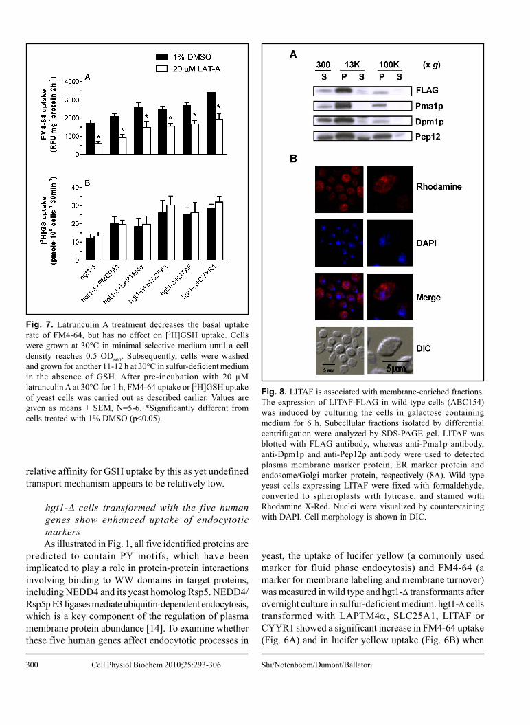
Fig. 8. LITAF is associated with membrane-enriched fractions.The expression of LITAF-FLAG in wild type cells (ABC154)was induced by culturing the cells in galactose containingmedium for 6 h. Subcellular fractions isolated by differentialcentrifugation were analyzed by SDS-PAGE gel. LITAF wasblotted with FLAG antibody, whereas anti-Pma1p antibody,anti-Dpm1p and anti-Pep12p antibody were used to detectedplasma membrane marker protein, ER marker protein andendosome/Golgi marker protein, respectively (8A). Wild typeyeast cells expressing LITAF were fixed with formaldehyde,converted to spheroplasts with lyticase, and stained withRhodamine X-Red. Nuclei were visualized by counterstainingwith DAPI. Cell morphology is shown in DIC.
Shi/Notenboom/Dumont/BallatoriCell Physiol Biochem 2010;25:293-306

301
compared to the wild type cells or to hgt1-Δ cells,consistent with the hypothesis that these genes arestimulating membrane turnover in hgt1-Δ cells. PMEPA1expressing cells also exhibited slightly increasedaccumulation of these two endocytotic markers, althoughthe difference was not statistically significant (Fig. 6).
The enhanced [3H]GSH uptake in hgt1-Δ cellstransformed with PMEPA1, LAPTM4α,SLC25A1, LITAF or CYYR1 is not inhibited bylatrunculin ATo test for possible connections between the
enhanced GSH uptake and the increased endocytoticmarker uptake, latrunculin A (LAT-A) treatment wasapplied to hgt1-Δ transformants prior to measuring theuptake of FM4-64 and of [3H]GSH in these cells. LAT-Adisrupts the actin cytoskeleton by sequestering actinmonomers, and therefore has been widely used as anendocytotic inhibitor in yeasts [15]. hgt1-Δ transformantswere pre-treated with 20 µM LAT-A or with 1% DMSO(vehicle control) for 60 minutes. As shown in Fig. 7A,LAT-A pretreatment resulted in decreased FM4-64uptake in each tested strain, suggesting that endocytosisin these cells was inhibited by LAT-A treatment. Inagreement with the results of figure 6A, cells expressingeach of the five genes had higher rates of FM4-64 uptakethan that of hgt1-Δ cells; however, LAT-A did not appearto be effective at inhibiting the FM4-64 uptake that wasstimulated by each of the five genes (Fig. 7A). Note forexample, that whereas LAT-A decreased FM4-64 uptakeby about 65% in the control hgt1-Δ cells, it only decreasedFM4-64 uptake by 38% in cells expressing LITAF (Fig.7A).
LAT-A had no effect on either the basal [3H]GSHuptake or the enhanced [3H]GSH uptake observed in cellstransformed with the five genes (Fig. 7B). These resultssuggest that neither the increased GSH uptake, nor theincreased uptake of general endocyttotic markers in hgt1-Δ cells transformed with PMEPA1, LAPTM4α,SLC25A1, LITAF or CYYR1 is directly due to anincrease in conventional endocytotic processes. Althoughthe mechanism of the enhanced [3H]GSH uptake is notknown, the present results suggest that it is due toincreased activity or membrane delivery of the as yetunidentified endogenous GSH uptake mechanism.
Subcellular localization of LITAFTo further characterize the potential mechanism by
which these proteins stimulate GSH uptake, additionalstudies examined the intracellular localization of LITAF
Fig. 9. PY motif mutations largely abolish the effect of LITAFon intracellular GSH levels, and on [3H]GSH and FM4-64 uptake.PPSY motifs in LITAF were changed to PPSA by site-directedmutagenesis. Wild type strain ABC154 was transformed withempty vector, LITAF, or LITAF with PY mutations and wholecell lysate of each transformant was subjected to SDS-PAGEgel. The blot was probed with anti-FLAG antibody for LITAFexpression, and with anti-actin for loading control (9A). Valuesare given as means ± SEM, N=3-6. *Significantly differentintracellular glutathione concentration (9B), [3H]GSH uptake(9C) and FM4-64 uptake (9D) when compared to wild type cellstransformed with the empty pESC-URA vector (p<0.05).
in yeast cells. When expressed in yeast, FLAG taggedLITAF was recognized as a protein of about 25 kDa,which is similar to the size of LITAF detected in
Human PY-containing Proteins and Membrane Function Cell Physiol Biochem 2010;25:293-306

302
mammalian cells [16]. Upon subcellular fractionation,LITAF was mainly present in the P13000 fraction, but wasalso detected in the P100000 fraction, albeit at much lowerlevels (Fig. 8A). Dpm1p (Dol-P-Man Synthase, anendoplasmic reticulum membrane marker protein) andPma1p (Plasma Membrane H+-ATPase, a plasmamembrane marker protein) also showed a predominantdistribution in the P13000 fraction with low levels in theP100000 fraction, whereas Pep12p (carboxyPEPtidase Y-deficient, an endosome/Golgi membrane marker protein)was detected in the P13000 and P100000 fractions at roughlycomparable levels (Fig. 8A). Essentially no LITAF wasdetected in the S100000 fraction, suggesting that LITAF ismembrane protein. The distribution of LITAF betweenP13000 and P100000 indicates that LITAF is likely associatedwith large organelles such as the endoplasmic reticulumand the plasma membrane.
Intracellular localization of LITAF was also examinedusing indirect immunostaining and confocal fluorescencemicroscopy (Fig. 8B). The results demonstrate that LITAFwas expressed diffusely throughout the cell, and was foundaround the periphery of the cell. Taken together, the resultsin Fig. 8 indicate that LITAF is a membrane protein, andis associated with the membrane of intracellular organellesand with the plasma membrane (Fig. 8B).
Mutation of the two predicted PY motifs of LITAFlargely abolishes the ability of this protein toincrease intracellular glutathione levels, and tostimulate [3H]GSH and FM4-64 uptakeTo examine the functional significance of the PY
motifs in these genes, LITAF was selected for mutationalanalysis. The two predicted PY motifs of LITAF weremutated from PPSY to PPSA, and wild type yeast cellswere transformed with both wild type LITAF and threeLITAF mutants. Mutation of these two PY motifs hadonly minimal effects on the expression level of LITAFprotein (Fig. 9A); however, they essentially abolished theability of LITAF to increase intracellular glutathione levels,and to stimulate [3H]GSH and FM4-64 uptake (Fig. 9B-D). As illustrated in figure 9, wild type cells transformedwith LITAF had higher intracellular glutathione levels,and enhanced [3H]GSH and FM4-64 uptake, comparedwith untransformed cells, consistent with results obtainedfrom hgt1-Δ cells transformed with LITAF (Figs. 3, 4and 6) suggesting that the functions of LITAF are notspecifically to complement the HGT1 deficiency, but aremore likely to regulate membrane turnover by an commonmechanism. Intracellular glutathione levels, [3H]GSHuptake rates and FM4-64 uptake rates of wild type cells
transformed with LITAF mutants were generallycomparable to those of control cells (wild type cellstransformed with empty vector pESC-URA), indicatingthat these effects of LITAF in yeast depend on its PYmotifs. The subcellular localization of these mutants inyeasts was also examined, however no differences weredetected using indirect immunofluorescence (data notshown).
Expression of mouse Ndfip2 (N4WBP5) has noeffect on [3H]GSH uptake in either the wild typeor the hgt1-Δ yeast strainTo examine whether another PY-containing protein
has similar effects on [3H]GSH uptake in yeast cells,mouse Ndfip2 (Nedd4 family interacting protein 2) wasexpressed in both the wild type yeast strain and the hgt1-Ä strain. [3H]GSH uptake was measured for 30 min inyeast cells transformed with the pESC-URA vector orNdfip2 after being grown overnight in a sulfur-deficientmedium that contained galactose. However, theexpression of Ndfip2 had no effect on [3H]GSH uptakein either strain (data not shown), suggesting that thetransport activity of the endogenous yeast low affinityGSH uptake mechanism is not modulated by Ndfip2.
Discussion
Bourbouloux et al. [5] previously demonstrated thatHgt1p is quantitatively the major GSH uptake transporterin S. cerevisiae. Without this transporter, the cells haveminimal ability to take up GSH from the culture mediumand show marked growth retardation when GSH is theonly sulfur source. However, hgt1-Δ cells do retain someability to take up GSH at a slow rate (e.g., see Fig. 4A),and to grow slowly using GSH as a sulfur source (Fig.2). Thus, yeast cells have an alternate mechanism fortaking up GSH, although this alternative mechanism hasnot been identified at the molecular level and has not beencharacterized biochemically.
The present study identifies five human genes,PMEPA1, LAPTM4α, SLC25A1, LITAF, and CYYR1that can functionally complement the growth defect ofhgt1-Δ cells cultured in a medium in which GSH is theonly sulfur source. Each of these genes encodes a proteincontaining PY motifs that are thought to be important forregulating protein cell surface expression via interactingwith the ubiquitin ligases NEDD4/Rsp5p [14, 17]. Thefirst PY motif was identified through a functional cDNAlibrary screen for putative ligands of the WW domain of
Shi/Notenboom/Dumont/BallatoriCell Physiol Biochem 2010;25:293-306

303
the Yes-associated protein [18]. The presence of thesemotifs allows the PY motif-containing proteins to bind toWW domains in target proteins, including the E3 ubiquitinligase NEDD4 [19]. NEDD4 protein belongs to the Hect-domain family of E3 ubiquitin-protein ligase. A majorfunction of such proteins is to down-regulate the cellsurface expression of membrane proteins via ubiquitin-dependent endocytosis, followed by degradation in bothproteasomes and lysosomes [14, 20]. Mutations in thePY motifs of the epithelial Na+ channel disrupt itsinteraction with NEDD4, which leads to constitutivelyincreased channel activity and hypertension in the Liddlesyndrome [21]. Rsp5p is the unique yeast homologue ofNEDD4. The direct interaction between Rsp5p and itstarget protein through PY-WW binding is essential formultiple-vesicular body targeting and endosomal sortingof several yeast membrane proteins [22, 23] Indeed, somestudies have also indicated possible associations ofNEDD4 with PMEPA1 [24], LAPTM4α [25], andLITAF [26]. Regulation of plasma membrane proteinturnover by this mechanism may either occur directly, bybinding of Rsp5p/NEDD4 to specific membrane proteinsthat contain PY motifs, or indirectly, by binding of Rsp5p/NEDD4 to PY-containing adapter molecules that thentarget the ubiquitin ligase activity to specific plasmamembrane proteins [17, 27].
The present results are consistent with a model inwhich these five human genes enhance GSH uptake byincreasing the activity of an undefined yeast low affinityGSH uptake mechanism [5, 6]. As illustrated in figure 5,the enhanced [3H]GSH uptake in cells expressing thefive human genes was uniformly inhibited by highconcentrations of unlabeled GSH, GSSG, and ophthalmicacid, suggesting that the effects of these genes on[3H]GSH uptake involves a common low affinity uptakepathway. Although the mechanism by which these genesenhance the activity of this alternate mode of GSH uptakeis unknown, they may do so by increasing production,intracellular targeting, or the stability of components ofthe uptake machinery. One possibility is that expressionof the PY-containing genes titrates the Rsp5p-associateddegradation pathway away from some endogenous yeasttransporter, thereby enhancing its activity. The enhancedturnover of general endocytotic markers detected in cellsexpressing the PY-containing genes might then reflectincreased turnover of membrane proteins via an alternativelatrunculin-A-insensitive pathway.
By analogy, the PY-containing protein Ndfip2appears to increase cell surface expression of theamiloride-sensitive epithelial sodium channel (ENaC) by
interfering with the Nedd4-mediated regulation of ENaC[28]. Ndfip2 contains three transmembrane domains andtwo amino terminal PPxY motifs, which physically interactwith the WW domains of Nedd4 family proteins. However,this stimulatory effect of Ndfip2 on ENaC activity appearsto be relatively selective, as Ndfip1, a highly related proteinthat also contains PY motifs, has no effect on ENaCactivity [28]. In addition, Ndfip2 did not increase thetransport activity of CFTR or Kir1.1a, indicating selectivityfor the target proteins. In the present study, expressionof Ndfip2 in yeast cells did not produce stimulatory effectsin GSH uptake, but this negative result does not necessarilyexclude the proposed model, as there are many possiblereasons for the lack of effect, including differentialspecificity, sensitivity, and affinity for possible binding sitesin yeast proteins.
Expression of the five genes also led to greateraccumulation of the endocytotic markers lucifer yellowand FM4-64 in yeast. Whereas the basal accumulationof FM4-64 was inhibited by LAT-A, neither the higherrates of FM4-64 uptake nor the enhanced GSH uptakeobserved in cells expressing the five human genes wasaffected by LAT-A, indicating that the enhanced GSHuptake associated with expression of the human genes isnot mediated by a LAT-A-sensitive endocytoticmechanism. Upon subcellular fractionation, LITAF wasfound in the P13000 fraction which primarily contains largeorganelles and the plasma membrane. Indirectimmunostaining of LITAF resulted in a staining patterncharacteristic of localization to the plasma membrane andintracellular membranes. Mutation of the two PY motifsof LITAF resulted in loss of the observed effects,indicating that this motif plays an essential role in theeffects of LITAF expression in yeast.
To date, little is known about the biological functionsof the five genes identified in the present study. SLC25A1is mapped to chromosomal region 22q11, and micro-deletions in this region are associated with DiGeorgesyndrome, velo-cardio-facial syndrome and conotruncalanomaly face syndrome [29]. Although it is not knownwhether SLC25A1 plays a role in GSH homeostasis, somemembers of SLC25A family may contribute tomitochondrial GSH uptake. In particular, the dicarboxylate(SLC25A10) and 2-oxoglutarate (SLC25A11) carriers ofthe inner mitochondrial membrane are believed to catalyzeuptake of GSH into the matrix through an exchangemechanism [30]. The three members of the lysosomal-associated protein transmembrane (LAPTM) family,LAPTM4α, LAPTM4β and LAPTM5 are thought tofunction as transporters or channels, although their precise
Human PY-containing Proteins and Membrane Function Cell Physiol Biochem 2010;25:293-306

304
function is unknown. When expressed in a drug-sensitivestrain of S. cerevisiae, mouse LAPTM4α regulatesintracellular compartmentalization of amphipathic solutesand confers cellular resistance or hypersensitivity to arange of drugs [31]. Both CYYR1 and PMEPA1 havebeen implicated in human diseases, although their functionsare also unknown. CYYR1 is expressed in a broad rangeof human tissues, and the central cysteine-tyrosine-richdomain is strongly conserved from lower vertebrates tohumans [32]. Alternative splicing and expression of theCYYR1 transcript has been reported in humanneuroendocrine tumors [33]. PMEPA1 was first describedas an androgen-induced gene. It is widely expressed innormal and solid malignant tissues, with the highestexpression in prostate tissue [34]. The expression ofPMEPA1 is increased in primary and metastasized colontumors and several other solid tumors [35, 36], whereasit is decreased in prostate tumor tissues [34]. In addition,PMEPA1 is also induced by tumor growth factor β [36].LITAF is present in most human tissues and its expressioncan be induced by multiple pathways, including TNFα-and p53-activated pathways [16, 37, 38]. Elevated mRNAand protein expression of LITAF has been found inintestinal tissues from patients with Crohn’s disease andulcerative colitis [39, 40]. In addition, mutations of LITAFcause Charcot-Marie-Tooth disease type 1C (CMT1C),an autosomal dominant demyelinating peripheralneuropathy [41]. Although the etiology of CMT1C is notunderstood yet, a role of LITAF in modulating proteintrafficking and turnover has been proposed to be involved[26, 42].
In summary, the present study identifies five humangenes that rescue growth of a HGT1-deficient yeaststrain, and provides clues into the potential function ofthese genes. Currently, the functions of the five genesare unknown, although four of them have been implicated
in human diseases [35, 39, 41, 43-45]. A better under-standing of their physiological functions may be expectedto clarify the etiology and pathogenesis of these diseases.
Abbreviations
CYYR1 (cysteine/tyrosine-rich1); FM4-64 (N-(3-triethylammoniumpropyl)-4-(6-(4-(diethylamino)phenyl)hexatrienyl) pyridinium dibromide); GSH (reducedglutathione); LAPTM4α (lysosomal-associated proteintransmembrane 4 alpha); LITAF (lipopolysaccharide-induced TNF factor); Ndfip2 (Nedd4 family interactingprotein 2); NEDD4 (neural precursor cell expressed,developmentally down-regulated 4); PY (xPPxY, P = Pro,Y = Tyr, x = any amino acid); RSP5 (reverses Spt-phenotype); SLC25A1 (solute carrier family 25, member1); PMEPA1 (prostate transmembrane protein, androgeninduced 1).
Acknowledgements
This work was supported in part by National Institutesof Health (NIH) Grants DK048823 and DK067214, andNational Institute of Environmental Health SciencesCenter Grant ES01247 and Training Grant ES07026.We thank Dr. Anand K. Bachhawat (Institute of MicrobialTechnology, Chandirgarh, India) for providing the hgt1-ΔS. cerevisiae strain, Dr. Stephen G. Oliver (University ofManchester, Manchester, UK) for providing the humanmammary gland cDNA library, Dr. David A. Pearce(University of Rochester, Rochester, New York) forsharing anti-Dpm1p antibody, Dr. Zhifeng Cui for hisadvice in this project, and David McMillan for technicalassistance.
References
1 Ballatori N, Krance SM, Notenboom S,Shi S, Tieu K, Hammond CL: Glutathionedysregulation and the etiology andprogression of human diseases. Biol Chem2009;390:191-214.
2 Meister A, Anderson ME: Glutathione.Annu Rev Biochem 1983;52:711-760.
3 Ballatori N, Hammond CL, CunninghamJB, Krance SM, Marchan R: Molecularmechanisms of reduced glutathionetransport: Role of the MRP/CFTR/ABCCand OATP/SLC21A families ofmembrane proteins. Toxicol ApplPharmacol 2005;204:238-255.
4 Ballatori N, Krance SM, Marchan R,Hammond CL: Plasma membraneglutathione transporters and their rolesin cell physiology and pathophysiology.Mol Aspects Med 2008;30:13-28.
5 Bourbouloux A, Shahi P, Chakladar A,Delrot S, Bachhawat AK: Hgt1p, a highaffinity glutathione transporter from theyeast saccharomyces cerevisiae. J BiolChem 2000;275:13259-13265.
Shi/Notenboom/Dumont/BallatoriCell Physiol Biochem 2010;25:293-306

305
6 Miyake T, Hazu T, Yoshida S, KanayamaM, Tomochika K, Shinoda S, Ono B:Glutathione transport systems of thebudding yeast saccharomyces cerevisiae.Biosci Biotechnol Biochem1998;62:1858-1864.
7 Zhang N, Osborn M, Gitsham P, Yen K,Miller JR, Oliver SG: Using yeast to placehuman genes in functional categories.Gene 2003;303:121-129.
8 Gietz RD, Woods RA: Transformationof yeast by lithium acetate/single-stranded carrier DNA/polyethyleneglycol method. Methods Enzymol2002;350:87-96.
9 Baker MA, Cerniglia GJ, Zaman A:Microtiter plate assay for themeasurement of glutathione andglutathione disulfide in large numbers ofbiological samples. Anal Biochem1990;190:360-365.
10 Dulic V, Egerton M, Elguindi I, Raths S,Singer B, Riezman H: Yeast endocytosisassays. Methods Enzymol1991;194:697-710.
11 Emans N, Zimmermann S, Fischer R:Uptake of a fluorescent marker in plantcells is sensitive to brefeldin A andwortmannin. Plant Cell 2002;14:71-86.
12 Rieder SE, Emr SD: Overview ofsubcellular fractionation procedures forthe yeast saccharomyces cerevisiae. CurrProtoc Cell Biol 2001;Chapter 3:Unit3.7.
13 Pringle JR, Adams AE, Drubin DG, HaarerBK: Immunofluorescence methods foryeast. Methods Enzymol 1991;194:565-602.
14 Rotin D, Staub O, Haguenauer-Tsapis R:Ubiquitination and endocytosis of plasmamembrane proteins: Role of Nedd4/Rsp5p family of ubiquitin-protein ligases.J Membr Biol 2000;176:1-17.
15 Ayscough KR, Stryker J, Pokala N,Sanders M, Crews P, Drubin DG: Highrates of actin filament turnover inbudding yeast and roles for actin inestablishment and maintenance of cellpolarity revealed using the actin inhibitorlatrunculin-A. J Cell Biol 1997;137:399-416.
16 Moriwaki Y, Begum NA, Kobayashi M,Matsumoto M, Toyoshima K, Seya T:Mycobacterium bovis bacillus calmette-guerin and its cell wall complex induce anovel lysosomal membrane protein,SIMPLE, that bridges the missing linkbetween lipopolysaccharide and p53-inducible gene, LITAF(PIG7), andestrogen-inducible gene, EET-1. J BiolChem 2001;276:23065-23076.
17 Staub O, Abriel H, Plant P, Ishikawa T,Kanelis V, Saleki R, Horisberger JD, SchildL, Rotin D: Regulation of the epithelialna+ channel by Nedd4 and ubiquitination.Kidney Int 2000;57:809-815.
18 Chen HI, Sudol M: The WW domain ofyes-associated protein binds a proline-rich ligand that differs from the consensusestablished for src homology 3-bindingmodules. Proc Natl Acad Sci U S A1995;92:7819-7823.
19 Staub O, Dho S, Henry P, Correa J,Ishikawa T, McGlade J, Rotin D: WWdomains of Nedd4 bind to the proline-rich PY motifs in the epithelial na+channel deleted in liddle’s syndrome.EMBO J 1996;15:2371-2380.
20 Harvey KF, Kumar S: Nedd4-likeproteins: An emerging family ofubiquitin-protein ligases implicated indiverse cellular functions. Trends CellBiol 1999;9:166-169.
21 Abriel H, Loffing J, Rebhun JF, Pratt JH,Schild L, Horisberger JD, Rotin D, StaubO: Defective regulation of the epithelialna+ channel by Nedd4 in liddle’ssyndrome. J Clin Invest 1999;103:667-673.
22 Stawiecka-Mirota M, Pokrzywa W,Morvan J, Zoladek T, Haguenauer-TsapisR, Urban-Grimal D, Morsomme P:Targeting of Sna3p to the endosomalpathway depends on its interaction withRsp5p and multivesicular body sortingon its ubiquitylation. Traffic2007;8:1280-1296.
23 Bhattacharya S, Zoladek T, Haines DS:WW domains 2 and 3 of Rsp5p playoverlapping roles in binding to the LPKYmotif of Spt23p and Mga2p. Int JBiochem Cell Biol 2008;40:147-157.
24 Xu LL, Shi Y, Petrovics G, Sun C,Makarem M, Zhang W, Sesterhenn IA,McLeod DG, Sun L, Moul JW, SrivastavaS: PMEPA1, an androgen-regulatedNEDD4-binding protein, exhibits cellgrowth inhibitory function and decreasedexpression during prostate cancerprogression. Cancer Res 2003;63:4299-4304.
25 Pak Y, Glowacka WK, Bruce MC, PhamN, Rotin D: Transport of LAPTM5 tolysosomes requires association with theubiquitin ligase Nedd4, but not LAPTM5ubiquitination. J Cell Biol 2006;175:631-645.
26 Shirk AJ, Anderson SK, Hashemi SH,Chance PF, Bennett CL: SIMPLEinteracts with NEDD4 and TSG101:Evidence for a role in lysosomal sortingand implications for charcot-marie-tooth disease. J Neurosci Res2005;82:43-50.
27 Lin CH, MacGurn JA, Chu T, Stefan CJ,Emr SD: Arrestin-related ubiquitin-ligaseadaptors regulate endocytosis and proteinturnover at the cell surface. Cell2008;135:714-725.
28 Konstas AA, Shearwin-Whyatt LM, FotiaAB, Degger B, Riccardi D, Cook DI,Korbmacher C, Kumar S: Regulation ofthe epithelial sodium channel byN4WBP5A, a novel Nedd4/Nedd4-2-interacting protein. J Biol Chem.2002;277(33):29406-29416.
29 Goldmuntz E, Wang Z, Roe BA, BudarfML: Cloning, genomic organization, andchromosomal localization of humancitrate transport protein to the DiGeorge/velocardiofacial syndrome minimalcritical region. Genomics 1996;33:271-276.
30 Lash LH: Mitochondrial glutathionetransport: Physiological, pathologicaland toxicological implications. ChemBiol Interact 2006;163:54-67.
31 Hogue DL, Kerby L, Ling V: Amammalian lysosomal membraneprotein confers multidrug resistance uponexpression in saccharomyces cerevisiae.J Biol Chem 1999;274:12877-12882.
32 Vitale L, Casadei R, Canaider S, Lenzi L,Strippoli P, D’Addabbo P, Giannone S,Carinci P, Zannotti M: Cysteine andtyrosine-rich 1 (CYYR1), a novelunpredicted gene on human chromosome21 (21q21.2), encodes a cysteine andtyrosine-rich protein and defines a newfamily of highly conserved vertebrate-specific genes. Gene 2002;290:141-151.
33 Vitale L, Frabetti F, Huntsman SA,Canaider S, Casadei R, Lenzi L, FacchinF, Carinci P, Zannotti M, Coppola D,Strippoli P: Sequence, “subtle”alternative splicing and expression of theCYYR1 (cysteine/tyrosine-rich 1)mRNA in human neuroendocrine tumors.BMC Cancer 2007;7:66.
34 Xu LL, Shanmugam N, Segawa T,Sesterhenn IA, McLeod DG, Moul JW,Srivastava S: A novel androgen-regulatedgene, PMEPA1, located on chromosome20q13 exhibits high level expression inprostate. Genomics 2000;66:257-263.
35 Rae FK, Hooper JD, Nicol DL, ClementsJA: Characterization of a novel gene,STAG1/PMEPA1, upregulated in renalcell carcinoma and other solid tumors.Mol Carcinog 2001;32:44-53.
36 Brunschwig EB, Wilson K, Mack D,Dawson D, Lawrence E, Willson JK, LuS, Nosrati A, Rerko RM, Swinler S, BeardL, Lutterbaugh JD, Willis J, Platzer P,Markowitz S: PMEPA1, a transforminggrowth factor-beta-induced marker ofterminal colonocyte differentiationwhose expression is maintained inprimary and metastatic colon cancer.Cancer Res 2003;63:1568-1575.
Human PY-containing Proteins and Membrane Function Cell Physiol Biochem 2010;25:293-306

306
3 7 Myokai F, Takashiba S, Lebo R, Amar S:A novel lipopolysaccharide-inducedtranscription factor regulating tumornecrosis factor alpha gene expression:Molecular cloning, sequencing,characterization, and chromosomalassignment. Proc Natl Acad Sci U S A1999;96:4518-4523.
3 8 Baumann B, Seufert J, Rolf O, Jakob F,Goebel S, Eulert J, Rader CP:Upregulation of LITAF mRNAexpression upon exposure to TiAlV andpolyethylene wear particles in THP-1macrophages. Biomed Tech (Berl)2007;52:200-207.
39 Stucchi A, Reed K, O’Brien M, Cerda S,Andrews C, Gower A, Bushell K, Amar S,Leeman S, Becker J: A new transcriptionfactor that regulates TNF-alpha geneexpression, LITAF, is increased inintestinal tissues from patients with CDand UC. Inflamm Bowel Dis2006;12:581-587.
40 Huang Y, Bennett CL: Litaf/Simpleprotein is increased in intestinal tissuesfrom patients with CD and UC, but isunlikely to function as a transcriptionfactor. Inflamm Bowel Dis 2007;13:120-121.
41 Street VA, Bennett CL, Goldy JD, ShirkAJ, Kleopa KA, Tempel BL, Lipe HP,Scherer SS, Bird TD, Chance PF:Mutation of a putative proteindegradation gene LITAF/SIMPLE incharcot-marie-tooth disease 1C.Neurology 2003;60:22-26.
42 Saifi GM, Szigeti K, Wiszniewski W, ShyME, Krajewski K, Hausmanowa-Petrusewicz I, Kochanski A, Reeser S,Mancias P, Butler I, Lupski JR: SIMPLEmutations in charcot-marie-tooth diseaseand the potential role of its proteinproduct in protein degradation. HumMutat 2005;25:372-383.
43 Matsumura Y, Matsumura Y, NishigoriC, Horio T, Miyachi Y: PIG7/LITAF genemutation and overexpression of its geneproduct in extramammary paget’sdisease. Int J Cancer 2004;111:218-223.
44 Giannini G, Ambrosini MI, Di MarcotullioL, Cerignoli F, Zani M, MacKay AR,Screpanti I, Frati L, Gulino A: EGF- andcell-cycle-regulated STAG1/PMEPA1/ERG1.2 belongs to a conserved genefamily and is overexpressed and amplifiedin breast and ovarian cancer. MolCarcinog 2003;38:188-200.
45 Williams NM, Spurlock G, Norton N,Williams HJ, Hamshere ML, KrawczakM, Kirov G, Nikolov I, Georgieva L,Jones S, Cardno AG, O’Donovan MC,Owen MJ: Mutation screening and LDmapping in the VCFS deleted region ofchromosome 22q11 in schizophreniausing a novel DNA pooling approach.Mol Psychiatry 2002;7:1092-1100.
46 Hogue DL, Ellison MJ, Young JD, CassCE: Identification of a novel membranetransporter associated with intracellularmembranes by phenotypiccomplementation in the yeastsaccharomyces cerevisiae. J Biol Chem1996;271:9801-9808.
47 Cabrita MA, Hobman TC, Hogue DL,King KM, Cass CE: Mouse transporterprotein, a membrane protein thatregulates cellular multidrug resistance, islocalized to lysosomes. Cancer Res1999;59:4890-4897.
48 Mestre-Escorihuela C, Rubio-MoscardoF, Richter JA, Siebert R, Climent J,Fresquet V, Beltran E, Agirre X, MaruganI, Marin M, Rosenwald A, Sugimoto KJ,Wheat LM, Karran EL, Garcia JF,Sanchez L, Prosper F, Staudt LM, PinkelD, Dyer MJ, Martinez-Climent JA:Homozygous deletions localize noveltumor suppressor genes in B-celllymphomas. Blood 2007;109:271-280.
49 Bennett CL, Shirk AJ, Huynh HM, StreetVA, Nelis E, Van Maldergem L, DeJonghe P, Jordanova A, GuergueltchevaV, Tournev I, Van Den Bergh P, SeemanP, Mazanec R, Prochazka T, KremenskyI, Haberlova J, Weiss MD, TimmermanV, Bird TD, Chance PF: SIMPLEmutation in demyelinating neuropathyand distribution in sciatic nerve. AnnNeurol 2004;55:713-720.
50 Cardillo MR, Di Silverio F: Prostate-specific G protein couple receptor genesand STAG1/PMEPA1 in peripheral bloodfrom patients with prostatic cancer. IntJ Immunopathol Pharmacol2006;19:871-878.
51 Stoffel M, Karayiorgou M, EspinosaR,3rd, Beau MM: The humanmitochondrial citrate transporter gene(SLC20A3) maps to chromosome band22q11 within a region implicated inDiGeorge syndrome, velo-cardio-facialsyndrome and schizophrenia. Hum Genet1996;98:113-115.
52 Heisterkamp N, Mulder MP, LangeveldA, ten Hoeve J, Wang Z, Roe BA, GroffenJ: Localization of the humanmitochondrial citrate transporter proteingene to chromosome 22Q11 in theDiGeorge syndrome critical region.Genomics 1995;29:451-456.
Shi/Notenboom/Dumont/BallatoriCell Physiol Biochem 2010;25:293-306