Identification and characterization of additional flagellin genes from Vibrio anguillarum
11
JOURNAL OF BACTERIOLOGY, Sept. 1996, p. 5188–5198 Vol. 178, No. 9 0021-9193/96/$04.0010 Copyright q 1996, American Society for Microbiology Identification and Characterization of Additional Flagellin Genes from Vibrio anguillarum KAREN MCGEE, 1 PER HO ¨ RSTEDT, 2 AND DEBRA L. MILTON 1 * Department of Cell and Molecular Biology 1 and the Department of Pathology, 2 Umeå University, S-901 87 Umeå, Sweden Received 8 April 1996/Accepted 21 June 1996 Previously, the flagellar filament of Vibrio anguillarum was suggested to consist of flagellin A and three additional flagellin proteins, FlaB, -C, and -D. This study identifies the genes encoding FlaB, -C, and -D and a possible fifth flagellin gene that may encode FlaE. The flagellin genes map at two separate DNA loci and are most similar to the four polar flagellin genes of Vibrio parahaemolyticus, also located at two DNA loci. The genetic organization of these two loci is conserved between both organisms. For each gene, in-frame deletions of the entire gene, the 5* end, and the 3* end were made. Mutant analysis showed that each mutation, except those in flaE, caused a loss of flagellin from the filament. However, no obvious structural loss in the filament, as determined by electron microscopy, and only slight decreases in motility were seen. Virulence analysis indicated that all but two of the mutations gave a wild-type phenotype. The 5*-end deletions of flaD and flaE decreased virulence significantly (>10 4 -fold) of infections via both the intraperitoneal and immersion routes. These results indicate that, like FlaA, FlaD and FlaE may also be involved in virulence. For several bacteria, the flagellum has been suggested to be involved in virulence either as a motility organelle or as an organelle that carries an adhesive component. In Campy- lobacter jejuni studies, the flagellum has been suggested to aid the bacterium either in the adherence to (20) or in the inter- nalization (7) within cultured epithelial cells. Additional stud- ies with C. jejuni (32) have shown that motility and not flagellin A is required for the invasion of intestinal cells; however, flagellin A can serve as a secondary adhesin for the adherence to intestinal cultured cells. For Pseudomonas aeruginosa,a study utilizing isogenic motility mutants in the burned mouse model showed that motility contributes to the invasiveness of this organism (5). For Vibrio cholerae, earlier studies with mo- tility mutants suggested that either motility (8) or an adhesin (2, 11), proposed to be associated with the flagellum, is needed for the colonization of intestinal tissues, whereas a later study (27) indicated that motility but not the flagellar structure is essential for the colonization of rabbits. The polar flagellum of Vibrio anguillarum, the causative agent of vibriosis in marine fish, is a complex structure. The filament is believed to be composed of four flagellin proteins (23) and is covered by a sheath (25). Recently, studies have been initiated to analyze the role that the flagellum plays in the virulence of V. anguillarum. Chemotactic motility has been shown to be required for the invasion of rainbow trout when they are immersed in infected seawater; however, chemotactic motility is not essential for virulence after the fish integument is crossed (26). Moreover, flagellin A has been suggested to have two possible roles in virulence (23). A mutant carrying a deletion of the flaA gene was shown to be partially motile and to be defective in its ability to invade rainbow trout immersed in infected seawater; however, virulence was not decreased if the mutant was introduced into the fish by intraperitoneal injection. Thus, flagellin A is needed for the full motility which is essential for efficient invasion of rainbow trout. A second mutant that contained an in-frame deletion at the 39 end of the flaA gene and that was also partially motile as a result of the loss of flagellin A in the filament was avirulent by intraperito- neal injection, indicating a potential second role for the flagel- lin protein. One proposal for this second role was that the truncated flagellin protein may remain in the cytoplasm be- cause of the loss of export functions and thus may repress the expression of virulence genes. To clarify the role of the flagellum and of the flagellin pro- teins in the virulence of V. anguillarum, the present study continues the characterization of the flagellar filament. Four additional flagellin genes, flaB,-C,-D, and -E, were cloned and sequenced. In-frame deletions of the full gene, the 59 end, and the 39 end were made in each gene similar to those of the flaA gene in a previous study (23). All mutations affected motility only slightly, if at all. Since motility was not strongly affected, virulence was only slightly decreased for all but two mutations, the flaD and flaE 59-end deletions. For these two mutations, virulence was strongly decreased (10 4 - to 10 5 -fold) via both immersion and intraperitoneal infections. MATERIALS AND METHODS Bacterial strains, bacteriophage, plasmids, and media. V. anguillarum NB10 (serotype O1) is an isolate of our laboratory from the Gulf of Bothnia outside the Norrby Laboratory, Umeå, Sweden (24). The more competent Escherichia coli SY327 [D(lac pro) argE(Am) rif malA recA56] (21) was used for transformation after subcloning fragments into either pNQ705-1 or pDM4. E. coli S17-1 (thi pro hsdR hsdM 1 recA RP4-2-Tc::Mu-Km::Tn7) (29) was used as the donor strain for all plasmids transferred to V. anguillarum by a previously described method for bacterial mating (23). SY327 and S17-1 are bacteriophage lambda lysogens carrying the pir gene, which is needed for replication of the suicide vectors pNQ705-1 and pDM4. E. coli XL1-Blue (28) (recA1 lac endA1 gyrA96 thi hsdR17 supE44 relA1 [F9 proAB lacI q lacZDM15 Tn10]) was used for bacteriophage lambda infections and for the transformation of plasmids not requiring the pir gene. Construction of the V. anguillarum genomic library with the Lambda Zap II system from Stratagene, handling of this bacteriophage, and the excision of the pBluescript plasmid derivatives have been described previously (22). pBSFla8-1, carrying a 6.5-kb fragment with flaA and flaC, and pBSFlaD9-1, carrying a 7-kb fragment with flaEDB, were excised from isolated bacteriophage clones. Plasmids used in this study are described in Table 1. The suicide vectors, pDM4 and pNQ705-1, were employed in making chromosomal deletions and insertions, respectively. pNQ705-1 is a derivative of the previously described pNQ705 (22) that was altered by exchanging the linker region of pNQ705 for the * Corresponding author. Mailing address: Department of Cell and Molecular Biology, Umeå University, S-901 87 Umeå, Sweden. Phone: 46 90 10 25 36. Fax: 46 90 77 14 20. Electronic mail address: Tord [email protected]. 5188 on May 28, 2016 by guest http://jb.asm.org/ Downloaded from
-
Upload
independent -
Category
Documents
-
view
5 -
download
0
Transcript of Identification and characterization of additional flagellin genes from Vibrio anguillarum

JOURNAL OF BACTERIOLOGY, Sept. 1996, p. 5188–5198 Vol. 178, No. 90021-9193/96/$04.0010Copyright q 1996, American Society for Microbiology
Identification and Characterization of Additional FlagellinGenes from Vibrio anguillarum
KAREN MCGEE,1 PER HORSTEDT,2 AND DEBRA L. MILTON1*
Department of Cell and Molecular Biology1 and the Department of Pathology,2
Umeå University, S-901 87 Umeå, Sweden
Received 8 April 1996/Accepted 21 June 1996
Previously, the flagellar filament of Vibrio anguillarum was suggested to consist of flagellin A and threeadditional flagellin proteins, FlaB, -C, and -D. This study identifies the genes encoding FlaB, -C, and -D anda possible fifth flagellin gene that may encode FlaE. The flagellin genes map at two separate DNA loci and aremost similar to the four polar flagellin genes of Vibrio parahaemolyticus, also located at two DNA loci. Thegenetic organization of these two loci is conserved between both organisms. For each gene, in-frame deletionsof the entire gene, the 5* end, and the 3* end were made. Mutant analysis showed that each mutation, exceptthose in flaE, caused a loss of flagellin from the filament. However, no obvious structural loss in the filament,as determined by electron microscopy, and only slight decreases in motility were seen. Virulence analysisindicated that all but two of the mutations gave a wild-type phenotype. The 5*-end deletions of flaD and flaEdecreased virulence significantly (>104-fold) of infections via both the intraperitoneal and immersion routes.These results indicate that, like FlaA, FlaD and FlaE may also be involved in virulence.
For several bacteria, the flagellum has been suggested to beinvolved in virulence either as a motility organelle or as anorganelle that carries an adhesive component. In Campy-lobacter jejuni studies, the flagellum has been suggested to aidthe bacterium either in the adherence to (20) or in the inter-nalization (7) within cultured epithelial cells. Additional stud-ies with C. jejuni (32) have shown that motility and not flagellinA is required for the invasion of intestinal cells; however,flagellin A can serve as a secondary adhesin for the adherenceto intestinal cultured cells. For Pseudomonas aeruginosa, astudy utilizing isogenic motility mutants in the burned mousemodel showed that motility contributes to the invasiveness ofthis organism (5). For Vibrio cholerae, earlier studies with mo-tility mutants suggested that either motility (8) or an adhesin(2, 11), proposed to be associated with the flagellum, is neededfor the colonization of intestinal tissues, whereas a later study(27) indicated that motility but not the flagellar structure isessential for the colonization of rabbits.The polar flagellum of Vibrio anguillarum, the causative
agent of vibriosis in marine fish, is a complex structure. Thefilament is believed to be composed of four flagellin proteins(23) and is covered by a sheath (25). Recently, studies havebeen initiated to analyze the role that the flagellum plays in thevirulence of V. anguillarum. Chemotactic motility has beenshown to be required for the invasion of rainbow trout whenthey are immersed in infected seawater; however, chemotacticmotility is not essential for virulence after the fish integumentis crossed (26). Moreover, flagellin A has been suggested tohave two possible roles in virulence (23). A mutant carrying adeletion of the flaA gene was shown to be partially motile andto be defective in its ability to invade rainbow trout immersedin infected seawater; however, virulence was not decreased ifthe mutant was introduced into the fish by intraperitonealinjection. Thus, flagellin A is needed for the full motility whichis essential for efficient invasion of rainbow trout. A second
mutant that contained an in-frame deletion at the 39 end of theflaA gene and that was also partially motile as a result of theloss of flagellin A in the filament was avirulent by intraperito-neal injection, indicating a potential second role for the flagel-lin protein. One proposal for this second role was that thetruncated flagellin protein may remain in the cytoplasm be-cause of the loss of export functions and thus may repress theexpression of virulence genes.To clarify the role of the flagellum and of the flagellin pro-
teins in the virulence of V. anguillarum, the present studycontinues the characterization of the flagellar filament. Fouradditional flagellin genes, flaB, -C, -D, and -E, were cloned andsequenced. In-frame deletions of the full gene, the 59 end, andthe 39 end were made in each gene similar to those of the flaAgene in a previous study (23). All mutations affected motilityonly slightly, if at all. Since motility was not strongly affected,virulence was only slightly decreased for all but two mutations,the flaD and flaE 59-end deletions. For these two mutations,virulence was strongly decreased (104- to 105-fold) via bothimmersion and intraperitoneal infections.
MATERIALS AND METHODS
Bacterial strains, bacteriophage, plasmids, and media. V. anguillarum NB10(serotype O1) is an isolate of our laboratory from the Gulf of Bothnia outside theNorrby Laboratory, Umeå, Sweden (24). The more competent Escherichia coliSY327 [D(lac pro) argE(Am) rif malA recA56] (21) was used for transformationafter subcloning fragments into either pNQ705-1 or pDM4. E. coli S17-1 (thi prohsdR hsdM1 recA RP4-2-Tc::Mu-Km::Tn7) (29) was used as the donor strain forall plasmids transferred to V. anguillarum by a previously described method forbacterial mating (23). SY327 and S17-1 are bacteriophage lambda lysogenscarrying the pir gene, which is needed for replication of the suicide vectorspNQ705-1 and pDM4. E. coli XL1-Blue (28) (recA1 lac endA1 gyrA96 thi hsdR17supE44 relA1 [F9 proAB lacIq lacZDM15 Tn10]) was used for bacteriophagelambda infections and for the transformation of plasmids not requiring the pirgene.Construction of the V. anguillarum genomic library with the Lambda Zap II
system from Stratagene, handling of this bacteriophage, and the excision of thepBluescript plasmid derivatives have been described previously (22). pBSFla8-1,carrying a 6.5-kb fragment with flaA and flaC, and pBSFlaD9-1, carrying a 7-kbfragment with flaEDB, were excised from isolated bacteriophage clones.Plasmids used in this study are described in Table 1. The suicide vectors,
pDM4 and pNQ705-1, were employed in making chromosomal deletions andinsertions, respectively. pNQ705-1 is a derivative of the previously describedpNQ705 (22) that was altered by exchanging the linker region of pNQ705 for the
* Corresponding author. Mailing address: Department of Cell andMolecular Biology, Umeå University, S-901 87 Umeå, Sweden. Phone:46 90 10 25 36. Fax: 46 90 77 14 20. Electronic mail address: [email protected].
5188
on May 28, 2016 by guest
http://jb.asm.org/
Dow
nloaded from

linker region of pDM4 (23). For each mutation made, a PCR fragment of thechromosomal region to be altered was made and cloned into a suicide vector. Forcomplementation of each mutation, pNQ705-1-derived plasmids containing aPCR fragment with a flagellin gene and its promoter were used. The comple-mentary regions of the PCR fragments for each of these plasmids are given inTable 1.The medium used routinely for E. coli was TYS broth (10 g of Bacto Tryptone,
5 g of Bacto Yeast Extract, and 10 g of sodium chloride [all per liter]), and forV. anguillarum, Trypticase soy medium (BBL) was used. The vibrio-selectivemedium TCBS (Difco) containing 5 mg of chloramphenicol per ml was used forthe selection of V. anguillarum transconjugants when crosses were made with E.coli. Motility agar consisted of Trypticase soy broth plus 0.25% agar.Antibiotic concentrations for all E. coli strains were as follows: ampicillin, 100
mg/ml; chloramphenicol, 25 mg/ml. For V. anguillarum, chloramphenicol wasused at a concentration of 5 mg/ml.DNA techniques. Unless otherwise stated, all conditions for the various DNA
techniques were as described by Sambrook et al. (28). Oligonucleotides forprimers were synthesized with an Applied Biosystems DNA-RNA synthesizer(model 394). Double-strand DNA sequencing was performed by the dideoxychain termination method with T7 DNA polymerase (Pharmacia). Both strandsof the flaC gene and the flaEDB genes were sequenced by primer walking fromthe previously sequenced flaA gene for flaC and from the 110-bp PCR fragmentused to identify the flaD gene locus.Screening the genomic library. Screening of the V. anguillarum genomic li-
brary was done as described previously (23) with one exception. For the flaD-containing bacteriophage, the temperature for prehybridization and hybridiza-tion was changed to 508C, and after hybridization, the filters were washed at 508Cfor 5 min with 63 SSC (13 SSC is 0.15 M NaCl plus 0.015 M sodium citrate) and0.5% sodium dodecyl sulfate (SDS). The probe for flaC was a 700-bp fragmentfrom the downstream region of the previously sequenced flaA gene (23). Theprobe for the flaD gene locus was a 75-bp fragment (residues 1762 to 1837 [seeFig. 3]) derived from a 110-bp PCR fragment from the conserved 59 end of theV. anguillarum flaD gene. This fragment was generated with previously described(23) primers complementary to a similar region of the P. aeruginosa flaA geneand chromosomal DNA from a previously described flaA mutant, RO8, in whichmost of the flaA gene had been deleted (23). For detection, the probes werelabelled with [a-32P]dCTP as described previously (23).PCR conditions. Cycling conditions for each PCR began with a 3-min dena-
turation at 948C and five cycles of the following varying annealing times: 948C for30 s, 50 to 558C (1 degree change each cycle) for 30 s, 728C for 30 s. The PCRwas completed with an additional 25 cycles of 948C for 30 s, 558C for 30 s, and728C for 30 s (on the last cycle, 728C for 5 min). Taq DNA polymerase (Perkin-
Elmer) was used for most reactions; however, Pfu DNA polymerase (Strat-agene), a high-fidelity polymerase, was used when creating fragments for in-frame mutagenesis to decrease the number of mutations caused by the PCR.Template DNA was obtained from either a chromosomal DNA preparation (100ng was used) (28), bacterial cells, or a PCR. Chromosomal DNA was obtainedfrom a single bacterial colony by touching a sterile pipette tip to a colony andthen mixing the cells into the PCR mixture. When PCR-generated fragmentswere used, the fragments were purified from a 1.5% agarose gel with the Sepha-glas kit from Pharmacia, and 10 to 100 ng was added to the PCR mixture.Construction of the flagellin gene mutations. The construction of in-frame
deletions in V. anguillarum by allelic exchange has been described previously indetail (23). pDM4 derivatives that were utilized for the in-frame deletion muta-tions and the pNQ705-1 derivatives that were used in testing for polarity effectson downstream genes are listed in Table 1.The deletion mutations were confirmed by cloning and sequencing PCR frag-
ments containing the mutated regions of DNA. All PCR fragments utilized werefree from PCR errors except that for pDMFlaD3, which carried a base pairsubstitution (guanine to adenine) at position 2485 (see Fig. 3). This substitutionresulted in an amino acid substitution from glycine to glutamic acid. In addition,all mutant strains were checked for growth rate variability as described previously(23).Motility measurements.Motility was measured by movement of bacterial cells
through Trypticase soy broth containing 0.25% agar. The optical density at 600nm was determined for overnight cultures of all strains used. Equal amounts ofcells in 6 ml were spotted in the center of the plates, and movement away fromthe center was measured after 24 h of growth at room temperature.SDS-PAGE of flagellar proteins. The isolation of crude flagella was done as
described previously (23). For separation of the flagellar proteins, SDS–11%polyacrylamide gel electrophoresis (PAGE) as described by Lugtenberg et al.(18) was used. Protein concentrations were measured with a microbicinchoninicacid protein assay kit from Pierce. Six micrograms of crude flagellar protein wasapplied to a gel slot from each preparation. The gels were fixed and stained with0.1% Coomassie brilliant blue in 40% methanol–10% acetic acid and thendestained in 40% methanol–10% acetic acid.Electron microscopy. To view the structure of the flagella of each mutant,
negative stains were prepared. Overnight cultures of each strain were fixed with0.1% glutaraldehyde in 0.2 M PIPES [piperazine-N,N9-bis(2-ethanesulfonicacid)] buffer (pH 7) for 5 min. The cells were washed twice in 0.2 M PIPES buffer(pH 7). Formvar-coated grids were floated on drops containing the bacterialcells. The grids were then floated on a drop of 0.2 M PIPES buffer and moved toa drop of 1% uranyl acetate (pH 4.2) and a final drop of water. Specimens wereexamined with a Zeiss EM 109 transmission electron microscope operated at an
TABLE 1. Plasmids used in this study
Plasmida Description Reference
ParentpDM4 Cmlr, R6K origin requires pir gene, contains sacBR from B. subtilis 23pNQ705-1 Cmlr, R6K origin requires pir gene This study
flaB-containingpDMFlaB1 Contains a PCR fragment with the gene deletion (residues 2820 to 3047 and 4110 to 4358 [Fig. 3]) This studypDMFlaB2 Contains a PCR fragment with a 59-end deletion (residues 2820 to 3083 and 3264 to 3519 [Fig. 3]) This studypDMFlaB3 Contains a PCR fragment with a 39-end deletion (residues 3700 to 3956 and 4110 to 4358 [Fig. 3]) This studypNQFlaB Contains a PCR fragment with the flaB gene and its promoter (residues 2820 to 4358 [Fig. 3]) This study
flaC-containingpDMFlaC1 Contains a PCR fragment with the gene deletion (residues 1 to 255 and 1390 to 1644 [Fig. 2]) This studypDMFlaC2 Contains a PCR fragment with a 59-end deletion (residues 1 to 291 and 472 to 719 [Fig. 2]) This studypDMFlaC3 Contains a PCR fragment with a 39-end deletion (residues 960 to 1215 and 1369 to 1644 [Fig. 2]) This studypNQFlaC4 Contains a PCR fragment downstream of flaC (residues 1485 to 1739 [Fig. 2]) This studypNQFlaC Contains a PCR fragment with the flaC gene and its promoter (residues 1 to 1644 [Fig. 2]) This study
flaD-containingpDMFlaD1 Contains a PCR fragment with a gene deletion (residues 1400 to 1628 and 2763 to 3000 [Fig. 3]) This studypDMFlaD2 Contains a PCR fragment with a 59-end deletion (residues 1400 to 1664 and 1845 to 2119 [Fig. 3]) This studypDMFlaD3 Contains a PCR fragment with a 39-end deletion (residues 2418 to 2588 and 2742 to 2899 [Fig. 3]) This studypNQFlaD4 Contains a PCR fragment downstream of flaD (residues 2840 to 2930 [Fig. 3]) This studypNQFlaD Contains a PCR fragment with the flaD gene and its promoter (residues 1400 to 1628 [Fig. 3]) This study
flaE-containingpDMFlaE1 Contains a PCR fragment with the gene deletion (residues 21 to 251 and 1386 to 1600 [Fig. 3]) This studypDMFlaE2 Contains a PCR fragment with a 59-end deletion (residues 21 to 287 and 468 to 703 [Fig. 3]) This studypDMFlaE3 Contains a PCR fragment with a 39-end deletion (residues 981 to 1211 and 1365 to 1600 [Fig. 3]) This studypNQFlaE4 Contains a PCR fragment downstream of flaE (residues 1417 to 1520 [Fig. 3]) This studypNQFlaE Contains a PCR fragment with the flaE gene and its promoter (residues 21 to 1600 [Fig. 3]) This study
ORF3-containingpNQORF3-1 Contains a PCR fragment from within ORF3 (residues 4310 to 4435 [Fig. 3]) This study
a The first two capital letters of the plasmid names indicate the parent plasmid (DM, pDM4; NQ, pNQ705-1).
VOL. 178, 1996 FLAGELLIN GENE CHARACTERIZATION IN V. ANGUILLARUM 5189
on May 28, 2016 by guest
http://jb.asm.org/
Dow
nloaded from

accelerating voltage of 50 kV. All micrographs presented are a representativeaverage of the flagellar length of at least 80% of the bacterial cells studied fromtwo separate experiments.Computer analysis. Data base searches were done with the Genetics Com-
puter Group Sequence Analysis software (4) of the Genetics Computer Group,Inc. (University of Wisconsin).Fish infections. Immersion and intraperitoneal infections were done as de-
scribed previously (23).Nucleotide sequence accession numbers. The DNA sequences reported have
been submitted to GenBank under accession numbers U52198 (for the flaEDBlocus) and U52199 (for the flaC gene).
RESULTS
Cloning, sequencing, and characterization of the flagellinloci. Previously, protein analysis (23) indicated that the flagel-lum of V. anguillarum contains four flagellin proteins, FlaA,FlaB, FlaC, and FlaD. To date, only the flaA gene has beenidentified. To identify the additional flagellin genes, we ratio-nalized that the predicted four flagellin genes of V. anguillarummay be located at two different loci on the chromosome as hasrecently been shown for the four Vibrio parahaemolyticusflagellin genes (19). If this is true, then there should be at leastone flagellin gene in tandem to flaA and a second locus whichneeds to be identified.To identify flagellin genes located at the same locus as that
of the flaA gene, the DNA sequence upstream and down-stream of flaA was reanalyzed (GenBank accession numberL47122 [23]). No open reading frames (ORFs) or promotersequences were found in the 350 bp downstream of flaA, but apartial ORF upstream was detected. This ORF encodes a70-amino-acid polypeptide that shows 82% identity to theflagellin hook-associated protein 3 (HAP3) from V. parahae-molyticus (19) that is located upstream of flaC, the gene whichhas highest homology to flaA of V. anguillarum (23). This ORFwill be designated flgL, like the gene which encodes HAP3 inE. coli. Therefore, a second flagellin gene may be located
downstream of flaA, since V. parahaemolyticus has a flagellingene, flaD, downstream of flaC (19). A clone was isolated, andsequencing was continued with sequences downstream of theflaA gene. A second flagellin gene was found, and the mutantanalysis described below shows that this gene encodes flaC.Downstream of flaC, ORF1 (Fig. 1), which showed similarity toseveral putative GTP-binding proteins found in the GenBank,was found. The highest homology (78% identity over the entireORF1) was to the 39 end of a probable GTP-binding proteinfrom Haemophilus influenzae. The nucleotide sequence andthe deduced amino acid sequence of flaC are shown in Fig. 2.Since no other flagellin gene was found in tandem to flaA
and flaC, then flaD and flaB were likely to be located at an-other locus on the chromosome. A 75-bp PCR fragment fromthe N terminus of flaD (see Materials and Methods) was usedas a probe to isolate a clone which contains a fragment with theflaD and flaB genes. The complete DNA sequence and thededuced amino acid sequence from this region are shown inFig. 3. Surprisingly, instead of two flagellin genes, three possi-ble flagellin genes were identified. The deduced N-terminalamino acid sequence from two of the genes was 100% identicalto that previously determined for FlaD and FlaB (23). Thethird gene, flaE, possibly encodes an as-yet-unidentified flagel-lin protein that is highly homologous to the other flagellingenes. For the sake of comparisons in this study, we will con-sider flaE as a gene. ORF2, located upstream of flaE, showedno homology to other proteins in the GenBank, and ORF3,located downstream of flaB (Fig. 1), showed 57% identity toflaG from V. parahaemolyticus (19), which encodes a protein ofunknown function. ORF3 also showed 26% identity to a po-tential 13-kDa protein of Bacillus subtilis, the gene for which islocated in the intergenic region between the genes that encodefor flagellin and HAP2, the flagellar cap protein. The geneticorganization of both flagellin loci is shown in Fig. 1.
FIG. 1. Schematic diagram of the genetic organization of the flagellin gene loci of V. anguillarum. Flagellin genes and ORFs are indicated by open boxes for V.anguillarum. The homologous gene products from V. parahaemolyticus are listed below the respective homolog with the percent identity between the two gene products.Arrows indicate the direction of transcription.
5190 MCGEE ET AL. J. BACTERIOL.
on May 28, 2016 by guest
http://jb.asm.org/
Dow
nloaded from
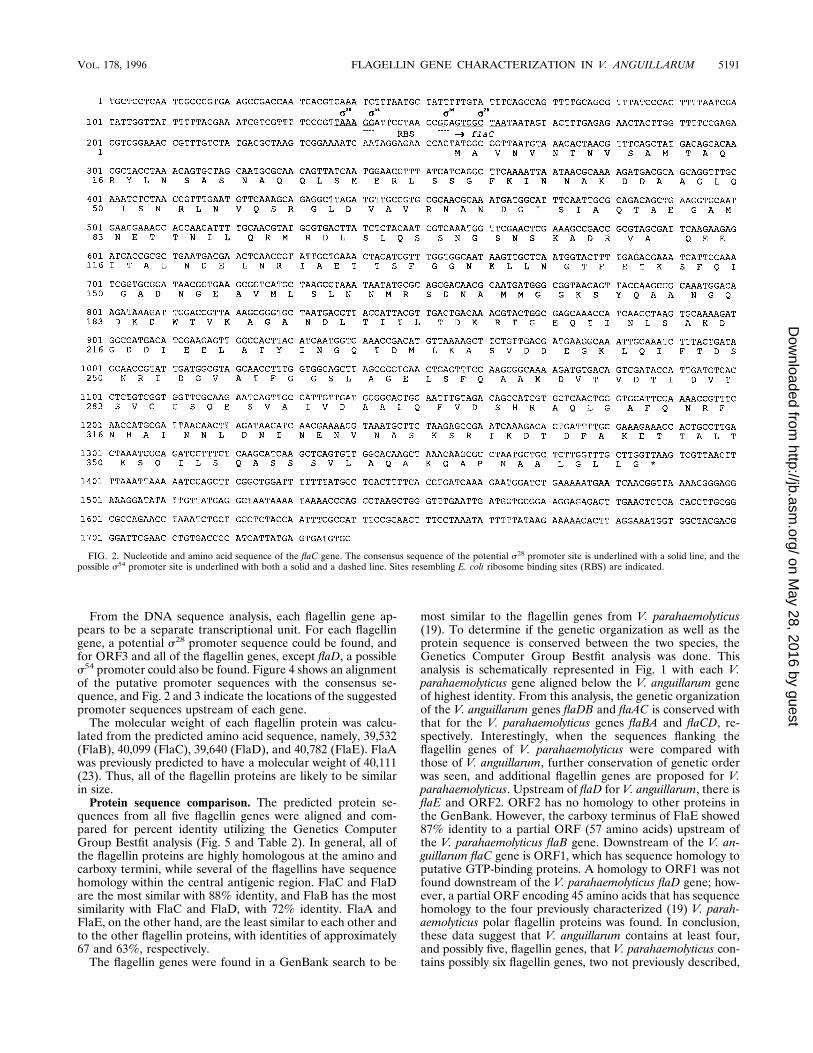
From the DNA sequence analysis, each flagellin gene ap-pears to be a separate transcriptional unit. For each flagellingene, a potential s28 promoter sequence could be found, andfor ORF3 and all of the flagellin genes, except flaD, a possibles54 promoter could also be found. Figure 4 shows an alignmentof the putative promoter sequences with the consensus se-quence, and Fig. 2 and 3 indicate the locations of the suggestedpromoter sequences upstream of each gene.The molecular weight of each flagellin protein was calcu-
lated from the predicted amino acid sequence, namely, 39,532(FlaB), 40,099 (FlaC), 39,640 (FlaD), and 40,782 (FlaE). FlaAwas previously predicted to have a molecular weight of 40,111(23). Thus, all of the flagellin proteins are likely to be similarin size.Protein sequence comparison. The predicted protein se-
quences from all five flagellin genes were aligned and com-pared for percent identity utilizing the Genetics ComputerGroup Bestfit analysis (Fig. 5 and Table 2). In general, all ofthe flagellin proteins are highly homologous at the amino andcarboxy termini, while several of the flagellins have sequencehomology within the central antigenic region. FlaC and FlaDare the most similar with 88% identity, and FlaB has the mostsimilarity with FlaC and FlaD, with 72% identity. FlaA andFlaE, on the other hand, are the least similar to each other andto the other flagellin proteins, with identities of approximately67 and 63%, respectively.The flagellin genes were found in a GenBank search to be
most similar to the flagellin genes from V. parahaemolyticus(19). To determine if the genetic organization as well as theprotein sequence is conserved between the two species, theGenetics Computer Group Bestfit analysis was done. Thisanalysis is schematically represented in Fig. 1 with each V.parahaemolyticus gene aligned below the V. anguillarum geneof highest identity. From this analysis, the genetic organizationof the V. anguillarum genes flaDB and flaAC is conserved withthat for the V. parahaemolyticus genes flaBA and flaCD, re-spectively. Interestingly, when the sequences flanking theflagellin genes of V. parahaemolyticus were compared withthose of V. anguillarum, further conservation of genetic orderwas seen, and additional flagellin genes are proposed for V.parahaemolyticus. Upstream of flaD for V. anguillarum, there isflaE and ORF2. ORF2 has no homology to other proteins inthe GenBank. However, the carboxy terminus of FlaE showed87% identity to a partial ORF (57 amino acids) upstream ofthe V. parahaemolyticus flaB gene. Downstream of the V. an-guillarum flaC gene is ORF1, which has sequence homology toputative GTP-binding proteins. A homology to ORF1 was notfound downstream of the V. parahaemolyticus flaD gene; how-ever, a partial ORF encoding 45 amino acids that has sequencehomology to the four previously characterized (19) V. parah-aemolyticus polar flagellin proteins was found. In conclusion,these data suggest that V. anguillarum contains at least four,and possibly five, flagellin genes, that V. parahaemolyticus con-tains possibly six flagellin genes, two not previously described,
FIG. 2. Nucleotide and amino acid sequence of the flaC gene. The consensus sequence of the potential s28 promoter site is underlined with a solid line, and thepossible s54 promoter site is underlined with both a solid and a dashed line. Sites resembling E. coli ribosome binding sites (RBS) are indicated.
VOL. 178, 1996 FLAGELLIN GENE CHARACTERIZATION IN V. ANGUILLARUM 5191
on May 28, 2016 by guest
http://jb.asm.org/
Dow
nloaded from

FIG. 3. Nucleotide and amino acid sequence of the flaEDB genes. The consensus sequences of the potential s28 and s54 promoter sites are underlined. Sitesresembling E. coli ribosome binding sites (RBS) are indicated.
5192 MCGEE ET AL. J. BACTERIOL.
on May 28, 2016 by guest
http://jb.asm.org/
Dow
nloaded from

and that the genetic organization of these two flagellin loci areconserved between these two species (Fig. 1).Mutant construction. To analyze what possible functional
and structural roles the flagellin genes have, mutagenesis wasdone in a manner similar to that described previously for flaA(23). For each gene, three different in-frame deletion muta-tions were made by allelic exchange. First, a full-gene deletionwhich took away all codons including the stop codon was made.For flaB, however, the last 66 bp were left since they containeda potential s54 promoter for ORF3 (Fig. 3). The second mu-tation deleted 180 bp from the 59 end of each gene. ThisN-terminal deletion was identical for all four flagellin proteins.The third mutation deleted 153 bp from the 39 end of eachgene. For three of the four flagellin proteins, this C-terminaldeletion was identical. For flaB, this deletion was moved 66 bpupstream because of the potential overlapping promoter ofORF3. In addition, a plasmid insertion was made in the inter-genic region downstream of each gene, except flaB, to test forpotential polarity effects on downstream genes. Since the func-tion of the ORF3 polypeptide has not been determined, a
FIG. 4. DNA sequence alignment of the potential s28 and s54 promoterconsensus sequences. The consensus sequence for each sigma factor is in boldtype. The s54 promoter consensus is derived from the transcription initiationsites of various NtrC- and NifA-activated promoters from several different spe-cies (9). The s28 promoter consensus sequence is derived from S. typhimuriumflagellar operons (15).
FIG. 3—Continued.
VOL. 178, 1996 FLAGELLIN GENE CHARACTERIZATION IN V. ANGUILLARUM 5193
on May 28, 2016 by guest
http://jb.asm.org/
Dow
nloaded from

plasmid insertion into the 59 end of this ORF was made toassess the significance of this protein in motility. Figure 5indicates the exact amino acids deleted for each protein, andTable 1 gives the base pairs that were deleted.Since growth rate can affect both the motility and the viru-
lence analyses, it was tested for each flagellin mutant and thepolarity control strains. For all strains, the growth rate did notvary from that of the wild type.Protein analysis of the flagellum. Flagellar preparations
were made from each mutant to determine how each mutationaffected the protein content of the flagellar filament. All mu-tations for each gene, except flaE, caused a loss of that partic-ular flagellin protein in the flagellar filament as determined bySDS-PAGE. There are several explanations for this FlaE ob-servation. flaE could be a silent gene or it could be expressed,but FlaE does not make up a part of the filament. On the otherhand, FlaE could be a very minor part of the filament, andthus, the effect of the flaE mutations on the flagellar filament
is not detectable with SDS-PAGE. Since mutations in flaEaffect the virulence phenotype (see below), flaE is probablyexpressed; however, identification of the protein product re-mains to be determined. The filament protein profile for onemutant of each fla gene is given in Fig. 6. All intergenic plasmidinsertions gave the same filament protein profile as that of thewild type (data not shown), indicating that there is no polareffect on downstream flagellar genes.Interestingly, the molecular masses calculated from the pre-
dicted amino acid sequences of FlaB, -C, and -D (see above)differ from the sizes of these flagellin proteins determinedfrom SDS-PAGE. FlaA, for which the predicted molecularmass agrees with the size determined from SDS-PAGE (23), is
FIG. 5. Alignment of the deduced amino acid sequences of all five V. anguillarum flagellin proteins. Amino acids that are identical for three or more of the flagellinproteins are capitalized and in bold type, whereas those that are different are in lowercase letters. An asterisk indicates an amino acid that is identical for all five flagellinproteins. The arrows indicate regions deleted for the N-terminal deletions, the C-terminal deletions, and the full gene deletions of the flaA, -B, -C, -D, and -E genes.
FIG. 6. Protein profiles of flagellar filament preparations. Flagella were pre-pared as described in Materials and Methods. Six micrograms of each flagellarpreparation was electrophoresed through an SDS–11% polyacrylamide gel. Afilament preparation from a flaA mutant was included for comparison since theFlaA and FlaB proteins migrate so close to one another. WT, a flagellar prep-aration from the wild type; MW, molecular mass marker;2, a full-gene deletion;1, complementation of the full-gene deletions by the insertion of a plasmidcarrying the deleted flagellin gene into the chromosomal region where the genewas deleted. For the flaA gene, complementation of the deleted flagellin genewas done in trans with a plasmid carrying the flaA gene, pFlaA-2 (23).
TABLE 2. Percent identity between the flagellin protein sequences
Protein% Identity
FlaA FlaB FlaC FlaD FlaE
FlaA 100 67 67 67 63FlaB 67 100 72 73 63FlaC 67 72 100 88 64FlaD 67 73 88 100 64FlaE 63 63 64 64 100
5194 MCGEE ET AL. J. BACTERIOL.
on May 28, 2016 by guest
http://jb.asm.org/
Dow
nloaded from

calculated to be larger than FlaB, -C, and -D. However, allthree of these proteins migrate slower in SDS-PAGE. Onepossible explanation for this difference is posttranslationalmodification of FlaB, -C, and -D.Functional analysis. The functional analysis of the mutant
flagella was done to ascertain the importance of each flagellinprotein in maintaining wild-type motility. Light microscopy wasutilized, and the motilities of all strains were similar to that ofthe wild type. In addition, each mutant was tested for its abilityto swim in 0.25% Trypticase soy agar to attain some measureof the speed of movement. The results are given in Table 3.The loss of FlaB, FlaC, or FlaE had almost no effect on mo-tility, whereas, the loss of FlaD had a slightly greater effect,resulting in 10 to 25% less motility than that of the wild type.The ORF3 mutant, KD23, showed an 11% decrease in thewild-type motility, indicating a potential role late in the regu-latory cascade of flagellar biogenesis. Interestingly, a plasmidinsertion in the intergenic region between flaD and flaB causeda slightly hypermotile effect.Structural analysis. To observe any structural differences in
the mutant flagellar filaments, electron microscopy was done.All mutants except the flaB full-gene deletion mutant KD17and the ORF3 insertion mutant, KD23, appeared to have fla-gella of wild-type length. Surprisingly, KD17 and KD23 hadvery elongated flagella (Fig. 7). Since only the full-gene dele-tion and not the 59- and 39-end deletions of flaB caused flagel-lar elongation, it is likely that in the KD17 mutant, a region wasdeleted from the possible s54 promoter of ORF3 which isessential for its function, such as an upstream activating se-quence (30). A second explanation could be a polar effect onan unidentified flagellar gene downstream of ORF3.Virulence analysis. Previously, FlaA was shown to be essen-
tial for the virulence of V. anguillarum (23) (virulence resultsare given in Table 3 for comparison). To determine if any ofthe other flagellin proteins are also essential for virulence, the50% lethal dose (LD50) for infection via the immersion andintraperitoneal routes in a rainbow trout model was deter-
mined for each mutant and for the polarity control strainscontaining plasmid insertions in the intergenic regions. Theresults are presented in Table 3. As expected, since motilitywas altered only slightly, all mutations, except the 59-end de-letions of flaD and flaE, showed only slight, if any, increases inthe LD50 for infection via the immersion route. Because 10-fold fluctuations are often seen with the wild type via immer-sion infections, these small differences are probably not signif-icant. However, 59-end deletions of flaD and flaE showedsignificant increases in the LD50 (10
4- to 105-fold) via bothroutes of infection. Hence, FlaD and FlaE, like FlaA (23), mayhave a role in virulence unrelated to motility. Strains thatcontained plasmids inserted into the intergenic regions showedno effect on virulence, suggesting that the decreases seen withthe virulence assay are not due to polarity effects on down-stream virulence genes.Since the flagellin genes are rather homologous to one an-
other, there is the potential for these genes to recombine withone another to create a recombinant, active gene. Therefore,each flagellin mutant strain and the polarity control strainswere isolated from the kidney of a fish which had died fromvibriosis, usually 3 to 4 days after infection, and PCR was doneto confirm that the original mutation was still present. In ad-dition, motility was also tested on all strains isolated from thefish to ensure that suppression of the flagellin mutations didnot occur during the infection. All strains had motility ratessimilar to those before infection (data not shown).Complementation studies. To confirm that the effects ob-
served with each mutation were due to that particular alter-ation, complementation studies were done. Since we have notfound a plasmid that stably replicates in V. anguillarum duringthe infection process, complementation was achieved by sub-cloning each flagellin gene into a suicide vector and then in-serting these plasmids into the chromosome at the locus of themutated flagellin gene. Protein analysis of the flagellumshowed that all mutants regained a wild-type protein profile ofthe flagellum when complemented with a wild-type copy of the
TABLE 3. Virulence and motility analyses of flagellin mutants from V. anguillaruma
Strain Mutation
Motility (% of wild type) LD50 of mutant strain
Mutantstrains
Mutant strains with thecomplementing plasmid
Intraperitonealinjection
(no. of bacteria)
Immersion(no. of bacteria/ml)
NB10 None (wild type) 100 22 23 102
RO2 flaA 59-end deletion 56 87 6 13 105
DM16 flaA 39-end deletion 58 84 83 105 .1 3 107
RO8 flaA gene deletion 53 87 41 63 105
KD19 flaB 59-end deletion 94 103 28 23 102
KD21 flaB 39-end deletion 94 97 65 23 102
KD17 flaB gene deletion 87 99 13 23 104
KD4 flaC 59-end deletion 91 96 21 43 102
KD6 flaC 39-end deletion 80 93 10 63 102
KD2 flaC full-gene deletion 96 97 45 43 103
KD7 flaC intergenic insertion 96 NDb 6 6 3 102
KD12 flaD 59-end deletion 83 97 83 106 .1 3 107
KD14 flaD 39-end deletion 72 96 33 102 3 3 104
KD10 flaD full-gene deletion 87 100 30 13 104
KD15 flaD intergenic insertion 115 ND 26 33 103
KD27 flaE 59-end deletion 92 93 83 105 .1 3 107
KD29 flaE 39-end deletion 95 101 5 13 104
KD25 flaE full-gene deletion 94 94 8 13 104
KD30 flaE intergenic insertion 97 ND 3 53 103
KD23 ORF3 plasmid insertion 89 ND 36 23 104
a Flagellin A results have been reported previously (23).b ND, not determined.
VOL. 178, 1996 FLAGELLIN GENE CHARACTERIZATION IN V. ANGUILLARUM 5195
on May 28, 2016 by guest
http://jb.asm.org/
Dow
nloaded from

altered gene. One example for each gene set is shown in Fig. 6.For those mutants that showed a decrease in motility, thewild-type motility was regained as measured by the soft agartest (Table 3). Structural analysis (Fig. 7) showed that the flaBfull-gene deletion mutant regained a normal-length flagellum,indicating that any effects on ORF3 or another unidentifiedgene due to the possible loss of promoter function were cor-rected by the gene insertion. Complementation of the 59-enddeletions of flaD and flaE did not give back the wild-typevirulence phenotype. Since the stability of the plasmid couldhave an effect on the complementation, all complemented
strains were reisolated from fish that had died of vibriosis(usually 3 to 4 days after infection) and then tested for chlor-amphenicol resistance, which was present on the inserted plas-mid carrying complementing fla genes. One hundred coloniesfrom each strain were tested, and 97% or more were stillpositive for the presence of the plasmid. One possible expla-nation for these negative complementation results for the vir-ulence phenotypes could be that these mutations have a phe-notype dominant to that of the wild type.
DISCUSSION
The animal model for V. anguillarum is ideal for studying theinvasive mechanism of a bacterial pathogen since the assay forhost invasion uses a natural route of infection. In other words,the fish are allowed to swim in seawater containing bacteria.The virulence of bacterial strains via this route of infection canbe compared with the virulence via intraperitoneal infection todetermine if there is a defect in the invasive capabilities of thebacterium. By the use of this model, the flagellum of V. an-guillarum has been shown previously to be essential for cross-ing the integument of rainbow trout. In a previous study, weshowed that chemotactic motility was needed for efficient in-vasion into the fish host, but once the fish integument wascrossed, chemotactic motility was not required for the progres-sion of vibriosis (26). Similarly, mutations in the flaA gene alsoshowed a requirement for FlaA in the invasion of the fish host(23). Presumably, this is due to the decrease in motility thatoccurs with the loss of FlaA in the flagellar filament. Compar-atively, the FlaB, -C, -D, and -E deletions had no significanteffect on invasion of the fish host. However, motility was de-creased only slightly, if at all, for the FlaBCDE mutants,thereby confirming the requirement for motility in invasion ofthe fish host.In addition to its role in invading the host, FlaA was shown
to have a second role once the fish integument was crossed(23). How the FlaA protein is involved in virulence at this levelis unknown. However, it was suggested that, possibly, when theconserved C terminus was deleted from FlaA, a potential trun-cated protein that could not be exported from the cytoplasm tothe flagellar filament was made. This truncated protein maythen regulate virulence factors such that, even after an intra-peritoneal injection, the strain carrying this mutation de-creased virulence 104-fold. This second phenotype was alsoseen with the 59-end deletion mutants of flaD and flaE, and thisphenotype may be dominant to the wild-type phenotype sincecomplementation of the mutant phenotype was not possible.Interestingly, these mutants carried a deletion at the aminoterminus of the proteins instead of the carboxy terminus as forFlaA. In support of this hypothesis, the flagellum-specific ex-port pathway has been suggested to involve the recognition ofa structural conformation of the flagellar proteins at the sec-ondary level or higher (10). The amino termini of two flagellarproteins, E. coli flagellin (16) and Caulobacter flagellar hookprotein (14), have been shown to be essential for the secretionof these proteins. In addition, a recent study (3) identified apotential antisense RNA that is induced in vivo and that istranscribed from the 39 end of a flagellin gene in V. cholerae.Although the biological significance of this antisense transcriptis unknown, Camilli and Mekalanos (3) speculated that it maydown-regulate flagellin synthesis. If this is true, then with fur-ther speculation the involvement of flagellin proteins in theregulation of virulence determinants could be explained in thefollowing way. In vitro or outside the fish model, the flagellinantisense RNA is uninduced. This allows flagellar biosynthesis,
FIG. 7. Negative staining of the flagella from mutant strains with an elon-gated phenotype. Overnight cultures were fixed with 0.1% glutaraldehyde,spread onto Formvar-coated grids, and then negatively stained with 1% uranylacetate (pH 4.2). (A) NB10, wild type; (B) KD17-B, the complemented flaBfull-gene deletion mutant; (C) KD17, the flaB full-gene deletion mutant; (D)KD23, the ORF3 insertion mutant. Bars, 0.5 mm. All micrographs represent anapproximate average flagellar length for at least 80% of the bacteria examinedfrom two separate specimen preparations.
5196 MCGEE ET AL. J. BACTERIOL.
on May 28, 2016 by guest
http://jb.asm.org/
Dow
nloaded from

which is required for the motility needed to enter the fish host.Once V. anguillarum enters the fish host, motility is not re-quired for the progression of vibriosis, and thus the transcrip-tion of antisense RNA molecules which repress the expressionof FlaA, FlaD, and FlaE is induced. Once the flagellin genesare repressed, then virulence genes required for the progres-sion of disease after the bacterium enters the fish are dere-pressed.The analysis of the flagellar proteins of V. anguillarum by
SDS-PAGE indicated that FlaA, -B, -C, and -D differed inmolecular mass (i.e., 40, 41, 42, and 45 kDa, respectively), eventhough the DNA sequence predicted proteins that are almostidentical in size (i.e., 40.1, 39.5, 40, and 39.6 kDa). This obser-vation suggests posttranslational modification. Other organ-isms have been shown to have flagellar proteins that are post-translationally modified. For example, the flagellar filaments ofP. aeruginosa contain phosphorylated tyrosines (13), and thoseof Campylobacter spp. have been suggested to have phosphor-ylated serines (17). Salmonella typhimurium contains ε-N-methyllysine residues in its flagellar filaments (12), andhalobacterial flagellins are sulfated glycoproteins (31). Thefunction, if any, of the modification of the flagellin proteinsremains to be determined.Although the polar flagella of V. anguillarum and V. para-
haemolyticus appear to be structurally very similar with thisinitial examination, there is one noticeable difference whencomparing the results of the V. parahaemolyticus study (19)with those of this study and the previous V. anguillarum study(23). McCarter (19) reported that the loss of any single flagel-lin protein from the flagellar filament only slightly decreasedthe motility of V. parahaemolyticus. While the same is true ifeither FlaB, -C, -D, or -E is removed from the filament of V.anguillarum, loss of FlaA from the filament diminished motilityto 50% of that for the wild type (23). Several possible expla-nations for this difference may be made. First, the flagellinlocalization within the filaments of the two species may bedifferent. Thus, the structural role of the flagellin homologsmay be different. Second, the two ORFs upstream of the flaBgene and downstream of the flaD gene in V. parahaemolyticusmay code for two additional flagellin proteins that may struc-turally differentiate the flagellar filament of these two organ-isms. Clearly, localization of the flagellin proteins within thefilament of V. anguillarum and V. parahaemolyticus may helpelucidate this difference in phenotype.Recently, flagellar length was suggested to be regulated at
the level of export since cells that contain multiple copies ofthe flagellin gene have abnormally long filaments (1). In thisstudy, we show that a plasmid insertion in ORF3 causes anelongated flagellar filament. This suggests that either ORF3,for which no function is known, or a downstream gene has apossible role either in regulating export of flagellin proteins orin the regulation of flagellin gene expression. If ORF3 has arole in export, then it could aid in the identification of theunidentified export machinery for the flagellar proteins.In summary, this work continues the analysis of the complex
structure of the flagellum of V. anguillarum and of the role thatthe flagellum plays in the virulence of this organism. Fouradditional flagellin genes, flaBCDE, were identified, and theprotein products of three of these genes have been shown to bea part of the flagellar filament. The potential for this manyflagellin subunits present in a flagellar filament is unusual.There is only one bacterium to have been reported with fiveflagellin genes, Halobacterium halobium (6). Virulence analysisof strains with defects in these genes suggests that FlaD andFlaE, like FlaA, may play a role in the regulation of virulence.
ACKNOWLEDGMENT
This work was supported by grant 42.0045/92 from the SwedishCouncil for Forestry and Agricultural Research.
REFERENCES
1. Aizawa, S.-I. 1996. Flagellar assembly in Salmonella typhimurium. Mol. Mi-crobiol. 19:1–5.
2. Attridge, S. R., and D. Rowley. 1983. The role of the flagellum in theadherence of Vibrio cholerae. J. Infect. Dis. 147:864–872.
3. Camilli, A., and J. J. Mekalanos. 1995. Use of recombinase gene fusions toidentify Vibrio cholerae genes induced during infection. Mol. Microbiol.18:671–683.
4. Devereux, J., P. Haeberli, and O. Smithies. 1984. A comprehensive set ofsequence analysis programs for the VAX. Nucleic Acids Res. 12:387–395.
5. Drake, D., and T. C. Montie. 1988. Flagella, motility, and invasive virulenceof Pseudomonas aeruginosa. J. Gen. Microbiol. 134:43–52.
6. Gerl, L., and M. Sumper. 1988. Halobacterial flagellins are encoded by amultigene family. J. Biol. Chem. 263:13246–13251.
7. Grant, C. C. R., M. E. Konkel, W. Cieplak, Jr., and L. S. Tompkins. 1993.Role of flagella in adherence, internalization, and translocation of Campy-lobacter jejuni in nonpolarized and polarized epithelial cell cultures. Infect.Immun. 61:1764–1771.
8. Guentzel, M. N., and L. J. Berry. 1975. Motility as a virulence factor forVibrio cholerae. Infect. Immun. 11:890–897.
9. Gussin, G. N., C. W. Ronson, and F. M. Ausubel. 1986. Regulation ofnitrogen fixation genes. Annu. Rev. Genet. 20:567–591.
10. Homma, M., D. DeRosier, and R. M. Macnab. 1990. Flagellar hook andhook-associated proteins of Salmonella typhimurium and their relationship toother axial components of the flagellum. J. Mol. Biol. 211:819–832.
11. Jones, G. W., and R. Freter. 1976. Adhesive properties of Vibrio cholerae:nature of the interaction with isolated rabbit brush border membranes andhuman erythrocytes. Infect. Immun. 14:240–245.
12. Joys, T. M., and J. Kim. 1978. Identification of ε-N-methllysine residues inthe phase-1 flagella protein of Salmonella typhimurium. Microbios Lett.7:65–68.
13. Kelly-Wintenberg, K., T. Anderson, and T. C. Montie. 1990. Phosphorylatedtyrosines in the flagellum filament protein of Pseudomonas aeruginosa. J.Bacteriol. 172:5135–5139.
14. Kornacker, J. G., and A. Newton. 1994. Information essential for cell-cycle-dependent secretion of the 591-residue Caulobacter hook protein isconfined to a 21-amino-acid sequence near the N-terminus. Mol. Microbiol.14:73–85.
15. Kutsukake, K., Y. Ohya, and T. Iino. 1990. Transcriptional analysis of theflagellar regulon of Salmonella typhimurium. J. Bacteriol. 172:741–747.
16. Kuwajima, G., I. Kawagishi, M. Homma, J.-I. Asaka, E. Kondo, and R. M.Macnab. 1989. Export of an N-terminal fragment of Escherichia coli flagellinby a flagellum-specific pathway. Proc. Natl. Acad. Sci. USA 86:4953–4957.
17. Logan, S. M., T. J. Trust, and P. Guerry. 1989. Evidence for posttranslationalmodification and gene duplication of Campylobacter flagellin. J. Bacteriol.171:3031–3038.
18. Lugtenberg, B., J. Meijers, R. Peters, P. van der Hoek, and L. van Alphen.1975. Electrophoretic resolution of the “major outer membrane protein” ofEscherichia coli K12 into four bands. FEBS Lett. 58:254–258.
19. McCarter, L. L. 1995. Genetic and molecular characterization of the polarflagellum of Vibrio parahaemolyticus. J. Bacteriol. 177:1595–1609.
20. McSweegan, E., and R. I. Walker. 1986. Identification and characterizationof two Campylobacter jejuni adhesins for cellular and mucous substrates.Infect. Immun. 53:141–148.
21. Miller, V. L., and J. J. Mekalanos. 1988. A novel suicide vector and its usein construction of insertion mutations: osmoregulation of outer membraneproteins and virulence determinants in Vibrio cholerae requires toxR. J.Bacteriol. 170:2575–2583.
22. Milton, D. L., A. Norqvist, and H. Wolf-Watz. 1992. Cloning of a metallo-protease gene involved in the virulence mechanism of Vibrio anguillarum. J.Bacteriol. 174:7235–7244.
23. Milton, D. L., R. O’Toole, P. Horstedt, and H. Wolf-Watz. 1996. Flagellin Ais essential for the virulence of Vibrio anguillarum. J. Bacteriol. 178:1310–1319.
24. Norqvist, A., Å. Hagstrom, and H. Wolf-Watz. 1989. Protection of rainbowtrout against vibriosis and furunculosis by the use of attenuated strains ofVibrio anguillarum. Appl. Environ. Microbiol. 55:1400–1405.
25. Norqvist, A., and H. Wolf-Watz. 1993. Characterization of a novel chromo-somal virulence locus involved in expression of a major surface flagellarsheath antigen of the fish pathogen Vibrio anguillarum. Infect. Immun. 61:2434–2444.
26. O’Toole, R., D. L. Milton, and H. Wolf-Watz. 1996. Chemotactic motility isrequired for invasion of the host by the fish pathogen Vibrio anguillarum.Mol. Microbiol. 19:625–637.
27. Richardson, K. 1991. Roles of motility and flagellar structure in pathoge-nicity of Vibrio cholerae: analysis of motility mutants in three animal models.Infect. Immun. 59:2727–2736.
VOL. 178, 1996 FLAGELLIN GENE CHARACTERIZATION IN V. ANGUILLARUM 5197
on May 28, 2016 by guest
http://jb.asm.org/
Dow
nloaded from

28. Sambrook, J., E. F. Fritsch, and T. Maniatis. 1989. Molecular cloning: alaboratory manual, 2nd ed. Cold Spring Harbor Laboratory, Cold SpringHarbor, N.Y.
29. Simon, R., U. Priefer, and A. Puhler. 1983. A broad host range mobilizationsystem for in vivo genetic engineering: transposon mutagenesis in gramnegative bacteria. Bio/Technology 1:787–796.
30. Thony, B., and H. Hennecke. 1989. The 224/212 promoter comes of age.
FEMS Microbiol. Rev. 63:341–358.31. Wieland, F., G. Paul, and M. Sumper. 1985. Halobacterial flagellins are
sulfated glycoproteins. J. Biol. Chem. 260:15180–15185.32. Yao, R., D. H. Burr, P. Doig, T. J. Trust, H. Niu, and P. Guerry. 1994.
Isolation of motile and non-motile insertional mutants of Campylobacterjejuni: the role of motility in adherence and invasion of eukaryotic cells. Mol.Microbiol. 14:883–893.
5198 MCGEE ET AL. J. BACTERIOL.
on May 28, 2016 by guest
http://jb.asm.org/
Dow
nloaded from