
I know what you are doing
11
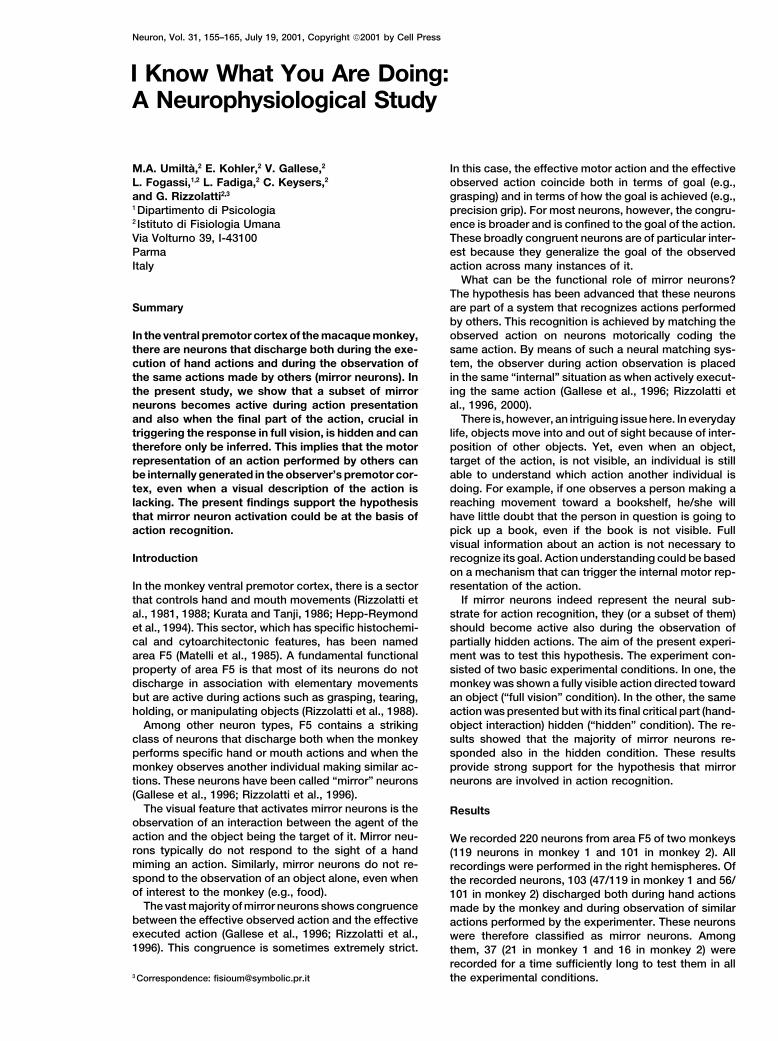
Neuron, Vol. 31, 155–165, July 19, 2001, Copyright 2001 by Cell Press I Know What You Are Doing: A Neurophysiological Study In this case, the effective motor action and the effective observed action coincide both in terms of goal (e.g., grasping) and in terms of how the goal is achieved (e.g., M.A. Umilta `, 2 E. Kohler, 2 V. Gallese, 2 L. Fogassi, 1,2 L. Fadiga, 2 C. Keysers, 2 and G. Rizzolatti 2,3 1 Dipartimento di Psicologia precision grip). For most neurons, however, the congru- ence is broader and is confined to the goal of the action. 2 Istituto di Fisiologia Umana Via Volturno 39, I-43100 These broadly congruent neurons are of particular inter- est because they generalize the goal of the observed Parma Italy action across many instances of it. What can be the functional role of mirror neurons? The hypothesis has been advanced that these neurons are part of a system that recognizes actions performed Summary by others. This recognition is achieved by matching the observed action on neurons motorically coding the In the ventral premotor cortex of the macaque monkey, there are neurons that discharge both during the exe- same action. By means of such a neural matching sys- tem, the observer during action observation is placed cution of hand actions and during the observation of the same actions made by others (mirror neurons). In in the same “internal” situation as when actively execut- ing the same action (Gallese et al., 1996; Rizzolatti et the present study, we show that a subset of mirror neurons becomes active during action presentation al., 1996, 2000). There is, however, an intriguing issue here. In everyday and also when the final part of the action, crucial in triggering the response in full vision, is hidden and can life, objects move into and out of sight because of inter- position of other objects. Yet, even when an object, therefore only be inferred. This implies that the motor representation of an action performed by others can target of the action, is not visible, an individual is still able to understand which action another individual is be internally generated in the observer’s premotor cor- tex, even when a visual description of the action is doing. For example, if one observes a person making a reaching movement toward a bookshelf, he/she will lacking. The present findings support the hypothesis that mirror neuron activation could be at the basis of have little doubt that the person in question is going to pick up a book, even if the book is not visible. Full action recognition. visual information about an action is not necessary to recognize its goal. Action understanding could be based Introduction on a mechanism that can trigger the internal motor rep- resentation of the action. In the monkey ventral premotor cortex, there is a sector that controls hand and mouth movements (Rizzolatti et If mirror neurons indeed represent the neural sub- strate for action recognition, they (or a subset of them) al., 1981, 1988; Kurata and Tanji, 1986; Hepp-Reymond et al., 1994). This sector, which has specific histochemi- should become active also during the observation of partially hidden actions. The aim of the present experi- cal and cytoarchitectonic features, has been named area F5 (Matelli et al., 1985). A fundamental functional ment was to test this hypothesis. The experiment con- sisted of two basic experimental conditions. In one, the property of area F5 is that most of its neurons do not discharge in association with elementary movements monkey was shown a fully visible action directed toward an object (“full vision” condition). In the other, the same but are active during actions such as grasping, tearing, holding, or manipulating objects (Rizzolatti et al., 1988). action was presented but with its final critical part (hand- object interaction) hidden (“hidden” condition). The re- Among other neuron types, F5 contains a striking class of neurons that discharge both when the monkey sults showed that the majority of mirror neurons re- sponded also in the hidden condition. These results performs specific hand or mouth actions and when the monkey observes another individual making similar ac- provide strong support for the hypothesis that mirror neurons are involved in action recognition. tions. These neurons have been called “mirror” neurons (Gallese et al., 1996; Rizzolatti et al., 1996). The visual feature that activates mirror neurons is the Results observation of an interaction between the agent of the action and the object being the target of it. Mirror neu- We recorded 220 neurons from area F5 of two monkeys rons typically do not respond to the sight of a hand (119 neurons in monkey 1 and 101 in monkey 2). All miming an action. Similarly, mirror neurons do not re- recordings were performed in the right hemispheres. Of spond to the observation of an object alone, even when the recorded neurons, 103 (47/119 in monkey 1 and 56/ of interest to the monkey (e.g., food). 101 in monkey 2) discharged both during hand actions The vast majority of mirror neurons shows congruence made by the monkey and during observation of similar between the effective observed action and the effective actions performed by the experimenter. These neurons executed action (Gallese et al., 1996; Rizzolatti et al., were therefore classified as mirror neurons. Among 1996). This congruence is sometimes extremely strict. them, 37 (21 in monkey 1 and 16 in monkey 2) were recorded for a time sufficiently long to test them in all the experimental conditions. 3 Correspondence: [email protected]
-
Upload
independent -
Category
Documents
-
view
6 -
download
0
Transcript of I know what you are doing
Neuron, Vol. 31, 155–165, July 19, 2001, Copyright 2001 by Cell Press
I Know What You Are Doing:A Neurophysiological Study
In this case, the effective motor action and the effectiveobserved action coincide both in terms of goal (e.g.,grasping) and in terms of how the goal is achieved (e.g.,
M.A. Umilta,2 E. Kohler,2 V. Gallese,2
L. Fogassi,1,2 L. Fadiga,2 C. Keysers,2
and G. Rizzolatti2,3
1 Dipartimento di Psicologia precision grip). For most neurons, however, the congru-ence is broader and is confined to the goal of the action.2 Istituto di Fisiologia Umana
Via Volturno 39, I-43100 These broadly congruent neurons are of particular inter-est because they generalize the goal of the observedParma
Italy action across many instances of it.What can be the functional role of mirror neurons?
The hypothesis has been advanced that these neuronsare part of a system that recognizes actions performedSummaryby others. This recognition is achieved by matching theobserved action on neurons motorically coding theIn the ventral premotor cortex of the macaque monkey,
there are neurons that discharge both during the exe- same action. By means of such a neural matching sys-tem, the observer during action observation is placedcution of hand actions and during the observation of
the same actions made by others (mirror neurons). In in the same “internal” situation as when actively execut-ing the same action (Gallese et al., 1996; Rizzolatti etthe present study, we show that a subset of mirror
neurons becomes active during action presentation al., 1996, 2000).There is, however, an intriguing issue here. In everydayand also when the final part of the action, crucial in
triggering the response in full vision, is hidden and can life, objects move into and out of sight because of inter-position of other objects. Yet, even when an object,therefore only be inferred. This implies that the motor
representation of an action performed by others can target of the action, is not visible, an individual is stillable to understand which action another individual isbe internally generated in the observer’s premotor cor-
tex, even when a visual description of the action is doing. For example, if one observes a person making areaching movement toward a bookshelf, he/she willlacking. The present findings support the hypothesis
that mirror neuron activation could be at the basis of have little doubt that the person in question is going topick up a book, even if the book is not visible. Fullaction recognition.visual information about an action is not necessary torecognize its goal. Action understanding could be basedIntroductionon a mechanism that can trigger the internal motor rep-resentation of the action.In the monkey ventral premotor cortex, there is a sector
that controls hand and mouth movements (Rizzolatti et If mirror neurons indeed represent the neural sub-strate for action recognition, they (or a subset of them)al., 1981, 1988; Kurata and Tanji, 1986; Hepp-Reymond
et al., 1994). This sector, which has specific histochemi- should become active also during the observation ofpartially hidden actions. The aim of the present experi-cal and cytoarchitectonic features, has been named
area F5 (Matelli et al., 1985). A fundamental functional ment was to test this hypothesis. The experiment con-sisted of two basic experimental conditions. In one, theproperty of area F5 is that most of its neurons do not
discharge in association with elementary movements monkey was shown a fully visible action directed towardan object (“full vision” condition). In the other, the samebut are active during actions such as grasping, tearing,
holding, or manipulating objects (Rizzolatti et al., 1988). action was presented but with its final critical part (hand-object interaction) hidden (“hidden” condition). The re-Among other neuron types, F5 contains a striking
class of neurons that discharge both when the monkey sults showed that the majority of mirror neurons re-sponded also in the hidden condition. These resultsperforms specific hand or mouth actions and when the
monkey observes another individual making similar ac- provide strong support for the hypothesis that mirrorneurons are involved in action recognition.tions. These neurons have been called “mirror” neurons
(Gallese et al., 1996; Rizzolatti et al., 1996).The visual feature that activates mirror neurons is the Results
observation of an interaction between the agent of theaction and the object being the target of it. Mirror neu- We recorded 220 neurons from area F5 of two monkeysrons typically do not respond to the sight of a hand (119 neurons in monkey 1 and 101 in monkey 2). Allmiming an action. Similarly, mirror neurons do not re- recordings were performed in the right hemispheres. Ofspond to the observation of an object alone, even when the recorded neurons, 103 (47/119 in monkey 1 and 56/of interest to the monkey (e.g., food). 101 in monkey 2) discharged both during hand actions
The vast majority of mirror neurons shows congruence made by the monkey and during observation of similarbetween the effective observed action and the effective actions performed by the experimenter. These neuronsexecuted action (Gallese et al., 1996; Rizzolatti et al., were therefore classified as mirror neurons. Among1996). This congruence is sometimes extremely strict. them, 37 (21 in monkey 1 and 16 in monkey 2) were
recorded for a time sufficiently long to test them in allthe experimental conditions.3 Correspondence: [email protected]
Neuron156
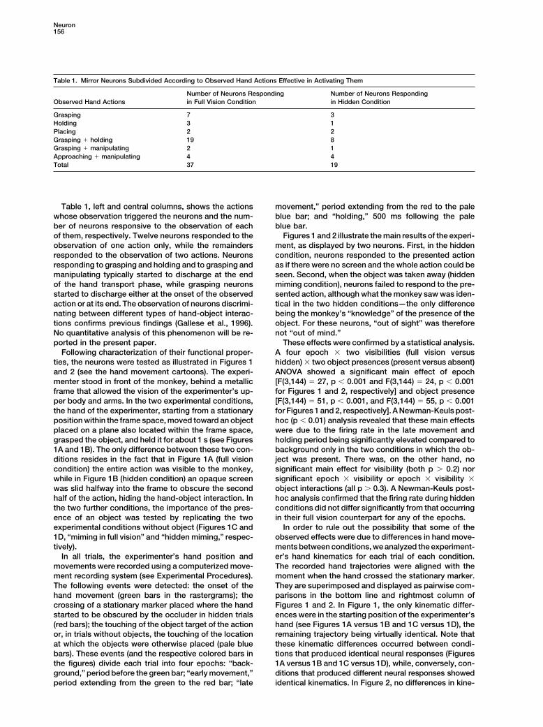
Table 1. Mirror Neurons Subdivided According to Observed Hand Actions Effective in Activating Them
Number of Neurons Responding Number of Neurons RespondingObserved Hand Actions in Full Vision Condition in Hidden Condition
Grasping 7 3Holding 3 1Placing 2 2Grasping � holding 19 8Grasping � manipulating 2 1Approaching � manipulating 4 4Total 37 19
Table 1, left and central columns, shows the actions movement,” period extending from the red to the paleblue bar; and “holding,” 500 ms following the palewhose observation triggered the neurons and the num-
ber of neurons responsive to the observation of each blue bar.Figures 1 and 2 illustrate the main results of the experi-of them, respectively. Twelve neurons responded to the
observation of one action only, while the remainders ment, as displayed by two neurons. First, in the hiddencondition, neurons responded to the presented actionresponded to the observation of two actions. Neurons
responding to grasping and holding and to grasping and as if there were no screen and the whole action could beseen. Second, when the object was taken away (hiddenmanipulating typically started to discharge at the end
of the hand transport phase, while grasping neurons miming condition), neurons failed to respond to the pre-sented action, although what the monkey saw was iden-started to discharge either at the onset of the observed
action or at its end. The observation of neurons discrimi- tical in the two hidden conditions—the only differencebeing the monkey’s “knowledge” of the presence of thenating between different types of hand-object interac-
tions confirms previous findings (Gallese et al., 1996). object. For these neurons, “out of sight” was thereforenot “out of mind.”No quantitative analysis of this phenomenon will be re-
ported in the present paper. These effects were confirmed by a statistical analysis.A four epoch � two visibilities (full vision versusFollowing characterization of their functional proper-
ties, the neurons were tested as illustrated in Figures 1 hidden) � two object presences (present versus absent)ANOVA showed a significant main effect of epochand 2 (see the hand movement cartoons). The experi-
menter stood in front of the monkey, behind a metallic [F(3,144) � 27, p � 0.001 and F(3,144) � 24, p � 0.001for Figures 1 and 2, respectively] and object presenceframe that allowed the vision of the experimenter’s up-
per body and arms. In the two experimental conditions, [F(3,144) � 51, p � 0.001, and F(3,144) � 55, p � 0.001for Figures 1 and 2, respectively]. A Newman-Keuls post-the hand of the experimenter, starting from a stationary
position within the frame space, moved toward an object hoc (p � 0.01) analysis revealed that these main effectswere due to the firing rate in the late movement andplaced on a plane also located within the frame space,
grasped the object, and held it for about 1 s (see Figures holding period being significantly elevated compared tobackground only in the two conditions in which the ob-1A and 1B). The only difference between these two con-
ditions resides in the fact that in Figure 1A (full vision ject was present. There was, on the other hand, nosignificant main effect for visibility (both p � 0.2) norcondition) the entire action was visible to the monkey,
while in Figure 1B (hidden condition) an opaque screen significant epoch � visibility or epoch � visibility �object interactions (all p � 0.3). A Newman-Keuls post-was slid halfway into the frame to obscure the second
half of the action, hiding the hand-object interaction. In hoc analysis confirmed that the firing rate during hiddenconditions did not differ significantly from that occurringthe two further conditions, the importance of the pres-
ence of an object was tested by replicating the two in their full vision counterpart for any of the epochs.In order to rule out the possibility that some of theexperimental conditions without object (Figures 1C and
1D, “miming in full vision” and “hidden miming,” respec- observed effects were due to differences in hand move-ments between conditions, we analyzed the experiment-tively).
In all trials, the experimenter’s hand position and er’s hand kinematics for each trial of each condition.The recorded hand trajectories were aligned with themovements were recorded using a computerized move-
ment recording system (see Experimental Procedures). moment when the hand crossed the stationary marker.They are superimposed and displayed as pairwise com-The following events were detected: the onset of the
hand movement (green bars in the rastergrams); the parisons in the bottom line and rightmost column ofFigures 1 and 2. In Figure 1, the only kinematic differ-crossing of a stationary marker placed where the hand
started to be obscured by the occluder in hidden trials ences were in the starting position of the experimenter’shand (see Figures 1A versus 1B and 1C versus 1D), the(red bars); the touching of the object target of the action
or, in trials without objects, the touching of the location remaining trajectory being virtually identical. Note thatthese kinematic differences occurred between condi-at which the objects were otherwise placed (pale blue
bars). These events (and the respective colored bars in tions that produced identical neural responses (Figures1A versus 1B and 1C versus 1D), while, conversely, con-the figures) divide each trial into four epochs: “back-
ground,” period before the green bar; “early movement,” ditions that produced different neural responses showedidentical kinematics. In Figure 2, no differences in kine-period extending from the green to the red bar; “late
Recognition of Hidden Actions157
matics are visible between conditions. Hand kinematics vision conditions (neurons shown in Figures 1 and 2 aredrawn from this category). For 9/19 neurons, responsestherefore cannot account for the observed neural re-
sponses. were more pronounced in the full vision condition. For1/19 neurons, the response was significantly more pro-Some intertrial variation in the firing rate of the neurons
is visible in the rasters. This variability is probably due nounced in the hidden condition. Two neurons finallyshowed an ambivalent effect. For one of these latterto unsystematic variations in the cognitive state of the
animal, as there are no obvious differences between two, the response was stronger during the late move-ment epoch in the hidden condition, while it waskinematics in trials with higher and lower firing rate.
To test whether responses to hidden actions were stronger during the holding epoch in the full vision condi-tion. For the other neuron, the peak response occurredpresent overall in the population of 37 neurons studied,
their responses were analyzed together as a population. during the holding phase in the hidden condition andimmediately after the analyzed holding phase in the fullThe responses in full vision and hidden conditions were
therefore compared. Since neurons differed in their firing vision condition. Figure 4 illustrates examples of neu-rons belonging to those different categories.rates, the firing rates of each neuron were normalized by
subtracting the activity occurring during the background Neuron 1 of Figure 4 exemplifies the behavior of amirror neuron belonging to the nine neurons showingepoch from that occurring during the early movement,
late movement, and holding epochs (“net activity”). After a significant but smaller response in the hidden com-pared to the full vision condition. Rasters and histo-this normalization, the activity during the background
period was always zero. This net activity was then aver- grams are aligned at the moment in which the experi-menter’s hand touched the object (blue bars and blackaged over all ten trials and divided by the highest aver-
age activity occurring for that neuron in full vision. Figure lines, respectively). Note the specificity of the neuron’sresponse to the effective observed action (grasping and3 illustrates the average over all 37 neurons of that activ-
ity as a function of epoch and condition. A four epoch � holding versus placing and holding) in both full visionand hidden conditions. As for all mirror neurons pre-two conditions ANOVA showed significant main effects
for epoch [F(3,108) � 25, p � 0.001] and condition sented in this article, this neuron also responded whilethe monkey performed an action similar to the one effec-[F(1,36) � 9, p � 0.01] as well as a significant interaction
[F(3,108) � 6, p � 0.001]. A Newman-Keuls post-hoc tive when observed (Figure 4E1). When the monkey per-formed other types of hand actions (data not shown),analysis (p � 0.01) revealed that the firing rate in all but
the early movement epoch for the hidden condition was no response was observed.Neuron 2 of Figure 4 shows the behavior of one ofsignificantly higher than during the background activity.
This indicates that the population as a whole responded the two ambivalent mirror neurons. As one can see,the increase in activity during grasping (late movementto an action even while the action is hidden. The post-
hoc analysis also revealed that during early movement, epoch) may be attributed to a shift of the responseduring hidden condition from holding to grasping. Infiring rates did not differ between the two conditions.
During the late movement and holding epoch, on the full vision, the neuron discharged mostly during holding(Figure 4A2). In hidden condition, the discharge startedother hand, the full vision condition produced a larger
population response than the hidden condition. Note, as soon as the experimenter’s hand disappeared fromvision, peaked before grasping completion, and ceasedfinally, that the post-hoc analysis revealed that the re-
sponse during the late movement and holding epoch during the early holding phase (Figure 4B2). The lack ofresponse when the same action was mimed (Figureswas higher than that during the early movement and
background. Hence, in the hidden condition, the re- 4C2 and 4D2) indicates that the activity in the formertrials was related to the monkey’s knowledge of thesponse was maximal after the hand disappeared.
To isolate the contribution of individual neurons to object presence.Neuron 3 of Figure 4 exemplifies the behavior of athe effect demonstrated by the population, all 37 neu-
rons were tested using a two visibilities (full vision versus mirror neuron belonging to the remaining 18/37 neuronsnot responding significantly in the hidden condition.hidden) � four epochs ANOVA with ten trials in each
condition. Neurons were classified as responding signif- The characteristics of all neurons of the present studyremained constant during the whole testing period. Inicantly in a particular condition if they showed a signifi-
cant main effect of epoch or an epoch � visibility interac- particular, neurons that did not respond in the hiddencondition continued not to respond even when arousingtion and when a Newman-Keuls analysis showed that
one movement epoch produced an activity higher than stimuli were delivered between trials to the monkey anddespite the fact that a reward was given after every trialthe background epoch (all p � 0.01). According to this
criterion, 19/37 neurons were found to respond signifi- (see Experimental Procedures). Furthermore, neuronsresponding in hidden condition and those responding incantly during the hidden condition (12/21 in monkey 1,
7/16 in monkey 2), and, as expected, all 37/37 neurons full vision condition only were not segregated in differentparts of F5. In electrode penetrations in which we wereresponded significantly in the full vision condition (see
Table 1). able to study more than one neuron, it occurred thatboth classes of neurons were represented.For the 19 neurons responsive during the hidden con-
dition, we then examined, epoch per epoch, how the In pilot experiments, eye movements during full vision,hidden, miming in full vision, and hidden miming condi-responses in the hidden condition compared with those
in the full vision condition, using the same Newman- tions were measured. Analysis of the data revealed thateye tracking of the experimenter’s hand was very rareKeuls analysis. In 7/19 neurons, there was no significant
difference between the responses in the hidden and full in all conditions. A typical example of eye position during
Neuron158
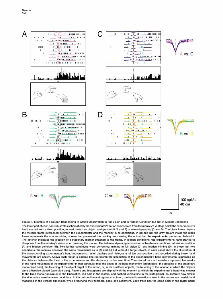
Figure 1. Example of a Neuron Responding to Action Observation in Full Vision and in Hidden Condition but Not in Mimed Conditions
The lower part of each panel illustrates schematically the experimenter’s action as observed from the monkey’s vantage point: the experimenter’shand started from a fixed position, moved toward an object, and grasped it (A and B) or mimed grasping (C and D). The black frame depictsthe metallic frame interposed between the experimenter and the monkey in all conditions. In (B) and (D), the gray square inside the blackframe represents the opaque sliding screen that prevented the monkey from seeing the action that the experimenter performed behind it.The asterisk indicates the location of a stationary marker attached to the frame. In hidden conditions, the experimenter’s hand started todisappear from the monkey’s vision when crossing this marker. The behavioral paradigm consisted of two basic conditions: full vision condition(A) and hidden condition (B). Two further conditions were performed: miming in full vision (C) and hidden miming (D). In these last twoconditions, the monkey observed the same movements as in (A) and (B) but without a target object. In each panel above the illustration ofthe corresponding experimenter’s hand movements, raster displays and histograms of ten consecutive trials recorded during these handmovements are shown. Above each raster, a colored line represents the kinematics of the experimenter’s hand movements, expressed asthe distance between the hand of the experimenter and the stationary marker over time. The colored bars in the rasters represent landmarksof the hand movement of the experimenter in that particular trial: the onset of the hand movement (green bars), the crossing of the stationarymarker (red bars), the touching of the object target of the action, or, in trials without objects, the touching of the location at which the objectswere otherwise placed (pale blue bars). Rasters and histograms are aligned with the moment at which the experimenter’s hand was closestto the fixed marker (minimum in the kinematics, red bars in the rasters, and dashed vertical line in the histograms). To illustrate how similarthe kinematics were between conditions, in the bottom line and rightmost column, the hand kinematics shown in the rasters are overlaid andmagnified in the vertical dimension while preserving their temporal scale and alignment. Each trace has the same color in the raster panel
Recognition of Hidden Actions159
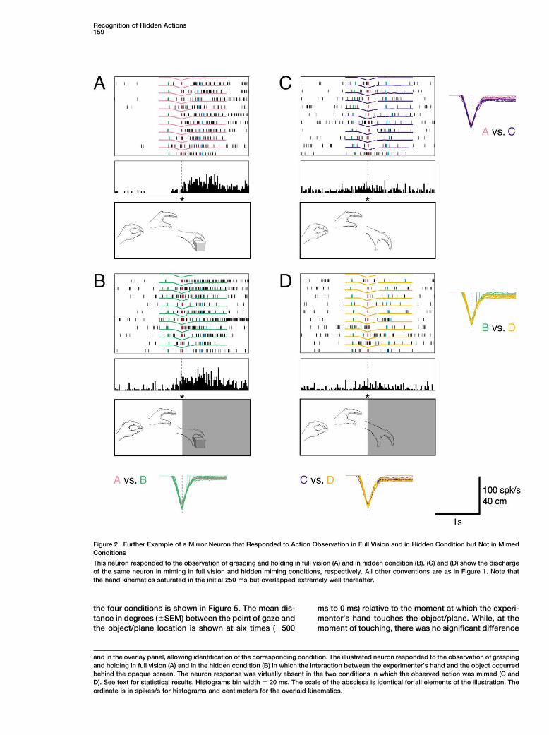
Figure 2. Further Example of a Mirror Neuron that Responded to Action Observation in Full Vision and in Hidden Condition but Not in MimedConditions
This neuron responded to the observation of grasping and holding in full vision (A) and in hidden condition (B). (C) and (D) show the dischargeof the same neuron in miming in full vision and hidden miming conditions, respectively. All other conventions are as in Figure 1. Note thatthe hand kinematics saturated in the initial 250 ms but overlapped extremely well thereafter.
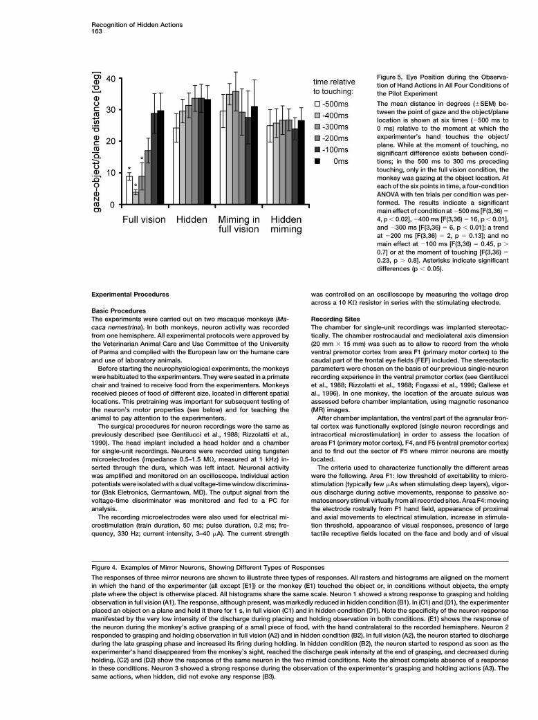
the four conditions is shown in Figure 5. The mean dis- ms to 0 ms) relative to the moment at which the experi-menter’s hand touches the object/plane. While, at thetance in degrees (�SEM) between the point of gaze and
the object/plane location is shown at six times (�500 moment of touching, there was no significant difference
and in the overlay panel, allowing identification of the corresponding condition. The illustrated neuron responded to the observation of graspingand holding in full vision (A) and in the hidden condition (B) in which the interaction between the experimenter’s hand and the object occurredbehind the opaque screen. The neuron response was virtually absent in the two conditions in which the observed action was mimed (C andD). See text for statistical results. Histograms bin width � 20 ms. The scale of the abscissa is identical for all elements of the illustration. Theordinate is in spikes/s for histograms and centimeters for the overlaid kinematics.
Neuron160
gering the neuron was repeated without an object be-hind the occluder, the response was absent, in spite ofthe fact that what the monkey saw was exactly the samemovement in the two conditions. Furthermore, in manyneurons, and specifically in those discharging during thelate phase of grasping and during holding, the responsewas maximal at the end of the hidden action, and, overallin the population, activity was significantly larger whenthe hand was hidden compared to the early movementepoch when the hand was still visible (see Figure 3).The opposite should be expected if the responses weresimply triggered by the initial arm movement.
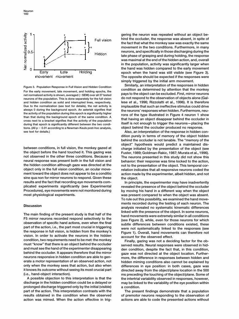
Similarly, an interpretation of the responses in hiddenFigure 3. Population Response in Full Vision and Hidden Conditioncondition as determined by attention that the monkey
For the early movement, late movement, and holding epochs, thepays to the object can be excluded. First, mirror neuronsnet normalized activity is shown, averaged (�SEM) over all 37 testeddo not respond to the observation of objects alone (Gal-neurons of the population. This is done separately for the full visionlese et al., 1996; Rizzolatti et al., 1996). It is thereforeand hidden condition as solid and interrupted lines, respectively.
Due to the normalization (see text for details), the net activity is implausible that such an ineffective stimulus could drivealways 0 during the background epoch. An asterisk signifies that the neurons’ responses when hidden. Furthermore, neu-the activity of the population during this epoch is significantly higher rons of the type illustrated in Figure 4 neuron 1 showthan that during the background epoch of the same condition. A
that having an object disappear behind the occluder incross next to a bracket signifies that the activity of the populationitself is not enough to trigger the neurons—placing theduring that epoch is significantly different between the two condi-object behind the occluder produced no response.tions. (All p � 0.01 according to a Newman-Keuls post-hoc analysis,
see text for details.) Also, an interpretation of the response in hidden con-dition purely in terms of memory of the object hiddenbehind the occluder is not tenable. The “memory of theobject” hypothesis would predict a maintained dis-
between conditions, in full vision, the monkey gazed at charge initiated by the presentation of the object (seethe object before the hand touched it. This gazing was Fuster, 1989; Goldman-Rakic, 1987; Murata et al., 1996).not observed in the other three conditions. Because a The neurons presented in this study did not show thisneural response was present both in the full vision and behavior: their response was time locked to the action,the hidden condition although gaze was directed at the not to the presentation of the object. This temporal cor-object only in the full vision condition, an ocular move- relation indicates that all responsive neurons coded thement toward the object does not appear to be a conditio action made by the experimenter, albeit hidden, and notsine qua non for mirror neurons to respond. Given these the object.results and the fact that recording eye movements com- In principle, the experimenter may have inadvertentlyplicated experiments significantly (see Experimental revealed the presence of the object behind the occluderProcedures), eye movements were not monitored during by moving his hand in a different way when the objectmost physiological experiments. was present compared to when the object was absent.
To rule out this possibility, we examined the hand move-ments recorded during the testing of each neuron. The
Discussion analysis revealed no systematic kinematic differenceslinked with the presence of the object. In some neurons,
The main finding of the present study is that half of the hand movements were extremely similar in all conditionsF5 mirror neurons recorded respond selectively to the (see Figure 2), while, even for those neurons for whichobservation of specific hand actions, even when the final subtle differences between conditions existed, thesepart of the action, i.e., the part most crucial in triggering were not systematically linked to the responses (seethe response in full vision, is hidden from the monkey’s Figure 1). Overall, hand movements can therefore notvision. In order to activate the neurons in the hidden account for the observed effect.condition, two requirements need to be met: the monkey Finally, gazing was not a deciding factor for the ob-must “know” that there is an object behind the occluder served results. Neural responses were observed in hid-and must see the hand of the experimenter disappearing den condition, despite the fact that, in this condition,behind the occluder. It appears therefore that the mirror gaze was not directed at the object location. Further-neurons responsive in hidden condition are able to gen- more, the difference in responses between hidden anderate a motor representation of an observed action, not hidden miming conditions also cannot be explained byonly when the monkey sees that action, but also when differences in eye position: in both cases, gaze wasit knows its outcome without seeing its most crucial part directed away from the object/plane location in the 500(i.e., hand-object interaction). ms preceding the touching of the object/plane. Some of
A possible objection to this interpretation is that the the intertrial variability observed in responses, however,discharge in the hidden condition could be a delayed or may be linked to the variability of the eye position withinprolonged discharge triggered only by the initial (visible) a condition.part of the action. This interpretation contrasts with the The present findings demonstrate that a populationresults obtained in the condition when the observed of premotor neurons responding to the observation of
actions are able to code the presented actions withoutaction was mimed. When the action effective in trig-
Recognition of Hidden Actions161
full vision of them. Although neurons endowed with such ence can only be inferred from past perceptual history,a property were never described before, visually respon- providing a form of knowledge about the presence ofsive neurons discharging when the effective visual stim- an object. What the findings of the present study demon-uli are not visible were reported before by Assad and strate is that this knowledge can be used to go a stepMaunsell (1995), Graziano et al. (1997b), and by Perrett further: representing actions of which the inferred objectand his coworkers (Baker et al., 2001; Jellema and Per- is a target. What mirror neurons code is not the mererett, 2001). presence of an object but rather that a specific action
Assad and Maunsell (1995) recorded from neurons in is directed toward an existing object (Gallese et al., 1996;the posterior parietal cortex (PP) responding to move- Rizzolatti et al., 1996). Hence, to selectively respondments of a simple visual stimulus, even when the move- in hidden conditions, mirror neurons have to infer andment could only be inferred. In all their conditions, a represent the occluded specific action in addition to thestationary stimulus was first presented for 100 ms. In inferred object, which is the goal of the action.movement blocks, either the monkey could see the stim- Recently, Filion et al. (1996) demonstrated that mon-ulus move toward the center of fixation (full vision trials) keys are able to infer the movement of a hidden object.or the stimulus disappeared to reappear 500 ms later These authors trained monkeys to hit a target eitheras if it had moved behind an invisible occluder (occlusion when it reappeared out of an occluding surface or, in atrials). These movement blocks were alternated with further experiment, when it was still hidden. The resultsblocks of “blink” trials in which the stimulus, after a showed that the monkeys were able to hit a target aspresentation of 100 ms, disappeared for 500 ms and
soon as it reappeared, in spite of the fact that the objectthen reappeared at the location where it had disap-
trajectory changed from trial to trial. Similarly, they werepeared—as if it never moved. Interestingly, the PP neu-able to learn to hit the target during the phase in whichrons responded during the invisible part of occlusionits trajectory was hidden. These findings are in goodtrials but not during the physically identical part of blinkagreement with the conclusions drawn from neurophysi-trials. The context of the block therefore enabled PPological experiments on the neuronal capacity to repre-neurons to “infer” the movement of the stimulus whilesent the invisible stimuli and their movement.it was occluded.
In the present study, 49% of the tested neurons didGraziano et al. (1997b) studied the response proper-not respond to hidden action observation. There are twoties of the monkey ventral premotor cortex neurons. Inmain possible explanations for this finding. One relatesagreement with previous studies (Gentilucci et al., 1983,to the cognitive state of the animal. To represent the1988; Fogassi et al., 1996), they showed that many ven-hidden action, the monkey must pay attention to thetral premotor neurons are “bimodal,” responding to tac-behavior of the experimenter, remember the presencetile stimuli located on the monkey’s face or arm and toof the object behind the occluder, and, most importantly,visual stimuli presented in the space around the tactilereconstruct the “missing” part of the action. It is possiblereceptive fields. These neurons, which are predomi-therefore that some of the neurons did not respondnantly located in the ventral premotor area F4 (Gentiluccibecause during testing the monkey did not make thiset al., 1988), code space in somatocentered coordinatescognitive effort.(Fogassi et al., 1996; Graziano et al., 1997a). What is
most interesting for the present discussion is the obser- An alternative possibility is that the neurons that didvation made by Graziano et al. (1997b) that a subset of not respond in hidden condition lacked input from areasF4 neurons that discharged tonically when an object was enabling mirror neurons to internally generate the actionintroduced within their visual receptive field continued to in this condition. What makes this second hypothesisfire also when the lights were turned off and the monkey more likely is the fact that, typically, when a neuroncould not see the object anymore. responded to the hidden action, despite minor intertrial
The study of Perrett and his coworkers regarded the fluctuations of its firing rate, it continued to respondfunctional properties of the neurons of the superior tem- throughout the period of testing. Conversely, the neu-poral sulcus region (STS) (for review, see Carey et al., rons that responded in full vision and did not show any1997). They showed that in the anterior STS there are response to hidden actions did so consistently. If themany neurons that discharge when the monkey ob- first hypothesis were true, one should expect major andserves biological movements. Among them, they de- consistent oscillations in the response according to thescribed a set that selectively responds when the monkey
cognitive state of the monkey.sees an individual that moves and hides behind an oc-
In conclusion, the results of the present study showcluding screen (Baker et al., 2001; Jellema and Perrett,that a population of mirror neurons is able to represent2001). An important feature of these STS neurons is thatactions also when crucial parts of these actions aretheir response is highly dependent on how the individualhidden and can only be inferred. This indicates that, evenhides. Their discharge is maximal when the individualwhen visual cues are limited, the activation of mirrorgradually disappears behind a screen, while it is muchneurons can place the observer in the same internalweaker when he/she abruptly disappears from mon-state as when actively executing the same action. Thiskey’s sight by the brisk closure of a shutter. Interestinglywould enable the observer to recognize the hidden ac-enough, behavioral studies show that gradual occlusiontion. The present findings further corroborate the pre-is one of the main cues for object permanence in humansviously suggested hypothesis that the mirror neurons’(Michotte, 1963; Gibson, 1979).matching mechanism could underpin action under-Taken together, these three studies demonstrate thatstanding (Gallese et al., 1996; Rizzolatti et al., 1996,the perceptual apparatus of the macaque brain is able
to represent objects or visual stimuli, while their pres- 2000).
Recognition of Hidden Actions163
Figure 5. Eye Position during the Observa-tion of Hand Actions in All Four Conditions ofthe Pilot Experiment
The mean distance in degrees (�SEM) be-tween the point of gaze and the object/planelocation is shown at six times (�500 ms to0 ms) relative to the moment at which theexperimenter’s hand touches the object/plane. While at the moment of touching, nosignificant difference exists between condi-tions; in the 500 ms to 300 ms precedingtouching, only in the full vision condition, themonkey was gazing at the object location. Ateach of the six points in time, a four-conditionANOVA with ten trials per condition was per-formed. The results indicate a significantmain effect of condition at �500 ms [F(3,36) �
4, p � 0.02], �400 ms [F(3,36) � 16, p � 0.01],and �300 ms [F(3,36) � 6, p � 0.01]; a trendat �200 ms [F(3,36) � 2, p � 0.13]; and nomain effect at �100 ms [F(3,36) � 0.45, p �
0.7] or at the moment of touching [F(3,36) �
0.23, p � 0.8]. Asterisks indicate significantdifferences (p � 0.05).
Experimental Procedures was controlled on an oscilloscope by measuring the voltage dropacross a 10 K� resistor in series with the stimulating electrode.
Basic ProceduresThe experiments were carried out on two macaque monkeys (Ma- Recording Sites
The chamber for single-unit recordings was implanted stereotac-caca nemestrina). In both monkeys, neuron activity was recordedfrom one hemisphere. All experimental protocols were approved by tically. The chamber rostrocaudal and mediolateral axis dimension
(20 mm � 15 mm) was such as to allow to record from the wholethe Veterinarian Animal Care and Use Committee of the Universityof Parma and complied with the European law on the humane care ventral premotor cortex from area F1 (primary motor cortex) to the
caudal part of the frontal eye fields (FEF) included. The stereotacticand use of laboratory animals.Before starting the neurophysiological experiments, the monkeys parameters were chosen on the basis of our previous single-neuron
recording experience in the ventral premotor cortex (see Gentilucciwere habituated to the experimenters. They were seated in a primatechair and trained to receive food from the experimenters. Monkeys et al., 1988; Rizzolatti et al., 1988; Fogassi et al., 1996; Gallese et
al., 1996). In one monkey, the location of the arcuate sulcus wasreceived pieces of food of different size, located in different spatiallocations. This pretraining was important for subsequent testing of assessed before chamber implantation, using magnetic resonance
(MR) images.the neuron’s motor properties (see below) and for teaching theanimal to pay attention to the experimenters. After chamber implantation, the ventral part of the agranular fron-
tal cortex was functionally explored (single neuron recordings andThe surgical procedures for neuron recordings were the same aspreviously described (see Gentilucci et al., 1988; Rizzolatti et al., intracortical microstimulation) in order to assess the location of
areas F1 (primary motor cortex), F4, and F5 (ventral premotor cortex)1990). The head implant included a head holder and a chamberfor single-unit recordings. Neurons were recorded using tungsten and to find out the sector of F5 where mirror neurons are mostly
located.microelectrodes (impedance 0.5–1.5 M�, measured at 1 kHz) in-serted through the dura, which was left intact. Neuronal activity The criteria used to characterize functionally the different areas
were the following. Area F1: low threshold of excitability to micro-was amplified and monitored on an oscilloscope. Individual actionpotentials were isolated with a dual voltage-time window discrimina- stimulation (typically few As when stimulating deep layers), vigor-
ous discharge during active movements, response to passive so-tor (Bak Eletronics, Germantown, MD). The output signal from thevoltage-time discriminator was monitored and fed to a PC for matosensory stimuli virtually from all recorded sites. Area F4: moving
the electrode rostrally from F1 hand field, appearance of proximalanalysis.The recording microelectrodes were also used for electrical mi- and axial movements to electrical stimulation, increase in stimula-
tion threshold, appearance of visual responses, presence of largecrostimulation (train duration, 50 ms; pulse duration, 0.2 ms; fre-quency, 330 Hz; current intensity, 3–40 A). The current strength tactile receptive fields located on the face and body and of visual
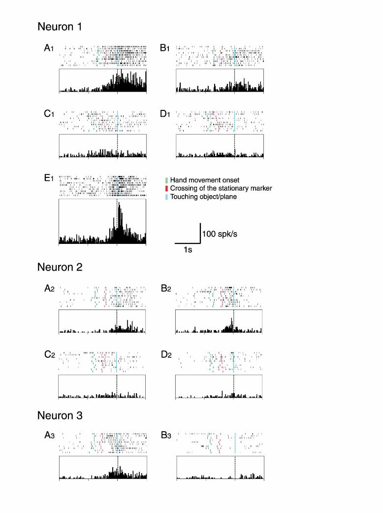
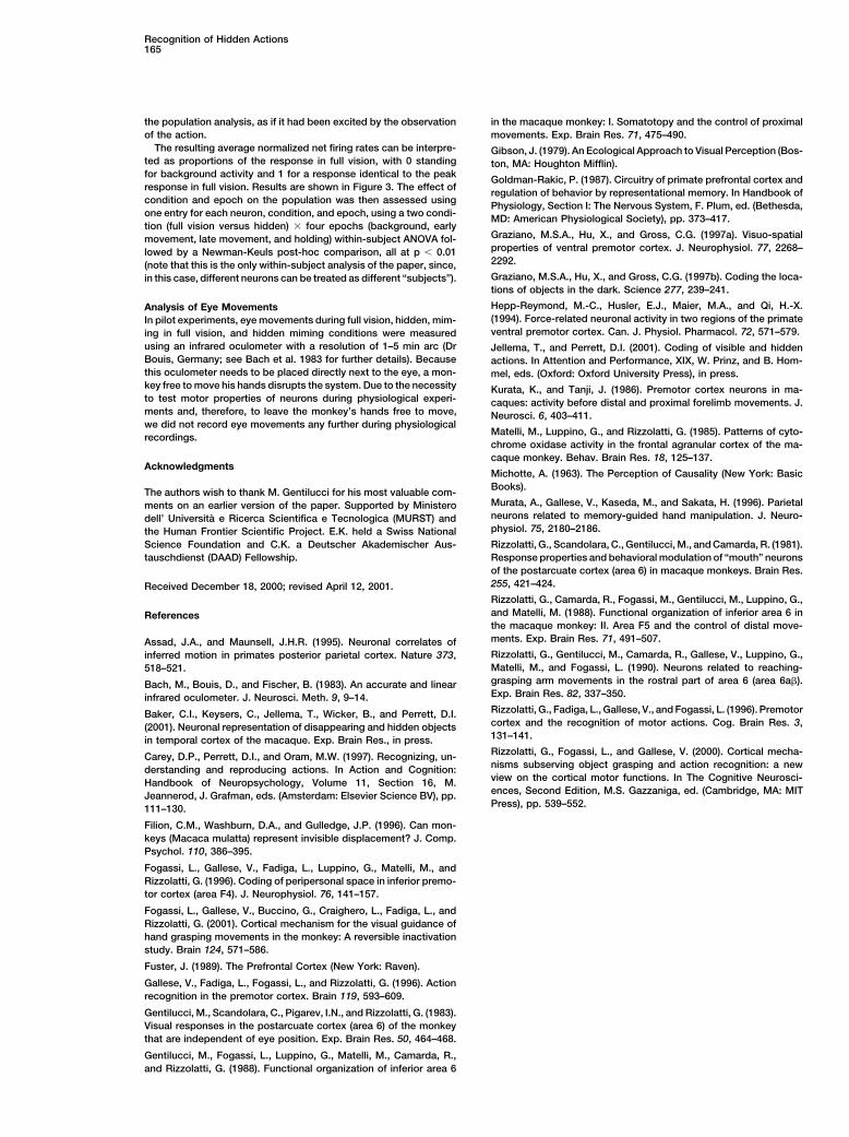
Figure 4. Examples of Mirror Neurons, Showing Different Types of Responses
The responses of three mirror neurons are shown to illustrate three types of responses. All rasters and histograms are aligned on the momentin which the hand of the experimenter (all except [E1]) or the monkey (E1) touched the object or, in conditions without objects, the emptyplate where the object is otherwise placed. All histograms share the same scale. Neuron 1 showed a strong response to grasping and holdingobservation in full vision (A1). The response, although present, was markedly reduced in hidden condition (B1). In (C1) and (D1), the experimenterplaced an object on a plane and held it there for 1 s, in full vision (C1) and in hidden condition (D1). Note the specificity of the neuron responsemanifested by the very low intensity of the discharge during placing and holding observation in both conditions. (E1) shows the response ofthe neuron during the monkey’s active grasping of a small piece of food, with the hand contralateral to the recorded hemisphere. Neuron 2responded to grasping and holding observation in full vision (A2) and in hidden condition (B2). In full vision (A2), the neuron started to dischargeduring the late grasping phase and increased its firing during holding. In hidden condition (B2), the neuron started to respond as soon as theexperimenter’s hand disappeared from the monkey’s sight, reached the discharge peak intensity at the end of grasping, and decreased duringholding. (C2) and (D2) show the response of the same neuron in the two mimed conditions. Note the almost complete absence of a responsein these conditions. Neuron 3 showed a strong response during the observation of the experimenter’s grasping and holding actions (A3). Thesame actions, when hidden, did not evoke any response (B3).
Neuron164
peripersonal RFs around the tactile ones. Area F5: going further conditions, the experimenter’s actions were performed as in thebasic conditions, but there was no object. Note that from the mon-rostrally, reappearance of distal movements requiring higher stimu-
lation currents than F1, visual responses present either in response key’s vantage point, in the hidden conditions there was no differencebetween object-directed and mimed action. All conditions consistedto the presentation of 3D objects or to observation of complex
actions, presence of a large number of neurons discharging in asso- of ten consecutive trials. In all conditions, reward (a piece of food)was given to the monkey at the end of each trial.ciation with goal-directed hand movements. The validity of identifi-
cation of area F5 on the basis of its functional properties has been In order to correlate neurons’ discharge with the different phasesof the experimenter’s hand action, hand movements were recordedhistologically confirmed in several previous experiments (see Rizzo-
latti et al., 1988, 1996; Gallese et al., 1996; Fogassi et al., 2001). by means of a computerized motion analysis system (ELITE system,BTS srl, Milano, Italy) that utilizes infrared light-reflecting passivemarkers. Two markers were placed on the distal phalanxes of theNeuron Selectionthumb and index finger of the experimenter’s hand. A third stationaryEach neuron, once isolated, was first tested “clinically” in order tomarker (indicated by the asterisk in Figures 1 and 2) was placed onascertain its motor and visual properties. In brief, the monkey wasthe metallic frame in correspondence with the edge of the opaquepresented with a variety of objects of different size and shape. Theyscreen, marking the position in which the experimenter’s handconsisted of food items and objects at hand in the laboratory. Thestarted to disappear to the monkey’s sight in the hidden conditions.objects were presented within and outside the reaching distanceThe 3D position of the three markers was continuously calculatedof the monkey. The monkey was trained to fixate them and, whenby means of a specially designed computer program elaboratingat reaching distance, to grasp them (for details, see Rizzolatti et al.,the markers’ bidimensional data acquired by two infrared-sensitive1988, 1990).cameras. After online calibration and photogrammetric procedures,Mirror properties were tested by performing a series of handthe distances between the experimenter’s index finger and thumbactions in front of the monkey. These actions were related to objectand those between the two fingers and the fixed marker were calcu-grasping (presenting the object to the monkey, putting it on a sur-lated every 10 ms and fed to a computer together with the neuron’sface, grasping it, giving it to a second experimenter, or taking itdischarge.away from her/him), object manipulation (breaking, tearing, folding),
In order to measure the neuron activity during the various phasesor were intransitive (i.e., non-object related) actions. These wereof the experimenter’s hand actions, in each trial, the following eventswith or without “emotional” content (e.g., threatening gestures orwere detected and marked: (1) onset of the hand movement; (2)lifting the arms, waving the hand, respectively, see Gallese et al.crossing of the stationary frame marker by the experimenter’s hand;1996).and (3) hand-object or hand-plane (in mimed actions) contact.In order to verify whether the recorded neuron coded specificallyEvents (1) and (2) were computed by means of kinematic analysishand-object interactions, the following actions were also performed:with the ELITE system (for details, see Fogassi et al., 1996). Eventmovements of the hand miming grasping in the absence of the(3) was computed by means of a contact-detecting device whoseobject and prehension movements of food or other objects per-signals were fed to a PC. Rasters and histograms could be alignedformed with tools (e.g., forceps and pincers).with each of the three different events for further analysis.Only neurons that had mirror properties related to hand actions
(motor responses during monkey’s actions and responses to similaractions performed by the experimenter) and that presented stable Analysis of Neuronal Responses for Single Neurons
All physiological data reported in this paper were acquired fromresponses were selected for further testing.trials of the behavioral task described above. The neurons’ activityduring the behavioral task was analyzed by subdividing the neuralBehavioral Paradigmdischarge during each trial in the following epochs. (1) Background,Once a neuron was classified as a mirror neuron and the effectivetime before the onset of the experimenter’s hand movement (indi-observed action(s) determining its discharge assessed, it was stud-cated by green bars in rasters). This activity is taken to reflect theied as follows. A metallic frame (dimensions: 86 � 66 cm) wasspontaneous activity of the neuron. (2) Early movement, period frominterposed between the experimenter and the monkey. This framemovement onset to crossing of the stationary marker (indicated bywas at about 2 meters of distance from the monkey. To this frame,red bars in rasters). (3) Late movement, period from crossing of thea plane was attached on which objects could be placed. The objectsstationary marker to object or plane contact (indicated by pale bluewere the targets of the experimenter’s actions. A sliding opaquebars in rasters). (4) Holding, 500 ms following object or plane contact.screen (dimensions: 43 � 66 cm) was mounted on the frame and
For each neuron, the mean firing rate (spikes/s) was calculatedcould be moved so as to enable or prevent the monkey from seeingfor each epoch and compared with the activity in other conditionsthe experimenter’s action performed behind it.and epochs using multiway between-subject ANOVA followed byThe behavioral paradigm consisted of two basic experimentalNewman-Keuls post-hoc analyses as described in the text. All analy-conditions: full vision condition and hidden condition. In both condi-sis were performed with p � 0.01 as significance criterion.tions, the monkey was presented with a series of actions made by
the experimenter behind the frame. The experimenter’s hand alwaysmoved parallel to the frame. The experimenter’s hand started from Population Analysis
There were substantial differences in the firing rates of differenta fixed position visible to the monkey within the frame. Before move-ment onset, the hand was kept in pinch position (without object) neurons, both in terms of spontaneous and peak firing rate activity.
To compare the responses in different epochs and conditions overfor touching and grasping actions and with object in the case ofplacing. The final position of the hand movement on the plane was the entire population of neurons, a normalization procedure had to
be used.constant. Thus, the hand trajectories from start to the endpoint werealways approximately the same (see below). First, to account for differences in spontaneous activity, firing
rates were transformed into net firing rates by subtraction of theIn full vision condition (see Figure 1A), all phases of the experi-menter’s action from its onset to its completion were visible to firing rate observed during the background epoch of the same trial.
For each neuron, condition, and epoch, separately, this net firingthe monkey. The actions were touching, grasping, manipulating,holding, releasing, or placing the object on the plane. In hidden rate was then averaged over all available trials to obtain a single
number.condition (see Figure 1B), the opaque screen was interposed so asto prevent the monkey from seeing the hand-object interaction. Second, to account for differences in peak firing rate between
neurons, these average net firing rates were divided by the highestBefore the beginning of each trial, the opaque screen was brieflyslid back to show the monkey the presence of the target object. net average firing rate observed in the full vision condition for that
neuron. Three of the 37 neurons were inhibited during the observa-The screen was then slid on again, and the hand of the experimenter,starting from a position from which it was still visible to the monkey, tion of the action and showed negative net firing rates. The activity
of these three neurons was therefore normalized by dividing the netmoved behind the opaque screen to interact with the object.Two further conditions were also employed: miming in full vision firing rates of all epoch with the minimum (negative) net firing rate
in the full vision condition to rectify the response of the neuron for(see Figure 1C) and hidden miming (see Figure 1D). In these further
Recognition of Hidden Actions165
the population analysis, as if it had been excited by the observation in the macaque monkey: I. Somatotopy and the control of proximalmovements. Exp. Brain Res. 71, 475–490.of the action.
The resulting average normalized net firing rates can be interpre- Gibson, J. (1979). An Ecological Approach to Visual Perception (Bos-ted as proportions of the response in full vision, with 0 standing ton, MA: Houghton Mifflin).for background activity and 1 for a response identical to the peak Goldman-Rakic, P. (1987). Circuitry of primate prefrontal cortex andresponse in full vision. Results are shown in Figure 3. The effect of regulation of behavior by representational memory. In Handbook ofcondition and epoch on the population was then assessed using Physiology, Section I: The Nervous System, F. Plum, ed. (Bethesda,one entry for each neuron, condition, and epoch, using a two condi- MD: American Physiological Society), pp. 373–417.tion (full vision versus hidden) � four epochs (background, early
Graziano, M.S.A., Hu, X., and Gross, C.G. (1997a). Visuo-spatialmovement, late movement, and holding) within-subject ANOVA fol-properties of ventral premotor cortex. J. Neurophysiol. 77, 2268–lowed by a Newman-Keuls post-hoc comparison, all at p � 0.012292.(note that this is the only within-subject analysis of the paper, since,Graziano, M.S.A., Hu, X., and Gross, C.G. (1997b). Coding the loca-in this case, different neurons can be treated as different “subjects”).tions of objects in the dark. Science 277, 239–241.
Hepp-Reymond, M.-C., Husler, E.J., Maier, M.A., and Qi, H.-X.Analysis of Eye Movements(1994). Force-related neuronal activity in two regions of the primateIn pilot experiments, eye movements during full vision, hidden, mim-ventral premotor cortex. Can. J. Physiol. Pharmacol. 72, 571–579.ing in full vision, and hidden miming conditions were measured
using an infrared oculometer with a resolution of 1–5 min arc (Dr Jellema, T., and Perrett, D.I. (2001). Coding of visible and hiddenBouis, Germany; see Bach et al. 1983 for further details). Because actions. In Attention and Performance, XIX, W. Prinz, and B. Hom-this oculometer needs to be placed directly next to the eye, a mon- mel, eds. (Oxford: Oxford University Press), in press.key free to move his hands disrupts the system. Due to the necessity Kurata, K., and Tanji, J. (1986). Premotor cortex neurons in ma-to test motor properties of neurons during physiological experi- caques: activity before distal and proximal forelimb movements. J.ments and, therefore, to leave the monkey’s hands free to move, Neurosci. 6, 403–411.we did not record eye movements any further during physiological
Matelli, M., Luppino, G., and Rizzolatti, G. (1985). Patterns of cyto-recordings.chrome oxidase activity in the frontal agranular cortex of the ma-caque monkey. Behav. Brain Res. 18, 125–137.
AcknowledgmentsMichotte, A. (1963). The Perception of Causality (New York: BasicBooks).The authors wish to thank M. Gentilucci for his most valuable com-Murata, A., Gallese, V., Kaseda, M., and Sakata, H. (1996). Parietalments on an earlier version of the paper. Supported by Ministeroneurons related to memory-guided hand manipulation. J. Neuro-dell’ Universita e Ricerca Scientifica e Tecnologica (MURST) andphysiol. 75, 2180–2186.the Human Frontier Scientific Project. E.K. held a Swiss National
Science Foundation and C.K. a Deutscher Akademischer Aus- Rizzolatti, G., Scandolara, C., Gentilucci, M., and Camarda, R. (1981).Response properties and behavioral modulation of “mouth” neuronstauschdienst (DAAD) Fellowship.of the postarcuate cortex (area 6) in macaque monkeys. Brain Res.255, 421–424.Received December 18, 2000; revised April 12, 2001.Rizzolatti, G., Camarda, R., Fogassi, M., Gentilucci, M., Luppino, G.,and Matelli, M. (1988). Functional organization of inferior area 6 inReferencesthe macaque monkey: II. Area F5 and the control of distal move-ments. Exp. Brain Res. 71, 491–507.Assad, J.A., and Maunsell, J.H.R. (1995). Neuronal correlates ofRizzolatti, G., Gentilucci, M., Camarda, R., Gallese, V., Luppino, G.,inferred motion in primates posterior parietal cortex. Nature 373,Matelli, M., and Fogassi, L. (1990). Neurons related to reaching-518–521.grasping arm movements in the rostral part of area 6 (area 6a).Bach, M., Bouis, D., and Fischer, B. (1983). An accurate and linearExp. Brain Res. 82, 337–350.infrared oculometer. J. Neurosci. Meth. 9, 9–14.Rizzolatti, G., Fadiga, L., Gallese, V., and Fogassi, L. (1996). PremotorBaker, C.I., Keysers, C., Jellema, T., Wicker, B., and Perrett, D.I.cortex and the recognition of motor actions. Cog. Brain Res. 3,(2001). Neuronal representation of disappearing and hidden objects131–141.in temporal cortex of the macaque. Exp. Brain Res., in press.Rizzolatti, G., Fogassi, L., and Gallese, V. (2000). Cortical mecha-Carey, D.P., Perrett, D.I., and Oram, M.W. (1997). Recognizing, un-nisms subserving object grasping and action recognition: a newderstanding and reproducing actions. In Action and Cognition:view on the cortical motor functions. In The Cognitive Neurosci-Handbook of Neuropsychology, Volume 11, Section 16, M.ences, Second Edition, M.S. Gazzaniga, ed. (Cambridge, MA: MITJeannerod, J. Grafman, eds. (Amsterdam: Elsevier Science BV), pp.Press), pp. 539–552.111–130.
Filion, C.M., Washburn, D.A., and Gulledge, J.P. (1996). Can mon-keys (Macaca mulatta) represent invisible displacement? J. Comp.Psychol. 110, 386–395.
Fogassi, L., Gallese, V., Fadiga, L., Luppino, G., Matelli, M., andRizzolatti, G. (1996). Coding of peripersonal space in inferior premo-tor cortex (area F4). J. Neurophysiol. 76, 141–157.
Fogassi, L., Gallese, V., Buccino, G., Craighero, L., Fadiga, L., andRizzolatti, G. (2001). Cortical mechanism for the visual guidance ofhand grasping movements in the monkey: A reversible inactivationstudy. Brain 124, 571–586.
Fuster, J. (1989). The Prefrontal Cortex (New York: Raven).
Gallese, V., Fadiga, L., Fogassi, L., and Rizzolatti, G. (1996). Actionrecognition in the premotor cortex. Brain 119, 593–609.
Gentilucci, M., Scandolara, C., Pigarev, I.N., and Rizzolatti, G. (1983).Visual responses in the postarcuate cortex (area 6) of the monkeythat are independent of eye position. Exp. Brain Res. 50, 464–468.
Gentilucci, M., Fogassi, L., Luppino, G., Matelli, M., Camarda, R.,and Rizzolatti, G. (1988). Functional organization of inferior area 6