HƯỚNG DẪN SỬ DỤNG LƯU HÀNH TẠI VIỆT NAM Tài liệu
37
HƯỚNG DẪN SỬ DỤNG LƯU HÀNH TẠI VIỆT NAM Tài liệu được xác nhận bằng chữ ký số Hà Nội, ngày 23 tháng 12 năm 2020 Người đại diện hợp pháp của cơ sở GIÁM ĐỐC Trịnh Diệu Hương
-
Upload
khangminh22 -
Category
Documents
-
view
5 -
download
0
Transcript of HƯỚNG DẪN SỬ DỤNG LƯU HÀNH TẠI VIỆT NAM Tài liệu
HƯỚNG DẪN SỬ DỤNG LƯU HÀNH TẠI VIỆT NAM
Tài liệu được xác nhận bằng chữ ký số
Hà Nội, ngày 23 tháng 12 năm 2020 Người đại diện hợp pháp của cơ sở
GIÁM ĐỐC
Trịnh Diệu Hương
Trang 1/ 19
HƯỚNG DẪN SỬ DỤNG
Thuốc thử làm kháng sinh đồ:
trực khuẩn gram âm hiếu khí và hiếu-kị khí tùy tiện
MicroScan Neg MIC Panel Type 45
Mã sản phẩm: B1017-424
MỤC ĐÍCH SỬ DỤNG
MicroScan Neg MIC Panel Type 45 được sử dụng để xác định tính nhạy cảm kháng sinh của các trực khuẩn
gram âm hiếu khí và hiếu-kị khí tùy tiện.
Panel MicroScan Neg MIC Panel Type 45 thuộc nhóm các panel MicroScan Dried Gram Negative
MIC/Combo, do đó, tài liệu Hướng dẫn sử dụng tiếng Việt của sản phẩm được chuẩn bị dựa trên tài liệu
MicroScan Dried Gram Negative Procedural Manual (Mã tài liệu: 3251-3424AB) của hãng Beckman Coulter
ban hành vào tháng 05/2018 (Tài liệu áp dụng chung cho các panel MicroScan Dried Gram Negative
MIC/Combo và panel Dried Gram Negative Breakpoint Combo).
TÓM TẮT VÀ NGUYÊN TẮC SỬ DỤNG
Các xét nghiệm tính nhạy cảm kháng sinh là phiên bản thu nhỏ của xét nghiệm tính nhạy cảm pha loãng trong
canh thang (broth) đã được khử nước. Các chất kháng sinh khác nhau được pha loãng trong môi trường canh
thang Mueller-Hinton chứa canxi và magie để thu được dải nồng độ phù hợp với lâm sàng. Canh thang
Trimethoprim và Trimethoprim/Sulfamethoxazol được bổ sung thêm enzyme thymidin phosphorylase để giảm
nồng độ thymidin có mặt trong môi trường. Sau khi panel được hoàn nguyên và cấy huyền phù chứa chủng
chuẩn, panel được ủ trong tối thiểu 16 giờ ở 35°C, nồng độ ức chế tối thiểu (MIC) của vi khuẩn được xác định
bằng cách quan sát nồng độ kháng sinh thấp nhất có khả năng ức chế sự phát triển của vi khuẩn.
Các panel có chứa kháng sinh: ceftazidim, aztreonam, cefotaxim hoặc ceftriaxon ở nồng độ 1 µg/mL hoặc
cefpodoxim với nồng độ 1 hoặc 4 µg/mL (tùy thuộc vào loại panel) có thể dùng để sàng lọc Escherichia coli,
Klebsiella oxytoca, hoặc K. pneumoniae nghi ngờ sản xuất beta-lactamase phổ rộng (ESBL). Chức năng sàng
lọc ESBL của giếng Ceftriaxon bị hủy bỏ trên phần mềm LabPro v4.11, phiên bản cập nhật 05 cho các loại
panel đang báo cáo kết quả giá trị ngưỡng diễn giải (breakpoint) đã được cập nhật cho Họ vi khuẩn đường ruột
và chứa tối thiểu các mức pha loãng ceftriaxon là 1 và 2 µg/mL và KHÔNG CHỨA các kháng sinh sử dụng
trong xét nghiệm xác nhận ESBL (ví dụ: Cft/CA, Caz/CA). Đối với các chủng Proteus mirabilis, chỉ có thể sử
dụng các vi giếng chứa ceftazidim, cefotaxim và cefpodoxim để sàng lọc ESBL. Panel có vi giếng
ceftazidim/acid clavulanic và cefotaxim/acid clavulanic có thể sử dụng để xác nhận sự có mặt của các vi khuẩn
ESBL. Xét nghiệm xác nhận có ý nghĩa khi nồng độ MIC của vi sinh vật giảm ≥ 3 lần pha loãng gấp đôi khi
thử với kháng sinh ceftazidim hoặc cefotaxim phối hợp một nồng độ cố định của acid clavulanic, so với giá trị
MIC khi xét nghiệm với kháng sinh đơn độc. Quy trình trong tài liệu hướng dẫn sử dụng và nhãn (ví dụ: tiêu
chí diễn giải, thời gian đọc kết quả, hạn chế) có thể khác với tiêu chí trong phần mềm LabPro do sự khác biệt
về phần mềm được cập nhật.
QUY CÁCH ĐÓNG GÓI
Quy cách đóng gói: Hộp 20 panel.
Mô tả sản phẩm: Hộp gồm 20 panel, mỗi panel được đựng trong một túi nhôm.
Trang 2/ 19
THẬN TRỌNG
1. Chỉ sử dụng trong chẩn đoán in vitro.
2. Tuân theo các kỹ thuật vô khuẩn và biện pháp phòng ngừa đã thiết lập để tránh nguy cơ nhiễm khuẩn
trong tất cả các quy trình, với nhận thức đặc biệt rằng các panel đã được cấy có chứa các vi sinh vật có
khả năng gây bệnh.
3. Vật phẩm này có chứa các tác nhân lây nhiễm bệnh, và cần được thải bỏ theo phương pháp phù hợp đối
với chất thải có nguy cơ sinh học.
4. Các kết quả xét nghiệm này phải luôn được diễn giải kết hợp với tiền sử của bệnh nhân, triệu chứng lâm
sàng và các biểu hiện khác.
5. Thận trọng. Chỉ dùng cho cán bộ chuyên môn. Luật Liên bang Hoa Kỳ giới hạn thiết bị này chỉ được cung
cấp bởi hoặc theo yêu cầu của những người được cấp phép.
BẢO QUẢN
Bảo quản các panel MicroScan Neg MIC Panel Type 45 ở nhiệt độ 2-25°C.
Sản phẩm chỉ sử dụng một lần. Các panel chỉ được mở khi cần sử dụng.
Lưu ý: Bảo quản sản phẩm ở các điều kiện khác với khuyến cáo trong thời gian dài có thể làm mất hoạt lực
của các chất kháng sinh. Không sử dụng sản phẩm hết hạn sử dụng. Liên hệ với đại diện hoặc nhà phân phối
của hãng Beckman Coulter để được hỗ trợ thêm.
LẤY VÀ CHUẨN BỊ MẪU
Thực hiện lấy, vận chuyển và cấy mẫu thích hợp vào trong môi trường phân lập ban đầu theo quy trình được
khuyến cáo trong Sổ tay Vi sinh lâm sàng (Manual of Clinical Microbiology).1
CÁC NGUYÊN VẬT LIỆU ĐƯỢC CUNG CẤP
Các thành phần cụ thể của panel được cung cấp trên nhãn hộp sản phẩm tương ứng.
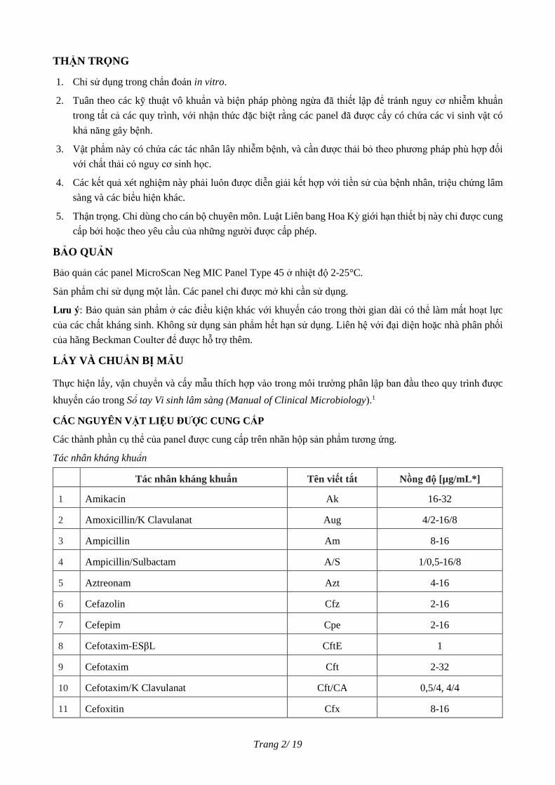
Tác nhân kháng khuẩn
Tác nhân kháng khuẩn Tên viết tắt Nồng độ [µg/mL*]
1 Amikacin Ak 16-32
2 Amoxicillin/K Clavulanat Aug 4/2-16/8
3 Ampicillin Am 8-16
4 Ampicillin/Sulbactam A/S 1/0,5-16/8
5 Aztreonam Azt 4-16
6 Cefazolin Cfz 2-16
7 Cefepim Cpe 2-16
8 Cefotaxim-ESβL CftE 1
9 Cefotaxim Cft 2-32
10 Cefotaxim/K Clavulanat Cft/CA 0,5/4, 4/4
11 Cefoxitin Cfx 8-16
Trang 3/ 19
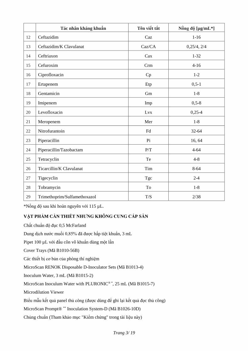
Tác nhân kháng khuẩn Tên viết tắt Nồng độ [µg/mL*]
12 Ceftazidim Caz 1-16
13 Ceftazidim/K Clavulanat Caz/CA 0,25/4, 2/4
14 Ceftriaxon Cax 1-32
15 Cefuroxim Crm 4-16
16 Ciprofloxacin Cp 1-2
17 Ertapenem Etp 0,5-1
18 Gentamicin Gm 1-8
19 Imipenem Imp 0,5-8
20 Levofloxacin Lvx 0,25-4
21 Meropenem Mer 1-8
22 Nitrofurantoin Fd 32-64
23 Piperacillin Pi 16, 64
24 Piperacillin/Tazobactam P/T 4-64
25 Tetracyclin Te 4-8
26 Ticarcillin/K Clavulanat Tim 8-64
27 Tigecyclin Tgc 2-4
28 Tobramycin To 1-8
29 Trimethoprim/Sulfamethoxazol T/S 2/38
*Nồng độ sau khi hoàn nguyên với 115 µL.
VẬT PHẨM CẦN THIẾT NHƯNG KHÔNG CUNG CẤP SẴN
Chất chuẩn độ đục 0,5 McFarland
Dung dịch nước muối 0,85% đã được hấp tiệt khuẩn, 3 mL
Pipet 100 µL với đầu côn vô khuẩn dùng một lần
Cover Trays (Mã B1010-56B)
Các thiết bị cơ bản của phòng thí nghiệm
MicroScan RENOK Disposable D-Inoculator Sets (Mã B1013-4)
Inoculum Water, 3 mL (Mã B1015-2)
MicroScan Inoculum Water with PLURONIC® *, 25 mL (Mã B1015-7)
Microdilution Viewer
Biểu mẫu kết quả panel thủ công (được dùng để ghi lại kết quả đọc thủ công)
MicroScan Prompt® ** Inoculation System-D (Mã B1026-10D)
Chủng chuẩn (Tham khảo mục "Kiểm chứng" trong tài liệu này)
Trang 4/ 19
Biểu mẫu báo cáo QC
MicroScan RENOK Rehydrating/Inoculating System (Mã B1018-14) hoặc thiết bị tương tự
Máy đo độ đục MicroScan Turbidity Meter
Máy trộn Vortex
Barcode Label Paper (Mã B1018-129)
WalkAway Tray Lids (Mã B1018-18)
* Chất hoạt động bề mặt PLURONIC®, một nhãn hiệu đã được đăng ký của BASF Corporation, Parsippany,
NJ USA
** 3M, St. Paul, MN USA.
TÓM TẮT QUY TRÌNH
a. Chuẩn bị panel
1. Lấy panel khỏi nơi bảo quản. Không sử dụng panel khi bao bì không còn nguyên vẹn (đã mở niêm
phong, thủng, rách).
2. Mở bao bì, nếu panel được bảo quản trong tủ lạnh, lấy panel ra khỏi túi bọc kim loại ngay lập tức.
3. Không sử dụng panel trong bất kỳ trường hợp nào dưới đây:
a. Không có gói hút ẩm hoặc gói hút ẩm bị rách.
b. Các giếng của panel bị đổi màu (ví dụ: giếng có chứa nhiều tác nhân kháng khuẩn).
4. Để panel ổn định về nhiệt độ phòng trước khi hoàn nguyên. Các panel có thể được xếp chồng lên nhau
với khay đậy (Cover Tray) sạch ở trên. Các panel chỉ được mở khi cần sử dụng.
b. Chuẩn bị dịch cấy
CLSI khuyến cáo định kỳ kiểm tra mật độ dịch cấy bằng cách đếm khuẩn lạc (tham khảo tài liệu CLSI M7-
A1017 để biết hướng dẫn đếm số lượng khuẩn lạc). Giá trị mong đợi đối với chủng E. coli ATCC 25922 cần
đạt khoảng 5 x 10⁵ CFU/mL ở nồng độ xét nghiệm cuối cùng. Cần thận trọng trong quá trình chuẩn bị
huyền phù vi khuẩn, đặc biệt đối với phương pháp thủ công phụ thuộc vào kỹ thuật như: sử dụng Prompt
System hoặc chuẩn bị huyền phù vi khuẩn mà không có sự hỗ trợ của thiết bị đo quang.
Lưu ý: Các kỹ thuật liên quan đến pha lũy thừa (Log Phase) và pha ổn định (Stationary Phase) không được
hỗ trợ với các sản phẩm của MicroScan.
1. Kỹ thuật chuẩn độ đục (phương pháp nuôi cấy cơ bản)
Khuyến cáo sử dụng kỹ thuật chuẩn độ đục trong nuôi cấy trực tiếp tất cả trực khuẩn gram âm hiếu khí.
a. Sử dụng que lấy mẫu vô khuẩn (đầu vòng tròn hoặc quấn bông) chạm vào bề mặt của 4-5 khuẩn lạc
lớn hoặc 5-10 khuẩn lạc nhỏ được phân lập tốt, có hình thái tương tự nhau từ đĩa thạch nuôi cấy không
ức chế sinh trưởng sau 18-24 giờ.
b. Phân tán mẫu vào 3 mL dung dịch nuôi cấy MicroScan Inoculum Water (nước khử ion đã được hấp
tiệt khuẩn).
c. Đậy nắp lọ và trộn bằng máy Vortex trong 2-3 giây. Độ đục cuối cùng của huyền phù vi khuẩn phải
tương đương với độ đục chuẩn 0,5 McFarland. Có thể sử dụng MicroScan Turbidity Meter để đo độ
Trang 5/ 19
đục, độ đục thu được phải tương đương với dải 0,08 ± 0,02.
d. Sử dụng pipet 0,1 mL (100 µL) để hút và phân phối huyền phù đã được chuẩn hóa vào 25 mL dung
dịch MicroScan Inoculum Water with PLURONIC. Đậy chặt nắp lọ. Trộn bằng cách đảo 8-10 lần.
2. Prompt System
Có thể sử dụng Prompt System cấy trực khuẩn Gram âm. Tham khảo hướng dẫn sử dụng của Prompt
System để biết quy trình sử dụng đúng.
c. Hoàn nguyên panel/Cấy khuẩn
Sử dụng MicroScan RENOK Disposable D-Inoculator Sets để thực hiện hoàn nguyên và cấy khuẩn. Tham
khảo hướng dẫn sử dụng của thiết bị. Nếu sử dụng một thiết bị khác, hoàn nguyên với 115 µL ± 10 µL dung
dịch MicroScan Inoculum Water with PLURONIC. Nồng độ cuối cùng ở mỗi giếng cần nằm trong dải 3-7
x 105 CFU/mL. Để đảm bảo khả năng sinh trưởng và độ tinh khiết của vi sinh vật xét nghiệm, một đĩa tinh
khiết nên được chuẩn bị bằng phương pháp ria (streaking) dịch cấy vào một đĩa thạch thích hợp và ủ trong
điều kiện phù hợp. Nếu có từ 2 loại khuẩn lạc trở lên xuất hiện trên đĩa tinh khiết, cần tiến hành phân lập
và xét nghiệm lại.
d. Ủ panel
Panel có thể được ủ trong thiết bị WalkAway hoặc ủ bên ngoài theo các bước sau đây:
a. Để đảm bảo phân bố nhiệt đồng đều trong suốt quá trình ủ, xếp chồng một nhóm từ 3 đến 5 panel.
b. Đặt khay đậy (Cover Tray) sạch lên trên mỗi panel trong nhóm để ngăn ngừa tình trạng bay hơi. Khay
đậy có thể tái sử dụng. Không làm sạch khay đậy bằng cồn. Có thể làm sạch khay đậy bằng xà phòng và
nước, rửa sạch và để khô ngoài không khí.
c. Ủ panel trong tối thiểu 16 – 20°C giờ ở 35°C ± 1°C trong buồng ủ không có CO2.
e. Đọc kết quả trên panel
Có thể đọc thủ công bằng thiết bị MicroScan Microdilution Viewer và ghi lại kết quả vào Biểu mẫu kết quả
panel thủ công (Manual Panel Worksheet) (xem mục "Vật liệu cần thiết nhưng không cung cấp sẵn") hoặc
các thiết bị MicroScan (autoSCAN-4 và WalkAway). Thao khảo hướng dẫn sử dụng của thiết bị tương ứng
để biết cách đọc kết quả.
1. Lấy panel ra khỏi buồng ủ sau khi ủ 16-20 giờ.
2. Lau mặt dưới panel để loại bỏ nước ngưng tụ hoặc cặn bẩn bằng khăn lau không để lại xơ vải.
3. Chỉ đọc kết quả panel khi giếng Growth (có vi sinh vật phát triển) đục. Không đọc kết quả kháng sinh
đồ nếu giếng Control bị đục hoặc nếu không có sự phát triển trong giếng Growth. Sự phát triển của vi
sinh vật trong các giếng tạo nên độ đục cho các giếng, xuất hiện ở dạng một đám trắng dạng sương mù
ở khắp giếng, hoặc một nhúm màu trắng ở giữa giếng, hoặc sự phát triển dạng hạt nhỏ trong khắp giếng.
Sự phát triển không hoàn toàn của vi sinh vật được biểu hiện bằng màu trắng nhẹ trong giếng hoặc môi
trường giếng trong suốt.
4. Nếu đọc kết quả thủ công, ghi kết quả vào biểu mẫu phù hợp.

Trang 6/ 19
5. Đọc kết quả độ nhạy cảm kháng sinh:
a. Đọc tất cả các giếng có chứa kháng sinh và giếng CET trên nền màu đen (không có ánh sáng trực
tiếp chiếu vào).
b. Ghi lại kết quả MIC theo các bước sau:
1) Sau 16-20 giờ ủ, ghi nồng độ ức chế tối thiểu là nồng độ kháng sinh thấp nhất cho thấy ức chế sự
phát triển vi sinh vật.
2) Khi có sự phát triển vi sinh vật ở tất cả các nồng độ của một kháng sinh, nồng độ ức chế tối thiểu
được ghi là ‘lớn hơn’ (>) nồng độ cao nhất.
3) Khi không có sự phát triển vi sinh vật ở bất kỳ nồng độ nào của kháng sinh, nồng độ ức chế tối
thiểu được ghi là ‘nhỏ hơn hoặc bằng’ (≤) nồng độ thấp nhất.
4) Nếu có một giếng trong suốt trong dãy các giếng phát triển, ví dụ có vi sinh vật phát triển ở các
giếng nồng độ 1, 2 và 8 μg/mL, nhưng không phát triển ở giếng 4 μg/mL thì được gọi là ‘giếng bị bỏ
qua’ và giếng này nên được bỏ qua.
5) Sự phát triển kiểu đốm trong các giếng phân lập cho biết có sự tạp nhiễm. Xét nghiệm cần được
thực hiện lại.
6) Hiệu ứng kéo dài (trailing effect) có thể quan sát được trong một số trường hợp kết hợp kháng
sinh/ vi sinh vật như Proteus với Cefuroxim (Crm) và Imipenem (Imp), Serratia với kháng sinh beta-
lactam (VD: Imipenem (Imp) và Piperacillin/Tazobactam(P/T)); B. cepacia và B. pseudomallei với
Ceftazidim (Caz) và Piperacillin (Pi).
Hiệu ứng “trailing effect” cũng có thể quan sát thấy khi sử dụng giếng trimethoprim/ sulfamethoxazol
(T/S) và trimethoprim (T) cùng với việc sử dụng RENOK Rehydrating/Inoculating System. Trong
trường hợp này, hiệu ứng “trailing effect” xuất hiện do nồng độ dịch cấy. Điểm cuối (endpoint) được
đọc là nồng độ thấp nhất mà khi so sánh với giếng Growth cho thấy:
a) Giảm sự phát triển (giếng T/S) khoảng 80%
b) một đám màu trắng có đường kính nhỏ hơn 2 mm hoặc
c) một đám màu trắng hơi mờ.1
KIỂM CHỨNG
Các chất kháng sinh phải được kiểm tra bằng cách xét nghiệm các vi sinh vật có các dải nồng độ ức chế tối
thiểu đã biết. Kết quả của các chủng vi sinh vật kiểm chứng American Type Culture Collection (ATCC**)
được trình bày trong bảng dưới đây.
Bảng kiểm chứng (QC) là một bảng đầy đủ bao gồm các dải MIC để bao phủ toàn bộ các nồng độ pha loãng
trên panel MicroScan Dried Panels; đọc kết quả sau khi ủ 16-20 giờ. Người dùng có thể cần ngoại suy từ bảng
QC, dựa trên các nồng độ pha loãng trên loại panel sử dụng. Các thông tin QC đặc trưng cho panel có thể có
trên Các Biểu mẫu Báo cáo Kiểm chứng (Quality Control Report Forms). Dưới đây là các ví dụ về phương
pháp ngoại suy.
** American Type Culture Collection, Manassas, VA USA
Trang 7/ 19
Chủng kiểm chứng Viết
tắt
Bảng kiểm
chứng
Các nồng độ pha loãng
trên panel
Dải kiểm chứng
ngoại suy
E. coli ATCC 25922 Ak ≤ 2-8 2, 8-32 ≤ 2-8
E. coli ATCC 25922 A/S 1/0.5-4/2 8/4-16/8 ≤ 8/4
P. aeruginosa ATCC 27853 Te 8, 128 1-8 8 - >8
P. aeruginosa ATCC 27853 Tim ≤ 8/2-32/2 16/2, 64/2 ≤ 16/2, 64/2
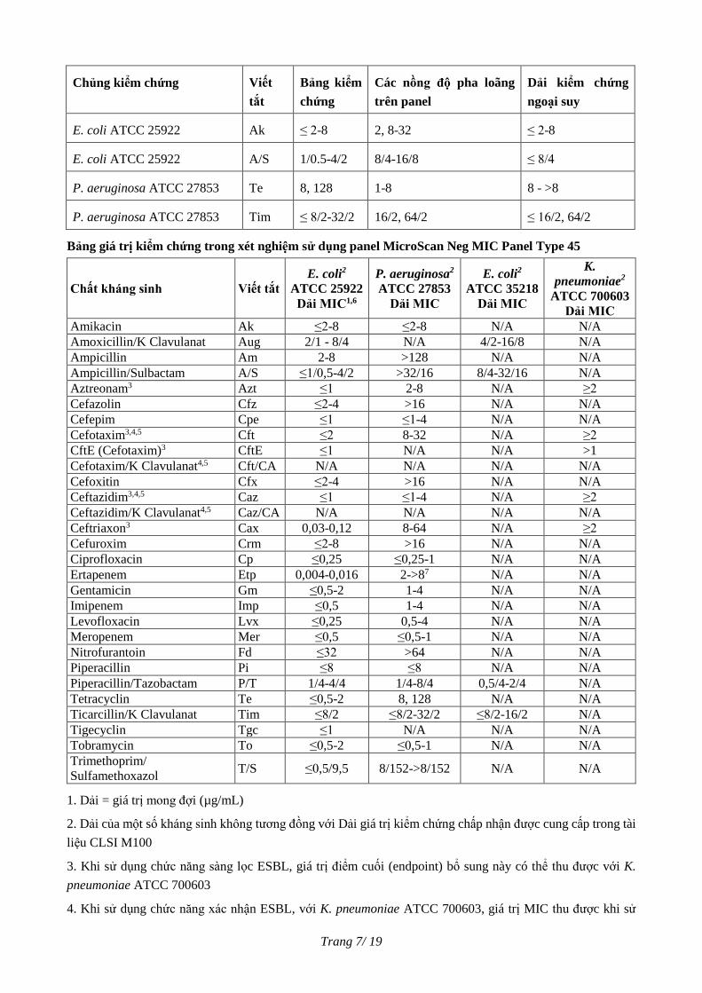
Bảng giá trị kiểm chứng trong xét nghiệm sử dụng panel MicroScan Neg MIC Panel Type 45
Chất kháng sinh Viết tắt
E. coli2
ATCC 25922
Dải MIC1,6
P. aeruginosa2
ATCC 27853
Dải MIC
E. coli2
ATCC 35218
Dải MIC
K.
pneumoniae2
ATCC 700603
Dải MIC
Amikacin Ak ≤2-8 ≤2-8 N/A N/A
Amoxicillin/K Clavulanat Aug 2/1 - 8/4 N/A 4/2-16/8 N/A
Ampicillin Am 2-8 >128 N/A N/A
Ampicillin/Sulbactam A/S ≤1/0,5-4/2 >32/16 8/4-32/16 N/A
Aztreonam3 Azt ≤1 2-8 N/A ≥2
Cefazolin Cfz ≤2-4 >16 N/A N/A
Cefepim Cpe ≤1 ≤1-4 N/A N/A
Cefotaxim3,4,5 Cft ≤2 8-32 N/A ≥2
CftE (Cefotaxim)3 CftE ≤1 N/A N/A >1
Cefotaxim/K Clavulanat4,5 Cft/CA N/A N/A N/A N/A
Cefoxitin Cfx ≤2-4 >16 N/A N/A
Ceftazidim3,4,5 Caz ≤1 ≤1-4 N/A ≥2
Ceftazidim/K Clavulanat4,5 Caz/CA N/A N/A N/A N/A
Ceftriaxon3 Cax 0,03-0,12 8-64 N/A ≥2
Cefuroxim Crm ≤2-8 >16 N/A N/A
Ciprofloxacin Cp ≤0,25 ≤0,25-1 N/A N/A
Ertapenem Etp 0,004-0,016 2->87 N/A N/A
Gentamicin Gm ≤0,5-2 1-4 N/A N/A
Imipenem Imp ≤0,5 1-4 N/A N/A
Levofloxacin Lvx ≤0,25 0,5-4 N/A N/A
Meropenem Mer ≤0,5 ≤0,5-1 N/A N/A
Nitrofurantoin Fd ≤32 >64 N/A N/A
Piperacillin Pi ≤8 ≤8 N/A N/A
Piperacillin/Tazobactam P/T 1/4-4/4 1/4-8/4 0,5/4-2/4 N/A
Tetracyclin Te ≤0,5-2 8, 128 N/A N/A
Ticarcillin/K Clavulanat Tim ≤8/2 ≤8/2-32/2 ≤8/2-16/2 N/A
Tigecyclin Tgc ≤1 N/A N/A N/A
Tobramycin To ≤0,5-2 ≤0,5-1 N/A N/A
Trimethoprim/
Sulfamethoxazol T/S ≤0,5/9,5 8/152->8/152 N/A N/A
1. Dải = giá trị mong đợi (µg/mL)
2. Dải của một số kháng sinh không tương đồng với Dải giá trị kiểm chứng chấp nhận được cung cấp trong tài
liệu CLSI M100
3. Khi sử dụng chức năng sàng lọc ESBL, giá trị điểm cuối (endpoint) bổ sung này có thể thu được với K.
pneumoniae ATCC 700603
4. Khi sử dụng chức năng xác nhận ESBL, với K. pneumoniae ATCC 700603, giá trị MIC thu được khi sử
Trang 8/ 19
dụng kết hợp ceftazidim hoặc cefotaxim với K. Clavulanat giảm ≥ 3 mức pha loãng gấp đôi so với giá trị MIC
thu được khi chỉ dùng mỗi kháng sinh đơn độc. Dải giá trị QC thực tế cho ceftazidim hoặc cefotaxim với K.
Clavulanat chưa được thiết lập.
5. Khi sử dụng chức năng xác nhận ESBL, với E. Coli ATCC 25922, giá trị MIC thu được khi sử dụng kết hợp
ceftazidim hoặc cefotaxim với K. Clavulanat giảm < 3 mức pha loãng gấp đôi so với giá trị MIC thu được khi
chỉ dùng mỗi kháng sinh đơn độc. Dải giá trị QC thực tế cho ceftazidim hoặc cefotaxim với K. Clavulanat
chưa được thiết lập.
6, FDA và CLSI đã đưa ra khuyến cáo cho dải QC; có thể được ngoại suy để khớp với các mức nồng độ pha
loãng trong panel.
7. Chủng vi sinh vật chỉ được dùng trong quy trình Kiểm chứng.
KẾT QUẢ
A. Diễn giải kết quả MIC
Độ nhạy cảm của vi khuẩn với kháng sinh được xác định bằng cách so sánh giá trị MIC của một vi khuẩn với
nồng độ kháng sinh có thể đạt được trong máu hoặc nước tiểu. Bảng dưới đây đưa ra tiêu chí diễn giải trong
tài liệu CLSI. Một số tiêu chí khác với các giá trị ngưỡng diễn giải (interpretive breakpoints) của nhà sản xuất
được liệt kê trong Tài liệu tham khảo Physicians’ Desk Reference.
B. Diễn giải kết quả ESBL
1. Một số loài nhất định trong họ Enterobacteriaceae, đặc biệt là Klebsiella spp., Escherichia coli và Proteus
mirabilis có thể sản xuất các enzyme beta-lactamase mới có khả năng thủy phân các cephalosporin phổ
rộng (Ví dụ: cefotaxim, ceftriaxon, ceftizoxim và ceftazidim) và aztreonam. Các enzyme mới này thường
có tính nhạy cảm khác thường với các kháng sinh thuộc nhóm beta-lactam. Các mẫu phân lập lâm sàng
của vi khuẩn Klebsiella oxytoca, K. pneumoniae, và Escherichia coli có giá trị MIC tăng (≥2 µg/mL) với
kháng sinh ceftazidim, aztreonam, cefotaxim, ceftriaxon, hoặc giá trị MIC với cefpodoxim ≥2 hoặc ≥8
µg/mL (phụ thuộc vào loại panel) có thể nghi ngờ "dương tính" với enzyme beta-lactamase phổ rộng.
Chức năng sàng lọc ESBL của giếng Ceftriaxon bị hủy bỏ trên phần mềm LabPro v4.11, phiên bản cập
nhật 05 cho các loại panel đang báo cáo kết quả giá trị ngưỡng diễn giải (breakpoint) đã được cập nhật
cho Họ vi khuẩn đường ruột và chứa tối thiểu các mức pha loãng ceftriaxon là 1 và 2 µg/mL và KHÔNG
CHỨA các kháng sinh sử dụng trong xét nghiệm xác nhận ESBL (ví dụ: Cft/CA, Caz/CA).
Đối với các loài P. mirabilis, chỉ có ceftazidim, cefotaxim và cefpodoxim được sử dụng để sàng lọc ESBL.
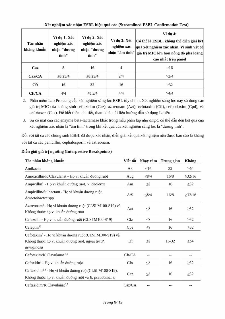
Mẫu phân lập lâm sàng được coi là "dương tính" với xét nghiệm xác nhận ESBL nếu giá trị MIC giảm ≥
3 mức pha loãng gấp đôi (tương ứng với giảm 3 giếng) khi xét nghiệm với kháng sinh phối hợp acid
clavulanic so với khi xét nghiệm với kháng sinh đơn độc. Khi đó, khuyến cáo thử độ nhạy cảm với cả hai
kháng sinh cefotaxim và ceftazidim, có kết hợp/không kết hợp với acid clavulanic, nếu xét nghiệm cho
kết quả "dương tính" (tức là giá trị MIC giảm ≥ 3 lần pha loãng gấp đôi), diễn giải kết quả xét nghiệm xác
nhận kiểu hình ESBL là "dương tính".
Trang 9/ 19
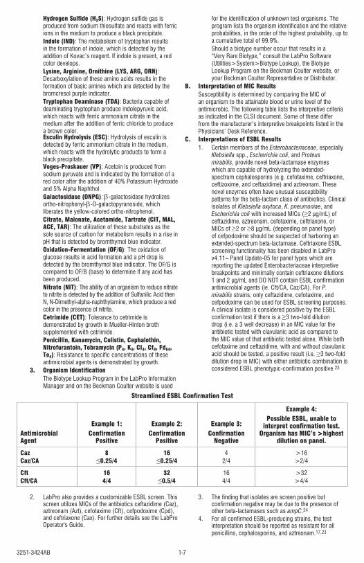
Xét nghiệm xác nhận ESBL hiệu quả cao (Streamlined ESBL Confirmation Test)
Tác nhân
kháng khuẩn
Ví dụ 1: Xét
nghiệm xác
nhận "dương
tính"
Ví dụ 2: Xét
nghiệm xác
nhận "dương
tính"
Ví dụ 3: Xét
nghiệm xác
nhận "âm tính"
Ví dụ 4:
Có thể là ESBL, không thể diễn giải kết
quả xét nghiệm xác nhận. Vi sinh vật có
giá trị MIC lớn hơn nồng độ pha loãng
cao nhất trên panel
Caz 8 16 4 >16
Caz/CA ≤0,25/4 ≤0,25/4 2/4 >2/4
Cft 16 32 16 >32
Cft/CA 4/4 ≤0,5/4 4/4 >4/4
2. Phần mềm Lab Pro cung cấp xét nghiệm sàng lọc ESBL tùy chỉnh. Xét nghiệm sàng lọc này sử dụng các
giá trị MIC của kháng sinh ceftazidim (Caz), aztreonam (Azt), cefotaxim (Cft), cefpodoxim (Cpd), và
ceftriaxon (Cax). Để biết thêm chi tiết, tham khảo tài liệu hướng dẫn sử dụng LabPro.
3. Sự có mặt của các enzyme beta-lactamase khác trong mẫu phân lập như ampC có thể dẫn đến kết quả của
xét nghiệm xác nhận là "âm tính" trong khi kết quả của xét nghiệm sàng lọc là "dương tính".
Đối với tất cả các chủng sinh ESBL đã được xác nhận, diễn giải kết quả xét nghiệm nên được báo cáo là kháng
với tất cả các penicillin, cephalosporin và aztreonam.
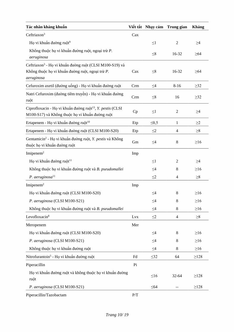
Diễn giải giá trị ngưỡng (Interpretive Breakpoints)
Tác nhân kháng khuẩn Viết tắt Nhạy cảm Trung gian Kháng
Amikacin Ak ≤16 32 ≥64
Amoxicillin/K Clavulanat - Họ vi khuẩn đường ruột Aug ≤8/4 16/8 ≥32/16
Ampicillin2 - Họ vi khuẩn đường ruột, V. cholerae Am ≤8 16 ≥32
Ampicillin/Sulbactam - Họ vi khuẩn đường ruột,
Acinetobacter spp. A/S ≤8/4 16/8 ≥32/16
Aztreonam3 - Họ vi khuẩn đường ruột (CLSI M100-S19) và
Không thuộc họ vi khuẩn đường ruột Azt ≤8 16 ≥32
Cefazolin - Họ vi khuẩn đường ruột (CLSI M100-S19) Cfz ≤8 16 ≥32
Cefepim12 Cpe ≤8 16 ≥32
Cefotaxim3 - Họ vi khuẩn đường ruột (CLSI M100-S19) và
Không thuộc họ vi khuẩn đường ruột, ngoại trừ P.
aeruginosa
Cft ≤8 16-32 ≥64
Cefotaxim/K Clavulanat 6,7 Cft/CA -- -- --
Cefoxitin5 - Họ vi khuẩn đường ruột Cfx ≤8 16 ≥32
Ceftazidim2,3 - Họ vi khuẩn đường ruột(CLSI M100-S19),
Không thuộc họ vi khuẩn đường ruột và B. pseudomallei Caz ≤8 16 ≥32
Ceftazidim/K Clavulanat6,7 Caz/CA -- -- --
Trang 10/ 19
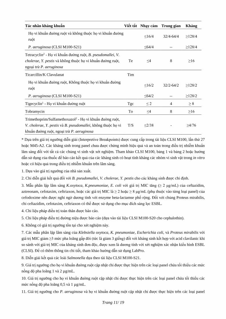
Tác nhân kháng khuẩn Viết tắt Nhạy cảm Trung gian Kháng
Ceftriaxon3 Cax
Họ vi khuẩn đường ruột9 ≤1 2 ≥4
Không thuộc họ vi khuẩn đường ruột, ngoại trừ P.
aeruginosa ≤8 16-32 ≥64
Ceftriaxon3 - Họ vi khuẩn đường ruột (CLSI M100-S19) và
Không thuộc họ vi khuẩn đường ruột, ngoại trừ P.
aeruginosa
Cax ≤8 16-32 ≥64
Cefuroxim axetil (đường uống) - Họ vi khuẩn đường ruột Crm ≤4 8-16 ≥32
Natri Cefuroxim (đường tiêm truyền) - Họ vi khuẩn đường
ruột Crm ≤8 16 ≥32
Ciprofloxacin - Họ vi khuẩn đường ruột13, Y. pestis (CLSI
M100-S17) và Không thuộc họ vi khuẩn đường ruột Cp ≤1 2 ≥4
Ertapenem - Họ vi khuẩn đường ruột10 Etp ≤0,5 1 ≥2
Ertapenem - Họ vi khuẩn đường ruột (CLSI M100-S20) Etp ≤2 4 ≥8
Gentamicin2 - Họ vi khuẩn đường ruột, Y. pestis và Không
thuộc họ vi khuẩn đường ruột Gm ≤4 8 ≥16
Imipenem2 Imp
Họ vi khuẩn đường ruột11 ≤1 2 ≥4
Không thuộc họ vi khuẩn đường ruột và B. pseudomallei ≤4 8 ≥16
P. aeruginosa11 ≤2 4 ≥8
Imipenem2 Imp
Họ vi khuẩn đường ruột (CLSI M100-S20) ≤4 8 ≥16
P. aeruginosa (CLSI M100-S21) ≤4 8 ≥16
Không thuộc họ vi khuẩn đường ruột và B. pseudomallei ≤4 8 ≥16
Levofloxacin8 Lvx ≤2 4 ≥8
Meropenem Mer
Họ vi khuẩn đường ruột (CLSI M100-S20) ≤4 8 ≥16
P. aeruginosa (CLSI M100-S21) ≤4 8 ≥16
Không thuộc họ vi khuẩn đường ruột ≤4 8 ≥16
Nitrofurantoin5 - Họ vi khuẩn đường ruột Fd ≤32 64 ≥128
Piperacillin Pi
Họ vi khuẩn đường ruột và không thuộc họ vi khuẩn đường
ruột ≤16 32-64 ≥128
P. aeruginosa (CLSI M100-S21) ≤64 -- ≥128
Piperacillin/Tazobactam P/T
Trang 11/ 19
Tác nhân kháng khuẩn Viết tắt Nhạy cảm Trung gian Kháng
Họ vi khuẩn đường ruột và không thuộc họ vi khuẩn đường
ruột ≤16/4 32/4-64/4 ≥128/4
P. aeruginosa (CLSI M100-S21) ≤64/4 -- ≥128/4
Tetracyclin2 - Họ vi khuẩn đường ruột, B. pseudomallei, V.
cholerae, Y. pestis và không thuộc họ vi khuẩn đường ruột,
ngoại trừ P. aeruginosa
Te ≤4 8 ≥16
Ticarcillin/K Clavulanat Tim
Họ vi khuẩn đường ruột, Không thuộc họ vi khuẩn đường
ruột ≤16/2 32/2-64/2 ≥128/2
P. aeruginosa (CLSI M100-S21) ≤64/2 -- ≥128/2
Tigecyclin1 - Họ vi khuẩn đường ruột Tgc ≤ 2 4 ≥ 8
Tobramycin To ≤4 8 ≥16
Trimethoprim/Sulfamethoxazol2 - Họ vi khuẩn đường ruột,
V. cholerae, Y. pestis và B. pseudomallei, không thuộc họ vi
khuẩn đường ruột, ngoại trừ P. aeruginosa
T/S ≤2/38 - ≥4/76
* Dựa trên giá trị ngưỡng diễn giải (Interpretive Breakpoints) được cung cấp trong tài liệu CLSI M100, lần thứ 27
hoặc M45-A2. Các kháng sinh trong panel chưa được chứng minh hiệu quả và an toàn trong điều trị nhiễm khuẩn
lâm sàng đối với tất cả các chủng vi sinh vật xét nghiệm. Tham khảo CLSI M100, bảng 1 và bảng 2 hoặc hướng
dẫn sử dụng của thuốc để báo cáo kết quả của các kháng sinh có hoạt tính kháng các nhóm vi sinh vật trong in vitro
hoặc có hiệu quả trong điều trị nhiễm khuẩn trên lâm sàng.
1. Dựa vào giá trị ngưỡng của nhà sản xuất.
2. Chỉ diễn giải kết quả đối với B. pseudomallei, V. cholerae, Y. pestis cho các kháng sinh được chỉ định.
3. Mẫu phân lập lâm sàng K.oxytoca, K.pneumoniae, E. coli với giá trị MIC tăng (≥ 2 μg/mL) của ceftazidim,
aztreonam, cefotaxim, ceftriaxon, hoặc các giá trị MIC là ≥ 2 hoặc ≥ 8 μg/mL (phụ thuộc vào từng loại panel) của
cefodoxime nên được nghi ngờ dương tính với enzyme beta-lactamse phổ rộng. Đối với chủng Proteus mirabilis,
chỉ ceftazidim, cefotaxim, ceftriaxon có thể được sử dụng cho mục đích sàng lọc ESBL.
4. Chỉ liệu pháp điều trị toàn thân được báo cáo.
5. Chỉ liệu pháp điều trị đường niệu được báo cáo (dựa vào tài liệu CLSI M100-S20 cho cephalothin).
6. Không có giá trị ngưỡng tồn tại cho xét nghiệm này.
7. Các mẫu phân lập lâm sàng của Klebsiella oxytoca, K. pneumoniae, Escherichia coli, và Proteus mirabilis với
giá trị MIC giảm ≥3 mức pha loãng gấp đôi (tức là giảm 3 giếng) đối với kháng sinh kết hợp với acid clavilanic khi
so sánh với giá trị MIC của kháng sinh đơn độc, được xem là dương tính với xét nghiệm xác nhận kiểu hình ESBL
(CLSI). Để có thêm thông tin chi tiết, tham khảo hướng dẫn sử dụng LabPro.
8. Diễn giải kết quả các loài Salmonella dựa theo tài liệu CLSI M100-S21.
9. Giá trị ngưỡng cho họ vi khuẩn đường ruột cập nhật chỉ được thực hiện trên các loại panel chứa tối thiểu các mức
nồng độ pha loãng 1 và 2 µg/mL.
10. Giá trị ngưỡng cho họ vi khuẩn đường ruột cập nhật chỉ được thực hiện trên các loại panel chứa tối thiểu các
mức nồng độ pha loãng 0,5 và 1 µg/mL.
11. Giá trị ngưỡng cho P. aeruginosa và họ vi khuẩn đường ruột cập nhật chỉ được thực hiện trên các loại panel

Trang 12/ 19
chứa tối thiểu các mức nồng độ pha loãng 1, 2 và 4 µg/mL.
12. Diễn giải kết quả cho họ vi khuẩn đường ruột dựa theo tài liệu CLSI M100-S23.
13. Diễn giải kết quả cho loài Salmonella khác Salmonella ser. Typhi được dựa theo tài liệu CLSI M100-S21.
GHI CHÚ: Đối với các nước ngoài nước Mỹ, thực hiện theo hướng dẫn thay thế, tham khảo Phần quản lý dữ liệu
LabPro để có tiêu chí diễn giải kết quả.
GHI CHÚ: Các tiêu chí diễn giải kết quả được cung cấp trên nhãn có thể khác với các tiêu chí trên Phần Quản lý
dữ liệu của LabPro do những khác biệt giữa phiên bản phần mềm và các loại panel được đưa ra thị trường.
GHI CHÚ: Các chất kháng sinh được cung cấp trong bảng giá trị ngưỡng diễn giải có thể không có trên mỗi số loại
panel.
HẠN CHẾ CỦA QUY TRÌNH
1. Vi khuẩn khó nuôi cấy - Haemophilus và các chủng vi khuẩn Gram âm khó nuôi cấy khác không thể phát
triển trong môi trường Mueller-Hinton nếu không bổ sung các yếu tố hỗ trợ sinh trưởng.
2. Vi khuẩn kỵ khí – Các panel kháng sinh và quy trình được mô tả trong tài liệu này chỉ phù hợp với trực
khuẩn gram âm hiếu khí và hiếu-kị khí tùy tiện.
3. Các kết quả thu được với những sự kết hợp vi sinh vật/kháng sinh được liệt kê dưới đây cho thấy các giá
trị MIC khác nhau khi so sánh với phương pháp tham chiếu qua đêm. Nếu kháng sinh đóng vai trò rất
quan trọng trong điều trị bệnh nhân, nên sử dụng một phương pháp thay thế khác.
Vi khuẩn
Các thuốc, nếu quan trọng trong
điều trị thì cần sử dụng phương
pháp thay thế
Thuốc sẽ không được báo cáo1
Shigella spp. Ampicillin
Acinetobacter spp.
Imipenem,
Piperacillin/ Tazobactam,
Tigecyclin
Aeromonas spp. Doripenem
B. pseudomallei Amoxicillin/ K Clavulanat
Không thuộc họ vi khuẩn đường ruột Tigecyclin
Không thuộc họ vi khuẩn đường ruột (trừ
A. baumannii và Pseudomonas spp.) Doripenem
P. shigelloides Doripenem
P. mirabilis Imipenem2,
Tigecyclin
P. aeruginosa Moxifloxacin
Tigecyclin
S. maltophilia
Piperacillin
Piperacillin/Tazobactam
Tigecyclin
Vibrio spp. (gồm V. cholerae) Doripenem
1. Khi việc in kết quả không được hủy bỏ bởi phần mềm LabPro, hủy bỏ kết quả bằng phương pháp thủ công. Tham
khảo tài liệu hướng dẫn sử dụng LabPro để biết thêm chi tiết.
2. Hạn chế chỉ có thể áp dụng với tiêu chí diễn giải họ vi khuẩn đường ruột: S ≤ 1, I = 2, R ≥ 4
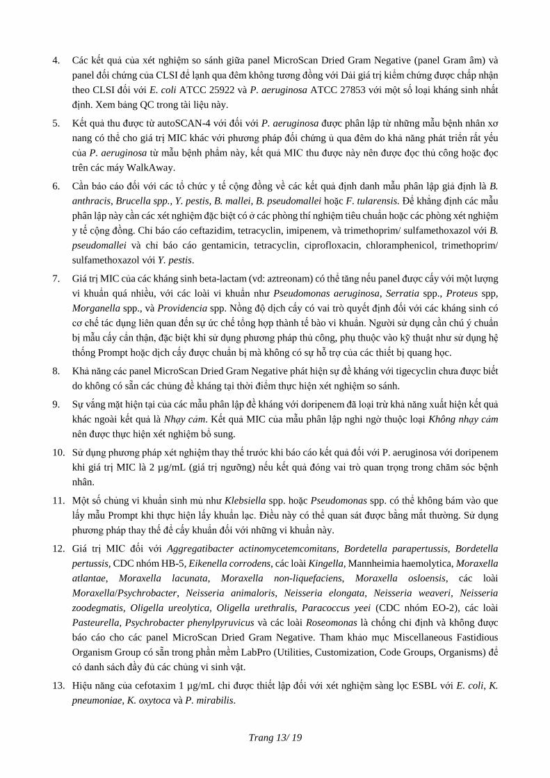
Trang 13/ 19
4. Các kết quả của xét nghiệm so sánh giữa panel MicroScan Dried Gram Negative (panel Gram âm) và
panel đối chứng của CLSI để lạnh qua đêm không tương đồng với Dải giá trị kiểm chứng được chấp nhận
theo CLSI đối với E. coli ATCC 25922 và P. aeruginosa ATCC 27853 với một số loại kháng sinh nhất
định. Xem bảng QC trong tài liệu này.
5. Kết quả thu được từ autoSCAN-4 với đối với P. aeruginosa được phân lập từ những mẫu bệnh nhân xơ
nang có thể cho giá trị MIC khác với phương pháp đối chứng ủ qua đêm do khả năng phát triển rất yếu
của P. aeruginosa từ mẫu bệnh phẩm này, kết quả MIC thu được này nên được đọc thủ công hoặc đọc
trên các máy WalkAway.
6. Cần báo cáo đối với các tổ chức y tế cộng đồng về các kết quả định danh mẫu phân lập giả định là B.
anthracis, Brucella spp., Y. pestis, B. mallei, B. pseudomallei hoặc F. tularensis. Để khẳng định các mẫu
phân lập này cần các xét nghiệm đặc biệt có ở các phòng thí nghiệm tiêu chuẩn hoặc các phòng xét nghiệm
y tế cộng đồng. Chỉ báo cáo ceftazidim, tetracyclin, imipenem, và trimethoprim/ sulfamethoxazol với B.
pseudomallei và chỉ báo cáo gentamicin, tetracyclin, ciprofloxacin, chloramphenicol, trimethoprim/
sulfamethoxazol với Y. pestis.
7. Giá trị MIC của các kháng sinh beta-lactam (vd: aztreonam) có thể tăng nếu panel được cấy với một lượng
vi khuẩn quá nhiều, với các loài vi khuẩn như Pseudomonas aeruginosa, Serratia spp., Proteus spp,
Morganella spp., và Providencia spp. Nồng độ dịch cấy có vai trò quyết định đối với các kháng sinh có
cơ chế tác dụng liên quan đến sự ức chế tổng hợp thành tế bào vi khuẩn. Người sử dụng cần chú ý chuẩn
bị mẫu cấy cẩn thận, đặc biệt khi sử dụng phương pháp thủ công, phụ thuộc vào kỹ thuật như sử dụng hệ
thống Prompt hoặc dịch cấy được chuẩn bị mà không có sự hỗ trợ của các thiết bị quang học.
8. Khả năng các panel MicroScan Dried Gram Negative phát hiện sự đề kháng với tigecyclin chưa được biết
do không có sẵn các chủng đề kháng tại thời điểm thực hiện xét nghiệm so sánh.
9. Sự vắng mặt hiện tại của các mẫu phân lập đề kháng với doripenem đã loại trừ khả năng xuất hiện kết quả
khác ngoài kết quả là Nhạy cảm. Kết quả MIC của mẫu phân lập nghi ngờ thuộc loại Không nhạy cảm
nên được thực hiện xét nghiệm bổ sung.
10. Sử dụng phương pháp xét nghiệm thay thế trước khi báo cáo kết quả đối với P. aeruginosa với doripenem
khi giá trị MIC là 2 µg/mL (giá trị ngưỡng) nếu kết quả đóng vai trò quan trọng trong chăm sóc bệnh
nhân.
11. Một số chủng vi khuẩn sinh mủ như Klebsiella spp. hoặc Pseudomonas spp. có thể không bám vào que
lấy mẫu Prompt khi thực hiện lấy khuẩn lạc. Điều này có thể quan sát được bằng mắt thường. Sử dụng
phương pháp thay thế để cấy khuẩn đối với những vi khuẩn này.
12. Giá trị MIC đối với Aggregatibacter actinomycetemcomitans, Bordetella parapertussis, Bordetella
pertussis, CDC nhóm HB-5, Eikenella corrodens, các loài Kingella, Mannheimia haemolytica, Moraxella
atlantae, Moraxella lacunata, Moraxella non-liquefaciens, Moraxella osloensis, các loài
Moraxella/Psychrobacter, Neisseria animaloris, Neisseria elongata, Neisseria weaveri, Neisseria
zoodegmatis, Oligella ureolytica, Oligella urethralis, Paracoccus yeei (CDC nhóm EO-2), các loài
Pasteurella, Psychrobacter phenylpyruvicus và các loài Roseomonas là chống chỉ định và không được
báo cáo cho các panel MicroScan Dried Gram Negative. Tham khảo mục Miscellaneous Fastidious
Organism Group có sẵn trong phần mềm LabPro (Utilities, Customization, Code Groups, Organisms) để
có danh sách đầy đủ các chủng vi sinh vật.
13. Hiệu năng của cefotaxim 1 µg/mL chỉ được thiết lập đối với xét nghiệm sàng lọc ESBL với E. coli, K.
pneumoniae, K. oxytoca và P. mirabilis.
Trang 14/ 19
LƯU Ý KHI DIỄN GIẢI KẾT QUẢ
Tiêu chuẩn CLSI M07-A10 và M45-A2 chỉ ra những kết quả gây nhầm lẫn nguy hiểm có thể xảy ra với một
số kháng sinh được kiểm tra và cho kết quả là “Nhạy cảm” đối với một số loài vi khuẩn đặc biệt. Những kết
hợp này bao gồm (nhưng không bị giới hạn):
1. Các cephalosporin thế hệ 1 và 2, các cephamycin, và các aminoglycoside đối với Salmonella và Shigella
spp.
2. Kháng sinh beta-lactam với Y. pestis
Hệ thống LabPro sẽ chỉ báo cáo kết quả MIC, không diễn giải kết quả khi xảy ra sự kết hợp giữa các kháng
sinh/vi khuẩn trên đây.
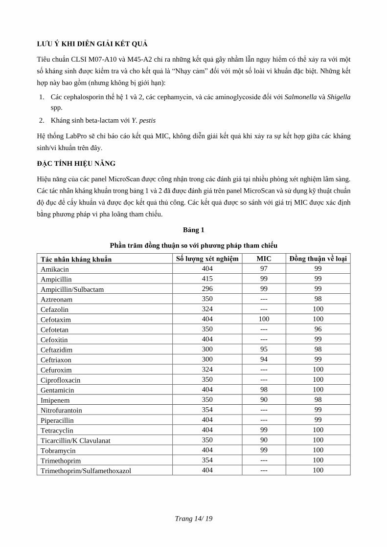
ĐẶC TÍNH HIỆU NĂNG
Hiệu năng của các panel MicroScan được công nhận trong các đánh giá tại nhiều phòng xét nghiệm lâm sàng.
Các tác nhân kháng khuẩn trong bảng 1 và 2 đã được đánh giá trên panel MicroScan và sử dụng kỹ thuật chuẩn
độ đục để cấy khuẩn và được đọc kết quả thủ công. Các kết quả được so sánh với giá trị MIC được xác định
bằng phương pháp vi pha loãng tham chiếu.
Bảng 1
Phần trăm đồng thuận so với phương pháp tham chiếu
Tác nhân kháng khuẩn Số lượng xét nghiệm MIC Đồng thuận về loại
Amikacin 404 97 99
Ampicillin 415 99 99
Ampicillin/Sulbactam 296 99 99
Aztreonam 350 --- 98
Cefazolin 324 --- 100
Cefotaxim 404 100 100
Cefotetan 350 --- 96
Cefoxitin 404 --- 99
Ceftazidim 300 95 98
Ceftriaxon 300 94 99
Cefuroxim 324 --- 100
Ciprofloxacin 350 --- 100
Gentamicin 404 98 100
Imipenem 350 90 98
Nitrofurantoin 354 --- 99
Piperacillin 404 --- 99
Tetracyclin 404 99 100
Ticarcillin/K Clavulanat 350 90 100
Tobramycin 404 99 100
Trimethoprim 354 --- 100
Trimethoprim/Sulfamethoxazol 404 --- 100
Trang 15/ 19
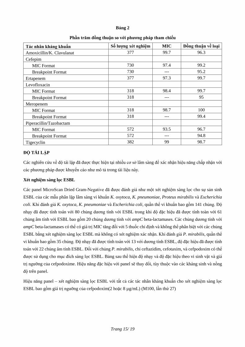
Bảng 2
Phần trăm đồng thuận so với phương pháp tham chiếu
Tác nhân kháng khuẩn Số lượng xét nghiệm MIC Đồng thuận về loại
Amoxicillin/K. Clavulanat 377 99.7 96.3
Cefepim
MIC Format 730 97.4 99.2
Breakpoint Format 730 --- 95.2
Ertapenem 377 97.3 99.7
Levofloxacin
MIC Format 318 98.4 99.7
Breakpoint Format 318 --- 95
Meropenem
MIC Format 318 98.7 100
Breakpoint Format 318 --- 99.4
Piperacillin/Tazobactam
MIC Format 572 93.5 96.7
Breakpoint Format 572 --- 94.8
Tigecyclin 382 99 98.7
ĐỘ TÁI LẬP
Các nghiên cứu về độ tái lập đã được thực hiện tại nhiều cơ sở lâm sàng để xác nhận hiệu năng chấp nhận với
các phương pháp được khuyến cáo như mô tả trong tài liệu này.
Xét nghiệm sàng lọc ESBL
Các panel MicroScan Dried Gram-Negative đã được đánh giá như một xét nghiệm sàng lọc cho sự sản sinh
ESBL của các mẫu phân lập lâm sàng vi khuẩn K. oxytoca, K. pneumoniae, Proteus mirabilis và Escherichia
coli. Khi đánh giá K. oxytoca, K. pneumoniae và Escherichia coli, quần thể vi khuẩn bao gồm 141 chủng. Độ
nhạy đã được tính toán với 80 chủng dương tính với ESBL trong khi độ đặc hiệu đã được tính toàn với 61
chủng âm tính với ESBL bao gồm 20 chủng dương tính với ampC beta-lactamases. Các chủng dương tính với
ampC beta-lactamases có thể có giá trị MIC tăng đối với 5 thuốc chỉ định và không thể phân biệt với các chủng
ESBL bằng xét nghiệm sàng lọc ESBL mà không có xét nghiệm xác nhận. Khi đánh giá P. mirabilis, quần thể
vi khuẩn bao gồm 35 chủng. Độ nhạy đã được tính toán với 13 với dương tính ESBL, độ đặc hiệu đã được tính
toán với 22 chủng âm tính ESBL. Đối với chủng P. mirabilis, chỉ ceftazidim, cefotaxim, và cefpodoxim có thể
được sử dụng cho mục đích sàng lọc ESBL. Bảng sau thể hiện độ nhạy và độ đặc hiệu theo vi sinh vật và giá
trị ngưỡng của cefpodoxime. Hiệu năng đặc hiệu với panel sẽ thay đổi, tùy thuộc vào các kháng sinh và nồng
độ trên panel.
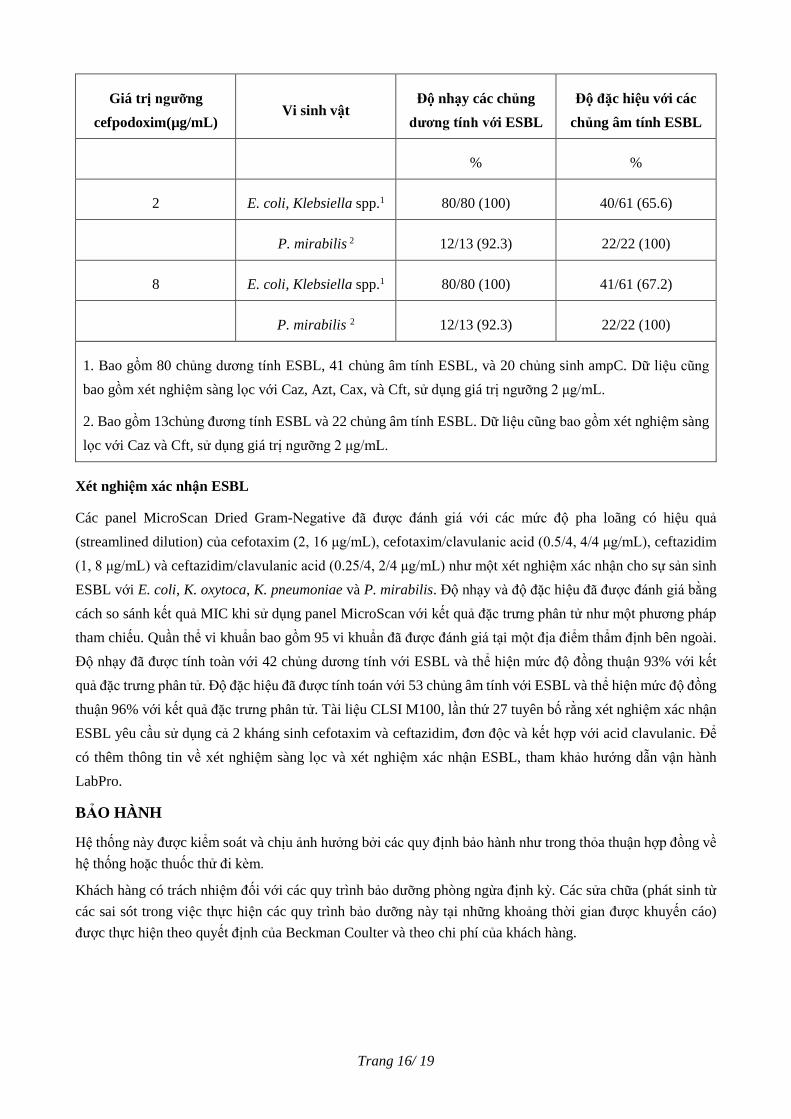
Hiệu năng panel – xét nghiệm sàng lọc ESBL với tất cả các tác nhân kháng khuẩn cho xét nghiệm sàng lọc
ESBL bao gồm giá trị ngưỡng của cefpodoxim(2 hoặc 8 µg/mL) (M100, lần thứ 27)
Trang 16/ 19
Giá trị ngưỡng
cefpodoxim(µg/mL) Vi sinh vật
Độ nhạy các chủng
dương tính với ESBL
Độ đặc hiệu với các
chủng âm tính ESBL
% %
2 E. coli, Klebsiella spp.1 80/80 (100) 40/61 (65.6)
P. mirabilis 2 12/13 (92.3) 22/22 (100)
8 E. coli, Klebsiella spp.1 80/80 (100) 41/61 (67.2)
P. mirabilis 2 12/13 (92.3) 22/22 (100)
1. Bao gồm 80 chủng dương tính ESBL, 41 chủng âm tính ESBL, và 20 chủng sinh ampC. Dữ liệu cũng
bao gồm xét nghiệm sàng lọc với Caz, Azt, Cax, và Cft, sử dụng giá trị ngưỡng 2 μg/mL.
2. Bao gồm 13chủng đương tính ESBL và 22 chủng âm tính ESBL. Dữ liệu cũng bao gồm xét nghiệm sàng
lọc với Caz và Cft, sử dụng giá trị ngưỡng 2 μg/mL.
Xét nghiệm xác nhận ESBL
Các panel MicroScan Dried Gram-Negative đã được đánh giá với các mức độ pha loãng có hiệu quả
(streamlined dilution) của cefotaxim (2, 16 μg/mL), cefotaxim/clavulanic acid (0.5/4, 4/4 μg/mL), ceftazidim
(1, 8 μg/mL) và ceftazidim/clavulanic acid (0.25/4, 2/4 μg/mL) như một xét nghiệm xác nhận cho sự sản sinh
ESBL với E. coli, K. oxytoca, K. pneumoniae và P. mirabilis. Độ nhạy và độ đặc hiệu đã được đánh giá bằng
cách so sánh kết quả MIC khi sử dụng panel MicroScan với kết quả đặc trưng phân tử như một phương pháp
tham chiếu. Quần thể vi khuẩn bao gồm 95 vi khuẩn đã được đánh giá tại một địa điểm thẩm định bên ngoài.
Độ nhạy đã được tính toàn với 42 chủng dương tính với ESBL và thể hiện mức độ đồng thuận 93% với kết
quả đặc trưng phân tử. Độ đặc hiệu đã được tính toán với 53 chủng âm tính với ESBL và thể hiện mức độ đồng
thuận 96% với kết quả đặc trưng phân tử. Tài liệu CLSI M100, lần thứ 27 tuyên bố rằng xét nghiệm xác nhận
ESBL yêu cầu sử dụng cả 2 kháng sinh cefotaxim và ceftazidim, đơn độc và kết hợp với acid clavulanic. Để
có thêm thông tin về xét nghiệm sàng lọc và xét nghiệm xác nhận ESBL, tham khảo hướng dẫn vận hành
LabPro.
BẢO HÀNH
Hệ thống này được kiểm soát và chịu ảnh hưởng bởi các quy định bảo hành như trong thỏa thuận hợp đồng về
hệ thống hoặc thuốc thử đi kèm.
Khách hàng có trách nhiệm đối với các quy trình bảo dưỡng phòng ngừa định kỳ. Các sửa chữa (phát sinh từ
các sai sót trong việc thực hiện các quy trình bảo dưỡng này tại những khoảng thời gian được khuyến cáo)
được thực hiện theo quyết định của Beckman Coulter và theo chi phí của khách hàng.
Trang 17/ 19
TÀI LIỆU THAM KHẢO
1. Jorgensen, J.H., M.A. Pfaller, K.C. Carroll, G. Funke, M.L. Landry, S.S. Richter, D.W. Warnock (eds),
2015. Manual of Clinical Microbiology, 11th ed. American Society for Microbiology, Washington D.C.
2. Barry, A. L. 1976. The Antimicrobic Susceptibility Test, Principles and Practices. Lea and Febiger.
Philadelphia. PA. p. 26.
3. Barry, A. L., F. Garcia, and L. D. Thrupp, 1970. An improved single disc method for testing the antibiotic
susceptibility of rapidly growing pathogens. Am. J. Clin. Path. 53:149.
4. Barry, A. L., and L. J. Effinger. 1974. Evaluation of a semi-automated system for preparing microdilutions
for antibiotic susceptibility testing. Abst. Annu. Meet. Am. Soc. Micro. #109.
5. Barry, A. L., R. N. Jones, and T. L. Gavan. 1978. Evaluation of the Micro-Media System for quantitative
antimicrobic susceptibility testing: a collaborative study. Antimicrob. Agents Chemother. 13:61.
6. Chitwood, L. A. 1969. Tube dilution antimicrobial susceptibility testing: efficacy of a microtechnique
applicable to diagnostic laboratories. Appl. Microbiol. 17:707.
7. Gavan, T. L., N. W. McFadden, Jr., and E. L. Cheatle. 1971. Antimicrobial Susceptibility Testing. Am.
Soc. Clin. Path. Comm. on Cont. Ed., Chicago, IL.
8. Gavan, T. L., and M. A. Town. 1969. Microdilution method for antibiotic susceptibility testing. Bacteriol.
Proc. 73.
9. Gavan, T. L., and M. A. Town. 1970. A microdilution method for antibiotic susceptibility testing. Amer. J.
Clin. Path. 53:880.
10. Gavan, T. L., and D. A. Butler. 1974. An automated microdilution method for antimicrobial susceptibility
testing p. 88. In A. Balows (ed) Current Techniques for Antimicrobial Susceptibility Testing. C.C. Thomas,
Springfield, IL.
11. Gerlach, E. H. 1974. Microdilution I: A comparative study, p. 63. In A. Balows (ed) Current Techniques
for Antibiotic Susceptibility Testing. C. C. Thomas, Springfield, IL.
12. Gerlach, E. H., R. J. Taylor, and B. Bauman. 1969. The comparison of a manual and an automated method
of routinely performed serial dilution antibiotic sensitivity tests in a large hospital. Amer. J. Clin. Path.
52:748.
13. Klastersky, J., D. Daneau, G. Swings, and D. Weerts, 1974. Antibacterial activity in serum and urine as a
therapeutic guide in bacterial infections. J. Infect. Dis. 129:187.
14. MacFaddin, J. F. 2000. Biochemical Tests for Identification of Medical Bacteria, 3rd ed., Williams and
Wilkens, Baltimore, MD.
15. MacLowry, J. D., and H. H. Marsh. 1968. Semi-automatic microtechnique for serial dilution antibiotic
sensitivity testing in the clinical laboratory. J. Lab. Clin. Med. 72:685.
16. Marymont, Jr., J. H., and R. M. Wentz. 1966. Serial dilution antibiotic sensitivity testing with the microtiter
system. Am. J. Clin. Path. 45:548.
Trang 18/ 19
17. Methods for Dilution Antimicrobial Susceptibility Tests for Bacteria that Grow Aerobically. 2015.
Approved Standard M07-A10. Clinical and Laboratory Standards Institute, Wayne, PA.
18. Tilton, R. C., and L. Newberg. 1974. Standardization of the microdilution susceptibility test, p. 77. In A.
Balows (ed.) Current Techniques for Antimicrobial Susceptibility Testing.
19. Tilton, R. C., L. Lieberman, and E. H. Gerlach. 1973. Microdilution antibiotic susceptibility test:
examination of certain variables. Appl. Microbiol. 26:5, 658.
20. Turck, M., R. Lindemeyer, and R. Petersdorf. 1963. Comparison of single-disc and tube-dilution techniques
in determining antibiotic sensitivities of gram negative pathogens. Ann Int. Med. 58:56.
21. Washington, J. A., E. Warren, and A. G. Karlson. 1973. Stability of barium sulfate turbidity standards.
Appl. Microbiol. 24:6, 1013.
22. World Health Organization. Standardization of methods for conducting microbic sensitivity tests. Second
Report of the Expert Committee on Antibiotics. Technical Report Series #210, p. 1. Geneva, 1961.
23. Performance Standards for Antimicrobial Susceptibility Testing. 2007, 2009, 2010, 2011, 2012, 2013, and
2017. CLSI Document M100-S17, S19, S20, S21, S22, S23, and 27th ed. Clinical and Laboratory Standards
Institute, Wayne, PA.
24. Thompson, K.S. 2001. Controversies about Extended - Spectrum and AmpC Beta - Lactamases. Emerg.
Infect. Dis. 7: 1-4.
25. Quality Control for Commercial Microbial Identification Systems. 2008. CLSI Document M50-A. Clinical
and Laboratory Standards Institute, Wayne, PA
26. Methods for Antimicrobial Dilution and Disk Susceptibility Testing of Infrequently Isolated or Fastidious
Bacteria. 2012. CLSI Document M45-A2. Clinical and Laboratory Standards Institute, Wayne, PA.
Trang 19/ 19
KÝ HIỆU
Không tái sử dụng
Hạn dùng
Số lô
Mã sản phẩm
Nhà sản xuất
Ngày sản xuất
Đại diện được ủy quyền tại châu Âu
Chứa đủ cho n xét nghiệm
Thiết bị y tế dùng trong chẩn đoán in vitro
Giới hạn nhiệt độ
Tham khảo Hướng dẫn sử dụng
Dán nhãn CE
Thành phần
Viết tắt tác nhân kháng khuẩn
Bảng dữ liệu an toàn hóa chất
Sản xuất tại Mỹ
Chỉ dùng cho mục đích xuất khẩu
Thể tích hoàn nguyên
Phiên bản cập nhật tháng 05/2018
Chủ sở hữu sản phẩm: Beckman Coulter, Inc.
Địa chỉ: 250 S. Kraemer Blvd., Brea, CA 92821, USA
Cơ sở sản xuất: Beckman Coulter, Inc.
Địa chỉ: 2040 Enterprise Blvd., West Sacramento, CA 95691, USA
MicroScan
3251-3424AB
05/2018
Dried Gram Negative Procedural Manual 1B1020-100B, B1017-301, B1017-401, B1017-404, B1017-406, B1017-407, B1017-408, B1017-409, B1017-411, B1017-412, B1017-413, B1017-414, B1017-416, B1017-417, B1017-419, B1017-420, B1017-421, B1017-422, B1017-423, B1017-424, B1017-425, B1017-426, B1017-427, B1020-100B, B1017-428, B1017-429, B1017-430, B1017-431
Hướng dẫn sử dụng tiếng Anh
thuong.tth
Highlight
thuong.tth
Rectangle
3251-3424AB
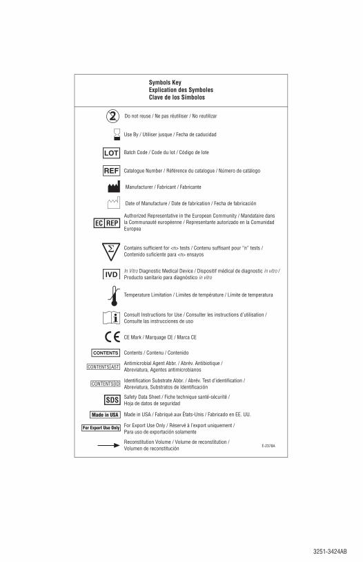
Symbols KeyExplication des SymbolesClave de los Símbolos
E-2378A
Use By / Utiliser jusque / Fecha de caducidad
Batch Code / Code du lot / Código de lote
Catalogue Number / Référence du catalogue / Número de catálogo
Authorized Representative in the European Community / Mandataire dans la Communauté européenne / Representante autorizado en la Comunidad Europea
Antimicrobial Agent Abbr. / Abrév. Antibiotique / Abreviatura, Agentes antimicrobianos
In Vitro Diagnostic Medical Device / Dispositif médical de diagnostic in vitro /Producto sanitario para diagnóstico in vitro
Temperature Limitation / Limites de température / Límite de temperatura
Consult Instructions for Use / Consulter les instructions d’utilisation /Consulte las instrucciones de uso
Reconstitution Volume / Volume de reconstitution / Volumen de reconstitución
Contains sufficient for <n> tests / Contenu suffisant pour “n” tests / Contenido suficiente para <n> ensayos
Identification Substrate Abbr. / Abrév. Test d’identification / Abreviatura, Substratos de Identificación
CE Mark / Marquage CE / Marca CE
Manufacturer / Fabricant / Fabricante
Contents / Contenu / Contenido
Safety Data Sheet / Fiche technique santé-sécurité / Hoja de datos de seguridad
Made in USA / Fabriqué aux États-Unis / Fabricado en EE. UU.
For Export Use Only / Réservé à l’export uniquement / Para uso de exportación solamente
Do not reuse / Ne pas réutiliser / No reutilizar
Date of Manufacture / Date de fabrication / Fecha de fabricación

3251-3424AB 1-13251-3424AB 1-1
Dried Gram Negative Procedural ManualATTENTION: Procedure Manual changes are indicated by shading.
Intended UseFor use with MicroScan Dried Gram Negative MIC/Combo Panels and Dried Gram Negative Breakpoint Combo Panels. MicroScan panels are designed for use in determining antimicrobial agent susceptibility and/or identification to the species level of aerobic and facultatively anaerobic gram-negative bacilli.Summary and PrinciplesThe antimicrobial susceptibility tests are miniaturizations of the broth dilution susceptibility test which have been dehydrated. Various antimicrobial agents are diluted in Mueller-Hinton broth supplemented with calcium and magnesium to concentrations bridging the range of clinical interest.17,23 Trimethoprim, and trimethoprim/sulfamethoxazole broth contain thymidine phosphorylase to reduce thymidine levels in the medium. After inoculation and rehydration with a standardized suspension of organism and incubation at 35°C for a minimum of 16 hours, the minimum inhibitory concentration (MIC) for the test organism is determined by observing the lowest antimicrobial concentration showing inhibition of growth.1-12,15,16,18-23
Modified conventional and chromogenic tests are used for the identification of fermentative and non-fermentative gram-negative bacilli. Identification is based on detection of pH changes, substrate utilization, and growth in the presence of antimicrobial agents after 16-42 hours incubation at 35°C.13,14 Panels containing ceftazidime, aztreonam, cefotaxime, or ceftriaxone at 1 µg/mL or cefpodoxime at 1 or 4 µg/mL (depending on panel type) can be used to screen for Escherichia coli, Klebsiella oxytoca, or K. pneumoniae strains suspected of producing extended-spectrum beta-lactamases (ESBLs). Ceftriaxone ESBL screening functionality has been disabled in LabPro v4.11– Panel Update-05 for panel types which are reporting the updated Enterobacteriaceae interpretive breakpoints and minimally contain ceftriaxone dilutions 1 and 2 µg/mL and DO NOT contain ESBL confirmation antimicrobial agents (ie. Cft/CA, Caz/CA). For Proteus mirabilis strains, only ceftazidime, cefotaxime, and cefpodoxime can be used for ESBL screening purposes. Panels containing ceftazidime/clavulanic acid and cefotaxime/clavulanic acid can be used to confirm the presence of ESBLs. The confirmation test is a ≥3 twofold dilution decrease in MICs of suspected organisms to ceftazidime or cefotaxime in the presence of a fixed concentration of clavulanic acid, versus its MIC when tested alone. The procedural manual content listed in the labeling (e.g., interpretive criteria, panel read times, limitations) may differ from the criteria in the LabPro Information Manager due to differences in software and panel releases.Precautions1. For in vitro diagnostic use only.2. Observe aseptic techniques and established precautions
against microbiological hazards throughout all procedures, with special awareness that the inoculated panels contain potentially pathogenic organisms.
3. This material contains infectious agents and should be discarded as appropriate for biohazardous waste.
4. Results of this test should always be interpreted in conjunction with the patient’s medical history, clinical presentation and other findings.
5. Caution: For prescription use only. US Federal Law restricts this device to sale by or on the order of a licensed practitioner.
StorageDried Gram Negative panels must be stored at 2–25°C.Product DeteriorationProlonged exposure to storage conditions other than those recommended may result in loss of potency of the antimicrobial
agents or hydrolysis of identification substrates. Do not use beyond the expiration date. Contact your Beckman Coulter Representative or Distributor for further assistance.Specimen Collection and PreparationAppropriate specimens should be collected, transported, and placed on primary isolation media according to procedures recommended in the Manual of Clinical Microbiology.1
ProcedureMaterials ProvidedSee box label for panel specific contents.Materials Required but not Provided0.5 McFarland Turbidity Standard0.5% N, N-Dimethyl-alpha-naphthylamine, 30 mL (B1010-45A)
or 250 mL (B1015-45)0.8% Sulfanilic Acid, 30 mL (B1010-44A) or 250 mL (B1015-44)0.85% Autoclaved Saline, 3 mL100 µL pipetter with disposable sterile tips 10% Ferric Chloride, 30 mL (B1010-48A) or 250 mL (B1015-48)40% Potassium Hydroxide, 30 mL (B1010-43A) or 250 mL
(B1015-43)5% Alpha Naphthol, 30 mL (B1010-42A)Cover Trays (B1010-56B)General Laboratory EquipmentInoculator-D Set (B1013-4)Inoculum Water, 3 mL (B1015-2)Inoculum Water with PLURONIC®*, 25 mL (B1015-7)Kovac’s Reagent, 30 mL (B1010-41A) or 250 mL (B1015-41)Manual Report Forms, MIC (B1014-92) or Breakpoint
(B1014-47)Microdilution Viewer Mineral Oil, 60 mL (B1010-40)Mineral Oil, 250 mL - only for WalkAway SI and WalkAway plus
instruments and WalkAway overlay feature (B1010-40A)Oxidase reagentManual Panel Worksheets (used for recording manually read
results)Prompt®** Inoculation System (B1026-10D)Quality Control Organisms (Refer to QC section of this manual)Quality Control Report FormsReagent Dropper Kit (B1013-12A)RENOK - Rehydrator/Inoculator (B1018-14) or equivalentSeal Strips (B1010-51)Turbidity MeterVortexBarcode Label Paper (B1018-129)WalkAway Tray Lids (B1018-18)* PLURONIC® surfactants, a registered trademark of BASF
Corporation, Parsippany, NJ USA** 3M, St. Paul, MN USA.Procedure OutlineA. Panel Preparation
1. Remove the panels to be used from storage. Do not use if the integrity of the packaging is compromised (unsealed, punctured, or torn).
2. Cut open the pouch and remove the panel. If stored in the refrigerator, remove the panel immediately from the foil pouch.
3. Panels should not be used if any of the following conditions exist:a. desiccant is not present or is broken.b. panel wells are discolored (e.g., DCB, various
antimicrobics).
3251-3424AB1-2 3251-3424AB1-2
4. Allow panels to equilibrate to room temperature prior to rehydration. Panels may be stacked with a clean Cover Tray on top. All opened panels should be used within the same day or discarded.
B. Inoculum PreparationCLSI recommends periodically checking your inoculum densities by doing colony counts. Refer to CLSI document M07-A1017 for colony count recommendations. The expected results for E. coli ATCC 25922 should closely approximate 5 X 10⁵ CFU/mL for the final test concentrations. The user should pay careful attention to inoculum preparation, especially with manual methods that are technique dependent such as the Prompt System or inoculum prepared without the aid of a photometric device.Note: Log and Stationary phase techniques are not supported with MicroScan products.1. Turbidity Standard Technique - Primary
Inoculation MethodThe turbidity standard technique is recommended for direct inoculation of all aerobic gram-negative bacilli.a. Using a sterile applicator stick, loop or swab,
touch the surface of 4-5 large or 5-10 small morphologically similar, well-isolated colonies from an 18-24 hour non-inhibitory agar plate.
b. Emulsify in 3 mL of Inoculum Water (autoclaved deionized water).
c. Cap tightly and vortex the suspension for 2-3 seconds. The final turbidity should be equivalent to that of a 0.5 McFarland Turbidity Standard. An equivalent turbidity can be achieved by using a MicroScan Turbidity Meter with a range of 0.08 ± 0.02.
d. Pipet 0.1 mL (100 µL) of the standardized suspension into 25 mL of Inoculum Water with PLURONIC. Cap tightly. Invert 8-10 times to mix.
2. Prompt SystemThe Prompt System may be used to inoculate gram-negative bacilli. Refer to the Prompt Inoculation procedural manual for the proper use of the Prompt System.
C. Oxidase TestPerform an oxidase test prior to inoculating panels. Record results in the appropriate space on the worksheet or as required by the instrumentation. Oxidase results support separation of taxa and early reporting of non-fermenters (e.g., Acinetobacter baumannii complex). For WalkAway instruments, oxidase results are recommended to be entered prior to printing barcodes. Some non-fermenters may still hold to 42 hours, depending on growth. The recommended oxidase reagent is tetramethyl-p-phenylene-diamine-dihydrochloride.
D. Panel Rehydration/InoculationRehydration and inoculation is performed using the RENOK system with Inoculators-D. Refer to the RENOK Operator’s Manual for use. If an alternate system is used, rehydrate with 115 µL ± 10 µL of Inoculum Water (PLURONIC). A final well concentration of 3-7x10⁵ CFU/mL should be achieved. To ensure viability and purity of the organism tested,a purity plate should be prepared by streaking the inoculum to an appropriate agar plate and incubating under the appropriate conditions. If two or more colony types are present on the purity plate, reisolate the colonies and retest.
E. Biochemical Overlays1. Using a dropper bottle, overlay the GLU, URE, H2S,
LYS, ARG, ORN and DCB with 3 drops of mineral oil. (These wells are underlined on the panel.)
2. The media in the wells must be completely covered with mineral oil, but the oil should not overflow the wells.
NOTE: WalkAway SI and WalkAway plus instruments (and WalkAway instrument upgraded with the automated oil overlay feature) automatically add oil to the appropriate wells.
F. Seal StripFor oxidase positive organisms only, place a seal strip over the CIT, MAL, ONPG, TAR, ACE, CET, OF/G, OF/B and DCB wells. The quarter-inch locator hole in the tape should be aligned over the DCB well. For WalkAway Systems, the lid is used in lieu of a seal strip.
G. Incubation1. Panels can be incubated in a WalkAway System or
incubated off-line using the following steps:a. To ensure even thermal distribution during
incubation, stack the panels in groups of 3-5.b. Place a clean Cover Tray on top of each group
of panels to prevent evaporation. Cover Trays may be reused. Do not decontaminate Cover Trays with alcohol. They may be cleaned with soap and water. Rinse well and allow to air dry.
c. Incubate the panels for a minimum of 16–20 hours at 35°C ± 1°C in a non-CO2 incubator.
H. Reading the PanelsPanels can be read manually using the MicroScan Microdilution Viewer and results recorded on a Manual Panel Worksheet or on MicroScan instrumentation (autoSCAN-4, and WalkAway Systems). Refer to LabPro Operator's Guide for reading panels with MicroScan instrumentation.1. Following 16-20 hours incubation, remove the panels
from the incubator.2. Wipe off the bottom of the panel with a lint-free tissue to
remove any condensation or debris that may be present.
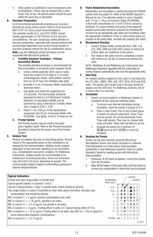
Typical Indicators Control well clear; large button in Growth well. Typical growth buttons in columns 1 and 2; Column 2 demonstrates a “skip” in growth wells, which should be ignored. This single button in column 3 bounded by clear wells above and below indicates spot
contamination and should be ignored. MIC in column 1 = 2 µg/mL (lowest concentration clear well). MIC in column 2 = > 16 µg/mL (growth in all wells). MIC in column 3 = ≤ 0.12 µg/mL (no growth in all wells). MIC in column 4 = 2 µg/mL (Trailing effect in wells 2-8; Typical trailing effect of T/S). MIC in column 5 = ≤ 0.12 µg/mL (Trailing effect in all wells, thus MIC is ≤. This is typical of
some antimicrobic/organism combinations). MIC in column 6 = ≤ 0.12 µg/mL.
A
GROWTHANTIMICROBIC WELLS μg/mL
CONTROL
COLUMN
0.12
0.25
0.5
1
2
4
8
16
3251-3424AB 1-33251-3424AB 1-3
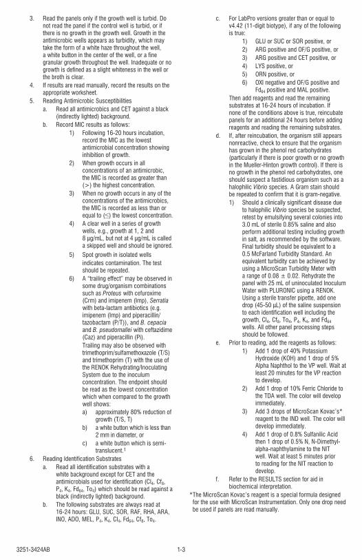
3. Read the panels only if the growth well is turbid. Do not read the panel if the control well is turbid, or if there is no growth in the growth well. Growth in the antimicrobic wells appears as turbidity, which may take the form of a white haze throughout the well, a white button in the center of the well, or a fine granular growth throughout the well. Inadequate or no growth is defined as a slight whiteness in the well or the broth is clear.
4. If results are read manually, record the results on the appropriate worksheet.
5. Reading Antimicrobic Susceptibilitiesa. Read all antimicrobics and CET against a black
(indirectly lighted) background.b. Record MIC results as follows:
1) Following 16-20 hours incubation, record the MIC as the lowest antimicrobial concentration showing inhibition of growth.
2) When growth occurs in all concentrations of an antimicrobic, the MIC is recorded as greater than (>) the highest concentration.
3) When no growth occurs in any of the concentrations of the antimicrobics, the MIC is recorded as less than or equal to (≤) the lowest concentration.
4) A clear well in a series of growth wells, e.g., growth at 1, 2 and 8 µg/mL, but not at 4 µg/mL is called a skipped well and should be ignored.
5) Spot growth in isolated wells indicates contamination. The test should be repeated.
6) A “trailing effect” may be observed in some drug/organism combinations such as Proteus with cefuroxime (Crm) and imipenem (Imp), Serratia with beta-lactam antibiotics (e.g. imipenem (Imp) and piperacillin/ tazobactam (P/T)), and B. cepacia and B. pseudomallei with ceftazidime (Caz) and piperacillin (Pi). Trailing may also be observed with trimethoprim/sulfamethoxazole (T/S) and trimethoprim (T) with the use of the RENOK Rehydrating/Inoculating System due to the inoculum concentration. The endpoint should be read as the lowest concentration which when compared to the growth well shows:a) approximately 80% reduction of
growth (T/S, T)b) a white button which is less than
2 mm in diameter, orc) a white button which is semi-
translucent.16. Reading Identification Substrates
a. Read all identification substrates with a white background except for CET and the antimicrobials used for identification (Cl4, Cf8, P4, K4, Fd64, To4) which should be read against a black (indirectly lighted) background.
b. The following substrates are always read at 16-24 hours: GLU, SUC, SOR, RAF, RHA, ARA, INO, ADO, MEL, P4, K4, Cl4, Fd64, Cf8, To4.
c. For LabPro versions greater than or equal to v4.42 (11-digit biotype), if any of the following is true:
1) GLU or SUC or SOR positive, or2) ARG positive and OF/G positive, or3) ARG positive and CET positive, or4) LYS positive, or5) ORN positive, or6) OXI negative and OF/G positive and
Fd64 positive and MAL positive. Then add reagents and read the remaining
substrates at 16-24 hours of incubation. If none of the conditions above is true, reincubate panels for an additional 24 hours before adding reagents and reading the remaining substrates.
d. If, after reincubation, the organism still appears nonreactive, check to ensure that the organism has grown in the phenol red carbohydrates (particularly if there is poor growth or no growth in the Mueller-Hinton growth control). If there is no growth in the phenol red carbohydrates, one should suspect a fastidious organism such as a halophilic Vibrio species. A Gram stain should be repeated to confirm that it is gram-negative.1) Should a clinically significant disease due
to halophilic Vibrio species be suspected, retest by emulsifying several colonies into 3.0 mL of sterile 0.85% saline and also perform additional testing including growth in salt, as recommended by the software. Final turbidity should be equivalent to a 0.5 McFarland Turbidity Standard. An equivalent turbidity can be achieved by using a MicroScan Turbidity Meter with a range of 0.08 ± 0.02. Rehydrate the panel with 25 mL of uninoculated Inoculum Water with PLURONIC using a RENOK. Using a sterile transfer pipette, add one drop (45-50 μL) of the saline suspension to each identification well including the growth, Cl4, Cf8, To4, P4, K4, and Fd64 wells. All other panel processing steps should be followed.
e. Prior to reading, add the reagents as follows:1) Add 1 drop of 40% Potassium
Hydroxide (KOH) and 1 drop of 5% Alpha Naphthol to the VP well. Wait at least 20 minutes for the VP reaction to develop.
2) Add 1 drop of 10% Ferric Chloride to the TDA well. The color will develop immediately.
3) Add 3 drops of MicroScan Kovac’s* reagent to the IND well. The color will develop immediately.
4) Add 1 drop of 0.8% Sulfanilic Acid then 1 drop of 0.5% N, N-Dimethyl-alpha-naphthylamine to the NIT well. Wait at least 5 minutes prior to reading for the NIT reaction to develop.
f. Refer to the RESULTS section for aid in biochemical interpretation.
* The MicroScan Kovac’s reagent is a special formula designed for the use with MicroScan Instrumentation. Only one drop need be used if panels are read manually.
3251-3424AB1-4 3251-3424AB1-4
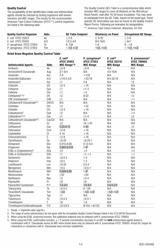
Quality Control Organism Abbr. QC Table Endpoint Dilution(s) on Panel Extrapolated QC RangeE. coli ATCC 25922 Ak ≤ 2-8 2, 8-32 ≤ 2-8 E. coli ATCC 25922 A/S 1/0.5-4/2 8/4-16/8 ≤ 8/4 P. aeruginosa ATCC 27853 Te 8, 128 1-8 8 - >8 P. aeruginosa ATCC 27853 Tim ≤ 8/2-32/2 16/2, 64/2 ≤ 16/2, 64/2
Dried Gram-Negative Quality Control Table
Antimicrobial Agents Abbr.
E. coli 2
ATCC 25922MIC Range1,6
P. aeruginosa2
ATCC 27853MIC Range
E. coli 2
ATCC 35218MIC Range
K. pneumoniae2
ATCC 700603MIC Range
Amikacin Ak ≤2-8 ≤2-8 N/A N/AAmoxicillin/K Clavulanate Aug 2/1-8/4 N/A 4/2-16/8 N/AAmpicillin Am 2-8 >128 N/A N/AAmpicillin/Sulbactam A/S ≤1/0.5-4/2 >32/16 8/4-32/16 N/AAztreonam3 Azt ≤1 2-8 N/A ≥2Cefazolin Cfz ≤2-4 >16 N/A N/ACefepime Cpe ≤1 ≤1-4 N/A N/ACefixime Cfe ≤1 >2 N/A N/ACefotaxime3,4,5 Cft ≤2 8-32 N/A ≥2CftE (Cefotaxime)3 CftE ≤1 N/A N/A >1Cefotaxime/K Clavulanate4,5 Cft/CA N/A N/A N/A N/ACefotetan Ctn ≤4 >32 N/A N/ACefoxitin Cfx ≤2-4 >16 N/A N/ACefpodoxime3 Cpd ≤2 N/A N/A >4Ceftazidime3,4,5 Caz ≤1 ≤1-4 N/A ≥2Ceftazidime/K Clavulanate4,5 Caz/CA N/A N/A N/A N/ACeftizoxime Cz ≤2 16->32 N/A N/ACeftriaxone3 Cax 0.03-0.12 8-64 N/A ≥2Cefuroxime Crm ≤2-8 >16 N/A N/ACephalothin Cf 4-16 >16 N/A N/AChloramphenicol C ≤2-8 >16 N/A N/ACiprofloxacin Cp ≤0.25 ≤0.25-1 N/A N/ADoripenem Dor 0.015-0.06 0.12-0.5 N/A N/AErtapenem Etp 0.004-0.016 2-87 N/A N/AESBL-a (Cefpodoxime)3 ESa ≤4 >4 N/A >4ESBL-b (Ceftazidime)3 ESb ≤1 ≤1->1 N/A >1Gentamicin Gm ≤0.5-2 1-4 N/A N/AImipenem Imp ≤0.5 1-4 N/A N/ALevofloxacin Lvx ≤0.25 0.5-4 N/A N/AMeropenem Mer ≤0.5 ≤0.5-1 N/A N/AMoxifloxacin Mxf 0.008-0.06 1-87 N/A N/ANitrofurantoin Fd ≤32 >64 N/A N/ANorfloxacin Nxn ≤2 ≤2-4 N/A N/APiperacillin Pi ≤8 ≤8 N/A N/APiperacillin/Tazobactam P/T 1/4-4/4 1/4-8/4 0.5/4-2/4 N/ATetracycline Te ≤0.5-2 8, 128 N/A N/ATicarcillin/K Clavulanate Tim ≤8/2 ≤8/2-32/2 ≤8/2-16/2 N/ATigecycline Tgc ≤1 N/A N/A N/ATobramycin To ≤0.5-2 ≤0.5-1 N/A N/ATrimethoprim T ≤8 >8 N/A N/ATrimethoprim/Sulfamethoxazole T/S ≤0.5/9.5 8/152->8/152 N/A N/A
1. Range = Expected value (µg/mL)2. The range of some antimicrobics do not agree with the Acceptable Quality Control Ranges listed in the CLSI M100 Document.3. When using the ESBL screening function, this additional endpoint may be obtained with K. pneumoniae ATCC 700603.4. When using the ESBL confirmation function, a ≥ 3 twofold concentration decrease in an MIC for both antimicrobial agents tested in
combination with K. Clavulanate versus its MIC when tested alone may be obtained with K. pneumoniae ATCC 700603. Actual QC ranges for ceftazidime or cefotaxime with K. Clavulanate have not been established.
Quality ControlThe acceptability of the identification media and antimicrobial agents should be checked by testing organisms with known reactions and MIC ranges. The results for the recommended American Type Culture Collection (ATCC**) control organisms are listed in the following table.
The Quality Control (QC) Table is a comprehensive table which includes MIC ranges to cover all dilutions on the MicroScan Dried Panels; read after 16-20 hours incubation. You may need to extrapolate from the QC Table, based on the panel type. Panel specific QC information can also be found on the Quality Control Report Forms. Following are examples for extrapolation.** American Type Culture Collection, Manassas, VA USA
3251-3424AB 1-5
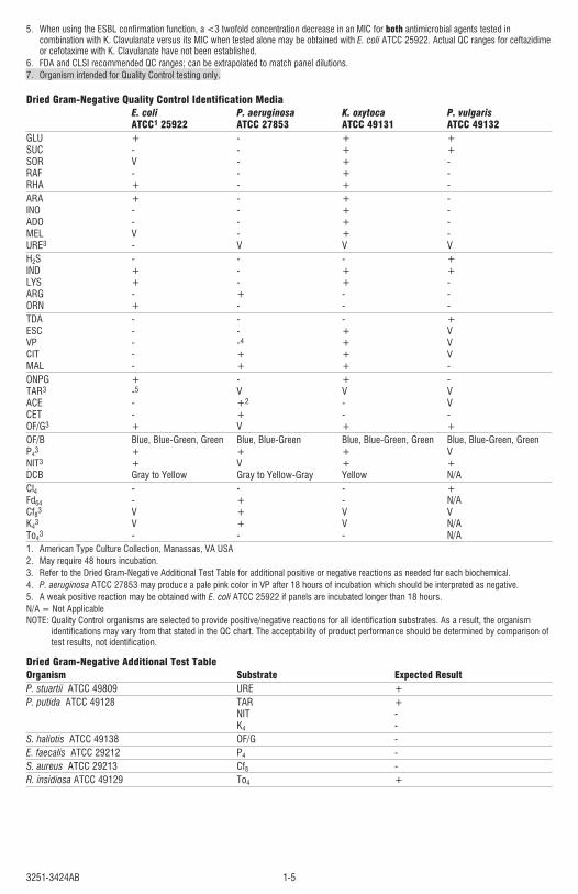
5. When using the ESBL confirmation function, a <3 twofold concentration decrease in an MIC for both antimicrobial agents tested in combination with K. Clavulanate versus its MIC when tested alone may be obtained with E. coli ATCC 25922. Actual QC ranges for ceftazidime or cefotaxime with K. Clavulanate have not been established.
6. FDA and CLSI recommended QC ranges; can be extrapolated to match panel dilutions.7. Organism intended for Quality Control testing only.
Dried Gram-Negative Quality Control Identification Media E. coli P. aeruginosa K. oxytoca P. vulgaris ATCC1 25922 ATCC 27853 ATCC 49131 ATCC 49132GLU + - + + SUC - - + + SOR V - + - RAF - - + - RHA + - + -ARA + - + - INO - - + - ADO - - + - MEL V - + - URE3 - V V VH2S - - - + IND + - + + LYS + - + - ARG - + - - ORN + - - -TDA - - - + ESC - - + V VP - -4 + V CIT - + + V MAL - + + -ONPG + - + - TAR3 -5 V V V ACE - +2 - V CET - + - - OF/G3 + V + +OF/B Blue, Blue-Green, Green Blue, Blue-Green Blue, Blue-Green, Green Blue, Blue-Green, Green P43 + + + V NIT3 + V + + DCB Gray to Yellow Gray to Yellow-Gray Yellow N/ACl4 - - - + Fd64 - + - N/A Cf83 V + V V K43 V + V N/A To43 - - - N/A 1. American Type Culture Collection, Manassas, VA USA2. May require 48 hours incubation.3. Refer to the Dried Gram-Negative Additional Test Table for additional positive or negative reactions as needed for each biochemical.4. P. aeruginosa ATCC 27853 may produce a pale pink color in VP after 18 hours of incubation which should be interpreted as negative.5. A weak positive reaction may be obtained with E. coli ATCC 25922 if panels are incubated longer than 18 hours.N/A = Not ApplicableNOTE: Quality Control organisms are selected to provide positive/negative reactions for all identification substrates. As a result, the organism
identifications may vary from that stated in the QC chart. The acceptability of product performance should be determined by comparison of test results, not identification.
Dried Gram-Negative Additional Test TableOrganism Substrate Expected ResultP. stuartii ATCC 49809 URE +P. putida ATCC 49128 TAR + NIT - K4 -S. haliotis ATCC 49138 OF/G -E. faecalis ATCC 29212 P4 -S. aureus ATCC 29213 Cf8 -R. insidiosa ATCC 49129 To4 +
3251-3424AB1-6
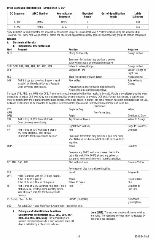
Dried Gram Neg Identification - Streamlined ID QC*
QC Organism ATCC Number Key Indicator Substrate
Expected Result
Out-of-Specification Result
Labile Substrate
E. coli 25922 ONPG + - Yes
E. coli 25922 ARA + - Yes
* Key indicators for Quality Control are provided for streamlined QC per CLSI document M50-A.25 Before implementing the streamlined QC program, refer to the M50-A document for details and check with applicable regulatory agencies and inspecting groups to confirm acceptance.
ResultsA. Biochemical Results
1. Biochemical InterpretationsWell Reagent Positive NegativeGLU Strong Yellow only Orange to Red Some non-fermenters may produce a golden color which should be considered negative.SUC, SOR, RAF, RHA, ARA, INO, ADO, MEL Yellow to Yellow/Orange Orange to RedURE Magenta to Pink Yellow, Orange or Light PinkH2S Black Precipitate or Black Button No BlackeningIND Add 3 drops (or one drop if panel is read Pink to Red Pale Yellow to visually) of MicroScan Kovac’s Reagent. Orange Color develops immediately. Providencia sp. may produce a pale pink ring which should be considered positive.Compare LYS, ARG, and ORN with DCB. These wells must be overlaid with oil for results to be valid. Purple is considered positive when comparing to a gray DCB well. Gray is considered positive when comparing to a yellow DCB well. For non-fermenters, a positive test must be significantly more purple than the base control. If the base control is purple, the basal medium has been alkalinized and the LYS, ARG and ORN should all be recorded as negative. Achromobacter species and Ochrobactrum anthropi tend to do this. Fermenters: LYS Purple to Gray Yellow ARG Non-fermenters: ORN Purple Colorless to GrayTDA Add 1 drop of 10% Ferric Chloride. Any Shade of Brown Yellow to Orange Color develops immediately.ESC Light Brown to Black Beige to ColorlessVP Add 1 drop of 40% KOH and 1 drop of Red Colorless 5% Alpha Naphthol. Wait at least 20 minutes for the reaction to develop. Some non-fermenters may produce a pale pink color after 18 hours incubation which should be considered negative.ONPG Yellow Colorless Compare any ONPG well which looks clear to the cetrimide well. If the ONPG shows any yellow as compared to the cetrimide well, record as positive.CIT, MAL, TAR, ACE Blue to Blue-Green Green to Yellow Any shade of blue is considered positiveCET Growth No growthOF/G NOTE: Compare with the OF base control. If the OF base is green: Yellow Green to Blue If the OF base is blue or blue-green: Yellow to Green BlueNIT Add 1 drop of 0.8% Sulfanilic Acid then 1 drop Red Colorless to of 0.5% N, N-Dimethyl-alpha-naphthylamine. Pale Pink Wait at least 5 minutes for the reaction to develop.P4, K4, Cl4, Fd64, To4, Cf8 Growth (Resistant) No Growth (Susceptible)LOC For autoSCAN-4 and WalkAway System panel recognition only.
2. Principles of Identification ReactionsCarbohydrate Fermentation (GLU, SUC, SOR, RAF, RHA, ARA, INO, ADO, MEL): The fermentation of a specific carbohydrate results in acid formation and a pH drop is detected by a phenol red indicator.
Urea (URE): The enzyme urease splits urea forming ammonia. The resulting increase in pH is detected by the phenol red indicator.
3251-3424AB 1-7
Hydrogen Sulfide (H2S): Hydrogen sulfide gas is produced from sodium thiosulfate and reacts with ferric ions in the medium to produce a black precipitate.Indole (IND): The metabolism of tryptophan results in the formation of indole, which is detected by the addition of Kovac’s reagent. If indole is present, a red color develops.Lysine, Arginine, Ornithine (LYS, ARG, ORN): Decarboxylation of these amino acids results in the formation of basic amines which are detected by the bromcresol purple indicator.Tryptophan Deaminase (TDA): Bacteria capable of deaminating tryptophan produce indolepyruvic acid, which reacts with ferric ammonium citrate in the medium after the addition of ferric chloride to produce a brown color.Esculin Hydrolysis (ESC): Hydrolysis of esculin is detected by ferric ammonium citrate in the medium, which reacts with the hydrolytic products to form a black precipitate.Voges-Proskauer (VP): Acetoin is produced from sodium pyruvate and is indicated by the formation of a red color after the addition of 40% Potassium Hydroxide and 5% Alpha Naphthol.Galactosidase (ONPG): β-galactosidase hydrolizes ortho-nitrophenyl-β-D-galactopyranoside, which liberates the yellow-colored ortho-nitrophenol.Citrate, Malonate, Acetamide, Tartrate (CIT, MAL, ACE, TAR): The utilization of these substrates as the sole source of carbon for metabolism results in a rise in pH that is detected by bromthymol blue indicator.Oxidation-Fermentation (OF/G): The oxidation of glucose results in acid formation and a pH drop is detected by the bromthymol blue indicator. The OF/G is compared to OF/B (base) to determine if any acid has been produced.Nitrate (NIT): The ability of an organism to reduce nitrate to nitrite is detected by the addition of Sulfanilic Acid then N, N-Dimethyl-alpha-naphthylamine, which produce a red color in the presence of nitrite.Cetrimide (CET): Tolerance to cetrimide is demonstrated by growth in Mueller-Hinton broth supplemented with cetrimide.Penicillin, Kanamycin, Colistin, Cephalothin, Nitrofurantoin, Tobramycin (P4, K4, CI4, Cf8, Fd64, To4): Resistance to specific concentrations of these antimicrobial agents is demonstrated by growth.
3. Organism IdentificationThe Biotype Lookup Program in the LabPro Information Manager and on the Beckman Coulter website is used
for the identification of unknown test organisms. The program lists the organism identification and the relative probabilities, in the order of the highest probability, up to a cumulative total of 99.9%. Should a biotype number occur that results in a “Very Rare Biotype,” consult the LabPro Software (Utilities>System>Biotype Lookup), the Biotype Lookup Program on the Beckman Coulter website, or your Beckman Coulter Representative or Distributor.
B. Interpretation of MIC ResultsSusceptibility is determined by comparing the MIC of an organism to the attainable blood or urine level of the antimicrobic. The following table lists the interpretive criteria as indicated in the CLSI document. Some of these differ from the manufacturer’s interpretive breakpoints listed in the Physicians’ Desk Reference.
C. Interpretations of ESBL Results1. Certain members of the Enterobacteriaceae, especially
Klebsiella spp., Escherichia coli, and Proteus mirabilis, provide novel beta-lactamase enzymes which are capable of hydrolyzing the extended-spectrum cephalosporins (e.g. cefotaxime, ceftriaxone, ceftizoxime, and ceftazidime) and aztreonam. These novel enzymes often have unusual susceptibility patterns for the beta-lactam class of antibiotics. Clinical isolates of Klebsiella oxytoca, K. pneumoniae, and Escherichia coli with increased MICs (≥2 µg/mL) of ceftazidime, aztreonam, cefotaxime, ceftriaxone, or MICs of ≥2 or ≥8 µg/mL (depending on panel type) of cefpodoxime should be suspected of harboring an extended-spectrum beta-lactamase. Ceftriaxone ESBL screening functionality has been disabled in LabPro v4.11– Panel Update-05 for panel types which are reporting the updated Enterobacteriaceae interpretive breakpoints and minimally contain ceftriaxone dilutions 1 and 2 µg/mL and DO NOT contain ESBL confirmation antimicrobial agents (ie. Cft/CA, Caz/CA). For P. mirabilis strains, only ceftazidime, cefotaxime, and cefpodoxime can be used for ESBL screening purposes. A clinical isolate is considered positive by the ESBL confirmation test if there is a ≥3 two-fold dilution drop (i.e. a 3 well decrease) in an MIC value for the antibiotic tested with clavulanic acid as compared to the MIC value of that antibiotic tested alone. While both cefotaxime and ceftazidime, with and without clavulanic acid should be tested, a positive result (i.e. ≥3 two-fold dilution drop in MIC) with either antibiotic combination is considered ESBL phenotypic-confirmation positive.23
Streamlined ESBL Confirmation Test
Antimicrobial Agent
Example 1:Confirmation
Positive
Example 2:Confirmation
Positive
Example 3:Confirmation
Negative
Example 4:Possible ESBL, unable to
interpret confirmation test. Organism has MIC’s >highest
dilution on panel.
CazCaz/CA
8≤0.25/4
16≤0.25/4
42/4
>16>2/4
CftCft/CA
164/4
32≤0.5/4
164/4
>32>4/4
2. LabPro also provides a customizable ESBL screen. This screen utilizes MICs of the antibiotics ceftazidime (Caz), aztreonam (Azt), cefotaxime (Cft), cefpodoxime (Cpd), and ceftriaxone (Cax). For further details see the LabPro Operator's Guide.
3. The finding that isolates are screen positive but confirmation negative may be due to the presence of other beta-lactamases such as ampC.24
4. For all confirmed ESBL-producing strains, the test interpretation should be reported as resistant for all penicillins, cephalosporins, and aztreonam.17,23
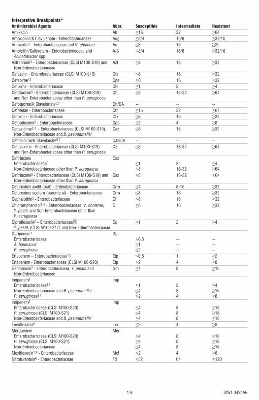
3251-3424AB1-8
Interpretive Breakpoints*Antimicrobial Agents Abbr. Susceptible Intermediate ResistantAmikacin Ak ≤16 32 ≥64Amoxicillin/K Clavulanate - Enterobacteriaceae Aug ≤8/4 16/8 ≥32/16Ampicillin2 - Enterobacteriaceae and V. cholerae Am ≤8 16 ≥32 Ampicillin/Sulbactam - Enterobacteriaceae and A/S ≤8/4 16/8 ≥32/16 Acinetobacter spp. Aztreonam3 - Enterobacteriaceae (CLSI M100-S19) and Azt ≤8 16 ≥32 Non-Enterobacteriaceae Cefazolin - Enterobacteriaceae (CLSI M100-S19) Cfz ≤8 16 ≥32Cefepime12 Cpe ≤8 16 ≥32Cefixime - Enterobacteriaceae Cfe ≤1 2 ≥4 Cefotaxime3 - Enterobacteriaceae (CLSI M100-S19) Cft ≤8 16-32 ≥64 and Non-Enterobacteriaceae other than P. aeruginosa Cefotaxime/K Clavulanate6,7 Cft/CA -- -- --Cefotetan - Enterobacteriaceae Ctn ≤16 32 ≥64Cefoxitin - Enterobacteriaceae Cfx ≤8 16 ≥32Cefpodoxime3 - Enterobacteriaceae Cpd ≤2 4 ≥8 Ceftazidime2,3 - Enterobacteriaceae (CLSI M100-S19), Caz ≤8 16 ≥32 Non-Enterobacteriaceae and B. pseudomallei Ceftazidime/K Clavulanate6,7 Caz/CA -- -- --Ceftizoxime - Enterobacteriaceae (CLSI M100-S19) Cz ≤8 16-32 ≥64 and Non-Enterobacteriaceae other than P. aeruginosa Ceftriaxone Cax Enterobacteriaceae9 ≤1 2 ≥4 Non-Enterobacteriaceae other than P. aeruginosa ≤8 16-32 ≥64Ceftriaxone3 - Enterobacteriaceae (CLSI M100-S19) and Cax ≤8 16-32 ≥64 Non-Enterobacteriaceae other than P. aeruginosa Cefuroxime axetil (oral) - Enterobacteriaceae Crm ≤4 8-16 ≥32Cefuroxime sodium (parenteral) - Enterobacteriaceae Crm ≤8 16 ≥32Cephalothin5 - Enterobacteriaceae Cf ≤8 16 ≥32Chloramphenicol2,4 - Enterobacteriaceae, V. cholerae, C ≤8 16 ≥32 Y. pestis and Non-Enterobacteriaceae other than P. aeruginosa Ciprofloxacin2 – Enterobacteriaceae13, Cp ≤1 2 ≥4 Y. pestis (CLSI M100-S17) and Non-EnterobacteriaceaeDoripenem1 Dor Enterobacteriaceae ≤0.5 -- -- A. baumannii ≤1 -- -- P. aeruginosa ≤2 -- -- Ertapenem – Enterobacteriaceae10 Etp ≤0.5 1 ≥2Ertapenem - Enterobacteriaceae (CLSI M100-S20) Etp ≤2 4 ≥8Gentamicin2 - Enterobacteriaceae, Y. pestis and Gm ≤4 8 ≥16 Non-Enterobacteriaceae Imipenem2 Imp Enterobacteriaceae11 ≤1 2 ≥4 Non-Enterobacteriaceae and B. pseudomallei ≤4 8 ≥16 P. aeruginosa11 ≤2 4 ≥8 Imipenem2 Imp Enterobacteriaceae (CLSI M100-S20) ≤4 8 ≥16 P. aeruginosa (CLSI M100-S21) ≤4 8 ≥16 Non-Enterobacteriaceae and B. pseudomallei ≤4 8 ≥16Levofloxacin8 Lvx ≤2 4 ≥8Meropenem Mer Enterobacteriaceae (CLSI M100-S20) ≤4 8 ≥16 P. aeruginosa (CLSI M100-S21) ≤4 8 ≥16 Non-Enterobacteriaceae ≤4 8 ≥16Moxifloxacin1,4 - Enterobacteriaceae Mxf ≤2 4 ≥8Nitrofurantoin5 - Enterobacteriaceae Fd ≤32 64 ≥128
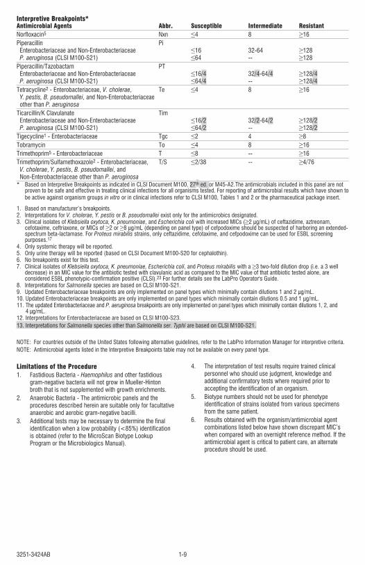
3251-3424AB 1-9
Interpretive Breakpoints*Antimicrobial Agents Abbr. Susceptible Intermediate ResistantNorfloxacin5 Nxn ≤4 8 ≥16Piperacillin Pi Enterobacteriaceae and Non-Enterobacteriaceae ≤16 32-64 ≥128 P. aeruginosa (CLSI M100-S21) ≤64 -- ≥128Piperacillin/Tazobactam PT Enterobacteriaceae and Non-Enterobacteriaceae ≤16/4 32/4-64/4 ≥128/4 P. aeruginosa (CLSI M100-S21) ≤64/4 -- ≥128/4Tetracycline2 - Enterobacteriaceae, V. cholerae, Te ≤4 8 ≥16 Y. pestis, B. pseudomallei, and Non-Enterobacteriaceae other than P. aeruginosa Ticarcillin/K Clavulanate Tim Enterobacteriaceae and Non-Enterobacteriaceae ≤16/2 32/2-64/2 ≥128/2 P. aeruginosa (CLSI M100-S21) ≤64/2 -- ≥128/2Tigecycline1 - Enterobacteriaceae Tgc ≤2 4 ≥8Tobramycin To ≤4 8 ≥16Trimethoprim5 - Enterobacteriaceae T ≤8 -- ≥16Trimethoprim/Sulfamethoxazole2 - Enterobacteriaceae, T/S ≤2/38 -- ≥4/76 V. cholerae, Y. pestis, B. pseudomallei, and Non-Enterobacteriaceae other than P. aeruginosa* Based on Interpretive Breakpoints as indicated in CLSI Document M100, 27th ed. or M45-A2.The antimicrobials included in this panel are not
proven to be safe and effective in treating clinical infections for all organisms tested. For reporting of antimicrobial results which have shown to be active against organism groups in vitro or in clinical infections refer to CLSI M100, Tables 1 and 2 or the pharmaceutical package insert.
1. Based on manufacturer’s breakpoints.2. Interpretations for V. cholerae, Y. pestis or B. pseudomallei exist only for the antimicrobics designated. 3. Clinical isolates of Klebsiella oxytoca, K. pneumoniae, and Escherichia coli with increased MICs (≥2 µg/mL) of ceftazidime, aztreonam,
cefotaxime, ceftriaxone, or MICs of ≥2 or ≥8 µg/mL (depending on panel type) of cefpodoxime should be suspected of harboring an extended-spectrum beta-lactamase. For Proteus mirabilis strains, only ceftazidime, cefotaxime, and cefpodoxime can be used for ESBL screening purposes.17
4. Only systemic therapy will be reported.5. Only urine therapy will be reported (based on CLSI Document M100-S20 for cephalothin).6. No breakpoints exist for this test.7. Clinical isolates of Klebsiella oxytoca, K. pneumoniae, Escherichia coli, and Proteus mirabilis with a ≥3 two-fold dilution drop (i.e. a 3 well
decrease) in an MIC value for the antibiotic tested with clavulanic acid as compared to the MIC value of that antibiotic tested alone, are considered ESBL phenotypic-confirmation positive (CLSI).23 For further details see the LabPro Operator's Guide.
8. Interpretations for Salmonella species are based on CLSI M100-S21.9. Updated Enterobacteriaceae breakpoints are only implemented on panel types which minimally contain dilutions 1 and 2 µg/mL.10. Updated Enterobacteriaceae breakpoints are only implemented on panel types which minimally contain dilutions 0.5 and 1 µg/mL.11. The updated Enterobacteriaceae and P. aeruginosa breakpoints are only implemented on panel types which minimally contain dilutions 1, 2, and
4 µg/mL.12. Interpretations for Enterobacteriaceae are based on CLSI M100-S23. 13. Interpretations for Salmonella species other than Salmonella ser. Typhi are based on CLSI M100-S21.
NOTE: For countries outside of the United States following alternative guidelines, refer to the LabPro Information Manager for interpretive criteria.NOTE: Antimicrobial agents listed in the Interpretive Breakpoints table may not be available on every panel type.
Limitations of the Procedure1. Fastidious Bacteria - Haemophilus and other fastidious
gram-negative bacteria will not grow in Mueller-Hinton broth that is not supplemented with growth enrichments.
2. Anaerobic Bacteria - The antimicrobic panels and the procedures described herein are suitable only for facultative anaerobic and aerobic gram-negative bacilli.
3. Additional tests may be necessary to determine the final identification when a low probability (<85%) identification is obtained (refer to the MicroScan Biotype Lookup Program or the Microbiologics Manual).
4. The interpretation of test results require trained clinical personnel who should use judgment, knowledge and additional confirmatory tests where required prior to accepting the identification of an organism.
5. Biotype numbers should not be used for phenotype identification of strains isolated from various specimens from the same patient.
6. Results obtained with the organism/antimicrobial agent combinations listed below have shown discrepant MIC’s when compared with an overnight reference method. If the antimicrobial agent is critical to patient care, an alternate procedure should be used.
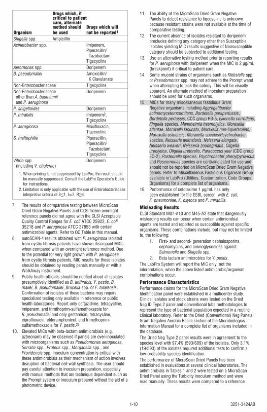
3251-3424AB1-10
Drugs which, if critical to patient care, alternate method should Drugs which will Organism be used not be reported1
Shigella spp. Ampicillin Acinetobacter spp. Imipenem, Piperacillin/ Tazobactam, TigecyclineAeromonas spp. Doripenem B. pseudomallei Amoxicillin/ K ClavulanateNon-Enterobacteriaceae TigecyclineNon-Enterobacteriaceae Doripenem other than A. baumannii and P. aeruginosa P. shigelloides DoripenemP. mirabilis Imipenem2, TigecyclineP. aeruginosa Moxifloxacin, Tigecycline S. maltophilia Piperacillin, Piperacillin/ Tazobactam, TigecyclineVibrio spp. Doripenem (including V. cholerae) 1. When printing is not suppressed by LabPro, the result should
be manually suppressed. Consult the LabPro Operator’s Guide for instructions.
2. Limitation is only applicable with the use of Enterobacteriaceae interpretive criteria of S≤1, I=2, R≥4.
7. The results of comparative testing between MicroScan Dried Gram Negative Panels and CLSI frozen overnight reference panels did not agree with the CLSI Acceptable Quality Control Ranges for E. coli ATCC 25922, E. coli 35218 and P. aeruginosa ATCC 27853 with certain antimicrobial agents. Refer to QC Table in this manual.
8. autoSCAN-4 results obtained with P. aeruginosa isolated from cystic fibrosis patients have shown discrepant MICs when compared with an overnight reference method. Due to the potential for very light growth with P. aeruginosa from cystic fibrosis patients, MIC results for these isolates should be obtained by reading panels manually or with a WalkAway instrument.
9. Public health officials should be notified about all isolates presumptively identified as B. anthracis, Y. pestis, B. mallei, B. pseudomallei, Brucella spp. or F. tularensis. Confirmation of isolates of these bacteria may require specialized testing only available in reference or public health laboratories. Report only ceftazidime, tetracycline, imipenem, and trimthoprim-sulfamethoxazole for B. pseudomallei and only gentamicin, tetracycline, ciprofloxacin, chloramphenicol, and trimethoprim-sulfamethoxazole for Y. pestis.26
10. Elevated MICs with beta-lactam antimicrobials (e.g. aztreonam) may be observed if panels are over-inoculated with microorganisms such as Pseudomonas aeruginosa, Serratia spp., Proteus spp., Morganella spp., and Providencia spp. Inoculum concentration is critical with these antimicrobials as their mechanism of action involves disruption of bacterial cell wall synthesis. The user should pay careful attention to inoculum preparation, especially with manual methods that are technique dependent such as the Prompt system or inoculum prepared without the aid of a photometric device.
11. The ability of the MicroScan Dried Gram Negative Panels to detect resistance to tigecycline is unknown because resistant strains were not available at the time of comparative testing.
12. The current absence of isolates resistant to doripenem precludes defining any category other than Susceptible. Isolates yielding MIC results suggestive of Nonsusceptible category should be subjected to additional testing.
13. Use an alternative testing method prior to reporting results for P. aeruginosa with doripenem when the MIC is 2 µg/mL (breakpoint) if critical to patient care.
14. Some mucoid strains of organisms such as Klebsiella spp. or Pseudomonas spp. may not adhere to the Prompt wand when attempting to pick the colony. This will be visually apparent. An alternate method of inoculum preparation should be used for such organisms.
15. MICs for many miscellaneous fastidious Gram Negative organisms including Aggregatibacter actinomycetemcomitans, Bordetella parapertussis, Bordetella pertussis, CDC group HB-5, Eikenella corrodens, Kingella species, Mannheimia haemolytica, Moraxella atlantae, Moraxella lacunata, Moraxella non-liquefaciens, Moraxella osloensis, Moraxella species/Psychrobacter species, Neisseria animaloris, Neisseria elongata, Neisseria weaveri, Neisseria zoodegmatis, Oligella ureolytica, Oligella urethralis, Paracoccus yeei (CDC group EO-2), Pasteurella species, Psychrobacter phenylpyruvicus and Roseomonas species are contraindicated for use and should not be reported on MicroScan Dried Gram Negative panels. Refer to Miscellaneous Fastidious Organism Group available in LabPro (Utilities, Customization, Code Groups, Organisms) for a complete list of organisms.
16. Performance of cefotaxime 1 µg/mL has only been established for the ESBL screen with E. coli, K. pneumoniae, K. oxytoca and P. mirabilis.
Misleading ResultsCLSI Standard M07-A10 and M45-A2 state that dangerously misleading results can occur when certain antimicrobial agents are tested and reported as susceptible against specific organisms. These combinations include, but may not be limited to, the following:
1. First- and second- generation cephalosporins, cephamycins, and aminoglycosides against Salmonella and Shigella spp.
2. Beta lactam antimicrobics for Y. pestis. The LabPro System will report the MIC only, not the interpretation, when the above listed antimicrobic/organism combinations occur.Performance CharacteristicsPerformance claims for the MicroScan Dried Gram Negative Identification panel were established in a multicenter study. Clinical isolates and stock strains were tested on the Dried Neg ID Type 2 panel and conventional tube methodologies to represent the type of bacterial population expected in a routine clinical laboratory. Refer to the Dried (Conventional) Neg Panels: Gram-Negative Aerobic Bacilli section of the Microbiologics Information Manual for a complete list of organisms included in the database.The Dried Neg Type 2 panel results were in agreement to the species level with 97.4% (593/609) of the isolates. Only 3.1% (19/593) of the isolates required additional tests to confirm a low-probability species identification. The performance of MicroScan Dried Panels has been established in evaluations at several clinical laboratories. The antimicrobials in Tables 1 and 2 were tested on a MicroScan Dried Panel using the Turbidity inoculum method and were read manually. These results were compared to a reference
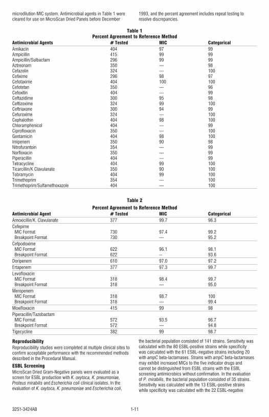
3251-3424AB 1-11
microdilution MIC system. Antimicrobial agents in Table 1 were cleared for use on MicroScan Dried Panels before December
1993, and the percent agreement includes repeat testing to resolve discrepancies.
Table 1Percent Agreement to Reference Method
Antimicrobial Agents # Tested MIC CategoricalAmikacin 404 97 99 Ampicillin 415 99 99 Ampicillin/Sulbactam 296 99 99 Aztreonam 350 --- 98 Cefazolin 324 --- 100 Cefixime 296 98 97 Cefotaxime 404 100 100 Cefotetan 350 --- 96 Cefoxitin 404 --- 99 Ceftazidime 300 95 98 Ceftizoxime 324 99 100 Ceftriaxone 300 94 99 Cefuroxime 324 --- 100 Cephalothin 404 98 100 Chloramphenicol 404 --- 99 Ciprofloxacin 350 --- 100 Gentamicin 404 98 100 Imipenem 350 90 98 Nitrofurantoin 354 --- 99 Norfloxacin 350 --- 99 Piperacillin 404 --- 99 Tetracycline 404 99 100 Ticarcillin/K Clavulanate 350 90 100 Tobramycin 404 99 100 Trimethoprim 354 --- 100 Trimethoprim/Sulfamethoxazole 404 --- 100
Table 2Percent Agreement to Reference Method
Antimicrobial Agent # Tested MIC Categorical Amoxicillin/K. Clavulanate 377 99.7 96.3 Cefepime MIC Format 730 97.4 99.2 Breakpoint Format 730 --- 95.2 Cefpodoxime MIC Format 622 96.1 98.1 Breakpoint Format 622 -- 93.6 Doripenem 610 97.0 97.2Ertapenem 377 97.3 99.7 Levofloxacin MIC Format 318 98.4 99.7 Breakpoint Format 318 --- 95.0 Meropenem MIC Format 318 98.7 100 Breakpoint Format 318 --- 99.4 Moxifloxacin 415 99 98 Piperacillin/Tazobactam MIC Format 572 93.5 96.7 Breakpoint Format 572 --- 94.8 Tigecycline 382 99 98.7
ReproducibilityReproducibility studies were completed at multiple clinical sites to confirm acceptable performance with the recommended methods described in the Procedural Manual.ESBL ScreeningMicroScan Dried Gram-Negative panels were evaluated as a screen for ESBL production with K. oxytoca, K. pneumoniae, Proteus mirabilis and Escherichia coli clinical isolates. In the evaluation of K. oxytoca, K. pneumoniae and Escherichia coli,
the bacterial population consisted of 141 strains. Sensitivity was calculated with the 80 ESBL-positive strains while specificity was calculated with the 61 ESBL-negative strains including 20 with ampC beta-lactamases. Strains with ampC beta-lactamases may exhibit increased MICs to the five indicator drugs and cannot be distinguished from ESBL strains with the ESBL screening antimicrobics without confirmation. In the evaluation of P. mirabilis, the bacterial population consisted of 35 strains. Sensitivity was calculated with the 13 ESBL-positive strains while specificity was calculated with the 22 ESBL-negative
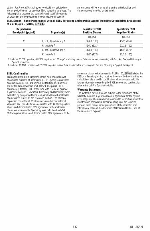
3251-3424AB1-12
strains. For P. mirabilis strains, only ceftazidime, cefotaxime, and cefpodoxime can be used for ESBL screening purposes. The following table presents the sensitivity and specificity results by organism and cefpodoxime breakpoints. Panel-specific
performance will vary, depending on the antimicrobics and concentrations included on the panel.
ESBL Screen - Panel Performance with all ESBL Screening Antimicrobial Agents including Cefpodoxime Breakpoints of 2 or 8 µg/mL (M100, 27th ed.)
Cefpodoxime Breakpoint (µg/mL) Organism(s)
Sensitivity ESBL Positive Strains
Specificity ESBL Negative Strains
No. (%) No. (%)2 E. coli, Klebsiella spp.1 80/80 (100) 40/61 (65.6)
P. mirabilis 2 12/13 (92.3) 22/22 (100)8 E. coli, Klebsiella spp.1 80/80 (100) 41/61 (67.2)
P. mirabilis 2 12/13 (92.3) 22/22 (100)
1. Includes 80 ESBL positive, 41 ESBL negative, and 20 ampC producing strains. Data also includes screening with Caz, Azt, Cax, and Cft using a 2 µg/mL breakpoint.
2. Includes 13 ESBL positive and 22 ESBL negative strains. Data also includes screening with Caz and Cft using a 2 µg/mL breakpoint.
ESBL Confirmation MicroScan Dried Gram-Negative panels were evaluated with streamlined dilutions of cefotaxime (2, 16 µg/mL), cefotaxime/clavulanic acid (0.5/4, 4/4 µg/mL), ceftazidime (1, 8 µg/mL) and ceftazidime/clavulanic acid (0.25/4, 2/4 µg/mL) as a confirmatory test for ESBL production with E. coli, K. oxytoca, K. pneumoniae and P. mirabilis. Sensitivity and Specificity were evaluated by comparing MicroScan panel MICs with molecular characterized results as the reference method. The bacterial population consisted of 95 strains evaluated at one external validation site. Sensitivity was calculated with 42 ESBL-positive strains and demonstrated 93% agreement to the molecular characterization results. Specificity was calculated with 53 ESBL-negative strains and demonstrated 96% agreement to the
molecular characterization results. CLSI M100, 27th ed. states that ESBL confirmatory testing requires the use of both cefotaxime and ceftazidime, alone and in combination with clavulanic acid. For further information regarding the ESBL screen and confirmation refer to the LabPro Operator's Guide.Warranty StatementThe system is covered by and subject to the provisions of the warranty included in your contractual agreement for the system or its reagents. The customer is responsible for routine preventive maintenance procedures. Repairs arising from the failure to perform these maintenance procedures at the indicated time intervals are made at the discretion of Beckman Coulter, and at the customer’s expense.
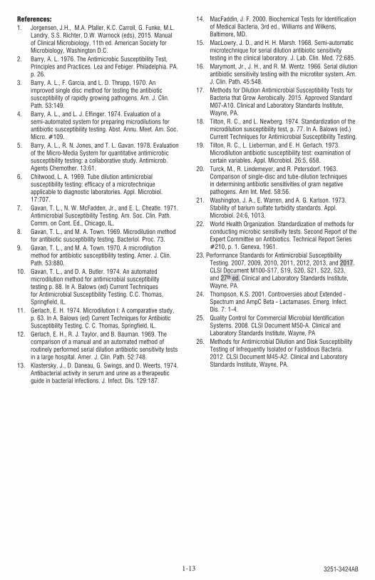
3251-3424AB1-13
References:1. Jorgensen, J.H., M.A. Pfaller, K.C. Carroll, G. Funke, M.L.
Landry, S.S. Richter, D.W. Warnock (eds), 2015. Manual of Clinical Microbiology, 11th ed. American Society for Microbiology, Washington D.C.
2. Barry, A. L. 1976. The Antimicrobic Susceptibility Test, Principles and Practices. Lea and Febiger. Philadelphia. PA. p. 26.
3. Barry, A. L., F. Garcia, and L. D. Thrupp, 1970. An improved single disc method for testing the antibiotic susceptibility of rapidly growing pathogens. Am. J. Clin. Path. 53:149.
4. Barry, A. L., and L. J. Effinger. 1974. Evaluation of a semi-automated system for preparing microdilutions for antibiotic susceptibility testing. Abst. Annu. Meet. Am. Soc. Micro. #109.
5. Barry, A. L., R. N. Jones, and T. L. Gavan. 1978. Evaluation of the Micro-Media System for quantitative antimicrobic susceptibility testing: a collaborative study. Antimicrob. Agents Chemother. 13:61.
6. Chitwood, L. A. 1969. Tube dilution antimicrobial susceptibility testing: efficacy of a microtechnique applicable to diagnostic laboratories. Appl. Microbiol. 17:707.
7. Gavan, T. L., N. W. McFadden, Jr., and E. L. Cheatle. 1971. Antimicrobial Susceptibility Testing. Am. Soc. Clin. Path. Comm. on Cont. Ed., Chicago, IL.
8. Gavan, T. L., and M. A. Town. 1969. Microdilution method for antibiotic susceptibility testing. Bacteriol. Proc. 73.
9. Gavan, T. L., and M. A. Town. 1970. A microdilution method for antibiotic susceptibility testing. Amer. J. Clin. Path. 53:880.
10. Gavan, T. L., and D. A. Butler. 1974. An automated microdilution method for antimicrobial susceptibility testing p. 88. In A. Balows (ed) Current Techniques for Antimicrobial Susceptibility Testing. C.C. Thomas, Springfield, IL.
11. Gerlach, E. H. 1974. Microdilution I: A comparative study, p. 63. In A. Balows (ed) Current Techniques for Antibiotic Susceptibility Testing. C. C. Thomas, Springfield, IL.
12. Gerlach, E. H., R. J. Taylor, and B. Bauman. 1969. The comparison of a manual and an automated method of routinely performed serial dilution antibiotic sensitivity tests in a large hospital. Amer. J. Clin. Path. 52:748.
13. Klastersky, J., D. Daneau, G. Swings, and D. Weerts, 1974. Antibacterial activity in serum and urine as a therapeutic guide in bacterial infections. J. Infect. Dis. 129:187.
14. MacFaddin, J. F. 2000. Biochemical Tests for Identification of Medical Bacteria, 3rd ed., Williams and Wilkens, Baltimore, MD.
15. MacLowry, J. D., and H. H. Marsh. 1968. Semi-automatic microtechnique for serial dilution antibiotic sensitivity testing in the clinical laboratory. J. Lab. Clin. Med. 72:685.
16. Marymont, Jr., J. H., and R. M. Wentz. 1966. Serial dilution antibiotic sensitivity testing with the microtiter system. Am. J. Clin. Path. 45:548.
17. Methods for Dilution Antimicrobial Susceptibility Tests for Bacteria that Grow Aerobically. 2015. Approved Standard M07-A10. Clinical and Laboratory Standards Institute, Wayne, PA.
18. Tilton, R. C., and L. Newberg. 1974. Standardization of the microdilution susceptibility test, p. 77. In A. Balows (ed.) Current Techniques for Antimicrobial Susceptibility Testing.
19. Tilton, R. C., L. Lieberman, and E. H. Gerlach. 1973. Microdilution antibiotic susceptibility test: examination of certain variables. Appl. Microbiol. 26:5, 658.
20. Turck, M., R. Lindemeyer, and R. Petersdorf. 1963. Comparison of single-disc and tube-dilution techniques in determining antibiotic sensitivities of gram negative pathogens. Ann Int. Med. 58:56.
21. Washington, J. A., E. Warren, and A. G. Karlson. 1973. Stability of barium sulfate turbidity standards. Appl. Microbiol. 24:6, 1013.
22. World Health Organization. Standardization of methods for conducting microbic sensitivity tests. Second Report of the Expert Committee on Antibiotics. Technical Report Series #210, p. 1. Geneva, 1961.
23. Performance Standards for Antimicrobial Susceptibility Testing. 2007, 2009, 2010, 2011, 2012, 2013, and 2017. CLSI Document M100-S17, S19, S20, S21, S22, S23, and 27th ed. Clinical and Laboratory Standards Institute, Wayne, PA.
24. Thompson, K.S. 2001. Controversies about Extended - Spectrum and AmpC Beta - Lactamases. Emerg. Infect. Dis. 7: 1-4.
25. Quality Control for Commercial Microbial Identification Systems. 2008. CLSI Document M50-A. Clinical and Laboratory Standards Institute, Wayne, PA
26. Methods for Antimicrobial Dilution and Disk Susceptibility Testing of Infrequently Isolated or Fastidious Bacteria. 2012. CLSI Document M45-A2. Clinical and Laboratory Standards Institute, Wayne, PA.

3251-3424AB 1-14

3251-3424AB1-15
© 2018 Beckman Coulter, Inc. All Rights Reserved.
05/2018
Spec: 3240-2844
Beckman Coulter, the stylized logo, and the Beckman Coulter product and service marks mentioned herein are trademarks or registered trademarks of Beckman Coulter, Inc. in the United States and other countries.
Beckman Coulter Eurocenter S.A.22, rue Juste-OlivierCase Postale 1044CH – 1260 Nyon 1, SwitzerlandTel: +41 (0) 22 365 36 11
Made in USABeckman Coulter, Inc.250 S. Kraemer Blvd.Brea, CA 92821 United Stateswww.beckmancoulter.com