Human mastoid periosteum-derived stem cells: promising candidates for skeletal tissue engineering
11
JOURNAL OF TISSUE ENGINEERING AND REGENERATIVE MEDICINE RESEARCH ARTICLE J Tissue Eng Regen Med 2008; 2: 136–146. Published online 31 March 2008 in Wiley InterScience (www.interscience.wiley.com) DOI: 10.1002/term.75 Human mastoid periosteum-derived stem cells: promising candidates for skeletal tissue engineering J. Ringe 1# *, I. Leinhase 2# , S. Stich 1 , A. Loch 3 , K. Neumann 4 , A. Haisch 5 , T. H¨ aupl 1 , R. Manz 6 , C. Kaps 4 and M. Sittinger 1 1 Tissue Engineering Laboratory and Berlin-Brandenburg Center for Regenerative Therapies, Department of Rheumatology and Clinical Immunology, Charit´ e-University Medicine Berlin, Tucholskystrasse 2, 10117 Berlin, Germany 2 Department of Trauma and Reconstructive Surgery, Charit´ e-University Medicine Berlin, Hindenburgdamm 30, 12200 Berlin, Germany 3 Department of Otorhinolaryngology, Charit´ e-University Medicine Berlin, Schumannstrasse 20/21, 10117 Berlin, Germany 4 TransTissue Technologies GmbH, Tucholskystrasse 2, 10117 Berlin, Germany 5 Department of Otorhinolaryngology, Charit´ e-University Medicine Berlin, Hindenburgdamm 30, 12200 Berlin, Germany 6 Humoral Immunology, German Rheumatism Research Centre, Schumannstrasse 21/22, 10117 Berlin, Germany Abstract Currently, mesenchymal stem cells (MSCs) are considered as the most eligible cells for skeletal tissue engineering. However, factors such as difficult stimulation and control of differentiation in vivo hamper their clinical use. In contrast, periosteum or periosteum-derived cells (PCs) are routinely clinically applied for bone and cartilage repair. PCs have often been named MSCs but, although cells of osteochondrogenic lineages arise from MSCs, it is unclear whether periosteum really contains MSCs. Our aim was to investigate the MSC-like character of PCs derived from the periosteum of mastoid bone. Harvesting of periosteum from mastoid bone is easy, so mastoid represents a good source for the isolation of PCs. Therefore, we analysed the MSC-like growth behaviour and the expression of embryonic, ectodermal, endodermal and mesodermal markers by microarray and FACS technology, and the multilineage developmental capacity of human PCs. Regarding clinical relevance, experiments were performed in human serum-supplemented medium. We show that PCs do not express early embryonic stem cell markers such as Oct4 and Nanog, or the marker of haematopoietic stem cells CD34, but express some other MSC markers. Osteogenesis resulted in the formation of calcified matrix, increased alkaline phosphatase activity, and induction of the osteogenic marker gene osteocalcin. Staining of proteoglycans and deposition of type II collagen documented chondrogenic development. As shown for the first time, adipogenic stimulation of mastoid-derived PCs resulted in the formation of lipid droplets and expression of the adipogenic marker genes aP2 and APM1. These results suggest MSC-like PCs from mastoid as candidates for therapy of complex skeletal defects. Copyright 2008 John Wiley & Sons, Ltd. Received 13 November 2007; Accepted 13 February 2008 Keywords human periosteal cells; mastoid; surface marker expression; multilineage potential; microarray; chondrogenesis; osteogenesis; adipogenesis; tissue engineering *Correspondence to: J. Ringe, Tissue Engineering Laboratory, Department of Rheumatology, Charit´ e-University Medicine Berlin, Tucholskystrasse 2, 10117 Berlin, Germany. E-mail: [email protected] # These authors contributed equally to this work. 1. Introduction Regenerative medicine approaches using autologous cells for bone and cartilage repair have progressed ‘from bench to bedside’ (Brittberg et al., 1994; Ossendorf et al., 2007; Schmelzeisen et al., 2003). Stem cells have become very prominent candidates for such cell therapy approaches Copyright 2008 John Wiley & Sons, Ltd.
Transcript of Human mastoid periosteum-derived stem cells: promising candidates for skeletal tissue engineering
JOURNAL OF TISSUE ENGINEERING AND REGENERATIVE MEDICINE R E S E A R C H A R T I C L EJ Tissue Eng Regen Med 2008; 2: 136–146.Published online 31 March 2008 in Wiley InterScience (www.interscience.wiley.com) DOI: 10.1002/term.75
Human mastoid periosteum-derived stem cells:promising candidates for skeletal tissueengineering
J. Ringe1#*, I. Leinhase2#, S. Stich1, A. Loch3, K. Neumann4, A. Haisch5, T. Haupl1, R. Manz6,C. Kaps4 and M. Sittinger1
1Tissue Engineering Laboratory and Berlin-Brandenburg Center for Regenerative Therapies, Department of Rheumatology and ClinicalImmunology, Charite-University Medicine Berlin, Tucholskystrasse 2, 10117 Berlin, Germany2Department of Trauma and Reconstructive Surgery, Charite-University Medicine Berlin, Hindenburgdamm 30, 12200 Berlin, Germany3Department of Otorhinolaryngology, Charite-University Medicine Berlin, Schumannstrasse 20/21, 10117 Berlin, Germany4TransTissue Technologies GmbH, Tucholskystrasse 2, 10117 Berlin, Germany5Department of Otorhinolaryngology, Charite-University Medicine Berlin, Hindenburgdamm 30, 12200 Berlin, Germany6Humoral Immunology, German Rheumatism Research Centre, Schumannstrasse 21/22, 10117 Berlin, Germany
Abstract
Currently, mesenchymal stem cells (MSCs) are considered as the most eligible cells for skeletal tissueengineering. However, factors such as difficult stimulation and control of differentiation in vivohamper their clinical use. In contrast, periosteum or periosteum-derived cells (PCs) are routinelyclinically applied for bone and cartilage repair. PCs have often been named MSCs but, although cellsof osteochondrogenic lineages arise from MSCs, it is unclear whether periosteum really containsMSCs. Our aim was to investigate the MSC-like character of PCs derived from the periosteum ofmastoid bone. Harvesting of periosteum from mastoid bone is easy, so mastoid represents a goodsource for the isolation of PCs. Therefore, we analysed the MSC-like growth behaviour and theexpression of embryonic, ectodermal, endodermal and mesodermal markers by microarray andFACS technology, and the multilineage developmental capacity of human PCs. Regarding clinicalrelevance, experiments were performed in human serum-supplemented medium. We show thatPCs do not express early embryonic stem cell markers such as Oct4 and Nanog, or the markerof haematopoietic stem cells CD34, but express some other MSC markers. Osteogenesis resultedin the formation of calcified matrix, increased alkaline phosphatase activity, and induction of theosteogenic marker gene osteocalcin. Staining of proteoglycans and deposition of type II collagendocumented chondrogenic development. As shown for the first time, adipogenic stimulation ofmastoid-derived PCs resulted in the formation of lipid droplets and expression of the adipogenicmarker genes aP2 and APM1. These results suggest MSC-like PCs from mastoid as candidates fortherapy of complex skeletal defects. Copyright 2008 John Wiley & Sons, Ltd.
Received 13 November 2007; Accepted 13 February 2008
Keywords human periosteal cells; mastoid; surface marker expression; multilineage potential;microarray; chondrogenesis; osteogenesis; adipogenesis; tissue engineering
*Correspondence to: J. Ringe, Tissue Engineering Laboratory,Department of Rheumatology, Charite-University MedicineBerlin, Tucholskystrasse 2, 10117 Berlin, Germany.E-mail: [email protected]# These authors contributed equally to this work.
1. Introduction
Regenerative medicine approaches using autologous cellsfor bone and cartilage repair have progressed ‘from benchto bedside’ (Brittberg et al., 1994; Ossendorf et al., 2007;Schmelzeisen et al., 2003). Stem cells have become veryprominent candidates for such cell therapy approaches
Copyright 2008 John Wiley & Sons, Ltd.
Human mastoid periosteum-derived stem cells 137
because of their pronounced expansion capacity, theirremarkable plasticity and, therefore, their potentialto regenerate complex tissue defects. For connectivetissue repair, adult stem cells derived from sourcessuch as adipose tissue (Zuk et al., 2001), synovialmembrane (De Bari et al., 2003), fetal membranes(Soncini et al., 2007), trabecular bone (Noth et al.,2002) and bone marrow (Caplan et al., 1994; Reyeset al., 2002) have been extensively characterized but arenot routinely used in the clinic so far. Despite manycell and molecular biological approaches characterizingmesenchymal stem cells (MSCs), their multilineagedifferentiation pathways, their immunoprivileged statusand their in vivo applicability (Barry and Murphy, 2004),very limited clinical studies have been performed (Picinichet al., 2007). Factors such as failure of stable ectopictissue formation, difficult stimulation and control ofdifferentiation in vivo and occasionally insufficient cellsurvival after seeding on a construct and subsequenttransplantation hamper their application.
In marked contrast to MSCs, periosteum as awhole or periosteum-derived progenitor cells (PCs)have been used in thousands of surgeries, e.g. as acovering layer to prevent chondrocyte leakage duringautologous chondrocyte implantation (ACI; Brittberget al., 1994), as a graft for reconstruction of the patellararticulation (Hoikka et al., 1990) and as tissue-engineeredbone transplant for maxillary sinus floor augmentation(Schmelzeisen et al., 2003). Interestingly, in contrast toMSCs, PCs are barely characterized at the cellular andmolecular level.
Periosteum plays a major role in bone growth, bonedevelopment, bone fracture healing and in corticalblood supply (Decker et al., 1996). The periosteum ofendochondral bones (bones formed by endochondralossification), and also of membranous bones (bonesformed by intramembranous ossification in which boneformation is not preceded by cartilage formation innormal development) consists of two layers. The outerlayer is fibrous, contains fibroblasts and abundantcollagen fibres and provides attachment of muscles,tendons and ligaments to the bone. The inner cambiallayer contains progenitor cells with osteogenic andchondrogenic capacity and is involved in bone healing(Buckwalter and Cooper, 1987; Nakahara et al., 1991;O’Driscoll and Fitzsimmons, 2001). Since the pioneerwork in the 1990s of A. Caplan, who has investigated theosteogenic potential of PCs in the field of bone repair, thecapacity of PCs to develop into bone and cartilage hasbeen evaluated in several studies (De Bari et al., 2001;Fukumoto et al., 2003; Groger et al., 2003; Nakase et al.,1993).
On the basis of their osteochondrogenic developmentalpotential, PCs have often been named ‘multipotent MSCs’.Really, although cells of osteogenic and chondrogeniclineages generally arise from mesenchymal stem cells(Marks and Popoff, 1988), it is unclear whetherperiosteum contains such stem cells. The fact thateven periosteum from membranous bones expresses
cartilage markers and differentiates into chondrocyteshas been interpreted in different ways, based ondistinct experimental results: chondrocytes may arisefrom bi- or multipotent stem cells, from a particularchondroprogenitor cell population, or from osteogeniccells (Fang and Hall, 1997). So far it has not beenshown that PCs have the same potential as MSCs toform all varieties of connective tissue cells. For instance,their adipogenic development is not well characterizedon the cellular and molecular level. Furthermore, veryfew data regarding the expression profile of embryonic,ectodermal, endodermal, mesodermal and MSC markershave been reported until now.
The aim of this study was to investigate the MSC-like character of human periosteal cells derived fromperiosteum of the endochondral mastoid bone. Harvestingof periosteum from mastoid bone is relatively easy and so,this tissue represents a good cell source for the isolationof PCs for regenerative medicine. We have studied theirexpansion capacity using clinically applied cell culturetechniques (e.g. human serum), their stem cell-relatedsurface marker expression profile applying genome-wide microarray and FACS analysis, their osteogenicand chondrogenic potential, and for the first time onthe cellular and molecular level their time course ofadipogenesis. We hypothesize, that periosteal cells ingeneral and thus, also from the mastoid bone, areeven more MSC-like then known so far and therefore,are very promising candidates for cell therapies ofcomplex skeletal tissue defects such as osteochondraldefects.
2. Materials and methods
2.1. Cell isolation and expansion
Periosteal autografts (0.5 cm2) were harvested from thehuman mastoid of 15 patients undergoing mastoidectomy.PCs were isolated as described previously (Zheng et al.,2006). Briefly, periosteal tissues were rinsed withPBS (Biochrom, Berlin, Germany) and Hanks’ solution(Biochrom), minced and digested in DMEM/Ham’s F12medium (Biochrom) containing 10 000 U/ml collagenaseII (Biochrom), 10% human allogeneic serum (GermanRed Cross, Berlin) and 1% antibiotic–antimycotic solution(Biochrom). The tissues were digested for 3 h at 37 ◦Cand the cells were subsequently harvested, resuspendedin DMEM/Ham’s F12 medium containing 10% humanallogeneic serum, plated and allowed to attach for4–10 days. Adherent growing PCs were cultured understandard cell culture conditions and the medium wasreplaced every 2–3 days. On reaching 90% confluence,the PCs were subcultured by treatment with 0.5%trypsin–EDTA (Biochrom) and subsequently replated ata density of 6000 cells/cm2. The study was approved bythe ethical committee of the Charite-University MedicineBerlin.
Copyright 2008 John Wiley & Sons, Ltd. J Tissue Eng Regen Med 2008; 2: 136–146.DOI: 10.1002/term
138 J. Ringe et al.
2.2. Genome-wide gene expression profiling
To analyse the expression of marker genes, total RNAfrom passage 2 (P2) PC cultures (n = 6) was isolated asdescribed previously (Chomczynski, 1993) and was usedfor genome-wide microarray analysis with the AffymetrixHG-U133 plus 2.0 array (Affymetrix, Santa Clara,USA) according to the manufacturer’s recommendations.Briefly, cDNA was synthesized from 1 µg total RNA andsubmitted to in vitro transcription (ENZO Biochem, NewYork, USA) to generate biotin-labelled complementaryRNA (cRNA); 15 µg of fragmented cRNA were hybridizedto gene chips for 16 h at 45 ◦C. The gene chips werewashed and stained as recommended (fluidics station)and scanned using the GeneArray scanner. AffymetrixGCOS 1.4 software was used to generate DAT, CELand EXP files and to process and normalize the rawdata for signal and detection call calculation. Wewere exclusively interested in genes that were alreadypublished as stem cell markers. Out of the hugenumber of such candidates, we selected those geneswhose detection call was ‘present’ or ‘absent’ on all sixmicroarrays (100% ‘present’ or 100% ‘absent’). Fromthese selected candidates the mean signal values andmean standard deviations (SD) of the signal values werecalculated.
2.3. Flow cytometric analysis (FACS)
Single cell suspensions of PCs (n = 3, P4) were washed inPBS/0.5% BSA. The cells were incubated with titratedprimary staining reagents for 15 min on ice (2.5 ×10⁵ cells/0.1 ml in PBS/0.5% BSA). Primary stainingreagent unlabelled monoclonal mouse anti-human SH2(CD105) was provided by the German RheumatismResearch Center. Fluorescein isothiocyanate (FITC)-labelled mouse anti-human CD44, CD45 and CD90, andR-phycoerythrin (PE)-labelled mouse anti-human SH3(CD73), CD14 and ALCAM (CD166) were purchased fromPharmingen (Heidelberg, Germany). Monoclonal mouseanti-human CD34 labelled to PE was purchased fromMiltenyi (Bergisch Gladbach, Germany). FITC-labelledmonoclonal mouse anti-human Stro-1 was a kind giftfrom Dr Pierre Charbord (Inserm, Tours, France). Fordouble staining with SH2, the cells were incubatedwith unlabelled SH2, washed with PBS/0.5% BSA andincubated with biotinylated goat anti-mouse IgG for10 min on ice. After washing again and incubation onice with streptavidin coupled to cytochrome 5 (Cy5)for an additional 10 min, the cells were washed againand finally incubated with PE or FITC antibodies specificfor a second surface molecule, such as SH3, CD14,CD34, CD44, CD45 CD90 or CD166. Prior to analysisin a LSR I cytometer (Heidelberg), the cell sampleswere washed. Dead cells and debris were stainedwith PI (Sigma, Taufkirchen, Germany) and excluded.Data were evaluated using CellQuest software (Becton-Dickinson).
2.4. Cell differentiation studies
The differentiation potential of human PCs (n = 3, P4)was demonstrated by culturing these cells under con-ditions that promote mesenchymal stem cell differenti-ation (Pittenger et al., 1999). To induce osteogenesis,6000 PCs/cm2 were seeded and induced in DMEM/Ham’sF12 (5% human serum) containing 100 nM dexametha-sone (Dex; Sigma), 0.05 mM L-ascorbic acid 2-phosphate(AsAP; Sigma), 10 mM β-glycerophosphate (Sigma) and1.5 mM calcium chloride dihydrate (Sigma). For chondro-genesis, PCs were centrifuged to form a micromass andwere cultured in a defined medium consisting of DMEM(Biochrom), ITS+1 (Sigma), 100 nM Dex, 0.17 mM AsAPand 10 ng/ml transforming growth factor-β1 (TGFβ1)or TGFβ3 (R&D Systems, Wiesbaden, Germany). Toinduce adipogenesis the PCs were treated with induc-tion medium, consisting of DMEM supplemented with10% human serum, 1 µM Dex, 0.2 mM indomethacin(Sigma), 10 µg/ml insulin (Novo Nordisk, Mainz, Ger-many), 0.5 mM 3-isobutyl-1-methylxanthine (Sigma), andmaintenance medium containing DMEM, human serumand 10 µg/ml insulin, within three cycles (3 days induc-tion, 2 days maintenance).
2.5. Polymerase chain reaction
To demonstrate osteogenesis and adipogenesis at themRNA level, total RNA was isolated (Chomczynski,1993). Subsequently, 5 µg total RNA were reverse-transcribed after annealing with 500 ng oligo-(dT)primers (Gibco, Karlsruhe, Germany) and 5 U Superscriptreverse transcriptase (Gibco) in 70 µl reaction mixture.The housekeeping gene GAPDH was used to normalizemarker gene expression in each sample in differentconcentrations. Real-time PCR applying the BioRadiCycler (BioRad, Munich, Germany) was performed with1 µl cDNA sample, using the SYBR Green PCR CoreKit (Applied Biosystems, Darmstadt, Germany). Relativequantitation of marker genes (Table 1) was performed asdescribed (ABI Prism 7700, 1997) and the results givenas percentages of the GAPDH product.
2.6. Histological methods andimmunohistochemistry
During osteogenesis, cells express high levels of alkalinephosphatase, which was visualized by staining with Sigmafast BCIP/NBT (Sigma). von Kossa staining demonstratedthe deposition of a bone-specific mineralized matrix.Chondrogenesis was documented by histological stainingof cartilage proteoglycans with Alcian blue 8GS (Roth,Karlsruhe) as well as by immunohistochemical methodsusing the EnVision + System, Peroxidase MouseKit (Dako, Hamburg, Germany). Cryosections (6 µm)were incubated for 1 h with primary antibodies (rabbitanti-human types I and II collagen; DPC Biermann,
Copyright 2008 John Wiley & Sons, Ltd. J Tissue Eng Regen Med 2008; 2: 136–146.DOI: 10.1002/term
Human mastoid periosteum-derived stem cells 139
Table 1. Oligonucleotide sequences
Gene Accession No. Oligonucleotides (5′ → 3′) (up/down) Product size (bp)
GAPDH XM 006959 GGC GAT GCT GGC GCT GAG TAC 149TGG TCC ACA CCC ATG ACG A
Collagen type Ia1 NM 000088 CGA TGG CTG CAC GAG TCA CAC 180CAG GTT GGG ATG GAG GGA GTT TAC
Osteocalcin X 51699 GAG CCC CAG TTC CCC TAC CC 103GCC TCC TGA AAG CCG ATG TG
aP2 NM 000134 CCT TAG ATG GGG GTG TCC TGG TA 156AAT GTC CCT TGG CTT ATG CTC TC
APM1 NM 004797 GGC CTG CCC AGC TCT CGT AT 83CTC TCC TCT TTG GGC ATC ACC
bp, base pairs.
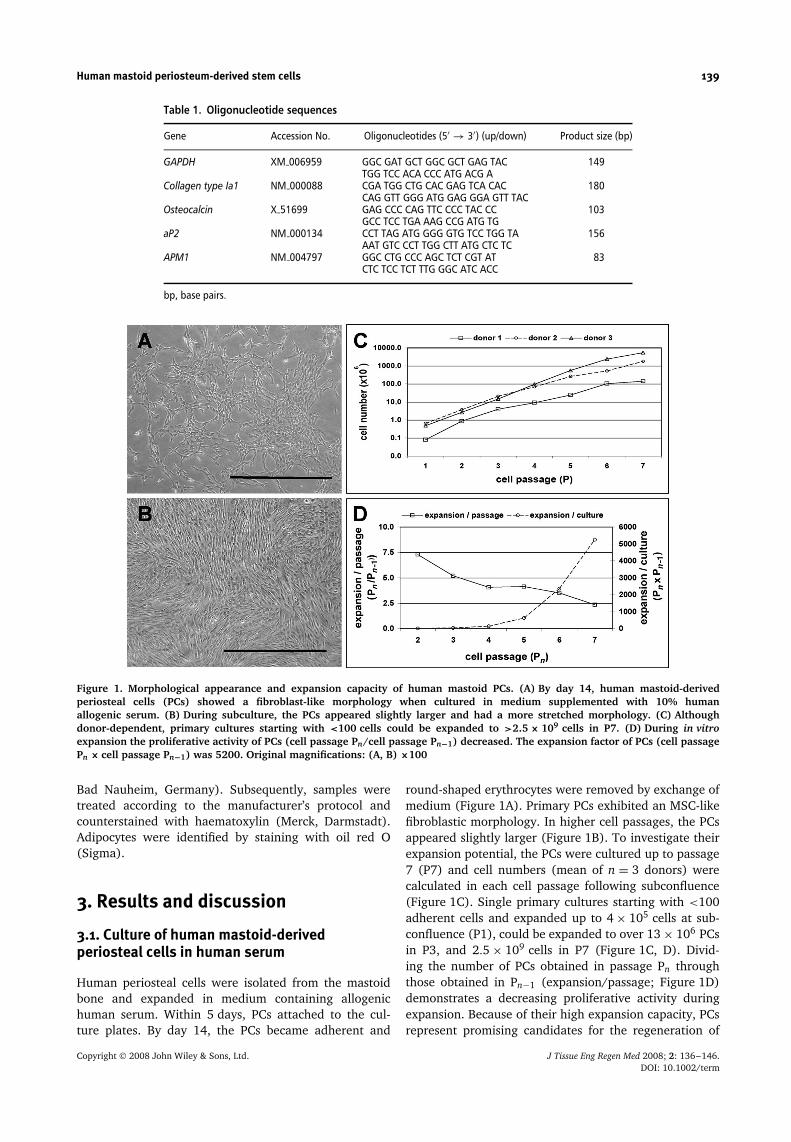
Figure 1. Morphological appearance and expansion capacity of human mastoid PCs. (A) By day 14, human mastoid-derivedperiosteal cells (PCs) showed a fibroblast-like morphology when cultured in medium supplemented with 10% humanallogenic serum. (B) During subculture, the PCs appeared slightly larger and had a more stretched morphology. (C) Althoughdonor-dependent, primary cultures starting with <100 cells could be expanded to >2.5 × 10⁹ cells in P7. (D) During in vitroexpansion the proliferative activity of PCs (cell passage Pn/cell passage Pn−1) decreased. The expansion factor of PCs (cell passagePn × cell passage Pn−1) was 5200. Original magnifications: (A, B) ×100
Bad Nauheim, Germany). Subsequently, samples weretreated according to the manufacturer’s protocol andcounterstained with haematoxylin (Merck, Darmstadt).Adipocytes were identified by staining with oil red O(Sigma).
3. Results and discussion
3.1. Culture of human mastoid-derivedperiosteal cells in human serum
Human periosteal cells were isolated from the mastoidbone and expanded in medium containing allogenichuman serum. Within 5 days, PCs attached to the cul-ture plates. By day 14, the PCs became adherent and
round-shaped erythrocytes were removed by exchange ofmedium (Figure 1A). Primary PCs exhibited an MSC-likefibroblastic morphology. In higher cell passages, the PCsappeared slightly larger (Figure 1B). To investigate theirexpansion potential, the PCs were cultured up to passage7 (P7) and cell numbers (mean of n = 3 donors) werecalculated in each cell passage following subconfluence(Figure 1C). Single primary cultures starting with <100adherent cells and expanded up to 4 × 10⁵ cells at sub-confluence (P1), could be expanded to over 13 × 106 PCsin P3, and 2.5 × 10⁹ cells in P7 (Figure 1C, D). Divid-ing the number of PCs obtained in passage Pn throughthose obtained in Pn−1 (expansion/passage; Figure 1D)demonstrates a decreasing proliferative activity duringexpansion. Because of their high expansion capacity, PCsrepresent promising candidates for the regeneration of
Copyright 2008 John Wiley & Sons, Ltd. J Tissue Eng Regen Med 2008; 2: 136–146.DOI: 10.1002/term
140 J. Ringe et al.
large defects. In our own studies, we have shown thatthe osteogenic potential of PCs is reproducibly stableuntil cell passage 6 (data not shown). In addition, inin vitro (Zheng et al., 2006) and in vivo (Schmelzeisenet al., 2003) studies, we observed that bone transplantsof 0.5 cm2 containing 2.5 × 10⁶ PCs are suitable for theregeneration of bone defects. From 0.2 cm2 small biopsies,at P4 we isolated about 60 × 106 PCs, at P5 280 × 106.Extrapolated, after P4 theoretically a defect area of 12 cm2
and after P5 of 56 cm2 might be filled. However, vascu-larization of the repair tissue has to be assured, whichis still a main problem in most approaches regarding theregeneration of large defects. As expected for primarycells, the number of PCs obtained strongly varied fromdonor to donor. Circumstances such as the periosteumharvesting procedure, cell isolation by enzymatic diges-tion and donor age and state influence the number andalso the developmental potential of PCs and other mes-enchymal cells (Brownlow et al., 2000; O’Driscoll et al.,1999). From about 15% of the biopsies, no cells could beisolated. This may be partly due to the remaining PCs con-taining cambial layer on the bone during mastoidectomy.Enhanced techniques for tissue harvest, such as hydraulicelevation (Marini et al., 2004) or the use of a raspatory,ensures the harvest of the cambial layer. Nevertheless, P7mastoid-derived PCs showed a 6000-fold increased cellnumber compared to primary cultures. Interestingly, forhuman bone marrow-derived MSCs, a similar expansionpotential has been reported (Haynesworth et al., 1992).In contrast, Agata et al. (2007) reported a significantlyhigher proliferation potential of mandibular-derived PCsthan that of MSCs. However, the average age of theperiosteum donors was 20 years and of the bone mar-row donors 37 years, indicating an age-related effect. A
progressive age-associated decline in the growth rate ofPCs is known from other studies (De Bari et al., 2006).Regarding cell therapy applications, besides their impress-ing expansion capacity it is important that PCs, as shownin our study, grow in allogenic human serum, main-taining their developmental capacity. In contrast, MSCsadherence and growth strongly depends on proper serumbatches (Lennon et al., 1996).
3.2. Mastoid PCs express mesenchymal stemcell-related surface marker
Genome-wide Affymetrix HG-U133 plus 2.0 array analysiswas performed (n = 6 donors) to investigate whether PCsexpress early embryonic, ectodermal, endodermal andmesodermal genes and haematopoietic and mesenchymalstem cell markers. In summary (see Table 2 andSupplementary Material), they do express embryonicstem cell marker genes such as CD9 (100% detectedas present), but do not express the most prominent earlymarkers, such as the transcription factor Oct4/POU5F1,NANOG, SSEA4/SIAT6 and TERT (100% detected asabsent). In contrast, subpopulations of human bonemarrow MSCs express Oct4/Pou5F1, TERT and distinctother ES genes (Pochampally et al., 2004), indicatinga more ‘primitive’ character of MSCs. MSCs obtainedfrom amniotic fluid also express Oct4/Pou5F1 (Tsai et al.,2004) and human term placenta-derived MSCs expressSSEA4/SIAT6 (Yen et al., 2005). Like human MSCs(Tremain et al., 2001), PCs also express marker genesof cells present in the three germ layers. This impliesa broad developmental potential of mastoid-derivedPCs. Nevertheless, most of the prominent ectodermal,
Table 2. Marker genes expressed by human PCs (microarray data, n = 6 donors)
Marker genes expressed by PC expression profile
Pluripotent embryonic stemcells (ES)
Present: CD9, COMMD3, DIAPH2, GJA1, IFITM1, IFITM2, IL6ST, NUMB, PTEN, sFRP2
Absent: CLDN6, CFC1, FGF4, GABRB3, GDF3, Lefty1, Lefty2, LIN28, NANOG, NR5A2, Oct-4/POU5F1, REX1, SOX1,SSEA4/SIAT6, TERT, UTF1
Ectoderm Present: CRABP2, nestin, TUBB3, vimentinAbsent: ISL1, MAP2, MSI1, NeuroD1, Olig2, PAX6, SOX18, synaptophysin
Endoderm Present: decorin, fibronectin1, GATA6, laminin-γ 1, PDHX, Smad2Absent: α-fetoprotein, FGF8, FOXA2, GATA4, glucagon, HHEX, HNF4A, IAPP, insulin, laminin-α1, nodal, Otx2,PAX4, SOX17, TAT, Wnt3
Mesoderm Present: Col I, MSX1, CBFA1Absent: α-actin, Col II, desmin, haemoglobin-β, haemoglobin-ξ , HLXB9, NPPA, WT1
Multipotent haematopoieticstem cells (HSCs)
Absent: CD14, CD34, CD45, CDCP1, ITGAL, ITGAM
Present:Extracellular matrix components: Col-I, Col-III, Col-IV, Col-V, Col-VI, Col-VII, Col-VIII, Col-XI, Col-XII, Col-XIII,Col-XIV, Col-XVI, perlecan, versican, syndecan, hyaluronan and proteoglycans, link protein 3, fibronectin, lamininGrowth factors and cytokines: IL-6, IL-7, IL-8, M-CSF, LIF, SCF, TGFβ1, TGFβ2
Multipotent mesenchymalstem cells (MSCs)
Matrix receptors: ICAM3, VCAM1, LFA3
Growth factors and cytokine receptors: IL-1R, IL-9R, IL-10R, IL-11R, IL-13R, PDGFR, TNFRSF-1, TNFRSF-10,TNFRSF-11, TNFRSF-12, FAS, TGFβ1R, TGFβ2R, TGFβ3R, IFNγ R1, IFNγ R 2, EGFRSurface antigens: CD44, CD58, CD71, CD73, CD90, CD105, CD166, HLA-ABCIntegrins: α4, α5, α7, α10, β1, β3, β5 chains
Further details of ES, ectodermal, endodermal, mesodermal, HSC and MSC expression can be found, for example, in the following references: ES,ectodermal, endodermal, mesodermal: Adewumi et al., 2007; Brandenberger et al., 2004; Chiu and Rao, 2003; Ginis et al., 2004: HSCs, Wognumet al., 2003; MSC: Barry and Murphy, 2004; Haynesworth et al., 1996; Majumdar et al., 1998; Pittenger et al., 1999.
Copyright 2008 John Wiley & Sons, Ltd. J Tissue Eng Regen Med 2008; 2: 136–146.DOI: 10.1002/term
Human mastoid periosteum-derived stem cells 141
endodermal and mesodermal marker genes could notbe detected. PCs showed no expression of typicalhaematopoietic stem cell surface antigens such as CD11a,CD11b and the early haematopoietic stem cell markerCD34. This is in concordance with the expression profile ofthese markers by MSCs (Pittenger et al., 1999). Moreover,PCs showed a similar but not identical expression ofmarker genes also expressed by mesenchymal stem cells(see Table 2 and Supplementary Material): they expressa broad pattern of: (a) cell surface antigens, such asthe hyaluronan receptor CD44, the transferrin receptorCD71, the ecto-5′-nucleotidase (SH3, CD73), the Thy-1cell surface antigen CD90, the TGFβ1 and -β3 receptorendoglin (SH2, CD105) and the activated leukocytecell adhesion molecule (ALCAM, CD166); (b) growthfactors, cytokines and their receptors; (c) extracellularmatrix components, such as collagens, fibronectin andlaminin; and (4) integrins. However, compared withMSCs, PCs clearly expresses fewer interleukins, integrins,growth factors and their receptors. The expressionof such molecules underlines the role of MSCs inthe bone marrow for formation and function of thestromal microenvironment, which produces inductive andregulatory signals not only for MSCs but also for thedevelopment of haematopoietic progenitors and othernon-mesenchymal stromal cells (Majumdar et al., 1998).This implies a more restricted function of PCs limited tobone remodelling and repair. Interestingly, both PCs andMSCs express CD44, a receptor that plays an importantrole in the organization of the extracellular matrix in bonemarrow or in bone, respectively (Yamazaki et al., 1999).
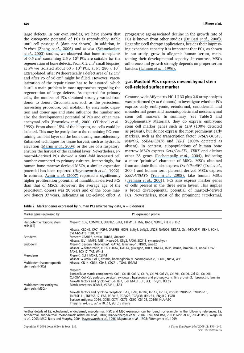
To verify some of the microarray data and to fur-ther characterize mastoid-derived PCs with respect totheir MSCs resemblance, cells were FACS analysed(n = 3 donors) for the presentation of typical human MSCmarker epitopes (Pittenger et al., 1999). Analysis of P4PCs showed a homogeneous cell population (Figure 2).They were uniformly negative for cell epitopes of the
lipopolysaccharid receptor CD14 (Figure 2A), the leuko-cyte common antigen CD45 (Figure 2A, D), and CD34(Figure 2B). In addition, they were negative for Stro-1(95%) (Figure 2F). In contrast, periosteal cells showed ahigh presentation of the MSC markers CD44 (hyaluronanreceptor; Figure 2C), CD73 (SH3, ecto-5′-nucleotidase;Figure 2E, F), CD90 (Thy-1; Figure 2G), CD166 (ALCAM;Figure 2H) and CD105 (SH2; Figure 2B–E, G, H). A simi-lar FACS profile has also been reported for periosteal cells,e.g. derived from periosteum of the distal insertion of adissected semitendinosus tendon (Sakaguchi et al., 2005),for mesenchymal progenitor cells derived from trabecularbone (Tuli et al., 2003), from term placenta (Yen et al.,2005) and from adipose tissue (Lee et al., 2004). Thefrequency of Stro-1, originally suggested as marker forhuman MSCs (Gronthos et al., 1999), was <5%. Low PCexpression of this marker was shown in one current inves-tigation (Sakaguchi et al., 2005) and was also observedin some studies using human MSCs (Colter et al., 2001).However, Stro-1 antigen is progressively lost during cellculture (Stewart et al., 1999).
3.3. Multilineage potential: osteogenic andchondrogenic development
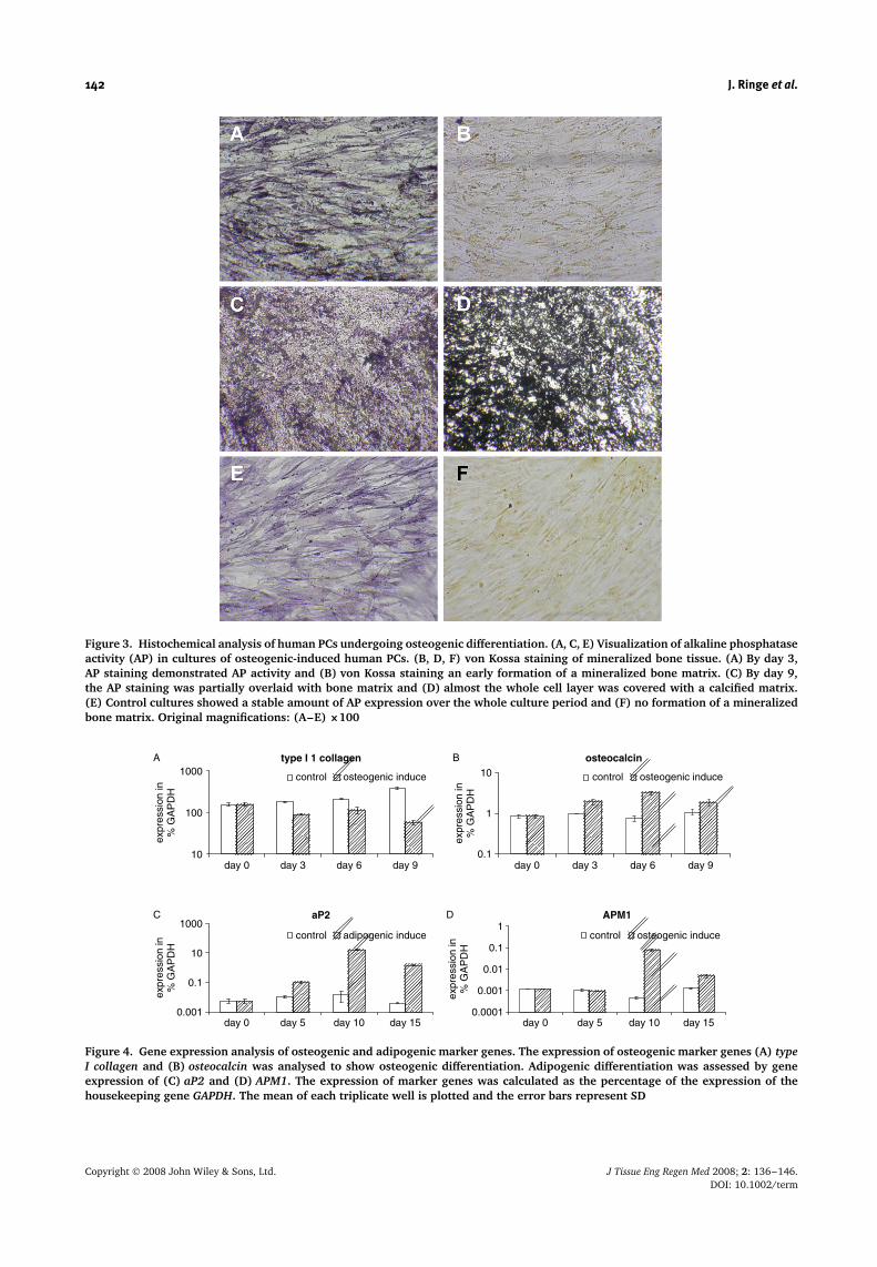
To address the question of whether mastoid-derivedperiosteal cells, like bone marrow MSCs, can developinto the osteochondrogenic and adipogenic lineage, PCs(n = 3, P4) were cultured under conditions that werefavourable for osteogenic, chondrogenic and adipogenicdevelopment of human MSCs (Pittenger et al., 1999).Osteogenesis was documented by visualization of alkalinephosphatase (AP) activity (Figure 3A, C, E), by matrixdeposition of calcium (Figure 3B, D, F) and by real-timePCR analysis (Figure 4). During osteogenic stimulation,the cells remained confluent and formed multilayerclusters of mineralized matrix. AP and von Kossa stainingof induced PCs demonstrated AP activity and an early
Figure 2. Flow cytometric analysis of human mastoid-derived periosteal cells. (A–H) Analysis of surface marker routinely used forthe characterization of human bone marrow-derived mesenchymal stem cells (MSCs) demonstrated that, similar to MSCs, humanmastoid-derived PCs were negative for reactivity to antigens CD14, CD34 and CD45 but positive for reactivity to antigens SH2(CD105), CD44, SH3 (CD73), CD90 and ALCAM (CD166). Unlike MSCs, PCs were more or less negative for Stro-1
Copyright 2008 John Wiley & Sons, Ltd. J Tissue Eng Regen Med 2008; 2: 136–146.DOI: 10.1002/term
142 J. Ringe et al.
Figure 3. Histochemical analysis of human PCs undergoing osteogenic differentiation. (A, C, E) Visualization of alkaline phosphataseactivity (AP) in cultures of osteogenic-induced human PCs. (B, D, F) von Kossa staining of mineralized bone tissue. (A) By day 3,AP staining demonstrated AP activity and (B) von Kossa staining an early formation of a mineralized bone matrix. (C) By day 9,the AP staining was partially overlaid with bone matrix and (D) almost the whole cell layer was covered with a calcified matrix.(E) Control cultures showed a stable amount of AP expression over the whole culture period and (F) no formation of a mineralizedbone matrix. Original magnifications: (A–E) ×100
10
100
1000
day 0 day 3 day 6 day 9 day 0 day 3 day 6 day 9
day 0 day 5 day 10 day 15 day 0 day 5 day 10 day 15
expr
essi
on in
% G
AP
DH
expr
essi
on in
% G
AP
DH
expr
essi
on in
% G
AP
DH
expr
essi
on in
% G
AP
DH
control osteogenic induce control osteogenic induce
control osteogenic inducecontrol adipogenic induce
0.0001
0.001
0.01
0.1
1
1
0.1
10
A type I 1 collagen osteocalcin
APM1aP2
0.001
0.1
10
1000
B
DC
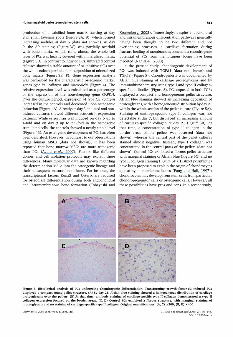
Figure 4. Gene expression analysis of osteogenic and adipogenic marker genes. The expression of osteogenic marker genes (A) typeI collagen and (B) osteocalcin was analysed to show osteogenic differentiation. Adipogenic differentiation was assessed by geneexpression of (C) aP2 and (D) APM1. The expression of marker genes was calculated as the percentage of the expression of thehousekeeping gene GAPDH. The mean of each triplicate well is plotted and the error bars represent SD
Copyright 2008 John Wiley & Sons, Ltd. J Tissue Eng Regen Med 2008; 2: 136–146.DOI: 10.1002/term
Human mastoid periosteum-derived stem cells 143
production of a calcified bone matrix starting at day3 in small layering spots (Figure 3A, B), which formedincreasing nodules at day 6 (data not shown). At day9, the AP staining (Figure 3C) was partially overlaidwith bone matrix. At this time, almost the whole celllayer of PCs was heavily covered with mineralized matrix(Figure 3D). In contrast to induced PCs, untreated controlcultures showed a stable amount of AP-positive cells overthe whole culture period and no deposition of mineralizedbone matrix (Figure 3E, F). Gene expression analysiswas performed for the characteristic osteogenic markergenes type Ia1 collagen and osteocalcin (Figure 4). Therelative expression level was calculated as a percentageof the expression of the housekeeping gene GAPDH.Over the culture period, expression of type Ia1 collagenincreased in the controls and decreased upon osteogenicinduction (Figure 4A). Already on day 3, induced and non-induced cultures showed different osteocalcin expressionpatterns. While osteocalcin was induced on day 6 up to4-fold and on day 9 up to 2.5-fold in the osteogenicstimulated cells, the controls showed a nearly stable level(Figure 4B). An osteogenic development of PCs has oftenbeen described. However, in contrast to our observationsusing human MSCs (data not shown), it has beenreported that bone marrow MSCs are more osteogenicthan PCs (Agata et al., 2007). Factors like differentdonors and cell isolation protocols may explain thesedifferences. Many molecular data are known regardingthe determination MSCs into the osteogenic lineage andtheir subsequent maturation to bone. For instance, thetranscriptional factors Runx2 and Osterix are requiredfor osteoblast differentiation during both endochondraland intramembranous bone formation (Kobayashi and
Kronenberg, 2005). Interestingly, despite endochondraland intramembranous differentiation pathways generallyhaving been thought to be two different and notoverlapping processes, a cartilage formation duringfracture healing of membranous bone and a chondrogenicpotential of PCs from membranous bones have beenreported (Nah et al., 2000).
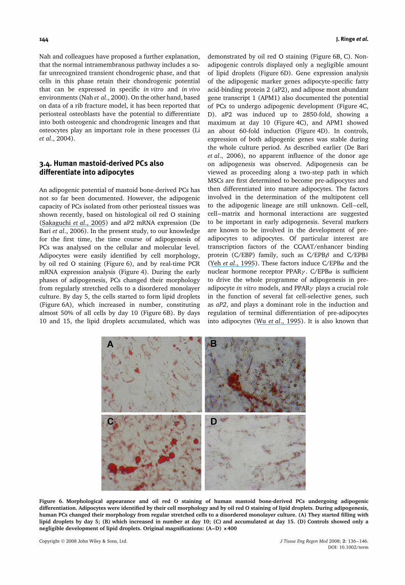
In the present study, chondrogenic development ofPCs was induced with TGFβ1 (data not shown) andTGFβ3 (Figure 5). Chondrogenesis was documented byAlcian blue staining of cartilage proteoglycans and byimmunohistochemistry using type I and type II collagen-specific antibodies (Figure 5). PCs exposed to both TGFsdisplayed a compact and homogeneous pellet structure.Alcian blue staining showed an increasing deposition ofproteoglycans, with a homogeneous distribution by day 21within the whole section of the pellet culture (Figure 5A).Staining of cartilage-specific type II collagen was notdetectable at day 7, but displayed an increasing amountof cartilage-specific collagen at day 21 (Figure 5B). Atthat time, a concentration of type II collagen in theborder areas of the pellets was observed (data notshown), whereas the central part of the pellet culturesstained almost negative. Instead, type I collagen wasconcentrated in the central parts of the pellets (data notshown). Control PCs exhibited a fibrous pellet structurewith marginal staining of Alcian blue (Figure 5C) and notype II collagen staining (Figure 5D). Distinct possibilitieshave been proposed to explain the origin of chondrocytesappearing in membrane bones (Fang and Hall, 1997):chondrocytes may develop from stem cells, from particularchondroprogenitor cells or osteogenic cells. However, allthese possibilities have pros and cons. In a recent study,
Figure 5. Histological analysis of PCs undergoing chondrogenic differentiation. Transforming growth factor-β3 induced PCsdisplayed a compact round pellet structure. (A) By day 21, Alcian blue staining showed a homogeneous distribution of cartilageproteoglycans over the pellets. (B) At that time, antibody staining of cartilage-specific type II collagen demonstrated a type IIcollagen expression focused on the border areas. (C, D) Control PCs exhibited a fibrous structure, with marginal staining ofproteoglycans and no staining of cartilage-specific type II collagen. Original magnifications: (A, C) ×200; (B, D) ×400
Copyright 2008 John Wiley & Sons, Ltd. J Tissue Eng Regen Med 2008; 2: 136–146.DOI: 10.1002/term
144 J. Ringe et al.
Nah and colleagues have proposed a further explanation,that the normal intramembranous pathway includes a so-far unrecognized transient chondrogenic phase, and thatcells in this phase retain their chondrogenic potentialthat can be expressed in specific in vitro and in vivoenvironments (Nah et al., 2000). On the other hand, basedon data of a rib fracture model, it has been reported thatperiosteal osteoblasts have the potential to differentiateinto both osteogenic and chondrogenic lineages and thatosteocytes play an important role in these processes (Liet al., 2004).
3.4. Human mastoid-derived PCs alsodifferentiate into adipocytes
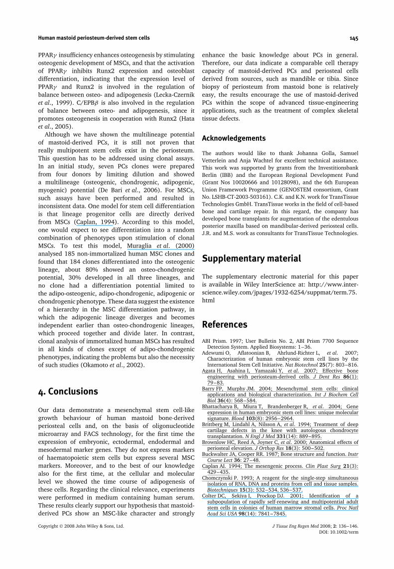
An adipogenic potential of mastoid bone-derived PCs hasnot so far been documented. However, the adipogeniccapacity of PCs isolated from other periosteal tissues wasshown recently, based on histological oil red O staining(Sakaguchi et al., 2005) and aP2 mRNA expression (DeBari et al., 2006). In the present study, to our knowledgefor the first time, the time course of adipogenesis ofPCs was analysed on the cellular and molecular level.Adipocytes were easily identified by cell morphology,by oil red O staining (Figure 6), and by real-time PCRmRNA expression analysis (Figure 4). During the earlyphases of adipogenesis, PCs changed their morphologyfrom regularly stretched cells to a disordered monolayerculture. By day 5, the cells started to form lipid droplets(Figure 6A), which increased in number, constitutingalmost 50% of all cells by day 10 (Figure 6B). By days10 and 15, the lipid droplets accumulated, which was
demonstrated by oil red O staining (Figure 6B, C). Non-adipogenic controls displayed only a negligible amountof lipid droplets (Figure 6D). Gene expression analysisof the adipogenic marker genes adipocyte-specific fattyacid-binding protein 2 (aP2), and adipose most abundantgene transcript 1 (APM1) also documented the potentialof PCs to undergo adipogenic development (Figure 4C,D). aP2 was induced up to 2850-fold, showing amaximum at day 10 (Figure 4C), and APM1 showedan about 60-fold induction (Figure 4D). In controls,expression of both adipogenic genes was stable duringthe whole culture period. As described earlier (De Bariet al., 2006), no apparent influence of the donor ageon adipogenesis was observed. Adipogenesis can beviewed as proceeding along a two-step path in whichMSCs are first determined to become pre-adipocytes andthen differentiated into mature adipocytes. The factorsinvolved in the determination of the multipotent cellto the adipogenic lineage are still unknown. Cell–cell,cell–matrix and hormonal interactions are suggestedto be important in early adipogenesis. Several markersare known to be involved in the development of pre-adipocytes to adipocytes. Of particular interest aretranscription factors of the CCAAT/enhancer bindingprotein (C/EBP) family, such as C/EPBβ and C/EPBδ
(Yeh et al., 1995). These factors induce C/EPBα and thenuclear hormone receptor PPARγ . C/EPBα is sufficientto drive the whole programme of adipogenesis in pre-adipocyte in vitro models, and PPARγ plays a crucial rolein the function of several fat cell-selective genes, suchas aP2, and plays a dominant role in the induction andregulation of terminal differentiation of pre-adipocytesinto adipocytes (Wu et al., 1995). It is also known that
Figure 6. Morphological appearance and oil red O staining of human mastoid bone-derived PCs undergoing adipogenicdifferentiation. Adipocytes were identified by their cell morphology and by oil red O staining of lipid droplets. During adipogenesis,human PCs changed their morphology from regular stretched cells to a disordered monolayer culture. (A) They started filling withlipid droplets by day 5; (B) which increased in number at day 10; (C) and accumulated at day 15. (D) Controls showed only anegligible development of lipid droplets. Original magnifications: (A–D) ×400
Copyright 2008 John Wiley & Sons, Ltd. J Tissue Eng Regen Med 2008; 2: 136–146.DOI: 10.1002/term
Human mastoid periosteum-derived stem cells 145
PPARγ insufficiency enhances osteogenesis by stimulatingosteogenic development of MSCs, and that the activationof PPARγ inhibits Runx2 expression and osteoblastdifferentiation, indicating that the expression level ofPPARγ and Runx2 is involved in the regulation ofbalance between osteo- and adipogenesis (Lecka-Czerniket al., 1999). C/EPBβ is also involved in the regulationof balance between osteo- and adipogenesis, since itpromotes osteogenesis in cooperation with Runx2 (Hataet al., 2005).
Although we have shown the multilineage potentialof mastoid-derived PCs, it is still not proven thatreally multipotent stem cells exist in the periosteum.This question has to be addressed using clonal assays.In an initial study, seven PCs clones were preparedfrom four donors by limiting dilution and showeda multilineage (osteogenic, chondrogenic, adipogenic,myogenic) potential (De Bari et al., 2006). For MSCs,such assays have been performed and resulted ininconsistent data. One model for stem cell differentiationis that lineage progenitor cells are directly derivedfrom MSCs (Caplan, 1994). According to this model,one would expect to see differentiation into a randomcombination of phenotypes upon stimulation of clonalMSCs. To test this model, Muraglia et al. (2000)analysed 185 non-immortalized human MSC clones andfound that 184 clones differentiated into the osteogeniclineage, about 80% showed an osteo-chondrogenicpotential, 30% developed in all three lineages, andno clone had a differentiation potential limited tothe adipo-osteogenic, adipo-chondrogenic, adipogenic orchondrogenic phenotype. These data suggest the existenceof a hierarchy in the MSC differentiation pathway, inwhich the adipogenic lineage diverges and becomesindependent earlier than osteo-chondrogenic lineages,which proceed together and divide later. In contrast,clonal analysis of immortalized human MSCs has resultedin all kinds of clones except of adipo-chondrogenicphenotypes, indicating the problems but also the necessityof such studies (Okamoto et al., 2002).
4. Conclusions
Our data demonstrate a mesenchymal stem cell-likegrowth behaviour of human mastoid bone-derivedperiosteal cells and, on the basis of oligonucleotidemicroarray and FACS technology, for the first time theexpression of embryonic, ectodermal, endodermal andmesodermal marker genes. They do not express markersof haematopoietic stem cells but express several MSCmarkers. Moreover, and to the best of our knowledgealso for the first time, at the cellular and molecularlevel we showed the time course of adipogenesis ofthese cells. Regarding the clinical relevance, experimentswere performed in medium containing human serum.These results clearly support our hypothesis that mastoid-derived PCs show an MSC-like character and strongly
enhance the basic knowledge about PCs in general.Therefore, our data indicate a comparable cell therapycapacity of mastoid-derived PCs and periosteal cellsderived from sources, such as mandible or tibia. Sincebiopsy of periosteum from mastoid bone is relativelyeasy, the results encourage the use of mastoid-derivedPCs within the scope of advanced tissue-engineeringapplications, such as the treatment of complex skeletaltissue defects.
Acknowledgements
The authors would like to thank Johanna Golla, SamuelVetterlein and Anja Wachtel for excellent technical assistance.This work was supported by grants from the InvestitionsbankBerlin (IBB) and the European Regional Development Fund(Grant Nos 10020666 and 10128098), and the 6th EuropeanUnion Framework Programme (GENOSTEM consortium, GrantNo. LSHB-CT-2003-503161). C.K. and K.N. work for TransTissueTechnologies GmbH. TransTissue works in the field of cell-basedbone and cartilage repair. In this regard, the company hasdeveloped bone transplants for augmentation of the edentulousposterior maxilla based on mandibular-derived periosteal cells.J.R. and M.S. work as consultants for TransTissue Technologies.
Supplementary material
The supplementary electronic material for this paperis available in Wiley InterScience at: http://www.inter-science.wiley.com/jpages/1932-6254/suppmat/term.75.html
References
ABI Prism. 1997; User Bulletin No. 2, ABI Prism 7700 SequenceDetection System. Applied Biosystems: 1–36.
Adewumi O, Aflatoonian B, Ahrlund-Richter L, et al. 2007;Characterization of human embryonic stem cell lines by theInternational Stem Cell Initiative. Nat Biotechnol 25(7): 803–816.
Agata H, Asahina I, Yamazaki Y, et al. 2007; Effective boneengineering with periosteum-derived cells. J Dent Res 86(1):79–83.
Barry FP, Murphy JM. 2004; Mesenchymal stem cells: clinicalapplications and biological characterization. Int J Biochem CellBiol 36(4): 568–584.
Bhattacharya B, Miura T, Brandenberger R, et al. 2004; Geneexpression in human embryonic stem cell lines: unique molecularsignature. Blood 103(8): 2956–2964.
Brittberg M, Lindahl A, Nilsson A, et al. 1994; Treatment of deepcartilage defects in the knee with autologous chondrocytetransplantation. N Engl J Med 331(14): 889–895.
Brownlow HC, Reed A, Joyner C, et al. 2000; Anatomical effects ofperiosteal elevation. J Orthop Res 18(3): 500–502.
Buckwalter JA, Cooper RR. 1987; Bone structure and function. InstrCourse Lect 36: 27–48.
Caplan AI. 1994; The mesengenic process. Clin Plast Surg 21(3):429–435.
Chomczynski P. 1993; A reagent for the single-step simultaneousisolation of RNA, DNA and proteins from cell and tissue samples.Biotechniques 15(3): 532–534, 536–537.
Colter DC, Sekiya I, Prockop DJ. 2001; Identification of asubpopulation of rapidly self-renewing and multipotential adultstem cells in colonies of human marrow stromal cells. Proc NatlAcad Sci USA 98(14): 7841–7845.
Copyright 2008 John Wiley & Sons, Ltd. J Tissue Eng Regen Med 2008; 2: 136–146.DOI: 10.1002/term
146 J. Ringe et al.
De Bari C, Dell’Accio F, Luyten FP. 2001; Human periosteum-derivedcells maintain phenotypic stability and chondrogenic potentialthroughout expansion regardless of donor age. Arthritis Rheum44(1): 85–95.
De Bari C, Dell’Accio F, Vandenabeele F, et al. 2003; Skeletal musclerepair by adult human mesenchymal stem cells from synovialmembrane. J Cell Biol 160(6): 909–918.
De Bari C, Dell’Accio F, Vanlauwe J, et al. 2006; Mesenchymalmultipotency of adult human periosteal cells demonstrated bysingle-cell lineage analysis. Arthritis Rheum 54(4): 1209–1221.
Decker JD, Marshall JJ, Herring SW. 1996; Differential cellreplication within the periosteum of the pig mandibular ramus.Acta Anat (Basel) 157(2): 144–150.
Fang J, Hall BK. 1997; Chondrogenic cell differentiation frommembrane bone periostea. Anat Embryol (Berl) 196(5): 349–362.
Fukumoto T, Sperling JW, Sanyal A, et al. 2003; Combined effects ofinsulin-like growth factor-1 and transforming growth factor-beta1on periosteal mesenchymal cells during chondrogenesis in vitro.Osteoarthritis Cartilage 11(1): 55–64.
Ginis I, Luo Y, Miura T, et al. 2004; Differences between human andmouse embryonic stem cells. Dev Biol 269(2): 360–380.
Groger A, Klaring S, Merten HA, et al. 2003; Tissue engineeringof bone for mandibular augmentation in immunocompetentminipigs: preliminary study. Scand J Plast Reconstr Surg HandSurg 37(3): 129–133.
Gronthos S, Zannettino AC, Graves SE, et al. 1999; Differential cellsurface expression of the STRO-1 and alkaline phosphataseantigens on discrete developmental stages in primary culturesof human bone cells. J Bone Miner Res 14(1): 47–56.
Hata K, Nishimura R, Ueda M, et al. 2005; A CCAAT/enhancerbinding protein beta isoform, liver-enriched inhibitory protein,regulates commitment of osteoblasts and adipocytes. Mol Cell Biol25(5): 1971–1979.
Haynesworth SE, Baber MA, Caplan AI. 1996; Cytokine expressionby human marrow-derived mesenchymal progenitor cells in vitro:effects of dexamethasone and IL-1α. J Cell Physiol 166(3):585–592.
Haynesworth SE, Goshima J, Goldberg VM, et al. 1992; Character-ization of cells with osteogenic potential from human marrow.Bone 13(1): 81–88.
Hoikka VE, Jaroma HJ, Ritsila VA. 1990; Reconstruction of thepatellar articulation with periosteal grafts. 4-year follow-up of13 cases. Acta Orthop Scand 61(1): 36–39.
Kobayashi T, Kronenberg H. 2005; Minireview: transcriptionalregulation in development of bone. Endocrinology 146(3):1012–1017.
Lecka-Czernik B, Gubrij I, Moerman EJ, et al. 1999; Inhibition ofOsf2/Cbfa1 expression and terminal osteoblast differentiation byPPARγ 2. J Cell Biochem 74(3): 357–371.
Lee RH, Kim B, Choi I, et al. 2004; Characterization and expressionanalysis of mesenchymal stem cells from human bone marrow andadipose tissue. Cell Physiol Biochem 14(4–6): 311–324.
Lennon DP, Haynesworth SE, Bruder SP, et al. 1996; Human andanimal mesenchymal progenitor cells from bone marrow:identification of serum for optimal selection and proliferation.In Vitro Cell Dev Biol 32: 602–611.
Li M, Amizuka N, Oda K, et al. 2004; Histochemical evidence ofthe initial chondrogenesis and osteogenesis in the periosteum ofa rib fractured model: implications of osteocyte involvement inperiosteal chondrogenesis. Microsc Res Tech 64(4): 330–342.
Majumdar MK, Thiede MA, Mosca JD, et al. 1998; Phenotypicand functional comparison of cultures of marrow-derivedmesenchymal stem cells (MSCs) and stromal cells. J Cell Physiol176(1): 57–66.
Marini RP, Steven MM, Langer R, et al. 2004; Hydraulic evaluationof the periosteum: a novel technique for periosteal harvest. J InvestSurg 17(4): 229–233.
Marks SC Jr, Popoff SN. 1988; Bone cell biology: the regulation ofdevelopment, structure, and function in the skeleton. Am J Anat183(1): 1–44.
Muraglia A, Cancedda R, Quarto R. 2000; Clonal mesenchymalprogenitors from human bone marrow differentiate in vitroaccording to a hierarchical model. J Cell Sci 113(7): 1161–1166.
Nah HD, Pacifici M, Gerstenfeld LC, et al. 2000; Transientchondrogenic phase in the intramembranous pathway duringnormal skeletal development. J Bone Miner Res 15(3): 522–533.
Nakahara H, Goldberg VM, Caplan AI. 1991; Culture-expandedhuman periosteal-derived cells exhibit osteochondral potentialin vivo. J Orthop Res 9(4): 465–476.
Nakase T, Nakahara H, Iwasaki M, et al. 1993; Clonal analysis fordevelopmental potential of chick periosteum-derived cells: agar gelculture system. Biochem Biophys Res Commun 195(3): 1422–1428.
Noth U, Osyczka AM, Tuli R, et al. 2002; Multilineage mesenchymaldifferentiation potential of human trabecular bone-derived cells. JOrthop Res 20(5): 1060–1069.
O’Driscoll SW, Fitzsimmons JS. 2001; The role of periosteum incartilage repair. Clin Orthop 391: (suppl): S190–207.
O’Driscoll SW, Meisami B, Miura Y, et al. 1999; Viability of periostealtissue obtained postmortem. Cell Transplant 8(6): 611–616.
Okamoto T, Aoyama T, Nakayama T, et al. 2002; Clonalheterogeneity in differentiation potential of immortalized humanmesenchymal stem cells. Biochem Biophys Res Commun 295(2):354–361.
Ossendorf C, Kaps C, Kreuz PC, et al. 2007; Treatment ofposttraumatic and focal osteoarthritic cartilage defects of the kneewith autologous polymer-based three-dimensional chondrocytegrafts: 2-year clinical results. Arthritis Res Ther 9(2): R41.
Picinich SC, Mishra PJ, Glod J, et al. 2007; The therapeutic potentialof mesenchymal stem cells. Cell- and tissue-based therapy. ExpertOpin Biol Ther 7(7): 965–973.
Pittenger MF, Mackay AM, Beck SC, et al. 1999; Multilineagepotential of adult human mesenchymal stem cells. Science284(5411): 143–147.
Pochampally RR, Smith JR, Ylostalo J, et al. 2004; Serumdeprivation of human marrow stromal cells (hMSCs) selects for asubpopulation of early progenitor cells with enhanced expressionof OCT-4 and other embryonic genes. Blood 103(5): 1647–1652.
Reyes M, Dudek A, Jahagirdar B, et al. 2002; Origin of endothelialprogenitors in human postnatal bone marrow. J Clin Invest 109(3):337–346.
Sakaguchi Y, Sekiya I, Yagishita K, et al. 2005; Comparison of humanstem cells derived from various mesenchymal tissues: superiorityof synovium as a cell source. Arthritis Rheum 52(8): 2521–2529.
Schmelzeisen R, Schimming R, Sittinger M. 2003; Making bone:implant insertion into tissue-engineered bone for maxillary sinusfloor augmentation-a preliminary report. J Craniomaxillofac Surg31(1): 34–39.
Soncini M, Vertua E, Gibelli L, et al. 2007; Isolation andcharacterization of mesenchymal cells from human fetalmembranes. J Tissue Eng Regen Med 1(4): 296–305.
Stewart K, Walsh S, Screen J, et al. 1999; Further characterization ofcells expressing STRO-1 in cultures of adult human bone marrowstromal cells. J Bone Miner Res 14(8): 1345–1356.
Tremain N, Korkko J, Ibberson D, et al. 2001; MicroSAGE analysisof 2353 expressed genes in a single cell-derived colony ofundifferentiated human mesenchymal stem cells reveals mRNAsof multiple cell lineages. Stem Cells 19(5): 408–418.
Tsai MS, Lee JL, Chang YJ, et al. 2004; Isolation of humanmultipotent mesenchymal stem cells from second-trimesteramniotic fluid using a novel two-stage culture protocol. HumReprod 19(6): 1450–1456.
Tuli R, Tuli S, Nandi S, et al. 2003; Characterization ofmultipotential mesenchymal progenitor cells derived from humantrabecular bone. Stem Cells 21(6): 681–693.
Wognum AW, Eaves AC, Thomas TE. 2003; Identification andisolation of haematopoietic stem cells. Arch Med Res 34(6):461–475.
Wu Z, Xie Y, Bucher NL, Farmer SR. 1995; Conditional ectopicexpression of C/EBPβ in NIH-3T3 cells induces PPARγ andstimulates adipogenesis. Genes Dev 9(19): 2350–2363.
Yamazaki M, Nakajima F, Ogasawara A, et al. 1999; Spatial andtemporal distribution of CD44 and osteopontin in fracture callus.J Bone Joint Surg Br 81(3): 508–515.
Yeh WC, Cao Z, Classon M, McKnight SL. 1995; Cascade regulationof terminal adipocyte differentiation by three members of theC/EBP family of leucine zipper proteins. Genes Dev 9(2): 168–181.
Yen BL, Huang HI, Chien CC, et al. 2005; Isolation of multipotentcells from human term placenta. Stem Cells 23(1): 3–9.
Zheng YX, Ringe J, Liang Z, et al. 2006; Osteogenic potential ofhuman periosteum-derived progenitor cells in PLGA scaffold usingallogeneic serum. J Zhejiang Univ Sci B 7(10): 817–824.
Zuk PA, Zhu M, Mizuno H, et al. 2001; Multilineage cells fromhuman adipose tissue: implications for cell-based therapies. TissueEng 7(2): 211–228.
Copyright 2008 John Wiley & Sons, Ltd. J Tissue Eng Regen Med 2008; 2: 136–146.DOI: 10.1002/term