Human Keratinocytes That Express hTERT and Also Bypass a p16INK4a-Enforced Mechanism That Limits...
13
10.1128/MCB.20.4.1436-1447.2000. 2000, 20(4):1436. DOI: Mol. Cell. Biol. Frederick P. Li and James G. Rheinwald Ronfard, Jenny Y. Wu, Robert A. Weinberg, David N. Louis, Mark A. Dickson, William C. Hahn, Yasushi Ino, Vincent Differentiation Characteristics Immortal yet Retain Normal Growth and Mechanism That Limits Life Span Become -Enforced INK4a and Also Bypass a p16 Human Keratinocytes That Express hTERT http://mcb.asm.org/content/20/4/1436 Updated information and services can be found at: These include: REFERENCES http://mcb.asm.org/content/20/4/1436#ref-list-1 at: This article cites 61 articles, 30 of which can be accessed free CONTENT ALERTS more» articles cite this article), Receive: RSS Feeds, eTOCs, free email alerts (when new http://journals.asm.org/site/misc/reprints.xhtml Information about commercial reprint orders: http://journals.asm.org/site/subscriptions/ To subscribe to to another ASM Journal go to: on October 19, 2014 by guest http://mcb.asm.org/ Downloaded from on October 19, 2014 by guest http://mcb.asm.org/ Downloaded from
Transcript of Human Keratinocytes That Express hTERT and Also Bypass a p16INK4a-Enforced Mechanism That Limits...
10.1128/MCB.20.4.1436-1447.2000.
2000, 20(4):1436. DOI:Mol. Cell. Biol. Frederick P. Li and James G. RheinwaldRonfard, Jenny Y. Wu, Robert A. Weinberg, David N. Louis, Mark A. Dickson, William C. Hahn, Yasushi Ino, Vincent Differentiation CharacteristicsImmortal yet Retain Normal Growth andMechanism That Limits Life Span Become
-EnforcedINK4aand Also Bypass a p16Human Keratinocytes That Express hTERT
http://mcb.asm.org/content/20/4/1436Updated information and services can be found at:
These include:
REFERENCEShttp://mcb.asm.org/content/20/4/1436#ref-list-1at:
This article cites 61 articles, 30 of which can be accessed free
CONTENT ALERTS more»articles cite this article),
Receive: RSS Feeds, eTOCs, free email alerts (when new
http://journals.asm.org/site/misc/reprints.xhtmlInformation about commercial reprint orders: http://journals.asm.org/site/subscriptions/To subscribe to to another ASM Journal go to:
on October 19, 2014 by guest
http://mcb.asm
.org/D
ownloaded from
on O
ctober 19, 2014 by guesthttp://m
cb.asm.org/
Dow
nloaded from
MOLECULAR AND CELLULAR BIOLOGY,0270-7306/00/$04.0010
Feb. 2000, p. 1436–1447 Vol. 20, No. 4
Copyright © 2000, American Society for Microbiology. All Rights Reserved.
Human Keratinocytes That Express hTERT and Also Bypass ap16INK4a-Enforced Mechanism That Limits Life Span
Become Immortal yet Retain Normal Growthand Differentiation Characteristics
MARK A. DICKSON,1 WILLIAM C. HAHN,2,3 YASUSHI INO,4 VINCENT RONFARD,5 JENNY Y. WU,1
ROBERT A. WEINBERG,2 DAVID N. LOUIS,4 FREDERICK P. LI,6 AND JAMES G. RHEINWALD1*
Division of Dermatology, Department of Medicine and Harvard Skin Disease Research Center, Brigham and Women’sHospital,1 Department of Adult Oncology, Dana-Farber Cancer Institute, and Department of Medicine, Brigham and
Women’s Hospital,3 Department of Pathology and Neurosurgical Service, Massachusetts General Hospital,4
and Department of Adult Oncology, Dana-Farber Cancer Institute,6 Harvard Medical School, Boston,Whitehead Institute for Biomedical Research and Department of Biology, Massachusetts Institute
of Technology, Cambridge,2 and Laboratory of Cell and Tissue Development,Organogenesis, Inc., Canton,5 Massachusetts
Received 11 August 1999/Returned for modification 11 October 1999/Accepted 18 November 1999
Normal human cells exhibit a limited replicative life span in culture, eventually arresting growth by aprocess termed senescence. Progressive telomere shortening appears to trigger senescence in normal humanfibroblasts and retinal pigment epithelial cells, as ectopic expression of the telomerase catalytic subunit,hTERT, immortalizes these cell types directly. Telomerase expression alone is insufficient to enable certainother cell types to evade senescence, however. Such cells, including keratinocytes and mammary epithelial cells,appear to require loss of the pRB/p16INK4a cell cycle control mechanism in addition to hTERT expression toachieve immortality. To investigate the relationships among telomerase activity, cell cycle control, senescence,and differentiation, we expressed hTERT in two epithelial cell types, keratinocytes and mesothelial cells, anddetermined the effect on proliferation potential and on the function of cell-type-specific growth control anddifferentiation systems. Ectopic hTERT expression immortalized normal mesothelial cells and a premalignant,p16INK4a-negative keratinocyte line. In contrast, when four keratinocyte strains cultured from normal tissuewere transduced to express hTERT, they were incompletely rescued from senescence. After reaching thepopulation doubling limit of their parent cell strains, hTERT1 keratinocytes entered a slow growth phase ofindefinite length, from which rare, rapidly dividing immortal cells emerged. These immortal cell lines fre-quently had sustained deletions of the CDK2NA/INK4A locus or otherwise were deficient in p16INK4a expres-sion. They nevertheless typically retained other keratinocyte growth controls and differentiated normally inculture and in xenografts. Thus, keratinocyte replicative potential is limited by a p16INK4a-dependent mech-anism, the activation of which can occur independent of telomere length. Abrogation of this mechanismtogether with telomerase expression immortalizes keratinocytes without affecting other major growth controlor differentiation systems.
Normal human somatic cells have a limited capacity to rep-licate in culture, even under conditions that appear to satisfytheir nutritional and mitogen requirements (53, 56). Thesecells proliferate initially but eventually enter a state of perma-nent growth arrest termed senescence, clearly distinct fromdifferentiation, in which they can remain metabolically activeindefinitely. Progressive shortening of the telomeres, DNA-protein structures located at the ends of linear eukaryotic chro-mosomes, occurs during the 50- to 100-population-doubling(PD) life span of human fibroblasts in culture (19). The erosionof telomeric DNA with successive cell replications has led tothe proposal that telomeres not only function to protect thechromosomes from end-to-end fusions but, when disrupted byshortening, also signal the onset of senescence (2).
Unlike most normal human somatic cell types, most ad-vanced-stage cancer cells are replicatively immortal and ex-
press the enzyme telomerase. Telomerase is a multimeric ri-bonucleoprotein containing an RNA component that includesin its sequence the template for telomere synthesis (14) and acatalytic protein subunit that is a reverse transcriptase (34, 38).The expression of telomerase in immortal cancer cells appar-ently is responsible for their maintenance of a stable telomerelength through an indefinite number of cell divisions (11).Although the telomerase RNA component is expressed con-stitutively (19), the catalytic subunit, hTERT, is expressed onlyin germ cells and in immortal cancer cells (34, 38), suggestingthat hTERT is the activity-limiting component of the telomer-ase holoenzyme. Introduction of hTERT into presenescenthuman fibroblasts and retinal pigment epithelial cells wasfound to confer telomere maintenance and unlimited replica-tive potential to these cell types (5), giving strong support tothe model that telomere shortening determines the onset ofsenescence. This simple interpretation, however, may not ap-ply to all cell types, as it was reported recently that ectopicexpression of hTERT is not sufficient to immortalize normalhuman keratinocytes and mammary epithelial cells (25).
We have sought to investigate the role of telomerase in
* Corresponding author. Mailing address: Harvard Institutes ofMedicine, Room 664, 77 Ave. Louis Pasteur, Boston, MA 02115.Phone: (617) 525-5553. Fax: (617) 525-5571. E-mail: [email protected].
1436
on October 19, 2014 by guest
http://mcb.asm
.org/D
ownloaded from
cellular senescence, to identify potential ancillary genetic al-terations necessary for immortalization of epithelial cells, andto determine the effects of immortalization on cell-type-spe-cific growth control and differentiation mechanisms. We haveexpressed hTERT in two different types of epithelial cells,mesothelial cells and keratinocytes, both of which exhibit afinite life span in vitro and have well-characterized growthcontrol systems and differentiation programs (10, 16, 47, 52).Our experiments indicate that these two epithelial cell typesbehave very differently in response to ectopically expressedhTERT and that such expression is not sufficient to immortal-ize keratinocytes. We have identified a complex pattern ofp16INK4a expression in keratinocytes associated with senes-cence which functions independent of telomere shortening.Keratinocytes that express hTERT and also acquire a defect intriggering p16INK4a expression become immortalized but oth-erwise display normal growth characteristics and differentia-tion potential, indicating that the process of senescence in thiscell type is complex but separate from mechanisms that regu-late growth and differentiation.
MATERIALS AND METHODS
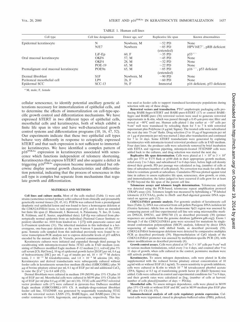
Cell lines and culture media. Most of the cells studied (Table 1) were cellstrains (sometimes termed primary cells) cultured from clinically and presumablygenetically normal tissues (28, 45, 61). POE9n was cultured from a premalignantdysplastic oral epithelial lesion and was found to have a homozygous deletion atthe CDKN2A/INK4A locus, to lack expression of p53, and to have an extendedbut finite life span (J. Rheinwald, J. Benwood, A. Palanisamy, Y. Ino, D. Louis,R. Feldman, and E. Sauter, unpublished data). LiF-Ep was cultured from phe-notypically normal epidermis from an individual (National Cancer Institute re-pository identifier no. 1010 from kindred no. 1 [27]) with pancreatic cancer andLi-Fraumeni syndrome (31). The inherited mutation in this individual is a het-erozygous, one-base-pair deletion at the exon 9-intron 9 junction of the TP53gene. Somatic cells sampled from this individual previously were found by re-verse transcription-PCR analysis not to express detectable levels of p53 mRNAencoded by the mutant allele (S. Verselis, personal communication).
Keratinocyte cultures were initiated and expanded through third passage bycocultivating with mitomycin-treated Swiss 3T3J2 cells in FAD medium (con-sisting of Dulbecco modified Eagle medium–F-12 medium [1:1, vol/vol] plus 5%calf serum [CS; HyClone], 10 ng of epidermal growth factor [EGF] per ml, 0.4 mgof hydrocortisone [HC] per ml, 5 mg of insulin per ml, 10 3 10210 M choleratoxin, 2 3 10211 M triiodothyronine, and 1.8 3 1024 M adenine [44, 46]).Keratinocytes and derived transductants were subsequently cultured withoutfeeder cells in keratinocyte serum-free medium (K-sfm) (GIBCO/BRL) plus 30mg of bovine pituitary extract per ml, 0.1 ng of EGF per ml and additional CaCl2to raise the [Ca21] to 0.4 mM (52).
Dermal fibroblasts were cultured in medium 199 (M199) plus 15% CS plus 10ng of EGF per ml. Mesothelial cells were cultured in M199 plus 15% CS plus 10ng of EGF per ml plus 0.4 mg of HC per ml (10). 3T3J2 cells and PT67 retroviralvector producer cells (17) were cultured in pyruvate-free Dulbecco modifiedEagle medium (GIBCO/BRL)–10% CS. A multiple-drug-resistant fibroblastfeeder cell line, 3T3J2NHP, was generated by sequentially transducing 3T3J2with the retroviral vectors LXSN (35), BABE/hygro, and BABE/puro (36) toconfer resistance to G418, hygromycin, and puromycin, respectively. This line
was used as feeder cells to support transduced keratinocyte populations duringselection with any of these drugs.
Retroviral vectors and transduction. PT67 amphotropic packaging cells pro-ducing BABE-hygro-hTERT and BABE-puro-hTERT (17) or control BABE-hygro and BABE-puro (36) retroviral vectors were used to generate retroviralsupernatants in K-sfm, which was passed through a 0.45-mm-pore-size filter andstored at 280°C until use. Human cells plated 1 day earlier at ;105 cells per9-cm2 well were transduced by refeeding them for 5 to 7 h with retroviralsupernatant plus Polybrene (4 mg/ml; Sigma). The treated cells were subculturedthe next day into 75-cm2 flasks. Drug selection (5 to 10 mg of hygromycin per mlor 1 mg of puromycin per ml) was started 2 days after transduction and continuedfor 7 to 14 days. Some hTERT transductants were generated by coplatingkeratinocytes with mitomycin-treated retroviral producer cells in FAD medium.Four days later, the producer cells were selectively removed by brief incubationwith EDTA and vigorous pipetting, mitomycin-treated 3T3J2NHP cells wereadded back to the cultures, and drug selection was started the next day.
Replicative life span determination. Cells were plated at 1 3 105 to 3 3 105
cells per T75 or T175 flask or p100 dish in their appropriate growth medium,refed every 2 to 3 days, and subcultured 5 to 8 days later, before high cell densityslowed their growth. PD per passage was calculated as log2 (number of cells attime of subculture/number of cells plated). No correction was made for cells thatfailed to reinitiate growth at subculture. Cumulative PD was plotted against totaltime in culture to assess replicative life span, senescence, slow growth, or crisis,and immortalization, the latter judged to have occurred if cells grew for at least50 PD beyond the life span of the parent cell line.
Telomerase assays and telomere length determination. Telomerase activitywas detected using the PCR-based, telomerase repeat amplification protocol(TRAP) assay (24). Telomere length was measured by hybridizing a 32P-labeledtelomeric (CCCTAA)3 probe to HinfI- and RsaI-digested genomic DNA sepa-rated on agarose gels (12).
CDKN2A/INK4A genomic analysis. For genomic analysis of keratinocyte celllines (Table 2), DNA was extracted from cell pellets Puregene DNA isolation kit(Gentra Systems). Allelic loss on the chromosome 9p21 region was assessed bycomparing parent cell line-experimental cell line sets using microsatellite mark-ers D9S126, D9S741, and D9S1748 (3) as described previously (30) (primersequences are available from the genome database [gdbwww.gdb.org]). Exons 1through 3 of the CDKN2A/INK4A gene were screened for mutations by single-strand conformation polymorphism (SSCP) analysis, followed by direct DNAsequencing of samples with shifted bands, as described previously (58).CDKN2A/INK4A homozygous deletions were detected by comparative multiplexPCR as described previously (39). Hypermethylation of CpG islands of theCDKN2A/INK4A promoter was assessed by methylation-specific PCR (20), withminor modifications as described previously (8).
Growth control assays. Cells were plated at 103 to 3 3 103 cells per 9-cm2 wellin various medium formulations, refed every 3 to 4 days, and counted after 9 to11 days of growth, when cells cultured in the control, permissive medium werestill actively proliferating.
Keratinocytes. To assess mitogen dependence, cells were plated in K-sfmsupplemented with the reduced bovine pituitary extract concentration of 10mg/ml with or without EGF (0.1 ng/ml). To assess sensitivity to growth inhibitors,cells were cultured in K-sfm to which 1029 M tetradecanoyl phorbol acetate(TPA; Sigma) or 0.3 ng of transforming growth factor b1 (R&D Systems) wasadded. Cells were cultured in control and experimental conditions for 7 to 9 days,and their growth rates were calculated as [log2 (number of cells at harvest/number of cells plated)]/number of days 5 PD/day.
Mesothelial cells. To assess mitogen dependence, cells were plated in M199plus 15% CS with or without EGF and HC and in M199 medium plus EGF plusHC plus 1% CS (10, 57).
Immunochemical analysis of cell cycle regulatory protein expression. Cul-tured cells were trypsinized, rinsed in phosphate-buffered saline (PBS), pelleted,
TABLE 1. Human cell lines
Cell type Cell line designation Donor age, sexa Replicative life span Known abnormalities
Epidermal keratinocyte N Newborn, M ;52 PD NoneN/E7 Newborn ;85 PD
(extended)HPV16E71, pRB deficient
LiF-Ep 60, F ;35 PD p531/2
Oral mucosal keratinocyte OKF6 57, M ;45 PD NoneOKF4 28, M ;32 PD NonePOE-19 65, M ;22 PD None
Premalignant oral mucosal keratinocyte POE9n 65, M ;85 PD(extended)
p162/2, p53 deficient
Dermal fibroblast S1F Newborn, M ;90 PD NonePeritoneal mesothelial cell LP9 26, F ;60 PD NoneEpidermal SCC SCC-13 56, F Immortal p16 deficient, p53 deficient
a M, male; F, female
VOL. 20, 2000 hTERT AND p16INK4a IN KERATINOCYTE IMMORTALIZATION 1437
on October 19, 2014 by guest
http://mcb.asm
.org/D
ownloaded from
and lysed in 20 mM Tris buffer (pH 7.3)–2% sodium dodecyl sulfate–1 mMdithiothreitol. Twenty to 100 mg of protein was separated by sodium dodecylsulfate-polyacrylamide gel electrophoresis using a polyacrylamide concentrationof 14% for p16INK4a, 10 or 12% for cyclin D1, cdk4, and cdk6, or 7% for pRB.Gels were electrotransferred to nitrocellulose paper, and proteins were detectedwith antibodies specific for p16INK4a (JC1; a gift from E. Harlow, MassachusettsGeneral Hospital, Boston), pRB (G3-245; PharMingen), p53 (DO-1), cdk4 (C-22), cdk6 (C21), and cyclin D1 (HD11) (the latter four antibodies all from SantaCruz Biotechnologies), followed by peroxidase-labeled secondary antibody(Southern Biotechnologies) and enhanced chemiluminescence reagent (Amer-sham Corp.).
Cells growing on culture dishes, some which received 0.5 nM actinomycin Dfor the final 2 days to elicit a DNA damage-stimulated increase in p53 levels (23),were fixed in 4% paraformaldehyde in PBS for 30 min, permeabilized with 0.1%Triton X-100 in PBS for 5 min, and incubated for 30 min with antibodies specificfor p16INK4a (JC8 or JC2 [8]; a gift from E. Harlow) or p53 (BP53.12; Zymed).Antibody binding was detected using ABC peroxidase (Vector Laboratories)with NovaRed colorimetric substrate.
Cell differentiation and histogenesis assays. Keratinocyte differentiation-re-lated proteins were detected immunocytochemically by ABC peroxidase staining(Vector Laboratories) as described elsewhere (52). Cultures were fixed in coldmethanol and immunostained for involucrin (antibody SY5 [21]; from F. Watt,ICRF Laboratories, London, England), for keratin K10 (antibody AE20 [32];from C. A. Loomis, New York University School of Medicine, New York, N.Y.),and for keratin K13 (antibody AE8 [13]; from T.-T. Sun, New York UniversitySchool of Medicine).
The ability of hTERT-immortalized keratinocytes to form a differentiated,stratified squamous epithelium was assessed in organotypic culture (40, 52).Keratinocytes were seeded at 2 3 105 cells per ;1-cm2 surface area ontocollagen gels containing human foreskin fibroblasts (strain B256), cultured sub-merged for 4 days, and then at the air-liquid interface for 10 days. Cell lines oforal epithelial origin (i.e., OKF6/TERT-1, OKF6/TERT-2, and POE9n/TERT-1)received 1028 M retinoic acid, which provides for more accurate recapitulationof in vivo histology by oral mucosal keratinocytes (52). Cultures were fixed informalin and embedded in paraffin or were frozen in OCT compound. Sectionswere stained with hematoxylin and eosin (H&E).
For grafting, athymic NIH Swiss (nu/nu) mice were anesthetized with ket-amine-xylazine, and a 1-cm2 area of full-thickness skin was excised from themiddle of the back. An organotypic culture was transferred to the site and heldin place with Vaseline-impregnated gauze covered by a Band-Aid. Grafts werekept covered for 1 week, after which they were exposed to the air. Mice weresacrificed 24 or 48 days postgrafting; the grafts were excised, fixed in formalin,paraffin embedded, sectioned, and stained with H&E or with a human involu-crin-specific antiserum (BTI-601 [48]; Biomedical Technologies, Inc.) to distin-guish human from mouse epithelium.
RESULTS
Effect of hTERT expression on human mesothelial cells, asimple squamous epithelial cell type. To examine the effects ofhTERT expression on the growth of epithelial cells, we usedamphotropic retroviral vectors that transduced hTERT and adrug resistance selection marker, or a control vector expressingonly the marker gene, into mid-life-span cultures of the normalmesothelial cell strain LP9. We also transduced these genesinto the normal dermal fibroblast strain S1F to serve as acontrol for the function of our hTERT vector, since hTERThas previously been shown to immortalize normal human fi-broblasts (5, 25). The resulting hTERT-expressing transduc-tants exhibited readily detectable telomerase activity, whereasthe control cells did not (data not shown). Later-passage LP9and S1F control cells had short (;3-kb) telomeres (Fig. 1b),whereas the respective hTERT transductants acquired andmaintained average telomere lengths of ;10 kb.
Control LP9 mesothelial cells (Fig. 2a) and S1F fibroblasts(data not shown) ultimately ceased dividing after 55 and 91PD, respectively, which corresponded approximately to thereplicative life spans previously determined for the parent cellstrains (T. O’Connell-Willstaedt, J. Benwood, and J. Rhein-wald, unpublished data). In contrast, the hTERT-transducedLP9 cells, designated LP9/TERT-1, and the hTERT-trans-duced S1F cells, designated S1F/TERT-1, continued to dividerapidly and indefinitely for at least 50 PD beyond their normallimit (Fig. 2a; Table 3). We concluded that these cells had
TA
BL
E2.
p16IN
K4a
geno
mic
anal
ysis
ofke
ratin
ocyt
ece
lllin
esex
pres
sing
hTE
RT
Cel
llin
e
Del
etio
nan
alys
isa
SSC
Pm
utat
ion
anal
ysis
bSe
quen
cing
resu
ltc
Prom
oter
met
hyla
tiond
D9S
126
D9S
741
D9S
1748
Mul
tiple
xPC
RE
xon
1E
xon
2(A
)E
xon
2(B
)E
xon
3E
xon
Cod
onM
utat
ion
(or
poly
mor
phis
m)
Am
ino
acid
chan
ge
LP9
(23
PD,m
id-li
fesp
an)
1212
12N
NN
NN
UL
P9/T
ER
T-1
(90
PD,R
DI)
1212
12N
NN
NN
UN
(42
PD,l
ate
life
span
)12
NI
12N
NN
NN
UN
/TE
RT
-1(1
00PD
,RD
I)12
NI
12N
NN
NN
UL
iF-E
p(2
5PD
,mid
-life
span
)N
I12
12N
NN
S1N
Se2
148
(GC
G3
AC
G)
Ala3
Thr
U3
nt49
9C3
G39
UT
RL
iF-E
p/T
ER
T-1
(49
PD,R
DI)
NI
LO
HL
OH
NN
NSe
214
8(G
CG3
AC
G)
Ala3
Thr
U3
nt49
9C3
G39
UT
RO
KF
6(4
2PD
,lat
elif
esp
an)
1212
12N
NN
NN
UO
KF
6/T
ER
T-1
(42
PD,e
arly
SGP)
1212
12N
DN
DN
DN
DN
DN
DO
KF
6/T
ER
T-1
(63
PD,R
DI)
LO
HL
OH
LO
HN
NN
NN
UO
KF
6/T
ER
T-1
R(6
5PD
,RD
I)12
1212
ND
ND
ND
ND
ND
ND
OK
F6/
TE
RT
-2(5
2PD
,RD
I)12
12(H
D)
HD
(HD
)(H
D)
(HD
)(H
D)
(HD
)
aC
hang
esfr
omth
epa
rent
cell
line
are
note
din
bold
face
.PC
Rpr
imer
sets
wer
eus
edto
ampl
ifyth
epo
lym
orph
icm
icro
sate
llite
sequ
ence
sD
9S12
6,D
9S74
1,an
dD
9S17
48to
dete
ctlo
ssof
hete
rozy
gosi
ty(L
OH
)in
the
9p21
regi
on.1
2,tw
oal
lele
sde
tect
ed;N
I,no
tinf
orm
ativ
e;L
OH
,alle
liclo
ssco
mpa
red
with
pare
ntce
lllin
eor
norm
alce
llsfr
omsa
me
dono
r.M
ultip
lex
PCR
was
perf
orm
edw
ithex
on2
prim
ers
and
prim
ers
that
ampl
ified
sequ
ence
sfr
oma
cont
roll
ocus
asde
scri
bed
inM
ater
ials
and
Met
hods
.N,e
xon
2se
quen
ces
dete
cted
;HD
,hom
ozyg
ous
dele
tion;
ND
,not
dete
rmin
ed;(
HD
),no
PCR
prod
ucts
obse
rved
beca
use
ofH
D.
bN
ote
that
two
over
lapp
ing
regi
ons
ofex
on2
(des
igna
ted
Aan
dB
)w
ere
ampl
ified
sepa
rate
ly.N
,PC
Rpr
oduc
tmig
ratin
gas
expe
cted
for
wild
-typ
eal
lele
;S,s
hift
inm
igra
tion
com
pare
dw
ithth
atof
the
wild
-typ
eal
lele
.T
heco
don
148
mis
sens
eG
-to-
Aal
tera
tion
inL
iF-E
p,id
entifi
edas
shift
edin
the
SSC
Pan
alys
is,i
sa
poly
mor
phis
mth
atdo
esno
tre
sult
inal
tere
dbi
olog
ical
activ
ityof
p16IN
K4
a(r
efer
ence
42;
note
that
the
old
codo
nnu
mbe
ring
syst
emin
this
pape
rde
sign
ated
codo
n14
8as
140)
.c
nt,n
ucle
otid
e;U
TR
,unt
rans
late
dre
gion
.d
Det
erm
ined
bym
ethy
latio
n-se
nsiti
vePC
Rto
dete
ctm
ethy
latio
nof
the
CpG
isla
ndin
the
p16IN
K4
apr
omot
erm
ostc
omm
only
foun
dto
behy
perm
ethy
late
din
hum
anca
ncer
cells
,as
desc
ribe
din
Mat
eria
lsan
dM
etho
ds.
U,u
nmet
hyla
ted;
M,o
nly
met
hyla
ted
sequ
ence
sde
tect
ed.
eC3
Gsu
bstit
utio
nin
nucl
eotid
e49
4of
the
39un
tran
slat
edre
gion
was
iden
tified
inth
eal
lele
ofL
iF-E
pth
atw
asre
tain
edby
LiF
-Ep/
TE
RT
-1.
1438 DICKSON ET AL. MOL. CELL. BIOL.
on October 19, 2014 by guest
http://mcb.asm
.org/D
ownloaded from
become immortal, similar to the results reported for humanfibroblasts transfected with hTERT (5).
After LP9/TERT-1 cells had divided 50 PD beyond the nor-mal replicative limit of LP9, we tested them for in vitro growthand differentiation phenotypes characteristic of this cell type(10, 57). LP9/TERT-1 cells were indistinguishable from thecontrol cells with respect to dependence on EGF, HC, andserum for growth. When control and hTERT-expressing cellswere deprived of EGF, they both became reversibly growtharrested and exhibited induction of keratin K18, characteristicof normal mesothelial cells (data not shown). These resultsserved to validate our retroviral vectors and enabled us toinclude mesothelial cells among the cell types that can beimmortalized by expression of hTERT. Moreover, they dem-onstrated that hTERT-mediated acquisition of unlimited pro-liferative potential was not accompanied by loss of othergrowth or differentiation control mechanisms.
Effects of expressing hTERT in normal human keratino-cytes. We then extended these experiments to the study of adifferent epithelial cell type, the keratinocyte. We used cellscultured from specimens of normal human epidermis (strainN) and from normal oral mucosal epithelium (strains OKF6and OKF4). Mid-life-span cultures of N and OKF6 were in-
fected with amphotropic retroviral vectors encoding hTERT ora drug resistance marker alone, as before. In each case, cellstransduced with a retrovirus encoding hTERT demonstratedclearly detectable telomerase activity, while control cells weretelomerase negative (Fig. 1a). Corresponding to this acquisi-tion of telomerase activity, hTERT-expressing cells maintainedtheir telomeres at lengths longer than those of control cells(Fig. 1b).
In each case, cells transduced with the control vector se-nesced between 35 and 55 PD, as evidenced by a lack of netpopulation increase during passage (Fig. 2b to e) and completecessation of division accompanied by enlargement of all cells inthe culture. This senescence occurred after approximately thesame number of cumulative PD as we had observed previouslyfor the untransduced parent cell strains (data not shown).Senescent keratinocytes did not reattach efficiently when sub-cultured, resulting in progressive loss of cells at subsequentpassages.
A different outcome was observed for the hTERT-express-ing keratinocytes. After the same cumulative PD at whichcontrol cells senesced, further expansion of the hTERT-trans-duced population was severely curtailed but not completelyarrested. Instead, the cells entered a slow-growth phase (SGP)
FIG. 1. Telomerase activity and telomere maintenance in hTERT transductants. (a) TRAP assays of telomerase activity in control (vector) and hTERT-transducedcell lines tested at indicated PD levels. The group of three lanes designated by each number show assay results for 2 mg of extract preincubated with RNase as a controland for 1 and 2 mg of extract assayed. Groups: 1, N/BABE-2F, 50 PD; 2, N/TERT-2F, 48 PD (pre-SGP); 3, N/TERT-1, 57 PD (early SGP); 4, LiF-Ep/BABE-1, 35 PD;5, LiF-Ep/BABE-2, 31 PD; 6, LiF-Ep/TERT-2, 39 PD (late SGP); 7, LiF-Ep/TERT-1, 45 PD (RDI); 8, OKF6, 18 PD; 9, OKF6/BABE-1, 39 PD; 10, OKF6/TERT-1,42 PD (beginning of SGP); 11, OKF6/TERT-1, 60 PD (early RDI); 12, OKF6/TERT-1, 107 PD; 13, POE9n/BABE-1, 32 PD; 14, POE9n/BABE-1, 83 PD; 15,POE9n/TERT-1, 38 PD; 16, POE9n/TERT-1, 127 PD. HT, reaction mixture heat treated before thermocycling; IC, internal (positive) control amplification productshowing activity of PCR reagents.
Terminal restriction fragment hybridization to determine telomere length. Numbers at the left indicate migration of DNA length markers (kilobases). In thefollowing lane descriptions, “T1n” indicates the number of population doublings after hTERT transduction. (b) Lanes: 1, S1F/BABE-1, 65 PD (late life span); 2,S1F/TERT-1, 68 PD (T116); 3, LP9/BABE-1, 50 PD (late life span); 4, LP9/TERT-1, 47 PD (T116); 5, N, 50 PD (late life span); 6, N/TERT-1, 64 PD (T125, lateSGP). (c) Lanes: 1, N/BABE-2F, 50 PD (T118); 2, N/TERT-2F, 48 PD (T116); 3, N/E7/BABE-1, 85 PD (T122); 4, N/E7/TERT-1, 85 PD (T122); 5, LiF-Ep/BABE-1,35 PD (T111, late life span); 6, LiF-Ep/TERT-1, 90 PD (T166); 7, LiF-Ep/TERT-2, 35 PD (T110, SGP); 8, OKF6/BABE-3, 48 PD (T113, late life span); 9,OKF6/TERT-1, 42 PD (T117, early SGP); 10, OKF6/TERT-1, 107 PD (T181); 11, POE9n/BABE-1, 32 PD (T115); 12, POE9n/BABE-1, 83 PD (T166); 13,POE9n/TERT-1, 38 PD (T121); 14, POE9n/TERT-1, 127 PD (T1110).
VOL. 20, 2000 hTERT AND p16INK4a IN KERATINOCYTE IMMORTALIZATION 1439
on October 19, 2014 by guest
http://mcb.asm
.org/D
ownloaded from
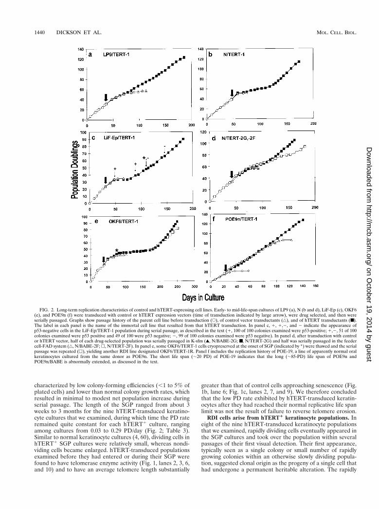
characterized by low colony-forming efficiencies (,1 to 5% ofplated cells) and lower than normal colony growth rates, whichresulted in minimal to modest net population increase duringserial passage. The length of the SGP ranged from about 3weeks to 3 months for the nine hTERT-transduced keratino-cyte cultures that we examined, during which time the PD rateremained quite constant for each hTERT1 culture, rangingamong cultures from 0.03 to 0.29 PD/day (Fig. 2; Table 3).Similar to normal keratinocyte cultures (4, 60), dividing cells inhTERT1 SGP cultures were relatively small, whereas nondi-viding cells became enlarged. hTERT-transduced populationsexamined before they had entered or during their SGP werefound to have telomerase enzyme activity (Fig. 1, lanes 2, 3, 6,and 10) and to have an average telomere length substantially
greater than that of control cells approaching senescence (Fig.1b, lane 6; Fig. 1c, lanes 2, 7, and 9). We therefore concludedthat the low PD rate exhibited by hTERT-transduced keratin-ocytes after they had reached their normal replicative life spanlimit was not the result of failure to reverse telomere erosion.
RDI cells arise from hTERT1 keratinocyte populations. Ineight of the nine hTERT-transduced keratinocyte populationsthat we examined, rapidly dividing cells eventually appeared inthe SGP cultures and took over the population within severalpassages of their first visual detection. Their first appearance,typically seen as a single colony or small number of rapidlygrowing colonies within an otherwise slowly dividing popula-tion, suggested clonal origin as the progeny of a single cell thathad undergone a permanent heritable alteration. The rapidly
FIG. 2. Long-term replication characteristics of control and hTERT-expressing cell lines. Early- to mid-life-span cultures of LP9 (a), N (b and d), LiF-Ep (c), OKF6(e), and POE9n (f) were transduced with control or hTERT expression vectors (time of transduction indicated by large arrow), were drug selected, and then wereserially passaged. Graphs show passage history of the parent cell line before transduction (E), of control vector transductants (‚), and of hTERT transductants (■).The label in each panel is the name of the immortal cell line that resulted from that hTERT transduction. In panel c, 1, 1,2, and 2 indicate the appearance ofp53-negative cells in the LiF-Ep/TERT-1 population during serial passage, as described in the text (1, 100 of 100 colonies examined were p53-positive; 1,2, 51 of 100colonies examined were p53 positive and 49 of 100 were p53 negative; 2, 99 of 100 colonies examined were p53 negative). In panel d, after transduction with controlor hTERT vector, half of each drug-selected population was serially passaged in K-sfm (Œ, N/BABE-2G; ■, N/TERT-2G) and half was serially passaged in the feedercell-FAD system (‚, N/BABE-2F; h, N/TERT-2F). In panel e, some OKF6/TERT-1 cells cryopreserved at the onset of SGP (indicated by *) were thawed and the serialpassage was repeated (h), yielding another RDI line designated OKF6/TERT-1R. Panel f includes the replication history of POE-19, a line of apparently normal oralkeratinocytes cultured from the same donor as POE9n. The short life span (;20 PD) of POE-19 indicates that the long (;85-PD) life span of POE9n andPOE9n/BABE is abnormally extended, as discussed in the text.
1440 DICKSON ET AL. MOL. CELL. BIOL.
on October 19, 2014 by guest
http://mcb.asm
.org/D
ownloaded from
dividing variant cells were uniformly small and had a highcolony-forming efficiency and population growth rate, typicallysimilar to that of early-passage cells of the parent line. Theserapidly dividing, immortalized (RDI) cells expressed telomer-ase activity (Fig. 1a) and maintained telomeres at lengthsgreater than or equal to that of control cells (Fig. 1c). Theycontinued to divide for at least 50 PD beyond the normal lifespan of the parent line. We therefore concluded that they hadundergone immortalization.
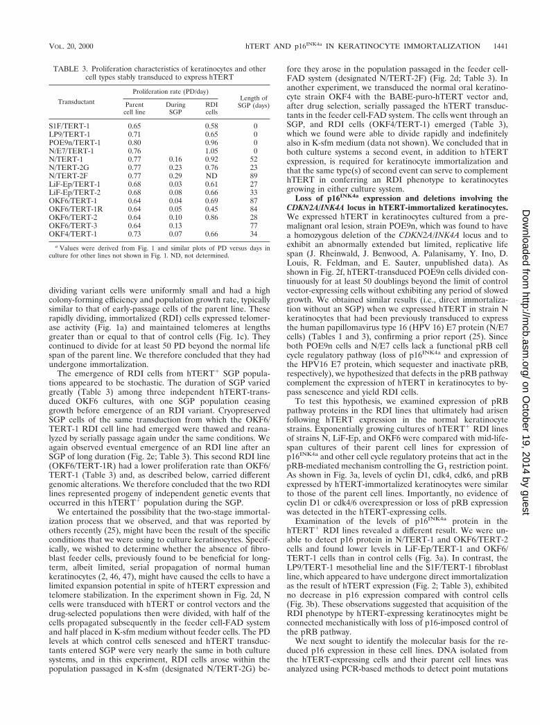
The emergence of RDI cells from hTERT1 SGP popula-tions appeared to be stochastic. The duration of SGP variedgreatly (Table 3) among three independent hTERT-trans-duced OKF6 cultures, with one SGP population ceasinggrowth before emergence of an RDI variant. CryopreservedSGP cells of the same transduction from which the OKF6/TERT-1 RDI cell line had emerged were thawed and reana-lyzed by serially passage again under the same conditions. Weagain observed eventual emergence of an RDI line after anSGP of long duration (Fig. 2e; Table 3). This second RDI line(OKF6/TERT-1R) had a lower proliferation rate than OKF6/TERT-1 (Table 3) and, as described below, carried differentgenomic alterations. We therefore concluded that the two RDIlines represented progeny of independent genetic events thatoccurred in this hTERT1 population during the SGP.
We entertained the possibility that the two-stage immortal-ization process that we observed, and that was reported byothers recently (25), might have been the result of the specificconditions that we were using to culture keratinocytes. Specif-ically, we wished to determine whether the absence of fibro-blast feeder cells, previously found to be beneficial for long-term, albeit limited, serial propagation of normal humankeratinocytes (2, 46, 47), might have caused the cells to have alimited expansion potential in spite of hTERT expression andtelomere stabilization. In the experiment shown in Fig. 2d, Ncells were transduced with hTERT or control vectors and thedrug-selected populations then were divided, with half of thecells propagated subsequently in the feeder cell-FAD systemand half placed in K-sfm medium without feeder cells. The PDlevels at which control cells senesced and hTERT transduc-tants entered SGP were very nearly the same in both culturesystems, and in this experiment, RDI cells arose within thepopulation passaged in K-sfm (designated N/TERT-2G) be-
fore they arose in the population passaged in the feeder cell-FAD system (designated N/TERT-2F) (Fig. 2d; Table 3). Inanother experiment, we transduced the normal oral keratino-cyte strain OKF4 with the BABE-puro-hTERT vector and,after drug selection, serially passaged the hTERT transduc-tants in the feeder cell-FAD system. The cells went through anSGP, and RDI cells (OKF4/TERT-1) emerged (Table 3),which we found were able to divide rapidly and indefinitelyalso in K-sfm medium (data not shown). We concluded that inboth culture systems a second event, in addition to hTERTexpression, is required for keratinocyte immortalization andthat the same type(s) of second event can serve to complementhTERT in conferring an RDI phenotype to keratinocytesgrowing in either culture system.
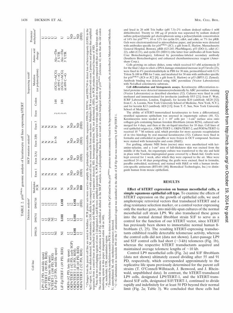
Loss of p16INK4a expression and deletions involving theCDKN2A/INK4A locus in hTERT-immortalized keratinocytes.We expressed hTERT in keratinocytes cultured from a pre-malignant oral lesion, strain POE9n, which was found to havea homozygous deletion of the CDKN2A/INK4A locus and toexhibit an abnormally extended but limited, replicative lifespan (J. Rheinwald, J. Benwood, A. Palanisamy, Y. Ino, D.Louis, R. Feldman, and E. Sauter, unpublished data). Asshown in Fig. 2f, hTERT-transduced POE9n cells divided con-tinuously for at least 50 doublings beyond the limit of controlvector-expressing cells without exhibiting any period of slowedgrowth. We obtained similar results (i.e., direct immortaliza-tion without an SGP) when we expressed hTERT in strain Nkeratinocytes that had been previously transduced to expressthe human papillomavirus type 16 (HPV 16) E7 protein (N/E7cells) (Tables 1 and 3), confirming a prior report (25). Sinceboth POE9n cells and N/E7 cells lack a functional pRB cellcycle regulatory pathway (loss of p16INK4a and expression ofthe HPV16 E7 protein, which sequester and inactivate pRB,respectively), we hypothesized that defects in the pRB pathwaycomplement the expression of hTERT in keratinocytes to by-pass senescence and yield RDI cells.
To test this hypothesis, we examined expression of pRBpathway proteins in the RDI lines that ultimately had arisenfollowing hTERT expression in the normal keratinocytestrains. Exponentially growing cultures of hTERT1 RDI linesof strains N, LiF-Ep, and OKF6 were compared with mid-life-span cultures of their parent cell lines for expression ofp16INK4a and other cell cycle regulatory proteins that act in thepRB-mediated mechanism controlling the G1 restriction point.As shown in Fig. 3a, levels of cyclin D1, cdk4, cdk6, and pRBexpressed by hTERT-immortalized keratinocytes were similarto those of the parent cell lines. Importantly, no evidence ofcyclin D1 or cdk4/6 overexpression or loss of pRB expressionwas detected in the hTERT-expressing cells.
Examination of the levels of p16INK4a protein in thehTERT1 RDI lines revealed a different result. We were un-able to detect p16 protein in N/TERT-1 and OKF6/TERT-2cells and found lower levels in LiF-Ep/TERT-1 and OKF6/TERT-1 cells than in control cells (Fig. 3a). In contrast, theLP9/TERT-1 mesothelial line and the S1F/TERT-1 fibroblastline, which appeared to have undergone direct immortalizationas the result of hTERT expression (Fig. 2; Table 3), exhibitedno decrease in p16 expression compared with control cells(Fig. 3b). These observations suggested that acquisition of theRDI phenotype by hTERT-expressing keratinocytes might beconnected mechanistically with loss of p16-imposed control ofthe pRB pathway.
We next sought to identify the molecular basis for the re-duced p16 expression in these cell lines. DNA isolated fromthe hTERT-expressing cells and their parent cell lines wasanalyzed using PCR-based methods to detect point mutations
TABLE 3. Proliferation characteristics of keratinocytes and othercell types stably transduced to express hTERT
Transductant
Proliferation rate (PD/day)Length of
SGP (days)Parentcell line
DuringSGP
RDIcells
S1F/TERT-1 0.65 0.58 0LP9/TERT-1 0.71 0.65 0POE9n/TERT-1 0.80 0.96 0N/E7/TERT-1 0.76 1.05 0N/TERT-1 0.77 0.16 0.92 52N/TERT-2G 0.77 0.23 0.76 23N/TERT-2F 0.77 0.29 ND 89LiF-Ep/TERT-1 0.68 0.03 0.61 27LiF-Ep/TERT-2 0.68 0.08 0.66 33OKF6/TERT-1 0.64 0.04 0.69 87OKF6/TERT-1R 0.64 0.05 0.45 84OKF6/TERT-2 0.64 0.10 0.86 28OKF6/TERT-3 0.64 0.13 77OKF4/TERT-1 0.73 0.07 0.66 34
a Values were derived from Fig. 1 and similar plots of PD versus days inculture for other lines not shown in Fig. 1. ND, not determined.
VOL. 20, 2000 hTERT AND p16INK4a IN KERATINOCYTE IMMORTALIZATION 1441
on October 19, 2014 by guest
http://mcb.asm
.org/D
ownloaded from
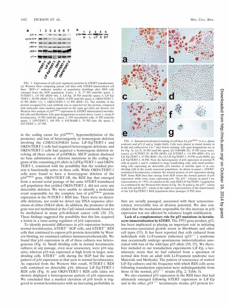
in the coding exons for p16INK4a, hypermethylation of thepromoter, and loss of heterozygosity or homozygous deletioninvolving the CDKN2A/INK4A locus. LiF-Ep/TERT-1 andOKF6/TERT-1 cells had acquired heterozygous deletions, andOKF6/TERT-2 cells had acquired a homozygous deletion in-volving all three exons of p16INK4a. SSCP analysis disclosedno base substitution or deletion mutations in the coding re-gions of the remaining p16 allele in LiFEp/TERT-1 and OKF6/TERT-1, consistent with the possibility that the residual pro-tein is functionally active in these cells. While OKF6/TERT-1cells were found to have a heterozygous deletion of thep16INK4a gene, OKF6/TERT-1R, the RDI line that emergedfrom a second serial passage of the same hTERT-transducedcell population that yielded OKF6/TERT-1, did not carry anydetectable deletion. We were unable to identify a molecularevent responsible for the complete loss of p16INK4a proteinexpression in the N/TERT-1 RDI line. There were no detect-able deletions, nor could we detect any DNA sequence alter-ations of either INK4A allele. In addition, the promoter of thisgene was not methylated at the CpG island commonly found tobe methylated in many p16-deficient cancer cells (20, 33).These findings suggested the possibility that this line acquireda lesion in a trans-acting regulator of p16 expression.
To identify potential differences in p16 expression betweennormal keratinocytes, hTERT1 SGP cells, and hTERT1 RDIcells that continued to express p16 protein detectable by West-ern blotting, we examined cultures immunocytochemically. Wefound that p16 expression in all of these cultures was hetero-geneous (Fig. 4). Small dividing cells in normal keratinocytecultures at any passage, even near senescence, were p16 neg-ative, whereas p16 was expressed at high levels in larger, non-dividing cells. hTERT1 cells during the SGP had the samepattern of p16 expression as that seen in normal keratinocytes.As expected from the Western blot result, N/TERT-1 RDIcells contained no detectable p16, whereas LiF-Ep/TERT-1RDI cells (Fig. 4) and OKF6/TERT-1 RDI cells (data notshown) displayed a heterogeneous pattern of p16 expression.We concluded that a marked elevation of p16 levels is trig-gered in normal keratinocytes with an increasing probability as
they are serially passaged, associated with their senescence-related, irreversible loss of division potential. We also con-cluded that the mechanism responsible for this increase in p16expression was not affected by telomere length stabilization.
Lack of a complementary role for p53 mutations in keratin-ocyte immortalization by hTERT. The TP53 gene product, p53,has been implicated as playing an important role in initiatingsenescence-associated growth arrest in fibroblasts and othercell types (53). It has been reported that cells cultured fromindividuals with Li-Fraumeni (inherited (p531/2) syndromemay occasionally undergo spontaneous immortalization asso-ciated with loss of the wild-type p53 allele (50, 55). We there-fore included in our transduction experiments LiF-Ep, a ker-atinocyte strain that we had cultured from a specimen ofnormal skin from an adult with Li-Fraumeni syndrome (seeMaterials and Methods). The pattern of senescence of controlLiF-Ep cultures and the frequency with which RDI cells arosefollowing transduction expression were indistinguishable fromthose of the normal, p531/1 strains (Fig. 2; Table 3).
We also examined p53 expression in the RDI lines that hadultimately emerged following hTERT expression in LiF-Epand in the other, p531/1 keratinocyte strains. p53 protein lev-
FIG. 3. Expression of cell cycle regulatory proteins by hTERT transductants.(a) Western blots comparing parent cell lines with hTERT-immortalized celllines. “RDI1n” indicates number of population doublings after RDI cellsemerged from the SGP population. Lanes: 1, N, 37 PD (mid-life span); 2,N/TERT-1, 135 PD (RDI168); 3, LiF-Ep, 20 PD (mid-life span); 4, LiF-Ep/TERT-1, 90 PD (RDI155); 5, OKF6, 19 PD (mid-life span); 6, OKF6/TERT-1,63 PD (RDI111); 7, OKF6/TERT-2, 52 PD (RDI115). The mobility of theprotein recognized by each antibody was as expected for the protein, comparedwith molecular mass markers separated on the same gel (data not shown). (b)Western blot analysis of p16INK4a expression by hTERT-immortalized mesothe-lial cells and fibroblasts. Life span designations as noted above. Lanes 1, strain Nkeratinocytes, 33 PD (mid-life span); 2, LP9 mesothelial cells, 41 PD (mid-lifespan); 3, LP9/TERT-1, 108 PD; 4, S1F/BABE-1, 70 PD (late life span); 5,S1F/TERT-1, 147 PD.
FIG. 4. Immunocytochemical staining of cell lines for p16INK4a (a to e, phasecontrast) and p53 (f and g, bright field). Cells were plated at clonal density inK-sfm and cultured for 4 to 7 days before staining. Life span designations are asfor Fig. 3a. (a) N, 28 PD (mid-life span); (b) N/BABE-2G, 55 PD (near senes-cence); (c) N/TERT-2F, 80 PD (SGP); (d) N/TERT-1, 114 PD (RDI143); (e)LiF-Ep/TERT-1, 67 PD (RDI128); (f) LiF-Ep/TERT-1, 39 PD (early RDI); (g)LiF-Ep/TERT-1, 48 PD. Note the heterogeneity of p16 expression in normal Ncells in panels a and b, confined to large nondividing cells, with small prolifer-ating cells expressing no detectable p16 whether at mid-life span or in raredividing cells in the nearly senescent population. As shown in panel c, hTERT-transduced keratinocytes continue the normal pattern of p16 expression duringSGP. Some RDI lines that emerge from SGP retain the normal pattern of p16expression, while some cease expressing p16. The p531 colonies in panel f arerepresentative of .99% of colonies in the early RDI LiF-Ep/TERT-1 population(as confirmed by the Western blot shown in Fig. 3b). In panel g, the p531 colonyat the left and the p532 colony at the right are representative of the mixed natureof the LiF-Ep/TERT-1 RDI population three passages (9 PD) later.
1442 DICKSON ET AL. MOL. CELL. BIOL.
on October 19, 2014 by guest
http://mcb.asm
.org/D
ownloaded from
els expressed by the hTERT1 RDI lines during exponentialgrowth were similar to those of the respective parent cell lineswith the exception of LiF-Ep/TERT-1 (Fig. 3a). When exam-ined at 90 PD (55 PD after emergence of RDI cells), LiF-Ep/TERT-1 cells had no p53 protein detectable by Westernblotting. We therefore analyzed LiF-Ep/TERT-1 cells immu-nocytochemically for p53 expression and function at varioustimes during the SGP and after emergence of the RDI line.Normal keratinocytes express low levels of p53 protein duringexponential growth, but cells with functionally normal p53 ex-pression and function greatly increase their levels of this pro-tein in response to DNA damage, as results from exposure tolow levels of actinomycin D (23). As shown in Fig. 4 and 2c,p53 protein-negative, presumably genetically p532/2 cells firstappeared in the LiF-Ep/TERT-1 culture long after the eventresponsible for conversion of SGP cells to RDI status. TheLiF-Ep/TERT-1 population consisted of at least 99% p531
cells at the time rapidly dividing variants first emerged fromthe SGP (at 39 PD). Fifty percent of the cells in the RDIpopulation were p53 negative three passages later (at 48 PD),and the culture consisted entirely of p53-negative cells by 66PD (Fig. 4 and 2c).
A different hTERT transduction of LiF-Ep yielded an RDIline, LiF-Ep/TERT-2 (Table 3), in which apparently all of theRDI cells (i.e., 150 of 150 colonies examined immunocyto-chemically [data not shown]) remained phenotypically normalfor p53 expression and function at least 20 PD after theiremergence from SGP. We therefore concluded that p53-defi-cient keratinocytes do not have a selective advantage inhTERT1 populations during SGP and, therefore, that loss ofp53 does not complement hTERT for keratinocyte immortal-ization.
Growth regulation and differentiation characteristics ofhTERT-immortalized keratinocytes. We wished to determinewhether any aspect of immortalization, especially p16INK4a
deficiency, which is a frequent characteristic of keratinocytetransformation in vivo (9) and of human squamous cell carci-noma (SCC) cell lines that can grow in culture (29, 37), im-paired specific keratinocyte growth control and differentiationmechanisms. Four of the hTERT transductants were com-pared with their respective parent cell lines in several in vitroassays for function of several keratinocyte growth regulatorymechanisms that are frequently lost in SCC cells (41, 45, 51). Incontrol conditions, the hTERT lines, tested at 45 to 68 PDafter emergence as RDI cells, all had higher PD rates thanmid- to late-life-span cells of their respective parent cell strains(Fig. 5). N/TERT-1, LiF-Ep/TERT-1, and OKF6/TERT-1 cellsstill retained dependence upon EGF for growth and sensitivityto growth inhibition by the phorbol ester TPA, exhibiting onlyminimal growth (less than one (,1-PD increase over the plat-ing density during the course of the experiment) in the absenceof EGF or presence of TPA. The exception was OKF6/TERT-2, which remained TPA sensitive but proliferated in theabsence of EGF at 60% of the rate in the presence of EGF,approaching the degree of EGF independence of SCC-13.The four hTERT-transductants examined, including OKF6/TERT-2, were growth inhibited by transforming growth factorb (data not shown). We concluded that loss of normal kera-tinocyte growth regulatory mechanisms is not a necessary con-sequence of immortalization, even when the senescence-re-lated pRB/p16INK4a growth arrest mechanism has beenabrogated, as it is in N/TERT-1. However, our results couldnot eliminate the possibility that homozygous deletion of thep16INK4a locus was responsible for the EGF independenceexhibited by OKF6/TERT-2 cells (Fig. 5a) and by POE9n andPOE9n/TERT cells (data not shown).
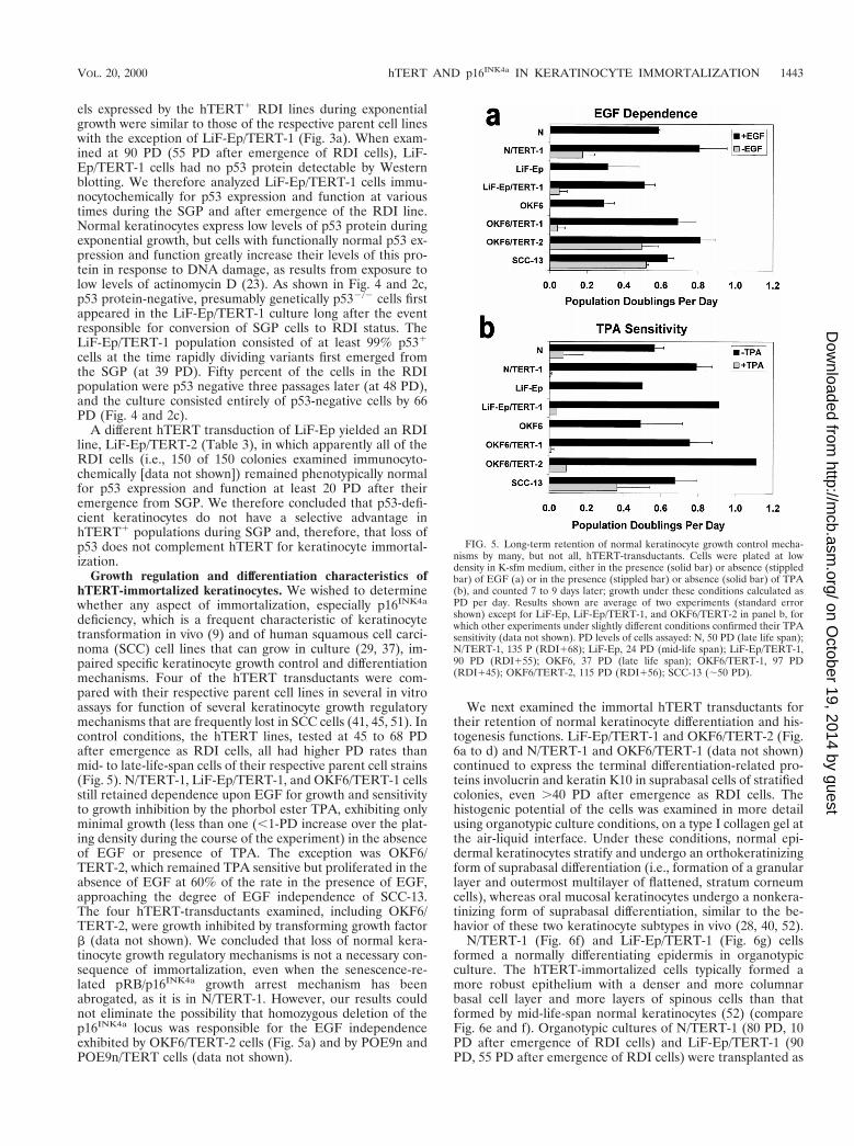
We next examined the immortal hTERT transductants fortheir retention of normal keratinocyte differentiation and his-togenesis functions. LiF-Ep/TERT-1 and OKF6/TERT-2 (Fig.6a to d) and N/TERT-1 and OKF6/TERT-1 (data not shown)continued to express the terminal differentiation-related pro-teins involucrin and keratin K10 in suprabasal cells of stratifiedcolonies, even .40 PD after emergence as RDI cells. Thehistogenic potential of the cells was examined in more detailusing organotypic culture conditions, on a type I collagen gel atthe air-liquid interface. Under these conditions, normal epi-dermal keratinocytes stratify and undergo an orthokeratinizingform of suprabasal differentiation (i.e., formation of a granularlayer and outermost multilayer of flattened, stratum corneumcells), whereas oral mucosal keratinocytes undergo a nonkera-tinizing form of suprabasal differentiation, similar to the be-havior of these two keratinocyte subtypes in vivo (28, 40, 52).
N/TERT-1 (Fig. 6f) and LiF-Ep/TERT-1 (Fig. 6g) cellsformed a normally differentiating epidermis in organotypicculture. The hTERT-immortalized cells typically formed amore robust epithelium with a denser and more columnarbasal cell layer and more layers of spinous cells than thatformed by mid-life-span normal keratinocytes (52) (compareFig. 6e and f). Organotypic cultures of N/TERT-1 (80 PD, 10PD after emergence of RDI cells) and LiF-Ep/TERT-1 (90PD, 55 PD after emergence of RDI cells) were transplanted as
FIG. 5. Long-term retention of normal keratinocyte growth control mecha-nisms by many, but not all, hTERT-transductants. Cells were plated at lowdensity in K-sfm medium, either in the presence (solid bar) or absence (stippledbar) of EGF (a) or in the presence (stippled bar) or absence (solid bar) of TPA(b), and counted 7 to 9 days later; growth under these conditions calculated asPD per day. Results shown are average of two experiments (standard errorshown) except for LiF-Ep, LiF-Ep/TERT-1, and OKF6/TERT-2 in panel b, forwhich other experiments under slightly different conditions confirmed their TPAsensitivity (data not shown). PD levels of cells assayed: N, 50 PD (late life span);N/TERT-1, 135 P (RDI168); LiF-Ep, 24 PD (mid-life span); LiF-Ep/TERT-1,90 PD (RDI155); OKF6, 37 PD (late life span); OKF6/TERT-1, 97 PD(RDI145); OKF6/TERT-2, 115 PD (RDI156); SCC-13 (;50 PD).
VOL. 20, 2000 hTERT AND p16INK4a IN KERATINOCYTE IMMORTALIZATION 1443
on October 19, 2014 by guest
http://mcb.asm
.org/D
ownloaded from

surface skin grafts onto athymic mice. N/TERT-1 and LiF-Ep/TERT-1 grafts recovered at 24 days (data not shown) and 48days (Fig. 6h), respectively, were found to have formed a nor-mally differentiating epidermis with no evidence of invasion ofunderlying connective tissue. OKF6/TERT-1 and -2 cells inorganotypic culture underwent a nonkeratinizing form of strat-ified squamous epithelial differentiation, including suprabasalexpression of keratin K13 (Fig. 6i and j), characteristic offloor-of-mouth mucosa (52), from which the parent line OKF6
was cultured. We concluded that no alteration necessary forimmortalization, including loss of the ability to expressp16INK4a, interferes with keratinocyte differentiation.
DISCUSSION
Several lines of evidence have implicated telomere erosionin limiting the proliferative potential of human cells, but theresults presented here clearly indicate that a different mecha-
FIG. 6. Differentiation-related protein expression and epithelial tissue formation by hTERT-immortalized keratinocytes. Cells were cultured on conventional plasticdishes for 7 to 9 days in K-sfm (a and b) or in the feeder cell-FAD system (c and d) or were seeded on collagen gels to generate organotypic cultures (e to j), one ofwhich was then grafted to the skin of an athymic mouse (h). Panels a (LiF-Ep, 28 PD) and b (LiF-Ep/TERT-1, 66 PD [RDI143]) were immunostained for the suprabasalepidermal differentiation keratin K10. Panels c (OKF6, 30 PD) and d (OKF6/TERT-2, 122 PD [RDI163]) were immunostained for the keratinocyte terminaldifferentiation protein involucrin. (e) N, 37 PD (mid-life span); f, N/TERT-1, 91 PD (RDI124); g, LiF-Ep/TERT-1, 90 PD (RDI155); h, a replicate organotypic cultureof the one shown in panel g, 42 days after transplantation as a skin graft to an athymic mouse; i and j, OKF6/TERT-2, 86 PD (RDI147). Panels e to i were stainedwith H&E (note that the intense blue of the cell layers below the stratum corneum in the epithelium shown in panel h is the result of a slightly different stainingformulation and has no biological significance). Panel j was immunostained for keratin K13. Arrowheads point to the basal layer of the organotypic epithelium, showingthat K13 expression begins several cell layers above the first suprabasal layer.
1444 DICKSON ET AL. MOL. CELL. BIOL.
on October 19, 2014 by guest
http://mcb.asm
.org/D
ownloaded from
nism is responsible for determining the replicative life span ofhuman keratinocytes in culture. We have identified a precipi-tous increase in p16INK4a protein levels accompanying end-of-life-span growth arrest in normal keratinocytes, which appearsto be triggered by a mechanism that is activated in cells withincreasing probability as a cell population is serially propa-gated. Loss of this mechanism, whether by p16INK4a gene de-letion, mutation, or altered regulation of expression, togetherwith telomere stabilization effected by hTERT expression isnecessary to enable a keratinocyte to become immortalized.Perhaps surprisingly, immortalization of keratinocytes byforced expression of telomerase and subsequent spontaneousevents leading to loss of this p16INK4a-dependent mechanismgenerally does not disrupt other normal growth control mech-anisms or affect the ability of the cells to form a differentiatedepithelium. In contrast, senescence arrest is abrogated in cul-tured fibroblasts and retinal pigment epithelial cells by expres-sion of hTERT (5), and our observations have added the me-sothelial cell, a mesoderm-derived epithelial cell, to the groupof cell types for which a telomere length-sensitive mechanismappears responsible for initiating senescence.
We have shown that expression of hTERT alone permitskeratinocytes to escape complete growth arrest and to enter aphase of slow growth of variable length from which rapidlydividing immortal variants emerge. Such immortalized cellstypically have identifiable defects in p16INK4a expression butretain functional p53. These results therefore confirm and ex-tend those of Kiyono et al. (25), who demonstrated that ex-pression of hTERT in combination with expression of theHPV16 E7 oncoprotein allowed human foreskin keratinocytesand mammary epithelial cells to bypass senescence. We havedemonstrated that telomere length stabilization alone is un-able to permit keratinocytes to bypass senescence, but that thesubsequent slow, indefinite continued growth permitted by te-lomerase expression permits rare immortalized variants toarise.
Our observations clearly provide evidence supporting recentproposals (43, 62) that multiple “clocks” function to limit theproliferative capacity of human cells. The mechanism that trig-gers p16INK4a accumulation appears to sense the proliferativehistory of the keratinocyte, but preventing telomere erosiondoes not avoid its activation. In hTERT-expressing keratino-cyte populations a small proportion of the cells indefinitelyevade arrest, however. It is possible, therefore, that senes-cence-associated p16INK4a regulation is under two types ofcontrol, one that is tightly telomere length sensitive and an-other that is telomere length independent and stochastic.Whether the latter detects an aspect of cell aging related tonumber of cell divisions or to chronological time in cultureremains to be determined because it is not possible to adjustculture conditions to slow or arrest keratinocyte growth with-out triggering irreversible commitment to terminal differenti-ation. The molecular mechanisms regulating p16INK4a geneexpression are only beginning to be elucidated (18). It has beenproposed that in vivo a subpopulation of keratinocytes serve asstem cells by possessing a very long or indefinite replicativepotential (for example, see references 26 and 59). Whetherstem cell status involves a special mechanism for avoidingsenescence-related p16INK4a expression remains to be deter-mined.
Mutations in the pRB/p16INK4a tumor suppressor pathwayare found in the majority of human cancers (54). The obser-vations presented here implicate this pathway as an essentialcontrol mechanism that must be subverted to create immortalcells. The role of p16INK4a in limiting epithelial cell prolifera-tion is suggested by findings that variants of mammary epithe-
lial cells that are unable to express p16 owing to promoterhypermethylation exhibit an extended replicative life span (6,15); that p16 levels, but not p21 levels, progressively increase inkeratinocyte cultures as they approach senescence (37); andthat immortal SCC cell lines (29) and immortal variants ofHPV16 E6-transfected prostate epithelial cell cultures (22) arefound consistently to have lost p16 expression as a result ofmutation or promoter hypermethylation.
It has been reported (25, 37) that p16INK4a levels, detectedby Western blot analysis, increase in keratinocyte cultures asthey are serially propagated. These data would support amodel in which slowly increasing levels of p16INK4a in dividingkeratinocytes eventually result in levels at which a subsequentG1-S transition can no longer occur. Our immunocytochemicalanalysis of normal and hTERT-transduced keratinocyte cul-tures revealed an unsuspected complexity in p16INK4a expres-sion. We detected marked heterogeneity in p16INK4a levelswithin normal keratinocyte cultures at both early and latepassage. p16INK4a protein was undetectable in small, rapidlydividing cells at any stage of the life span, at low to moderatelevels in slightly larger cells in more slowly dividing colonies,and at very high levels in large, nondividing cells. This heter-ogeneous pattern of p16INK4a expression continued inhTERT1 keratinocyte populations during the SGP. These ob-servations suggest that telomere-independent mechanisms sig-nal senescence in keratinocytes by triggering a precipitous in-crease in p16INK4a levels that then causes cell cycle arrest.Moreover, they suggest that the incrementally increasing levelsof p16INK4a observed in serially passaged keratinocyte culturesresult from the increasing representation of cells that expresshigh levels of p16INK4a amid a majority population of cells thatremain p16INK4a negative. Since p21cip1 protein does not in-crease in senescent keratinocyte cultures (37), increasedp16INK4a levels are likely to serve as the effector mechanismthat enforces senescence-related growth arrest in keratino-cytes.
In the majority of the rapidly dividing immortal variants thatarose within the hTERT1 keratinocyte cultures examinedhere, we identified alterations compromising p16INK4a expres-sion. Such lesions included homozygous deletion involving allp16INK4a exons, heterozygous deletion of the CDKN2A/INK4Alocus with continued, heterogeneous expression of p16INK4a,and no detectable deletion, mutation, or promoter hypermeth-ylation but complete loss of p16INK4a protein expression. Therapidly dividing immortal keratinocyte lines that continue toexpress p16INK4a may have mutations in other proteins in-volved in the pRB/p16INK4a pathway, or they may have ac-quired a lesion in a specific, senescence-related inducer ofp16INK4a.
The deletions we detected at CDKN2A/INK4A also includedloss of at least the second exon of p14ARF, another potentialcell cycle inhibitor encoded in part by an alternate readingframe of the p16INK4a exon 2. p14ARF functions by preventingMDM2 from targeting p53 for degradation, thereby resultingin elevated levels of the latter and consequent cell cycle arrest(63). A recent study (37) has found that p14ARF levels do notincrease during keratinocyte senescence and that all of 20immortal human SCC lines examined had mutations compro-mising p16INK4a and none had mutations specifically affectingp14ARF. Our preliminary analyses also indicate that p14ARF isstill expressed by hTERT-immortalized lines that still have atleast one intact CDKN2A/INK4A allele (Z. Guo and J. Rhei-nwald, unpublished data), supporting the conclusion that it isloss of p16INK4a that is required to complement hTERT forkeratinocyte immortalization.
We found no evidence that inactivation of any p53-depen-
VOL. 20, 2000 hTERT AND p16INK4a IN KERATINOCYTE IMMORTALIZATION 1445
on October 19, 2014 by guest
http://mcb.asm
.org/D
ownloaded from
dent pathway is necessary for immortalization of keratinocytesthat express hTERT. In neither of two hTERT transductionsof the p531/2 LiF-Ep strain did we find RDI conversion asso-ciated with loss of the wild-type p53 allele. These observationsare consistent with earlier studies suggesting that p53 is anessential component of the telomere length-sensitive growtharrest signal in fibroblasts (62), in that hTERT-expressing ker-atinocytes would be expected to permanently avoid activatingsuch a signal.
Despite expression of hTERT and loss of p16INK4a function,the immortal keratinocytes described here are able to initiatetheir program of terminal differentiation, express suprabasaldifferentiation-specific proteins, and form differentiated epi-thelia in vitro and in vivo. In particular, the N/TERT-1 andLiF-Ep/TERT-1 lines continued to show a normal pattern ofepidermal histogenesis in organotypic culture and in grafts toathymic mice. These results clearly indicate that p16 does notplay an essential role in the irreversible growth arrest thatprecedes normal stratified squamous epithelial differentiation.Three of the four RDI TERT lines that we studied in detailalso retained EGF dependence for growth at low density andsensitivity to irreversible growth arrest by TPA, which are nor-mal keratinocyte growth control mechanisms consistentlyfound to be lost in advanced SCCs (41, 45). Although OKF6/TERT-2, which had undergone homozygous deletion of theCDKN2A/INK4A locus, showed EGF-independent growth,our observations clearly demonstrate that the mechanisms in-volved in keratinocyte senescence and immortalization are dis-tinct from those essential for other aspects of tissue growthcontrol and differentiation.
Finally, the hTERT-immortalized keratinocyte lines that wehave described here may have significant potential value for awide variety of investigations into human epithelial biology,including characterization of the effects of expression or loss ofspecific gene products on acquisition of malignant phenotypes.Although we have demonstrated that hTERT-immortalizedimmortal cells can retain normal growth and differentiationcontrol mechanisms, it is possible that the loss of the p16-mediated growth arrest mechanism and unlimited replicativepotential predisposes such cells to further changes that mayresult in malignant transformation. Indeed, it has been shownrecently that expression of hTERT cooperates with the simianvirus 40 large T oncoprotein and oncogenic ras to transformhuman fibroblasts and kidney epithelial cells to tumorigenicity(17). For this reason, enthusiasm for potential clinical appli-cations of hTERT-immortalized epithelial cell lines as thera-peutic transplants should be tempered with caution.
ACKNOWLEDGMENTS
We thank J. Benwood, D. Long-Woodward, and K. O’Toole fortechnical assistance and S. Verselis for information about the mutantp53 allele in our Li-Fraumeni cell line. We thank D. Galloway for theHPV16 E7 retroviral vector, J. Koh, S. C, Ngwu, and E. Harlow forp16INK4a antibodies, and T.-T. Sun, C. A. Loomis, and F. M. Watt forkeratin and involucrin antibodies.
This research was supported by Oral Cancer Program Project grantPO1 DE12467 from the NIDCR, Skin Disease Research Center grantP30 AR42689 from the NIAMS, and a research grant from Organo-genesis, Inc. (J.G.R.). In addition, portions of this work were sup-ported by a Daniel K. Ludwig and American Cancer Society Profes-sorship (R.A.W.), a Culpeper Biomedical Initiative Pilot Grant(W.C.H. and R.A.W.), a Damon-Runyon/Walter Winchell Cancer Re-search Fund award, a Howard Hughes Medical Institute postdoctoralfellowship, and a Herman and Margaret Sokol postdoctoral fellowship(W.C.H.), and a Starr Foundation and American Cancer Society Clin-ical Research Professorship (F.P.L.).
REFERENCES
1. Alcorta, D. A., Y. Xiong, D. Phelps, G. Hannon, D. Beach, and J. C. Barrett.1996. Involvement of the cyclin-dependent kinase inhibitor p16 (INK4a) inreplicative senescence of normal human fibroblasts. Proc. Natl. Acad. Sci.USA 93:13742–13747.
2. Allsopp, R. C., H. Vazin, C. Patterson, S. Goldstein, E. V. Younglai, A. B.Futcher, C. W. Greider, and C. B. Harley. 1992. Telomere length predictsreplicative capacity of human fibroblasts. Proc. Natl. Acad. Sci. USA 89:10114–10118.
3. Bahuau, M., D. Vidaud, R. B. Jenkins, I. Bieche, D. W. Kimmel, B. Assou-line, J. S. Smith, B. Alderete, J. M. Cayuela, J. P. Harpey, B. Caille, and M.Vidaud. 1998. Germ-line deletion involving the INK4 locus in familial prone-ness to melanoma and nervous system tumors. Cancer Res. 58:2298–2303.
4. Barrandon, Y., and H. Green. 1985. Cell size as a determinant of the clone-forming ability of human keratinocytes. Proc. Natl. Acad. Sci. USA 82:5390–5394.
5. Bodnar, A. G., M. Ouelette, M. Frolkis, A. E. Holt, C.-P. Chiu, G. B. Morin,C. B. Harley, J. W. Shay, S. Lichsteiner, and W. E. Wright. 1998. Extensionof lifespan by introduction of telomerase into normal human cells. Science279:349–352.
6. Brenner, A. J., M. R. Stampfer, and C. M. Aldaz. 1998. Increased p16expression with first senescence arrest in human mammary epithelial cellsand extended growth capacity with p16 inactivation. Oncogene 17:199–205.
7. Brown, J. P., W. Wei, and J. M. Sedivy. 1997. Bypass of senescence afterdisruption of p21cip1/waf1 gene in normal diploid human fibroblasts. Science277:831–834.
8. Burns, K. L., K. Ueki, S. L. Jhung, J. Koh, and D. N. Louis. 1998. Moleculargenetic correlates of p16, cdk4, and pRb immunohistochemistry in glioblas-tomas. J. Neuropathol. Exp. Neurol. 57:122–130.
9. Califano, J., P. Van der Riet, W. Westra, H. Nawroz, G. Clayman, S. Pianta-dosi, R. Corio, D. Lee, B. Greenberg, W. Koch, and D. Sidransky. 1996.Genetic progression model for head and neck cancer: implications for fieldcancerization. Cancer Res. 56:2488–2492.
10. Connell, N. D., and J. G. Rheinwald. 1983. Regulation of the cytoskeleton inmesothelial cells: reversible loss of keratin and increase in vimentin duringrapid growth in culture. Cell 34:245–253.
11. Counter, C. M., A. A. Avilion, C. E. LeFeuvre, N. G. Stewart, C. W. Greider,C. B. Harley, and S. Bacchetti. 1992. Telomere shortening associated withchromosome instability is arrested in immortal cells which express telomer-ase activity. EMBO J. 11:1921–1929.
12. Counter, C. M., F. M. Botelho, P. Wang, C. B. Harley, and S. Bacchetti. 1994.Stabilization of short telomeres and telomerase activity accompany immor-talization of Epstein-Barr virus-transformed human B lymphocytes. J. Virol.68:3410–3414.
13. Dhouailly, D., C. Xu, M. Manabe, A. Schermer, and T.-T. Sun. 1989. Ex-pression of hair-related keratins in a soft epithelium: subpopulations ofhuman and mouse dorsal tongue keratinocytes express keratin markers forhair-, skin-, and esophageal-types of differentiation. Exp. Cell Res. 181:141–158.
14. Feng, J., W. D. Funk, S. S. Wang, S. L. Weinrich, A. A. Avilion, C. P. Chiu,R. R. Adams, E. Chang, R. C. Allsopp, J. Yu, S. Le, M. D. West, C. B. Harley,W. H. Andrews, C. W. Greider, and B. Villeponteau. 1995. The RNA com-ponent of human telomerase. Science 269:1236–1241.
15. Foster, S. A., D. J. Wong, M. T. Barrett, and D. A. Galloway. 1998. Inacti-vation of p16 in human mammary epithelial cells by CpG island methylation.Mol. Cell. Biol. 18:1793–1801.
16. Fuchs, E. 1990. Epidermal differentiation: the bare essentials. J. Cell Biol.111:2807–2814.
17. Hahn, W. C., C. M. Counter, A. S. Lundberg, R. L. Beijersbergen, M. W.Brooks, and R. A. Weinberg. 1999. Creation of human tumor cells withdefined genetic elements. Nature 400:464–468.
18. Hara, E., R. Smith, D. Parry, H. Tahara, S. Stone, and G. Peters. 1996.Regulation of p16INK4a expression and its implications for cell immortaliza-tion and senescence. Mol. Cell. Biol. 16:859–867.
19. Harley, C. B., A. B. Futcher, and C. W. Greider. 1990. Telomeres shortenduring ageing of human fibroblasts. Nature 345:458–460.
20. Herman, J. G., J. R. Graff, S. Myohanen, B. D. Nelkin, and S. B. Baylin.1996. Methylation-specific PCR: a novel PCR assay for methylation status ofCpG islands. Proc. Natl. Acad. Sci. USA 93:9821–9826.
21. Hudson, D. L., K. L. Weiland, T. P. Dooley, M. Simon, and F. M. Watt. 1992.Characterisation of eight monoclonal antibodies to involucrin. Hybridoma11:367–379.
22. Jarrard, D. F., S. Sarkar, Y. Shi, T. R. Yeager, G. Magrane, H. Kinoshita, N.Nassif, L. Meisner, M. A. Newton, F. M. Waldman, and C. A. Reznikoff.1999. p16/pRb pathway alterations are required for bypassing senescence inhuman prostate epithelial cells. Cancer Res. 59:2957–2964.
23. Kastan, M. B., O. Onyekwere, D. Sidransky, B. Vogelstein, and R. W. Craig.1991. Participation of p53 protein in the cellular response to DNA damage.Cancer Res. 51:6304–6311.
24. Kim, N. W., and F. Wu. 1997. Advances in quantification and characteriza-tion of telomerase activity by the telomeric repeat amplification protocol(TRAP). Nucleic Acids Res. 25:2595–2597.
1446 DICKSON ET AL. MOL. CELL. BIOL.
on October 19, 2014 by guest
http://mcb.asm
.org/D
ownloaded from
25. Kiyono, T., S. A. Foster, J. I. Koop, J. K. McDougall, D. A. Galloway, andA. J. Klingelhutz. 1998. Both Rb/p16ink4a inactivation and telomerase activityare required to immortalize human epithelial cells. Nature 396:84–88.
26. Lavker, R. M., and T.-T. Sun. 1983. Epidermal stem cells. J. Investig. Der-matol. 81:1211s–127s.
27. Li, F. P., J. F. Fraumeni, J. J. Mulvihill, W. A. Blattner, M. G. Dreyfuss,M. A. Tucker, and R. W. Miller. 1988. A cancer family syndrome in twenty-four kindreds. Cancer Res. 48:5358–5362.
28. Lindberg, K., and J. G. Rheinwald. 1990. Three distinct keratinocyte sub-types identified in human oral epithelium by their patterns of keratin ex-pression in culture and in xenografts. Differentiation 45:230–241.
29. Loughran, O., A. Malliri, D. Owens, P. H. Gallimore, M. A. Stanley, B.Ozanne, M. C. Frame, and E. K. Parkinson. 1996. Association of CDKN2A/P16INK4a with human head and neck keratinocyte replicative senescence:relationship of dysfunction to immortality and neoplasia. Oncogene 13:561–568.
30. Louis, D. N., A. von Deimling, and B. R. Seizinger. 1992. A (CA)n dinucle-otide repeat assay for evaluating loss of allelic heterozygosity in small andarchival human brain tumor specimens. Am. J. Pathol. 141:777–782.
31. Malkin, D., F. P. Li, L. C. Strong, J. F. Fraumeni, C. E. Nelson, D. H. Kim,J. Kassel, M. A. Gryka, F. Z. Bischoff, and M. A. Tainsky. 1990. Germlinep53 mutations in a familial syndrome of breast cancer, sarcomas, and otherneoplasms. Science 250:1233–1238.
32. Manabe, M., H. W. Lim, M. Winzer, and C. A. Loomis. 1999. Architecturalorganization of filiform papillae in normal and black hairy tongue epithe-lium: dissection of differentiation pathways in a complex human epitheliumaccording to their patterns of keratin expression. Arch. Dermatol. 135:177–181.
33. Merlo, A., J. G. Herman, L. Mao, D. J. Lee, E. Gabrielson, P. C. Burger, S. B.Baylin, and D. Sidransky. 1995. 59 CpG island methylation is associated withtranscriptional silencing of the tumour suppressor p16/CDKN2/MTS1 inhuman cancers. Nat. Med. 1:686–692.
34. Meyerson, M., C. M. Counter, E. N. Eaton, L. W. Ellisen, P. Steiner, S. D.Caddle, L. Ziaugru, R. L. Beijersbergen, M. J. Davidoff, Q. Liu, S. Bachetti,D. A. Haber, and R. A. Weinberg. 1997. hEST2, the putative human telom-erase catalytic subunit gene, is up-regulated in tumor cells and during im-mortalization. Cell 90:785–795.
35. Miller, A. D., and G. J. Rosman. 1989. Improved retroviral vectors for genetransfer and expression. BioTechniques 7:980–990.
36. Morgenstern, J. P., and H. Land. 1990. Advanced mammalian gene transfer:high titer retroviral vectors with multiple drug selection markers and acomplementary helper-free packaging cell line. Nucleic Acids Res. 18:3585–3596.
37. Munro, J., F. J. Stott, K. H. Vousden, G. Peters, and E. K. Parkinson. 1999.Role of the alternative INK4A proteins in human keratinocyte senescence:evidence for the specific inactivation of p16INK4a upon immortalization.Cancer Res. 59:2516–2521.
38. Nakamura, T. M., G. B. Morin, K. B. Chapman, S. L. Weinrich, W. H.Andrews, J. Lingner, C. B. Harley, and T. R. Cech. 1997. Telomerase cata-lytic subunit homologs from fission yeast and human. Science 277:955–959.
39. Ono, Y., T. Tamiya, T. Ichikawa, K. Kunishio, K. Matsumoto, T. Furuta, T.Ohmoto, K. Ueki, and D. N. Louis. 1996. Malignant astrocytomas withhomozygous CDKN2/p16 gene deletions have higher Ki-67 proliferationindices. J. Neuropathol. Exp. Neurol. 55:1026–1031.
40. Parenteau, N. L., C. M. Nolte, P. Bilbo, M. Rosenberg, L. M. Wilkins, E. W.Johnson, S. Watson, V. S. Mason, and E. Bell. 1991. Epidermis generated invitro: practical considerations and applications. J. Cell Biochem. 45:245–251.
41. Parkinson, E. K., P. Grabham, and A. Emmerson. 1983. A subpopulation ofcultured human keratinocytes which is resistant to the induction of terminaldifferentiation-related changes by phorbol, 12-myristate, 13-acetate: evi-dence for an increase in the resistant population following transformation.Carcinogenesis 4:857–861.
42. Ranade, K., C. J. Hussussian, R. S. Sikorski, H. E. Varmus, A. M. Goldstein,M. A. Tucker, M. Serrano, G. J. Hannon, D. Beach, and N. C. Dracopoli.1995. Mutations associated with familial melanoma impair p16ink4 function.Nat. Genet. 10:114–116.
43. Reddel, R. R. 1998. A reassessment of the telomere hypothesis of senes-cence. Bioessays 20:977–984.
44. Rheinwald, J. G. 1989. Methods for clonal growth and serial cultivation ofnormal human epidermal keratinocytes and mesothelial cells, p. 81–94. In R.Baserga (ed.), Cell growth and division: a practical approach. IRL Press,Oxford, England.
45. Rheinwald, J. G., and M. A. Beckett. 1981. Tumorigenic keratinocyte linesrequiring anchorage and fibroblast support cultured from human squamouscell carcinomas. Cancer Res. 41:1657–1663.
46. Rheinwald, J. G., and H. Green. 1975. Serial cultivation of strains of humanepidermal keratinocytes: the formation of keratinizing colonies from singlecells. Cell 6:331–344.
47. Rheinwald, J. G., and H. Green. 1997. Epidermal growth factor and themultiplication of cultured human epidermal keratinocytes. Nature 265:421–424.
48. Rice, R. H., and H. Green. 1979. Presence in human epidermal cells of asoluble protein precursor of the cross-linked envelope: activation of thecross-linking by calcium ions. Cell 18:681–694.
49. Robles, S. J., and G. R. Adami. 1998. Agents that cause DNA double strandbreaks lead to pi6INK4A enrichment and the premature senescence ofnormal fibroblasts. Oncogene 16:1113–1123.
50. Rogan, E. M., T. M. Bryan, B. Hukku, K. Maclean, C.-M. Chang, E. L. Moy,A. Englezou, S. G. Warneford, L. Dalla-Pozza, and R. R. Reddel. 1995.Alterations in p53 and p16INK4a expression and telomere length duringspontaneous immortalization of Li-Fraumeni syndrome fibroblasts. Mol.Cell. Biol. 15:4745–4753.
51. Rollins, B. J., T. M. O’Connell, G. Bennett, L. E. Burton, C. D. Stiles, andJ. G. Rheinwald. 1989. Environment-dependent growth inhibition of humanepidermal keratinocytes by recombinant human transforming growth fac-tor-b. J. Cell Physiol. 139:455–462.
52. Schon, M., and J. G. Rheinwald. 1996. A limited role for retinoic acid andthe RARa and RARb receptors in regulating keratin 19 expression andkeratinization in oral and epidermal keratinocytes. J. Investig. Dermatol.107:428–438.
53. Sedivy, J. M. 1998. Can ends justify the means?: telomeres and the mecha-nisms of replicative senescence and immortalization in mammalian cells.Proc. Natl. Acad. Sci. USA 95:9078–9081.
54. Sellers, W. R., and W. G. Kaelin, Jr. 1997. Role of the retinoblastomaprotein in the pathogenesis of human cancer. J. Clin. Oncol. 15:3301–3312.
55. Shay, J. W., G. Tomlinson, W. A. Piatyszek, and L. S. Gollahon. 1995.Spontaneous in vitro immortalization of breast epithelial cells from a patientwith Li-Fraumeni syndrome. Mol. Cell. Biol. 15:425–432.
56. Smith, J. R., and O. M. Pereira-Smith. 1996. Replicative senescence: impli-cations for in vivo aging and tumor suppression. Science 273:63–67.
57. Tubo, R. A., and J. G. Rheinwald. 1987. Normal human mesothelial cells andfibroblasts transfected with the EJras oncogene become EGF-independent,but are not malignantly transformed. Oncogene Res. 1:407–21.
58. Ueki, K., M. P. Rubio, V. Ramesh, K. M. Correa, J. L. Rutter, A. vonDeimling, A. J. Buckler, J. F. Gusella, and D. N. Louis. 1994. MTS1/CDKN2gene mutations are rare in primary human astrocytomas with allelic loss ofchromosome 9p. Hum. Mol. Genet. 3:1841–1845.
59. Watt, F. M. 1998. Epidermal stem cells: markers, patterning and the controlof stem cell fate. Philos. Trans. R. Soc. Lond. Ser. B 353:831–837.
60. Watt, F. M., and H. Green. 1981. Involucrin synthesis is correlated with cellsize in human epidermal cultures. J. Cell Biol. 90:738–742.
61. Wu, Y.-J., L. M. Parker, N. E. Binder, M. A. Beckett, J. H. Sinard, C. T.Griffiths, and J. G. Rheinwald. 1982. The mesothelial keratins: a new familyof cytoskeletal proteins identified in cultured mesothelial cells and nonke-ratinizing epithelia. Cell 31:693–703.
62. Wynford-Thomas, D. 1999. Cellular senescence and cancer. J. Pathol. 187:100–111.
63. Zhang, Y., Y. Xiong, and W. G. Yarbrough. 1998. ARF promotes MDM2degradation and stabilizes p53: ARF-INK4a locus deletion impairs both theRb and p53 tumor suppression pathways. Cell 92:725–734.
VOL. 20, 2000 hTERT AND p16INK4a IN KERATINOCYTE IMMORTALIZATION 1447
on October 19, 2014 by guest
http://mcb.asm
.org/D
ownloaded from