AN APPROACH TO INTEGRATE THE ENVIRONMENTAL IMPACT ASSESSMENT PROCESS IN THE EARLY STAGES OF DESIGN

Human Induced Pluripotent Stem Cells are a NovelSource of Neural Progenitor Cells (iNPCs) thatMigrate and Integrate in the Rodent Spinal Cord
Dhruv Sareen12 Genevieve Gowing1 Anais Sahabian1 Kevin Staggenborg1 Renee Paradis1
Pablo Avalos1 Jessica Latter1 Loren Ornelas1 Leslie Garcia1 and Clive N Svendsen121Regenerative Medicine Institute Cedars-Sinai Medical Center Los Angeles CA 900482Department of Biomedical Sciences Cedars-Sinai Medical Center Los Angeles CA 90048
ABSTRACTTransplantation of human neural progenitor cells (NPCs)
into the brain or spinal cord to replace lost cells modu-
late the injury environment or create a permissive
milieu to protect and regenerate host neurons is a
promising therapeutic strategy for neurological dis-
eases Deriving NPCs from human fetal tissue is feasi-
ble although problematic issues include limited sources
and ethical concerns Here we describe a new and
abundant source of NPCs derived from human induced
pluripotent stem cells (iPSCs) A novel chopping tech-
nique was used to transform adherent iPSCs into free-
floating spheres that were easy to maintain and were
expandable (EZ spheres) (Ebert et al [2013] Stem Cell
Res 10417ndash427) These EZ spheres could be differenti-
ated towards NPC spheres with a spinal cord pheno-
type using a combination of all-trans retinoic acid (RA)
and epidermal growth factor (EGF) and fibroblast
growth factor-2 (FGF-2) mitogens Suspension cultures
of NPCs derived from human iPSCs or fetal tissue have
similar characteristics although they were not similar
when grown as adherent cells In addition iPSC-derived
NPCs (iNPCs) survived grafting into the spinal cord of
athymic nude rats with no signs of overgrowth and with
a very similar profile to human fetal-derived NPCs
(fNPCs) These results suggest that human iNPCs
behave like fNPCs and could thus be a valuable alterna-
tive for cellular regenerative therapies of neurological
diseases J Comp Neurol 000000ndash000 2014
VC 2014 Wiley Periodicals Inc
INDEXING TERMS Human iPS cells neural progenitor cells astrocytes cell transplantation regenerative therapy
ALS
Fetal neural progenitor cells (fNPCs) can be isolated
from different regions of the developing human brain
expanded in culture and then differentiated into neu-
rons and glia (Kriegstein and Alvarez-Buylla 2009 Conti
and Cattaneo 2010) We have previously shown that
fNPCs transplanted into the brain spinal cord or retina
in animal models of disease can survive and migrate
and provide beneficial effects in some cases (Wang
et al 2008 Andres et al 2011 Nichols et al 2013)
Moreover we have genetically engineered these cells
to produce therapeutic molecules for neuroprotection
following transplantation in animal models of Parkin-
sonrsquos disease and amyotrophic lateral sclerosis (ALS)
(Suzuki et al 2007 Ebert et al 2008) Other groups
have also generated and shown the potential of human
fNPCs which in some cases have been taken forward
into Federal Drug Administration-approved clinical trials
for a number of neurological disorders with no reported
serious adverse effects (Tamaki et al 2002 Taupin
2006 StemCells 2006 2011 2012 ReNeuron 2010
Neuralstem and Emory University 2011 Glass et al
2012 Azienda Ospedaliera Santa Maria et al 2012
Robberecht and Philips 2013) However due to the
Grant sponsor Cedars-Sinai Institutional startup funds (CNS) Grantsponsor California Institute for Regenerative Medicine Grant numbersRT-02040 (to CNS DS) and DR2A-05320 (to CNS) and NationalInstitute of Health Grant 5RC2NS069422-02 The funders had no rolein study design data collection and analysis decision to publish orpreparation of the article
CORRESPONDENCE TO Dhruv Sareen PhD Regenerative MedicineInstitute and Department of Biomedical Sciences Cedars-Sinai MedicalCenter AHSP 8418 8700 Beverly Blvd Los Angeles CA 90048E-mail dhruvsareencshsorg or Clive N Svendsen PhD RegenerativeMedicine Institute and Department of Biomedical Sciences Cedars-SinaiMedical Center AHSP A8404 8700 Beverly Blvd Los Angeles CA90048 E-mail clivesvendsencshsorg
Received October 1 2013 Revised February 10 2014Accepted February 11 2014DOI 101002cne23578Published online March 8 2014 in Wiley Online Library(wileyonlinelibrarycom)VC 2014 Wiley Periodicals Inc
The Journal of Comparative Neurology | Research in Systems Neuroscience 0000ndash00 (2014) 1
RESEARCH ARTICLE
limited supply concerns of chromosomal aberrations
(aneuploidies) during expansion (Sareen et al 2009)
and ethical concerns associated with the use of
aborted human fetal tissues there is a pressing need
for alternative sources
Human pluripotent stem cells (hPSCs) including
embryonic stem cells (ESCs) derived from the blastocyst
of a developing embryo and induced PSCs (iPSCs)
derived from reprogrammed adult somatic cells have
great potential for generating cells for use in regenerative
and cell replacement strategies (Yamanaka 2012 Robin-
ton and Daley 2012 Okano et al 2013) They are
essentially immortal allowing limitless cellular expansion
and banking and extremely plastic allowing differentia-
tion into any cell type Human iPSCs also offer an unprec-
edented opportunity for autologous transplantation
possibly circumventing the complexities surrounding
immunological rejection with allogeneic human cell trans-
plantation (Okita et al 2011b Zhao et al 2011 Araki
et al 2013 Kaneko and Yamanaka 2013 Liu et al
2013) Human iPSCs can efficiently develop into neural
cells (Zhou et al 2010 Kobayashi et al 2012 Lee
et al 2012 Chetty et al 2013 Ebert et al 2013) how-
ever before iPSC-derived neural cells can be used in clin-
ical transplantation trials they must 1) be shown to be
safe 2) maintain a normal cytogenetic status 3) be
devoid of residual pluripotent cells to avoid possible
malignant tumor formation 4) be reproducibly expanded
in large numbers and finally 5) survive and integrate into
relevant central nervous system (CNS) regions
Neuronal replacement is one strategy to use for
future clinical transplantation trials However in fact
astroglial cells are the most abundant cell type in the
human brain and spinal cord and are now understood
to be as important as neurons for brain function (Ober-
heim et al 2006) They have also been implicated in a
number of neurodegenerative diseases with perhaps
the best example being ALS where glial dysfunction
has been shown to lead to non-cell autonomous death
of the motor neurons (Nagai et al 2007 Di Giorgio
et al 2007 Yamanaka et al 2008 Haidet-Phillips
et al 2011) Replacement of astrocytes (Lepore et al
2008 2011 Nichols et al 2013) either naive or
secreting growth factors (Suzuki et al 2007) has been
shown to be beneficial in ALS models We have previ-
ously shown that fNPCs can give rise to astroglial pro-
genitors that then differentiate to immature and mature
astrocytes within the rodent brain and spinal cord over
long time periods (Svendsen et al 1997 Klein et al
2005 Suzuki et al 2007 Gowing et al 2013) Human
PSCs can also be directed into more mature astrocytes
(Krencik et al 2011 Juopperi et al 2012 Yuan et al
2013) While such PSC-derived mature astrocytes may
survive transplantation immature NPCs generated from
iPSCs may provide cells that are easier to culture and
expand in vitro and better suited to migrate integrate
and restore function in vivo
Here we report a new protocol for the production of
expandable human iPSC-derived neural progenitor cells
(iNPCs) These human iNPCs could be easily propagated
over the long term as suspension cultures and have a simi-
lar profile to human fNPCs Following direct parenchymal
injection iNPCs were successfully engrafted into athymic
nude rats with no signs of tumor formation or overgrowth
and again appeared similar to fNPC transplants Our results
describe a new source of human neural progenitor cells
that do not have the supply expansion and ethical con-
cerns of fNPCs and hence could be ideal for stem cell-
based therapeutic approaches
MATERIALS AND METHODS
Generation of non-integrating human iPSCsusing episomal plasmids
Apparently healthy human fibroblast cell lines
(GM05400 03814 and 02183) were obtained from the
Cornell Institute for Medical Research under their con-
sent and privacy guidelines All protocols were per-
formed in accordance with the Institutional Review
Boardrsquos guidelines at the Cedars-Sinai Medical Center
under the auspice IRB-SCRO Protocols Pro00028429
(Transplantation of iPS-derived human neural progeni-
tors) Pro00021505 and Pro00032834 Upon iPSC gen-
eration at Cedars Sinai they were renamed 00iCTR-n2
15iCTR-n5 and 83iCTR-n1 to reflect catalog number
control line and clone number (Luong et al 2011
Sareen et al 2012) Fibroblasts were reprogrammed
into virus-free iPSC lines using the Amaxa Human Der-
mal Fibroblast Nucleofector Kit to express episomal
plasmids with six factors OCT4 SOX2 KLF4 L-MYC
LIN28 and p53 shRNA (Addgene) (Okita et al 2011a)
This method has a significant advantage over viral
transduction because exogenously introduced genes do
not integrate and are instead expressed episomally in a
transient fashion Briefly fibroblasts (08 3 106 cells
per nucleofection) were harvested centrifuged at 200g
for 5 minutes resuspended carefully in Nucleofector
Solution (VPD-1001 Lonza) and the U-023 program
was applied All cultures were maintained under norm-
oxygen conditions (5 O2) during reprogramming which
further enhance the efficiency of iPSC generation The
media was kept for 48 hours and gradually changed to
chemically defined mTeSR1 medium (STEMCELL Tech
Canada) containing small molecules to enhance reprog-
ramming efficiency The small molecules used were 1)
sodium butyrate (05 mM Sigma St Louis MO) 2)
D Sareen et al
2 The Journal of Comparative Neurology |Research in Systems Neuroscience
glycogen synthase kinase 3b inhibitor of the Wntb-
catenin signaling pathway (CHIR99021 3 lM TOCRIS
Bristol UK) 3) MEK pathway inhibitor (PD 0325901
05 lM Stemgent San Diego CA) 4) selective inhibitor
of TGF-b type I receptor ALK5 kinase type I activin
nodal receptor ALK4 and type I nodal receptor ALK7 (A
83-01 05 lM TOCIRS) Colonies with ESiPSC-like
morphology appeared 25ndash31 days later Subsequently
colonies with the best morphology were transferred
onto a feeder-independent BD Matrigel Matrix and
maintained in mTeSR1 medium The iPSC clones were
further expanded and cryopreserved according to previ-
ously published protocols (Yu et al 2007)
Human iPSC characterizationHuman iPSCs were rigorously characterized at the
Cedars-Sinai iPSC core using several assays G-Band kar-
yotyping (see below) ensured a normal karyotype and
genomic DNA polymerase chain reaction (PCR) confirmed
the absence of episomal plasmid genes as previously
described (Muller et al 2011 Okita et al 2011a Sareen
et al 2012) Pluripotency was assessed by immunostain-
ing with surface and nuclear pluripotency markers for sub-
sequent flow cytometry quantification (gt80 SSEA4 and
Oct34 double positivity) by quantitative reverse-
transcription (RT)-PCR of endogenous pluripotency genes
and by gene-chip and bioinformatics-based PluriTest
assays Spontaneous embryoid body differentiation con-
firmed the capacity to form all germ layers
KaryotypeSpheres were incubated in Colcemid (100 ngmL Life
Technologies Bethesda MD) for 30 minutes at 37C
and then dissociated using TrypLE for 10 minutes They
were then washed in phosphate-buffered saline (PBS)
and incubated at 37C in 5 mL hypotonic solution (1 g
KCl 1 g Na citrate in 400 mL water) for 30 minutes The
cells were centrifuged for 25 minutes at 1500 RPM and
resuspended in fixative (methanol acetic acid 31) at
room temperature for 5 minutes This was repeated
twice and finally cells were resuspended in 500 ll of fix-
ative solution and submitted to the Cedars-Sinai Clinical
Cytogenetics Core for G-Band karyotyping
EZ sphere generation for astrocyteformation
We have recently developed an easy (ldquoEZrdquo) and robust
method to generate multipotent neural stem cells (NSCs)
from human iPSCs which we termed EZ spheres (Ebert et
al 2013) Briefly to generate EZ spheres a 6-well plate of
iPSCs was cultured to80 confluence and differentiating
colonies were removed media was then aspirated iPSC
colonies were washed with PBS treated with dispase (1
mgmL) at 37C for 15 minutes gently washed with PBS
and gently collected in Stemline Neural Expansion media
(Sigma St Louis MO) with a 5-ml pipette into a 15-mL con-
ical tube After the primarily intact colonies settled by grav-
ity media were aspirated and the colonies were
resuspended in Stemline media supplemented with epider-
mal growth factor (EGF 100 ngml Millipore Bedford
MA) fibroblast growth factor-2 (FGF-2 100 ngml Milli-
pore) and heparin (5 lgml Sigma) (termed StemHi EF
H) and placed into one T75 flask coated with poly-HEMA to
prevent attachment at 20 O2 in a 37C incubator Fresh
media was replaced after the first 2 days of EZ sphere gen-
eration and subsequently every 3ndash4 days The proliferating
spheres were maintained in suspension passaged by chop-
ping with an automated tissue chopper into 200-lm
spheres every 7ndash10 days as described previously (Svend-
sen et al 1998 Ebert et al 2013) and were efficiently cry-
opreserved for banking and subsequent thaw EZ spheres
after passage 10 are a stable population of neural stem cell
aggregates with an excellent propensity for directed differ-
entiation towards an astrocyte lineage (see below)
Neural progenitor cell generationfrom iPSC-derived EZ spheres
EZ spheres generated from iPSCs can be differenti-
ated to a culture of neural progenitor cells in suspen-
sion (iNPCsSU) with astroglial predisposition After
EGFFGF2heparin withdrawal EZ spheres were cau-
dalized using all-trans retinoic acid (RA 05 lM) in Neu-
ral Induction Media (NIM) (DMEMF12 1 NEAA 1
N2 heparin 2 lgml Sigma) This media was replaced
every 2 days for the next 11 days after which a stable
population of iNPCsSU was reintroduced into StemHi E
FH for expansion by weekly chopping (similar to EZ
spheres) The iNPCsSU maintain their proliferative
potential and astroglial generation propensity for 26ndash30
passages and can be efficiently cryopreserved In addi-
tion format of iNPCs grown as adherent cultures and
termed iNPCsAD were generated by accutase dissocia-
tion of EZ spheres for plating on growth factor-reduced
Matrigel (Corning Corning NY) at a density of 10000
cellscm2 in StemHi EFH and passaged weekly using
TrypLE (Life Technologies) For differentiation to astro-
cytes iNPCsSU were dissociated to single cells with
accutase (BD Biosciences San Jose CA) or iNPCsAD
were harvested with TrypLE then cells were plated on
poly-l-ornithineMatrigel coated glass coverslips at
25000 cellscm2 in NIM for 7ndash21 days
Fetal-derived human neural progenitorsHuman fetal neural progenitor cells (fNPCs) isolated
from 8-week-old fetal cortex were maintained and
Human neural progenitor cells
The Journal of Comparative Neurology | Research in Systems Neuroscience 3
expanded as free-floating spheres as previously described
(Svendsen et al 1997 1998 Sareen et al 2009) Fetal
NPC spheres were maintained in Stemline media supple-
mented with EGF (100 ngml) leukemia inhibitory factor
(LIF 100 ngml Millipore) Anti-Anti penicillin-streptomy-
cin (1 Life Technologies) and passaged by mechanical
chopping (Svendsen et al 1998) Following expansion
cells were prepared for transplantation at p28
CryopreservationSpheres were collected and settled by gravity in a
15-mL conical tube The media was aspirated and the
spheres were resuspended in serum-free 87 dimethyl
sulfoxide (DMSO)-supplemented cell freezing media
(Sigma) Cells were frozen at 280C in an isopropyl
alcohol chamber for 24 hours followed by long-term
storage in liquid nitrogen
Antibody CharacterizationAntibodies used in this study for immunocytochemi-
cal and histological analyses are summarized in Table 1
and have been tested for specificity The anti-A2B5
monoclonal antibody (MAB312 Clone A2B5ndash105 Milli-
pore) has been characterized by others to recognize
A2B5-positive cells in fluorescence activated cell sorting
(Windrem et al 2004) and immunocytochemistry (Jiang
et al 2013) The anti-Aldh1L1 monoclonal antibody
(73ndash140 clone N10339 Antibodies Davis CA) recog-
nizes a specific band at a molecular weight of 100 kDa
on adult rat brain immunoblots (manufacturerrsquos techni-
cal information) The anti-Aquaporin 4 (AQP4) polyclonal
antibody (HPA014784 Sigma) has been validated by
the manufacturer with two or more antibodies showing
similar staining patterns consistent with gene character-
ization data and a band of predictive size 37 KDa
observed on immunoblot obtained from various cell
lysates The GLAST (EAAT-1) polyclonal antibody has
been characterized by the manufacturer with predictive
bands obtained at 60 and 120 kDa on immunoblots
The anti-Nestin (AB5922 Millipore) polyclonal antibody
corresponds to a detected 200ndash220-kDa band in
human and monkey brain cell lysates (Messam et al
2000) The anti-GFAP (Z0334 Dako Carpinteria CA)
polyclonal antibody has been solid-phase absorbed with
human and cow serum proteins and shows one distinct
precipitate (GFAP) with cow brain extract via Coomassie
brilliant blue (manufacturerrsquos technical information) and
labels a major band at the expected 50 kDa on western
blots of mouse retina (Smith et al 1997) The
STEM123 (Stem Cells Newark CA) monoclonal anti-
body reacts specifically with human GFAP but not
rodent cells by immunohistochemistry (Gowing et al
2013) Moreover laboratory testing has shown that
colocalization of signal is obtained by immunocyto-
chemistry when using STEM123 and anti-GFAP antibod-
ies (Z0334 Dako) on human cells The anti-Ki67
(ab27619 clone SP6 Abcam Cambridge MA) monoclo-
nal antibody is a FITC conjugated equivalent to
ab16667 Ab16667 recognizes a single band at 359
kDa on immunoblots from cell lysates (manufacturerrsquos
technical information) and an increase in Ki67 labeling
has been previously shown to correlate with increased
bromodeoxyuridine (BrdU) labeling another marker of
cell proliferation (Bonzo et al 2012) The anti-bIII-tubu-
lin (Tuj1 Clone SDL3D10 T8660 Sigma) monoclonal
antibody was shown to specifically recognize the pro-
tein on immunoblots (Lee et al 1990) The anti-S100B
(S2532 Clone SH-B1 Sigma) monoclonal antibody has
been extensively characterized by immunohistochemis-
try and western blotting (20 kDa band in mouse homog-
enates) (Liao et al 2008) The STEM101 (Stem Cells)
monoclonal antibody reacts specifically with the Ku80
Protein located in nuclei of human cells only It has
been shown to specifically label the nuclei of human
cells but not rodent or monkey cells by immunohisto-
chemistry (Salazar et al 2010 Mattis et al 2014)
The STEM121 (Stem Cells) has been shown to specifi-
cally recognize the cytoplasm of human but not rodent
cells or primate cells by immunohistochemistry (Kelly
et al 2004 Cummings et al 2005 Guzman et al
2007 Salazar et al 2010 Kriks et al 2011 Uchida
et al 2012 Piltti et al 2013 Mattis et al 2014) All
pluripotency antibodies (OCT34 SOX2 NANOG
SSEA4 TRA-1ndash60 TRA-1ndash81 Table 1) have been exten-
sively tested and validated in studies for immunocyto-
chemistry (Sareen et al 2013) The OCT34-PE
conjugated antibody (561556 BD Biosciences) and
SSEA4-AF647 conjugated antibody (560796 BD Bio-
sciences) have been extensively tested for flow cytome-
try applications (Sareen et al 2013)
ImmunocytochemistryThe spheres were dissociated using accutase and
plated on coverslips at a density of 36000 cellscm2
in Stemline media with appropriate mitogens for differ-
entiation Following differentiation in culture plated
cells were fixed with paraformaldehyde (PFA) rinsed in
PBS blocked in 5ndash10 goat or donkey serum with 02
Triton X-100 and incubated with primary antibodies
OCT34 NANOG SOX2 SSEA4 TRA-1ndash60 TRA-1ndash81
GFAP Ald1hL1 S100B A2B5 Nestin and Tuj1 at dilu-
tions indicated in Table 1 After 1 hour at ambient tem-
perature or overnight incubation at 4C cultures were
rinsed and incubated in species-specific AF488 or
AF594-conjugated secondary antibodies followed by
Hoechst 33258 (05 lgml Sigma) to counterstain
D Sareen et al
4 The Journal of Comparative Neurology |Research in Systems Neuroscience
nuclei Cells were imaged using NikonLeica micro-
scopes and quantified using MetaMorph Offline soft-
ware (Molecular Devices Eugene OR)
Quantitative RT-PCRTotal RNA was isolated using the RNeasy Mini Kit (Qia-
gen Chatsworth CA) and RNA (1 lg) was first DNase
treated and then reverse transcribed to cDNA with oligo(dT)
using the Promega (Madison WI) Reverse Transcriptase
System Reactions were performed in three technical repli-
cates using SYBR Green master mix (Applied Biosystems
Foster City CA) using specific primer sequences (Table 2)
Each PCR cycle consisted of 95C for 10 minutes 95C 30
seconds 2gt58C for 60 seconds for 50 cycles and 72C
for 5 minutes The melting curve was measured and
recorded from 65C to 95C in increments of 005C to
05C Genes of interest were normalized to housekeeping
gene RPL13A (ribosomal protein RL13A) and calculated
by the 2-DDCT method (Schmittgen and Livak 2008 Livak
and Schmittgen 2001)
Cell preparation for transplantationCells were incubated in TrypLE (Life Technologies)
rinsed with Dulbeccorsquos modified Eaglersquos medium
(DMEM) incubated in DNaseDMEM (5050) rinsed
and dissociated with a fire-polished glass Pasteur pip-
ette into a single cell suspension Cells were passed
through a 30-lm filter (Miltenyi Biotec Auburn CA)
TABLE 1
Antibody Characterization for Immunohistocytochemistry
Antigen Immunogen band size on immunoblots Dilution Hostisotype Company
A2B5 Embryonic Chicken Retinal Cells 1500 Mouse IgM MilliporeAldh1L1 Full length protein (aa 1ndash902) Recognizes a band
of 100 kDa125 Mouse IgG1 Antibodies Inc
Aquaporin4 (AQP4)
CPDVEFKRRFKEAFSKAAQQTKGSYMEVEDNRSQ-VETDDLILKPGVVHVIDVDRGEEKKGKDQS-GEVLSSV Recognizes a band of 37 kDa
12500 Rabbit Sigma
GFAP GFAP isolated from cow spinal cord Recognizes aband of 50 kDa
11000 Rabbit Dako
GLAST(EAAT-1) E coli-derived recombinant human EAAT1GLAST-1 His146-Ser237 Specific bands were detectedat approximately 60 and 120 kDa for humanneuroblastoma cell lines
120 Sheep IgG RampD Systems
Ki67 Synthetic peptide from C terminus of human Ki67Recognizes a single band at 359 kDa
1500 Rabbit IgG Abcam
NANOG Synthetic peptide corresponding to residues nearthe amino terminus of human Nanog proteinRecognizes bands of a 42 kDa of 35 kDa on iPScell lysates
1400 Rabbit IgGmonoclonal
Cell SignalingTechnology
Nestin Fusion protein Recognizes 200ndash220 kDa band 110000 Rabbit IgG MilliporeOCT34 Synthetic peptide (residues 300 to the C-terminus
of human Oct4) conjugated to KLH1250 Rabbit IgG
polyclonalStemgent
OCT34-PE Human Oct34 Isoform A Recombinant protein 1100 Mouse IgG1 j BD BiosciencesS100B Bovine brain S-100b Recognizes an epitope
located on the b chain (ie in S-100a andS-100b) but not on thea chain of S-100 (ie inS-100a and S-100ao) Predicted to recognize a20 kDa band
1250 Mouse IgG1 Sigma
SOX2 Recombinant protein fragment containing asequence corresponding to a region withinamino acid 45 and 261 of human Sox2
1100 Rabbit IgGpolyclonal
Stemgent
SSEA4 Human embryonic carcinoma cell line 2102 Ep 1100 Mouse IgG3 StemgentSSEA4-AF647 Human Teratocarcinoma Cell Line 1100 Mouse IgG3 BD BiosciencesSTEM101(Ku80) Human brain cell suspension 1200 Mouse IgG1 Stem Cell IncSTEM121 Recognizes human cytoplasm (see references in
materials and methods)12000 Mouse IgG1 Stem Cell Inc
STEM123 (hGFAP) Human peptide for sequence specific to humanGFAP
12000 Mouse IgG1 Stem Cell Inc
TRA-1ndash60 Human embryonal carcinoma cell line 2102Ep 1100 Mouse IgM StemgentTRA-1ndash81 Human embryonal carcinoma cell line 2102Ep 1100 Mouse IgM j StemgentTuj1 (bIII-tubulin) Synthetic peptide corresponding to the C-terminal
sequence of human b-tubulin isotype III coupledto BSA
11000 Mouse IgG2b Sigma
Human neural progenitor cells
The Journal of Comparative Neurology | Research in Systems Neuroscience 5
centrifuged (200rcf 5 min) and resuspended in hiber-
nate media (22 gL KCl 09 gL glucose 005gL
MgCl26H2O 151 gL NaH2PO4H2O 089 gL Na2H-
PO42H2O 02 lactic acid pH 72 with KOH pellets
255 gL sorbitol) at the appropriate cell concentration
(see below) and kept on ice for transplantation
Cell transplantationAll animal procedures were carried out in accordance
with the Cedars-Sinai Medical Center Institutional Ani-
mal Care and Use Committee under protocol 3133
(Neural Stem Cells for Amyotrophic Lateral Sclerosis)
and National Institutes of Health standards of animal
care Athymic nude rats (HsdRH-Foxn1rnu Harlan Labo-
ratories Houston TX) were transplanted with 2 ll of
fNPCs iNPCsAD or iNPCsSU in three distinct sites 1
mm apart at a concentration of 20000 cellsll
60000 cellsll or 100000 cellsll Briefly rats were
anesthetized with isofluorane and transferred to a ste-
reotaxic frame (David Kopf Tujunga CA) The 12th rib
of the rat was identified and an incision was performed
in the skin and muscle to expose the lumbar vertebrae
A hemi-laminectomy was performed on the side of the
surgery to expose the spinal cord and dura was cut
Cells were loaded into a 45 beveled glass micropipette
connected to a 10 ll Hamilton (Reno NV) syringe and
a microinjection pump Cells were then injected directly
into the parenchyma (08 mm mediolateral 18 mm
dorsoventral) at a rate of 1 llminute
Tissue collection and histologyRats were anesthetized and transcardially perfused
with 09 NaCl and fixed with 4 PFA (EMS 1224SK-
SP) Tissues were collected and post-fixed overnight in
4 PFA and transferred into 30 sucrose for 48 hours
prior to sectioning (35 lm) on a sliding microtome
Prior to sectioning the side contralateral to surgery
was identified by notching the dorsal horn Every
112th sample through the lumbar spinal cord was
immunostained with the following antibodies STEM101
STEM121 STEM123 Nestin Ki67 and Aquaproin-4
(AQP4) (Table 1) according to standard techniques
Sections were stained with the Alexa-488 or Alexa-594
coupled secondary antibodies (Life Technologies) and
nuclei were counterstained with 4rsquo 6-diamidino-
2-phenylindole (DAPI Life Technologies)
Stereology for immunohistologicalquantifications
Stereological quantifications were performed using
the optical fractionator method from StereoInvestigator
software (MBF Biosciences Williston VT) associated
with an Axio Imager M2 microscope (Zeiss) at 603
magnification Stereological analysis was performed at
112th sample interval for STEM101 and Ki67 cell
counts and the entire ipsilateral side of the spinal cord
sections within the grafted area (traced) The counting
parameters were distance between counting frames
(500 lm) counting frame size (75 3 75 lm) dissector
height (24 lm) and the guard zone (2 lm) The range
of coefficient of error (CE Gundersen m 5 1) and num-
ber of markers counted are reported in the figures
associated with stereological counts Overall the lowest
number of markers counted and highest CE were
observed in the data obtained from iNPCAD cultures
and associated with the overall small size and localized
nature of the surviving graft To assess absolute cell
migration the total number of sections (out of 112th
series) containing STEM1011 cells were counted and
multiplied by a factor of 12 (series) and by the section
thickness (35 lm)
Statistical analysisPrism software (GraphPad Software La Jolla CA) was
used for statistical analyses Immunocytohistochemi-
cal analyses and cell survival quantification were
expressed as mean values 6 SEM and analyzed by two-
tailed t-test or two-way analysis of variance (ANOVA)
with Bonferroni post-hoc test Differences were consid-
ered significant when Plt 005
RESULTS
Neural stem cell spheres are generatedfrom human iPSCs
Skin fibroblasts were reprogrammed from multiple
healthy individuals to iPSCs using a non-integrating sys-
tem based on oriPEBNA1 (Epstein-Barr nuclear anti-
gen-1) This episomal plasmid vector system was used
to avoid potential deleterious effects of proviral sequen-
ces randomly inserted into the genome (Yu et al 2009
Okita et al 2011a Sareen et al 2012) Multiple clonal
iPSC lines were generated showing typical PSC-like
morphology with a high nuclear-cytoplasmic ratio
(Sareen et al 2012) alkaline phosphatase activity and
surface and nuclear expression of pluripotency markers
SSEA4 TRA-1-81 TRA-1-60 OCT34 SOX2 NANOG
(Fig 1A) An iPSC line from one individual (00iCTR-n1)
was picked for further in-depth studies and character-
ized using a battery of pluripotency assays The iPSC
population maintained greater than 80 OCT34 and
SSEA4 double-positive cells (Fig 1B) and a normal
karyotype over many passages (Fig 1C) PluriTest anal-
ysis a validated open-access bioinformatics assay for
assessing pluripotency using transcriptome profiling
D Sareen et al
6 The Journal of Comparative Neurology |Research in Systems Neuroscience
showed that the 00iCTR line had a high pluripotency
score and low novelty score similar to other PSC lines
and in contrast to differentiated fibroblasts and NPCs
(Fig 1D) Importantly qRT-PCR and Southern blot analy-
ses confirmed that the 00iCTR line lacked expression of
exogenous transgenes demonstrating that that the
TABLE 2
Primer sets qRT-PCR
Genes Accession Sequences
POU5F1 OCT34 (CDS) NM_0027015 CCC CAG GGC CCC ATT TTG GTA CCACC TCA GTT TGA ATG CAT GGG AGA GC
POU5F1 OCT34 (pla) CAT TCA AAC TGA GGT AAG GGTAG CGT AAA AGG AGC AAC ATA G
SOX2 (CDS) NM_0031063 TTC ACA TGT CCC AGC ACT ACC AGATCA CAT GTG TGA GAG GGG CAG TGT GC
SOX2 (pla) TTC ACA TGT CCC AGC ACT ACC AGATTT GTT TGA CAG GAG CGA CAA T
KLF4 (CDS) NM_0042354 ACC CAT CCT TCC TGC CCG ATC AGATTG GTA ATG GAG CGG CGG GAC TTG
KLF4 (pla) CCA CCT CGC CTT ACA CAT GAA GATAG CGT AAA AGG AGC AAC ATA G
LIN28 (CDS) NM_0246744 AGC CAT ATG GTA GCC TCA TGT CCG CTCA ATT CTG TGC CTC CGG GAG CAG GGT AGG
LIN28 (pla) AGC CAT ATG GTA GCC TCA TGT CCG CTAG CGT AAA AGG AGC AAC ATA G
L-MYC (CDS) NM_0010330822 GCG AAC CCA AGA CCC AGG CCT GCT CCCAG GGG GTC TGC TCG CAC CGT GAT G
L-MYC (pla) GGC TGA GAA GAG GAT GGC TACTTT GTT TGA CAG GAG CGA CAA T
GAPDH NM_0020464 ACC ACA GTC CAT GCC ATC ACTCC ACC ACC CTG TTG CTG TA
EBNA1 (pla) ATC AGG GCC AAG ACA TAG AGA TGGCC AAT GCA ACT TGG ACG TT
AQP4 NM_0016504 GCG AGG ACA GCT CCT ATG ATACT GGT GCC AGC ATG AAT C
GFAP NM_0020554 CAC CAC GAT GTT CCT CTT GAGTG CAG ACC TTC TCC AAC CT
GLAST SLC1A3 NM_0041724 ATC CTT GGA TTT ACC CTC CGACGC CAT TCC TGT GAC AAG AC
S100B NM_0062722 TCC ACA ACC TCC TGC TCT TTGGA GAC AAG CAC AAG CTG AA
SLC26A7 NM_0528323 AGA AGG CGA CTG CCC ATT TTACT GCC AAC ATT ATC CCA GAC A
SLC38A1 NM_0306743 TGA CAG TGC CCG AGG ATG ATATGG CTG TTT GTG AGA CTT CTT C
KROX20EGR2 NM_0003993 TGG CCG GAG ATG GCA TGATAG GTG CAG AGA CGG GAG CA
ISL1 NM_0022022 TCA CGA AGT CGT TCT TGC TGCAT GCT TTG TTA GGG ATG GG
HOXB3 NM_0021464 CCA GTG CCA CTA GCA ACA GCGT TTG CCT CGA CTC TTT CAT C
HOXB4 NM_0240154 ACG AGT CAG GGG TCG GAA TACAT GGA GGG AAC TTG GGG TC
SPRACL1 NM_0046844 GCA CCT GAC AAC ACT GCA ATCTTT CAG CCT TAT GGT GGG AAT C
NTRK2 NM_0061803 TGT TCA GCA CAT CAA GCG ACAGCT CAG GAC AGA GGT TAT AGC AT
FOXG1 NM_0052494 TGC GCA AAT GCC GCA TAA ATACA CGG GCA TAT GAC CAC AG
SIX3 NM_0054133 GTC CAT GGT ATT CCG CTC CCATG GAG CGG TGG TGA GAA TC
NKX21 NM_0033173 GCC ATC TCC CGC TTC ATGCAG CGG GGC CAT GTT CTT G
RPL13A NM_0124233 CCT GGA GGA GAA GAG GAA AGA GATGA GGA CCT CTG TGT ATT TGT CAA
CDS for detection of cording sequence
Pla for detection of plasmid vector-derived expression
Human neural progenitor cells
The Journal of Comparative Neurology | Research in Systems Neuroscience 7
oriPEBNA1 plasmid-based method generated foot-
print-free iPSC lines (Fig 1EF)
We have recently developed and reported an easy
(EZ) and robust method to generate multipotent neural
stem cells (NSCs) from human iPSCs without using
embryoid body formation which we termed EZ spheres
(Ebert et al 2013) Here we used the same approach
where iPSC colonies were lifted from feeder-free Matrigel
Figure 1 Human iPSC and EZ sphere generation (A) Brightfield images of the reprogrammed iPSC colonies from healthy subject fibroblasts
maintained on MatrigelmTeSR1 show high nuclear-to-cytoplasmic ratio typical of standard pluripotent stem cells (hESCs and hiPSCs)
Successfully reprogrammed iPSC lines show positive staining for alkaline phosphatase Immunostaining shows expression of embryonic and
pluripotency stem cell surface antigens SSEA4 TRA-1ndash60 TRA-1ndash81 and nuclear pluripotency markers OCT34 SOX2 and NANOG (B)
Flow cytometry scatterplot analysis shows that 00iCTR maintained SSEA4 and OCT34 double positive pluripotent cell population gt80
over long-term passaging (C) The 00iCTR line maintained a normal G-band karyotype (D) Transciptomics and bioinformatics based PluriT-
est characterization (Muller et al 2011) confirmed pluripotency of 00iCTR Chart combines pluripotency score on y-axis and novelty score
on x-axis The red and blue background hint at the empirical distribution of the pluripotent (red) and non-pluripotent (blue) samples in test
dataset (Muller et al 2011) (E) Quantitative RT-PCR analyses of POU5F1 (OCT4) KLF4 SOX2 LIN28 L-MYC and EBNA-1 expression in
00iCTR iPSCs relative to H9 hESCs CDS indicates that primers designed for the coding sequence measured expression of the total
endogenous gene expression only whereas Pla indicates that primers designed for the plasmid transgene expression including EBNA-1
from plasmid backbone were not detected Data are represented as mean 6 SEM (F) Southern blot analysis of genomic DNA from 00iCTR
with a plasmid back-bone specific probe common to the reprogramming plasmids used shows lack of exogenous transgene-integration
Negative control H9 hESC line Positive controls Other integrating iPSC lines generated using the episomal plasmid technique showing
plasmid-based gene integration Technical control A single copy of the plasmid DNA can be detected using this technique (G) Schematic
depicting EZ sphere generation from 00iCTR iPSCs Representative data and images for iPSC and EZ sphere generation here are depicted
from a healthy individual iPSC line (00iCTR) with similar results for other iPSC lines Scale bars 5 75 lm in A
D Sareen et al
8 The Journal of Comparative Neurology |Research in Systems Neuroscience
and cultured in suspension medium containing high concen-
trations of EGF (100 ngml) and FGF2 (100 ngml) supple-
mented with heparin Spherical aggregates of pre-rosette
NSCs formed within 2 weeks and could be expanded as EZ
spheres for over 40 weeks in culture (Fig 1G)
Human iPSC-derived EZ spheres can be dif-ferentiated into neural progenitor cells(iNPCs)
We have published that EZ spheres are a very primi-
tive type of neural stem cell at a stage prior to early
Figure 2 Generation of iPSC-derived neural progenitor cells (iNPCs) (A) Schematic illustrating iNPC generation from iPSCs via EZ spheres
formation using adherent (iNPCsAD) and suspension (iNPCsSU) culture protocols (B) The 00iCTR-derived iNPCsSU line subjected to G-Band
karyotyping at multiple passages (p) 8 and 25 maintained a stable normal male 46 XY karyotype (C) Light microscopy image of a represen-
tative iNPCSU sphere in suspension Black box in (C) shows a region on the sphere magnified in the right adjacent image (C1) Black box in
(C1) shows a representative region on the sphere with pseudopodia extensions protruding from an iNPCSU sphere and magnified in the right
adjacent image (C2) Black arrows in (C2) point to the pseudopodia extensions of the iNPCsSU (DndashH) Light microscopy images depicting
temporal progression of iNPCsAD formation from (D) EZ spheres dissociated and plated on Matrigel at p16 (E) after all-trans retinoic acid
(RA) addition for 11 days (F) removal of ATRA reintroduction of EGF1FGF2 iNPCsAD formation and culture at p5 and (G) at p12 after
which they senesce (H) Morphology of mature astrocytes generated from iNPCsAD after differentiation following mitogen withdrawal and
CNTF addition showing more complex astroglial processes and ramifications (IJ) Effect of LIF on iNPCsSU (I) RT-PCR for iNPCsAD iNPCsSU
and EZ spheres show strong gene expression of LIF receptors gp130 and LIFRb in iNPCSU cultures (J) Light microscopy image of represen-
tative iNPCSU spheres after LIF addition to EGF1FGF2 cultures or EGF cultures Both cultures senesced rapidly after LIF addition and lost
the pseudopodia extensions of iNPCSU spheres maintained in EGF1FGF2 media shown in Figure 2CndashC2 Results are representative of at
least one of three independent biological samples with similar results Scale bars 5 50 lm in CDndashHJ 10 lm in C1-C2
Human neural progenitor cells
The Journal of Comparative Neurology | Research in Systems Neuroscience 9
neural tube (rosette) formation (Ebert et al 2013) and
that such cells do not engraft well into the spinal cord
(Fig 6 and other unpublished data) In order to gener-
ate a cell type more similar to NPCs isolated from fetal
tissues we directed EZ spheres towards a caudalized
spinal cord fate by withdrawing EGF and FGF2 and
applying RA for 11 days a morphogen known to drive
hindbrain and spinal cord development (Sockanathan
and Jessell 1998 Pierani et al 1999 Wichterle et al
2002 Novitch et al 2003 Li et al 2005) Mitogen
withdrawal and RA addition were used with two differ-
ent approaches shown in Figure 2A Either iNPCs were
cultured on Matrigel substrate as an adherent mono-
layer (iNPCsAD) or cultured as aggregates in suspension
(iNPCsSU) Following caudalization in RA iNPC mono-
layers and spheres were reintroduced into high EGF
and FGF-2 mitogens for subsequent expansion
The iNPCsAD cultures could only be expanded enzy-
matically for 12ndash15 weeks while the iNPCsSU expanded
using the chopping technique (similar to fNPCs) main-
tained proliferative capacity without senescing for 26ndash
30 weeks and a stable normal karyotype at different
passages (Fig 2B) The iNPCsSU showed many pseudo-
podia or cilium-like structures (Fig 2C-C2) as has also
been observed with fNPCs (Svendsen et al 1998 Lobo
et al 2003) The iNPCsAD were healthy following plate-
down from EZ spheres (Fig 2D) and during RA pattern-
ing (Fig 2E) Upon reintroduction of mitogens for
expansion their morphological features on Matrigel sub-
strate appeared similar to adherent neural stem and
progenitor cells in other studies (Tamaki et al 2002
Taupin 2006 Koch et al 2009) (Fig 2F) After over 10
weeks in culture they adopted a more mature cellular
morphology with larger cell size and visible cytoskeletal
structures (Fig 2G) Mitogen withdrawal and ciliary neu-
rotropic factor (CNTF) addition could differentiate the
iNPCsAD into cell types with a typical mature astrocyte
morphology and processes (Fig 2H) comparable to
protoplasmic astrocytes reported to possess dense
ramifications and spongiform morphology (Bushong
et al 2004)
Ultimately both cultures displayed senescence fol-
lowing continual passaging suggesting that these cells
are more similar to fNPCs (Ostenfeld et al 2000
Wright et al 2006) rather than immortal PSCs (Thom-
son et al 1998 Takahashi et al 2007) We have previ-
ously shown that early senescence of fNPCs can be
avoided by adding leukemia inhibitory factor (LIF) to the
cultures (Wright et al 2003 2006) In order to estab-
lish if iNPCs could also respond to LIF we first con-
firmed that both suspension and adherent iNPCs
expressed LIF receptors (Fig 2I) Although the LIF
receptor was expressed it appeared that LIF addition
did not change the senescence pattern observed for
cultures of either the iNPCsAD or iNPCsSU ultimately
losing the pseudopodia-like structures (Fig 2J) This
interesting difference in response to LIF suggests that
while some aspects of the iNPC and fNPC profiles are
similar they are certainly not identical
Molecular and cellular characterization ofiNPCs reveals a predominant astroglialprogenitor composition
As the plated iNPCsAD appeared morphologically like
astrocytes we wanted to further establish how similar
these cultures were to neural progenitors or astroglia
To do this we assessed gene expression of multiple
astrocyte markers Aquaporin 4 (AQP4) glial fibrillary
associated protein (GFAP) glial high affinity glutamate
transporter (GLASTSLC1A3 EAAT-1) S100B and
functional astroglia genes SLC26A7 (Anion exchange
transporterSolute Carrier Family 26 Member 7) and
SLC38A1 (Sodium-coupled neutral amino acid trans-
porter 1 Solute Carrier Family 38 Member 1) (Kren-
cik et al 2011) Analysis by qRT-PCR showed iNPCsAD
had increased levels of GFAP mRNA compared to iPSC
and EZ spheres however the other markers were
expressed in similar levels to EZ spheres Interestingly
expression of AQP4 the primary water conductance
transport protein in astrocytes was not detected in
iNPCsAD (Fig 3A) In contrast both iNPCsSU and fNPCs
showed a clear up-regulation of these astroglia genes
when compared to iPSCs and EZ spheres (Fig 3A)
In order to assess the cell types and their morphol-
ogy present within the NPC cultures we plated cells
onto Matrigel-coated coverslips for 24 hours and used
immunocytochemistry to detect the neural progenitor
and astrocyte markers nestin GFAP S100B Aldh1L1
the pluripotency marker OCT34 and the neuronal
marker TuJ1 (b3-tubulin) As shown previously by Ebert
et al (2013) cells plated directly from EZ spheres
mainly expressed nestin and GFAP with minimal
S100B TuJ1 or OCT34 staining (Fig 3BndashF) Qualita-
tively there was low expression of A2B5 (Fig 3CGKP)
and Aldh1L1 (Fig 3DHLQ) in iNPCs and fNPCs when
compared to the EZ spheres The iNPCsAD showed an
increase in nestin however no increase was observed
for GFAP and S100B protein levels compared to EZ
spheres (Fig 3EI) which is in contrast to the increased
GFAP mRNA expression (Fig 3A) Both iNPCsSU and
fNPCs showed an increase in nestin (Fig 3KP) GFAP
and S100B (Fig 3MR) expression compared to EZ
spheres (Fig 3BCE) In addition to this similar protein
expression profile for iNPCsSU and fNPCs the morphol-
ogy of astroglia from iNPCsSU and fNPCs closely
D Sareen et al
10 The Journal of Comparative Neurology | Research in Systems Neuroscience
resembled each other while the astroglia from iNPCsAD
displayed a more spindly morphology None of the cul-
tures expressed OCT34-positive cells (Fig 3FJNS)
and very few stained for TuJ1-positive neurons (Fig
3BFJNS) Together these data show that both
iNPCsAD and iNPCsSU have a predominately astroglial
Figure 3
Human neural progenitor cells
The Journal of Comparative Neurology | Research in Systems Neuroscience 11
progenitor composition although the molecular and
morphological astroglial characteristics of iNPCsSU are
more akin to fNPCs
Since our goal was to obtain transplantable iNPCs with a
CNS caudalizedposterior regional identity we determined
whether our protocol for generating iNPCsSU led to specifi-
cation of rostrocaudal hindbrainspinal cord andor ven-
tral fates by examining distinct expression of region-
specific genes including caudal homeodomain transcrip-
tion factors HOXB3 and HOXB4 Differential treatment of
iPSC-derived EZ spheres with patterning morphogens pro-
moting CNS-specific regional fates including caudalpos-
terior (retinoic acid) ventral (purmorphamine [PMN] Shh
agonist) and rostralanterior (Dikkopf1 [Dkk1] WNT sig-
naling antagonist) generates iNPCsSU with distinct expres-
sion of region-specific genes confirmed by qRT-PCR gene
expression analysis (Fig 3T) The iNPCsSU produced using
RA iNPCsSU (RA) significantly up-regulate genes specific
to hindbrainspinal cord (KROX20 ISL1) and caudal
(HOXB4 HOXB3 SPARCL1 NTRK2) identities In contrast
iNPCsSU produced by patterning EZ spheres with DKK1
and PMN iNPCsSU (DKK11PMN) up-regulate gene expres-
sion specific to rostral (DLX1 FOXG1 SIX3) and ventral
(NKX21) identities and lack signatures of spinal cord and
caudal gene expression (Fig 3T)
Differentiation of iNPCs leads to maturationof astroglia
To determine phenotypes upon differentiation we
assessed the astroglial progenitor and mature astrocyte
marker profile of iPSC-derived neural stem and progeni-
tor cells (EZ spheres and iNPCs) compared to fNPCs
For differentiation cells were plated on lamininmatri-
gel coated coverslips in mitogen-free media (withdrawal
of EGF and FGF2) for 7 days We observed that astro-
glial progenitor markers (A2B5 Aldh1L1) were down-
regulated in differentiated iNPCsAD (Fig 4EF) iNPCsSU
(Fig 4IJ) and fNPCs (Fig 4MN) when compared to EZ
spheres (Fig 4AB) The developmental astrocyte
markers nestin and GFAP and mature marker S100B
were strongly expressed in both iNPCsSU and fNPCs
(Fig 4KO) when compared to EZ spheres and iNPCsAD
(Fig 4CG) The pluripotency marker was undetectable
and the TuJ1 pan-neuronal marker was at similar levels
in all cell types (Fig 4DHLP) Significantly differentia-
tion led to robust expression of mature and functional
astrocyte proteins AQP4 and GLAST (SLC1A3EAAT-1)
which was similar between iNPCsSU and fNPCs (Fig
4QRTU) Both iNPCsSU and fNPCs contained proliferat-
ing populations of cells 29 6 2 and 36 6 4 respec-
tively as determined by Ki67 marker staining and
counts (Fig 4SV)
Transcriptome profiling shows thatsuspension culture iNPCsSU and humanfNPCs are similar
We further investigated the relationship between the
fNPCs and iNPCs (adherent and suspension) by whole
transcriptome analysis and bioinformatics testing based
on a pluripotency characterization assay known as Plu-
riTest at wwwpluritestorg (Muller et al 2011) In this
test human microarrays are performed and mRNA
expression values of all probes including pluripotency-
associated genes are scored against samples in the
stem cell model matrix consisting of 264 pluripotent
cell lines (223 hESC and 41 human iPSC) and 204 sam-
ples derived from somatic cells and tissues A novelty
score is generated based on overall gene expression
patterns of well-characterized pluripotent samples
against the sample of interest A low novelty score indi-
cates that the test sample can be well reconstructed
based on existing data from other well-characterized
iPSC and ESC lines A high novelty score indicates that
there are patterns in the tested sample that are not
Figure 3 Phenotype characterization of iNPCs (A) Quantitative RT-PCR characterization of known astroglia developmental and functional
genes AQP4 GFAPGLAST (SLC1A3EAAT-1) S100B SLC26A7 and SLC38A1 showed strong expression of these genes in iNPCsSU as in
fNPCs AQP4 S100B GLAST and SLC26A7 were expressed at significantly greater levels (1012104-fold) in iNPCsSU vs iNPCsAD Error
bars are mean 6 SEM of three technical replicates (B) Immunocytochemical staining cell counts of nestin GFAP S100B and Tuj1 (bIII-
tubulin)-positive cells following short-term (24 hours) plate-down of EZ spheres iNPCsSU iNPCsAD and fNPCs There were significantly
more S100B1 cells in iNPCsSU and fNPCs vs iNPCsAD or EZ spheres Plt 001 Plt 005 two-way ANOVA (Bonferroni post-test) Error
bars are mean 6 SEM of three biological replicates Representative immunostaining images of (CndashF) EZ spheres (GndashJ) iNPCsAD (KndashN)
iNPCsSU and (PndashS) fNPCs using protein markers for astroglial progenitors (CGKP) A2B5 and nestin (DHLQ) ADH1L1 and GFAP differ-
entiated astroglia (EIMR) S100B and GFAP (FJNS) pan-neurons Tuj1 and pluripotency OCT34 (T) Determination of regional identity
(rostrocaudal hindbrainspinal cord ventral) of iNPCsSU CNS patterning morphogens promoting caudalposterior (RA) ventral (PMN) and
rostralanterior (Dkk1) fates generated iNPCsSU with distinct expression of region-specific genes as shown in qRT-PCR expression histo-
gram plots The iNPCsSU generated with RA patterning iNPCsSU (RA) (in black bars) expressed hindbrainspinal cord (KROX20 ISL1) and
caudal (HOXB4 HOXB3 SPARCL1 NTRK2) genes The iNPCsSU (DKK11PMN) made with DKK1 and PMN (in blue bars) expressed genes
of rostral (DLX1 FOXG1 SIX3) and ventral (NKX21) identities and lacked spinal cord or caudal genes Experiments on all cell types were
performed at similar passage numbers (between p8ndash25) using independent biological samples in at least three replicates Results are
mean 6 SEM from representative experiments Scale bar 5 100 lm in P (applies to CndashS)
D Sareen et al
12 The Journal of Comparative Neurology | Research in Systems Neuroscience
Figure 4 Differentiation of iNPCs into astrocytes Differentiation phenotypes of EZ spheres iNPCs and fNPCs were compared by immuno-
staining for astroglial and cell proliferation markers at similar passage numbers (between p8ndash25) following dissociation plate-down and
differentiation by mitogen withdrawal (EGF and FGF2) for 7 days Astroglial progenitor (A2B5 Aldh1L1) markers were downregulated in dif-
ferentiated (EF) iNPCsAD (IJ) iNPCsSU and (MN) fNPCs when compared to (AB) EZ spheres while astrocyte developmental (nestin
GFAP) and mature markers (S100B) were robustly and equivalently expressed in (K) iNPCsSU and (O) fNPCs when compared to (G)
iNPCsAD and (C) EZ spheres (DHLP) OCT34 (pluripotency) protein was undetectable and TuJ1 (pan-neurons) protein was at similar lev-
els in all cell types examined (QRTU) Functional astrocyte genes AQP4 and GLAST were actively expressed at similar levels between
iNPCsSU and fNPCs (SV) Human iNPCsSU (29) and fNPCs (36) maintained cell proliferation rates as determined by Ki67 staining and
counting Representative images are depicted from at least three independent biological replicates with similar results
Human neural progenitor cells
The Journal of Comparative Neurology | Research in Systems Neuroscience 13
concordant with gene expression patterns of PSC and
instead of more variant cell types thus making them
novel As expected iPSC lines including 001iCTR
expressed few novel genes and mostly pluripotent genes
leading to a score of lt16 in this test (Fig 5A green
bars) In contrast EZ spheres iNPCsSUAD and fNPCs
had higher novelty scores (Fig 5A red bars) Another
way to plot the gene expression data is to rank pluripo-
tency based on a positive score index Samples with
positive values are more similar to the pluripotent sam-
ples in the model matrix than to all other classes of
samples in the matrix The area between the red lines
indicates the range that contains 95 of the pluripo-
tent samples tested while the blue lines indicate those
scores that were observed in 95 percent of the non-
pluripotent samples (Fig5B) Again iPSCs including
001iCTR were between the red dashed lines whereas
all NPCs scored below the blue dashed lines
Figure 5 Transcriptome profiling of iNPCs (A) Novelty score graph A score based in well-characterized pluripotent samples in the stem-
cell model matrix Samples are color coded green (pluripotent) and red (not pluripotent or closer to somatic) All iPSC-derived neural
derivatives including EZ spheres iNPCsSU iNPCsAD and fNPCs (red bars) were more dissimilar to the pluripotent samples in the model
matrix than other pluripotent samples (green bars) EZ spheres were the least novel from all neural derivatives tested (B) The pluripotency
score giving an indication if a sample contains a pluripotent signature above the red-dotted line shows that all iPSC-derived neural deriva-
tives were not pluripotent The EZ spheres possessed the most and iNPCsAD second-most pluripotent signature closer to PSCs than
iNPCsSU and other NPC derivatives (C) Chart combines pluripotency score on y-axis and novelty score on x-axis The red and blue back-
ground hint at the empirical distribution of the pluripotent (red) and non-pluripotent (blue) samples The iNPCsSU were closer to fNPCs and
other somatic cells (blue cloud) in the PluriTest test dataset (D) Hierarchical clustering of the samples based on PluriTest Gene Expres-
sion profile shows that fNPCs and iNPCsSU clustered close to each other Whole-transcriptome gene expression scatterplots of (E) EZ
spheres vs iNPCsSU (F) iNPCsAD vs iNPCsSU (G) fNPCs vs iNPCsAD and (H) differentiated fNPCs vs differentiated iNPCsSU showed that
even though EZ spheres iNPCsAD and iNPCsSU were derived from the same individual the iNPCsSU were more similar to fNPCs derived
from a different individual along with their respective differentiate cell types The gene expression profiles depicted were performed with
independent biological samples in duplicate
D Sareen et al
14 The Journal of Comparative Neurology | Research in Systems Neuroscience
Interestingly EZ spheres were found to maintain a pluri-
potency score (2518) higher and closer to PSCs than
any non-PSC sample tested thus falling between PSCs
and more differentiated somatic cells further support-
ing their pre-rosette NSC status (Ebert et al 2013)
Both the fNPCs (2939) and iNPCsSU (2827) had simi-
lar much lower scores on this scale as did their differ-
entiated (Df) counterparts 00iNPCsSU Df and fNPCs
Df (Fig 5B) The combination of the two scores pro-
vides a more comprehensive perspective of these data
by combining the pluripotency score on the y-axis and
the novelty score on the x-axis The red and blue back-
ground show the empirical distribution of the pluripo-
tent (red) and non-pluripotent (blue) samples in the
stem cell model matrix Using this analysis it confirmed
that the iNPCsSU and fNPCs were similar to each other
while the iNPCsAD were further away (Fig 5C) The EZ
spheres were not pluripotent but remained closer to
the iPSCs (Fig 5C) This paradigm was also validated
by hierarchical clustering analysis that grouped the
fNPCs and iNPCsSU together (Fig 5D)
Next we used comprehensive transciptomics profil-
ing and scatterplot analysis to compare iNPCsSU with
EZ spheres (Fig 5E) iNPCsAD (Fig 5F) and fNPCs (Fig
5G) When the differentiated iNPCsSU were also com-
pared with differentiated fNPCs their profiles coincided
significantly (Fig 5H) The iNPCsSU had acquired differ-
ent transcription profiles making them more distant
from EZ spheres and iNPCsAD (Fig 5EF) and rather
much closer in gene expression patterns to fNPCs (Fig
5G) further supporting the idea that they were being
patterned towards a human fNPC phenotype In this
array data the astroglia-related genes AQP4 S100B
SLC1A2 NTRK2 and SLC1A3 were found to be highly
expressed in iNPCsSU compared to iNPCsAD (Fig 5F)
confirming previous observations (Fig 3A)
Figure 6 Survival and integration of transplanted human iNPCsSU in the rodent spinal cord (AB) In vitro immunostaining of (A) iNPCsSU
and (B) fNPCs from identical batches of cells transplanted in rat spinal cords did not show FITC-conjugated Annexin V staining nor
pyknotic or condensed apoptotic nuclei (Hoechst) suggesting low levels of cell death Rather the differentiated cells displayed robust
astrocyte differentiation by GFAP staining (CD) Representative confocal microscopy images of spinal cord histological sections from athy-
mic nude rats (n 5 3ndash4 per group) transplanted with (C) iNPCsSU and (D) fNPCs show adjacent costaining of human cytoplasm (red
STEM123) and AQP4 (green) White crosshairs show transplanted human cells that stained for both AQP4 and human cytoplasm-specific
STEM121 markers in the same focal plane Adjacent panels show orthogonal views in yz and xz planes at the location of the white cross-
hairs confirming costaining in three dimensions Scale bars 5 50 lm in AB 25 lm in CD
Human neural progenitor cells
The Journal of Comparative Neurology | Research in Systems Neuroscience 15
Suspension culture iNPCsSU exhibit greatersurvival in rat spinal cords
Prior to transplantation the dissociation of NPC
sphere cultures may compromise cell viability To
address this we assessed cell death and integration
parameters for identical batches of NPCs transplanted
into athymic rat spinal cords For in vitro evaluation the
NPCs used for transplantation studies were plated and
differentiated for 7 days Immunostaining with FITC con-
jugated Annexin V an early cell death marker did not
show any cytotoxicity (Fig 6AB) and quantification dem-
onstrated gt98 cell viability (data not shown) Further
pyknotic or condensed apoptotic nuclei were undetect-
able by nuclear Hoechst dye labeling (Fig 6AB) a previ-
ously validated cell death assay (Sareen et al 2006)
Instead robust GFAP1 astrocyte differentiation and cell
Figure 7
D Sareen et al
survival were observed in both NPC cultures (Fig 6AB)
In addition spinal cord sections from animals trans-
planted with the same batch of NPCs were analyzed
using a well-known functional astrocyte marker (AQP4)
and human-specific cytoplasmic antibody (STEM121)
Thorough examination by confocal microscopy confirmed
that the transplanted human iNPCsSU and fNPCs can
express the functional astroglial marker AQP4 in vivo
and can anatomically integrate in the rodent spinal cord
also (Fig 6CD)
To next establish whether the iNPCs behaved in a
similar way to fNPCs in vivo we transplanted EZ
spheres iNPCsAD iNPCsSU and fNPCs into the spinal
cord of athymic nude rats at three different cell doses
(20000 60000 and 100000 cellsll) (Fig 7) At 3
weeks after transplantation there were clear differences
in cell survival and distribution All grafts labeled with
STEM101 (human-specific nuclear staining) and
STEM123 (human-specific GFAP staining) or nestin EZ
spheres (Fig 7AndashE) and iNPCsAD (Fig 7FndashJ) showed
much poorer graft survival compared to iNPCsSU (Figs
7KndashO) The iNPCsSU also showed robust survival com-
parable to fNPC grafts (Figs 7PndashT) These observations
were confirmed by stereological counts showing that
cell survival of iNPCsSU was significantly greater com-
pared to iNPCsAD and similar to (or slightly greater
than) that seen with fNPC transplants (Fig 7UV)
Indeed the significantly greater cell survival of iNPCsSU
compared to iNPCsAD was maintained at each trans-
plant dose Interestingly the numbers of proliferating
cells detected at the time of sacrifice using the prolifer-
ative marker Ki67 was not different between the three
groups (Fig 7W) This suggests that the increased num-
ber of cells was a result of increased survival rather
than proliferation however we cannot rule out early
proliferation of the cells prior to sacrifice There was no
significant difference in cell migration as the iNPCsSU
appeared similar to fNPCs (Fig 7X)
DISCUSSION
Neural stem and progenitor cell transplantation holds
great potential for providing cell replacement and sup-
port to the brain or spinal cord afflicted by neurodege-
nerative disorders or injury Fetal-derived neural
progenitor cells (NPCs) grown either as aggregates or
monolayer cultures have existed over a decade can be
successfully propagated and have advanced into
human clinical trials (Taupin 2006 StemCells 2006
2011 2012 Neuralstem and Emory University 2011)
However they are limited by ethical and cell expansion
considerations By using immortal iPSCs to generate EZ
spheres (Ebert et al 2013) as the starting source of
material we then created novel suspension NPC cul-
tures that proliferated for extensive periods Therefore
this study provides a much-needed simple protocol for
the generation of a stable easily maintained highly
expandable and bankable population of human iPSC-
derived NPCs (iNPCs) and describes their properties in
culture and upon transplantation
To generate iNPCs with ease and in abundance we
avoided embryoid body formation and instead used EZ
spheres a stable source of pre-rosette multipotent
stem cell spheres generated from human iPSCs (Ebert
et al 2013) The EZ spheres were directed through
caudal patterning using all-trans retinoic acid to pro-
duce iNPCs which were then expandable after reintro-
duction of EGF and FGF-2 mitogens The iNPCs were
maintained either as adherent cultures (iNPCsAD) that
were passaged enzymatically for up to 15 weeks past
EZ sphere stage or as suspension cultures (iNPCsSU)
that were passaged for up to 30 weeks using a
Figure 7 Engraftment of neural stem and progenitor cells in the rodent spinal cord (AndashE) EZ spheres (FndashJ) iNPCsAD (KndashO) iNPCsSU and
(PndashT) fNPCs were transplanted into the spinal cord of athymic nude rats Transplanted cells from each cell type survived to varying
degrees and were observed at 7 and 21 days post-transplantation by STEM101 nestin and GFAP immunofluorescence staining in histolog-
ical sections Representative images with similar results are shown from n 5 3ndash4 animals that were transplanted in each group (U) Graph
showing cell concentration dosage(2 3 104 6 3 104 and 1 3 105 cellsll) and cell survival of transplanted iNPCsSU vs iNPCsAD as
determined by stereological counting of STEM1011 cells and 112th section sampling in 7 day post-transplantation spinal cords Much
greater cell survival was noted at all cell dosages in iNPCsSU Plt 0001 Plt 001 Plt 005 unpaired t-test (two-tailed) (V) Greater
cell survival (STEM1011) was also observed in iNPCsSU vs iNPCsAD at 21 day posttransplantation However no significant difference in
survival was observed between fNPCs vs iNPCsSU or iNPCsAD by stereological counting and 112th section sampling Plt 005 one-way
ANOVA (Bonferroni post-test) (UV) STEM101 counts Coefficient of error (CE) range was 003ndash008 and markers counted were 193ndash
2956 One sample in the [2 3 104] iNPCsAD group had CE of 028 and 13 markers counted (W) Proliferation of grafted cells (Ki67) was
analyzed on tissue from animals having received cells at [6 3 104] cellsll No significant differences were observed in cell proliferation
by Ki67 CE ranges were 01ndash022 (Ki67) and 008ndash017 (STEM101) Markers counted 22ndash83 (Ki67) and 193ndash585 (STEM101) As
expected in all stereological counts in these datasets the lowest CE and number of markers counted were observed in the iNPCsAD sam-
ples associated with the very small grafts (X) No significant differences were observed in cell migration between iNPCsAD iNPCsSU and
fNPCs one-way ANOVA Plt 0001 Plt 001 Plt 005 unpaired t-test (two-tailed) (UndashX) n 5 3ndash4 rats per group were transplanted
and analyzed Scale bars 5 75 lm in ACDFHIKMNPRS 10 lm in BEGJLOQT
Human neural progenitor cells
The Journal of Comparative Neurology | Research in Systems Neuroscience 17
chopping technique we have described previously for
fNPCs (Svendsen et al 1998) and EZ spheres (Ebert
et al 2013) An interesting difference found in culture
was related to chromosomal abnormalities and aneu-
ploidies which occur stochastically in ES and iPS cell
cultures (trisomy 12 17 and X) and in various fNPC
lines (trisomy 7 and 19) lines (Sareen et al 2009) but
were not detected in the iNPC line even after extensive
culturing Following reprogramming none of the episo-
mal plasmid genes were still expressed which impor-
tantly demonstrated that the genome is free from
potentially deleterious exogenous transgenes This fact
along with the stable karyotype overtime makes these
iPSC-derived NPCs reliable for researchers and likely
safe for future clinical trials
A number of different culture protocols exist for the
generation of PSC-derived neural progenitor cells
including a complex four-stage culture system involving
RA-mediated induction (Yuan et al 2013) stromal cell
co-culture adherent culture in EGF and FGF-2 (Sun
et al 2008 Koch et al 2009) conditioned and serum-
free medium induction (Daadi et al 2008) and neural
rosette isolation However these methods can be
lengthy laborious and costly provide low yields and
none of these studies have reliably shown good cell
engraftment capacity in the spinal cord RA has been
demonstrated to promote human ESC differentiation
into neural stem cells but efficiency was low and the
survival capacity was limited (Strubing et al 1995
Fraichard et al 1995 Bain et al 1995 Rohwedel
et al 1999 Guan et al 2001 Baharvand et al 2007
Zhou et al 2008) In contrast only a few hESC or
hiPSC protocols have been described that produce
NPCs capable of generating astrocytes and the most
thorough method required over 100 days of differentia-
tion (Krencik et al 2011 Krencik and Zhang 2011)
Astrocytes comprise the majority of the brain play a
prominent role in protection through glutamate update
and are known to be defective in some neurological dis-
orders like ALS Providing a population of cells that
can easily generate astrocytes would facilitate both
researchers interested in further understanding the role
of astrocytes and clinicians wanting to assess their ben-
efits in clinical trials Here we investigated human
iPSCs as a promising alternative to provide NPCs that
would be abundantly available and then go on to pro-
duce astrocytes following transplantation Upon differ-
entiation in culture both iNPCsAD and iNPCsSU showed
a propensity towards an astroglial lineage with iNPCsSU
being more propitious and resembling a differentiation
pattern similar to what was previously reported for
fNPCs In addition iNPCsSU could be regionalized to
caudal spinal cord fate with RA and also shared similar
transcriptomes and gene expression profiles with
fNPCs The propensity towards an astroglial lineage
makes these iNPCs a promising choice for transplanta-
tion trials needing astrocytes
The transplantation of neural progenitors that can
mature into astrocytes in the spinal cord is being pur-
sued as a novel treatment for ALS where the astrocytes
may protect dying motor neurons (Suzuki et al 2007
Suzuki and Svendsen 2008 Lepore et al 2011 Neu-
ralstem and Emory University 2011) Here we show
that iNPCs survived in the spinal cord of athymic nude
rats with expression of mainly nestin after 1 week and
GFAP after 3 weeks These results show that grafted
iNPCs survive well in the spinal cord and have the
potential to differentiate into astrocytes However 3
weeks is likely insufficient for complete differentiation
as we have previously shown that with fNPCs it may
take up to 3 months to completely differentiate into
GFAP-positive astrocytes in large numbers (Ostenfeld
et al 2000) Interestingly we found that the iNPCsSU
and fNPCs grown as free-floating spheres survive and
integrate better following transplantation than the
iNPCsAD grown as a monolayer suggesting that main-
taining a three-dimensional cellular aggregate niche
may provide better culture conditions for long-term sur-
vival and cell engraftment in the host Importantly both
human iNPCsSU and fNPCs also express GFAP and
another putative astroglial marker Aquaporin 4 (a water
channel protein) in vivo post-transplantation suggesting
that they mature to astroglia and anatomically integrate
in the rodent spinal cord In contrast three-dimensional
EZ sphere suspension cultures did not survive well in
the spinal cord suggesting that differentiation towards
a neural progenitor phenotype is required to optimize
transplant survival Overall these data suggest iNPCs
may be an ideal candidate for transplantation in partic-
ular for diseases such as ALS where cell therapy is
being actively pursued (Gowing and Svendsen 2011
Miller et al 2014)
Clearly we were concerned with possible tumor for-
mation from remaining pluripotent cells within the iNPC
cultures as this has been an going concern for the field
(Blum and Benvenisty 2009 Kawai et al 2010 Zhang
et al 2011 2012 Ghosh et al 2011 Bonnamain
et al 2012) However in no case following iNPC trans-
plantation did we discover any histopathological evi-
dence of increased cell proliferation and tumor-forming
capacities One possible reason for this is because prior
to transplantation the iPSCs were first converted to EZ
spheres and cultured for greater than 10 weeks and
then subsequently driven to iNPC cultures for a further
10 weeks However 3 weeks is certainly not long
enough to fully assess potential tumor development
D Sareen et al
18 The Journal of Comparative Neurology | Research in Systems Neuroscience
and hence long-term testing of iNPC safety profile is
still required
In summary we demonstrated that human iPSCs can
be used to efficiently derive NPCs that are easy to cul-
ture and bank have an astroglial differentiation propen-
sity and survive transplantation These characteristics
along with their transcriptome profiles pluripotency
and novelty scores are all very similar to the human
fetal-derived NPCs that our lab has extensively charac-
terized (Svendsen et al 1998 Ostenfeld et al 2000
Wright et al 2003 2006) In addition to avoiding the
ethical issues of using human fetal tissues iPSC-
derived NPCs permit the future possibility of autologous
transplantation thus avoiding complex immune sup-
pression problems that currently plague neural trans-
plantation therapies As such human iPSC-derived
NPCs are a novel and promising resource for cell-based
therapies aiming to replace cells or to modulate the
environment for host neuron protection or regeneration
in injury and disease
ACKNOWLEDGMENTThe authors thank Dr Soshana Svendsen for critical
review and editing of the article
CONFLICT OF INTEREST
The authors declare that no competing interests
exist
AUTHOR CONTRIBUTIONS
Conceived and designed the experiments DS GG
CNS Performed the experiments DS GG AS KS RP
PA JL LO LG Analyzed the data DS GG CNS Con-
tributed reagentsmaterialsanalysis tools DS GG
CNS Wrote the article DS GG CNS
LITERATURE CITEDAndres RH Horie N Slikker W Keren-Gill H Zhan K Sun G
Manley NC Pereira MP Sheikh LA McMillan EL SchaarBT Svendsen CN Bliss TM Steinberg GK 2011 Humanneural stem cells enhance structural plasticity and axo-nal transport in the ischaemic brain Brain 1341777ndash1789
Araki R Uda M Hoki Y Sunayama M Nakamura M Ando SSugiura M Ideno H Shimada A Nifuji A Abe M 2013Negligible immunogenicity of terminally differentiatedcells derived from induced pluripotent or embryonicstem cells Nature 494100ndash104
Azienda Ospedaliera Santa Maria TI Azienda Ospedaliero Uni-versitaria Maggiore della Carita Universita di Padova I2012 Human neural stem cell transplantation in amyotro-phic lateral sclerosis (ALS) (hNSCALS) ClinicalTrialsgov[Internet] Bethesda MD National Library of Medicine (US)2000 [cited 2012 July 11] Available from httpwwwclinicaltrialsgovct2showNCT01640067 NLM IdentifierNCT01640067
Baharvand H Mehrjardi NZ Hatami M Kiani S Rao MHaghighi MM 2007 Neural differentiation from humanembryonic stem cells in a defined adherent culture con-dition Int J Dev Biol 51371ndash378
Bain G Kitchens D Yao M Huettner JE Gottlieb DI 1995Embryonic stem cells express neuronal properties invitro Dev Biol 168342ndash357
Blum B Benvenisty N 2009 The tumorigenicity of diploid andaneuploid human pluripotent stem cells Cell Cycle 83822ndash3830
Bonnamain V Neveu I Naveilhan P 2012 Neural stemprogenitorcells as a promising candidate for regenerative therapy ofthe central nervous system Front Cell Neurosci 617
Bonzo JA Ferry CH Matsubara T Kim JH Gonzalez FJ 2012Suppression of hepatocyte proliferation by hepatocytenuclear factor 4alpha in adult mice J Biol Chem 2877345ndash7356
Bushong EA Martone ME Ellisman MH 2004 Maturation ofastrocyte morphology and the establishment of astrocytedomains during postnatal hippocampal development IntJ Dev Neurosci 2273ndash86
Chetty S Pagliuca FW Honore C Kweudjeu A Rezania AMelton DA 2013 A simple tool to improve pluripotentstem cell differentiation Nat Methods 10553ndash556
Conti L Cattaneo E 2010 Neural stem cell systems physio-logical players or in vitro entities Nat Rev Neurosci 11176ndash187
Cummings BJ Uchida N Tamaki SJ Salazar DL HooshmandM Summers R Gage FH Anderson AJ 2005 Humanneural stem cells differentiate and promote locomotorrecovery in spinal cord-injured mice Proc Natl Acad SciU S A 10214069ndash14074
Daadi MM Maag AL Steinberg GK 2008 Adherent self-renewable human embryonic stem cell-derived neuralstem cell line functional engraftment in experimentalstroke model PLoS One 3e1644
Di Giorgio FP Carrasco MA Siao MC Maniatis T Eggan K2007 Non-cell autonomous effect of glia on motor neu-rons in an embryonic stem cell-based ALS model NatNeurosci 10608ndash614
Ebert AD Beres AJ Barber AE Svendsen CN 2008 Humanneural progenitor cells over-expressing IGF-1 protectdopamine neurons and restore function in a rat model ofParkinsonrsquos disease Exp Neurol 209213ndash223
Ebert AD Shelley BC Hurley AM Onorati M Castiglioni VPatitucci TN Svendsen SP Mattis VB McGivern JVSchwab AJ Sareen D Kim HW Cattaneo E SvendsenCN 2013 EZ spheres a stable and expandable culturesystem for the generation of pre-rosette multipotentstem cells from human ESCs and iPSCs Stem Cell Res10417ndash427
Fraichard A Chassande O Bilbaut G Dehay C Savatier PSamarut J 1995 In vitro differentiation of embryonicstem cells into glial cells and functional neurons J CellSci 108(Pt 10)3181ndash3188
Ghosh Z Huang M Hu S Wilson KD Dey D Wu JC 2011Dissecting the oncogenic and tumorigenic potential ofdifferentiated human induced pluripotent stem cells andhuman embryonic stem cells Cancer Res 715030ndash5039
Glass JD Boulis NM Johe K Rutkove SB Federici T Polak MKelly C Feldman EL 2012 Lumbar intraspinal injectionof neural stem cells in patients with amyotrophic lateralsclerosis results of a phase I trial in 12 patients StemCells 301144ndash1151
Gowing G Svendsen CN 2011 Stem cell transplantation formotor neuron disease current approaches and futureperspectives Neurotherapeutics 8591ndash606
Human neural progenitor cells
The Journal of Comparative Neurology | Research in Systems Neuroscience 19
Gowing G Shelley B Staggenborg K Hurley A Avalos PVictoroff J Latter J Garcia L Svendsen CN 2013 Glialcell line-derived neurotrophic factor-secreting humanneural progenitors show long-term survival maturationinto astrocytes and no tumor formation following trans-plantation into the spinal cord of immunocompromisedrats Neuroreport [Epub ahead of print]
Guan K Chang H Rolletschek A Wobus AM 2001 Embryonicstem cell-derived neurogenesis Retinoic acid inductionand lineage selection of neuronal cells Cell Tissue Res305171ndash176
Guzman R Uchida N Bliss TM He D Christopherson KKStellwagen D Capela A Greve J Malenka RC MoseleyME Palmer TD Steinberg GK 2007 Long-term monitor-ing of transplanted human neural stem cells in develop-mental and pathological contexts with MRI Proc NatlAcad Sci U S A 10410211ndash10216
Haidet-Phillips AM Hester ME Miranda CJ Meyer K Braun LFrakes A Song S Likhite S Murtha MJ Foust KD RaoM Eagle A Kammesheidt A Christensen A Mendell JRBurghes AH Kaspar BK 2011 Astrocytes from familialand sporadic ALS patients are toxic to motor neuronsNat Biotechnol 29824ndash828
Jiang P Chen C Wang R Chechneva OV Chung SH Rao MSPleasure DE Liu Y Zhang Q Deng W 2013 hESC-derived Olig21 progenitors generate a subtype of astro-glia with protective effects against ischaemic braininjury Nat Commun 42196
Juopperi TA Kim WR Chiang CH Yu H Margolis RL Ross CAMing GL Song H 2012 Astrocytes generated frompatient induced pluripotent stem cells recapitulate fea-tures of Huntingtonrsquos disease patient cells Mol Brain 517
Kaneko S Yamanaka S 2013 To be immunogenic or not tobe thatrsquos the iPSC question Cell Stem Cell 12385ndash386
Kawai H Yamashita T Ohta Y Deguchi K Nagotani S ZhangX Ikeda Y Matsuura T Abe K 2010 Tridermal tumori-genesis of induced pluripotent stem cells transplanted inischemic brain J Cereb Blood Flow Metab 301487ndash1493
Kelly S Bliss TM Shah AK Sun GH Ma M Foo WC Masel JYenari MA Weissman IL Uchida N Palmer T SteinbergGK 2004 Transplanted human fetal neural stem cellssurvive migrate and differentiate in ischemic rat cere-bral cortex Proc Natl Acad Sci U S A 10111839ndash11844
Klein SM Behrstock S McHugh J Hoffmann K Wallace KSuzuki M Aebischer P Svendsen CN 2005 GDNF deliv-ery using human neural progenitor cells in a rat model ofALS Hum Gene Ther 16509ndash521
Kobayashi Y Okada Y Itakura G Iwai H Nishimura S YasudaA Nori S Hikishima K Konomi T Fujiyoshi K Tsuji OToyama Y Yamanaka S Nakamura M Okano H 2012Pre-evaluated safe human iPSC-derived neural stem cellspromote functional recovery after spinal cord injury incommon marmoset without tumorigenicity PLoS One 7e52787
Koch P Opitz T Steinbeck JA Ladewig J Brustle O 2009 Arosette-type self-renewing human ES cell-derived neuralstem cell with potential for in vitro instruction and synap-tic integration Proc Natl Acad Sci U S A 1063225ndash3230
Krencik R Zhang SC 2011 Directed differentiation of func-tional astroglial subtypes from human pluripotent stemcells Nat Protoc 61710ndash1717
Krencik R Weick JP Liu Y Zhang ZJ Zhang SC 2011 Specifi-cation of transplantable astroglial subtypes from humanpluripotent stem cells Nat Biotechnol 29528ndash534
Kriegstein A Alvarez-Buylla A 2009 The glial nature ofembryonic and adult neural stem cells Annu Rev Neuro-sci 32149ndash184
Kriks S Shim JW Piao J Ganat YM Wakeman DR Xie ZCarrillo-Reid L Auyeung G Antonacci C Buch A Yang LBeal MF Surmeier DJ Kordower JH Tabar V Studer L2011 Dopamine neurons derived from human ES cellsefficiently engraft in animal models of Parkinsonrsquos dis-ease Nature 480547ndash551
Lee MK Tuttle JB Rebhun LI Cleveland DW Frankfurter A1990 The expression and posttranslational modificationof a neuron-specific beta-tubulin isotype during chickembryogenesis Cell Motil Cytoskeleton 17118ndash132
Lee G Ramirez CN Kim H Zeltner N Liu B Radu C BhinderB Kim YJ Choi IY Mukherjee-Clavin B Djaballah HStuder L 2012 Large-scale screening using familial dys-autonomia induced pluripotent stem cells identifies com-pounds that rescue IKBKAP expression Nat Biotechnol301244ndash1248
Lepore AC Rauck B Dejea C Pardo AC Rao MS RothsteinJD Maragakis NJ 2008 Focal transplantation-basedastrocyte replacement is neuroprotective in a model ofmotor neuron disease Nat Neurosci 111294ndash1301
Lepore AC OrsquoDonnell J Kim AS Williams T Tuteja A Rao MSKelley LL Campanelli JT Maragakis NJ 2011 Humanglial-restricted progenitor transplantation into cervicalspinal cord of the SOD1 mouse model of ALS PLoS One6e25968
Li XJ Du ZW Zarnowska ED Pankratz M Hansen LO PearceRA Zhang SC 2005 Specification of motoneurons fromhuman embryonic stem cells Nat Biotechnol 23215ndash221
Liao CW Fan CK Kao TC Ji DD Su KE Lin YH Cho WL2008 Brain injury-associated biomarkers of TGF-beta1S100B GFAP NF-L tTG AbetaPP and tau were concom-itantly enhanced and the UPS was impaired during acutebrain injury caused by Toxocara canis in mice BMCInfect Dis 884
Liu P Chen S Li X Qin L Huang K Wang L Huang W Li SJia B Zhong M Pan G Cai J Pei D 2013 Low immuno-genicity of neural progenitor cells differentiated frominduced pluripotent stem cells derived from less immu-nogenic somatic cells PLoS One 8e69617
Livak KJ Schmittgen TD 2001 Analysis of relative geneexpression data using real-time quantitative PCR and the2(-Delta Delta C(T)) method Methods 25402ndash408
Lobo MV Alonso FJ Redondo C Lopez-Toledano MA Caso EHerranz AS Paino CL Reimers D Bazan E 2003 Cellu-lar characterization of epidermal growth factor-expandedfree-floating neurospheres J Histochem Cytochem 5189ndash103
Luong MX Auerbach J Crook JM Daheron L Hei D Lomax GLoring JF Ludwig T Schlaeger TM Smith KP Stacey GXu RH Zeng F 2011 A call for standardized naming andreporting of human ESC and iPSC lines Cell Stem Cell8357ndash359
Mattis VB Wakeman DR Tom C Dodiya HB Yeung SY TranAH Bernau K Ornelas L Sahabian A Reidling J SareenD Thompson LM Kordower JH Svendsen CN 2014Neonatal immune-tolerance in mice does not preventxenograft rejection Exp Neurol 25490mdash98
Messam CA Hou J Major EO 2000 Coexpression of nestinin neural and glial cells in the developing human CNSdefined by a human-specific anti-nestin antibody ExpNeurol 161585ndash596
Miller GM Gowing G Svendsen S Svendsen CN 2014 Thepast present and future of stem cell clinical trials forALS Exp Neurol (in press)
D Sareen et al
20 The Journal of Comparative Neurology | Research in Systems Neuroscience
Muller FJ Schuldt BM Williams R Mason D Altun GPapapetrou EP Danner S Goldmann JE Herbst ASchmidt NO Aldenhoff JB Laurent LC Loring JF 2011A bioinformatic assay for pluripotency in human cellsNat Methods 8315ndash317
Nagai M Re DB Nagata T Chalazonitis A Jessell TMWichterle H Przedborski S 2007 Astrocytes expressingALS-linked mutated SOD1 release factors selectivelytoxic to motor neurons Nat Neurosci 10615ndash622
Neuralstem Inc Emory University 2011 Human spinal cordderived neural stem cell transplantation for the treat-ment of amyotrophic lateral sclerosis (ALS) Clinical-Trialsgov [Internet] Bethesda MD National Library ofMedicine (US) 2000 [cited 2013 May 8] Available fromhttpwwwclinicaltrialsgovct2showNCT01348451NLM Identifier NCT01348451
Nichols NL Gowing G Satriotomo I Nashold LJ Dale EASuzuki M Avalos P Mulcrone PL McHugh J SvendsenCN Mitchell GS 2013 Intermittent hypoxia and stemcell implants preserve breathing capacity in a rodentmodel of amyotrophic lateral sclerosis Am J Respir CritCare Med 187535ndash542
Novitch BG Wichterle H Jessell TM Sockanathan S 2003 Arequirement for retinoic acid-mediated transcriptionalactivation in ventral neural patterning and motor neuronspecification Neuron 4081ndash95
Oberheim NA Wang X Goldman S Nedergaard M 2006Astrocytic complexity distinguishes the human brainTrends Neurosci 29547ndash553
Okano H Nakamura M Yoshida K Okada Y Tsuji O Nori SIkeda E Yamanaka S Miura K 2013 Steps toward safecell therapy using induced pluripotent stem cells CircRes 112523ndash533
Okita K Matsumura Y Sato Y Okada A Morizane AOkamoto S Hong H Nakagawa M Tanabe K Tezuka KShibata T Kunisada T Takahashi M Takahashi J Saji HYamanaka S 2011a A more efficient method to gener-ate integration-free human iPS cells Nat Methods 8409ndash412
Okita K Nagata N Yamanaka S 2011b Immunogenicity ofinduced pluripotent stem cells Circ Res 109720ndash721
Ostenfeld T Caldwell MA Prowse KR Linskens MH JauniauxE Svendsen CN 2000 Human neural precursor cellsexpress low levels of telomerase in vitro and show dimin-ishing cell proliferation with extensive axonal outgrowthfollowing transplantation Exp Neurol 164215ndash226
Pierani A Brenner-Morton S Chiang C Jessell TM 1999 Asonic hedgehog-independent retinoid-activated pathwayof neurogenesis in the ventral spinal cord Cell 97903ndash915
Piltti KM Salazar DL Uchida N Cummings BJ Anderson AJ2013 Safety of epicenter versus intact parenchyma as atransplantation site for human neural stem cells for spi-nal cord injury therapy Stem Cells Transl Med 2204ndash216
ReNeuron Limited 2010 Pilot investigation of stem cells instroke (PISCES) ClinicalTrialsgov [Internet] BethesdaMD National Library of Medicine (US) 2000 [cited 2013March 27] Available from httpwwwclinicaltrialsgovct2showNCT01151124 NLM Identifier NCT01151124
Robberecht W Philips T 2013 The changing scene of amyo-trophic lateral sclerosis Nat Rev Neurosci 14248ndash264
Robinton DA Daley GQ 2012 The promise of induced pluri-potent stem cells in research and therapy Nature 481295ndash305
Rohwedel J Guan K Wobus AM 1999 Induction of cellulardifferentiation by retinoic acid in vitro Cells TissuesOrgans 165190ndash202
Salazar DL Uchida N Hamers FP Cummings BJ AndersonAJ 2010 Human neural stem cells differentiate andpromote locomotor recovery in an early chronic spinalcord injury NOD-scid mouse model PLoS One 5e12272
Sareen D van Ginkel PR Takach JC Mohiuddin ADarjatmoko SR Albert DM Polans AS 2006 Mitochon-dria as the primary target of resveratrol-induced apopto-sis in human retinoblastoma cells Invest Ophthalmol VisSci 473708ndash3716
Sareen D McMillan E Ebert AD Shelley BC Johnson JAMeisner LF Svendsen CN 2009 Chromosome 7 and 19trisomy in cultured human neural progenitor cells PLoSOne 4e7630
Sareen D Ebert AD Heins BM McGivern JV Ornelas LSvendsen CN 2012 Inhibition of apoptosis blockshuman motor neuron cell death in a stem cell model ofspinal muscular atrophy PLoS One 7e39113
Sareen D OrsquoRourke JG Meera P Muhammad AK Grant SSimpkinson M Bell S Carmona S Ornelas L SahabianA Gendron T Petrucelli L Baughn M Ravits J HarmsMB Rigo F Bennett CF Otis TS Svendsen CN BalohRH 2013 Targeting RNA foci in iPSC-derived motor neu-rons from ALS patients with a C9ORF72 repeat expan-sion Sci Transl Med 5208ra149
Schmittgen TD Livak KJ 2008 Analyzing real-time PCR databy the comparative C(T) method Nat Protoc 31101ndash1108
Smith SB Brodjian S Desai S Sarthy V 1997 Glial fibrillaryacidic protein (GFAP) is synthesized in the early stagesof the photoreceptor cell degeneration of the mivitmivit(vitiligo) mouse Exp Eye Res 64645ndash650
Sockanathan S Jessell TM 1998 Motor neuron-derived reti-noid signaling specifies the subtype identity of spinalmotor neurons Cell 94503ndash514
StemCells I 2006 Study of HuCNS-SC cells in patients withinfantile or late infantile neuronal ceroid lipofuscinosis(NCL) ClinicalTrialsgov [Internet] Bethesda MDNational Library of Medicine (US) 2000 [cited 2010November 5] Available from httpwwwclinicaltrialsgovct2showNCT00337636 NLM Identifier NCT00337636
StemCells I 2011 Long-term follow-up study of human stemcells transplanted in subjects with connatal Pelizaeus-Merzbacher disease (PMD) ClinicalTrialsgov [Internet]Bethesda MD National Library of Medicine (US) 2000-[cited 2012 November 7] Available from httpwwwclinicaltrialsgovct2showNCT01391637 NLM Identi-fier NCT01391637
StemCells I 2012 Long-term follow-up of transplanted humancentral nervous system stem cells (HuCNS-SC) in spinalcord trauma subjects ClinicalTrialsgov [Internet]Bethesda MD National Library of Medicine (US) 2000-[cited 2012 November] Available from httpwwwclini-caltrialsgovct2showNCT01725880 NLM IdentifierNCT01725880
Strubing C Ahnert-Hilger G Shan J Wiedenmann BHescheler J Wobus AM 1995 Differentiation of pluripo-tent embryonic stem cells into the neuronal lineage invitro gives rise to mature inhibitory and excitatory neu-rons Mech Dev 53275ndash287
Sun Y Pollard S Conti L Toselli M Biella G Parkin G WillattL Falk A Cattaneo E Smith A 2008 Long-term tripo-tent differentiation capacity of human neural stem (NS)cells in adherent culture Mol Cell Neurosci 38245ndash258
Suzuki M Svendsen CN 2008 Combining growth factor andstem cell therapy for amyotrophic lateral sclerosisTrends Neurosci 31192ndash198
Human neural progenitor cells
The Journal of Comparative Neurology | Research in Systems Neuroscience 21
Suzuki M McHugh J Tork C Shelley B Klein SM AebischerP Svendsen CN 2007 GDNF secreting human neuralprogenitor cells protect dying motor neurons but nottheir projection to muscle in a rat model of familial ALSPLoS One 2e689
Svendsen CN Caldwell MA Shen J ter Borg MG Rosser AETyers P Karmiol S Dunnett SB 1997 Long-term survivalof human central nervous system progenitor cells trans-planted into a rat model of Parkinsonrsquos disease ExpNeurol 148135ndash146
Svendsen CN ter Borg MG Armstrong RJ Rosser AEChandran S Ostenfeld T Caldwell MA 1998 A newmethod for the rapid and long term growth of humanneural precursor cells J Neurosci Methods 85141ndash152
Takahashi K Tanabe K Ohnuki M Narita M Ichisaka TTomoda K Yamanaka S 2007 Induction of pluripotentstem cells from adult human fibroblasts by defined fac-tors Cell 131861ndash872
Tamaki S Eckert K He D Sutton R Doshe M Jain GTushinski R Reitsma M Harris B Tsukamoto A Gage FWeissman I Uchida N 2002 Engraftment of sortedexpanded human central nervous system stem cells fromfetal brain J Neurosci Res 69976ndash986
Taupin P 2006 HuCNS-SC (StemCells) Curr Opin Mol Ther8156ndash163
Thomson JA Itskovitz-Eldor J Shapiro SS Waknitz MASwiergiel JJ Marshall VS Jones JM 1998 Embryonicstem cell lines derived from human blastocysts Science2821145ndash1147
Uchida N Chen K Dohse M Hansen KD Dean J Buser JRRiddle A Beardsley DJ Wan Y Gong X Nguyen TCummings BJ Anderson AJ Tamaki SJ Tsukamoto AWeissman IL Matsumoto SG Sherman LS Kroenke CDBack SA 2012 Human neural stem cells induce func-tional myelination in mice with severe dysmyelinationSci Transl Med 4155ra136
Wang S Girman S Lu B Bischoff N Holmes T Shearer RWright LS Svendsen CN Gamm DM Lund RD 2008Long-term vision rescue by human neural progenitors ina rat model of photoreceptor degeneration Invest Oph-thalmol Vis Sci 493201ndash3206
Wichterle H Lieberam I Porter JA Jessell TM 2002 Directeddifferentiation of embryonic stem cells into motor neu-rons Cell 110385ndash397
Windrem MS Nunes MC Rashbaum WK Schwartz THGoodman RA McKhann G Roy NS Goldman SA 2004Fetal and adult human oligodendrocyte progenitor cellisolates myelinate the congenitally dysmyelinated brainNat Med 1093ndash97
Wright LS Li J Caldwell MA Wallace K Johnson JA SvendsenCN 2003 Gene expression in human neural stem cellseffects of leukemia inhibitory factor J Neurochem 86179ndash195
Wright LS Prowse KR Wallace K Linskens MH SvendsenCN 2006 Human progenitor cells isolated from thedeveloping cortex undergo decreased neurogenesis andeventual senescence following expansion in vitro ExpCell Res 3122107ndash2120
Yamanaka S 2012 Induced pluripotent stem cells past pres-ent and future Cell Stem Cell 10678ndash684
Yamanaka K Chun SJ Boillee S Fujimori-Tonou N YamashitaH Gutmann DH Takahashi R Misawa H Cleveland DW2008 Astrocytes as determinants of disease progressionin inherited amyotrophic lateral sclerosis Nat Neurosci11251ndash253
Yu J Vodyanik MA Smuga-Otto K Antosiewicz-Bourget JFrane JL Tian S Nie J Jonsdottir GA Ruotti V StewartR Slukvin II Thomson JA 2007 Induced pluripotentstem cell lines derived from human somatic cells Sci-ence 3181917ndash1920
Yu J Hu K Smuga-Otto K Tian S Stewart R Slukvin IIThomson JA 2009 Human induced pluripotent stemcells free of vector and transgene sequences Science324797ndash801
Yuan T Liao W Feng NH Lou YL Niu X Zhang AJ Wang YDeng ZF 2013 Human induced pluripotent stem cell-derived neural stem cells survive migrate differentiateand improve neurological function in a rat model of mid-dle cerebral artery occlusion Stem Cell Res Ther 473
Zhang Y Wang D Chen M Yang B Zhang F Cao K 2011Intramyocardial transplantation of undifferentiated ratinduced pluripotent stem cells causes tumorigenesis inthe heart PLoS One 6e19012
Zhang G Shang B Yang P Cao Z Pan Y Zhou Q 2012Induced pluripotent stem cell consensus genes implica-tion for the risk of tumorigenesis and cancers in inducedpluripotent stem cell therapy Stem Cells Dev 21955ndash964
Zhao T Zhang ZN Rong Z Xu Y 2011 Immunogenicity ofinduced pluripotent stem cells Nature 474212ndash215
Zhou JM Chu JX Chen XJ 2008 An improved protocol thatinduces human embryonic stem cells to differentiate intoneural cells in vitro Cell Biol Int 3280ndash85
Zhou J Su P Li D Tsang S Duan E Wang F 2010 High-effi-ciency induction of neural conversion in human ESCsand human induced pluripotent stem cells with a singlechemical inhibitor of transforming growth factor betasuperfamily receptors Stem Cells 281741ndash1750
D Sareen et al
22 The Journal of Comparative Neurology | Research in Systems Neuroscience
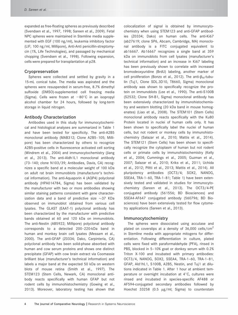
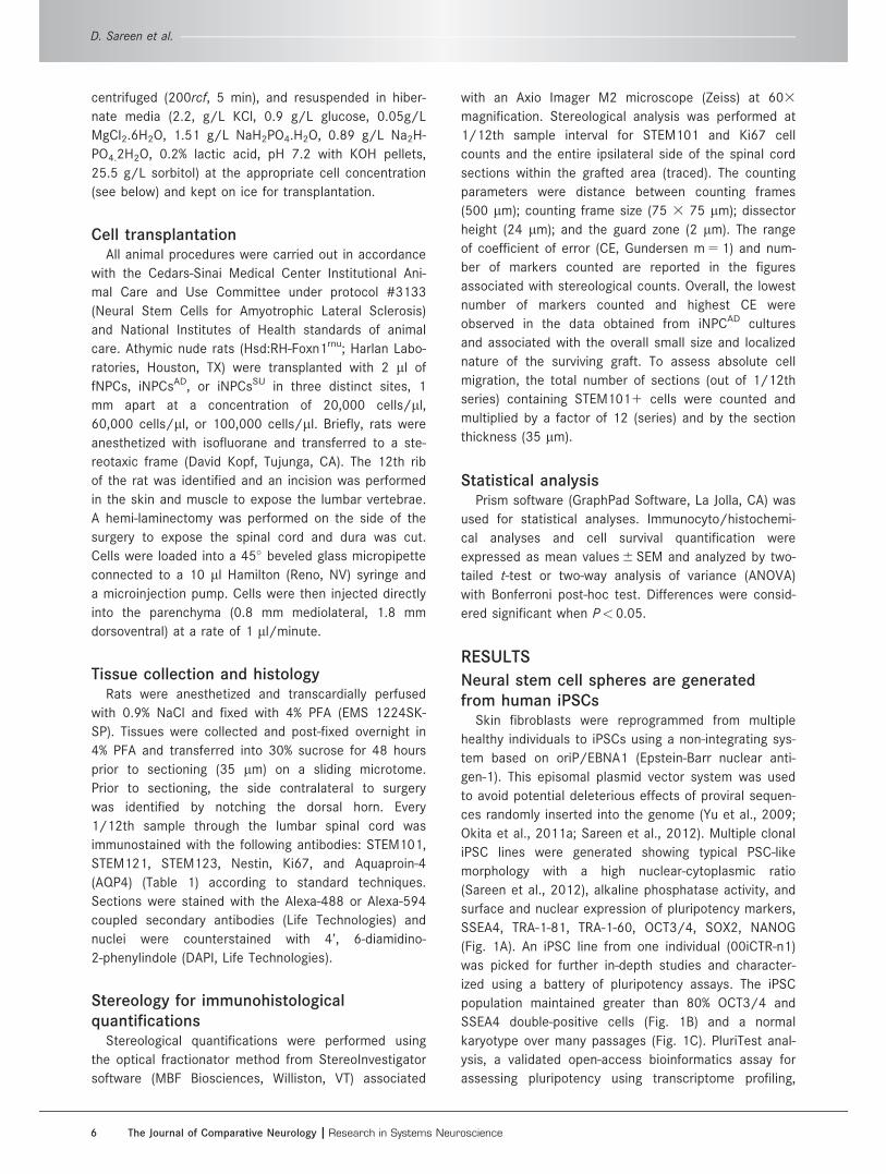
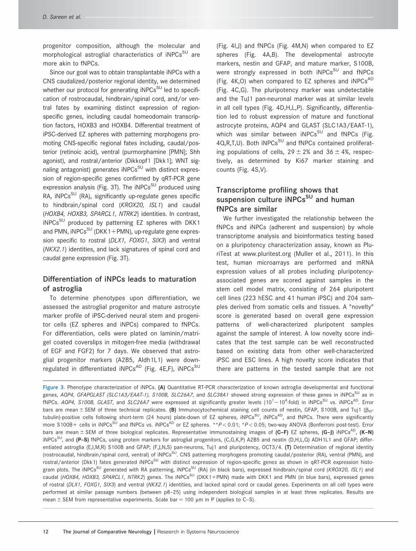
Copyright © 2022 FDOKUMEN