How bees distinguish black from white
9
© 2014 Horridge. This work is published by Dove Medical Press Limited, and licensed under Creative Commons Attribution – Non Commercial (unported, v3.0) License. The full terms of the License are available at http://creativecommons.org/licenses/by-nc/3.0/. Non-commercial uses of the work are permitted without any further permission from Dove Medical Press Limited, provided the work is properly attributed. Permissions beyond the scope of the License are administered by Dove Medical Press Limited. Information on how to request permission may be found at: http://www.dovepress.com/permissions.php Eye and Brain 2014:6 9–17 Eye and Brain Dovepress submit your manuscript | www.dovepress.com Dovepress 9 ORIGINAL RESEARCH open access to scientific and medical research Open Access Full Text Article http://dx.doi.org/10.2147/EB.S70522 How bees distinguish black from white Adrian Horridge Biological Sciences, Australian National University, Canberra, ACT, Australia Correspondence: Adrian Horridge 76 Mueller Street, Yarralumla, ACT 2600, Australia Email [email protected] Abstract: Bee eyes have photoreceptors for ultraviolet, green, and blue wavelengths that are excited by reflected white but not by black. With ultraviolet reflections excluded by the apparatus, bees can learn to distinguish between black, gray, and white, but theories of color vision are clearly of no help in explaining how they succeed. Human vision sidesteps the issue by constructing black and white in the brain. Bees have quite different and accessible mechanisms. As revealed by extensive tests of trained bees, bees learned two strong signals displayed on either target. The first input was the position and a measure of the green recep- tor modulation at the vertical edges of a black area, which included a measure of the angular width between the edges of black. They also learned the average position and total amount of blue reflected from white areas. These two inputs were sufficient to help decide which of two targets held the reward of sugar solution, but the bees cared nothing for the black or white as colors, or the direction of contrast at black/white edges. These findings provide a small step toward understanding, modeling, and implementing in silicon the anti-intuitive visual system of the honeybee, in feeding behavior. Keywords: vision, detectors, black/white, color, visual processing Introduction There has been much interest in how bees detect the shapes and colors of flowers and of landmarks by which they recognize places, but the way that they distinguish between black and white appears to have been overlooked. The three types of photoreceptors of the bee compound eye have spectral sensitivity peaks in the green, blue, and ultra- violet (UV) wavelengths, 1 so it has usually been accepted that color is detected from the interaction of the responses in these three channels. 2,3 However, the strength of the stimulus at each receptor type tells nothing about the subsequent neural processing. All three receptor types respond to white and shades of gray, including UV, and the bee’s preferences for colors are indifferent to sunlight or shade. However, the rapid changes of sensitivity over a 1,000-fold range seen with changes of light intensity, 4 make it unlikely that the bee’s discrimination of gray, white, or color is based simply on the ratios of the intensities at the receptors. A good deal is already known about the peripheral neural input pathways. Electrophysiology shows that the responses to steady illumination adapt rapidly. 5 The responses of the photoreceptors, and particularly of the second-order neurons, are rapidly changing potentials (phasic responses), called modulations. 5 Bees detect changes of intensity caused by scanning across edges. In brief, the spatial and temporal properties
Transcript of How bees distinguish black from white
© 2014 Horridge. This work is published by Dove Medical Press Limited, and licensed under Creative Commons Attribution – Non Commercial (unported, v3.0) License. The full terms of the License are available at http://creativecommons.org/licenses/by-nc/3.0/. Non-commercial uses of the work are permitted without any further
permission from Dove Medical Press Limited, provided the work is properly attributed. Permissions beyond the scope of the License are administered by Dove Medical Press Limited. Information on how to request permission may be found at: http://www.dovepress.com/permissions.php
Eye and Brain 2014:6 9–17
Eye and Brain Dovepress
submit your manuscript | www.dovepress.com
Dovepress 9
O r i g i n a l r E s E a r c h
open access to scientific and medical research
Open access Full Text article
http://dx.doi.org/10.2147/EB.S70522
how bees distinguish black from white
adrian horridgeBiological sciences, australian national University, canberra, acT, australia
correspondence: adrian horridge 76 Mueller street, Yarralumla, acT 2600, australia Email [email protected]
Abstract: Bee eyes have photoreceptors for ultraviolet, green, and blue wavelengths that
are excited by reflected white but not by black. With ultraviolet reflections excluded by the
apparatus, bees can learn to distinguish between black, gray, and white, but theories of color
vision are clearly of no help in explaining how they succeed. Human vision sidesteps the
issue by constructing black and white in the brain. Bees have quite different and accessible
mechanisms. As revealed by extensive tests of trained bees, bees learned two strong signals
displayed on either target. The first input was the position and a measure of the green recep-
tor modulation at the vertical edges of a black area, which included a measure of the angular
width between the edges of black. They also learned the average position and total amount of
blue reflected from white areas. These two inputs were sufficient to help decide which of two
targets held the reward of sugar solution, but the bees cared nothing for the black or white as
colors, or the direction of contrast at black/white edges. These findings provide a small step
toward understanding, modeling, and implementing in silicon the anti-intuitive visual system
of the honeybee, in feeding behavior.
Keywords: vision, detectors, black/white, color, visual processing
IntroductionThere has been much interest in how bees detect the shapes and colors of flowers and
of landmarks by which they recognize places, but the way that they distinguish between
black and white appears to have been overlooked. The three types of photoreceptors
of the bee compound eye have spectral sensitivity peaks in the green, blue, and ultra-
violet (UV) wavelengths,1 so it has usually been accepted that color is detected from
the interaction of the responses in these three channels.2,3 However, the strength of the
stimulus at each receptor type tells nothing about the subsequent neural processing.
All three receptor types respond to white and shades of gray, including UV, and the
bee’s preferences for colors are indifferent to sunlight or shade. However, the rapid
changes of sensitivity over a 1,000-fold range seen with changes of light intensity,4
make it unlikely that the bee’s discrimination of gray, white, or color is based simply
on the ratios of the intensities at the receptors.
A good deal is already known about the peripheral neural input pathways.
Electrophysiology shows that the responses to steady illumination adapt rapidly.5 The
responses of the photoreceptors, and particularly of the second-order neurons, are rapidly
changing potentials (phasic responses), called modulations.5 Bees detect changes of
intensity caused by scanning across edges. In brief, the spatial and temporal properties
Eye and Brain 2014:6submit your manuscript | www.dovepress.com
Dovepress
Dovepress
10
horridge
of the peripheral neurons are optimized to detect moving
stimuli and edges,6 not areas of black, white, or color.
In return for a reward of sugar solution, bees readily learn
to come to the “choice” chamber where they can choose
between the patterns or colors displayed on two targets
(Figure 1). Earlier tests of trained bees in the apparatus
showed that the positions of the black or gray panels were
located very well in the vertical direction, but poorly along
the horizontal,7 despite the abundant contrast at the verti-
cal edges. Even more curious, a black/white edge was not
distinguished from a white/black edge,8 which suggests that
the edge detectors are symmetrical.
In previous work with black/white patterns, bees dis-
criminated mainly by the modulation of the green receptor
pathway. They detected edge orientation and radial and
circular edges as components of patterns.9 They showed no
evidence that patterns or shapes were reassembled, and plenty
to show that they did not. The bees measured modulations
of the blue and green receptors separately10 and recognized
them only in the positions where they had been learned on
the retina.11
Therefore, with UV excluded from the experiments, there
are four independent inputs at each local region of the eye
that could respond to a white, gray, or colored pattern. These
are the green receptor intensity; the blue receptor intensity,
called tonic responses; and also the respective changes
(modulation) of intensity at each of these receptors, called
phasic responses. The aim of this study was to discover which
of these inputs the bees use for black versus white on gray
backgrounds.
Materials and methodsThe way to train honeybees and to test their preferences
in forced choices between carefully selected pairs of
test patterns has been described many times.4,7–11 The
bees used in the study were ordinary brown honeybees
(Apis mellifera L.) flying freely from a neighboring hive
for 9 months of the year. Ten to fifteen bees were individu-
ally color-marked, and only these were allowed to enter
the apparatus (Figure 1). A greater number of bees would
have risked visual contact between bees, and this matters
(only) in the tests.
The apparatus (Figure 1) was made with wooden sides
and a transparent polycarbonate sheet top which excluded
UV light. Further protection was provided by a transparent
plastic roof over the whole. The floor and inside walls of
the apparatus were painted white. The reward was a solu-
tion of cane sugar that was adjusted in strength between
2% and 7% w/w so that marked bees continued to return
for more, but recruits were not attracted to the weaker
solution. The test pattern and the reward changed sides
every 5 minutes (Figure 1) to prevent bees from learning
which side to go to and to equalize any chance of spuri-
ous cues from unequal olfactory cues or side preferences.
The reward was provided during the test phase, so that
the bees would not continue to search. Test phases were
for 5 minutes, with intervening continued training for 20
minutes. At a different time, the test was repeated with
the two sides reversed, and each test was followed by a
different test pattern. No one test was repeated in the same
day. Training and testing continued all week until sufficient
counts (100 or 200) accumulated.
Canson (Annonay, France) colored papers were used dur-
ing tests. These can be ordered online (www.canson-infinity.
com/en/values.asp) or purchased from art supply shops. The
manufacturer names of the colored papers used, and details
of the spectra’s calibration of sunlight reflected from these
papers are given in Table 1. The methods of calibrating the
papers are available in the literature.7,12,13
Statistics are scarcely necessary because the training score
depends on the length of training, the tests were designed so
that the results were unambiguous, and a variety of different
training patterns with numerous tests supported each other.
The decisions of the bees are independent, and the scores are
Connectingslots
Patternon target
Transparentbaffles
Choicechamber
Bees flyin here
Noreward
RewardboxLeg
Feeder
Rewardhole
29 cm+ −
Figure 1 This Y-choice apparatus was used in all the experiments. The bees fly in at the front and make a choice at a fixed distance from the two targets. The criterion for success is when the bee passes over one of the transparent baffles. The two targets, together with the reward, change sides every 5 minutes. The bees exit by the way they entered.Note: insets – the targets seen from behind, showing the support legs and the reward box behind one of them.
Eye and Brain 2014:6 submit your manuscript | www.dovepress.com
Dovepress
Dovepress
11
how bees distinguish black from white
frequencies, so the standard deviation (SD) can be calculated
from the formula
SD = √{(p) ⋅ (1 – p)/n} (1)
where p is the fraction of correct choices and n is the number
of choices counted.8 A score of 60% correct with n=100,
or 57% correct with n=200 was considered significant at
the 5% level. In some of the interesting critical tests where
the bees failed, the score was near 50%, but that can be a
meaningful result, leading to new conclusions. Care and
common sense is required when comparing scores because
each test is a forced choice between two unfamiliar targets.
A poor score may mean poor learning in the training or little
to distinguish in the test. The most informative tests were
when the bees failed and the missing input could be supplied
in a further test.
ResultsWhite on gray is easily distinguished from black on grayThe first experimental training patterns (Figure 2A) were
rectangles subtending 16°×35° on a background of gray (50%
black). The stimulus difference between white on gray and
black on gray was the same but in opposite directions, and the
rewarded target reflected more light. The bees did not learn
the contrast at the outer edges because it was the same on both
targets. Learning was rapid to a high score. The trained bees
failed to distinguish the rewarded training target from a plain
gray target (Figure 2B), showing that they did not recognize
the white panel nor prefer the lighter target. On the other
hand, the plain gray target was distinguished very well from
the black panel on gray (score 81% correct) (not illustrated),
showing that the strongest preferred memory originated from
Table 1 relative receptor stimuli from the different papers, relative to the white paper (100%), and contrasts between two pairs of papers
Canson colored papers Blue receptor (%) Green receptor (%)
hemp 374 34.2 56.3Ultramarine 590 33.8 20.7Billiards green 576 17.0 22.3Buff 384 25.7 41.7Blue 595 54.2 40.0White copy paper 100 100contrast 374/590 0.006 0.46contrast 384/595 0.36 0.02
Note: color names and numbers are those used by the manufacturer, canson (annonay, France; www.canson-infinity.com/en/values.asp).
A
B
C
D
E
F
G
H
75%, n=100
93%, n=100
71%, n=100
62%, n=100
81%, n=100
47%, n=100
95%, n=100
Train
51%, n=100
100%
100%
Test
Test
55º
100%Test
100%Test
100%Test
100%Test
100%
100%
Test
−+
Figure 2 White was distinguished from black on a gray background.Notes: (A) Training patterns. (B) The trained bees failed to distinguish the white panel from plain gray. (C) They had learned the black panel very well. (D and E) They avoided the vertical parallel lines but the black panel even more so. (F) reversing the contrast of the panels made little difference to the score. (G) They failed to distinguish a gray panel on white from the rewarded white panel on gray. (H) The trained bees avoided the contrast at black edges (arrows).
the black panel. Learning the unrewarded target first is a
consequence of the learning by trial and error in the choice
apparatus. They learn when they make an error.
On a gray background, the trained bees avoided two verti-
cal black lines 1° wide (Figure 2D), but they avoided the black
Eye and Brain 2014:6submit your manuscript | www.dovepress.com
Dovepress
Dovepress
12
horridge
panel even more (Figure 2C), showing that they had learned
to avoid the contrast at the vertical edges of the black panel,
and also that the black panel was a stronger signal than the
two black vertical lines. These results are consistent with the
conclusion that the bees detected and learned the strongest
signal in the training, at the vertical edges of black on the
unrewarded target.
The trained bees strongly preferred a gray panel on
white from a gray panel on black (Figure 2F), showing
that they had not learned the colors of the panels in the
training. They failed to distinguish a gray panel on white
from a white panel on gray (Figure 2G), showing that
they did not detect the white panel, the gray background,
or the levels of contrast at the boundaries of the panels.
When tested with plain white versus black, they preferred
the white, confirming that they avoided strong contrast at
vertical edges.
Nothing in these results distinguishes between the actions
of the green and the blue receptors or between intensity
inputs (tonic) and modulated inputs (phasic). Because the
two receptor types respond differently to colors, the trained
bees were tested next with equiluminant colored patterns
that reduce the number of unknown variables and test the
receptor types separately.
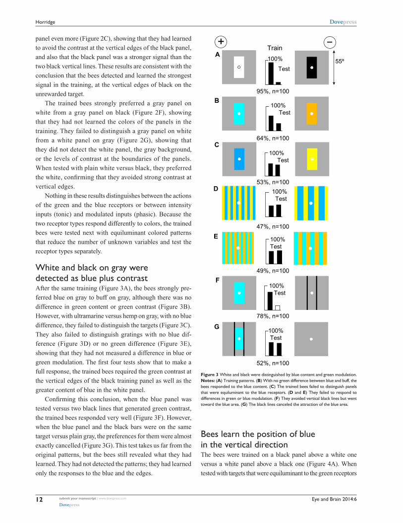
White and black on gray were detected as blue plus contrastAfter the same training (Figure 3A), the bees strongly pre-
ferred blue on gray to buff on gray, although there was no
difference in green content or green contrast (Figure 3B).
However, with ultramarine versus hemp on gray, with no blue
difference, they failed to distinguish the targets (Figure 3C).
They also failed to distinguish gratings with no blue dif-
ference (Figure 3D) or no green difference (Figure 3E),
showing that they had not measured a difference in blue or
green modulation. The first four tests show that to make a
full response, the trained bees required the green contrast at
the vertical edges of the black training panel as well as the
greater content of blue in the white panel.
Confirming this conclusion, when the blue panel was
tested versus two black lines that generated green contrast,
the trained bees responded very well (Figure 3F). However,
when the blue panel and the black bars were on the same
target versus plain gray, the preferences for them were almost
exactly cancelled (Figure 3G). This test takes us far from the
original patterns, but the bees still revealed what they had
learned. They had not detected the patterns; they had learned
only the responses to the blue and the edges.
A
B
C
D
E
F
G
95%, n=100
64%, n=100
53%, n=100
47%, n=100
49%, n=100
78%, n=100
52%, n=100
Train
55º100%
Test
100% Test
100% Test
100% Test
100%Test
100% Test
100%Test
+ −
Figure 3 White and black were distinguished by blue content and green modulation.Notes: (A) Training patterns. (B) With no green difference between blue and buff, the bees responded to the blue content. (C) The trained bees failed to distinguish panels that were equiluminant to the blue receptors. (D and E) They failed to respond to differences in green or blue modulation. (F) They avoided vertical black lines but went toward the blue area. (G) The black lines canceled the attraction of the blue area.
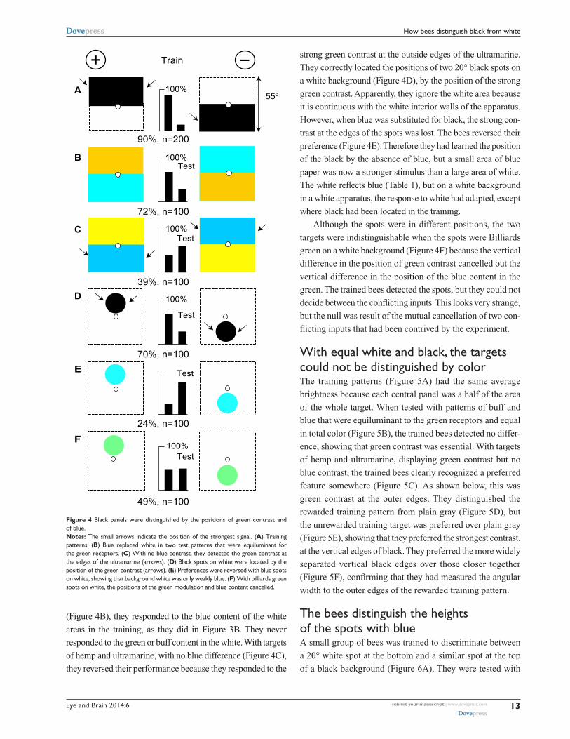
Bees learn the position of blue in the vertical directionThe bees were trained on a black panel above a white one
versus a white panel above a black one (Figure 4A). When
tested with targets that were equiluminant to the green receptors
Eye and Brain 2014:6 submit your manuscript | www.dovepress.com
Dovepress
Dovepress
13
how bees distinguish black from white
(Figure 4B), they responded to the blue content of the white
areas in the training, as they did in Figure 3B. They never
responded to the green or buff content in the white. With targets
of hemp and ultramarine, with no blue difference (Figure 4C),
they reversed their performance because they responded to the
Train
A
B
C
D
E
F
100%
100%Test
100%Test
100%
Test
Test
100%Test
90%, n=200
55º
72%, n=100
39%, n=100
70%, n=100
24%, n=100
49%, n=100
Ultramarine
+ −
Figure 4 Black panels were distinguished by the positions of green contrast and of blue.Notes: The small arrows indicate the position of the strongest signal. (A) Training patterns. (B) Blue replaced white in two test patterns that were equiluminant for the green receptors. (C) With no blue contrast, they detected the green contrast at the edges of the ultramarine (arrows). (D) Black spots on white were located by the position of the green contrast (arrows). (E) Preferences were reversed with blue spots on white, showing that background white was only weakly blue. (F) With billiards green spots on white, the positions of the green modulation and blue content cancelled.
strong green contrast at the outside edges of the ultramarine.
They correctly located the positions of two 20° black spots on
a white background (Figure 4D), by the position of the strong
green contrast. Apparently, they ignore the white area because
it is continuous with the white interior walls of the apparatus.
However, when blue was substituted for black, the strong con-
trast at the edges of the spots was lost. The bees reversed their
preference (Figure 4E). Therefore they had learned the position
of the black by the absence of blue, but a small area of blue
paper was now a stronger stimulus than a large area of white.
The white reflects blue (Table 1), but on a white background
in a white apparatus, the response to white had adapted, except
where black had been located in the training.
Although the spots were in different positions, the two
targets were indistinguishable when the spots were Billiards
green on a white background (Figure 4F) because the vertical
difference in the position of green contrast cancelled out the
vertical difference in the position of the blue content in the
green. The trained bees detected the spots, but they could not
decide between the conflicting inputs. This looks very strange,
but the null was result of the mutual cancellation of two con-
flicting inputs that had been contrived by the experiment.
With equal white and black, the targets could not be distinguished by colorThe training patterns (Figure 5A) had the same average
brightness because each central panel was a half of the area
of the whole target. When tested with patterns of buff and
blue that were equiluminant to the green receptors and equal
in total color (Figure 5B), the trained bees detected no differ-
ence, showing that green contrast was essential. With targets
of hemp and ultramarine, displaying green contrast but no
blue contrast, the trained bees clearly recognized a preferred
feature somewhere (Figure 5C). As shown below, this was
green contrast at the outer edges. They distinguished the
rewarded training pattern from plain gray (Figure 5D), but
the unrewarded training target was preferred over plain gray
(Figure 5E), showing that they preferred the strongest contrast,
at the vertical edges of black. They preferred the more widely
separated vertical black edges over those closer together
(Figure 5F), confirming that they had measured the angular
width to the outer edges of the rewarded training pattern.
The bees distinguish the heights of the spots with blueA small group of bees was trained to discriminate between
a 20° white spot at the bottom and a similar spot at the top
of a black background (Figure 6A). They were tested with
Eye and Brain 2014:6submit your manuscript | www.dovepress.com
Dovepress
Dovepress
14
horridge
both training targets versus a plain black, showing that they
had learned only the unrewarded one (Figure 6B and C).
When tested with blue spots on a black background, they
correctly recognized the spot positions (Figure 6D) but failed
with hemp spots on an ultramarine background with no blue
difference (Figure 6E). When tested with black spots on a
white background, they reversed their preference because
the average position of blue on the targets was reversed, and
that was apparently their only cue.
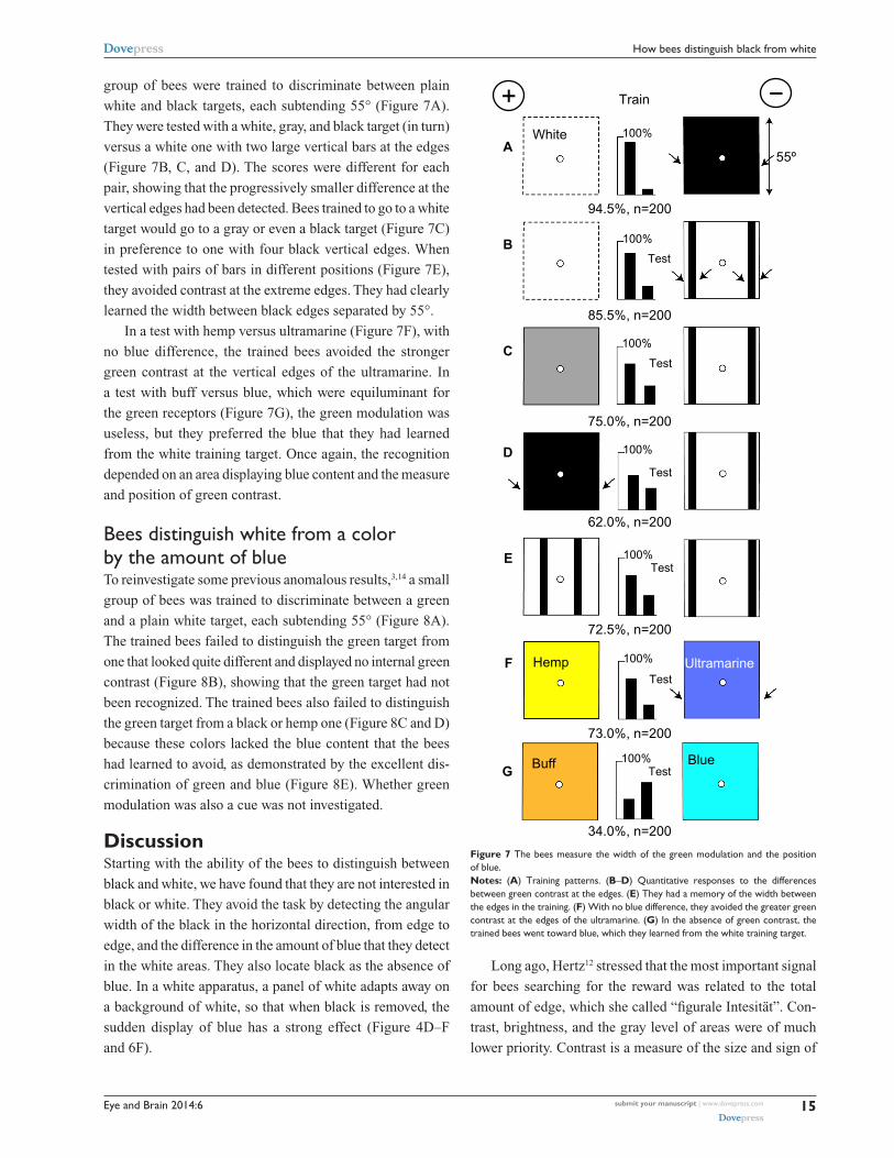
The bees measure the width of large areas of blackHaving now the key to understanding what the bees consis-
tently detected, we can approach the main question. A small
A
B
C
D
E
F
89.0%, n=200
48.5%, n=200
65.5%, n=200
84.5%, n=200
40%, n=100
63%, n=100
Train
100%
100%Test
100%
Test
100%
Test
100%Test
100%
Test
55º
−+
Figure 5 Targets of equal white and black, displayed no color difference.Notes: (A) Training patterns. (B) The trained bees could not distinguish patterns that were equiluminant to the green receptors. (C) They responded well to green contrast at the edges of patterns that were equiluminant to the blue receptors. (D and E) in the training, they had learned to go to the vertical edge of the wider pattern. (F) They preferred the outer vertical edge to the inner one, as in (A). arrows show vertical edges.
A
B
C
D
E
F
78%, n=100
51%, n=100
69%, n=100
67%, n=100
49%, n=100
28%, n=100
Train
100%
100%
Test
Test
100%
Test
100%
Test
100%
Test
100%Test
55º
−+
Figure 6 Blue was used to detect the position of a white spot on black.Notes: (A) Training patterns. (B and C) The trained bees failed to recognize the rewarded target versus black. (D) Blue spots were distinguished. (E) The trained bees failed to distinguish targets with abundant green contrast but no blue difference. (F) The preference for the positions reversed with black spots on white, which removed blue from its expected retinotopic position.
Eye and Brain 2014:6 submit your manuscript | www.dovepress.com
Dovepress
Dovepress
15
how bees distinguish black from white
A
B
C
D
E
F
G
94.5%, n=200
85.5%, n=200
75.0%, n=200
62.0%, n=200
72.5%, n=200
73.0%, n=200
Train
White 100%
100%
Test
100%
Test
100%
Test
100%Test
100%
Test
Test
34.0%, n=200
Buff
Hemp
Blue
Ultramarine
100%
55º
−+
Figure 7 The bees measure the width of the green modulation and the position of blue.Notes: (A) Training patterns. (B–D) Quantitative responses to the differences between green contrast at the edges. (E) They had a memory of the width between the edges in the training. (F) With no blue difference, they avoided the greater green contrast at the edges of the ultramarine. (G) in the absence of green contrast, the trained bees went toward blue, which they learned from the white training target.
group of bees were trained to discriminate between plain
white and black targets, each subtending 55° (Figure 7A).
They were tested with a white, gray, and black target (in turn)
versus a white one with two large vertical bars at the edges
(Figure 7B, C, and D). The scores were different for each
pair, showing that the progressively smaller difference at the
vertical edges had been detected. Bees trained to go to a white
target would go to a gray or even a black target (Figure 7C)
in preference to one with four black vertical edges. When
tested with pairs of bars in different positions (Figure 7E),
they avoided contrast at the extreme edges. They had clearly
learned the width between black edges separated by 55°.
In a test with hemp versus ultramarine (Figure 7F), with
no blue difference, the trained bees avoided the stronger
green contrast at the vertical edges of the ultramarine. In
a test with buff versus blue, which were equiluminant for
the green receptors (Figure 7G), the green modulation was
useless, but they preferred the blue that they had learned
from the white training target. Once again, the recognition
depended on an area displaying blue content and the measure
and position of green contrast.
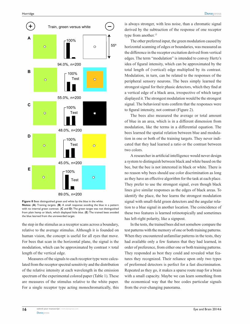
Bees distinguish white from a color by the amount of blueTo reinvestigate some previous anomalous results,3,14 a small
group of bees was trained to discriminate between a green
and a plain white target, each subtending 55° (Figure 8A).
The trained bees failed to distinguish the green target from
one that looked quite different and displayed no internal green
contrast (Figure 8B), showing that the green target had not
been recognized. The trained bees also failed to distinguish
the green target from a black or hemp one (Figure 8C and D)
because these colors lacked the blue content that the bees
had learned to avoid, as demonstrated by the excellent dis-
crimination of green and blue (Figure 8E). Whether green
modulation was also a cue was not investigated.
DiscussionStarting with the ability of the bees to distinguish between
black and white, we have found that they are not interested in
black or white. They avoid the task by detecting the angular
width of the black in the horizontal direction, from edge to
edge, and the difference in the amount of blue that they detect
in the white areas. They also locate black as the absence of
blue. In a white apparatus, a panel of white adapts away on
a background of white, so that when black is removed, the
sudden display of blue has a strong effect (Figure 4D–F
and 6F).
Long ago, Hertz12 stressed that the most important signal
for bees searching for the reward was related to the total
amount of edge, which she called “figurale Intesität”. Con-
trast, brightness, and the gray level of areas were of much
lower priority. Contrast is a measure of the size and sign of
Eye and Brain 2014:6submit your manuscript | www.dovepress.com
Dovepress
Dovepress
16
horridge
the step in the stimulus as a receptor scans across a boundary,
relative to the average stimulus. Although it is founded on
human vision, the concept is useful for all eyes that move.
For bees that scan in the horizontal plane, the signal is the
modulation, which can be approximated by contrast × total
length of the vertical edge.
Measures of the signals to each receptor type were calcu-
lated from the receptor spectral sensitivity and the distribution
of the relative intensity at each wavelength in the emission
spectrum of the experimental colored paper (Table 1). These
are measures of the stimulus relative to the white paper.
For a single receptor type acting monochromatically, this
A
Train, green versus white
100%
100%Test
100%Test
100%Test
100%
Test
94.0%, n=200
55.0%, n=200
48.0%, n=200
45.0%, n=200
89.0%, n=200
B
C
E
D
55º
+ −
Figure 8 Bees distinguished green and white by the blue in the white. Notes: (A) Training targets. (B) a small response avoiding the blue in a pattern with no internal green contrast. (C and D) The green target was not distinguished from plain hemp or black, which displayed little blue. (E) The trained bees avoided the blue learned from the unrewarded target.
is always stronger, with less noise, than a chromatic signal
derived by the subtraction of the response of one receptor
type from another.15
The other preferred input, the green modulation caused by
horizontal scanning of edges or boundaries, was measured as
the difference in the receptor excitation derived from vertical
edges. The term “modulation” is intended to convey Hertz’s
idea of figural intensity, which can be approximated by the
total length of (vertical) edge multiplied by its contrast.
Modulation, in turn, can be related to the responses of the
peripheral sensory neurons. The bees simply learned the
strongest signal for their phasic detectors, which they find at
a vertical edge of a black area, irrespective of which target
displayed it. The strongest modulation would be the strongest
signal. The behavioral tests confirm that the responses were
to figural intensity, not contrast (Figure 2).
The bees also measured the average or total amount
of blue in an area, which is in a different dimension from
modulation, like the terms in a differential equation. The
bees learned the spatial relation between blue and modula-
tion in one or both of the training targets. They never indi-
cated that they had learned a ratio or the contrast between
two colors.
A researcher in artificial intelligence would never design
a system to distinguish between black and white based on the
bee, but the bee is not interested in black or white. There is
no reason why bees should use color discrimination as long
as they have an effective algorithm for the task at each place.
They prefer to use the strongest signal, even though black
lines give similar responses as the edges of black areas. To
identify the place, the bee learns the strongest modulation
signal with small-field green detectors and the angular rela-
tion to a blue signal in another location. The coincidence of
these two features is learned retinotopically and sometimes
has left-right polarity, like a signpost.
In the tests, the trained bees did not somehow compare the
test patterns with the memory of one or both training patterns.
When they encountered unfamiliar patterns in the tests, they
had available only a few features that they had learned, in
order of preference, from either one or both training patterns.
They responded as best they could and revealed what fea-
tures they recognized. Their reliance upon only two types
of preformed detectors is perfect for a fast discrimination.
Repeated as they go, it makes a sparse route map for a brain
with a small capacity. Maybe we can learn something from
the economical way that the bee codes particular signals
from the ever-changing panorama.
Eye and Brain
Publish your work in this journal
Submit your manuscript here: http://www.dovepress.com/eye-and-brain-journal
Eye and Brain is an international, peer-reviewed, open access journal focusing on clinical and experimental research in the f ield of neuro-ophthalmology. All aspects of patient care are addressed within the journal as well as basic research. Papers covering original research, basic science, clinical and epidemiological studies, reviews and
evaluations, guidelines, expert opinion and commentary, case reports and extended reports are welcome. The manuscript management system is completely online and includes a very quick and fair peer-review system, which is all easy to use. Visit http://www.dovepress.com/ testimonials.php to read real quotes from published authors.
Eye and Brain 2014:6 submit your manuscript | www.dovepress.com
Dovepress
Dovepress
Dovepress
17
how bees distinguish black from white
AcknowledgmentsThe author thanks Mr Richard Johnston (Canberra Bee-
keepers Association), for the loan of a hive of bees, and M
Srinivasan, for help with calibrating the reflection spectra of
the colored papers in sunlight.
DisclosureThe author reports no conflict of interest in this work.
References1. Autrum H, von Zwehl V. Die spektral Empfindlichkeit einzelner Sehzellen
des Bienenauges. Z Vgl Physiol. 1964;48(4):357–384. German.2. von Frisch K. Der Farbesinn und Formensinn der Biene [Perception
of color and form by the bee]. Zool Jahrb Physiol. 1914;35(1):1–188. German.
3. Hertz M. New experiments on colour vision in bees. J Exp Biol. 1939;16(1):1–8.
4. Hess C. Beiträge zur Frage nach einen Farbensinne bei Bienen [A contribution to the question of a color sense in bees]. Pflügers Arch. 1918;170(7–9):337–366. German.
5. Laughlin SB, Hardie RC. Common strategies for light adaptation in the peripheral visual systems of fly and dragonfly. J Comp Physiol. 1978;128(5):319–340.
6. Srinivasan MV, Laughlin SB, Dubs A. Predictive coding: a fresh view of inhibition in the retina. Proc R Soc Lond B Biol Sci. 1982;216(1205): 427–459.
7. Horridge A. Pattern vision of the honeybee (Apis mellifera): blue and green receptors in the discrimination of translocation. Neurobiol Learn Mem. 2000;74(1):1–16.
8. Friedlaender M. Zur Bedeutung des Fluglochs im optischen Feld der Biene bei senkrechter Dressuranordnung [The importance of the reward hole in the visual field of bees trained to vertical patterns]. Z Vgl Physiol. 1931;15(2):193–260. German.
9. Horridge A. What Does the Honeybee See? And How Do We Know? Canberra: ANU Press; 2009. Available from: http://epress.anu.edu.au/honeybee_citation.html. Accessed August 28, 2014.
10. Horridge GA. Visual resolution of gratings by the compound eye of the bee Apis mellifera. J Exp Biol. 2003;206(Pt 13):2105–2110.
11. Horridge GA. Discrimination of single bars by the honeybee (Apis mellifera). Vision Res. 2003;43(11):1257–1271.
12. Hertz M. Über figurale Intensität und Qualitäten in der optische Wahrne-hmung der Biene [Figural intensity and quality in the visual perception of the bee]. Biologische Zentralblatte. 1933;53(1):10–40. German.
13. Giger AD, Srinivasan MV. Pattern recognition in honeybees: chromatic properties of orientation analysis. J Comp Physiol A. 1996;178(6): 763–769.
14. Chittka L. Bees, white flowers, and the color hexagon – a reassessment? No, not yet. Naturwissenschaften. 1999;86(8):595–597.
15. Muntz WR. Microelectrode recordings from the diencephalon of the frog (Rana pipiens) and a blue-sensitive system. J Neurophysiol. 1962;25:699–711.