High levels of SOX5 decrease proliferative capacity of human B cells, but permit plasmablast...
12
High Levels of SOX5 Decrease Proliferative Capacity of Human B Cells, but Permit Plasmablast Differentiation Mirzokhid Rakhmanov 1 *, Heiko Sic 1 , Anne-Kathrin Kienzler 1 , Beate Fischer 1 , Marta Rizzi 1 , Maximilian Seidl 1 , Kerstina Melkaoui 1 , Susanne Unger 1 , Luisa Moehle 1 , Nadine E. Schmit 1 , Sachin D. Deshmukh 1 , Cemil Korcan Ayata 2 , Wolfgang Schuh 3 , Zhibing Zhang 4 , Franc ¸ois-Loic Cosset 5 , Els Verhoeyen 5,6 , Hans-Hartmut Peter 1,7 , Reinhard E. Voll 1,7 , Ulrich Salzer 1,7 , Hermann Eibel 1,7. , Klaus Warnatz 1,7. 1 Center for Chronic Immunodeficiency (CCI), University Medical Center and University Freiburg, Freiburg, Germany, 2 Department of Pneumology, University Medical Center and University Freiburg, Freiburg, Germany, 3 Division of Molecular Immunology, Department of Internal Medicine III, Nikolaus Fiebiger Center, University of Erlangen-Nu ¨ rnberg, Erlangen, Germany, 4 Department of Obstetrics and Gynecology, Virginia Commonwealth University, Richmond, Virginia, United States of America, 5 CIRI, INSERM U1111, EVIR, Ecole Normale Supe ´rieure de Lyon, Universite ´ de Lyon, Lyon, France, 6 INSERM U1065, C3M, Metabolic control of cell deaths, Nice, France, 7 Division of Rheumatology and Clinical Immunology, University Medical Center Freiburg, Freiburg, Germany Abstract Currently very little is known about the differential expression and function of the transcription factor SOX5 during B cell maturation. We identified two new splice variants of SOX5 in human B cells, encoding the known L-SOX5B isoform and a new shorter isoform L-SOX5F. The SOX5 transcripts are highly expressed during late stages of B-cell differentiation, including atypical memory B cells, activated CD21 low B cells and germinal center B cells of tonsils. In tonsillar sections SOX5 expression was predominantly polarized to centrocytes within the light zone. After in vitro stimulation, SOX5 expression was down- regulated during proliferation while high expression levels were permissible for plasmablast differentiation. Overexpression of L-SOX5F in human primary B lymphocytes resulted in reduced proliferation, less survival of CD138 neg B cells, but comparable numbers of CD138 + CD38 hi plasmablasts compared to control cells. Thus, our findings describe for the first time a functional role of SOX5 during late B cell development reducing the proliferative capacity and thus potentially affecting the differentiation of B cells during the germinal center response. Citation: Rakhmanov M, Sic H, Kienzler A-K, Fischer B, Rizzi M, et al. (2014) High Levels of SOX5 Decrease Proliferative Capacity of Human B Cells, but Permit Plasmablast Differentiation. PLoS ONE 9(6): e100328. doi:10.1371/journal.pone.0100328 Editor: Simon Fillatreau, DRFZ, Germany Received January 14, 2014; Accepted May 23, 2014; Published June 19, 2014 Copyright: ß 2014 Rakhmanov et al. This is an open-access article distributed under the terms of the Creative Commons Attribution License, which permits unrestricted use, distribution, and reproduction in any medium, provided the original author and source are credited. Funding: This research was funded by the German Federal Ministry of Education and Research (BMBF 01 EO 0803) (to K.W., H.E., U.S. and R.V.), the German Research Foundation (DFG) Grant SFB620, Projects C1 (to K.W. and H.H.P.) and C7 (to US), the 7th Framework Program (FP7) of the European Union Grant Nr. HEALTH-F2-2008-201549 (to K.W., H.E. and U.S.); FP7-HEALTH-2007-B/222878 "PERSIST" and FP7-E-Rare ‘‘GENTHALTHER’’ Grants (to EV and FLC); Virginia Commonwealth University (VCU) Presidential Research Incentive Program (PRIP), VCU Massey Cancer Center/American Cancer Society Institutional Research Awards and NIH Award HD076257 (to Z.Z.); University Freiburg (UF) Medical Faculty Research Committee and UF Scientific Society Freiburg Research Grants (to M.Ra.). The authors are responsible for the contents of this publication. The funders had no role in study design, data collection and analysis, decision to publish, or preparation of the manuscript. Competing Interests: The authors have declared that no competing interests exist. * E-mail: [email protected] . These authors contributed equally to this work. Introduction Sox (sex determining region Y ( SRY)-related high-mobility- group (HMG)-b ox) family of proteins are encoded by 20 genes in humans and mice and are classified into eight groups - group SoxA to SoxH - according to the sequence identity in their DNA- binding HMG-domain and other conserved regions (reviewed in [1,2]). Sox proteins function as transcription factors and play important roles in many developmental and cellular processes. Although most Sox proteins predominantly serve as transcriptional activators, there is also evidence for transcriptional repression and architectural roles (reviewed in [3]). Essential roles and key functions in cell fate decisions have been identified for Sox proteins in sex differentiation, neurogenesis and gliogenesis, neural crest development, skeletogenesis, cardiogenesis and angiogenesis as well as in hematopoiesis [1,3]. Sox5 belongs to the SoxD group composed of Sox5, Sox6 and Sox13, in vertebrates (reviewed in [4]). SoxD group genes and protein structures are highly conserved in the family-specific HMG-domain and in the group-specific leucine zipper and coiled- coil domains [3,4]. The SoxD HMG-box domain preferentially binds the DNA consensus sequence of AACAAT [5] and SoxD proteins can act as either transcriptional activators or repressors [4]. Each SoxD gene is expressed in a limited subset of cell types [4]. High levels of Sox5 and Sox6 gene co-expression are found in spermatids, neurons, oligodendrocytes and chondrocytes [6–9]. The human SOX5 protein exists in a short (S-SOX5) and long (L-SOX5) isoform, encoded by a unique transcript for S-SOX5 and by several transcript variants for L-SOX5 isoforms. While in humans the short isoform is expressed mainly in the testes [10], high levels of long isoforms are found in fetal brain [10], striated muscles and chondrocytes [11]. Knock-out mouse models PLOS ONE | www.plosone.org 1 June 2014 | Volume 9 | Issue 6 | e100328
-
Upload
independent -
Category
Documents
-
view
1 -
download
0
Transcript of High levels of SOX5 decrease proliferative capacity of human B cells, but permit plasmablast...

High Levels of SOX5 Decrease Proliferative Capacity ofHuman B Cells, but Permit Plasmablast DifferentiationMirzokhid Rakhmanov1*, Heiko Sic1, Anne-Kathrin Kienzler1, Beate Fischer1, Marta Rizzi1,
Maximilian Seidl1, Kerstina Melkaoui1, Susanne Unger1, Luisa Moehle1, Nadine E. Schmit1,
Sachin D. Deshmukh1, Cemil Korcan Ayata2, Wolfgang Schuh3, Zhibing Zhang4, Francois-Loic Cosset5,
Els Verhoeyen5,6, Hans-Hartmut Peter1,7, Reinhard E. Voll1,7, Ulrich Salzer1,7, Hermann Eibel1,7.,
Klaus Warnatz1,7.
1Center for Chronic Immunodeficiency (CCI), University Medical Center and University Freiburg, Freiburg, Germany, 2Department of Pneumology, University Medical
Center and University Freiburg, Freiburg, Germany, 3Division of Molecular Immunology, Department of Internal Medicine III, Nikolaus Fiebiger Center, University of
Erlangen-Nurnberg, Erlangen, Germany, 4Department of Obstetrics and Gynecology, Virginia Commonwealth University, Richmond, Virginia, United States of America,
5CIRI, INSERM U1111, EVIR, Ecole Normale Superieure de Lyon, Universite de Lyon, Lyon, France, 6 INSERM U1065, C3M, Metabolic control of cell deaths, Nice, France,
7Division of Rheumatology and Clinical Immunology, University Medical Center Freiburg, Freiburg, Germany
Abstract
Currently very little is known about the differential expression and function of the transcription factor SOX5 during B cellmaturation. We identified two new splice variants of SOX5 in human B cells, encoding the known L-SOX5B isoform and anew shorter isoform L-SOX5F. The SOX5 transcripts are highly expressed during late stages of B-cell differentiation, includingatypical memory B cells, activated CD21low B cells and germinal center B cells of tonsils. In tonsillar sections SOX5 expressionwas predominantly polarized to centrocytes within the light zone. After in vitro stimulation, SOX5 expression was down-regulated during proliferation while high expression levels were permissible for plasmablast differentiation. Overexpressionof L-SOX5F in human primary B lymphocytes resulted in reduced proliferation, less survival of CD138neg B cells, butcomparable numbers of CD138+CD38hi plasmablasts compared to control cells. Thus, our findings describe for the first timea functional role of SOX5 during late B cell development reducing the proliferative capacity and thus potentially affectingthe differentiation of B cells during the germinal center response.
Citation: Rakhmanov M, Sic H, Kienzler A-K, Fischer B, Rizzi M, et al. (2014) High Levels of SOX5 Decrease Proliferative Capacity of Human B Cells, but PermitPlasmablast Differentiation. PLoS ONE 9(6): e100328. doi:10.1371/journal.pone.0100328
Editor: Simon Fillatreau, DRFZ, Germany
Received January 14, 2014; Accepted May 23, 2014; Published June 19, 2014
Copyright: � 2014 Rakhmanov et al. This is an open-access article distributed under the terms of the Creative Commons Attribution License, which permitsunrestricted use, distribution, and reproduction in any medium, provided the original author and source are credited.
Funding: This research was funded by the German Federal Ministry of Education and Research (BMBF 01 EO 0803) (to K.W., H.E., U.S. and R.V.), the GermanResearch Foundation (DFG) Grant SFB620, Projects C1 (to K.W. and H.H.P.) and C7 (to US), the 7th Framework Program (FP7) of the European Union Grant Nr.HEALTH-F2-2008-201549 (to K.W., H.E. and U.S.); FP7-HEALTH-2007-B/222878 "PERSIST" and FP7-E-Rare ‘‘GENTHALTHER’’ Grants (to EV and FLC); VirginiaCommonwealth University (VCU) Presidential Research Incentive Program (PRIP), VCU Massey Cancer Center/American Cancer Society Institutional ResearchAwards and NIH Award HD076257 (to Z.Z.); University Freiburg (UF) Medical Faculty Research Committee and UF Scientific Society Freiburg Research Grants (toM.Ra.). The authors are responsible for the contents of this publication. The funders had no role in study design, data collection and analysis, decision to publish,or preparation of the manuscript.
Competing Interests: The authors have declared that no competing interests exist.
* E-mail: [email protected]
. These authors contributed equally to this work.
Introduction
Sox (sex determining region Y (SRY)-related high-mobility-
group (HMG)-box) family of proteins are encoded by 20 genes in
humans and mice and are classified into eight groups - group SoxA
to SoxH - according to the sequence identity in their DNA-
binding HMG-domain and other conserved regions (reviewed in
[1,2]). Sox proteins function as transcription factors and play
important roles in many developmental and cellular processes.
Although most Sox proteins predominantly serve as transcriptional
activators, there is also evidence for transcriptional repression and
architectural roles (reviewed in [3]). Essential roles and key
functions in cell fate decisions have been identified for Sox proteins
in sex differentiation, neurogenesis and gliogenesis, neural crest
development, skeletogenesis, cardiogenesis and angiogenesis as
well as in hematopoiesis [1,3].
Sox5 belongs to the SoxD group composed of Sox5, Sox6 and
Sox13, in vertebrates (reviewed in [4]). SoxD group genes and
protein structures are highly conserved in the family-specific
HMG-domain and in the group-specific leucine zipper and coiled-
coil domains [3,4]. The SoxD HMG-box domain preferentially
binds the DNA consensus sequence of AACAAT [5] and SoxD
proteins can act as either transcriptional activators or repressors
[4]. Each SoxD gene is expressed in a limited subset of cell types
[4]. High levels of Sox5 and Sox6 gene co-expression are found in
spermatids, neurons, oligodendrocytes and chondrocytes [6–9].
The human SOX5 protein exists in a short (S-SOX5) and long
(L-SOX5) isoform, encoded by a unique transcript for S-SOX5
and by several transcript variants for L-SOX5 isoforms. While in
humans the short isoform is expressed mainly in the testes [10],
high levels of long isoforms are found in fetal brain [10], striated
muscles and chondrocytes [11]. Knock-out mouse models
PLOS ONE | www.plosone.org 1 June 2014 | Volume 9 | Issue 6 | e100328
demonstrated crucial roles of L-SOX5 in developmental and
cellular processes during chondrogenesis [12] and neurogenesis
[13,14], but very little is known about its expression and function
in B lymphocytes. Comparative transcriptome analysis of different
memory B-cell subpopulations from healthy donor (HD) tonsils
revealed differential regulation of SOX5, along with other markers
like RUNX2, DLL1 and AICDA, as a distinctive signature profile of
FCRL4+ memory B cells [15]. Later, dysregulation of SOX5 gene
expression was reported in the innate-like CD21low B-cell
subpopulation of patients with common variable immunodeficien-
cy (CVID) [16] and patients with hepatitis C virus-associated
mixed cryoglobulinemia [17]. Attempts to test the function of
SOX5 in the activation of FCRL4 promoters did not reveal any
significant influence of SOX5 in the regulation of the FCRL4 gene
expression [15]. Since the function of SOX5 in B cells still remains
elusive, we aimed in this study to investigate the expression and
function of SOX5 in human B cells. We describe the differential
expression of SOX5 transcripts during B cell development.
Combined with functional assays in vitro these findings expose a
new role and function of SOX5 in human terminal B cell
differentiation.
Materials and Methods
HD Individuals’ MaterialThe study was approved by the internal ethics board (University
Hospital Freiburg 313/04 and 121/11).Iinformed written consent
was obtained from each individual before participation in the
study, in accordance with the Declaration of Helsinki.
B Cell Isolation and In vitro StimulationB cells were isolated by negative magnetic bead selection using
the MACS B Cell Isolation Kit II (Miltenyi Biotec) according to
manufacturer’s instructions. The purity of .95% was reached in B
cell fractions. The cells were stimulated in vitro for 9 days at 37uCin RPMI 1640 medium containing 10% FCS either in the
presence of IL4, IL21, CD40L or a combination of IL4+ CD40L
+/2 IL21. IL4 (ImmunoTools) was used at the final concentration
of 100 U/ml. Preparation of CD40L and IL21 was previously
described [18]. Prior to use CD40L and IL21 containing
supernatants were concentrated and titrated.
Preparation of Tonsillar B CellsTonsillar single cell suspensions were prepared by tissue
mincing, filtration through 70-mm nylon filters and centrifugation
on a Ficoll gradient. The cells were stained with appropriate
antibodies and subjected to cell sorting.
Flow Cytometry and Cell SortingThe following antibodies were used: FITC-anti-CD38 (BD
Pharmingen), PE-anti-IgD (Southern Biotechnology Associates,
Inc.), PE-anti-CD138 (Coulter-Immunotech), PerCP-Cy5.5-anti-
CD27 (Biolegend), PE-Cy7-anti-CD21 (clone B-ly4, BD Pharmin-
gen), PE-Cy7-anti-CD3 (Beckman Coulter), Cy5-anti-IgM (Jack-
son ImmunoResearch Laboratories, Inc.) and APC-H7-anti-CD19
(clone SJ25C1, BD Biosciences).
FACS CantoII and LSR II (BD Biosciences) cytometers were
used to perform flow cytometric analysis. FACS data were
analyzed using FlowJo (Tree Star Inc.) software.
For cell sorting PBMCs were sorted into
CD19+IgM+CD21+CD38+CD272 naıve B cells, CD19+IgM-hiCD21+ CD38+CD27+ MZ-like B cells,
CD19+IgM2CD21+CD38+CD27+ switched memory B cells,
CD19+IgM2CD21+CD38+CD272 non-classical memory B cells
and CD19+IgM+/2CD21low CD38low B cells, as well as tonsillar B
cells into CD19+IgD+CD272CD382 follicular naive B cells,
CD19+IgD2CD272/+CD38+ GC B cells,
CD19+IgD2CD27++CD38++CD138+ plasma cells (PC) and
CD19+IgD2CD27+CD382 memory B cell populations to a purity
of .95% using a MoFlow cell sorter (Beckman Coulter).
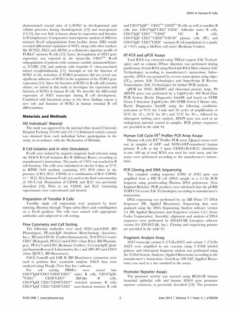
RT-PCR and qPCR AssaysTotal RNA was extracted using TRIzol reagent (Life Technol-
ogies) and on column DNase digestion was performed during
purification of total RNA using PureLink RNA Mini columns (Life
Technologies) according to manufacturer’s instructions. Subse-
quently, cDNA was prepared by reverse transcription using oligo-
(dT)20 primer (Life Technologies) and SuperScript II Reverse
Transcriptase (Life Technologies) for further PCR analysis.
qPCR for SOX5, BLIMP1 and ribosomal protein, large, P0
(RPLP0) genes was performed by a LightCycler 480 Real-Time
PCR System (Roche Diagnostics GmbH) and based on SYBR
Green I detection (LightCycler 480 SYBR Green I Master mix,
Roche Diagnostics GmbH) using the following conditions:
activation at 95uC for 5 min and 50 cycles of amplification at
95uC for 10 s, 63uC for 20 s and 72uC for 30 s., followed by
subsequent melting curve analysis. RPLP0 gene was used as an
endogenous internal control in samples. PCR primer sequences
are provided in the table S1.
Human Cell Cycle RT2 Profiler PCR Array AssaysHuman cell cycle RT2 Profiler PCR array (Qiagen) assays were
run in samples of GFP- and SOX5-GFP-transduced human
primary B cells at day 3 upon CD40L+IL4+IL21 stimulation
in vitro. 400 ng of total RNA was used for each array and the
assays were performed according to the manufacturer’s instruc-
tions.
PCR Cloning and DNA SequencingThe complete coding sequence (CDS) of SOX5 gene was
amplified from a HD B cell cDNA sample as a 3.1 kb PCR
fragment using proof-reading Phusion DNA polymerase (New
England BioLabs). PCR products were subcloned into the pCRII
TOPO TA vector (Life Technologies) according to manufacturer’s
instructions.
DNA sequencing was performed by an ABI Prism 377 DNA
Sequencer (PE, Applied Biosystems). Sequencing data were
analyzed using the DNA Sequencing Analysis software version
3.4 (PE Applied Biosystems) and Sequencer version 3.4.1 (Gene
Codes Corporation). Assembly, alignment and analysis of DNA
sequences were performed by DNASTAR Lasergene software
version 8.0 (DNASTAR, Inc.). Cloning and sequencing primers
are provided in the table S1.
Fragment Analysis AssaySOX5 transcript variant 6 (59L3L-SOX5) and variant 7 (59L3S-
SOX5) were amplified in one reaction using 59-FAM labeled
primers and subsequent fragment analysis was performed using
the 3130xl Genetic Analyzer (Applied Biosystems) according to the
manufacturer’s instructions. GeneScan–500 LIZ (Applied Biosys-
tems) was used as a size standard in the assays.
Promoter Reporter AssaysThe promoter activity was assessed using BEAS-2B human
bronchial epithelial cells and human SPAG6 gene promoter
reporter constructs, as previously described [19]. The promoter
SOX5 In Human B Cells
PLOS ONE | www.plosone.org 2 June 2014 | Volume 9 | Issue 6 | e100328

activity was measured with the Dual Luciferase Reporter Assay
system (Promega) according to manufacturer’s instructions.
Immunofluorescence Staining and MicroscopyCyto-spins of RAJI cells were fixed for 15 min using 4%
solution of freshly prepared formaldehyde in PBS and permeabi-
lized with 0.5% Triton-X for another 15 min. After rinsing and
blocking using 3% BSA in PBS the cells were stained with rabbit
anti-human SOX5 antibodies (Santa Cruz Biotechnolgies, Inc.)
over night at 4uC in the blocking buffer, followed by staining with
TRITC-conjugated swine anti-rabbit IgG antibodies (DakoCyto-
mation) for 1 h at room temperature (RT). After appropriate
washing steps DAPI was applied on slides for 10 min to stain
nuclei.
Confocal microscopy images were collected using the LSM 710
Laser Scanning Microscope and the Image software ZEN black
2011 (Carl Zeiss) with following settings: 100X magnification-
Plan-Apochromat 100X/1.40 oil differential interference contrast
objective lens, 266 mm numerical aperture/pinhole, static sample
with respect to temperature and DAKO mounting medium.
Images were presented using Photoshop CS5 gamma 0.45.
Cryosections (10 mm) of tonsillar tissues were fixed with acetone
and blocked using 5% goat serum (Vector Labs) in PBS for 1.5 h
at RT. Primary (goat anti-human IgD-bio (Southern Biotech.);
rabbit anti-human SOX5 (Santa Cruz Biotechnolgies, Inc.);
mouse anti-human Ki-67 (BD) and mouse anti-human CD138
(Dako) antibodies) and secondary antibody (goat anti-mouse IgG-
A405 (Invitrogen); goat anti-rabbit IgG-A568 (Invitrogen) and SA-
FITC (Dako)) staining was performed in the blocking buffer
containing 5% goat serum for 1 h at RT. Microscopic images
were acquired using Axio Observer and AxioCam MRm (Zeiss)
and were prepared using Axiovision (Ver. 4.8.2.0) and Photoshop
CS5 softwares.
Engineering of the SOX5-GFP Fusion Protein Constructand Lentiviral Transduction
A 2.4 kb complete CDS fragment of the 59L3S-SOX5 transcript
was cloned into the pNL-EGFP/CEF lentiviral vector [20,21] and
a C-terminus-GFP fusion construct – pNL-L-SOX5F-EGFP/
CEF, expressing L-SOX5F-EGFP fusion protein, was generated
using EcoRI and BamHI cloning sites. In the following sections the
pNL-EGFP/CEF control vector and pNL-L-SOX5F-EGFP/CEF
fusion constructs were designated as GFP and SOX5-GFP,
respectively. Lentiviral particles were prepared by co-transfection
of pLTR-G envelop [22] and pCD/NL-BH* helper [23]
constructs in 293T cells, as a packaging cell line, using FuGENE
6 (Roche Diagnostics). Viral particles were harvested after 48 h
and after two rounds of spin infection RAJI cells were GFP sorted
for further analysis. Cloning primers are provided in the table S1.
Isolated quiescent human peripheral blood primary B cells were
transduced by novel lentiviral vectors (LVs) displaying at their
surface the Edmonston measles virus (MV) glycoproteins hemag-
glutinin (H) and fusion protein (F), (H/F-LVs) technique [24,25].
In order to transduce primary B cells, H/F-LV vector pseudotypes
were prepared for the SOX5-GFP fusion as well as for GFP
encoding vector, as a control and the cells were transduced using
the above viral particles. The efficiency of transduction up to
85.0% was reached in SOX5-GFP H/F-LV transduced B cells
and it was even higher in the case of GFP control H/F-LVs (up to
99.0%) after two rounds of spinoculation/infection.
Statistical AnalysisAll data are expressed as mean 6 standard deviation (6 SD).
Statistical comparison between samples was carried out by
Kruskal-Wallis (KW) one way analysis of variance (ANOVA) test
and in cases of significant differences, t-test was applied to indicate
the significance between two groups of samples using GraphPad
Prism 5 software (GraphPad Software, La Jolla, CA).
Results
Expression of SOX5 Transcript Variants in Human B CellsSOX5 transcript variants are encoded in total by 22 exons, out of
which 17 are coding exons (Fig. 1A). In order to analyze the
expression of SOX5 transcripts in B cells, cDNA was prepared
from three different fractions of peripheral blood mononuclear
cells (PBMCs) of three HD volunteers: fraction A – original pool of
PBMCs, fraction B – ‘‘untouched’’ B cells and fraction C – non-B
cells (Fig. 1B). PCR analyses did not reveal detectable signals for
SOX5 transcript variant 1 (primer pair a) and 5 (primer pair b) in
human B cells or other peripheral blood lymphocytes, while the
signal was readily detectable in human costal cartilage cells
(Fig. 1B). B cells expressed low levels of SOX5 variant 3 (primer
pair c) compared to the non-B cell fraction and human testis tissue
(Fig. 1B). A weak signal at about expected 209 bp - corresponding
to variant 2 - and a strong signal at the expected 284 bp -
corresponding to variant 4 - were detected in B cells by primer pair
d (Fig. 1B). Since the presence or absence of exons 17 and 18 is an
additional difference between SOX5 transcript variants 2 and 4, a
new set of primer pair (in exons 15 and 20, primer pair e) was
designed discriminating between these two variants (Fig. 1A).
Surprisingly, the results revealed no detectable signal at 300 bp
corresponding to the variant 4, but a strong signal at the expected
624 bp fragment corresponding to the SOX5 transcript variant 2
(Fig. 1C). These data indicated that SOX5 transcript variant 4 is
not expressed in B cells and suggested that the observed 209 bp
and 284 bp fragments correspond to sub-variants of SOX5
transcript variant 2. Overall, B cells were the main source among
human peripheral blood lymphocytes, expressing high levels of
long transcript variants of SOX5 (primer pairs – d, e and f; Fig. 1B
and 1C).
Human B Cells Express at Least 3 Different L-SOX5Transcript Variants
Due to the predominance of SOX5 transcript variant 2, we
decided to amplify the complete coding sequence (CDS) of the
SOX5 transcript variant 2 (primer pair g) from human peripheral
blood B lymphocytes, in order to subclone the PCR fragments and
verify the sequence of B cell-specific SOX5 transcripts. Sequencing
indeed revealed a transcript corresponding to the SOX5 transcript
variant 2 (NM_152989.3) encoding the L-SOX5B isoform
(NP_694534.1). Additionally, we identified two new SOX5
transcript variants and numbered them accordingly as variant 6
(GenBank accession number: JX570584) and variant 7 (GenBank
accession number: JX570585), both of which contained the 75 bp
non-coding exon 5 at the 59-UTR known from variant 4, but in
contrast to variant 4 and similar to variant 2, variant 6 and 7
enclose coding exons 12 and 13 (Fig. 2A). The coding exon 3 was
present in its full length of 232 bp (coding exon 3a) in SOX5
transcript variant 6 and it was 105 bp shorter due to a truncated
127-bp coding sequence (coding exon 3b) in transcript variant 7
(Fig. 2A). Therefore, the three SOX5 transcripts were designated as
59S3L-SOX5, corresponding to the known transcript variant 2
(NM_152989.3), as well as 59L3L-SOX5 (transcript variant 6) and
59L3S-SOX5 (transcript variant 7), representing two additional new
SOX5 In Human B Cells
PLOS ONE | www.plosone.org 3 June 2014 | Volume 9 | Issue 6 | e100328

transcript variants of SOX5, where 59S describes the existence of
shorter and 59L - of longer 59-UTR sequences, whereas 3L
indicates the longer and 3S - the shorter coding exon 3,
respectively. According to the analysis of open reading frames,
both transcript variant 2 (59S3L-SOX5, NM_152989.3) and variant
6 (59L3L-SOX5, JX570584) encode the same protein isoform, i.e.
the 750 amino acid (aa) long L-SOX5B protein (NP_694534.1).
The transcript variant 7 (59L3S-SOX5, JX570585) encodes a new,
35 aa shorter form of L-SOX5, due to the 105 bp shorter version
of coding exon 3b (Fig. 2A). We named the new isoform L-SOX5F
(GenBank accession number to be assigned).
As expected, a PCR assay recognizing all SOX5 transcript
variants in one reaction (primer pair h) revealed the existence of at
least three different transcript variants, which were confirmed by
single clone sequencing in human B cells (Fig. 2B). Intriguingly, in
B lymphocytes the 59L- transcript variants of SOX5, containing the
75-bp-non-coding exon 5, were more abundant compared to the
59S- transcript variant of SOX5 (Fig. 1B and Fig. 2B).
Expression of SOX5 in Human B Cell PopulationsNext, we analyzed SOX5 expression in circulating naive
CD19+IgM+CD21+CD38+CD272 B cells, CD19+IgM-hiCD21+CD38+CD27+ marginal zone (MZ)-like B cells,
CD19+IgM2CD21+CD38+CD27+ switched memory B cells,
CD19+IgM2CD21+CD38+CD272 non-classical memory B cells
and CD19+IgM+/2CD21lowCD38low innate-like CD21low B cells.
Quantitative RT-PCR analysis, recognizing all transcript variants
of SOX5 expressed in B cells (primer pair – f), showed that
expression levels of SOX5 transcripts were slightly, but not
significantly higher in MZ-like and switched memory B cells
compared to naive B cells. In contrast, levels of SOX5 transcripts
were 25.7617.2 fold (n = 4; p = 0.0393) and 19.361.2 fold (n = 4;
p,0.0001) higher in non-classical memory and CD21low B cells,
respectively, than in naive B cells (Fig. 3A).
In addition to circulating B cells we examined tonsillar B cell
subsets sorted into CD19+IgD+CD272CD382 follicular naive B
cells, CD19+IgD2CD272/+CD38+ germinal center (GC) B cells,
CD19+IgD2CD27++CD38++CD138+ plasma cells (PCs) and
Figure 1. Expression of SOX5 transcript variants in human B cells. (A) Schematic representation of human SOX5 transcript variants. Non-coding exons are depicted as open rectangles, partial coding exons - as half open rectangles and coding exons - as filled rectangles. Primer regionsare indicated with appropriate arrows. Exons and coding exons are numbered according to their location along the genomic sequence, which aredrawn as black lines. (B) RT-PCR analysis for the expression of b-actin, CD19 genes and SOX5 transcript variants. HD PBMCs were separated into: A –PBMCs; B – B cells and C – non-B lymphocytes. Except for SOX5 transcript variant 3 (SOX5-var 3) in which human testis RNA sample served as a control,human costal cartilage cells used as a positive control in all RT-PCR reactions. In agarose gel pictures DNA markers were cut out, since they wereloaded between the tested samples and the control sample. (C) RT-PCR assay performed to discriminate between SOX5 transcript variant 2 andvariant 4 in samples of peripheral blood lymphocytes: A – PBMCs; B – B cells and C – non-B lymphocytes.doi:10.1371/journal.pone.0100328.g001
SOX5 In Human B Cells
PLOS ONE | www.plosone.org 4 June 2014 | Volume 9 | Issue 6 | e100328

CD19+IgD2CD27+CD382 memory B cells. Compared to follic-
ular naive B cells GC B cells expressed 110.5676.4 fold (n = 3; p
0.0398) higher levels of SOX5 transcripts (Fig. 3B). The expression
levels of SOX5 transcripts were not significantly higher in memory
B cells, whereas PCs expressed significantly (p = 0.0440) lower
levels of SOX5 transcripts compared to the GC B cells.
Consistently, immunofluorescence staining of tonsillar tissues
revealed polarized expression of SOX5 protein preferentially in
centrocytes within the light zone of germinal centers, but not in
terminally differentiated CD138hi extrafollicular plasma cells
(Fig. 3C).
In order to distinguish between the two prominent SOX5
transcript variants we designed a qPCR assay recognizing only the
transcript variant 6 (59L3L-SOX5, Fig. S1A, primer pair – i) in B
cells. Except for the non-classical memory B cells in the blood,
transcript varrinat 6 (59L3L-SOX5) expression reflected (Fig. S1B
and S1C) the expression pattern of total SOX5 variants in
peripheral blood and tonsillar naıve, GC and memory B cells
(Fig. 3A and 3B). Subsequent fragment analysis using 59-FAM-
labeled primers (Fig. S1A, primer pair - j) revealed a nearly equal
distribution between transcript variant 7 (59L3S-SOX5) and variant
6 (59L3L-SOX5) in all tested tonsillar B cell populations (Fig. S1D).
While the fragment analysis was not sensitive enough to analyse
peripheral blood B cells ex vivo, the analysis of B cells stimulated
in vitro (Fig. S1E) showed a similar expression pattern of these
variants. Collectively, these data demonstrate that both transcript
variant 6 (59L3L-SOX5) and variant 7 (59L3S-SOX5) are equally
expressed in B cells ex vivo as well as in B cells during activation
in vitro.
SOX5 Expression during Activation and Differentiation ofB Cells In vitro
Since high levels of SOX5 mRNA were found at some activation
dependent stages of B cell differentiation in the tonsil, we asked
next whether specific signaling pathways might induce SOX5
expression during in vitro stimulation of B cells. Isolated peripheral
blood B cells were activated either with a single stimulus (IL4, IL21
and CD40L) or combinations of stimuli (CD40L+IL4 and
CD40L+IL4+IL21) in vitro. Flow cytometric analysis at days 3, 6
and 9 revealed strong differentiation of CD38hiCD138+ plasma-
blasts upon CD40L+IL4+IL21 stimulation and only a weak
differentiation upon CD40L+IL4 stimulation, whereas no differ-
Figure 2. Human B cells express at least three different transcript variants of SOX5. (A) Schematic representation of sequence verifiedhuman SOX5 transcript variants in B lymphocytes. Non-coding exons are depicted as open rectangles, partial coding exons - as half open rectanglesand coding exons - as filled rectangles. Cloning primer locations are indicated with appropriate arrows. Exons and coding exons are numberedaccording to their location along the genomic sequence, which are drawn as black lines. (B) PCR analysis of SOX5 transcript expression in B cells andin single clones picked for sequence analysis. B lymphocytes express at least three different SOX5 transcript splice variants as evidenced byrepresentative single TOPO clones 1, 2 and 3.doi:10.1371/journal.pone.0100328.g002
SOX5 In Human B Cells
PLOS ONE | www.plosone.org 5 June 2014 | Volume 9 | Issue 6 | e100328

entiation was detectable in cells activated with IL4 or IL21 or
CD40L alone (Fig. 4A). Proliferation, measured by CFSE labeling
of cells, occurred to a small degree upon CD40L stimulation,
stronger in cells stimulated with CD40L+IL4 and the strongest
after CD40L+IL4+IL21 stimulation (Fig. 4A), while B cells
stimulated with IL4 and IL21 alone did not proliferate. We
noticed a significant decrease of SOX5 expression (p,0.05) in
unstimulated B cells at day 3 compared to freshly isolated B cells
(Fig. 4B). Subsequently, SOX5 transcripts were highly up-regulated
upon stimulation with IL21 (Fig. 4B). In contrast, all proliferating
cells after IL4+CD40L stimulation expressed the lowest amounts
of SOX5, which was slightly but significantly increased by the
addition of IL-21 (n = 5, p-values,0.05) (Fig. 4B). These data
suggested a reduction of SOX5 expression during proliferation,
while higher levels of SOX5 were present in culture conditions
inducing plasmablast differentiation. In order to test whether
SOX5 is induced not only during IL21-mediated differentiation,
isolated peripheral B cells were stimulated with CpG in vitro. Upon
CpG stimulation, CD38hiCD138+ plasmablast differentiation was
observed at days 6 and 9 (Fig. S2A) and this process was also
associated with increased levels of SOX5 transcripts in the cells
(Fig. S2B).
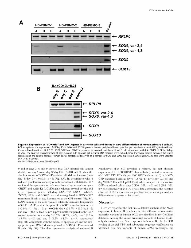
Expression of SOX-trio and SOX13 Genes in HumanEx vivo B Cells and during In vitro B Cell Differentiation
Since Sox5 is co-expressed with Sox6 and Sox9 in chondrocytes,
oligodendrocytes and melanocytes [4], we asked whether the other
two members of the SOX-trio [26] are expressed in human B cells.
Figure 3. Expression of SOX5 in human B cell subpopulations. (A) Relative quantification of SOX5 by RT-qPCR in peripheral blood naive, MZ-like, switched memory (sw mem), non-classical memory (nc mem) and CD21low B cells. (B) Relative quantification RT-qPCR assay for SOX5 expressionin follicular naive, germinal center B cells (GC), memory B cells and plasma cells (PC) from tonsils. T-test p-values indicate the significance ofdifferences between the samples. Relative expression levels of SOX5 are shown as mean 6 SD. RPLP0 gene was used as an internal control in thesamples. (C) Immunofluorescence staining for the expression of SOX5 protein in tonsillar tissues. IgD staining was used to stain mantle zones, Ki67staining for proliferating centroblasts within germinal centers and CD138 as a marker for extrafollicular plasma cells.doi:10.1371/journal.pone.0100328.g003
SOX5 In Human B Cells
PLOS ONE | www.plosone.org 6 June 2014 | Volume 9 | Issue 6 | e100328

Human SOX9 gene is represented by a single transcript, while at
least four different transcript variants exist for the human SOX6
gene. PCRs for SOX9 and SOX6 transcripts, recognizing all four
transcript variants of SOX6, showed weak signals for SOX6
transcript variants in PBMCs and non-B cell fractions, but were
absent in B cells (Fig. 5A). SOX9 was undetectable in any
lymphocyte population of the peripheral blood, while the signal
was readily detected in human costal cartilage cells (Fig. 5A).
Since co-expression of Sox5 and Sox13 genes was reported in
mouse pancreatic epithelial cells [27], we tested co-expression of
the SOX13 gene in human B lymphocytes. While SOX13 was
expressed in non-B cell fractions of the PBMC, there was no
detectable signal in B cell fractions, indicating that SOX13 is not
expressed in human B cells (Fig. 5A).
Because freshly isolated B cells did not express SOX6 and SOX9
or SOX13 ex vivo, we tested whether these genes are induced during
in vitro B cell differentiation. As shown in Fig. 5B, none of the
tested genes were induced at days 3, 6 or 9 after IL4+CD40L+IL21 stimulation.
SOX5 Overexpression Reduces Proliferation, WhilePlasmablast Differentiation Still Occurs
Because of its novelty and the prominent expression in B cells,
we decided to clone the transcript variant 7 of SOX5 (59L3S-SOX5)
in order to study its function in human B cells. A SOX5-GFP
fusion protein was engineered and its functionality was tested by
using the previously reported SPAG6 promoter reporter assays. In
BEAS-2B epithelial cells expressing the SOX5-GFP fusion protein,
the SPAG6 promoter activities reached much higher induction
levels, 14.261.2 fold for pGL3-1-SOX5 (n = 3; p = 0.0005) and
10.260.6 fold for pGL3-4-SOX5 (n = 3; p,0.0001) when
compared with controls (Fig. S3A), indicating that our SOX5-
GFP fusion protein construct is functional.
Immunofluorescence staining revealed a barely detectable signal
for endogenous SOX5 in GFP-transduced RAJI cells (Fig. S3B);
whereas SOX5 was highly expressed and the co-expression and
nuclear localization of exogenous SOX5 and GFP was readily
documented in SOX5-GFP-transduced cells (Fig. S3C).
Subsequently, expression levels of previously reported SOX5
target genes RhoB [28], S100A1 and S100B [29] were examined in
sorted GFP-low and GFP-hi control cells and SOX5-GFP-low,
SOX5-GFP-intermediate (int) and SOX5-GFP-hi cells (Fig. S3D).
While RHOB and S100B genes seemed not to be expressed in
RAJI cells (Fig. S3E), the S100A1 gene was expressed and its
expression levels correlated with the expression levels of 59L3S-
SOX5 in SOX5-GFP transduced RAJI cell fractions (Fig. S3D and
S3E), compatible with the regulation of the S100A1 gene by
exogenous SOX5. Unlike in chondrocytes [29], the regulation of
the S100A1 did not depend on SOX-trio activity, since SOX6 and
SOX9 were not expressed in RAJI cells (Fig. S3E).
After confirmation of the expression and function of the fusion
protein in a human B cell line, we analyzed the function of L-
SOX5F in primary human B lymphocytes. Primary B lymphocytes
were transduced with SOX5-GFP fusion and GFP control
constructs. Isolated cells were stimulated for 9 days in vitro
(Fig. 6). Analysis of absolute counts of GFP-positive cells at day
Figure 4. Induction of SOX5 during in vitro B cell differentiation. (A) Isolated peripheral blood B cells were either left for 3 days without anystimulus or stimulated for 9 days in vitro either with a single stimulus (IL4, IL21 and CD40L) or the combination of these (CD40L+IL4+/2 IL21). Thecells were analyzed by FACS for the plasma cell markers CD138 and CD38 at days 3, 6 and 9. Gates indicate the frequency of CD138+CD38hi
plasmablasts in each plot. CFSE at day 9 was measured as an indicator of proliferation in the cells. Representative FACS plots of five independentexperiments are shown. (B) RT-qPCR analysis of SOX5 expression in ex vivo B cells and samples either unstimulated (unstim.) for 3 days or stimulatedwith a single stimulus (IL4, IL21 and CD40L) or the combination of these (CD40L+IL4+/2 IL21). Significant differences are depicted and appropriate t-test p-values (n = 5; p,0.05) are indicated. Relative expression levels of SOX5 are shown as mean 6 SD. RPLP0 gene served as an internal control inthe samples.doi:10.1371/journal.pone.0100328.g004
SOX5 In Human B Cells
PLOS ONE | www.plosone.org 7 June 2014 | Volume 9 | Issue 6 | e100328
0 and at days 3, 6 and 9 showed that GFP-infected cells almost
doubled on day 3 (ratio day 3/day 0 = 1.760.8; n = 3), while the
absolute counts of SOX5-GFP-positive cells did not increase (ratio
day 3/day 0 = 1.060.5; n = 3; Fig. 6A). In accordance with a
reduced proliferative capacity of cells transduced with SOX5-GFP
we found the up-regulation of a negative cell cycle regulator gene
CHEK1 and cyclin E1 (CCNE1) gene, whereas several positive cell
cycle regulator genes, including CCND1/2, CDK4, CDC25A,
TFDP2, E2F4 and MKI67, were down-regulated in SOX5-GFP
transduced B cells at day 3 compared to the GFP control (Fig. S4).
DAPI staining of the cells revealed relatively increased frequencies
of GFP+ DAPI+ dead cells upon SOX5-GFP transductions at day
3 (2.9% 61.1%; n = 3; p = 0.0092), day 6 (14.7% 616.5%; n = 3)
and day 9 (21.5% 67.6%; n = 3; p = 0.0204) compared to the GFP
control transductions at day 3 (1.2% 60.7%; n = 3), day 6 (4.9%
63.7%; n = 3) and day 9 (9.3% 64.6%; n = 3), respectively
(Fig. 6B). Compatible with the increased apoptosis we saw the anti-
apoptotic gene BIRC5 down-regulated in SOX5-GFP transduced
B cells (Fig. S4). The flow cytometric analysis of cultured B
lymphocytes (Fig. 6C) revealed a relative, but not absolute
expansion of CD138+CD38hi plasmablasts (counted as numbers
of CD38hi CD138+ cells per 1000 GFP+ cells at day 0) in SOX5-
GFP-transduced cells at day 6 (10676741; n = 3; p = 0.0194) and
day 9 (6026345; n = 3; p = 0.0347), when compared to the control
GFP-transduced cells at days 6 (4206261; n = 3) and 9 (3846331;
n = 3), respectively (Fig. 6D). These data corroborate the negative
effect of SOX5 expression on proliferation, whereas plasmablast
differentiation appears to be spared.
Discussion
Here we report for the first time a detailed analysis of the SOX5
expression in human B lymphocytes. Five different representative
transcript variants of human SOX5 are identified in the GenBank
database. Among the known transcript variants of human SOX5,
only the variants 2 and 3 are expressed in human B cells. By PCR
cloning of the full CDS and subsequent sequence verification we
identified two new variants of human SOX5 transcripts, the
Figure 5. Expression of ‘‘SOX-trio’’ and SOX13 genes in ex vivo B cells and during in vitro differentiation of human primary B cells. (A)PCR analysis for the expression of RPLP0, SOX6, SOX9 and SOX13 genes in human peripheral blood lymphocyte populations: A – PBMCs; B – B cells andC – non-B cell fractions. (B) RPLP0, SOX6, SOX9 and SOX13 expression in isolated peripheral blood B cells stimulated with IL4+CD40L+IL21 for 9 daysin vitro. The analysis was performed at days 3, 6 and 9. In agarose gel pictures DNA markers were cut out, since they were loaded between the testedsamples and the control sample. Human costal cartilage cells served as a control for SOX6 and SOX9 expression, whereas BEAS-2B cells were used forSOX13 as a control.doi:10.1371/journal.pone.0100328.g005
SOX5 In Human B Cells
PLOS ONE | www.plosone.org 8 June 2014 | Volume 9 | Issue 6 | e100328

variants 6 (59L3L-SOX5) and 7 (59L3S-SOX5), for which
GenBank accession numbers JX570584 and JX570585 were
assigned, respectively. Similar to the variant 2, both of the new
transcript variants carry 18 exons, but bear an additional 75 bp
non-coding exon 5. The two new SOX5 transcript variants differ in
the length of coding exon 3. In the transcript variant 6 the coding
exon 3 (coding exon 3a) is present in its full length of 232 bp
coding for 77 aa, whereas in the transcript variant 7 the coding
exon 3 (coding exon 3b) is only 127 bp long sustaining the same
translation initiation site, but coding for only 42 aa (Fig. 2A). Like
transcript variant 2, the transcript variant 6 encodes the known
750 aa protein isoform - L-SOX5B (NP_694534.1), whereas the
transcript variant 7 encodes a new, shorter, 715 aa long protein
isoform of SOX5, which we designated L-SOX5F. The missing 35
aa residues in the L-SOX5F isoform do not involve any of the
known functional domains of L-SOX5 (Fig. S5). Both new variants
are expressed in nearly equal amounts in all tested tonsillar B cell
populations and during B cell activation in vitro, suggesting similar
regulation of expression due to identical regulatory elements.
However, due to the low sensitivity and limitations of the fragment
analysis technique applied in this study, we cannot exclude
differential expression of the two SOX5 variants in some of the
circulating B cells, especially in circulating CD21low and atypical
memory B cells.
SoxD proteins have no known transactivation or transrepression
domains and therefore require interaction partners to participate
in transcriptional activation and repression as demonstrated in
different cell types [3,4]. The SOX-trio, consisting of SOX5,
SOX6 and SOX9 [26], is so far the best-examined complex
involving the action of Sox5 in chondrocytes [8,30], oligodendro-
cytes and melanocytes [4,9,31]. Depending on the constellation
Sox5 and Sox6 heterodimers cooperate with Sox9 in transactiva-
tion of chondrocyte-specific genes [3,4] or act as transrepressors by
competing with SoxE proteins in oligodendrocytes and melano-
cytes by directly recruiting the co-repressor CtBP2 and the histone
deacetylase Hdac1 [3,4]. Beside the interaction of L-Sox5 and
Sox6 proteins [32], co-expression of L-Sox5 and Sox13 was shown
in mouse pancreatic epithelial cells [27]. However, our data clearly
indicate that neither SOX6, SOX9, nor SOX13 are expressed in
circulating B cells nor induced after in vitro activation of human B
lymphocytes. Therefore other interaction partners for SOX5 must
exist in human B cells and point to a cell type-dependent
expression of interaction partners for SOX5.
SOX5 transcription varied between peripheral blood B-cell
subpopulations, showing the highest expression levels in circulat-
Figure 6. SOX5 modulates in vitro terminal B cell differentiation. (A) Proliferation measured as the ratio of absolute numbers of GFP+ cells inthe samples. Absolute cell counts of GFP+ cells on day 0 are taken as 1.0 and the data are expressed as mean 6 SD. Summary of three independentexperiments are depicted. (B) Frequencies of GFP+ DAPI+ cells within GFP (control) and SOX5-GFP-transduced peripheral blood B cells cultivatedin vitro. (C) Plasma cell differentiation analyzed by FACS in peripheral blood B cells stably transduced either with GFP (control) or SOX5-GFP fusionconstruct upon in vitro stimulation with IL4+CD40L+IL21. DAPI-negative GFP+ gated cells were analyzed by FACS for the plasma cell markers CD138and CD38 at days 3, 6 and 9. Gates indicate the frequencies of CD138+CD38hi plasmablasts in each FACS plot. Representative FACS plots of threeindependent experiments are shown. (D) Numbers of CD138+CD38hi plasmablasts in GFP (control) and SOX5-GFP-transduced B cells at days 3, 6 and 9referring to CD38hi CD138+ cells per 1000 GFP+ cells at day 0. Cell numbers are depicted as mean 6 SD and t-test p-values indicate the significantdifferences. Summary of three independent experiments are shown.doi:10.1371/journal.pone.0100328.g006
SOX5 In Human B Cells
PLOS ONE | www.plosone.org 9 June 2014 | Volume 9 | Issue 6 | e100328

ing innate-like CD21low B cells and non-classical CD272 memory
B cell populations, as we had previously seen in CD21low B cells of
patients with CVID [16] and others in FCRL4+ tissue-like
memory B cells of tonsils [15]. The comparison of tonsillar B
cell populations in healthy donors revealed the highest expression
levels of SOX5 transcript in GC B cells as previously reported [15].
In tonsillar sections the expression of SOX5 protein was polarized
to centrocytes within the light zones of germinal centers, but not in
extrafollicular CD138+ plasma cells, suggesting that SOX5
expression is upregulated during the GC response of B cells and
switched off after terminal differentiation.
In order to dissect the expression pattern of SOX5 during B cell
activation, proliferation and differentiation more closely, we
investigated the SOX5 expression at different time points after
stimulation of B cells in vitro with IL4, IL21, CD40L alone or a
combination of these. The highest SOX5 transcript levels were
found in cells stimulated with IL21 alone, while SOX5 transcript
levels were significantly reduced in cells undergoing proliferation
(CD40L+IL4). Interestingly, adding IL21 to this stimulation not
only caused a dramatic increase in plasmablast differentiation but
also a slight, but significant up-regulation of SOX5 expression.
Given the high expression of SOX5 protein in centrocytes in the
light zone of tonsils, these data suggest that SOX5 plays a role
during IL21-mediated differentiation of activated B cells. This
role, however, is not restricted to IL21 activation, since SOX5 was
also induced in B lymphocytes undergoing plasmablast differen-
tiation upon CpG stimulation in vitro.
In order to demonstrate the effect of high SOX5 expression
levels during B cell activation further, the complete CDS of the
prominently expressed transcript variant 7, encoding the L-
SOX5F, was cloned together with a GFP gene into a lentiviral
expression vector. After transduction of human primary B cells
with SOX5-GFP we found less SOX5-GFP positive cells at days 3,
6 and 9 compared to GFP transduced controls, after adjusting for
transduction efficiency. SOX5-GFP positive cells failed to expand
on day three after stimulation. In support of a negative influence of
high SOX5 levels on the cell cycle we observed an up-regulation of
negative regulators of the cell cycle in combination with the down-
regulation of positive regulatory genes in the SOX5-GFP
transduced cells. The increased expression of a negative regulator
CHEK1 and especially CCNE1 is suggestive of a block at the G1-S
phase transition [33]. This is consistent with the previous results in
SOX5 expressing FCRL4 B cells, which don’t proliferate [34] and
are mostly in G1 phase [15]. Similarly, in CD21low B cells of
CVID patients, the high level of SOX5 expression is associated
with poor proliferation, while a residual potential to differentiate
into antibody secreting cells persists [16,35]. There is multiple
evidence for an anti-proliferative effect of SOX5 in non-B cells.
Thus in neuronal progenitor cells high SOX5 expression is
associated with a premature cell cycle exit [36] and in human
glioma cells [37] with a poor proliferative capacity. Also the high
expression of Sox5 in chondrocytes [8] is compatible with its role
in the non-proliferative state of cells. A recent study corroborates
that lentiviral overexpression of murine Sox5 proteins (Sox5-BLM
and L-Sox5) inhibits cell cycle progression and results in increased
apoptosis of human multiple myeloma cells [38]. We also observed
an increased apoptosis rate of GFP+DAPI+ B cells upon
transduction with SOX5-GFP. However, in contrast to the
reported results, in our hands other B cells underwent apoptosis
more readily and the percentage of CD138+CD38hi plasmablasts
accumulated with time significantly more in SOX5-GFP trans-
duced B cells compared to GFP transduced B cells suggesting that
plasma cells might be less strongly affected by high SOX5
expression levels. This difference might be reconciled by the fact,
that a multiple myeloma line reflects rather terminally differen-
tiated plasma cells which in deed express only very low levels of
SOX5, while according to our in vitro results SOX5 is naturally up-
regulated during the process of plasma cell differentiation.
In conclusion, we reported here the identification of two new
transcript variants of human SOX5, variant 6 and variant 7, which
are expressed in B cells and encode the known 750 aa L-SOXB
isoform and a new isoform of 715 aa L-SOX5F, respectively.
SOX5 expression is stage dependent during B cell differentiation.
It shows the highest expression in centrocytes in the light zones of
germinal centers, in circulating atypical memory and exhausted
CD21low B cells. The interaction partners in B cells remain
unknown but functional assays suggest that it is induced by IL21
and that high levels of L-SOX5F modify terminal B cell
differentiation by suppressing proliferation, but permitting plas-
mablast development.
Supporting Information
Figure S1 Expression of 59L3L-SOX5 and 59L3S-SOX5transcripts in human B cell subpopulations. (A) A scheme
for primer locations and (B) relative quantification of 59L3L-
SOX5 by RT-qPCR in peripheral blood naive, MZ-like, switched
memory (sw mem), non-classical memory (nc mem) and CD21low
B cells as well as (C) in follicular naive, germinal center B cells
(GC) and memory B cells from tonsils. T-test p-values indicate the
significance of differences between the samples. Relative expres-
sion levels of 59L3L-SOX5 are shown as mean 6 SD. RPLP0
gene was used as an internal control in the samples. Fragment
analysis for the relative expression of 59L3S-SOX5 transcript (D)
in tonsillar B cell populations and (E) in CD19+ peripheral blood B
cells upon stimulation in vitro at days 3, 6 and 9. The cells were
activated either with a single stimulus or with a combination of
stimuli, as indicated in the figure (E).
(PDF)
Figure S2 Expression of SOX5 transcripts upon CpG-mediated B cell differentiation in vitro. (A) Differentiation
of B cells upon stimulation with CpG in vitro. The gates depict
CD138+CD38hi plasmablasts at days 3, 6 and 9. (B) RT-qPCR
analysis of SOX5 expression in samples stimulated with CpG. T-
test p-values indicate the significance of differences between the
samples. Relative expression levels of SOX5 are shown as mean 6
SD. RPLP0 gene served as an internal control in the samples.
(PDF)
Figure S3 Construction of the SOX5-GFP fusion proteinand its functionality upon lentiviral transduction in RAJIcells. (A) Luciferase promoter reporter assays for GFP-control
and SOX5-GFP fusion constructs in BEAS-2B cells. Stably
transduced BEAS-2B cells either expressing GFP alone or
SOX5-GFP fusion protein were GFP-sorted and subsequently
transient transfection was performed to measure the promoter
activity. pGL3-Basic plasmid was used as a control for human
SPAG6 promoter constructs, pGL3-1-SOX5 and pGL3-4-SOX5.
Appropriate t-test p-values indicate the significance of differences
between GFP control and SOX5-GFP expressing cells. (B) and (C)
Immunofluorescence staining for SOX5 protein in RAJI cells.
RAJI cells were transduced either with GFP control vector (B) or
SOX5-GFP fusion construct (C). Co-localization of GFP (green)
and SOX5 (red – TRITC) and nuclear translocation is shown.
DAPI staining is indicative of cellular nuclei. (D) Lentiviral
expression of GFP and SOX5-GFP fusion proteins in RAJI cells
analyzed by flow cytometry. Stably transduced RAJI cells were
sorted into GFP-low and GFP-hi as well as SOX5-GFP-low,
SOX5 In Human B Cells
PLOS ONE | www.plosone.org 10 June 2014 | Volume 9 | Issue 6 | e100328

SOX5-GFP-int and SOX5-GFP-hi fraction and RT-PCR analy-
ses for the expression of GAPDH and 59L3S-SOX5 transcript
were performed. (E) RT-PCR analysis for the expression of known
SOX5 target genes: RHOB, S100A1 and S100B as well as SOX-
trio genes, SOX6 and SOX9 in stably transduced and GFP-sorted
RAJI cell fractions. In agarose gel pictures DNA markers were cut
out, since they were loaded between the tested samples and the
control sample. Human costal cartilage cells served as a control.
(PDF)
Figure S4 Expression of human cell cycle genes inSOX5-transduced and in vitro differentiated humanprimary B cells. Human cell cycle RT2 Profiler PCR Array
containg 84 pathway specific genes were run for RNA samples of
GFP- and SOX5-GFP-transduced human primary B cells at day 3
upon CD40L+IL4+IL21 stimulation in vitro. Negative regulatory
genes of cell cycle are indicated in red, whereas positive regulatory
genes of cell cycle are depicted in green. Black color shows the
regulation of RAD51 gene, which is involved in the homologous
recombination and repair of DNA. Dashed lines indicate a
threshold of 2 fold up- or down-regulation of genes.
(PDF)
Figure S5 Comparison of known functional domains inL-SOX5B and L-SOX5F proteins. Both SOX5 proteins
contain the same known functional domains and share the same
translation initiation site, except for different exon 3 (exon 3a and
exon 3b). NL – nuclear localization domain.
(PDF)
Table S1 Primer sequences used in this study. Primer
sequences are listed and subdivided into sections according to the
assay type and in the order as they appear in the text.
(PDF)
Acknowledgments
The authors thank Jan Bodinek-Wersing (Centre for Chronic Immunode-
ficiency), Klaus Geiger and Dr. Marie Follo (Core Facility) for their
technical support in cell sorting, AG Idzko (Deptartment of Pneumology)
for BEAS-2B cells and AG Mehlhorn (Department of Orthopedy and
Traumatology) for HCC cells (all at the University Medical Center
Freiburg, Freiburg, Germany). We thank all healthy donor volunteers who
willingly participated in this study.
Author Contributions
Conceived and designed the experiments: M. Rakhmanov HHP HE KW.
Performed the experiments: M. Rakhmanov HS AKK BF M. Rizzi MS
KM SU LM NES SDD CKA WS US. Analyzed the data: M. Rakhmanov
HS AKK M. Rizzi SDD CKA HHP REV US HE KW. Contributed
reagents/materials/analysis tools: ZZ FLC EV. Wrote the paper: M.
Rakhmanov KW.
References
1. Lefebvre V, Dumitriu B, Penzo-Mendez A, Han Y, Pallavi B (2007) Control of
cell fate and differentiation by Sry-related high-mobility-group box (Sox)
transcription factors. Int J Biochem Cell Biol 39: 2195–2214. S1357-
2725(07)00175-6 [pii]; 10.1016/j.biocel.2007.05.019 [doi].
2. Chew LJ, Gallo V (2009) The Yin and Yang of Sox proteins: Activation and
repression in development and disease. J Neurosci Res 87: 3277–3287. 10.1002/
jnr.22128 [doi].
3. Wegner M (2010) All purpose Sox: The many roles of Sox proteins in gene
expression. Int J Biochem Cell Biol 42: 381–390. S1357-2725(09)00196-4 [pii];
10.1016/j.biocel.2009.07.006 [doi].
4. Lefebvre V (2010) The SoxD transcription factors–Sox5, Sox6, and Sox13–are
key cell fate modulators. Int J Biochem Cell Biol 42: 429–432. S1357-
2725(09)00205-2 [pii]; 10.1016/j.biocel.2009.07.016 [doi].
5. Connor F, Cary PD, Read CM, Preston NS, Driscoll PC, et al. (1994) DNA
binding and bending properties of the post-meiotically expressed Sry-related
protein Sox-5. Nucleic Acids Res 22: 3339–3346.
6. Denny P, Swift S, Connor F, Ashworth A (1992) An SRY-related gene expressed
during spermatogenesis in the mouse encodes a sequence-specific DNA-binding
protein. EMBO J 11: 3705–3712.
7. Connor F, Wright E, Denny P, Koopman P, Ashworth A (1995) The Sry-related
HMG box-containing gene Sox6 is expressed in the adult testis and developing
nervous system of the mouse. Nucleic Acids Res 23: 3365–3372. 5w0113 [pii].
8. Lefebvre V, Li P, de CB (1998) A new long form of Sox5 (L-Sox5), Sox6 and
Sox9 are coexpressed in chondrogenesis and cooperatively activate the type II
collagen gene. EMBO J 17: 5718–5733. 10.1093/emboj/17.19.5718 [doi].
9. Stolt CC, Schlierf A, Lommes P, Hillgartner S, Werner T, et al. (2006) SoxD
proteins influence multiple stages of oligodendrocyte development and modulate
SoxE protein function. Dev Cell 11: 697–709. S1534-5807(06)00357-1 [pii];
10.1016/j.devcel.2006.08.011 [doi].
10. Wunderle VM, Critcher R, Ashworth A, Goodfellow PN (1996) Cloning and
characterization of SOX5, a new member of the human SOX gene family.
Genomics 36: 354–358. S0888-7543(96)90474-7 [pii]; 10.1006/geno.1996.0474
[doi].
11. Ikeda T, Zhang J, Chano T, Mabuchi A, Fukuda A, et al. (2002) Identification
and characterization of the human long form of Sox5 (L-SOX5) gene. Gene 298:
59–68. S0378111902009277 [pii].
12. Smits P, Li P, Mandel J, Zhang Z, Deng JM, et al. (2001) The transcription
factors L-Sox5 and Sox6 are essential for cartilage formation. Dev Cell 1: 277–
290. S1534-5807(01)00003-X [pii].
13. Kwan KY, Lam MM, Krsnik Z, Kawasawa YI, Lefebvre V, et al. (2008) SOX5
postmitotically regulates migration, postmigratory differentiation, and projec-
tions of subplate and deep-layer neocortical neurons. Proc Natl Acad Sci U S A
105: 16021–16026. 0806791105 [pii]; 10.1073/pnas.0806791105 [doi].
14. Lai T, Jabaudon D, Molyneaux BJ, Azim E, Arlotta P, et al. (2008) SOX5
controls the sequential generation of distinct corticofugal neuron subtypes.
Neuron 57: 232–247. S0896-6273(08)00026-3 [pii]; 10.1016/j.neu-
ron.2007.12.023 [doi].
15. Ehrhardt GR, Hijikata A, Kitamura H, Ohara O, Wang JY, et al. (2008)
Discriminating gene expression profiles of memory B cell subpopulations. J Exp
Med 205: 1807–1817. jem.20072682 [pii]; 10.1084/jem.20072682 [doi].
16. Rakhmanov M, Keller B, Gutenberger S, Foerster C, Hoenig M, et al. (2009)
Circulating CD21low B cells in common variable immunodeficiency resemble
tissue homing, innate-like B cells. Proc Natl Acad Sci U S A 106: 13451–13456.
0901984106 [pii]; 10.1073/pnas.0901984106 [doi].
17. Charles ED, Brunetti C, Marukian S, Ritola KD, Talal AH, et al. (2011) Clonal
B cells in patients with hepatitis C virus-associated mixed cryoglobulinemia
contain an expanded anergic CD21low B-cell subset. Blood 117: 5425–5437.
blood-2010-10-312942 [pii]; 10.1182/blood-2010-10-312942 [doi].
18. Warnatz K, Salzer U, Rizzi M, Fischer B, Gutenberger S, et al. (2009) B-cell
activating factor receptor deficiency is associated with an adult-onset antibody
deficiency syndrome in humans. Proc Natl Acad Sci U S A 106: 13945–13950.
0903543106 [pii]; 10.1073/pnas.0903543106 [doi].
19. Kiselak EA, Shen X, Song J, Gude DR, Wang J, et al. (2010) Transcriptional
regulation of an axonemal central apparatus gene, sperm-associated antigen 6,
by a SRY-related high mobility group transcription factor, S-SOX5. J Biol
Chem 285: 30496–30505. M110.121590 [pii]; 10.1074/jbc.M110.121590 [doi].
20. Reiser J (2000) Production and concentration of pseudotyped HIV-1-based gene
transfer vectors. Gene Ther 7: 910–913. 10.1038/sj.gt.3301188 [doi].
21. Reiser J, Lai Z, Zhang XY, Brady RO (2000) Development of multigene and
regulated lentivirus vectors. J Virol 74: 10589–10599.
22. Reiser J, Harmison G, Kluepfel-Stahl S, Brady RO, Karlsson S, et al. (1996)
Transduction of nondividing cells using pseudotyped defective high-titer HIV
type 1 particles. Proc Natl Acad Sci U S A 93: 15266–15271.
23. Zhang XY, La Russa VF, Bao L, Kolls J, Schwarzenberger P, et al. (2002)
Lentiviral vectors for sustained transgene expression in human bone marrow-
derived stromal cells. Mol Ther 5: 555–565. 10.1006/mthe.2002.0585 [doi];
S152500160290585X [pii].
24. Funke S, Maisner A, Muhlebach MD, Koehl U, Grez M, et al. (2008) Targeted
cell entry of lentiviral vectors. Mol Ther 16: 1427–1436. mt2008128 [pii];
10.1038/mt.2008.128 [doi].
25. Frecha C, Costa C, Levy C, Negre D, Russell SJ, et al. (2009) Efficient and stable
transduction of resting B lymphocytes and primary chronic lymphocyte leukemia
cells using measles virus gp displaying lentiviral vectors. Blood 114: 3173–3180.
blood-2009-05-220798 [pii]; 10.1182/blood-2009-05-220798 [doi].
26. Ikeda T, Kamekura S, Mabuchi A, Kou I, Seki S, et al. (2004) The combination
of SOX5, SOX6, and SOX9 (the SOX trio) provides signals sufficient for
induction of permanent cartilage. Arthritis Rheum 50: 3561–3573. 10.1002/
art.20611 [doi].
27. Lioubinski O, Muller M, Wegner M, Sander M (2003) Expression of Sox
transcription factors in the developing mouse pancreas. Dev Dyn 227: 402–408.
10.1002/dvdy.10311 [doi].
28. Perez-Alcala S, Nieto MA, Barbas JA (2004) LSox5 regulates RhoB expression
in the neural tube and promotes generation of the neural crest. Development
131: 4455–4465. 10.1242/dev.01329 [doi]; dev.01329 [pii].
SOX5 In Human B Cells
PLOS ONE | www.plosone.org 11 June 2014 | Volume 9 | Issue 6 | e100328
29. Saito T, Ikeda T, Nakamura K, Chung UI, Kawaguchi H (2007) S100A1 and
S100B, transcriptional targets of SOX trio, inhibit terminal differentiation of
chondrocytes. EMBO Rep 8: 504–509. 7400934 [pii]; 10.1038/sj.em-
bor.7400934 [doi].
30. Han Y, Lefebvre V (2008) L-Sox5 and Sox6 drive expression of the aggrecan
gene in cartilage by securing binding of Sox9 to a far-upstream enhancer. Mol
Cell Biol 28: 4999–5013. MCB.00695-08 [pii]; 10.1128/MCB.00695-08 [doi].
31. Stolt CC, Lommes P, Hillgartner S, Wegner M (2008) The transcription factor
Sox5 modulates Sox10 function during melanocyte development. Nucleic Acids
Res 36: 5427–5440. gkn527 [pii]; 10.1093/nar/gkn527 [doi].
32. Lefebvre V (2002) Toward understanding the functions of the two highly related
Sox5 and Sox6 genes. J Bone Miner Metab 20: 121–130. 10.1007/
s007740200017 [doi].
33. da Silva GN, de Camargo EA, Salvadori DM (2012) Toxicogenomic activity of
gemcitabine in two TP53-mutated bladder cancer cell lines: special focus on cell
cycle-related genes. Mol Biol Rep 39: 10373–10382. 10.1007/s11033-012-1916-
1 [doi].
34. Moir S, Ho J, Malaspina A, Wang W, DiPoto AC, et al. (2008) Evidence for
HIV-associated B cell exhaustion in a dysfunctional memory B cell compartmentin HIV-infected viremic individuals. J Exp Med 205: 1797–1805. jem.20072683
[pii]; 10.1084/jem.20072683 [doi].
35. Isnardi I, Ng YS, Menard L, Meyers G, Saadoun D, et al. (2010) Complementreceptor 2/. Blood 115: 5026–5036. blood-2009-09-243071 [pii]; 10.1182/
blood-2009-09-243071 [doi].36. Martinez-Morales PL, Quiroga AC, Barbas JA, Morales AV (2010) SOX5
controls cell cycle progression in neural progenitors by interfering with the
WNT-beta-catenin pathway. EMBO Rep 11: 466–472. embor201061 [pii];10.1038/embor.2010.61 [doi].
37. Tchougounova E, Jiang Y, Brasater D, Lindberg N, Kastemar M, et al. (2009)Sox5 can suppress platelet-derived growth factor B-induced glioma development
in Ink4a-deficient mice through induction of acute cellular senescence.Oncogene 28: 1537–1548. onc20099 [pii]; 10.1038/onc.2009.9 [doi].
38. Edwards SK, Desai A, Liu Y, Moore CR, Xie P (2014) Expression and function
of a novel isoform of Sox5 in malignant B cells. Leuk Res 38: 393–401. S0145-2126(13)00448-7 [pii]; 10.1016/j.leukres.2013.12.016 [doi].
SOX5 In Human B Cells
PLOS ONE | www.plosone.org 12 June 2014 | Volume 9 | Issue 6 | e100328