Heat shock factors HsfB1 and HsfB2b are involved in the regulation of Pdf1.2 expression and pathogen...
14
Molecular Plant • Volume 2 • Number 1 • Pages 152–165 • January 2009 RESEARCH ARTICLE Heat Shock Factors HsfB1 and HsfB2b Are Involved in the Regulation of Pdf1.2 Expression and Pathogen Resistance in Arabidopsis Mukesh Kumar a , Wolfgang Busch b , Hannah Birke c , Birgit Kemmerling d , Thorsten Nu ¨ rnberger d and Friedrich Scho ¨ ffl c,1 a Present address: Heinrich-Pette-Institut, Martinistrabe 52, D-20251 Hamburg, Germany b Max-Planck-Institut fu ¨ r Entwicklungsbiologie, Abteilung Molekularbiologie, Speemannstrabe 37–39, D-72076 Tu ¨ bingen, Germany c Universita ¨ t Tu ¨ bingen, Zentrum fu ¨ r Molekularbiologie der Pflanzen (ZMBP)—Allgemeine Genetik, Auf der Morgenstelle 28, D-72076 Tu ¨ bingen, Germany d Universita ¨ t Tu ¨ bingen, Zentrum fu ¨ r Molekularbiologie der Pflanzen (ZMBP)—Biochemie der Pflanzen, Auf der Morgenstelle 5, D-72076 Tu ¨ bingen, Germany ABSTRACT In order to assess the functional roles of heat stress-induced class B-heat shock factors in Arabidopsis, we investigated T-DNA knockout mutants of AtHsfB1 and AtHsfB2b. Micorarray analysis of double knockout hsfB1/hsfB2b plants revealed as strong an up-regulation of the basal mRNA-levels of the defensin genes Pdf1.2a/b in mutant plants. The Pdf expression was further enhanced by jasmonic acid treatment or infection with the necrotrophic fungus Alternaria brassicicola. The single mutant hsfB2b and the double mutant hsfB1/B2b were significantly improved in disease resistance after A. brassicicola infection. There was no indication for a direct interaction of Hsf with the promoter of Pdf1.2, which is devoid of perfect HSE consensus Hsf-binding sequences. However, changes in the formation of late HsfA2-dependent HSE binding were detected in hsfB1/B2b plants. This suggests that HsfB1/B2b may interact with class A-Hsf in regulating the shut-off of the heat shock response. The identification of Pdf genes as targets of Hsf-dependent negative regulation is the first evidence for an interconnection of Hsf in the regulation of biotic and abiotic responses. Key words: Abiotic/environmental stress; transcriptional control and transcription factors; transcriptome analysis; de- fense responses; disease resistance; Arabidopsis. INTRODUCTION Environmental adaptation of plants depends on elaborate sys- tems for stress sensing and signaling, common and stress- specific responses, and probably also on a hierarchical control of reactions. There is evidence that heat shock factors (Hsfs) play important roles in stress sensing and signaling of different environmental stresses and that different stresses, including also high temperature, induce reactive oxygen species (ROS) in plants (Dat et al., 1998). ROS, particularly H 2 O 2 , are important components in biotic and abiotic stress response and signaling mechanisms. Hsfs recognize consensus binding motifs, so-called heat stress elements (HSE: 5#-nGAAnnTTCnnGAAn-3# or 5#-nTTCnn- GAAnnTTCn-3’) conserved in promoters of heat-inducible genes of all eukaryotes. The classical target genes are the heat shock protein (Hsp) genes, which are (including the HSE motifs in the promoter region) highly conserved in eukaryotes (Wu, 1995). Hsfs display a modular structure with a highly conserved N-terminal DNA-binding domain (DBD), which is characterized by a central helix-turn-helix motif, and an adjacent bipartite oligomerization domain (HR-A/B) composed of hydrophobic heptad repeats. Hsf trimerization via the formation of a triple stranded alpha-helical coiled coil is a prerequisite for high-af- finity DNA binding and subsequently for transcriptional acti- vation of heat shock genes. Other functional domains of Hsf include clusters of basic amino acid residues (NLS) essential for nuclear import, leucine-rich export sequences in the HR- C region (NES) and—less conserved—a C-terminal activator do- main (CTAD) rich in aromatic, hydrophobic, and acidic amino acids, the so-called AHA motifs (Nover et al., 1996, Do ¨ ring et al., 2000). Sequence comparisons indicate that the combina- tion of a C-terminal activator motif (AHA) with the consensus 1 To whom correspondence should be addressed. E-mail friedrich. schoeffl@zmbp.uni-tuebingen.de, fax +49-7071-295042, tel. +49-7071- 2978831. ª The Author 2009. Published by the Molecular Plant Shanghai Editorial Office in association with Oxford University Press on behalf of CSPP and IPPE, SIBS, CAS. doi: 10.1093/mp/ssn095 Received 9 September 2008; accepted 26 November 2008
-
Upload
independent -
Category
Documents
-
view
6 -
download
0
Transcript of Heat shock factors HsfB1 and HsfB2b are involved in the regulation of Pdf1.2 expression and pathogen...
Molecular Plant • Volume 2 • Number 1 • Pages 152–165 • January 2009 RESEARCH ARTICLE
Heat Shock Factors HsfB1 and HsfB2b Are Involvedin the Regulation of Pdf1.2 Expression andPathogen Resistance in Arabidopsis
Mukesh Kumara, Wolfgang Buschb, Hannah Birkec, Birgit Kemmerlingd, Thorsten Nurnbergerd andFriedrich Schofflc,1
a Present address: Heinrich-Pette-Institut, Martinistrabe 52, D-20251 Hamburg, Germanyb Max-Planck-Institut fur Entwicklungsbiologie, Abteilung Molekularbiologie, Speemannstrabe 37–39, D-72076 Tubingen, Germanyc Universitat Tubingen, Zentrum fur Molekularbiologie der Pflanzen (ZMBP)—Allgemeine Genetik, Auf der Morgenstelle 28, D-72076 Tubingen, Germanyd Universitat Tubingen, Zentrum fur Molekularbiologie der Pflanzen (ZMBP)—Biochemie der Pflanzen, Auf der Morgenstelle 5, D-72076 Tubingen, Germany
ABSTRACT In order to assess the functional roles of heat stress-induced class B-heat shock factors in Arabidopsis, we
investigated T-DNA knockout mutants of AtHsfB1 and AtHsfB2b. Micorarray analysis of double knockout hsfB1/hsfB2b
plants revealed as strong an up-regulation of the basal mRNA-levels of the defensin genes Pdf1.2a/b in mutant plants.
The Pdf expression was further enhanced by jasmonic acid treatment or infection with the necrotrophic fungus Alternaria
brassicicola. The single mutant hsfB2b and the double mutant hsfB1/B2b were significantly improved in disease resistance
after A. brassicicola infection. There was no indication for a direct interaction of Hsf with the promoter of Pdf1.2, which is
devoid of perfect HSE consensus Hsf-binding sequences. However, changes in the formation of late HsfA2-dependent HSE
binding were detected in hsfB1/B2b plants. This suggests that HsfB1/B2b may interact with class A-Hsf in regulating the
shut-off of the heat shock response. The identification of Pdf genes as targets of Hsf-dependent negative regulation is the
first evidence for an interconnection of Hsf in the regulation of biotic and abiotic responses.
Key words: Abiotic/environmental stress; transcriptional control and transcription factors; transcriptome analysis; de-
fense responses; disease resistance; Arabidopsis.
INTRODUCTION
Environmental adaptation of plants depends on elaborate sys-
tems for stress sensing and signaling, common and stress-
specific responses, and probably also on a hierarchical control
of reactions. There is evidence that heat shock factors (Hsfs)
play important roles in stress sensing and signaling of different
environmental stresses and that different stresses, including
also high temperature, induce reactive oxygen species (ROS)
in plants (Dat et al., 1998). ROS, particularly H2O2, are important
components in biotic and abiotic stress response and signaling
mechanisms.
Hsfs recognize consensus binding motifs, so-called heat
stress elements (HSE: 5#-nGAAnnTTCnnGAAn-3# or 5#-nTTCnn-
GAAnnTTCn-3’) conserved in promoters of heat-inducible
genes of all eukaryotes. The classical target genes are the heat
shock protein (Hsp) genes, which are (including the HSE motifs
in the promoter region) highly conserved in eukaryotes (Wu,
1995). Hsfs display a modular structure with a highly conserved
N-terminal DNA-binding domain (DBD), which is characterized
by a central helix-turn-helix motif, and an adjacent bipartite
oligomerization domain (HR-A/B) composed of hydrophobic
heptad repeats. Hsf trimerization via the formation of a triple
stranded alpha-helical coiled coil is a prerequisite for high-af-
finity DNA binding and subsequently for transcriptional acti-
vation of heat shock genes. Other functional domains of Hsf
include clusters of basic amino acid residues (NLS) essential
for nuclear import, leucine-rich export sequences in the HR-
C region (NES) and—less conserved—a C-terminal activator do-
main (CTAD) rich in aromatic, hydrophobic, and acidic amino
acids, the so-called AHA motifs (Nover et al., 1996, Doring
et al., 2000). Sequence comparisons indicate that the combina-
tion of a C-terminal activator motif (AHA) with the consensus
1 To whom correspondence should be addressed. E-mail friedrich.
[email protected], fax +49-7071-295042, tel. +49-7071-
2978831.
ª The Author 2009. Published by the Molecular Plant Shanghai Editorial
Office in association with Oxford University Press on behalf of CSPP and
IPPE, SIBS, CAS.
doi: 10.1093/mp/ssn095
Received 9 September 2008; accepted 26 November 2008

sequence FWxx(F/L) (F/I/L) to an adjacent nuclear export signal
(NES) represents a signature domain for many plant activator
Hsfs (Koskull-Doring et al., 2007).
Based on structural characteristics and phylogenetic com-
parison, plant Hsfs are subdivided into three classes and several
subgroups. Recent studies in Arabidopsis showed that differ-
ent class A-Hsfs play important roles during early and late
stages of stress response. Early Hsfs are considered to be consti-
tutively expressed in the cell and may become activated immedi-
atelyuponstresstreatment.LateHsffunctionsareconsideredfor
thoseHsfswhoseexpressionissignificantlyenhanceduponstress
treatment, which suggests that these Hsfs require the action of
early Hsfs for their own expression (Wunderlich et al., 2007).
Most research on Hsfs was focused on the functional char-
acterization of class A-Hsfs of Arabidopsis, which comprises 15
different genes/proteins, some of which are known to function
as transcriptional activators of stress target genes. Recent evi-
dence obtained from the identification of class A-Hsf-knockout
mutants and microarray expression profiling indicates that
some early, like HsfA1a and HsfA1b (Busch et al., 2005), and
the late HsfA2 share an overlapping set of target genes.
Much less is known about the function of class B-Hsfs, which
differ from class A by a structural variation within the oligo-
merization domain and the lack of an AHA-motif that is re-
quired for transcriptional activation function of class A-Hsfs
(Koskull-Doring et al., 2007). There are only five different class
B-Hsf genes present in the Arabidopsis genome, two of which
are considered to have early functions (B3 and B4), three are
considered to act as late Hsfs (HsfB1, HsfB2a, HsfB2b) because
their mRNA expression is significantly increased upon heat
stress and there is evidence that this heat-induced expression
requires the combined action of the early class A-HsfA1a and -
A1b (Lohmann et al., 2004; Busch et al., 2005).
In contrast to Hsfs of class A, classes B- and C-Hsfs have no
evident function as transcription activators on their own
(Kotak et al., 2004; Czarnecka-Verna et al., 2000, 2004). There
is evidence that tomato HsfB1 interacts with HsfA1a and may
also be involved in CREB-dependent expression of housekeep-
ing genes during recovery (Bharti et al., 2004). Interestingly,
the orthologous Arabidopsis HsfB1 did not show such proper-
ties in the same assay systems (Bharti et al., 2004). In Arabidop-
sis, HsfB1 is the most strongly heat-induced class B-Hsf (Busch
et al., 2005). Surprisingly, transgenic overexpression of Hsf4
(synonymous for HsfB1) had no effect on the expression of
heat shock proteins (Hsp) or the development of thermotoler-
ance (Prandl et al., 1998). Since class B-Hsfs have the capacity to
bind to similar or the same sites in the heat shock gene pro-
moters as class A-Hsfs, it was proposed that they may act as
repressors of target gene expression (Czarnecka-Verna et al.,
2000, 2004).
In the present study, we investigated T-DNA knockout
mutants of HsfB1 and HsfB2b for understanding their func-
tional roles during the later phases of heat shock response.
HsfB1/B2b are the only two out of three class B-Hsfs, which
are clear targets of class Hsf-dependent enhancement of ex-
pression and for which viable knockout mutants were avail-
able. Microarray analysis for the identification of putative
target genes revealed that, in hsfB1/hsfB2b double knockout
plants, the major targets are Pdf1.2 genes, which are involved
in immunity against infection by necrotrophic microorganisms.
The identification ofPdfgenes as targets of Hsf-dependent neg-
ative regulation is the first evidence for an interconnection of
Hsf in the regulation of biotic and abiotic responses.
RESULTS
Isolation and Characterization of hsfB1 and hsfB2b
Knockout Mutants
In order to analyze the function of the heat shock-induced Hsf
genes, AtHsfB1 and AtHsfB2b, a loss-of-function strategy was
employed. The T-DNA insertion lines Salk_012292 (insertion in
HsfB1) and Salk_047291 (insertion in HsfB2b) were obtained
from the stock collection of mutants available at the Signal Salk
institute (http://signal.salk.edu/cgi-bin/tdnaexpress) and char-
acterized.Homozygousmutantswere identifiedviaPCRscreen-
ing using gene-specific and T-DNA-specific primers. The
positions of the T-DNA insertions were confirmed and precisely
mapped by DNA sequencing. InHsfB1, the insertion maps to the
second exon 367 nucleotides upstream of the stop codon, in
HsfB2b to a position 277 nucleotides downstream from the first
exon border (Figure 1A). The homozygous mutantshsfB1–/–and
hsfB2b–/– are designated as hsfB1 and hsfB2b, respectively.
The expression of the respective Hsf in hsfB1 and hsfB2b
plants was investigated at the transcript level using qRT–PCR
both at control temperature and after 1 h of heat stress in
comparison to wild-type (WT) plants. It was shown that, at
22�C, HsfB1 and HsfB2b are expressed at low levels (15% for
HsfB1 and 4% for HsfB2b compared to actin2 = 100%) in
WT plants (Figure 1B). After heat stress, the transcript levels
of HsfB1 were increased in WT and hsfB2b plants (approxi-
mately 12- and 30-fold) but no HsfB1 transcripts were detected
in hsfB1 plants with or without HS. HsfB2b mRNA levels were
up-regulated in WTand hsfB1 plants by factors of three to five-
fold after HS but HsfB2b transcripts were not detected in
hsfB2b plants with or without HS. These results indicate that
the T-DNA insertions caused a complete knockout of the ex-
pression of the respective Hsf genes in hsfB1 and hsfB2b single
mutant plants.
However, there was no obvious phenotypic effect on mor-
phology or thermotolerance associated with either of the sin-
gle hsfB1 or hsfB2b mutant lines. Therefore, we isolated
a double knockout mutant hsfB1–/–/hsfB2b–/– (abbreviated
hsfB1/B2b). Following a crossing of the homozygous single
knockout lines, we isolated a double knockout mutant homo-
zygous for both Hsf mutations, in subsequent generations by
PCR screening. The transcript levels of HsfB1 and HsfB2b were
determined in a time course experiment from 0 to 8 h heat
stress in the hsfB1/B2b double mutant compared to WT plants
using qRT–PCR. As previously shown by Lohmann et al. (2004),
Kumar et al. d Hsf-Regulated Pdf-Expression–Pathogen Resistance | 153
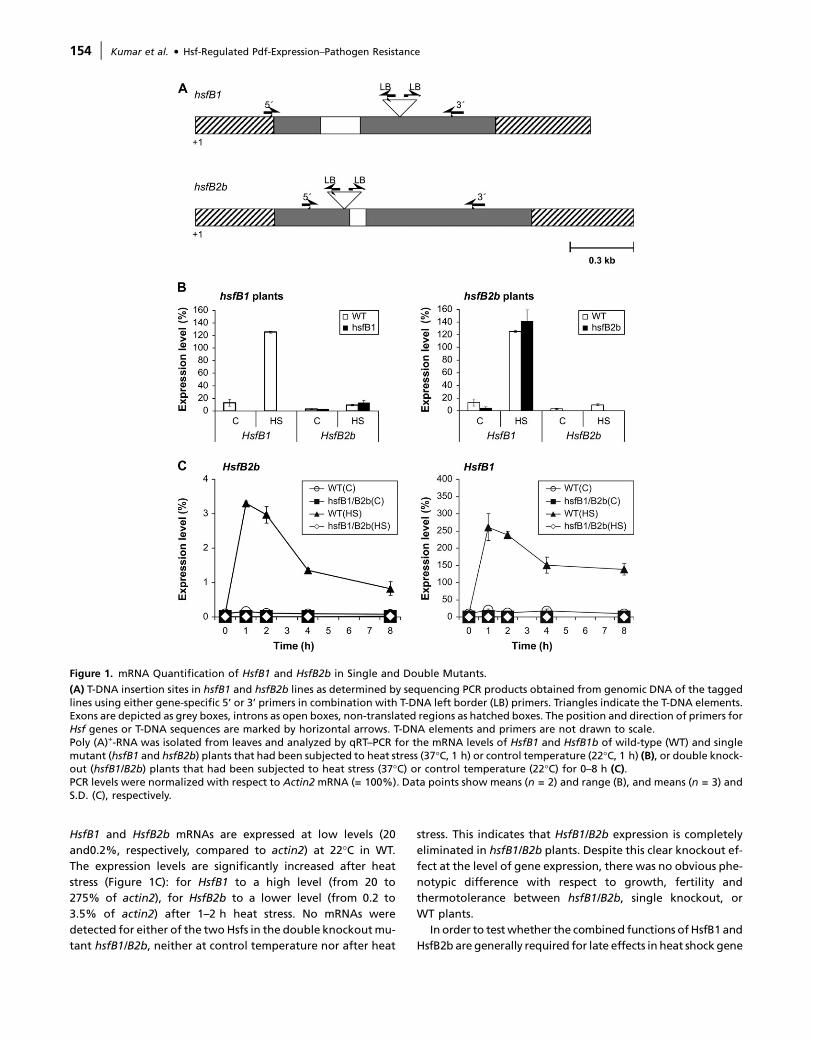
HsfB1 and HsfB2b mRNAs are expressed at low levels (20
and0.2%, respectively, compared to actin2) at 22�C in WT.
The expression levels are significantly increased after heat
stress (Figure 1C): for HsfB1 to a high level (from 20 to
275% of actin2), for HsfB2b to a lower level (from 0.2 to
3.5% of actin2) after 1–2 h heat stress. No mRNAs were
detected for either of the two Hsfs in the double knockout mu-
tant hsfB1/B2b, neither at control temperature nor after heat
stress. This indicates that HsfB1/B2b expression is completely
eliminated in hsfB1/B2b plants. Despite this clear knockout ef-
fect at the level of gene expression, there was no obvious phe-
notypic difference with respect to growth, fertility and
thermotolerance between hsfB1/B2b, single knockout, or
WT plants.
In order to test whether the combined functions of HsfB1 and
HsfB2b are generally required for late effects in heat shock gene
Figure 1. mRNA Quantification of HsfB1 and HsfB2b in Single and Double Mutants.
(A) T-DNA insertion sites in hsfB1 and hsfB2b lines as determined by sequencing PCR products obtained from genomic DNA of the taggedlines using either gene-specific 5’ or 3’ primers in combination with T-DNA left border (LB) primers. Triangles indicate the T-DNA elements.Exons are depicted as grey boxes, introns as open boxes, non-translated regions as hatched boxes. The position and direction of primers forHsf genes or T-DNA sequences are marked by horizontal arrows. T-DNA elements and primers are not drawn to scale.Poly (A)+-RNA was isolated from leaves and analyzed by qRT–PCR for the mRNA levels of HsfB1 and HsfB1b of wild-type (WT) and singlemutant (hsfB1 and hsfB2b) plants that had been subjected to heat stress (37�C, 1 h) or control temperature (22�C, 1 h) (B), or double knock-out (hsfB1/B2b) plants that had been subjected to heat stress (37�C) or control temperature (22�C) for 0–8 h (C).PCR levels were normalized with respect to Actin2 mRNA (= 100%). Data points show means (n = 2) and range (B), and means (n = 3) andS.D. (C), respectively.
154 | Kumar et al. d Hsf-Regulated Pdf-Expression–Pathogen Resistance
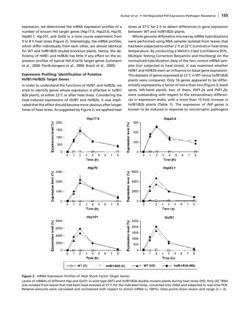
expression, we determined the mRNA expression profiles of a
number of known Hsf target genes (Hsp17.6, Hsp23.6, Hsp70,
Hsp83.1, Hsp101, and GolS) in a time course experiment from
0 to 8 h heat stress (Figure 2). Interestingly, the mRNA profiles,
which differ individually from each other, are almost identical
for WT and hsfB1/B2b double knockout plants. Hence, the de-
ficiency of HsfB1 and HsfB2b has little if any effect on the ex-
pression profiles of typical Hsf-A1a/1b target genes (Lohmann
et al., 2004; Panikulangara et al., 2004; Busch et al., 2005).
Expression Profiling: Identification of Putative
HsfB1/HsfB2b Target Genes
In order to understand the functions of HsfB1 and HsfB2b, we
tried to identify genes whose expression is affected in hsfB1/
B2b plants, at either 22�C or after heat stress. Considering the
heat-induced expressions of HsfB1 and HsfB2b, it was impli-
cated that the effect should become more obvious after longer
times of heat stress. As suggested by Figure 2, we applied heat
stress at 37�C for 2 h to detect differences in gene expression
between WT and hsfB1/B2b plants.
Whole genome Affymetrix microarray mRNA hybridizations
were performed using RNA samples isolated from leaves that
had been subjected to either 2 h at 22�C (control) or heat stress
temperature. By conducting a Welch’s t-test (confidence 95%,
Multiple Testing Correction Benjamini and Hochberg) on the
normalized hybridization data of the two control mRNA sam-
ples (not subjected to heat stress), it was examined whether
HsfB1 and HsfB2b exert an influence on basal gene expression.
The datasets of genes expressed at 22�C in WT versus hsfB1/B2b
plants were compared. Only 16 genes appeared to be differ-
entially expressed by a factor of more than two (Figure 3; black
spots, left-hand panel), two of them, Pdf1.2a and Pdf1.2b,
were outstanding with respect to the extraordinary differen-
ces in expression levels, with a more than 15-fold increase in
hsfB1/B2b plants (Table 1). The expression of Pdf genes is
known to be induced in response to necrotrophic pathogens
Figure 2. mRNA Expression Profiles of Heat Shock Factor Target Genes.
Levels of mRNAs of different Hsp and GolS1 in wild-type (WT) and hsfB1/B2b double mutant plants during heat stress (HS). Poly (A)+-RNAwas isolated from leaves that had been heat-stressed at 37�C for the indicated times, converted into cDNA and subjected to real-time PCR.Relative amounts were calculated and normalized with respect to Actin2 mRNA (= 100%). Data points show means and range (n = 3).
Kumar et al. d Hsf-Regulated Pdf-Expression–Pathogen Resistance | 155
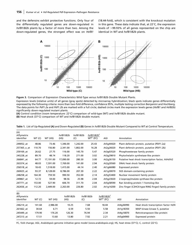
and the defensins exhibit protective functions. Only four of
the differentially regulated genes are down-regulated in
hsfB1/B2b plants by a factor of more than two. Among the
down-regulated genes, the strongest effect was on HsfB1
(18.44-fold), which is consistent with the knockout mutation
in this gene. These data indicate that, at 22�C, the expression
levels of .99.93% of all genes represented on the chip are
identical in WT and hsfB1/B2b plants.
Figure 3. Comparison of Expression Characteristics Wild-Type versus hsfB1/B2b Double Mutant Plants.
Expression levels (relative units) of all genes (gray spots) detected by microarray hybridization; black spots indicate genes differentiallyexpressed by the following criteria: more than two-fold difference, confidence 95%, multiple testing correction Benjamini and Hochberg.The data points for Pdf1.2a and Pdf1.2b are marked with a full circle; dashed circles mark the expression levels genes (HsfB1 and HsfB2b)significantly down-regulated mutant plants.(A) Control condition (room temperature: 22�C) comparison of wild-type (WT) and hsfB1/B2b double mutant.(B) Heat shock (37�C) comparison of WT and hsfB1/B2b double mutant.
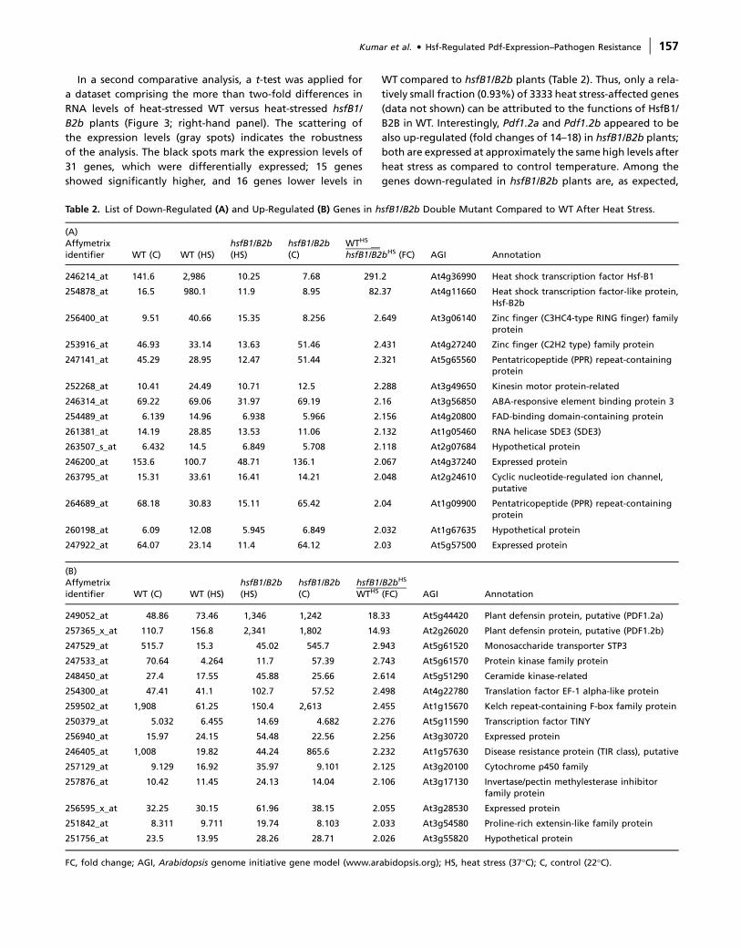
Table 1. List of Up-Regulated (A) and Down-Regulated (B) Genes in hsfB1/B2b Double Mutant Compared to WT at Control Temperature.
(A)Affymetrixidentifier WT (C) WT (HS)
hsfB1/B2b(HS)
hsfB1/B2b(C)
hsfB1/B2bC
WTC (FC) AGI Annotation
249052_at 48.86 73.46 1,346.00 1,242.00 25.43 At5g44420 Plant defensin protein, putative (PDF1.2a)
257365_x_at 110.70 156.80 2,341.00 1,802.00 16.28 At2g26020 Plant defensin protein, putative (PDF1.2b)
259169_at 26.62 27.75 116.00 145.70 5.47 At3g03520 Phosphoesterase family protein
245228_at 89.70 48.74 118.20 271.00 3.02 At3g29810 Phytochelatin synthetase-like protein
266841_at 64.77 17,191.00 17,090.00 280.30 3.00 At2g26150 Putative heat shock transcription factor, AtHsfA2
263374_at 48.03 7,591.00 7,769.00 141.00 2.94 At2g20560 DNAJ heat shock family protein
256518_at 18.43 1,518.00 1,415.00 44.14 2.40 At1g66080 Expressed protein
260025_at 93.37 8,128.00 8,786.00 207.30 2.22 At1g30070 SGS domain-containing protein
248258_at 164.30 759.50 990.50 352.00 2.14 At5g53400 Nuclear movement family protein
249867_at 12.72 59.00 98.56 25.98 2.04 At5g23020 2-isopropylmalate synthase-like protein
247771_at 153.00 527.90 606.70 311.70 2.04 At5g58590 Ran binding protein 1 homolog-like
262656_at 112.20 2,449.00 2,263.00 226.80 2.02 At1g14200 Zinc finger (C3HC4-type RING finger) family protein
(B)Affymetrixidentifier WT (C) WT (HS)
hsfB1/B2b(HS)
hsfB1/B2b(C)
hsfB1/B2bC
WTC (FC) AGI Annotation
246214_at 141.60 2,986.00 10.25 7.68 18.44 At4g36990 Heat shock transcription factor Hsf4
261662_at 30.64 6.71 6.59 5.50 5.58 At1g18350 MAP kinase kinase 5, putative
245449_at 179.90 176.20 126.30 76.94 2.34 At4g16870 Retrotransposon-like protein
247213_at 17.51 13.60 13.80 7.92 2.21 At5g64900 Expressed protein
FC, fold change; AGI, Arabidopsis genome initiative gene model (www.arabidopsis.org); HS, heat stress (37�C); C, control (22�C).
156 | Kumar et al. d Hsf-Regulated Pdf-Expression–Pathogen Resistance
In a second comparative analysis, a t-test was applied for
a dataset comprising the more than two-fold differences in
RNA levels of heat-stressed WT versus heat-stressed hsfB1/
B2b plants (Figure 3; right-hand panel). The scattering of
the expression levels (gray spots) indicates the robustness
of the analysis. The black spots mark the expression levels of
31 genes, which were differentially expressed; 15 genes
showed significantly higher, and 16 genes lower levels in
WT compared to hsfB1/B2b plants (Table 2). Thus, only a rela-
tively small fraction (0.93%) of 3333 heat stress-affected genes
(data not shown) can be attributed to the functions of HsfB1/
B2B in WT. Interestingly, Pdf1.2a and Pdf1.2b appeared to be
also up-regulated (fold changes of 14–18) in hsfB1/B2b plants;
both are expressed at approximately the same high levels after
heat stress as compared to control temperature. Among the
genes down-regulated in hsfB1/B2b plants are, as expected,
Table 2. List of Down-Regulated (A) and Up-Regulated (B) Genes in hsfB1/B2b Double Mutant Compared to WT After Heat Stress.
(A)Affymetrixidentifier WT (C) WT (HS)
hsfB1/B2b(HS)
hsfB1/B2b(C)
WTHS __hsfB1/B2bHS (FC) AGI Annotation
246214_at 141.6 2,986 10.25 7.68 291.2 At4g36990 Heat shock transcription factor Hsf-B1
254878_at 16.5 980.1 11.9 8.95 82.37 At4g11660 Heat shock transcription factor-like protein,Hsf-B2b
256400_at 9.51 40.66 15.35 8.256 2.649 At3g06140 Zinc finger (C3HC4-type RING finger) familyprotein
253916_at 46.93 33.14 13.63 51.46 2.431 At4g27240 Zinc finger (C2H2 type) family protein
247141_at 45.29 28.95 12.47 51.44 2.321 At5g65560 Pentatricopeptide (PPR) repeat-containingprotein
252268_at 10.41 24.49 10.71 12.5 2.288 At3g49650 Kinesin motor protein-related
246314_at 69.22 69.06 31.97 69.19 2.16 At3g56850 ABA-responsive element binding protein 3
254489_at 6.139 14.96 6.938 5.966 2.156 At4g20800 FAD-binding domain-containing protein
261381_at 14.19 28.85 13.53 11.06 2.132 At1g05460 RNA helicase SDE3 (SDE3)
263507_s_at 6.432 14.5 6.849 5.708 2.118 At2g07684 Hypothetical protein
246200_at 153.6 100.7 48.71 136.1 2.067 At4g37240 Expressed protein
263795_at 15.31 33.61 16.41 14.21 2.048 At2g24610 Cyclic nucleotide-regulated ion channel,putative
264689_at 68.18 30.83 15.11 65.42 2.04 At1g09900 Pentatricopeptide (PPR) repeat-containingprotein
260198_at 6.09 12.08 5.945 6.849 2.032 At1g67635 Hypothetical protein
247922_at 64.07 23.14 11.4 64.12 2.03 At5g57500 Expressed protein
(B)Affymetrixidentifier WT (C) WT (HS)
hsfB1/B2b(HS)
hsfB1/B2b(C)
hsfB1/B2bHS
WTHS (FC) AGI Annotation
249052_at 48.86 73.46 1,346 1,242 18.33 At5g44420 Plant defensin protein, putative (PDF1.2a)
257365_x_at 110.7 156.8 2,341 1,802 14.93 At2g26020 Plant defensin protein, putative (PDF1.2b)
247529_at 515.7 15.3 45.02 545.7 2.943 At5g61520 Monosaccharide transporter STP3
247533_at 70.64 4.264 11.7 57.39 2.743 At5g61570 Protein kinase family protein
248450_at 27.4 17.55 45.88 25.66 2.614 At5g51290 Ceramide kinase-related
254300_at 47.41 41.1 102.7 57.52 2.498 At4g22780 Translation factor EF-1 alpha-like protein
259502_at 1,908 61.25 150.4 2,613 2.455 At1g15670 Kelch repeat-containing F-box family protein
250379_at 5.032 6.455 14.69 4.682 2.276 At5g11590 Transcription factor TINY
256940_at 15.97 24.15 54.48 22.56 2.256 At3g30720 Expressed protein
246405_at 1,008 19.82 44.24 865.6 2.232 At1g57630 Disease resistance protein (TIR class), putative
257129_at 9.129 16.92 35.97 9.101 2.125 At3g20100 Cytochrome p450 family
257876_at 10.42 11.45 24.13 14.04 2.106 At3g17130 Invertase/pectin methylesterase inhibitorfamily protein
256595_x_at 32.25 30.15 61.96 38.15 2.055 At3g28530 Expressed protein
251842_at 8.311 9.711 19.74 8.103 2.033 At3g54580 Proline-rich extensin-like family protein
251756_at 23.5 13.95 28.26 28.71 2.026 At3g55820 Hypothetical protein
FC, fold change; AGI, Arabidopsis genome initiative gene model (www.arabidopsis.org); HS, heat stress (37�C); C, control (22�C).
Kumar et al. d Hsf-Regulated Pdf-Expression–Pathogen Resistance | 157

HsfB1 (fold-change 291) and HsfB2b (fold-change 82) mRNAs.
This clearly proves the Hsf-gene knock character of the mutant
line. All other differentially expressed genes, up- or down-
regulated in hsfB1/B2b plants, show only relatively little differ-
ences in expression (fold change 2–3) compared to WT.
In order to test the reliability of the microarray data, we re-
examined the mRNA levels of Pdf1.2 and a number of other
differentially expressed genes using qRT–PCR (Table 3). By
and large, the results of qRT–PCR confirm the gene-chip hy-
bridization data but, as expected, the PCR data of the strongly
heat-induced genes such as AtHsfA2 and AtHsfB1 show
a greater discrepancy in expression levels to the microarray
data. Similar observations have been reported in the analysis
of other hsf mutants (Busch et al., 2005).
Analysis of Pdf1.2 Gene Promoter Region and
Hsf–HSE Binding
Pdf1.2 genes are up-regulated in hsfB1 and hsfB1/B2b plants
and therefore we inspected the promoter region of Pdf1.2a
and Pdf1.2b. Both genes are highly conserved in coding and
in promoter regions. Using the internet tool http://arabidopsis.
med.ohio-state.edu/AtcisDB/, several other putative binding
motifs in the promoter sequence of Pdf1.2a and Pdf1.2b were
identified. Both promoters share a number of common motifs,
such as DPBF1 and -2, RAV1-A, BoxII, GATA box, GCC-box, and
Ibox. The GCC-box mediates jasmonic acid-induced activation of
Pdf1.2 expression in Arabidopsis (Brown et al., 2003).
However, there was no perfect match to the three-boxed HSE
consensus sequences nGAAn/nTTCn/nGAAn, the preferred bind-
ing site for Hsfs. One variant HSE, representing only two boxes
(nGAAn/nTTCn), could be detected in Pdf1.2b at position –629
to–619upstreamofthe transcriptionstart site,while this was ab-
sentinPdf1.2a.However,another imperfectHSE(nGAAn/nATCn)
that represents only one complete box consensus sequence was
present in both promoters, at positions –594/–584 ofPdf1.2aand
at positions –678/–668 in Pdf1.2b. Since both genes and pro-
motersappearhighlyhomologousandco-regulated,weconcen-
trated further investigations only on the Pdf1.2a promoter.
Using EMSA, we were unable to identify any difference in
the formation of binding-specific complexes in the Pdf1.2a
promoter between WTand hsf-mutant plants, neither without
nor after heat stress (not shown). This result suggests that no
Hsf can directly bind to the promoter upstream region.
In a second approach, we assayed for any changes in late Hsf
binding capacity in WTand hsfB1/B2b plants by using synthetic
HSEs, which contain the perfect three-box consensus sequence
nGAAn/nTTCn/nGAAn (Lohmann et al., 2004) as a probe. In
a time course experiment covering up to 4 h heat stress prior
to protein extraction, there was a reproducible difference in
the EMSA profiles between WT and hsfB1/B2b plants (Figure
4). The hsfB1/B2b plants show an earlier onset (60 min), a pro-
longed appearance (240 min), and a stronger intensity of a sig-
nal that represents the major late Hsf–HSE binding complex. In
WT, the same signal appears with a maximum after 2 h heat
stress and is already diminished after 240 min.
Effects of Pathogen Infection on Pdf Expression and
Pathogen Resistance in WT and Hsf-Knockout Plants
There is ample evidence that Pdf1.2a and Pdf1.2b are induced
during pathogen attack or jasmonic acid application
Table 3. Comparison of Gene Expression Levels Determined byGene Chip Hybridization and qRT–PCR.
GeneWT (C) WT (HS) hsfB1/B2b (C) hsfB1/B2b (HS)
RT–PCRa GCb RT–PCRa GCb RT–PCRa GCb RT–PCRa GCb
Pdf1.2a 1.3 0.9 2.3 2.0 39.1 23.7 47.6 37.4
Pdf1.2b 1.4 2.1 2.1 4.2 36.2 34.4 61.8 65.1
AtHsfA2 1.8 1.2 1965.4 459.5 7.6 5.3 1922.9 475.7
DnaJ 1.3 0.9 267.1 202.8 2.9 2.7 293.1 215.8
Atmkk7 1.7 0.6 0.0 0.2 0.0 0.1 0.0 0.2
AtHsfB1 12.9 2.7 238.6 79.6 0.5 0.1 0.4 0.3
AtHsfB2b 0.1 0.3 3.0 26.2 0.0 0.2 0.0 0.3
Stp3 16.2 9.7 0.7 0.4 18.2 10.4 1.2 1.3
F7H2.2 16.9 36.1 0.7 1.6 22.4 49.7 1.1 4.2
a mRNA level determined quantitative PCR. Relative amountswere calculated and normalized with respect to Act2 mRNA(= 100%). Data represents mean 6 range (n = 2).b Expression estimates based on the gene chip (GC) analysis; datarepresent normalized and averaged intensity values of thereplicates relative to the signal of Act2 mRNA hybridization(= 100%).
Figure 4. The Formation of Late Hsf-HSE Binding Complexes inWild-Type and hsfB1/hsfB2b Plants.
Leaves ofArabidopsiswild-type (WT) (Col-0) and doubleHsfmutant(hsfB1/hsfB2b) plants were incubated at 37�C for different periodsof time (0, 10, 30, 60, 120, and 240 min). Total protein was extractedfrom each sample and subjected to EMSA using radioactive-labeledsynthetic HSE consensus sequence as a probe. The first lane containsonly the free probe without protein. The major late Hsf–HSE bind-ing complex (Lohmann et al., 2004) is marked by an arrowhead; theunspecific constitutive binding complexes (Lohmann et al., 2004)are marked by asterisks.
158 | Kumar et al. d Hsf-Regulated Pdf-Expression–Pathogen Resistance

(Penninckx et al., 1998). We tested whether Pdf genes are still
responsive to jasmonic acid induction in double mutants
(hsfB1/B2b) and single Hsf-mutant (hsfB1 or hsfB2b) plants.
Leaves of 5-week-old plants were sprayed with 100 lM MeJ
and samples were collected after 6 h treatment for mRNA
analysis. The results show (Figure 5A) that (1) Pdf1.2a mRNA
is induced by MeJ to higher levels in hsf mutant plants, and
(2) the induced levels in the mutants exceed the induced levels
of WT plants.
We examined whether the infection with the necrotrophic
fungus Alternaria brassicicola leads to enhanced induction of
Pdf1.2a expression and an improved pathogen tolerance in
Hsf-knockout plants. Leaves of 6-week-old plants were inocu-
lated with A. brassicicola in a spray assay for the analysis of
Pdf1.2a mRNA levels using qRT–PCR (Figure 5B) and in a spot
assay (two 5-lL drops of 5 3 105 conidial spores suspended in
water per leaf) for the determination of the disease index (Fig-
ure 5C). The results show that Pdf1.2a expression is signifi-
cantly induced at 72 h post inoculation. This timing in
Arabidopsis WT is in good agreement with published data
(Schenk et al., 2003) showing a significant increase in mRNA
levels 72 h after infection. The mRNA levels appeared to be in-
duced to higher levels in the Hsf knockout plants compared to
WT.
The disease resistance of WTand mutant lines was monitored
on days 3, 5, 7, 10, and 12 after infection with A. brassicicola
(Figure 5C). Infected leaves of double knockout plants (hsfB1/
hsfB2b) provided significantly higher levels of tolerance (lower
disease index) against A. brassicicola infection compared to WT
plants. These differences in pathogen resistance can be seen
from the beginning (3 days after infection (DAI)) throughout
the whole experiment (12 DAI). Comparing the disease index
of all four lines on day 10 after infection, there is a significant
difference between hsfB2b and hsfB1/hafB2b plants, which
were more resistant, compared to WTand single knockouthsfB1
plants (Figure 5D).
DISCUSSION
Considering the complexity of the Hsf gene family, the isola-
tion of knockout mutants for individual Hsf genes is crucial for
the determination of their functional role and biological im-
portance in plants. Although single and double mutations
in HsfB1 and HsfB2b lead to a complete loss of mRNA accumu-
lation, there was no detectable effect on the heat stress re-
sponse. This finding contrasts the functions of class A-Hsfs in
197 52
689425
1980
11311130
9
1376
0
1000
2000
3000
4000
5000
0 72 96hours after spray infection
ExP
df1
.2a/
ExA
ctin
(%
)
WThsfB1hsfB2bhsfB1/B2b
2 10 8 6
344
1596
579
965
0
500
1000
1500
2000
2500
3000
WT hsfB1 hsfB2b hsfB1/B2b
ExP
df1
.2a/
ExA
ctin
(%
)ControlMeJA
B
A
0
100
200
300
400
0 3 5 7 10 12DAI
dis
ease
ind
ex
WThsfB1/B2b
0
50
100
150
200
250
300
350
WT hsfB1 hsfB2b hsfB1/B2b
dis
ease
ind
ex
C
D
Figure 5. Effects MeJ and A. brassicicola Infection on Pdf1.2a Ex-pression and Pathogen Resistance in Arabidopsis Wild-Type, Single,and Double Knockout Hsf Mutants.
Expression of Pdf1.2a was quantified after 6 h treatment with MeJ(A) or after 72 and 96 h post infection with A. brassicicola (B). Datawere normalized with respect to Actin 2 expression (= 100%),points show means and S.D. (n = 3).
(C) Leaves of 6-week-old Arabidopsis wild-type (WT) (Col-0), andhsfB1/B2b plants were drop-inoculated with spores of A. brassici-cola. The disease index was calculated 3, 5, 7, 10, and 12 d after in-oculation (DAI). Data points show means and S.D. (n = 3–5).(D) The disease index calculated on day 10 post inoculation of WT(Col-0), double knockout hsfB1/B2b, and single knockout mutantshsfB1, hsfB2b plants. Columns represent means and S.D. (n = 3–5).Significant differences are indicated by asterisks (a = 5).
Kumar et al. d Hsf-Regulated Pdf-Expression–Pathogen Resistance | 159
which double knockout mutations in hsfA1a/hsfA1b caused
a strong attenuation of the induction of a number of target
genes includingHsp17.6,Hsp70,Hsp83.1, andHsp101 (Lohmann
et al., 2004). In our present study, the class B-Hsf mutations
hsfB1/B2B had no effect on the expression of these HsfA1a/
HsfA1b target genes or any other known heat shock genes in
Arabidopsis. Hence, HsfB1 and HsfB2b are not directly involved
in the regulation of the onset of the heat shock response. This is
reminiscent to tomato, in which, when the expression of
LbHsfB1 was compromised in a transgenic approach, plants
showed no obvious effect on the heat shock response (Mishra
et al., 2002).
Plant Defensin Genes Are De-Repressed in the hsfB1/B2b
Mutant Plants
In order to understand the biological roles of HsfB1/B2b, we
performed microarray expression analysis to identify possible
target genes and pathways affected in hsfB1/B2b mutant
plants. The total numbers of differentially expressed genes
(control versus heat stress) in WT (3333 = 15%) and hsfB1/
B2b plants (2875 = 13%) are relatively high but only 44 genes
were identified as targets of the HsfB1/B2b-dependent tran-
scriptional regulation. Comparable expression profiling results
have been previously obtained by Busch et al. (2005) in a dif-
ferent genetic background of Arabidopsis (ecotype Wassilew-
skija), where a total of 112 class A-Hsf target genes has been
identified.
Out of the 44 putative HsfB1/B2B-dependent target genes, 15
were differentially regulated (12 up and three down-regulated)
at control temperature, 29 (15 up- and 14 down-regulated)
upon heat stress in mutant plants compared to WT. The major
effect was the enhanced expression (16 to 25-fold) of Pdf1.2a
and Pdf1.2b, both at heat stress and control temperature con-
ditions. Pdf1.2a and Pdf1.2b encode small polypeptides that
protect plants against pathogen attack (Penninckx et al.,
1996, 1998; Broekaert et al., 1995; Thomma et al., 2002).
The almost selective up-regulation of Pdf1.2a and Pdf1.2b
does not exclude the importance of other differentially
expressed genes with a fold change of two to three. Besides
Pdf1.2a and Pdf1.2b, three other genes de-repressed in
hsfB1/B2b plants are involved in plant defense: (1) Stp3, a green
leaf-specific, low-affinity monosaccharide transporter (Buttner
et al., 2000), (2) a kelch repeat-containing F-box family protein,
which has been shown to be induced after pathogen attack
(www.genevestigator.ethz.ch/), and (3) a TIR class disease resis-
tance gene, which is highly induced by pathogens (www.gene-
vestigator.ethz.ch/). These genes are down-regulated after
heat stress in WT but heat stress has only little effect on the
repression of these genes in hsfB1/B2b plants. Another gene,
encoding a 2-isopropylmalate synthase-like protein (also
known as methylthioalkymalate synthase-like protein), which
might be involved in resistance to insect herbivores (Kroymann
et al., 2003), shows an enhanced mRNA level (approximately
two-fold) in hsfB1/B2b plants at control temperature.
Interestingly, among the other genes up-regulated in hsfB1/
B2b plants is the class-A transcription factor AtHsfA2 (approx-
imately three-fold up-regulated basal level), which is the stron-
gest heat-inducible class A-Hsf of Arabidopsis (Busch et al.,
2005). It is proposed that AtHsfA2 forms a heteromeric com-
plex with AtHsfA1a and AtHsfA1b to regulate the expression
of target genes during early and late stages of the heat shock
response (Schramm et al., 2006). AtHsfA2 appears to act dom-
inantly as a late, and AtHsfA1a and AtHsfA1b as early Hsfs that
share a number of common target genes including Apx2,
Hsp26.5, Hsp25.3, Hsp22.0, Hsp18.1, Hsp70, Hsp101, and DNAJ
(Wunderlich et al., 2007).
HsfB2b Is a Negative Regulator of Defensin Gene
Expression and Pathogen Resistance
There is ample evidence for the involvement of jasmonic acid
in the regulation of Pdf1.2 expression and the protective role
of defensins in plant pathogen resistance (Brown et al., 2003;
Nandi et al., 2005; Thomma et al., 1998). Jasmonic acid appli-
cation induces the expression of Pdf1.2 genes. The Arabidopsis
triple mutant, fad3-2 fad7-2 fad8, which is unable to accumu-
late jasmonic acid, is susceptible to infection by Pythium spp.
(Vijayan et al., 1998). Moreover, the mutants in jasmonate sig-
naling pathway such as coi1 and jar1 showed enhanced suscep-
tibility to fungal pathogens, such as A. brassicicola, Botrytis
cinerea, etc. (Penninckx et al., 1996; Thomma et al., 1998)
and suppressed Pdf1.2 expression (Penninckx et al., 1998).
MeJ treatment led to further enhanced Pdf1.2 mRNA levels
in hsfB1, hsfB2b, and in double knockout plants by a factor of
approximately 100. There is a clear tendency that the mutant
lines accumulate much higher mRNA levels compared to WT.
This indicates that, in Hsf-mutant plants, the de-repression is
not only restricted to the low basal levels of Pdf1.2a expres-
sion.
The effects of MeJ on Pdf1.2a expression are largely re-
produced in experiments using biotic stress infection with
A. brassicicola for induction. Pdf1.2a and Pdf1.2b are induced
to very high levels when plants are infected withA. brassicicola
compared to other pathogens (genevestigator data).
The results of our patho-assays are in good agreement with
the hypothesis that defensin expression correlates with path-
ogen resistance. The single mutant hsfB2b and the double mu-
tant hsfB1/B2b showed significantly improved resistance levels
(lower disease index) compared to WT and, surprisingly, to
hsfB1 plants. The discrepancy between high levels of Pdf1.2
mRNA and only WT-level of disease resistance suggests that
other molecular processes of the pathogen response are neg-
atively affected by HsfB2b and thus result in improved patho-
gen resistance in plants unable to express HsfB2b. HsfB2b acts
as a negative regulator of Pdf1.2 expression and pathogen re-
sistance. The effect of hsfB2b is reminiscent to another reces-
sive mutation, the overexpressor of cationic peroxidase 3
(Ocp3), which confers a constitutive expression of Pdf1.2 that
results in an enhanced resistance of ocp3 plants to B. cinerea
and P. cucumerina (Coego et al., 2005). The protective role of
160 | Kumar et al. d Hsf-Regulated Pdf-Expression–Pathogen Resistance
defensin gene expression against fungal infections in plants
has been further demonstrated for transgenic rice (Kanzaki
et al., 2002) or potato (Khan et al., 2006) plants overexpressing
the wasabi defensin gene.
HsfB1 and HsfB2b seem to exert similar effects on Pdf ex-
pression, suggesting that they cooperate in repressing the
basal and pathogen-induced levels. The involvement of HsfB1
in Pdf1.2 expression and pathogen response is strongly sup-
ported by expression profiling data (www.genevestigator.
ethz.ch/at/). Many pathogens (P. syringae, P. infestans, B. cin-
erea, A. brassicicola) and a number of signaling mutants like
Cpr5 (Bowling et al., 1997; Clarke et al., 2001), andnahG (Delaney
et al., 1994) affect the expression of both Pdf genes.
Functional Roles of HsfB1 and HsfB2b
The regulation of Pdf1.2 expression through jasmonate signal-
ing seems to require a region located at position –255 to
–277 bp upstream of the Pdf1.2a transcription start site. This re-
gion includes the GCC-box, which is a common motif in the pro-
moters of various genes encoding defense-related proteins, and
has been identified as the specific binding site for members of
the ERF subfamily of AP2/ERF transcription factors (Buttner and
Singh, 1997; Ohme-Takagi and Shinshi, 1995; Zhou et al., 1997).
It is possible that general transcription factors have general
binding capability to different cis-regulatory elements and
may activate gene expression through interactions with ERFs.
Co-regulation of gene expression by the GCC-box and other
promoter elements has been observed. For example, the
GCC-box is required but not sufficient for high-level induction
of the tobacco osmotin gene by ethylene (Raghothama et al.,
1997). A G-box is also linked to jasmonate responsiveness in de-
fense-associated genes in various plant species (Kim et al., 1992;
Mason et al., 1993).
Perfect HSEs (three-box sequences), the regulatory elements
for Hsf-dependent gene expression, are present neither in this
region of the Pdf1.2a promoter nor in the introns or the other
non-translated regions of the gene. An imperfect two-box var-
iant HSE is unable to bind Hsf, as indicated by our negative
results in detecting any changes in protein binding to pro-
moter segments (not shown). This suggests that the negative
effect of HsfB1/B2b on Pdf expression in WT must be indirect,
possibly through interaction with other proteins in the chro-
matin or in the cell. According to the Arabidopsis Small RNA
Project Database (http://asrp.cgrb.oregonstate.edu/; Gustafson
et al., 2005), there are no microRNAs in Arabidopsis thaliana
that target the transcripts of the Pdf1.2.
Using perfect synthetic HSE sequences as a probe, we were
able to detect a positive effect on the formation of the late
Hsf–HSE complex, which appears earlier, persists longer, and
is more intense in hsfB1/B2b plants compared to WT. The for-
mation of this complex requires HsfA2, the major heat stress-
and high light-induced class A-Hsf in Arabidopsis (Wunderlich
et al., 2007). A higher binding capacity of the HsfA2-dependent
late complex in hsfB1/B2b plants suggests that HsfB1/B2b may
interact with class A-Hsf in regulating the shut-off of the heat
shock response. The absence of other HsfB1/B2b in knockout
plants was not associated with the loss of HSE-binding capac-
ity. This further supports the hypothesis that these Hsfs are not
directly involved in binding to the HSE in vivo. It is conceivable
that these class B-Hsf form heteromeric complexes with class
A-Hsf with an increasing probability during the heat shock re-
sponse. There is evidence that the tomato LpHsfB1 (ortholog
of AtHsfB1) can interact with the class A-Hsf LpHsfA1a but also
with other general factors like HAC1/CBP and thus specifying
a co-activator function (Bharti et al., 2004). In a similar way, the
Arabidopsis HsfB1 may interact with other Hsfs (possibly with
HsfA2) and/or other general transcription factors or chromatin
components for exerting a negative (repressive) role on target
gene expression. Further studies identifying target genes dur-
ing later stages of the heat shock response or during recovery
and interaction partners of class B-Hsf will shed more light on
the functions and molecular mechanism of Hsf-dependent re-
pression of genes.
Biological Role of Limiting Pdf Expression
The identification of Pdf genes as targets of Hsf-dependent
negative regulation is the first evidence for an interconnection
of Hsf in the regulation of biotic and abiotic responses. The
question arises as to why plant defensin expression is down-
regulated by heat-inducible Hsfs. Regarding plant defense,
a simple explanation may be that the pathogens are not capa-
ble of efficient infection at high temperature and thus plants
do not require the expression of defensins for protection. This
is consistent with our findings that A. brassicicola does not
grow under heat stress temperature conditions, that a heat
stress early after infection causes a delay in the development
of disease symptoms, and that jasmonate induction of Pdf1.2
expression is blocked by heat treatment (not shown). More-
over, higher levels of Pdf1.2 expression may be detrimental
to plant growth and development, which might become effec-
tive only on a long-term evolutionary scale. In eukaryotic cells,
it has been demonstrated that beta-defensins have little effect
on the epithelial cells at any concentration. In contrast, alpha-
defensin promotes proliferation of the epithelial cells at low
concentration but has a cytotoxic effect at high concentration
and may have adverse effects on the host (Nishimura et al.,
2004). In a similar way, Arabidopsis plants carrying the iop1
mutation show much higher induced expression levels of
Pdf1.2, but, during their entire lifespan, the iop1 mutants
stayed significantly smaller (Penninckx et al., 2003).
METHODS
Plant Material, Growth Condition, and Heat Treatment
Arabidopsis thaliana (Columbia-0) was used in experiments.
Col-0 T-DNA insertion lines were obtained from the Signal Salk
institute (http://signal.salk.edu/tdnaprimers.2.html).
Seeds were spread on soil or MS media and kept in the dark
at 4–8�C for 2 d to achieve a uniform germination. Ten DAG
Kumar et al. d Hsf-Regulated Pdf-Expression–Pathogen Resistance | 161
(days after germination), seedlings were singled out in individ-
ual pots and grown in soil with a light/dark cycle of 16/8 h
at 22�C, 60% relative humidity and a light intensity of 5000
lumen m�2. All experiments were carried out with 5-week-
old plants unless specified.
For heat treatment (HS), fully expanded leaves were col-
lected and pooled, incubated at 37�C (for 1, 2, 4, or 8 h,
depending on the experiment performed) in pre-warmed
SIB (1 mM potassium phosphate, pH 6, 1% (W/V) sucrose),
then placed in a shaking water bath with 40 oscillations per
minute. As a control (C), leaf pools were incubated under
the same experimental conditions but at room temperature
(22�C). After heat treatment, excess SIB was rinsed off with
sterile water; leaf tissue was blotted dry with filter paper
and immediately frozen in liquid nitrogen. Samples were
stored at –70�C for future use.
RNA Isolation, Labeling, and Microarray Hybridization
Frozen leaf tissue (–80�), which had been collected from 45 in-
dividual plants per biological replicate, was crushed by using
mortar and pistil and RNA was extracted with the Plant RNeasy
Mini Kit (Qiagen, Hilden, Germany). A total of 5 lg RNA was
used as starting material for double-stranded cDNA synthesis
using the Superscript Choice System (Invitrogen, Karlsruhe,
Germany) and an oligo dT-T7 primer (Genset, Paris, France).
Biotinylated cRNA was synthesized from cDNA template using
BioArray High Yield Transcript Labeling Kit (Enzo, Farming-
dale, NY, USA). RNeasy columns (Qiagen) were employed to
clean the biotinylated cRNA. The fragmentation of cRNA
was performed according to GeneChip protocol (Affymetrix,
Santa Clara, CA, USA).
The fragmented cRNAs were hybridized to Arabidopsis ATH
1-12151 (Affymetrix Gene-Chips), which represents 22 810
probe sets. GeneChip arrays were hybridized according to
the manufacturer’s protocol (Affymetrix). Hybridized Gene-
Chips were scanned with the GeneChip Scanner 3000 (Affyme-
trix). Samples were quality-controlled by examining of 3’ to 5’
intensity ratios of control genes.
Analysis of Expression Data
Data were imported in Genespring 7.2 (Silicon Genetics, Red-
wood City, CA, USA) and retransformed in linear values. Data
for every condition were available in two replicates of each
experiment. Raw data were quantile-normalized and expres-
sion estimates were calculated by gcRMA (Wu et al., 2004)
implemented in R. Statistical analysis of data was carried
out on gene sets after eliminating all genes that showed less
then two-fold change between conditions, by Welch’s t-test to
compare the different conditions and to find differentially
expressed genes (selected P-value 0.05); the Benjamini and
Hochberg False Discovery Rate Multiple Testing Correction
was used to reduce the detection of false positives. The data
are deposited in a MIAME compliant fashion in the ArrayEx-
press database (www.ebi.ac.uk/microarray/) under accession
E-MEXP-1725.
Isolation of mRNA and Preparation of cDNA for
Quantitative Real-Time PCR
This method has been used for the verification of knockout
mutants and the expression profiles of individual Hsf target
genes. Poly (A)+-RNA was isolated from 60–80 mg frozen leaf
material using the chemagic mRNA Direct kit (Chemagen) and
the amounts were quantified using the dye RiboGreen. cDNA
was synthesized from 100 ng of mRNA using the iScript� cDNA
synthesis kit (Biorad). The amount of cDNA was quantified us-
ing PicoGreen. Quantitative RT–PCR was performed in tripli-
cates using undiluted (1 ng) and 1/8 and 1/64 diluted cDNA
as template. Real-time PCR was performed in a 50-ll reaction
volume. The primers shown in Table 4 were used.
Isolation of mRNA and Preparation of cDNA for
Quantitative Real-Time PCR
This method was used for determining the Pdf1.2-mRNA ex-
pression after MeJ treatment or Alternaria spray assay. Poly
(A)+-RNA was isolated from 50–70 mg frozen leaf material us-
ing the chemagic mRNA Direct kit (Chemagen). A total volume
of 15 ll was used for cDNA synthesis using the iScript�cDNA
kit (Biorad). Quantitative RT–PCR was performed with three
dilutions (1, 1/8, and 1/64) of cDNA and in duplicate samples.
As a control, undiluted mRNA was used. The reaction volume
Table 4. Primers Used for Real-Time PCR.
Target gene Primer sequence
Actin2 5#-AAGCTGGGGTTTTATGAATGG-3’
(At3g18780) 5#-TTGTCACACACAAGTGCATCAT-3’
Hsp17.6 5#-GGTGAGTGGCAAAAGACAGA-3’
(At1g59860) 5#-AAACTTCCCCATCCTCCTCT-3’
Hsp23.6 5#-CGATGAGATTAAGGCGGAGA-3’
(At4g25200) 5#-TCGACGTTTTTAGTTGATCTCG-3’
Hsp70 5#-AGGAGCTCGAGTCTCTTTGC-3’
(At2g32120) 5#-AGGTGTGTCGTCATCCATTC-3’
Hsp83.1 5#-GCTGCTAGGATTCACAGGATG-3’
(At5g52640) 5#-TCCTCCATCTTGCTCTCTTCA-3’
Hsp101 5#-TAACGGGCCAAAGAGAAGTG-3’
(At1g74310) 5#-CACACGTTGGAGGTCAAGACT-3’
AtHsfB1 5#-GGACCGGGATGAAAAGAATTA-3’
(At36990) 5#-CACGCTGGTTTGAACAGTCTT-3’
AtHsfB2b 5#-TGGAGGAGAATAACTCCGGTAA-3’
(At11660) 5#-ATGCAATGGGGATTCAGTAACA-3’
GolS1 5#-AGCTTAGCCACAATATAATCATCG-3’
(At1g56600) 5#-ATCCTCCAAAACCCATAAAAATTA-3’
Pdf1.2a 5#-CCAAGTGGGACATGGTCAG-3’
(At5g44420) 5#-ACTTGTGTGCTGGGAAGACA-3’
Pdf1.2b 5#-GGTACTTGGTCAGGAGTTTGC-3’
(At2g26020) 5#-ACTTGTGAGCTGGGAAGACA-3’
162 | Kumar et al. d Hsf-Regulated Pdf-Expression–Pathogen Resistance
was 30 ll. mRNA expression level of Pdf1.2a was analyzed with
respect to Actin2. The following primers were used:
Actin2-F3 (5#-AAGCTGGGGTTTTATGAATGG-3’),
Actin2-R3 (5#-TTGTCACACACAAGTGCATCAT-3’),
Pdf1.2a_for (5#-TGCTTTCGACGCACCGGC-3’),
Pdf1.2a_rev (5#-TGTAAAATACACACGATTTAGCACC-3’)
Pathogen Treatment: Spot Assay
Plants were grown in soil with a light/dark cycle of 8/16 h at
22�C, 100% relative humidity and a light intensity of 5000 lu-
men m�2. Pathogen (Alternaria brassicicola) treatment was car-
ried out by apoplectic puncturing 6-week-old plants. Care was
taken to choose leaves with approximately equal size and hav-
ing the same developmental stage in WT as well as in mutants.
Two leaves per plant were chosen for fungal inoculation.
Plants were inoculated at a concentration of 5 3 105 conidial
spores per milliliter. Each leaf was inoculated with two 5-lL
drops of a suspension in water. Inoculated plants were incu-
bated at 100% RH and 22�C for control conditions and 35�Cfor heat stress treatment, respectively. The spread of fungal
colonization on each leaf was analyzed 3, 5, 7, 10, and
12 DAI. As a control, one plant per line was treated with water.
Detection of Pathogen Colonization on Inoculated Plants
and Disease Index
Pathogen-inoculated leaves were bonitated according to the
disease symptoms shown in Table 5.
The disease index (DI) for each DAI was calculated from the
results of bonitation by the following formula:
DI =�X
Bonitation scores=Number of inoculated leaves��100:
Methyl Jasmonate Treatment
Five-week-old plants were sprayed with methyl jasmonate
(MeJ) solution (100 lM). Mock inoculations were only sprayed
with water.
Combined heat stress and MeJ treatment was carried out as
follows: plants were first sprayed with MeJ and then kept in
a growth chamber (set at 37�C, light/dark cycle of 16/8 h, rel-
ative humidity 60%, light intensity 5000 lumen m�2). As a con-
trol, water-sprayed plants were kept in a growth chamber at
22�C (light/dark cycle of 16/8 h, relative humidity 60%, light
intensity 5000 lumen m�2).
Pathogen Treatment: Spray Assay
Plants were grown in soil with a light/dark cycle of 8/16 h at
22�C, 100% relative humidity and light intensity 5000 lumen
m�2. For pathogen treatment (Alternaria brassicicola), 6-
week-old plants were sprayed with a concentration of
5 3 105 conidial spores per milliliter. Leaf material was col-
lected immediately after spray infection and after 72 and
96 h post inoculation, respectively.
Isolation of Single and Double Hsf Mutants
Individual T-DNA insertion lines for AtHsfB1 and AtHsfB2b
were obtained from the Salk Institute. Suitable primers for
testing the T-DNA insertion (as described by the Salk website:
http://signal.salk.edu/tdnaprimers.2.html) were generated
and used in PCR reactions with genomic DNA as the template.
Using 5’ and 3’ gene-specific primers, WT plants were identi-
fied and eliminated from segregating population. Mutants
were identified by specific PCR fragments obtained using
a T-DNA and a gene-specific primer.
To generate a doubleHsfmutant, 8–9-week-old single homo-
zygous mutant plants were selected for pollination. For $
(hsfB2b mutant plants), closed flower buds were selected. Flow-
ers were opened with a pin set and anthers were removed.
Flowers were then left opened for 2 d before pollination with
pollen from hsfB1. After 2 d, healthy and non-pollinated stig-
mas were chosen for fertilization. For # (hsfB1 mutant plants),
fully mature anthers with ripened pollens were chosen.
The resulting F1 heterozygous population was selfed to get
homozygous hsfB1/B2b double mutant plants. WT plants were
identified using gene-specific primers for hsfB1 and hsfB2b.
Accordingly, the double mutation was identified with T-
DNA left border primer and the gene-specific primer (Table 6).
Electrophoretic Mobility Shift Assay (EMSA)
The Hsf binding to DNA has been tested essentially as de-
scribed by Lohmann et al. (2004), and the same synthetic
HSE sequences (5#-CCAGAAGCTTCCAGAAGCC) were used as
a probe. Unspecific binding complexes are discriminated
Table 5. Pathogen Inoculated Leaves Bonitated According toDisease Symptoms.
Score Symptoms
1 No symptom
2 Little brownish at point of inoculation
3 Dark-brown spot at point of inoculation
4 Necrotic spots spreading aroundthe point of inoculation
5 Maceration
6 Sporulation
7 Leaf detachment
Table 6. Double Mutations Identified with T-DNA Left BorderPrimer and the Gene-Specific Primers.
AtHsfB1 (At36990) 5#-AAAAGTTCGCCGGAGATGACG-3’5#-GTCGCAACCTTCGCACTCACT-3’
AtHsfB2b (At11660) 5#-CACAGAGGTCAATTCCGACGC-3’5#-CTTCTTCCTCTGCAGCACCCA-3’
T-DNA (Lba1) 5#-ATGGTTCACGTAGTGGGCCATC-3’
Kumar et al. d Hsf-Regulated Pdf-Expression–Pathogen Resistance | 163
according to Lohmann et al. (2004) by the ability to be also
formed with mutated HSE, while specific binding complexes
are formed only with perfect consensus HSE sequences but
not with mutated HSE.
FUNDING
The research was funded by grants of the Deutsche Forschungsge-
meinschaft (SFB446, project A2).
ACKNOWLEDGMENTS
We thank Dr Jasmin Doll and Markus Wunderlich (ZMBP-Allgemeine
Genetik, Universitat Tubingen) for advice and fruitful discussions. No
conflict of interest declared.
REFERENCES
Bharti, K., Koskull-Doring, von P., Bharti, S., Kumar, P., Tintschl-
Korbitzer, A., Treuter, E., and Nover, L. (2004). Tomato heat stress
transcription factor HsfB1 represents a novel type of general
transcription coactivator with a histone-like motif interacting
with the plant CREB binding protein ortholog HAC1. Plant Cell.
16, 1521–1535.
Bowling, S.A., Clarke, J.D., Liu, Y., Klessig, D.F., and Dong, X. (1997).
The cpr5 mutant of Arabidopsis expresses both NPR1-dependent
and NPR1-independent resistance. Plant Cell. 9, 1573–1584.
Broekaert, W.F., Terras, F.R.G., Cammue, B.P.A., and Osborn, R.W.
(1995). Plant defensins: novel antimicrobial peptides as compo-
nents of the host defense system. Plant Physiol. 108, 1353–1358.
Brown, R.L., Kazan, K., McGrath, K.C., Maclean, D.J., and
Manners, J.M. (2003). A role for the GCC-box in jasmonate-
mediated activation of the PDF1.2 gene of Arabidopsis. Plant
Physiol. 132, 1020–1032.
Busch, W., Wunderlich, M., and Schoffl, F. (2005). Identification of
novel heat shock factor dependent genes and biochemical path-
ways in Arabidopsis thaliana. Plant J. 41, 1–14.
Buttner, M., and Singh, K.B. (1997). Arabidopsis thaliana ethylene-
responsive element binding protein (AtEBP), an ethylene-inducible,
GCC box DNA-binding protein interacts with an ocs element bind-
ing protein. Proc. Natl Acad. Sci. U S A. 94, 5961–5966.
Buttner, M., Truernit, E., Baier, K., Scholz-Starke, J., Sontheim, M.,
Lauterbach, C., Huss, V.A.R., and Sauer, N. (2000). AtSTP3, a green
leaf-specific, low affinity monosaccharide H +-symporter of Ara-
bidopsis thaliana. Plant Cell Environ. 23, 175–184.
Clarke, J.D., Aarts, N., Feys, B.J., Dong, X., and Parker, J.E. (2001).
Constitutive disease resistance requires EDS1 in the Arabidopsis
mutants cpr1 and cpr6 and is partially EDS1-dependent in cpr5.
Plant J. 26, 409–420.
Coego, A., Ramirez, V., Gil, M.J., Flors, V., Mauch-Mani, B., and
Vera, P. (2005). An Arabidopsis homeodmain transcription factor,
overexpressor of cationic peroxidase 3, mediates resistance to in-
fection by necrotrophic pathogens. Plant Cell. 17, 2123–2137.
Czarnecka-Verner, E., Pan, S., Salem, T., and Gurley, W.B. (2004).
Plant class B Hsfs inhibit transcription and exhibit affinity for
TFIIB and TBP. Plant Mol. Biol. 56, 57–75.
Czarnecka-Verner, E., Yuan, C.-X., Scharf, K.-D., English, G., and
Gurley, W.B. (2000). Plants contain a novel multi-member class
of heat shock factors without transcriptional activator potential.
Plant Mol. Biol. 43, 459–471.
Dat, J.F., Foyer, C.H., and Scott, I.M. (1998). Changes in salicylic acid
and antioxidants during induction of thermotolerance in mus-
tard seedlings. Plant Physiol. 118, 1455–1461.
Delaney, T.P., et al. (1994). A central role of salicylic acid in plant
disease resistance. Science. 266, 1247–1250.
Doring,P.,Treuter,E.,Kistner,C.,Lyck,R.,Chen,A.,andNover,L.(2000).
The role of AHA motifs in the activator function of tomato heat
stress transcriptionfactorsHsfA1andHsfA2.PlantCell.12,265–278.
Gustafson, A.M., Allen, E., Givan, S., Smith, D., Carrington, J.C., and
Kasschau, K.D. (2005). ASRP: the Arabidopsis small RNA project
database. Nucleic Acids Res. 33, D637–D640.
Kanzaki, H., Nirasawa, S., Saitoh, H., Ito, M., Nishihara, M.,
Terauchi, R., and Nakamura, I. (2002). Overexpression of the wa-
sabi defensin gene confers enhanced resistance to blast fungus
(Magnaporthe grisea) in transgenic rice. Theor. Appl. Genet. 105,
809–814.
Khan, S.R., Nishihara, M., Yamamura, S., Nakamura, I., and Mii, M.
(2006). Transgenic potatoes expressing wasabi defensin peptide
confer partial resistance to gray mold (Botrytis cinerea). Plant
Biotech. 23, 179–183.
Kim, S.R., Choi, J.L., Costa, M.A., and An, G. (1992). Identification of
G-box sequence as an essential element for methyl jasmonate
response of potato proteinase inhibitor II promoter. Plant Phys-
iol. 99, 627–631.
Koskull-Doring, P., Scharf, k-D., and Nover, L. (2007). The diversity of
plant heat stress transcription factors. Trends Plant Sci. 12,
452–457.
Kotak, S., Port, M., Ganguli, A., Bicker, F., and Koskull-Doring, P.
(2004). Characterization of C-terminal domains of Arabidopsis
heat stress transcription factors (Hsfs) and identification of
a new signature combination of plant class A Hsfs with AHA
and NES motifs essential for activator function and intracellular
localization. Plant J. 39, 98–112.
Kroymann, J., Donnerhacke, S., Schnabelrauch, D., and Mitchell-
Olds, T. (2003). Evolutionary dynamics of an Arabidopsis insect
resistance quantitative trait locus. Proc. Natl Acad. Sci. U S A.
100, 14587–14592.
Lohmann, C., Eggers-Schumacher, G., Wunderlich, M., and
Schoffl, F. (2004). Two different heat shock transcription factors
regulate immediate early expression of stress genes in Arabidop-
sis. Mol. Gen. Genom. 271, 11–21.
Mason, H.S., DeWald, D.B., and Mullet, J.E. (1993). Identification of
a methyl jasmonate-responsive domain in the soybean vspB pro-
moter. Plant Cell. 5, 241–251.
Mishra,S.K.,Tripp,J.,Winkelhaus,S.,Tschiersch,B.,Theres,K.,Nover,L.,
andScharf,K.D. (2002). Inthecomplexfamilyofheatstress transcrip-
tion factors, HsfA1 has a unique role as master regulator of thermo-
tolerance in tomato. Genes Develop. 16, 1555–1567.
Nandi, A., Moeder, W., Kachroo, P., Klessig, D.F., and Shah, J. (2005).
Arabidopsis ssi2-conferred susceptibility to Botrytis cinerea is
dependent on EDS5 and PAD4. Molec. Plant Microbe Interact.
18, 363–370.
Nishimura, M., Abiko, Y., Kurashige, Y., Takeshima, M.,
Yamazaki, M., Kusano, K., Saitoh, M., Nakashima, K., Inoue, T.,
and Kaku, T. (2004). Effect of defensin peptides on eukaryotic
164 | Kumar et al. d Hsf-Regulated Pdf-Expression–Pathogen Resistance
cells: primary epithelial cells, fibroblasts and squamous cell carci-
noma cell lines. J. Dermatol. Sci. 36, 87–95.
Nover, L., Scharf, K.D., Gagliardi, D., Vergne, P., Czarnecka-
Verner, E., and Gurley, W.B. (1996). The Hsf world: classification
and properties of plant heat stress transcription factors. Cell
Stress Chaperones. 1, 215–223.
Ohme-Takagi, M., and Shinshi, H. (1995). Ethylene-inducible DNA
binding proteins that interact with an ethylene-responsive ele-
ment. Plant Cell. 7, 173–182.
Panikulangara, T.J., Eggers-Schumacher, G., Wunderlich, M.,
Stransky, H., and Schoffl, F. (2004). Galactinol synthase 1, a novel
heat-inducible and Hsf-target gene responsible for heat-induced
synthesis of Raffinose Family Oligosaccharides in Arabidopsis.
Plant Physiol. 136, 3148–3158.
Penninckx, I.A., Eggermont, K., Schenk, P.M., van den
Ackerveken, G., Cammun, P.B., and Thomma, B.P. (2003). TheAra-
bidopsis mutant iop1 exhibits induced over-expression of the
plant defensin gene PDF1.2 and enhanced pathogen resistance.
Mol. Plant Pathol. 4, 479–486.
Penninckx, I.A., Eggermont, K., Terras, F.R., Thomma, B.P., De
Samblanx, G.W., Buchala, A., Metraux, J.P., Manners, J.M., and
Broekaert, W.F. (1996). Pathogeninduced systemic activation of
a plant defensin gene in Arabidopsis follows a salicylic acid in-
dependent pathway. Plant Cell. 8, 2309–2323.
Penninckx, I.A., Thomma, B.P., Buchala, A., Metraux, J.P., and
Broekaert, W.F. (1998). Concomitant activation of jasmonate
and ethylene response pathways is required for induction of
a plant defensin gene in Arabidopsis. Plant Cell. 10, 2103–2113.
Prandl, R., Hinderhofer, K., Eggers-Schumacher, G., and Schoffl, F.
(1998). Hsf3, a new heat shock factor from Arabidopsis thaliana,
derepresses the heat shock response and confers thermotoler-
ance when overexpressed in transgenic plants. Mol. Gen. Genet.
258, 269–278.
Raghothama, K.G., Maggio, A., Narasimhan, M.L.,
Kononowicz, A.K., Wang, G., D’Urzo, M.P., Hasegawa, P.M.,
and Bressan, R.A. (1997). Tissue-specific activation of the osmo-
tin gene by ABA, C2H4 and NaCl involves the same promoter re-
gion. Plant Mol. Biol. 34, 393–402.
Schenk, P.M., Kazan, K., Manners, J.M., Anderson, J.P.,
Simpson, R.S., Wilson, I.W., Somerville, S.C., and
Maclean, D.C. (2003). Systemic gene expression in Arabidopsis
during an incompatible interaction with Alternaria brassicicola.
Plant Physiol. 132, 999–1010.
Schramm, F., Ganguli, A., Kiehlmann, E., Englich, G., Walch, D., and
Koskull-Doring, P. (2006). The heat stress transcription factor
HsfA2 serves as a regulatory amplifier of a subset of genes in
the heat stress response in Arabidopsis. Plant Mol. Biol. 60,
759–772.
Thomma, B.P.H.J., Camme, B.P.A., and Thevissen, K. (2002). Plant
defensins. Planta. 216, 193–202.
Thomma, B.P.H.J., Eggermont, K., Penninckx, I.A.M.A., Mauch-
Mani, B., Vogelsang, R., Cammune, B.P.A., and Broekaert, W.
(1998). Separate jasmonate-dependent and salicylate-
dependent defense-response pathways in Arabidopsis are
essential for resistance to distinct microbial pathogens. Proc. Natl
Acad. Sci. U S A. 95, 15107–15111.
Vijayan, P., Shockey, J., Levesque, C.A., Cook, R.J., and Browse, J.
(1998). A role for jasmonate in pathogen defence of Arabidopsis.
Proc. Natl Acad. Sci. U S A. 95, 7209–7214.
Wu, C. (1995). Heat shock transcription factors: structure and reg-
ulation. Ann. Review Cell Dev. Biol. 11, 441–469.
Wu, Z., Irizarry, R.A., Gentleman, R., Murillo, F.M., and Spencer, F.A.
(2004). A model based background adjustment for oligonucleo-
tide expression arrays (Working Paper 1, Dept. of Biostatistics
Working Papers: Johns Hopkins University).
Wunderlich, M., Doll, J., Busch, W., Kleindt, C.K., Lohmann, C., and
Schoffl, F. (2007). Heat shock factors: regulators of early and late
functions in plant stress response. Plant Stress. 1, 16–22.
Zhou, J.M., Tang, X.Y., and Martin, G.B. (1997). The Pto kinase con-
ferring resistance to tomato bacterial speck disease interacts
with proteins that bind a cis-element of pathogenesis-related
genes. EMBO J. 16, 3207–3218.
Kumar et al. d Hsf-Regulated Pdf-Expression–Pathogen Resistance | 165