Harmony and Discord: Bioarchaeology
286
Transcript of Harmony and Discord: Bioarchaeology


TOTAH
TIME AND THE RIVERS FLOWING:EXCAVATIONS IN THE LA PLATA VALLEY
VOLUME 5
HARMONY AND DISCORD: BIOARCHAEOLOGY
Debra L. MartinNancy J. Akins
Alan H. GoodmanH. Wolcott Toll
Alan C. Swedlund
ARCHAEOLOGY NOTES 242
OFFICE OF ARCHAEOLOGICAL STUDIESSANTA FE 2001 NEW MEXICO


ADMINISTRATIVE SUMMARY
In connection with improvements to NM 170 by the New Mexico Highway and TransportationDepartment (NMSHTD), the La Plata archaeological project stretched over ten years, from 1981 to 1991.This work, conducted by the Research Section of the Museum of New Mexico (MNM), which laterbecame the Office of Archaeological Studies, included survey and testing, resurvey and further testingconducted in 1988 and 1989, and excavations in 1985 and 1988-1991. The NMSHTD has widened andrealigned 14 miles of the La Plata Highway between Farmington and La Plata, northwestern New Mexico.
This report concerns human burials encountered during excavations in 1988-1991 at the followingsites: LA 1897, LA 37591, LA 37592, LA 37593, LA 37594, LA 37595, LA 37598, LA 37599, LA 37600,LA 37601, LA 37603, LA 37605, LA 37606, LA 60751, LA 65029, LA 65030, and LA 65031. The textprovides a detailed description of the methods used for data collection and the analytical techniques forprocessing the information. Skeletal remains from the project fall into two main categories: articulatedburials and disarticulated remains. Sixty-seven individuals were collected and analyzed as intact burials,and at least 68 additional individuals are represented by disarticulated remains. The major objectives ofthe research include the construction of demographic and health profiles for the burial population. Themortuary complex is described and discussed particularly as it relates to delineating the full range of vari-ability in burial and interment practices. Baseline data on age, sex, pathologies, morphological measure-ments, and other kinds of data are presented in a way that will contribute to the growing database on bio-logical remains from the Southwest. Vital to the analysis of the disarticulated remains was determining thenumbers of individuals and taphonomic processes resulting in deposition. Comparisons between the LaPlata Valley population and other contemporaneous groups, particularly groups within the Chaco Canyonand Mesa Verde regions, are made whenever possible.
The analysis and reporting were done as a cooperative effort between contractors Debra L. Martin andAlan Goodman (Hampshire College); Alan Swedlund (University of Massachusetts); and Nancy Akinsand Wolcott Toll, staff members of the Office of Archaeological Studies.
The NMSHTD provided the funding for this project.
NMSHTD Project ST[S]-1331[201], CN 1496MNM Project 41.419 (testing), 41.454 (data recovery)Permits SE-37 (testing) SE-46 (data recovery)
iii


CONTENTS
ADMINISTRATIVE SUMMARY . . . . . . . . . . . . . . . . . . . . . . . . . . . . . . . . . . . . . . . . . . . . . . . . . . . iii
CHAPTER 1. LA PLATA VALLEY HUMAN REMAINS: A BIOARCHAEOLOGICAL STUDY. . . . 1Raison d'Être . . . . . . . . . . . . . . . . . . . . . . . . . . . . . . . . . . . . . . . . . . . . . . . . . . . . . . . . . . . . . . . . . 1Modeling the Effects of Stress and Change Using Skeletal Remains . . . . . . . . . . . . . . . . . . . . . . . 7Populations at Risk in La Plata Valley . . . . . . . . . . . . . . . . . . . . . . . . . . . . . . . . . . . . . . . . . . . . . . 9Integrating Demography and Health with Archaeological Reconstruction in the Southwest . . . . . 10Research Objectives of the La Plata Highway Human Remains Analysis . . . . . . . . . . . . . . . . . . . 11
CHAPTER 2. FIELD AND LABORATORY METHODS . . . . . . . . . . . . . . . . . . . . . . . . . . . . . . . . . 13Excavation and Curation of the La Plata Human Remains . . . . . . . . . . . . . . . . . . . . . . . . . . . . . . 13Studies of Human Remains and Contract Archaeology. . . . . . . . . . . . . . . . . . . . . . . . . . . . . . . . . 14Methods Used in the Analysis of the La Plata Human Remains . . . . . . . . . . . . . . . . . . . . . . . . . . 15Summary . . . . . . . . . . . . . . . . . . . . . . . . . . . . . . . . . . . . . . . . . . . . . . . . . . . . . . . . . . . . . . . . . . . 27
CHAPTER 3. MORTUARY AND DEMOGRAPHIC CONTEXT OF THE LA PLATA VALLEYHUMAN REMAINS . . . . . . . . . . . . . . . . . . . . . . . . . . . . . . . . . . . . . . . . . . . . . . . . . . . . . . . . . . 33Mortuary Patterning of the La Plata Burials . . . . . . . . . . . . . . . . . . . . . . . . . . . . . . . . . . . . . . . . . 33Demographic Features of the La Plata Skeletal Population. . . . . . . . . . . . . . . . . . . . . . . . . . . . . . 37Discussion . . . . . . . . . . . . . . . . . . . . . . . . . . . . . . . . . . . . . . . . . . . . . . . . . . . . . . . . . . . . . . . . . . 41
CHAPTER 4. HEALTH PROFILE OF THE LA PLATA VALLEY COMMUNITIES . . . . . . . . . . . . 65Porotic Hyperostosis . . . . . . . . . . . . . . . . . . . . . . . . . . . . . . . . . . . . . . . . . . . . . . . . . . . . . . . . . . 65Periosteal Reaction and Other Infections . . . . . . . . . . . . . . . . . . . . . . . . . . . . . . . . . . . . . . . . . . . 67Linear Enamel Hypoplasias and Other Developmental Defects . . . . . . . . . . . . . . . . . . . . . . . . . . 71Subadult Growth . . . . . . . . . . . . . . . . . . . . . . . . . . . . . . . . . . . . . . . . . . . . . . . . . . . . . . . . . . . . . 75Anthropometry of Adults . . . . . . . . . . . . . . . . . . . . . . . . . . . . . . . . . . . . . . . . . . . . . . . . . . . . . . . 76Osteoarthritis . . . . . . . . . . . . . . . . . . . . . . . . . . . . . . . . . . . . . . . . . . . . . . . . . . . . . . . . . . . . . . . . 77Metastatic Cancer . . . . . . . . . . . . . . . . . . . . . . . . . . . . . . . . . . . . . . . . . . . . . . . . . . . . . . . . . . . . 78Trauma . . . . . . . . . . . . . . . . . . . . . . . . . . . . . . . . . . . . . . . . . . . . . . . . . . . . . . . . . . . . . . . . . . . . 82Discussion . . . . . . . . . . . . . . . . . . . . . . . . . . . . . . . . . . . . . . . . . . . . . . . . . . . . . . . . . . . . . . . . . . 93
CHAPTER 5. DIET AND RELATED HEALTH ISSUES IN THE LA PLATA VALLEYPOPULATION. . . . . . . . . . . . . . . . . . . . . . . . . . . . . . . . . . . . . . . . . . . . . . . . . . . . . . . . . . . . . . 105Stable Isotopic Analysis . . . . . . . . . . . . . . . . . . . . . . . . . . . . . . . . . . . . . . . . . . . . . . . . . . . . . . . 105Diet and Dental Health. . . . . . . . . . . . . . . . . . . . . . . . . . . . . . . . . . . . . . . . . . . . . . . . . . . . . . . . 106
CHAPTER 6. DISARTICULATED HUMAN REMAINS . . . . . . . . . . . . . . . . . . . . . . . . . . . . . . . . 121Methodological Considerations . . . . . . . . . . . . . . . . . . . . . . . . . . . . . . . . . . . . . . . . . . . . . . . . . 121Damage and Breakage in the Burial Sample. . . . . . . . . . . . . . . . . . . . . . . . . . . . . . . . . . . . . . . . 123Site by Site Information on the Disarticulated Human Remains . . . . . . . . . . . . . . . . . . . . . . . . . 132Assemblage Comparisons . . . . . . . . . . . . . . . . . . . . . . . . . . . . . . . . . . . . . . . . . . . . . . . . . . . . . 165Discussion . . . . . . . . . . . . . . . . . . . . . . . . . . . . . . . . . . . . . . . . . . . . . . . . . . . . . . . . . . . . . . . . . 168
CHAPTER 7. CONCLUSIONS . . . . . . . . . . . . . . . . . . . . . . . . . . . . . . . . . . . . . . . . . . . . . . . . . . . . 191Health and Demography . . . . . . . . . . . . . . . . . . . . . . . . . . . . . . . . . . . . . . . . . . . . . . . . . . . . . . 191Patterns in Trauma . . . . . . . . . . . . . . . . . . . . . . . . . . . . . . . . . . . . . . . . . . . . . . . . . . . . . . . . . . . 192
v

Culturally Modified Disarticulated Assemblage . . . . . . . . . . . . . . . . . . . . . . . . . . . . . . . . . . . . . 193The La Plata Health Profile . . . . . . . . . . . . . . . . . . . . . . . . . . . . . . . . . . . . . . . . . . . . . . . . . . . . 195Comparing Health Profiles of La Plata, Mesa Verde, and Chaco Canyon Populations. . . . . . . . . 196The Ethical Aspects of What We Do . . . . . . . . . . . . . . . . . . . . . . . . . . . . . . . . . . . . . . . . . . . . . 197
NOTES . . . . . . . . . . . . . . . . . . . . . . . . . . . . . . . . . . . . . . . . . . . . . . . . . . . . . . . . . . . . . . . . . . . . . . 201
REFERENCES CITED . . . . . . . . . . . . . . . . . . . . . . . . . . . . . . . . . . . . . . . . . . . . . . . . . . . . . . . . . . 205
Appendix 1. Site Location Information (removed from copies for public distribution) . . . . . . . . . . . 223Appendix 2. Human Skeletal and Dental Remains Data Management. . . . . . . . . . . . . . . . . . . . . . . . 231Appendix 3. Burial Inventory . . . . . . . . . . . . . . . . . . . . . . . . . . . . . . . . . . . . . . . . . . . . . . . . . . . . . . 245Appendix 4. Postcranial Measurements . . . . . . . . . . . . . . . . . . . . . . . . . . . . . . . . . . . . . . . . . . . . . . 277
Figures
1.1. Map of the greater Southwest, showing locations mentioned in this report . . . . . . . . . . . . . . . . . . 21.2. Map of the La Plata Valley, showing greathouses and communities . . . . . . . . . . . . . . . . . . . . . . . . 31.3. Map of the Totah region, showing Totah greathouses. . . . . . . . . . . . . . . . . . . . . . . . . . . . . . . . . . . 41.4. General model for integrating human remains with archaeological context . . . . . . . . . . . . . . . . . . 82.1. Moderate unremodeled porotic hyperostosis in the orbital region of a subadult with a
midpoint age of two years. LA 37592, B1 . . . . . . . . . . . . . . . . . . . . . . . . . . . . . . . . . . . . . . . . . . 182.2. Moderate unremodeled periosteal reaction on the right tibia of a subadult with a midpoint
age of nine years. LA 37601, B8 . . . . . . . . . . . . . . . . . . . . . . . . . . . . . . . . . . . . . . . . . . . . . . . . . 182.3. Threshold model for understanding the formation of LEHs. Defect formation is modeled
as an all-or-none phenomenon. Unknown etiological factors, diet, and morbidity combineto disrupt enamel development to a degree that an LEH may occur . . . . . . . . . . . . . . . . . . . . . . . 21
2.4. Distinctive LEH formation on maxillary anterior permanent teeth. LA 65030, B6 . . . . . . . . . . . . 223.1. Survivorship (lx): La Plata and other southwestern groups. . . . . . . . . . . . . . . . . . . . . . . . . . . . . . 433.2. Survivorship (lx): La Plata and later southwestern groups . . . . . . . . . . . . . . . . . . . . . . . . . . . . . . 433.3. Probability of dying (qx): La Plata and other southwestern groups . . . . . . . . . . . . . . . . . . . . . . . 443.4. Probability of dying (qx): La Plata and later southwestern groups . . . . . . . . . . . . . . . . . . . . . . . . 444.1. Distribution of osteomyelitic lesions. LA 37601, B4 . . . . . . . . . . . . . . . . . . . . . . . . . . . . . . . . . . 694.2. Osteomyelitis of the sternum. LA 37601, B4 . . . . . . . . . . . . . . . . . . . . . . . . . . . . . . . . . . . . . . . . 704.3. Osteomyelitic and normal humerus. LA 37601, B4 . . . . . . . . . . . . . . . . . . . . . . . . . . . . . . . . . . . 704.4. Vertebrae showing tubercular involvement. LA 65030, FS 510 . . . . . . . . . . . . . . . . . . . . . . . . . . 714.5. Prevalence of LEH by tooth: La Plata and other southwestern groups . . . . . . . . . . . . . . . . . . . . . 734.6. LEH per tooth (mean): La Plata and other southwestern groups. . . . . . . . . . . . . . . . . . . . . . . . . . 734.7. LEH by age at formation: La Plata . . . . . . . . . . . . . . . . . . . . . . . . . . . . . . . . . . . . . . . . . . . . . . . 744.8. Femoral distance curves (ages 0-7): La Plata and comparison groups . . . . . . . . . . . . . . . . . . . . . 744.9. Distribution of lesions due to metastatic cancer. LA 37601, B2 . . . . . . . . . . . . . . . . . . . . . . . . . . 794.10. Left femur showing osteolytic lesions of metastatic cancer. LA 37601, B2 . . . . . . . . . . . . . . . . 804.11. Right innominate showing osteolytic lesions of metastatic cancer. LA 37601, B2 . . . . . . . . . . . 804.12. Parietal showing large and small lesions due to metastatic cancer. LA 37601, B2 . . . . . . . . . . . 814.13. Cervical vertebrae demonstrating osteolytic lesions. LA 37601, B2 . . . . . . . . . . . . . . . . . . . . . . 814.14. Lumbar vertebrae demonstrating osteolytic lesions. LA 37601, B2 . . . . . . . . . . . . . . . . . . . . . . 824.15. Depression fracture on the right parietal. LA 37599, B5 . . . . . . . . . . . . . . . . . . . . . . . . . . . . . . 834.16. Multiple traumatic lesions on the frontal bone. LA 37601, B4 . . . . . . . . . . . . . . . . . . . . . . . . . . 834.17. Cranial trauma on parietals and occipital. LA 65030, B9 . . . . . . . . . . . . . . . . . . . . . . . . . . . . . . 83
vi

4.18. Depression fracture of the occipital region. LA 65030, B6. . . . . . . . . . . . . . . . . . . . . . . . . . . . . 844.19. Approximate location and size of injuries on the combined female crania . . . . . . . . . . . . . . . . . 864.20. Female, age 25, no trauma, Pit Structure 1, upper fill. LA 37595, B1. . . . . . . . . . . . . . . . . . . . . 864.21. Female, age 20, cranial and postcranial trauma, Pit Structure 1, lower fill. LA 65030, B8 . . . . . 874.22. Female, age 33, cranial and postcranial trauma, Pit Structure 1, lower fill. LA 65030, B9 . . . . . 874.23. Age 10.5, no trauma, Pit Structure 1, lower fill. LA 65030, B7 . . . . . . . . . . . . . . . . . . . . . . . . . 884.24. Female, age 38, cranial and postcranial trauma, Pit Structure 1, lower fill. LA 65030, B6 . . . . . 884.25. Female, age 28, cranial trauma, Pit Structure 8, lower fill. LA 65030, B16 . . . . . . . . . . . . . . . . 894.26. Female, age 25, cranial and postcranial trauma, Pit Structure 2, middle fill. LA 37601, B4 . . . . 894.27. Male, age 25, cranial trauma, Pit Structure 2, middle fill. LA 37599, B5 . . . . . . . . . . . . . . . . . . 905.1. La Plata dental wear: maxillary and mandibular teeth . . . . . . . . . . . . . . . . . . . . . . . . . . . . . . . . 1075.2. Molar attrition gradients: La Plata versus Black Mesa . . . . . . . . . . . . . . . . . . . . . . . . . . . . . . . . 1075.3. Scatterplot of age and attrition: La Plata and Black Mesa . . . . . . . . . . . . . . . . . . . . . . . . . . . . . 1085.4. Molar attrition gradients: males versus females . . . . . . . . . . . . . . . . . . . . . . . . . . . . . . . . . . . . . 1085.5. Percent abscessing. . . . . . . . . . . . . . . . . . . . . . . . . . . . . . . . . . . . . . . . . . . . . . . . . . . . . . . . . . . 1096.1. Humerus broken by backhoe. Note remaining plasticity and peel on this eroded bone.
LA 37601, B1 . . . . . . . . . . . . . . . . . . . . . . . . . . . . . . . . . . . . . . . . . . . . . . . . . . . . . . . . . . . . . . 1226.2. Erosion of surface of right parietal (cf. White 1992: Figs. 7.7, 7.21). LA 37601, B8 . . . . . . . . . 1256.3. Crisp breaks produced by backhoe. LA 37601, B5. . . . . . . . . . . . . . . . . . . . . . . . . . . . . . . . . . . 1256.4. Recent peel on mandible. LA 37601, B3 . . . . . . . . . . . . . . . . . . . . . . . . . . . . . . . . . . . . . . . . . . 1266.5. Natural deterioration of mandibular margins. LA 37601, B1 . . . . . . . . . . . . . . . . . . . . . . . . . . . 1266.6. Weathered clavicle. LA 37601, B5 . . . . . . . . . . . . . . . . . . . . . . . . . . . . . . . . . . . . . . . . . . . . . . . . . 6.7. Carnivore-damaged ribs. LA 65030, B13. . . . . . . . . . . . . . . . . . . . . . . . . . . . . . . . . . . . . . . . . . 1276.8. Erosion of tibia shaft (cf. White 1992: Fig. 6.29). LA 37601, B1 . . . . . . . . . . . . . . . . . . . . . . . . 1286.9. Old and fresh spiral breaks on the same fibula. LA 37605, FS 201-1 . . . . . . . . . . . . . . . . . . . . . 1286.10. Carnivore punctures on left ilium. LA 65030, B13. . . . . . . . . . . . . . . . . . . . . . . . . . . . . . . . . . 1306.11. Carnivore-damaged ascending ramus. LA 37595, B2 . . . . . . . . . . . . . . . . . . . . . . . . . . . . . . . . 1306.12. Carnivore-damaged long bones (hole in fourth element from top is a sample location)
LA 37595, B2 . . . . . . . . . . . . . . . . . . . . . . . . . . . . . . . . . . . . . . . . . . . . . . . . . . . . . . . . . . . . . . 1316.13. Carnivore damage on remains of a burial . LA 37599, B0.1 . . . . . . . . . . . . . . . . . . . . . . . . . . . 1316.14. Placed long bone and cranial elements in Layer 1 of Pit Structure 1, LA 37592 . . . . . . . . . . . . 1336.15. Cranial elements. LA 37592, FS 229 . . . . . . . . . . . . . . . . . . . . . . . . . . . . . . . . . . . . . . . . . . . . 1346.16. Altered bone: humerus, radius, ulna, and miscellaneous long bone fragments.
LA 37592, FS 315 . . . . . . . . . . . . . . . . . . . . . . . . . . . . . . . . . . . . . . . . . . . . . . . . . . . . . . . . . . . 1346.17. Altered bone: femora, patella, hand, and foot elements. LA 37592, FS 315 . . . . . . . . . . . . . . . 1356.18. Altered bone: femur, rib, ulna, tibia, and long bones. LA 37592, FS 326 . . . . . . . . . . . . . . . . . 1356.19. Occipital with cut marks from a two-year-old child. LA 37592, FS 216-6 . . . . . . . . . . . . . . . . 1376.20. Damage to inferior border of mandible. LA 37592, FS 241-1 . . . . . . . . . . . . . . . . . . . . . . . . . 1376.21. Frontal with abrasions and possible bite marks. LA 37592, FS 327-6. . . . . . . . . . . . . . . . . . . . 1386.22. Mandible with peel. LA 37592, FS 227-4 and FS 327-9. . . . . . . . . . . . . . . . . . . . . . . . . . . . . . 1386.23. Detail of cuts, frontal inferior broken edge. LA 37592, FS 229-15 . . . . . . . . . . . . . . . . . . . . . . 1396.24. Frontal cuts near sagittal suture. LA 37592, FS 229-15 . . . . . . . . . . . . . . . . . . . . . . . . . . . . . . 1396.25. Peel on an immature proximal ulna. LA 37592, FS 326-13 . . . . . . . . . . . . . . . . . . . . . . . . . . . 1406.26. Right orbit with many small linear marks. LA 37592, FS 70-1. . . . . . . . . . . . . . . . . . . . . . . . . 1406.27. Left humerus with cut marks, distal and posterior. LA 37592, FS 327-7. . . . . . . . . . . . . . . . . . 1416.28. Crushed proximal femur. LA 37592, FS 315-2 and 315-3 . . . . . . . . . . . . . . . . . . . . . . . . . . . . 1416.29. Ectocranial release. LA 37592, FS 216 . . . . . . . . . . . . . . . . . . . . . . . . . . . . . . . . . . . . . . . . . . 1426.30. Parietal fragments with external vault release. LA 37592, FS 563-48 and FS 563-49 . . . . . . . . 1426.31. Right parietal and occipital with impact notch. LA 37592, FS 216-14 . . . . . . . . . . . . . . . . . . . 144
vii

6.32. Cut marks on a clavicle. LA 37592, FS 315-1 . . . . . . . . . . . . . . . . . . . . . . . . . . . . . . . . . . . . . 1446.33. Cut marks on femur neck. LA 37592, FS 315-15 . . . . . . . . . . . . . . . . . . . . . . . . . . . . . . . . . . . 1456.34. Tibia with crenelated edge. LA 37592, FS 315-4 . . . . . . . . . . . . . . . . . . . . . . . . . . . . . . . . . . . 1456.35. Unusual breakage/alteration patterns on clavicle, humerus, and tibia. LA 37592, FS 326 . . . . . 1466.36. Child's clavicle with cuts or rodent gnawing. LA 37592, FS 326-45 . . . . . . . . . . . . . . . . . . . . 1466.37. Bite marks in cervical vertebrae. LA 37592, FS 326-13 . . . . . . . . . . . . . . . . . . . . . . . . . . . . . . 1486.38. Burned fragments of bone. LA 37593, FS 551-30 . . . . . . . . . . . . . . . . . . . . . . . . . . . . . . . . . . 1486.39. Child cranium in situ. LA 37593, FS 528. . . . . . . . . . . . . . . . . . . . . . . . . . . . . . . . . . . . . . . . . 1496.40. Cranial break, possibly caused by rock. LA 37593, FS 528-33. . . . . . . . . . . . . . . . . . . . . . . . . 1496.41. Child cranium in situ. LA 37593, FS 520. . . . . . . . . . . . . . . . . . . . . . . . . . . . . . . . . . . . . . . . . 1506.42. Left parietal with breaks. LA 37593, FS 563-70. . . . . . . . . . . . . . . . . . . . . . . . . . . . . . . . . . . . 1506.43. Breaks above the left orbit. LA 37593, FS 564 . . . . . . . . . . . . . . . . . . . . . . . . . . . . . . . . . . . . 1516.44. Bone layer with numerous cobbles. LA 37593 . . . . . . . . . . . . . . . . . . . . . . . . . . . . . . . . . . . . . 1516.45. Bone layer with cobbles. LA 37593 . . . . . . . . . . . . . . . . . . . . . . . . . . . . . . . . . . . . . . . . . . . . . 1526.46. Problematic break. LA 37593, FS 563-54 . . . . . . . . . . . . . . . . . . . . . . . . . . . . . . . . . . . . . . . . 1526.47. Gash in a metacarpal. LA 37593, FS 852-4 . . . . . . . . . . . . . . . . . . . . . . . . . . . . . . . . . . . . . . . 1536.48. Left femur shaft. LA 37601, FS 403 . . . . . . . . . . . . . . . . . . . . . . . . . . . . . . . . . . . . . . . . . . . . 1566.49. Breaks caused by waterline trenching. LA 37601, B5 . . . . . . . . . . . . . . . . . . . . . . . . . . . . . . . 1566.50. Fresh backhoe breaks on femur and humerus. LA 37601, B5. . . . . . . . . . . . . . . . . . . . . . . . . . 1576.51. Burned human bone. LA 37603, FS 169 and FS 178 . . . . . . . . . . . . . . . . . . . . . . . . . . . . . . . . 1576.52. Fresh spiral break on fibula, unusual damage on femur midshaft, probably by carnivore.
LA 37605, FS 82 . . . . . . . . . . . . . . . . . . . . . . . . . . . . . . . . . . . . . . . . . . . . . . . . . . . . . . . . . . . . 1596.53. Carnivore-damaged elements. LA 65030, FS 516 . . . . . . . . . . . . . . . . . . . . . . . . . . . . . . . . . . 1616.54. Burned elements. LA 65030, FS 513 . . . . . . . . . . . . . . . . . . . . . . . . . . . . . . . . . . . . . . . . . . . . 1616.55. LA 65030, FS 13 and FS 14. Burned femur, maxilla, and mandible . . . . . . . . . . . . . . . . . . . . . 1626.56. Healed trauma on right parietal. LA 65030, FS 511-11 . . . . . . . . . . . . . . . . . . . . . . . . . . . . . . 1626.57. Parietal fracture along suture, endocranial release. LA 65030, FS 509-20 . . . . . . . . . . . . . . . . 1636.58. Breakage. LA 65030, FS 514-38 . . . . . . . . . . . . . . . . . . . . . . . . . . . . . . . . . . . . . . . . . . . . . . . 1636.59. Fine abrasion along temporal and parietal. LA 65030, FS 514-38 . . . . . . . . . . . . . . . . . . . . . . 1646.60. Location of abrasions on frontal. LA 65030, FS 509 and FS 514-32 . . . . . . . . . . . . . . . . . . . . 1646.61. Right parietal with abrasion. LA 65030, FS 514-33 . . . . . . . . . . . . . . . . . . . . . . . . . . . . . . . . . 1656.62. Left parietal percussion pit. LA 65030, FS 514-28 . . . . . . . . . . . . . . . . . . . . . . . . . . . . . . . . . . 1656.63. Frontal impact and radiating crack on exterior (with vessel impressions).
LA 65030, FS 516-43. . . . . . . . . . . . . . . . . . . . . . . . . . . . . . . . . . . . . . . . . . . . . . . . . . . . . . . . . 1676.64. Endocranial vault release. LA 65030, FS 516-43 . . . . . . . . . . . . . . . . . . . . . . . . . . . . . . . . . . . 1676.65. LA 65030, FS 514-10. Temporal with unusual breakage . . . . . . . . . . . . . . . . . . . . . . . . . . . . . 1686.66. Percentage of element counts: LA 37592, LA 37593, LA 65030, Mancos . . . . . . . . . . . . . . . . 169
Tables (following each chapter)
2.1. Summary of skeletal and dental indicators of stress . . . . . . . . . . . . . . . . . . . . . . . . . . . . . . . . . . . 282.2. Summary of La Plata burials . . . . . . . . . . . . . . . . . . . . . . . . . . . . . . . . . . . . . . . . . . . . . . . . . . . . 292.3. Regression equations for estimation of age for linear hypoplasia formation . . . . . . . . . . . . . . . . . 322.4. Assessment of dental wear. . . . . . . . . . . . . . . . . . . . . . . . . . . . . . . . . . . . . . . . . . . . . . . . . . . . . . 323.1. Subadult burials by age, location, and grave goods . . . . . . . . . . . . . . . . . . . . . . . . . . . . . . . . . . . 453.2. Male burials by age, location, and grave goods . . . . . . . . . . . . . . . . . . . . . . . . . . . . . . . . . . . . . . 463.3. Female burials by age, location, and grave goods . . . . . . . . . . . . . . . . . . . . . . . . . . . . . . . . . . . . 473.4. Adults of unknown age or sex by location and grave goods . . . . . . . . . . . . . . . . . . . . . . . . . . . . . 483.5. Subadult burial position. . . . . . . . . . . . . . . . . . . . . . . . . . . . . . . . . . . . . . . . . . . . . . . . . . . . . . . . 48
viii

3.6. Male burial position . . . . . . . . . . . . . . . . . . . . . . . . . . . . . . . . . . . . . . . . . . . . . . . . . . . . . . . . . . 493.7. Female burial position . . . . . . . . . . . . . . . . . . . . . . . . . . . . . . . . . . . . . . . . . . . . . . . . . . . . . . . . . 503.8. Adults of unknown age or sex by burial position . . . . . . . . . . . . . . . . . . . . . . . . . . . . . . . . . . . . . 503.9. La Plata burial location . . . . . . . . . . . . . . . . . . . . . . . . . . . . . . . . . . . . . . . . . . . . . . . . . . . . . . . . 513.10. Burial position of La Plata burials . . . . . . . . . . . . . . . . . . . . . . . . . . . . . . . . . . . . . . . . . . . . . . . 513.11. La Plata Valley burials reported by Morris (1939) . . . . . . . . . . . . . . . . . . . . . . . . . . . . . . . . . . . 533.12. Mesa Verde burials (A.D. 900-1300) . . . . . . . . . . . . . . . . . . . . . . . . . . . . . . . . . . . . . . . . . . . . . 593.13. Chaco Canyon burials . . . . . . . . . . . . . . . . . . . . . . . . . . . . . . . . . . . . . . . . . . . . . . . . . . . . . . . . 593.14. La Plata burial goods. . . . . . . . . . . . . . . . . . . . . . . . . . . . . . . . . . . . . . . . . . . . . . . . . . . . . . . . . 603.15. Distribution of age-at-death . . . . . . . . . . . . . . . . . . . . . . . . . . . . . . . . . . . . . . . . . . . . . . . . . . . . 603.16. Summary by age and sex . . . . . . . . . . . . . . . . . . . . . . . . . . . . . . . . . . . . . . . . . . . . . . . . . . . . . . 613.17. Age composition of New World prehistoric sites . . . . . . . . . . . . . . . . . . . . . . . . . . . . . . . . . . . . 613.18. Life table: La Plata burial population. . . . . . . . . . . . . . . . . . . . . . . . . . . . . . . . . . . . . . . . . . . . . 623.19. Life expectancy at birth values for Southwest series . . . . . . . . . . . . . . . . . . . . . . . . . . . . . . . . . 633.20. Ranked comparisons of mortality and fertility estimates . . . . . . . . . . . . . . . . . . . . . . . . . . . . . . 644.1. Subadults: presence of lesions indicative of anemia and infection . . . . . . . . . . . . . . . . . . . . . . . . 944.2. Males: Presence of lesions indicative of anemia and infection . . . . . . . . . . . . . . . . . . . . . . . . . . . 954.3. Females: Presence of lesions indicative of anemia and infection . . . . . . . . . . . . . . . . . . . . . . . . . 954.4. Porotic hyperostosis: Comparison across Southwest groups. . . . . . . . . . . . . . . . . . . . . . . . . . . . . 964.5. Periosteal reactions: Comparison across Southwest groups . . . . . . . . . . . . . . . . . . . . . . . . . . . . . 964.6. Frequencies of LEH per tooth . . . . . . . . . . . . . . . . . . . . . . . . . . . . . . . . . . . . . . . . . . . . . . . . . . . 974.7. Percentage of males and females with one or more hypoplasias by tooth . . . . . . . . . . . . . . . . . . . 974.8. Frequency of LEH per tooth and by half-year development periods. . . . . . . . . . . . . . . . . . . . . . . 984.9. Adult metrics for upper extremities: males and females . . . . . . . . . . . . . . . . . . . . . . . . . . . . . . . . 994.10. Adult metrics for lower extremities: Males and females. . . . . . . . . . . . . . . . . . . . . . . . . . . . . . . 994.11. Adult stature and robusticity: Males and females . . . . . . . . . . . . . . . . . . . . . . . . . . . . . . . . . . . 1004.12. Stature of select Southwest populations . . . . . . . . . . . . . . . . . . . . . . . . . . . . . . . . . . . . . . . . . . 1004.13. Female osteoarthritis . . . . . . . . . . . . . . . . . . . . . . . . . . . . . . . . . . . . . . . . . . . . . . . . . . . . . . . . 1014.14. Male osteoarthritis . . . . . . . . . . . . . . . . . . . . . . . . . . . . . . . . . . . . . . . . . . . . . . . . . . . . . . . . . . 1024.15. Trauma . . . . . . . . . . . . . . . . . . . . . . . . . . . . . . . . . . . . . . . . . . . . . . . . . . . . . . . . . . . . . . . . . . 1034.16. Frequencies of healed trauma . . . . . . . . . . . . . . . . . . . . . . . . . . . . . . . . . . . . . . . . . . . . . . . . . 1034.17. Gender differences. . . . . . . . . . . . . . . . . . . . . . . . . . . . . . . . . . . . . . . . . . . . . . . . . . . . . . . . . . 1044.18. Frequencies of traumatic injury in project area population samples . . . . . . . . . . . . . . . . . . . . . 1045.1. Stable carbon isotope values . . . . . . . . . . . . . . . . . . . . . . . . . . . . . . . . . . . . . . . . . . . . . . . . . . . 1135.2. Attrition score by tooth for the total sample . . . . . . . . . . . . . . . . . . . . . . . . . . . . . . . . . . . . . . . . 1145.3. Attrition gradients for maxillary and mandibular molars . . . . . . . . . . . . . . . . . . . . . . . . . . . . . . 1145.4. Dental abscessing by tooth type for the total sample . . . . . . . . . . . . . . . . . . . . . . . . . . . . . . . . . 1155.5. Frequencies of grades of alveolar resorption by dental quadrant. . . . . . . . . . . . . . . . . . . . . . . . . 1155.6. Frequency of caries by tooth type, maxillary teeth . . . . . . . . . . . . . . . . . . . . . . . . . . . . . . . . . . . 1165.7. Frequency of caries by tooth type, mandibular teeth . . . . . . . . . . . . . . . . . . . . . . . . . . . . . . . . . 1175.8. Frequency of occlusal and nonocclusal caries for total sample . . . . . . . . . . . . . . . . . . . . . . . . . . 1175.9. Frequency of premortem tooth loss by tooth type for total sample . . . . . . . . . . . . . . . . . . . . . . . 1185.10. Frequencies of dental caries . . . . . . . . . . . . . . . . . . . . . . . . . . . . . . . . . . . . . . . . . . . . . . . . . . . 1195.11. Frequencies of premortem loss. . . . . . . . . . . . . . . . . . . . . . . . . . . . . . . . . . . . . . . . . . . . . . . . . 1196.1. Summary of La Plata disarticulated human bone . . . . . . . . . . . . . . . . . . . . . . . . . . . . . . . . . . . . 1726.2. Parts missing from La Plata burials . . . . . . . . . . . . . . . . . . . . . . . . . . . . . . . . . . . . . . . . . . . . . . 1726.3. Summary of parts missing from the La Plata breakage study burial sample . . . . . . . . . . . . . . . . 1746.4. Fracture form of old and fresh breaks in the La Plata burial sample . . . . . . . . . . . . . . . . . . . . . . 1756.5. Number of individuals represented by element, LA 37592. . . . . . . . . . . . . . . . . . . . . . . . . . . . . 176
ix

6.6. Summary of altered bone by element, LA 37592 . . . . . . . . . . . . . . . . . . . . . . . . . . . . . . . . . . . . 1776.7. Summary of alteration by age, LA 37592. . . . . . . . . . . . . . . . . . . . . . . . . . . . . . . . . . . . . . . . . . 1786.8. Burned human bone, LA 37592 . . . . . . . . . . . . . . . . . . . . . . . . . . . . . . . . . . . . . . . . . . . . . . . . . 1786.9. Number of individuals represented by element, LA 37593. . . . . . . . . . . . . . . . . . . . . . . . . . . . . 1796.10. Alteration type by element, LA 37593 . . . . . . . . . . . . . . . . . . . . . . . . . . . . . . . . . . . . . . . . . . . 1806.11. Alteration by age, LA 37593 . . . . . . . . . . . . . . . . . . . . . . . . . . . . . . . . . . . . . . . . . . . . . . . . . . 1806.12. Elements probably from the same individual and elements in articulation, LA 37593 . . . . . . . 1816.13. Comparison of clustered, altered, and carnivore-damaged proportions, LA 37593 . . . . . . . . . . 1826.14. Number of individuals represented by element, LA 37595 . . . . . . . . . . . . . . . . . . . . . . . . . . . . 1836.15. Number of individuals represented by element, LA 37598 . . . . . . . . . . . . . . . . . . . . . . . . . . . . 1836.16. Number of individuals represented by element, LA 37599 . . . . . . . . . . . . . . . . . . . . . . . . . . . . 1846.17. Number of individuals represented by element, LA 37600 . . . . . . . . . . . . . . . . . . . . . . . . . . . . 1846.18. Number of individuals represented by element, LA 37601 . . . . . . . . . . . . . . . . . . . . . . . . . . . . 1856.19. Number of individuals represented by element, LA 37603 . . . . . . . . . . . . . . . . . . . . . . . . . . . . 1856.20. Number of individuals represented by element, LA 37605 . . . . . . . . . . . . . . . . . . . . . . . . . . . . 1866.21. Number of individuals represented by element, LA 37606 . . . . . . . . . . . . . . . . . . . . . . . . . . . . 1866.22. Number of individuals represented by element, LA 65030 . . . . . . . . . . . . . . . . . . . . . . . . . . . . 1876.23. Alteration type by element, LA 65030 . . . . . . . . . . . . . . . . . . . . . . . . . . . . . . . . . . . . . . . . . . . 1876.24. Alteration type by age, LA 65030 . . . . . . . . . . . . . . . . . . . . . . . . . . . . . . . . . . . . . . . . . . . . . . 1886.25. Comparison of bone assemblages from LA 37592, LA 37593, and LA 65030 . . . . . . . . . . . . . 1886.26. Percent of elements, La Plata sites and Mancos . . . . . . . . . . . . . . . . . . . . . . . . . . . . . . . . . . . . 1897.1 Age composition of selected Southwest burial populations compared to age composition of
disarticulated remains. . . . . . . . . . . . . . . . . . . . . . . . . . . . . . . . . . . . . . . . . . . . . . . . . . . . . . . . . 1997.2 Comparison of La Plata with selected Southwest skeletal populations . . . . . . . . . . . . . . . . . . . . 200
x

CHAPTER 1
LA PLATA VALLEY HUMAN REMAINS:A BIOARCHAEOLOGICAL STUDY
This report summarizes information obtained fromhuman skeletal material retrieved during the La PlataHighway Archaeological Project. This skeletal popula-tion is important for several reasons. It represents pre-contact people living in the La Plata Valley in northwestNew Mexico, a region that has been underutilized in theinterpretation of precolonial Pueblo local and regionalpolitical-economic dynamics in the Four Corners region(Figs. 1.1 and 1.2; Sebastian 1992:151; H. Toll 1993).The burials include 67 different individuals and addi-tional disarticulated remains representing at least 68others. Demographic representation in the collection isgood, and the sample size is sufficient to make observa-tions about patterning in mortuary behavior, demogra-phy, dietary adequacy, and health status.
The osteological analyses demonstrate that thispopulation was unique in several ways. Although themean age of death at birth is 24.5, the collection repre-sents a rather youthful population, with relatively fewindividuals in the oldest age categories. This is notunlike the sample from Grasshopper Ruin, studied byHinkes (1983), where subadults were over-represented.However, in the La Plata sample, the relative abundanceof young adults may be a function of the large propor-tion of females dying under the age of 40 (versus goodrepresentation of males over the age of 40). The pat-terning of traumatic lesions and pathologies suggeststhat A.D. 1000-1200 was a dangerous time to be femalein the La Plata Valley. These data, combined with thefinding of a mass burial with signs of violent death anddismemberment, leave little doubt that there was strifewithin the La Plata Valley region (Figs. 1.2, 1.3).
Another intriguing feature of this collection is thelow prevalence of pathological lesions indicative ofnutritional problems and a moderate prevalence of thoseindicative of infectious disease. That is, as a population,frequencies for many of the nonspecific indicators ofstress usually associated with malnutrition and/or highrates of transmissible infectious diseases are moderatewhen compared with other Southwest groups. However,good health was not universal, as demonstrated by asevere and advanced case of carcinoma, a serious caseof systemic osteomyelitis, and at least two cases oftuberculosis.
This report provides a detailed description of themethods used for data collection and analysis. Themajor objectives of the research include the constructionof demographic and health profiles for the burial popu-
lation. The mortuary complex is described and dis-cussed particularly because it relates to delineating thefull range of variability in burial and interment prac-tices. Baseline data on age, sex, pathologies, morpho-logical measurements, and other kinds of data are pre-sented in a way that will contribute to the growing database on biological remains from the Southwest.Comparisons between the La Plata Valley populationand other contemporaneous groups, particularly groupswithin the Chaco Canyon and Mesa Verde regions, aremade whenever possible.
This project represents a collaborative approachbetween archaeologists and biological anthropologists.Although this is not unprecedented in the AmericanSouthwest, the great majority of skeletal reports are cul-tural resource management documents that are difficultto locate, brief appendices attached to larger reports,monographs with limited readership, and unpublishedpapers on file (many written by people no longer work-ing within this field). Thus, human biological remainsare almost always separate from other aspects of archae-ological interpretation and synthesis. Many of the moreinterpretive studies of human remains from theSouthwest have focused on adaptation to scarceresources and marginal environmental conditions(Martin et al. 1991; Palkovich 1980; Stodder 1987). TheLa Plata context provides an alternative view. Resourcesfor a time may have been adequate, with permanentwater and abundant crop land (M. Toll 1993). The addi-tional recovery of individuals in disarticulated (nonbur-ial) contexts suggests that some elective modes of cul-tural and mortuary behavior were operating within thisregion, and for this reason it warrants special attention.
RAISON D’ÊTRE
Human remains represent a uniquely rich data setfor a wide range of investigations emanating from sub-disciplines such as archaeology, biological anthropolo-gy, forensic medicine, disease ecology, and publichealth. The passage of the Native American GravesProtection and Repatriation Act of 1990 (25 U.S.C. §3001) provides an opportunity for the descendants ofindigenous people to participate fully in decisionsregarding the retrieval, analysis, curation, and ultimaterepatriation of all ancient human remains in NorthAmerica. In accordance with this legislation, the LaPlata Highway Project is indebted to the NativeAmericans and Museum of New Mexico officials whoworked out a mutually agreeable plan for the excava-tion, curation, and analysis of the human remains.1Archaeologists have begun to work more collaborative-ly with the Native American community (Barrios 1993;Coughlin 1994; Echo-Hawk 1993), and biological
1

2
Figure 1.1. Map of the greater Southwest, showing locations mentioned in this report.

3
Figure 1.2. Map of the La Plata Valley, showing greathouses and communities.

4
Figure 1.3. Map of the Totah region, showing Totah greathouses.

anthropologists have moved in this direction as well(Cameron 1994; Martin 1993).
Native American scholars have rightly criticizedthe treatment of human remains by archaeologists(Deloria 1989). Especially in the American Southwest,burials found in archaeological contexts were rarelyappropriately curated, systematically studied, or inte-grated into a research program (Martin et al. 1991:5-6).Historically, biological anthropologists worked in isola-tion on their analyses of human biological material, andarchaeologists were infrequent or random consumers ofthe resulting data. However, as pointed out by EdmundLadd, a Zuni anthropologist (see Coughlin 1994:A16),and Richard West (1993), director of the NationalMuseum of the American Indian, biological remainsrepresent a data base with the potential to bring impor-tant information to Native Americans and new researchagendas to biological anthropologists and archaeolo-gists. In an attempt to rectify certain misunderstandings(on all sides) of what the potential of biological data is,bioarchaeology as a field of study has emerged as a wayto integrate human remains into research programs thatare responsive to Native American concerns, as well asinto scientific investigations that are multidisciplinaryand synthetic.
The study of the La Plata Highway human remainswas guided by analytical approaches that have emergedfrom bioarchaeology. Briefly, bioarchaeology tookshape in the early 1980s as processual archaeologybegan to provide a set of scientific principles and tofocus on ecological explanations (Binford and Binford1968). Concurrently, human adaptability developedwithin biological anthropology as a means of combininginterests in evolutionary change with concern for thevarious adaptive problems faced by humans today, espe-cially those living in limited and ecologically marginalenvironments (Buikstra and Cook 1980; Huss-Ashmoreet al. 1982; Larsen 1987; Goodman et al. 1988). Withquestions focusing on how humans manage to surviveand adapt (behaviorally, physiologically, developmen-tally, or genetically) to environmental constraints andstressors, human adaptability clearly shared an ecologi-cal perspective with processual archaeology.Bioarchaeology is a de facto interdisciplinary researchprogram, that is, it ensures that data collection and, moreimportantly, data interpretation will be scrutinized andchallenged by people from a number of backgroundswith a variety of viewpoints on the appropriate use andmeaning of data derived from human skeletal remains.
While working on this project, the biologicalanthropologists involved were repeatedly asked byonlookers: Why is it important to use ancient skeletalremains to document patterns of health and disease forindigenous precontact groups, especially if the current
condition of native populations is arguably more press-ing? Why not concentrate efforts on people living today,because the need there is so great? Our reply is thatoften, the ultimate cause of poor health and maladapta-tion is not proximally located; rather, it is an “upstream”manifestation of a situation displaced temporally and/orspatially (McKinlay and McKinlay 1974). Furthermore,bioarchaeologists have the methods to extract informa-tion about the past that encompass environmental, cul-tural, and biological factors. Disease can be located intime and space, and an examination of the interrelated-ness of ecological, behavioral, and biological variablescan be made (Goodman et al. 1984a; Larsen 1987).
It has only been through the archaeological recordthat anthropologists and historians have come to under-stand how changes over time in environment, politicaland economic structure, subsistence and diet, and settle-ment patterns can and do have profound effects on pop-ulation structure and rates of morbidity and mortality. Aparticularly commanding set of examples for this can befound in Paleopathology at the Origins of Agriculture,which focuses on relating changes in health to shifts insubsistence economy in many different locales aroundthe world (Cohen and Armelagos 1984).
Disease has greatly affected the course of humanhistory (Armelagos and Dewey 1970). Colton (1936)and others (Jett 1964; Kunitz 1970; Kunitz and Euler1972) have argued that disease may have played a majorrole in the rise and fall of populations in different partsof the Southwest at different times in prehistory. Fewmodels of culture change in the Southwest currently relyon disease as a primary causal factor, but empiricaldemographic and disease data analyzed on regional lev-els do suggest the role of poor health, dietary inadequa-cies, or differential mortality during periods of stabilityand centralization, or instability and abandonment(Martin 1994; Nelson et al. 1994).
There is little doubt that depopulation in some areasof the Southwest and aggregation in other areasoccurred throughout the precontact period. Regionalpopulations grew steadily until about A.D. 1000, fluctu-ated, then declined rapidly after A.D. 1200 (Dean et al.1994:73). A variety of factors, none of which explain thephenomenon entirely, have been offered. To name but afew, droughts, internal warfare, cultural and climaticchanges affecting the landscape and the amount of till-able soil, attacks by Athapaskan raiders, disease, and ashortening of the growing season brought on by a dropin the mean temperature have all been postulated ascauses of this decline (Hevly 1988; Dean et al. 1994).
Early scholars, in the absence of empirical data,visualized the precontact past relative to historic andcontemporary Pueblo Indians. For example, Colton(1936) was heavily influenced by his stay in a Hopi vil-
5

lage, where he witnessed communities with a high den-sity of people living in close proximity to trash and con-taminated water. Using ethnographic analogy, he sug-gested that settled village life in prehistory must havebeen fraught with disease and sickness. Titiev (1972), acultural anthropologist living in a Hopi village in 1933,repeatedly mentioned the unsanitary conditions andgenerally poor health of many of the inhabitants, oftenrelating contemporary attitudes about health and sick-ness to earlier ancestral conditioning to such a lifestyle.Interestingly, Colton felt that because Pueblos are sicktoday, they must have been sick in the past. Titievthought that it is because they were sick in the past thatthey are sick today as well. Colton’s and Titiev’s obser-vations may be relevant to our understanding of theHopi experience as well as ancestral Pueblo peoples, butthese hypotheses need to be tested against all availableempirical evidence of disease and death, which mustcome largely from analysis of human remains.
Another requirement of empirical data on healthand disease in the Pueblo past is to reevaluate the earlyand entrenched idea that although life was harsh forancient Pueblo people, there was an intuitive under-standing of complex ecological systems. Morris workedin the La Plata Valley from 1915 for almost fifteenyears. He, along with other archaeologists (such asHolmes and Prudden), systematically surveyed andexcavated numerous large sites ranging fromBasketmaker II through Pueblo III occupations(Hannaford 1993). In the publication of his work fromthis region, Morris states, “Originally there existed adelicate natural balance which, as long as it remainedundisturbed, permitted the land to be vastly more pro-ductive than it is today” (Morris 1939:6). These senti-ments, expressed by Morris and others, even today growout of the assumption that precontact people wereexceptionally good preservationists and ecologists.Embedded even deeper in these ideas rests the assump-tion that politics, economics, and ideology were deem-phasized or nonoperational in precontact times.Although one could argue that many archaeologiststoday do not subscribe to Morris’s romanticized view ofthe Pueblo world, there is often a tendency to reduce thePueblo experience to ecological variables (e.g., Euler etal. 1979; Dean 1988). Thus, in the absence of empiricaldata about the effects of environmental change on mor-bidity, fertility, and mortality, it is difficult to testhypotheses regarding the availability of food andresources to precontact Pueblo people.
The linking of demographic, biological, and cultur-al processes within an ecological context is essential fordealing with the kinds of questions that interest archae-ologists and biological anthropologists today. Theseinclude understanding the relationship between political
centralization and illness, the impact of population reor-ganization or collapse on mortality, and the relationshipbetween social stratification, differential access toresources, and health. These kinds of problems demanda multidimensional approach because they cross overnumerous disciplinary boundaries.
The interpretation of data derived from humanskeletal remains requires an evaluation of the individ-ual’s resistance to stressors (by examining the presence,severity, and status of skeletal lesions), the source of thestressor (environmental or cultural), and the effect of thebuffering systems. For analysis of human biologicaladaptation in the precontact Southwest in general andthe La Plata Valley in particular, we are fortunate tohave an abundance of information on many of these fac-tors (volumes in this series). The ecological, behavioral,and cultural factors most crucial for understandingdietary and disease stress are known, and hypothesesconcerning the range of expected skeletal responses andchanges can be generated and tested.
The concept of adaptation to stress is complex.Stodder (1987) points out that some archaeologists arecritical of the use of stress as a catch-all for populationchange and decline in the Southwest. We use the term ina very specific way and agree that it is not useful as asimple cause-effect explanation. Linear models, whichrely on stress as the impetus for change, often end upwith a circular explanation that “stress causes stress.”We argue that as a concept, adaptation to stress can beused to define the parameters of possible responses, andit sets in motion a series of testable hypotheses by whichempirical data can be analyzed. Stress, as we use it inbioarchaeology, is the physiological disruption thatresults from any insult (Goodman et al. 1988:177). Mostimportantly, stress can be measured and evaluated basedon empirical evidence garnered from human skeletalremains. In addition, Minnis (1985) presents a thoroughand detailed account of indirect measures of food stressin the Mimbres population. Although Minnis did nothave access to skeletal remains to test his hypothesesregarding food stress, he does provide a theoreticalmodel that considers many aspects of food productionand distribution. These kinds of strong inference fromthe archaeological record, combined with skeletalanalyses, can provide a more useful way to get at theunderlying factors that create and maintain responses tostressful conditions.
For the La Plata Valley, the impact of changes insocial and ecological spheres, combined with demo-graphic shifts, were most likely complex and producedsome stressors. They need to be carefully explored. Forexample, the area where the San Juan, Animas, and LaPlata Rivers meet (referred to as the Totah) may havesupported a substantial population that peaked during
6

the late Pueblo II and Pueblo III periods. With tillableland and permanent water, this region may have beenrelatively luxuriant when compared with surroundingareas. Changes in population size, density, and distribu-tion probably had a significant impact on the diseaseload and the ability to buffer individuals from conta-gions such as bacteria.
Degree of sedentism, subsistence regime, anddemographic composition are major factors in under-standing the adequacy of the diet and the availability ofhigh-quality nutrients for subgroups that are most vul-nerable. Likewise, changes in the relative proportion ofmeat to cultigens and wild plants have bearing on theavailability of the full range of micronutrients necessaryfor optimal health. For example, analysis of the botani-cal remains from the La Plata Valley reveal an intensifi-cation of maize agriculture over time, with concomitantdecreases in the productivity of maize, giving way tocorncobs that were more underdeveloped and irregular(M. Toll 1993).
The La Plata Valley, situated within the Totahregion, was juxtaposed between Chaco Canyon andMesa Verde, long considered political-economic cen-ters. The intriguing question here is to what degree com-munities in the La Plata Valley were participating andsupporting ritual and economic activities at ChacoCanyon. H. Toll (1993) suggests that the archaeologicalevidence at La Plata supports a model of self-sufficien-cy with an autonomous political-economic base. He fur-ther states that it is possible that “there were numbers oflocal populations making their own adaptation to thegeneral pattern” (H. Toll 1993:7). The combined influ-ences of political autonomy, economic stability, andfavorable living conditions present numerous hypothe-ses regarding adaptation that can be tested against datafrom human remains. Were people better off living inthis area than in contiguous areas of the region? Was thediet adequate? Was this area a magnet attracting immi-grants from other areas (and thus resulting in a multi-ethnic population)? Is there evidence of interpersonalstrife? Does health decline over time? Can intermentpractices be linked with other behaviors or features ofPueblo adaptation? Evidence from the human remainscan be used to clarify the boundaries of adaptability inthese kinds of circumstances.
Biological anthropologists have long used diseaseas one measure of human adaptability, particularly dur-ing stressful periods of rapid change or instability.Goodman et al. (1988), in a review of the literature on“biocultural” approaches to stress and adaptation asused in anthropological analyses, define disease as astate of disrupted biobehavioral functioning in whichthe effects of the stressors have overridden the capacityof individuals to respond effectively. Disease states
compromise individual responses but also can have animpact on activities at the household and communitylevels. Thus, the analysis of health and disease can serveto link biological and social consequences of change inhuman groups. Using a biocultural model of health anddisease is an amplification and modification of simplermodels used in epidemiology and considers the interac-tion of the stressor (insult), the host (individuals andpopulations), and the environment.
We must remind ourselves that those events weregard as demographic or epidemiological on the aggre-gate scale are life-history events on the individual leveland are important to members of a social group(Swedlund 1994). Births, puberty, sicknesses, mar-riages, and death are biological transition points thatfind expression through ritual and behavior in virtuallyall cultures. They provide the timing for generationalhistories and points of focus for kin and group identities.Taken in their cumulative context, they provide the datafor estimation of those larger processes of growth andregulation, population density, structure, composition,and the epidemiological profile; they also provide a tan-gible and graphic reminder of how a society perceives itis doing. The loss of an infant to a family, an epidemicepisode to the larger group, each presents a concreteexperience requiring ideological and adaptive adjust-ments.
MODELING THE EFFECTS OF STRESS AND CHANGE USINGSKELETAL REMAINS
To deal with the complex issues of demography andhealth of precolonial southwestern societies, severalapproaches are useful. In order to focus more clearly onmajor spheres of interaction, a systematic analysis iscalled for. Methods for the analysis of skeletal remainshave advanced tremendously in the last ten years, andthis has increased the capacity for researchers to obtainbiological information on diet and health that was pre-viously unavailable. Historically, skeletal analyses wereprimarily descriptive, with the goal of identifying thegeographic distribution and evolution of disease throughtime, and establishing genetic relationships betweengroups (Brothwell and Sandison 1967). Recent empha-sis on the interactions between biology and culture inthe disease process has proven to be extremely usefuland yields direct information concerning human health(for examples, see chapters in Cohen and Armelagos1984; Merbs and Miller 1985).
The study of dietary and disease stress in ancientpopulations requires an understanding of skeletalresponses to stress and change within the context ofvariables that affect the skeletal system’s ability torespond. Quantifiable changes in the skeleton and denti-
7

tion reflect disturbances in growth and development, aswell as in bone maintenance and repair (Ortner andPutschar 1981; Steinbock 1976; Ubelaker 1978;Zimmerman and Kelly 1982). The cultural and noncul-tural stressors that cause observed bone changes canoften be inferred. Occurrence of stress markers at dif-ferent stages in the life cycle can be examined and com-pared to the mortality rates of the group as a whole.
A model (Fig. 1.4) provides a systematic frame-work for integrating information regarding humanadaptability and health with the larger biocultural andecological context. In this model, the physical environ-ment is viewed as the source of resources essential for
survival. If there are constraints on the resources, thenthe ability of the population to survive may be limitedaccordingly (Fig. 1.4, box 1). The adaptation of humanpopulations is enhanced by a cultural system that buffersthe population from environmental stressors (Fig. 1.4,box 2). The technology, social organization, and eventhe ideology of a group provide a filter through whichenvironmental stressors pass. A variety of other culturaland behavioral responses most likely operated in theSouthwest. For example, during periods of low popula-tion density, Plog and Powell (1984:213) suggest thatthe mating networks of given communities were proba-bly quite large and widespread. As communities becamemore sedentary and densely populated, social and mat-ing networks would have become more proximallylocated. This may have increased cooperation and socialintegration within villages. At the same time, localexchange between villages may have become moreimportant as a buffering aid by increasing productivevariation. Thus, as population size and density increased(through a combination of increased fertility and immi-gration), groups at La Plata may have been forced toorganize themselves into cohesive social networks with-
in which food and other resources could be shared.Alternatively, communities could become stratified,some members having unequal access to food and otherresources. This would place some members of the groupat a higher risk for morbidity and early mortality andcould lead to increased strife among competing oroppressed members of the group.
Although cultural and behavioral responses mayhave effectively buffered inhabitants during some envi-ronmental perturbations, it can be argued that theSouthwest was environmentally marginal enough toproduce stressors of a magnitude that could not be effec-tively buffered. For example, if cultigens were relied on
increasingly through time, it would make it difficult tomeet dietary requirements should there be crop failureseveral years in a row. This problem would be com-pounded if the group size was growing and if there wasan investment in a rigid set of adaptive strategies. On theother hand, increased sharing, storage capacity, trading,and redistribution of limited resources along with a flex-ibility in resource type and procurement could offset thestress produced by crop production. Thus, reliance oncultigens is perceived as both a buffer during ecologi-cally favorable times and a stressor during periods ofdrought.
Through archaeological reconstruction, many of thevariables important in the interpretation of health (suchas food resources, water, settlement patterning, housing,trade, and cultural buffering) are available for the LaPlata Valley. Archaeobotanical (M. Toll), faunal(Blinman), and ceramic data (Wilson), along with analy-ses of production and exchange (Blinman and Wilson),regional dynamics (H. Toll), and other important fea-tures of the La Plata Valley cultural landscape, havebeen explored and interpreted (volumes in this series).
8
Figure 1.4. General model for integrating human remains with archaeological context.

POPULATIONS AT RISK IN THE LA PLATA VALLEY
In light of the complex interaction of ecological andcultural/behavioral factors operating through time at LaPlata, hypotheses concerning the biological responses tothese interactions can be generated. Host resistance fac-tors (Fig. 1.4, box 3) refer to the fact that not all indi-viduals within a group are equally at risk. Stresses orig-inating from ecological and cultural stressors most seri-ously affect infants, weaning-age children, reproduc-tively active females, and individuals with compro-mised immune systems (such as those already ill orheavily parasitized). These groups are immunologically,metabolically, and nutritionally the most at risk duringtimes of food shortage and high disease loads(Population Reports 1975). In addition, conditions suchas an inadequate diet or physical abuse could worsen thehealth of those individuals most at risk.
Individuals in good health can often meet the chal-lenge of even a severe stressor. On the other hand, anindividual who is suffering from trauma and not in goodhealth may find it difficult to resist even a relativelyminor stressor. For example, an infectious diseaseresulting in gastroenteritis will have a much greaterimpact on a poorly nourished individual than on onewho is well nourished (Keusch and Farthing 1986).Certain segments of the population may be at greaterrisk because their biological requirements are notmatched by biological resources. Newborns, for exam-ple, have very immature immune systems, and theymust rely on immunity conferred during their time inutero and transferred via breast milk from the mother(Chandra 1975). Because of their biological immaturity,infants are frequently unable to rally from stressors thathave only mild effects on a more mature individual.Mortality is particularly high during the first year inmany marginal communities (Chavez 1985; Chavez andMartinez 1982). Indeed, Colton (1960:114) states that“the mortality of Hopi children under two years of ageis very great, especially after the summer rainy season. .. . [V]ery many of the children under the age of twoyears died of infantile dysentery at Shungopovi.”
Adair et al. (1988) present historical and contempo-rary information on morbidity and mortality in Indiangroups living in the Four Corners area (primarily Hopiand Navajo). They find that most of the deaths under theage of one were from the pneumonia-diarrhea complex.They state: “Thus, the most prevalent disease among theliving was also the leading cause of death” (Adair et al.1988:187). Once weaning begins, a second peak in bothmorbidity and mortality is frequently seen in ThirdWorld groups (Gordon et al. 1963, 1967). Infants andyoung children become dependent on their own naturaldefenses when these defenses are just beginning to
develop. If nutrition is inadequate, then these defenseswill be further hindered. Thus, it is not unusual to seeweaning-age infants and children undergoing repeatbouts of chronic diarrhea, upper respiratory disease, andmalnutrition (McNeish 1986).
Sometimes infants and children rebound from ill-ness and make it through these high-risk periods.Despite recovery, the repeated insults may have a last-ing adaptive cost in terms of functional abilities such asgrowth, reproduction, activity patterns, cognition,behavior, and social performance (Allen 1984). Today,in poor communities where resources are limited, thechances are less than 50 percent that children will sur-vive to adulthood (Dyson 1984).
The inability of an individual to resist a stressorresults in physiological disruptions (Fig. 1.4, box 4).The severity of the disruption depends on many factors.Age, sex, health status, genetic composition, and nutri-tional constitution are especially critical factors. Forexample, a nutritional deficiency that occurs during acritical phase of growth may affect several biologicalsystems. Decreased activity, increased use of fat stores,and decreased skeletal growth are a few of the possibleresponses (Allen 1984). A similar deficiency that occursafter growth ceases may have little lasting effect on thebiological system.
Target organs must be considered in studying theimpact of stressors. For example, the adult human skele-tal system is relatively immune to mild and short-termnutritional stress (Garn 1970). However, the skeletalsystem is in constant communication and cooperationwith other systems. The primary functions of the skele-ton are support and locomotion, storage and regulationof minerals (especially calcium and phosphorus), pro-tection of the brain, spinal cord, and other organs, andthe production of red blood cells (White 1991). Thediversity of functions in this one system indicates thedegree to which the entire body dependents on theskeleton. Thus, a careful “reading” of subtle morpho-logical changes can be very revealing of physiologicaldisruptions.
Although the record is far from complete, manystressors leave markers on bones and teeth. These mark-ers can be used to reconstruct the history of morbidityexperienced during infancy and childhood. From thisrecord of the type, severity, frequency, and distributionof ill health, we can begin to draw inferences about thepresence of stress and its functional and adaptive effectson the individual and on the group. The adult skeletonmay not show effects of mild stressors, but the growingbones and teeth of children often are altered in measur-able ways (Adams 1969). Specifically, chronic orepisodic physiological stress can disrupt growth, andthese disruptions often leave permanent markers on
9

bone and teeth, which persist into adulthood (Huss-Ashmore et al. 1982; Larsen 1987). These retrospectiveindicators of previous physiological insults are amongthe most useful indicators of diet and disease in skeletalremains.
Multiple stress indicators are used to determine thedegree and patterning of the stress. Stress can be acuteor chronic. Patterns differ for age and sex subgroups, asdoes the severity of stress and response to pathogens inthe environment.
Understanding physiological disruption and theimpact of stress on the population feeds directly backinto the understanding of cultural buffering and envi-ronmental constraints. It is presented in the model as afeedback mechanism (Fig. 1.4, box 5). It is extremelyimportant to understand how disease and death haveimportant functional and adaptive consequences for thecommunity. Poor health can reduce work capacity ofadults without necessarily causing death (Leatherman etal. 1986; Leatherman 1987). Decreased reproductivecapacity may occur if maternal morbidity and mortalityis high in the youngest adult females (PopulationReports 1988). Individuals experiencing debilitating orchronic health problems may disrupt the patterning ofsocial interactions and social unity and may strain thesystem of social support.
We propose that the documentation of patterns ofdisease in prehistory should ultimately be channeledback into the discussion of human behavior and culturechange. In modern society, health of infants and chil-dren is delicately linked to the function of mothers, fam-ilies, and communities. We can assume similar dynam-ics for all human groups, and these interrelated issuesmust be explored for precolonial communities. Thearchaeologist is in a unique position to monitor thedynamics between changes in the ecological and cultur-al environment and changes in human response.
In order to address these hypotheses, the demo-graphic and biological impact of stress must be meas-ured by skeletal indicators of growth disruption, disease,and death. Pathological alterations on bone are assessedprimarily thorough the systematic description of lesions.Patterns of growth and development also provide infor-mation on stress. Typically, a great majority of thehuman remains recovered from archaeological sites areunder the age of 18, and we are able to document growthand development of both dental and skeletal tissue dur-ing critical stages and compare this to known values forwell-nourished and healthy groups, as well as moderngroups living in similar environmentally stressed areas.Identifiable, age-specific disruption in growth yieldsimportant information on patterns of childhood devel-opmental disturbances and physiological disruption.The distribution and frequency of specific diseases
(nutritional, infectious, degenerative) is also an essentialpart of the osteological analysis. The patterning and fre-quencies of nutritional diseases such as iron deficiencyanemia are documented for many precontact popula-tions and have obvious implications for understandingadequacy of diet. Infectious diseases, likewise well doc-umented for many skeletal series, provide an indicatorof demographic patterning, population density, anddegree of sedentism.
INTEGRATING DEMOGRAPHY AND HEALTH WITHARCHAEOLOGICAL RECONSTRUCTION IN THE SOUTHWEST
In the course of human occupation in theSouthwest, there have most likely been dramaticchanges in disease ecology that relate to cultural trajec-tories as they have been played out in terms of socialorganization, subsistence, and resource use.Agriculturalists experience a distinctly different diseaseecology from nomadic gatherer-hunters, and for the pre-contact Southwest, agriculture has been documented asan important part of the subsistence base since at least1000 B.C. (Wills and Huckell 1994). Increased seden-tism, population size and density, and the domesticationof animals would all increase disease load. It is estimat-ed that a population of about 300,000 (such as inMexico at Teotihuacan) is required for a disease such asmeasles to become endemic. The lower population sizesand densities estimated for the American Southwest(i.e., Arizona and New Mexico) indicate that this levelwas never reached (Thornton 1987:29). However, whenpopulations are in continuous contact with groups ofthis size, it is possible for the so-called “crowd diseases”(measles, smallpox, etc.) to be maintained.
In the precontact Southwest, turkeys (domesticatedby at least A.D. 200 during Basketmaker II at Canyon deChelly) may have been a source of ornithoses, shigella,and salmonella (Kunitz and Euler 1972). Sedentismassociated with agriculture brings populations in closecontact with their own wastes. Often, disposal of excre-ment in or near the source of potable water increases thepotential for contamination. Studies of coprolites fromarchaeological settlements reveal at least eight speciesof helminthic parasites (Reinhard 1988).
The aggregation of populations in the Southwestcreated the potential for contagious diseases. Woodbury(1965) suggests that at Glen Canyon, dogs, rabbits, andcoyotes carried tick-borne fevers, Q fever, rabies,tularemia, giardiasis, and sylvatic plague. Van Blerkom(1985) provides a comprehensive list of bacteria andviruses thought to have been present in the precontactSouthwest, including staphylococcus, streptococcus,some forms of herpes and hepatitis, poliomyelitis, per-tussis, and rhinoviruses.
10

Agricultural populations have a tendency to reducetheir birth spacing. The sedentary nature of their subsis-tence system allows them to wean infants earlier.Reduction in birth spacing can be an adjustment to anincrease in mortality in order to maintain or increasetheir numbers. By contrast, zoonotic disease in huntersand gatherers may have been more socially disruptive,since it is more likely to strike the producers who are incontact with animals that are the carriers of disease. Itthus also has the effect of reducing potential fertilitythrough loss of reproduction.
Empirical studies support the impact of change insubsistence on the health of precolonial populations (forexamples, see Cohen and Armelagos 1984). Analyses ofthe transition from gathering and hunting to primaryfood production in prehistory suggest that with the onsetof sedentism, there is an increase in infectious disease,whether the population is involved in primary food pro-duction or not. Furthermore, as groups intensify agricul-tural production and begin to rely on single-crop diets,health in general deteriorates.
In summary, our interest in the inhabitants of the LaPlata Valley is not to learn about specific health prob-lems in the precontact Southwest so much as it is tolearn about humans in general and their unique ways ofcoping with change over time. Human skeletal analysisused as anthropological inquiry takes advantage ofdietary and health data to provide time depth and geo-graphic variability to the understanding of short- andlong-term consequences and mechanisms of adaptationto change.
RESEARCH OBJECTIVES OF THE LA PLATA HIGHWAYHUMAN REMAINS ANALYSIS
This biocultural study emphasizes the interaction ofmany variables and their effect on morbidity and mor-tality. The underutilization of skeletal remains byarchaeologists in the past has contributed to the povertyof studies that integrate interpretations of precolonialadaptation with the biological consequences of thatadaptation. The archaeological evidence in theSouthwest suggests a variety of strategies were used,including agriculture, use of wild plants, and huntingthroughout the major portion of the occupation.
Several major points need to be emphasized in theanalysis of demography and health. First, human bio-logical remains are essential to an understanding of theadaptation of populations in the American Southwest.Second, biological remains can provide importantinsights into the adaptation of human groups for the last5,000 years, but their full potential has yet to be real-ized. Third, with the development of good chronologies,recognition of changes occurring over time, and an
understanding of environmental changes, we can identi-fy biocultural adjustments made by human groups on aregional scale. There are a number of specific questionsthat would benefit from regional analyses: Is there a pat-tern of disease in the Southwest and has it changedthough time? What has been the process of sedentism,and what is the relationship of sedentism to health?What has been the impact of sedentism on populationgrowth? What has been the process of aggregation, andhow has it affected the pattern of disease? If there hasbeen an increase in disease, how have the populationsresponded to the increase in disease load? What was theimpact of disease with European contact?
The answers to these questions will require cooper-ation in the selection and evaluation of the indicators ofhealth and disease. There will have to be agreement asto the evaluation of specific indicators. The applicationsof chemical analysis (trace mineral analysis, stable iso-topes and DNA analysis) should be undertaken in thecontext of a set of problems that can be solved in a sys-tematic fashion, with input from archaeologists and bio-logical anthropologists.
This report on the La Plata Highway remains con-tributes to the standardization of reporting by usingobjective data categories and more subjective narrativesummaries, and makes the data more widely accessible.Raw data from the skeletal remains are provided in away that will be useful in building large comparativedatabases. Appendix 3 contains a detailed systematiclisting of important features of each burial that may beuseful to other researchers, as well as a narrative sum-mary of each burial that highlights interesting or unusu-al features. This analysis was completed before Rose etal.’s (1991) Paleopathology Association Skeletal DataBase Recommendations standards were published.However, raw data from the skeletal remains are pro-vided in a way that will be useful in building large com-parative data bases.
Skeletal analysis used as anthropological inquirytakes advantage of dietary and health data to providetime depth and geographic variability to the understand-ing of short- and long-term consequences and mecha-nisms of adaptation to change. Studies on the health anddisease of Southwestern groups must incorporate skele-tal remains to address health status over time and pro-vide indisputable evidence of aspects of diet, health, anddeath. We focus on the La Plata skeletal population as ameans to generate data that will complement and extendour understanding of health and coping mechanisms ofpopulations enduring stress. It is difficult to assess howthoroughly human groups perceive the deterioration ofhealth, but the question of changes in human behavior tocope with disease and death is an intriguing one. Ouranalysis of the La Plata remains permits us to enter into
11

the larger debates concerning human adaptability interms of patterns of morbidity and mortality andchanges over time.
To summarize, this bioarchaeological study of theLa Plata Valley was guided by five major objectives: (1)to define demographic, paleopathological, and paleonu-tritional trends based on the data available from theskeletal remains; (2) to use these data to look at group
adaptation within the archaeological context; (3) tocompare the findings with other skeletal series, as wellas Southwestern populations in general, to fit La Platainto a regional model of adaptation in the AmericanSouthwest; (4) to assess the degree to which thesegroups may have been stratified by class and/or gender;and (5) to provide baseline data that are comparable andreadily available to other researchers.
12

CHAPTER 2
FIELD AND LABORATORY METHODS
Skeletal material is a very distinctive part of thearchaeological record because it is the only chronicle ofhumans as biological entities influenced by their cultur-al and natural environments. Larsen (1987:340-341)discusses the cumulative nature of skeletal series, sug-gesting that a record of events reflecting a variety of cir-cumstances such as diet, disease, population size, mobil-ity, physical exercise, and demographic variables arerepresented in the skeletal material. However, as withother aspects of the archaeological record, the recoveryof skeletal remains is forever hampered by differentialpreservation, archaeological recovery techniques, cul-turally mediated mortuary practices, problems with tem-poral assignment, and a host of other cultural and non-cultural circumstances associated with when, where,and how deceased individuals are deposited in the natu-ral environment and how they come to be analyzed inthe laboratory.
Multiple processes need to be explicitly dealt within any analysis of archaeological human remains.Clearly, site formation processes affect the ultimaterecovery of human remains. For La Plata, rooms and pitstructures were altered in modern times by the construc-tion of ditches, trenches, and roadways, and by vehicles.Water damage was introduced by proximity to arroyosand topographic features. Taphonomic processes (ani-mal, water, root, and other natural agents) operatingthrough time had an impact on the overall preservationof the skeletal remains. Excavation processes, althoughmeticulous and well documented, introduced biasthrough artificial boundaries within sites, limiting thenumber and kind of human remains retrieved. Finally,laboratory procedures involving cleaning and analysisof the remains further introduced processes that con-tributed to the loss of elements and information. Theseinfluences are addressed throughout the report, particu-larly as they relate to the analysis and interpretation ofthe disarticulated and fragmentary human remains(Chapter 6).
EXCAVATION AND CURATION OF THE LA PLATA HUMANREMAINS
Between March and December 1988, March andDecember 1989, March and June 1990, and Februaryand June 1991, La Plata Highway excavations were con-ducted by the Museum of New Mexico’s Office ofArchaeological Studies (OAS). The sites excavatedwere adjacent to New Mexico Highway 170, north of
Farmington, New Mexico. The sites were in two distinctlocales, near Jackson Lake and at Barker Arroyo, a fewmiles north. Most of the burials date between A.D. 1000and 1300, although one burial is from the BasketmakerIII/Pueblo I period. Given the restricted nature of theexcavation boundaries, it is difficult to assess howbiased the skeletal collection is relative to an actual liv-ing population. Because only those portions of the sitewithin the highway construction zone were excavated, itis unlikely that all burials associated with these siteswere retrieved or that all segments of a given populationare represented.
The skeletal remains were excavated and givendetailed in situ observation and examination by archae-ological personnel in the field. The remains were placedin labeled bags or boxes and sent to San Juan College orthe Office of Archaeological Studies in Santa Fe forcleaning. Individuals carrying out this initial part of thecuration process used a variety of techniques thatincluded cleaning with brushes and dental picks.
All of the skeletal material was then assembled atthe Museum of New Mexico OAS osteology laboratory.Here the material was handled and analyzed by NancyAkins and Linda Mick-O’Hara, each conducting sepa-rate analyses. Mixed, commingled, articulated, and dis-articulated bone was sorted and catalogued according tothe different research agendas by Akins (demographyand paleopathology) and Mick-O’Hara (taphonomy ofdisarticulated remains). Because the remains were unla-beled, some confusion occurred when the entire assem-blage was reanalyzed by Martin, Akins, Goodman, andSwedlund. This team conducted a thorough and detailedpaleo-epidemiological analysis of the discrete burials,Akins conducted a detailed and systematic analysis ofbreakage patterns in the burial sample, and Akins andSwedlund analyzed morphological features in the disar-ticulated assemblage. A portion of the disarticulatedassemblage was also examined by Christy andJacqueline Turner in June 1992, and they have sincepublished these data (Turner 1993; Turner and Turner1999).
Although the multiple analyses caused some com-plications, the problems have all now been rectified forthe La Plata human skeletal material. The Museum ofNew Mexico Office of Archaeological Studies now hasa policy of supervised cleaning, immediate curation, andappropriate storage for all human remains. This study,like many others before, demonstrates the need for sys-tematic curation to avoid loss of information or theintroduction of postmortem damage to the material. Forthis assemblage in particular, and for southwestern col-lections in general, this is a critical protocol, given thatthe analysis of perimortem alteration of skeletal materi-al is part of an emerging research agenda (Turner 1993).
13

Collections which employ sequential multipleresearchers need to ensure that elements will notbecome damaged or reassembled in different ways aftereach analysis.
For the La Plata collection, in an attempt to preventloss of information, major elements were labeled withprovenience information. We located and reassembledall of the material and established a procedure wherebydiscrete burials and disarticulated elements could befound, analyzed, photographed, seriated, compared, andotherwise handled without confusion over where ele-ments belonged. Much of the initial work on this collec-tion involved the discovery of discrepancies in the pre-viously reported inventory and analysis, and rectifyingthe problem of misplaced and lost elements.
We are confident that most if not all of the problemswere adequately resolved. Factors affecting the materi-als available for analysis are complex, and there aremany unknowns. They include post-depositionalprocesses and conditions of recovery. A significant pro-portion of the burials demonstrates weathering, root androdent disturbance, and water and carnivore damage(see Chapter 6), processes that lead to missing elementsand elimination of observable features. Pothunting, pre-vious highway and utility line construction in the area,and erosion of some areas added yet other dimensions tothe preservational qualities. Many of the burials werediscovered during backhoe trenching, and thus theywere retrieved in poor condition and with many missingelements. Working within the project corridor leads toincomplete excavation of sites and unquantifiable sam-pling fractions. These problems make statistical analysisdifficult. To counter this, the data are described as fullyas possible and interpretations made with attention tothe areas where the introduction of error would be great-est. In view of these practical considerations, the pat-terns of health and disease discussed in this report clear-ly need to be regarded as provisional.
Counterposing some of these problems, the skeletalremains used in this study have excellent documentationand site provenience information. Site reports describ-ing the excavations, feature and burial forms and notesfilled out at the time of excavation, assigned field spec-imen numbers, field notes on the structures and featuresfrom the site, photographs of the burials in situ, and per-sonal accounts of the excavation by one or more of thearchaeologists have all enhanced our understanding ofthe archaeological context of the skeletal remains.
STUDIES OF HUMAN REMAINS AND CONTRACTARCHAEOLOGY
The pace and context of most contemporary archae-ology and the protocols negotiated under NAGPRA cre-
ate contingencies regarding how skeletal material isexcavated, curated, handled, and ultimately analyzed. Insome instances, time and budget constraints created byconstruction-related projects present researchers withfurther challenges.
Data collected under these conditions exhibit awide range of presentations. Minimally, most skeletalreports present a description of the human remains anda careful listing of pathological conditions. Stodder’s(1989) review of the skeletal populations excavated andreported on in the literature for the Albuquerque Districtof the Army Corps of Engineers (New Mexico, a portionof southern Colorado, and a small portion of northeast-ern Texas) showed that at least 3,339 precontact Puebloskeletons have been excavated and reported upon. Inpublished accounts of these remains, she found that agewas reported for 59 percent of the skeletons, sex for 44percent, pathology for 25 percent, cultural modificationfor 22 percent, craniometry for 21 percent, postcranialmetrics for 20 percent, cranial nonmetric traits for 15percent, skeletal anomalies for 13 percent, postcranialnonmetric traits for 8 percent, and radiography for 7 per-cent (Stodder 1988:9). Thus, about a quarter of thehuman remains excavated have been descriptively ana-lyzed for pathologies and other important observations.
Stodder (1988:9) further estimates that syntheticand integrated studies have been conducted on only 12percent of the skeletal collections. Historically this hasled some archaeologists to have low expectationsregarding the analysis of the skeletal remains, therebycreating a generation of skeletal biologists who did notfeel it necessary to integrate and synthesize the humanremains into the larger research program. Descriptivelisting of forensic observations and metrics in a theoret-ical and analytical vacuum do little to link the work toanthropology. These detached and sterile data-drivenreports must surely contribute to the misunderstanding,misinterpretation, and misconceptions that helped fuelthe feeling among Native Americans that there is little tobe learned from the scientific community regardinghuman remains.
Another problem with descriptive skeletal reports isthat only rarely do they contain background informationon how decisions were made regarding the collection ofdata, or the theoretical and methodological concernsregarding the interpretation of these data. That is, fewresearchers provide the theoretical and methodologicalframework within which they are working. Becausemuch of the work in paleopathology, paleodemography,and paleoepidemiology is based on a combination ofsubjective and objective criteria, it is critical for othersto know what protocols were employed, how decisionsabout scoring were made, and how studies could berepeated using exactly the same methodologies. Thus, it
14

makes it extremely difficult to use much of the descrip-tive burial reports in comparative studies (Martin1994:96-97).
In order to rectify some of these problems, we pres-ent a detailed rationale and theoretical background forthe methods and procedures used in this study of the LaPlata Valley human remains, and background informa-tion on how decisions about subjective analytical tech-niques were handled. It is hoped that by providing thismore detailed introductory section to the report, otherresearchers will understand how the data can be usedcomparatively.
METHODS USED IN THE ANALYSIS OF THE LA PLATAHUMAN REMAINS
The bioarchaeology community has long been con-cerned with the inconsistent and ultimately noncompa-rable ways that skeletal data are collected, analyzed, andreported. In an effort to encourage consistency and com-parability, the Paleopathology Association publishedguidelines for the collection of data (Rose et al. 1991).These standards simply list the most commonly usedmetrical and observational analyses. A more thoroughtreatment of the topic with a team of bioarchaeologists(Buikstra and Ubelaker 1994) and a computer softwarepackage for complete data collection from human skele-tal remains were completed after the La Plata analysiswas completed. This project, referred to as the “ChicagoStandards,”1 will revolutionize the manner in which dataare collected if researchers are properly trained in themethodology and if they have the time and resources tocollect the information. In light of these developments,we designed a data-collection sheet that approximatesthe Paleopathology Association Skeletal DatabaseRecommendations and the Standards for DataCollection from Human Skeletal Remains (Appendix2).2 Data on mortuary context, the condition and statusof the skeletal remains themselves, the location, severi-ty, and status of pathological and nonpathologicallesions, metric and nonmetric observations, and demo-graphics are systematically recorded for each discreteindividual. The data were analyzed using SPSSx, a sta-tistical software package.
In general, our interest in reconstructing diet,demography, and disease focused our attention withinthe larger data base on the indicators of stress outlinedin Table 2.1. These indicators provide maximum com-parability with other published studies and conform tothe standardization of skeletal and dental indicators ofstress (Goodman et al. 1984a; Martin 1994). The fol-lowing section briefly overviews the rationale andmethodology behind the major categories of skeletaland dental data collected for the La Plata Highway
skeletal material. It is not meant to be an exhaustiveoverview of the techniques, but rather an introduction tothe topic, highlighting the areas where problems inreporting can occur. This overview of methods willallow future researchers to evaluate the data collectionand decision-making processes that went into the inter-pretation and reporting of the skeletal and dental find-ings.
Assessment of Age and Sex
Assignment of age for all individuals and sex deter-mination for adult individuals is critical to any analysisthat involves interpretations of demography, illness anddeath, and differential susceptibility of subgroups in thelarger population. Age and sex form the basis for allsubsequent analyses, and errors or biases at that levelhave an impact that multiplies as one begins to do morestatistical manipulations of the data. Most analyses ofdeath, diet, and disease involve the partitioning of indi-viduals by age and sex according to a variety ofobserved conditions such as pathologies or metrics.
Skeletal populations are more readily diagnosed forage and sex than individuals. Errors are minimized byuse of multiple methodologies. Accuracy of age and sexestimates is also greatly improved by understanding therange of variability of a population. Arranging individu-als in order of increasing relative age or similarity indimorphic features (referred to as seriation) is one wayof minimizing errors. Whole skeletons within a givenpopulation can be used as references for partial skele-tons. Differences in morbidity and mortality betweenmales and females have important implications for themaintenance, longevity, and social organization ofhuman groups. The correct determination of sex ofskeletal remains is very important to the study of ancientbehavior and population dynamics.
For the La Plata study, establishment of age and sexprovided the foundation upon which every single analy-sis relied. Using multiple methodologies and the mostrecently established criteria, the collection was inde-pendently aged by at least two researchers (Akins andMartin) and often spot-checked and reaffirmed by twoothers (Goodman and Swedlund). When possible, allavailable specimens were seriated so that the range ofvariation for a given set of criteria could be best evalu-ated.
Because many of the La Plata burials containedfragmented or partial skeletons, three major areas wereevaluated to assure that some significant measures forassessment of sex could be obtained for almost all theadults: the pelvis, cranium, and femur. The observationsfrom the pelvis include the angle of the sciatic notch, thepresence or absence of the preauricular sulcus, the mag-
15

nitude of the subpubic angle, the width of the medialaspect of the ischio-pubic ramus, and the ischio-pubicindex. The cranium was measured and observed for thelength of the mastoid process, the degree of prominenceof the muscular ridges (temporal line, nuchal crest,supraorbital ridges, and posterior root of the zygomaticprocess), the bicondylar breadth (width) of themandible, the breadth of the ascending ramus of themandible, and the palatal index. For the femur, the ver-tical diameter of the head and the bicondylar width weremeasured.
The La Plata subadults were aged whenever possi-ble by dental eruption and calcification and secondarilyby long bone growth. No individuals were assigned agesbased on epiphyseal union. All subadult dentitions wereseriated with respect to crown and root developmentfrom youngest to oldest on the basis of dental develop-ment. This was accomplished by comparing the degreeof calcification and, when appropriate, the sequence oferuption.
Determination of the age of La Plata adults is basedon several criteria. Techniques for aging using both the10-phase system (Meindl et al. 1985a, 1985b; Todd1920, 1921), and the three-component system (Gilbertand McKern 1973; McKern and Stewart 1957) wereemployed. Also used was the method described byLovejoy et al. (1985) on the morphological changes inthe auricular surface, and Phenice (1969) on the ventralarc. Ectocranial suture closure was observed using themethodology of Meindl and Lovejoy (1985) whenpelves were not available. Using the methods of Miles(1963, 1978) and standards in White (1991), subadultdentitions in the 6-18 age range were used to determinea functional rate of wear for each of the three molars.The rate of wear determined from these individuals wasthen used to estimate the age of adults by means of seri-ation and reference to those of “known” ages. An esti-mated developmental age was arrived at for most of theadults based on confirmation of results using more thanone criteria for age. The midpoint of the estimated agerange was established based on all available criteria andcomparison of individuals (Table 2.2).
Paleopathology
Through a systematic approach to paleopathology,information is gained in several areas. First, we definethe demographic pattern, geographic distribution, andchronological limits of disease at La Plata. Second, weuse this information to understand group biological andcultural adaptation in a regional perspective. Finally, weprovide an explanation of the processes that underlie thedisease patterns seen in the archaeological record. Inthis way, human skeletal analysis and anthropological
inquiry are united.Because differential diagnosis is often difficult and
requires multiple confirmations, the collection of dataon pathological lesions was based on a thoroughdescription of the condition. This method, pioneered byPalkovich (1980), Magennis (1986), and Powell (1988),relies on a careful set of descriptions based on the typeof pathological lesions present (Appendix 2). Becausebone has a limited response to any kind of physiologicaldisruption, it can be broken down into four basic cate-gories: osteoclastic or resorptive lesions, osteoblastic orproliferative lesions, lesions related to trauma, and ararely used miscellaneous category when the other threedo not quite fit the observed condition. After scoring thebone lesion within these large descriptive categories, afurther assessment can be made. For example, if therewas an osteoclastic or resorptive lesion, it can be furtherdescribed by choosing among the following: superficialcortex only, subcortical involvement, granular walled,stellate, porotic hyperostosis, osteoporosis or osteope-nia, and a miscellaneous category for all other descrip-tors. If there was an osteoblastic or proliferative lesion,it can be further described in the following manner: cor-tical pitting/striations only, periostitis with subperiostealapposition, osteomyelitis with destruction of the cortex,a combination of the above, osteitis and increase in bonedensity, osteoma/benign tumor, osteophytosis, and amiscellaneous category. The location and status of thelesion is likewise recorded using a series of promptedresponses (Appendix 2).
What follows is a very brief overview of the ration-ale behind the method of collecting individual skeletonsfrom the La Plata Valley. A summary of morbidity andmortality indicators is provided in Table 2.1. These arealso the pathological conditions most frequently cited inpublications on patterns of health and disease for otherarchaeological populations living in adjacent areas suchas Mesa Verde (Bennett 1975; El-Najjar et al. 1975,1976; Stodder 1987) and Chaco Canyon (Akins 1986;Palkovich 1985).
Porotic Hyperostosis
We are interested in answering a number of ques-tions concerning the nutritional status of the La Platapopulations. How frequently did the La Plata populationsuffer from nutritional deficiencies? Is there a differencein the nutritional status of the La Plata populationthrough time? Are particular segments of the populationmore at risk than others?
A major breakthrough in analyzing nutritional dis-ease resulted from a movement away from using singleindicators of stress to an approach that considers multi-ple indicators, which are systematically analyzed to pro-
16

vide an understanding of nutritional disease stress.There are now a number of lesions, such as porotichyperostosis, defects in enamel development, growthretardation, and poor maintenance of bone, that can pro-vide documentation of general nutritional adequacy orinadequacy.
Of these indicators of nutritional stress, porotichyperostosis (resulting from anemia) is among the best-studied indicators for archaeological populations.Anemias can potentially affect any bone of the skeletonthat is involved in the production of red blood cells. Theextent of the involvement of postcranial as well as cra-nial bones usually indicates how severe an anemia isand whether it is associated with genetic abnormalitiesof hemoglobin or with nutritionally induced anemia(Stuart-Macadam 1985, 1987).
Porotic hyperostosis is a descriptive term forlesions on the cranium and the roof of the eye orbits.This condition has been called many names in the liter-ature (such as cribra orbitalia, symmetrical osteoporosis,cribra cranii, spongy hyperostosis), but all refer to thesame condition. Cribra orbitalia (as in Appendix 3descriptions) refers to the occurrence of this lesion inthe eye orbits. These lesions are produced by bone mar-row proliferation that is diagnostic of anemia. Thelesion, as the name implies, has a very porous (coral-like) appearance that develops when diploe (the trabec-ular portion of the cranial bone that separates the innerand outer surfaces) expands. With the expansion of thediploe, the outer layer of bone becomes thinner and mayeventually disappear, exposing the trabecular bone(diploe), which is quite porous.
The lesions of porotic hyperostosis typicallyinvolve thinning and destruction of the outer tables ofthe cranial vault, accompanied by thickening and expo-sure of the deeper diploic tissue. Porotic hyperostosis isusually symmetrically distributed and presents as a tightcluster of small porous opening that are visible to thenaked eye. All such occurrences were recorded for theLa Plata series.
Cranial bones were scored by two separateobservers (Akins and Martin) and later rechecked byGoodman and a practicing physician, Gregory Gordon,M.D.3 The scoring system distinguishes lesions byseverity in expression, location, and amount of remod-eling that had occurred. Remodeling in this case meansthe amount of new bone that has formed in response tothe lesion because almost all bone destruction triggersnew bone formation. If the disease persists, the effect ofremodeling will not be seen because bone will bedestroyed as quickly as it is formed. If the disease ceas-es or lessens, however, there will be a visible replace-ment of formerly diseased bone with newly mineralizedbone. Thus, the amount of healing is subject to the
length of time that the disease has been acting on bone,the severity of the disease response, the speed of newbone formation, and the overall health status of the indi-vidual.
Severity in expression was scored as being slight,moderate, or severe. We recognize that the qualitativescale we used is particular to the La Plata skeletal pop-ulation. It is possible that some of the cases that werescored as moderate might be deemed slight by otherresearchers; likewise, cases that we called slight mightin fact have been overlooked by other researchers. Thisreflects the La Plata data on porotic hyperostosis:although lesions were encountered across age and sexcategories, they were largely slight in expression.
A rating of slight was given to lesions that exhibit-ed small, pinpoint-sized porosities on the vault that weremacroscopically visible and that showed a nonrandom-ly distributed pattern or cluster. Moderate was given tospecimens that had larger areas of involvement, lesionsthat were more pronounced, and a thinning of the corti-cal tables such that the underlying diploe (trabecularbone) was evident (Fig. 2.1). The category of severe wasreserved for all other cases of porotic hyperostosis thatcould be characterized as having trabecular-like forma-tions rising above the surface of the cranial bone tissue.
Stodder (1987) presents one of the more thoroughanalyses of porotic hyperostosis for the Mesa Verde areaskeletal remains. She found frequencies of 84 percentfor subadults and 69 percent for adults (out of a totalsample of 248). She noted differences over time. Adultsand older children in the Pueblo III group had anincreased frequency of porotic hyperostosis. She attrib-utes this to an increase in the consumption of maize, areduced availability of animal protein, and the increasedopportunity for infection that would have modifiednutrient intake and utilization.
Palkovich (1987) added a different dimension to theanalysis of porotic hyperostosis, using subadult skeletalmaterial from Arroyo Hondo. Although many interpre-tations of porotic hyperostosis have included the proba-ble stress of weaning on childhood disease rates,Palkovich demonstrates that in different situations,weaning may not be the major cause of porotic hyper-ostosis. For Arroyo Hondo, the early age of onset ofporotic hyperostosis coincides with active periostitis ininfants from birth to age one. Her interpretation of thisfinding suggests that a chronically poor diet was affect-ing the pregnant females and their fetuses, “acting syn-ergistically with immediately acquired infections, notweaning diets” to produce the pattern of porotic hyper-ostosis seen at Arroyo Hondo (Palkovich 1987:527).
These examples provide ample evidence that wemust interpret porotic hyperostosis carefully for eachdifferent archaeological series and that generalizations
17

18
Figure 2.1. Moderate unremodeled porotic hyperostosis in the orbital region of a subadult with a mid-point age of two years. LA 37592, B1.
Figure 2.2. Moderate unremodeled periosteal reaction on the right tibia of a subadult with a midpointage of nine years. LA 37601, B8.

must not be made concerning its etiology without firstlooking at the patterning, distribution, and extent of thedisease at the level of the population. Furthermore, eco-logical and cultural variables play an important role inthe interpretation of porotic hyperostosis, for withoutthem, interpretations will be neither complete nor cor-rect.
Nonspecific Infection
Although it is true that only a limited number ofdisease conditions leave diagnostic markers on theskeletal system, it is fortunate for skeletal biologists thatsome common and highly prevalent microorganismsthat cause illness do initiate changes in the morphologyof bone tissue. Lesions that affect bone are primarilyfrom chronic conditions. Acute or epidemic conditionsdo not usually affect the skeleton because microbialattack is swift, and death occurs soon after (Ortner andPutschar 1981).
The two types of infection (chronic and acute) pro-vide different kinds of information concerning past pop-ulations. Epidemics reveal information on populationresponses to relatively short-term crises and high deathrates. Chronic (and typically nonlethal) conditions areimportant to track at the community level because itmay be these illnesses that shed the most light on every-day occurrences of nutritional adequacy, diet, the levelof transmissible diseases, the state of waste disposal,and hygiene. In other words, low-level, lingering, butnonlethal bouts of infection can reveal something aboutlifestyle and group living that the more virulent and epi-demic infections cannot.
Most examples of infectious disease found in skele-tal remains are nonspecific in nature. That is, the lesionscan be caused by a number of pathological conditions,and differential diagnosis concerning exact etiology isoften difficult. The most common causes of infectiousdisease are microorganisms such as staphylococcus andstreptococcus, making up nearly 90 percent of cases(Ortner and Putschar 1981:106).
The general inflammatory response always beginsas a vascular phenomenon (Ortner and Putschar1981:104). Dilated capillary walls burst, and cells nor-mally retained in the circulatory system are released.These cells, which include albumins, globulins, andfibrogen, along with leukocytes, travel to the site, wherethere are bacteria. Leukocytes can engulf and destroybacteria, or the bacteria, if numerous or virulent, candisintegrate the leukocytes and continue to increase innumber. Pus is produced when leukocytes (along withproteins and fibrin) are at the site of the bacterial inva-sion. The severity of the inflammatory response is tem-pered by the number of microorganisms left to multiply
in the system. There is some disagreement among paleopatholo-
gists on the use of descriptive terms and diagnostic cri-teria. For example, osteomyelitis results from the intro-duction of pyogenic infection (pus-producing—not allinfections are), usually via the bloodstream, and theskeletal response involves the periosteum, cortex, andmedullary cavity of the bone. It results from a systemicbacterial invasion (usually from bacteria such as staphy-lococcus or streptococcus) of the body. Osteitis is anoth-er form of this phenomenon, but the reaction is primari-ly localized within the cortical bone. Osteitis can onlybe radiologically diagnosed. Periostitis occurs when thereaction is restricted to the outer shaft, or periosteum. Itcan occur as a direct response to a skin infection,through trauma, through systemic bacterial invasions, orfrom infections of soft tissue, such as muscle or tendon(Ortner and Putschar 1981). Diagnosis and cause of theinfection can be difficult. Some paleopathologists haveadvocated using general descriptive categories for clas-sification of the skeletal changes (Palkovich 1980;Larsen 1987; Powell 1988; Martin et al. 1991). Referredto as nonspecific infectious lesions, the skeletal lesionsare categorized as periosteal reactions because most ofthe skeletal response takes place on the outer periostealsurface of bone.
The La Plata skeletal remains were analyzed usingdescriptions of pathological alterations that characterizethe skeletal condition of periosteal reactions. No attemptwas made to diagnose specific etiological categories ofinfections, nor were distinctions made among possiblecauses. The general nondiagnostic term of periostealreactions is used throughout. Data collection from theLa Plata skeletons consisted of gross analyses, aided bylow magnification microscopy, of all bones available.The protocol for data collection was patterned after sev-eral published accounts (Goodman et al. 1984a;Mensforth et al. 1978; Palkovich 1980; Martin et al.1991). Each bone was scored for the severity in expres-sion of the periosteal pitting (always an osteoblasticresponse), the location of the pitting, and the amount ofremodeling, or healing, that occurred prior to death. Forassignment of severity (trace/slight, moderate, orsevere) we looked for the extent of the involvement, thenature of the tissue destruction, and the overall amountof destruction. Location of the lesion was specific to thebone. For flat cranial and pelvic bones, location wasrecorded for quadrants such as upper left, and for longbones, location was recorded with respect to proximal,distal, and midshaft locations. Also, cranial bones wereanalyzed for periosteal reactions endocranially, whenpossible.
The amount of healing was assigned as no healing(or active and unremodeled), some healing (remodeling
19

in progress), and totally healed (with only remnant pit-ting or scars from the previous insult). Unremodeledlesions generally display a very fibrous and vascularizedirregular new layer of bone (Fig. 2.2). Remodeledlesions show resorption and redistribution of new boneas it becomes incorporated into the normal cortex. Itappears as dense, smooth bone with some minor but pat-terned irregularities. Periosteal reactions due to infec-tious diseases are usually systemic in nature, affectingmultiple long bones, bilaterally in most cases. Whenscoring a femur for periosteal reactions, as an example,other long bones were observed in concert to see if theywere involved as well. If there were other bonesinvolved, the femur was scored as having a periostealreaction. If it seemed to be an isolated event, it was con-sidered to be a localized traumatic response and wasscored as a trauma. Ortner and Putschar (1981) point outthat trauma-induced periosteal reactions tend to besmall, localized, and nondestructive. Systemic infec-tious diseases tend to be generalized and destructive,and they often affect multiple bones. Thus, the label ofperiosteal reaction was reserved to confer the status ofsystemic infectious disease response.
Specific diagnoses were attempted when there werelesions that seemed to fit the pattern reported for tre-ponemal or tubercular infections, because there havebeen a number of reported precontact cases in theSouthwest (Fink 1985; Micozzi and Kelly 1985;Sumner 1985).
Enamel Defects
Enamel hypoplasias, defined as developmentaldefects in enamel thickness, are easily studied and pro-vide an indelible indicator of periods of stress duringtooth crown development (prenatally to 12 months fordeciduous teeth, and birth to 7 years for permanentteeth). They have been increasingly favored as indica-tors of stress in studies of precontact and historic skele-tal populations (Blakey and Armelagos 1985; Cook andBuikstra 1979; Corruccini et al. 1985; El-Najjar et al.1978; Hutchinson and Larsen 1988; Schulz andMcHenry 1975; Sciulli 1977; Swärdstedt 1966).
In the vast majority of cases, defects found inarchaeological materials fit a chronologic pattern andappear to be the result of systemic metabolic stress(Goodman et al. 1984b; Rose et al. 1985). Thus, they arefrequently referred to as chronologic or linear enamelhypoplasias, reflecting the linear and chronologic natureof the defects caused by systemic stress at a specificpoint in time (Goodman et al. 1984b). Although enamelhypoplasias due to systemic stresses are common andeasily discerned from defects due to nonsystemic fac-tors, it is difficult to attribute a more exact cause to these
defects (Pindborg 1982). It is not surprising that enamelhypoplasias are currently among the most frequentlystudied stress indicators in archaeological populations.Fifteen of eighteen studies included in the Cohen andArmelagos (1984) volume, for instance, report enamelhypoplasia data.
Numerous reports have presented data on thechronological distribution of enamel defects of perma-nent teeth (Corruccini et al. 1985; Goodman et al.1984b; Hillson 1979; Powell 1988). Many show amarked central tendency between ages two and five.Some of these authors have considered this peak to berelated to weaning stresses. Goodman and Armelagos(1985a, 1985b) have explored whether some of the vari-ability in chronologies of defects might be related to theclass of teeth examined. They have concluded that thechronology for later-developing teeth, such as canines,will result in a later central tendency, as compared toearlier-developing teeth. For these reasons, along withknown variations in susceptibility to hypoplasias amongteeth, Goodman and Armelagos (1985a, 1985b) haveadvocated that teeth be considered separately.
Thus, enamel hypoplasias have been used to pro-vide a variety of inferences about precontact popula-tions. Goodman and Rose (1990) have proposed athreshold model for considering the etiology of linearenamel hypoplasias (LEH) (Fig. 2.3). Enamel develop-mental disruption is viewed as the additive conse-quences of four factors: unknown etiological factors,underlying nutritional status, disease, and tooth suscep-tibility. Although the exact nature of the physiologicalstress cannot be deduced, the model illustrates how oneor several factors can come together to place an individ-ual at risk for developing dental defects. In general, thedata suggest that many precontact populations arestressed to some degree, and when compared with con-temporary populations from underdeveloped areas, theyoften have similar or higher frequencies of defects. Thechronology of defects suggests that the postweaningperiod is particularly stressful. Variability in rates with-in and between populations provides evidence that thisindicator is sensitive to stress differences across age,sex, and temporally divided subgroups, and betweenpopulations.
For our analyses, we primarily analyze LEH, a classof developmental defects of enamel (DDE), becausethese are among the most commonly used osteologicalindicators of nutrition and health status. LEHs are easi-ly observed, indelible once formed, time specific, andrelatively unambiguously linked to past periods of phys-iological stress (Goodman and Rose 1991; Fig. 2.4).
For La Plata, all permanent and deciduous anterior teeth were assessed for enamel hypoplasias. Molars andpremolars (which are more resistant to developmental
20

disruption) are not included, since they add little addi-tional information. Similarly, because of the high degreeof left-right symmetry, only the most complete toothfrom either side is analyzed. Thus, enamel developmen-tal defects are studied on up to six teeth, depending onpresence and condition: one each of upper and lowercanines, and central and lateral incisors.
In order to approximate the developmental age ofindividuals at the time of defect formation, the locationof defects relative to the cemento-enamel junction ismeasured with a thin-tipped caliper to 0.1 mm. Theselocation measurements are then converted to a develop-mental age based on the tooth development chronologyof Massler et al. (1941). For La Plata we developed aseries of regression equations by which we estimatedthe developmental age of a defect based on its locationrelative to the cemento-enamel junction (Table 2.3).
For the permanent teeth, LEH data are “clustered”into half-year developmental periods for presentationpurposes. For example, the maxillary central incisor isdivided into nine half-year periods from birth through0.5, and 4.0 to 4.5 years of age. Each of the six teeth pro-vide chronological records of stress during their time ofcrown development: birth to 4.5 years for the upper cen-tral incisor, l.0 to 4.5 years for the upper lateral incisor,birth to 6.0 for the upper canine, birth to 4.0 years for
the lower incisors, and 0.5 to 6.5 years for the lowercanines.
For the deciduous dentition, defects were groupedinto six developmental zones following the method ofGoodman et al. (1987). These zones vary in length oftime from 5.5 weeks to over 10 weeks, depending on thetooth studied.
Data are presented both as “chronologies” ofdefects and as overall frequencies of defects per tooth.Because most teeth exhibited some degree of attrition,one or more developmental periods were frequentlyrecorded as missing. Therefore, in order to increasesample size, the tooth specific rates are based on the fre-quency of defects per middle and cervical third.
Subadult and Adult Size
Our ability to assess subadult growth and adultmorphology from skeletal remains provides a powerfultool for the assessment of nutritional status and diseasestates in archaeological populations. Metric studies ofskeletal populations have great potential for providinginformation concerning variation in adaptation to envi-ronments. Variation in size among contemporarygroups, at least to the age of ten, is almost completelydependent on the environment (Habicht et al. 1974). The
21
Figure 2.3. Threshold model for understanding the formation of LEHs. Defect formation is modeled as an all-or-none phenomenon. Unknown etiological factors, diet, and morbidity combine to disrupt enamel development to adegree that an LEH may occur.

problem of controlling for genetic differences amongpopulations is reduced in studies where variation isexamined in time-successive populations or amonggroups with known genetic relatedness. Thus, it is pos-sible to discern environmental changes and differencesbased on their effect on skeletal morphology. Despitethe potential for growth studies in precontact popula-tions, a number of practical factors have limited theirdevelopment. Archaeological series are frequentlyplagued by small sample size, particularly after fiveyears of age. That is the primary reason for the paucityof comparative studies of growth of precontactsubadults.
There is the technical problem of measuring longbones with and without epiphyses (the unattached grow-ing ends of long bones), which are frequently lost in thevery young. This irregularity has the potential of addingconsiderable measurement error. Likewise, the deathassemblage entails a cross-sectional design (i.e., indi-viduals are measured at a single point in time at death)and can be used to infer periods of peak stress onlywhen conditions are relatively stable over time.Cemetery-based studies do not represent the healthy or“average” child, but rather those who died. It is difficultto ascertain the degree to which the analysis of a “deathassemblage” affects the obtained estimates of growth,
though it clearly biases toward a more dampened curve.Studies looking at subadult growth and develop-
ment in archaeological samples have met with somesuccess. Lallo (1973) observed a decrease in growtharound the time of weaning in an agricultural groupfrom Dickson Mounds, Illinois. Other authors, such asArmelagos et al. (1972), Jantz and Owsley (1984),Merchant and Ubelaker (1977), Mensforth (1985),Sundick (1978), and Y’Edynak (1976), have also stud-ied childhood growth in precolonial series. Most ofthose authors have found some evidence for growthdampening, especially around the ages of two to fiveyears. That appears to be a period of increased vulnera-bility for the growing child, and diet, nutrition, health,and activity patterns all may have contributed to thepoor growth of children in prehistory. The difficult partof the interpretation of growth curves centers on differ-entiating among the various factors that may causegrowth dampening.
Studies of adult stature and morphology are notconstrained to a great degree by problems of assignmentof age and sex, nor are small sample sizes as frequentlya limiting factor. However, the loss of the most stressedsegment of the population due to death before adult-hood, coupled with the ability to catch up in growth,renders adult morphology potentially less sensitive toenvironmental variation when compared to subadultgrowth and development.
A wide variety of studies of adult stature has beenperformed. Of relevance to the La Plata Valley is areview by Genoves (1967) of precontact populationsfrom Mesoamerica. He finds that female staturedecreases from a mean of around 62 inches (157 cm) innorthern Mesoamerica and the American Southwest toabout 58 inches (147 cm) in southern Mesoamerica.Male stature also declines from around 66 inches (168cm) in the north to around 62 inches (157 cm) in thesouth. Genoves suggests that environmental and relatedsubsistence differences are responsible for the differ-ences in height between the two regions.
Other researchers have found that stature is respon-sive to changes in the environment. Nickens (1976), forexample, was among the first to explore the hypothesisthat broad regional changes in stature might be correlat-ed with subsistence patterns. In reviewing much of thesame data from Mesoamerica that was used by Genoves,Nickens found that stature declines in the south. He sug-gests that the decline is due to a greater reliance on agri-culture and that this trend can be charted through time.Finally, some researchers have looked at stature varia-tion within a single population as a means for accessingdifferences in resource allocation. For example,Haviland (1967) suggests that intragroup variation instature at Tikal (a large precolumbian Mayan center in
22
Figure 2.4. Distinctive LEH formation on maxillaryanterior permanent teeth. LA 65030, B6.

Guatemala) may be related to social-class differences.Although the measurement of long bone lengths
and stature and the interpretations based on these dataare relatively simple, a great deal of recent controversyhas surrounded the measurement and interpretation ofrobusticity and sexual dimorphism (Hamilton 1982).This controversy involves the methods of measuringrobusticity and sexual dimorphism as well as the mean-ing of these measures. Sexual dimorphism is most fre-quently computed as the simple ratio of mean male tomean female heights (Gray and Wolfe 1980) or ratios ofbone lengths to thickness (Bass 1971). In either case, itis possible to compare changes in robusticity throughtime or among groups and to compare the relative dif-ferences in robusticity between males and females (sex-ual dimorphism).
The key question here concerns the use and mean-ing of these measures. Kennedy (1989) presents anexhaustive account of the ways in which the humanskeleton can provide information on division of labor,occupational specialization, and biological features rep-resentative of specific activities. Other researchers havesuggested that robusticity indexes may provide a meansof analyzing the physical demands placed on adults(Larsen 1987; Lovejoy and Trinkaus 1980). The relativedegree of robusticity between males and females canprovide a means for assessing the degree and type oflabor delegated to males and females.
For the La Plata study, dimensions of long boneshave been measured using standard anthropometrictechniques (Bass 1971; Brothwell 1981) (Appendix 2).They have been used to construct a variety of robustici-ty measures (Bass 1971; Lovejoy and Trinkaus 1980). Inaddition, stature has been estimated from maximumfemoral length in centimeters, using the formulas ofGenoves (1967:76): Male stature (cm) = 2.26 (maxi-mum femur length) + 66.379 ± 3.417; female stature(cm) = 2.59 (maximum femur length) + 49.742 ± 3.816.(Note that the ± value represents the standard deviationsof the derived stature using these formulas.)
Degenerative Disease
Osteoarthritis is among the oldest and most com-monly known diseases afflicting humans. Measuring theamount of arthritic involvement with skeletal remains issometimes difficult because of the potentially largenumber of areas to be assessed (each vertebra and alljoint systems) and the range of variation in bonyresponse among individuals. While many factors maycontribute to the breakdown of skeletal tissue, the pri-mary cause of osteoarthritis is related to biomechanicalwear and tear and functional stress (Jurmain 1977;Ortner and Putschar 1981). Biomechanical wear is most
apparent at the articular surfaces of long bone joint sys-tems and is referred to as degenerative joint disease(DJD). The patterning of DJD has been linked to behav-ioral factors and individuals who habitually engage inactivities that put strain on the joint system are morelikely to demonstrate a breakdown in bone (Merbs1983). There also may be a relationship between DJDand other health problems. For example, a study corre-lating the incidence of DJD and periosteal reactions(infection) was undertaken for the Dickson Mound pop-ulation (Martin et al. 1979). Individuals with multiplejoint involvement demonstrated a statistically higherpercentage of periosteal reactions. Both infectiouslesions and DJD appear to be a function of age in thispopulation. Furthermore, females demonstrated higherfrequencies than age-matched males of DJD in theshoulder and elbows, suggesting that subsistence activi-ties such as corn grinding may have been women’swork.
Degenerative joint disease is generally defined bychanges in the articular surface areas of joint systems.Following the exposure of subchondral bone, the articu-lar surface regions become pitted, with marginal lippingand erosion. Eventually, eburnation takes place.Eburnation is the formation of a very hard callus onbone surfaces which are rubbing together without beingcushioned by lubricating fluids. DJD is not an inflam-matory disease but develops on the basis of agingchanges and breakdown of the cartilage and lubricatingsystem. The condition is slowly progressive, but is notfound to occur in all older adults in the same form. Thus,the condition probably is the accumulation of years ofalterations of the articular cartilage and breakdown ofthe joint and occurs with extreme variability across indi-viduals. Lifestyle and activity play an important role inbuffering an individual from arthritis or enhancing thechance that the condition will appear. The weight-bear-ing joints such as the lower back, hip, and knees andthose exposed to chronic trauma such as the shoulderand elbow are most frequently affected (Jurmain 1977).The pattern, distribution, severity, and onset by age classand sex in adults can be used to interpret the role of cul-tural activity, as well in the overall understanding ofquality of life for individuals within the community.
Vertebral osteophytosis is a form of degenerationcharacterized by lipping (extra bony growths usually inlong spikes) on the vertebral bodies. This has been asso-ciated with changes in the intervertebral discs(Chapman 1965). Commonly found in ancient and mod-ern populations, this degeneration typically begins at 30years of age and affects almost all individuals by the ageof 60 (Steinbock 1976). The lipping may range from aslight sharpness to complete fusion of contiguous verte-bral bodies.
23

For analysis of the bony response to biomechanicalwear and tear on joint systems and vertebral elements,we scored two basic morphological characteristics.These include the bony growth of osteophytes (hyper-trophic osteogenesis) and the destruction of the jointsurface (macroporosity and bone breakdown) (Mannand Murphy 1990). On joint systems and vertebral ele-ments, the degenerative changes occur on the joint sur-face (where two bones articulate), the areas that are con-tiguous with but peripheral to the joint, and on variousparts of the bone that come indirectly into contact withother bones in the movement of joint systems (such asvertebral spines and processes and various fossae andlandmarks on long bones).
The systematic assessment of DJD and vertebralosteophytosis can provide an indicator of lifestyle andwork habits in ancient populations. However, cliniciansworking in the area of gerontology and arthritis todaycaution the direct correlation of osteoarthritic changesand pain and dysfunction (Aegerter and Kirkpatrick1968). Any morphological changes short of total fusionof vertebral bodies or joint systems cannot be directlylinked to the inability of individuals to function, nor canseverity of osteoarthritis be linked directly to job per-formance. Furthermore, physicians suggest that there islittle correlation between severity of osteoarthritis andan individual’s level of pain or range of motion(Gordon, personal communication, 1992). Apparentlythere is tremendous individual variability in the expres-sion of osteoarthritis and how debilitating it is. Thus,although levels of pain and dysfunction in precontactgroups may not be ascertained, the presence and statusof osteoarthritis can be used as a indicator of simple bio-mechanical stress, which is indicative of the accumula-tive stresses and strains of people’s habitual use of theirmusculoskeletal system. In this sense, it is also relatedto quality of life in terms of how intense workloadsmight have been and how division of labor may haveaffected males or females preferentially.
Trauma
Traumatic lesions encompass a broad range of clin-ical classifications that include fractures, crushinginjuries, wounds caused by weapons and other devices,dislocations, and an assortment of degenerative prob-lems such as exostoses, osteochondritis dissecans, andspondylolysis (Steinbock 1976; Ortner and Putschar1981; Merbs 1989). These types of injury are primarilycaused by physical force or by contact with sharp orblunt objects. The cause of traumatic lesions can oftenbe determined by analyzing the intensity and directionof the force. Interpretations concerning trauma are gen-erally more direct than other kinds of pathologies, espe-
cially if the age, sex, and health status of the individualsare known. For example, if the traumatic lesion occurswith periosteal reaction and infectious inflammation, asevere condition that originally involved the soft tissue,as well as the bone, is implied. Simple fractures that donot break through the soft tissue and skin rarely becomeinfected (Steinbock 1976). Also, the degree to which atrauma has healed provides a clue to the relationshipbetween the event and the possible contribution of thetrauma to morbidity and mortality.
Fractures in long bones, ribs, and vertebrae are themost frequently reported of the traumatic lesions in thepaleopathology literature (Merbs 1989) and the mosteasily assessed. Fractures can be classified into a num-ber of categories ranging from micro stress fractures togreenstick breaks to comminuted and complete breaks.However, the response of bone to any kind of fracture isthe same. There is immediate vascularization, and newbone forms within a few days after the break occurs.Calcium salts are released from dead bone fragmentsand from the living bone and are used in calcifying thecallous matrix, which forms a binding and connectingsheath around the two fractured ends. Within twoweeks, calcification is underway, and the internalremodeling and reorganization of the bone callusbegins. The healing process can last for months or years,depending on the age and health of the individual andthe severity of the break (Ortner and Putschar 1981).Even a poorly aligned or unaligned bone will eventual-ly mend itself if infection does not interfere with thehealing process. The process occurs much more quicklyin children than in adults. Union of two bone ends canbe complete in four to six weeks in children, while inadults the process can take four or five months (Merbs1989).
Depression fractures occur most frequently on cra-nia and have been reported in many specimens in thearchaeological record (Ortner and Putschar 1981).Merbs (1989) defines a depression fracture as one pro-duced by a force applied to just one side of a bone,whereas compression fractures are produced when thereis force from two sides. However, these distinctions canbe difficult to make in archaeological specimens, andshallow holes in the cranium are often referred to asdepression fractures. Depression fractures usually resultfrom a blow to the head by a blunt object. On the crani-um, this results in a depression in the outer bony table,and if the skin is broken, there will be some infectiousresponse as well. The pathophysiological responses aresimilar in cranial fractures: there is a coagulation ofblood at the site with resultant formation of new bone atthe fracture site. After the site has completely remodeledand healed, a telltale depression in the cranium at theoriginal site usually remains.
24

In archaeological specimens, fractures and traumat-ic lesions in the process of healing or with completehealing are fairly straightforward in diagnosis.However, when traumatic events occur around the timeof death, it can be difficult to distinguish the perimortembone damage from postmortem changes. Althoughnumerous researchers have attempted to isolate the dif-ferences between perimortem and postmortem breakswithout other information on the context of the burialand the nature of the death, it is almost impossible tomake firm diagnostic interpretations (White 1992). Forexample, although bone crushed from the blow of ablunt object will shatter bone differently when it isfresh, rather than later, when it is dry, recovery of all ofthe pieces of bone is necessary for distinguishing thetiming of the breaks (Mann and Murphy 1990). Theamount of bone beveling and the type of fracturing (spi-ral versus straight) has been used as an important indi-cator of the timing of the traumatic event. In reality,however, these are related to plasticity of the bone at thetime of the event. Another example of problematic diag-nosis is that the nonunion of a fractured end of a longbone could be interpreted upon recovery as an amputa-tion if the distal end is not also recovered.
Bones that are in the process of healing need to becautiously interpreted. The rate of repair and remodelingis modified by age, type of fracture, where the fractureoccurs, degree of vascularization, amount of motionbetween the broken ends, and presence of infection(Steinbock 1976). Infection at the site of the bone frac-ture can seriously hamper repair, and the determinationof the timing of the fracture on archaeological speci-mens is rarely possible without determining the natureof the healing process in conjunction with a wide num-ber of variables having to do with the individual.Careful observation of the entire skeleton of individualswith trauma can aid in the understanding of not only thetiming of the event, but also related health problems.For example, a healed fractured femoral neck may con-tribute significantly to osteoporosis and osteoarthritis inadjacent bones (Merbs 1989). Asymmetry in body pro-portions may occur when unaligned bones heal, makingcompensatory biomechanical changes necessary. Thesekinds of secondary changes are important to notebecause they could contribute significantly to our under-standing of the quality of life and changes in health thatmay accompany a traumatic lesion.
The extent to which fractures disable and deformindividuals can sometimes be assessed, and this infor-mation can be very important in understanding commu-nity health dynamics. Adults crippled by unaligned frac-tures could be less productive in subsistence activities.Furthermore, lifelong accumulated adjustments in theform of limping and inefficient gait would also enhance
osteoarthritic changes in joints and other health prob-lems. The medical aspects of trauma in precontactgroups is largely speculative, although Merbs (1989)has reviewed a number of cases where “bonesetting”was clearly a skill that some groups possessed.
Specific types of trauma can provide a direct infer-ence about behavioral patterns. Certain activities predis-pose individuals to certain types of accidental or inten-tional trauma. Moreover, various forms of interpersonalviolence (warfare, scalping, mutilation, lacerations, can-nibalism) and of surgical intervention (trephination,amputation) can sometimes be specifically identified(Merbs 1989; White 1992). Fractures of the forearm(radius and ulna) can reveal information about the activ-ities of the group. A common fracture seen in manyarchaeological specimens is at the distal end, at thewrist, and these are generally referred to as Colles frac-tures. They result when an individual who is fallingextends the arms to break or soften the fall. Fracturesthat occur farther up along the forearm may result fromthe raising of the arm in front of the face to ward off ablow (these are called parry fractures).
The patterning of the trauma within a populationcan be very enlightening regarding environments con-ducive to accidents, as well as intergroup and intragroupstrife. The occurrence of multiple injuries, injuries fromartifacts and weapons, and the demographic pattern byage and sex can provide insights into the use of force orviolence in a society or the potential problems inlifestyle and subsistence activities that lead to accidents.
Dental Wear, Caries, and Premortem Loss
Teeth were categorized according to the followingconditions: present, lost premortem, lost postmortem,unerupted, agenesized (congenital absence), andunknown (due to missing alveolar bone ). The distinc-tion between premortem and postmortem loss was madeon the basis of presence of bone remodeling in the rootsocket. Although this is a standard technique, it mayslightly under-enumerate the number of teeth lost pre-mortem. For the purposes of this analysis, the mostimportant category involves the frequency of pre-mortem loss versus the frequency of combined presentteeth and those lost after death.
The following dental pathologies are summarized:dental wear (attrition), carious lesions (cavities), andpremortem tooth loss. Dental wear is a general termreferring to the loss of the occlusal or chewing surfaceof teeth and also to the interproximal surface betweenteeth. Wear may be divided into two components: den-tal attrition, due to direct tooth-on-tooth contact, anddental abrasion, due to the introduction of foreign mat-ter (Scott and Turner 1988). Strictly speaking, dental
25

wear is not a pathological condition. Rather, as much asany condition that is tracked, it is best considered a nor-mal consequence of chewing. As such, it is a highly age-related phenomenon. Indeed, one of the primary reasonsthat skeletal biologists analyze dental wear is as a tech-nique for aging individuals (Lovejoy 1985).
Dental wear was estimated based on an eight-pointscale, following a variety of researchers (Molnar 1968;Powell 1985; Smith 1984) (Table 2.4). This scale isbracketed by no wear on one end and complete loss ofthe anatomic crown on the other. The scale, or slightvariations from it, is in common use and is believed tobe quite reliable (Smith 1984). Identification of thedegree of wear is relatively unambiguous. In analysis ofthe La Plata dentition, tooth wear is presented by toothtype for the whole sample and by age, sex, and timeperiod.
Rates of wear are computed based on the differ-ences in mean eruption ages of the molars, which pro-vide differences in years of exposure to the oral cavity.Thus, rates of wear can be computed between groups asmean differences in wear, controlling for age, and meandifferences in degree of wear, between teeth such asM1s and M2s (Scott and Turner 1988). In addition tomean levels of attrition, data are also presented on thedifference in degree of wear between molars in the sametooth quadrant. These differences (e.g., M1-M3, M2-M3) provide an estimate of the rate of attrition.
Despite the strong relationship between wear (orattrition) and age, wear also frequently varies betweengroups and within groups because of factors other thanage. These differences provide information on the habitsand diets of the groups under study. For example, bothqualitative and quantitative aspects of dental wear havebeen used to distinguish between hunters/gatherers andagriculturalists (Molnar 1971; Smith 1984).
The absolute amount of wear is generally assumedto be less among agriculturalists than hunters and gath-erers (Molnar 1971). This is postulated on an increase infood processing. However, Scott and Turner (1988) sug-gest that this processing might not fully offset thegreater abrasiveness of plant versus animal foods.Furthermore, methods of food processing, while gener-ally assumed to reduce wear, might also add abrasives tothe food.
Pre- or antemortem tooth loss is also of interest,both because it reflects dental health and because it mayentail functional impairment. Premortem loss is fre-quently associated with dental abscessing, extreme alve-olar resorption, excess attrition, and caries. Premortemloss may, in fact, be due to any one or a combination ofthese factors.
In a sense, premortem tooth loss represents the ulti-mate diminution of functional, masticatory capacity.
Rampant tooth loss may have a strong effect on thehealth and nutritional status of the individual (Geisslerand Bates 1984). With endemic tooth loss, foodsbecome increasingly more difficult to chew, thus limit-ing the range of dietary options. Based on the assump-tion that most dental pathologies increase with agricul-ture, one might hypothesize that tooth loss will alsoincrease as a secondary effect of dietary shifts.
Because tooth loss in recent populations has beenassociated with carious lesions, dental epidemiologistsoften assume that it is a result of caries. Epidemiologistsmost often express caries rates as percents of teeth thatare decayed, missing, or filled (DMF rates). This con-vention is problematic for those working with archaeo-logically recovered remains. Teeth and the surroundingbone are frequently missing; thus, it is difficult toextrapolate a DMF (or DM) rate per mouth.Furthermore, teeth are frequently lost premortem forreasons other than invasive caries. Nonetheless, it isimportant to express caries rates in a manner that iscomparable and meaningful in the broadest sense.
Having reviewed the literature on the frequency ofcarious lesions in anthropological populations, Turner(1979) concludes that frequencies may be diagnostic ofthe introduction of agricultural foods into the diet.Hunters and gatherers generally have caries rates of lessthan two percent, mixed economies have more variablerates with a mean around 5 percent, and agriculturaleconomies have higher and also more variable rates,from 2 to over 25 percent, with a central tendency ofaround 10 percent (Keene 1980). Those data have beenconfirmed by several studies in Paleopathology at theOrigins of Agriculture (Cohen and Armelagos 1984), inwhich 13 of 19 case studies report caries data. Themajority of these studies show dramatic increases in car-ious teeth, frequently on the order of tenfold or greater.
In addition to the overall increase in the prevalenceof caries with the adoption or intensification of agricul-ture, a few authors have noted a shift in type of caries.In a study of the skeletal remains of 1,338 Hawaiians,Keene (1986) finds an increased frequency of root sur-face caries. These increased in size with age of the indi-vidual. Keene suggests that they were an important con-tributor to tooth loss.
In the La Plata analysis, identification of cariouslesions was aided by use of a binocular microscope anddental probe. Teeth were scored as not having caries orhaving any of eight types of caries: occlusal fissure, cus-pal, severe occlusal, interproximal contact area, inter-proximal root (at cemento-enamel junction), buccal root(at cemento-enamel junction), buccal or lingual fissure,and smooth surface. Although no measure of severitywas included, the category of severe occlusal caries infact represents a degree of severity beyond the cate-
26

gories of fissure and cuspal caries. Severe occlusal car-ious lesions are common and, in combination with fis-sure and cuspal caries, provide a measure of the totalamount of occlusal surface caries.
Alveolar resorption was recorded as a ordinal valuefrom one to four based on a general or averaged estimatefor the entire mandible and maxilla, following Costa(1982). A score of 1 equals normal or no alveolar resorp-tion (resorption of less than 1 mm with intact alveolarbone); 2 signifies a slight or light degree of resorption(1-3 mm of resorption, with some lipping and/or lightpitting); 3 signifies medium degree of resorption (3-4.5mm of resorption, with heavy pitting); and 4 signifiessevere resorption (< 4.5 mm of resorption with severepitting).
Dental abscessing was recorded whenever alveolarbone was present and in a condition that allowed a firmassessment of the occurrence and degree of abscessing.Abscessing is marked by a smoothly worn hole or fur-row through the buccal (cheek or outer side) of the alve-olar bone, usually at the root apex. Abscesses are noteasily confused with generalized alveolar resorption orpostmortem bone destruction. However, they may beobliterated by postmortem bone diagenesis.
Finally, two types or degrees of abscessing werescored: apical abscessing and severe abscessing. Apicalabscesses are roughly circular holes through the alveo-lar bone at the apical root tips. Usually, these are lessthan 2-3 mm in diameter. Severe abscesses are likely tohave started as apical abscesses but have progressed to
a point where they involve bone destruction thatincludes the alveolar margin. Like the other pathologies,data on tooth abscessing are presented for the wholesample, and comparisons are made by age and sex.
SUMMARY
The indicators of stress reviewed here are not anexhaustive survey of every available type of analysis. Itis a selection that maximizes information on demogra-phy and disease and allows future researchers to rapidlyevaluate what is presented here for comparative purpos-es. Indeed, each indicator of stress could form the basisof a major comparative study, but that is not the purposeof this report. We have been selective about what wefocus on; however, we also recognize that otherresearchers may be interested in additional types of data.Thus, the information coded on the forms presented inAppendix 2 may form the basis of future studies. In thisrespect, it is relatively thorough and as systematic aspossible given the combined objective and subjectivenature of skeletal analysis.
What we do focus on in this report is mortuary con-text and demography (Chapter 3), indicators of stress(Chapter 4), dietary reconstruction (Chapter 5), the dis-articulated assemblages (Chapter 6), and a synthesis offindings, conclusions, and suggestions for futureresearch (Chapter 7). Descriptive and narrative sum-maries of each burial are provided in Appendix 3.
27
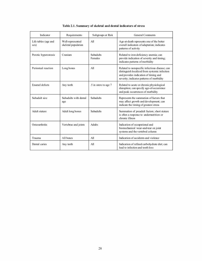
28
Table 2.1. Summary of skeletal and dental indicators of stress
Indicator Requirements Subgroups at Risk General Comments
Life tables (age andsex)
Well-representedskeletal population
All Age-at-death represents one of the betteroverall indicators of adaptation; indicatespatterns of activity
Porotic hyperostosis Cranium SubadultsFemales
Related to iron deficiency anemia; canprovide indication of severity and timing;indicates patterns of morbidity
Periosteal reaction Long bones All Related to nonspecific infectious disease; candistinguish localized from systemic infectionand provides indication of timing andseverity; indicates patterns of morbidity
Enamel defects Any teeth .5 in utero to age 7 Related to acute or chronic physiologicaldisruption; can specify age-of-occurrenceand peak occurrences of morbidity
Subadult size Subadults with dentalage
Subadults Represents the summation of factors thatmay affect growth and development; canindicate the timing of greatest stress
Adult stature Adult long bones Subadults Summation of preadult factors; short statureis often a response to undernutrition orchronic illness
Osteoarthritis Vertebrae and joints Adults Indication of occupational andbiomechanical wear-and-tear on jointsystems and the vertebral column
Trauma All bones All Indication of accidents and violence
Dental caries Any teeth All Indication of refined carbohydrate diet; canlead to infection and tooth loss

29
Site / Burial
FSNumber
Sex Age (y =years; m = months) Stature(cm)
Pelvis DentalAttrition
Other MidpointRange
Femur
LA 37592 B 1 200 2 y ± 8 m - dentition
1-2 y - long bones 2 y
B 2 184 6 y ± 24 m - dentition2.5-3.5 y - long bones
6 y
B 3 197 preterm fetus - size fetus
B 4 350 6 - 9 months - size .6 y
B 5 587 female? adult unknown
B 6 618 15 y ± 36 m dentition14.5-15.5 - long bones
15 y
B 7 662 female 40-55 45-55 50 y 155.8
B 0.1 137,138
5 y ± 12 m - dentition3.5 long bones
5 y
LA 37593 B 1 755 6 m ± 3 m dentition .5 y
B 2 756 female 35-45 22-27 sternal rib 35 y
B 3 735 male 40-49 - auricular surface37-57 - sternal rib
48 y
B 4 579 4.5-6.5 - long bones 6 y
LA 37594 B 1 808 4 y ± 12 m 4 y
LA 37595 B 1 103 female 20-30 25 y
B 2 159 16-20 15 ± 36 m - dentition15-21 - fusion
15 y
LA 37598 B 1 199 male? 40-55 40-44 - auricular surface
28-44 - endocranial 45 y 149.9
LA 37599 B 1 73 unknown adult - size unknown
B 3 187 3 y ± 12 m - dentition 3 y
B 4 232 male 34+ 40-49 - auricular surface31-65 - endocranial
45 y
B 5 241 male 22-30 24-30 25-29 - auricular surface17-27 - sternal rib
25 y 160.3
B 6 361 10 y ± 30 m - dentition 10 y
Table 2.2. Summary of La Plata burials

30
Site / Burial
FSNumber
Sex Age (y =years; m = months) Stature(cm)
Pelvis DentalAttrition
Other MidpointRange
Femur
B 7 372 female? 18-22 < 30 - fusion 25 y
B 8 443 2 y ± 8 m - dentition 2 y
B 9 559 male 22-39 20-30 < 25-30 - fusion
25-29 - auricular surface
18-30 - sternal rib
25 y 160.5
B 10 759 male 30-40 30-34? - auricular surface 32 y
B 0.1 452 unknown 45-55 50 y
LA 37600 B 1 10 & 77 female 45-55 40-44 - auricular surface 45 y 152.2
B 2 610 4 y ± 12 m - dentition 4 y
B 3 81 male 36-59 45-55 60+ - auricular surface 50 y
B 4 615 male 52-59 45-55 50 y 160.3
B 6 623 &624
unknown adult - size unknown
B 0.1 9 female? adult - size unknown
LA 37601 B 1
166 male 22-43 24-35 30 y 162.3
B 2 307 female? 18-19 18-22 19 y
B 3 151 female adult - size unknown
B 4 400 female 22-29 20-30 25 y - auricular surface 25 y 150.7
B 5 190 male 35-39 30-35+ 35 y 173.1
B 6 196 male 22-35 20-24 34 y - auricular surface20-23 - sternal rib
28 y 162.3
B 7 324 male 23-44 30-40 50 y - auricular surface 42 y 162.5
B 8 430 9 y ± 24 m - dentition6-8 y - long bones
9 y
B 9 606 3 y ± 12 m - dentition2.5-3.5 y - long bones
3 y
B 10 721 female 30-45 38 y
B 11 722 9 y ± 12 m - dentition5.5-7.5 y - long bones
9 y
B 12 991 male 18-23 22 y 158.8
LA 37603 B 1 295 male 32-52 40-55 40-44 - auricular surface 45 y

31
Site / Burial
FSNumber
Sex Age (y =years; m = months) Stature(cm)
B 2.1 410 female 23-39 20+ 30-34 - auricular surface18-30 - sternal rib
30 y 151.7
B 2.2 410 fetus - long bones fetus
LA 37605 B 1 1102 18 ± 6 m - dentition 1.5 y
B 3 6020 8 y ± 24 m - dentition 8 y
B 4 9301 6+ y - dentition5.5-7.5 y - long bones
6 y
LA 65029 B 1 536 1 y ± 4 m - dentition
1-2 y - long bones 1 y
LA 65030 B 1 11 & 12 2 - 3 y - dentition
.5 - 1.5 y - long bones 2 y
B 2 40 female 20-24 22 y
B 3 3 3 y ± 12 m - dentition 3 y
B 4 141 3 y ± 12 m - dentition.5 - 1.5 y - long bones
3 y
B 5 124 female 20-24 20-24 22 y
B 6 159 female 35-40 34-41 - auricular surface 38 y 149.4
B 7 169 10.5 y ± 30 m - dentition10 - 11 - long bones
10.5 y
B 8 241 female 13-24 18-22 20 y - auricular surface 20 y 154.3
B 9 242 female 30-47 20-30 33 y 151.7
B 10 352 female? adult - size unknown
B 12 510 male 20-23 20 y - auricular surface 22 y 160.9
B 13 511 11.5-12.5 y - long bones 11 y
B 14 514-515
18 ± 6 months - dentition 1.5 y
B 15 517 male 44-54 50+ 50 y - auricular surface44-56 - sternal rib
48 y 165.0
B 16 615 female 25-26 24-30 28 y
B 17 648 3 ± 1 y - dentition 3 y

32
Table 2.3. Regression equations for estimation of age for linear hypoplasia formation
Tooth Formulas1
Maxillary Teeth
I1 Age = -(.454 x Ht) + 4.5
I2 Age = -(.402 x Ht) + 4.5
C Age = -(.625 x Ht) + 6.0
Pm1 Age = -(.494 x Ht) + 6.0
Pm2 Age = -(.467 x Ht) + 6.0
M1 Age = -(.448 x Ht) + 3.5
M2 Age = -(.625 x Ht) + 7.5
Mandibular Teeth
I1 Age = -(.460 x Ht) + 4.0
I2 Age = -(.417 x Ht) + 4.0
C Age = -(.588 x Ht) + 6.5
Pm1 Age = -(.641 x Ht) + 6.0
Pm2 Age = -(.641 x Ht) + 7.0
M1 Age = -(.449 x Ht) + 3.5
M2 Age = -(.580 x Ht) + 7.0
Age = age in years; Ht = distance of the LEH in mm from CEJ1 Regression equations are based on mean crown heights of Swärdstedt (1966)and the crown formation standard of Massler et al. (1941).
Table 2.4. Assessment of dental wear
Category No. Incisors and Canines Premolars Molars
1 No attrition No attrition No attrition
2 Enamel polish Enamel polish Enamel polish
3 Light line of dentine exposure
4 Dentine patch (wider line) 2 or more dentine patches 3 or more dentine patches
5 Dentine patch (larger line) Exposure of 2d dentine withabove
3 or more large dentinepatches
6 Secondary dentine Heavy dentine Enamel around entire crown
7 <---------------------------------Enamel worn away at least on one side----------------------------------->
8 <-------------------------------------------Crown worn to root(s)--------------------------------------------->

CHAPTER 3
MORTUARY AND DEMOGRAPHICCONTEXT OF THE LA PLATA VALLEY
HUMAN REMAINS
Throughout its long history, issues of demographyand health have played a significant role in southwest-ern archaeology. Site reports often report burial age andsex and are thus sources of data for reconstructing mor-tuary behavior and demographic profiles. Many of themost significant cultural historical events highlighted byarchaeologists are also demarcated by significant demo-graphic corollaries (Swedlund 1994). Indeed, one of themajor questions that plagued the earliest researchers—what happened to the northern Pueblo people—wasframed in terms of population processes. Major eventsin southwestern prehistory are catalogued or character-ized by demographic manifestation, such as periods of“aggregation,” “abandonment,” and migration (Nelsonet al. 1994:59). Likewise, complexity itself has demo-graphic resonances that imply measures of rate, density,and scale.
Although demography and health are critical fac-tors in the analysis of human remains, the context with-in which the remains are found can likewise be reveal-ing (Alekshin 1983). Seminal works by Bartel (1982),Binford (1971), Saxe (1970), and Ucko (1969) makeclear the kinds of information that can be gleaned frommortuary analyses. Numerous studies that focus on themortuary component of Southwest human burials haverevealed a great deal about social ranking (Akins 1986;Ravesloot 1988), variability in location and position(Stodder 1986), social organization (Palkovich 1980),treatment of elites (Frisbie 1978), and the relationshipbetween burial location and settlement patterns(Schlanger n.d.; Toll and Schlanger 1998). On the onehand, these and other studies have revealed considerableinformation about how the ancestral Pueblo people dealtwith their dead; on the other hand, even with this inten-sive focus, we actually know very little. Identifyingstrong burial patterns and practices among the ancientPueblos is still quite elusive. Although not random atany given location, burials are found within a number ofcontexts (middens, room and pit structure floors, storagepits, extramural pits, and in abandoned structures), in anumber of positions (flexed, semiflexed, extended,sprawled) with grave offerings ranging from nothing toone vessel to extensive offerings (that could includeturquoise, bead necklaces, multiple ceramic bowls,ladles, jars, pitchers, mugs, birds, and dogs). Orientationof burials may cluster around east-facing, but everyother possible compass reading is found as well.
Statistically significant relationships among variablessuch as location, orientation, grave goods, age, and sexhave likewise not been forthcoming.1
This variability in mortuary context suggests thatburial customs probably varied among groups livingwithin the same regions or even within the same site.Rather than a single set of customs surrounding burial,there were probably several, and these rules were prob-ably “context sensitive.” That is, depending on who wasbeing buried by whom and for what reason, there werecertain practices that would be followed to the degreepossible. Given this complexity and the archaeologicalinaccessibility of some of the important contextualinformation, burial patterns can only be generally char-acterized for the precontact Pueblo. For example,although a preference for flexed or semiflexed primaryinterments is demonstrated in Pueblo graves, the loca-tion of these varies widely. There does seem to be a pref-erence for placing ceramics with these primary inter-ments, although burials with no associated artifacts arequite common. Earlier researchers (e.g., Frisbie 1978;Morris 1939) have presented overviews for shifts in bur-ial customs across temporal units (Pueblo I, II, and III)to which can be added the increased dimensionality pro-vided by great quantities of new information.
Analysis of the mortuary context of the 67 La PlataValley burials and up to 82 individuals represented innonburials (described in Chapter 6) does not revealstrong patterns or trends across sites. The patterning ofburial practices does suggest that it is this rich and com-plicated variability that is interesting and important todocument, rather than simply assigning burials to one ofseveral large and ambiguous categories (i.e., location,orientation, position, presence/absence of grave goods).The variability hints at some interesting differences thatmay be related to larger phenomena of resettlement orrelocation, secondary burials (Chapter 6), and status dif-ferences. Stodder (1986; 1987; 1990) and Schlanger(n.d.) advocate a more precise and in-depth analysis ofburials within sites and between contiguous sites, andfocusing more on the relationship of the mortuary fea-tures to settlement patterns, organization of subsistenceand ritual activities, and demographic variables such asfamily size, community size, and population density.Although it is not within the scope of this project toundertake an exhaustive study of the La Plata burialconfigurations, we review the mortuary context here toprovide a framework and data for future analyses.
MORTUARY PATTERNING OF THE LA PLATA BURIALS
The characteristics of the La Plata Valley burials asa group are described and individual assessments of themortuary component are more fully portrayed for each
33

burial in Appendix 3. The single best source of informa-tion for the burial context comes from the original fieldforms and notes, which relate in extensive detail thecontext, condition, deposition, and associations of eachburial. Photographs, drawings, and maps likewise areavailable from the original excavation field forms.2 Forour purposes, we collected a subset of information fromthese and incorporated it into the skeletal remains database (see Appendix 2, first page of the Data BaseManagement form). Briefly, location was recorded asmidden, pit structure, storage pit, room, extramural bur-ial pit, or other. Data on strata were categorized bystratigraphic association. For midden burials, it wasnoted if the remains were in shallow pits or if they wereplaced deeper into sterile soil. For structures, it wasrecorded whether the burials were in the upper, middle,or lower fill. Position was recorded as semiflexed,flexed, extended, disturbed, or other. Based on an analy-sis of burials within sites, they were divided into cate-gories that distinguished single from multiple burials,primary from secondary burials, and articulated fromdisturbed remains. Finally, associated artifacts werearbitrarily placed into categories of none, small, moder-ate, and extensive offerings.3
The burials from the La Plata Highway Project rep-resent the full range of variability in location andamount of grave goods (Tables 3.1-3.4) and position(Tables 3.5-3.8) as defined here, although burials withlarge quantities of burial goods are absent.4 In the statis-tical analysis of a variety of factors (age, sex, date ofsite, location, position, goods), no significant associa-tions could be found. Although this may be a function ofsmall sample size, an alternative explanation is that thecultural rules for how individuals are to be interred arecomplex and responsive to a variety of factors.
Grave Type
A great majority of the burials from the La PlataValley sample are single, articulated, primary interments(approximately 90 percent). Although some of theseburials were badly disturbed at the time of excavation(n=16: 12 by mechanical equipment, 2 by carnivores,and 2 by carnivores and mechanical equipment), the dis-turbance is largely due to diagenetic and natural causes.Multiple articulated primary interments account foranother 4.5 percent of the collection, and these burialsare found at LA 65030 in one pit structure (discussed atlength in subsequent chapters). These frequencies arequite different from those reported by Stodder (1987)for the Dolores Archaeological Project burials. Shefound that 51 percent of the burials were single primaryinterment, while the other burials were multiple primaryand secondary graves. Akins (1986) noted that the
majority of the burials from the Chaco small sites weresingle primary interments. For Kayenta region burialcollections, Ryan (1977) and Martin et al. (1991) reportthat the majority of the burials encountered were like-wise single primary interments.
Grave Location
For the La Plata Valley burials that could be posi-tively assigned to a specific location within the site(n=61), over half (55.7 percent) were located in pitstructures (Table 3.9). The others were located in extra-mural features (19.7 percent), rooms (13.1 percent),extramural burial pits (9.8 percent), or middens (1.6 per-cent). In looking at location in terms of adults andsubadults, there are similar patterns of distribution,although subadults were more likely to be found inroom contexts than adults, and most of those found inextramural burial pits were male. Pit structure burialsaccount for the majority of burials in all age, sex, andperiod categories except Pueblo III subadults, where pitstructure burials are still common. The total volumeexcavated by the highway project was heavily weightedtoward pit structures, so that materials from that contextare more likely. Nonetheless, there is a strong patternindicating that burials were placed in various pit struc-ture locations, including on and beneath floors and inthe fill.
In looking for more subtle variations, femalesburied in storage pits were sometimes in the upper filland sometimes in the lower. Of the 9 females in pitstructures, 5 were in intrusive pits in the upper fill, 3were in the roof fall, and 1 was in the lower fill. The twofemales associated with rooms were below floors. Twomales found in extramural features were in the upperfill, and one was near the base of the feature, althoughthe feature had begun to fill when the burial was placed.Of the 9 males located in pit structures, 4 were in intru-sive pits or upper fill, 2 were in the lower fill, and 3 werein subfloor features or a tunnel. There were three malesin extramural burial pits. None was associated withrooms. This general pattern of variability describes thesubadults as well.
Of special note is the recovery of two fetuses. Theyounger one (4-6 lunar months) was placed in a largecorrugated jar and placed below the floor of a room. Theother one (approximately 8-9 lunar months) was com-mingled with an adult female in an intrusive pit in theupper fill of a pit structure.
The most striking feature of this assemblage is thelocation of burials in domestic locales and the place-ment of the dead in what appear to be largely abandonedstructures. Placement within the fill of pit structures, forexample, ranges from individuals at the bottom (above
34

the floor) and throughout the fill, suggesting repeateduse of the abandoned structures through time. Only 12.2percent (7) of the individuals were interred in sites occu-pied at the time of burial (presumably the subfloor roomburials and a majority of the extramural burial pits), andsome of these structures may have gone out of use afterburial took place. Occupation of the location may havecontinued, but most of the structures used for buriallocales had ceased to be used as structures.
The preference for using abandoned structures andfeatures at the La Plata Valley is very similar to the pat-tern found by Stodder (1987) for early Pueblo (PI)Dolores area burials. Over 70 percent of the burialsrecovered were in the fill of structures, although moreDolores burials were directly in middens than at LaPlata. Stodder’s (1987:354) review of burial locationsfor northern Pueblo I and Pueblo II sites in MancosCanyon, on Mesa Verde, in the Piedra District, and atAlkali Ridge shows that nearly all these fill burials werefrom midden deposits.
Burial Position
Most of the burials (78 percent) were found inflexed or semiflexed leg position (Table 3.10), althoughthere is great variability in leg, arm, axial skeleton, andface positioning (Tables 3.5-3.8). The pattern is similarfor adults of both sexes, and for adults and children.Subadults were more likely to be placed in positionsindicating deliberate placement (20 of 22) than adults(males 11 of 13; females 11 of 14). Most were flexed orsemiflexed. However, some burials were described inthe La Plata field notes as “sprawled,” “flung,” or“thrown.” These are discussed in more depth in subse-quent sections. It should be noted that some of the indi-viduals categorized as semiflexed also demonstrate amore inexact positioning; sometimes arms or body partscross over each other in a haphazard manner. For exam-ple, in the case of one female, the field notes state that itwas as if the body was pushed into a pit, and ended upin a semiflexed but disarranged position.
Thus, deliberate placement of bodies in flexed orsemiflexed positions was practiced on the majority ofindividuals, but in 14.6 percent of the population, thebody was disposed of in ways that disregarded orderlyplacement of limbs relative to the torso (Table 3.10).Examination of the photographs in Morris’s (1939) LaPlata report suggests that the flexed burials found at Site41 were mostly orderly in placement, although he, too,found disarticulated and less formally placed burials(Table 3.11). Stodder (1987:25) notes that of the extend-ed burials in the Dolores sample, some were found inpositions which seemed “rather sprawled.” In compar-ing the burials excavated from Dolores, there are some
remarkable similarities in the frequency of these morehaphazardly placed burials.
A comparison of position with Mesa Verde (Table3.12) and Chaco Canyon (Table 3.13) burials is enlight-ening in several regards. Although semiflexed andflexed burials dominate, extended burials occur at bothlocations, especially at Chaco Canyon, where they reachnearly 20 percent during Pueblo II. In some instancesamong the Mesa Verde burials, semiflexed burials werereminiscent of the more haphazard or “pushed” attitudethat some of the La Plata burials demonstrated, althoughit was difficult to systematically cull this informationfrom the site reports. It was particularly difficult to esti-mate frequencies of burial location from the site reportsfor Mesa Verde, but for Chaco Canyon, there are cleardifferences when compared to the La Plata Valleygraves. Middens and rooms account for the great major-ity of the burial locations at Chaco Canyon, with asteady progression from most burials in middens tomost burials in rooms. The midden burials in Chaco arenot located in the huge “trash mounds” associated withthe great house sites but are in the middens associatedwith the small sites. What burials there are at great-house sites are located in rooms (Akins 1986). At Morris41, in the La Plata Valley, burials were placed in mid-dens prior to Pueblo III, but were mostly in rooms dur-ing Pueblo III, chronologically later than the great-house burials in Chaco. Most of the burials reported byMorris (1939) date to the last phase of occupation atMorris 41. Many of these late burials were simplyplaced on the floors of abandoned rooms, although theywere formal burials. Many others were placed in sub-floor pits in rooms, and some were placed in other con-struction locations (Table 3.11), but compared to theburials from the highway project, surprisingly few werefound in pit structure fills.
Differences in burial location at Chaco, MesaVerde, and La Plata can be attributed to a number of fac-tors. The highway project excavated disproportionatelyfew rooms, and a smaller proportion of late Pueblo IIIcontexts than at Morris 41. Late Pueblo III rooms exca-vated at LA 37592 and LA 65030 do contain burials, inconformance with the pattern. Pit structures and theirfills are much less susceptible to modern surface distur-bance and therefore are over-represented in this sample.Even taking that into account, however, the occurrenceof burials in various pit structure contexts seems veryhigh in the La Plata Highway sample. In some cases, pitstructures contain middens (such as at LA 37592), blur-ring the “midden” and “pit structure” categories usedhere. In other cases, pit structures may have been near-ly or completely filled, blurring the “extramural burialpit” and “pit structure” categories. Also important incomparing locations is period of interment. As Morris
35

notes (1939:115), there was a shift toward burial inrooms in late Pueblo III. This shift is most apparent atMorris 41 (Table 3.11) and Aztec (Morris 1924) butshould be taken into account in other late contexts aswell.
Burial Goods
Burials at La Plata with associated artifacts accountfor a little over half of the sample (58.6 percent of 58cases with known associations). Of the burials thatcould be positively identified as having grave offeringsamong adult males, the frequency was 57.1 percent, andfor females, 68.8 percent. Half of the subadults (53.6percent) had associated grave goods. These numbers areconsiderably higher than for the Dolores sample, whereStodder (1987) found that slightly more than one-thirdhad been buried with grave goods. In examining the dis-tribution of grave goods (small, moderate, extensive),there is no significant pattern between sexes, acrossages, or through time (Tables 3.1-3.4, 3.14). Females aremost likely to have burial goods and to have moregoods. Subadults have the least goods. The most com-mon offerings are ceramic bowls. A few burials containjars, and fewer had ladles or pitchers, unlike the RioPuerco of the West, south of Chaco, where pitchers arecommon among ceramic grave goods (T. C. Windes,personal communication, 1993; Mills 1993:306). Oneburial had a bone awl, and a few burials had groundstone or worked sherds. Of burials with grave associa-tions known (Tables 3.1-3.4, 3.14), around a quarter(25.9 percent) had a small (1 object) number of offer-ings, another quarter (24.1 percent) had moderate (2 to3 objects) offerings, and 10.3 percent had what could beconsidered extensive offerings (more than 3 objects).
The five individuals with more than three graveofferings are a 9 year old (37601 B11), a 22-year-oldfemale (65030 B5), a 35-year-old female (LA 37593B2), an adult female of unknown age (65030 B10), anda 48-year-old male (37593 B3). The child was buriedduring early Pueblo III in a semiflexed position in a stor-age pit with three ceramic vessels. Also in the pit but notnecessarily associated were a turkey and a dog. Bothadults from LA 37593 were in large storage pits, butthey were from different periods. The male, buriedflexed in early Pueblo III, had a corrugated vessel, aladle bowl, and a rough-legged hawk, and was placed inthe pit with matting. The female, buried in mid PuebloII, was also accompanied by matting. Grave goodsincluded a corrugated vessel containing a number ofsmall rodents, a partial Mancos Black-on-white bowl,the base of a plain gray jar reworked into a saucer, andtwo two-hand manos. Two adult females at LA 65030had extensive grave goods. One (Burial 10) had a corru-
gated jar, a plain gray jar, an organic paint Black-on-white bowl, a jet pendant, and possibly some groundstone tools associated. This burial dates to after A.D.1100 and was in an extramural storage pit. A youngadult female from LA 65030 (Burial 5) was buriedbeneath the floor of a room in late Pueblo III. This indi-vidual may have been wrapped in a shroud and was themost elaborately accompanied burial in the highwayproject. Her grave goods include a bone bead necklace,three bowls, small pieces of turquoise, a polished peb-ble, a lightning stone, ground pigment, and a groundstone palette. With the exception of LA 37593 B2, theburials with extensive accompaniments are from PuebloIII contexts. Morris (1939; Table 3.11) reports severalPueblo III burials with large grave inventories, and itseems that extensive grave goods are more common inthe late period than they are in Pueblo II, the time peri-od accounting for the majority of highway project con-texts (see Table 3.9). It is noteworthy that someBasketmaker III burials from Morris 41 also had sub-stantial grave goods.
The La Plata Highway Project did not excavate anyburials with large quantities of offerings, but the major-ity of eastern prehistoric Pueblo burials contain nodurable grave goods or fewer than two items (see, e.g.,Table 3.11; Brown 1993). Burials with large numbers ofnonperishable items are relatively rare. Morris (1939)describes burials from Site 41 with multiple vessels,bone tools, wood artifacts including arrows, and occa-sional ornaments. Table 3.11 lists burials from the LaPlata Valley that are included in Morris’s report (1939).It should be noted that far from all of the burials thatMorris excavated are included in the report, and thatburials with elaborate accompaniments were more like-ly to be included than those with few materials.Although some of the many burials from Morris 41 docontain larger numbers of items, the norm is again forthere to be few or none—the burial assemblages fromthe highway project, then, are within the norm. Burialswith vast numbers of goods such as those at PuebloBonito (Akins 1986), the Warrior at Aztec (Morris1924:193-195), or the burial at the Dominguez Ruinnear Dolores (Reed 1979:101) are exceptions to the nor-mal pattern, showing the extreme to which goods can bepresent.
Mortuary Behavior at La Plata
The La Plata Highway Project is among only ahandful of projects in the Southwest that has integratedthe human remains and the mortuary component into theongoing research strategy. Examples of the inclusion ofmortuary behavior along with paleopathological andpaleodemographic analyses for Pueblo skeletal popula-
36

tions include those by Akins (1986), Palkovich (1980),and Stodder (1987). An analysis focused only on the LaPlata remains is hampered by the small sample size, butit can serve to contribute to the growing data base onmortuary variability and stands ready to be used by oth-ers in more comprehensive and inclusive studies in thefuture.
Schlanger (n.d.; Toll and Schlanger 1998) has pro-vided a careful and illuminating analysis of the mortu-ary component of the burials at La Plata.5 This impor-tant study demonstrates that the burials are not random-ly distributed across features in the landscape. Rather,there appear to be preferred strategies for the disposal ofthe dead. She finds three characteristics of the mortuarybehavior in the La Plata Valley that distinguish it fromprecontact burials from other parts of North America:lack of a formal burial ground, great variability in thenumber of burials at any given site, and frequent use of“places for the living” as resting places for the dead. AtLa Plata, burials are found with greatest frequency in pitstructure fill contexts. Extramural features, floors, sub-floor settings, and surface rooms are other places whereburials have been located.
Most interesting of the findings by Schlanger is theoccurrence of burials on or near the benches of mostlyfilled pit structures. She suggests that some graves con-tain the remains of former inhabitants of abandonedsites. For example, four of the eleven burials recoveredfrom LA 37601 show distinctive genetic anomalies ofthe spine (refer to Appendix 3). These include one adultmale with an extra thoracic vertebra; two adult males,each with an extra lumbar vertebra; and one subadultwith a first sacral vertebra that demonstrates lumbar fea-tures. This co-occurrence of genetic anomalies suggeststhat it is possible that family members were buried at alocation that had some meaning for them. This line ofinquiry demonstrates the need for archaeologists towork closely with skeletal biologists in blending theanalyses. Lane and Sublett (1972) have demonstratedthat skeletal material can be used to confirm hypothesesabout ethnicity, endogamy, and residence patterns.Future collaborative and interdisciplinary studies needto be initiated in this area for the La Plata Valley region.
At La Plata, the frequency of bench burials and thepresence of disarticulated human skeletal elements thatcannot be explained by natural depositional events sug-gest that the management and treatment of the dead bythe La Plata communities went well beyond simpleinterment. Because so few studies have included thetype of careful analysis afforded by Schlanger, there areonly a handful of comparable studies.
Stodder (1986) has provided a similarly insightfulstudy of the Pueblo I Dolores mortuary program, andthere are interesting parallels to be made that highlight
how little is known regarding what “normal” precontactburial patterns are. The Dolores Archaeological Projectrecovered 45 discrete burials in the postoccupational fillof habitation and storage structures, under floors of pitstructures and habitations areas, and in middens. Aswith the La Plata remains, Stodder found no patterningor association with respect to age or sex for location,position, or amount of grave goods.
The occurrence of several individuals in what hasbeen described as “sprawled” position on the fill ofabandoned pit structures at La Plata is not unique, but ithas also not been intensively described by others work-ing with burial populations except for Stodder’s (1986)report on the earlier (Pueblo I) Dolores sample.
It is difficult at this stage to make any kind of sum-mary statement that accurately situates the La PlataValley burials vis-à-vis burials from other sites in theSouthwest. There are questions that could be framed forburials that would shift the focus into different arenas.For example, what similarities exist in overall healthstatus and treatment of the dead? Do intragroup or inter-group tensions and strife enter into a group’s mortuarypractices? Is there a pattern relating cause of death(admittedly difficult to determine) and mortuary behav-ior? Is status (and therefore differential treatment atdeath) defined in political, economic, social or spiritualterms? Can information gleaned from the skeletons beused in inferring ethnicity, endogamy, and residencepatterns? These types of questions shift the focus of tra-ditional research into mortuary behavior and could linkburials with a broader discussion of biocultural process-es underlying adaptability and population dynamicsthrough time and across space.
DEMOGRAPHIC FEATURES OF THE LA PLATA SKELETALPOPULATION
Mortality data on skeletal populations derive fromassessment of individual ages at death. Traditional pre-sentations of mortality data involve the use of survival(from one age to another) to graph survivorship curves.Life expectancy as a function of survival has also beenused in many studies. Angel (1969) has argued againstmathematical modeling more complex than relativelysimple statistical procedures. For example, he suggeststhat calculations such as proportions of infants and chil-dren to adults or comparison of survivorship curves areall that is required for demographic analysis of precon-tact populations.
Swedlund and Armelagos (1969), among others,promote the use of life tables for paleodemographicanalysis. The use of a life table provides all the basiccalculations that Angel suggested but provides two addi-tional key measures: probability of dying and life
37

expectancy for each age. Thus, using a life table for eachspecific age interval is characterized by the number ofdeaths, the proportion of all deaths, the percentage sur-viving to that age, the probability of death within thatinterval for those that reach the age interval, and the lifeexpectancy of those that reach the age interval.
Presentations of mortality data involve the directestimation of life expectancy at birth (based on the meanage at death) or the construction of life tables. In addi-tion to estimating life expectancy at birth, probability ofdying and survivorship for all age classes can be esti-mated. At this level of analysis, we are greatly removedfrom the actual skeletal remains because the estimatesinvolve statistical manipulation and only use numbers ofindividuals dying in different age categories. It is criti-cal to fully acknowledge and explore where biases mayexist in the archaeological series. Differential mortuarypractices and poor presentation of some segments of thepopulation are the two most influential sources of errors,assuming that excavation procedures were systematicand consistent in recovery protocol (Buikstra andMielke 1985; Weiss 1972).
The statistical manipulation of age and sex profilesand the construction of life tables are actually the easierpart of paleodemographic analyses. The difficulty lies ininterpretation of the data. Johansson and Horowitz(1986:234) succinctly summarize the range ofunknowns and assumptions that must be taken as agiven when reconstructing demographic patterns fromskeletal populations. For example, they present fourphases or levels of analytical factors that need to beacknowledged when dealing with skeletal populations“whose demographic characteristics are unknown . . .(who) buried their dead over an unknown period of timeaccording to unknown rules.” Phase one, according toJohansson and Horowitz, involves archaeologicalrecovery that can present biases in age, sex, class, andother spheres because of mortuary practices and differ-ential preservation. Phase two includes the anatomicaland paleopathological analysis of the recovered skeletalmaterial for age and sex. Problems encountered at thislevel include the margin of unknown error involved inestimations of age and sex, the small sample sizes in ageand sex categories, and the inability to determine causeof death in the vast majority of the cases. Phase threeinvolves demographic analysis of mortality and fertility.Here Johansson and Horowitz point out that it isextremely difficult to prove whether a population wasstable, stationary, and closed (assumptions that must bemade in order to compare precontact age and sex distri-butions with model life tables derived from living pop-ulations). Finally, phase four involves historical recon-struction and theory building based on the mortalityestimates. Problems at this level include the inability to
establish how valid the derived mortality/fertility esti-mates are.
Errors in phase one or two can greatly affect thevalidity of the data generated in successive levels ofanalysis. However, in spite of all the potential problems,there is ample evidence that paleodemography is centraland critical to our understanding of precontact life anddeath and should be undertaken in all possible cases(Buikstra and Konigsberg 1985). The major limitationsof the paleodemographic method are practical ones.They include the representativeness and size of the sam-ple and the ability to provide accurate assessment ofdevelopmental age (Lovejoy et al. 1977). However, thebottom line is that age at death stands as perhaps themost important single indicator of stress as well.
With a recognition and appreciation for these prob-lems, the paleodemographic analyses for La Plata arepresented as plausible scenarios that reflect the likelyexperience of this group. Age and sex composition, lifetable data, and survivorship curves are presented for LaPlata and interpreted using other known variablesregarding subsistence, health, and settlement. In thisway, the statistical manipulation of the age data is usedas a check against other kinds of data from the La Plataproject to assess how well the living population wasdoing.
Age at Death
A total of 67 individual burials were recovered andcould be aged ranging from preterm fetuses to individu-als aged fifty and older (Table 3.15).6 The age categoriesare generally well represented, although there may beunder- enumeration in the infant (newborn to one year)category. The adult population is likely to be under-rep-resenting the oldest ages, and there are more females inthe young adult category than there are males. Theseindividual burials were retrieved from 12 different sites,and the number of burials recovered from any given siteranged from 1 to 16 (Table 3.16). Trends in the distribu-tion of burials by age-at-death and sex across sites couldnot be statistically verified. Therefore it seems safe toassume that any biases introduced into the sample werelikely the result of excavation constraints and not mor-tuary behavior that may have clustered individuals byage or sex in spatial units.
In comparing the age composition of a number ofarchaeological populations from the New World, thedistribution of burials recovered at La Plata do not varymarkedly except to verify that the infant category mayin fact be under-represented. This is also reflected in thehigher frequency of the subadult category, one to nineyears (Table 3.17). Here La Plata has the highest fre-quency reported (29.8 percent). Given the problems
38

with the younger age categories, the percentage of LaPlata adults (56.7) is indistinguishable from a widerange of precontact New World populations. Given thatthe La Plata Valley sample is represented by a relativelysmaller sample size, yet holds to the same proportionaldistributions across major age categories (except forinfancy) as the much larger Southwest and other region-al samples, it is likely that La Plata is not so biased as togreatly skew the collective age-at-death composition.
The perennial problem of the life table for archaeo-logical samples is that we do not know the growth rate(Weiss and Smouse 1976). This is important because thelife table calculations assume that the population repre-sented by the burials has undergone internal and regularrates of change. That is, no migration is assumed to haveoccurred, and any growth or decline is assumed to bederived from the inherent mortality or fertility. If theseconditions are met (which it is not possible to know forLa Plata), stable population theory allows us to derive anumber of attributes about the population by calculatingthe probability of dying from birth to the oldest age cat-egory. Problems such as these have caused some toargue that life tables should not be used on archaeolog-ically derived populations. However, as Nelson et al.(1994:68) state: “Given all these concerns, our basicanalytical procedure is to bring multiple sources ofinference to bear on the age at death distributions.Rather than attempting to fit the distributions to somereference model to estimate parameters (such as lifeexpectancy), we simply point to what the respective dis-tributions suggest in terms of health and demography....The mean age at death values should be viewed in rela-tive terms as opposed to interpreting them as absolutevalues.”
The La Plata burial population is admittedly small,and therefore there is the possibility that it will departfrom characteristics of the living population due to sto-chastic effects. However, Weiss and Smouse (1976)studied living populations and, based on simulationstudies, demonstrated that stability is inaugurated intosmall groups relatively early and quickly after eventsthat cause population disruption. Regarding archaeolog-ical burial collections, Buikstra and Mielke (1985:364)state that assemblages that have “accumulated over aperiod of years should provide a reasonably accuratemeans of characterizing the vital rates of a population.”
Table 3.18, a life table, presents data for the com-bined La Plata burial population. The small sample sizeprohibits conducting truncated life tables for adult malesand females. All of the individuals except for one childfall into the time period A.D. 900-1300, and the greatestmajority of the burials are in the A.D. 1000-1200 range.7The combined sample aggregates all the individualsduring the entire occupational span. A decision was
made not to use running averages to “smooth” the data,as others have done (Buikstra and Mielke 1985;Palkovich 1980; Powell 1988). In general, these studiescite Weiss (1973), whose actual discussion on smooth-ing refers to living nonindustrialized groups, wherepractices such as age heaping by respondents can pro-duce variation that would distort the analysis. This iscertainly not the case with archaeological populationssuch as La Plata. Also of note, adult individuals ofunknown age were differentially reapportioned acrossthe adult age categories. The six individuals from LaPlata who could only be identified as adult were subse-quently redistributed, using the corresponding mortalityfor all adult age categories, weighted according to thenumber of individuals in each.
The life table reveals a mean age at death (ex0) of24.5. Although this life expectancy figure must be con-sidered provisional, and only as a means to generatehypotheses regarding population parameters at La Plata,it does suggest a robust population.8 In looking at lifeexpectancy estimates for a number of archaeologicalpopulations (most of which represent either so-calledAnasazi or Mogollon groups), La Plata Valley ranksfairly high (Table 3.19). The archaeological populationsthat have higher or similar life expectancies are mostextraordinary for their temporal and spatial differences.For example, Pueblo Bonito is likely to represent anelite subgroup living within Chaco Canyon (PII). BlackMesa (PI-II), on the other hand, in northeastern Arizona,is considered to represent small, autonomous (“poorcousins”), hamlet-type communities. Precontact peopleliving on Black Mesa, however, were doing relativelywell with sustained population growth from A.D. 800 to1050, a relatively low disease rate, and an abundance ofindividuals who reached the age of 50 (Martin et al.1991:17). Point of Pines, generally characterized asMogollon, with a long span of habitation (A.D. 900-1450), may represent the final destination point ofmigrating Kayenta peoples (Bennett 1973:2). The MesaVerde Early sample represents a period of growth (PI-II)and expansion in the Four Corners region. CasesGrandes was a large, politically centralized populationthat is similar to Chaco in some regards (e.g., rankedsociety, architectural symbolism, economic specializa-tion) (Nelson et al. 1994:92). Thus, the life expectancyrate alone does little to provide information on why lifeexpectancy was good at La Plata relative to other groupsand suggests that it is a function of a wide variety of cul-tural, environmental, and biological factors.
It is telling, however, that life expectancy is only1.8 years higher at Pueblo Bonito, which may representthe largest politically centralized economic unit in theregion. Nelson et al. (1994) suggest from an analysis ofa series of Southwest burial populations that being polit-
39

ically autonomous and living in dispersed communitiesaffords better health and community well-being thanwhen groups are part of a large and/or densely populat-ed community (such as at Chaco and Casas Grandes).The exception to this is elites within such a system, whomay be afforded differential access to resources andtherefore better health and longevity.
This kind of bracketing of La Plata in terms of lifeexpectancy permits the following observations. LaPlata, situated between the Chaco Canyon and MesaVerde regions, may have shared the negative healtheffects of high population density (which can signifi-cantly influence morbidity and mortality). The basic tra-jectory and time frame in all three of these regions issimilar (especially when a large enough area is includedto encompass elevational movements through time), andall three regions were abandoned by the early A.D.1300s. Population size in the La Plata Valley was mostlikely as great as or greater than that in nearby regionalsystems such as Chaco Canyon and Mesa Verde.
Nelson et al. (1994) also present a comparison ofmortality and fertility data for a select group ofSouthwest archaeological populations (Table 3.20).These groups were chosen for comparison to test thehypothesis that demography and health were related tosocial organization and political centralization. It hasbeen suggested that the ratio of individuals over the ageof 30 to the number of individuals over the age of 5 isinversely proportional to the fertility rate. The lower thevalue, the higher the fertility. Of course, there are manyprovisos attached with these data. They are explored byBuikstra et al. (1986), but just as with life table data,they can be used to speculate on the magnitude of dif-ferences among groups, and more importantly, whythese differences exist.
In comparison with these groups, La Plata fallstoward the high range in terms of mean age at death, andfertility is likewise located in the middle range. To somedegree, these values meet our expectations. Mesa Verderepresents an aggregated population that grew rapidly,peaked, and then abandoned the area (circa A.D. 1300).It may seem counter-intuitive to have the highest fertil-ity in the later Mesa Verde sample (representing theperiod from A.D. 975-1300) during what must haveincluded the later years of retrenchment and abandon-ment. However, much of the growth in the early MesaVerde group (A.D. 600-975) may have been a result ofthe migration of people into the area, and therefore themoderate fertility rate at this time makes sense. It is pos-sible that what the high fertility ranking in the late MesaVerde sample is tracking is a function of decreasedmigration into the area (which would look archaeologi-cally like population decrease). Thus, although fertilityremained high in the later periods on Mesa Verde, pop-
ulation did decline. It is difficult to interpret the PuebloBonito data because this sample most likely over-repre-sents high-ranking adults given special mortuary treat-ment. La Plata, with a fertility rate of .4318,9 appearsmoderate (by Southwest standards). A relatively lowdisease load (discussed in Chapter 4) may have con-tributed to the low to moderate probability of dying, andthis in turn may have contributed to relatively well-offcommunities at Jackson Lake and Barker Arroyo duringPueblo II-III.
The lowest fertility rates were in the Black Mesaand Arroyo Hondo populations and likely represent twocompletely different processes. Arroyo Hondo was apopulation in retreat suffering from inadequate nutritionand sociopolitical disorganization (Palkovich 1980;Wetterstrom 1986). Black Mesa, on the other hand,seemed to intentionally keep family size small by spac-ing births or through some other means (Martin et al.1991). This may have been an adaptive response to thehighly marginal and unproductive landscape that theylived upon.
Additional information can be gained by comparingthe number of individuals alive at the beginning of eachage category (lx) to track survivorship of individualsacross the life span. The survivorship curve for La PlataValley is plotted along with early and late Mesa Verdepopulations and the Pueblo Bonito and Black Mesasamples (Fig. 3.1). In general, the curves follow a simi-lar shape, although there are several distinguishing fea-tures of the La Plata curve. Comparison of La Plata withthe Mesa Verde samples demonstrates that the rate ofsurvivorship in the five-to-ten-year-old category islower at La Plata and continues to be lower until reach-ing adulthood (and most closely resembles the contem-poraneous late Mesa Verde sample). During youngadulthood, La Plata survivorship is higher than the latesample and, until the age of 35, closely follows thecurve for the early sample. After the age of 35, La Plataexceeds both Mesa Verde groups and has higher sur-vivorship.
The survivorship curve for Pueblo Bonito clearlydisplays high numbers of survivors in all age categoriesleading up to mid-adulthood. From 30 onward, BlackMesa has the higher figure (in part because of the highproportion of elderly in that population). However, thesurvivorship curves for Black Mesa and La Plata areidentical for children up to the age of 15. Black Mesahas been interpreted as a relatively healthy and robustpopulation (Martin et al. 1991; Nelson et al. 1994).
More support for the hypothesis that La Plata sur-vivorship curves suggest a population with relativelylow child mortality and good health is gained by exam-ining the survivorship curves of La Plata and groupslater in time representing a precontact Pueblo (Arroyo
40

Hondo), a Mogollon (Grasshopper), and a NorthernMexico (Casas Grandes) series (Fig. 3.2). Survivorshipat La Plata was remarkably similar to that at CasasGrandes for the first 20 years of life. Whereas ArroyoHondo and Grasshopper demonstrate low survivorshipof children in and young adults in general, individuals atLa Plata enjoyed, as a group, higher survivorshipthroughout the lifespan than its counterparts living ahundred years or so later in other areas of the Southwest.
Life table analysis also allows for a close look at theprobability of dying (qx) for individuals in each age cat-egory. In comparing La Plata with the Mesa Verde Earlyand Late samples, and the Pueblo Bonito and BlackMesa samples, La Plata appears largely situated in themiddle, with neither higher nor lower probability rates(Fig. 3.3). The exception to this is in the youngest agecategories, where there is a higher rate, but the La Platacurve drops to among the lowest for children and thenremains steadily in the middle. Comparing the probabil-ity of dying at La Plata with the later precontact Pueblo,Mogollon, and Northern Mexico sample, a similar pat-tern holds true: La Plata demonstrates the same or lowerprobability rates in most age categories (Fig. 3.4).
DISCUSSION
Two different but related kinds of information onthe La Plata burial population are reviewed in this chap-ter. Analysis of the mortuary context, critical for theassessment of biases introduced into the burial assem-blage by mortuary practices or excavation procedures,demonstrated several things. First, although there aresome patterns of interment that hold across sites (singlearticulated primary burials in semiflexed or flexed posi-tion), there was a significant degree of variation in bur-ial location, associated grave goods, and in age and sexdistribution of the burials. The mortuary componentcompares most closely with that of the earlier (PI)Dolores Archaeological Project burials, which also dis-played a great deal of variability in interment. La Plataburials are different from contemporaneous Mesa Verderegion burials, La Plata sites excavated by Morris (suchas Site 41), and Chaco small sites. Toll and Schlanger(1998) suggested that the use and reuse of abandoneddomestic features is meaningful and can be used to bet-ter understand the motivating factors behind burial pat-terns and ties to certain places in the landscape.
The demographic analyses suggest a fairly repre-sentative population, at least to the extent that it mayportray, in Berry’s (1985:45) words, a “snapshot” viewof the parent population. Although life tables are actual-ly death tables, they present a convenient and standard-ized way to look at the mortality experience as a func-tion of the life experiences. The most significant place
where the La Plata life table data may be biased is in theunder-enumeration of infants. Brothwell (1981) reportsthat the proportion of infants under the age of 1shouldfall between 25 and 75 percent of the total sample ofindividuals under the age of 20. If proportional frequen-cies fall within this range, the population is most likelyunbiased relative to the living population. The La Platasample of infants is quite small, and the frequency there-fore is only 16.7 percent. If the children assigned a mid-point age range of 1.5 are included, the proportion goesup to 26.7 percent (leaving the 19-year-old female withthe adults). This is just above the minimal expected fre-quency for a nonindustrialized population. Why mightthere be fewer infants in this sample? Beaglehole andBeaglehole (1935) and Dennis (1940) suggest that his-toric Hopi infants that are stillborn or those who diebefore 20 days are not yet considered members of thecommunity and are buried away from the usual places,and this may explain why so few infants are in aban-doned domestic structures at La Plata. Because of thesituation of the sites along the La Plata Highway, sur-face rooms are under-represented in the sample. Sinceinfants and small children are often found beneath roomfloors (and were in this sample, such as LA 37592Burials 2 and 3, LA 65029 Burial 1), some of the under-representation may result from fewer surface roomshaving been excavated. Poor preservation of fragileinfant bone in open, often shallow contexts and tapho-nomic processes may also help explain low numbers ofinfants at La Plata, but the extent to which they areunderenumerated cannot ever be fully explained.
Compared to other Southwest series, the lifeexpectancy at birth (e0
0-.9) of the La Plata sample is onthe high end at 24.5 (Table 3.19) and suggests a fairlyrobust population. Values cited by Weiss (1973) for pre-colonial and protohistoric North American Indian popu-lations range from 15 to 25 years. All comparisons con-sidered, La Plata seems to fall in the moderate range formortality figures. There is some suggestion that maleslived longer. The mean for 16 males is 38.75, while for15 females, it is 30.5. Half of the males are over 40,while only 2 of the females are. Combined with thederived vital statistic on birth rate (fertility) of .4318,these data are interesting because they suggest a moder-ate birth rate when compared to other Southwest series.Buikstra et al. (1986:534) demonstrated an increase inthe birth rate from a low of .7791 to .4123 inMississippian populations shifting from gathering-hunt-ing to agricultural subsistence regimes. Larsen et al.(1990) revealed the devastating effect of colonization onindigenous southeastern peoples living in what wascalled “La Florida.” Fertility decreased dramaticallyfrom an all-time high of .2642 in precontact times to
41

.7263 in postcontact La Florida. To summarize, the La Plata burial population con-
sists of 67 individuals composed of 30 subadults and 37adults (19 females, 16 males, and 3 unknowns).Survivorship and probability of dying curves suggest apopulation with high survivorship in the childhoodyears and relatively moderate to low probability ofdying in the adult years. Because a truncated life tablecould not be done for males and females, differential lifeexpectancy could not be examined. The survivorshipcurve is similar to a “class 2” curve of nonindustrialpopulations (Swedlund and Armelagos 1969). The sur-vivorship curve is remarkably similar to that of theLibben population (A.D. 800 to 1100, Western BasinTradition, Ohio). At Libben, life expectancy at birth was19.8. In a paleodemographic reconstruction for thislarge assemblage (n=1239), Lovejoy et al. (1977:292)suggested that such a curve “yields a crude birthrate of.050, a mean family size of 3.8, and a generation lengthof 26.6 years.” These figures, although highly specula-tive, provide a way to link the burial assemblage to ideasabout the living population from which they are derived.
When compared with other Southwest series (pri-marily precontact Pueblo and Mogollon, contemporane-ous or later in time), La Plata tends to situate in the mid-dle position, with rates that are neither high nor low rel-ative to the other groups. Of particular interest is themoderate birthrate at La Plata. If the population size atLa Plata was increasing significantly during Pueblo II-III, this growth may be only partially explained in termsof increased births. Immigration of people into the area
would have to be considered as a possibility. It is feasi-ble that people were moving from Mesa Verde duringthe later (PII-III) into the La Plata area (rememberingthat Mesa Verde Late has the highest fertility rate of theseven groups plotted). Increased population density anddecreasing quality of life at Mesa Verde (verified by thework of Stodder 1987) may have forced those inhabi-tants to look elsewhere. The moderate birth rate, rela-tively good quality of life, and a dependable supply ofresources at La Plata may have looked inviting to MesaVerdeans and others.
These data, combined with an analysis of the indi-cators of physiological disruption and ill-health (dis-cussed in the next chapter), suggest a fairly robust andviable community, at least for most members. An agree-able life expectancy, moderate birthrate, location in awell-watered area, low endemic disease rates, and prox-imity to major political centers would have presented anappealing and advantageous community within which tolive. However, although endemic disease is not high,rates of trauma in the form of compression fracturessuggest discord among community members. It is feasi-ble that with an influx of new people, the growing LaPlata Valley communities could not provide a means forameliorating conflict. Alternatively, because many ofthe most obvious examples of trauma occur in youngadult women, domestic violence may be the precipitat-ing cause. Yet another possibility might include abduc-tion and raiding of other areas to procure a work force.These alternative hypotheses are further explored insubsequent chapters.
42

43
Figure 3.1. Survivorship (lx): La Plata and other southwestern groups.
Figure 3.2. Survivorship (lx): La Plata and later southwestern groups.

44
Figure 3.3. Probability of dying (qx): La Plata and other southwestern groups.
Figure 3.4. Probability of dying (qx): La Plata and later southwestern groups.

45
Table 3.1. Subadult burials by age, location, and grave goods
Burial Date A.D. Age Location Goods
37592 B3* 1180-1300 Fetus Room; subfloor in pot None
37603 B2.2 1200-1300 Fetus Pit structure; upper fill; with B2.1 None
37600 B5 1000-1125 Infant Extramural burial pit Small
37593 B1* 1075-1125 .5 Extramural storage pit Small
37592 B4* 1050-1250 .6 Pit structure; fill Small?
37605 B1 1075-1125 1.5 Room; lower fill None
65030 B14 1000-1075 1.5 Pit structure; lower fill None
65029 B1 1075-1125 1.5 Room; subfloor Small
65030 B1 1200-1300 2 Room; subfloor Moderate
37592 B1* 1180-1300 2 Pit structure; upper fill None
37599 B8 1000-1075 2 Pit structure; fill ?
37599 B3 1000-1075 3 Pit structure; upper fill Small
37601 B9 1000-1075 3 Pit structure; fill Small
65030 B3 1200-1300 3 Room; subfloor None
65030 B4 1125-1300 3 Extramural burial pit None
65030 B17 1000-1075 3 Pit structure; vent shaft None
37594 B1* 1000-1100 4 Pit structure; lower fill Small
37600 B2 1000-1125 4 Midden None
37592 B0.1* 1050-1250 5 Extramural storage pit None
37592 B2* 1180-1300 6 Room; pit in floor Moderate
37593 B4* 1075-1125 6 Pit structure; upper fill ?
37605 B4 1000-1125 6 Extramural heating or roasting pit Small
37605 B3 600-750 8 Pit structure; intrusive pit Moderate
37601 B8 1125-1180 9 Pit structure; tunnel fill Small
37601 B11 1125-1180 9 Extramural storage pit Extensive
37599 B6 1000-1075 10 Pit structure; fill Small
65030 B7 1200-1300 10.5 Pit structure; roof fall None
65030 B13 1000-1075 11 Pit structure; lower fill None
37595 B2* 1000-1075 15 Pit structure; upper fill None
37592 B6* 1075-1150 15 Extramural burial pit Moderate
Table 3.1. Subadult burials by age, location, and grave goods

46
Table 3.2. Male burials by age, location, and grave goods
Burial Date A.D. Age Location Goods
37601 B12 1000-1200 22 ? ?
65030 B12 1000-1075 22 Pit structure; lower fill None
37599 B9 1000-1075 25 Pit structure; floor pit Extensive1
37599 B5 1000-1075 25 Pit structure; intrusive pit None
37601 B6 1000-1075 28 Extramural storage pit Small
37601 B1 1125-1180 30 Pit structure; tunnel Moderate
37599 B10 1000-1075 32 Pit structure; intrusive pit Small
37601 B5 1125-1200 35 Pit structure; upper fill ?
37601 B7 1000-1075 42 Pit structure; lower fill Small
37599 B4 1000-1075 45 Extramural burial pit Moderate
37603 B1 1200-1300 45 Extramural storage pit Moderate
37598 B1* 1000-1075 45 Extramural storage pit None
65030 B15 1000-1075 48 Pit structure; lower fill None
37593 B3* 1100-1150 48 Extramural storage pit Extensive
37600 B4 1125-1300 50 Extramural storage pit None
37600 B3 1125-1300 50 Pit structure; intrusive pit None
* = Jackson Lake sites. Grave goods: small = one associated artifact; moderate = 2 to 3 associated artifacts; extensive = more thanthree. The age is the midpoint of the assigned age range.1 The association of some objects is not clear, but many objects are in the same provenience.

47
Table 3.3. Female burials by age, location, and grave goods
Burial Date A.D. Age Location Goods
37592 B5* 1050-1250 ? Room; pit in floor 2 ?
37600 B0.1 1000-1300 ? ? ?
37601 B3 1075-1125 ? Extramural storage pit Moderate
65030 B10 1100-1300 ? Extramural storage pit Extensive
37601 B2 1000-1075 19 Pit structure; intrusive pit Small
65030 B8 1200-1300 20 Pit structure; roof fall None
65030 B2 1000-1125 22 Extramural burial pit Moderate
65030 B5 1200-1300 22 Room; subfloor Extensive
37599 B7 1000-1075 25 Pit structure; intrusive pit Moderate
37601 B4 1125-1200 25 Pit structure; intrusive pit None
37595 B1* 1000-1075 25 Pit structure; intrusive pit Moderate
65030 B16 1000-1075 28 Pit structure; lower fill None
37603 B2.1 1200-1300 30 Pit structure; intrusive pit Moderate
65030 B9 1200-1300 33 Pit structure; roof fall None
37593 B2* 1000-1075 35 Extramural storage pit Extensive
37601 B10 1125-1200 38 ? ?
65030 B6 1200-1300 38 Pit structure; roof fall None
37600 B1 1000-1150 45 Extramural cist or roasting pit Small
37592 B7* 1000-1075 50 Extramural storage pit Moderate
* = Jackson Lake sites. Grave goods: small = one associated artifact; moderate = 2 to 3 associated artifacts; extensive = more thanthree. The age is the midpoint of the assigned age range.

48
Table 3.4. Adults of unknown age or sex by location and grave goods
Burial Date A.D. Age Location Goods
37599 B1 1125-1300 ? Extramural midden Moderate
37600 B6 1000-1300 ? Extramural midden ?
37599 B0.1 1000-1075 50 ? ?
* = Jackson Lake sites. Grave goods: small = one associated artifact; moderate = 2 to 3 associated artifacts; extensive = more thanthree. The age is the midpoint of the assigned age range.* = Jackson Lake sites. Grave goods: small = one associated artifact; moderate = 2 to 3 associated artifacts; extensive = more thanthree. The age is the midpoint of the assigned age range.
Burial Leg Position Arm Position AxialSkeleton
Face Interment
37592 B3* unknown unknown right side unknown inside a pot37603 B2.2 unknown unknown unknown unknown with B2.137600 B5 unknown R along side?
L unknownright side right side deliberate; rodent,
disturbed by weathering,backhoe
37593 B1* unknown extended along body unknown unknown deliberate37592 B4* unknown unknown unknown up or left side disturbed by backhoe;
cranium and upper bodyonly
37605 B1 L loosely flexed;R unknown
L unknown;R missing right side? right side deliberate
65030 B14 unknown L unknown;R flexed left side left side deliberate/disturbed by backhoe
65029 B1 loosely flexed;knees tightlyflexed
L unknown;R extended along side?
left side up, slightly tothe right
deliberate
65030 B1 upper flexed unknown unknown unknown deliberate/disturbed
37592 B1* tightly flexed L unknown; R behind body back up deliberate37599 B8 unknown unknown unknown unknown disarticulated37599 B3 unknown unknown back? up disturbed by water-line
trench37601 B9 flexed; to left along sides with hands on
abdomenback up deliberate
65030 B3 unknown unknown chest down deliberate65030 B4 unknown L extended along side;
R missingright side right side deliberate
65030 B17 unknown unknown back up deliberate37594 B1* unknown L unknown;
R flexed across chestback? up deliberate
37600 B2 flexed straight along sides back left side deliberate37592B0.1*
unknown unknown unknown unknown disarticulated
37592 B2* flexed L forearm over abdomen;R along body
left side left side deliberate
37593 B4* unknown unknown unknown unknown unknown; found inbackhoe back dirt
37605 B4 unknown L missing;R extended
unknown unknown deliberate/disturbed by utility line
37605 B3 semiflexed withknees up
crossed over pelvis back right side deliberate
37601 B8 tightly flexed flexed; hands betweenknees
left side left side deliberate
Table 3.5. Subadult burial position

49
Burial Leg Position Arm Position AxialSkeleton
Face Interment
37601 B11tightly flexed andto right
L behind back, hand onpelvis; R along side withhand on chest
chest thrown backand to the left
deliberate
37599 B6 flexed L flexed with hand to chin;R flexed behind back
chest right side deliberate
65030 B7 extended extended out and bent upat elbows
back back sprawled
65030 B13 semiflexed,almost extended
unknown right side unknown sprawled?/ disturbed bycarnivores
37595 B2* unknown unknown unknown unknown disturbed by carnivores37592 B6* tightly flexed unknown back up deliberate
* Jackson Lake
Note: In Tables 3.5-3.8, flexed means that the angle between the axis of the trunk and the femur, the femur and tibia, and/or the humerus andradius/ulna is between 0 and 90 degrees. Semiflexed means that the angle between these elements is between 90 and 180 degrees. Extended meansthat the angle is 180 degrees. Interment refers to the manner in which a burial appears to have been placed in a grave. Types of interment includedeliberate (indicating careful placement), unprepared (placed but not arranged), and sprawled (tossed into an available feature). Disturbance bycarnivores and other agents is also noted.
Table 3.6. Male burial position
Burial Leg Position Arm Position AxialSkeleton
Face Interment
37601 B12 unknown unknown unknown unknown disturbed bybackhoe
65030 B12 L loosely flexed at hipand flexed at knee;upper R semiflexed
unknown left side unknown disturbed bycarnivores andbackhoe
37599 B9 extended, R bent atknee
extended, hands at pelvis back right side deliberate
37599 B5 L semiflexed; R flexed L upper along side, hand atchin; R upper along side,lower missing
left side left side deliberate
37601 B6 hips at right angle;knees tightly flexed
along sides and folded up tochest
right side right side deliberate
37601 B1 flexed L along side and handbetween knees; R along sideunder torso
right side unknown deliberate
37599 B10 unknown unknown back left side deliberate/ disturbedby backhoe
37601 B5 unknown unknown unknown unknown disturbed bybackhoe
37601 B7 extended at hips, kneestightly flexed
along sides and crossed onpelvis
chest left side deliberate
37599 B4 flexed L across abdomen; R longside with hand at chin
right side up and onthe rightside
deliberate
37603 B1 loosely flexed at hips;knees tightly flexed
flexed under body down down deliberate
37598 B1* L against chest andextended; R partiallyextended
L across chest; R extendedoutward
back tilted back tossed into a pit butdeliberate
65030 B15 semiflexed to the left L extended under body, Rextended in front of head
upper onback; lowerto the left
left unprepared
37593 B3* tightly flexed tucked against stomach down down? deliberate37600 B4 tightly flexed flexed left side left side deliberate37600 B3 tightly flexed to left crossed over abdomen back back deliberate
* Jackson Lake

50
Table 3.7. Female burial position
Burial Leg Position Arm Position Axial Skeleton Face Interment37592 B5* unknown unknown unknown unknown lower legs only;
removed by waterline
37601 B0.1 unknown unknown unknown unknown unknown37601 B3 unknown unknown unknown right side unknown,
fragmentary65030 B10 flexed, knees to left L unknown; R upper
along torsounknown up deliberate/ disturbed
by backhoe37601 B2 tightly flexed to left upper along sides,
lower L under chin, Racross chest
back left side deliberate
65030 B8 extended L extended out; Rextended and bent upat elbows
back left side anddown
sprawled
65030 B16 L semiflexed; Rflexed
L along side and underpelvis; R extended outand bent up at elbow
back up deliberate? butunprepared
65030 B2 loosely flexed; kneestightly flexed
extended in front oftorso
left side left side deliberate
65030 B5 upper extended;knees tightly flexedunder upper
L folded over chest; Runknown
back unknown deliberate/ disturbedby mechanicalequipment
37599 B7 flexed flexed at chest right side down and right deliberate37601 B4 L missing; R upper
semiflexed, flexed atknee
L extended out and up;R along side
back left side deliberate
37595 B1* tightly flexed L semiflexed overchest; R flexed overchest
back? twisted up deliberate
37603 B2.1 almost extended athips, flexed at knees,to right
L semiflexed alongtrunk; R disturbed
back ? deliberate; partremoved bymechanicalequipment
65030 B9 L extended; Rslightly flexed
L extended out andbent up; R extendedout add semiflexed up
back back with chinup
sprawled
37593 B2* L semiflexed? L across pelvis; Rextended?
back left side deliberate/ disturbedby backhoe
37601 B10 unknown unknown unknown unknown disturbed bybackhoe
65030 B6 upper at right angle,knees tightly flexed
along sides andbetween legs
down down deliberate
37600 B1 unknown unknown unknown right side probably deliberatedisturbed bybackhoe
37592 B7* upper extended;knees tightly flexed
L across neck; Rextended along side
back up but twisted unusual position butdeliberate
* Jackson Lake
Table 3.8. Adults of unknown age or sex by burial position
Burial Leg Position Arm Position Axial Skeleton Face Interment37599 B1 unknown unknown unknown unknown disturbed by backhoe; badly deteriorated37600 B6 unknown unknown unknown unknown disturbed by backhoe37599 B0.1 unknown unknown unknown unknown disarticulated and carnivore gnawed
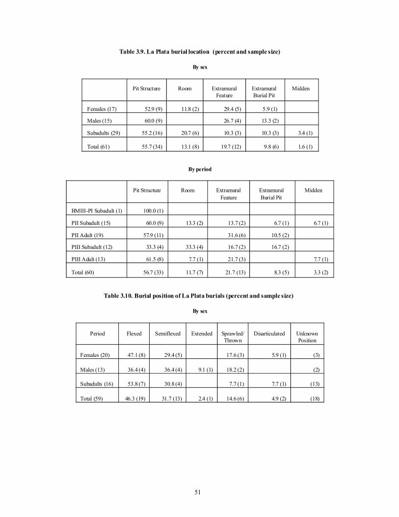
51
Table 3.9. La Plata burial location (percent and sample size)
By sex
Pit Structure Room ExtramuralFeature
ExtramuralBurial Pit
Midden
Females (17) 52.9 (9) 11.8 (2) 29.4 (5) 5.9 (1)
Males (15) 60.0 (9) 26.7 (4) 13.3 (2)
Subadults (29) 55.2 (16) 20.7 (6) 10.3 (3) 10.3 (3) 3.4 (1)
Total (61) 55.7 (34) 13.1 (8) 19.7 (12) 9.8 (6) 1.6 (1)
By period
Pit Structure Room ExtramuralFeature
ExtramuralBurial Pit
Midden
BMIII-PI Subadult (1) 100.0 (1)
PII Subadult (15) 60.0 (9) 13.3 (2) 13.7 (2) 6.7 (1) 6.7 (1)
PII Adult (19) 57.9 (11) 31.6 (6) 10.5 (2)
PIII Subadult (12) 33.3 (4) 33.3 (4) 16.7 (2) 16.7 (2)
PIII Adult (13) 61.5 (8) 7.7 (1) 21.7 (3) 7.7 (1)
Total (60) 56.7 (33) 11.7 (7) 21.7 (13) 8.3 (5) 3.3 (2)
Table 3.10. Burial position of La Plata burials (percent and sample size)
By sex
Period Flexed Semiflexed Extended Sprawled/Thrown
Disarticulated UnknownPosition
Females (20) 47.1 (8) 29.4 (5) 17.6 (3) 5.9 (1) (3)
Males (13) 36.4 (4) 36.4 (4) 9.1 (1) 18.2 (2) (2)
Subadults (16) 53.8 (7) 30.8 (4) 7.7 (1) 7.7 (1) (13)
Total (59) 46.3 (19) 31.7 (13) 2.4 (1) 14.6 (6) 4.9 (2) (18)

52
By period
Period Flexed Semiflexed Extended Sprawled/Thrown
Disarticulated UnknownPosition
BMIII-PI Subadult (1) 100.0 (1)
Pueblo II Subadult (17) 50.0 (4) 25.0 (2) 25.0 (2) (9)
Pueblo II Adult (19) 31.2 (5) 43.8 (7) 6.2 (1) 6.2 (1) 12.5 (2) (3)
Pueblo III Subadult (13) 85.7 (6) 14.3 (1) (6)
Pueblo III Adult (12) 44.4 (4) 33.3 (3) 22.2 (2) (3)
Total (62) 46.3 (19) 31.7 (13) 2.6 (1) 15.4 (6) 4.9 (2) (21)
Percentages do not include cases with unknown position.
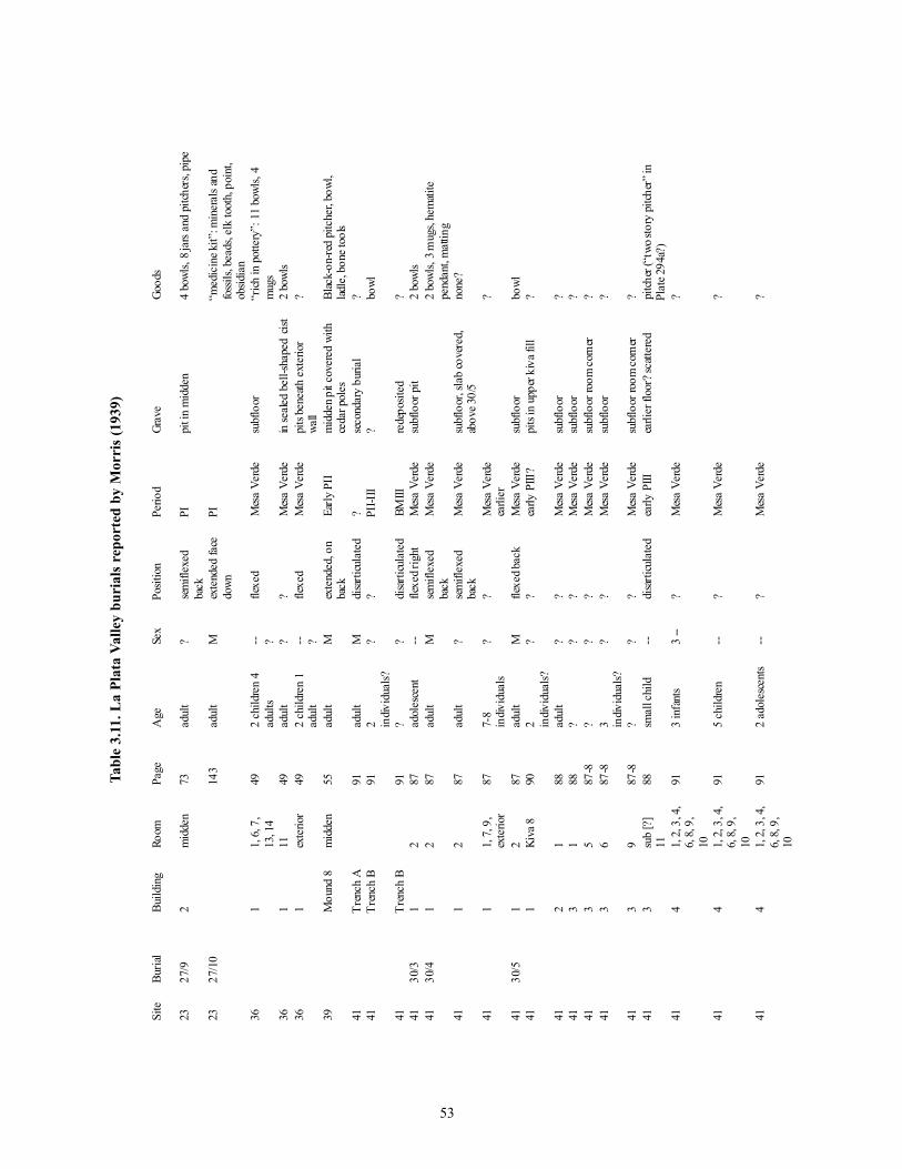
53
Site
Buria
lBu
ildin
gRo
omPa
geA
geSe
xPo
sitio
nPe
riod
Grav
eGo
ods
2327
/92
mid
den
73ad
ult
?se
mifl
exed
back
PIpi
t in
mid
den
4 bo
wls,
8 jar
s and
pitc
hers
, pip
e
2327
/10
143
adul
tM
exte
nded
face
down
PI“m
edic
ine k
it”: m
iner
als a
ndfo
ssils
, bea
ds, e
lk to
oth,
poi
nt,
obsid
ian
361
1, 6,
7,
13, 1
449
2 ch
ildre
n 4
adul
ts -- ?
flexe
dM
esa
Verd
esu
bflo
or“r
ich in
pot
tery
”: 1
1 bo
wls,
4m
ugs
361
1149
adul
t?
?M
esa
Verd
ein
seale
d be
ll-sh
aped
cist
2 bo
wls
361
exte
rior
492
child
ren
1ad
ult
-- ?fle
xed
Mes
a Ve
rde
pits
bene
ath ex
terio
rwa
ll?
39M
ound
8m
idde
n55
adul
tM
exte
nded
, on
back
Early
PII
mid
den p
it co
vere
d wi
thce
dar p
oles
Blac
k-on
-red
pitc
her,
bowl
,lad
le, b
one t
ools
41Tr
ench
A91
adul
tM
disa
rticu
late
d?
seco
ndar
y bu
rial
?41
Tren
ch B
912 in
divid
uals?
??
PII-I
II?
bowl
41Tr
ench
B91
??
disa
rticu
late
dBM
IIIre
depo
sited
?41
30/3
12
87ad
oles
cent
--fle
xed r
ight
Mes
a Ve
rde
subf
loor
pit
2 bo
wls
4130
/41
287
adul
tM
sem
iflex
edba
ckM
esa
Verd
e2
bowl
s, 3 m
ugs,
hem
atite
pend
ant,
matt
ing
411
287
adul
t?
sem
iflex
edba
ckM
esa
Verd
esu
bflo
or, s
lab
cove
red,
abov
e 30/
5no
ne?
411
1, 7,
9,
exte
rior
877-
8in
divid
uals
??
Mes
a Ve
rde
earli
er?
4130
/51
287
adul
tM
flexe
d bac
kM
esa
Verd
esu
bflo
orbo
wl41
1Ki
va 8
902 in
divid
uals?
??
early
PIII
?pi
ts in
upp
er k
iva f
ill?
412
188
adul
t?
?M
esa
Verd
esu
bflo
or?
413
188
??
?M
esa
Verd
esu
bflo
or?
413
587
-8?
??
Mes
a Ve
rde
subf
loor
room
corn
er?
413
687
-83 in
divid
uals?
??
Mes
a Ve
rde
subf
loor
?
413
987
-8?
??
Mes
a Ve
rde
subf
loor
room
corn
er?
413
sub
[?]
1188
smal
l chi
ld--
disa
rticu
late
dea
rly P
IIIea
rlier
floo
r? sc
atter
edpi
tcher
(“tw
o sto
ry p
itche
r” in
Plat
e 294
a?)
414
1, 2,
3, 4
,6,
8, 9
,10
913
infa
nts
3 --
?M
esa
Verd
e?
414
1, 2,
3, 4
,6,
8, 9
,10
915
child
ren
--?
Mes
a Ve
rde
?
414
1, 2,
3, 4
,6,
8, 9
,10
912
adol
esce
nts
--?
Mes
a Ve
rde
?
Tabl
e 3.
11. L
a Pl
ata
Valle
y bu
rial
s rep
orte
d by
Mor
ris (
1939
)

54
Site
Buria
lBu
ildin
gRo
omPa
geA
geSe
xPo
sitio
nPe
riod
Grav
eGo
ods
414
1, 2,
3, 4
,6,
8, 9
,10
9110
adul
ts?
?M
esa
Verd
e?
4130
/27
43
91ch
ild, 2
adul
ts?
?M
esa
Verd
eon
floo
r3
bowl
s, lad
le, m
attin
g
4130
/37
(1)
42
92ad
ult
Mfle
xed
Mes
a Ve
rde
subf
loor
cist
with
slab
cove
rm
ug, m
iniat
ure m
ug, 2
min
iatur
ebo
wls,
30 ar
rows
(4 st
one
poin
ts), b
ow, m
attin
g41
30/3
7 (2
)4
292
adol
esce
ntF
flexe
dM
esa
Verd
ePl
aced
on
top
of 3
0/37
(1)
4145
, 48,
49,
50, 5
2, 5
3Tr
ench
C91
6 indiv
idua
ls??
?PI
I-III
?“f
orei
gn”
seed
jar +
?
4130
/54
G (4
-5)
95?
??
PIm
idde
n?
4130
/55
G (4
-5)
95?
??
PII
mid
den
?41
30/5
6G
(4-5
)95
??
?PI
Im
idde
n?
4130
/62
62
96?
??
Mes
a Ve
rde
subf
loor
pit
?41
30/6
36
refu
se96
, 127
adul
tM
flexe
d, rig
htBM
IIIpi
t in
mid
den;
cove
red
with
cobb
lespo
t with
chip
ped s
tone
, 3 p
oint
s,pl
ain ja
r, 2
pain
ted
bowl
s, aw
l,co
ncre
tion,
crin
oid,
sele
nite
stone
, bow
l with
pig
men
t41
61,
2, 3
, 495
child
--?
Mes
a Ve
rde
floor
leve
l?
416
1, 2,
3, 4
954
adul
ts?
?M
esa
Verd
eflo
or p
its, a
nd fl
oor l
evel
?41
62 s
ub[et
c?]
951
infa
nt 2
child
ren
1ad
oles
cent
3ad
ults
4--3
??
BMIII
in an
d be
neath
mid
den
jar
4130
/68
513
93ad
ult
Mfle
xed l
eft
Mes
a Ve
rde
slab
cove
red,
walle
dsu
bflo
or ci
stbo
wl,2
pitc
hers
, lad
le, d
iggi
ngsti
ck, 2
0 ar
rows
(4 st
one p
oint
s),bo
w, m
attin
g, be
ddin
g41
30/6
95
1393
adul
t?
flexe
d rig
htPI
Ise
para
ted b
y th
in sl
abs
from
30/6
8bo
wl, l
adle
, man
os, m
attin
g
415
1, 2,
3,
2693
3 ch
ildre
n--
?PI
I-III
pits
?
415
1, 2,
3,
2693
5 ad
ults
??
PII-I
IIpi
ts?
4130
/70
(1)
74
97ad
ult
Fse
mifl
exed
left
pre-
Mes
aVe
rde?
slab
cove
red,
aban
done
dsto
rage
pit
pitch
er, 2
bow
ls, la
dle,
mug
,tw
ine,
blan
kets,
matt
ing
4130
/70
(2)
74
97in
fant
--se
mifl
exed
left
pre-
Mes
aVe
rde
with
30/
70 (1
)se
e 30
/70
(1)
4130
/71
(1)
75
9816
-18
--fa
ce do
wn,
crow
ded
Mes
a Ve
rde
slab
cove
red,
uprig
htsla
b re
taini
ng w
all
bowl
, mug
, plaq
ue, p
eele
d stic
k,sh
eave
s, fir
e dril
l, ha
fted
ham
mer
, matt
ing
4130
/71
(2)
75
9818
?--
face
down
,cr
owde
dM
esa
Verd
esla
b co
vere
d, up
right
slab
retai
ning
wal
lse
e 30
/71
(1)

55
Site
Buria
lBu
ildin
gRo
omPa
geA
geSe
xPo
sitio
nPe
riod
Grav
eGo
ods
4130
/72
75
98sm
all c
hild
--fle
xed r
ight
Mes
a Ve
rde
subf
loor
pit
ladle
, gra
y ja
r41
30/7
37
598
child
--di
sarti
cula
ted
PIII
subf
loor
, muc
h po
rotic
hype
rosto
sisno
ne
4130
/74
78
98ad
ult
?se
mifl
exed
right
Mes
a Ve
rde
subf
loor
pit
bowl
4130
/75
78
98ad
ult
?fle
xed r
ight
Mes
a Ve
rde
subf
loor
pit
bowl
with
horn
ed li
zard
4130
/76
710
99ad
ult
?se
mifl
exed
left
PII-I
IIm
idde
n, un
der w
all
none
4130
/77
711
99ch
ildad
oles
cent
adul
t adu
lt[?
] old
adu
lt
----
F M
Fth
rown
Mes
a Ve
rde
“bot
tle-n
ecke
d” c
ist, n
otfo
rmall
y pl
aced
none
4130
/78
711
99sm
all c
hild
,ad
oles
cent
, 1M
, 1 F
, 1 ol
dad
ult
----
MF?
sem
iflex
edlef
tM
esa
Verd
esc
atter
ed re
mai
ns in
cist
none
4130
/79
713
99ad
ult
?se
mifl
exed
right
Mes
a Ve
rde
slab
cove
red p
it3
bowl
s
4130
/80
713
99in
fant
--fle
xed l
eft
Mes
a Ve
rde
subf
loor
pit
none
4130
/81
713
99in
fant
--fle
xed r
ight
Mes
a Ve
rde
subf
loor
pit
none
4130
/82
713
99ad
ult
?fle
xed r
ight
Mes
a Ve
rde
slab-
cove
red
subf
loor
pit
2 bo
wls
4130
/83
7ki
vain
terio
r99
adul
tF?
sem
iext
ende
dlef
tPI
I-III
pit b
enea
th in
terst
itial
fill
bowl
, gra
y jar
sher
d gro
und
into
bowl
4130
/84
(1)
7ki
vain
terio
r99
adul
t?
flexe
d rig
htPI
I-III
pit b
enea
th in
terst
itial
fill
none
4130
/84
(2)
7ki
vain
terio
r99
adul
t?
flexe
d rig
htPI
I-III
pit b
enea
th in
terst
itial
fill
none
4130
/85
716
100
adul
t?
flexe
d lef
tM
esa
Verd
esla
b-co
vere
d su
bflo
or,
subw
all p
itno
ne
4130
/86
716
100
smal
l chi
ld--
sem
iflex
edlef
tM
esa
Verd
esla
b-co
vere
d pit
none
4130
/87
718
100
adul
tF
sem
iflex
edrig
htM
esa
Verd
esu
bflo
or p
itbo
wl, c
ante
en
4130
/88
718
100
adul
t?
disa
rticu
late
dM
esa
Verd
eirr
egul
ar p
it; p
artia
l,sc
atter
edno
ne?
4130
/89
(1)
718
100
adul
t?
sem
iflex
edlef
tM
esa
Verd
epi
t no
ne?
4130
/89
(2)
718
100
adol
esce
nt?
flexe
d rig
htM
esa
Verd
epi
t; on
top
of 3
0/89
(1)
none
?41
30/9
07
1810
0ad
ult
?di
sturb
edM
esa
Verd
epi
t; di
sturb
ed, p
artia
lno
ne?
4130
/91
718
100
child
--fle
xed l
eft
Mes
a Ve
rde
slab-
cove
red,
fille
d ci
stpa
rtial
olla
4130
/92
718
100
infa
nt--
?M
esa
Verd
esu
bwal
l and
floo
r pit
none
4130
/93
718
100
smal
l chi
ld--
flexe
d lef
tM
esa
Verd
esu
bflo
or p
it ne
xt to
subf
loor
ves
sel
ladle
, par
tial g
ray
pot
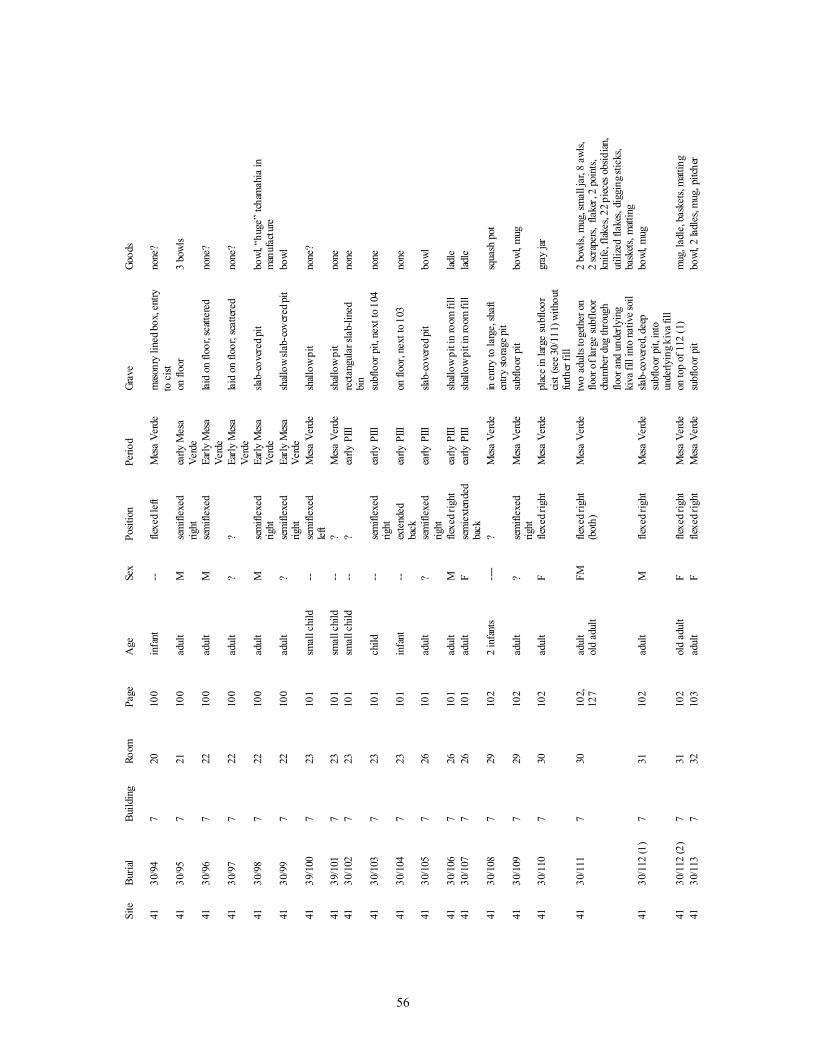
56
Site
Buria
lBu
ildin
gRo
omPa
geA
geSe
xPo
sitio
nPe
riod
Grav
eGo
ods
4130
/94
720
100
infa
nt--
flexe
d lef
tM
esa
Verd
em
ason
ry li
ned b
ox, e
ntry
to c
istno
ne?
4130
/95
721
100
adul
tM
sem
iflex
edrig
htea
rly M
esa
Verd
eon
floo
r3
bowl
s
4130
/96
722
100
adul
tM
sem
iflex
edEa
rly M
esa
Verd
elai
d on
floo
r, sc
atter
edno
ne?
4130
/97
722
100
adul
t?
?Ea
rly M
esa
Verd
elai
d on
floo
r; sc
atter
edno
ne?
4130
/98
722
100
adul
tM
sem
iflex
edrig
htEa
rly M
esa
Verd
esla
b-co
vere
d pit
bowl
, “hu
ge”
tcham
ahia
inm
anuf
actu
re41
30/9
97
2210
0ad
ult
?se
mifl
exed
right
Early
Mes
aVe
rde
shall
ow sl
ab-c
over
ed pi
tbo
wl
4139
/100
723
101
smal
l chi
ld--
sem
iflex
edlef
tM
esa
Verd
esh
allow
pit
none
?
4139
/101
723
101
smal
l chi
ld--
?M
esa
Verd
esh
allow
pit
none
4130
/102
723
101
smal
l chi
ld--
?ea
rly P
IIIre
ctang
ular
slab
-line
dbi
nno
ne
4130
/103
723
101
child
--se
mifl
exed
right
early
PIII
subf
loor
pit,
nex
t to 1
04no
ne
4130
/104
723
101
infa
nt--
exte
nded
back
early
PIII
on fl
oor,
next
to 1
03no
ne
4130
/105
726
101
adul
t?
sem
iflex
edrig
htea
rly P
IIIsla
b-co
vere
d pit
bowl
4130
/106
726
101
adul
tM
flexe
d rig
htea
rly P
IIIsh
allow
pit
in ro
om fi
lllad
le41
30/1
077
2610
1ad
ult
Fse
mie
xten
ded
back
early
PIII
shall
ow p
it in
room
fill
ladle
4130
/108
729
102
2 in
fant
s---
-?
Mes
a Ve
rde
in en
try to
larg
e, sh
aft
entry
stor
age p
itsq
uash
pot
4130
/109
729
102
adul
t?
sem
iflex
edrig
htM
esa
Verd
esu
bflo
or p
itbo
wl, m
ug
4130
/110
730
102
adul
tF
flexe
d rig
htM
esa
Verd
epl
ace i
n lar
ge su
bflo
orcis
t (se
e 30/
111)
with
out
furth
er fi
ll
gray
jar
4130
/111
730
102,
127
adul
tol
d ad
ult
FMfle
xed r
ight
(bot
h)M
esa
Verd
etw
o ad
ults
toge
ther
on
floor
of l
arge
sub
floor
cham
ber d
ug th
roug
hflo
or an
d un
derly
ing
kiva
fill
into
nat
ive s
oil
2 bo
wls,
mug
, sm
all j
ar, 8
awls,
2 sc
rape
rs, f
lake
r, 2
poin
ts,kn
ife, f
lakes
, 22 p
ieces
obs
idian
,ut
ilize
d fla
kes,
digg
ing s
ticks
,ba
skets
, matt
ing
4130
/112
(1)
731
102
adul
tM
flexe
d rig
htM
esa
Verd
esla
b-co
vere
d, de
epsu
bflo
or p
it, in
toun
derly
ing k
iva f
ill
bowl
, mug
4130
/112
(2)
731
102
old
adul
tF
flexe
d rig
htM
esa
Verd
eon
top
of 1
12 (1
)m
ug, l
adle
, bas
kets,
matt
ing
4130
/113
732
103
adul
tF
flexe
d rig
htM
esa
Verd
esu
bflo
or p
itbo
wl, 2
ladl
es, m
ug, p
itche
r

57
Site
Buria
lBu
ildin
gRo
omPa
geA
geSe
xPo
sitio
nPe
riod
Grav
eGo
ods
4130
/127
3Ki
va 9
90ol
d ad
ult
Ffle
xed
Mes
a Ve
rde
pit i
n fil
l abo
ve n
orth
pilas
ter;
slab
cove
red
bowl
, gra
y jar
, she
rd, b
lanke
t
418
710
3in
fant
--?
early
PIII
slab-
lined
bin
?41
86
103
adul
t?
?ea
rly P
III?
?41
88
103
adul
t?
?M
esa
Verd
e?
mug
? ves
sel,
seed
jar
418
sub
3-4
104
13 ad
ults
??
PII-I
II;pr
edom
inan
tly P
II
pits
in m
idde
n“l
ittle
potte
ry”:
14 v
esse
l pla
tesin
cludi
ng b
owls
and
ladl
es, a
ndpi
tcher
418
sub
3-4
104
2 ch
ildre
n--
?PI
I-III
pits
in m
idde
nse
e ab
ove
4130
/131
8-9?
127
??
?PI
II?
knife
, cor
es, f
lakes
+?
4130
/132
94
104
smal
l chi
ld--
flexe
d lef
tM
esa
Verd
esu
bflo
or; s
lab su
ppor
ted
by d
iggi
ng st
ick
bask
et-m
olde
d M
esa
Verd
ebo
wl, b
owl,
blan
ket,
matt
ing
4111
104
infa
nt--
?M
esa
Verd
e?su
bflo
or?
4111
104
adul
t?
?M
esa
Verd
e?su
bflo
or?
4113
210
52
child
ren
--pr
epar
edM
esa
Verd
epl
aced
on f
loor
, not
burie
d2
bowl
s, m
ug, b
lack
-on-
white
jar, s
eed
jar41
132
105
8 ad
ults
?pr
epar
edM
esa
Verd
epl
aced
on f
loor
, not
burie
dse
e ab
ove
4114
sub
510
5ad
ult
??
PII
mid
den
2 bo
wls
4114
610
52
infa
nts
--di
sarti
cula
ted
Mes
a Ve
rde
subf
loor
none
?41
14su
b 3-
410
54
adul
ts?
disa
rticu
late
dPI
Idi
sarti
cula
ted,
proc
esse
dun
finish
ed bo
wl?
4114
sub
3-4
105
adol
esce
nt--
disa
rticu
late
dPI
Ilim
bs on
lyun
finish
ed bo
wl?
4114
sub
3-4
105
smal
l chi
ld--
disa
rticu
late
dPI
Ipa
rtial
unfin
ished
bowl
?41
15su
b 6
106
child
--?
early
PIII
subf
loor
pit
into
earli
erstr
uctu
repa
rtial
jar
4115
1210
6ad
ult
??
Mes
a Ve
rde
deep
sub
floor
pit
2 sm
all g
ray
jars,
small
bow
l,bo
wl?
mug
, 2 ir
on o
re cy
linde
rs41
16su
b 6-
810
8ad
ult
??
pre-
Mes
aVe
rde
mid
den
?
4116
sub
1010
8ch
ild--
?pr
e-M
esa
Verd
em
idde
n?
4116
sub
2110
8ad
ult
??
pre-
Mes
aVe
rde
mid
den
?
Total
s23
2 ad
ults
1 M1?
2 PI
364
child
ren
6 ad
ults
4 --
6?10
Mes
a Ve
rde
391
adul
tM
Early
PII

58
Site
Buria
lBu
ildin
gRo
omPa
geA
geSe
xPo
sitio
nPe
riod
Grav
eGo
ods
4116
infa
nts
30 ch
ildre
n11
adol
esce
nts
95 ad
ults
5 ol
d ad
ults
30?
= 1
87
55 su
badu
lts14
M13
F10
2?
= 18
4
9 BM
III1 P
I3 P
II35
PII-
III15
early
PIII
4 PIII
113
Mes
a Ver
de6 p
re-la
te P
III1? =
187
Sum
mar
y: "
The
only
con
spic
uous
cha
nge
in b
uria
l cus
tom
s du
ring
the
entir
e cy
cle
of o
ccup
atio
n w
as, t
hen,
from
ext
ram
ural
to in
tram
ural
inte
rmen
t in
the
Mes
a Ve
rde
phas
e of
Pue
blo
III"
(Mor
ris 1
939:
115)
. Bur
ials
des
crib
ed a
s "l
oose
ly fl
exed
" ha
ve b
een
ente
red
here
as
sem
iflex
ed, a
s ha
ve b
uria
ls w
ith b
ent b
ut n
ot ti
ghtly
flex
ed li
mbs
. Sem
iext
ende
din
dica
tes
mor
eex
tend
ed th
an fl
exed
but
with
som
e be
nt li
mbs
. Mes
a Ve
rde
indi
cate
s the
pre
senc
e of
Mes
a Ve
rde
Bla
ck-o
n-w
hite
. Ear
ly P
III i
nclu
des c
arbo
n-on
-whi
te v
esse
ls si
mila
r to
McE
lmo
Bla
ck-
on-w
hite
and
min
eral
-pai
nted
ves
sels
that
we
wou
ld p
lace
in la
te P
uebl
o II
. We
wou
ld a
lso
plac
e so
me
of M
orris
's B
MII
I in
Pueb
lo I.

59
Table 3.12. Mesa Verde burials (A.D. 900-1300)
Mug HouseAdobe Cave
Big JuniperHouse
BadgerHouse
LongHouse
Total
Number of Burials 46 23 33 39 141
Age Range
Adult 39.1% 52.1% 63.6% 53.8% 51.0%
Subadult (under 15) 60.8% 47.8% 36.3% 46.1% 48.9%
Position
Flexed 30.4% 21.7% 18.1% 56.4% 33.3%
Semiflexed 26.0% 39.1% 36.3% 5.1% 24.8%
Extended 6.5% 4.3% 0 0 2.8%
Unknown 36.9% 34.7% 45.4% 38.4% 39.0%
Burials with Grave Goods 39.1% 69.5% 78.7% 53.8% 57.4%
Adapted from Cattanach (1980), Hayes and Lancaster (1975), Rohn (1971), and Swannack (1969).
Table 3.13. Chaco Canyon burials
Ceramic Period Early Red Mesa/ RedMesa (A.D. 900-1050)
Gallup(A.D. 1030-1150)
McElmo(A.D. 1100-1175)
Mesa Verde(A.D. 1175-1300)
Number of Burials 48 66 41 11
Age Range
Adult 47.9% 54.5% 56.0% 45.4%
Subadult (under 15) 18.7% 34.8% 43.9% 54.5%
Unknown 33.3% 10.6% 0 0
Location
Room 25.0% 51.5% 73.1% 100%
Midden 75.0% 43.9% 12.1% 0
Miscellaneous 0 4.5% 14.6% 0
Position
Flexed 10.4% 39.3% 21.9% 9.1%
Semiflexed 50.0% 40.9% 36.5% 36.3%
Extended 18.7% 18.1% 14.6% 9.1%
Unknown 20.8% 1.5% 26.8% 45.4%
Adapted from Akins (1986).

60
Table 3.14. La Plata burial goods ( percents and sample size)
None Small (1) Moderate (2-3) Extensive (4+)
Subadults (28) 46.4 (13) 35.7 (10) 14.3 (4) 3.6 (1)
Males (14) 42.8 (6) 21.4 (3) 21.4 (3) 14.3 (2)
Females (16) 31.2 (5) 12.5 (2) 37.5 (6) 18.8 (3)
Totals (58) 41.4 (24) 27.6 (16) 24.1 (14) 10.3 (6)
X2= 7.839, 6 d.f., p=.250 (expected values less than 5 in over half of the cells)
Table 3.15. Distribution of age-at-death (n=67)
Age Number Breakdown by Sex
Fetal 2
nb - .9 2
1 - 4.9 13
5 - 9.9 7
10 - 14.9 3
15 - 19.9
Total
3
30
Males (16) Females (18)
20 - 24.9 5 2 3
25 - 29.9 7 3 4
30 - 34.9 4 2 2
35 - 39.9 4 1 3
40 - 44.9 1 1 0
45 - 49.9 6 5 1
50 4 2 1
Unknown
Total
6
37
15 - 19.9 3
Unknown 6

61
Table 3.16. Summary by age and sex
LANo.
0 -3y
4 -8 y
9 -18y
19-25 26-35 36-45 46+ Adult Totals
& % & % & % & % & ? All & % Sub-adults
37592 3 2 1 1 1 8 2 6
37593 1 1 1 1 4 1 1 2
37594 1 1 1
37595 1 1 2 1 1
37598 1 1 1
37599 2 1 1 2 1 1 2 10 1 4 3
37600 1 1 2 1 1 6 2 2 1
37601 1 2 2 1 3 1 1 1 12 4 5 3
37603 1 1 1 3 1 1 1
37605 1 2 3 3
65029 1 1 1
65030 5 2 3 1 2 1 1 1 16 7 2 7
Totals 15 7 7 7 4 4 4 3 4 1 4 4 3 67 19 16 29
Table 3.17. Age composition of New World prehistoric sites (percent)
Population (N) Age
<1 1-9 10-18 >18
La Plata (67) 6.0 29.8 7.5 56.7
Mesa Verde Early (150) 10.6 18.0 14.0 57.3
Mesa Verde Late (178) 16.8 18.5 23.5 48.6
Pueblo Bonito (93) 1.0 16.1 17.2 40.4
Black Mesa (165) 10.4 24.2 14.5 50.9
Casas Grandes (612) 10.0 22.0 14.0 54.0
Pecos Pueblo (1722) 18.7 14.0 8.0 59.0
Tlajinga, Mexico (166) 41.3 10.3 10.6 38.6
Arikara Villages (1487) 31.5 24.0 9.5 45.5
Libben, Ohio (1239) 18.0 22.0 14.0 46.0
Compiled from Martin et al. (1991), Storey (1988), and Nelson et al. (1994).

62
Table 3.18. Life table: La Plata burial population
x Dx dx lx qx Lx Tx e0x
0-.9 4 60.6 1000.0 0.0606 969.7 24539.4 24.54
1-4.9 13 197.0 939.4 0.2097 3878.8 23569.7 25.09
5-9.9 6 90.9 742.4 0.1255 4204.5 19690.9 26.52
10-14.9 3 45.5 651.5 0.0698 3484.7 15486.4 23.77
15-19.9 3 45.5 606.0 0.0751 2916.2 12001.7 19.80
20-24.9 5.97 90.5 560.5 0.1615 2576.2 9085.5 16.21
25-29.9 8.35 126.5 470.0 0.2684 2033.7 6509.3 13.85
30-34.9 4.77 72.3 343.5 0.2105 1536.7 4475.6 13.03
35-39.9 4.77 72.3 271.2 0.2666 1175.2 2938.9 10.84
40-44.9 1.19 18.0 198.9 0.0905 949.5 1763.7 8.87
45-49.9 7.16 108.5 180.9 0.5998 633.2 814.2 4.50
50-54.9 4.77 72.3 72.4 1.0000 181.0 181.0 2.50
x = age intervalDx = number of skeletons with ages within the limits of xlx = survivorship, or percentage of sample entering xqx = probability of dying during interval xLx = total number of years lived by all individuals during xTx = number of years remaining in lifetimes of all entering xe0
x = average life expectancy in years of individuals entering x

63
Table 3.19. Life expectancy at birth values for Southwest series
Site e00 Date Designation
Pueblo Bonito (95) 26.3 PII Anasazi
Kayenta Region (38) 26.2 PII Anasazi
Chaco Basin (91) 25.6 PI-II Anasazi
Black Mesa (165) 25.1 PI-II Anasazi
La Plata (66) 24.5 PII-III Anasazi
Point of Pines (508) 22.9 PII/III-IV Mogollon
Mesa Verde Early (150) 21.2 BMIII-PI/II Anasazi
Casas Grandes (612) 20.2 PIII-IV Northern Mexico
Mesa Verde Late (178) 18.3 PII-III Anasazi
San Cristobal (271) 18.6 PIV-Historic Pueblo
Transwestern (37) 17.6 PII Anasazi
Hawikku (191) 17.1 PIV-Historic Pueblo
Navajo Reservoir (166) 16.8 PI-II Anasazi
Arroyo Hondo (108) 16.2 PIV Pueblo
Grasshopper (614) 14.2 PIV Mogollon
Salmon Ruin (111) 12.7 PII-III Anasazi
Total samples size is in parentheses. Adapted from Berry (1985), Herrmann (1993), Martin et al. (1991), Ryan (1977), Stodder(1990), and Nelson et al. (1994).
Note: e00 was recalculated for Black Mesa, Mesa Verde, Casas Grandes, Pueblo Bonito, and Arroyo Hondo (see Nelson et al.
1994).
Table 3.19. Life expectancy at birth values for Southwest series

64
Table 3.20. Ranked comparisons of mortality and fertility estimates
Mortality
Ranked from Low to High Mean Age at Death (e 00)
1. Arroyo Hondo 16.2
2. Mesa Verde Late 18.3
3. Casas Grandes 20.2
4. Mesa Verde Early 21.2
5. La Plata 24.5
6. Black Mesa 25.1
7. Pueblo Bonito 26.3
Fertility (ratio = deaths 30 $/ deaths 5$)
Ranked from High to Low Birthrate
1. Mesa Verde Late .3212
2. Casas Grandes .3318
3. Mesa Verde Early .3496
4. La Plata .4318
5. Pueblo Bonito .4419
6. Arroyo Hondo .5424
7. Black Mesa .576
Adapted from Nelson et al. (1994:103).

CHAPTER 4
HEALTH PROFILE OF THE LA PLATAVALLEY COMMUNITIES
As with demographic variables, the health status ofsouthwestern precontact societies has long been recog-nized as a fundamental aspect of understanding andinterpreting the past. From Matthews et al. (1893),Hrdlicka (1908), and Hooton (1930), we see earlyattempts to characterize the health of the ancient PuebloIndians through their skeletal remains. Readers of thisearly literature are simultaneously impressed that theseresearchers write with such descriptive detail but arealso frustrated by their inability to link their observa-tions in systematic ways with other facets of archaeo-logical reconstruction. The impressions recorded aboutthe biological characteristics of ancient Pueblo peoplewere often a blend of subjective and personal ideasregarding how they lived and why they died (e.g.,Colton 1960; Cummings 1940, 1953; Fewkes 1904).Most of the discussions are cast in a rosy glow about thenoble ancient ones who have survived the slings andarrows of outrageous fortune, or there is a tone thatimplies that these people were unenlightened and back-ward in their knowledge of sanitation and health care.The focus of many of the reports on health and diseasecontinues to be tabulation of medical anomalies andpathologies for individual skeletons (e.g., Bennet 1966,1973; 1975; Berry 1983; Hrdlicka 1935; Miles 1966,1975; Nickens 1975; Reed 1946, 1965).
It is tempting to interpret signs of hardship directlyfrom the skeleton. It is unfortunate that the finding ofdisease and early death leads some to believe that thelives of ancient people were ruled by pain and sorrow.Caution must be used in so doing (Dettwyler 1991),because just as we cannot say that skeletons withoutpathological lesions were healthy (and therefore happy,fortunate, etc.), we cannot assume that severe skeletalpathology necessarily caused disability and pain.1
There are, in fact, hundreds of health problems thatdo not leave signatures on the skeletal system, but manyof these are rare or unusual diseases that primarily affectindividuals over 65 or are diseases not believed to havebeen present in precontact New World individuals(Kiple 1993). It is fortunate for us that common andhighly prevalent microorganisms that cause illness doinitiate changes in the morphology of bone tissue, suchas anemia, staph and strep infections, respiratory ail-ments, gastrointestinal problems, and dysentery(Steinbock 1976; Ortner and Putschar 1981). These areprecisely the conditions that contemporary populationsliving in marginalized and Third World settings die from
in high frequencies (Allen 1984; Dyson 1984;Leatherman 1987). In general, acute or epidemic condi-tions do not leave evidence in bone (Ortner and Putschar1981). Lesions that do affect bone are primarily fromchronic conditions. We argue that it is the endemic, day-to-day stresses that are important to document, for thesereveal more about the environmental and cultural fac-tors that people must deal with on a regular basis.
Patterns of death and disease are not random occur-rences. They are intimately linked to every facet oflifestyle from diet and climate to occupation, socialstructure, and religion (Wells 1964:87). Although deathis the ultimate indicator of maladaptation, its timing andpatterning within populations reveals a variety of chal-lenges in the physical and social environment. Somehave criticized the ability of skeletal biologists to makeuseful inferences from paleodemographic and pale-opathological data (Wood et al. 1992), but Goodman(1993) has addressed and rebutted many of these. Hepoints out that skeletal biologists focus on multiple indi-cators of stress, which, when combined with an under-standing of the ecological and cultural context (see Fig.1.1), can be used to construct health profiles thatapproach biological realities.
The principal intent of this study is to establishhealth parameters for the people living in the La PlataValley. The small size of the sample limits an extensivestatistical study.2 In this study we have been able to con-firm poor health among subgroups within the popula-tion. Also, we can begin to locate places where definitesigns of biological afflictions may be linked to culturalpractices. Categories of skeletal manifestations of dis-ease are discussed with an eye toward establishing pat-terns within the population and noting any trends inpoor health through time or by community (JacksonLake and Barker Arroyo). When possible, comparisonsto other skeletal populations are made.
POROTIC HYPEROSTOSIS
The history, background, and methodology of thestudy of porotic hyperostosis is reviewed in Chapter 2.The results of the analysis presented here suggest thatiron deficiency anemia and its resultant health compli-cations were ubiquitous but not a severe health problem.For the subadult population as a whole (aged newbornthrough fifteen), over half (57.9 percent) of the individ-uals who could be scored demonstrate cranial involve-ment (Table 4.1). Of the eleven subadults with lesions,four (36.4 percent) were scored as slight in expression,and seven (63.6 percent) as moderate. None of the LaPlata children had lesions that could be characterized assevere in expression. All of the cases of porotic hyper-ostosis in the subadult population were either healing or
65

healed at the time of death. The majority of the cases ofporotic hyperostosis (63.6 percent) are modally distrib-uted in the age category 1.5 through 3 (although this ismost likely a function of poor representation in theyounger age categories).
By geographic locale, the 19 subadults that couldbe scored for porotic hyperostosis at Barker Arroyoinclude nine individuals (47.4 percent) who have lesionsand six (31.6 percent) that do not. At Jackson Lake, two(50 percent) of the four have lesions. Temporal trendsare difficult to assess, but there does not appear to be aclustering of cases of porotic hyperostosis by period (PIIversus PII-III). Likewise, severity (slight versus moder-ate) or status of the lesions (active versus remodeled)does not appear to be associated with a particular tem-poral unit, nor do cases appear to change in patterningover time.
In the adult portion of the sample (Tables 4.2 and4.3), anemia appears to have been an equal liability formales and females, suggesting that it is a function ofshared dietary and lifestyle activities. In the 11 femalesthat could be scored for porotic hyperostosis, 4 (36.4percent) were slight in expression, with signs of healing.Of the 12 males that could be scored, again 4 (33.3 per-cent) showed lesions, equally divided between slightand moderate in expression, all cases demonstratinghealing at the time of death. Thus for the total adult pop-ulation, 45.2 percent were affected by an anemic condi-tion. (Of the three adults that could not be assigned asex, none could be scored for lesions due to poor preser-vation and missing crania.)
The small number of adults recovered from theJackson Lake sites makes comparisons highly problem-atic. Four adults (2 female and 2 male) from JacksonLake show no lesions on the cranial portions. Thus, allof the cases of anemia are in the Barker Arroyo sample,42 percent of this sample demonstrating lesions. Notemporal trends could be discerned from the combinedadult sample.
Looking at the combined La Plata Valley popula-tion, the frequency is 45.2 percent (19 cases of anemiaout of 42 individuals that could be assessed). Comparedto groups living approximately the same time in theSouthwest, this is among the lower frequencies reported(Table 4.4). Comparison of childhood frequencies ofporotic hyperostosis show that the La Plata Valley sam-ple is among the lowest. Although these comparisonssuggest that La Plata Valley individuals experienced lessof the side effects of iron deficiency anemia than theircounterparts, it is difficult to interpret these data direct-ly. There have been a number attempts to summarize allpublished accounts of porotic hyperostosis forSouthwest archaeological populations, the most notablebeing El-Najjar et al. (1976), Walker (1985), and
Stodder (1987, 1989). Problems exist in the manner thatdata are reported. Often, frequencies are given for casesof porotic hyperostosis in the orbital area (referred to ascribra orbitalia) separately from cases on the cranialvault. It is also problematic that some of the earliestresearch by El-Najjar et al. failed to elucidate the exactmethodology for collecting porotic hyperostosis data.Research into the etiology of the lesion over the last 10years has undoubtedly affected how skeletal biologistscollect data on porotic hyperostosis.3
Taking these problems of comparison into mind, itdoes appear that the La Plata population demonstrateslower frequencies during Pueblo II-III than at someother sites. Danforth et al. (1994) report that 4 out of 5subadults aged two or under (80 percent) at this PuebloIII site in Arizona had lesions at the time of death(although the frequency for the total population is about23 percent). La Plata frequencies are lower than thosereported for other sites, as well as later collections. Forexample, the Black Mesa sample (PI-II) had an overallfrequency of 87 percent, and Canyon de Chelly (PII-III)shows a frequency of 55.1 percent (Walker 1985:143).For sites later in time (San Cristobal and Hawikku,PIV), Stodder (1990:223) reports frequencies of 87 per-cent and 74 percent, respectively.
Interpreting the La Plata Valley data on porotichyperostosis involves a simultaneous understanding of anumber of factors. Early interpretations of porotichyperostosis linked it exclusively to an iron-poor diet(e.g., El-Najjar et al. 1975, 1976), but it is now assumedthat diet per se may play a less prominent role in theexpression of anemia in a population. As summarized byWalker (1985:153): “The remarkable prevalence ofosseous lesions indicative of anemia among prehistoricSouthwest Indians apparently resulted from the interac-tion of a complex set of biological and cultural variablesrelating to nutrition and infectious disease. Lack of ironin the diet, prolonged breast feeding, diarrheal andhelminth infections, and living conditions conducive tothe spread of disease all appear to have contributed tothe prevalence of porotic hyperostosis.”
While it is important to note the iron content offoods likely to have been eaten, it is equally importantto examine how other micronutrients and culinary prac-tices interact with iron in the diet. Maize is itself a ratherpoor source of iron, and the phytic acid in it is primari-ly responsible for inhibiting iron absorption. Phytatesalso inhibit the absorption of zinc, whose interactionswith iron are important (Sandstrom et al. 1987). Phyticacid comprises 1 to 3 percent of all nuts, cereals,legumes, and oil seeds. Because of its chemical struc-ture, it can form chelates with minerals such as calciumand iron. Hurrel et al. (1992) found that reducing phy-tase levels to a specific concentration can increase iron
66

absorption up to five times. Different varieties of cornhave variable concentration of phytase. Hopi cornranges from 0.4 to 2.2 percent, and Kuhnlein et al.(1979) found that the practice of adding culinary ash tothe meal resulted in a calcium-phytase complex that per-mitted greater iron availability. Maize prepared into nix-tamal (or masa) for preparation of tortillas or othermaize foods by being heated in boiling water to whichlime has been added and left overnight to soak increas-es calcium content by over 2,000 percent (Kuhnlein etal. 1979). Other kinds of ash are also beneficial.Mesquite ash can increase calcium content eleven times,and juniper branches, corncobs, and other materialsadded as culinary ash could likewise increase levels ofcalcium, iron, copper, zinc, and strontium in meal pre-pared from maize. Vitamin C, needed to increase ironabsorption, is present in significant amounts in pricklypear and cholla, as well as squash, pumpkin, and ama-ranth.
The complex interaction of micronutrients and ironabsorption are only a part of the picture. There is oftena synergistic effect between dietary deficiencies andsusceptibility to transmissible infectious diseases. At LaPlata, the co-occurrence of porotic hyperostosis insubadults with periosteal reactions (lesions indicative ofsystemic infectious disease) is relatively frequent (Table4.1). Out of 13 cases in which both cranial and postcra-nial material were available to assess for lesions ofporotic hyperostosis and periosteal reaction, seven (53.8percent) children have lesions. Of these, three (23.1 per-cent) have either porotic hyperostosis or periosteal reac-tions, while four (30.8 percent) demonstrate both.Evidence suggests that infectious disease finds a more“willing” host in the anemic individual, and Keusch andFarthing (1986:145) demonstrate that iron deficiencyanemia can predispose children to respiratory and gas-trointestinal infections. For the Black Mesa population,co-occurrence was 61.9 percent for the subadults, butthe patterning was quite different than at La Plata. Thecases of co-occurrence show a distinctly bimodal distri-bution at La Plata, with cases at ages 2 and 3, and againat ages 9 and 10, although this may be largely due topoor representation in the youngest age categories.
In a 1966 study of Pueblo Indian children living atAcoma Pueblo in New Mexico, Corbett (1968) demon-strated that there was a high incidence of iron deficien-cy anemia in young children visiting the local medicalclinic. At 15 months, 100 percent of the children hadlower than normal mean corpuscular hemoglobin con-centrations. Rates tapered off to between 40 and 60 per-cent in older children. He also found a co-occurrence ofanemia and respiratory infections in 73 percent of thechildren. These data reflect on findings of iron deficien-cy anemia and infectious disease in precolonial times.
The infant mortality rate in New Mexico during the1960s, around 30 deaths per 1,000 live births, wasshown to be highly associated with low birth weight(less than 1,499 g) (State of New Mexico Public HealthDivision 1983:5). The leading cause of death after con-genital anomalies for children under the age of one was,and continues to be, influenza and pneumonia, and sud-den infant death syndrome (SIDS) in Indian infants liv-ing in New Mexico (State of New Mexico Public HealthDivision 1989:42). Thus, the presence of iron deficien-cy anemia in the La Plata Valley sample of children isnot unlike that found recently in Pueblo children in theRio Grande Valley.
The presence of iron deficiency anemia as con-firmed by the presence of porotic hyperostosis suggeststhat iron deficiency anemia was endemic to both com-munities, but more likely it contributed to morbidity andquality of life more so than to mortality. Understandingthe source of the anemia, however, is more difficult. Thelikely preparation of corn into nixtamal and the additionof any kind of ash served to make the iron bioavailableso that the signs of iron deficiency anemia seen in the LaPlata sample may be due to factors other than dietaryinadequacy. Instead of viewing a maize dependent dietas deficient in nutrients and a contributor to poor health,it can be seen as a storable and dependable source ofcarbohydrate, lipids, and protein that was probably ade-quate for most segments of the population. Waste man-agement and proximity to people with transmissibleinfectious diseases may account for more of the anemiathan did the diet.
Regardless of its root cause, iron deficiency in evenslight to moderate rates should be considered a red flag.Iron deficiency anemia is the single most common nutri-tional deficiency in the world, with 25 percent of allinfants and 66 percent of children affected in developingnations today (Wardlaw 1993:458-485). The symptomsof iron deficiency anemia (which do not preserve in thearchaeological record) are pale skin, brittle nails,fatigue, apathy, poor temperature regulation, and loss ofappetite. Learning ability, work performance, andimmune status can be significantly compromised bypoor iron stores. Thus, although we feel that the fre-quencies at La Plata are low to moderate by Southweststandards, we do not want to overlook or underempha-size that any manifestation suggests the potential toaffect the most vulnerable of the group, in this case chil-dren. Combined with other problems, like staph andstrep, the synergistic effect could be deadly.
PERIOSTEAL REACTION AND OTHER INFECTIONS
Lesions resulting from inflammations of the perios-teum are indicative of systemic infectious disease or
67

may result from trauma. Among the subadult portion ofthe La Plata Valley population, 5 of 17 cases in whichthe condition was observable (29.4 percent) demonstrat-ed lesions indicative of generalized nonspecific infec-tion, and one has a trauma- related infection (Table 4.1).Of children with periosteal reactions, the majority of thecases were moderate in expression, and all were activeand/or active with some healing at the time of death. Itis difficult to assess the age of onset of infectious dis-ease in this subgroup because of missing information inthe younger age categories, but two of the five cases ofperiosteal reaction are in children aged 2 and 3, and theother three cases are in children aged 9 and 10. Four outof the five cases co-occur with porotic hyperostosis. Interms of community differences, one of the five cases isin a two-year-old child from Jackson Lake. The othercases are from Barker Arroyo, and these are the childrenaged 3 and 9 to 10. No temporal trends could be dis-cerned.
For the adult population, the overall rate ofperiosteal reactions is 25.0 percent: 7 cases out of 28total observations (Tables 4.2 and 4.3). By sex, there isonly one male (6.6 percent) that demonstrates general-ized lesions and one other that has lesions associatedwith trauma. For females there are five individuals (38.5percent) with generalized lesions, one with lesions in theorbits and on the ribs in an individual with metastaticcancer, and another with a severe case of osteomyelitis.For the generalized lesions, three are slight, and two aremoderate in expression. Likewise, two of the casesdemonstrate active lesions, while three of the casesshow some healing. All of the females with lesions werefrom Barker Arroyo sites.
Comparison of the La Plata Valley sample as awhole with select contemporaneous samples indicatesthat the frequencies are somewhat higher than other bur-ial populations from the area (Table 4.5). One detail thatunderscores the frequency at La Plata is that it is dis-proportionately weighted with adult females. Table 4.3indicates that five females (versus one male) hadlesions, and more importantly, three females had activemanifestations at the time of death.
One female (LA 37601, B4, aged 25) had a severecase of osteomyelitis (Fig. 4.1). The sternum is longitu-dinally convex, and parts of the manubrium are thick-ened with osteophytic reactive bone covering the anteri-or, superior, and lateral surfaces (Fig. 4.2). Some partsof the bone surface appear smooth and rounded, andthese areas were likely caused by lytic lesions and sub-sequent sclerotic processes (see Ortner and Putschar1981:111). Both scapulae have thin ventral margins (asif the epiphysis did not develop properly). The acromionof the right is covered with reactive bone similar to thesternum; the left likewise has damaged surface areas
and reactive lesions (with some cloacal openings).Clavicles show similar lesions. Portions are massivelyremodeled and affected by the same process. The distalportion of one humerus is massively remodeled andaffected by the same process (Fig. 4.3).
Differential diagnosis of this individual (afterexamination of x-rays) suggests osteomyelitis (seeOrtner and Putschar 1981:105-109). The pus-producingbacteria that cause osteomyelitis can reach the skeletonvia direct infection from a traumatic wound, from infec-tions that start in the skin and expand to bone, or by anyvascular route from the point of sepsis. Although wecannot definitely know the original cause of this mas-sive infection confined to the shoulder and chest area,the localized nature of the infection demands that weask what caused it. Possibly, it was the result of a trau-matic injury to that area. The right scapula (near thespine) and the sixth and seventh ribs show rougheneddepressed areas that are reminiscent of trauma in theform of healed fractures. There appear to be localized,trauma-induced osteophytes on the third through thefifth cervical vertebrae. This woman also has a series ofdistinct healed compression fractures on the frontalbone and the left parietal, suggesting that considerableforce was applied with an implement both heavy andhard enough to have split open the cranium in that area.4This individual is discussed further in the section on fre-quencies of trauma for the La Plata group.
Tuberculosis, a chronic infectious disease, waslocated on two individuals. One case came from the bur-ial population discussed here, and one example waslocated in a disarticulated assemblage (discussed inChapter 6). From site LA 37599 B6, a child of aboutnine years of age demonstrated a pattern of lesions andvertebral fusion that suggests tuberculosis. Although thevertebral elements are highly fragmentary and poorlypreserved, evidence of destruction and resorption of thearticular facets and lytic lesions on the body of some ofthe thoracic vertebrae represent a pattern that has beenlinked to tuberculosis. On several of the thoracic verte-brae, the inferior and superior articular facets exhibitresorption and osteolytic destruction. The severity ofdestruction has caused the collapse of the thoracic body,and the exposed cancellous bone is dense and showssclerotic development. The example from LA 65030 (FS510) involves a vertebrae from a subadult found in thefill of Pit Structure 8 with evidence of tuberculosis (Fig.4.4).
Tuberculosis on precontact Southwest specimenshas been largely associated with sites dating to latePueblo III through Pueblo IV (Merbs 1989). However, afew cases have been noted to occur in Pueblo II andPueblo III sites (Chavez Pass, Pueblo Bonito, TalusUnit, and Point of Pines). Stodder (1990) found cases at
68

69
Figure 4.1. Distribution of osteomyelitic lesions. LA 37601, B4.

70
Figure 4.2. Osteomyelitis of the sternum. LA 37601, B4.
Figure 4.3. Osteomyelitic and normal humerus. LA 37601, B4.

both San Cristóbal and Hawikku, and she suggests thatthere was an epidemic wave of tuberculosis, which ishighly contagious. Immuno-compromised individualsare most at risk. The clustering of several cases of tuber-culosis at San Cristóbal suggested to her a highly com-promised group.
Tuberculosis can be considered an index of popula-tion density and lowered immune resistance, and it waspresent in very high frequencies in the historic Pueblogroups encountered by Hrdlicka (1908). Ortner andPutschar (1981) estimate that only a small proportion(less than 10 percent) of individuals with tuberculosisever have symptoms. Thus, presence of two cases at LaPlata suggests that a much higher frequency of individ-uals would have been carriers of the disease.
LINEAR ENAMEL HYPOPLASIAS AND OTHERDEVELOPMENTAL DEFECTS
Linear enamel hypoplasias (LEH), a class of devel-opmental defects of enamel (DDE), are among the mostcommonly used osteological indicators of nutrition andhealth status. LEHs are easily observed, indelible onceformed, time specific, and relatively unambiguouslylinked to past periods of physiological stress (Goodmanand Rose 1991).
Data are presented both as “chronologies” ofdefects and as overall frequencies of defects per tooth.Because most teeth exhibited some degree of attrition,one or more developmental periods were frequentlyrecorded as missing. Therefore, in order to increasesample size, the tooth specific rates are based on the fre-quency of defects per middle and cervical third.
Deciduous Dentition
There was only one instance (11.1 percent) of a
clear dental defect in La Plata subadult deciduous denti-tion. Additionally, at least two other teeth displayeddeciduous caries (22.2 percent). Their location onsmooth anterior surfaces suggests that they may havebeen secondary to an enamel developmental defect. Nocases of deciduous canine pits as identified by Skinnerand Hung (1989) were found. In comparison, 23 percentof the Black Mesa (PII) subadults with anterior teethexhibited a hypoplasia (Martin et al. 1991). The BlackMesa deciduous data are very similar to data presentedfor some contemporary populations living in marginalenvironments. Goodman et al. (1987) note hypoplasiarates of less than 14 percent for each deciduous anteriortooth in Mexican children, and Infante and Gillespie(1974) note rates of between 18 and 24 percent inGuatemalan children from three villages. Perhaps themost interesting comparison is with contemporaryWhite Mountain Apache children, for whom Infante(1974) notes a prevalence rate of 19.4 percent, quitesimilar to the late Pueblo Black Mesa rate of 23.4 per-cent
Relatively higher rates of deciduous hypoplasiashave been reported in other archaeological populations.For example, Blakey and Armelagos (1985) found that36 percent of 50 individuals from Dickson Mounds,Illinois (A.D. 950-1300), had deciduous hypoplasias.Malville (1994) has reported very high rates (over 50percent affected) in a small sample from MontezumaCounty, Colorado. The largest of these samples isPueblo II and III Yellow Jacket (5MT1/5MT3).
We cannot provide an unambiguous explanation ofthe frequency of deciduous enamel hypoplasias at LaPlata, which places it at the low end of most other south-western, precolonial, and contemporary Third Worldpopulations. The sample size is small, and the some ofthe teeth are fragmentary. Thus, all speculations are pre-liminary. Also, one needs to consider that highlystressed individuals could have died and been selectedout, and there is clear variation in minimal criteria forscoring a deciduous defect. However, these ambiguitiesaside, it is likely that the data reflect a comparativelylow degree of in-utero and neonatal physiological dis-ruption.
Dental Opacities
Compared to other archaeological populations, theLa Plata sample had a relatively high frequency of indi-viduals with permanent tooth enamel opacities. Twelveindividuals had an enamel opacity on one or more per-manent anterior teeth. In general, two patterns of opaci-ty could be distinguished: either the opacity was in theform of diffuse mottling and affected all or nearly alltooth crowns, or it was localized to just one or two teeth.
71
Figure 4.4. Vertebrae showing tubercular involve-ment. LA 65030, FS 510.

In the latter pattern, canines seemed to be most oftenaffected. Diffuse mottling was particularly prevalentand severe at LA 37600 (PII and PIII, Barker Arroyo).Of the six individuals from this site, all demonstratedsome degree of opacities, and three of the four scorableindividuals also had iron deficiency anemia at the timeof death. The co-occurrence with dental mottling sug-gests that these people were physiologically stressed atthe time of death, as well as having been biologicallycompromised as children.
It is very difficult at this stage to do more than spec-ulate on the etiology and meaning of the opacities.Opacities can result from many things, such as high flu-oride consumption and other difficulties in the incorpo-ration of trace elements into growing enamel hydroxya-patite.
Linear Enamel Hypoplasias
Enamel hypoplasias on permanent dentition fromLa Plata varied tremendously in size and form. Most fre-quently found were mild linear defects, although a fewsevere defects were also found. In addition, a number ofpit type defects and “pit patches” (Goodman et al.1992), areas of disrupted enamel, were also found.Goodman et al. (1993) suggest that pit patches found atBlack Mesa may indicate chronic low-level stress.
The percent of anterior permanent teeth with LEHranges from 26.7 percent for the mandibular centralincisor to 66.7 percent for the maxillary central incisor(Table 4.6). The mean number of defects per toothranges from .27 for the mandibular central incisor to1.15 for the mandibular canine.
The pattern of variation in frequency of defects bytooth is not unusual. In a detailed study of the pattern ofenamel defects among teeth, Goodman and Armelagos(1985b) found that the maxillary central incisor andmandibular canine were most often hypoplastic, and thispattern has been shown in a number of subsequent stud-ies. What is unusual is the low prevalence of defectscompared to other Pueblo populations and especiallycompared to other southwestern populations.
The frequency (prevalence) of LEH by tooth for LaPlata is shown in comparison to other southwesterngroups in Figure 4.5. For all teeth, the La Plata preva-lence is less than that at Black Mesa (Martin et al. 1991)and Yellow Jacket (Malville 1994), but is relatively sim-ilar in rate to Dolores and Mesa Verde (A.D. 600-1300)(Stodder 1987).
Whereas the La Plata LEH frequencies are not par-ticularly low, the mean number of defects per tooth isdemonstrably lower when compared across Southwestgroups (Fig. 4.6). The mean number of LEHs per toothfor the La Plata sample is less than all comparative sam-
ples (Yellow Jacket, Black Mesa, Mesa Verde, Dolores)in 29 of 30 comparisons. The sole exception is that LaPlata displays a slightly higher mean number of LEHson the maxillary second (lateral) incisor compared toMesa Verde (Stodder 1987). These data suggest thatstress periods may be somewhat less cyclical or evenseasonal at La Plata than in other groups.
There is slight evidence that females have moreLEH than males (Table 4.7). Females have more defectsthan males in four of six teeth. This result is not statisti-cally significant, and the differences are quite small rel-ative to sample sizes. Nonetheless, they provide somesuggestion of higher stress in young girls than in youngboys.
One of the most important aspects of LEH is anability to provide a chronological patterns of defects.Table 4.8 presents the distribution of enamel defects byhalf-year developmental periods (also see Fig. 4.7). Allteeth display a more-or-less normal distribution ofdefects, with a single peak. In general, there is a verylow frequency of defects before two years. All teethexcept the lower canine have a peak frequency ofdefects between 2.0-2.5 to 3.0-3.5 years. the lowercanine peak occurs at 5.0-5.5 years.
This peak is quite typical of other precontact popu-lations and seems to be neither unusually advanced ordelayed. For example, Stodder (1987) finds a peak at2.5-3.0 for incisors and 4.0-5.0 for canines at MesaVerde. Malville (1994) quite similarly detects a centralincisor peak at around 2.5 years for Yellow JacketPueblo II and Pueblo III and upper and lower caninepeaks between 3.5 and 5.0 years. Goodman andArmelagos (1985b) also find peaks between 2.0-2.5 forupper central incisors and 3.5-4.0 for canines forDickson populations. The Black Mesa peaks are at 2.5-3.0 for upper central incisors and 4.0-4.5 for lowercanines (Martin et al. 1991).
What explains a peak in LEH (stress) at 2.0 to 5.0years? Factors to be considered include a low qualityand quantity of postweaning food, and increased sus-ceptibility to infectious disease. Dennis (1940:99) statesthat the Hopi infant “is seldom weaned under one yearof age and frequently is not weaned before two years.”There is a clustering of cases of iron deficiency anemiabetween 1.5 and 2 years (Table 4.1). The data indicatethat this subgroup is at higher risk than other age groupsof morbidity and mortality. This is neither surprising norhighly revelatory except to add, in important ways, amore richly textured understanding of childhood life atLa Plata.
Summary
The data on dental defects suggest there is a rela-
72

73
Figure 4.5. Prevalence of LEH by tooth: La Plata and other southwestern groups.
Figure 4.6. LEH per tooth (mean): La Plata and other southwestern groups.

74
Figure 4.7. LEH by age at formation: La Plata.
Figure 4.8. Femoral distance curves (ages 0-7): La Plata and comparison groups.

tively high frequency of opacities at La Plata. The sig-nificance of these defects is yet to be determined.However, with some speculation, we suggest that thehigh frequency of opacities may be related to high fluo-ride intake or an unusual pattern of intake of other traceminerals, which affected the calcification of enamelhydroxyapatite.
There is a very low frequency of LEH in permanentteeth, suggesting a low level of physiological perturba-tion during infancy and early childhood. These data arequite clear. However, they need to be considered in thecontext of the pattern of mortality and other indicatorsof general and specific morbidity. As with other archae-ological and southwestern populations, the greatest fre-quency of LEH is found around the age of 3-4.0 years.A paucity of LEH in the first two years is likely relatedto the fact that individuals with severe stress likely diedand were thus selected out. We suggest that this peak is“real” in that it reflects a heightened period of stressbetween 3 and 4 years of age.
Goodman et al. (1987) have studied the frequencyand chronological distribution of enamel hypoplasias inMexican children from five rural agricultural communi-ties in the Solis Valley, Highland Mexico. These com-munities were selected for study because of the presenceof endemic mild-to-moderate malnutrition (children at60 to 95 percent weight-for-age). The diet is tortillas andsmall amounts of meat and vegetables. They found oneor more hypoplasias on 46.7 percent of 300 childrenexamined. Among the unworn and completely eruptedteeth, the highest prevalence was found on permanentteeth and especially the maxillary central incisor (44.4percent were hypoplastic). Fourteen percent of thedeciduous maxillary central incisors were hypoplastic.This prevalence is slightly less than but similar to thefigures of Infante and Gillespie (1974) and Sweeney etal. (1971) from rural Guatemalan children.
In the Solis Valley children, most deciduous toothdefects appear to occur around the last trimester andneonatally. For the permanent teeth, there is a clear cen-tral tendency toward hypoplasia occurrence between 1.5and 3.0 years. Because weaning generally takes place inthe second year in these Mexican communities,Goodman et al. suggest that the increased frequency ofhypoplasias may result from stresses associated withweaning.
In a follow-up to the Solis study, Goodman et al.(1992) analyzed the relation between LEH, nutritionalstatus, and socioeconomic conditions. They found thatLEH that developed around 2-6 years of age is associat-ed with lower achieved growth in height and weight atthe age of 7-10 and lower family socioeconomic status,based on material style of life. These cross-sectionalstudies are supported by results from a prospective
study. Goodman et al. (1991) found that children fromTezonteopan, Mexico, who were given nutritional sup-plements during tooth formation had a 40 to 50 percentdecrease in LEHs compared to nonsupplemented con-trols.
The Tezonteopan study also provided some prelim-inary data suggesting that disease (especially respirato-ry and diarrheal) might also be key to the formation of aLEH. In a subsample it was shown that individuals withLEH also tended to have experienced more illness. And,in fact, the nutritional supplementation data may alsopoint to the importance of disease, because nutritionalsupplementation is also associated with a significantdecrease in the number of days ill (Chavez and Martinez1982).
The first data have emerged suggesting that nutri-tional status and illness have additive effects on theprevalence of enamel defects. May et al. (1993) studiedLEH in Guatemalan children who received varyingtypes and degrees of nutritional supplementation duringtooth crown development. They found that individualswith LEH occurring from about birth to three years ofage were more likely to have had a high frequency of ill-ness and a low rate of calorie supplementation.Similarly, Goodman et al. (1992) studied rural Egyptianchildren and found that number of days ill and estimat-ed caloric intake from 18 to 30 months had an additiveeffect on the probability of developing an LEH.
These studies of contemporary rural agriculturalgroups provide an important framework for contemplat-ing the ways that subgroups such as postweaning-agechildren experience morbidity and mortality. Studies ofcontemporary populations have shown a consistentincrease in the prevalence of enamel hypoplasias forgroups that live in poor and underdeveloped communi-ties. While the association of enamel defects and anthro-pometric status suggests a nutritional cause, it is notclear how important nutrition is, which nutrients aremost critical, and how nutrition interacts with other fac-tors such as infectious disease in the etiology of enameldefects. Regardless of the specific unknowns, enamelhypoplasias have been consistently associated with mal-nutrition and disease. It is our opinion that enameldefects are reliable indicators of the types of stress weare interested in documenting for the La Plata popula-tion. Although the exact etiology is unknown, the pat-terning and distribution of LEH by age, sex, and devel-opmental time period provide a well-documentedmethod for studying the prevalence and distribution ofearly life stresses.
SUBADULT GROWTH
The La Plata subadults were analyzed for trends in
75

long bone growth and development. It is unfortunate forthe La Plata study that very few subadults had longbones sufficiently preserved to record length. Of 29subadults, only 8 femora were adequately intact toobtain a length. There were even fewer tibiae and otherlong bones present. Figure 4.8 shows where theyoungest La Plata individuals (n=3 under the age of 7)stand in relation to other southwestern and NorthAmerican series: Black Mesa (Martin et al. 1991),Chaco Canyon (Akins 1986), the Arikara fromMobridge, South Dakota (Merchant and Ubelaker1977), and Dickson Mound, Illinois (Lallo 1973, Fig. 5).La Plata individuals fall within the same range, althoughthe values tend to fall on the lower side.
ANTHROPOMETRY OF ADULTS
Studies of adult morphology provide an interestingcontrast in terms of limitations and potentials to studiesof subadults. Studies of adults are not constrained to agreat degree by problems of assignment of age and sex,nor are small sample sizes as frequently a limiting fac-tor. As in studies of adult anthropometry of living popu-lations, the drawbacks to studies in prehistory revolvearound the loss of sensitivity for clarifying underlyingprocesses affecting growth and ultimate size at adult-hood. The loss of the most stressed segment of the pop-ulation due to death before adulthood, coupled with theability to catch up in growth, renders adult morphologypotentially less sensitive to environmental variationcompared to subadult growth and development.
Adult long bone dimensions and robusticity index-es provide a way to examine adult size and proportion,and sexual dimorphism in these measures (Tables 4.9-4.11). The data reveal that La Plata males are larger thanfemales for almost every measure of long bones in theupper body (Table 4.9). The exception to this is in themaximum diameter of the humerus (male/female ratioof 0.768). Here females are 23.2 percent on averagelarger than males. The measure relates in a general wayto the size and robustness of the biceps, triceps, and del-toid muscle groups (Grant et al. 1981). There is a strongrelationship between habitual use of these muscles andsize of the deltoid crest. In general, the male/femaleratios suggest a consistent degree of sexual dimorphismthat ranges from a high of an 15.5 percent difference inthe size of the head of the humerus, to a low of 4.2 per-cent difference in the breadth of the scapula. For theupper body as a whole (disregarding the larger femalesize of the humerus midshaft), there is on average abouta 10 percent difference in body size.
For the lower extremity measures, males are largerthan females for every single measure (Table 4.10).Differences are found in measures of length, as well as
diameters and breadths. Although males are consistent-ly larger than females, the mean differences are usuallyon the order of 10 percent. The largest relative differ-ence, 14.1 percent, is found in the mediolateral meas-urement of the tibia at the nutrient foramen. Values forthe head of the femur reveal a 12.6 percent difference.The lowest amount of dimorphism was in the length ofthe fibula (5.0 percent difference).
Despite the fact that males are consistently largerthan females, measures of robusticity suggest thatfemales are on the sturdy side as well (Table 4.11).Robusticity indexes are used to express the relative sizeof the shaft in relationship to the overall length of thebone. Thus it takes several variables relating to bothwidth and length into consideration. For the femur, themidshaft anterior/posterior and medial/lateral dimen-sions are added and multiplied by 100. This figure isthen divided by the physiological length of the femur(Bass 1971:214). The humerus index takes the least cir-cumference of the humerus shaft, multiplied by 100, anddivides that value by the maximum length (Bass1971:148). The clavicle index for robusticity takes themidclavicular circumference, multiplied by 100, anddivides that value by maximum length (Bass 1971:127).These data demonstrate a male edge with 3 percent dif-ference in the robusticity of the femur, and a 13.5 per-cent difference in clavicle. However, for the humerus,females are 4.0 percent more robust.
These data are distinctly different when comparedto males and females from Black Mesa (Martin et al.1991:93). There were virtually no differences in robus-ticity measures for males and females. Females wereidentical to males in every measure, suggesting thatoccupational and habitual use of musculature were sim-ilar for both sexes. The dimensions and lengths of theupper and lower body at Black Mesa were very similarto those at La Plata: males were consistently larger onevery measure, but not necessarily more robust. Whatthese data reveal is that although there is a tendency formales to be larger in overall size and length of the bones,robusticity measures (because they express relative sizewith respect to length) allow for the comparison of fac-tors that relate to occupational or habitual use of certainmuscles groups. Females on Black Mesa are interpretedto have been as robust as their male counterparts, per-haps because of shared labor tasks. At La Plata, the dif-ferences between males and females suggest a greaterdegree of sexual division of labor.
The mean stature for males and females, as com-puted from Genoves’s (1967) formula for the femur (seeChapter 2), approximates 5’3½”, or 161.4 cm (n=11),and 5’0”, or 152.5 cm (n=7), respectively (Table 4.11).These data suggest that La Plata adults, when comparedwith other Southwest contemporaries, were not experi-
76

encing major problems in attainment of adult stature.The degree of sexual dimorphism is estimated by theratio of male to female stature for a number ofSouthwest populations (Table 4.12). In compiling thiscomparative chart, it was not always clear which longbone or formula was used in the calculations, althoughit is assumed that the majority was based on femoralmeasurements and the formulas published by Genoves(1967). The amount of difference in stature across allgroups ranges between 4 and 7 percent. Hrdlicka (1935)measured 105 men and 34 women in Pueblo villagesalong the Rio Grande and found that the men rangedbetween 4’10” and 5’9” (147-175 cm), and femalesranged between 4’8” and 5’3” (142-160 cm). Cummings(1940:93) reported a male from the Kinishba site whomeasured 6’2” (188 cm) “from the top of his head to thebottom of his heel.” Morris (1924) also reported astature of over 6’ (183 cm) for a male buried at AztecRuin.
Adult stature at La Plata indicates that individualssurviving to adulthood were not seriously growth-stunt-ed (Stinson 1985). Nickens (1976) has shown that sexu-al dimorphism in height decreases in Mesoamericangroups as a response to food shortage and malnutrition.None of these factors appear to be operating at La Plata.
From both metrical analysis and observations madein the laboratory, both males and females from La Plataexhibit quite robust skeletons, although there wereclearly exceptions to this in a few adults who seemedquite gracile. The data suggest that life at La Plata mayhave demanded a high degree of physical labor, but itwas differentially apportioned between males andfemales. The fact that robusticity is different in malesand females suggests that labor was differentially divid-ed between the sexes.
Gray and Wolfe (1980) computed the mean statureof males and females based on a broad survey of 216societies. Interestingly, their mean heights for males andfemales, 163.5 cm and 151.9 cm, respectively, are near-ly identical to the mean heights for the La Plata sample.Both the achieved heights and the relative difference inheights between males and females at La Plata are sim-ilar to those of many other precolonial and historicgroups living in traditional and marginal societies. Thedata suggest at least mild-to-moderate-degree nutrition-al stress, as is likely to have been experienced by mostof the groups sampled by Gray and Wolfe (1980), giventheir cultural and environmental contexts.
The complex interplay of genetic factors and devel-opmental conditions may have acted to slightly depressachieved stature throughout the Southwest. Stini (1969,1971) suggested that one would find less variationamong the sexes in times of greater nutritional stressbecause males are more vulnerable to such stresses.
However, it must be kept in mind that this biologicalvariation may be overshadowed by cultural practicessuch as differential feeding of male and female children(Stinson 1985).
OSTEOARTHRITIS
An assessment of pathology is provided for adultsat La Plata with respect to the age-related mechanicalbreakdown of joint systems. This includes degenerativejoint disease (DJD), and vertebral osteophytic lipping(Tables 4.13 and 4.14). Although highly detailed data onseveral locations on single bones were collected (seeAppendix 2), the very small sample size for many ofthese observations limits a large-scale investigation ofosteoarthritis. Based on a combined assessment of jointsystems for individuals, adults were ranked as having anoverall score of slight, moderate, or severe DJD, or ver-tebral changes associated with osteoarthritis (Tables4.13 and 4.14).
Because DJD and vertebral osteophytosis are agerelated and usually begin in mid-adulthood, progres-sively affecting the skeleton into old age, assessment ofosteoarthritic changes is made more difficult by theskewed age and sex distribution of males and females.For the total La Plata sample, 10 of the adults (27.0 per-cent) are 46 or older, but this is differentially appor-tioned with females, who have only 1 (5.2 percent of thefemales) individuals in that age category, while maleshave 4 (25.0 percent of the males). This clearly limitswhat can be said regarding sex differences and age-related changes for males and females.
Out of the total sample of 35 adults, only 27 couldbe scored for DJD. Of these, 15 (55.5 percent) showsome involvement with osteoarthritic joints. All cases ofDJD are slight to moderate in the ages up to 50. The twooldest males (50+) both have moderate to severe skele-tal changes. For vertebral arthritic changes, out of 23observations, 12 (52.2 percent) have some form of tis-sue destruction, but only 10 (43.5 percent) of the casesare moderate to severe in expression.
Older males in general showed signs of slight tomoderate vertebral degeneration that may have resultedin complaints about their “aching backs.” As stated ear-lier, we must be careful not to directly associate bonychanges with pain. There sometimes can be pain in theabsence of bony changes, and bony changes also canappear in the face of clinically inactive disease.Although there was some osteoporotic bone thinningbased on x-ray analysis of some specimens, the loss didnot seem clinically significant (Gordon, personal com-munication, 1993). DJD was variable across individu-als, but at least two elderly males were somewhat hin-dered by moderate to severe cases of arthritis.
77

Only 4 (6 percent) of the La Plata individuals havebeen assigned to the age category of 50+.Approximately 1 percent of the Mesa Verde regionalsample skeletal remains (Stodder 1987) and 6.4 percentof the Grasshopper Pueblo remains (Berry 1985) areover the age of 50. For Pecos, Ruff (1981) estimates thatat least 13 percent of the individuals are over 50, and forChaco Canyon, approximately 2 percent of the collec-tion is over 50 (Akins 1986). Using different categoriesof aging, Ryan (1977) reports 3.9 percent of Kayentaremains are aged over 56, and Wade (1970) reports 13.6percent of the Houck remains are over that age. Thisrange of variability makes it difficult to know howunder-represented elderly are in this population. This inturn makes it difficult to assess the pathological condi-tions most associated with aging.
Historical and demographic information on the pro-portion of elderly in traditional Pueblo societies is diffi-cult to find. Dukepoo (1978) has compiled a generalaccount of growing old in a variety of Native AmericanIndian cultures. For living Pueblo Indians in NewMexico, Rogers and Gallion (1978) report a variety ofsociocultural and health problems plaguing elderly whodo not have access to resources and live in conditions ofpoverty.
In the detailed ethnographic accounts presented byEggan (1950) and Dozier (1970), the elderly are pre-sented as integral parts of the kinship and clan systems.For example, Dozier (1970:137) states, “The oldestwoman of this household is the head of the clan.” Thus,it seems that individuals can occupy preeminent posi-tions within the clan based on seniority. There are alsoaccounts of the elderly performing a variety of ritualceremonies and passing down to the children informa-tion concerning origin myths and religious stories.However, in neither of these accounts of Pueblo cultureis there explicit information regarding health andlifestyle of the elderly. In a sense, this can be interpret-ed to mean that being old in Pueblo society simply wasin a continuum with the roles and duties of adults in gen-eral.
METASTATIC CANCER
A young adult, probably a female (age 18 to 20),from LA 37601 B2 has a series of lesions on multiplebones that warranted x-ray analysis to establish theextent and range of involvement. Osteolytic lesions(Fig. 4.9) are found in the cranial vault, mandibularramus, most vertebrae (bodies, arches, and transverseand spinous processes), sacrum, sternum, scapular bod-ies and acromion, clavicle shafts, half of the ribs (artic-ular ends and shafts), proximal humeri, all portions ofthe innominates, and proximal femora. Lesions range in
size from 2 to 3 mm on some vertebrae, ribs, and thescapulae to more than 20 mm on the cranial vault, ver-tebrae, sacrum, clavicle, ribs, humerus, innominates,and right femur. Damage is most extensive on thepelvis, where the right innominate is in two pieces, theischial public ramus has largely been destroyed, and thepubic symphysis is completely destroyed. The acetabu-lum has a 14 mm lesion with some remodeling, and theilium has an 11 by 16 mm lesion. The left innominatehas four lesions in the sacro-iliac joint surface, a num-ber on the ilium, two large lesions in the acetabulum (20mm), a lesion (21 mm) and periosteal reactive bone atthe pubis, and a 35 by 22 mm lesion with some remod-eling at the edges of the ischial tuberosity. The first andsacral bodies are almost entirely destroyed by lesions, asare most of the lateral portions. These osteolytic lesionsappear to have obliterated the primarily cancellous bonein parts and have eroded the outer cortex of some of thebones as well (Figs. 4.10-4.14).
Analysis of the x-rays revealed that there weremany other such lesions occurring within the femurhead, ilium, and vertebral bodies. Based on the distribu-tion of the lesions, a diagnosis of metastatic carcinomais made. The patterning fits the descriptive aspects ofthe pathology as it affects skeletal tissue (Ortner andPutschar 1981:392; Steinbock 1976:386). By way ofvascular or marrow passages, cancer cells from the pri-mary site of a tumor travel throughout the body. Skeletaltissue responds differentially to various kinds of tumors,and the lesions can be either osteolytic or osteoblastic.
Osteolytic lesions are produced by malignantmelanoma, thyroid, lung, renal, colon, breast, andprostate cancers (Ortner and Putschar 1981:394;Steinbock 1976:385). Bone tissue is involved in up to 70percent of malignancies and appears most commonly onthe spine, sacrum, femur, ribs, sternum, skull, pelvis,and humerus (Tenney 1991). Breast cancer, for example,results in osteolytic lesions of the type found on thisindividual.
Cancers are relatively rare in the archaeologicalrecord. Ortner and Putschar (1981) provide a broadrange of tumors and types of cancers that have beendiagnosed in ancient material from around the world.Gertszen and Allison (1991) have reviewed the litera-ture on tumors in antiquity and have found that the mostfrequent finding for skeletal specimens is primary andmetastatic bone tumors. A great majority of the focus inantiquity has been on mummies, because soft tissue can-cers are by far the more frequent.
Untreated individuals with metastatic carcinomadie fairly early in the course of the disease. The patternsof osteolytic lesions can aid in suggesting a primary site.Here, the greatest involvement appears to be in thepelvic area, where some of the lesions exhibit some
78

79
Figure 4.9. Distribution of lesions due to metastatic cancer. LA 37601, B2.

80
Figure 4.10. Left femur showing osteolytic lesions of metastatic cancer. LA 37601, B2.
Figure 4.11. Right innominate showing osteolytic lesions of metastatic cancer. LA 37601, B2.

81
Figure 4.12. Parietal showing large and small lesions due to metastatic cancer. LA 37601, B2.
Figure 4.13. Cervical vertebrae demonstrating osteolytic lesions. LA 37601, B2.

remodeling, the spine, and the ribs. This may indicate anorigin in the gastrointestinal or renal areas. This indi-vidual was difficult to sex, and were this person a youngmale, a likely source of metastasis would be testicularcancer.
TRAUMA
Evidence of trauma in skeletal remains is one of themore easily diagnosed pathologies, although even withfractures and morphological changes in bone relating toexternal forces, there is the problem of understandingthe context within which the traumatic event occurred.Evidence of trauma in the La Plata burial series comeslargely from healed fractures or traumatic injuries. Inthe burials, there were no unambiguous perimortembone breaks or fractures. Fractures and traumaticinjuries occurring during the lifetime of individuals arereviewed in this section.
A site-by-site description of individuals with cra-nial and/or postcranial pathology related to traumaclearly shows different patterns between adult malesand females (Table 4.15). Young children were general-ly free of fractures. Only one 15 year old (37592 B6)had a healed compression fracture on the left parietal.For males, there are three cases of cranial trauma. One25 year old has a healed compression fracture of theright parietal (37599 B5; Fig. 4.15), another 25 year old
has a healed fracture at the corner of the left eye (37599B9), and a 35 year old has a healed depression fractureon the left parietal (37601 B5). Male postcranial frac-tures include a healed Colle’s fracture (37600 B4) of theright radius and ulna (the type one gets when breaking afall), a healed fractured right thumb (37599 B4), and anindividual with several healed rib and vertebrae frac-tures (37593 B3). All of these postcranial traumas are inmales who are over 45 years old, and none co-occurwith the three cases of cranial trauma. Cranial injury isfound in younger adult males (ages 25-35), while thepostcranial trauma occurs exclusively in males over theage of 45.
Six females show healed cranial trauma (largely inthe form of depression fractures), and the ages of thesewomen range from 20 to 38 (similar to the male pat-tern). However, the inventory of healed nonlethal cra-nial wounds for the females is longer and more exten-sive, and 3 of the 6 cases involve multiple head wounds(Fig. 4.16). The youngest female (age 20) has a healedbroken nose (65030 B8). Another young female (age 28)with a cranial trauma demonstrates two depression frac-tures, one on the forehead and one on the back of thehead (65030 B16). A 25 year old has multiple depres-sion fractures about the front and side of her head(37601 B4). A 33 year old has a large unreunited buthealed series of fractures at the top of her head (65030B9; Fig. 4.17). Of the two 38-year-old females, one has
82
Figure 4.14. Lumbar vertebrae demonstrating osteolytic lesions. LA 37601, B2.

83
Figure 4.15. Depression fracture on the right parietal. LA 37599, B5.
Figure 4.16. Multiple traumatic lesions on the frontalbone. LA 37601, B4.
Figure 4.17. Cranial trauma on parietals andoccipital. LA 65030, B9.

a healed fracture above her right eye (37601 B10), andone has a depression fracture at the back of the head(65030 B6; Fig. 4.18).
In addition to this, five females demonstrate post-cranial trauma. However, two features of lower bodytrauma are distinctly different from the male pattern: Infour out of six cases, the cranial and postcranial frac-tures co-occur; and the postcranial fractures in thefemales occur in younger age categories, ranging from20 to 38. The youngest female (age 20) has fractures inthe atlas and axis of the neck vertebrae (she also had abroken nose). The 25 year old has several fractures(right shoulder, ribs, and upper neck) along with multi-ple depression fractures about the head. This female alsohad the severe case of osteomyelitis described earlier.The 33 year old demonstrates fractures of the left hip aswell as on the top of her head. The oldest female (age38) also showed healed fractures on the left hip and alsohad a depression fracture at the back of her head. Thesingle case of a solo lower body fracture was in a 30year old female (who may have died during childbirth
because a term fetus was found commingled). She had aright distal radius fracture.
Thus, the frequencies of healed trauma in adults atLa Plata reveal that females have three times the fre-quency of cranial trauma among males (23.1 versus 60.0percent), and 2.5 times the frequency in postcranial trau-ma (20.0 versus 50.0 percent) (Table 4.16). Adult fre-quencies greatly outnumber those in subadults, whohave an overall rate of 6.2 percent for cranial trauma andno cases of postcranial trauma.
In reviewing other factors associated with health inadult males and females at La Plata, females have morecases of infection (38.5 percent) than males (6.7 per-cent), and some of these may be related to sequelae fromthe injury that produced the fractures (Table 4.17).Although anemia was a condition equally shared bymales and females, sexual dimorphism in stature sug-gests that males were not nutritionally compromised.Females demonstrated higher frequencies of enameldefects (in 4 out of 6 teeth, females had more hypoplas-tic lines). Females with cranial trauma have more enam-el defects than females without. For example, at LA65030, where there are four females with cranial trau-ma, each has severe or multiple hypoplastic defects,whereas the other females from this site have few or nodefects. Other characteristics of the females with cranialtrauma are that these women as a group generally havemore frequent involvement with anemia and systemicinfection. A final observation regarding women withcranial trauma is that they exhibit more asymmetry inlong bone proportions (three individuals in particularare very asymmetrical: 65030 B6, B8, and B9) and morepostcranial ossified ligaments, osteophytes at joint sur-faces (unrelated to general osteoarthritis or degenerativejoint disease), and localized periosteal reactions (enthe-siopathies).
The cranial wounds at La Plata fit the description ofdepression fractures caused by blows to the head (e.g.,Walker 1989; Merbs 1989; Courville 1948; Stewart andQuade 1969). The variation in overall size and dimen-sions of the depressions suggest that any number ofimplements could have been used. The location of twoout of three of the male fractures is on the parietals (oneon the left and one on the right, both towards the back).For females, the lesions are largely around the front ofthe head, or on the far back (occipital) portion of thehead (Fig. 4.19).
It is difficult to verify exactly what type of imple-ment was used in each case of cranial trauma at LaPlata, but modern forensic information suggests thatfractures of the head can be made with any number ofblunt or sharp implements (Petty 1980). In their reviewof artifacts associated with warfare and hand combat,Wilcox and Haas (1994:223-224) find little evidence ofthe manufacture of objects to be used solely as weapons.The strongest evidence that they could garner was oftwo bipointed axes found with a male burial at Aztec,and wooden swordlike implements found at ChacoCanyon. While it is easy to envision a stone axe, ham-merstone, core, chopper, or projectile point causingdamage, it is equally likely that bone, antler, and woodobjects could be used. For example, a forensic caseinvolving cranial and postcranial wounds similar tothose at La Plata were caused when being struck repeat-
84
Figure 4.18. Depression fracture of the occipitalregion. LA 65030, B6.
edly with a common wooden yard broom (Bhootra1985) not unlike the size and shape of a Pueblo diggingstick (Colton 1960:96). Digging sticks were most likelycommon in an agricultural community such as theBarker Arroyo sites at La Plata, and the use of suchobjects was primarily within the domain of men, at leastin historic Pueblo societies (Dozier 1970). Colton(1960:98) states that sometimes wooden digging sticksalso had a hoe made of hafted stone, or with triangularpieces of basalt or sandstone. In addition, a variety ofstone tools such as tchamahias and axes were found inthe La Plata Valley, and any of these items could be usedto cause injury.
Bhootra (1985:567) asserts, “No injury of the headis too trivial to be ignored,” and that in all deaths thatresult from violence, one-fourth are attributed to headinjuries in contemporary society. As background (takenfrom Gurdjian 1973:94-98), depression fractures beginwith a traumatic event such as a blow to the head thatruptures blood vessels in the bone marrow and perios-teum. There is formation of a hematoma within six toeight hours. This gradually is replaced by young con-nective tissue, and it transforms into a fibrous callous.Through remodeling, this fibrous callous becomes grad-ually replaced with new bone. Depression fractures areproduced by a force applied to just one side of the bone.The outer cortex of bone is clearly depressed inward,while the underlying diploe space becomes compressed.There are three characteristics of depression fractures:There are usually fine cracks that radiate from thedepressed areas; within the depressed area, the innertable of bone is beveled at the edges; and the surround-ing areas of the depression is raised as it rebounds fromthe pressure build-up. With healing, these all but disap-pear, but there is usually a diagnostic depression for along period after the trauma. The depression fracturestays depressed long after healing because of bonenecrosis. Traumatic interruption of blood supply willresult in the death of bone cells and a sloughing off ofdead tissue.
For individuals who survive the initial effect of ablow to the head, one consequence of the process is thatnot enough oxygen may get to the brain (called hypox-ia). Hypoxia further increases swelling and edema,which in turn causes increased intra cranial pressure.This can lead to brain herniating. In general, however,moderate increases in intra cranial pressure can be sur-vived, but there may be long-lasting neurological prob-lems stemming from the healing process of the originalinjury. For example, children who survive head injuriesare more likely to be hyperactive and have learning dis-abilities. Injuries to the back of the head are particularlyproblematic because they knock the brain forwardagainst the skull, which can do damage to the frontal
lobe (Curless 1992:164). Head injuries can produce neurological side effects
such as “amnesia, vertigo, epilepsy, poor concentration,reduced rate of information processing, fatigue,headache, irritability, emotional instability, attacks ofemotional instability, and antisocial conduct” (Walker1989:322). These symptoms can reveal themselvesmonths or years after the original trauma. Injury to theleft frontal lobe of the brain can cause personalitychanges (such as loss of inhibition) or hallucination(Allen et al. 1985:31). Although well healed, some ofthe more significant fractures (such as La Plata females37601 B4, 65030 B9, and 65030 B16) may have causedlasting neurological problems that may have had aneffect on these individuals’ ability to interact and behavein culturally appropriate ways.
A clear association emerges when the mortuarycontexts of the individuals with cranial trauma areexamined. As discussed in Chapter 3, a majority of theburials are flexed or semiflexed and placed within aban-doned structures or in storage pits. Often, burials con-tain associated objects, usually ceramic vessels orground stone (for example, Fig. 4.20). But none of thefemales with cranial trauma had associated grave goods.Their burial positions include one semiflexed, twoloosely flexed, one more or less extended, and onesprawled. Thus, the mortuary context of females withcranial trauma reveals that, unlike their age-matchedcounterparts without signs of trauma, they were oftenhaphazardly placed in abandoned pit structures.
At Barker Arroyo, LA 65030, three individualsfound in the lower fill of Pit Structure 1 (Figs. 4.21-4.23) include two adult women aged 20 and 33 and a10.5-year-old child. All are spread awkwardly, as ifthrown from a higher elevation. Given the burial con-text, it is assumed that these individuals died at approx-imately the same time and were deposited together. Alsoin Pit Structure 1, although located in the middle fill ontop of the roof fall, another female aged 38 was placedfacing downward in a semiflexed position with no graveofferings (Fig. 4.24). In the lower fill of Pit Structure 8,a 28-year-old female was placed in a semiflexed posi-tion with no grave offerings (Fig. 4.25). At LA 37601, a25-year-old female withcranial and postcranial traumawas found in a similar position with no grave goods(Fig. 4.26). Of the three males with cranial trauma, atleast one 25 year old from LA 37599 was placed in PitStructure 2 in a fashion similar to the women (Fig.4.27).5
To summarize the association of healed cranialtrauma and mortuary context, out of a total sample sizeof 10 adult females with crania, two have trauma andwere found in sprawled positions with no grave goods.One female with cranial trauma could not be assigned to
85

86
Figure 4.19. Approximate location and size of injuries on the combined female crania.
Figure 4.20. Female, age 25, no trauma, Pit Structure 1, upper fill. LA 37595, B1.

87
Figure 4.21. Female, age 20, cranial and postcranial trauma, Pit Structure 1, lower fill. LA 65030, B8.
Figure 4.22. Female, age 33, cranial and postcranial trauma, Pit Structure 1, lower fill. LA 65030, B9.

88
Figure 4.23. Age 10.5, no trauma, Pit Structure 1, lower fill. LA 65030, B7.
Figure 4.24. Female, age 38, cranial and postcranial trauma, Pit Structure 1, lower fill. LA 65030, B6.

89
Figure 4.25. Female, age 28, cranial trauma, Pit Structure 8, lower fill. LA 65030, B16.
Figure 4.26. Female, age 25, cranial and postcranial trauma, Pit Structure 2, middle fill. LA 37601, B4.

a mortuary condition, three were flexed or semiflexedwith no grave goods, three have no trauma and were ina flexed or semiflexed position with associated gravegoods, and one female has no cranial trauma but was inan unknown mortuary context. Of the 12 males thatcould be assessed for cranial trauma, five have no cra-nial trauma and were flexed burials with grave goods,and four have no cranial trauma but also no grave goods(with a variety of positions ranging from semiflexed toflexed). Of the three males with cranial trauma, one hadgrave goods, one did not, and the third is unknown. Onewas in a semiflexed position, and the other almostextended with one knee bent. There is definitely morevariability in the relationship between cranial traumaand burial treatment in the males, and in general, moremales have no grave goods.
Interpersonal strife may have placed a significantamount of stress on some members of the La Plata com-munity at Barker Arroyo. Trauma is nearly absent inchildren and generally more benign in adult males (par-ticularly the postcranial trauma, which was all minorand occurred in older males). Females carry the unequalburden of traumatic injuries in this group. The locationand size of the cranial injuries showed that by overalldimensions and size, female injuries covered a largerarea, involved more bony elements, often occurred inmultiples, and caused internal (endocranial) damage insome cases.6 Furthermore, the comorbidity factors of
cranial and postcranial trauma, infections, anddecreased life expectancy (there were very few femalesin the older age categories) suggest truly suboptimalconditions for some adult females. Females with thesehealth problems are more likely to have been in mortu-ary contexts described best as haphazardly thrown ordiscarded and with no associated grave offerings. As agroup, they were younger when they died than femaleswho had traditional prepared graves.
Location and patterning of depression fractures canaid in differentiating among explanations. For example,Wilkinson and Van Wagenen (1993) conducted a studyof depression fractures on a sample of precontact craniafrom the Riviere aux Vase site, in Michigan. Healedlesions were found extensively on adult females, prima-rily on the front and sides of the head, although all partsof the head were implicated. Males, on the other hand,had lesions largely on the forehead. These authors pres-ent a number of alternative scenarios that may explainthis pattern of violence against women (females hadthese wounds four times more than males), includingspouse abuse, captured slaves, polygyny (wherecowives beat each other), and warfare, raiding, and theabduction of females. They argue that the combinedethnographic and skeletal data suggest that raiding andabduction of women into the group is the best explana-tion. Furthermore, women of reproductive age tended tobe the target of such actions, and the age distribution at
90
Figure 4.27. Male, age 25, cranial trauma, Pit Structure 2, middle fill. LA 37599, B5.
Rivière aux Vase supports this as well.In contrast, data from precontact populations on the
California coast (Walker 1989) demonstrated thathealed depression fractures for groups on the coast (ver-sus the nearby islands) were different. Cranial traumawas higher (18.5 percent) for the islanders than for thecoastal people (7.5 percent). Walker attributed this tointense competition over resources on the circumscribedisland. There were no significant gender differences inthe patterning of lesions, but males were more frequent-ly involved. The back of the head was never involved,and Walker ultimately attributes the high rates of non-lethal blows to the head as part of ritual warfare, inwhich people get hit, but the blows do not cause injuriesand death.
The La Plata location of depression fractures is fair-ly close to the Riviere aux Vase distribution. There is lit-tle in the ethnographic record from protohistoric andhistoric Pueblo Indians that would support a hypothesisof warfare, raiding of other villages, and abduction ofwomen. However, given the enormous impact of thecolonization process, and the taboo nature of the subjectmatter, not finding examples of raiding and abduction inthe ethnographic record does not rule these behaviorsout for precontact groups. The distribution of lesions bylocation (front, side and back of the head) and sex(mostly female) does not support ritualized warfarebeatings as suggested by Walker for the California coastgroups. Spousal or domestic abuse is possible but notlikely because there were a number of reproductive agedwomen and older who showed no trauma and who wereafforded preferential treatment at death.
The La Plata pattern of trauma appears to be arather unique phenomenon, judging from the publishedliterature on trauma for contemporary Southwestgroups. Admittedly, it is difficult to find studies fromSouthwest precontact material that address trauma inspecific and quantifiable ways. Miles (1975), anorthopaedic surgeon, analyzed 179 burials fromWetherill Mesa. Although these burials range in datefrom PI to PIII, the majority of the material came fromBadger House, Long House, and Mug House, all con-temporaneous with the La Plata material (PII-III). Miles(1975:20) states, “The relative absence of fractures ofmajor external force indicates that these people lived arather quiet life without frequent warfare, and that theydid not sustain many serious falls from the cliffs andmesas where they lived.” He further states, “There wereno depressed skull fractures, and no arrowheads or otherforeign bodies imbedded in bone” (Miles 1975:24).Likewise, there was little evidence of trauma in theChaco Canyon small site series (Akins 1986). However,a male (Number 14 from Room 33 at Pueblo Bonito)had “two holes and a gash in the frontal bone,” suggest-
ing that he died in a confrontation (Akins 1986:116-117). In skeletal remains from Chaco small sites, therewere a few cases of postcranial fractures and one female(age 30+) who had four ovoid depression fractures onthe parietal. Herrmann (1993:89) notes that five PIIIadult females and one subadult from the Upper RioPuerco of the West and the Little Colorado had postcra-nial fractures in the fibula, sacrum, radius, and tibia.Several women had multiple healed fractures on theirlower body. One female had three depression fractureson the frontal (she also had postcranial healed fractures),and one female had a perimortem fracture on the maxil-la. At Carter Ranch (PIII), Danforth et al. (1994:96)summarize trauma in the following manner: “One-quar-ter of 24 scorable adults had healed fractures. There aretwo nasal fractures, one associated with a brokenmandible and the other with a broken humerus, tworadius fractures, a clavicle fracture and a femur frac-tures. Four of the six cases can be interpreted as theresult of blows.” The sex of the individuals is not spec-ified.
Stewart and Quade (1969) present one of the morethorough accounts of bone lesions from North Americanprecontact series. They present data only for the frontalbone, and they were primarily interested in distinguish-ing traumatic lesions from inflammatory lesions (the lat-ter possibly being cases of endemic syphilis).Unfortunately, the manner in which they present theirdata makes it difficult to extract the cases of trauma,particularly depression fractures, and because they onlycollected data for the frontal bone, the frequencies donot represent the full range of traumatic involvement ofthe crania. Nevertheless, they provide information onfrontal lesions from Pueblo Bonito and Hawikku(together) and derived a population frequency rate of 9percent for males and 5.8 percent for females. They sug-gest that only one individual (female) from PuebloBonito demonstrated massive cranial involvement,which may have been related more to an inflammatoryresponse than to trauma. Otherwise, the authors statethat most of the lesions they saw from the Pueblo sitesare due to trauma (Stewart and Quade 1969:89).
An analysis of cranial trauma in the remains fromthe Kayenta region sites on Black Mesa demonstratedvery low frequencies (about 4 percent) for both malesand females. One female and two males out of 68 adultshad healed fractures. The female (a small, slight eight-een year old) had three healed depression fractures. Onemale, a very robust twenty year old, showed a severeblow to the left side of the mouth and cheek (Martin etal. 1991).
For the Pecos collection, Hooton (1930) presents adetailed inventory of cranial trauma by sex. Out of atotal sample size of 581, he found 20 cases of cranial
91

trauma, representing a 3.4 percent frequency. Of these20 cases, 5 (25 percent) are in females and the rest arein males (75 percent). The depression fractures arelargely on the frontal bones, although other areas of thecrania are implicated as well.
Table 4.18 lists the frequencies compiled byStodder (1989:187) for archaeological populations inthe Greater Southwest (New Mexico, parts of Texas, andsouth-central Colorado). Regarding these data, Stodder(1989:187) states, “[That] the Gallina sample exhibitsthe highest reported frequencies of postcranial and cra-nial trauma is not surprising, as they are most oftenidentified as warlike, with defensive architecture in rel-atively isolated locations.” The relatively high rates ofcranial injury at San Cristobal was primarily in themales, “suggesting that they were engaged in warfare”(Stodder 1989:187). However, the distribution of cranialand postcranial by male and female is generally notavailable.
Allen et al. (1985) have analyzed the evidence ofscalping at Nuvakwewtaqa (A.D. 1200-1300) andGrasshopper Ruin (A.D. 1300), and they discuss 10cases. Some of the individuals who had been scalpedexhibited depression fractures as well. For example, atNavakwewtaqa, there were four males ranging in agefrom 25 to 40+ who were scalped, and three femalesranging in age from 25 to 35. One female had a depres-sion fracture on the left frontal, and one female had anovoid-shaped hole in the left parietal, suggesting pene-tration by a weapon and the probable cause of death. AtGrasshopper, two males (ages between 35 and 40+)were scalped, and a young female (age 15) exhibited adepression fracture above the left orbit, as well.Interestingly, many of these individuals were buriedwith grave goods such as bowls, beads, bone awls, andquartz crystals, which “indicates that it was members ofthese two communities themselves who were the vic-tims of the practice [scalping]” (Allen et al. 1985:30).However, the authors go on to state, “Skeletal evidencefor violence at the two sites is almost nonexistent” andit is “possible that the ten individuals described herewere victims of isolated raids” (Allen et al. 1985:30).Although there is no evidence of scalping at La Plata,and the circumstances are very different, this is one ofthe few published accounts of healed cranial trauma thatdiscusses both the sex of the individuals and the mortu-ary component.
How can the patterns of trauma in the La Platafemales be explained? As a group, who were thesewomen, and are they distinguishable from otherwomen? A closer look at some of their attributes sug-gests that they were part of the Pueblo culture to theextent that most of these women have occipital or lamb-doidal flattening consistent with the use of cradleboard-
ing during infancy. Only one female with a compressionfracture (37601 B10) did not show flattening.7 Anexamination of upper and lower body metrics(Appendix 4) does not reveal a subgroup that is smalleror larger in any dimensions. Cranial metrics and cranialand postcranial discrete traits are available for such asmall number of females that it is impossible to deter-mine if there are statistically significant differencesbetween the two groups of women. This is very unfor-tunate, because to understand the physical injury anddifferential mortuary treatment, it would be important toverify if the women with trauma were genetically relat-ed to the group as a whole.
The one physical characteristic that distinguishes atleast several of the women with trauma is a pattern ofnonpathological lesions and abnormalities associatedmore with occupational stress or habitual use of selectmuscle groups. For example, both females in PitStructure 1, LA 65030 (B8 and B9), demonstrate asym-metrical measurements in many of the length and widthproportions of the long bones. Particularly, the humerus,radius, and ulna are affected. Trinkaus et al. (1994) haveexamined modern, extant, and extinct groups and col-lected data on the humerus. They found humeral bilater-al asymmetry related most often to activity-related func-tional interpretations. Another attribute of some of thesewomen is the findings of isolated osteophytes in placesthat correspond to muscle insertions. Because as a groupthese women are too young to have the osteoarthriticchanges associated with aging, these morphologicalchanges could be related to habitual use of certain mus-cles, which can lead to the buildup of bone and changesat the site of the greatest biomechanical stress. Bridges(1990) examined the osteological correlates of weaponuse in two precontact groups from Alabama, and sheshowed that the shift from hunting and gathering toagriculture can be correlated with nonpathologicalchanges in morphology relating to use of tools andweapons. Bridges noted that there were changes inporosity and osteophytic lipping at the shoulder jointsand the elbow in particular. She also found bilateralasymmetry in the diameters of the radius and ulnabetween the groups. Although the study is somewhatsubjective at this point, many of the La Plata femaleswithin the subgroup do demonstrate osteophytes andasymmetries, and therefore the most distinguishing ele-ment of their physical make-up is developmental andrelates to occupation or habitual performance of certainactivities.
Although the subgroup sample sizes limit a detailedquantitative analysis of occupational stress markers, it ispossible to speculate on a division of labor that was bothby sex and possibly by “class,” as well. Spencer andJennings (1965), Titiev (1972), and Dozier (1970) sum-
92

marize sexual division of labor in Pueblo people, sug-gesting that traditionally, women ground corn, preparedfood, gathered wood, built and mended houses, madepottery and clothing, gathered wild foods, and madebaskets. Men were responsible for farming, occasionalhunting, and religious and ceremonial activities. Thedifficult task of grinding corn into meal belonged to thewomen, who might spend as many as eight to nine hoursa day at the grindstone.
In traditional subsistence societies, with agricultur-al intensification there is often a concomitant pressureon women to increase their productivity simultaneouslywith a decrease in birth spacing (Harris and Ross1987:49). This places an enormous burden on women toapportion their time, energy, and activities between verydifferent and competing tasks: economic labor and bear-ing and rearing children. Harris and Ross (1987:50) citesummary data on the number of hours that women workdaily in agriculturally based villages, and it ranges from6.7 to 10.8 hours a day (the high end represents inten-sive agriculturalists).
It is possible that as the La Plata Valley populationincreased (through a combination of immigration andincreased fertility), several conditions could arise. Therewould be a need to increase production of food to feedthe increasing numbers of people, and therefore therewould be a need for an increased labor pool. As morepeople moved into the La Plata Valley, it is possible thatthe more local or “native” populations maintainedaccess and control of the resources. That is, natal groupswould have preferential access to food and otherresources over nonrelated newcomers. This could effec-tively establish an underclass of people who wereexploited in any number of ways. Reproductive-agedfemales would be the most advantageous group toexploit because they could aid in domestic tasks andfood production, as well as in child rearing. This wouldnot rule out the exploitation of males as well, althoughthey would be less vulnerable to physical injury.
These conclusions are provocative and speculative,but the pattern of violence against women at La Platacan be at least partially explained by increased popula-tion density and, to some degree, stratification. La Platacommunities may have felt it necessary to constructrigid rules about resource allocation, and they may havechosen a strategy that targeted a subgroup within thepopulation. In this case, reproductive-aged women who
migrated into the area may have, in a sense, becomeindentured servants to others who had the power todominate this subgroup.
DISCUSSION
The inventory of pathological conditions in the LaPlata population suggest a group that was plagued withthe normal range of common ailments, ranging fromiron deficiency anemia to easily transmissible diseasessuch as staph and strep. Taken as a whole, the indicatorsof physiological disruption suggest that these problemscontributed to morbidity, but not necessarily to large-scale mortality. The general physiological involvementis slight to moderate for anemia and infection, and whencompared to other Southwest groups, it is on the lowend of the frequency distribution. Subadult size, diffi-cult to assess for this group, seems “normal” for the fewindividuals that could be analyzed and compared. Thechronology for dental defects at La Plata likewise fallswithin the low end of the frequencies for contempora-neous Southwest groups. Adult morphology and staturefalls well within expected ranges of variability forextinct and extant Pueblo groups. The occurrence oftuberculosis in one of the subadult burials is not unusu-al; there have been many reported cases in precontactSouthwest skeletal populations (El-Najjar 1979). Thesingle case of metastatic cancer is unusual and may beamong the first documented in the Southwest.
The picture that emerges from this is one of an agri-cultural population that was doing well given the cir-cumstances of crowded living and subsistence farming.Anemia and infectious disease are expected outcomes ofgroup living and agrarian lifeways. In comparison tonearby groups in the Mesa Verde and Chaco Canyonregions, La Plata individuals seem to have fared quitewell.
However, all of this is shadowed by the high fre-quencies of cranial trauma found in the female subpop-ulation, which suggest strife and troubled times forsome living at La Plata. The only thing that distinguish-es these women is that they show signs of habitual bio-mechanical stress that other females do not. Their relat-edness to the group is unknown, and we are left to spec-ulate on the reasons behind the physical injuries inflict-ed.
93

94
Table 4.1. Subadults: presence of lesions indicative of anemia and infection
Burial Date Midpoint RangeAge
Porotic Hyperostosis Periosteal Reaction
37592 B3* PIII fetus ? ?37603 B2.2 PIII fetus ? ?37600 B5 PII infant ? ?37593 B1* PII .5 ? ? 37592 B4* PII-PIII .6 slight/active,
remodeled ?
37605 B1 PII 1.5 ? ? 65030 B14 PII 1.5 moderate/active,
remodeled ?
65029 B1 PII 1.5 slight/active,remodeled
none
65030 B1 PIII 2 none none37592 B1* PIII 2 moderate/active,
remodeledmoderate/active ribs andvertebra
37599 B8 PII 2 none ?37599 B3 PII 3 moderate/remodeled ?37601 B9 PII 3 slight/remodeled none65030 B3 PIII 3 moderate/active,
remodeled ?
65030 B4 PIII 3 moderate/remodeled severe/active andmoderate/remodeled
65030 B17 PII 3 none ?37594 B1* PII 4 ? ?37600 B2 PII 4 slight/active,
remodelednone
37592 B0.1* PIII 5 none ?37592 B2* PIII 6 none none37593 B4* PII 6 ? none37605 B4 PII 6 ? none37605 B3 BMIII 8 none none37601 B8 PII 9 ? moderate/active,
traumatic?37601 B11 PIII 9 moderate/remodeled moderate/active 37599 B6 PII 10 moderate/active,
remodeledmoderate/active,remodeled
65030 B7 PIII 10.5 none none65030 B13 PII 11 none none37595 B2* PII 15 ? none37592 B6* PII-III 15 ? none
* = Jackson Lake sites (Barker Arroyo sites have no *)? = no information availablePII = A.D. 1000-1125, PII-III = A.D. 1000-1300, PIII = A.D. 1125-1300. The age is representative of themidpoint of the assigned age range.

95
Table 4.2. Males: Presence of lesions indicative of anemia and infection
Burial Date MidpointAge Range
Porotic Hyperostosis Periosteal Reaction
37601 B12 PII-III 22 ? moderate/remodeled65030 B12 PII 22 ? none37599 B9 PII 25 moderate/remodeled none37599 B5 PII 25 none none37601 B6 PII 28 none none37601 B1 PIII 30 none none37599 B10 PII 32 ? ?37601 B5 PIII 35 moderate/remodeled none 37601 B7 PII 42 slight/remodeled none 37599 B4 PII 45 none none37603 B1 PIII 45 none none37598 B1* PII 45 none none65030 B15 PII 48 ? none37593 B3* Early PIII 48 none vertebrae; traumatic37600 B4 PIII 50 none none37600 B3 PIII 50 slight/remodeled none
* = Jackson Lake sites (Barker Arroyo sites have no *)? = no information availablePII = A.D. 1000-1125, PII-III = A.D. 1000-1300, PIII = A.D. 1125-1300. The age is representative of themidpoint of the assigned age range.
Table 4.3. Females: Presence of lesions indicative of anemia and infection
Burial Date MidpointAge Range
PoroticHyperostosis
Periosteal Reaction
37592 B5 PII-III ? ? moderate/remodeled37600 B0.1 PII-III ? ? none37601 B3 PII ? none ?65030 B10 PII-III ? ? ?37601 B2 PII 19 none orbits and ribs (metastatic
cancer)65030 B8 PIII 20 slight/remodeled slight/active 65030 B2 PII 22 slight/remodeled none 65030 B5 PIII 22 ? slight/remodeled 37599 B7 PII 25 ? ?37601 B4 PIII 25 none osteomyelitis: severe/active,
remodeled 37595 B1* PII 25 ? ?65030 B16 PII 28 ? crania moderate/active,
remodeled37603 B2.1 PIII 30 ? none65030 B9 PIII 33 none none 37593 B2* PII 35 none ?37601 B10 PIII 38 none ?65030 B6 PIII 38 slight/remodeled slight/active 37600 B1 PII-III 45 slight/remodeled none37592 B7* PII 50 none none
* = Jackson Lake sites (Barker Arroyo sites have no *)? = no information availablePII = A.D. 1000-1125, PII-III = A.D. 1000-1300, PIII = A.D. 1125-1300. The age is representative of themidpoint of the assigned age range.
MidpointAge Range
* = Jackson Lake sites (Barker Arroyo sites have no *)? = no information availablePII = A.D. 1000-1125, PII-III = A.D. 1000-1300, PIII = A.D. 1125-1300. The age is representative of themidpoint of the assigned age range.
Table 4.3. Females: Presence of lesions indicative of anemia and infection
MidpointAge Range
PoroticHyperostosis
orbits and ribs (metastaticcancer)
osteomyelitis: severe/active,remodeled
crania moderate/active,remodeled
* = Jackson Lake sites (Barker Arroyo sites have no *)? = no information availablePII = A.D. 1000-1125, PII-III = A.D. 1000-1300, PIII = A.D. 1125-1300. The age is representative of themidpoint of the assigned age range.

96
Table 4.4. Porotic hyperostosis: Comparison across Southwest groups
Site n Period Subadults (0-10 years)*
TotalSample
Reference
La Plata Valley 42 PII-III 57.9% 45.2%
Chaco Canyon 32 PII-III 83.0% 71.8% Stodder 1989:179
Pueblo Bonito 20 PII 25.0% Stodder 1989:179
Mesa Verderegion
93 PII-III 87.8% 59.0% Stodder 1987
Dolores 33 PII 82.0% Stodder 1987
n = total number of sample available for assessment of porotic hyperostosis* In this study, subadults were analyzed using the larger age category of 0 to 15.Note: Chaco Canyon refers to a study by Akins (1986) of 135individuals from 30 sites in and around Chaco Canyon. Pueblo Bonitorefers to a study by Palkovich (1984) of 95 burials from PuebloBonito at the Smithsonian Institution.
Table 4.5. Periosteal reactions: Comparison across Southwest groups
Site n Period Total Sample Reference
La Plata Valley 45 PII-PIII 26.6%
Chaco Canyon 135 PI-III 17.0% Stodder 1989:184
Chaco Basin 36 PI-III 8.0% Stodder 1989:184
Mesa Verde 179 PII-III 2.7% Miles 1966:37-41
Dolores 66 PII-III 9.0% Stodder 1987:184
Salmon Ruin 97 PII-III 6.0% Stodder 1989:184
n = total number of sample available for assessment of periosteal reaction on long bones* generalized periosteal reactions only
Note: Chaco Canyon refers to a study by Akins (1986) of 135 individuals from 30 sites inand around Chaco Canyon. Chaco Basin refers to a study by Berry (1983) of 91individuals from three sites in Chaco Canyon, Aztec Ruin, and Thoreau Pueblo.

97
Table 4.6. Frequencies of LEH per tooth
Tooth n % with 1 or More Defects
Mean NumberDefects
SD Range
Max I1 21 66.7 1.00 0.84 0-2
Max I2 20 60.0 .80 0.83 0-3
Max C 18 44.4 .61 0.78 0-2
Man I1 15 26.7 .27 0.46 0-1
Man I2 18 44.4 .56 0.71 0-2
Man C 20 65.0 1.15 1.14 0-4
The sample size (n) is given for maxillary (Max)and mandibular (Man), incisors (I), and canines(C). Both the central incisors (I1) and lateralincisors (I2) are used.
Table 4.7. Percentage of males and females with one or more hypoplasias by tooth
Males Females
n % n %
Maxilla
I1 8 50.0 6 83.3
I2 8 37.5 4 66.7
C 8 62.5 2 33.3
Mandible
I1 3 0.00 6 33.3
I2 4 50.0 6 33.3
C 6 50.0 8 75.0
Table 4.7. Percentage of males and females with one or more hypoplasias by tooth

98
Table 4.8. Frequency of LEH per tooth and by half-year development periods
Maxilla Mandible
AGE I1H/T(%)
I2H/T(%)
CH/T(%)
I1H/T(%)
I2H/T(%)
CH/T(%)
nb-0.5 0/5(0.0)
0/3(0.0)
0/1(0.0)
0/2
0.5-1.0 0/10(0.0)
0/7(0.0)
0/5(0.0)
0/8 0/3(0.0)
1.0-1.5 1/15(6.7)
0/3(0.0)
0/11(0.0)
0/9(0.0)
2/13 1/7(14.3)
1.5-2.0 0/19(0.0)
15/8(12.5)
0/15(0.0)
0/10(0.0)
2/14 0/14(0.0)
2.0-2.5 4/20(20.0)
1/15(6.7)
1/17(5.9)
2/14(14.3)
1/17 0/16(0.0)
2.5-3.0 6/20(30.0)
5/19(26.3)
0/18(0.0)
1/15(6.7)
5/18 0/17(0.0)
3.0-3.5 9/21(42.9)
6/20(30.0)
4/18(22.2)
1/15(6.7)
1/18 0/16(0.0)
3.5-4.0 3/21(14.3)
4/20(20.0)
3/18(16.7)
0/15(0.0)
0/18 5/19(26.3)
4.0-4.5 0/21(0.0)
1/20(5.0)
3/18(16.7)
5/20(25.0)
4.5-5.0 2/18(11.1)
17/55(30.9)
5.0-5.5 0/18(0.0)
18/55(32.7)
5.5-6.0 0/18(0.0)
7/55(12.7)
6.0-6.5 1/55(1.8)
Data are presented by the number of hypoplasticlines per total developmental periods (H/T).Highlighted frequencies are shown in bold.

99
Table 4.9. Adult metrics for upper extremities: males and females (cm)(burials at least 20 years of age)
N MeanMales
SD N MeanFemales
SD M/F Ratio
Scapula height 2 14.85 .35 4 13.87 .26 1.070
Scapula breadth 6 10.21 .42 3 9.78 .55 1.044
Clavicle length 6 14.95 1.50 5 12.88 .49 1.161
Clavicle circumference 7 3.21 .45 6 2.78 .23 1.155
Humerus maximum length 7 31.37 1.23 4 28.65 1.05 1.095
Humerus epicondyle breadth 5 5.95 .30 5 5.32 .41 1.118
Humerus maximum diameter 8 2.12 .16 6 2.76 .81 0.768
Humerus minimum diameter 8 1.56 .14 4 1.35 .09 1.155
Humerus head diameter 7 4.38 .14 5 3.70 1.24 1.184
Humerus minimum circumference 9 5.77 .33 4 5.46 .30 1.057
Radius length 6 24.05 1.47 5 21.88 1.13 1.099
Ulna length 8 25.55 1.11 4 23.85 1.15 1.071
Table 4.10. Adult metrics for lower extremities: Males and females (cm)(burials at least 20 years of age)
N MeanMales
SD N MeanFemales
SD M/F Ratio
Femur maximum length 11 42.12 2.41 7 39.61 .84 1.063
Femur physiological length 11 41.93 2.32 7 39.44 1.17 1.063
Femur head diameter 12 4.37 1.81 9 3.82 .18 1.144
Femur subtrochanter anterior-posterior 15 2.29 .17 11 2.09 .15 1.096
Femur subtrochanter medial-lateral 15 3.04 .20 11 2.78 .26 1.093
Femur midshaft anterior-posterior 12 2.53 .13 7 2.36 .25 1.072
Femur midshaft medial-lateral 12 2.53 .23 8 2.30 .19 1.100
Femur epicondyle breadth 7 7.69 .18 6 6.61 .85 1.163
Femur circumference 11 7.75 .37 8 7.19 .54 1.078
Tibia maximum length 9 34.96 1.72 7 32.34 1.05 1.081
Tibia nutrient foramen anterior-posterior 12 3.44 .26 8 2.92 .21 1.178
Tibia nutrient foramen medial-lateral 11 2.06 .15 8 1.77 .09 1.164
Tibia anterior-posterior 9 3.11 .23 8 2.73 .21 1.139
Tibia medial-lateral 9 1.97 .16 8 1.72 .09 1.145
Fibula length 8 34.12 .19 4 32.42 1.07 1.052
Calcaneous length 9 7.37 .34 6 6.41 .19 1.150
MeanMales
MeanFemales
Table 4.10. Adult metrics for lower extremities: Males and females (cm)(burials at least 20 years of age)
MeanMales
MeanFemales

100
Table 4.11. Adult stature and robusticity : Males and females
Males Females
Stature
N Mean SD N Mean SD
10 161.4 5.43 7 152.2 2.15
Robusticity
Femur 10 12.06 .54 7 11.7 .72
Humerus 7 18.49 1.32 3 19.26 .87
Clavicle 6 24.82 1.21 5 21.46 2.14
Stature is calculated based on the formulae in Genoves (1967) and is presented in centimeters. The robusticity measures are fromBass (1971) and are presented as indices.
Table 4.12. Stature of select Southwest populations
Group Male Female M/F Ratio Reference
La Plata 161.4 152.2 1.060
Puye 160.0 149.5 1.070 Corruccino 1974
Arroyo Hondo 162.0 148.5 1.091 Palkovich 1980
Point of Pines (Middle) 159.4 150.7 1.058 Bennett 1973
Point of Pines (Late) 162.6 151.3 1.075 Bennett 1973
Black Mesa (Early) 167.0 156.5 1.067 Martin et al. 1991
Black Mesa (Late) 163.1 152.5 1.070 Martin et al. 1991
Carter Ranch 162.2 147.7 1.098 Danforth et al. 1994
Hawikku 160.5 150.0 1.070 Corruccino 1974
Transwestern Anasazi 165.7 154.0 1.075 Herrmann 1993
Chaco Canyon 164.7 157.4 1.046 Akins 1986
Pueblo Bonito 166.9 157.5 1.060 Akins 1986
Mesa Verde 162.1 155.6 1.041 Stodder 1987
1930s Pueblo Indians 163.7 152.0 1.076 Hrdli…ka 1935
Note: Pueblo Bonito here refers to the individuals at the American Museum of Natural History in New York.

101
Table 4.13. Female osteoarthritis
Burial Age DJD Vertebral
37592 B5 ? slight ?
37600 B0.1 ? ? ?
37601 B3 ? ? ?
65030 B10 ? ? ?
37601 B2 19 none none
65030 B8 20 none none
65030 B2 22 none ?
65030 B5 22 none? ?
37599 B7 25 ? ?
37601 B4 25 none none
37595 B1 25 ? ?
65030 B16 28 ? ?
37603 B2.1 30 none none
65030 B9 33 none none
37593 B2 35 none-slight none-moderate
37601 B10 38 ? ?
65030 B6 38 none none-slight
37600 B1 45 moderate moderate
37592 B7 50 slight-moderate
severe

102
Table 4.14. Male osteoarthritis
Burial Age DJD Vertebral
37601 B12 22 none none
65030 B12 22 none none
37599 B9 25 none-slight none
37599 B5 25 none-slight none
37601 B6 28 none none
37601 B1 30 none none
37599 B10 32 ? ?
37601 B5 35 none-slight none-slight
37601 B7 42 none-slight slight-moderate
37599 B4 45 slight-moderate none-moderate
37603 B1 45 slight-moderate severe
37598 B1 45 slight ?
65030 B15 48 none-slight slight-moderate
37593 B3 48 slight-moderate moderate-severe
37600 B4 50 moderate-severe severe
37600 B3 50 moderate-severe moderate-severe

103
Table 4.15. Trauma
Burial Age/Sex Cranial Postcranial
37592 B6 15 L parietal 7x12 mm depressionfracture
37592 B5 ? F R fibula, healed fracture
37593 B3 48 M R rib 6, L rib 9 healed fractures vertebraethoracic 8, 9 osteophytes and wedging
37599 B4 45 M R metacarpal l healed fracture
37599 B5 25 M R parietal 30x30 mm depressionfracture (partly unreunited)
37599 B9 25 M L frontal, orbit 6x9 mm depressionfracture
37600 B4 50 M R radius and ulna distal ends with remodeled,misshapen lesions; healed Colle’s fracture
37601 B4 25 F L parietal and center on frontalabove orbits, 6+ well-roundeddepression fractures ranging in size
R scapula, roughened depression near spine; Rribs 6+ 7, L rib 8 healed fractures, remodeledwith depression; C3-5 trauma-inducedosteophytes; L1 healed fracture
37601 B5 35 M L parietal near occipital 20x15 mmdepression fracture
37601 B10 38 F R frontal above eye 9x5 mmdepression fractures with densebone and depressed ring
37603 B2.1 30 F R radius distal healed Colle’s fracture
65030 B6 38 F occipital 24x24 mm depressionfracture
L hip,acetabulum and related area osteophytesand raised
65030 B8 20 F R and L nasals, healed brokennose
vertebra cervical 1, 2 healed fracture
65030 B9 33 F at bregma, 57x77 mm area withlarge bump, sutures unreunited
L pelvis, fracture at pubic plate
65030 B16 28 F R frontal 17x17 mm depressionfracture; occipital 7x7 mmdepression fracture
Table 4.16. Frequencies of healed trauma
Children Males Females
Cranial 1/16 (6.2%) 3/13 (23.1%) 6/10 (60.0%)
Postcranial 0/16 (0.0%) 3/15 (20.0%) 6/12 (50.0%)

104
Table 4.17. Gender differences
Male Female
Anemia 4/12 (33.3%) 4/11 (36.4%)
Infection 1/15 (06.7%) 5/13 (38.5%)
Cranial trauma 3/13 (23.1%) 6/10 (60.0%)
Postcranial trauma 3/15 (20.0%) 6/12 (50.0%)
Stature 161.4 152.2
Table 4.18. Frequencies of traumatic injury in project area population samples
Site Stage/Date Cranial Postcranial
N % N %
Gallina Sites Pueblo I-Pueblo III (41) 20 (41) 22
La Plata Pueblo II-Pueblo III (39) 26 (43) 21
Paa’ko Pueblo III-Pueblo IV (57) 3 (57) 16
Tijeras Pueblo Pueblo IV (64) 2 (64) 14
El Morro Pueblo IV (26) 4 (26) 8
Cochiti Pueblo IV (101) 4 (101) 5
Pecos Pueblo Pueblo III-Historic (581) 5 (581) 4
San Cristobal Pueblo IV-Historic (247) 8 (232) 14
Hawikku Pueblo IV-Historic (181) 5 (151) 17
San Antonio de Padua Pueblo IV-Historic (40) 5 (40) 0
Adapted from Stodder (1989:187)

CHAPTER 5
DIET AND RELATED HEALTH ISSUESIN THE LA PLATA VALLEY
POPULATION
Although it is clear that the La Plata communitieswere committed to maize as a major portion of the diet,their health profile suggests a unique picture amongNew World precontact agriculturalists. The exact com-position of the diet is not yet known, but an examinationof childhood health, growth, and development canestablish the adequacy of the diet in individuals andgroups. For example, porotic hyperostosis, a response toiron deficiency anemia in the Southwest, is an indicatorof physiological response to a decrease in the body’sability to transport oxygen to tissues. Comparing theoverall frequency of porotic hyperostosis in childrenbetween birth and age ten, the La Plata prevalence fallsin the middle of the range. These frequencies include allexpressions of the lesion as well as different stages ofhealing. The important point is that the La Plata casesare generally quite mild in expression. When the chil-dren at La Plata experienced bouts of iron deficiencyanemia, the expression was generally less severe. Theroot causes of iron deficiency are multiple and complexand likely include not only inadequate nutrients, but alsopoor sanitation, infections, and other childhood dis-eases. Whatever the cause, compared to other Southwestgroups, the La Plata data suggest better overall child-hood health, which is enhanced if the diet is adequateduring growth and development.
The general adequacy of diet during development issupported by an additional data set. Enamel develop-mental defects in the form of hypoplastic lines areindelible markers of childhood physiological disruption.They result when some systemic health problem dis-rupts the formation of new enamel. The resulting defects(seen as indentations running across multiple teeth) canbe assigned an age of occurrence as well as simply doc-umenting disturbances during childhood. Comparingthe average number of dental defects per tooth (for thecentral maxillary incisor and the maxillary andmandibular canine) suggests that the La Plata childrendid not carry the burden of morbidity that children didfrom many other southwestern groups. The La Plataaverage of one hypoplastic line is distinctively low.Thus, conditions that result in childhood sickness seemless in the La Plata community than at other Southwestsites.
While health indicators can give some sense of thelevel of physiological disruption, additional informationon dietary composition can be obtained from analysis of
stable isotopes in bone remains. Combined with adetailed examination of dental health, more informationabout diet can be layered into the overall picture ofhealth. For example, dental caries result from an infec-tious disease process characterized by a demineraliza-tion of the dental tissues by organic acids. These acidscome from a fermentation of dietary carbohydrates.Increased frequencies have been noted in a number ofgroups as they shift from gathering-hunting to agricul-ture. The La Plata frequencies suggest a carbohydratediet, but the rates are on the low end when comparedwith other Southwest groups, which may indicate thatother foods in the diet offset the plaque-forming effectof maize.
STABLE ISOTOPIC ANALYSIS
The analysis of stable carbon isotopes from bonecollagen can provide information on the relativeamounts of two kinds of plants in the diet.1 Because C3
and C4 plants have different photosynthetic pathways,they metabolize 12C and 13C differently, the two stableisotopes of carbon (Vogel 1977). C3 plants include nuts,beans, wheat, rice, and tubers, and C4 plants includemaize, amaranth, chenopodium, portulaca, many com-mon grasses, sorghum, agave, yucca, and prickly pear.Humans on a maize or other C4 plant diet would have ahigher (less negative) 13C/14C value than those eatingprimarily C3 plants. Individuals eating primarily C4
plants would have a delta13C of about -7.5‰. Isotoperatios are expressed as per mil (using the symbol ‰)deviations from the corresponding ratios of widely rec-ognized standards. The deviations are quoted as deltavalues. Bone collagen has a carbon turnover rate ofapproximately 10 years, so an isotopic reading providesan average for the diet over a long period. Seasonalchanges or food shortages are thus difficult to track.
The mean delta13C for a preliminary sample of 13La Plata adults is -7.17‰, with a range of -5.68‰ to-8.65‰ and a standard deviation of .624‰ (Table 5.1).2There are no statistically significant differences betweenmales and females or individuals with and without evi-dence of traumatic pathology. These values reveal a pat-tern of dietary intake consistent with a maize or C4 plantnutritional base. An analytical model generated for var-ious human diets reveals that the La Plata values fallwell within the “mixed, mainly maize” range (Kruegerand Sullivan 1984:219).
Martin et al. (1991:74) sampled 10 adults from theBlack Mesa population (A.D. 800-1150), and thedelta13C mean was -8.05‰ with a range of -7.3‰ to -8.8‰. Decker and Tieszen (1989:38) sampled 35 indi-
105

viduals from numerous Mesa Verde sites ranging fromPueblo I to Pueblo III. The delta13C ranged from-6.37‰ to -10.81‰ with a mean value of -8.52‰ (stan-dard deviation .64‰). The enduring problem with iso-topic interpretations in the Southwest centers on theinability to distinguish the use of amaranth seeds fromthat of maize. Although the small grains of the amaranthplant are more difficult to harvest and process, amaranthdoes thrive in the desert, is weedlike in its distributionand growth, and is an extremely good source of dietaryprotein, even in relatively small quantities (Sokolov1986). When grain amaranth flour is added to maizemeal (12 percent by weight), the lipid and proteinrequirements of young children are more easily met(Morales et al. 1988). Maize alone does not satisfy theserequirements. Wetterstrom (1986) emphasizes the utili-ty of chenopodium and amaranth seeds in the diet interms of the protein and amino acid composition. Shesuggests that at Arroyo Hondo, these hardy perennialsmay have been an important source of essential nutri-ents in the diet.
That the mean La Plata delta13C value of -7.17‰ isvery close to a pure maize diet (-7.5‰) cannot be inter-preted to suggest that the people were only eating maize.In this case, it is compounded by the problem of othernaturally occurring and edible C4 plants such as prick-ly pear and amaranth and other wild grasses.3Interpretation is further complicated if individuals at LaPlata consumed animals that were fed maize, such asdomesticated turkeys or dogs (see also Ezzo 1994).
The La Plata isotope data at this point allow us onlyto suggest that maize formed a significant part of thediet. The tight distribution of values around the meansuggests that everyone was eating about the same thing.Although neither a surprising nor novel finding, it doeslend further empirical evidence to the importance ofmaize to the entire group.
DIET AND DENTAL HEALTH
“The dentition may well provide a direct record of con-ditions under which the individual lived. The state of hishealth, his diet, age, and certain aspects of his materialculture are indicated by the appearance of the teeth andthe supporting bone. The problem is how to read thisrecord.” Molnar (1971:188)
Because of the physically close and highly func-tional relationship between dentition and diet, the con-dition of teeth strongly reflects the diet and other cultur-al aspects of the individual. The problem, as Molnar(1971) states, is how to extract from dental patterns andpathologies the record of this relationship. In this sec-
tion we report on the La Plata data on dental patholo-gies: alveolar resorption, dental abscessing, dental wear,caries, and premortem tooth loss. Dental health is ana-lyzed mainly as it relates to diet and potential dietaryshifts. The overall impression is of a low to moderaterate of dental caries, tooth loss, and related pathologicalconditions, suggesting a largely agricultural diet. Thefollowing analyses are based on permanent dentitiononly. As previously mentioned, dental pathologies,except for moderate to high levels of wear, were infre-quently found in the deciduous teeth. Because samplesvary by analysis, a more detailed description of theircomposition is provided in reference to the specific por-tion of the study.
Dental Attrition
Mean attrition scores for individuals (10 years orolder) by tooth suggest a moderate degree of attrition(Table 5.2). Most means are in the range of 2.9 to 4.1,where a score of 3 represents the beginning of cuspaldentine exposure. Attrition is fairly uniform between themandibular and maxillary teeth, left and right sides(antimeres), and anterior and posterior teeth (Fig. 5.1).Attrition scores tend to be slightly greater on themandibular teeth, and the lowest mean attrition is foundon the third molars, the last erupted teeth.
The rate of attrition of the molars was assessed bycomputing the mean difference in attrition scorebetween molars in the same quadrant (Table 5.3). Thisdifference provides an assessment of the amount of attri-tion that occurred in the time between eruption of firstand third molars (about 10-14 years) and first and sec-ond molars (about 5-6 years). As expected, the attritiondifferences are greater between third molars (two tothree stages) than first and second molars (slightlygreater than one stage mean difference).
When compared to similarly collected data fromBlack Mesa, somewhat paradoxical results are obtained.The attrition scores (overall attrition) are much greateron Black Mesa, but the rates of molar attrition (the attri-tion gradient differences) are quite comparable betweenthe two populations (Fig. 5.2). In fact, for the mandibu-lar teeth the attrition gradient is greater at La Plata,implying more rapid attrition. We suggest that the rate ofattrition is indistinguishable at the two locations. Thisposition is supported further by examination of a scat-terplot of the relationship between attrition score andage for the lateral incisor at Black Mesa (Fig. 5.3). Thesimilarity in pattern is remarkable.
Rates of molar attrition are also comparablebetween males and females (Fig. 5.4). Of four compar-isons, males have a higher rate of attrition for two com-parisons, and the reverse is true for the other two com-
106

107
Figure 5.1. La Plata dental wear: maxillary and mandibular teeth.
Figure 5.2. Molar attrition gradients: La Plata versus Black Mesa.

108
Figure 5.3. Scatterplot of age and attrition: La Plata and Black Mesa.
Figure 5.4. Molar attrition gradients: males versus females.

parisons.Understanding dental wear is challenging because
it is a general term referring to the loss of the occlusal orchewing surface of teeth, but there are two components:dental attrition from direct tooth-on-tooth contact, anddental abrasion, which results from the introduction offoreign matter (Scott and Turner 1988). Generally, themethod of food processing, which can reduce wear bybreaking tough fibrous food into smaller and softerparts, might also add abrasives to the food. For example,Wells (1975) quotes a personal communication fromMiles suggesting that the “spectacular” wear of theMesa Verde inhabitants is due to grinding corn withsandstone, which, presumably, easily finds its way intothe corn meal and renders the meal highly abrasive.
To shed some light on the way that attrition studieshave been used in understanding diet, four studiesreported in Paleopathology at the Origins of Agriculture(Cohen and Armelagos 1984) show a decrease in attri-tion rates with agricultural adoption and/or intensifica-tion: Kennedy (1984) on Southeast Asia, Cook (1984)on the west-central Illinois River Valley, Cassidy (1984)on Indian Knoll, Kentucky, and Smith et al. (1984) onthe Levant. Smith et al. (1984) assert that their resultsare independent of age. They also suggest that for themolar wear gradient, the difference in wear betweenM1s and M2s and between M2s and M3s of the samequadrant is greater in the pre-Neolithic–preagricultural
groups. Assuming that the wear gradient provides ameans of assessing the rate of wear, the La Plata datasuggest more rapid wear than in the pre-Neolithicgroups.
Dental Abscessing and Alveolar Resorption
Two potential consequences of a high degree ofattrition are dental abscessing and alveolar resorption.The frequency of dental abscesses by tooth type is pre-sented in Table 5.4 (also see Fig. 5.5). Abscessingoccurs at a rate of about 5 percent for both maxillaryteeth and just slightly higher for mandibular teeth (5.4percent). These rates are substantially lower than thosefound at Black Mesa (Martin et al. 1991:197). For com-parison, Martin et al. report an average rate of 16.98 per-cent of teeth with abscesses in the Black Mesa maxillae.Whereas La Plata adults are younger than Black Mesaadults, we doubt this greater than threefold difference isexplicable by age differences alone.
Dental abscesses are caused by the build-up of pus,a product of infection, usually at the apical root tips.Coupled with underlying bacterial infection, an abscessmay result from the progression of other dental condi-tions such as caries, wear, periodontal disease, and trau-matic injury (Patterson 1984). As the dental pulp pro-vides little space and circulation, an inflammatoryprocess at this site is highly likely to result in a need for
109
Figure 5.5. Percent abscessing.
drainage, hence the development of an abscess. In adaptive terms, dental abscessing is of interest
for a variety of reasons. First, abscessing is indicative ofunderlying infection, which may vary within and amongpopulations, as any infection does, by host resistanceand environmental conditions. Secondly, in addition tobeing caused by other dental pathologies, dentalabscessing leads to a further degeneration of the under-lying bone and is thought to be a main factor causativeof tooth loss in precontact populations (Brothwell 1963;Leigh 1925a, 1925b; Patterson 1984). Finally, abscess-ing may place a burden on individuals, possibly leadingto a deficient dietary intake (due to mastication prob-lems) and other functional losses. This point has recent-ly been made in a convincing fashion by Gibson andCalcagno (1988). In reviewing clinical cases, they showhow a “simple” dental abscess may drain into cranialnerves and cause a great deal of disability. An individualwith such an abscess in a culture without the dental sur-gical means to provide an alternative draining routewould be at risk of great pain and possible death.
A variety of studies have considered the frequencyand severity of dental abscessing in precontact popula-tions. Scott and DeWalt (1980) find a decrease in thefrequency of abscessing with food production in coastalPeru. However, the severity of abscessing increases withfood production. They postulate that the causal relation-ship among dental pathologies was altered with thecommencement of agriculture. Path analysis shows den-tal wear to be linked to abscessing before agriculturalintensification, while after intensification, both dentalattrition and caries are linked to abscessing viaincreased pulp exposure (Scott and DeWalt 1980).
Due to the secondary relationship of dental abscess-ing with other pathologies, it is assumed to be an age-related condition. There is little agreement on possiblechanges in rates of abscessing within a group or ingroups differing in diet, food technology, or some otherfactors that would modify the oral environment or hostresistance (Wells 1975).
Just as the degree of abscessing is rather moderateat La Plata, so is the degree of alveolar resorption. Table5.5 provides a distribution frequency of the percent ofquadrants with none (< 1 mm), slight (1-3 mm), moder-ate (3-4.5 mm), and severe (> 4.5 mm) alveolar resorp-tion. The data from each quadrant are quite consistent inshowing that about three-quarters of the population haveslight to no alveolar resorption, and only about one ineight to ten have severe alveolar resorption.
Periodontal disease, one of the most common ofhuman afflictions, is a general term used to refer toinflammation and degeneration of the soft tissues sur-rounding and supporting the teeth. In many populationsalmost all adults have some degree of periodontal
inflammation (World Health Organization 1961). Dentalplaque (and its attack by bacteria) is believed to be theprime cause of periodontal disease. Clarke et al. (1986)suggest that it is assumed that over a period of 30-40years, pathogenic bacteria prompt the host into a defen-sive reaction, leading to periodontal inflammation anddisease (Clarke et al. 1986; Loe et al. 1978). However, awide variety of other factors such as nutrient deficiency,food consistency, and tooth morphology may also becausal (Patterson 1984). Chronic periodontitis may leadto destruction of the underlying alveolar bone and alve-olar resorption. The end result is loss of alveolar bonefrom around the neck of a tooth. Alveolar resorption isan ancient pathology, at least dating back to the middlepaleolithic (Wells 1975). It has been frequently studiedin precontact populations, most often in conjunctionwith other dental pathologies and age. For example,Cook (1984) suggests that alveolar resorption is gener-ally secondary to dental wear and is therefore greater inpopulations that have experienced a high degree of mas-ticatory stress (Costa 1982). In general, it appears thatalveolar bone loss might be seen as an intermediatestage of tooth morbidity leading to tooth loss. However,Clarke et al. (1986) suggest that periodontal diseasealone is unlikely to have ever been an important causeof tooth loss in precontact populations.
While the available data suggest that alveolarresorption generally increases with agricultural intensi-fication and increased use of refined carbohydrates(Wells 1975; Patterson 1984), the cause of this increaseis less certain. Confounding factors, such as the rela-tionship between attrition and resorption, noted by Cook(1984), may modify the relationship between diet andalveolar bone resorption. Larsen (1984), in his study ofGeorgia coast populations, suggests that alveolar resorp-tion may be greater in lower-class individuals. This rela-tionship may be due to general increases in infection,decreased hygiene, or dietary changes. Unfortunately,there have been few studies of alveolar resorption inrelationship to such factors as socioeconomic status, his-tory of infections, or nutritional status.
Dental Caries
Dental caries rates can be quite diagnostic of carbo-hydrate quality and quantity in the diet and are also ofimportance because of the role of caries in infirmity andtooth loss. Thus, dental caries are significant as patho-logical conditions and as dietary indicators. Dentalcaries are defined as areas of demineralization due to theaction of acidogenic bacteria such as Streptococcusmutans, which grow in plaque on tooth surfaces.
Tables 5.6 and 5.7 present the frequency of caries ofdifferent types by tooth type, respectively for the maxil-
110

lary and mandibular teeth. There are very few caries onthe anterior teeth, and rates are also low for molars andpremolars. The most common site of carious involve-ment is the occlusal surface, especially of the molars. Ascan be seen in Table 5.8, occlusal caries reach rates ofover 20 percent of teeth affected for third molars. Of 32individuals with four or more teeth available for evalu-ation of caries, 14 (44 percent) had one or more caries.
Caries are a multifactorial disease. Their extentdepends on a variety of interacting variables, includinghost resistance, the pathogenic agent, and the environ-ment (Powell 1985:317). Caries rates, like other dentalconditions, tend to vary in relationship to age structure.Whereas caries rates increase with age, this pattern iscomplicated by dental attrition and tooth loss.
Dental caries are perhaps of greatest interest toarchaeologists because of their strong evolutionary rela-tionship to changes in food production and diet. Keene(1980) finds extremely low frequencies of caries beforethe origins of agriculture and quantum increases afteragriculture and with the introduction of refined carbo-hydrates. Especially for agricultural groups, understand-ing the relationship between caries and attrition is com-plex.
Armelagos and Rose (1972) have suggested that amoderate to high rate of attrition rate may have an anti-cariogenic effect, while slight attrition promotes caries.This relationship may partially explain the lower-than-expected caries rate in a maize-dependent society likeLa Plata. The attrition rate may have acted in a benefi-cial way to keep tooth surfaces relatively clean of aci-dogenic bacteria through an abrasive action.
Premortem Tooth Loss
The ultimate indicator of poor dental health is pre-mortem loss of teeth. Loss can occur via a variety ofroutes: dental caries, trauma, excessive attrition, dentalabscessing, and excessive resorption. As with all othermeasures of dental health, the rate of tooth loss at LaPlata was relatively minimal, averaging about one toothper quadrant (Table 5.9). Of 32 adults with four or moretooth sockets available for scoring, exactly half had oneor more teeth lost premortem. One individual had com-plete maxillary tooth loss.
Premortem tooth loss is of interest both as a reflec-tion of dental health and because it may entail function-al impairment. Premortem loss is frequently associatedwith the above-noted dental conditions: dental abscess-ing, extreme alveolar resorption, excess attrition, andcaries. Premortem loss may, in fact, be due to any one ora combination of these factors. Tooth loss represents theultimate diminution of functional, masticatory capacity.Rampant tooth loss may have a strong effect on the
health and nutritional status of the individual (Geisslerand Bates 1984; Nagao et al. 1989). With endemic toothloss, foods become increasingly more difficult to chew,thus limiting the range of dietary options. Nagao et al.(1989) estimate a 20 percent loss of nutrient intake inedentates.
Based on the position that most dental pathologiesincrease with agriculture, one might assume that toothloss will also increase as a secondary effect of thisdietary shift. An increase in tooth loss with agriculturalintensification has been shown by Cook (1984) andSmith et al. (1984). However, Cassidy (1984) andMeiklejohn et al. (1984) have found the opposite trend.These contradictory results may be explained by con-current high gatherer-hunter attrition rates, which arealso causative of tooth loss.
An interesting example of variation in caries andtooth loss rates across samples and the concordance inthese rates is found in the study of Smith et al. (1984).Among five groups of Nautufian (Levant) adults, theyfind a range of variation in caries from .3 to 7.6 percentand a similar range of premortem tooth loss from .9 to15.0 percent. While the group with the lowest caries ratealso has the lowest rate of tooth loss, it is not true thatthe group with the highest caries rate has the highest rateof premortem loss. In fact, it has the second lowest rateof premortem loss. These data caution against assumingthat lost teeth are carious teeth.
La Plata Dental Health: Implications for Diet andComparisons to Other Southwest Sites
Although dental pathology data have been collect-ed from a large number of southwestern sites, compara-bility is rather problematic. Age, while difficult to con-trol for, seriously affects variation in dental pathologyrates. In addition, data are infrequently reported bytooth, as we prefer, and as we have done in the La Plataseries, even though caries and other dental pathologiesmay greatly vary by tooth type.
As previously discussed, the mean degree of attri-tion is greater at Black Mesa than La Plata, although therate of molar attrition is comparable between BlackMesa and La Plata. The greater degree of attrition isexplainable by an older adult population at Black Mesa.Expectation of life at age fifteen years is nearly ten yearsgreater at Black Mesa.
Implications of the comparability in rates of attri-tion are not startling. These data suggest that factorsinfluencing tooth abrasion—diet and means of prepara-tion—may have been comparable. Paleo-ethnobotanicaland carbon isotope data suggest a relatively high degreeof reliance on maize, but perhaps not over-reliance. Thedental pathology data do not raise contrary implications.
111

Caries distribution and rates at La Plata are neitherunusually high or low. The 44 percent of individualswith caries at La Plata is greater than at Chaco Basinsites, Salmon Ruins, Turkey Creek, and Point of Pines,but less than at Chaco Canyon, Grasshopper, SanCristobal, and Hawikku (Table 5.10). The tentativeimplication of this intermediate level of caries is a dietthat is certainly based on maize, but perhaps not to thedegree it may have been at other Puebloan sites.
In most cases, caries rates need to be evaluated inrelationship to premortem loss. The rate of premortemloss at La Plata is also not unusual among other south-western sites. For examples, the 50 percent rate of toothloss per adult is greater than at Chaco Basin sites andSalmon Ruins but less than at Chaco Canyon (Table5.11).
Discussion
Dental health at La Plata is generally comparable tothat of other southwestern sites. The frequency of indi-viduals with caries falls in the middle of a select groupof southwestern sites. This suggests a diet high in maize,but not unusually high. The data on dental attrition are
consistent with a young sample with a relatively highrate of attrition. While the mean attrition scores are quitelow, the molar attrition gradients are moderately high.Thus, a young population with a high rate (but low over-all degree) of attrition might contribute to low rates ofdental pathology. These data suggest a mildly abrasiveand somewhat varied diet.
The analysis of diet in the La Plata population sug-gests a reliance on maize, a finding consistent with thearchaeological recovery of floral and faunal remains, thebone chemistry data, and the skeletal and dental analy-ses. The lack of significant differences between malesand females suggests that everyone was eating approxi-mately the same kinds of food. The pattern of dentalattrition suggests a diet that was gritty, tough, abrasive,and starchy. Grit gets introduced into the diet throughprocessing of maize with ground stone and ingestion ofwild plants with tough and fibrous outer coverings. Anincrease in both maize processing and reliance on wildplants, stems, and seeds most likely contributed to themoderate to high rates of dental wear at La Plata, andthis in turn contributed to the lower frequencies of cari-ous lesions.
112

113
Table 5.1. Stable carbon isotope values
Burial Age/Sex Trauma *13C
37600 B1 45 F none -6.67
37601 B1 30 M none -7.45
37601 B4 25 F trauma -7.12
37601 B7 42 M none -7.04
37601 B12 22 M none -7.33
37592 B6 15 trauma -6.85
37592 B7 50+ F none -7.57
65030 B2 22 F cannot assess -5.68
65030 B6 38 F trauma -6.59
65030 B7 10.5 none -6.83
65030 B8 20 F trauma -8.65
65030 B9 33 F trauma -7.34
65030 B15 48 M none -7.63
Total 7 F 4 M 2 subadult
7 none5 trauma1 no assessment
mean= -7.17SD = .624
Burial Age/Sex Trauma *13C
7 F 4 M 2 subadult
7 none5 trauma1 no assessment
mean= -7.17SD = .624

114
Table 5.2. Attrition score by tooth for the total sample
Tooth Maxilla Mandible
Mean SD Mean SD
I1 Left 3.5 1.7 3.9 1.9
Right 3.8 2.1 3.8 1.9
I2 Left 3.4 1.5 3.6 1.9
Right 3.7 1.7 4.1 1.9
C Left 3.6 2.0 3.7 1.6
Right 3.7 1.8 3.8 1.9
Pm1 Left 2.9 1.0 3.5 1.8
Right 3.5 1.9 3.4 1.7
Pm2 Left 3.2 2.0 3.0 1.5
Right 3.6 2.0 3.0 1.5
M1 Left 3.5 1.6 3.8 1.4
Right 4.0 1.8 4.0 1.7
M2 Left 2.4 0.9 2.5 1.1
Right 2.6 0.6 2.8 1.4
M3 Left 1.8 0.4 1.9 0.9
Right 1.8 0.4 1.6 0.7
Table 5.3. Attrition gradients for maxillary and mandibular molars
Tooth Maxilla Mandible
Mean SD Mean SD
M3-M1 Right 2.23 0.93 2.39 0.65
Left 2.33 0.78 2.67 1.12
M2-M1 Right 1.43 0.76 1.06 0.73
Left 1.41 1.36 1.33 0.72

115
Table 5.4. Dental abscessing by tooth type for the total sample
Maxilla (%) Mandible (%)
I1 Right 5.6 11.1
Left 4.8 10.6
I2 Right 4.8 16.7
Left 4.8 4.5
C Right 5.0 4.2
Left 5.0 4.8
PM1 Right 9.6 4.2
Left 10.6 4.5
PM2 Right 0.0 4.3
Left 0.0 5.0
M1 Right 5.0 4.5
Left 10.2 7.7
M2 Right 0.0 0.0
Left 6.3 5.3
M3 Right 8.3 0.0
Left 0.0 0.0
Table 5.5. Frequencies of grades of alveolar resorption by dental quadrant
Maxillary Right Maxillary Left Mandible Right Mandible Left
0-1 mm 25.0 23.5 38.9 38.9
1-3 mm 50.0 58.8 33.3 33.3
3-4.5 mm 12.5 5.9 16.7 16.7
4.5+ mm 12.5 11.8 11.1 11.1

116
Table 5.6. Frequency of caries by tooth type, maxillary teeth
N None Occlusal InterproximalContact
BuccalRoot
Smooth
I1 Right 22 95.5 4.5
Left 21 100.0
I2 Right 23 95.7 4.3
Left 19 94.7 5.3
C Right 21 100.0
Left 22 95.5 4.5
PM1 Right 25 100.0
Left 21 95.2 4.8
PM2 Right 19 94.7 5.3
Left 19 89.5 5.3 5.3
M1 Right 20 90.0 5.0 5.0
Left 20 90.0 10.0
M2 Right 18 100.0
Left 19 100.0
M3 Right 13 84.6 15.4
Left 13 69.2 23.1 7.7

117
Table 5.7. Frequency of caries by tooth type, mandibular teeth
N None Occlusal InterproximalContact
InterproximalRoot
Buccal Root Fissure
I1 Right 18 100.0
Left 20 100.0
I2 Right 22 95.5 4.5
Left 16 100.0
C Right 22 90.9 4.5 4.5
Left 23 100.0
PM1 Right 24 100.0
Left 24 100.0
PM2 Right 22 86.4 13.6
Left 21 100.0
M1 Right 24 70.8 4.2 20.8 4.2
Left 20 80.0 5.0 5.0 5.0 5.0
M2 Right 16 87.5 6.3 6.3
Left 17 76.5 17.7 5.9
M3 Right 11 81.8 18.2
Left 14 78.6 21.4 7.7
Table 5.8. Frequency of occlusal and nonocclusal caries for total sample
Maxilla Mandible
Occlusal Nonocclusal Occlusal Nonacclusal
PM1 Right 0.0 0.0 0.0 0.0
Left 0.0 4.8 0.0 0.0
PM2 Right 0.0 5.3 0.0 13.6
Left 5.3 5.3 0.0 0.0
M1 Right 5.0 5.0 4.2 20.8
Left 10.0 0.0 5.0 15.0
M2 Right 0.0 0.0 6.3 6.3
Left 0.0 0.0 17.7 5.9
M3 Right 15.4 0.0 18.2 0.0
Left 23.1 7.7 21.4 7.7

118
Table 5.9 . Frequency of premortem tooth loss by tooth type for total sample
Maxilla Mandible
N % N %
I1 Right 28 3.6 28 7.1
Left 28 3.6 28 7.1
I2 Right 27 3.7 26 0.0
Left 28 7.1 29 6.9
C Right 25 8.0 26 0.0
Left 26 7.7 29 0.0
PM1 Right 25 4.0 28 0.0
Left 27 11.1 29 3.4
PM2 Right 24 16.7 27 14.8
Left 22 4.5 25 12.0
M1 Right 28 21.4 32 9.4
Left 27 18.5 30 6.0
M2 Right 24 16.7 27 29.6
Left 25 20.0 25 28.0
M3 Right 21 28.6 23 39.1
Left 21 23.8 22 31.8
Mean 12.4 12.2

119
Table 5.10. Frequencies of dental caries
Site Stage/Date N %
La Plata Pueblo II-Pueblo III 32 44
Dolores 700-1100 24 71
Black Mesa 800-1150 64 26
Chaco Basin Pueblo I-Pueblo III 49 8
Chaco Canyon Pueblo I-Pueblo III 27 85
Salmon Ruin Pueblo II-Pueblo III 20 20
Turkey Creek 1000-1285 91 9
Point of Pines 1000-1450 76 29
Sopris phase 1150-1250 24 13
Pindi Pueblo Pueblo II-Pueblo IV 52 13
Jornada sites 900-1400 45 13
Paa’ko, Tijeras Pueblo III-Pueblo IV 149 23
Grasshopper 1275-1400 168 52
Pottery Mound Pueblo IV 49 76
El Morro Pueblo IV 15 53
Pecos precontact 1300-1550 126 44
Pecos protohistoric 1550-1600 68 53
Pecos historic 1600-1800 68 43
San Cristobal 1300-Historic 136 57
Hawikku 1300-Historic 98 53
Chaco Canyon refers to a study by Akins (1986) of 135 individuals from 30 sites in and around Chaco Canyon. Chaco Basinrefers to a study by Berry (1983) of 91 individuals from three sites in Chaco Canyon, Aztec Ruin, and Thoreau Pueblo.
Table 5.11. Frequencies of premortem loss
Site N %
La Plata 32 50
Chaco Basin 43 37
Chaco Canyon 46 63
Salmon Ruin 12 25
Pindi Pueblo 52 19
Note: Chaco Canyon refers to a study by Akins (1986) of 135 individuals from 30 sites in and around Chaco Canyon. ChacoBasin refers to a study by Berry (1983) of 91 individuals from three sites in Chaco Canyon, Aztec Ruin, and Thoreau Pueblo.


CHAPTER 6
DISARTICULATED HUMAN REMAINS
In addition to the recovery of burials in mortuarycontexts suggesting primary interment, we found a sig-nificant amount of disarticulated human skeletal materi-al. Southwestern disarticulated human assemblages arereceiving increased attention, some controversial. Basedon a set of six criteria, Christy Turner II of Arizona StateUniversity has examined a number of disarticulatedassemblages and found that more than 40 of theseresulted from cannibalism (Turner 1993:421). Informedof the recovery of the La Plata disarticulated assem-blages by former OAS director David A. Phillips Jr.,Turner requested and received permission to study theLa Plata collection. In June 1992, he and JacquelineTurner studied a portion of the OAS La Plata collec-tions. While Turner thought he had examined most ofthe collection, and the entire collection was available forhis inspection, his summary sheets (letter to H. W. Toll,October 16, 1992) indicate that 291 (66.6 percent) of the437 elements from LA 37592, 232 (10.5 percent) of the2,203 from LA 37593, and 119 (29.7 percent) of the 400from LA 65030 were actually examined.
Turner and Turner (1999:311-318) concluded thatall three of the sites they examined showed evidence ofcannibalism. While we concur that violence occurred atLA 37592 and LA 65030, our closer examination of theremains indicates that only at LA 37592 is there clearevidence for perimortem violence, and even there theremains can be interpreted in several additional veins.There are a number of inaccuracies in the informationpublished in Turner and Turner. These collections arediscussed in detail below (see also Martin 2000).Ventura Perez of the University of Massachusetts is con-ducting further analysis of these materials, examiningalterations on bone using casting and microscopy (Perezand Martin 1999).
Because Martin, Akins, Goodman, and Swedlunddid not concur with Turner’s assessment, especiallyregarding LA 37593, each element was reexamined andrecorded in the format described below. In addition, astudy of breakage patterns and missing elements in theLa Plata burial population provided critical informationon carnivore damage, postmortem breakage, andtaphonomy. Our approach throughout this section isconservative. We believe that the data should be care-fully considered and have endeavored to examine mul-tiple lines of evidence.
A total of 3,542 isolated elements or fragments ofhuman bones were recovered from 17 of the excavatedLa Plata sites (Table 6.1). Site samples ranged from a
few to over 2,000 elements.1 Included in the total ele-ment count are 199 small fragments of bone that couldnot be positively identified as human at seven of thesites. Frequencies of unidentified bone in assemblagesof any size range from a high of 12.3 percent of theassemblage at LA 37603 to a low of 2.2 percent at LA37600.
METHODOLOGICAL CONSIDERATIONS
An initial study of the disarticulated remains2
recorded the following variables using a pro forma com-puter coded format: provenience and control numbers;taxon; element; portion; laterality; gross age; age range;sex; element development; environmental, animal, ther-mal, and human alteration; quantification of cut marks;number of impacts; and modification. Errors and incon-sistencies in the original data set compelled a reanalysisfocused on refinement and further identification of ele-ments, assignment of age and sex when possible, and amore intensive inquiry into bone fragments initiallycoded as burned or altered.3 La Plata burials aged bydental development were used to determine the age ofimmature elements in the disarticulated assemblage.Aging of elements tends to increase the proportion ofimmature individuals represented in a populationbecause subadults of different ages can be fairly reliablydistinguished even when represented by only a singlebone. Conversely, isolated adult bones can rarely beassigned to an age category or distinguished as repre-senting single individuals. When possible, adults wereaged by dental attrition (White 1991:312) or the pres-ence of advanced arthritis. Evidence of sexual dimor-phism in the burial population allowed elements fromsome gracile adults to be classified as females and larg-er robust ones as males (see Chapter 2, “Assessment ofAge and Sex”). The large group of elements fallingbetween these extremes were left at “indeterminate”adult status.
Light to dark brown discoloration caused by envi-ronmental factors is sometimes misinterpreted as burn-ing or scorching. As analysts became familiar with therange of bone discoloration from a variety of mortuarycontexts at La Plata, the number of bones recorded asburned decreased dramatically.
Likewise, the assessment of “human alteration”provided a challenge. Butchering marks generally resultfrom skinning, disarticulation, or filleting (Binford1981:47). Cuts made with stone tools tend to be shortand occur in groups of parallel marks (Binford1981:105; Marshall 1989:17). They occur in low fre-quencies on bones of animals processed in replicativeexperiments (Marshall 1989:17). Marks closely resem-bling stone tool cuts can be produced by trampling by
121

hoofed animal on a sandy substrate (Gifford-Gonzalez1989:192-193). Other agents reported to cause groove-like cut marks or slicelike scratches include excavatorsor preparators, carnivore gnawing, rodent gnawing, rockfall, water transport, and diagenic movement (Marshall1989:12).
The earlier La Plata analysis did not distinguishmorphology from causation, that is, fresh breaks andcleaning marks from ancient activities. Initially, linearmarks were recorded as cuts, straight breaks as snaps orsplits, and many irregular breaks as impact fractures.Furthermore, there was no variable that allowed the ana-lyst to question the causation as anything but human. Inthe reanalysis, a more conservative approach was taken.All marks were examined under a binocular microscope(10X to 40X), which provided enough information todistinguish among marks produced by dental picks andother excavation tools, natural features of the bone suchas blood vessel impressions or indentations, and othermarks from perimortem cuts. To be recorded as a cut, amark had to have a V or V-like cross section, and at leasttwo persons had to agree it could be a cut. Other marksthat resemble cuts but did not meet these criteria weregenerally considered abrasions. Problematical cuts wereflagged by adding a code to the modification variableindicating difficulty in interpreting the exact nature ofthe morphological feature.4
Describing fracture morphology and determiningthe timing of breaks were other areas of concern.Distinguishing perimortem from postmortem breakageis extremely difficult, and there is no set of standard cri-teria for their determination (Bonnichsen 1983; Morlan1983). On the other hand, a great deal of considerationhas been given to whether certain fracture types arestrictly human in origin and the classification of formsof breakage. Gifford-Gonzalez (1989:188) favors astrictly descriptive typology rather than one imputingcause. She records three major break shapes for compactbone (transverse, longitudinal, and spiral) and notes thetexture of the break surface as smooth or stepped. Herdata (Gifford-Gonzalez 1989:235) show that impactfractures (indicated by internal and external flaking) canresult in almost any combination of shapes and textures.The most common long bone breaks in an ethnoarchae-ological assemblage from a seasonally occupied resi-dential camp were spiral smooth (n=36), closely fol-lowed by transverse stepped (n=34), spiral stepped(n=30), and longitudinal smooth (n=21). Few weretransverse smooth (n=6) or longitudinal stepped (n=6)(Gifford-Gonzalez 1989:228). A second data set from aforaging camp (Gifford-Gonzalez 1989:239) had onlyspiral smooth (n=18), transverse stepped (n=14), andspiral stepped (n=1) breaks.
Spiral fractures frequently occur in paleontological
collections (Myers et al. 1980:486). Human percussion,marrow processing, trampling, rock fall, carnivores,water transport, diagenic movement, cryoturbation(freezing and thawing), and traumatic accidents have allbeen reported to cause spiral fractures. Similarly,spalling or bone flake removal has been attributed tohuman percussion and marrow processing, tool manu-facture, trampling, carnivore gnawing, rock fall, watertransport, and cryoturbation (Marshall 1989:12, 20).
Experimental studies indicate that bones exposedfor about a year had spiral fractures, longitudinal cracks,concentric flakes, and spalling from the outer surfacesafter being stepped on by the experimenters (Myers etal. 1980:488). Another researcher, amazed at findingrecent, green-appearing and older dry spiral fractures onthe same bone, proposed that the bone had absorbedenough moisture to fracture in a fresh manner and thatbone deposited in cold and damp contexts could remainmechanically fresh for some time (Oliver 1989:84-85).Analysis of human bone from the La Plata burial popu-lation indicates that bone can in fact remain mechani-cally fresh for hundreds of years, and “green fractures”can occur throughout this time (Fig. 6.1; further evi-dence is provided in the following section).
Faced with multiple causes for virtually every mod-ification that has been attributed to humans, a great dealof caution is necessary when evaluating breakage in abone assemblage. This is especially true when theassemblage contains human remains. As noted, few ofthose analyzing human bone define how they distin-guish perimortem from postmortem or premortembreaks. The assumption many make is that if the fractureis smooth, the bone was fresh, and damage occurredaround the time of death. For example, Luebben andNickens (1982:77) considered all spiral fractures andsharp-angled breaks on cranial elements from an earlyPueblo III kiva at the Grinnell Site in southwestern
122
Figure 6.1. Humerus broken by backhoe. Noteremaining plasticity and peel on this eroded bone. LA37601, B1.

Colorado as products of human activity. These, com-bined with cut marks and burned bones, are interpretedas suggesting abusive treatment of the dead and possiblycannibalism.
Some attention has been directed at distinguishingpotential lethal blows to the head. Frayer (1993:8) givesthree main criteria for distinguishing perimortem frompostmortem holes in the cranial vault. These are thebeveling of the fracture walls, increasing from theectocranial to the endocranial surface, hinge fracturesand spalling of the endocranial surface, and compres-sion fractures radiating from the hole on the ectocranialsurface. Milner et al. (1991:583) describe perimortemcranial injuries as holes with the internal edges borderedby areas where pieces of the table and diploe have bro-ken off. Cracks radiate out from the area of the blow.Citing forensic information, White (1992:133-134)notes that the common fracture pattern is internal vaultrelease, where the inner or endocranial table is releasedfrom the diploe. He also notes that breaks and cracksoften follow meningeal impressions and sutures (White1992:172).
In the La Plata analysis format, the analyst is giventhe choice of describing the “human alteration” as cuts,splits, chops, percussion pits, grooves, impact fractures,spiral fractures, abrasions, snap breaks, scrape marks,peeling, crushing, or drilling. For this study, the humanalteration variable was used to describe morphology onold breaks, rather than causation. Longitudinal fractureswere coded as longitudinal splits, and transverse frac-tures as transverse splits based on morphology. To be animpact fracture, external flakes, notches, concentriccracks, or some definite indication was required. Onlyone possible percussion pit was noted, and no groovingor drilling was observed in the La Plata assemblage.
DAMAGE AND BREAKAGE IN THE BURIAL SAMPLE
The La Plata project benefitted from having tradi-tional burials and disarticulated bone assemblagestogether in some sites. This provided the opportunity toexamine the color, condition, breakage, and generalmorphological features of fully articulated humanremains that represent primary interments with no peri-mortem modification. To aid in interpreting the disartic-ulated human assemblage, 24 of the 67 burials from theLa Plata project were examined for patterns of breakage,missing parts, and suspicious marks. Condition of theseburials ranged from poor to excellent.5 Condition wasrated excellent when there was little or no deterioration,good when there was a small amount of deterioration,fair when many of the bones were deteriorated and bro-ken, and poor when most were deteriorated and broken.
The burials used in this study came from a variety
of contexts. Ten were found in the lower fill or featuresin pit structures, seven in the upper fill of pit structures,one in a room subfloor feature, three in extramural fea-tures, and three in the extramural burial pits. Five hadevidence of carnivore disturbance, and six had beenimpacted by mechanical equipment. Although dis-turbed, these burials are important for the informationthey contain on the impact of carnivores on burials andindividual bones, and on fracture morphology producedby both agents. The sample is well balanced in age andsex and was drawn from a number of sites.
Every burial was missing some parts (Table 6.2).Even those in excellent condition with no disturbancelacked carpals, phalanges, or portions of ribs. In severalinstances, whole limbs were gone with no sign of dis-turbance evident to the excavator. For example, Burial1, LA 37592, was missing the left arm and half of theatlas vertebra. These were found with the disarticulatedhuman bone and were probably transported by rodents.Burial 5, LA 37599, was missing the right arm and hand.In this case, the distal humerus had chewing and toothpunctures, indicating that carnivores removed the lowerarm. Much of the right hand was missing from Burial 2,LA 37601, probably part of the rodent disturbance notedby the excavators. Burial 4 from that site was missingthe right hand and left leg with no evident disturbance.Burial 11, LA 37601, was missing the lower right armand much of both hands and feet. Rodent disturbancewas noted, and furrows on the distal right humerus indi-cate the lower arm was removed by carnivores. Burial 1,LA 37603, was missing some ribs and the left radius andulna. The left hand was nearly complete. Rodent andpossibly backhoe damage could be responsible for themissing parts. Burial 4, LA 65030, was missing most ofthe legs, and root and possible rodent disturbance werenoted. Burial 7, LA 65030, lacked several left ribs, withno disturbance noted. The broken edges of a number ofrib fragments were rounded and polished, suggestive ofcarnivore licking. Some ribs were completely missing,while others were represented by the proximal ends anda portion of the shaft. The body was presumably in anadvanced state of decomposition in order for the ribs tohave been completely removed without damaging thevertebrae.
Table 6.3 summarizes the elements missing fromthe entire sample and the 17 burials in good or excellentcondition (excluded are burials in fair or poor condi-tion). The carnivore-damaged burials (LA 37595 B2 andLA 65030 B13) and the burial removed by the backhoe(LA 37601 B5) are also not included in Table 6.3.Finally, parts likely to have been removed by mechani-cal equipment are not counted as missing.
As expected, those elements composed mainly ofcancellous bone are more likely to be missing from buri-
123

als where the condition is rated poor to fair. Vertebrae,scapulae, innominates, carpals, and tarsals are suscepti-ble to deterioration and may simply disappear. Othercancellous bones—the sternum, sacrum, patella, andmost phalanges—are fairly equally represented regard-less of condition. The only parts found in all of the sam-ple burials, or accounted for by mechanical or carnivoredisturbance, are the entire cranium and right humerus.Every other part either deteriorated or was removed by“natural phenomena” in one or more of the burials.
Breakage observed in the study is difficult todescribe and quantify (Table 6.4). The morphology ofold and fresh breaks and relative frequencies aredescribed by element in the text to provide a back-ground for the site by site discussions that follow.Numbers in parentheses are the number of individuals,not the number of breaks, in which a condition occurs.
Seven crania are intact, with virtually no damage ordeterioration, and two burials are missing the crania.One is an incomplete, carnivore-disturbed burial, andthe other was removed by a mechanical posthole digger.The remainder are incomplete or recovered in a numberof pieces. For the vault portion, disarticulation at suturesis relatively common (n=8), indicating that this type ofseparation frequently occurs naturally. The age range forthese individuals includes infants (n=1), children (n=3),relatively young adults (n=3, 20 to 30 years), and aslightly older adult (age 38). An older individual (45years or older) has a break that runs along a suture, anda child (11 years) has breaks that cross rather than fol-low the sutures. Old breaks, presumably from theweight of fill or activity above the crania, are generallyirregular (n=6), although an infant has both crispstraight and irregular breaks caused by earth pressure. Intwo cases, the irregular breaks have rounded, almostpolished edges. Since the pieces in both were in correctanatomical position when uncovered, the rounding mustbe attributed to natural soil processes. Breaks or partsmissing because of deterioration were observed on fourvaults (one infant, two children, and one adult).Deterioration resembling heat damage was found (Fig.6.2). Recent breaks, caused by agents such as the exca-vator, preparator, or mechanical equipment, includeboth irregular/jagged (n=8) and straight/crisp breaks(n=3). Fresh cracks (n=3) are irregular/jagged in form.A backhoe was able to reproduce not only green frac-tures but peeling (Fig. 6.3), which is normally consid-ered a pattern found only on bone broken when fresh(White 1992:140). No concentric cracks or spalls wereobserved along the fresh or old fractures.
Only two of the broken crania have faces with nodamage. One other had been carnivore disturbed and hasno face. Breakage of the face bones closely follows thatof the vault. Three are disarticulated at sutures, two have
partially deteriorated, and six have old irregular/jaggedfractures. The recent breaks are mostly irregular (n=5)but also include crisp (n=1).
Cranial bases are rarely intact (n=1). Old breaks areirregular (n=11) and usually occur in the area near theforamen magnum, where the bone is thin and lacks adiploe. Three others have deteriorated bases. Recentbreaks (n=6) are also irregular, presumably due to thecharacter of the bone in this locale.
Eight of the burials have an intact or nearly intactmandible. One is missing due to carnivore disturbance,and another has the corners and condyles damaged bycarnivores. Old (n=5) and recent (n=9) breaks tend to beirregular/jagged, but one old break is straight/crisp, andrecent breaks produced by a backhoe are crisp and peel-like (Fig. 6.4). Deterioration was noted on the condylesof two and on the lower margins of four (Fig. 6.5). Oldcracks occur on two others.
Few burials have all vertebrae intact (n=2) or withonly minor marginal erosion (n=2). Two burials aremissing the cervical vertebrae, five are missing thoracicvertebrae, one lacks all but one thoracic vertebrae due tomechanical equipment, and one lacks the lumbar verte-brae (the latter due to a utility trench). Most have dam-age to the spines (n=14) and transverse processes(n=16), either irregular breaks or deterioration. Damageto the body is less common (n=8) and rarely affectsmore than the surface. Four have badly broken verte-brae, and two have crushed vertebrae. Other than minorsurface or edge damage, recent damage is relatively rare(n=4). Carnivore damage was primarily to the spinesand processes in one partially intact burial. Only threepunctures were noted in the vertebrae. The more usualdamage consists of broken or chewed vertebral process-es.
Scapulae are missing from one burial and damagedby carnivores in two others. For the remainder, none arecomplete. Five have only marginal breakage or smallcracks, six have marginal and ridge deterioration andminor breakage, and nine have much deterioration andbreakage. Fresh damage consists of crushing (n=1),cracks (n=1), and missing parts of the blade (n=2).
Thirteen of the 24 burials have one or both claviclesintact. Two burials are missing the clavicles. Minor enddamage was noted on six and old irregular breaks onthree (one medial, two lateral). Midshaft erosion wasnoted on two. One of these (Fig. 6.6) resembles a con-dition attributed by White (1992:162-163, his Figs. 6.29and 6.31) to roasting. An old crushed end was observedon the same individual. Fresh damage resulted in a peel-like break, and irregular breaks to both ends (n=1), lat-eral (n=2), and medial ends (n=1). Another has a mid-shaft spiral break with a stepped surface.
Sterna are complete or have slight marginal erosion
124

125
Figure 6.2. Erosion of surface of right parietal (cf. White 1992: Figs.7.7, 7.21). LA 37601, B8.
Figure 6.3. Crisp breaks produced by backhoe. LA 37601, B5.

126
Figure 6.4. Recent peel on mandible. LA 37601, B3.
Figure 6.5. Natural deterioration of mandibular margins. LA 37601,B1.

in ten of the burials. In one other, the manubrium iscomplete but the body absent. Sterna are missing fromseven, two due to carnivores and another from mechan-ical equipment. Old breakage includes one with onlyminor damage and breaks, one with irregular breaks,two that are deteriorated into fragments, and one that isreduced to fragments. Recent damage (n=2) is in theform of irregular breaks.
Nine of the burials have essentially complete ribs.Old and fresh breaks are common. Old breaks are irreg-ular (n=5), transverse irregular (n=6), and diagonalsmooth (n=2) or irregular (n=2). Smooth diagonalcracks were noted in two burials. Two have breaks dueto complete deterioration. The ribs tended to break inone or more of three locations: the constriction just backfrom the articular facet, and the shaft about a third of theway from either the proximal or distal end. Old fracturesare proximal (n=8), about a third of the length from theproximal end (n=6), and about a third of the length fromthe distal end (n=8). Each individual generally hasbreaks in more than one of these locations (n=9). Whiteobserved similar breakage patterns in the Mancos col-lection, and he attributes the patterning, “ubiquitousseparation” of the proximal end from the rest of theshaft, to leveraged removal of rib slabs (White1992:224). Carnivore-damaged ribs from one La Plataburial exhibit spalls and peeling (Fig. 6.7). Fresh breaksare transverse (n=2), diagonal (n=2), and irregular(n=6). A fresh peel was also noted.
Humeri tend to be complete (n=5) or complete withonly minor erosion or deterioration (n=10) at the proxi-mal (n=4), distal (n=3), or both ends (n=3).Deterioration badly damaged the distal end of one andboth ends of another. Breaks are irregular (distal n=1,shaft n=2), transverse (shaft n=1), or spiral (shaft n=1).One has proximal crushing, possibly from a utility line,and another has a transverse irregular crack. Carnivore
damage includes nibbles and furrows on both ends ofone and chewing and perpendicular furrows just abovethe distal end of another. Four burials lack one (n=3) orboth (n=1) humeri, three due to carnivores. Fresh break-age is generally irregular (distal n=2, shaft n=2), butincludes single individuals with smooth spiral and lon-gitudinal breaks produced by a backhoe, spiral breaksproduced by a utility line, peeling-like damage causedby a backhoe, and crushing or warping caused bymechanical equipment.
Seven individuals are missing one or both radii.Sixteen have radii that are complete (n=3) or completewith slight marginal erosion (distal n=6, proximal anddistal n=11). Old breaks are generally caused by deteri-oration (proximal n=2, distal n=2, both ends n=1), butone is irregular with a smooth fracture. One individualhas only a small fragment of the right radius. Freshbreaks are irregular (proximal n=3, distal n=4, shaftn=1).
The ulnae follow a similar pattern. Seven individu-als are missing one or both ulnae, two due to carnivores,and 16 have ulnae that are complete (n=3) or completewith minor marginal erosion (proximal n=6, distal n=5,proximal and distal n=6). A proximal end has deteriorat-ed away on one, as have the distal ends of two. Oldbreaks are irregular (distal n=1, proximal and distaln=1). One has a longitudinal crack. Fresh breaks areirregular (distal n=2, proximal and distal n=1, shaft n=2)or transverse (n=l). Another is crushed.
Five of the burials have complete hand elements,and 15 others have only minor erosion or deterioration.Three have broken or damaged hand bones. Every bur-ial is missing hand elements, and complete hands aremissing from three.
Sacra are missing from two burials and are com-plete or have only slight marginal erosion in six.Damage is deterioration or erosion rather than breaks.
127
Figure 6.6. Weathered clavicle. LA 37601, B5.

Portions are missing from the inferior portion (n=3), theedges (n=3), or the surface (n=3). One has been reducedto fragments. Fresh irregular breaks were observed onsix.
Innominates are badly carnivore-damaged in oneand missing from two of the burials. Eleven burials haveinnominates that are complete or have only slight mar-ginal erosion and small cracks. Deterioration wasobserved in the ilium (n=2) and ischium (n=1). Whenfound, breaks are generally irregular. Seven have brokenilia, and five of the ischia are broken, as is one pubis.One has a straight crisp break of the ilium. One otherwas reduced to fragments. Recent breaks, also irregular,are found on two of the ilia, three of the ischium, andtwo of the pubes.
Femora are missing from five burials, one due tocarnivores. Two are complete, but many others exhibitonly slight erosion or marginal deterioration (proximaln=4, distal n=7, proximal and distal n=7, shaft n=1, endsand shaft n=2). Old breaks are uncommon; one is foundproximally and is irregular stepped, and one is at the dis-tal end and is irregular but smooth. Longitudinal cracksare present on five, one due to carnivore crushing of thebone. Fresh breaks on five femora are irregular (proxi-mal n=2, distal n=2, shaft n=2), three caused bymechanical equipment. One smooth spiral break wasobserved, and two others have longitudinal or transversebreaks.
Tibiae are missing from three burials. Two weredamaged by carnivores. One has the ends chewed off,and the other has cracks and furrows. Only one is com-plete with no erosion. Slight marginal erosion or deteri-
oration is common (proximal n=4, distal n=1, proximaland distal n=11, shaft n=2, ends and shaft n=1). One ofthese resembles a pattern attributed to roasting by White(1992:162-163) (Fig. 6.8). Old breaks on the same indi-vidual are irregular (distal n=1, proximal and distaln=1). An irregular and a longitudinal crack were alsoobserved. One burial, recovered from between two util-ity lines by a backhoe, has old and fresh breaks. The oldbreaks are longitudinal and transverse and smooth, witha possible impact flake on one. The fresh breaks areirregular, transverse, and longitudinal shaft breaks.Other fresh breaks are irregular (proximal n=2, proxi-mal and distal n=1) and longitudinal.
Fibulae are missing from three of the burials.Carnivore damage on two others consists of irregularcrisp breaks from chewing on the ends of one and gnaw-ing and longitudinal cracks on the others. In three buri-
128
Figure 6.7. Carnivore-damaged ribs. LA 65030, B13.
Figure 6.8. Erosion of tibia shaft (cf. White 1992: Fig.6.29). LA 37601, B1.

als the fibulae are virtually complete. Deterioration iscommon (proximal n=1, distal n=2, proximal and distaln=10, ends and shaft n=2). Old breaks are irregular(proximal n=3, distal n=1 ). Fresh breaks on five fibulaeare also irregular (proximal n=3, distal n=1, midshaftn=2). One has a longitudinal crack. The burial with oldutility line and fresh backhoe damage has an old trans-verse break, an old irregular break at a proximal end,fresh spiral breaks on both, and a longitudinal crack onone (Fig. 6.9).
One or both patellae are missing from nine burials.They are complete in five, have only marginal erosion inseven, surface erosion in one, and marginal and surfaceerosion in one. Two are deteriorated, and irregularbreaks are found on three.
Only one burial in the sample has all of the footbones; between one and all elements are missing fromthe rest. Complete elements are found in two, and 16have surface or marginal erosion or damage. One hasbadly broken foot bones.
Carnivore Damage to Burials
A number of burials were disturbed by carnivores.Reporting and treatment of the disturbed burials in thefield varied. Minor damage was seldom identified by theexcavators, and sometimes considerable damage wentunnoted, for example, Burial 11, LA 37601, and Burial7, LA 65030. Burial 11 has parts of the right humerusdamaged, with the distal lateral condyle chewed off andtooth furrows perpendicular to the shaft. The rightradius and ulna are missing, as are parts of both hands,the patellae, and some foot bones. Ends are missing andprobably chewed off of two of the three metacarpalspresent. The right ilium has dents that are probably car-nivore tooth punctures. Burial 7 is missing some com-plete ribs. Portions of the remaining left ribs have diag-onal or transverse breaks that are well polished, possiblyfrom carnivore licking. Despite this, no damage defi-
nitely attributable to carnivores was noted.Burial 13, LA 65030, consists of an articulated ver-
tebral column, ribs, pelvis, legs, and nearby skull frag-ments. The cranial case fragments include the right pari-etal, temporal, and sphenoid, the left parietal, and mostof the occipital. Some disarticulation is at the sutures,otherwise breaks are straight or irregular. Damage to thevertebral column was largely from rib removal, andpunctures occur on the spines and transverse processes.Virtually all of the processes are broken or chewed off,yet there is little damage to the bodies. Only two toothpunctures were noted on the vertebrae. A fragment ofthe medial border is all that remains of the scapulae. Theclavicles and sternum are missing. Sixteen proximalends, two shafts, and a number of small rib fragmentsare present. The ribs have a single tooth puncture andirregular, diagonal, and V-shaped breaks. Chipping orimpact spalls were noted on the interiors of three ribsand peeling on seven (see Fig. 6.7). Punctures, crenulat-ed edges, and cracking occur on the ilium (Fig. 6.10).
Five other burials consist of scattered and disturbedremains. These include 37595 B2, 37599 B0.1, 37600B0.1, and parts of 65030 B11 and 65030 B12. Burial 2,LA 37595, consists of the disturbed and scatteredremains of a 15 year old redeposited by washing in thedepression in the upper fill of a pit structure. Themandible, the left scapula, clavicle, and humerus, sever-al ribs, the right femur, and both tibiae and fibulae wererecovered. The mandible has the corners and condyleschewed off, with a single puncture on the interior sur-face (Fig. 6.11). The scapula consists of the glenoid areawith the coracoid chewed off. The clavicle is represent-ed by a shaft with an irregular transverse and peel-likebreak on one end. The ribs are all shafts with tooth punc-tures, crushing breaks, and jagged breaks. The humerusis a shaft with the ends chewed off and an irregular nib-bled appearance. There are a few furrows at the proxi-mal end (Fig. 6.12). The lower leg bones are represent-ed by shafts with the ends chewed off. The femur has nofurrows or punctures. The tibiae have longitudinalcracks, and one has furrows at the distal end and mid-shaft. Fibulae ends are jagged and chewed with dentsand chipping back from the ends.
Burial 37599 B0.1 consists of only a few elements.Nine teeth and gnawed zygomatic arches represent thecranium. Both clavicles have the ends gnawed, and onehas a tooth puncture. Ribs are represented by 33 shaftfragments, one with a tooth puncture. A possible tibiafragment is gnawed, as are the 14 metatarsal fragments (Fig. 6.13).
Burial 37600 B0.1 came from a backhoe trench andmay not represent the complete deposit of this individ-ual. Elements recovered are a right humerus, rightradius, right ulna, and both femurs. All are carnivore
129
Figure 6.9. Old and fresh spiral breaks on the samefibula. LA 37605, FS 201-1.

130
Figure 6.10. Carnivore punctures on left ilium. LA 65030, B13.
Figure 6.11. Carnivore-damaged ascending ramus. LA 37595, B2.

131
Figure 6.12. Carnivore-damaged long bones. LA 37595, B2.
Figure 6.13. Carnivore damage on remains of a burial. LA 37599, B0.1.

gnawed.Burials 11 and 12, LA 65030, were concentrations
of elements in the lower fill of a pit structure. These con-centrations represent more than one individual, andmost were treated as disarticulated elements duringexcavation. The elements collected as Burial 12 includea left clavicle with the ends chewed off, both innomi-nates, with punctures, and the left femur, fibula, andtalus.
In summary, there is ample evidence that carnivoresare responsible for some of the disturbance and disartic-ulation of burials found at the La Plata sites. It is alsoclear that not all of the carnivore-disturbed elementshave marks that clearly identify the damage as carni-vore. Punctures and furrows are relatively rare and mayreflect the amount of time a carnivore had to work onthe bone (e.g., Kent 1981:370). Fragmentation andirregular breakage lacking these diagnostic marks areindistinguishable from damage commonly attributed toperimortem human alteration.
Patterns of Breakage in the La Plata Burial Population
Understanding the pattern of breakage, carnivoredamage, and overall morphological changes to humanremains that have not been altered by perimortemhuman activities can aid in discerning how primaryinterments become disarticulated through natural tapho-nomic processes, as well as by modern equipment andother forces. Yet, at least in the precontact context, fewstudies have been undertaken. Critical both as baselinedata for the interpretation of the disarticulated remainsand for understanding the full range of variability inpreservation of human remains, this study revealed sev-eral interesting findings. Although burials in a goodstate of preservation have fewer cracked, broken, andmissing bones, a significant proportion of the burials aremissing elements, and some do show spalling, cracking,and irregular breakage. It is crucial to document the pat-tern of missing elements for sites where there are bothburials and disarticulated remains, because it is highlyprobable that some of the disarticulated elements wereonce part of a burial.
Burials disturbed by carnivores or heavy equipmentshow the full range of breaks (smooth, transverse, spi-ral), spalling and flaking, “cuts,” splits, abrasions, andpeeling. For example, at La Plata, a backhoe producednot only a green fracture but also peeling on cranial andpostcranial fragments (normally considered breaksindicative of fresh bone). Midshaft erosion in several ofthe burials due to natural causes closely resembled acondition attributed to “roasting” (White 1992:162-163). The pattern of rib breakage in burials from LaPlata due to natural causes was identical to breakage
patterns attributed by White (1992:224) to the humanactivity of removing ribs in slabs, presumably for roast-ing and consumption.
Patterns attributed to human activity in other stud-ies were in this study found to be the product of modernor natural taphonomic processes. For example, peelingand smooth spiral and longitudinal breaks were pro-duced by a backhoe, spiral breaks were produced by autility line, and crushing and warping of human bonewas caused by mechanical equipment. Finally, carnivoredamage is only sometimes distinguishable from otherkinds of damage; at other times, it produces patterns ofdispersal, breakage, and changes in appearance thatcould be mistaken for other causes.
In summary, documentation of the full range ofvariability demonstrates that any number of agents canproduce alteration in human remains that resemblechanges attributable to perimortem modification byhumans. This study highlights the need for such baselinedata, particularly in project areas that contain both artic-ulated burials and disarticulated assemblages. The fol-lowing data analysis and interpretation of the disarticu-lated remains from the La Plata project area was greatlyinformed by our understanding of the normal range ofvariability in preservation and perimortem and post-mortem alterations in human bone.
SITE-BY-SITE INFORMATION ON THE DISARTICULATEDHUMAN REMAINS
In this section, the disarticulated assemblage fromeach site is described. Factors that may account for thepresence of scattered human bone are considered. Manyof these sites were occupied for long periods of time,resulting in ample opportunity for rodents, carnivores,and precontact human building activities to rearrangedeposits. In addition, because these sites are along ahighway, underground utility lines, drainage ditches,guard rails, fenceposts, and traffic signs are commonwithin the site areas and have the potential to accountfor a good deal of disturbance and mixing of deposits.
LA 1897
Excavation at LA 1897 (or Morris Site 39) was con-fined to a mixed trash deposit dating betweenBasketmaker III and Pueblo III. Two human bones wererecovered. The elements include an upper second pre-molar, the root development indicating a 10- to 14-year-old child, and over half a tarsal (navicular) from anadult. Tarsals are frequently missing from intact burials.Half of the burials in the breakage study were missingone or more tarsals. This and the tooth could easily rep-resent rodent or other natural activity at the site.
132

LA 37591
Only two isolated human infant bones were recov-ered during the excavation of a trash-filled pit structureand four extramural cists. These are a complete parietaland most of a rib from an individual about one year old.Small bones such as these could have easily been trans-ported by rodents or other natural disturbance.
LA 37592
Excavation at LA 37592 included three cobble sur-face rooms and underlying activity surfaces, a pit struc-ture with a major midden in the fill, several extramuralfeatures, and seven human burials dating after A.D.1050. Repeated use of the site area and the density offeatures could account for some scattering of humanbones. Rodent activity was definitely a factor at this site.For example, several elements from an infant burialwere found outside the burial pit.
LA 37592 has the second largest assemblage of dis-articulated human bone (437 elements), most of which(94.3 percent) was found in a trash deposit high in thefill of the pit structure. The remainder were found inextramural features (4.8 percent) and the roomblocks(0.9 percent). A relatively large proportion (48, or 11.0percent) are small fragments that may or may not behuman.6 A small amount of carnivore gnawing wasobserved (1.4 percent). Most challenging to account foris the amount of burned (9.1 percent) and altered bone(20.4 percent).
The bulk of the assemblage is from Pueblo III (A.D.1125 to 1300) deposits (90.4 percent), with a few fromPueblo II (A.D. 1000-1125) deposits (4.6 percent), andthe rest undated (5.0 percent). When considered as a sin-gle sample (Table 6.5), between 8 and 12 individuals arerepresented by the disarticulated human elements, rang-ing in developmental age from infants to adults. Adultelements represent over half of the sample (58.6 per-cent). Adults (using maxillary and mandibular dentition)include a male between 30 and 35 years of age, a malebetween 40 and 45 years, and a female over 40 years ofage. Unidentifiable fragments make up a considerableportion of the assemblage (32.3 percent). Few postcra-nial elements are complete (6.3 percent), and most (80.0percent) are represented by less than half of the element.The burials recovered from this site ranged in preserva-tion from poor to fair, and thus the amount of breakagein the disarticulated assemblage could reflect preserva-tion to some degree.
Potential human alteration appears on 89 elements,all but one from a dense trash midden layer in the upperfill of Pit Structure 1. In one instance (Grid 161N 144E,Level 4), the human bones were arranged with long
bones bundled within large portions of crania from twoindividuals (Fig. 6.14). In another area, a definite clus-ter of bones further indicates intentional placement ofbone. The stratigraphic unit containing this bone is thelast cultural deposit in the midden and in the pit struc-ture fill, and it may be the final cultural deposit at thesite.
Bone that demonstrates alteration (Table 6.6)includes alteration types that can occur naturally (longi-tudinal and spiral breaks), but the majority of the alter-ation at this site is more likely the result of human peri-mortem modifications. Alteration is found on a varietyof elements, but the cranium and long bones predomi-nate (Figs. 6.15-6.18). It is not unusual for more thanone kind of alteration to occur on a single element.
Alteration occurs on elements from several individ-uals (Table 6.7). The youngest was less than 2 years old,one to two others were 5 to 7 years, one or two were 9to 10 years, at least one was 15 to 20 years, and at leastone and probably two mature females and two males arerepresented. Elements from the youngest groups aremostly cranial. The adults have some cranial alteration,but long bone alteration predominates. The large countsfor femora and tibiae are partially attributable to a few
133
Figure 6.14. Placed long bone and cranial elements inLayer 1 of Pit Structure 1, LA 37592.

134
Figure 6.15. Cranial elements. LA 37592, FS 229.
Figure 6.16. Altered bone: humerus, radius, ulna, and miscellaneous long bone fragments. LA37592, FS 315.

135
Figure 6.17. Altered bone: femora, patella, hand, and foot elements. LA 37592, FS 315.
Figure 6.18. Altered bone: femur, rib, ulna, tibia, and long bones. LA 37592, FS 326.

bones that are broken into a number of pieces. Boneswere conjoined whenever possible, but each piece ofbone was counted separately.
Alteration on the youngest individual (FS 216-6) isa series of four parallel cuts on an occipital fragment(Fig. 6.19). The right lateral edge is broken, and the cutsextend from the break to about the center of the piece(5.6 mm). The inferior edge has both fresh and oldbreaks. A small corner along the old break is burned.The burning occurred after the piece was broken andpossibly after the bone had dried. The cuts are almostdiagonal below the nuchal crest. White (1992: Figs. 7.19and 7.20) found similar cuts on adults in the Mancosassemblage.
A partial mandible (FS 241-1; Fig. 6.20) from achild about 5 years old has an impact spall on the rightside and a small peel on the inferior margin of the leftside. The inferior and posterior edge is missing from theleft side. In addition, the right side is lightly scorched,and a tooth, also scorched, from this mandible (FS 235-1) was recovered in an adjacent area.
A frontal fragment (FS 327-6) from a child the sizeof burials aged at 6 to 7 years has small spalls indicatingat least one impact break, striae suggestive of an impactin the area of the sagittal suture, and possible bite markson the superior edge (Fig. 6.21). The bites are smalldents in both the endocranial and ectocranial surfacesand could represent human or animal activity.
Two halves of a mandible from a 9-year-old childwere recovered from two separate layers in differentquadrants of the pit structure (FS 227-14 and FS 327-9).An irregular break separates the two pieces, and bothhave peels on the interior (Fig. 6.22).
A frontal fragment (FS 229-15) from above theorbits to the coronal suture, consistent in size and thick-ness with an 8- to 10-year-old child, has one long (29mm) and a series of shorter cuts (8 mm) in the locationof the sagittal suture (Figs. 6.23 and 6.24). White (1992:Fig. 7.6) shows nearly identical cut marks on an adult inthe Mancos collection.
A tibia shaft fragment from a child about the size ofa 9 year old has a longitudinal break. Elements fromchildren in the 5 to 15 year age group, judged by size,surface texture, or unfused epiphyseal surfaces, aremostly small pieces of long bones (3 humeri, 3 ulnae, 1tibia, 1 unidentified long bone) but also include an orbitand a rib. The long bones have a spiral break, longitudi-nal breaks (n=3), impact breaks (n=3), and peels (n=2rib and ulna; Fig. 6.25). The orbit fragment (FS 243-1)has at least one impact break on the medial edge, and thelateral edge is crushed. None of these is burned.
Elements from the 15 to 20 year old group includea femur and at least two tibiae. All have spiral breaks,and none is burned. The two adult female elements are
part of a face and an innominate. The face (FS 70-1) isthe single piece of altered bone that was not recoveredfrom the midden. It is from a female over 40 years ofage, based on dental wear, and includes the right orbitand part of the maxilla. This isolated element was adja-cent to an unexcavated roomblock at the north edge ofthe site and comes from an earlier occupation of the sitethan the material in the midden. At the margin of theorbit are a number of shallow rounded marks (Fig. 6.26)referred to as abrasions in the tables. Some of the marksresemble shallow cuts; others occur in clusters sugges-tive of an impact abrasion. However, they also resemblethe marks that a single toothed rodent would make. Theface is badly exfoliated and root etched. Broken edgesare stepped irregular and rounded. The innominate (FS326-1) has a small peel along a break and a crenulatededge.
Altered adult male elements are long bone frag-ments (humerus n=3, radius n=1, ulna n=1, femurn=11)7 and a calcaneus. A right distal humerus (FS 327-7) has three elongated cuts (7.2 mm, 18.9 mm, and 21.2mm) parallel along the shaft on its posterior surface justabove the distal end, and a cluster of diagonal cuts (7.5mm) below and lateral to the elongated cuts (Fig. 6.27).The lateral condyle is damaged and mostly missing. Theanterior portion has five small transverse cuts (1.4 mmto 4.1 mm) just above the condyle. White (1992:241,Fig. 9.5) found cuts, similar to those on the anterioraspect of humeri in the Mancos collection, that he inter-prets as obvious disarticulation marks. A right proximalhumerus fragment (FS 326-9) has a spiral break andcrenulated edge. The third humerus (FS 326-3) has aspiral break. The radius (FS 327-1) is from the sameindividual as one of the humeri (FS 327-7). Also repre-sented is an ulna (FS 326-49) with a more ambiguousbreak. Both the radius and ulna have surface damage ontheir proximal anterior surfaces and are broken at differ-ent locations along the shaft. The radius fracture appearsto result from an impact. Seven of the eleven femurfragments are burned, mostly lightly or scorched, butone has a graded light to medium intensity burn. In con-joining, six of these are from the same right femur, andtwo others are from the same left femur. Alteration on asingle piece of a right femur (FS 326-18) includes a spi-ral break, a longitudinal break, an impact fracture, andcrushing. The six pieces from another right femur havespiral breaks and impacts. The left femur (FS 315-2 andFS 315-3) has several impacts and a crenulated proxi-mal edge (Fig. 6.28), and the cancellous bone is missingfrom the central portion of the shaft. The calcaneus (FS326-6) has an impact fracture and a crenulated edge.
Other adult elements include small cranial frag-ments with impact fractures (n=6; Figs. 6.29, 6.30).One, probably a parietal fragment (FS 216-13), also has
136

137
Figure 6.19. Occipital with cut marks from a two-year-old child. LA 37592, FS216-6.
Figure 6.20. Damage to inferior border of mandible. LA 37592, FS 241-1.

138
Figure 6.21. Frontal with abrasions and possiblebite marks. LA 37592, FS 327-6.
Figure 6.22. Mandible with peel. LA 37592, FS 227-4 and FS 327-9.

139
Figure 6.23. Detail of cuts, frontal inferior brokenedge. LA 37592, FS 229-15.
Figure 6.24. Frontal cuts near sagittal suture. LA37592, FS 229-15.

140
Figure 6.25. Peel on an immature proximal ulna. LA 37592, FS 326-13.
Figure 6.26. Right orbit with many small linear marks. LA37592, FS 70-1.

141
Figure 6.27. Left humerus with cut marks, distal and posterior. LA 37592, FS 327-7.
Figure 6.28. Crushed proximal femur. LA 37592, FS 315-2 and 315-3.

142
Figure 6.29. Ectocranial release. LA 37592, FS 216.
Figure 6.30. Parietal fragments with external vault release. LA 37592, FS 563-48 and FS 563-49.

two small cuts. Another is also burned. A parietal frag-ment (FS 216-11) has an impact and a parietal, and anoccipital fragment (FS 216-14) has two impact notchesand an abrasion, probably from an impact (Fig. 6.31).An orbit (FS 307-5) has an impact break.
Among the adult postcranial remains, a clavicle (FS315-1) has a cluster of small cuts (2.1 mm) on the supe-rior aspect (Fig. 6.32). Two humeri have singleinstances of impact and spiral breaks. A third (FS 301-2) has an irregular break at one end and is battered orchewed at the distal end. This configuration could havebeen caused by a carnivore or could result from humanactions. One radius (FS 315-11) has two impacts and apeel; a second (FS 216-22) has a longitudinal break.Two metacarpals have a peel and an impact break, andboth are burned. One right femur fragment from near theproximal end (FS 315-15) has three small cuts (2.1, 6.9,and 10.7 mm) on the posterior neck (Fig. 6.33), a spiralbreak, and an impact notch. Three other femur frag-ments have spiral breaks. A patella (FS 315-17) has acrenulated edge and is burned. Five unsided and oneright tibia shaft fragments have spiral breaks. Two arealso burned. One right tibia fragment (FS 315-4) has asmall diagonal mark, possibly a cut, on the posteriorshaft, an impact fracture, and a crenulated proximal end(Fig. 6.34). Furthermore, a cylinder of cancellous boneis missing from the proximal end of the shaft. Anotherright tibia shaft fragment (FS 326-47) has spiral andimpact breaks. Three pieces of a burned left tibia allhave spiral breaks, and one (FS 315-8) has a possiblechop transverse to the shaft just above the distal end.Two other left tibia fragments have longitudinal andimpact breaks. Finally, a fibula and talus both haveimpact breaks.
Burning, with and without other alteration, occursin 40 elements, largely cranial parts (40.0 percent) andlower limb bones (37.5 percent). All but two of theburned bones are from the trash deposit containing thealtered bone. Table 6.8 provides a breakdown of theburning by degree of burn, element, and age of the indi-vidual.
Other elements from this site may have been alteredbut were not counted as such. Ribs, metapodials, pha-langes, teeth, and small cranial fragments have straightand irregular breaks or cracks similar to those found incomplete burials and in the carnivore-damaged burials(Fig. 6.35). Among these are a child’s clavicle (FS 327-8) with small cuts or rodent gnawing (Fig. 6.36) and achild’s cervical vertebra with possible bite marks (FS326-45; Fig. 6.37). Thus, marks recorded as humanalterations are necessarily a conservative estimate.
In summary, using a conservative estimate of theminimum number of individuals represented in the dis-articulated remains from LA 37592, there were between
8 and 11 individuals (2 infants, 2-3 juveniles, 1 teenag-er, 1 adult female, 2 adult males, and up to 2 otheradults). Burning and human alteration occur on ele-ments from all age and sex categories, suggesting thatall of the individuals had bone elements that were cul-turally modified at the time of death or sometime after-wards. A more detailed discussion of this assemblagefollows in a later section of this report.
LA 37593
Three rooms, a pit structure, several extramural fea-tures, and four human burials were excavated at thisPueblo II–early Pueblo III site, about 120 m north of LA37592. Large quantities of disarticulated human bonewere found in the upper fill of the pit structure dating toPueblo II (A.D. 1000-1125). A water line passingthrough the fill of this structure scattered humanremains across the site surface.
The disarticulated human bones (n=2203) representat least 17 individuals (Table 6.9). These range in agefrom infants up to two years old to older adults. Adultswho could be aged and sexed (aged by maxillary andmandibular dental attrition and sexed by size and mor-phology) include a female and a male between 15 and20 years, a male between 25 and 29, a male between 35and 40, and two females over 40 years of age. Otherthan the adults, children between 6 and 10 years of ageare the most numerous (29 percent). This age group isgenerally healthy and not well represented in most bur-ial populations. Only 10.4 percent of the La Plata burialpopulation falls within the 6 to 10 year range. Within thedisarticulated assemblage, periostitis was noted on atibia from an individual between 6 and 10 years of age,and a 13 to 15 year old had well-healed periostitis on atibia. Periosteal reactions were present in three of theseven individuals in this age range in the burial popula-tion (see Chapter 4). This may suggest that 6 to 10 yearolds were particularly susceptible to some form of infec-tion.
Interestingly, elements highly susceptible to lossand movement within a site due to normal taphonomicprocesses are relatively common in this assemblage. Forexample, 12 individuals are represented by metacarpals,10 by metatarsals, and at least 12 by the 234 phalangesrecovered.
A small amount of carnivore damage was noted(2.5 percent), which indicates an additional source ofdisturbance at the site. Complete postcranial elementsare relatively common (31 percent), and a slight major-ity of cases are represented by less than half of the ele-ment (56.8 percent). Unidentified elements account foronly 14.4 percent of the assemblage. Partial burning wasrecorded for three small pieces of bone that may or may
143

144
Figure 6.31. Right parietal and occipital with impact notch. LA 37592, FS 216-14.
Figure 6.32. Cut marks on a clavicle. LA 37592, FS 315-1.

145
Figure 6.33. Cut marks on femur neck. LA 37592, FS 315-15.
Figure 6.34. Tibia with crenelated edge. LA 37592, FS 315-4.

146
Figure 6.35. Unusual breakage/alteration patterns on clavicle, humerus, and tibia. LA 37592, FS 326.
Figure 6.36. Child's clavicle with cuts or rodent gnawing. LA 37592, FS 326-45.

not be human. The fragments are from the end of a longbone, and the cancellous structure resembles that of amedium-sized artiodactyl (Fig. 6.38).
Much of the alteration observed is ambiguous.Breaks that are attributed to cobbles or the water linetrencher, based on photographs of the deposit, were notconsidered altered (Figs. 6.39-6.41). The element, alter-ation type, and age of the individual are given in Tables6.10 and 6.11.
All of the alterations in children (individuals lessthan 15 years of age) are spiral breaks (n=5 incidences).These occur on long bones (humerus n=1, femur n=2,tibia n=2) that tend to break in this manner. Alteration ofelements from two 16- to 20-year-old females (n=6 inci-dences) are impact breaks of the parietal (n=3) and orbitarea (n=2) in one and an impact break in the orbitalregion and a transverse break of a mandibular body in atleast one other. In the first female, two adjoining piecesof the left parietal (FS 563-70) have a circular hole witha concentric crack along one edge (Fig. 6.42). The bevelexpands from the endocranial to the ectocranial surface,and there is a radiating crack on the interior. The twoorbital pieces (FS 564-1 and FS 564-4) articulate, withan irregular piece missing (Fig. 6.43). Again, the bevelexpands from the endocranial to the ectocranial surface,suggesting that the direction of force was from the inte-rior. Photos of the grid demonstrate that this element insitu is face down on a cobble. The right parietal (FS 563-69) is essentially complete except for a half circle ofbone missing from along the suture line. Again the bevelexpands from the endocranial to the ectocranial surface.The adjoining piece of the left parietal (FS 563-70) hasa small spall missing along the suture. Other pieces ofthis same cranium are unaltered. Except for breaks ofthe left parietal and frontal, it is disarticulated alongsuture lines (frontal and parietals, parietals, parietals andtemporals), a largely natural postmortem process.Because the bevels are opposite what Frayer (1993:8)and Milner et al. (1991:583) consider the result of lethalblows, because the cranial bones quite likely were dis-articulated or separated by natural processes, andbecause the adjoining levels of the grid from whichthese were recovered (Grid 35N 66E, Levels 7 and 8)are full of cobbles (Figs. 6.44 and 6.45), these fracturesprobably occurred when the bones and cobbles weretossed into the pit structure. The other elements from16- to 20-year-old individuals are a mandible with atransverse break and peel on the interior body just belowthe teeth, and a transverse break of a parietal.
The majority of alteration is in adult elements. Anolder female mandible has a diagonal break, and anolder individual has a transverse break of the mandibu-lar body. The adult male element in Table 6.11 is a femurwith a spiral fracture. Other adult cranial elements have
numerous impact fractures. Of the 31 recorded cranialelements, 28 are small fragments, mostly of parietal andoccipital bones, often with bevels that expand from theendocranial to the ectocranial surface. Two zygomaticarches have diagonal breaks where the posterior portionjoins the temporal. Three cranial elements have marksbest described as abrasions. A left parietal (FS 563-48)has a 6 mm long mark with a U-shaped profile and astep on one edge, and a fine parallel line perpendicularto the sagittal suture. Two impact breaks are within afew centimeters. The break bevels expand from theendocranial to the ectocranial surface. A second abra-sion is on a right parietal fragment (FS 563-39). Fourscratches, the longest 14 mm, again are perpendicular tothe sagittal suture. An adjoining piece of the left parietalfrom the same cranium (FS 563-38) has fine randomstriae, concentric and pressure cracks, and at least oneimpact spall. The third is four small scratches on afrontal just above the orbit. Cracks radiate from a breakjust above the orbit and almost reach the striae.
Postcranial alteration of adult elements is mainly onlong bones. These include impact breaks of the humerus(n=2), femur (n=1; Fig. 6.46), and tibia (n=1); spiralbreaks of the humerus (n=2), radius (n=1), femur (n=7),tibia (n=2), and fibula (n=1); and horizontal breaks ofthe radius (n=1), tibia (n=2), and fibula (n=2). Onehumerus with an impact break also has a number ofsmall abrasions on the posterior and medial edge of theshaft above and perpendicular to the distal end. Fourribs have smooth diagonal breaks, a metacarpal has agash near the distal end (Fig. 6.47), and a calcaneus ismissing much of the inferior surface from some sort ofimpact, possibly mechanical equipment.
The relatively low frequencies and types of alter-ation are consistent with the excavators’ interpretationof the deposit as resulting from precontact human dis-turbance. The burials may have been encountered byearly inhabitants while cleaning out an abandoned pitstructure for reuse. Rather than excavating for a newstructure, they may have removed and redeposited thecontents of an existing structure in the depression left byan abandoned pit structure. This hypothesis, as opposedto one stating that the assemblage resulted from humanalteration at the time of death, suggests that the creationof the disarticulated and broken remains was largelyunintentional or secondary to human activity involvedin construction activities. The photos and notes indicatethat some of the bodies were partially intact whenmoved, but others were not.8 Intentional removal ofindividual elements and dumping of these along withcobbles could account for the breakage and abrasions.Bone would have been relatively fresh, so that diggingimplements and cobbles tossed or intentionally thrownat the remains in the pit could have caused the impact
147

148
Figure 6.37. Bite marks in cervical vertebrae. LA 37592, FS 326-13.
Figure 6.38. Burned fragments of bone. LA 37593,FS 551-30.

149
Figure 6.39. Child cranium in situ. LA 37593, FS 528.
Figure 6.40. Cranial break, possibly caused by rock. LA37593, FS 528-33.

150
Figure 6.41. Child cranium in situ. LA 37593, FS 520.
Figure 6.42. Left parietal with breaks. LA 37593, FS 563-70.

151
Figure 6.43. Breaks above the left orbit. LA 37593, FS 564.
Figure 6.44. Bone layer with numerous cobbles. LA 37593.

152
Figure 6.45. Bone layer with cobbles. LA 37593.
Figure 6.46. Problematic break. LA 37593, FS 563-54.

breaks and abrasions.9 Transverse and spiral breakscould have also occurred at that time. Fill in this layerconsisted of thin alluvial deposits. The nature of the filland location adjoining an arroyo may have caused somemovement and disturbance of human bone. The abra-sions could result from movement of the bones or cob-bles within this layer.
Table 6.12 lists the articulations and elements fromthe same individuals. Unfortunately, individually col-lected articulations were consolidated with other ele-ments from that excavation unit during the initial analy-sis, and some contextual information has been lost.Photos, notes, and field bags were checked to confirm asmany of these as possible. Those that could not be con-firmed but appear to be from the same individual arenoted as such with an asterisk.
In some instances, age groups tend to cluster with-in a grid and among levels within a grid. These, too, sug-gest that parts of individuals were placed in the samegeneral vicinity. Considering that the water line trenchbisected the structure, removing a meter-wide swathbetween 33N and 39N, and that some information waslost during initial processing, the clustering recordedrepresents only a part of the original articulations.
There is no relationship between the clustering ofarticulations and parts from the same individual, thealteration, and carnivore damage. Table 6.13 providesthis information by field specimen number (FS). Somegrids with a relatively large amount of carnivore damagealso have fair amounts of clustering. Altered boneoccurs in grids with no other observations (n=4), justcarnivore damage (n=2), just clustering (n=2), or bothcarnivore damage and clustering (n=4).
Taken as a whole, the assemblage from LA 37593appears to result from a set of circumstances involvingthe rearrangement of human remains across the site.There is little evidence to support a hypothesis of inten-tional perimortem human alteration of the individualsrepresented by fragments and disturbed skeletal ele-ments. Data on site formation processes affecting LA
37593 strongly suggest that movement of a number ofburials from one place to another during precontact con-struction activities, low-level carnivore activity, rodents,and modern construction activities can account for thepatterning evident in the collection from this site. Thatat least 17 individuals are represented by bones of thehands and feet suggests that the reinterment of many ofthese individuals occurred while limbs were still some-what articulated. The lack of cut marks or longitudinal-ly split bones rules out intentional dismemberment ofthe individuals. Because of proximity to cobbles, theskeletal elements were most likely removed in concertwith construction debris during the re-excavation of anabandoned pit structure or roomblock.
LA 37594
LA 37594 is immediately north of LA 37593.Excavated features include portions of two small cobbleroomblocks, two pit structures, extramural features, andone poorly preserved human burial. Most of the materi-al dates to Pueblo II or Pueblo III; however, one of thepit structures is early Basketmaker III. The site washeavily disturbed by the construction of two water linesand asphalt mixing.
Given the amount of disturbance, the recovery ofonly two isolated pieces of human bone is unusual. Theelements, two terminal hand phalanges, are from anadult in an extramural context. One is complete, and onemore than 75 percent complete. Neither is burned,gnawed, or altered. Since terminal hand phalanges areoften missing from burials (80.9 percent of the samplein the breakage study had missing hand phalanges), theirpresence suggests rodent or other natural disturbance.
LA 37595
Although heavily affected by water and telephoneline construction, asphalt mixing, and construction ofthe original highway, one Basketmaker III and threePueblo II pit structures, all categories of artifacts, andtwo human burials were discovered at LA 37595. Themajority of datable materials from the site are Pueblo II.Nine isolated pieces of human bone, all from pit struc-tures, indicate two to three individuals (Table 6.14). A15 to 20 year old is represented by a clavicle, three handphalanges, and a femur; a small adult, probably afemale, by a radius; and an adult of unknown sex by arib and an upper central incisor.
Half of the postcranial elements are complete. Theremaining postcranial parts consist of less than half theelements (37.5 percent). Damage to the radius may befrom a carnivore but is ambiguous. Burial 2 from thissite was badly damaged by carnivores (Table 6.2). The
153
Figure 6.47. Gash in a metacarpal. LA 37593, FS 852-4.

15- to 20-year-old disarticulated elements could be fromBurial 2. No burning or potential human alteration wasnoted. Given the disturbance at this site, the site forma-tion processes discussed could account for the presenceof this small sample of human bone.
LA 37598
A kilometer north of LA 37595, this site datesslightly later, post A.D. 1100. Excavation of five roomsfrom two roomblocks, a pit structure, extramural fea-tures, and one poorly preserved adult human burial pro-duced 128 pieces of disarticulated human bone fromroom, pit structure, and extramural contexts. Erosionand utility line installation have caused disturbance ofthe site area. The human elements are from Pueblo II(n=53, 41.4 percent), Pueblo III (n=73 or 57.0 percent),and mixed (n=2, 1.6 percent) deposits.
At least four individuals are represented by disar-ticulated human bone (Table 6.15). These include cra-nial fragments from a child around two years of age andscattered elements from two children four to five yearsof age. A few adult elements are also present. The adultelements are those that are easily lost and moved inarchaeological sites: a tooth, and hand and foot bones.The tooth, an upper second molar, has substantial wear,indicating an individual over 40 years of age. The handbones are small and twisted suggesting a female, possi-bly of advanced age. When chronology is considered,the infant and 10 of the adult elements are from PuebloII deposits. Deposits identified as Pueblo III containedthose from the three- to five-year-old subadults and acranial fragment from an adult. The presence of adultelements in both Pueblo II and Pueblo III contextsincreases the likely minimum count to five individuals.
No carnivore damage was observed in the collec-tion. Few (5.1 percent) postcranial elements are com-plete, and most (81.0 percent) represent less than half ofthe element. Poor preservation and the large proportionof easily fragmented immature elements are responsiblefor much of the breakage. Erosion, weathering, andstaining were noted. The single burned bone is anunidentified fragment that may or may not be human.No potential human alteration was noted.
LA 37599
Few disarticulated human bones were found at thissite considering the number of features and the amountof excavation. Six cobble masonry rooms, eight pitstructures, numerous extramural features, and nine buri-als were at least partially excavated. Features have evi-dence of remodeling and, except for one intrusive burialand a midden area, date to the middle of Pueblo II. Two
water lines and two telephone cables cut through thesite, and the entire site area was used as cattle pen for anextended period.
The 39 human bones recovered are largely fromundated room and pit structure deposits. A minority (11or 28.2 percent) are from Pueblo II deposits. These rep-resent a minimum of three and probably four individu-als (Table 6.16). These include a cranial fragment froman infant less than six months old; cranial fragments,teeth, and ribs from at least one three- to six-year-oldchild; a small fibula from an adult female; cranial frag-ments from an adult male; and a scattering of postcra-nial adult elements. One element has carnivore bitemarks. A group of human bones from LA 37599 (B0.1),treated as a burial, consists of a collection of gnawedand punctured elements that include cranial parts, clav-icles, ribs, a tibia fragment, and metatarsals. No burningor alteration was noted. Breakage is moderate; 18.7 per-cent of the postcranial bones are complete, and 68.7 per-cent are represented by less than half of the element.
Nothing in this assemblage suggests unusual depo-sition, and all elements can be accounted for through thesite formation processes noted. The water line trenchdefinitely impacted one burial, another burial was miss-ing an arm and hand, and a third was missing the handsand one foot. Rodent disturbance was common, and car-nivores did some damage. These factors can account forthe small assemblage of disarticulated human bone fromthis site.
LA 37600
Seven pit structures, 37 extramural features, and 6burials were investigated at LA 37600, which is separat-ed from LA 37599 by the highway. The area within theright-of-way dates to Pueblo II, with numerous PuebloIII features superimposed. A borrow ditch for the exist-ing highway and previous road construction havecaused considerable surface modification at this site.
A relatively small sample of disarticulated humanbones was recovered from LA 37600 (n=93), mostlyfrom extramural contexts. Few are from dated deposits.Two (2.2 percent) are from Pueblo II deposits, and twoare from Pueblo III deposits. The rest are from mixed orundated deposits. These represent two or three individ-uals: a child by a fragment of a humerus, a probableadult male by a large fibula, tarsal, and metatarsal, andone or more additional adults by a scattering of cranialand postcranial elements (Table 6.17). Several foot ele-ments are quite arthritic, suggesting an individual over40 years of age.
No burned or altered bone was found in this assem-blage. One piece was carnivore gnawed, and parts foundin a backhoe trench and treated as a burial (B0.1) were
154
all carnivore gnawed. Missing hands, knees, and a footfrom Burial 2 were attributed to rodents by the excava-tor, and three other burials had rodent disturbance in thegeneral area. Two burials were impacted by a backhoeand severely weathered, and two others had largeamounts of broken bone. The disarticulated humanbones were also badly broken. Only 10.1 percent of thepostcranial elements are complete, and 84.3 percent rep-resent less than half of the element. The above factorsall suggest that preservation was relatively poor, andany disturbance was created by a variety of knownprocesses.
LA 37601
Excavations at LA 37601 included six masonryrooms, four pit structures, a variety of extramural fea-tures, a shallow midden, and 11 human burials datingbetween A.D. 1000 and 1200. Recent disturbance of thesite area consists of preparation for an adjacent pond, ahighway drainage ditch, easement fences, two telephoneand two water lines, a drainage ditch perpendicular tothe highway, a ranch access road, and light traffic.
Given the disturbance and the number of burials,the recovery of 50 isolated human elements is not sur-prising. When broken down by time period, a smallnumber (6.0) percent are from Early Pueblo III deposits,40.0 percent are from Pueblo III deposits, and the rest(54.0 percent) are from deposits not assigned a date.These represent from four to six individuals: a two-year-old child by two deciduous molars; long bone fragmentsconsistent in size and texture with those of a three to fiveyear old; phalanges from a child; a metacarpal, femur,tarsal, and metatarsal from an adult female; a femur andmetatarsal from an adult male; and 34 pieces of bonefrom unspecified adults (Table 6.18). One well-wornanterior tooth suggests that at least one older adult isrepresented.
No carnivore damage or burning was found in thedisarticulated assemblage. However, at least one burial(B7) had gnawing on an innominate, a distal humerus,and two metacarpals. The burial is missing the lowerarm, both hands, patellae, and some foot bones.Potential human alteration consists of spiral fractures ontwo adjoining pieces of a femur (Fig. 6.48). Since spiralfractures can occur without human intervention andindeed were found in a LA 37601 burial that was dam-aged by a water line trench, these breaks are probablyattributable to site formation processes rather than peri-mortem modification. Fill is shallow, and two waterlines pass through the structure where the burial wasrecovered. The lines may be responsible for the break.Complete postcranial elements are relatively common(52.5 percent). Those representing less than half of the
element are slightly less common (35.0 percent).Rodent and the utility line disturbance were noted
in the burials from this site. One burial was missing ahand and a leg, with no obvious cause of disturbance.Another had a lower arm and hand removed by a carni-vore. A third, found between two water line trenches,has numerous breaks, probably from the trenching(Figs. 6.49 and 6.50). Two other burials were represent-ed only by crania. This assemblage seems to result fromnatural and mechanical disturbance.
LA 37603
Two pit structures and extramural features in theright-of-way were fully excavated at this site, and threeother structures, mostly out of the right-of-way, weresampled. One of the pit structures dates to Late PuebloIII, and the others to Early Pueblo II. The site is on thenorth bank of Barker Arroyo, beneath Morris 39, andextensive disturbance from major erosion, blading, anda guard rail was present. One of the guard rail postholesremoved the upper torso and cranium of Burial 2. Theother burial was missing ribs and part of an arm.
A fairly large sample of disarticulated human ele-ments (Table 6.19) was recovered from this site (n=146,18 questionably human). Half (50.7 percent) are fromdeposits in three pit structures dating to Early Pueblo II.The remainder are from extramural deposits too mixedto be assigned a date. Between five and eight individu-als are indicated when considered as a single sample.These include cranial parts from an infant less than sixmonths old; a frontal and a rib from a three- to five-year-old child; a rib, metacarpals, phalanges, and metatarsalsfrom a six- to ten-year-old child; scattered parts from achild or children between five and fifteen years of age;a temporal, frontal, part of an orbit, and a metacarpalfrom at least one adult female; a large phalanx, probablyindicating a male; and numerous adult parts represent-ing most of the body.
Most postcranial elements are broken (only 20.0percent are complete), represented by less than half ofthe element (72.4 percent). Surprisingly few are identi-fiable only as long bone, flat bone, or unknown frag-ments (21 or 14.4 percent). Nine pieces (6.2 percent)have tooth punctures or have been gnawed by carni-vores. Six of these pieces are small fragments of whatmay or may not be human bone.
This site has the second largest proportion ofburned bone (6 or 4.1 percent) from the La Plata sites.The elements (Fig. 6.51) are a hand phalanx, mandibleand occipital fragments, and two pieces that may or maynot be human, one a cranial fragment. Burning is grad-ed, scorched brown to black or sooted. Teeth in themandible are broken. All appear to be from mature indi-
155

156
Figure 6.48. Left femur shaft. LA 37601, FS 403.
Figure 6.49. Breaks caused by waterline trenching. LA 37601, B5.

157
Figure 6.50. Fresh backhoe breaks on femur and humerus. LA 37601, B5.
Figure 6.51. Burned human bone. LA 37603, FS 169 and FS 178.

viduals. The assemblage also has a small number ofpotentially altered elements. These include spiral breakson three adult humerus shafts, a smooth diagonal breakand peel on one, and a peel on another adult rib.
The burned bone comes from deep within PitStructure 3, Levels 5 and 13. Fill in this structure isclean alluvial with very little cultural material. The fillhas erosional piping, which could have forced the bonedownward. Whatever the mechanism, these few piecesof burned bone were naturally deposited in the structurefrom an unknown location. Extensive excavations in thearea recovered no other burned bone. None of the poten-tially altered bone was from this structure. Altered bonewas all from near the surface in an extramural area thatalso had carnivore-altered bone. The forms of alterationon these elements could have resulted from carnivoreactivity and seem to be unrelated to the presence ofburned bone in the fill of Pit Structure 3.
LA 37605
Deposits at this site were displaced by modernhighway construction and maintenance, livestock man-agement activities, two telephone lines, two water lines,four telephone or electric poles, a pumphouse, andpothunting by a previous landowner. Portions of sixrooms, four pit structures, exterior features, and threehuman burials were excavated. Periods of occupationinclude Basketmaker III or Early Pueblo I and Pueblo II.
Only 17 isolated human bones were recovered fromLA 37605 (Table 6.20). Of these, the sacral fragment isfrom a Basketmaker III deposit, the child's tooth andseven mandible and long bone fragments are fromPueblo II deposits, and the rest are from deposits notassigned dates. When treated as a single sample, theserepresent at least four individuals. Elements includeteeth and a sacral wedge from a child or children lessthan two years of age; a premolar fragment from a child;a slender fibula, possibly from a female; and adultfemur, mandible, and long bone fragments.
Most postcranial elements are less than half com-plete (75.0 percent), probably due to poor bone preser-vation at the site. Two of the burials were in poor or poorto fair condition and rodent disturbed. The third is miss-ing most of the upper torso due to water line construc-tion, and the distal femur has been gnawed by carni-vores.
Carnivore gnawing was observed on two or possi-bly three of the disarticulated elements, the fibula, theunidentifiable long bone shaft fragment, and the femur.The fibula has an old spiral break at one end and aremarkably similar but fresh spiral break midshaft (Fig.6.52). An adult femur has a break formed by removal ofthree U-shaped flakes of bone by humans or carni-
vores.10 The proximal end is also damaged and looksgnawed. The long bone shaft fragment has a longitudi-nal break, and an adult fibula exhibits carnivore gnaw-ing.
All of the alteration from this site could result fromcarnivores. There is little to suggest that prehistoric cul-tural processes were involved in producing this assem-blage.
LA 37606
Eight surface rooms, part of a pit structure, andseven extramural features dating to Pueblo II andPueblo III were excavated at this site. Disturbanceincludes heavy rodent activity, a ditch, a utility pole, andthe highway. Much of the human bone (n=8) was foundin a large storage cist dating to Pueblo III. Two individ-uals are suggested by the size of the elements, a male,and a smaller individual, possibly a female (Table 6.21).A hand phalanx, metatarsal, and foot phalanx are con-sistent in size with La Plata females, while a rib,humerus, and two of the hand phalanges are larger andsuggest a male.
The humerus is carnivore gnawed. Half the ele-ments are complete, and only one phalanx is less thanhalf complete. The majority of the elements are smalland could have been transported by natural processes.
LA 60751
LA 60751 was cut by two water line trenches. ABasketmaker III pit structure and several hand-excavat-ed and backhoe trenches in areas with Pueblo II andPueblo III material were excavated. Only two humanbones were recovered, a partial parietal and an unidenti-fiable cranial fragment, possibly from the same adultindividual. The cranial fragments were from the surfaceand appear to have originated at nearby LA 37593.Excavation and filling of the water line trenches hastransported cultural material from that site. No carnivoredisturbance, burning, or potential human alteration werenoted.
LA 65029
Extensive excavations at this Pueblo II and III siteencountered two surface rooms, the edges of two otherroomblocks, trash deposits, a burned surface, two firepits, and an infant burial. Recent modification includeshighway construction and maintenance activities, twotelephone lines, two water lines, a historic irrigationditch complex, and cultivation. Recovered were twohuman bones, a deciduous canine from a three- to five-year-old child, and a tooth fragment recovered from
158

Pueblo II deposits. The small sample probably resultsfrom more or less natural processes in the site area.
LA 65030
Eight pit structures, two surface rooms, and 11extramural features were investigated at this site. Alldate after A.D. 1000. Seventeen burials were excavatedfrom this site in addition to three clusters of bones thatwere disturbed by carnivores. LA 65030 escaped theutility line trenching that affected many of the La Platasites, but shoulder maintenance and irrigation ditch con-struction have impacted the site. Precontact activities,carnivores, and rodent disturbance have caused somescattering of human bones within this site.
Although it has the largest sample in many artifactclasses, LA 65030 has the third largest sample of disar-ticulated human bone, 400 elements, mainly from theroof fall layer in Pit Structure 8 (75.0 percent).Carnivore bite marks and gnawing are relatively com-mon (10.5 percent), the largest percent for sites withfairly good sample sizes. Burned bone is present (4.0percent) in amounts similar to LA 37603 (4.1 percent),but not at the same level as at LA 37592 (9.1 percent).The same is true for altered bone at this site (5.7 per-cent) and at LA 37603 (4.0 percent), as compared to20.4 percent at LA 37592. Breakage is common. Mostof the postcranial elements are represented by less thanhalf of the element (75.2 percent), and only 12.4 percentare complete.
When considered as a single sample, the disarticu-lated assemblage represents between 8 and 12 individu-als (Table 6.22). Some of these are probably parts of dis-turbed burials found in Pit Structure 8 (for a full discus-sion of these, see Chapter 4). These include Burial 12, a
22-year-old male missing the cranium, hands, part of aleg, cervical vertebrae, sacrum, scapulae, and a clavicle.The burial was badly disturbed by the backhoe, andmany of the recovered elements are carnivore gnawed.Burial 13, an 11-year-old child, was badly damaged bya carnivore and consisted of a spine, pelvis, legs, andscattered cranium fragments that may or may not belongto this individual. Most remaining elements are carni-vore gnawed or punctured. Burial 14, a 1.5-year-oldchild, was missing parts of both arms, vertebrae, ribs,the pelvis, and legs. Three other burials within this layerwere essentially complete, although Burial 17 was badlydeteriorated.
Taking the missing parts of burials into considera-tion, the age distribution for the disarticulated includesan infant less than six months old; at least one otherinfant between one and two years of age; possibly athree to five year old; at least three additional six to tenyear olds, one with tuberculosis of the spine (Fig. 4.4);possibly, one individual between 11 and 15 years of age;adult females aged 15 to 20, 25 to 30, and 35 to 40; anadult male 30 to 40 years of age; and possibly one maleaged 20 to 30. The latter is represented by a mandiblefound at about the same level and within 1 m of Burial12 and could easily have been part of this carnivore- andbackhoe-disturbed burial.
All but two pieces of the carnivore-damaged boneare from Pit Structure 8. Elements come from the 6-to10-year-old age group (n=9), undetermined large chil-dren or young adults (n=21), and adults (n=12). Ribswere the predominant element (n=24), with a scatteringof other parts (Fig. 6.53): crania (n=1), mandible (n=2),thoracic vertebra (n=1), clavicle (n=1), innominate(n=1), humerus (n=2), femur (n=2), metatarsal (n=2),long bone fragment (n=2), and blade fragment (n=1).
159
Figure 6.52. Fresh spiral break on fibula, unusual damage on femur midshaft, probably by carnivore. LA 37605,FS 82.

Only three of the burned bones are from prove-niences other than Pit Structure 8. Most burned elementswere from adults. One was a large child or adult, andanother was from a child of undetermined age. Burningwas light (n=2), graded light to heavy (n=1), orheavy/sooted (n=13). Elements (Figs. 6.54 and 6.55) areindeterminate fragments (n=2), mandible fragments(n=2), cervical vertebrae (n=3, none from Pit Structure8), ilium fragments (n=3), humerus fragments (n=4),and femur fragments (n=2).
The alteration is primarily on three crania found inPit Structure 8. Of the 23 alterations, at least 17 (73.9percent) are on crania from a child around 10 years ofage (n=11), a female 25 to 30 years of age (n=4), and afemale 35 to 40 years of age (n=2). The older female(FS 511-11) has a healed traumatic lesion (Fig. 6.56), 10cm in diameter, on the right parietal. Impact breaks andscrapes or abrasions are the most common alterations(Tables 6.23 and 6.24). The peels and spiral fracture onpostcranial elements could easily be the result of carni-vore activity.
Alteration in the youngest group is a peel on a ribshaft and the impact and abrasions on the cranium fromPit Structure 8. Parts of this cranium came from two dif-ferent proveniences. The frontal, both temporals, leftmaxilla, part of the left parietal, and the occipital arefrom Layer 10, Level 1, and a piece that includes all ofthe right parietal and two pieces of the left parietal werecollected as Burial 11 but treated as disarticulated sincemore than one individual is represented. Burial 11 isalso from Layer 10. Unaltered pieces of the craniuminclude the frontal, left maxilla, malar and zygomatic,right temporal, a piece of the occipital, and the vomer.The right parietal (FS 509-20) has a small impact alongthe coronal suture (Fig. 6.57). The break expands fromthe ectocranial to the endocranial surface. It also hasthree small cracks radiating from the edges and suggest-ing pressure exerted on the piece. A piece (FS 514-38)that includes the maxilla, frontal, temporal, and much ofthe left parietal (Fig. 6.58) has at least three pressure-like cracks, a possible impact on the posterior portion ofthe parietal, and an unusual scrape or abrasion on thetemporal and parietal. The scrape consists of numerousvery fine and shallow scratches diagonally bridging theparietal/temporal suture (Fig. 6.59). The mark couldhave been caused by the bone moving against a piece ofcoarse sandstone or could be an isolated scrape mark.Two other pieces of the left parietal (FS 509-22 and FS514-40) have impact breaks, and one has a series of foursmall abrasions near the sagittal suture. Two pieces ofthe occipital (FS 514-39 and FS 514-47) have impactbreaks. One has a concentric crack around the impactand three small abrasions just off the crack.Disarticulation is generally at the sutures, except for the
large piece comprising much of the face and left side ofthe head. The left parietal has most of the breakage, withlesser amounts on the occipital.
Pieces of the cranium from a 25- to 30-year-oldfemale were found in three proveniences: Layer 9;Layer 10, Level 1; and Burial 11, in Layer 10. Fourpieces have alteration, eight do not. Pieces with no obvi-ous alteration include a maxillary fragment, two sphe-noid fragments, two pieces of the left parietal, the righttemporal, the occipital, and the base. The parietal frag-ment does have pressure cracks along the broken edge.Alteration of the frontal (FS 514-32) consists of twoseries of three small abrasions and a single abrasion(Fig. 6.60). These cluster in the same area, but one set isperpendicular to the sagittal suture, and the others aremore or less parallel to this suture. The anterior rightparietal fragment (FS 514-31) has an impact break alongthe coronal suture. It is roughly half-circular, and thebevel expands from the endocranial to the ectocranialsurface. There is no corresponding mark on the frontal.This and the direction of the bevel suggest the impact ispostmortem, well after the parietal separated from thefrontal. The posterior portion of the right parietal (FS514-4) has two small spalls along the break and abra-sions. The spalls are probably edge damage, but in thetables, they are treated as if they resulted from animpact. A series of four short scratches just off and diag-onal to the occipital suture comprise the abrasions (FS514-33; Fig. 6.61). The left parietal (FS 514-28) has apercussion pit, or in this case, a line or even a light chop,an impact fracture, and a series of three small scratchesthat are probably abrasions (Fig. 6.62). Many of the cra-nial fragments from Pit Structure 8 have recent abra-sions caused by cleaning and movement in the soilmatrix. It is very difficult to assign a cause to these verysimilar but not obviously fresh scratches. Again, muchof the disarticulation is along sutures. Breakage occurson both parietals, the right maxilla, and the occipital.
The cranium of an older female (35 to 40 years old)was found in four proveniences: a backhoe trench;Burial 11 in Layer 10; Burial 13 in Layer 10; and inLayer 10, Level 2. Two pieces are altered; 15 have noobvious alteration. Unaltered pieces include two uppermolars and a central incisor, the right temporal, threemaxilla fragments, the vomer, the zygoma, two piecesof the right parietal, two pieces of the occipital, a malar,and a sphenoid fragment. The left parietal is missing theportion along the temporal and part of the occipitalsuture and has two pressure cracks radiating out fromthis edge. The temporal has numerous recent scratchesand abrasions and one that could possibly be old. Breakson both temporal/zygomatics are well rounded, proba-bly from soil movement. Fill in much of Layer 10 isalluvial wash. The frontal (FS 516-43) has a small
160

161
Figure 6.53. Carnivore-damaged elements. LA 65030, FS 516.
Figure 6.54. Burned elements. LA 65030, FS 513.

162
Figure 6.55. LA 65030, FS 13 and FS 14. Burned femur, maxilla, and mandible.
Figure 6.56. Healed trauma on right parietal. LA 65030, FS 511-11.

163
Figure 6.57. Parietal fracture along suture, endocranial release. LA 65030, FS 509-20.
Figure 6.58. Breakage. LA 65030, FS 514-38.

164
Figure 6.59. Fine abrasion along temporal and parietal. LA 65030, FS 514-38.
Figure 6.60. Location of abrasions on frontal. LA 65030, FS 509 and FS 514-32.

165
Figure 6.61. Right parietal with abrasion. LA 65030, FS 514-33.
Figure 6.62. Left parietal percussion pit. LA 65030, FS 514-28.

impact depression and crack radiating out to the sutureline (Fig. 6.63). The bone still adheres to the interior(Fig. 6.64), with an acute bevel from the ectocranial tothe endocranial surface. An unusual sharp break occursacross the bridge of the nose. The right parietal (FS 509-11) has a crack along a broken edge that may result froman impact and blood vessel depressions that could bemistaken for cuts. Disarticulation is mainly along thesutures. Some breakage occurs in the orbital area, atbregma, the left parietal-temporal area, and the base ofthe occipital.
Other alteration on adults consists of a spiral breakon a metatarsal (FS 208-1, not from Pit Structure 8), aparietal fragment with an impact break (FS 513-8), anda long bone fragment with a peel (FS 516-22). The ele-ment from a large child or young adult is a femur frag-ment with a small peel (FS 514-7).
Two other elements from this site deserve mention.One is an almost complete right ulna with red pigmentstains (FS 331-1). Patches of pigment occur on the ante-rior surface just below the articular processes and scat-tered around the bottom third to half of the shaft. Ablack dot is on the anterior about a quarter of the wayfrom the distal end. The pigment coverage is patchy andtherefore may not represent a deliberate attempt to coatthe element. The other is much of a left temporal (FS514-1) from a large child or adult (Fig. 6.65). Small stepfractures and polish along the temporal suture edge sug-gest wear.
The disarticulated assemblage from this site differsfrom that at LA 37592 and LA 37593. Burning occursbut is largely complete burning or sooting, a pattern typ-ical of discard or accidental burning and one that is rarein the LA 37592 assemblage. For the most part, thebreakage is more like that from LA 37593, with lesslong bone damage. The break on the older female couldbe perimortem, but the impact breaks and the abrasionsare reminiscent of those caused by rocks at LA 37593.This set of disarticulated bones is interesting whenviewed in conjunction with the burials also found at thissite. In Pit Structure 1, as discussed in Chapter 4, sever-al burials were distinctive in their lack of formal prepa-ration and in the haphazard manner in which they weredisposed.
LA 65031
Surface indications at LA 65031 consisted of arti-facts along the existing highway. Backhoe trenchingrevealed two pit structures outside of the new right-of-way and a large roasting pit. Deposits at the site weredisturbed by rodents, plowing, and erosion.
Human remains were recovered from a backhoetrench in the vicinity of the roasting pit. These include a
fragment of an adult right ilium and a piece of long bonethat may or may not be human. Neither element isaltered or burned, and the presence of both probablyresults from natural site processes.
ASSEMBLAGE COMPARISONS
Three of the La Plata sites have human boneassemblages deserving further discussion: LA 37592,Pit Structure 1, upper fill; LA 37593, Pit Structure 1,upper fill; and LA 65030, Pit Structure 8, roof fall.11 TheLA 37592 assemblage dates to Pueblo III and the othersto Pueblo II. At LA 37592, the human bone is high in thepit structure fill at the top of a dense trash midden. Twogroups of elements suggest some were deliberatelyarranged. The LA 37593 deposit is in the fill above a pitstructure, but there is virtually no trash with the bones.Fill is a combination of windblown and ponded sedi-ments and an abundance of large cobbles, the primarybuilding material in the La Plata Valley. This depositappears to result from precontact activities involving theredeposition of human burials, probably as a result ofcleaning out a previously abandoned structure, whichhad been used for burial (Charles Hannaford, personalcommunication, 1993). At LA 65030, the humanremains were just above the floor in the roof fall layer.Again, trash was sparse, and fill was windblown andponded sediments. Both the LA 37593 and LA 65030assemblages are incomplete. A water line trench bisect-ed the LA 37593 deposits. Likewise, an exploratorybackhoe trench made by archaeologists in the course ofexcavation at LA 65030 removed an undeterminedamount of skeletal material from that sample as well(Stephen Lentz, personal communication, 1994).
The LA 37592 assemblage has many characteristicsconsidered by White (1992) and others (Turner andTurner 1999:311-314) to be the result of intentional dis-memberment and cooking. Assemblages from the othertwo sites have some of these same patterns but can bebetter explained by other kinds of prehistoric humanbehavior, taphonomic processes, site formation process-es, and modern activities.
Comparing the amount of postcranial breakage(cranial is not included because cranial bones werecoded to reflect how complete each part is), LA 37592has the most breakage, and LA 37593 the least (Table6.25). LA 65030 falls in between but has by far the mostcarnivore damage. Considering that much carnivoredamage goes undetected because it lacks actual punc-tures or furrows, carnivores probably contributed sub-stantially more to the breakage. In the LA 37592 assem-blage, the parts that have the most breakage are longbones. All of the femur fragments and most of the tibia(96.3 percent), humerus (84.6 percent), radius (62.5 per-
166

167
Figure 6.63. Frontal impact and radiating crack on exterior (with vessel impressions). LA 65030,FS 516-43.
Figure 6.64. Endocranial vault release. LA 65030, FS 516-43.

cent), and rib (85.2 percent) fragments represent lessthan half of the bone. In the LA 37593 and LA 65030assemblages, the elements with the most breakage areribs (88.7 and 94.1 percent) and vertebrae (52.0 and38.9 percent).
Burning, like breakage, is more common in the LA37592 assemblage. It occurs hardly at all in the LA37593 assemblage, and the elements involved are prob-ably not human.12 LA 65030 falls between the two. Burnintensity differs between LA 37592 and LA 65030.Burning in the LA 37592 assemblage tends toward lightbrown patchy and incomplete burns, while that at LA65030 is heavily and completely burned (sooted orsmoked). Heavy burning occurs when flesh has beenremoved (Gifford-Gonzalez 1989:193). Buikstra andSwegel (1989:252) found it was impossible to incineratea fleshed bone until it was deeply and uniformlysmoked. Burning of flesh produced calcination of someareas before all parts were smoked. This suggests thatmost of the burned bone from LA 65030 lacked fleshwhen burned, while that from LA 37592 did not.
Altered bone was relatively common at LA 37592and less so at the other two sites. lt also has the greatestvariety of alteration. Cuts, crenulated edges, and possi-ble hollowing occur only in this assemblage, and ele-ments are more likely to have more than one type ofalteration on a single piece.
When element representation is considered andcompared to the Mancos assemblage presented byWhite (1992) (Table 6.26; Fig. 6.66), LA 37592 resem-bles Mancos in most respects. Crania, ribs, and large legbones are the most represented parts. LA 37593 differsin that, except for ribs, there is a more even representa-tion. LA 65030 has many ribs and cranial parts and afairly low representation otherwise (Fig. 6.66).
Although LA 37592 strongly resembles assem-blages considered the result of cannibalistic activity by
White (1992), the La Plata assemblage has far less evi-dence of violent battering and mutilation than Mancos."Pot polish," percussion pits, and chops do not occur,and few pieces have adhering flakes. The percentage ofelements with cuts (1.7 percent) is at the low end of therange reported by White (1992:327), which ranges from1.0 percent at Grinnell to 11.7 percent at Mancos. Otheralterations, while they occur, seem to be less intensethan those found in the Mancos assemblage.
Breakage associated with movement of burials invarying states of decomposition, throwing cobbles onthe remains during the process, and modern constructionactivities can account for the LA 37593 assemblage. Theexplanation for the disarticulated assemblage at LA65030 is less clear, and there may be multiple causes.Carnivores certainly contributed to the breakage anddisarray in the assemblage. The burned elements andbroken crania may result from secondary disposal.
DISCUSSION
Earl Morris (1939:90-95), reporting his finds in theLa Plata Valley of Colorado and New Mexico, describedhuman bone assemblages similar to those recoveredduring the current project. At Site 41, he found individ-uals whose remains had been gathered up and tossed ina disorderly heap, others that were placed in emptyrooms and left exposed until covered by natural agen-cies, and scrambled skeletons he believed were dug upduring construction activities and tossed into abandonedchambers or placed in a shallow pit along with trash. Healso describes an assemblage attributed to the "residuumof a cannibalistic rite or orgy." Beneath a room in a pit2.45 m in diameter and 90 cm deep were refuse andbones from at least six individuals. Crania from the fouradults were split, and most other elements were split andbroken into pieces. Some were partially burned (Morris1939:105).
A rockshelter near Site 23, a large Basketmaker III-Pueblo I site by the La Plata River just inside Colorado,held the remains of two individuals embedded in aburned layer and inside a large corrugated jar. The skullswere broken, long bones splintered, and many elementswere charred (Morris 1939:75; White 1992:368). Morrisinterpreted these as the result of occasional strifebetween autonomous units, in which the inhabitants ofone valley may have attacked their better-off neighborswhen faced with local crop failure. Great house con-struction was seen as a defense against attack by raidersfrom the periphery (Morris 1939:43).
In summarizing human bone assemblages wherecannibalism has been suggested, White (1992:349-351)identified a number of similarities in skeletal represen-tation and bone modification. All have burning and con-
168
Figure 6.65. LA 65030, FS 514-10. Temporal withunusual breakage.

siderable breakage, and are in good condition.Percussion striae and cut marks are found in some butnot all. Examination of White's (1992:367-381) sum-maries of the better-documented site assemblages andsome of the more recently reported assemblages (thisvolume; Darling 1993; Ogilvie and Hilton 1993:97-128)from the Four Corners area confirms that all or nearly allhave burning, cut marks, and considerable breakageoften accompanied by percussion pits, spalls, andimpact notches. Dates vary considerably, ranging fromPueblo I (Cottonwood Canyon, Utah; and OCA:423-131, New Mexico) to early historic periods (PolaccaWash, Arizona). Four assemblages are attributed toEarly Pueblo II (Sambrito Village; Sanchez Site; BurntMesa; and OCA:423-124, New Mexico), three toPueblo II (Porter Pueblo or 5 MT1, Colorado; LerouxWash, Arizona; Yellow Jacket or 5 MT3, Colorado), twoto Late Pueblo II–Early Pueblo III (Big Hawk Valley,Arizona; and Marshview Hamlet, Colorado), three toEarly Pueblo III (La Plata 41, New Mexico; Mancos5MTURM-2346, Colorado; and Grinnell, Colorado),one from Pueblo III (LA 37592, New Mexico), and oneundated but probably Pueblo II or Pueblo III (La Plata23, New Mexico). There are presumably more such sitesin the Southwest region reported elsewhere (e.g., Turner1993).
The archaeological context of many of these assem-blages is most often pits and pit structure fill or floors,
and the remains may or may not be mixed with domes-tic trash. Most occur in concentrations that suggest sin-gle episodes rather than habitual discard. The ages of theindividuals involved range from infants and young chil-dren to older adults. The number of individuals rangesfrom 1 to 44.
Interpretations in the literature of these kinds ofdeposits vary from cautious to excessive. For example,a recent review by Turner (1993) finds that over 40human bone assemblages have indications of violenceor cannibalism. He has proposed an "institutionalizedviolence and cannibalism" model that revises his earlierlinkage of cannibalism to social pathology and devian-cy. The model of institutionalized violence and canni-balism is related to the rise of the Chacoan system(Turner 1993:434). In this view, the violence served tomake examples out of tribute or politically resistingindividuals or communities (Turner1993:433-434). Thismodel does little to explain disarticulated remains thatpredate the rise of the Chacoan system, and it reliesheavily on a Mesoamerican influence. Furthermore, themodel presumes that Chaco Canyon represents a hierar-chical, regional, and nucleated system that was built andmaintained on fear and retribution. Other ideas about thenature of power and political-economic influencesregarding Chaco suggest alternative viewpoints (Tainterand Plog 1994:170-173).
Turner's evolving ideas about violence and canni-
169
Figure 6.66. Percentage of element counts: LA 37592, LA 37593, LA 65030, Mancos.

balism in the Southwest rest on a number of assump-tions: (1) That precontact mortuary behavior was large-ly static through time and across regions; (2) that mostof the disarticulated assemblages represent unusualmortuary patterns, and when found, these almost alwayscontain "signatures" of human alteration; (3) that groupsliving in this area were uniformly hierarchical, withwidespread social, economic, and political influencethat was universal; and (4) that violence, dismember-ment, and cannibalism were practiced on individuals,families, or communities. Recent models regarding thedynamic nature of multiple political-economic spheresof interaction among Southwest groups suggest that uni-causal models will fall short (see, for examples, Crownand Judge 1991; Gumerman 1994; Gumerman and Gell-Mann 1994).
White (1991, 1992) has also provided an exhaustivereview of the literature and his own laboratory studies ofdisarticulated remains. Methodologically, he relies onanalogous faunal preparation techniques. He feels thatcannibalism is only demonstrated when there are simi-larities in processing and disposal between faunal andhuman bone assemblages (White 1992:339). Others findhis approach deficient (Bullock 1991, 1992; Darling1993) because it assumes that the perpetrators made nodistinction in treating prey, whether it was deer, rabbit,turkey, or their neighbor's entire family, and becausefinding similarities cannot be used as a definitive test(e.g., Darling 1993:11). As Bahn (1992:40) states,"Since the ways in which one can cut, break or burnbones do not vary markedly, it is hardly surprising thatdespite anatomical differences between food animalsand people there are considerable similarities." Bullock(1991, 1992) also argues that reliance on the faunalmodel is problematic. He demonstrates ways that vio-lent interactions and use of blunt force can mimic manyof the alterations on bone currently being attributed toprocessing for consumption.
White (1992:360) appears to favor environmentalstress as the cause of cannibalism, although he is fairlynoncommittal. He provides a brief overview of environ-mental factors in the Southwest and presents a few spec-ulative ideas about dietary stress and marginality ofresources. He suggests famine as a possible motivatingfactor for some cannibalism but notes correctly thatthere is no other data set to support local or regionalcases of famine in areas where there are altered humanbones. Thus, while Turner extends his analysis of thedisarticulated remains to include causation, White andothers (Stodder 1987; Ogilvie and Hilton 1993) ponderthe significance of such events but do not offer causa-tion theories.
Darling (1993:15, citing Baker 1990) favors anexplanation where violence functioned as a leveling
mechanism that minimized status rivalry and ensuredegalitarianism. Witchcraft and witch accusation are thusviewed as adjustive responses that reduced tensionsresulting from stress within the community. Accusationgenerally ensures social cooperation and may deter fac-tionalism. Slaying of witches in the Southwest peakedduring periods of drought, epidemics, and other stress-provoking events (Darling 1993:23-24). Subsistencestress in the highly unpredictable environment of theFour Corners area, as well as sociopolitical shifts, couldhave produced situations where witch accusation andexecution were an adaptive response (Darling 1993:24).Historical witch accusation included entire families,groups, and even communities (Darling 1993:30).Violent dismemberment, destruction, and burning of awitch's body served to prevent the witch's return.Cannibalism of a witch's remains is unlikely, since thisact is one attributed to witches, and fear of becoming awitch would deter such behavior (Darling 1993:44).Darling's explanation fits with the evidence from manyof the sites reviewed. Although it, too, is a form of insti-tutionalized violence, the motivating factors are farmore complex than those proposed by Turner (1993).The witchcraft model also would allow for a great dealof variability in how it is expressed, and this couldaccount for the variability in the disarticulated assem-blages among regions and through time.
Another hypothesis, which could explain much ofthe violent behavior and dismemberment seen in someof the disarticulated assemblages, is warfare and con-flict. Wilcox and Haas (1994) present an exhaustivereview of the instances of fortified sites, palisades, evi-dence for weaponry, and individual burials where thereis proof of violent death (such as points still embeddedin bones, etc.) from the Southwest. They provide com-pelling evidence that open conflict was present andintensified during some periods in some regions duringPueblo I through Pueblo III. That warfare and conflicthave been so underutilized in explanations for disarticu-lated assemblages suggests a reluctance to acknowledgeviolence in general in the Southwest and a focus on can-nibalism that has overshadowed all other avenues ofresearch.
There is no question that assemblages containingaltered, modified, burned, and broken human bone existin the Southwest during Pueblo I-III. However, as the LaPlata Valley sites illustrate, while there may be superfi-cial resemblances, not all human bone deposits resultfrom similar activities. Overzealous inclusion ofdeposits like those from LA 37593 can obscure any realpatterning and hamper attempts to understand the con-ditions that produced such behavior.
The range of variability is most likely pronouncedacross disarticulated assemblages. What is needed to
170

discern strong patterns across temporal and spatialboundaries is a more detailed discussion of the completecontext from which these disarticulated remains come.The La Plata Valley study demonstrates that three (orfour if LA 37603 is included) seemingly similar boneassemblages (LA 37592, LA 37593, and LA 65030)were the likely products of very different processes.Without contextual information on taphonomic and siteformation processes, curation and laboratory handlingof the material, and information on any burials locatedat the sites, the case for a strong pattern of untimelydeath, dismemberment, and cannibalism cannot bemade.
That the remains from LA 37592 were modifiedand altered around the time of death is clear; the behav-iors that produced the remains are not so clear.Cannibalism is only one of several possible competinghypotheses explaining modified and altered remains.Witchcraft and associated ritual could also explain theseremains. Warfare, conflict, "headhunting," and ritual-ized dismemberment are other working hypotheses.While all of these motivating factors for the violencemay be seen as a form of institutionalized behavior,regional and temporal variability must first be thor-
oughly understood. As Ogilvie and Hilton (1993:128)suggest, secondary mortuary practices may have beenpracticed with regularity and thus offer "reasonablealternatives to a cannibalism explanation." They alsocaution that "any interpretation based on observationsfrom these limited data would be strictly conjectural."
In order to broaden the discussion, an entirely dif-ferent set of questions can be asked regarding disarticu-lated assemblages in the precontact Southwest. Forexample, who exactly were the people in the disarticu-lated assemblages? Do they differ in any respect frompeople who were buried in single graves? Are there indi-cations that they are from different groups? Do isotopicand chemical analyses of the bones from disarticulatedremains reveal differences or similarities in diet fromburial samples? Are the practices that produce disarticu-lated assemblages linked in important ways to patternsof poor health, trauma, or mortality? Are there charac-teristic features of archaeological contexts where disar-ticulated assemblages are located that could reveal addi-tional insight? In the following chapter we address someof these questions, which push the discussion of disar-ticulated remains into other, potentially more revealing,areas.
171

172
Table 6.1. Summary of La Plata disarticulated human bone (includes parts missing from burials)
LA Number Elements Human ? MinimumMNI
CarnivoreGnawed
Burned Altered
1897 2 2 0 0 0 37591 2 1 0 0 0 37592 437 48 (11.0%) 8-12 6 (1.4%) 40 (9.1%) 89 (20.4%) 37593 2203 112 (5.1%) 17 56 (2.5%) 3* (.1%) 79 (3.6%) 37594 2 1 0 0 0 37595 9 2-3 0 0 0 37598 128 4 (3.1%) 4 0 1 (.8%) 0 37599 39 4 1 (2.6%) 0 0 37600 93 2 (2.2%) 2-3 1 (1.1%) 0 0 37601 50 4-6 0 0 2 (4.0%) 37603 146 18 (12.3%) 5-8 9 (6.2%) 6 (4.1%) 5 (3.4%) 37605 17 4 3 (17.6%) 0 3 (17.6%) 37606 8 2 1 (12.5%) 0 0 60751 2 1 0 0 0 65029 2 1 0 0 0 65030 400 14 (3.5%) 9-12 42(10.5%) 16(4.0%) 23(5.7%) 65031 2 1 (50.0%) 1 0 0 0
* human?Notes: Element refers to a single piece of bone whether it constitutes a whole bone or a fragment. Theminimum MNI is a conservative estimate of the individuals represented. The entire site sample is treated as asingle unit regardless of spatial and temporal distinctions at the site.
MinimumMNI
CarnivoreGnawed
* human?Notes: Element refers to a single piece of bone whether it constitutes a whole bone or a fragment. Theminimum MNI is a conservative estimate of the individuals represented. The entire site sample is treated as asingle unit regardless of spatial and temporal distinctions at the site.
Burial Age/Sex Condition Location Missing Parts 37592-1FS 200
2 excellent/rootsand rodents
pitstructure,upper fill
1.5 cervical vertebrae; @ 7 ribs; L humerus; L radius; L ulna;all carpals; 2 L and 5 R metacarpals; 5 first, 3 second, 6 thirdhand phalanges; sacrum S2-S5; both ischia and pubes; alltarsals; about 6 metatarsals; 4 first, 8 second, 8 third footphalanges
37592-2FS 184
6 fair to good/cutby cable trench
room, pit infloor
.5 cervical vertebra; 2 segments of the sacrum; 2 ribs; allcarpals; all R metacarpals; 6 first, 6 second, 10 third handphalanges; both ischia and pubis; both patellae; all tarsals,metatarsals and foot phalanges
37592-7FS 662
50female
fair/roots androdents
extramuralburial pit
much of sternum; 2 ribs; 5 R carpals; 2 R metacarpals; 2elements of the sacrum; over half of both patellae; 2 L and 1 Rtarsals; 2 L and 2 R metatarsals; 8 first, 8 second, 10 third footphalanges
37593-2FS 756
35female
good/bottom halfremoved bybackhoe
extramuralstorage pit
all lumbar vertebrae, much of the L scapula; most of the Lclavicle; sternum; 3 ribs; distal humeri and L radius; R radiusand ulna; 6 L and 8 R carpals; 3 L, 5 R metacarpals; 9 first, 7second, 10 third hand phalanges; elements from the lumbarvertebrae down (except the L distal femur and L patella);backhoe
37595-2FS 159
15 good/carnivoredamage
pitstructure,upper fill
poor representation; elements present include mandible; 1/3 Lscapula; shafts of L clavicle, 2 L, 3 R ribs, L humerus, Rfemur, both tibiae and fibulae
37599-5FS 241
25 male good/none pitstructure,intrusive pit
parts of cranial base; much of both scapulae; R radius andulna; 3 L, 7 R carpals; 5 R metacarpals; all R hand phalanges;1 second and 5 third foot phalanges
37599-9FS 559
25 male good/none pitstructure,floor pit
L half of face; 1/3 R scapula; 1/3 sternum; 1 rib; 6 R and 6 Lcarpals; 1 L metacarpal; 7 first, 1 second, 1 third handphalanges; 2 first, 5 second, 9 third foot phalanges
37601-1FS 166
30 male fair/backhoedamage to upperbody
pitstructure,tunnel
3 thoracic vertebrae, L scapula and most of the R scapula; @ 4L ribs; 6 L and 2 R carpals; 2 L metacarpals; 1 second and 5third hand phalanges; 5 first, 7 second, 10 third foot phalanges
Table 6.2. Parts missing from La Plata burials

173
Burial Age/Sex Condition Location Missing Parts 37601-2FS 307
19female?
fair to good/rodent pitstructure,intrusive pit
parts of both scapulae; 1 R carpal; 2 second hand phalanges;L patella; 1 L and 2 R tarsals; 2 L and 5 R metatarsals; all Rand 1 first, 3 second, and 2 third L foot phalanges
37601-4FS 400
25female
excellent/none pitstructure,intrusive pit
all R carpals, metacarpals, and phalanges except two firsthand phalanges; L femur, tibia, fibula; both patellae, all Ltarsals, metatarsals and foot phalanges, 2 first, 4 second, 4third R foot phalanges
37601-5FS 190
35 male good; between twowater lines andmost recoveredfrom backhoebackdirt
pitstructure,upper fill
part of cranium; 4 cervical vertebrae; much of R scapula;sternum; @ 2 ribs; 2 L, 1 R carpal; 3 R metacarpals; 2 first, 1second, 8 third hand phalanges; both patellae; 7 R tarsals; 4L metatarsals; 4 first, 5 second, 8 third foot phalanges; partsof 2 left feet in burial
37601-6FS 196
28 male excellent/none extramuralstorage pit;sealed withcobbles
L distal radius; 4 L and 4 R carpals; 4 third hand phalanges;9 first, 8 second, 10 third foot phalanges
37601-7FS 324
42 male excellent/none pitstructure,lower fill
parts of both scapulae; 4 L and 4 R ribs; 4 L and 2 R carpals;4 second, 10 third foot phalanges
37601-8FS 430
9 fair to good/none pit structuretunnel
1 cervical, 1 thoracic vertebra; much of both scapulae;sternum; @ 4 ribs; 9 carpals; 4 second, 10 third handphalanges; proximal L fibula; 2 L, 1 R tarsal; 4 first, 8second, 9 third foot phalanges
37601-1FS 722
9 excellent/rodent,carnivore
extramuralstorage pit
sternum body; R radius and ulna; all carpals; 7 metacarpals,7 first, 8 second, 10 third hand phalanges; 1 sacral element;both patellae; 5 L and 5 R tarsals; 9 first, 8 second, 10 thirdfoot phalanges
37603-1FS 295
45 male fair to good/roots,rodents, backhoe
extramuralburial pit
cranial base; 3 thoracic vertebrae; almost all of the Rscapula; 2/3 R clavicle; @ 10 ribs; R radius and ulna; 1 Rcarpal; 1 first, 1 second, 6 third hand phalanges; both ischia;1 L tarsal; 1 first, 2 second, 3 third foot phalanges
37603-2FS 410
30female
good/postholeremoved head andupper torso
pitstructure,intrusive pit
cranium; 7 cervical and 11 thoracic vertebrae; both scapulae,clavicles; sternum; @ 21 ribs; L humerus; 2/3 R humerus; 5L, 3 R carpals; 1 L, 2 R metacarpals; 5 first, 1 second, 5 thirdhand phalanges; 2 first, 2 second, 7 third foot phalanges
65030-4FS 141
3 good/root, possiblerodent
extramuralburial pit
@ 4 thoracic, 1 lumbar vertebrae, much of sternum; much ofR radius; all carpals; 2 metacarpals; 3 first, 4 second, 7 thirdhand phalanges; most of sacrum; R ischium and part ofpubis; R femur; both tibiae, fibulae, patellae; all tarsals,metatarsals, and foot phalanges
65030-6FS 159
38female
fair to poor/roots pitstructure,roof fall
parts of both scapulae; @ 2 ribs; 4 L, 7 R carpals; 2 L, 3 Rmetacarpals; 1 first, 3 second, 10 third hand phalanges; pubisportions of both innominates; 3 second, 5 third footphalanges
65030-7FS 169
10.5 good toexcellent/nonerecorded,carnivore?
pitstructure,roof fall
most of the sternum; 7 L ribs; 6 L, 3 R carpals; 1 second, 3third hand phalanges; 1 segment of sacrum; 1 second, 6 thirdfoot phalanges
65030-8FS 241
20female
excellent/none pitstructure,roof fall
5 L and 5 R carpals; 1 first, 2 second, 6 third handphalanges; 3 L, 2 R tarsals, 3 first, 7 second, 8 third footphalanges
65030-9FS 242
33female
excellent/none pitstructure,roof fall
1 rib; 1 third hand phalanx; 2 second, 3 third foot phalanges
65030-13FS 511
11 excellent/carnivore pitstructure,lower fill
much is missing, elements present include: part of cranium;all vertebrae; fragment of R scapula; 10-11 L, 6-7 R ribs; S1-3; both ilia and L ischium; both femora; L tibia; proximaland shaft of R tibia; both fibula shafts; L patella
65030-15FS 517
48 male excellent/none pitstructure,lower fill
1 L, 3 R carpals; 1 second foot phalanx

174
Table 6.3. Summary of parts missing from the La Plata breakage study burial sample
Element Burials with that Part Missing Elements N No.
Missing% Missing Range Mean
Number*SD
face 20 (13) 1 5.0cranial base 20 (13) 1 (1) 5.0 ( 7.7)cervical vertebrae 20 (13) 3 (1) 15.0 ( 7.7) .5-1.5 (1.5) 1.0 .5thoracic vertebrae 20 (13) 4 (1) 20.0 ( 7.7) 1-4 (4) 2.7 1.3lumbar vertebrae 20 (13) 1 (1) 5.0 ( 7.7) 1L scapula 20 (13) 3 (1) 15.0 ( 7.7)R scapula 20 (13) 3 15.0L clavicle 20 (13) 1 (1) 5.0 ( 7.7)R clavicle 20 (13) 1 5.0sternum 20 (13) 7 (5) 35.0 (38.5)ribs 20 (13) 11 (6) 55.0 (46.1) 1-10 (1-8) 4.3 (4.5) 3.1 (3.2)L humerus 20 (13) 1 (1) 5.0 ( 7.7)L radius 21 (14) 1 (1) 4.8 ( 7.1)R radius 21 (14) 5 (4) 23.8 (28.6)L ulna 21 (14) 1 (1) 4.8 ( 7.1)R ulna 21 (14) 4 (3) 19.0 (21.4)carpals 21 (14) 20 (12) 95.2 (85.7) 1-16 (4-16) 9.4 (10.7) 4.7 (4.1)metacarpals 21 (14) 12 (8) 57.1 (57.1) 1-8 (1-8) 4.3 (4.4) 2.3 (2.7)hand first phalanges 21 (14) 12 (7) 57.1 (50.0) 1-9 (1-9) 4.4 (5.0) 2.6 (2.8)hand secondphalanges
21 (14) 16 (9) 72.7 (64.3) 1-8 (1-8) 3.2 (3.1) 2.2 (2.7)
hand third phalanges 21 (14) 17 (11) 80.9 (64.7) 1-10 (1-10) 6.1 (5.2) 3.0 (3.0)sacrum (elements) 20 (13) 6 (4) 30.0 (30.8) 1-5 (1-5) 2.5 (2.7) 1.6 (2.1)L innominate 20 (13) 4 (1) 20.0 ( 7.7) 3 ischium
3 pubisR innominate 20 (13) 5 (2) 25.0 (15.4) 4 ischium
3 pubisL femur 20 (13) 1 5.0R femur 20 (13) 1 (1) 5.0 ( 7.7)L tibia 20 (13) 2 (2) 10.0 (15.4)R tibia 20 (13) 1 (1) 5.0 ( 7.7)L fibula 20 (13) 2 (2) 10.0 (15.4)R fibula 20 (13) 1 (1) 5.0 ( 7.7)L patella 20 (13) 4 (3) 20.0 (23.1)R patella 20 (13) 4 (3) 20.0 (23.1)tarsals 20 (13) 10 (5) 50.0 (38.5) 1-14 (5-14) 6.1 (10.0) 5.0(4.1)metatarsals 20 (13) 5 (3) 25.0 (23.1) 4-10 (5-10) 6.4 ( 7.0) 2.3(2.6)foot first phalanges 20 (13) 14 (9) 70.0 (69.2) 1-10 (2-10) 5.7 (5.7) 3.1(3.2)foot secondphalanges
20 (13) 20 (13) 100.0 (100.0) 1-8 (1-8) 5.3 (5.3) 2.9(2.9)
foot third phalanges 20 (13) 19 (12) 95.0 (92.3) 3-10 (3-10) 7.7 (8.1) 2.4(2.2)
Notes: Missing means over 2/3s of the part is absent. The numbers in parentheses indicate the number and percent of only thosethat have good and excellent preservation--the condition that corresponds to most of the disarticulated remains. Only body partsthat have frequencies of more than one per side (ribs, phalanges, etc.) will have a mean number.

175
Tab
le 6
.4. F
ract
ure
form
of o
ld a
nd fr
esh
brea
ks in
the
La
Plat
a bu
rial
sam
ple
(num
ber
of in
divi
dual
s w
ith fo
rm o
f bre
ak)
Part
Old
Bre
aks
Fre
sh B
reak
sD
iago
nal/
Spira
lSt
raig
ht/
Long
itudi
nal
Tran
sver
seIrr
egul
arD
iago
nal/
Spira
lSt
raig
ht/
Long
itudi
nal
Tran
sver
seIrr
egul
arPe
el
Cra
nial
cas
e1
63
8 (3
)2
fa
ce6
15
b
ase
116
Man
dibl
e1
5 (2
)1
91
Ver
tebr
al
spin
e14
2
pro
cess
161
bo
dy8
1C
lavi
cle
31
41
Ster
num
12
Rib
4 (2
)6
52
26
1H
umer
us1
1 (1
)3
21
41
Rad
ius
18
Uln
a(1
)2
15
Sacr
um6
Inno
min
ate
19
7Fe
mur
(5)
21
21
5Ti
bia
1 (1
)1
1(1)
21
4Fi
bula
41
(1)
15
Not
e: P
aren
thes
es in
dica
te a
cra
ck ra
ther
than
a c
ompl
ete
brea
k.

176
Table 6.5. Number of individuals represented by element, LA 37592
Element N 0-2 y 3-5 y 6-10 y 11-15 y Female Male AdultCranial case fragment 42 1 1 1 2Maxilla 6 1 1 1 2Mandible 9 1 1 1-2 1Tooth 8Other cranial 42Atlas 4 3* 1Axis 2 1 1Cervical vertebra 5 1 1 1Thoracic vertebra 5 1-2Lumbar vertebra 1 1Sacrum 4 1 1Vertebra fragment 0Scapula 3 1Clavicle 3 1 1 1Sternum 0Rib 30 1 1 1Humerus 14 1-2 1 1 1Radius 8 1 1 1 1Ulna 9 1 1 1 1 1Carpal 0Metacarpal 13 1 1 2Phalanx 13 1 1 2Innominate 2 1 1Femur 24 1 1 1Patella 2 1Tibia 27 1 2Fibula 7 2 1Tarsal 5 1 1 1Metatarsal 3 1 1Phalanx 1 1Hand or foot 4 2 1 1Long bone fragment 84Flat bone fragment 27Unknown fragment 30Total elements 437 34 15 34 30 3 22 232Number of individuals 8-12 2 0-1 2 1 1 2-3** 0-2
* One atlas is from Burial 1.** Maxilla and mandible are from different individualsNote: The number of individuals represented considers the age and size of the individual and the side and portion of the element.Numerical ranges indicate that there is a good probability that two pieces do not represent the same individual. For example, aproximal humerus may be more consistent with a one year old, while a distal from the same side is more like that of a two yearold. If there is no overlap in the shaft portion, we cannot be absolutely sure that two humeri are represented, but the size suggeststwo individuals.
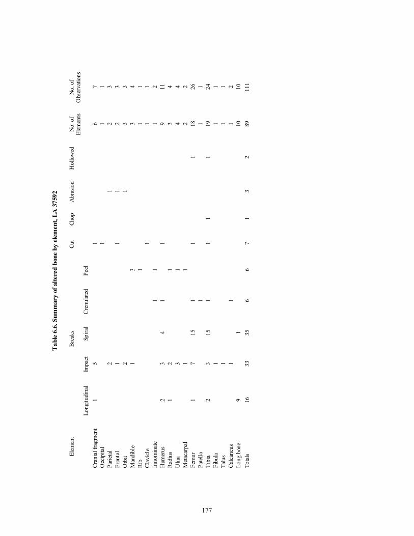
177
Tab
le 6
.6. S
umm
ary
of a
ltere
d bo
ne b
y el
emen
t, LA
375
92
Elem
ent
Bre
aks
Cut
Chop
Abr
asio
nH
ollo
wed
No.
of
Elem
ents
No.
of
Obs
erva
tions
Long
itudi
nal
Impa
ctSp
iral
Cre
nula
ted
Peel
Cra
nial
frag
men
t 1
5 1
6 7
Occ
ipita
l 1
1 1
Parie
tal
2 1
2 3
Fron
tal
1 1
1 2
3O
rbit
2 1
3 3
Man
dibl
e 1
3 3
4R
ib 1
1 1
Cla
vicl
e 1
1
1In
nom
inat
e 1
1 1
2H
umer
us 2
3 4
1 1
9 1
1R
adiu
s 1
2 1
3 4
Uln
a 3
1 4
4M
etac
arpa
l
1 1
2 2
Fem
ur 1
7 1
5 1
1 1
18
26
Pate
lla 1
1 1
Tib
ia 2
3 1
5 1
1 1
1 1
9 2
4Fi
bula
1 1
1Ta
lus
1 1
1C
alca
neus
1 1
1 2
Long
bon
e 9
1 1
0 1
0To
tals
16
33
35
6 6
7 1
3 2
89
111

178
Table 6.7. Summary of alteration by age, LA 37592
Age (years) < 2 5-7 9-10 5-15 15-20 Female Male Adult Indeter-minate
Total
Longitudinalbreak
1 3 1 11 16
Impact break 2 4 10 17 33Spiral break 1 5 9 19 1 35Crenelated 1 3 2 6Peel 2 1 2 1 2 8Cut 1 1 1 4 7Chop 1 1Abrasion 1 1 1 3Hollowed 1 1 2No. elements 1 3 3 10 5 2 16 48 1 89No. observations 1 5 3 10 5 3 25 58 1 111Also burned 1 2 0 2 0 0 7 9 0 21
Table 6.8. Burned human bone, LA 37592
Age/Element Light/Scorch Light to Heavy Heavy/Sooted Calcined0-2 years occipital 13-5 years mandible 1Tooth 15-15 years cranial fragment 2 3Frontal 1Ulna 2Adult cranial fragment 2 1Parietal 1Ossicle 1Mastoid 1Mandible 1Metacarpal 1 1Femur (male) 5 2Patella 1Tibia 5Fibula 2Long bone fragment 3 1Unknown fragment 1 Totals 23 10 6 1
Note: A light burn is brown, heavy or sooted is black, and calcined is white. For those with varying degrees ofburning, the most intense burn is used.

179
Table 6.9. Number of individuals represented by element, LA 37593
Element No. ofElements
0-2y 3-5y 6-10y 11-15y Female Male Adult
Cranial case fragment 114 1 1 3 1 2 1 2Maxilla 12 2 1 3Mandible 18 2 1 3 2 1Tooth 88 1 1 1 2Cranial fragment 303Atlas 11 1 1 6Axis 5 2 3Cervical vertebra 19 1 1 5Thoracic vertebra 54 1 1 1-2 3Lumbar vertebra 17 1 3Sacrum 10 2 2-3 2Vertebra fragment 48Scapula 23 1 1 2Clavicle 9 1 2 2Sternum 4 1 1Rib 386 1+ 1 1+Humerus 31 1 3 2 1 1 2Radius 25 5 1 1 3 2Ulna 23 3 1 2 2Carpal 66 1 2 4Metacarpal 80 2 3 1 1 5Phalanx 132 2 3 1 3Innominate 22 2 4 1 3 1Femur 45 1 1 4 1 4Patella 6 1 1 2Tibia 32 1 3 1 1 2 1-2Fibula 22 1 1 2 2 1 2-3 1Tarsal 75 1 3 1 2 2Metatarsal 56 1 3 2 1 3Phalanx 104 1 2 1-2 1 3Hand or foot 14Long bone fragment 99Flat bone fragment 143Unknown fragment 107Total elements 2203 10 100 274 233 28 47 1207Number of individuals 1 2 5 2 3 4
Because of the number of elements involved, most MNI calculations are based strictly on the number of elements or portions ofelements represented for that side and that age group (i.e., 4 left proximal femora from children aged 6 to 10 years of age = anMNI of 4). When a range is given, there is no duplication of parts; however, the two parts fall at the opposite ends of the range forthat group for children, or there are differences in size or age for the adults. For example, foot phalanges (aged by comparisonswith aged La Plata burials) range in age from 7 to 9 years and are considered to represent one or two individuals. The finalnumber of individuals in the table is the largest number for that column.

180
Tab
le 6
.10.
Alte
ratio
n ty
pe b
y el
emen
t, L
A 3
7593
Ele
men
tN
o.El
emen
tsLo
ngitu
dina
lBr
eak
Tran
sver
seBr
eak
Spira
lB
reak
Dia
gona
lB
reak
Impa
ctB
reak
Peel
Gas
h/C
hop
Abr
asio
n
Cran
ial f
ragm
ent
28
28
Fron
tal
1 1
1O
rbit
regi
on 3
3Zy
gom
atic
2 2
Parie
tal/o
ccip
ital
8 1
6 2
Man
dibl
e 3
2 1
1R
ib 4
4H
umer
us 5
3 2
1Ra
dius
2 1
1M
etac
arpa
l 1
1Fe
mur
10
9 1
Tibi
a 6
2 4
1Fi
bula
2 1
1Ca
lcan
eus
1 1
Long
Bon
e 3
3To
tals
79
4 3
21
7 4
3 1
1 4
Tab
le 6
.11.
Alte
ratio
n by
age
, LA
375
93
Age
No.
Ele
men
tsLo
ngitu
dina
lB
reak
Tran
sver
seB
reak
Spira
lBr
eak
Dia
gona
lB
reak
Impa
ctB
reak
Peel
Gas
h/C
hop
Abr
asio
n
0-2
1 1
7-1
0 2
211
-15
2 2
16-2
0 &
6 6
116
-20
2 2
Adu
lt %
1 1
1A
dult
63
4 1
5 6
37
1 3
40+ &
1 1
40+
1 1
Tota
ls 7
9 4
3 2
1 7
43
1 1
4

181
FS Grid Level Elements * Side Age500 33N 66E 1 metatarsals (2) * R 5-15508 35N 65E 11 femur, tibia, fibula, and foot (14) L 6-8
foot (11) R 7-9metacarpals (3) * R 8-10cervical vertebrae (3) 15-20cranial fragments (14) * 15-20cranial fragments (6) * 15-20femur, patella, tibia, fibula (5) L adult
520 33N 65E 7 radius and ulna (2) R 5-7cranium, mandible (2) 6-8cranial parts and fragments (28) * 6-8
528 34N 64E 8 humerus, radius, ulna, hand (19) R 8-10cranial parts (3) * 15-20radius, ulna (2) R adultmetacarpals (4) L adultmetacarpals (5) * L adulttibia, fibula, foot parts (4) L adult
529 34N 64E 9 cranial fragments (68) * 5-15teeth (9) * 5-15metacarpals (3) * L 5-15partial hand (16) * R 5-15hand phalanges (4) ? 5-15femur, tibia, fibula (3) R adulttarsals (3) L adultmetatarsals (5) R adult
532 33N 66E 10 thoracic vertebrae (4)* 20-30550 33N 64E 4 hand phalanges (9) R adult551 35N 66E 6 thoracic vertebrae (4) * 8-10
scapula pieces (2) * R 15-20cranial parts (5) * adulttibia fragments (2) * R adultlumbar vertebrae, sacrum (4) * 40+
556 35N 66E 1 cranial fragments (2) * adult557
33N 65E 8 tibia, fibula (2) R 2tibia, fibula (2) L 2metacarpals (2) L 3-5metatarsals and phalanges (10) R 9-10tarsals (7) R 9-10
558 33N 65E 9 two partial hands (20) R 3-5hand phalanges (3) 3-5tarsals (6) L 3-5foot phalanges (2)* 3-5lumbar vertebrae (2) 4-6pelvis, R femur (6) 4-6hand (24) L adultmetatarsals and phalanges (13) L adult
559 34N 64E 10 humerus, radius, ulna (3) L 6-8radius, ulna (2) R 6-8metacarpals and phalanges (29) R, L 6-8femur, tibia, fibula (3) R 6-8foot (16) L 6-8foot phalanges (3) ? adult
560 35N 65E 12 foot (14) R adult561 35N 65E 13 radius, ulna, partial hand (23) R 7-9
Table 6.12. Elements probably from the same individual and elements in articulation, LA 37593

182
FS Grid Level Elements * Side Age563 35N 66E 7 cranial parts (4) * adult
atlas, axis vertebrae (2) * adultaxis and cervical vertebrae 3 (2) * adultcervical vertebrae 4-7 (4) adultthoracic vertebrae (2) adult
563 and 564 cranial parts (10) * female, 15-20564 35N 66E 8 lumbar vertebrae (2) 15-20
radius, ulna (2) R 15-20thoracic vertebrae 1-7 (7) adulthumerus, radius, ulna (3) R adultmetacarpals (4) * R adulthand phalanges (2) * ? adult
569 (water linetrench)
maxilla (2) * 15-20
572 35N 67E 2 femur fragments (3) * L adult573 35N 67E 2 cranial parts (40) * L male? adult
femur pieces (2) * R 7-9574 33N 67E 3 foot (11) * L adult577 36N 68E profile cervical and thoracic vertebrae (12) 8-10
(Number of elements) For example, the left leg in FS 508 consists of a femur, tibia, fibula, and 11 foot elements.* Appear to be from the same individual based on stage of development, size, preservation, etc.
Table 6.13. Comparison of clustered, altered, and carnivore-damaged proportions, LA 37593
FS/Grid/Level No. ofElements
Articulated or SameIndividual
Altered Carnivore Damaged
N % N % N %127 39N 69E surface 1 1 100.0303 35N 65E Level 5 6 1 16.7500 33N 66E Level 1 15 2 13.3508 35N 65E Level 11 122 56 45.9 6 4.9 1 .8520 33N 65E Level 7 44 32 72.7 1 2.3528 34N 64E Level 8 75 37 49.3529 34N 64E Level 9 209 111 53.1 1 .5531 33N 66E Level 9 35 5 14.3 1 2.9532 33N 66E Level 10 102 4 3.9 1 1.0 3 2.9534 37N 66E Level 2 9 1 11.1550 33N 64E Level 4 10 9 90.0551 35N 66E Level 6 62 17 27.4 1 1.6556 35N 66E Level 1 6 2 33.3557 33N 65E Level 8 55 23 41.8558 33N 65E Level 9 104 76 73.1 1 1.0559 34N 64E Level 10 102 56 54.9 1 1.0560 35N 65E Level 12 37 14 37.8 1 2.7561 35N 65E Level 13 35 23 65.7 3 8.6563 35N 66E Level 7 335 19 5.7 22 6.6 29 8.6564 35N 66E Level 8 315 25 7.9 35 11.1 9 2.9569 water line trench 7 2 28.6571 33N 67E Level 1 10 1 10.0572 33N 67E Level 2 13 3 23.1 3 23.1573 35N 67E Level 2 111 42 37.8 1 1.0574 33N 67E Level 3 30 11 36.7 1 3.3575 35N 67E Level 3 46 1 2.2577 36N 68E profile 48 12+ 25.0 1 2.1852 42N 67E Level 1 8 1 12.5 1 12.5

183
Table 6.14. Number of individuals represented by element, LA 37595
Element No. Elements Individuals
15-20y Female? AdultTooth 1 1Rib 1 1Clavicle 1 1Radius 1 1Phalanx 3 1Femur 1 1Unknown fragment 1 1?Total elements 9 4 1 2Number ofindividuals
1 1 0-1
Table 6.15. Number of individuals represented by element, LA 37598
Element No. Elements Individuals0-2 y 3-5 y Adult
Cranial case fragment 2 1 1Maxilla 1 1Mandible 1 1Tooth 7 1 1 1Cranial fragment 38Atlas 1 1Axis 1 1Cervical vertebra 6 1Thoracic vertebra 8 1Vertebra fragment 1 1Scapula 2 2Clavicle 2 1Sternum 1 1Rib 31 1Metacarpal 2 1Phalanx 3 1Innominate 1 1Tarsal 2 1Total elements 128 6 71 13Number of individuals 1 2 1

184
Table 6.16. Number of individuals represented by element, LA 37599
Element No. Elements Individuals0-2 y 3-6 y Female? Male Adult
Cranial case fragment 18 1 1 1Maxilla 2 1 1Tooth 3 1Rib 9 1 1Humerus 1 1Femur 1 1Fibula 1 1Tarsal 1 1Phalanx 1 1Long bone fragment 1Unknown fragment 1Total elements 39 1 17 1 11 9Number of individuals 1 1 1 1
Table 6.17. Number of individuals represented by element, LA 37600
Element No. Elements Individuals5-15 y Male ? Adult
Cranial case fragment 1 1Mandible 1 1Tooth 2 1Humerus 1 1Femur 3 1Tibia 4 1Fibula 1 1Tarsal 5 1 1Metatarsal 7 1 1Phalanx 5 1Long bone fragment 35Flat bone fragment 2Unknown fragment 26 1Total elements 93 2 2 78*Number of individuals 1 1 0-1
* Sixty-nine of these are from a single provenience and probably represent a disintegratedburial.

185
Table 6.18. Number of individuals represented by element, LA 37601
Element No. Elements Individuals0-2 y 3-5 y 5-15 y Female Male Adult
Cranial case fragment 1 1Tooth 3 1 1Cranial fragment 6Thoracic vertebra 1 1Rib 1 1Humerus 1 1Carpal 4 1Metacarpal 8 1 1Phalanx 8 1Femur 4 1 1Fibula 1 1Tarsal 3 1Metatarsal 4 1 1Phalanx 2 1 1Long bone fragment 2 1?Unknown fragment 1Total elements 50 2 6 2 4 2 34Number of individuals 1 0-1 1 1 1 0-1
Table 6.19. Number of individuals represented by element, LA 37603
Element No. Elements Individuals0-2 y 3-5 y 6-10 y 5-15 y Female Male Adult
Cranial case fragment 14 1 1 1 1?Mandible 5 1-2Tooth 6 1 2Cranial fragment 16Axis 1 1Thoracic vertebra 11 1 1Vertebra fragment 1Rib 32 1 1 1? 1Clavicle 1 1Scapula 4 1Humerus 4 1Carpal 1 1Metacarpal 7 1 1Phalanx 16 1 1 1? 1 1Tarsal 1 1Metatarsal 3 1 1?Phalanx 1 1Hand or foot 1 1Long bone fragment 7Flat bone fragment 8Unknown fragment 6Total elements 146 6 2 6 8 4 1 105Number of individuals 1 1 1 0-1 1 1 0-2

186
Table 6.20. Number of individuals represented by element, LA 37605
Element No. Elements Individuals0-2 y 5-15 y Female? Adult
Mandible 6 1Tooth 3 1 1 1?Sacrum 1 1Femur 1 1Fibula 1 1Long bone fragment 1 1Unknown fragment 4Total elements 17 3 1 1 8Number of individuals 1 1 1 1
Table 6.21. Number of individuals represented by element, LA 37606
Element No. Elements IndividualsAdult Male
Rib 1 1Humerus 1 1Hand phalanx 3 1 1Metatarsal 1 1Foot phalanx 2 1Total elements 8 3 5Number of individuals 1 1
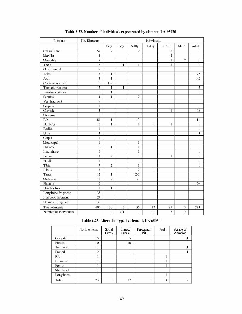
187
Table 6.22. Number of individuals represented by element, LA 65030
Element No. Elements Individuals0-2y 3-5y 6-10y 11-15y Female Male Adult
Cranial case 57 2 2 2 1Maxilla 4 2Mandible 7 1 2 1Tooth 17 1 1 1 1Other cranial 7Atlas 3 1 1-2Axis 3 1 1-2Cervical vertebra 6 1-2Thoracic vertebra 12 1 1 2Lumbar vertebra 6 1 1Sacrum 4 1 2Vert fragment 5Scapula 1 1Clavicle 3 1 1?Sternum 0Rib 81 1 1-3 1+Humerus 12 1 1 1 1 1Radius 1 1Ulna 4 3Carpal 1 1Metacarpal 1 1Phalanx 6 1 1 1Innominate 6 1 1Femur 12 2 3 1 1Patella 1 1Tibia 7 2 1 1Fibula 3 2 1Tarsal 12 1 2-3Metatarsal 11 2 1-3 1Phalanx 9 2+Hand or foot 1 1Long bone fragment 35Flat bone fragment 27Unknown fragment 35Total elements 400 30 2 55 18 39 3 233Number of individuals 2 0-1 3 0-1 3 2
Table 6.23. Alteration type by element, LA 65030
No. Elements SpiralBreak
ImpactBreak
PercussionPit
Peel Scrape orAbrasion
Occipital 5 5 1Parietal 10 10 1 4Temporal 1 1 1Frontal 2 1 1Rib 1 1Humerus 1 1Femur 1 1Metatarsal 1 1Long bone 1 1Totals 23 1 17 1 4 7
SpiralBreak
ImpactBreak
PercussionPit
Scrape orAbrasion

188
Table 6.24. Alteration type by age, LA 65030
No.Elements
SpiralBreak
ImpactBreak
PercussionPit
Peel Scrape orAbrasion
9-11 13 11 2 425-30 & 4 3 1 335-40 & 2 2Adult 3 1 1 1Large child/Adult 1 1Totals 23 1 17 1 4 7
Table 6.25. Comparison of bone assemblages from LA 37592, LA 37593, and LA 65030
Variable LA 37592 LA 37593 LA 65030Postcranial Breakage
Number of elements 304 1559 227% complete 2.0 32.8 6.6% > 50 percent 18.1 12.9 14.1% < 50 percent 79.9 54.3 79.3
Carnivore Damage (all elements)Number of elements 395 2049 300 % damaged 1.0 2.6 12.6
Burning (all elements)Number of elements 395 2049 300% light burn 5.8% light to heavy burn 2.3 .1 .3% heavy or smoked burn 1.3 4.0% calcined .3Total % burned 9.7 .1 4.3
Alteration (all elements)Number of elements 395 2049 300% longitudinal break 4.0 .3% transverse break .2% diagonal break .4% spiral break 9.6 .2% impact break 8.9 .8 5.7% peel 2.0 .1 1.3% percussion pit .3% chop .3 % cut 1.7% scrape or abrasion .3 2.0% crenulated edge 1.5% hollowed .5Total elements altered 87
22.0% 77
3.8% 28
9.3%
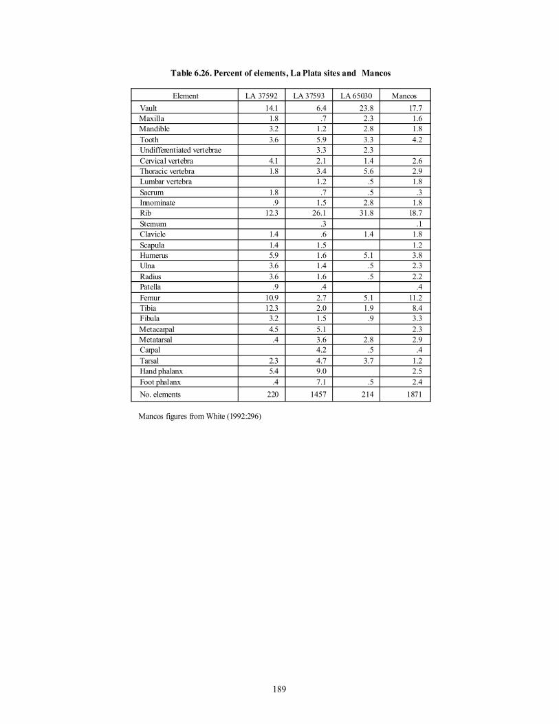
189
Table 6.26. Percent of elements, La Plata sites and Mancos
Element LA 37592 LA 37593 LA 65030 MancosVault 14.1 6.4 23.8 17.7Maxilla 1.8 .7 2.3 1.6Mandible 3.2 1.2 2.8 1.8Tooth 3.6 5.9 3.3 4.2Undifferentiated vertebrae 3.3 2.3 Cervical vertebra 4.1 2.1 1.4 2.6Thoracic vertebra 1.8 3.4 5.6 2.9Lumbar vertebra 1.2 .5 1.8Sacrum 1.8 .7 .5 .3Innominate .9 1.5 2.8 1.8Rib 12.3 26.1 31.8 18.7Sternum .3 .1Clavicle 1.4 .6 1.4 1.8Scapula 1.4 1.5 1.2Humerus 5.9 1.6 5.1 3.8Ulna 3.6 1.4 .5 2.3Radius 3.6 1.6 .5 2.2Patella .9 .4 .4Femur 10.9 2.7 5.1 11.2Tibia 12.3 2.0 1.9 8.4Fibula 3.2 1.5 .9 3.3Metacarpal 4.5 5.1 2.3Metatarsal .4 3.6 2.8 2.9Carpal 4.2 .5 .4Tarsal 2.3 4.7 3.7 1.2Hand phalanx 5.4 9.0 2.5Foot phalanx .4 7.1 .5 2.4No. elements 220 1457 214 1871
Mancos figures from White (1992:296)

CHAPTER 7
CONCLUSIONS
Synthesizing data from the human remains recov-ered by the La Plata Highway Project raised more ques-tions than it was able to answer. The goal of the analy-sis was not to provide descriptive forensics, which weargue is limiting and reductionist. Rather, the researchagenda included a broadly biocultural perspective thatfocused on questions derived from the archaeologicalcontext from which the remains came. This project rep-resents a truly collaborative approach between archaeol-ogists and biological anthropologists.
Sixty-seven burials representing the full range ofages and sexes were recovered. While many of the larg-er studies on human remains from the Southwest havefocused on adaptation to scarce resources and marginalenvironmental conditions, La Plata provides an alterna-tive view. Resources may have been better than ade-quate, at least during the periods ranging from A.D. 900to 1100. The additional recovery of at least 68 individu-als in disarticulated and nonburial contexts suggests thata wide variety of alternative modes of cultural and mor-tuary behavior operated within this region.
HEALTH AND DEMOGRAPHY
The goals of this study included demographic andhealth profiles of the people living in the La Plata RiverValley. We wanted to contribute to baseline data thatcould be compared with data from other Southwestgroups, with the eventual goal of providing a largerregional synthesis of the relationships between biologi-cal well-being and cultural context.
The demographic analysis of the burial populationdemonstrated a mean age at death1 of 24.5 years.Compared with other Southwest populations, this sug-gests a relatively robust population, especially whencombined with a moderate fertility rate. However, theunder-representation of the most elderly age categorysuggests that the population as a whole had few elderly.The age composition of 10 percent infants under age 2,30 percent children from 1 to 9, 10 percent teens from10 to 20, and 50 percent adults is almost identical to theage structure distribution for Black Mesa, CasasGrandes, and Pecos Pueblo, as well as non-Southwestsites such as Arikara villages and the Tlajinga (Mexico)sites. This suggests a relatively representative precon-tact population.
It is clear that the La Plata communities were com-mitted to a maize diet, since dental health reveals a pat-tern of wear and disease common to traditional agricul-
turalists. Caries result from an infectious disease processcharacterized by a demineralization of the dental tissuesby organic acids. These acids come from a fermentationof dietary carbohydrates. High frequencies have beennoted in a number of groups shifting from hunting andgathering to agriculture. The La Plata frequencies arecomparable to other groups in the Southwest.
Although exact dietary composition beyond maizeis not clear, an examination of childhood health andgrowth and development can suggest the adequacy ofthe diet. Porotic hyperostosis, a response to iron defi-ciency anemia in the Southwest, is an indicator of phys-iological response to a decrease in the body’s ability totransport oxygen to tissues. It results in an expansion ofthe thinner bones in the body because of an attempt toproduce more red blood cells. The outcome is bone thatappears porous on the vault or in the orbits. The fre-quency data on this lesion for La Plata Valley include nocases with severe involvement.
Compared to other southwestern populations, theLa Plata sample falls in the middle range for frequencyof porotic hyperostosis. These frequencies include allexpressions of severity as well as different stages ofhealing, yet the La Plata frequencies are generally abouthalf those of some Southwest skeletal collections. Theroot causes of iron deficiency are multiple and complexand may include poor diet, lack of sanitation, infections,and other childhood diseases. Whatever the basis, the LaPlata data suggest better overall health for its children.
This is supported by an additional data set. Enameldevelopmental defects in the form of hypoplastic linesare indelible markers of childhood physiological disrup-tion. They result when a systemic health problem shutsdown the formation of new enamel. The resultingdefects were assigned an age of occurrence, which pro-vides good documentation of disturbances during child-hood. Looking at the average number of dental defectsper tooth (for the central maxillary incisor and the max-illary and mandibular canine), La Plata children did notcarry the burden of morbidity that other southwesterngroups did. The average defect of one hypoplastic line isdistinctly less than in other Southwest series, such asBlack Mesa, Mesa Verde, Yellow Jacket, Dolores, andSand Canyon, where averages run from two to as highas four lines per tooth. This low frequency suggests thatsuboptimal conditions and the resulting burden of child-hood sickness were less problematic for children in theLa Plata communities than in other Southwest groups.
Just as the bone lesions from iron deficiency wereslight and mild in expression, the occurrence of sys-temic, nonspecific infections on the outer shafts of longbones was generally mild in expression. Compared withcases documented at other Southwest sites, where thedisease response was typically severe and active, La
191

Plata Valley residents seemed to have been bufferedfrom severe infection. Comparing total population fre-quencies across different Southwest sites, La Plata doesnot stand out as having frequencies inconsistent withother sites, and although these data do not reflect it, allof the La Plata cases are slight to moderate. Cases ofinfectious disease at Black Mesa, Chaco Canyon, andDolores were mostly moderate to severe, indicatinggreater morbidity than in La Plata. The large protohis-toric sites also demonstrated many severe cases.
Turning to the adult segment of the population,there are some notable patterns among males andfemales. Anemia was an almost equal liability for bothmales and females, suggesting that it was a function ofshared lifestyles. Differences in transmissible infectiousdisease, however, suggest that liability was not shared.Females demonstrate a five times greater involvement.
PATTERNS IN TRAUMA
Yet more distinctive patterns emerged from this col-lection. Frequencies of cranial and postcranial trauma(represented by healed or healing fractures) demonstratedisparate trends across subgroups. Children were almostfree of fractures. Only one 15 year old had a healedcompression fracture on the left parietal. There werethree cases of male cranial trauma: one 25-year-old malehad a healed compression fracture of the right parietal, asimilarly aged male had a healed fracture at the cornerof the left eye, and one aged 35 had a compression frac-ture on the left parietal near the occipital. Male postcra-nial fractures include a healed Colle’s fracture (distalends of the forearm bones), a healed thumb fracture, andhealed rib fractures. These lower body fractures did notco-occur with head trauma. In contrast, six females havehealed, nonlethal blows to the head, and five femalesdemonstrate lower body traumatic circumstances. Infour cases, the cranial and postcranial fractures co-occur. In the disarticulated remains, healed cranial trau-ma includes two adult occipitals with medium-sizeddepression fractures, and an 8- to 10-year-old child witha small lesion on the parietal. Disarticulated adult post-cranial trauma include a fibula fracture with a large cal-lus (LA 37592, FS 551-10) and a radius and ulna withhealed fractures near the distal end (LA 37592, FS 25and FS 26).
The kinds of compression fractures found infemales suggest nonlethal blows to the head with bluntobjects. Postcranial co-occurrences of fractures includetwo healed fractured necks (first cervical vertebrae; cer-vical vertebrae three through five), a healed fracture ofthe right fibula, traumatic dislocation of the left hip,healed distal right radius, and three females who showedmarked lower body asymmetry, in which one side was
larger and more robust.Further complicating the picture of health is the
case of metastatic cancer in a 19 year old. Anotherfemale aged 25 had not only multiple compression frac-tures on her head and a traumatic injury to her neck andfractured ribs, but she also had a severe case ofosteomyelitis rampant throughout her upper body andlimbs. The proximate cause of this severe infection isdifficult to diagnose. However, it could result from aseries of localized infectious sites that overwhelmed thetypical immune response, leading to full-blown sys-temic osteomyelitic infection.
A clear and surprising association emerged whenthe mortuary context of the battered women was exam-ined. In this sample, as well as in other Southwest col-lections, the great majority of the burials are flexed orsemiflexed, and placed within middens, abandonedstructures, or in storage pits. Burials often contain asso-ciated objects, usually ceramic vessels or ground stone.No female at La Plata with cranial trauma followed thismortuary pattern. Some women with cranial traumawere found in positions that were prostrate and/or sug-gestive of being hurled. Particularly at one BarkerArroyo site, LA 65030, several unusual burials dating tothe thirteenth century were located at in the fill of a pitstructure. One female (65030 B9) with a large healedfracture at the top of her head was on her back with legsand arms splayed outward. Another younger female(65030 B8), found on her back, had healed fractures onthe nose and base of the head. Another female (65030B6) with a healed fracture of the occipital was semi-flexed but on her face. Finally, a child (65030 B7, 10.5years) without traumatic lesions was found in this samestructure on the back with arms and legs splayed. At thissame site but in a different pit structure and an earliercontext (A.D. 1000-1075), another female (65030 B16)with compression fractures on the right forehead andoccipital was placed in a semiflexed but haphazard posi-tion near a pit structure wall with a piece of groundstone resting on the cranium. The pattern holds for bat-tered females from other sites at La Plata. For example,one female from Barker Arroyo, LA 37601, dating toA.D. 1125-1200 (B4), appears somewhat askew. Thisburial has no associated goods and multiple traumas.Although not as common the pattern of unaccompanied,haphazard burial and cranial trauma also occurs inmales. Both cases are from LA 37599 and date to theA.D. 1000s. A male with a large depressed fracture ofthe right parietal (37599 B5) was found in the fill of apit structure, semiflexed with no associated objects. Asecond male (37599 B9) with a fracture of the leftfrontal near the orbit was placed in a large subfloor cistwithin a pit structure more or less extended with one legbent at the knee and only a piece of ground stone, pos-
192

sibly an offering. The burial situations for one femaleand one male with cranial trauma are unknown. There isa 15 year old with a healed cranial trauma (37592 B6)who was formally buried and accompanied by gravegoods.
These patterns of cranial trauma and informal bur-ial are provocative, but we are limited in our ability tofully explain them. Their salient features are that exam-ples occur from contexts spanning three centuries, thatmore females than males are found fitting the pattern,and that all cases are adults. As discussed further below,this pattern may indicate the presence of a subgroup,which was treated differently from the rest of the inhab-itants.
CULTURALLY MODIFIED DISARTICULATED ASSEMBLAGE
Excavations at La Plata also recovered elementsrepresenting at least 68 individuals (based on a mini-mum numbers index), found mostly in beds of disartic-ulated bone at three sites. Some dislocation of the skele-tal elements is likely to have been postmortem due tomechanical and other kinds of disruption. However, thedisarticulation of bone at least at LA 37592, JacksonLake, can be explained in terms of perimortem behavior.This assemblage contained bones that were burned, cut,broken near the time of death, and arranged in unusualpositions.
The disarticulated human remains from La PlataValley clearly were the product of several differentdepositional or processual pathways and, while notcompletely understood, have some instructive features.For example, the 12 or so individuals recovered from PitStructure 1, LA 37592, Jackson Lake represent the fullrange of age categories, from infants to adults. Therewere up to two infants, one or two small children (aged5 to 7), one or two children a bit older (9 to 11), and atleast one teenager. There were at least two adult malesaged 30 to 35 and around 40, at least one female, andpossibly a couple of other adults (Table 7.1). Over 20percent of the skeletal elements from these individualsdemonstrate signs of human alteration that include spi-ral fractures, parallel cuts, burning, spalling, and longi-tudinal breaks.
Some of the bones were arranged in a way that sug-gests they were carefully organized. Split long boneswere placed in parallel fashion resting on portions ofcranial vaults in two different people.2 One piece of cra-nium was on the bottom, and another was on the top.This assemblage is similar to that reported at YellowJacket by Malville (1989) and Turner and Morris (1970)in the Southwest, but different from other disarticulatedassemblages (Swedlund 1965; White 1992; Turner1993). Interestingly, this is remarkably similar to assem-
blages found in northern Mexico at La Quemada (A.D.650), where skeletal elements were arranged in groupsof limb bones lying parallel to one another along withportions of cranial vaults. Interpretations include “pro-cessing enemy corpses after battle, sacrificial ritesinvolving either enemies or community members, can-nibalistic feasting, and charnel treatment, presumably ofrevered ancestors or community members” (Nelson etal. 1992:304). Numerous reports of disarticulatedassemblages from other sites in northern and centralMexico describe long bones piled together, along withbroken crania and other miscellaneous bones (Pickering1985). The bones often exhibit cut marks (primarily inthe regions of muscle insertions or near the distal ends).Thus, while not unheard of in the precontact NewWorld, this mortuary configuration presents a challengefor contextualization within the Four Corners region.
There is virtually nothing in the LA 37592 assem-blage of disarticulated and largely broken bone that dis-tinguishes it as biologically different from the remainsof individuals in formal burials. In terms of pathologies,one of the disarticulated infants (FS 206-3) has an activecase of porotic hyperostosis on the parietal, an adult hastwo distinct hypoplasia lines on a central incisor (FS228-1), and another adult has a healed fracture of a slen-der long bone, probably a fibula (FS 551-1). This is notunusual, given that a two of the infants from the burialpopulation also exhibited porotic hyperostosis andhypoplasia and fractures occur throughout the burialpopulation.
Stable carbon isotopes were run on one sample ofthe disarticulated adult bone from LA 37592 (FS 138),and the resulting data approximate the average value forthe La Plata burial population. With a delta value of -7.67, this individual cannot be distinguished from sam-ples tested from the burials. All this information reallyreveals is that these people had the same dietary base asother individuals recovered from the La Plata Valley.Had the delta values been substantially different, a casecould have been made that these people were from agroup with a different subsistence base. Further chemi-cal analyses of more of the disarticulated remains couldbe conducted, but we feel confident that the findingswould continue to support the notion that the subjects ofthese violent acts are not foreigners, but rather membersof the existing community.
Also retrieved from LA 37592 were eight discreteburials ranging in age from a preterm fetus, to severalsmall children and a teenager, and including an older(50+) female and an unaged adult female. Interestingly,no adult male burials were recovered from this site. Theteenager (age 15, sex unknown) has a healed depressionfracture on a parietal. This burial was intentionallyplaced and accompanied by two vessels. Four of these
193

burials were flexed or semiflexed, and four had associ-ated grave goods (burial context is unknown for three,and the fetus was found in a corrugated jar).
Morphometric and genetic data could not beobtained in any systematic fashion for the disarticulatedremains. This is unfortunate, because the relatedness ofthis group of people is important. If these remains rep-resent an extended family, their significance is quite dif-ferent from the presence of a group of unrelated people.An overall qualitative assessment of the size, muscular-ity, robusticity, and general morphology revealed no dis-tinguishing features from the burial population. Futureanalytical procedures based on an analysis of DNAextracted from the bone collagen may prove useful.
The age composition of this group is intriguing.Titiev (1972) and, more recently, Levy (1992) providein-depth studies of historic Hopi villages. Levy in par-ticular explores the size of families in protohistoric andhistoric times, and suggests that an extended family liv-ing in a matriarchal setting ranges from 9 to 20 individ-uals, depending on the number of daughters versus sons.A family with mostly sons will be on the smaller side,while daughters will cause the family size to grow ashusbands are brought into the matrilineal fold. It iscompelling to think about the group of disarticulatedindividuals from LA 37592 as an extended family unit.
As discussed in the previous chapter, accusations ofwitchcraft may account for the demise of these individ-uals. In a review of Pueblo witchcraft beliefs, Ellis(1989) writes that witches were traditionally blamed forenvironmental or personal problems that occur in vil-lages. Ellis quotes Bandelier’s 1890 manuscript, inwhich he details the butchering and mutilation of threemen and a woman at Nambe Pueblo in March 1855.Bandelier attributes the population decline at SantaClara, Nambe, and Zia to “constant interkilling due towitchcraft” (Ellis 1989:192). Neighbors and peoplewithin villages, as well as in adjacent villages, wereoften targets of witchcraft. The penalties and punish-ments for witchcraft, as documented in early historictimes, could account for the broken and fractured bonefound at LA 37592. Ellis (1989:213) notes that theaccused were beaten to death with clubs. Part of the rit-ual of killing witches involves dismembering the bodyto obliterate the essential human form.
Although others may choose to interpret this assem-blage as the product of perimortem activity associatedwith cannibalism, we choose to emphasize other, equal-ly plausible scenarios. Witchcraft is an alternativehypothesis. Another is small-scale warfare. One doesnot need to invoke data showing that there were hugeshifts in political ideology, or invading “outsiders.”Recent reports on the civil war among the Serbians andCroatians provide an interesting parallel. There are vir-
tually no biological or genetic differences between thesetwo groups, and historically and up to the present, theirmaterial culture looks quite similar. The fact that theyare now divided along culturally defined ethnic bound-aries that have more to do with political, historic, lin-guistic, and religious differences than any identifyingfeatures of their biology or material culture provides astrong cautionary tale. Recent reports revealed thatSerbian military have beaten, tortured, mutilated, andburned Croatians, including men, women, and children(CNN Headline News, 7 October 1994). Would notthese assemblages show the same criteria currently usedto interpret cannibalistic activity? So-called signaturesof cannibalism (Turner 1993) include burned and cutbones, splintered, fractured, and broken bones, bonewith peeling, adherent flakes, and burned elements. Allof these cultural modifications could result from analternative pathway that is motivated by anger and hate,with no cannibalism involved.
Much more research is necessary to even begin tountangle the web of possible underlying factors that tar-geted some people for violent deaths in the precontactSouthwest. The La Plata Valley disarticulated remainsprovide abundant evidence that the picture is not simple,and that the variability underlying the assemblagesneeds to be carefully explored.
Except for LA 37592, then, the disarticulatedremains from all other sites in the La Plata Valley repre-sent burials that became disarticulated and dispersedthrough a variety of means. For example, LA 37593,Jackson Lake most clearly represents burials thatbecame disarticulated during the re-excavation of a pre-viously abandoned structure. The arrangement of thehuman remains and the nature of the breakage can mostparsimoniously be explained by movement of the buri-als from one place to another.
The combined mortuary and pathology analysesrevealed some interesting information on precontactPueblo ideology and lifestyle. In terms of treatment ofthe dead, Toll and Schlanger (1998) demonstrate that itis likely that some individuals were intentionally buriedin abandoned structures that may have been their origi-nal home. The finding that some individuals buried atdifferent times in an abandoned pit structure shared agenetic predisposition for vertebral anomalies suggestsgenetic relatedness. Ideologically, this suggests that tiesto certain localities remains strong in spite of the ten-dency of families and villages to move relatively often.As Edmund Ladd (1991:34), a noted Zuni scholar,relates, “There are a lot of Anasazi ruins. They’re scat-tered all over the place because that’s where our ances-tors traveled. They weren’t traveling because there weredroughts or there was pestilence. They were travelingbecause they were looking, searching, for the center
194

place. . . . Each one, until they found their own centerplace, moved every four [years].”
Constant movement could also create situations inwhich abandoned villages were reoccupied by others.Renovation could create the types of assemblages foundat LA 37593, where remains buried in one spot areunceremoniously removed and placed elsewhere. In thecase of LA 37593, cobbles appear to have been inten-tionally thrown or placed on top of the excavatedremains. This suggests several things. Not all burialswere left alone or considered venerable; perhaps onlyburials from one’s natal group or village were deemedinviolable. If one interprets the cobbles as intentionallythrown at the remains and the breakage as intentional,then, in addition to lack of reverence, animosity mayhave been a motivating factor.
Surely, group identity based on birth or shared res-idence was an important feature of the ideology behindmortuary practices. Added to this notion of group iden-tity is the possibility of targeted subgroups either withingroups or among groups. The women found in aban-doned pit structures at LA 65030 shared two things:healed traumatic lesions and unceremonious burial.Furthermore, a child (10.5 years old) was found in asso-ciation with these burials, suggesting some form of rela-tionship to one or more of the women. Because thesewomen and the child are not distinguishable from therest of the burial population in terms of morphometry ordiscrete traits, they must have been considered “differ-ent” based on ideological grounds.
Individuals with cranial trauma were clearly in aseparate category from the individuals found disarticu-lated at LA 37592. The age and sex structure of the LA37592 disarticulated individuals suggests an extendedfamily. The violent subtext of these remains points tomultiple homicides. Again, it appears that these individ-uals were genetically related to others in the La PlataValley based on the overall morphological characteris-tics of the remains. As discussed, these skeletal remainswere cut, broken, burned, and disarticulated. More spec-ulatively, there may be bite marks and hollowing of longbones. Were these people eaten, or were they tortured,dismembered, and burned? It is premature to categori-cally state the intent behind the behaviors that createdthis assemblage, but we are leaning toward an explana-tion of ritual massacres that may involve a variety ofactivities.
These hypotheses remain to be tested and verifiedin future studies. These data need to be linked to broad-er theories about settlement patterns, group identity,population movement, and resource distribution in theLa Plata Valley and the larger Totah area, as well as theinteractions between the Totah region and others. Thesefindings are not unrelated or ancillary to other important
political and economic phenomena in the Four Cornersregion between A.D. 1000 and 1300, and archaeologistsneed to be involved in theory building about the data.Explanations of the targeted subgroups of battered indi-viduals and massacred families are inextricably linkedto what was happening in the area and can only beunderstood when looked at from broader and moreinterdisciplinary perspectives.
Taken as a whole, the disarticulated assemblage andthe burial population suggest several things, but themost salient feature is the range of variability embodiedin this relatively small set of sites occupied over severalhundred years. Multiple layers of causation underlaygroup morbidity and mortality, including a wide varietyof practices regarding the appropriate management anddisposition of the dead. The La Plata population was ahomogeneous group in that we found no quantitative orqualitative differences among the burial and nonburialassemblages. This recognition shifts the focus of inter-pretation into areas defined by local political economicrelations, religious and symbolic distinctions, and otherlocal or regional organizational features of these extantgroups.
With current techniques from forensic and medicalsciences, we are swayed by the evidence that the burialpopulation and the individuals represented by only par-tial or isolated bones were all Pueblo people. That is notto say that there were not linguistic and other distin-guishing features that may have differentiated groupsfrom one another in the La Plata Valley. In fact, cultur-ally defined ethnic differences must have distinguishedgroups, just as the Pueblos today have linguistic and cul-tural features that make each group a distinct entity.
THE LA PLATA HEALTH PROFILE
A preliminary profile of biological well-being fol-lows. It is a youthful sample. Only 6 percent of the deathassemblage is aged 50 or older, so it is not surprisingthat the inventory of disease does not emphasizeosteoarthritis. Most adults aged 40 and older at La Platado not show any clinically significant signs of thisdegenerative, wear-and-tear disability. Infectious dis-ease is present in a number of individuals, but its inten-sity is generally mild. The occurrence of anemia andgrowth defects is moderate compared to otherSouthwest groups. Thus, the morbidity burden on the LaPlata community may not have been one that placedmuch disability or hardship on the whole. Health at LaPlata was not compromised to the extent demonstratedat other Southwest sites. However, the presence ofcrowd diseases such as tuberculosis suggests that thecommunity was eventually quite large and densely situ-ated.
195

Interpersonal strife, however, may have placed asignificant amount of stress on some members of thecommunity. Trauma is almost absent in children andmore benign in adult males. Females carry the unequalburden of traumatic injuries in this group. Furthermore,the comorbidity factors of cranial and postcranial trau-ma, infections, and decreased life expectancy suggesttruly suboptimal conditions for some adult females.Females with these health problems are more likely tohave been in mortuary contexts best described as hap-hazardly thrown or discarded, with no associated graveofferings.
This is a community that also practiced, at least insome contexts, perimortem activities that resulted inpeople’s death, and these deaths were marked by activi-ties that included chopping and burning of the bodies.This group could have included individuals identified asposing a threat (to individuals, to the community, to theregion, to the cosmos). These individuals were mostlikely put to death in a ritualistic manner, resulting in thedisarticulated bone bed found at LA 37592.
Certainly, as new information comes to light, or asthe data collected are reanalyzed using new and moreexact techniques, these hypotheses may be modified,expanded, or rejected. Our collective view of the com-bined osteological and archaeological data has led us tothese conclusions.
COMPARING HEALTH PROFILES OF LA PLATA, MESAVERDE, AND CHACO CANYON POPULATIONS
Chaco Canyon contains the largest concentration ofAnasazi great houses known from the eleventh andtwelfth centuries. Contemporary communities in muchof the Southwest probably participated in events coordi-nated by Chaco, and the canyon may have housed thou-sands of individuals at least periodically. Occupationswithin the canyon range from Basketmaker III (600A.D.) to Pueblo III (A.D. 1200), when people were alsoliving in the La Plata Valley. The relationship of Chacoto other populations in the region is an importantresearch question for the La Plata Highway Project andothers.
Only a few hundred burials have been scientificallyrecovered from Chaco, and these have been the subjectof a number of studies. Akins (1986) contrasts 132 buri-als, mostly from small sites within Chaco Canyon, withburials from Pueblo Bonito, the largest Chaco Canyonsite (Palkovich 1984). The analysis addressed differ-ences in health of high status (Pueblo Bonito) and lowstatus (small site) individuals. This study suggests thatChaco Canyon Anasazi suffered from “subsistencestress” as indicated by growth disruption, high rates ofnutritional anemia, and degenerative diseases (Akins
1986:135). In the comparative analysis of pathologiesfrom the Pueblo Bonito and the smaller sites, Akins(1986:137-140) suggests that higher status individualshad greater access to nutritional resources and enjoyedbetter health. Although there is some indication of bet-ter health, both groups experienced degenerative dis-eases throughout adulthood. Although the study suffersfrom small sample size, it does begin to address theinterplay of cultural and biological variables.Specifically, it is useful for answering questions con-cerning community health among assumed high and lowstatus individuals. Detailed information provided by thearchaeological context is used to support interpretationsof the relationship between political structures andhealth.
Stodder (1987) provides a regional synthesis of thepaleopathology of the Mesa Verde remains. She divides466 burials into two temporal groups: Basketmaker IIIthrough Pueblo II (A.D. 600-975) and Pueblo II-III(A.D. 975-1275). These individuals represent the popu-lation in the Mesa Verde region during periods ofgrowth and decline in the northern San Juan region.Using paleoepidemiological methods, Stodder docu-ments a trend toward increasing morbidity and highermortality in younger ages. Integrating this informationwith data describing climatic, ecological, nutritional,and political changes occurring at the end of the laterperiod, Stodder’s interpretation suggests that communi-ty health became increasingly compromised.
Stodder’s work is important because it emphasizespopulation analyses that take many factors into consid-eration in the interpretation of community health. Herstudy contrasts with one undertaken by Miles (1975), aclinical orthopedist who examined 179 burials fromWetherill Mesa, part of Mesa Verde. Although he wasable to note the occurrence of arthritis, infections, trau-ma, and other ailments, Miles (1975:35) concluded thatthe Mesa Verde Anasazi were “a surprisingly healthypopulation . . . capable of great physical independence.”Because there is no archaeological context for his analy-sis, because frequencies are not presented for the popu-lation, and because it is a synchronic and descriptivepresentation of bone lesions in an analytical vacuum, itis difficult to evaluate his findings. His report suggestsan impression of community health that is at odds withStodder’s assessment.
Thus, comparing the health of individuals residingin the La Plata Valley to those at Chaco and Mesa Verdeis not a simple task. Within the paleopathological litera-ture, there are conflicting reports on health status notonly within sites, but between close sites, and amongsites farther apart. In an attempt to generalize and stepback from the individualized data sets, we categorizedselected Southwest skeletal populations according to
196

mortality, fertility, pathology, and stature (Table 7.2).We arrived at an overall assessment of groups vis-à-viseach other. Following Nelson et al. (1994), we com-pared the La Plata series to six skeletal series on meanage at death and fertility with a range of other healthvariables (porotic hyperostosis in subadults and adults,and infection, trauma, and dental defects in subadultsand adults combined). Admittedly partial, this compari-son at least allows us to begin to think about how wellgroups were doing on a variety of important biologicaland cultural levels.
A number of interesting trends emerge from thisexercise. It is generally assumed that where resourcesare high and stable (as near permanent water sources inwestern deserts), people will “do better,” and there willbe fewer internal pressures and therefore less violence.When resources are scarce and productivity is low, therewill be an increased amount of pressure and stress, andthis could lead to poor health and violence. This sim-plistic cause-and-effect model does little to help explainthe patterns seen in the Southwest. In fact, the exactopposite seems to be supported by the data in Table 7.2.Although most of the health indices are relatively goodfor La Plata, the incidence of trauma is the highest, and,as has been shown, much of the trauma is likely to haveresulted from interpersonal strife.
It is likely that an area such as the La Plata Valleyprovided a stable and high level of resources—archae-ologists have suggested that La Plata was a “bread bas-ket” (H. Toll 1993). An agreeable life expectancy, mod-erate fertility, location in a well-watered area, moderateendemic disease rates, and proximity to major politicalcenters would have presented an appealing and advanta-geous community to move into, especially during diffi-cult periods in other areas. With increased populationdensity, resources would have to be controlled and/ordifferentially distributed. Certain groups could controlboth labor and distribution of goods, producing a formof underclass. This scenario is supported by demo-graphic data that suggest a moderate birth rate (fertility)and by archaeological indications of increased popula-tion demonstrated by increased habitation areas andstorage facilities (Hannaford 1993). All indicators ofhealth and demography suggest a viable and robustcommunity, at least for most members. With concomi-tant empirical data suggesting high rates of traumaticepisodes and violence, discord among community mem-bers seems apparent. With an influx of new people, thegrowing La Plata communities may not have had effec-tive means for dealing with conflict. Anger and hostilitycould have been directed against targeted groups of peo-ple, who moved into the La Plata region. In short, amore secure economy could have been the precipitatingfactor that led to increased population density, decreased
mobility, increased stratification, and the need for morerigid rules about resource production and allocation.Violence against women in an area where there was anabundance of resources seems to make sense only in acontext of shifting regional political and economicstrategies.
Understanding the political, economic, cultural, andenvironmental determinants of health is an importantinitiative. We need to devise and utilize theoreticalframeworks that are responsive to the historical andcontextual factors that lead to illness and death, as wellas those that aid in the maintenance of good health andequitable treatment among community members.
THE ETHICAL ASPECTS OF WHAT WE DO
“The ‘old ones’ did not live according to an elaborateand formalized ideology of absolute truth . . . they livedknowing that this place, this time, is all that there is.This place is where it all happens—happiness, sadness,pain, obligation, responsibility, and joy. . . . It is under-stood that the achievement of harmony is transitorysince the powaha3 is a constant re-creation and transfor-mation.” Swentzell (1993:141)
It is sheer folly to think that we can look at thephysical remains of a people long gone and reconstructthe important events that made up their lives. Withoutthe textured layering of oral tradition and the voices ofthose most closely related to the people we study, as sci-entists we are destined to create a series of scenariosthat, although grounded in theoretical modeling andempirical observation, are wanting in exactness andauthenticity. Leslie Marmon Silko (1987:93), a Pueblowoman, has written about the scientific study of theancient Pueblo world: “The interpretation of our realitythrough patterns not our own serves only to make usever more unknown, ever less free, ever more solitary.”We agree that it is important not to overlay modernanalogs over the past to extract meaning. And we do notpresume to reconstruct any part of ancient Pueblo lifeexcept for the most rudimentary outline of basic rela-tionships: spatial arrangement, architecture, materialculture, birth, illness, death. Yet, much can be gainedfrom this exercise. At the very least, we contribute inimportant ways to the documentation of life in Americabefore European expansion. The evidence garneredthrough the lens of health suggests a rich and varied setof customs and lifestyles, complex beyond our compre-hension.
Tracking the health of men, women, and children isnever an empty exercise. It contributes in importantways to a better understanding of the kinds of stresses towhich most of the world’s population is subjected. The
197

information from the past suggests that we have notunderstood the relationship between politics, diet, dis-ease, birth, birth spacing, and biological cost, and thatthese factors contribute to poor health the world over.
Part of this exercise, important to scientists but notnecessarily to indigenous people, is to understand thelong, long history of men, women, and children living inharsh environments. Clearly, people survive, but what isthe cost? Although we have measured and quantifiediron deficiency anemia for the La Plata population, wehave not balanced this knowledge with an understand-ing of the kinds of medicinal plants that were used, orthe ceremonies and community events which most like-ly had powerful meaning for members of the group. Weknow that in the La Plata Valley, children, women, and
men were lost from the group due to illnesses and trau-matic events that emanated from lifestyle, but we arehard pressed to know the social mechanisms that helpedthese people continue with their lives in the face of hard-ship. Making these connections between lifestyle andhealth are critical not just for understanding the past, butfor understanding the present and future as well.
We acknowledge that some members of theAmerican Indian community today disagree that thesekinds of studies are useful in the bigger picture of glob-al health and adaptation. It is hoped that this kind ofresearch contributes to the breaking down of dangerousstereotypes and opens new ways of knowing about thepast.
198

199
Table 7.1. Age composition of selected Southwest burial populations compared to age composition ofdisarticulated remains
Population (N)Percentage
<1 year <2 years 2-9 years 1-9 years 10-18 years >18 years
La Plata burials (67) 6.0 30.3 9.0 54.5
Mesa Verde Early (150) 10.6 18.0 14.0 57.3
Mesa Verde Late (178) 16.8 18.5 23.5 48.6
Pueblo Bonito (93) 1.0 16.1 17.2 40.4
Black Mesa (165) 10.4 24.2 14.5 50.9
Casas Grandes (612) 10.0 22.0 14.0 54.0
Pecos Pueblo (1722) 18.7 14.0 8.0 59.0
Minimum MNI La Platadisarticulated (68)
16.2 26.5 11.8 45.6
Without LA 37592 (60) 15.0 26.7 11.7 46.7
LA 37592 (12) 16.6 25.0 8.3 50.0

200
Tab
le 7
.2. C
ompa
riso
n of
La
Plat
a w
ith se
lect
ed S
outh
wes
t ske
leta
l pop
ulat
ions
Grou
pLi
feEx
pecta
ncy
at B
irth
Ferti
lity
Rank
Poro
ticHy
pero
stosis
/Su
badu
lts (%
)
Poro
ticH
yper
osto
sis/
Adu
lts (%
)
Infe
ction
(%)
Trau
ma
(%)
Enam
elH
ypop
lasia
s (%
)H
eight
/Male
(cm
)He
ight
/Fem
ale
(cm
)
La P
lata
24.5
med
ium
6145
3121
6616
115
2
Blac
k M
esa
26.2
low
5187
244
8516
315
3
Mes
a Ve
rde
Early
21.3
high
8631
1213
6116
215
5
Mes
a Ve
rde
Late
18.4
high
9374
81
Pueb
lo B
onito
25.5
med
ium
2516
916
2
Chac
o Ca
nyon
8372
1717
8816
415
7

NOTES
CHAPTER 1
1. We are especially grateful to H. Wolcott Toll and EricBlinman for overseeing the administrative details of theanalyses involving human remains. Timothy Maxwell,director of the Office of Archaeological Studies,Museum of New Mexico, and Bruce Bernstein and EdLadd, cochairs of the Museum of New Mexico SensitiveMaterial Committee, have been extremely helpful infacilitating this study. Tom Merlan, former state historicpreservation officer, and Herman Agoyo, executivedirector of the Eight Northern Indian Pueblos Council,also helped the project obtain permission to carry outvarious stages of the analyses. Nancy Hunter Warrentook and printed the photographs.
CHAPTER 2
1. The Chicago Standards are the outgrowth of aNational Science Foundation–sponsored workshop heldat the Field Museum of Natural History in Chicago(January 1991) organized by Jonathan Haas. At thisworkshop, various leaders in the field of skeletal biolo-gy presented their recommendations for guidelines ondata collection. Although many skeletal biologists cur-rently use some manifestation of these recommenda-tions, the major contribution of this effort will be tostandardize the way the data are collected and analyzed.2. For data collection forms dealing with dental assess-ment, deciduous hypoplasia, hypoplasia, and definitionof dental measures, see Martin et al. (1991:235-240).3. The late Gregory Gordon was a practicing physicianand director of the Cancer Treatment Center at SaintVincent Hospital in Santa Fe, New Mexico. His interestsin paleopathology were backed by a good background insouthwestern prehistory and skeletal biology. He pro-vided x-ray analysis and aided the team in diagnosticsand etiological explanations. His work with the projectgreatly enhanced it, particularly in the area of differen-tial diagnosis.
CHAPTER 3
1. Adding further dimension to this variability in themortuary context of primary burials is the fact that thereare also disarticulated remains, mass commingled buri-als, secondary burials, and isolated bone elements.Adding these possibilities to Anasazi mortuary customsincreases the variability many times over. Disarticulatedremains from the La Plata Valley are discussed inChapters 6 and 7 and will not be reviewed here.
2. More detailed information on individual burial con-texts can be found in the volumes containing sitedescriptions.3. This information unfortunately falls short of our ownrecommendation that a more detailed description andcategorization of mortuary context be undertaken. Forexample, Ravesloot (1988:42-56) breaks many of thesedivisions down further into smaller classifications thatwould permit subtle but important variations to beobserved. For the La Plata Valley burials, Schlanger(n.d.) and Toll and Schlanger (1998) have taken a morein-depth look at burial location, and we hope that stud-ies such as theirs will form the basis for additionalinvestigations that seek to delineate variability in mor-tuary behavior.4. These tables show the assigned range of dates forburials. In some cases, date assignments represent aspan of 300 years. Other burials, such as nine of the tenburials from LA 37599, could be assigned a smallerrange, in this case, A.D. 1000-1075. As with manyarchaeological skeletal series, we analyze the burialslargely as one collective group, recognizing that theseindividuals accumulated over a period of approximately300 years at these sites. For ease of analysis, the dates(based on ceramic association and stratigraphic context)assigned in Tables 3.1-3.4 are recast as assignmentswithin the conventional Pueblo phase system in all sub-sequent analyses. Thus, burials dated A.D. 1000-1300are assigned Pueblo II-III. Burials with dates rangingbetween A.D. 1000-1125 have been assigned Pueblo II,and A.D. 1125-1300 becomes Pueblo III. At all times,associations between the earlier and later burials areattempted, but in every case, small sample size and buri-als with overlapping designations obscure any temporalanalysis. In terms of spatial distribution, the sites allcome from two geographic locales, Barker Arroyo andJackson Lake (distinguished in Tables 3.1-3.4 by thesymbol [*]). These locations are separated by a fewmiles. Site distribution indicates that these are definite-ly clusters, possibly representing two different commu-nities (H. Toll, personal communication, 1994). Wehave tried to discriminate among the two locales whenpossible, but usually the sample sizes were too small toreveal distinct differences in mortuary or demographicattributes.5. Dr. Schlanger was part of the original team of archae-ologists who excavated the remains studied here.Therefore, her insights into ideas about the nature of theburials have been most helpful.6. After a thorough assessment of developmental age forthe assemblage (see Chapter 2), by convention and forease of analysis, a midpoint age range was assigned.Sometimes the midpoint represents a very small plus-or-minus range such as 1-2 years. Other times, the age rep-
201
resents the midpoint of a ten-year approximation (this isparticularly true of some of the adults). In dealing withthis type of variability in age assignment, assignment ofa midpoint to the age range provides some measure ofcongruity for analysis but may obscure a more exactrevelation of age-associated trends.7. For this analysis, Burial 37605 B3, a Basketmaker IIIchild, was not used in the construction of the life table.8. Under-enumeration in the youngest age category,newborn to age one, does not significantly influence thecalculations in subsequent categories (Moore et al.1975).9. The La Plata fertility rate was based on a total samplesize of 61. We could not consider the 2 adults ofunknown age or the 4 females of unknown age. TheBasketmaker III child was also not used in this estima-tion. Thus, there were 19 individuals aged over 30, and44 individuals aged over 5.
CHAPTER 4
1. In working with physicians and dentists over theyears, we have been reminded that there is extreme vari-ation in the association of severity of pathology and painand discomfort. For example, at a presentation at OhioState University on trauma and arthritis in precontactpopulations, a physician specializing in skeletal disor-ders and fracture repair told us that he has seen hundredsof x-rays of people with arthritis and other bone prob-lems. He cautioned that some advanced cases ofosteoarthritis, involving total fusion of vertebrae, maynot be particularly problematic or painful. He alsoreminded us that some people with no radiological signsof osteological problems may be hindered by constantand debilitating back pain.2. Although the La Plata study utilized many of themethods of paleoepidemiology, we did not feel the sam-ple size warranted the use of elaborate statistical analy-ses. Frequencies combined with means, standard devi-ations, and ratios are adequate to begin to discern pat-terns.3. In the analysis of the Black Mesa skeletal remains(A.D. 800-1150), we originally assessed trace amountsof pitting as a sign of porotic hyperostosis (Martin et al.1991:152). A later reanalysis of the Black Mesa materi-al by Martin et al. (in Todd, 1994) using a more conser-vative diagnostic method revealed that the frequencieswere most likely lower by approximately 20 percent. AsWalker (1985:144) stated, slight pitting may be ignoredby some skeletal biologists and counted as a lesion byothers. For the La Plata Valley analysis, lesions neededto be demonstrably clear to the naked eye, and at leasttwo researchers needed to agree on its presence and sta-tus. Trace or slight pitting was not scored as porotic
hyperostosis.4. It should be noted that it does not take heavy stoneimplements to cause depression fractures to the crani-um. In a forensic case, Bhootra (1985) reports that aman beaten with a wood yard broom had laceratedwounds on the left side of his head and bruises on hisarms. He later died of suppurative meningitis, and uponautopsy, a wound 2 by 0.5 cm in diameter on the leftfrontal bone of the skull was revealed. Thus blunt trau-ma with a wood stick caused a depression fracture onthe skull that is quite similar in location and dimensionto the compression fractures noted in the La Plata spec-imen.5. Unfortunately, the other two males with cranial trau-ma cannot be assigned to a mortuary context.6. When possible, endocranial damage was assessed. Itwas difficult to assess some of the intact crania becausethe location of the depression fracture made it impossi-ble to see the endocranial table, even with a flashlight.7. This in itself is not unusual for the La Plata region.Morris (1939), Renaud (1927), and Bennett (1975) haveall noted that although most crania after ca. A.D. 700show occipital or lambdoidal flattening, not all skullsdo. This variability seems most pronounced in the LaPlata, Durango, and Mesa Verde regions.
CHAPTER 5
1. Permission to sample the femoral and rib bone wasobtained from the Museum of New Mexico Committeeon Sensitive Materials and the appropriate tribal coun-cils. Carbon and nitrogen isotope analyses are being car-ried out in the laboratory of Nikolaas van der Merwe,Landon T. Clay Professor of Scientific Archaeology andProfessor of Earth and Planetary Sciences, PeabodyMuseum, Harvard University, Cambridge, MA 02138.Only stable carbon has been analyzed at this time.2. We originally chose 20 La Plata adults to be examinedfor stable isotopes (both carbon and nitrogen) based onpreservation quality as well the availability of otherinformation on the burial such as age, sex, mortuarycomponent, metrics, and pathologies. We also wantedmales and females equally represented, and we wereinterested in equal representation of individuals withand without evidence of traumatic injury. Unfortunately,of the 20 samples run, there was diagenic change result-ing in insufficient collagen preservation in 7 of the sam-ples.3. Also, without the nitrogen data we cannot yet discussthe role of beans in the diet.
CHAPTER 6
1. The term element is used in this chapter to refer to a
202

single piece of bone, whether it constitutes a whole boneor a fragment.2. As discussed in Chapter 2, some of the humanremains (primarily those designated as burials in thefield) were cleaned and processed at San Juan College,then taken to the laboratories at OAS. The first set ofobservations recorded on the disarticulated bones wereby the faunal analysts under the direction of LindaMick-O’Hara of OAS.3. Nancy Akins, employed by OAS as human osteolo-gist, reanalyzed the disarticulated remains. An agree-ment was made with physical anthropologists AlanSwedlund, Alan Goodman, and Debra Martin to exam-ine the material, and ultimately they collaborated withAkins to prepare the final report.4. Difficulty in identifying cuts and abrasions in the dis-articulated sample was compounded by differentialtreatment of the bone in the laboratory at OAS. Someelements were vigorously dry brushed, while otherswere not. Adherent soil was sometimes chipped off withdental picks or brushes with stiff bristles.5. These 24 burials were chosen based on the followingcriteria: the burials co-occurred with disarticulatedmaterial on a site, generally the burials had over half ofthe skeleton represented and had been documented withcomplete field notes, drawings, and photographs, orthey were unquestionably damaged by carnivores ormechanical equipment.6. These fragments are presumed to be human in thefollowing discussion.7. These are unconjoined pieces.8. It is difficult to estimate the length of time it wouldtake for an individual buried in the Southwest to com-
pletely decompose to the point where ligaments andespecially tendons no longer held joint systems togeth-er. Estimates provided by a forensic specialist rangedfrom weeks to years (Charles Merbs, personal commu-nication, 1994). For groups such as the Plains Indians,with a documented history of secondary interment,ossuaries often contain articulated elements (such ashands or feet), suggesting that even after months ofweathering and exposure, bone elements will still beattached (Doug Owsley, personal communication,1994).9. If the remains were intentionally broken or peltedwith cobbles, this could suggest reoccupation by a groupunrelated to those represented in Pit Structure 1.10. See Binford (1981: Fig. 3.44) for similar breakagecaused by carnivores.11. LA 37603 is not included in this discussion becausethe assemblage has no context. The few burned boneswere washed into the fill of a pit structure.12. The burned elements from this site are most likelyartiodactyl.
CHAPTER 7
1. The mean age at death is equivalent to the lifeexpectancy at birth under stationary conditions. Meanage at death can be quite different under stable or quasi-stable conditions (Nelson et al. 1994).2. The exact arrangement of these and other disarticu-lated remains are described in great detail in the archae-ological report for this particular site (H. Toll in prep.).3. Powaha is the life force of the Pueblo people.
203


REFERENCES CITED
Adair, J., K. W. Deuschle, and C. R. Barnett1988 The People’s Health: Anthropology and
Medicine in a Navajo Community. Universityof New Mexico Press, Albuquerque.
Adams, P.1969 The Effect of Experimental Malnutrition on the
Development of Long Bones. BibliotecaNutritio et Dieta 13:69-73.
Aegerter, E., and J. A. Kirkpatrick Jr.1968 Orthopedic Diseases. 3rd ed. W. B. Saunders,
Philadelphia.
Akins, N. J. 1986 A Biocultural Approach to Human Burials
from Chaco Canyon, New Mexico. Reports ofthe Chaco Center No. 9. National Park Service,Santa Fe, New Mexico.
Alekshin, V. A.1983 Burial Customs as an Archaeological Source.
Current Anthropology 24:137-150.
Allen, L. H. 1984 Functional Indicators of Nutritional Status of
the Whole Individual or the Community.Clinical Nutrition 3:169-175.
Allen, W. H., C. F. Merbs, and W. H. Birkby1985 Evidence for Prehistoric Scalping at
Nuvakwewtaqa (Chavez Pass) andGrasshopper Ruin, Arizona. In Health andDisease in the Prehistoric Southwest, edited byC. F. Merbs and R. J. Miller, pp. 23-42.Arizona State University, Tempe.
Angel, J. L.1969 The Bases of Paleodemography. American
Journal of Physical Anthropology 30:427-437.
Armelagos, G. J., and J. R. Dewey 1970 Evolutionary Response to Human Infectious
Disease. In Health and the Human Condition,edited by M. H. Logan and E. E. Hunt, pp. 101-106. Duxbury Press, North Scituate,Massachusetts.
Armelagos, G. J., and J. C. Rose1972 Factors Contributing to Antemortem Tooth
Loss in Populations from Prehistoric Nubia.
Paper presented at the American Association ofPhysical Anthropology, Ann Arbor, Michigan.
Armelagos, G. J., J. H. Mielke, H. O. Kipling, D. P. VanGerven, J. R. Dewey, and P. E. Mahler 1972 Bone Growth and Development in Prehistoric
Populations from Sudanese Nubia. Journal ofHuman Evolution 1:89-119.
Bahn, P.1992 Anecdotal Cannibalism Gives Us Food for
Thought. New Scientist 134:40-41.
Baker, S. A.1990 Rattlesnake Ruin (42Sa 18434): A Case of
Violent Death and Perimortem Mutilation inthe Anasazi Culture of San Juan County, Utah.Brigham Young University, Salt Lake City.
Bandelier, A. F.1890-92
Final Reports of Investigations among theIndians of the Southwestern United States,Carried on Mainly in the Years from 1880-1885. Vols. 1-2. American Series 4. Papers ofthe Archaeological Institute of America,Cambridge.
Barrios, P.1993 Native Americans and Archaeologists Working
Together toward Common Goals in California.Society for American Archaeology Bulletin11(3):6-7.
Bartel, B.1982 A Historical Review of Ethnological and
Archaeological Analyses of Mortuary Practice.Journal of Anthropological Archaeology 1:32-58.
Bass, W. M. 1971 Human Osteology: A Laboratory and Field
Manual of the Human Skeleton. University ofMissouri Archaeological Museum Society,Columbia, Missouri.
Beaglehole, E., and P. Beaglehole1935 Hopi of the Second Mesa. Memoirs of the
American Anthropological Association 44:1-36.
Bennett, K. A.1966 Human Skeletal Remains. Appendix 3. In
Archaeological Investigations in Lower Glen
205

Canyon, Utah, 1959-1960, edited by P. V.Long, pp. 73-74. Bulletin No. 42. Museum ofArizona, Phoenix.
1973 The Indians of Point of Pines, Arizona. AComparative Study of Their PhysicalCharacteristics. Anthropology Papers No. 23,University of Arizona Press, Tucson, Arizona.
1975 Skeletal Remains from Mesa Verde NationalPark, Colorado. Publications in Archaeology7F, Wetherill Mesa Study. National ParkService, Washington, D.C.
Berry, D. R. 1983 Skeletal Remains from RB 568. In Honoring
the Dead: Anasazi Ceramics from the RainbowBridge Monument Valley Expedition, edited byH. K. Crotty, pp. 64-69. Monograph Series No.22. Museum of Cultural History, Los Angeles,California.
1985 Aspects of Paleodemography at GrasshopperPueblo, Arizona. In Health and Disease in thePrehistoric Southwest, edited by C. F. Merbsand R. J. Miller, pp. 43-64. AnthropologicalResearch Papers No. 34. Arizona StateUniversity, Tempe.
Bhootra, B. K.1985 An Unusual Penetrating Head Wound by a
Yard Brook and its Medicolegal Aspects.Journal of Forensic Sciences 30:567-571.
Binford, L. R.1971 Mortuary Practices: Their Study and Their
Potential. In Approaches to the SocialDimensions of Mortuary Practices, edited by J.A. Brown, pp. 6-29. Memoirs of the Society ofAmerican Archaeology.
Binford, L. R.1981 Bones: Ancient Men and Modern Myths.
Academic Press, New York.
Binford, S. R., and L. R. Binford1968 New Perspectives in Archaeology. Aldine,
Chicago.
Blakey, M. L., and G. J. Armelagos1985 Deciduous Dental Defects in Prehistoric
Americans from Dickson Mounds: Prenataland Postnatal Stress. American Journal ofPhysical Anthropology 66:371-380.
Bonnichsen, R.1983 The Broken Bone Controversy: Some Issues
Important for the Study of EarlyArchaeological Sites. In A Question of BoneTechnology, edited by G. M. LeMoine and A. S.MacEachern, pp. 241-270. University ofCalgary Press, Calgary.
Bridges, P. S.1990 Osteological Correlates of Weapon Use. In A
Life in Science: Papers in Honor of J.Lawrence Angel, edited by J. Buikstra, pp. 87-98. Scientific Papers 6. Center for AmericanArchaeology.
Brothwell, D. R.1963 The Macroscopic Dental Pathology of Some
Earlier Human Populations. In DentalAnthropology, edited by D. R. Brothwell, pp.271-288. MacMillan, New York.
1981 Digging Up Bones. Cornell University Press,Ithaca, New York.
Brothwell, D., and A. T. Sandison1967 Disease in Antiquity. Charles C. Thomas,
Springfield, Illinois.
Brown, K. L.1993 Burial and Associated Goods. In Across the
Colorado Plateau: Anthropological Studies forthe Transwestern Pipeline Expansion Project,Vol. 18, by Nicholas P. Herrmann, Marsha D.Ogilvie, Charles E. Hilton, and Kenneth L.Brown, pp. 129-151. Office of ContractArchaeology and Maxwell Museum ofAnthropology, Albuquerque
Buikstra, J. E., and D. C. Cook 1980 Paleopathology: An American Account.
Annual Reviews of Anthropology 9:433-470.
Buikstra, J. E., and D. H. Ubelaker1994 Standards for Data Collection from Human
Skeletal Remains. Arkansas ArchaeologySurvey Research Series No. 44.
Buikstra, J. E., and L. Konigsberg 1985 Paleodemography: Critiques and
Controversies. American Anthropologist87:316-333.
Buikstra, J. E., L. W. Konigsberg, and J. Bullington1986 Fertility and the Development of Agriculture in
206

the Prehistoric Midwest. American Antiquity51:528-546.
Buikstra, J. E., and J. H. Mielke 1985 Demography, Diet and Health. In The Analysis
of Prehistoric Diets, edited by R. I. Gilbert andJ. H. Mielke, pp. 359-422. Academic Press,New York.
Buikstra, J. E., and M. Swegle1989 Bone Modification due to Burning:
Experimental Evidence. In Bone Modification,edited by R. Bonnichsen and M. H. Sort, pp.247-258. Center for the Study of the FirstAmericans, Institute for Quaternary Studies,University of Maine, Orono.
Bullock, P. Y.1991 A Reappraisal of Anasazi Cannibalism. Kiva
57:5-16.
1992 A Return to the Question of Cannibalism. Kiva58:203-206.
Cameron, C. M.1994 Collaboration Approaches to Understanding
the Past. Society for American ArchaeologyBulletin 12(1):11-12.
Cassidy, C. M.1984 Skeletal Evidence for Prehistoric Subsistence
Adaptation in the Central Ohio River Valley. InPaleopathology at the Origins of Agriculture,edited by M. H. Cohen and G. J. Armelagos,pp. 307-346. Academic Press, New York.
Cattanach, G. S., Jr.1980 Long House, Mesa Verde National Park,
Colorado. Publications in Archaeology 7H,Wetherill Mesa Studies. National Park Service,U.S. Dept. of the Interior, Washington, D.C.
Chandra, R. K. 1975 Fetal Malnutrition and Postnatal
Immunocompetence. American Journal ofDiseases of Childhood 129:450-454.
Chapman, F. H.1965 Comparison of Osteoarthritis in Three
Aboriginal Populations. Proceedings of theIndiana Academy of Science 74:84-86.
Chavez, A. 1985 Early Life, Nutrition and Infant Mortality. In
Prevention of Infant Mortality and Morbidity,edited by F. Falkner, pp. 14-27. Karger, NewYork.
Chavez, A., and C. Martinez 1982 Growing Up in a Developing Community.
Instituto Nacional de la Nutrición, MexicoCity.
Clarke, N. G., S. E. Carey, W. Srikandi, R. S. Hirsch, andP. I. Leppard1986 Periodontal Disease in Ancient Populations.
American Journal of Physical Anthropology71:173-183.
Cohen, M. N., and G. J. Armelagos (editors)1984 Paleopathology at the Origins of Agriculture.
Academic Press, New York.
Colton, H. S. 1936 The Rise and Fall of the Prehistoric Population
of Northern Arizona. Science 84:337-343.
1960 Black San: Prehistory in Northern Arizona.University of New Mexico Press,Albuquerque.
Cook, D. C.1984 Subsistence and Health in Lower Illinois
Valley: Osteological Evidence. InPaleopathology at the Origins of Agriculture,edited by M. N. Cohen and G. J. Armelagos,pp. 235-269. Academic Press, New York.
Cook, D. C., and J. E. Buikstra 1979 Health and Differential Survival in Prehistoric
Populations: Prenatal Dental Defects.American Journal of Physical Anthropology51:649-664.
Corbett, T. H. 1968 Iron Deficiency Anemia in a Pueblo Indian vil-
lage. Journal of the American MedicalAssociation 205:136.
Corruccini, R. S.1974 The Biological Relationships of Some
Prehistoric and Historic Pueblo Populations.American Journal of Physical Anthropology37:373-388.
Corruccini, R. S., J. S. Handler, and K. P. Jacobi1985 Chronological Distribution of Enamel
Hypoplasias and Weaning in a Caribbean Slave
207

Population. Human Biology 57:699-711.
Costa, R. L.1982 Periodontal Disease in the Prehistoric Ipiutak
and Tigara Skeletal Remains from Point Hope,Alaska. American Journal of PhysicalAnthropology 59:97-110.
Coughlin, E. K.1994 Returning Indian Remains. Chronicle of
Higher Education 28:A8.
Courville, C. B.1948 Cranial Injuries among the Indians of North
America: A Preliminary Report. Los AngelesNeurological Society 13:181- 219.
Crown, P. L., and W. J. Judge1991 Chaco and Hohokam: Prehistoric Regional
Systems in the American Southwest. School ofAmerican Research Press, Santa Fe.
Cummings, B. 1940 Kinishba, A Prehistoric Pueblo of the Great
Pueblo Period. Hohokam Museum Associationand University of Arizona, Tucson.
1953 First Inhabitants of Arizona and the Southwest.Cummings Publication Council, Tucson,Arizona.
Curless, M. R.1992 New Thinking on Head Bumps. Redbook,
March, p. 164.
Danforth, M. E., D. C. Cook, and S. G. Knick III1994 The Human Remains from Carter Ranch
Pueblo, Arizona: Health in Isolation. AmericanAntiquity 59(1):88-101.
Darling, J. A.1993 Mass Inhumation and the Execution of Witches
in the North American Southwest. On file,Office of Archaeological Studies, Museum ofNew Mexico, Santa Fe.
Dean, J. S. 1988 Dendrochronology and Paleoenvironmental
Reconstruction on the Colorado Plateaus. InThe Anasazi in a Changing Environment, edit-ed by G. J. Gumerman, pp. 119-167.Cambridge University Press, Cambridge.
Dean, J. S., W. H. Doelle, and J. D. Orcutt1994 Environment and Demography. In Themes in
Southwest Prehistory, edited by G. J.Gumerman, pp. 53-86. School of AmericanResearch Press, Santa Fe.
Decker, K. W., and L. L. Tieszen1989 Isotopic Reconstruction of Mesa Verde Diet
from Basketmaker III to Pueblo III. Kiva55:33-46.
Deloria, V.1989 A Simple Question of Humanity: The Moral
Dimensions of the Reburial Issue. NARF LegalReview 14:1-2.
Dennis, W.1940 The Hopi Child. John Wiley, New York.
Dettwyler, K. A.1991 Can Paleopathology Provide Evidence for
“Compassion”? American Journal of PhysicalAnthropology 84:375-384.
Dozier, E. P. 1970 The Pueblo Indians of North America. Holt,
Rinehart and Winston, New York.
Dukepoo, F.1978 The Elder American Indian. Companile Press,
San Diego.
Dyson, T. 1984 Infant and Child Mortality in Developing
Countries. Critical Reviews in TropicalMedicine 2:39-76.
Echo-Hawk, R. C.1993 Working Together: Exploring Ancient World.
Society for American Archaeology Bulletin11(4):5-6.
Eggan, F. 1950 Social Organization of the Western Pueblos.
University of Chicago Press, Chicago.
Ellis, F. H.1989 Southwest: Pueblo. In Witchcraft and Sorcery
of the American Native Peoples, edited by D.E. Walker, pp. 191-222. University of IdahoPress, Moscow, Idaho.
El-Najjar, M. Y.1979 Human Treponematosis and Tuberculosis:
208
Evidence from the New World. AmericanJournal of Physical Anthropology 51:599-618.
El-Najjar, M. Y., M. V. DeSanti, and L. Ozebek 1978 Prevalence and Possible Etiological Factors of
Dental Enamel Hypoplasia. American Journalof Physical Anthropology 48:185-192.
El-Najjar, M., B. Lozoff, and D. Ryan 1975 The Paleoepidemiology of Porotic
Hyperostosis in the American Southwest:Radiological and Ecological Considerations.American Journal of Roentgenology, Radiumand Thermal Nuclear Medicine 125:918-924.
El-Najjar, M. Y., D. J. Ryan, C. G. Turner, and B. Lozoff 1976 The Etiology of Porotic Hyperostosis among
the Prehistoric and Historic Anasazi Indians ofSouthwestern United States. American Journalof Physical Anthropology 44:477-488.
Euler, R. C., G. J. Gumerman, T. N. V. Karlstrom, J. S.Dean, and R. H. Hevly 1979 The Colorado Plateaus: Cultural Dynamics and
Paleoenvironment. Science 205:1089-1101.
Ezzo, J. A.1994 Zinc as a Paleodietary Indicator: An Issue of
Theoretical Validity in Bone-ChemistryAnalysis. American Antiquity 59:606-621.
Fewkes, J. W. 1904 Two Summers’ Work in Pueblo Ruins. Bureau
of American Ethnology, Government PrintingOffice, Washington, D.C.
Fink, M. T.1985 AD 900-1300) Anasazi Child from New
Mexico. In Health and Disease in thePrehistoric Southwest, edited by C. F. Merbsand R. J. Miller, pp. 359-379. AnthropologicalResearch Papers No. 34. Arizona StateUniversity, Tempe.
Frayer, D. W.1993 Ofnet: Evidence for Violence in the Central
European Mesolithic. Paper presented at theNinety-second Annual Meeting of theAmerican Anthropological Association,Washington, D.C.
Frisbie, T. R.1978 High Status Burials in the Greater Southwest.
In Across the Chichimee Sea, edited by C. R.
Riley and B. C. Hedrick, pp. 202-227.Southern Illinois University Press, Carbondale.
Garn, S. M.1970 The Earlier Gain and Late Loss of Bone in
Nutritional Perspective. C. C. Thomas,Springfield, Illinois.
Geissler, C. A., and J. F. Bates1984 The Nutritional Effects of Tooth Loss.
American Journal of Clinical Nutrition39:478-489.
Genoves, S. 1967 Proportionality of the Long Bones and Their
Relation to Stature among Mesoamericans.American Journal of Physical Anthropology26:67-78.
Gertszen, E., and M. J. Allison1991 Human Soft Tissue Tumors in Paleopathology.
In Human Paleopathology, edited by D. J.Ortner and A. C. Aufderheide, pp. 257-260.Smithsonian Institution Press, Washington,D.C.
Gibson, K. R., and J. M. Calcagno1988 Teeth to Last through the Reproductive Span:
An Evolutionary Challenge (abstract).American Journal of Physical Anthropology75:213.
Gifford-Gonzalez, D.1989 Ethnographic Analogues for Interpreting
Modified Bones: Some Cases from East Africa.In Bone Modification, edited by RobsonBonnichsen and Marcella H. Sorg, pp. 179-240. Center for the Study of the FirstAmericans, Institute for Quaternary Studies,University of Maine, Orono.
Gilbert, B. M., and T. W. McKern 1973 A Method for Aging the Female Os Pubis.
American Journal of Physical Anthropology38:31-38.
Goodman, A. H.1993 On the Interpretation of Health from Skeletal
Remains. Current Anthropology 34:281-285.
Goodman, A. H., and G. J. Armelagos 1985a Factors Affecting the Distribution of Enamel
Hypoplasias within the Human PermanentDentition. American Journal of Physical
209

Anthropology 68:479-493.
1985b The Chronological Distribution of EnamelHypoplasias in Human Permanent Incisor andCanine Teeth. Archives of Oral Biology30:503-507.
Goodman, A. H., L. H. Allen, G. P. Hernandez, A.Amador, L. V. Arriola, A. Chavez, and G. H. Pelto 1987 Prevalence and Age at Development of Enamel
Hypoplasias in Mexican Children. AmericanJournal of Physical Anthropology 72:7-19.
Goodman, A. H., J. Lallo, G. J. Armelagos, and J. C.Rose 1984a Health Changes at Dickson Mounds, Illinois
(AD 950-1300). In Paleopathology at theOrigins of Agriculture, edited by M. N. Cohenand G. J. Armelagos, pp. 271-306. AcademicPress, New York.
Goodman, A. H., G. J. Armelagos, and J. C. Rose1984b The Chronological Distribution of Enamel
Hypoplasias from Prehistoric Dickson MoundsPopulations. American Journal of PhysicalAnthropology 65:259-266.
Goodman, A. H., R. B. Thomas, A. C. Swedlund, and G.J. Armelagos 1988 Biocultural Perspectives on Stress in
Prehistoric, Historical, and ContemporaryPopulation Research. Yearbook of PhysicalAnthropology 31:169-202.
Goodman, A. H., and J. C. Rose1990 Assessment of Systemic Physiological
Perturbations from Dental Enamel Hypoplasiaand Associated Histological Structures.Yearbook of Physical Anthropology 33:59-110.
1991 Dental Enamel Hypoplasias as Indicators ofNutritional Status. In Advances in DentalAnthropology, edited by M. A. Kelly and C.Larsen, pp. 279-293. Wiley-Liss, New York.
Goodman, A. H., G. H. Pelto, L. H. Allen, and A.Chavez1992 Socioeconomic and Anthropometric Correlates
of Linear Enamel Hypoplasia in Children fromSolis, Mexico. In Recent Contributions to theStudy of Enamel Developmental Defects, edit-ed by A. H. Goodman, and L. L. Capasso, pp.373- 380. Monographic Publication 2 of theJournal of Paleopathology. Edigrafital,
Termano, Italy.
Goodman, A. H., D. L. Martin, C. P. Klein, M. S. Peele,N. A. Cruse. L. R. McEwen, A. Saeed, and B. M.Robinson1993 Cluster Bands, Wilson Bands and Pit Patches:
Histological and Enamel Surface Indicators ofStress in Black Mesa Anasazi Population.Journal of Paleopathology 2:115-127.
Gordon, J. E., I. Chitkara, and J. Wyon 1963 Weanling Diarrhea. American Journal of
Medical Sciences 245:345-377.
Gordon, J. E., J. B. Wyon, and W. Ascoli 1967 The Second Year Death Rate in Less
Developed Countries. American Journal of theMedical Sciences, Sept., pp. 121-144.
Grant, P. H., P. H. Buschang, D. W. Drolet, and C.Pickerell1981 The Effect of Changes in Muscle Function and
Bone Growth on Muscle Migration. AmericanJournal of Physical Anthropology 54:547-553.
Gray, P. J., and L. D. Wolfe 1980 Height and Sexual Dimorphism of Stature
among Human Societies. American Journal ofPhysical Anthropology 53:441-456.
Gumerman, G. J.1994 Patterns and Perturbations in Southwest
Prehistory. In Themes in Southwest Prehistory,edited by G. J. Gumerman, pp. 3-10. School ofAmerican Research Press, Santa Fe.
Gumerman, G. J., and M. Gell-Mann1994 Cultural Evolution in the Prehistoric
Southwest. In Themes in Southwest Prehistory,edited by G. J. Gumerman, pp. 11- 31. Schoolof American Research Press, Santa Fe.
Gurdjian, E. S.1973 Head Injury from Antiquity to the Present with
Special Reference to Penetrating HeadWounds. Charles C. Thomas Press, Springfield,Illinois.
Habicht, J. P., R. Martorell, C. Yarborough, R. Malina,and R. Klein 1974 Height and Weight Standards for Preschool
Children: How Relevant Are EthnicDifferences in Growth Potential? Lancet1:611-615.
210

Hamilton, M. E.1982 Sexual Dimorphism in Skeletal Samples. In
Sexual Dimorphism in Homo Sapiens: AQuestion of Size, edited by R. L. Hall, pp. 107-163. Praeger, New York.
Hannaford, C. A.1993 Prehistoric Communities in the La Plata
Valley. Paper presented at the 5th OccasionalAnasazi Symposium, San Juan College,Farmington, New Mexico.
Harris, M., and E. B. Ross1987 Death, Sex, and Fertility: Population
Regulation in Preindustrial and DevelopingSocieties. Columbia University Press, NewYork.
Haviland, W. A. 1967 Stature at Tikal, Guatemala: Implications for
Ancient Maya Demography and SocialOrganization. American Antiquity 32:316-325.
Hayes, A. C., and J. A. Lancaster1975 Badger House Community, Mesa Verde
National Park, Colorado. Publications inArchaeology 7E, Wetherill Mesa Studies.National Park Service, U.S. Dept. of theInterior, Washington, D.C.
Herrmann, N. P.1993 Burial Interpretations. In Across the Colorado
Plateau: Anthropological Studies for theTranswestern Pipeline Expansion Project, edit-ed by C. Cohen, D. Bunds, and N. Cella, Vol.18, pp. 77-95. Maxwell Museum ofAnthropology, Albuquerque.
Hevly, R. H. 1988 Prehistoric Vegetation and Paleoclimates on
the Colorado Plateaus. In The Anasazi in aChanging Environment, edited by G. J.Gumerman, pp. 92-118. Cambridge UniversityPress, Cambridge.
Hillson, S. W. 1979 Diet and Dental Disease. World Archaeology
2:147-162.
Hinkes, M. J. 1983 Skeletal Evidence of Stress in Subadults:
Trying To Come of Age at Grasshopper Pueblo.Ph.D. dissertation, Department ofAnthropology, University of Arizona, Tucson.
Hooton, E. A.1930 The Indians of Pecos Pueblo: A Study of Their
Skeletal Remains. Papers of the SouthwesternExpedition 4. Yale University Press, NewHaven.
Hrdlicka, A.1908 Physiological and Medical Observations
among the Indians of the Southwestern UnitedStates and Northern Mexico. Bureau ofAmerican Ethnology Bulletin 37:103-112.
1935 The Pueblos, with Comparative Data on theBulk of the Tribes of the Southwest andNorthern Mexico. American Journal ofPhysical Anthropology 20:235-460.
Hurrel, R. F.1992 Soy Protein, Phytate, and Iron Absorption in
Humans. American Journal of ClinicalNutrition 56(3):573-578.
Huss-Ashmore, R., A. H. Goodman, and G. J.Armelagos 1982 Nutritional Inference from Paleopathology.
Advances in Archaeological Method andTheory 5:395-474.
Hutchinson, D. L., and C. P. Larsen 1988 Determination of Stress Episode Duration from
Linear Enamel Hypoplasias: A Case Studyfrom St. Catherine’s Island, Georgia. HumanBiology 60:93-110.
Infante, P. 1974 Enamel Hypoplasia in Apache Indian Children.
Ecology of Food and Nutrition 2:155-156.
Infante, P., and G. M. Gillespie 1974 An Epidemiological Study of Linear Enamel
Hypoplasia of Deciduous Anterior Teeth inGuatemalan Children. Archives of OralBiology 19:1055-1061.
Jantz, R. L., and D. W. Owsley 1984 Temporal Changes in Limb Proportionality
among Skeletal Samples of Arikara Indians.Annals of Human Biology 11:157-163.
Jett, S. C. 1964 Pueblo Indian Migrations: An Explanation of
the Possible Physical and CulturalDeterminants. American Antiquity 29:281-300.
211

Johansson, S. R., and S. Horowitz 1986 Estimating Mortality in Skeletal Populations:
Influences of the Growth Rate on theInterpretation of Levels and Trends during theTransition to Agriculture. American Journal ofPhysical Anthropology 71:233-250.
Jurmain, R. D.1977 Stress and the Etiology of Osteoarthritis.
American Journal of Physical Anthropology46:353-366.
Keene, H. J. 1980 History of Dental Caries in Human
Populations: The First Million Years. InAnimal Models in Cariology, edited by J. M.Tanzer, pp. 23-40. Information Retrieval,Washington, D.C.
1986 Dental Caries Prevalence in Early Polynesiansfrom the Hawaiian Islands. Journal of DentalResearch 65:935-938.
Kennedy, K. A. R.1984 Growth, Nutrition, and Pathology in Changing
Paleodemographic Settings in South Asia. InPaleopathology at the Origins of Agriculture,edited by M. N. Cohen and G. J. Armelagos,pp. 169-192. Academic Press, New York.
1989 Skeletal Markers of Occupational Stress. InReconstruction of Life from the Skeleton, edit-ed by M. Y. Iscan and K. A. R. Kennedy, pp.129-160. Alan R. Liss, New York.
Kent, S.1981 The Dog: An Archaeologist’s Best Friend or
Worst Enemy. The Spatial Distribution ofFaunal Remains. Journal of Field Archaeology8:367-372.
Keusch, G. T., and M. J. Farthing 1986 Nutrition and Infection. Annual Reviews in
Nutrition 6:131-154.
Kiple, K. F. 1993 The Cambridge World History of Human
Disease. Cambridge University Press,Cambridge.
Krueger, H. W., and C. H. Sullivan1984 Models for Carbon Isotope Fractionation
between Diet and Bone. ACS SymposiumSeries: Stable Isotopes in Nutrition 258:205-
220.
Kuhnlein, H. V., D. H. Calloway, and B. F. Harland1979 Composition of Traditional Hopi Foods.
Journal of the American Dietetic Association75:37-41.
Kunitz, S. J. 1970 Disease and Death among the Anasazi. El
Palacio 76:17-22.
Kunitz, S. J., and R. C. Euler1972 Aspects of Southwestern Paleoepidemiology.
Prescott College Anthropological Reports No.2. Prescott College Press, Prescott, Arizona.
Ladd, E.1991 On the Zuni View. In The Anasazi: Why Did
They Leave? Where Did They Go? A panel dis-cussion at the Anasazi Heritage Center,Dolores, Colorado. Sponsored by the Bureauof Land Management, Natural and CulturalHeritage Association, Albuquerque.
Lallo, J.1973 The Skeletal Biology of Three Prehistoric
Amerindian Populations from DicksonMounds. University of Massachusetts,Amherst, Massachusetts.
Lane, R. A., and A. J. Sublett1972 Osteology of Social Organization: Residence
Pattern. American Antiquity 37:186-201.
Larsen, C. S.1984 Health and Disease in Prehistoric Georgia: The
Transition to Agriculture. In Paleopathology atthe Origins of Agriculture, edited by M. Cohenand G. J. Armelagos, pp. 367-392. AcademicPress, New York.
1987 Bioarchaeological Interpretations ofSubsistence Economy and Behavior fromHuman Skeletal Remains. Advances inArchaeological Method and Theory 10:339-445.
Larsen, C. S., M. J. Schoeninger, D. L. Hutchinson, K.F. Russell, and C. B. Ruff1990 Beyond Demographic Collapse: Biological
Adaptation and Change in Native Populationsof La Florida. In Columbian Consequences,Vol. 2, edited by D. H. Thomas, pp. 379-398.Smithsonian Institution Press, Washington,
212

D.C.
Leatherman, T. L. 1987 Illness, Work and Social Relationships in the
Southern Peruvian Highlands. Ph.D. disserta-tion, Department of Anthropology, Universityof Massachusetts, Amherst.
Leatherman, T. L., J. S. Luerssen, L. B. Markowitz, andR. B. Thomas 1986 Illness and Political Economy: The Andean
Dialectic. Cultural Survival 10:19-21.
Leigh, R. W. 1925a Dental Pathology of the Eskimo. Dental
Cosmos 67:884-898.
1925b Dental Pathology of Indian Tribes of VariedEnvironmental and Food Conditions.American Journal of Physical Anthropology8:179-199.
Levy, J. E.1992 Orayvi Revisited. School of American
Research Press, Santa Fe.
Loe, H., A. Anerud, H. Boysen, and M. Smith1978 The Natural History of Periodontal Disease in
Man. Journal of Periodontology 49:607-620.
Lovejoy, C. O. 1985 Dental Wear in the Libben Population: Its
Functional Pattern and Role in theDetermination of Adult Skeletal Age at Death.American Journal of Physical Anthropology68:47-56.
Lovejoy, C. O., and E. Trinkaus1980 Strength and Robusticity of the Neanderthal
Tibia. American Journal of PhysicalAnthropology 53:465-470.
Lovejoy, C. O., R. S. Meindl, T. R. Pryzbeck, and R. P.Mensforth 1985 Chronological Metamorphosis of the Auricular
Surface of the Ilium: A New Method for theDetermination of Adult Skeletal Age at Death.American Journal of Physical Anthropology68:15-28.
Lovejoy, C. O., R. S. Meindl, T. R. Pryzbeck, T. S.Barton, K. Heiple, and D. Kotting 1977 Paleodemography of the Libben Site, Ottawa
County, Ohio. Science 198:291-293.
Luebben, R. A., and P. R. Nickens1982 A Mass Interment in an Early Pueblo III Kiva
in Southwestern Colorado. Journal ofIntermountain Archaeology 1:66- 79.
Magennis, A. L. 1986 The Physical Anthropology of the Indian Neck
Ossuary. In The Indian Neck Ossuary:Chapters in the Archaeology of Cape Cod, Vol.5, edited by F. P. McManamon, J. W. Bradley,and A. L. Magennis, pp. 51-183. CulturalResources Management Study No. 17. UnitedStates Department of the Interior, Boston.
Malville, N. J.1989 Two Fragmented Human Bone Assemblages
from Yellow Jacket, Southwestern Colorado.Kiva 55(1):3-22.
1994 Enamel Hypoplasias in Permanent Teeth fromYellow Jacket Sites 5MT-1 and 5MT-3,Southwestern Colorado. Kiva 59(3):345-362.
Mann, R. W., and S. P. Murphy1990 Regional Atlas of Bone Disease. C. C. Thomas,
Springfield, Illinois.
Marshall, L. G.1989 Bone Modification and “The Laws of Burial.”
In Bone Modification, edited by R. Bonnichsenand M. H. Sorg, pp. 7-24. Center for the Studyof the First Americans, Institute for QuaternaryStudies, University of Maine, Orono.
Martin, D. L.2000 Review of Man Corn: Cannibalism and
Violence in the Prehistoric Southwest, by C. G.Turner and J. A. Turner. American Antiquity65(1):199-201.
1993 Collaborative Approaches to Understandingthe Past: Overview. Paper presented at theAnnual Meetings of the AmericanAnthropological Association, Washington,D.C.
1994 Stress Profiles for the Prehistoric Southwest. InThemes in Southwest Prehistory, edited by G.M. Gumerman, pp. 87- 108. School ofAmerican Research Press, Santa Fe.
Martin, D. L., G. J. Armelagos, and J. King1979 Degenerative Joint Disease of the Long Bones
from Dickson Mound. Henry Ford Hospital
213

Medical Journal 27:60-64.
Martin, D. L., A. H. Goodman, G. J. Armelagos, and A.L. Magennis1991 Black Mesa Anasazi Health: Reconstructing
Life from Patterns of Death and Disease.Southern Illinois University Press, Carbondale.
Massler, M., I. Schour, and B. Sarnat 1941 Development of the Child as Reflected in
Calcification Patterns of the Teeth. AmericanJournal of Diseases of Childhood 62:33-67.
Matthews, W., J. L. Wortman, and J. S. Billings 1893 Human Bones of the Hemenway Collection in
the United States Medical Museum. Memoirsof the National Academy of Sciences 7:141-286.
May, R., A. H. Goodman, and R. Meindl1993 Response of Dental and Skeletal Development
to Caloric Supplementation amongMalnourished Guatemalan Children. AmericanJournal of Physical Anthropology 92:37-51.
McKern, T. W., and T. D. Stewart 1957 Skeletal Age Changes in Young American
Males. Quartermaster Research andDevelopment Command Technical Report EP-45.
McKinlay, J. B., and D. McKinlay 1974 A Case for Refocusing Upstream: The Political
Economy of Illness. In Applying BehavioralScience to Cardiovascular Risk, pp. 7-17.American Heart Association, Washington, D C.
McNeish, A. S. 1986 The Interrelationship between Chronic
Diarrhoea and Malnutrition. In Diarrhoea andMalnutrition in Childhood, edited by A. Smith-Walker and A. S. McNeish, pp. 1-6.Buttersworth, London.
Meiklejohn, C., C. Schentag, A. Venema, and P. Key1984 Socioeconomic Change and Patterns of
Pathology and Variation in the Mesolithic andNeolithic of Western Europe: SomeSuggestions. In Paleopathology at the Originsof Agriculture, edited by M. N. Cohen and G. J.Armelagos, pp. 75-100. Academic Press, NewYork.
Meindl, R. S., and C. O. Lovejoy 1985 Ectocranial Suture Closure: A Revised Method
for the Determination of Skeletal Age at DeathBased on the Lateral-Anterior Sutures.American Journal of Physical Anthropology68:57-66.
Meindl, R. S., C. O. Lovejoy, R. P. Mensforth, and L. D.Carlos 1985a Accuracy and Direction of Error in the Sexing
of the Skeleton: Implications forPaleodemography. American Journal ofPhysical Anthropology 68:79-86.
Meindl, R. S., C. O. Lovejoy, R. P. Mensforth, and R. A.Walker 1985b A Revised Method for Age Determination
Using the Os Pubic, with a Review and Tests ofAccuracy of Other Current Methods of PubicSymphyseal Aging. American Journal ofPhysical Anthropology 68:29-46.
Mensforth, R. P. 1985 Relative Tibia Long Bone Growth in the
Libben and BT-5 Prehistoric SkeletalPopulations. American Journal of PhysicalAnthropology 68:247-262.
Mensforth, R. P., C. O. Lovejoy, J. W. Lallo, and G. J.Armelagos 1978 The Role of Constitutional Factors, Diet and
Infectious Disease on the Etiology of PoroticHyperostosis and Periosteal Reactions inPrehistoric Infants and Children. MedicalAnthropology 2:1-59.
Merbs, C. F. 1983 Patterns of Activity-Induced Pathology in a
Canadian Inuit Population. Mercury SeriesPaper No. 119. National Museum of Man,Ottawa.
1989 Trauma. In Reconstruction of Life from theSkeleton, edited by M. Y. Iscan and K. A. R.Kennedy, pp. 161-199. Alan R. Liss, NewYork.
Merbs, C. F., and R. J. Miller (editors) 1985 Health and Disease in the Prehistoric
Southwest. Anthropological Research PapersNo. 34. Arizona State University, Tempe.
Merchant, V. A., and D. H. Ubelaker 1977 Skeletal Growth of the Protohistoric Arikara.
214

American Journal of Physical Anthropology46:61-72.
Micozzi, M. S., and M. A. Kelly 1985 Evidence for Pre-Columbian Tuberculosis at
the Point-of-Pines Site, Arizona: SkeletalPathology in the Sacro-Iliac Region. In Healthand Disease in the Prehistoric Southwest, edit-ed by C. F. Merbs and R. J. Miller, pp. 347-358. Anthropological Research Papers No. 34.Arizona State University, Tempe.
Miles, A. E. W. 1963 Dentition in the Assessment of Individual Age
in Skeletal Material. In Dental Anthropology,edited by D. R. Brothwell, pp. 191-209.Pergamon Press, Oxford.
1978 Teeth as an Indicator of Age in Man. InDevelopment, Function, and Evolution ofTeeth, edited by P. M. Butler and K. A. Josey,pp. 455-464. Academic Press, New York.
Miles, J. S. 1966 Diseases Encountered at Mesa Verde,
Colorado. Pt. 2, Evidences of Disease. InHuman Palaeopathology, edited by S. Jarcho,pp. 91-97. Yale University Press, New Haven.
1975 Orthopedic Problems of the Wetherill MesaPopulations. Publications in Archaeology 7G.National Park Service, Wetherill Mesa Studies,Washington, D.C.
Mills, B. J.1993 Functional Variation in the Ceramic
Assemblages. In Interpretation of CeramicArtifacts, by G. J. Mills, C. E. Goetze, and M.N. Zedeño, pp. 301-346. Vol. 16, Across theColorado Plateau: Anthropological Studies ofthe Transwestern Pipeline Expansion Project.Office of Contract Archeology, University ofNew Mexico, Albuquerque.
Milner, G. R., E. Anderson, and V. G. Smith1991 Warfare in Late Prehistoric West-Central
Illinois. American Antiquity 56(4):581-603.
Minnis, P. E. 1985 Social Adaptation to Food Stress: A Prehistoric
Southwestern Example. University of ChicagoPress, Chicago.
Molnar, S.1968 Some Functional Interpretations of Tooth Wear
in Prehistoric and Modern Man. Ph.D. disser-tation, Department of Anthropology,University of California, Santa Cruz.
1971 Human Tooth Wear, Tooth Function andCultural Variability. American Journal ofPhysical Anthropology 34:175-190.
Morales, E., J. Lembcke, and G. G. Graham 1988 Nutritional Value for Young Children of Grain
Amaranth and Maize-Amaranth Mixtures:Effect of Processing. Journal of Nutrition118:78-85.
Morlan, R. E.1983 Spiral Fractures on Limb Bones: Which Ones
Are Artificial? In A Question of BoneTechnology, edited by G. M. LeMoine and A. S.MacEachern, pp. 241-270. University ofCalgary Press, Calgary.
Morris, E. H.1924 Burials in the Aztec Ruin. Anthropological
Papers of the American Museum of NaturalHistory 31(3).
1939 Archaeological Studies in the La Plata District.Carnegie Institution, Washington, D.C.
Myers, T. P., M. R. Voorhies, and R. G. Corner1980 Spiral Fractures and Bone Pseudotools at
Palentological Sites. American Antiquity45(3):483-490.
Nagao, M.1989 The Effects of Aging on Mastication. Nutrition
Reviews 50(12):434-437.
Nelson, B. A., D. L. Martin, A. C. Swedlund, P. R. Fish,and G. J. Armelagos1994 Studies in Disruption: Demography and Health
in the Prehistoric American Southwest. InUnderstanding Complexity in the PrehistoricSouthwest, edited by G. Gumerman and M.Gell-Mann, pp. 59-112. Addison-Wesley,Reading, Massachusetts.
Nelson, B. A., J. A. Darling, and D. A. Chichi1992 Mortuary Practices and the Social Order at La
Quemada, Zacatecas, Mexico. Latin AmericanAntiquity 3:298-315.
215

Nickens, P. R. 1975 Osteological Analysis of Five Human Burials
from Mesa Verde National Park, Colorado.Southwestern Lore 41:13-26.
1976 Stature Reduction as an Adaptive Response toFood Production in Mesoamerica. Journal ofArchaeological Science 3:31-41.
Ogilvie, M. D., and C. E. Hilton1993 Analysis of Selected Human Skeletal Material
from Sites 423-124 and -131. In Across theColorado Plateau: Anthropological Studies forthe Transwestern Pipeline Expansion Project,Vol. 18, by N. P. Herrmann, M. D. Ogilvie, C.E. Hilton, and K. L. Brown, pp. 97-128. Officeof Contract Archaeology and MaxwellMuseum of Anthropology, Albuquerque.
Oliver, J. S.1989 Analogues and Site Context: Bone Damages
from Shield Trap Cave (24CB91), CarbonCounty, Montana, U.S.A. In BoneModification, edited by Robson Bonnichesenand Marcella H. Sorg, pp. 73-98. Center for theStudy of the First Americans, Institute forQuaternary Studies, University of Maine,Orono.
Ortner, D. J., and W. G. J. Putschar 1981 Identification of Pathological Conditions in
Human Skeletal Remains. SmithsonianInstitution Press, Washington, D.C.
Palkovich, A. M. 1980 The Arroyo Hondo Skeletal and Mortuary
Remains. School of American Research Press,Santa Fe, New Mexico.
1984 Disease and Mortality Patterns in the BurialRooms of Pueblo Bonito: PreliminaryConsiderations. In Recent Research on ChacoPrehistory, edited by W. W. Judge and J. D.Schelberg, pp. 103-113. Reports of the ChacoCenter, Albuquerque.
1985 Agriculture, Marginal Environments, andNutritional Stress in the Prehistoric Southwest.In Paleopathology at the Origins ofAgriculture, edited by M. N. Cohen and G. J.Armelagos, pp. 425-461. Academic Press, NewYork.
1987 Endemic Disease Patterns in Paleopathology:
Porotic Hyperostosis. American Journal ofPhysical Anthropology 74:527-537.
Patterson, D. K.1984 A Diachronic Study of Dental Paleopathology
and Attritional Status of Prehistoric OntarioPreIroquois and Iroquois Populations.Archaeological Survey of Canada 122.National Museum of Canada, Ottowa.
Perez, V. R., and L. Martin1999 Cultural Modification of Bone: A Study of
Cutmark Morphology, Location, andOrientation on Human Remains from NorthernMexico. American Journal of PhysicalAnthropology, Supplement 28:220.
Petty, C. S.1980 Death by Trauma: Blunt and Sharp Instrument
and Firearms. In Modern Legal Medicine,Psychiatry, and Forensic Science, edited by W.J. Curran, A. L. McGarry, and C. S. Petty, pp.100-121. Davis Publishers, Philadelphia.
Phenice, T. W. 1969 A Newly Developed Visual Method of Sexing
the Os Pubis. American Journal of PhysicalAnthropology 30:297-310.
Pickering, R. B.1985 Human Osteological Remains from Alta Vista,
Zacatecas: An Analysis of Isolated Bone. InThe Archaeology of West and NorthwestMesoamerica, ed. M. S. Fosler and P. C.Welgand, pp. 290-325. New York: WestviewPress.
Pindborg, J. J. 1982 Aetiology of Developmental Enamel Defects
not Related to Fluorosis. International DentalJournal 32:123-134.
Plog, S., and S. Powell1984 Patterns of Culture and Change: Alternative
Interpretations. In Papers on the Archaeologyof Black Mesa, Arizona, Vol. 2, edited by S.Plog and P. Powell, pp. 209-216. SouthernIllinois University Press, Carbondale.
Population Reports1975 Effects of Childrearing on Maternal Health.
Population Reports Issues in World Health,Series J, No. 8.
216

1988 Mother’s Lives Matter: Maternal Health in theCommunity. Population Reports Issues inWorld Health, Series L, No. 7.
Powell, M. L. 1985 The Analysis of Dental Wear and Caries for
Dietary Reconstruction. In The Analysis ofPrehistoric Diets, edited by R. I. Gilbert and J.Mielke, pp. 307-338. Academic Press,Orlando, Florida.
1988 Status and Health in Prehistory: A Case Studyof the Moundville Chiefdom. SmithsonianInstitution Press, Washington, D.C.
Ravesloot, J. C.1988 Mortuary Practices and Social Differentiation
at Cases Grandes, Chihuahua, Mexico.University of Arizona Press, Tucson.
Reed, A. D.1979 The Dominguez Ruin: A McElmo Phase
Pueblo in Southwestern Colorado. Pt. 1, TheArcheology and Stabilization of the Dominguezand Escalante Ruins. Bureau of LandManagement Cultural Resources Series No. 7.Colorado State Office, Denver.
Reed, E. K. 1946 The Distinctive Features and Distribution of
the San Juan Anasazi Culture. SouthwesternJournal of Anthropology 2:295-305.
1965 Human Skeletal Material from Site 34, MesaVerde National Park. El Palacio, autumn, pp.31-45.
Reinhard, K. J.1988 Cultural Ecology of Prehistoric Parasitism on
the Colorado Plateau as Evidenced byCoprology. American Journal of PhysicalAnthropology 77:355-366.
Rogers, C., and T. Gallion 1978 Characteristics of Elderly Pueblo Indians in
New Mexico. Gerontologist 18:482-487.
Rohn, A. H.1971 Mug House, Wetherill Mesa Excavations, Mesa
Verde National Park, Colorado.Archaeological Research Series 7D. NationalPark Service, United States Department of theInterior, Washington, D.C.
Rose, J. C., S. C. Anton, A. C. Aufderheide, J. E.Buikstra, L. Eisenberg, J. B. Gregg, E. E. Hunt, and B.Rothschild1991 Paleopathology Association Skeletal Data
Base Recommendations. PaleopathologyAssociation Press, Detroit.
Rose, J. C., W. W. Condon, and A. H. Goodman 1985 Diet and Dentition: Developmental
Disturbances. In The Analysis of PrehistoricDiets, edited by R. I. Gilbert and J. H. Mielke,pp. 281-306. Academic Press, New York.
Ruff, C. B.1981 A Reassessment of Demographic Estimates for
Pecos Pueblo. American Journal of PhysicalAnthropology 54:147-151.
Ryan, D. J. 1977 The Paleopathology and Paleoepidemiology of
the Kayenta Anasazi Indians of NortheasternArizona. Ph.D. dissertation, Department ofAnthropology, Arizona State University,Tempe.
Sandstrom, B., A. Almgren, B. Kivisto, and A.Cederblad1987 Zinc Absorption in Humans from Meals Based
on Rye, Barley, Oatmeal, Triticale and WholeWheat. American Institute of Nutrition.
Saxe, A.1970 Social Dimensions of Mortuary Practices.
Ph.D. Dissertation, University of Michigan,Ann Arbor.
Schlanger, S. H. n.d. Places for the Living, Places for the Dead:
Mortuary Variability in the NorthernSouthwest. Manuscript in possession of theauthor.
Schulz, P. D., and H. McHenry 1975 Age Distribution of Enamel Hypoplasias in
Prehistoric California Indians. Journal ofDental Research 54:913.
Sciulli, P. W. 1977 A Descriptive and Comparative Study of the
Deciduous Dentition of Prehistoric OhioValley Indians. American Journal of PhysicalAnthropology 47:71-80.
217

Scott, E. C., and B. R. DeWalt1980 Subsistence and Dental Pathology Etiologies
from Prehistoric Coastal Peru. MedicalAnthropology 4:263-290.
Scott, R. G., and C. G. Turner 1988 Dental Anthropology. Annual Review of
Anthropology 17:99-126.
Sebastian, L.1992 The Chaco Anasazi. Cambridge University
Press, Cambridge.
Silko, L. M.1987 Landscape, History and Pueblo Imagination. In
On Nature, edited by D. Halpern, pp. 83-94.Academy Press, New York.
Skinner, M. F., and J. T. W. Hung1989 Social and Biological Correlates of Localized
Enamel Hypoplasia of the Human DeciduousCanine Tooth. American Journal of PhysicalAnthropology 79:159-175.
Smith, B. H. 1984 Patterns of Molar Wear in Hunter-Gatherers
and Agriculturists. American Journal ofPhysical Anthropology 63:39-56.
Smith, P., O. Bar-Yosef, and A. Sillen1984 Archaeological and Skeletal Evidence for
Dietary Change during the LatePleistocene/Early Holocene in the Levant. InPaleopathology at the Origins of Agriculture,edited by M. N. Cohen and G. J. Armelagos,pp. 101-127. Academic Press, New York.
Sokolov, R. 1986 The Good Seed. Natural History, April, p. 102.
Spencer, R. F., and J. D. Jennings1965 The Native Americans: Prehistory and
Ethnology. Harper and Row, New York.
State of New Mexico Public Health Division1983 The Health of Mothers and Infants in New
Mexico. Government Documents,Albuquerque.
1989 Selected Health Statistics: Annual Report.Government Documents, Albuquerque.
Steinbock, R. T. 1976 Paleopathological Diagnosis and Interpre-
tation: Bone Diseases in Ancient HumanPopulations. Charles C. Thomas, Springfield,Illinois.
Stewart, T. D., and L. G. Quade1969 Lesions of the Frontal Bone in American
Indians. American Journal of PhysicalAnthropology 30:89-110.
Stini, W.1969 Nutritional Stress and Growth: Sex Differences
in Adaptive Response. American Journal ofPhysical Anthropology 31:417-426.
1971 Evolutionary Implications of ChangingNutritional Patterns in Human Populations.American Anthropologist 73:1019- 1030.
Stinson, S.1985 Sex Differences in Environmental Sensitivity
during Growth and Development. Yearbook ofPhysical Anthropology 28:123-147.
Stodder, A. W.1986 Complexity in Early Anasazi Mortuary
Behavior: Evidence from the DoloresArchaeological Program. Paper presented atthe 51st Annual Meeting of the Society forAmerican Archaeology, New Orleans.
1987 The Physical Anthropology and MortuaryPractice of the Dolores Anasazi: An EarlyPueblo Population in Local and RegionalPerspective. In Dolores ArchaeologicalProgram Supporting Studies: Settlement andEnvironment, edited by K. L. Petersen and J. D.Orcutt, pp. 339-504. U.S. Bureau ofReclamation Engineering and Research Center,Denver, Colorado.
1988 The Status of Bioarchaeological Research inthe American Southwest. Paper presented atthe 53rd Annual Meeting of the Society forAmerican Archaeology, Phoenix, Arizona.
1989 Bioarchaeological Research in the Basin andRange Region. In Human Adaptation andCultural Change in the Greater Southwest,edited by A. H. Simmons, A. L. Stodder, D. D.Dykeman, and P. A. Hicks, pp. 167-190.Arkansas Archaeological Survey ResearchSeries No. 32. Wrightsville, Arkansas.
1990 Paleoepidemiology of Eastern and Western
218

Pueblo Communities in Protohistoric NewMexico. Ph.D. dissertation.
Storey, R.1988 Preindustrial Urban Lifestyle and Child
Health. Paper presented at the 87th AnnualMeeting of the American AnthropologicalAssociation, Phoenix, Arizona.
Stuart-Macadam, P. 1985 Porotic Hyperostosis: Representative of
Childhood Condition. American Journal ofPhysical Anthropology 66:391-391.
1987 Porotic Hyperostosis: New Evidence ToSupport the Anemia Theory. American Journalof Physical Anthropology 74:521-526.
Sumner, D. R. 1985 A Probable Case of Prehistoric Tuberculosis
from Northeastern Arizona. In Health andDisease in the Prehistoric Southwest, edited byC. Merbs and R. J. Miller, pp. 340-346.Anthropological Research Papers No. 34.Arizona State University, Tempe.
Sundick, R. I.1978 Human Skeletal Growth and Age
Determination. Homo 29:228-249.
Swärdstedt, T. 1966 Odontological Aspects of a Medieval
Population from the Province ofJamtland/Mid-Sweden. Tiden Barnangen,Stockholm.
Swannack, J. D., Jr.1969 Big Juniper House, Mesa Verde National Park,
Colorado. Publications in Archaeology 7C.National Park Service, Washington, D.C.
Swedlund, A. C. 1965 Human Skeletal Material from the Yellow
Jacket Canyon Area, Southwestern Colorado.Master’s thesis, Department of Anthropology,University of Colorado, Boulder.
1994 Issues in Demography and Health. InUnderstanding Complexity in the PrehistoricSouthwest, edited by G. Gumerman and M.Gell-Mann, pp. 39-58. Addison-Wesley,Reading, Massachusetts.
Swedlund, A. C., and G. J. Armelagos
1969 Demographic Anthropology. BrownPublishers, Dubuque, Iowa.
Sweeney, E. A., J. A. Saffir, and R. deLeon1971 Linear Enamel Hypoplasias of Deciduous
Incisor Teeth in Malnourished Children.American Journal of Clinical Nutrition 24:29-31.
Swentzell, R.1991 Levels of Truth: Southwest Archaeologists and
Anasazi/Pueblo People. Vol. 17 of PuebloanPast and Present, edited by M. S. Duran and D.T. Kirkpatrick, pp. 177-181. ArchaeologicalSociety of New Mexico.
1993 Mountain Form, Village Form: Unity in thePueblo World. In Ancient Land, AncestralPlaces, pp. 139-147. Museum of New MexicoPress, Santa Fe.
Tainter, J. A., and F. Plog1994 The Formation of Puebloan Archaeology. In
Themes in Southwest Prehistory, edited by G. J.Gumerman, pp. 165-182. School of AmericanResearch Press, Santa Fe.
Tenney, J. M.1991 Identification and Study of Carcinoma in
Paleopathological Material: Present Status andFuture Directions. In Human Paleopathology,edited by D. J. Ortner and A. C. Aufderheide,pp. 261-265. Smithsonian Institution Press,Washington, D.C.
Thornton, R.1987 American Indian Holocaust and Survival.
University of Oklahoma Press, Norman.
Titiev, M.1972 The Hopi Indians of Old Oraibi. University of
Michigan Press, Ann Arbor.
Todd, T. W. 1920 Age Changes in the Pubic Bone. Pt. 1, The
White Male Pubis. American Journal ofPhysical Anthropology 3:285-284.
1921 Age Changes in the Pubic Bone. Pt. 2, ThePubis of the Male Negro-White Hybrid.American Journal of Physical Anthropology4:1-26.
Toll, H. W.1993 The Role of the Totah in Regions and Regional
219

Definitions. Paper presented at the 5thOccasional Anasazi Symposium, San JuanCollege, Farmington, New Mexico.
Toll, H. W., and S. H. Schlanger1998 Redefining and Reifying the Landscape: Reuse
of Cultural Locations in the La Plata Valley,New Mexico. Paper presented in BeyondSettlement Systems: Archaeology and CulturalLandscapes, at the 63rdAnnual Meeting of theSociety for American Archaeology, Seattle.
Toll, M. S.1993 The Archaeobotany of the La Plata Valley in
Totah Perspective. Paper presented at the 5thOccasional Anasazi Symposium, San JuanCollege, Farmington, New Mexico.
Trinkaus, E., S. E. Churchill, and C. B. Ruff1994 Postcranial Robusticity in Homo. Pt. 2,
Humeral Bilateral Asymmetry and BonePlasticity. American Journal of PhysicalAnthropology 93:1-34.
Turner, C. G., II1979 Dental Anthropological Indications of
Agriculture among the Jomon People ofCentral Japan. Pt. 10, Peopling of the Pacific.American Journal of Physical Anthropology51:619-636.
1993 Cannibalism in Chaco Canyon: The CharnelPit Excavated in 1926 at Small House Ruin byFrank H. H. Roberts Jr. American Journal ofPhysical Anthropology 91:421-439.
Turner, Christy G., and Jacqueline A.Turner1999 Man Corn: Cannibalism and Violence in the
Prehistoric Southwest. University of UtahPress, Salt Lake City.
Turner, G. C., II, and N. T. Morris1970 A Massacre at Hopi. American Antiquity
35(3):320-31.
Ubelaker, D. H. 1978 Human Skeletal Remains: Excavation,
Analysis, Interpretation. Aldine, Chicago.
Ucko, P. J.1969 Ethnography and Archaeological Interpretation
of Funerary Remains. World Archaeology1:262-281.
Van Blerkom, L. M.1985 The Evolution of Human Infectious Disease in
the Eastern and Western Hemispheres. Ph.D.dissertation, Department of Anthropology,University of Colorado, Boulder.
Vogel, J. C.1977 Fractionation of the Carbon Isotopes during
Photosynthesis. Springer, New York.
Wade, W. D. 1970 Skeletal Remains of a Prehistoric Population
from the Puerco Valley, Eastern Arizona.Master’s thesis, Department of Anthropology,University of Colorado, Boulder.
Walker, P. L.1985 Anemia among Prehistoric Indians of the
American Southwest. In Health and Disease inthe Prehistoric Southwest, ed. C. F. Merbs andR. J. Miller, pp. 139-163. AnthropologicalResearch Paper No. 34. University of Arizona,Tempe.
1989 Cranial Injuries as Evidence of Violence inPrehistoric Southern California. AmericanJournal of Physical Anthropology 80:313-323.
Wardlaw, G. M., and P. M. Insel1993 Perspectives in Nutrition. Mosby Publishing,
St. Louis.
Weiss, K. M.1972 On the Systematic Bias in Skeletal Sexing.
American Journal of Physical Anthropology37:239-249.
1973 Demographic Models for Anthropology.American Antiquity 38:1-186.
Weiss, K. M., and P. E. Smouse 1976 The Demographic Stability of Small Human
Populations. In The Demographic Evolution ofHuman Populations, edited by R. H. Ward andK. M. Weiss, pp. 59-74. Academic Press, NewYork.
Wells, C.1964 Bones, Bodies and Disease. Praeger, New
York.
1975 Prehistoric and Historic Changes in NutritionalDiseases and Association Conditions. Progressin Food and Nutritional Sciences1:729-779.
220

West, R. 1993 Research and Scholarship at the National
Museum of the American Indian. AnthropologyNotes 1993:7-11.
Wetterstrom, W. 1986 Food, Diet, and Population at Prehistoric
Arroyo Hondo Pueblo, New Mexico. School ofAmerican Research Press, Santa Fe.
White, T. D.1991 Human Osteology. Academic Press, Orlando.
1992 Prehistoric Cannibalism at Mancos 5MTUMR-2346. Princeton University Press, Princeton.
Wilcox, D. R., and J. Haas1994 Competition and Conflict in the Prehistoric
Southwest. In Themes in Southwest Prehistory,edited by G. J. Gumerman, pp. 211-238. Schoolof American Research Press, Santa Fe.
Wilkinson, R. G., and K. M. Van Wagenen1993 Violence against Women: Prehistoric Skeletal
Evidence from Michigan. MidcontinentalJournal of Archaeology 18:190-216.
Wills, W. H., and B. Huckell1994 Economic Implication of Changing Land-Use
Patterns in the Late Archaic. In Themes in
Southwest Prehistory, edited by G. Gumerman,pp. 33-52. School of American Research Press,Santa Fe.
Wood, J. W., G. R. Milner, H. C. Harpendig, and K. M.Weiss1992 The Osteological Paradox: Problems of
Inferring Prehistoric Health from SkeletalSamples. Current Anthropology 33:343-358.
Woodbury, R. B.1965 Prehistoric Agriculture at Point of Pines,
Arizona. Memoirs of the Society for AmericanArchaeology 17. Salt Lake City.
World Health Organization1961 Periodontal Disease: Report of an Expert
Committee on Dental Health. Annual Report,World Health Organization Press, Washington,D.C.
Y’Edynak, G. 1976 Long Bone Growth in Western Eskimo and
Aleut Skeletons. American Journal of PhysicalAnthropology 45:569-574.
Zimmerman, M. R., and M. A. Kelley1982 Atlas of Human Paleopathology. Praeger, New
York.
221


223
APPENDIX 1: LAND STATUS AND SITE LOCATION INFORMATION
Land status at the time of excavation, south to north
LA Number Treatment Private NMSHTD State
60741 tested only Trust
37626 tested only x Trust
37588 tested only x
37589 excavated x x
37590 excavated x x
60742* surveyed only Game & Fish
60743 excavated Game & Fish
60744 excavated Game & Fish
60745 excavated Game & Fish
60747 excavated Game & Fish
60748 surveyed only Game & Fish
60746 surveyed only Game & Fish
111902 surveyed only Game & Fish
37592 excavated Game & Fish
37591 excavated Game & Fish
60751 excavated Game & Fish
37593 excavated Game & Fish
37594 excavated Game & Fish
37595 excavated Game & Fish
60750 surveyed only Game & Fish
37596 excavated x x Game & Fish
60752 excavated x x
37597 excavated x
37598 excavated x x
60753 tested only x x
60754 surveyed only x
65024 excavated x x
65025 tested only x x
65026 tested only x x

224
LA Number Treatment Private NMSHTD State
37599 excavated x x
37600 excavated x x
1906 surveyed only x
37601 excavated x x
65027 surveyed only x
37602 tested only x x
37603 excavated x x
1897 excavated x x
37604 surveyed only x
37605 excavated x x
65028 excavated x x
65029 excavated x
65030 excavated x x
65031 excavated x x
29441 surveyed only x
29442 surveyed only x
37606 excavated x x
37607 excavated x
66708 tested only x
1927 surveyed only x

231
APPENDIX 2: HUMAN SKELETAL AND DENTAL REMAINS DATA MANAGEMENT(also see Martin et al. 1991:235-240)

232

233

234

235

236

237

PAGE 1 SCORING
4 Cultural Affiliation1=BMIII2=PI3=PII4=PIII5=PIV6=Historic
8 Sex1=Male2=Female3=Male?4=Female?
9 Location1=Midden2=Pitstructure3="Kiva"4=Storage pit5=Roomblock6=Other8=Extramural
10 Strata1=Midden shallow2=Midden deep/into sterile3=Structure fill upper4=Structure fill middle5=Structure fill lower6=Kiva features (shaft etc)7=Roomblock upper8=Roomblock middle9=Roomblock lower/subfloor0=Other
11 Position1=Semiflexed2=Flexed3=Extended4=Disarticulated5=Other5=Other
12 Type1=Single articulated primary2=Single disturbed primary3=Single disturbed secondary4=Multiple articulated primary5=Multiple disturbed primary6=Multiple disturbed secondary
13 Goods1=No associated offerings2=Small number3=Moderate number4=Extensive number
14 Completeness1=Cranial elements only2=Postcranial elements only3=Cranial and postcranial fair to good4=Cranial and postcranial fragmentary5=Isolated single cranial bone(s)6=Isolated single postcranial bone(s)
15 Premortem modification1=Cradleboard lambdoidal flattening2=Deformation of other skeletal elements3=Scalping with healing4=Other mutilation (amputation etc.)5=Dental filling6=Dental drilling7=Dental inlays and overlays8=Other
16 Cultural perimortem modifications1=Scalping2=Cutmarks3=Anvil/hammerstone abrasions4=Impact percussion marks5=Spiral fractures6=Compression fractures7=High bone fragment count8=Burning after butchering/breaking9=More than one of the above10=Chops
17 Natural postmortem alterations1=Erosion by water/exfoliation2=Mechanical/Archaeological3=Cracking/breakage4=Warping5=Insect Damage6=Animal gnawing/disturbance7=Root destruction8=Mineral deposition/concretions9=Multiple alterations
238

PAGE 2 SCORING
Cranial Inventory 28-571=bone present and complete2=bone present some fragmentation/exfoliation3=bone extremely fragmentary/exfoliated4=bone present but unreadable/burnedBlank=no bone
1 Type of Pathological lesions1=Osteoclastic/resorptive2=Osteoblastic/proliferative3=Trauma4=Other5=Congenital anomaly
2 Description of typeIf #1: Osteoclastic/resorptive1=Superficial cortex only2=Subcortical involvement3=Granular walled (tb)4=Stellate (trep)5=Porotic hyperostosis6=Osteoporosis/osteopenia7=Other
If #2: Osteoblastic/proliferative/osteolytic1=Cortical pitting/striations only2=Periostitis w/subperiosteal apposition3=Osteomyelitis w/destruction of cortex etc4=Combination of the above5=Osteitis inc bone density6=Osteoma/benign tumor7=Osteophytes8=Other
If #3: Trauma1=Fracture2=Compression fracture3=Cutting/piercing wound4=Pseudoarthrosis5=Scalping6=Mutilation7=Trephination8=Other
If #5: Congenital1=Fusion defect2=Cleft palate3=Asymmetry
3 Location of lesion1=Upper left2=Upper right3=Lower left4=Lower right5=Two quadrants involved6=Three quadrants involved7=Total involvement8=Along sutures only
9=Unknown/small fragment
4 Extent of Lesion1=Affected area localized +1/32=Affected area extensive 1/3-2/33=Affected area widespread +2/3
5 Status of Lesion1=Slight active2=Slight remodeled3=Slight both4=Moderate active5=Moderate remodeled6=Moderate both7=Severe active8=Severe remodeled9=Severe both
58 Summary: presence active1=Slight2=Moderate3=Severe
59 Summary: presence remodeled1=Slight2=Moderate3=Extensive
239

PAGE 3 SCORING
Post Cranial Inventory 60-891=bone present and complete2=bone present some fragmentation/exfoliation3=bone extremely fragmentary/exfoliated (readable)4=bone present but unreadable/burned/exfoliatedblank=no bone
1 Type of pathological lesions1=Osteoclastic/resorptive2=Osteoblastic/proliferative3=Trauma4=Other5=Congenital anomaly
2 Description of typeIf #1: Osteoclastic/resorptive1=Superficial cortex only2=Subcortical involvement3=Granular walled (tb)4=Stellate (trep)5=Vert disc lesion6=Porotic hyperostosis7=Osteoporosis/osteopenia8=Other
If #2: Osteoblastic/proliferative/osteolytic1=Cortical pitting/striations only2=Periostitis w/subperiosteal apposition3=Osteomyelitis w/destruction of cortex etc4=Combination of the above5=Osteitis inc bone density6=Osteoma/benign tumor7=Osteophytes8=Other
If #3: Trauma1=Fracture2=Compression fracture3=Cutting/piercing wound4=Pseudoarthrosis5=Exostoses6=Osteochondritis dessicans7=Mutilation8=Other
IF #5: Congenital1=Fusion defect2=Fusion defect or lesion3=Spina bifida4=Other
3 Location of lesion1=Proximal/anterior/medial/superior2=Distal/posterior/lateral/inferior3=Midshaft4=Proximal and distal5=Proximal and midshaft6=Distal and midshaft
7=Throughout shaft8=Proximal joint surface9=Distal joint surface10=Vertebral body
4 Extent of lesions1=Affected area localized +1/32=Affected area extensive 1/3-2/33=Affected area widespread +2/3
5 Status of lesion1=Slight active2=Slight remodeled3=Slight both4=Moderate active5=Moderate remodeled6=Moderate both7=Severe active8=Severe remodeled9=Severe both
90 Summary: presence active1=Slight 2=Moderate3=Severe
91 Summary: presence remodeled1=Slight2=Moderate3=Severe
240

PAGE 4 SCORING
Osetoarthritis 92-146Postcranial Osteophytosis
0=No lipping1=Slight osteophytosis2=Moderate osteophytosis3=Extensive osteophytosis4=Extreme osteophytosis/fusion5=spur/osteophytes
Joint Surface Degeneration0=No degeneration1=Slight surface porosity2=Moderate surface porosity3=Severe surface porosity/eburnation4=Osteochondritis dessecans5=Other6=Eburnation
Centrum and Facets Osteophytosis0=No lipping1=Slight osteophytosis2=Moderate osteophytosis3=Extensive osteophytosis4=Extreme osteophytosis/fusion5=Syndemophytes6=Other8=Macroporosity
Vertebral body morphological changes1=Squared anterior body (ankylosing spondylitis)2=Compression/wedging3=Biconcave/fish vertebrate4=Ankylosis (fusion/bridging)5=Spondylolysis6=Osteophyte/syndosmophyte7=Schmorl's node8=Macroporosity/distortion of end plates9=Other
241

PAGE 7 SCORING
Scoring Procedures for Preferred Traits
235 Metopic Suture (m): 0=absent1=partial2=complete9=unobservable
Supra Orbital Structures (b)- all foramina must present openings on both internal and
external surfaces- supra-trochlear notches are located at confluence of ver-
tical and horizontal aspects of the orbit, on the medial side.Foramina on the superior, horizontal aspects are consideredsupra-orbital.
236 a. Supra-orbital notch: 0=absent1=present2=multiple notches3=unobservable
237 b. Supra-orbital foramen: 0=absent1=present2=multiple foramina9=unobservable
238 c. Supra-trochlear notch: 0=absent1=present2=multiple notches9=unobservable
239 Infra-orbital Suture (b): 0=absent1=partial2=complete9=unobservable
240 Multiple Infra-orbital Foramina (b):-only those structures situated on the external anterior
surface of the maxilla below the infraorbital margin and abovethe canine fossa should be scored.
-probing with a bristle or other flexible object may benecessary to demonstrate that the observed foramen extendsinto a canal that opens onto the orbital floor.
0=absent1=partial internal division2=complete internal division3=two distinct foramina4=more than two distinct foramina9=unobservable
241 Parietal Foramen (b): 0=unobservable1=present, on parietal2=present, sutural9=unobservable
Ossicles-scored by location, as follows-should only be recorded for individuals who have adja-
cent sutural area open
-all should be scored according to the following protocol0=absent1=present9=unobservable
242 a. epipteric bone (b)243 b. coronal ossicle (b)244 c. bregmatic bone (m)245 d. sagittal ossicle (m)246 e. apical bone (m)247 f. Inca bone (m)248 g. lambdoid ossicle (b)249 h. asterionic bone (b)250 i. ossicle in occipito-mastoid suture (b)251 j. parietal notch bone (b)
Atlas Bridging (b):252 a. Lateral Bridging 0=absent
1=partial2=complete9=unobservable
253 b. Posterior Bridging 0=absent1=partial2=complete9=unobservable
254 Condylar Canal (b): 0=absent1=partial2=complete9=unobservable
255 Divided Hypoglossal Canal(b):0=absent1=partial, internal surface2=partial, within canal3=complete, internal surface4=complete, within canal9=unobservable
256 Tympanic Dehiscence (b):0=absent1=foramen only2=full defect present9=unobservable
257 Foramen Spinosum Incomplete (b):0=absent1=partial formation2=no definition of foramen9=unobservable
258 Foramen Ovale Incomplete (b):0=absent1=partial formation2=no definition of foramen9=unobservable
259 Pterygospinous Bridge (b):0=absent1=trace (spicule only)
242

2=partial bridge3=complete bridge9=unobservable
260 Pterygo-alar Bridge (b): 0=absent1=trace (spicule only)2=partial bridge3=complete bridge9=unobservable
262 Mandibular Torus (b): 0=absent1=trace (can palpate but not see)2=moderate3=extreme9=unobservable
262 Auditory Exostosis (b): 0=absent1= <1/3 canal occluded2= 1/3 < 2/3 canal occluded3= >2/3 canal occluded9=unobservable
263 Mastoid Foramen (b):a. Location 0=absent
1=temporal2=sutural3=occipital9=unobservable
264 b. Number 0=absent1=12=23=more than 29=unobservable
265 Zygomatico=facial Foramen (b):0=absent1=1 large2=1 large plus smaller 13=2 large4=2 large plus smaller 15=multiple small9=unobservable
266 Mental Foramen (b): 0=absent1=12=23=2+9=unobservable
267 Mylohyoid Bridge (b):a. Location 0=absent
1=mandibular f.2=canal9=unobservable
268 b. Degree 0=absent1=partial2=complete9=unobservable
269 Accessory Transverse F. (b):-in Cervical Vertebrae 3-7
0=absent1=partial2=complete9=unobservable
270 Septal Aperture (b): 0=absent1=foramen only2=true perforation9=unobservable
243

ADULT AGE AND SEX DETERMINATION WORKSHEET
Burial ID# ________
AGE DETERMINATION Estimated age range ________
Todd (1920)PhaseAge range
McKern and Stewart (1957)Component IComponent IIComponent IIITotal ScoreAge Range
Gilbert and McKern (1973)Component IComponent IIComponent IIITotal ScoreAge Range
Suchey et al. (1986)PhaseAge Range
Lovejoy et al. (1985) auricular surfaceAge Range
Meindle and Lovejoy (1985) ectocranial suture closingAge Range
Iscan et al. (1984) 4th sternal ribAge Range
Dental wear (White 1990) or Miles (1963; 1978)Age Range
Developmental degenerative changesAge Range
Other
SEX DETERMINATION
Sex: ________
Sciatic notch M F
Ventral arc M F
Subpubic concavity M F
Ischiopubic ramus M F
Preauricular sulcus M F
Ischiopubic index M F
Sacroiliac articulate M F
Sacral curvature M F
Femoral head M F
Bicondylar width M F
Mandibular shape M F
Mandibular width M F
Palatal index M F
Humeral head M F
SUBADULT AGE DETERMINATION WORKSHEET
Burial ID# _______
Dental calcification (Moorrees et al. 1963)
Dental eruption (Ubelaker 1978)
Long bone length (Ubelaker 1978)
Epiphyses union (Bass 1987)
Other age indicators
244

APPENDIX 3: BURIAL INVENTORY
Note: This listing of burials is organized by site and burialnumber in numerical order. It organizes information that wascollected from each burial, although it does not list every pieceof data collected. It was designed using the recommendationsof Rose et al. (1991) on standardization procedures for analy-sis of human skeletal remains. It is primarily useful as a guideto other researchers who wish to know what kinds of data wereand could be collected from the population. “RC” stands forreconstructible vessel. These vessels are numbered consecu-tively for each site and are reported in detail in project ceram-ic reports by C. D. Wilson (this collection of volumes).
IDENTIFICATION: LA 37592, FS 137, FS 138, B0.1,Jackson Lake
DEMOGRAPHIC CHARACTERISTICS:MIDPOINT AGE: 5SEX: Cannot assessCONDITION: Poor preservation and fragmentaryINVENTORY: Mostly cranium fragments with some ofthe pelvis and femora
ETIOLOGICAL CATEGORY:CARIES: 0/15ABSCESSING: NoneTOOTH LOSS: NoneHYPOPLASIAS: PresentSTATURE: Femur (L) length 203 mmROBUSTICITY: Cannot assessINFECTIONS: None on femoraANEMIA: None OSTEOARTHRITIS: Cannot assessNEOPLASM: Cannot assessTRAUMA: Cannot assessCONGENITAL: Cannot assess
MORTUARY CHARACTERISTICS: BURIAL POSITION: DisarticulatedBURIAL LOCATION: Storage pitGRAVE GOODS: NoneDATE (A.D.): 1050-1250
KEY AVAILABLE DATA: METRICS: Availability extremely limited GENETICS: None availableCHEMISTRY: Available
NARRATIVE SUMMARY: This five-year-old childwas recovered during hand excavation of Extramural 4,Feature 3 (a major storage cist). This cist was probablycontemporary with the use of Pit Structure 1.
IDENTIFICATION: LA 37592, FS 200, B1, JacksonLake
DEMOGRAPHIC CHARACTERISTICS: MIDPOINT AGE: 2SEX: Cannot assessCONDITION: Fair to good preservationINVENTORY: Relatively complete
ETIOLOGICAL CATEGORY:CARIES: 0/17ABSCESSING: NoneTOOTH LOSS: NoneHYPOPLASIAS: PresentSTATURE: Cannot assessROBUSTICITY: Cannot assessINFECTIONS: Moderate/active on ribs and thoracicvertebra archANEMIA: Moderate/active and remodeled in orbits andon the occipitalOSTEOARTHRITIS: NoneNEOPLASM: NoneTRAUMA: NoneCONGENITAL: NoneOTHER: Endocranial lesions moderate/active andremodeled (labyrinth-like lesions) on occipital and tem-porals
MORTUARY CHARACTERISTICS: BURIAL POSITION: Lower and upper legs tightlyflexed; lying on backBURIAL LOCATION: Pit structure, upper fillGRAVE GOODS: NoneDATE (A.D.): 1180-1300
KEY AVAILABLE DATA: METRICS: Availability extremely limitedGENETICS: Availability extremely limited CHEMISTRY: AvailableOTHER: X-ray of cranium, innominates, femora, tibiae,and vertebrae
NARRATIVE SUMMARY: This young child wasplaced in the midden in the upper fill of Pit Structure 1.The body was placed face up with legs tightly flexedand pressed toward the chest to either side of the body.Although the preservation and condition is good withgood representation of the body, damage to the ends ofthe long bones has made measurements of the longbones impossible. There are patches of porous surfacesnoted in areas such as the mandible, palate, long boneends, and pelvis, which could be related to rapid growthat these and other sites or could be pathological. There
245

are osteolytic lesions (moderate, active but partiallyremodeled) in the orbital region, suggesting porotichyperostosis with some healing.
IDENTIFICATION: LA 37592, FS 184, B2, JacksonLake
DEMOGRAPHIC CHARACTERISTICS: MIDPOINT AGE: 6SEX: Cannot assessCONDITION: Fair to poor preservationINVENTORY: Relatively complete
ETIOLOGICAL CATEGORY:CARIES: 9/24ABSCESSING: NoneTOOTH LOSS: NoneHYPOPLASIAS: PresentSTATURE: Cannot assessROBUSTICITY: Cannot assessINFECTIONS: NoneANEMIA: NoneOSTEOARTHRITIS: NoneNEOPLASM: NoneTRAUMA: NoneCONGENITAL: None
MORTUARY CHARACTERISTICS: BURIAL POSITION: Flexed, left side lower and upperlegs flexedBURIAL LOCATION: Pit in room floorGRAVE GOODS: 2 bowlsDATE (A.D.): 1180-1300
KEY AVAILABLE DATA: METRICS: Availability extremely limitedGENETICS: Availability extremely limitedCHEMISTRY: Available
NARRATIVE SUMMARY: This child was placed inan intentionally prepared pit in the floor (Floor 1) ofRoom 201. It was dug through an existing hearth, andtwo similar McElmo Black-on-white bowls (RC5, 6)were placed by the head and chest. A telephone linetrench did some damage to the pit and destroyed parts ofthe hands and feet. The body was placed on the left sideand tightly flexed. Although the preservation and condi-tion is fair to good with good representation of the body,the phone trench disturbance has made measurements ofthe long bones difficult. There are virtually no lesions onthe bones, nor anything unusual to note, except for thepresence of dental caries.
IDENTIFICATION: LA 37592, FS 197, B3, JacksonLake
DEMOGRAPHIC CHARACTERISTICS: MIDPOINT AGE: Fetus (estimate: 4-6 lunar months)SEX: Cannot assessCONDITION: Fragmentary and poorly preservedINVENTORY: Very broken cranium; small postcranialfragments
ETIOLOGICAL CATEGORY:CARIES: NoneABSCESSING: NoneTOOTH LOSS: NoneHYPOPLASIAS: NoneSTATURE: Cannot assessROBUSTICITY: Cannot assessINFECTIONS: Cannot assessANEMIA: Cannot assessOSTEOARTHRITIS: Cannot assessNEOPLASM: Cannot assessTRAUMA: Cannot assessCONGENITAL: Cannot assess
MORTUARY CHARACTERISTICS: BURIAL POSITION: On right sideBURIAL LOCATION: Room floor; inside a large sub-floor corrugated potGRAVE GOODS: NoneDATE (A.D.): 1180-1300KEY AVAILABLE DATA: METRICS: Not availableGENETICS: Not availableCHEMISTRY: Not available
NARRATIVE SUMMARY: This is either a pretermfetus or an infant born prematurely (probably 4 to 6lunar months based on the size of the mandible). Thefetus was placed in a large gray ware corrugated pot andplaced in the floor (floor 3) of Room 201. The bone isvery porous and striated, as is common for newly form-ing fetal bone. Due to its fragility, texture, and poorpreservation, few observations could be made. It is notassociated with any adult female burial. It suggests pre-mature labor and death of the infant. Placement of cor-rugated pots under floors as storage facilities is quitecommon; the association of the pot with the burial istherefore perhaps secondary.
IDENTIFICATION: LA 37592, FS 350, B4, JacksonLake
246

DEMOGRAPHIC CHARACTERISTICS: MIDPOINT AGE: .6SEX: Cannot assessCONDITION: Fragmentary and poor preservationINVENTORY: Very broken cranium; some postcranialfragments
ETIOLOGICAL CATEGORY:CARIES: Cannot assessABSCESSING: Cannot assessTOOTH LOSS: Cannot assessHYPOPLASIAS: Cannot assessSTATURE: Cannot assessROBUSTICITY: Cannot assessINFECTIONS: Cannot assessANEMIA: Slight/active and remodeled on R parietalOSTEOARTHRITIS: Cannot assessNEOPLASM: Cannot assessTRAUMA: Cannot assessCONGENITAL: Cannot assess
MORTUARY CHARACTERISTICS:BURIAL POSITION: UnknownBURIAL LOCATION: Pit structure fillGRAVE GOODS: Pitcher (?) association is conjecturalDATE (A.D.): 1050-1250
KEY AVAILABLE DATA: METRICS: Not availableGENETICS: Not availableCHEMISTRY: Not available
NARRATIVE SUMMARY: This burial was greatlydisturbed and partially destroyed by backhoe activity.Because it is a young infant, it is difficult to tell how itwas interred, other than it was in Pit Structure 1 (NWquadrant), and it was close to the edge of structure fill.It may have been placed on a bench. A Mancos Black-on-white pitcher was in the nearby fill and is consideredan “heirloom” vessel, possibly associated with the bur-ial. Because the infant bones are so disturbed and poor-ly preserved, few observations could be made.However, on the right parietal there were osteolyticlesions indicative of porotic hyperostosis in a moderate-ly remodeled stage.
IDENTIFICATION: LA 37592, FS 587, B5, JacksonLake
DEMOGRAPHIC CHARACTERISTICS: MIDPOINT AGE: AdultSEX: Cannot assess; possibly femaleCONDITION: Fragmentary and poor preservation
INVENTORY: Only portions of the lower legs
ETIOLOGICAL CATEGORY:CARIES: Cannot assessABSCESSING: Cannot assessTOOTH LOSS: Cannot assessHYPOPLASIAS: Cannot assessSTATURE: Cannot assessROBUSTICITY: Cannot assessINFECTIONS: Moderate/remodeled tibiaANEMIA: Cannot assessOSTEOARTHRITIS: Absent or slight on parts presentNEOPLASM: Cannot assessTRAUMA: Healed fracture of the right fibula,Severe/remodeledCONGENITAL: Cannot assess
MORTUARY CHARACTERISTICS: BURIAL POSITION: Cannot assessBURIAL LOCATION: Room, pit in floor 2GRAVE GOODS: Cannot assessDATE (A.D.): 1050-1250
KEY AVAILABLE DATA: METRICS: Not availableGENETICS: Not availableCHEMISTRY: Not available
NARRATIVE SUMMARY: This burial from a pit inRoom 201, Floor 2, was greatly disturbed by a waterlinetrench, and only parts of the lower legs were recovered.
IDENTIFICATION: LA 37592, FS 618, B6, JacksonLake
DEMOGRAPHIC CHARACTERISTICS: MIDPOINT AGE: 15SEX: Cannot assessCONDITION: Poor preservation; fragmentary withexfoliationINVENTORY: Very broken cranium; complete buthighly fragmentary skeleton
ETIOLOGICAL CATEGORY:CARIES: Cannot assessABSCESSING: Cannot assessTOOTH LOSS: Cannot assessHYPOPLASIAS: PresentSTATURE: Femur (R) 382 mm with epiphyses; 351 mmwithoutROBUSTICITY: Femur circumference 76 mmINFECTIONS: NoneANEMIA: Cannot assess
247

OSTEOARTHRITIS: Cannot assessNEOPLASM: Cannot assessTRAUMA: Healed compression, 7 by 12 mm depres-sion parietal (L) CONGENITAL: Cannot assessOTHER: Endocranial lesions, moderate/remodeled
MORTUARY CHARACTERISTICS: BURIAL POSITION: Lower and upper legs tightlyflexed, on backBURIAL LOCATION: Extramural burial pitGRAVE GOODS: 1 bowl, 1 corrugated jarDATE (A.D.): 1075-1150
KEY AVAILABLE DATA: METRICS: AvailableGENETICS: Limited availabilityCHEMISTRY: Available
NARRATIVE SUMMARY: This teenager was put in ashallow unlined pit with cultural fill in Extramural Area2. The tightly flexed body was damaged by water andother disturbances. The bone is largely fragmentary andexfoliated, and poor preservation of all elements makesanalysis difficult. A black-on-white bowl and a corru-gated jar were placed near the head in the pit (RC12, 8).A healed depression measuring 7 by 12 mm is present inthe left parietal and moderate remodeled endocraniallesions on one parietal fragment. It looks like a healedcompression fracture. Due to the very poor condition ofthe bone, not much information was retrieved.
IDENTIFICATION: LA 37592, FS 662, B7, JacksonLake
DEMOGRAPHIC CHARACTERISTICS: MIDPOINT AGE: 50SEX: Female CONDITION: Fair to good preservationINVENTORY: Relatively complete
ETIOLOGICAL CATEGORY:CARIES: 11/12ABSCESSING: NoneTOOTH LOSS: 13HYPOPLASIAS: PresentSTATURE: Femur (L) 410 mmROBUSTICITY: Femur circumference (L) 82.1INFECTIONS: NoneANEMIA: NoneOSTEOARTHRITIS: Thoracic and lumbar vertebraeshow severe lipping and degenerative changes in thebodies; most muscle ridges robust and some showing
ossified ligaments; several articular surfaces showdegenerative joint disease in slight to moderate degrees(slight in the shoulder, elbow; moderate in the hip; nonein the left knee, moderate in the right).NEOPLASM: Osteoma 5 by 5 mm on occipitalTRAUMA: Occipital and frontal have tiny pits that areodd. Are these trauma related? It is possible that theosteophytic lipping in the vertebral column is traumainduced, especially T 6-8. CONGENITAL: NoneOTHER: Endocranial lesion on frontal
MORTUARY CHARACTERISTICS: BURIAL POSITION: Lower legs flexed, upper legsextended, on back with head backBURIAL LOCATION: Extramural storage pitGRAVE GOODS: Corrugated jar and bowl; chert peb-bleDATE (A.D.): 1000-1075
KEY AVAILABLE DATA: METRICS: AvailableGENETICS: AvailableCHEMISTRY: AvailableOTHER: None available
NARRATIVE SUMMARY: This mature adult femalewas placed in a pit adjacent to, but perhaps predating PitStructure 1. It is probable that when the burial wasplaced it was in the base of an extramural storage pit,but the upper part of the feature was removed by a waterline. She was in a loosely flexed position, which sug-gests being pushed instead of placed in the pit. The faceis up with a slightly twisted torso; the left arm is bentacross the neck and head, and the right arm extendsalongside the body. The legs are flexed from the knee tounder the pelvis with the knees quite far apart. A corru-gated jar with handle (on top of the body, RC7) and aportion of a Mancos Black-on-white bowl (below thebody, RC3) were in association, as well as a chert peb-ble. This female has a button osteoma and a possibleendocranial lesion from a localized infection. There isdegenerative joint disease in the shoulder, elbow, andhip joints and osteophytic lipping in the thoracic andlumbar vertebrae. However, the bones appear dense, andthe muscle ridges suggest a fairly robust individual.Although the field notes suggest foul play in the hap-hazard position of this body, an alternative explanationis that the body was heavy and was therefore pushedinto a shallow pit without much rearrangement. (Thereis a suggestion that the body was placed on a wovenmat, but this needs confirmation from flotation analy-sis.) Although she shows some wear-and-tear arthriticresponses, the bones were fairly dense, and she
248

appeared otherwise well muscled. There were a lot ofcaries and premortem tooth loss. This profile is sugges-tive of a muscular, possibly hard-working older adultfemale who shows signs of aging although still in verygood biomechanical health.
IDENTIFICATION: LA 37593, FS 755, B1, JacksonLake
DEMOGRAPHIC CHARACTERISTICS: MIDPOINT AGE: .5SEX: Cannot assessCONDITION: Very poor preservationINVENTORY: Poor representation; very few bonespresent
ETIOLOGICAL CATEGORY:CARIES: 0/7ABSCESSING: NoneTOOTH LOSS: NoneHYPOPLASIAS: PresentSTATURE: Cannot assessROBUSTICITY: Cannot assessINFECTIONS: Cannot assessANEMIA: Cannot assessOSTEOARTHRITIS: Cannot assessNEOPLASM: Cannot assessTRAUMA: Cannot assessCONGENITAL: Cannot assess
MORTUARY CHARACTERISTICS: BURIAL POSITION: Arms extended; rest is unknownBURIAL LOCATION: Extramural storage cist, fillGRAVE GOODS: Ladle DATE (A.D.): 1075-1125
KEY AVAILABLE DATA: METRICS: Not availableGENETICS: Not availableCHEMISTRY: Not available
NARRATIVE SUMMARY: This young infant waswrapped in matting and put in a large bell-shaped pit 20cm above an adult burial. The bone is poorly preserved,with only a small amount of cranial and postcranialfragmentary remains. Exfoliation has made observa-tions of pathology impossible. A Mancos Black-on-white ladle fragment (RC8) positioned within a largeutility jar by the skull suggests that the large jar wasfilled (and possibly an offering to the adult burialbelow). The ladle was put by the infant’s head later.
IDENTIFICATION: LA 37593, FS 756, B2, JacksonLake
DEMOGRAPHIC CHARACTERISTICS: MIDPOINT AGE: 35SEX: FemaleCONDITION: Fair to good preservationINVENTORY: Primarily the upper half of the body
ETIOLOGICAL CATEGORY:CARIES: 5/16ABSCESSING: NoneTOOTH LOSS: 12HYPOPLASIAS: PresentSTATURE: Cannot assessROBUSTICITY: Cannot assessINFECTIONS: Cannot assessANEMIA: NoneOSTEOARTHRITIS: Moderate degenerative joint dis-ease in the right shoulder; no to moderate vertebralosteophytosis and wedging in thoracic vertebraeNEOPLASM: NoneTRAUMA: NoneCONGENITAL: None
MORTUARY CHARACTERISTICS: BURIAL POSITION: SemiflexedBURIAL LOCATION: Extramural storage cist, floorGRAVE GOODS: Corrugated jar, partial black-on-white bowl, possible puki, two two-hand manosDATE (A.D.): 1000-1075
KEY AVAILABLE DATA: METRICS: Limited availabilityGENETICS: Limited availabilityCHEMISTRY: Not available
NARRATIVE SUMMARY: This mature adult female(age 35) was placed on a mat on the floor of a majorstorage cist. Also in the cist were a Dolores Corrugatedjar (RC5) containing rodent skeletons, a portion of agray jar apparently reworked into a puki (RC10), a par-tial Mancos Black-on-white bowl (RC9), and two com-plete manos. The lower half of the body was complete-ly destroyed/removed by backhoe activity. Althoughthere is some rodent damage to the remaining material,it is generally in good condition although broken. Thiswoman had many caries and premortem dental loss. Themajor health problems appeared to be degenerative jointdisease of the shoulder area and thoracic vertebralosteophytosis (slight to moderate). There is some tho-racic wedging and collapse with macroporosity and dis-tortion of the endplates.
249

IDENTIFICATION: LA 37593, FS 735, B3, JacksonLakeDEMOGRAPHIC CHARACTERISTICS: MIDPOINT AGE: 48SEX: MaleCONDITION: Poor preservation with much breakageand exfoliation INVENTORY: Cranial fragments and many brokenpostcranial
ETIOLOGICAL CATEGORY:CARIES: Cannot assessABSCESSING: Cannot assessTOOTH LOSS: Mandibular fragment suggests exten-sive premortem lossHYPOPLASIAS: Cannot assessSTATURE: Cannot assessROBUSTICITY: Femur circumference 79 mm (estimat-ed)INFECTIONS: Periostitis on vertebra associated withtrauma on ribsANEMIA: NoneOSTEOARTHRITIS: Slight to moderate osteophytosisand body degeneration on thoracic and severe on thelumbar vertebrae; slight to moderate degenerative jointdisease on elbow, hip, and ankle articular surfacesNEOPLASM: NoneTRAUMA: 2 ribs (L) with healed fractures; osteophytesand wedging on thoracic vertebrae 8 and 9 could betraumatic CONGENITAL: None
MORTUARY CHARACTERISTICS: BURIAL POSITION: Flexed BURIAL LOCATION: Extramural storage cist, fillGRAVE GOODS: Corrugated vessel, ladle bowl, rough-legged hawkDATE (A.D.): 1100-1180
KEY AVAILABLE DATA: METRICS: Limited availabilityGENETICS: Limited availabilityCHEMISTRY: None
NARRATIVE SUMMARY: This mature adult male(age 48) was placed tightly flexed in a storage pit. Thebody was chest down on matting with both legs tightlyagainst the chest. Backhoe damage to the cranium anddeterioration of the postcranial bones made observationsand analysis difficult. Associated grave goods consistedof a partial Dolores Corrugated vessel (RC14), aMcElmo Black-on-white ladle bowl (RC18), and ahawk (placed on the lower back). Several left ribs have
healed fractures with associated periosteal reactions(remodeled) on the thoracic vertebrae (8 and 9).Shoulder joints show osteoarthritic degeneration, andthe vertebrae (primarily thoracic and lumbar) showosteophytes (slight to severe in expression).
IDENTIFICATION: LA 37593, FS 579, B4, JacksonLake
DEMOGRAPHIC CHARACTERISTICS: MIDPOINT AGE: 6SEX: Cannot assessCONDITION: Found in backhoe dirt; highly disturbedINVENTORY: Postcranial representation fair to good
ETIOLOGICAL CATEGORY:CARIES: Cannot assessABSCESSING: Cannot assessTOOTH LOSS: Cannot assessHYPOPLASIAS: Cannot assessSTATURE: Femur (R) 226 mm without epiphysesROBUSTICITY: Cannot assessINFECTIONS: NoneANEMIA: Cannot assessOSTEOARTHRITIS: NoneNEOPLASM: NoneTRAUMA: NoneCONGENITAL: Spina bifida in lumbar and sacrum
MORTUARY CHARACTERISTICS: BURIAL POSITION: UnknownBURIAL LOCATION: Pit structure, upper fillGRAVE GOODS: UnknownDATE (A.D.): 1075-1125
KEY AVAILABLE DATA: METRICS: Limited availabilityGENETICS: Not availableCHEMISTRY: Available
NARRATIVE SUMMARY: This 6-year-old child,found in backhoe back dirt, came from the upper fill ofPit Structure 1. There is no cranium or dentition. Thepostcranial remains do not show any lesions. Spina bifi-da, particularly noticeable in the sacrum, is present inthe lower vertebral column.
IDENTIFICATION: LA 37594, FS 808, B1, JacksonLake
DEMOGRAPHIC CHARACTERISTICS: MIDPOINT AGE: 4
250

SEX: Cannot assessCONDITION: Very fragmentary; encrusted with rootmassesINVENTORY: Cranial and postcranial fragments, butnot well represented
ETIOLOGICAL CATEGORY:CARIES: 0/19ABSCESSING: NoneTOOTH LOSS: Cannot assessHYPOPLASIAS: PresentSTATURE: Cannot assessROBUSTICITY: Cannot assessINFECTIONS: Cannot assessANEMIA: Cannot assessOSTEOARTHRITIS: Cannot assessNEOPLASM: Cannot assessTRAUMA: Cannot assessCONGENITAL: Cannot assessOTHER: Possible endocranial lesion and odd configu-ration vertebral bodies
MORTUARY CHARACTERISTICS: BURIAL POSITION: On back with right arm acrosschestBURIAL LOCATION: Pit structure, lower fillGRAVE GOODS: Bowl fragment near craniumDATE (A.D.): 1000-1100
KEY AVAILABLE DATA: METRICS: Not availableGENETICS: Not availableCHEMISTRY: Not availableOTHER: X-ray of vertebra and frontal
NARRATIVE SUMMARY: This child, age four, wasplaced in the fill of a pit structure, possibly on the backwith the right arm extended across the chest. Extensiveroot and water disturbance and backhoe trenching hasremoved much of the skeleton. The extreme etching andexfoliation of the periosteum has made observations onthe bone surface impossible. A portion of a MancosBlack-on-white bowl (RC4) was associated with theburial.
IDENTIFICATION: LA 37595, FS 103, B1, JacksonLake
DEMOGRAPHIC CHARACTERISTICS: MIDPOINT AGE: 25SEX: FemaleCONDITION: Poor preservation with root and rodentdisturbance
INVENTORY: Fragmentary cranial and postcranialremains
ETIOLOGICAL CATEGORY:CARIES: 2/32ABSCESSING: NoneTOOTH LOSS: 1HYPOPLASIAS: PresentSTATURE: Cannot assessROBUSTICITY: Femur circumference (L) 72 mmINFECTIONS: Cannot assessANEMIA: Cannot assessOSTEOARTHRITIS: Cannot assessNEOPLASM: Cannot assessTRAUMA: Cannot assessCONGENITAL: Cannot assessOTHER: Possible localized endocranial lesions infrontal; cranium is fragmented
MORTUARY CHARACTERISTICS: BURIAL POSITION: Flexed legs and armsBURIAL LOCATION: Pit structure fill, upper fillGRAVE GOODS: Red ware canteen, banded gray wareseed jarDATE (A.D.): 1000-1075
KEY AVAILABLE DATA:METRICS: Availability extremely limitedGENETICS: Availability extremely limitedCHEMISTRY: Not available
NARRATIVE SUMMARY: This young adult female(age 25) was placed tightly flexed into a burial pit in theupper fill of Pit Structure 1, slightly impacting thebench. Her head was propped against the wall of the pitwith a partial Deadmans Black-on-red canteen (RC2)and a banded gray ware jar with interior painted lines(RC1) in association. There is much root and rodent dis-turbance throughout the burial, with deterioration of theouter surface of the bone.
IDENTIFICATION: LA 37595, FS 159, B2, JacksonLake
DEMOGRAPHIC CHARACTERISTICS: MIDPOINT AGE: 15SEX: Cannot assessCONDITION: Poor preservationINVENTORY: Sparse; mandible and postcranial frag-ments
ETIOLOGICAL CATEGORY:CARIES: 2/11
251

ABSCESSING: NoneTOOTH LOSS: NoneHYPOPLASIAS: PresentSTATURE: Cannot assessROBUSTICITY: Cannot assessINFECTIONS: NoneANEMIA: Cannot assessOSTEOARTHRITIS: NoneNEOPLASM: Cannot assessTRAUMA: Clavicle (L) osteophytes (muscle pull)CONGENITAL: Cannot assess
MORTUARY CHARACTERISTICS: BURIAL POSITION: Cannot assessBURIAL LOCATION: Pit structure, upper fillGRAVE GOODS: NoneDATE (A.D.): 1000-1075
KEY AVAILABLE DATA: METRICS: Availability extremely limitedGENETICS: Availability extremely limitedCHEMISTRY: Available
NARRATIVE SUMMARY: This teenager (age 15)was found in the alluvial fill of Pit Structure 1. Washingand carnivore disturbance and damage were evident inthe dispersed nature of the bones. The bone is in poorshape, and it is difficult to make many observations,especially because of the excessive breakage and gnawmarks on them. There is a slight to moderate osteophyt-ic growth on the muscle attachment of the left clavicle,suggesting a healing pulled muscle.
IDENTIFICATION: LA 37598, FS 199, B1, JacksonLake
DEMOGRAPHIC CHARACTERISTICS: MIDPOINT AGE: 45SEX: Probable maleCONDITION: Poor preservation and fragmentaryINVENTORY: Good representation with cranial andpostcranial fragments
ETIOLOGICAL CATEGORY:CARIES: 2/19ABSCESSING: 1TOOTH LOSS: 5HYPOPLASIAS: PresentSTATURE: Femur length (L) 370 mmROBUSTICITY: Femur circumference (L) 71.6 mmINFECTIONS: NoneANEMIA: NoneOSTEOARTHRITIS: Slight osteoarthritis on the shoul-
der, knee, and ankle jointsNEOPLASM: NoneTRAUMA: NoneCONGENITAL: NoneOTHER: Endocranial lesions in frontal and parietal;localized and slightly remodeled
MORTUARY CHARACTERISTICS: BURIAL POSITION: Extended; SprawledBURIAL LOCATION: Extramural pit, lower fillGRAVE GOODS: NoneDATE (A.D.): 1000-1075
KEY AVAILABLE DATA: METRICS: Availability limitedGENETICS: Availability limitedCHEMISTRY: None available
NARRATIVE SUMMARY: This mature adult (age 45)was apparently tossed into a complex storage pit adja-cent to a roomblock and Pit Structure 2 while the jointswere flexible. The person was lying on his back with thehead backward underneath the neck. Arms are down atthe sides, with lower arms both pointing towards theleft. The right leg is bent and sprawled out to the side,while the left leg was drawn up over the chest. Rockswere placed directly on top of the burial and were pres-ent throughout pit fill to the top. Sex was difficult toassign because of poor preservation, but male morpho-logical features are present in the pelvis. There is slightdegenerative joint disease on the articular surfaces ofthe shoulder and knee, but other joints systems are miss-ing.
IDENTIFICATION: LA 37599, FS 452, B0.1, BarkerArroyo
DEMOGRAPHIC CHARACTERISTICS: MIDPOINT AGE: 50SEX: Cannot assessCONDITION: Poor preservationINVENTORY: Small number of fragments
ETIOLOGICAL CATEGORY:CARIES: 4/9ABSCESSING: Cannot assessTOOTH LOSS: Cannot assessHYPOPLASIAS: PresentSTATURE: Cannot assessROBUSTICITY: Cannot assessINFECTIONS: Cannot assessANEMIA: Cannot assessOSTEOARTHRITIS: Cannot assess
252

NEOPLASM: Cannot assessTRAUMA: Cannot assessCONGENITAL: Cannot assess
MORTUARY CHARACTERISTICS: BURIAL POSITION: Cannot assess; DisturbedBURIAL LOCATION: Pit structure, lower fillGRAVE GOODS: Cannot assessDATE (A.D.): 1000-1075
KEY AVAILABLE DATA: METRICS: None availableGENETICS: None availableCHEMISTRY: None available
NARRATIVE SUMMARY: This small amount ofbone fragments and nine teeth were found in PitStructure 1 but appear to have been washed there fromsomeplace else. No observations or judgements abouthealth can be made. Most parts of the cranium are car-nivore gnawed.
IDENTIFICATION: LA 37599, FS 73, B1, BarkerArroyo
DEMOGRAPHIC CHARACTERISTICS: MIDPOINT AGE: Cannot assess (Adult)SEX: Cannot assessCONDITION: Extremely fragmentary and poor preser-vationINVENTORY: Largely unidentifiable small fragments
ETIOLOGICAL CATEGORY:CARIES: Cannot assessABSCESSING: Cannot assessTOOTH LOSS: Cannot assessHYPOPLASIAS: Cannot assessSTATURE: Cannot assessROBUSTICITY: Cannot assessINFECTIONS: Cannot assessANEMIA: Cannot assessOSTEOARTHRITIS: Cannot assessNEOPLASM: Cannot assessTRAUMA: Cannot assessCONGENITAL: Cannot assess
MORTUARY CHARACTERISTICS: BURIAL POSITION: Cannot assessBURIAL LOCATION: Extramural Area 2; trash middenGRAVE GOODS: Bowl and canteenDATE (A.D.): 1125-1300
KEY AVAILABLE DATA:
METRICS: None availableGENETICS: None availableCHEMISTRY: None available
NARRATIVE SUMMARY: Extremely poor preserva-tion of the material made it virtually impossible to doany analyses. It was located in midden deposits and dis-turbed by a backhoe. A few cranial fragments weremostly identifiable. However, an unpainted white warebowl (RC7) near the head and a McElmo Black-on-white canteen (RC 6) found in the backhoe backdirt sug-gested grave offerings. It is probably an adult, but thisconclusion is based only on a well-developed mastoidprocess.
IDENTIFICATION: LA 37599, FS 187, B3, BarkerArroyo
DEMOGRAPHIC CHARACTERISTICS: MIDPOINT AGE: 3SEX: Cannot assess CONDITION: Poor preservation with much exfoliationINVENTORY: Cranial and upper body fragments
ETIOLOGICAL CATEGORY:CARIES: 0/17 (9 permanent)ABSCESSING: NoneTOOTH LOSS: NoneHYPOPLASIAS: PresentSTATURE: Cannot assessROBUSTICITY: Cannot assessINFECTIONS: Cannot assessANEMIA: Moderate/remodeledOSTEOARTHRITIS: NoneNEOPLASM: NoneTRAUMA: Cannot assessCONGENITAL: NoneOTHER: Endocranial lesions, moderate/remodeled
MORTUARY CHARACTERISTICS: BURIAL POSITION: Face up BURIAL LOCATION: Pit structure, upper fillGRAVE GOODS: Worked or utilized sherdDATE (A.D.): 1000-1075
KEY AVAILABLE DATA: METRICS: None availableGENETICS: None availableCHEMISTRY: None available
NARRATIVE SUMMARY: This young child (agethree) was found in an unlined pit in the fill of PitStructure 2. A waterline trench disturbed the right side
253

of the body and completely removed the lower body. Onthe cranial bones present, the left orbit demonstratedsigns of slight but remodeled cribra orbitalia.
IDENTIFICATION: LA 37599, FS 232, B4, BarkerArroyo
DEMOGRAPHIC CHARACTERISTICS: MIDPOINT AGE: 45SEX: MaleCONDITION: Poor preservation with root etching andexfoliationINVENTORY: Good representation of cranium andpostcranium
ETIOLOGICAL CATEGORY:CARIES: 6/19ABSCESSING: 1?TOOTH LOSS: 5HYPOPLASIAS: PresentSTATURE: Femur (R) length estimated 424+ mmROBUSTICITY: Femur circumference (R) 83 mmINFECTIONS: NoneANEMIA: NoneOSTEOARTHRITIS: Moderate degenerative joint dis-ease in hip and moderate osteophytic lipping on thoracicvertebraeNEOPLASM: NoneTRAUMA: Healed fracture of the right thumbCONGENITAL: NoneOTHER: Localized endocranial lesion, moderateremodeled
MORTUARY CHARACTERISTICS: BURIAL POSITION: Flexed BURIAL LOCATION: Extramural burial pitGRAVE GOODS: Pitcher, gourd-shaped dipperDATE (A.D.): 1000-1075
KEY AVAILABLE DATA: METRICS: AvailableGENETICS: AvailableCHEMISTRY: Available
NARRATIVE SUMMARY: This adult male (age 45)was placed in an oval shaped burial pit (outside of PitStructure 2) on his right side, face up, with his legs tight-ly flexed toward the chest. The left arm was on theabdomen, with the right arm tightly flexed and the handsunder the chin. There was a Mancos Black-on-whitecheckerboard design pitcher (RC8) by the chest, and aMancos Black-on-white gourd dipper with hatchureddesign (RC1) by the head. The burial is undisturbed and
in relatively good shape, with most of the body well rep-resented. There is a slight case of remodeled cribraorbitalia in both orbits, and several caries. Not all of thejoint systems could be assessed for osteoarthriticchanges, but there was noticeable lipping in the hipjoint. Thoracic vertebrae show moderate osteophyticchanges as well. The right first metacarpal shows bend-ing and a bone spur, most likely the result of a healedfracture.
IDENTIFICATION: LA 37599, FS 241, B5, BarkerArroyo
DEMOGRAPHIC CHARACTERISTICS: MIDPOINT AGE: 25SEX: MaleCONDITION: Good preservationINVENTORY: Good representation
ETIOLOGICAL CATEGORY:CARIES: 0/32ABSCESSING: NoneTOOTH LOSS: NoneHYPOPLASIAS: PresentSTATURE: Femur (L) length 416 mmROBUSTICITY: Femur (L) circumference 73.4 mmINFECTIONS: NoneANEMIA: NoneOSTEOARTHRITIS: Slight osteoarthritis in feet, spuron clavicle, wear and tear on muscles; spongy bone onscapulaNEOPLASM: NoneTRAUMA: Depression fracture (remodeled) on rightparietalCONGENITAL: Anklosing of cervical vertebrae 2 and3; bilateral spondylolysis lumbar 5OTHER: Endocranial lesions, pacchionian pits frontaland parietals
MORTUARY CHARACTERISTICS: BURIAL POSITION: SemiflexedBURIAL LOCATION: Pit structure, middle fillGRAVE GOODS: NoneDATE (A.D.): 1000-1075
KEY AVAILABLE DATA: METRICS: AvailableGENETICS: AvailableCHEMISTRY: AvailableOTHER: X-ray atlas and axis vertebrae
NARRATIVE SUMMARY: This young adult male(age 25) was placed in a pit within the fill of Pit
254

Structure 2 with no grave goods. He was lying semi-flexed on his left side with the legs (right leg flexed, leftleg semiflexed) and left arm flexed against the chest.The bone is fairly well preserved, and the burial wasundisturbed (except for carnivore damage on the rightdistal humerus and removal of the right forearm by car-nivores) resulting in the recovery of most of the skeletalelements. He seems to be a rather robust individual withno caries on any of the 32 teeth present. There is onlyslight arthritis on some of the foot bones. The other jointsystems and the vertebral bodies show no evidence ofosteophytic lipping. Of interest is a remodeled compres-sion fracture (30 mm diameter) on the right parietaltowards the back of the skull by lambda. Although it islargely healed, there is a 4 mm line that did not reunite.On the interior is a 5 mm length of sequestered bone.There are also slight porosities on the scapula, clavicle,and rib area, suggestion of localized trauma.
IDENTIFICATION: LA 37599, FS 361, B6, BarkerArroyo
DEMOGRAPHIC CHARACTERISTICS: MIDPOINT AGE: 10SEX: Cannot assess CONDITION: Fair preservationINVENTORY: Good representation; complete
ETIOLOGICAL CATEGORY:CARIES: 0/24ABSCESSING: NoneTOOTH LOSS: NoneHYPOPLASIAS: PresentSTATURE: Cannot assessROBUSTICITY: Cannot assessINFECTIONS: Moderate/remodeled on both tibia;moderate/active, remodeled on right fibula; tuberculosisof thoracic vertebrae/ribs/left ribs ANEMIA: Moderate/active, remodeledOSTEOARTHRITIS: NoneNEOPLASM: NoneTRAUMA: NoneCONGENITAL: NoneOTHER: Endocranial lesions, moderate/active andremodeled frontal and parietal
MORTUARY CHARACTERISTICS: BURIAL POSITION: FlexedBURIAL LOCATION: Pit structure fillGRAVE GOODS: Corrugated sherdsDATE (A.D.): 1000-1075
KEY AVAILABLE DATA:
METRICS: None availableGENETICS: None availableCHEMISTRY: None availableOTHER: X-ray thoracic vertebrae, ribs, fibula, tibia
NARRATIVE SUMMARY: This child (age 10) wasplaced in an oval burial pit within the fill of Pit Structure2. There were two corrugated sherds (part of the samevessel—RC12) in close association with the cranium.The body was tightly flexed on the chest down with avery large broken vessel by the face. This child hasactive cribra orbitalia (both orbits) and porotic hyperos-tosis on both parietals (although in some areas there aresigns of remodeling, suggesting an on-going case ofnutritional anemia). There also is periosteal reaction(moderate and remodeled) on the tibiae with someinvolvement of the fibula as well. The thoracic vertebraeshow fusion, and granular-walled resorptive lesions areon the associated ribs. The presentation of these lesionssuggests tuberculosis.
IDENTIFICATION: LA 37599, FS 372, B7, BarkerArroyo
DEMOGRAPHIC CHARACTERISTICS: MIDPOINT AGE: 25SEX: Probable femaleCONDITION: Poor preservation; very fragmentarywith root etchingINVENTORY: Fair to poor representation
ETIOLOGICAL CATEGORY:CARIES: 2/22ABSCESSING: Cannot assessTOOTH LOSS: Cannot assessHYPOPLASIAS: PresentSTATURE: Cannot assessROBUSTICITY: Cannot assessINFECTIONS: Cannot assessANEMIA: Cannot assessOSTEOARTHRITIS: Cannot assessNEOPLASM: Cannot assessTRAUMA: Cannot assessCONGENITAL: Cannot assessOTHER: Endocranial lesions
MORTUARY CHARACTERISTICS: BURIAL POSITION: FlexedBURIAL LOCATION: Pit structure, middle fillGRAVE GOODS: Bottom of large corrugated vessel;partial seed jar, partial smudged polychrome bowlDATE (A.D.): 1000-1075
255

KEY AVAILABLE DATA: METRICS: Availability extremely limitedGENETICS: None availableCHEMISTRY: None availableOTHER: None available
NARRATIVE SUMMARY: This individual is a prob-able female about 25 years old. She was placed in themiddle fill of Pit Structure 2 in the northwest quadrantwith no definable burial pit. The body was tightly flexedon the right side with knees and arms drawn into thechest area. The ceramic vessels were placed in the chestarea; all are partial vessels. They include half of aMogollon smudged bowl with an unusual spiral decora-tion in red slip on the exterior, the only occurrence ofsuch a vessel from the project (RC9), a partial gray jar(RC13), and a partial seed jar. Root disturbance hasdamaged the bone and it is largely fragmentary andexfoliated.
IDENTIFICATION: LA 37599, FS 443 and FS 444,B8, Barker Arroyo
DEMOGRAPHIC CHARACTERISTICS: MIDPOINT AGE: 2SEX: Cannot assess CONDITION: Cranial fragments onlyINVENTORY: Very poor representation of cranium
ETIOLOGICAL CATEGORY:CARIES: 0/4ABSCESSING: Cannot assessTOOTH LOSS: Cannot assessHYPOPLASIAS: PresentSTATURE: Cannot assessROBUSTICITY: Cannot assessINFECTIONS: Cannot assessANEMIA: NoneOSTEOARTHRITIS: Cannot assessNEOPLASM: Cannot assessTRAUMA: Cannot assessCONGENITAL: Cannot assess
MORTUARY CHARACTERISTICS: BURIAL POSITION: Cannot assess; disturbedBURIAL LOCATION: Pit structure, middle fillGRAVE GOODS: Large corrugated sherd next to crani-um (RC11)DATE (A.D.): 1000-1075
KEY AVAILABLE DATA: METRICS: None availableGENETICS: None available
CHEMISTRY: None available
NARRATIVE SUMMARY: This child’s (age 2.5) cra-nium was found in the fill of Pit Structure 1 and mayhave washed into this structure.
IDENTIFICATION: LA 37599, FS 559, B9, BarkerArroyo
DEMOGRAPHIC CHARACTERISTICS: MIDPOINT AGE: 25SEX: MaleCONDITION: Very good preservationINVENTORY: Good representation
ETIOLOGICAL CATEGORY:CARIES: 0/24ABSCESSING: 1TOOTH LOSS: 1HYPOPLASIAS: PresentSTATURE: Femur (L) length 417 mmROBUSTICITY: Femur (L) circumference 76.5 mmINFECTIONS: NoneANEMIA: Moderate/remodeledOSTEOARTHRITIS: Slight degenerative joint diseasein feet and elbowNEOPLASM: NoneTRAUMA: Compression fracture (remodeled) by sideof left eyeCONGENITAL: None
MORTUARY CHARACTERISTICS: BURIAL POSITION: Extended BURIAL LOCATION: Subfloor pit in pit structureGRAVE GOODS: Shaped ground stone, shale pendants,bone needle, bone bead, bone ringDATE (A.D.): 1000-1075
KEY AVAILABLE DATA: METRICS: AvailableGENETICS: AvailableCHEMISTRY: AvailableOTHER: X-ray of cranium
NARRATIVE SUMMARY: This 25 year old male wasplaced on his back in an extended fashion in the westwall of Pit Structure 4 with the legs extended, one bentat the knee, and arms to the side. A shaped ground stonewas placed at the foot of the extended leg. There isslight, remodeled cribra orbitalia in both orbits andporotic hyperostosis in the parietals. A healed compres-sion fracture (6 by 9 mm) on the left frontal adjacent/lat-eral to the orbit suggests a healed wound that would
256

have occurred at the side of the left eye. There is slightdegenerative joint disease on the feet and elbows, but noosteophytic lipping on the other joints or in the vertebralcolumn. The limb bones are gracile without well devel-oped muscle ridges. Because some of the adult femalesin this collection have quite pronounced muscle ridges,this male may not have been as active, or labor mayhave been relegated differentially by gender.
IDENTIFICATION: LA 37599, FS 759, B10, BarkerArroyo
DEMOGRAPHIC CHARACTERISTICS: MIDPOINT AGE: 32SEX: Probable MaleCONDITION: Bones fragmentary and exfoliatedINVENTORY: Poor cranial and postcranial representa-tion
ETIOLOGICAL CATEGORY:CARIES: 0/17ABSCESSING: Cannot assessTOOTH LOSS: Cannot assessHYPOPLASIAS: PresentSTATURE: Cannot assessROBUSTICITY: Cannot assessINFECTIONS: Cannot assessANEMIA: Cannot assessOSTEOARTHRITIS: Cannot assessNEOPLASM: Cannot assessTRAUMA: Cannot assessCONGENITAL: Cannot assessMORTUARY CHARACTERISTICS: BURIAL POSITION: UnknownBURIAL LOCATION: Pit structure, upper fillGRAVE GOODS: 3 large sherds from the same bowlDATE (A.D.): 1000-1075
KEY AVAILABLE DATA: METRICS: None availableGENETICS: Availability limitedCHEMISTRY: None available
NARRATIVE SUMMARY: This probable male, age35, was placed in a pit in the upper fill of Pit Structure7, a grinding room. He was placed on his back with hisface to the southwest with a PII-III mineral-on-whitebowl (RC14) near the right shoulder. Due to roots,water, rodent disturbance, and backhoe activity, theremains are fragmentary and in poor condition. Theright fibula has unusual scalloped/gouged marks, possi-bly caused by porcupine gnawing.
IDENTIFICATION: LA 37600, FS 9, B0.1, BarkerArroyo
DEMOGRAPHIC CHARACTERISTICS: MIDPOINT AGE: AdultSEX: Probable FemaleCONDITION: Poor preservation of fragmentary longbones onlyINVENTORY: Poor representation
ETIOLOGICAL CATEGORY:CARIES: Cannot assessABSCESSING: Cannot assessTOOTH LOSS: Cannot assessHYPOPLASIAS: Cannot assessSTATURE: Cannot assessROBUSTICITY: Cannot assessINFECTIONS: NoneANEMIA: Cannot assessOSTEOARTHRITIS: Cannot assessNEOPLASM: Cannot assessTRAUMA: Cannot assessCONGENITAL: Cannot assessOTHER: Large (12 by 22 cm) bone spur on linea aspera(R) femur
MORTUARY CHARACTERISTICS: BURIAL POSITION: Cannot assess; disturbedBURIAL LOCATION: Cannot assessGRAVE GOODS: Cannot assessDATE (A.D.): 1000-1300
KEY AVAILABLE DATA: METRICS: Availability extremely limitedGENETICS: None availableCHEMISTRY: Available
NARRATIVE SUMMARY: This adult female burial,disturbed during backhoe trenching, was found in a dis-articulated and fragmentary state. The postcranial ele-ments available for examination are poorly preservedand have much weathering and carnivore damage.
IDENTIFICATION: LA 37600, FS 77/10, B1, BarkerArroyo
DEMOGRAPHIC CHARACTERISTICS: MIDPOINT AGE: 45SEX: FemaleCONDITION: Poor preservation with exfoliation andetchingINVENTORY: Fair representation of cranial and post-
257

cranial remains
ETIOLOGICAL CATEGORY:CARIES: 3/6ABSCESSING: 2TOOTH LOSS: 9+HYPOPLASIAS: PresentSTATURE: Femur (L) length 396 mmROBUSTICITY: Femur (L) circumference 78.0 mmINFECTIONS: NoneANEMIA: Slight/remodeled in right orbitOSTEOARTHRITIS: Moderate to severe degenerativejoint disease in shoulder, elbow, hip, and knee joints.Lumbar vertebrae demonstrates moderate osteophyticlipping and biconcave collapseNEOPLASM: NoneTRAUMA: NoneCONGENITAL: NoneOTHER: Endocranial lesions, moderate/remodeled
MORTUARY CHARACTERISTICS: BURIAL POSITION: On right side facing south; rest isunknownBURIAL LOCATION: Extramural pitGRAVE GOODS: At least 1 bowlDATE (A.D.): 1000-1125
KEY AVAILABLE DATA: METRICS: Availability limitedGENETICS: Availability limitedCHEMISTRY: Available
NARRATIVE SUMMARY: Burial 1 (FS 77) consistedof a partial cranium and scattered postcranial fragmentsfound in Feature 14, a burned, bell-shaped cist or roast-ing pit truncated by a backhoe trench. FS 10 was recov-ered from fill of the backhoe trench. Matched freshbreaks were noted between the two FS numbers on bothcranial and postcranial fragments. In-place bonesinclude the cranium resting on the right side. A partialMancos Black-on-white bowl (RC2) was found in sev-eral pieces in front of the face.
IDENTIFICATION: LA 37600, FS 610, B2, BarkerArroyo
DEMOGRAPHIC CHARACTERISTICS: MIDPOINT AGE: 4SEX: Cannot assessCONDITION: Poor preservationINVENTORY: Cranial and postcranial fragments; claycovered and etched
ETIOLOGICAL CATEGORY:CARIES: 0/7ABSCESSING: Cannot assessTOOTH LOSS: Cannot assessHYPOPLASIAS: PresentSTATURE: Cannot assessROBUSTICITY: Cannot assessINFECTIONS: NoneANEMIA: Slight/active and remodeledOSTEOARTHRITIS: Cannot assessNEOPLASM: Cannot assessTRAUMA: Cannot assessCONGENITAL: Cannot assessOTHER: None
MORTUARY CHARACTERISTICS: BURIAL POSITION: FlexedBURIAL LOCATION: Extramural shallow depressionGRAVE GOODS: NoneDATE (A.D.): 1000-1125
KEY AVAILABLE DATA: METRICS: Availability very limitedGENETICS: Availability extremely limitedCHEMISTRY: None available
NARRATIVE SUMMARY: This four-year-old childwas placed in a shallow depression in a trash deposit.The body was placed on the back with the face to thesouthwest, legs loosely flexed, with the arms along thetorso. The bone is in very poor condition with clayadhering as well as water and root damage. The bone isvery friable. There appears to be a slight, remodeledcase of cribra orbitalia in the orbit (left), but little elsecan be ascertained. There are unusual pitting/vesselinsertions of unknown origin on arms and ribs.
IDENTIFICATION: LA 37600, FS 81, B3, BarkerArroyo
DEMOGRAPHIC CHARACTERISTICS: MIDPOINT AGE: 50SEX: MaleCONDITION: Fair to poor preservationINVENTORY: Good representation
ETIOLOGICAL CATEGORY:CARIES: 4/20ABSCESSING: NoneTOOTH LOSS: 6-8HYPOPLASIAS: PresentSTATURE: Cannot assessROBUSTICITY: Cannot assess
258

INFECTIONS: NoneANEMIA: Slight/remodeledOSTEOARTHRITIS: Moderate to severe degenerativejoint disease on shoulder, hand, hip and knee joints.Osteophytic involvement on cervical, thoracic, and lum-bar vertebrae severe.NEOPLASM: Tumor or cyst in left calcaneus (17 by 18by 28 mm)TRAUMA: NoneCONGENITAL: Cleft palate (left side)OTHER: Endocranial lesions moderate/remodeled onoccipital, parietals, and frontal
MORTUARY CHARACTERISTICS: BURIAL POSITION: Flexed BURIAL LOCATION: Pit structure, pit in upper fillGRAVE GOODS: NoneDATE (A.D.): 1125-1300
KEY AVAILABLE DATA: METRICS: AvailableGENETICS: AvailableCHEMISTRY: AvailableOTHER: X-ray of calcaneus and humerus
NARRATIVE SUMMARY: This elderly (age 50+)male was placed in a pit which was excavated into theback side wall of the existing but abandoned PitStructure 1. He was lying on his back with the legsflexed to the left and arms at the side and crossed overthe abdomen. Matting was placed on top most of thebody and tucked under the shoulders (but it is not actu-ally under the body). Some clay deposits and puddlingby the pelvis suggest that the body was buried while itwas raining. This male has the usual degenerativechanges associated with old age: moderate to severedegenerative joint disease and moderate osteophytosisof the vertebrae. He appears to have a mild remodeledcase of cribra orbitalia and both orbits. The calcaneus(left) has a large tumor or cyst on it (possibly thehumerus also). The individual had a cleft palate (the leftside is largely undeveloped). There is a moderate, par-tially remodeled lesion on the proximal portion of theright humerus.
IDENTIFICATION: LA 37600, FS 615, FS 619, FS620, B4, Barker Arroyo
DEMOGRAPHIC CHARACTERISTICS: MIDPOINT AGE: 50SEX: MaleCONDITION: Poor preservation; fragmentary and dam-aged
INVENTORY: Fair representation
ETIOLOGICAL CATEGORY:CARIES: 2/12ABSCESSING: 1TOOTH LOSS: 16+HYPOPLASIAS: PresentSTATURE: Femur (R) length 416 mmROBUSTICITY: Femur (R) circumference 85.0 mmINFECTIONS: NoneANEMIA: NoneOSTEOARTHRITIS: Moderate to severe degenerativejoint disease on the shoulder, elbow, hands, hip, knee,and ankles. Severe osteophytic lipping on cervical, tho-racic, and lumbar vertebrae with some wedging and col-lapse of the bodies. NEOPLASM: NoneTRAUMA: Severe healed fracture of distal right radiusand ulnaCONGENITAL: NoneOTHER: Endocranial lesions slight/remodeled
MORTUARY CHARACTERISTICS: BURIAL POSITION: Tightly flexed BURIAL LOCATION: Extramural pitGRAVE GOODS: NoneDATE (A.D.): 1125-1300
KEY AVAILABLE DATA: METRICS: AvailableGENETICS: AvailableCHEMISTRY: Available
NARRATIVE SUMMARY: This elderly (age 50+)adult was placed very tightly flexed into an existingextramural pit. The legs and arms were flexed into thechest area with the body and head placed on its left side.Some fibers in the area of the groin suggest clothing.Parallel vegetation remains (such as reed or grasses)suggest bundling of the body before placement into thepit. A healed severe fracture exists on the distal leftradius and ulna (from breaking a fall?). The individualhas fairly advanced arthritis in all major joints and thevertebral column with some fusion of the lower thoracicvertebrae.
IDENTIFICATION: LA 37600, FS 617, B5, BarkerArroyo
DEMOGRAPHIC CHARACTERISTICS: MIDPOINT AGE: Cannot assess; infantSEX: Cannot assessCONDITION: Very fragmentary
259

INVENTORY: None of this burial survived
MORTUARY CHARACTERISTICS: BURIAL POSITION: Right side, limb positionunknown, semiflexed?BURIAL LOCATION: Extramural burial pitGRAVE GOODS: Corrugated vesselDATE (A.D.): 1000-1125
NARRATIVE SUMMARY: A backhoe trenchremoved much of this burial. The remains were very fri-able with only small fragments present in a poorlydefined burial pit. The head and trunk rested on the rightside with the face to the east. A corrugated vessel wasplaced at the head.
IDENTIFICATION: LA 37600 FS 623/624 B6 BarkerArroyo
DEMOGRAPHIC CHARACTERISTICS: MIDPOINT AGE: Cannot assess; adultSEX: Cannot assessCONDITION: Very fragmentary INVENTORY: Poor representation, cranium, cervicalvertebra, femur only
ETIOLOGICAL CATEGORY:CARIES: Cannot assessABSCESSING: Cannot assessTOOTH LOSS: Cannot assessHYPOPLASIAS: Cannot assessSTATURE: Cannot assessROBUSTICITY: Cannot assessINFECTIONS: Cannot assessANEMIA: Cannot assessOSTEOARTHRITIS: Vertebrae show degenerativelesions and lippingNEOPLASM: Cannot assessTRAUMA: Cannot assessCONGENITAL: Cannot assess
MORTUARY CHARACTERISTICS: BURIAL POSITION: Cannot assessBURIAL LOCATION: Extramural trash areaGRAVE GOODS: Cannot assessDATE (A.D.): 1000-1300
KEY AVAILABLE DATA: METRICS: None availableGENETICS: None availableCHEMISTRY: None available
NARRATIVE SUMMARY: A backhoe trench
revealed cranial and vertebral fragments in a sheet trashdeposit. A piece of femur with carnivore gnawing wasrecovered from the backdirt of the backhoe trench.
IDENTIFICATION: LA 37601, FS 166, B1, BarkerArroyo
DEMOGRAPHIC CHARACTERISTICS: MIDPOINT AGE: 30SEX: MaleCONDITION: Poor preservation with fragmentary,damaged boneINVENTORY: Fair representation
ETIOLOGICAL CATEGORY:CARIES: 0/29ABSCESSING: 1TOOTH LOSS: NoneHYPOPLASIAS: PresentSTATURE: Femur (R) length 425 mmROBUSTICITY: Femur (R) circumference 79.4 mmINFECTIONS: NoneANEMIA: NoneOSTEOARTHRITIS: NoneNEOPLASM: Cannot assessTRAUMA: NoneCONGENITAL: Extra lumbar vertebrae fused tosacrum
MORTUARY CHARACTERISTICS: BURIAL POSITION: Legs loosely flexedBURIAL LOCATION: Pit structure tunnelGRAVE GOODS: Two bowls and a pitcherDATE (A.D.): 1125-1180
KEY AVAILABLE DATA: METRICS: Availability limitedGENETICS: Availability limitedCHEMISTRY: Available
NARRATIVE SUMMARY: This semiflexed burialrepresents a male aged about 30. The burial was in a tun-nel off of Pit Structure 2. The burial was placed looselyflexed on the right side on a mat on the floor of the fea-ture and covered with a slab. A bowl and an olla (RC3,4), both Mancos Black-on-white with organic paint, hadbeen placed in front of the body. Backhoe trench activi-ty and root damage have caused much of the bone to befragmentary and eroded. Of the bone surfaces availablefor observation, there appear to be no lesions or patholo-gies. Although many of the articular ends are broken,the hip joints and all vertebrae show no lipping ordegenerative changes.
260

IDENTIFICATION: LA 37601, FS 307, B2, BarkerArroyo
DEMOGRAPHIC CHARACTERISTICS: MIDPOINT AGE: 19SEX: Probable femaleCONDITION: Fair to good presentation INVENTORY: Good representation
ETIOLOGICAL CATEGORY:CARIES: 1/32ABSCESSING: NoneTOOTH LOSS: NoneHYPOPLASIAS: PresentSTATURE: Tibia (L) length 365 mm with epiphysesROBUSTICITY: Femur (L) circumference 78.4 mmINFECTIONS: Slight/active in both orbits and 3 ribsANEMIA: NoneOSTEOARTHRITIS: NoneNEOPLASM: Metastatic carcinoma (in cranium,mandible, vertebrae, sacrum, sternum, scapula, clavi-cles, ribs, humeri, innominates, femora)TRAUMA: NoneCONGENITAL: Spina bifida in sacrumOTHER: Endocranial lesions/pacchionian pits
MORTUARY CHARACTERISTICS: BURIAL POSITION: FlexedBURIAL LOCATION: Pit Structure 1, storage pit inupper fillGRAVE GOODS: BowlDATE (A.D.): 1000-1075
KEY AVAILABLE DATA: METRICS: AvailableGENETICS: AvailableCHEMISTRY: AvailableOTHER: X-rays of all long bones, pelvis, scapula, ver-tebrae
NARRATIVE SUMMARY: This 19-year-old individ-ual (probably female) was placed on the floor of a stor-age pit excavated into the upper fill of Pit Structure 1.The pit was filled with a layer of soil with cobbles. Thebody was placed on the back with legs and arms tightlyflexed into chest area. A hatchured Mancos Black-on-white bowl (RC22) was placed above the head. Thisindividual has fairly advanced metastatic cancer. Manylesions are visible in the pelvis, mandible, vertebrae,sternum and scapula. X-rays showed other lesions aswell.
IDENTIFICATION: LA 37601, FS 151, B3, BarkerArroyo
DEMOGRAPHIC CHARACTERISTICS: MIDPOINT AGE: Cannot assess; adultSEX: FemaleCONDITION: Poor preservationINVENTORY: Cranial, vertebral and clavicle fragments
ETIOLOGICAL CATEGORY:CARIES: Cannot assessABSCESSING: Cannot assessTOOTH LOSS: Cannot assessHYPOPLASIAS: Cannot assessSTATURE: Cannot assessROBUSTICITY: Cannot assessINFECTIONS: Cannot assessANEMIA: NoneOSTEOARTHRITIS: Cannot assessNEOPLASM: Cannot assessTRAUMA: Cannot assessCONGENITAL: Cannot assess
MORTUARY CHARACTERISTICS: BURIAL POSITION: FragmentaryBURIAL LOCATION: Extramural storage or trash pitGRAVE GOODS: Two bowls, one small gray jarDATE (A.D.): 1125-1180
KEY AVAILABLE DATA: METRICS: None availableGENETICS: None availableCHEMISTRY: None available
NARRATIVE SUMMARY: This adult cranium wasrecovered on or near the floor of a storage or trash pitnicked by a backhoe trench. The burial is extremelyfragmentary, but of interest is its lack of cradleboarding.The excavator found the postcranial bones in the back-dirt. The two associated bowls are decorated in MancosBlack-on-white style with organic paint (RC15, R17),and the small Mesa Verde Corrugated jar (RC20) alsoindicates relatively late dates.
IDENTIFICATION: LA 37601, FS 400, B4, BarkerArroyo
DEMOGRAPHIC CHARACTERISTICS: MIDPOINT AGE: 25SEX: FemaleCONDITION: Good preservationINVENTORY: Good representation
261

ETIOLOGICAL CATEGORY:CARIES: 3/28ABSCESSING: 2TOOTH LOSS: NoneHYPOPLASIAS: PresentSTATURE: Femur (R) length 390 mmROBUSTICITY: Femur (R) circumference 66.1 mmINFECTIONS: Severe/active and remodeledosteomyelitis in sternum, scapulae, clavicles, righthumerus, right radius, right ulnaANEMIA: NoneOSTEOARTHRITIS: Biomechanically induced osteo-phytes on ulnaNEOPLASM: NoneTRAUMA: Many healed compression fractures: six onfrontal, two on parietal; cervical vertebrae 3 and 4, ribsshow healed fracturesCONGENITAL: Absence of both I2
MORTUARY CHARACTERISTICS: BURIAL POSITION: Right leg semiflexed, right armalong trunk, left arm out and up; on back with head toleft sideBURIAL LOCATION: Pit structure, intrusive pitGRAVE GOODS: NoneDATE (A.D.): 1125-1180
KEY AVAILABLE DATA: METRICS: AvailableGENETICS: AvailableCHEMISTRY: AvailableOTHER: X-rays of cranium, chest area, humeri, radii,ulnae, and femur
NARRATIVE SUMMARY: This 25-year-old femalewas placed in a pit that was cut into the fill of abandonedPit Structure 2. Although semiflexed, the arms weresprawled, and she was on her back, with the right kneetightly flexed and heel under the pelvis. The left leg andright hand were missing in this otherwise well-pre-served and undisturbed pit. There was no evidence ofcarnivore damage; however, two phalanges from theright hand were recovered and indicate the parts weredisturbed well after the body was interred. The patholo-gies sustained by this young adult are numerous. It isclear that at some point she was beaten. There arenumerous compression fractures on her cranium, as wellas healed fractured ribs and neck vertebrae.Furthermore, there was a massive infection(osteomyelitis) in the chest and shoulder areas. The vio-lence inflicted upon this woman in her lifetime is remi-niscent of domestic battering; however, other theorieswould include someone who had been abducted and
kept hostage or enslaved.
IDENTIFICATION: LA 37601, FS 190, B5, BarkerArroyoDEMOGRAPHIC CHARACTERISTICS: MIDPOINT AGE: 35SEX: MaleCONDITION: Fair to good preservationINVENTORY: Good representation
ETIOLOGICAL CATEGORY:CARIES: 0/16ABSCESSING: 1TOOTH LOSS: 2HYPOPLASIAS: PresentSTATURE: Femur (L) 473 mmROBUSTICITY: Femur (R) circumference 85 mm esti-mateINFECTIONS: NoneANEMIA: Moderate/remodeledOSTEOARTHRITIS: Slight degenerative joint diseasein shoulder, elbow, ankles, and feet; slight vertebral lip-ping in thoracic and lumbarNEOPLASM: NoneTRAUMA: Healed compression fracture in left parietal(towards lambda on side)CONGENITAL: Spina bifida sacrum 4-5OTHER: Endocranial lesion
MORTUARY CHARACTERISTICS: BURIAL POSITION: UnknownBURIAL LOCATION: Pit structure, upper fillGRAVE GOODS: UnknownDATE (A.D.): 1125-1180
KEY AVAILABLE DATA: METRICS: AvailableGENETICS: AvailableCHEMISTRY: Available
NARRATIVE SUMMARY: This 35-year-old malewas discovered during backhoe trenching in PitStructure 2. Most of the skeleton was recovered, butthere was much damage. The long bones in particularshow both breakage that occurred prior to the back hoe,as well as fresh breaks. There is a healed lesion in theform of a depression on the left parietal towards theback and side of the head by lambda (20 by 15 mm).There is slight arthritis of the shoulder and ankles.Osteophytic lipping is slight and found mostly on thethoracic and lumbar vertebrae.
262

IDENTIFICATION: LA 37601, FS 196, B6, BarkerArroyo
DEMOGRAPHIC CHARACTERISTICS: MIDPOINT AGE: 28SEX: MaleCONDITION: Very good preservationINVENTORY: Good representation
ETIOLOGICAL CATEGORY:CARIES: 0/31ABSCESSING: NoneTOOTH LOSS: NoneHYPOPLASIAS: PresentSTATURE: Femur (L) length 425 mmROBUSTICITY: Femur (L) circumference 76.6 mmINFECTIONS: NoneANEMIA: NoneOSTEOARTHRITIS: NoneNEOPLASM: NoneTRAUMA: NoneCONGENITAL: Extra thoracic vertebrae
MORTUARY CHARACTERISTICS: BURIAL POSITION: FlexedBURIAL LOCATION: Extramural storage pit, floorGRAVE GOODS: jarDATE (A.D.): 1000-1075
KEY AVAILABLE DATA: METRICS: AvailableGENETICS: AvailableCHEMISTRY: Available
NARRATIVE SUMMARY: This 28-year-old malewas placed in the bottom of a previously constructedbell shaped storage cyst. Alluvial deposition in the northside of the pit suggests abandonment before it was usedfor the burial. It was sealed with cobbles. The body is onthe right side with the lower legs and arms flexed. Twoportions of corrugated vessels were present in the pit;one in the fill above the cobbles (RC1) may have beenassociated with the burial; a second, near the head(RC23), was probably placed with the burial. The burialis very well preserved and completely undisturbed.There are no pathologies, and dental health is excellent.
IDENTIFICATION: LA 37601, FS 324, B7, BarkerArroyo
DEMOGRAPHIC CHARACTERISTICS: MIDPOINT AGE: 42
SEX: MaleCONDITION: Good preservationINVENTORY: Excellent representation
ETIOLOGICAL CATEGORY:CARIES: 6/26ABSCESSING: 2TOOTH LOSS: 5HYPOPLASIAS: PresentSTATURE: Femur (L) length 426 mmROBUSTICITY: Femur (L) circumference 78.4 mmINFECTIONS: NoneANEMIA: Slight/remodeledOSTEOARTHRITIS: Slight degenerative joint diseaseon shoulder, elbow, hands, hip, knee and ankles; moder-ate osteophytic lipping on cervical and lumbar verte-brae; slight on thoracic vertebraeNEOPLASM: NoneTRAUMA: NoneCONGENITAL: Extra lumbar vertebra fused to sacrum
MORTUARY CHARACTERISTICS: BURIAL POSITION: Semiflexed, face downBURIAL LOCATION: Pit structure, lower fill GRAVE GOODS: Two jarsDATE (A.D.): 1000-1075
KEY AVAILABLE DATA: METRICS: Available GENETICS: AvailableCHEMISTRY: AvailableOTHER: X-ray of cranium and axis vertebra
NARRATIVE SUMMARY: This 43-year-old malewas placed above a Pit Structure 1 floor feature and cov-ered with cobbles. His chest was down with legs extend-ed at the hip and flexed under toward the back. Twocrushed jars, one gray ware and one white ware, werefound adjacent to the skull. This person had slight tomoderate arthritis in all joints and in the vertebral col-umn. The parietals and occipital show slight, remodeledporotic hyperostosis.
IDENTIFICATION: LA 37601, FS 430, B8, BarkerArroyo
DEMOGRAPHIC CHARACTERISTICS: MIDPOINT AGE: 9SEX: Cannot assess CONDITION: Good preservationINVENTORY: Good representation
ETIOLOGICAL CATEGORY:
263

CARIES: 0/20ABSCESSING: NoneTOOTH LOSS: NoneHYPOPLASIAS: PresentSTATURE: Femur (L) length 272 mmROBUSTICITY: Femur (L) circumference 47.4 mmINFECTIONS: Moderate/active; tibia (R) localizedANEMIA: Cannot assessOSTEOARTHRITIS: NoneNEOPLASM: NoneTRAUMA: Localized swelling/periosteal reaction ontibia may have been result of trauma (see above)CONGENITAL: Extra lumbar vertebrae
MORTUARY CHARACTERISTICS: BURIAL POSITION: FlexedBURIAL LOCATION: Pit structure, fill of tunnelGRAVE GOODS: Ladle fragmentDATE (A.D.): 1125-1180
KEY AVAILABLE DATA: METRICS: AvailableGENETICS: None availableCHEMISTRY: Available
NARRATIVE SUMMARY: This child (age 9) wasplaced tightly flexed on the left side in a small pit exca-vated into tunnel fill of Pit Structure 2. Waterline worksubsequently did a lot of damage to the bone, but ingeneral it is well represented. There is a very localizedcase of moderate, active periostitis and swelling in themidshaft of the right tibia on the posterior surface. Itappears to be caused by a traumatic event such as a fleshwound in that area. The presence of an extra lumbar ver-tebrae is interesting in that several other burials fromthis site also show that anomaly and may be geneticallyrelated. A partial McElmo Black-on-white ladle (RC18)was with the burial.
IDENTIFICATION: LA 37601, FS 606, B9, BarkerArroyo
DEMOGRAPHIC CHARACTERISTICS: MIDPOINT AGE: 3SEX: Cannot assessCONDITION: Good preservationINVENTORY: Fair to good representation
ETIOLOGICAL CATEGORY:CARIES: 0/19ABSCESSING: NoneTOOTH LOSS: NoneHYPOPLASIAS: Present
STATURE: Cannot assessROBUSTICITY: Cannot assessINFECTIONS: NoneANEMIA: Slight/remodeled in orbitsOSTEOARTHRITIS: NoneNEOPLASM: NoneTRAUMA: NoneCONGENITAL: None
MORTUARY CHARACTERISTICS: BURIAL POSITION: FlexedBURIAL LOCATION: Pit structure, middle fillGRAVE GOODS: Gray ware jarDATE (A.D.): 1000-1075
KEY AVAILABLE DATA: METRICS: AvailableGENETICS: None availableCHEMISTRY: Available
NARRATIVE SUMMARY: This three-year-old childwas placed into a pit excavated to fit the shape of thetightly flexed body in the southeast quadrant of PitStructure 4. The pit was sealed with a layer of river cob-bles. The body was face up on back with legs looselyflexed and the upper arms along the sides with handsfolded over the abdomen. A small Mummy Lake Grayjar (RC11) was placed near the body. The child had aslightly remodeled case of cribra orbitalia in both orbits.
IDENTIFICATION: LA 37601, FS 721, B10, BarkerArroyo
DEMOGRAPHIC CHARACTERISTICS:MIDPOINT AGE: 38SEX: FemaleCONDITION: Poor preservation of fragmentary crani-um onlyINVENTORY: Cranium only partially represented
ETIOLOGICAL CATEGORY:CARIES: 0/21ABSCESSING: 2TOOTH LOSS: Cannot assessHYPOPLASIAS: PresentSTATURE: Cannot assessROBUSTICITY: Cannot assessINFECTIONS: Cannot assessANEMIA: NoneOSTEOARTHRITIS: Cannot assessNEOPLASM: Osteoma right frontalTRAUMA: Moderate/remodeled fracture, lower rightfrontal
264

CONGENITAL: NoneOTHER: Endocranial lesions/pacchionian pits in pari-etal and frontals
MORTUARY CHARACTERISTICS: BURIAL POSITION: Cannot assessBURIAL LOCATION: Cannot assess, possibly in theupper portion of Feature 46, above Burial 11GRAVE GOODS: Cannot assessDATE (A.D.): 1125-1180
KEY AVAILABLE DATA: METRICS: None availableGENETICS: None availableCHEMISTRY: None available
NARRATIVE SUMMARY: This individual is repre-sented only by a cranium that turned up in a backhoetrench. This 38-year-old female does not appear to havecradleboard flattening, and the cranium seems longerand thinner than other La Plata remains. The buttonosteoma (4 by 8 mm) is located above the right orbit.There is slight porotic hyperostosis, remodeled, on theright parietal and occipital. An oval (9.3 by 5.6mm)dense lesion in the lower right frontal bone (above theright eye) suggests a healed fracture. Although highlyspeculative, this individual may be related (sisters?) toB3, LA 37601, who also demonstrates no cradleboardflattening, and a similarly long, thin shape of the crani-um.
IDENTIFICATION: LA 37601, FS 722, B11, BarkerArroyo
DEMOGRAPHIC CHARACTERISTICS: MIDPOINT AGE: 9SEX: Cannot assessCONDITION: Excellent preservationINVENTORY: Good representation
ETIOLOGICAL CATEGORY:CARIES: 2/20ABSCESSING: 2TOOTH LOSS: NoneHYPOPLASIAS: PresentSTATURE: Femur (L) length 254 mmROBUSTICITY: Femur (L) circumference 44.6 mmINFECTIONS: Moderate/active tibiae and fibulaeANEMIA: Slight/remodeled (orbits, occipital); moder-ate/remodeled (parietals)OSTEOARTHRITIS: NoneNEOPLASM: NoneTRAUMA: None
CONGENITAL: Spina bifida of sacrum
MORTUARY CHARACTERISTICS: BURIAL POSITION: FlexedBURIAL LOCATION: Extramural storage pitGRAVE GOODS: gray ware pitcher, two partial vessels,possibly an articulated turkey and an articulated dogDATE (A.D.): 1125-1180
KEY AVAILABLE DATA: METRICS: AvailableGENETICS: AvailableCHEMISTRY: AvailableOTHER: X-ray of femur, tibia, and fibula
NARRATIVE SUMMARY: This child of about nineyears of age was placed in a partially filled bell-shapedcist tightly flexed on the stomach with the knees to theright and covered with a layer of clay and cobbles. Theexcavator thought that this layer of cobbles might haveseparated Burials 10 and 11. Placed with the burial werea Dolores Corrugated pitcher (RC8), a large sherd froma McElmo Black-on-white bowl, and the base of corru-gated jar. A young turkey and a dog were within the fea-ture, one below and one above the child. Two largesherds may have held further offerings. At the time ofdeath the child had a slight, remodeled case of cribraorbitalia in both orbits, and a moderate remodeled caseof porotic hyperostosis on both parietals. There wasmoderate active periosteal reaction on both tibiae andfibulas. The vertebral column demonstrated spina bifidaof the sacrum. The distal right humerus, metacarpals,and ilium are carnivore gnawed. Missing parts, thelower right arm, parts of both hands, patellae, and somefoot bones, may have been removed by carnivores.
IDENTIFICATION: LA 37601, FS 850, B12, BarkerArroyo
DEMOGRAPHIC CHARACTERISTICS: MIDPOINT AGE: 22SEX: MaleCONDITION: Good preservationINVENTORY: Postcranial from pelvis down only
ETIOLOGICAL CATEGORY:CARIES: Cannot assessABSCESSING: Cannot assessTOOTH LOSS: Cannot assessHYPOPLASIAS: Cannot assessSTATURE: Femur (L) length 409 mmROBUSTICITY: Femur (L) circumference 76.9 mmINFECTIONS: Moderate/remodeled (femur), slight/
265

remodeled (tibiae) ANEMIA: Cannot assessOSTEOARTHRITIS: NoneNEOPLASM: NoneTRAUMA: None (lower body only)CONGENITAL: NoneOTHER: Very robust and pronounced muscle attach-ments on leg bones with some bowing of the tibiae
MORTUARY CHARACTERISTICS: BURIAL POSITION: Cannot assess BURIAL LOCATION: Cannot assessGRAVE GOODS: Cannot assessDATE (A.D.): 1000-1075
KEY AVAILABLE DATA: METRICS: Availability limitedGENETICS: Availability limitedCHEMISTRY: AvailableOTHER: X-ray of femur and tibia
NARRATIVE SUMMARY: This adult, about 24 yearsold, was found in backdirt from cutting a drainage canalduring highway construction, and thus no informationon mortuary context can be given. There are signs ofsystemic infection with periosteal reactions on the tibiaand one femur. The lower body is a bit odd because theleft tibia and fibula look curved, and the muscle attach-ments are robust. But there are definitely active lesionson the long bones, and they appear to be less dense(almost osteoporotic) than would be expected for ayoung adult. A possible explanation is the “enslavedlaborer hypothesis,” which suggests that this person wastreated poorly but was involved in strenuous activity.
IDENTIFICATION: LA 37603, FS 295, B1, BarkerArroyo
DEMOGRAPHIC CHARACTERISTICS: MIDPOINT AGE: 45SEX: MaleCONDITION: Fair to good preservationINVENTORY: Good representation
ETIOLOGICAL CATEGORY:CARIES: 0/11ABSCESSING: NoneTOOTH LOSS: 21HYPOPLASIAS: PresentSTATURE: Tibia (L) length 360 mmROBUSTICITY: Femur (L) circumference 78.8 mmINFECTIONS: NoneANEMIA: None
OSTEOARTHRITIS: Slight to moderate osteoarthritisin the shoulder, elbow, hip, knee and digit joints; severeosteophytosis on the cervical, thoracic, lumbar, andsacral vertebraeNEOPLASM: NoneTRAUMA: NoneCONGENITAL: NoneOTHER: Endocranial lesion; fusion defect or trauma onlumbar 5
MORTUARY CHARACTERISTICS: BURIAL POSITION: Flexed BURIAL LOCATION: Extramural storage cistGRAVE GOODS: Two bowls and a mugDATE (A.D.): 1200-1300
KEY AVAILABLE DATA: METRICS: AvailableGENETICS: AvailableCHEMISTRY: AvailableOTHER: X-ray of cranium and lumbar vertebra
NARRATIVE SUMMARY: This elderly male, age 45,was discovered near the base of an extramural storagecist. He was face down in a loosely flexed position. Thisindividual has no caries but has lost many of his teethprior to death. He also shows signs of arthritis in a mod-erate stage of development on all articular surfaces andthe vertebral column. The burial was accompanied by aMcElmo Black-on-white bowl, a Mesa Black-on-whitebowl, and a Mesa Verde Black-on-white mug (RC 2,RC5, RC6).
IDENTIFICATION: LA 37603, FS 408, 410, B2.1,Barker Arroyo
DEMOGRAPHIC CHARACTERISTICS: MIDPOINT AGE: 30SEX: FemaleCONDITION: Poor preservationINVENTORY: Fair representation; no crania
ETIOLOGICAL CATEGORY:CARIES: 0/1ABSCESSING: Cannot assessTOOTH LOSS: Cannot assessHYPOPLASIAS: PresentSTATURE: Femur (L) length 394 mmROBUSTICITY: Femur (L) circumference 71.0 mmINFECTIONS: NoneANEMIA: Cannot assessOSTEOARTHRITIS: NoneNEOPLASM: None
266

TRAUMA: Moderate/remodeled fracture in the distalright radiusCONGENITAL: None
MORTUARY CHARACTERISTICS: BURIAL POSITION: Semiflexed BURIAL LOCATION: Pit structure, upper fillGRAVE GOODS: Complete bowl, partial bowl withblue pigmentDATE (A.D.): 1200-1300
KEY AVAILABLE DATA: METRICS: Availability limitedGENETICS: Availability limitedCHEMISTRY: AvailableOTHER: X-ray of right radius fracture and diagnosis
NARRATIVE SUMMARY: This 30-year-old femalewas discovered in a pit in the upper fill of Pit Structure3. A guardrail post removed the cranium and part of theupper torso. She was in a semiflexed position with aMesa Verde Black-on-white bowl (RC1) and possiblyother grave goods including a partial McElmo Black-on-white bowl sherd containing azurite pigment. Thereis a healed fracture in the right distal radius. This femalewas buried with fetal parts. She was probably pregnantat the time of death, or she died during an early deliveryfrom prolonged and obstructed labor. The age of thefetus is about 8 lunar months.
IDENTIFICATION: LA 37603, FS 410, B2.2, BarkerArroyo
DEMOGRAPHIC CHARACTERISTICS: MIDPOINT AGE: Fetus (estimate 8-9 lunar months)SEX: Cannot assessCONDITION: Very fragile and fragmentaryINVENTORY: Fair representation
ETIOLOGICAL CATEGORY:CARIES: Cannot assessABSCESSING: Cannot assessTOOTH LOSS: Cannot assessHYPOPLASIAS: Cannot assessSTATURE: Femur (L) length 70.1 mmROBUSTICITY: Cannot assessINFECTIONS: Cannot assessANEMIA: Cannot assessOSTEOARTHRITIS: Cannot assessNEOPLASM: Cannot assessTRAUMA: Cannot assessCONGENITAL: Cannot assessOTHER: Possible endocranial pitting, moderate/active
MORTUARY CHARACTERISTICS: BURIAL POSITION: Cannot assessBURIAL LOCATION: Pit structure, B2.1, LA 37603GRAVE GOODS: NoneDATE (A.D.): 1200-1300
KEY AVAILABLE DATA: METRICS: Availability extremely limitedGENETICS: None availableCHEMISTRY: None available
NARRATIVE SUMMARY: This fetus/newborn wasoriginally excavated as part of B2.1 (female age 30) andwas not recognized in the field as a fetus/newborn. Itsclose association with a reproductive-aged female sug-gests association of mother and fetus and possible deathduring labor or delivery. The bones are too fragile andsmall to do any analytical procedures on, but the overallsize of the bones suggests 8-9 lunar months of develop-ment.
IDENTIFICATION: LA 37605, FS 1102, B1, BarkerArroyo
DEMOGRAPHIC CHARACTERISTICS: MIDPOINT AGE: 1.5SEX: Cannot assessCONDITION: Poor preservation and very fragmentaryINVENTORY: Small number of cranial and postcranialbones
ETIOLOGICAL CATEGORY:CARIES: 0/5ABSCESSING: Cannot assessTOOTH LOSS: Cannot assessHYPOPLASIAS: Cannot assessSTATURE: Cannot assessROBUSTICITY: Cannot assessINFECTIONS: Cannot assessANEMIA: Cannot assessOSTEOARTHRITIS: Cannot assessNEOPLASM: Cannot assessTRAUMA: Cannot assessCONGENITAL: Cannot assessOTHER: Endocranial lesions, slight/remodeled
MORTUARY CHARACTERISTICS: BURIAL POSITION: Legs loosely flexedBURIAL LOCATION: Room just above floorGRAVE GOODS: NoneDATE (A.D.): 1075-1125
KEY AVAILABLE DATA:
267

METRICS: None availableGENETICS: None availableCHEMISTRY: None available
NARRATIVE SUMMARY: This infant, aged at a yearand a half, was placed in a pit in the southwest corner ofRoom 101 in a loosely flexed position. The poor preser-vation and exfoliation of the bone surfaces make analy-sis difficult.
IDENTIFICATION: LA 37605, FS 6020, B3, BarkerArroyo
DEMOGRAPHIC CHARACTERISTICS: MIDPOINT AGE: 8SEX: Cannot assessCONDITION: Fair to poor preservation; fragmentaryINVENTORY: Fair representation
ETIOLOGICAL CATEGORY:CARIES: 0/22ABSCESSING: Cannot assessTOOTH LOSS: Cannot assessHYPOPLASIAS: PresentSTATURE: Cannot assessROBUSTICITY: Cannot assessINFECTIONS: NoneANEMIA: NoneOSTEOARTHRITIS: NoneNEOPLASM: NoneTRAUMA: NoneCONGENITAL: None
MORTUARY CHARACTERISTICS: BURIAL POSITION: SemiflexedBURIAL LOCATION: Pit structure, pit in upper fillGRAVE GOODS: Jar and two bowlsDATE (A.D.): 600-750
KEY AVAILABLE DATA: METRICS: Cannot assessGENETICS: Availability extremely limitedCHEMISTRY: Available
NARRATIVE SUMMARY: This child (age 8) wasplaced on the back with knees up and arms across theabdomen within a bell-shaped pit in the fill of PitStructure 2. A portion of the bench was taken out duringconstruction of the pit. Grave goods included twoChapin Black-on-white bowls and a Chapin Gray pitch-er, all with fugitive red exteriors (RC1, 2, 3). No evi-dence of lesions was apparent on the bones at the timeof death, although the material is in fragmentary and
weathered condition.
IDENTIFICATION: LA 37605, FS 9301, B4, BarkerArroyo
DEMOGRAPHIC CHARACTERISTICS: MIDPOINT AGE: 6SEX: Cannot assessCONDITION: Poor preservationINVENTORY: Fair representation of postcranial only
ETIOLOGICAL CATEGORY:CARIES: Cannot assessABSCESSING: Cannot assessTOOTH LOSS: Cannot assessHYPOPLASIAS: Cannot assessSTATURE: Tibia (L) length 185 mmROBUSTICITY: Cannot assessINFECTIONS: NoneANEMIA: Cannot assessOSTEOARTHRITIS: NoneNEOPLASM: NoneTRAUMA: NoneCONGENITAL: None
MORTUARY CHARACTERISTICS: BURIAL POSITION: Cannot assessBURIAL LOCATION: Extramural hearth or roasting pitGRAVE GOODS: Bone awlDATE (A.D.): 1075-1125
KEY AVAILABLE DATA: METRICS: Availability extremely limitedGENETICS: None availableCHEMISTRY: None available
NARRATIVE SUMMARY: This juvenile, age 6, wasplaced in a preexisting hearth or roasting pit, but thebone shows no burning. A bone awl was found associat-ed with the burial. Preservation was quite poor, and theburial was disturbed by a waterline trench.
IDENTIFICATION: LA 65029, FS 536, B1, BarkerArroyo
DEMOGRAPHIC CHARACTERISTICS: MIDPOINT AGE: 1.5SEX: Cannot assessCONDITION: Poor preservationINVENTORY: Fair representation but fragmentary
ETIOLOGICAL CATEGORY:
268

CARIES: Cannot assessABSCESSING: Cannot assessTOOTH LOSS: Cannot assessHYPOPLASIAS: Cannot assessSTATURE: Cannot assessROBUSTICITY: Cannot assessINFECTIONS: NoneANEMIA: Slight/active, remodeled (L orbit); L parietalis thickOSTEOARTHRITIS: NoneNEOPLASM: NoneTRAUMA: NoneCONGENITAL: None
MORTUARY CHARACTERISTICS: BURIAL POSITION: Loosely flexedBURIAL LOCATION: Room, subfloorGRAVE GOODS: Corrugated jarDATE (A.D.): 1075-1125
KEY AVAILABLE DATA: METRICS: None availableGENETICS: None availableCHEMISTRY: None available
NARRATIVE SUMMARY: This infant (age one and ahalf years) was loosely flexed on the left side in a pit inthe floor of Room 101 with a small Dolores Corrugatedjar (RC1). The burial is poorly preserved, but the infanthad a slight, remodeled case of cribra orbitalia in the leftorbit and porotic hyperostosis on the left parietal (whichalso showed diploe thickening). The femur and tibiaexhibited a slight but active case of periosteal reaction.
IDENTIFICATION: LA 65030, FS 11, FS 12, FS10125, B1, Barker Arroyo
DEMOGRAPHIC CHARACTERISTICS: MIDPOINT AGE: 2SEX: Cannot assessCONDITION: Poor condition and fragmentaryINVENTORY: Poor representation of cranium and post-cranium
ETIOLOGICAL CATEGORY:CARIES: 2/9ABSCESSING: NoneTOOTH LOSS: NoneHYPOPLASIAS: PresentSTATURE: Cannot assessROBUSTICITY: Femur (L) circumference 32.3 mmINFECTIONS: NoneANEMIA: None
OSTEOARTHRITIS: NoneNEOPLASM: NoneTRAUMA: (?) Round hole in left parietal CONGENITAL: (?)
MORTUARY CHARACTERISTICS: BURIAL POSITION: Upper legs flexedBURIAL LOCATION: Room, subfloorGRAVE GOODS: Bowl and canteenDATE (A.D.): 1200-1300
KEY AVAILABLE DATA: METRICS: Availability limitedGENETICS: None availableCHEMISTRY: Available
NARRATIVE SUMMARY: This two-year-old wasplaced in an unlined pit beneath the floor of Room 101,possibly predating the room, with one Mesa VerdeBlack-on-white bowl (RC 15) and a McElmo Black-on-white canteen (RC 22). Several pieces of ground stone(two partial manos, one whole mano, one slab) wereassociated with this burial but were probably not gravegoods. Blading and a rodent burrow badly disturbed andscattered the bones. There is an unusual round, smoothwalled hole (6 mm) through the parietal bone by thesagittal suture that suggests a trauma, intentionaldrilling, or congenital defect.
IDENTIFICATION: LA 65030, FS 40, B2, BarkerArroyo
DEMOGRAPHIC CHARACTERISTICS: MIDPOINT AGE: 22SEX: FemaleCONDITION: Poor preservation and fragmentaryINVENTORY: Poor representation
ETIOLOGICAL CATEGORY:CARIES: 0/30ABSCESSING: NoneTOOTH LOSS: NoneHYPOPLASIAS: PresentSTATURE: Cannot assessROBUSTICITY: Cannot assessINFECTIONS: NoneANEMIA: Slight/remodeled, thick L frontal and parietal OSTEOARTHRITIS: NoneNEOPLASM: NoneTRAUMA: Cannot assessCONGENITAL: None
MORTUARY CHARACTERISTICS:
269

BURIAL POSITION: Lying on the left side with armsextended along front of body; upper leg flexed, lowerleg tightly flexedBURIAL LOCATION: Extramural burial pitGRAVE GOODS: Two bowlsDATE (A.D.): 1000-1125
KEY AVAILABLE DATA: METRICS: Availability extremely limitedGENETICS: None availableCHEMISTRY: AvailableOTHER: X-ray of cranial fragments
NARRATIVE SUMMARY: This adult female, age 22,was placed on her left side with legs flexed and arms tothe front in an unlined, bell-shaped pit. A sagebrushoffering was made by burning a sprig of local sage, andtwo Mancos Black-on-white bowls (RC19, RC24) wereplaced by the head. The burial was disturbed by rodentactivity and mechanical equipment. Remodeled porotichyperostosis is suggested by the thickened diploe of thefrontal bone (9-12 mm thick). The rest of the bone frag-ments are too exfoliated and damaged to assess forlesions.
IDENTIFICATION: LA 65030, FS 3, B3, BarkerArroyo
DEMOGRAPHIC CHARACTERISTICS: MIDPOINT AGE: 3SEX: Cannot assessCONDITION: Fair preservation of cranium fragmentsINVENTORY: Fair representation of cranium, verte-brae, and ribs
ETIOLOGICAL CATEGORY:CARIES: 0/15ABSCESSING: NoneTOOTH LOSS: NoneHYPOPLASIAS: PresentSTATURE: Cannot assessROBUSTICITY: Cannot assessINFECTIONS: Cannot assessANEMIA: Moderate/active and remodeledOSTEOARTHRITIS: Cannot assessNEOPLASM: Cannot assessTRAUMA: Cannot assessCONGENITAL: Cannot assess
MORTUARY CHARACTERISTICS: BURIAL POSITION: Cannot assessBURIAL LOCATION: Room, subfloorGRAVE GOODS: None
DATE (A.D.): 1200-1300
KEY AVAILABLE DATA: METRICS: None availableGENETICS: None availableCHEMISTRY: None available
NARRATIVE SUMMARY: This child (age 3) wasplaced face down in a pit in the fill of Room 101, veryclose to the west wall. The postcranial skeleton was dis-integrated except for a few rib and vertebrae fragments.There was moderate and active porotic hyperostosis onboth parietals, but little else can be deduced from thisburial.
IDENTIFICATION: LA 65030, FS 141, B4, BarkerArroyo
DEMOGRAPHIC CHARACTERISTICS: MIDPOINT AGE: 3SEX: Cannot assessCONDITION: Fair to good preservationINVENTORY: Good representation of upper body, legsand feet absent
ETIOLOGICAL CATEGORY:CARIES: 5/20ABSCESSING: NoneTOOTH LOSS: NoneHYPOPLASIAS: PresentSTATURE: Cannot assessROBUSTICITY: Cannot assessINFECTIONS: Severe/active on parietals, slight/remod-eled on occipitalANEMIA: Moderate/remodeledOSTEOARTHRITIS: NoneNEOPLASM: NoneTRAUMA: NoneCONGENITAL: None
MORTUARY CHARACTERISTICS: BURIAL POSITION: Lying on the right side, flexurenot observableBURIAL LOCATION: Extramural pitGRAVE GOODS: NoneDATE (A.D.): 1125-1300
KEY AVAILABLE DATA: METRICS: Availability limitedGENETICS: None availableCHEMISTRY: Available
NARRATIVE SUMMARY: This three-year-old child
270

271
IDENTIFICATION: LA 65030 FS 124 B5 BarkerArroyo
DEMOGRAPHIC CHARACTERISTICS:MIDPOINT AGE: 22SEX: FemaleCONDITION: Very poor preservation; chalky and frag-mentedINVENTORY: Postcranial fragments and teeth
ETIOLOGICAL CATEGORY:CARIES: 1/23ABSCESSING: Cannot assessTOOTH LOSS: Cannot assessHYPOPLASIAS: PresentSTATURE: Cannot assessROBUSTICITY: Femur (R) circumference 67.5 mmINFECTIONS: Slight/remodeled on several femur frag-mentsANEMIA: Cannot assessOSTEOARTHRITIS: NoneNEOPLASM: NoneTRAUMA: Cannot assessCONGENITAL: Spina bifida in sacrum
MORTUARY CHARACTERISTICS: BURIAL POSITION: Lying on the back; lower legsflexedBURIAL LOCATION: Room, subfloorGRAVE GOODS: Three bowls, lapstone, bone beadnecklace, turquoise, pigment, lightning stone, pebbleDATE (A.D.): 1200-1300
KEY AVAILABLE DATA: METRICS: Availability limitedGENETICS: Availability limitedCHEMISTRY: Available
NARRATIVE SUMMARY: This 22-year-old femalewas placed in pit beneath the floor of Room 101 withmany grave goods, including three Mesa Verde Black-on-white bowls (RC26, RC27, RC28), bits of turquoise,a bone bead necklace, lapstone, lightning stone, pig-ment, and polished pebble. The body may have beenwrapped in an organic shroud. She was lying on herback with the upper legs extended and completelyflexed at the knees. The burial was disturbed by earlierroad construction, and decomposition was advanced.There was some slight, remodeled signs of periostealreaction on several femur fragments, but little else couldbe ascertained about this individual.
IDENTIFICATION: LA 65030, FS 159, B6, BarkerArroyo
DEMOGRAPHIC CHARACTERISTICS: MIDPOINT AGE: 38SEX: FemaleCONDITION: Good to excellent preservationINVENTORY: Good representation of cranial and post-cranial remains
ETIOLOGICAL CATEGORY:CARIES: 7/32ABSCESSING: NoneTOOTH LOSS: NoneHYPOPLASIAS: PresentSTATURE: Femur (L) length 385 mmROBUSTICITY: Femur (L) circumference 69.6 mmINFECTIONS: Slight/active on femurANEMIA: Slight remodeled on R parietalOSTEOARTHRITIS: Slight osteoarthritis in shoulder,hips and hands; some apparent osteopenia in sternum,vertebrae, sacrumNEOPLASM: NoneTRAUMA: Healed compression fracture on back ofoccipital; deformed left acetabulum appears traumainduced (there is also a pronounced asymmetry betweenright and left in femur measurements—left limbs are 1or more cm longer/wider than right), left pelvis (acetab-ulum) deformed, trauma induced.CONGENITAL: Spina bifida in sacrumOTHER: Distortion of ring plates on thoracic and lum-bar vertebrae with much macroporosity
MORTUARY CHARACTERISTICS: BURIAL POSITION: Face down, arms crossed onpelvisBURIAL LOCATION: Pit structure roof fallGRAVE GOODS: NoneDATE (A.D.): 1200-1300
KEY AVAILABLE DATA: METRICS: AvailableGENETICS: AvailableCHEMISTRY: AvailableOTHER: X-ray of cranial trauma
NARRATIVE SUMMARY: This 38-year-old womanwas placed on the roof fall in Pit Structure 1. She wasface down with her knees flexed. There were no gravegoods. The right parietal demonstrates a slight, remod-eled case of porotic hyperostosis. She had a slight,remodeled case of periosteal reaction on the left femur(posterior above the knee; probably localized), and

slight osteoarthritis on the hips, shoulder and hands.There is a shallow circular healed compression fractureat the occipital bun (24 by 24 mm). There is a curiouspattern of slight arthritis but fairly severe porosity in thevertebrae and sacrum (osteopenia). However, longbones are quite robust and do not show any aging. Thereis a deformation of the left acetabulum that appears to betrauma induced. There are rounded pores and a jaggedsuture-like line at the margin of the articular and nonar-ticular surfaces in the anterior portion. The most likelyexplanation is a healed fracture in this area. The leftfemur is longer than the right by 8 mm. There is nodevelopmental wear and tear on the joints, yet there isgeneralized osteopenia in the vertebral column and ster-num. The other bones appear robust, with well-delineat-ed muscle ridges.
IDENTIFICATION: LA 65030, FS 169, B7, BarkerArroyo
DEMOGRAPHIC CHARACTERISTICS: MIDPOINT AGE: 11 SEX: Cannot AssessCONDITION: Excellent preservationINVENTORY: Excellent representation
ETIOLOGICAL CATEGORY:CARIES: 1/25ABSCESSING: NoneTOOTH LOSS: NoneHYPOPLASIAS: PresentSTATURE: Femur (L) length 330 mm without epiphy-sesROBUSTICITY: Femur (L) circumference 58.7 mmINFECTIONS: NoneANEMIA: NoneOSTEOARTHRITIS: NoneNEOPLASM: NoneTRAUMA: NoneCONGENITAL: Spina bifida in sacrumOTHER: Deformed maxillary incisors
MORTUARY CHARACTERISTICS: BURIAL POSITION: Sprawled BURIAL LOCATION: Pit structure roof fallGRAVE GOODS: NoneDATE (A.D.): 1200-1300
KEY AVAILABLE DATA: METRICS: AvailableGENETICS: Availability limitedCHEMISTRY: AvailableOTHER: X-ray of tibia
NARRATIVE SUMMARY: This 11-year-old adoles-cent was lying on roof fall 10 cm above the floor of PitStructure 1 and appears to have been thrown into it fromabove. The arms are sprawled above the head and thelegs are shoulder width apart with feet in tossed posi-tion. There were no grave goods. There were no appar-ent lesions or trauma. The left central incisor has anunusual shape. This child was most likely interred at thesame time as B8 and B9, LA 65030. Several completeribs were missing from this individual. Portions of theremaining left ribs have diagonal and transverse breaksthat are well polished, possibly from carnivore licking.
IDENTIFICATION: LA 65030, FS 241, B8, BarkerArroyo
DEMOGRAPHIC CHARACTERISTICS: MIDPOINT AGE: 20SEX: FemaleCONDITION: Excellent preservationINVENTORY: Good representation
ETIOLOGICAL CATEGORY:CARIES: 2/32ABSCESSING: NoneTOOTH LOSS: NoneHYPOPLASIAS: PresentSTATURE: Femur (L) length 404 mmROBUSTICITY: Femur (L) circumference 69.0 mmINFECTIONS: Slight/active on femurANEMIA: Slight/remodeledOSTEOARTHRITIS: NoneNEOPLASM: NoneTRAUMA: Severe healed broken bridge of the nose;healed fractured neck (cervical 1, 2)CONGENITAL: NoneOTHER: Macroporosity of vertebral bodies and raisedring plates
MORTUARY CHARACTERISTICS: BURIAL POSITION: SprawledBURIAL LOCATION: Pit structure roof fallGRAVE GOODS: NoneDATE (A.D.): 1200-1300
KEY AVAILABLE DATA: METRICS: AvailableGENETICS: AvailableCHEMISTRY: AvailableOTHER: X-ray of nasal bone fracture and cervical ver-tebrae
NARRATIVE SUMMARY: This 20-year-old female
272

seems to have been thrown into Pit Structure 1 in thenorthwest quadrant. She is on her back with her armssprawled and legs extended. The face is twisted to theleft and facing down. She is lying on roof fall along thewall. She has slight, remodeled porotic hyperostosis onboth parietals and the occipital. She also demonstrateslocalized slight, remodeled periosteal reaction on theright femur in the anterior region behind the knee. Thereis a healed fracture of the nasal bones (they are flattenedand rough), suggesting a traumatic event that involvedthe bridge of the nose. Cervical vertebrae 1 and 2 aredeformed quite possibly because of some sort of traumato the neck. The dens of the axis is squashed down andheavily gnarled. The atlas shows much reactive bone tis-sue at its articulation with the dens. There is some dis-tortion in the vertebral bodies with raised end plates andmacroporosity throughout.
IDENTIFICATION: LA 65030, FS 242, B9, BarkerArroyo
DEMOGRAPHIC CHARACTERISTICS: MIDPOINT AGE: 33SEX: FemaleCONDITION: Very good preservationINVENTORY: Good representation
ETIOLOGICAL CATEGORY:CARIES: 5/32ABSCESSING: NoneTOOTH LOSS: NoneHYPOPLASIAS: PresentSTATURE: Femur (L) length 394 mmROBUSTICITY: Femur (L) circumference 71.8 mmINFECTIONS: None ANEMIA: NoneOSTEOARTHRITIS: NoneNEOPLASM: NoneTRAUMA: Severe healed fracture/trauma at bregma;healed fractured metatarsals and phalanges (fused)CONGENITAL: None
MORTUARY CHARACTERISTICS: BURIAL POSITION: SprawledBURIAL LOCATION: Pit structure roof fallGRAVE GOODS: NoneDATE (A.D.): 1200-1300
KEY AVAILABLE DATA: METRICS: AvailableGENETICS: AvailableCHEMISTRY: AvailableOTHER: X-ray of cranium
NARRATIVE SUMMARY: This 32-year-old femaleappears to have been thrown into Pit Structure 1 whilelimp, into uneven fill or roof fall. She is in a widesprawling attitude, with arms up over and to the sides ofthe head, and legs far apart and extended. On her back,her head is tilted backwards. A severe, largely healedtrauma at the top of the head is a 57 by 77 mm area witha large irregular bump (35 by 43 mm) with the coronaland sagittal sutures running through it. There appears tobe some interference with suture closure because of thetrauma. There is also a wide groove that runs for about43 mm along the sagittal suture. Apparently this depres-sion is the remains of a gash from a blow to the head thatdid quite extensive damage but did eventually heal. Theleft pelvis appears to have osteophytes and margin lip-ping near the obturator foramen and pubic plate, sug-gesting a trauma-induced response. The left and rightfifth metatarsals and phalanges have suffered traumawith angled distal ends and fused terminal phalanges.This woman exhibits left/right asymmetry in the dimen-sions of the leg and arm bones (the left side is morerobust), but it is not pathological.
IDENTIFICATION: LA 65030, FS 352, B10, BarkerArroyo
DEMOGRAPHIC CHARACTERISTICS: MIDPOINT AGE: Cannot assessSEX: Probable femaleCONDITION: Poor condition and very fragmentaryINVENTORY: Poor representation of cranial and post-cranial remains
ETIOLOGICAL CATEGORY:CARIES: Cannot assessABSCESSING: Cannot assessTOOTH LOSS: Cannot assessHYPOPLASIAS: Cannot assessSTATURE: Cannot assessROBUSTICITY: Cannot assessINFECTIONS: Cannot assessANEMIA: Cannot assessOSTEOARTHRITIS: Cannot assessNEOPLASM: Cannot assessTRAUMA: Cannot assessCONGENITAL: Cannot assess
MORTUARY CHARACTERISTICS: BURIAL POSITION: Facing upwards, upper right armalong side; knees flexedBURIAL LOCATION: Storage pit, middle fillGRAVE GOODS: Three partial vessels, jet pendant,abrader, and two manos. Matting possibly present.
273

DATE (A.D.): 1100-1300
KEY AVAILABLE DATA: METRICS: None availableGENETICS: None availableCHEMISTRY: Available
NARRATIVE SUMMARY: This individual wasplaced in an existing storage pit. Backhoe activityremoved some of the burial. The impact of mechanicalequipment made the skeletal material difficult to assess.No complete vessels were associated, but large portionsof two Pueblo II-III corrugated jars, and a McElmoBlack-on-white bowl were present. The associatedmanos and abrader are partial and may not be gravegoods. Excavator suggests that the burial may have beenmoved prehistorically.
IDENTIFICATION: LA 65030, FS 510, B12, BarkerArroyo
DEMOGRAPHIC CHARACTERISTICS: MIDPOINT AGE: 22SEX: MaleCONDITION: Poor preservationINVENTORY: Postcranial fragments only
ETIOLOGICAL CATEGORY:CARIES: Cannot assessABSCESSING: Cannot assessTOOTH LOSS: Cannot assessHYPOPLASIAS: Cannot assessSTATURE: Femur (L) length 419 mmROBUSTICITY: Femur (L) circumference 74.2 mmINFECTIONS: NoneANEMIA: Cannot assessOSTEOARTHRITIS: NoneNEOPLASM: Cannot assessTRAUMA: Cannot assessCONGENITAL: Cannot assess
MORTUARY CHARACTERISTICS: BURIAL POSITION: Upper right leg semiflexed, leftleg flexedBURIAL LOCATION: Pit structure, lower fillGRAVE GOODS: NoneDATE (A.D.): 1000-1075
KEY AVAILABLE DATA: METRICS: Availability limitedGENETICS: Availability limitedCHEMISTRY: AvailableNARRATIVE SUMMARY: This 22-year-old male in
the fill of Pit Structure 1 had the legs flexed, but littleelse is known. Backhoe activity has broken and dam-aged much of the bone. The postcranial fragments avail-able lack lesions. All elements have been chewed bycarnivores.
IDENTIFICATION: LA 65030, FS 511, B13(Individual E), Barker Arroyo
DEMOGRAPHIC CHARACTERISTICS: MIDPOINT AGE: 11SEX: Cannot assessCONDITION: Poor preservation and fragmentaryINVENTORY: Fair representation
ETIOLOGICAL CATEGORY:CARIES: Cannot assessABSCESSING: Cannot assessTOOTH LOSS: Cannot assessHYPOPLASIAS: Cannot assessSTATURE: Femur (L) length 342 mm without epiphy-sesROBUSTICITY: Femur (L) circumference 66.1 mmINFECTIONS: NoneANEMIA: NoneOSTEOARTHRITIS: NoneNEOPLASM: NoneTRAUMA: Cannot assessCONGENITAL: None
MORTUARY CHARACTERISTICS: BURIAL POSITION: Legs semiflexed (almost extend-ed) on right sideBURIAL LOCATION: Pit structure, lower fillGRAVE GOODS: NoneDATE (A.D.): 1000-1075
KEY AVAILABLE DATA: METRICS: Availability limitedGENETICS: None availableCHEMISTRY: Available
NARRATIVE SUMMARY: This 11-year-old adoles-cent was placed in Pit Structure 8 and presumably leftuncovered as alluvial fill washed in and covered it. Theburial is represented best by the vertebral column andlegs. The position of these elements suggest that thechild was thrown into the pit structure. Of the bonespresent, no pathologies were evident. There is severecarnivore damage to the vertebrae, ribs, and scapulae.Most other elements are missing entirely.
274

IDENTIFICATION: LA 65030, FS 514, FS 515, B14,Barker ArroyoDEMOGRAPHIC CHARACTERISTICS: MIDPOINT AGE: 1.5SEX: Cannot assessCONDITION: Poor preservationINVENTORY: Poor cranial and postcranial representa-tion
ETIOLOGICAL CATEGORY:CARIES: 0/2ABSCESSING: Cannot assessTOOTH LOSS: Cannot assessHYPOPLASIAS: PresentSTATURE: Cannot assessROBUSTICITY: Cannot assessINFECTIONS: Cannot assessANEMIA: Moderate/active and remodeledOSTEOARTHRITIS: NoneNEOPLASM: NoneTRAUMA: Cannot assessCONGENITAL: None
MORTUARY CHARACTERISTICS: BURIAL POSITION: Left side with right arm flexedBURIAL LOCATION: Pit structure, lower fillGRAVE GOODS: NoneDATE (A.D.): 1000-1075
KEY AVAILABLE DATA:METRICS: None availableGENETICS: None availableCHEMISTRY: None available
NARRATIVE SUMMARY: This infant (age 1.5) washit by mechanical equipment, and little is known regard-ing mortuary component. The remaining portion of thespine and cranium were on the left side, and the rightarm was flexed. There is slight, remodeled cribraorbitalia in the left orbit.
IDENTIFICATION: LA 65030, FS 517, B15, BarkerArroyo
DEMOGRAPHIC CHARACTERISTICS: MIDPOINT AGE: 48SEX: MaleCONDITION: Excellent preservation INVENTORY: Good representation of cranial and post-cranial remains
ETIOLOGICAL CATEGORY:
CARIES: 11/24ABSCESSING: 3TOOTH LOSS: 4-6HYPOPLASIAS: PresentSTATURE: Femur (L) length 437 mmROBUSTICITY: Femur (L) circumference 81.6 mmINFECTIONS: NoneANEMIA: Cannot assessOSTEOARTHRITIS: Slight degenerative joint diseaseon the shoulder, elbow, hip, knee, and temporal-mandibular joints; slight to moderate osteophytosis onthe cervical, thoracic, and lumbar vertebrae; osteopeniain sternum pronounced NEOPLASM: NoneTRAUMA: NoneCONGENITAL: None
MORTUARY CHARACTERISTICS: BURIAL POSITION: Semiflexed/sprawledBURIAL LOCATION: Pit structure, lower fillGRAVE GOODS: NoneDATE (A.D.): 1000-1075
KEY AVAILABLE DATA: METRICS: AvailableGENETICS: AvailableCHEMISTRY: Available
NARRATIVE SUMMARY: This 48-year-old malewas in Pit Structure 8 along the northern wall. He wason his side with his legs loosely flexed at the knee, andhis arms were askew with one flung over his neck andone out to the side and under the body. He appears tohave been thrown into the pit structure. This individualis very robust with marked muscle attachments. For hisage he has remarkably nonarthritic joints.
IDENTIFICATION: LA 65030, FS 615, B16, BarkerArroyo
DEMOGRAPHIC CHARACTERISTICS: MIDPOINT AGE: 28SEX: Probable femaleCONDITION: Fair preservation but fragmentaryINVENTORY: Fair representation of cranial and post-cranial remains
ETIOLOGICAL CATEGORY:CARIES: 0/27ABSCESSING: NoneTOOTH LOSS: NoneHYPOPLASIAS: PresentSTATURE: Femur (R) length 439 mm estimated
275

ROBUSTICITY: Femur (R) circumference 84 mm esti-matedINFECTIONS: Moderate/active and remodeled onfrontal and parietal; slight/remodeled on mastoids; mod-erate/ remodeled on malarsANEMIA: NoneOSTEOARTHRITIS: Cannot assessNEOPLASM: Cannot assessTRAUMA: Two healed compression fractures: one onthe right frontal above the eye and one on the occipitalbunCONGENITAL: NoneOTHER: Endocranial lesions/pacchionian pits frontal
MORTUARY CHARACTERISTICS: BURIAL POSITION: Facing upward, right leg flexed,upper left almost extended but bent at the kneeBURIAL LOCATION: Pit structure, lower fillGRAVE GOODS: NoneDATE (A.D.): 1000-1075
KEY AVAILABLE DATA: METRICS: Availability limitedGENETICS: Availability limitedCHEMISTRY: Available
NARRATIVE SUMMARY: This 22-year-old adultwas placed on the back with the right leg flexed and herleft extended and bent at the knee in the interior of PitStructure 8 in the northwest quadrant along the wallwith no grave goods. There was a ground stone slab onthe cranium, which caused postmortem damage to theskull. The placement of the body may be haphazard. Itmay have been placed with one leg flexed more than theother and one arm up and one down. There were twohealed compression fractures on the head: one above theleft eye on a frontal fragment (7 mm) and one on theback of the head. Identification of this individual as afemale is problematic. The stature and available meas-urements suggest a male.
IDENTIFICATION: LA 65030, FS 648, B17,(Individual B), Barker Arroyo
DEMOGRAPHIC CHARACTERISTICS: MIDPOINT AGE: 3SEX: Cannot assessCONDITION: Poor preservationINVENTORY: Cranium fragments only
ETIOLOGICAL CATEGORY:CARIES: 1/16ABSCESSING: Cannot assessTOOTH LOSS: Cannot assessHYPOPLASIAS: PresentSTATURE: Cannot assessROBUSTICITY: Cannot assessINFECTIONS: Cannot assessANEMIA: NoneOSTEOARTHRITIS: Cannot assessNEOPLASM: Cannot assessTRAUMA: Cannot assess; none on craniumCONGENITAL: Cannot assess; none on craniumOTHER: Endocranial lesion, frontal
MORTUARY CHARACTERISTICS: BURIAL POSITION: SemiflexedBURIAL LOCATION: Pit structure ventilator shaftGRAVE GOODS: NoneDATE (A.D.): 1000-1075
KEY AVAILABLE DATA: METRICS: None availableGENETICS: None availableCHEMISTRY: None available
NARRATIVE SUMMARY: This three-year-old childwas placed in the fill of Pit Structure 8 on theback with face up. Postcranial elements were complete-ly deteriorated.
276

277
APP
EN
DIX
4: P
OST
CR
AN
IAL
ME
ASU
RE
ME
NT
S
Tabl
e A
4.1.
Fem
ale
post
cran
ial m
easu
rem
ents
(cm
)
Fem
urTi
bia
Fibu
laC
alcan
eus
Stat
ure
Subt
roch
anter
Mid
shaf
tN
utrie
ntFo
ram
enM
idsh
aft
IDM
axim
umL
engt
hPh
ysica
lL
engt
hEp
icond
ylar
Bre
adth
Hea
dD
iamet
erA
P M
LA
PM
LC
irL
AP
ML
AP
ML
Leng
thL
engt
h
3759
2 B
531
.52.
861.
902.
531.
726.
30
3760
0 B
0.12.
002.
67
3760
1 B
2*4.
102.
512.
882.
752.
297.8
436
.53.
172.
152.
902.
0935
.37.
41
6503
0 B
840
.440
.26.
683.
651.
952.
632.
292.
146.9
132
.12.
851.
772.
591.
6531
.46.
3015
4.3
6503
0 B
23.
762.
182.
79
6503
0 B
56.
433.
762.
366.7
5
3759
9 B
71.
922.
772.
671.
67
3760
1 B
439
.038
.03.
632.
072.
512.
152.
126.6
132
.72.
561.
8315
0.7
3759
5 B
12.
032.
572.
761.
71
3760
3 B
2.139
.439
.73.
732.
052.
792.
352.
187.1
032
.33.
071.
782.
701.
7631
.66.
5815
1.7
6503
0 B
939
.438
.95.
023.
752.
082.
982.
172.
517.1
834
.13.
091.
632.
981.
6433
.36.
6015
1.7
3759
3 B
2
3760
1 B
106.
53
6503
0 B
638
.538
.16.
983.
931.
992.
422.
272.
106.9
630
.82.
771.
782.
561.
686.
1414
9.4
3760
0 B
139
.640
.07.
333.
932.
393.
302.
422.
567.8
032
.93.
051.
6315
2.2
3759
2 B
741
.041
.27.
244.
222.
333.
112.
902.
468.2
13.
291.
912.
871.
8633
.415
5.8
3759
3 (5
08-3
)34
.52.
841.
882.
521.
67
6503
0 (5
09-1
)40
.540
.06.
863.
912.
192.
822.
252.
277.0
515
4.6
* no
t all
epip
hyse
s ar
e fus
ed o
n th
is in
divi
dual
AP
= an
terio
r-pos
terio
rM
L =
med
ial-l
atera
lC
ir =
circu
mfe
renc
e

278
Tabl
e A4.
2. M
ale
post
cran
ial m
easu
rem
ents
(cm
)
Fem
urTi
bia
Fibu
laC
alca
neus
Statu
re
Subt
roch
anter
Mid
shaft
Nut
rient
For
amen
Mid
shaf
t
IDM
axim
umL
engt
hPh
ysica
lL
engt
hEp
icond
ylar
Bre
adth
Hea
dD
iam
eter
AP
ML
AP
ML
Cir
LA
PM
LA
PM
LL
engt
hL
engt
h
3760
1 B
1240
.940
.84.
032.
332.
982.
492.
427.
6934
.23.
092.
273.
102.
2315
8.8
6503
0 B
1241
.941
.44.
502.
243.
302.
472.
477.
4234
.83.
771.
993.
081.
7333
.916
0.9
3759
9 B
941
.742
.17.
714.
362.
262.
762.
562.
457.
6535
.03.
251.
953.
151.
847.
7016
0.5
3759
9 B
541
.642
.07.
492.
012.
912.
352.
397.
3434
.63.
442.
063.
071.
9333
.46.
9216
0.3
3760
1 B
642
.542
.07.
424.
142.
232.
822.
522.
327.
6636
.83.
212.
402.
912.
1934
.87.
0216
2.3
3760
1 B
142
.542
.04.
402.
123.
132.
542.
667.
9434
.616
2.3
3760
1 B
547
.347
.07.
764.
542.
453.
093.
262.
037.
9417
3.1
3760
1 B
742
.642
.07.
894.
332.
453.
172.
722.
657.
8436
.34.
002.
013.
571.
9735
.07.
4916
2.5
3759
9 B
44.
492.
643.
023.
481.
88
3760
3 B
17.
682.
202.
732.
642.
637.
8836
.03.
352.
053.
172.
0435
.07.
03
3759
8 B
137
.037
.02.
022.
802.
332.
167.
1631
.03.
321.
932.
731.
8230
.014
9.9
6503
0 B
1543
.743
.47.
884.
712.
313.
082.
762.
428.
1636
.03.
492.
093.
221.
9736
.37.
4416
5.0
3759
3 B
34.
362.
273.
282.
562.
753.
617.
26
3760
0 B
441
.641
.64.
352.
393.
332.
423.
048.
5016
0.3
3760
0 B
34.
262.
433.
247.
56
3759
3 (5
63-1
)37
.23.
522.
113.
481.
92
AP
= an
terio
r-pos
terio
rM
L =
med
ial-l
atera
lC
ir =
circu
mfe
renc
e

279
Tabl
e A
4.3.
Fem
ale
post
cran
ial m
easu
rem
ents
(cm
)
IDCl
av L
nCl
av C
irSc
ap L
nSc
ap B
rH
um L
nH
um M
axHu
m M
inH
um E
piH
ead-
vM
in C
irRa
d-L
UL-L
3759
2 B5
3760
0 B0.
1
3760
1 B2*
14.8
3.70
1.92
1.50
5.75
5.74
22.7
25.5
6503
0 B8
12.3
2.33
13.9
9.33
27.6
2.00
1.24
5.10
3.62
5.16
21.1
22.6
6503
0 B2
6503
0 B5
3.73
3759
9 B7
3760
1 B4
3759
5 B1
3760
3 B2.
120
.323
.2
6503
0 B9
13.2
2.77
13.5
9.62
28.9
2.26
1.47
4.98
3.60
5.51
23.0
24.5
3759
3 B2
13.4
2.76
14.0
3.87
3760
1 B10
6503
0 B6
12.4
3.07
4.98
3.62
5.30
3760
0 B1
13.1
2.90
28.1
2.29
1.33
5.75
3.84
22.5
3759
2 B7
2.88
14.1
10.4
030
.02.
441.
365.
803.8
45.8
622
.525
.1
3759
3 (56
3-22
)21
.9
3759
3 (56
3-25
)28
.22.
331.
515.
223.5
2
* not
all e
piph
yses
are f
used

280
Tab
le A
4.4.
Mal
e po
stcr
ania
l mea
sure
men
ts (c
m)
IDCl
av L
nCl
av C
irSc
ap L
nSc
ap B
rH
um L
nH
um M
axHu
m M
inHu
m E
piH
ead-
vM
in C
irRa
d-L
UL-
L
3760
1 B1
2
6503
0 B1
22.3
4
3759
9 B9
15.0
3.13
30.8
1.89
1.36
5.67
4.57
5.38
22.4
24.1
3759
9 B5
14.0
3.07
30.4
2.17
1.55
4.39
5.71
24.9
3760
1 B6
13.1
3.76
14.6
9.91
30.5
2.19
1.63
5.65
4.09
5.59
24.2
26.0
3760
1 B1
3760
1 B5
17.6
3.40
10.0
034
.02.
141.
565.9
24.4
35.5
926
.627
.4
3760
1 B7
9.89
31.0
2.24
1.60
4.33
5.90
23.3
24.9
3759
9 B4
3760
3 B1
15.0
3.31
9.94
1.86
1.39
6.16
5.47
23.2
24.6
3759
8 B1
6503
0 B1
515
.03.5
015
.110
.80
31.4
2.24
1.64
6.35
4.47
5.82
24.6
26.6
3759
3 B3
10.7
05.9
5
3760
0 B4
3760
0 B3
31.5
2.27
1.78
4.36
6.50
25.9
3759
3 (5
63-3
)32
.21.
971.
466.1
54.4
05.6
2
3759
3 (5
63-2
4)26
.5