Growing hearts in the right place: on the design of biomimetic materials for cardiac stem cell...
15
Concise Review: Growing Hearts in the Right Place: On the Design of Biomimetic Materials for Cardiac Stem Cell Differentiation YOHAN FAROUZ, a,b,c YONG CHEN, a ANDR E TERZIC, d PHILIPPE MENASCH E b,c,e Key Words. Biomaterials and tissue engineering • Cardiac development • Mechanotransduction • Microfabrication • Stem cells ABSTRACT Tissue engineering aims at recapitulating permissive conditions that enable cells to collaborate and form functional tissues. Applications range from human tissue modeling for diagnostic pur- poses to therapeutic solutions in regenerative medicine and surgery. Across this spectrum, human stem cells are the active ingredient, expandable virtually indefinitely and with the pro- pensity to generate new tissue. Engaging lineage-specific differentiation requires a precise con- certo of key spatial and temporal factors, such as soluble molecules and growth factors, but also physical and mechanical stimuli. These stimuli compete to modulate distinct developmental signaling pathways and ultimately affect the differentiation efficiency. The heart is a chemo- mechano-electrical biological system that behaves as both a sensor and an actuator. It can transduce electrical inputs to generate mechanical contraction and electrical wave propagation. Such a complex organ arises from multipart developmental events that interact with one another to self-regulate. Here, we overview the main events of heart development and the role of mechanical forces in modifying the microenvironment of the progenitor cells. We analyze the cascades regulating cardiac gene activation to illustrate how mechanotransduction is already involved in the most popular protocols for stem cell differentiation (SCD) into cardiomyocytes. We then review how forces are transmitted to embryonic stem cells by cell-substrate or cell-cell communications, and how biomaterials can be designed to mimic these interactions and help reproduce key features of the developmental milieu. Putting this back in a clinical perspective, many challenges needs to be overcome before biomaterials-based SCD protocols can be scaled up and marketed. STEM CELLS 2015;33:1021–1035 INTRODUCTION Rationale for Cardiomyocyte Production Beyond chemical signaling, mediated by solu- ble growth factors, providing the foundation for stem cell differentiation (SCD) protocols [1], there is growing evidence that environ- mental cues are also of prime importance in guiding differentiation events [2]. Indeed, it appears that the whole stem cell niche is important in determining cell fate. In addition to chemical factors (including transcription fac- tors and other proteins), oxygenation [3], extracellular matrix (ECM) proteins [4–7], innervation, support cells [8, 9], and mechani- cal loading [10] are some of the key parame- ters that have been identified. In the heart, cardiac cells form anisotropic layers able to contract in response to electrical signals. Therefore, mechanical properties are thought to contribute to differentiation and further maturation during embryogenesis. Here, we focus on the impact of the mechanical and topographical properties of materials used for cell culture on the differentiation of stem cells into cardiomyocytes. Adequate differentiation of stem cells into cardiomyocytes has signifi- cant medical applications offering the aptitude to recreate cardiac-like tissue for patient- specific in vitro drug toxicity assays [11, 12] as well as designing cell-based therapies for treat- ment of cardiac diseases [13–15]. Indeed, hopes were reinforced after historical observa- tions on the benefits of human embryonic stem cell-derived cardiomyocytes (hESC-CM) on infarcted rat hearts [16] have been recently extended to non-human primate hearts [17, 18]. Mechanical Forces During Cardiac Development, Systems Biology, and the Main Cardiac Differentiation Protocols With the formation of the four-chambered heart, the cellular arrangement of cells highly evolves from a cardiac crescent to a cardiac tube, followed by two looping events, the a Department of Chemistry, Paris Sciences et Lettres, Ecole Normale Sup erieure de Paris, CNRS UMR, Paris, France; b Sorbonne Paris Cit e, Paris Descartes University, Paris, France; c INSERM U970, Paris, France; d Mayo Clinic, Rochester, Minnesota, USA; e Assistance Publique— H^ opitaux de Paris, H^ opital Europ een Georges Pompidou, Department of Cardiovascular Surgery, Paris, France Correspondence: Philippe Menasch e, MD, PhD, Department of Cardiovascular Surgery, H^ opital Europ een Georges Pompidou, 20, rue Leblanc, 70015 Paris, France. Telephone: 33-1-56-09-36-22/ 29-62; Fax: 33-1-56-09-32-61; e-mail: philippe.menasche@egp. aphp.fr; or Yohan Farouz, MSc, MEng, D epartement de Chimie, P^ ole Microfluidique, CNRS UMR 8640, Ecole Normale Sup erieure de Paris, 24 rue Lhomond, 75005 Paris, France. Telephone: 33-1-44-32-24-31; e-mail: yohan.farouz@cri-paris. org Received June 27, 2014; accepted for publication December 1, 2014; first published online in STEM CELLS EXPRESS December 24, 2014. V C AlphaMed Press 1066-5099/2014/$30.00/0 http://dx.doi.org/ 10.1002/stem.1929 This is an open access article under the terms of the Creative Commons Attribution License, which permits use, distribution and reproduction in any medium, provided the original work is properly cited. STEM CELLS 2015;33:1021–1035 www.StemCells.com V C AlphaMed Press AlphaMed EMBRYONIC STEM CELLS/INDUCED PLURIPOTENT STEM CELLS
-
Upload
univ-paris5 -
Category
Documents
-
view
0 -
download
0
Transcript of Growing hearts in the right place: on the design of biomimetic materials for cardiac stem cell...
Concise Review: Growing Hearts in the RightPlace: On the Design of Biomimetic Materials forCardiac Stem Cell Differentiation
YOHAN FAROUZ,a,b,c YONG CHEN,a ANDR�E TERZIC,d PHILIPPE MENASCH�Eb,c,e
Key Words. Biomaterials and tissue engineering • Cardiac development • Mechanotransduction •
Microfabrication • Stem cells
ABSTRACT
Tissue engineering aims at recapitulating permissive conditions that enable cells to collaborateand form functional tissues. Applications range from human tissue modeling for diagnostic pur-poses to therapeutic solutions in regenerative medicine and surgery. Across this spectrum,human stem cells are the active ingredient, expandable virtually indefinitely and with the pro-pensity to generate new tissue. Engaging lineage-specific differentiation requires a precise con-certo of key spatial and temporal factors, such as soluble molecules and growth factors, butalso physical and mechanical stimuli. These stimuli compete to modulate distinct developmentalsignaling pathways and ultimately affect the differentiation efficiency. The heart is a chemo-mechano-electrical biological system that behaves as both a sensor and an actuator. It cantransduce electrical inputs to generate mechanical contraction and electrical wave propagation.Such a complex organ arises from multipart developmental events that interact with oneanother to self-regulate. Here, we overview the main events of heart development and the roleof mechanical forces in modifying the microenvironment of the progenitor cells. We analyze thecascades regulating cardiac gene activation to illustrate how mechanotransduction is alreadyinvolved in the most popular protocols for stem cell differentiation (SCD) into cardiomyocytes.We then review how forces are transmitted to embryonic stem cells by cell-substrate or cell-cellcommunications, and how biomaterials can be designed to mimic these interactions and helpreproduce key features of the developmental milieu. Putting this back in a clinical perspective,many challenges needs to be overcome before biomaterials-based SCD protocols can be scaledup and marketed. STEM CELLS 2015;33:1021–1035
INTRODUCTION
Rationale for Cardiomyocyte Production
Beyond chemical signaling, mediated by solu-ble growth factors, providing the foundationfor stem cell differentiation (SCD) protocols[1], there is growing evidence that environ-mental cues are also of prime importance inguiding differentiation events [2]. Indeed, itappears that the whole stem cell niche isimportant in determining cell fate. In additionto chemical factors (including transcription fac-tors and other proteins), oxygenation [3],extracellular matrix (ECM) proteins [4–7],innervation, support cells [8, 9], and mechani-cal loading [10] are some of the key parame-ters that have been identified. In the heart,cardiac cells form anisotropic layers able tocontract in response to electrical signals.Therefore, mechanical properties are thoughtto contribute to differentiation and furthermaturation during embryogenesis. Here, wefocus on the impact of the mechanical and
topographical properties of materials used forcell culture on the differentiation of stem cellsinto cardiomyocytes. Adequate differentiationof stem cells into cardiomyocytes has signifi-cant medical applications offering the aptitudeto recreate cardiac-like tissue for patient-specific in vitro drug toxicity assays [11, 12] aswell as designing cell-based therapies for treat-ment of cardiac diseases [13–15]. Indeed,hopes were reinforced after historical observa-tions on the benefits of human embryonicstem cell-derived cardiomyocytes (hESC-CM)on infarcted rat hearts [16] have been recentlyextended to non-human primate hearts [17,18].
Mechanical Forces During CardiacDevelopment, Systems Biology, and theMain Cardiac Differentiation Protocols
With the formation of the four-chamberedheart, the cellular arrangement of cells highlyevolves from a cardiac crescent to a cardiactube, followed by two looping events, the
aDepartment of Chemistry,Paris Sciences et Lettres,Ecole Normale Sup�erieure deParis, CNRS UMR, Paris,France; bSorbonne Paris Cit�e,Paris Descartes University,Paris, France; cINSERM U970,Paris, France; dMayo Clinic,Rochester, Minnesota, USA;eAssistance Publique—Hopitaux de Paris, HopitalEurop�een Georges Pompidou,Department ofCardiovascular Surgery, Paris,France
Correspondence: PhilippeMenasch�e, MD, PhD,Department of CardiovascularSurgery, Hopital Europ�eenGeorges Pompidou, 20, rueLeblanc, 70015 Paris, France.Telephone: 33-1-56-09-36-22/29-62; Fax: 33-1-56-09-32-61;e-mail: [email protected]; or Yohan Farouz, MSc,MEng, D�epartement de Chimie,Pole Microfluidique, CNRS UMR8640, Ecole Normale Sup�erieurede Paris, 24 rue Lhomond,75005 Paris, France. Telephone:33-1-44-32-24-31;e-mail: [email protected]
Received June 27, 2014;accepted for publicationDecember 1, 2014; firstpublished online in STEM CELLS
EXPRESS December 24, 2014.
VC AlphaMed Press1066-5099/2014/$30.00/0
http://dx.doi.org/10.1002/stem.1929
This is an open access articleunder the terms of the CreativeCommons Attribution License,which permits use, distributionand reproduction in anymedium, provided the originalwork is properly cited.
STEM CELLS 2015;33:1021–1035 www.StemCells.com VC AlphaMed Press AlphaMed
EMBRYONIC STEM CELLS/INDUCED
PLURIPOTENT STEM CELLS
formation of the four chambers, and finally septation (Fig.1A). During these steps, differential growth occurs as well asincreased blood flow and the initiation of electrical signals.Hence, cells are stretched, sheared, thereby resulting in differ-ent cell phenotypes at different stages of development [19,20].
However, in vitro SCD is often realized only by activatingsignaling cascades mimicking the way they are activated dur-ing embryogenesis. This is achieved by using transcription fac-tors [1], small molecules [1], or miRNAs [21, 22], oftenidentified from high-throughput screenings [23–26].
The most used protocols involve modulation of develop-mental signaling pathway such as the canonical Wnt ([Wint]family of genes related to major developmental pathways.Wnt is a portmanteau word made of int and Wg, for“Wingless-related integration site.”) pathway [25, 27–31], theWnt/planar cell polarity (PCP) (noncanonical) pathway[32–35], TGF-b and bone morphogenetic protein (BMP) path-ways [36–39], and all their combinations [40]. These protocolsare increasingly efficient and simpler than the original ones[41], and while the first versions relied on reagents that wereeither difficult to translate to the clinics or simply too expen-sive, a lot of effort is now made in order to create the sim-plest cocktails possible [42].
While the two main pathways used in in vitro cardiac dif-ferentiation are the Wnt/b-catenin pathway and the BMPpathway, it is becoming clearer that the common denomina-tors are mechanosensing and calcium signaling. The Wnt/PCPpathway is responsible for convergence/extension of the gas-trulating embryo [43–45] while the BMP pathway and smadgenes activation is promoted by membrane mechanosensors[46] as well as calcium signaling [47]. Calcium is also responsi-ble for stabilization of cell-cell interactions through N-cadherin[48]. As N-cadherins are ultimately coupled to b-catenin[49–51] and a-catenin, overpresence of N-cadherin contacts atthe membrane can trigger overactivation of N-cadherin as wellas inhibition of b-catenin translocation in the nucleus, which isequivalent to Wnt/b-catenin inhibition or GSK3B overactivation[34, 52]. Another example of the importance of forces and cal-cium signaling is the establishment of the Left/Right axis,which precedes cardiac looping. Combining two models, it isthought that a right-to-left shear flow induces the generationof a gradient of growth factors, but also induces the bendingof the primary cilia. Bending of the primary cilia will lead tothe increase in calcium on the left side, cooperating withNodal and BMP signaling to activate Pitx2 and trigger cardiaclooping [20, 53, 54]. Finally, calcium is also one of the mainactors of cardiac contraction, re-establishing the link betweenmechanical forces and calcium signaling (Fig. 1B).
Of importance, the Hippo pathway has also been found tobe involved in cardiogenesis through the Yes activation pro-tein (YAP)/TAZ molecules [55], already known to act asmechanical transducers in tissue-growth servo-regulationpathways [56, 57]. Similarly, myocardial differentiation wasobserved by GSK3b activation (i.e., Wnt/b-Catenin inhibition)in a signaling cascade involving the insulin-like growth factorpathways, under control of the YAP/TAZ pathway [58]. This isno longer surprising under the light of a study by Azzolinet al., where Wnt inhibition is reinforced by the presence ofYAP/TAZ in the beta-catenin destruction complex, while Wntactivation triggers both YAP/TAZ and beta-catenin release and
nuclear translocation, a phenomenon responsible for the inhi-bition of mesendodermal differentiation [59]. More details onthe relationship between Hippo, Wnt, and SCD can be foundin recent reviews by Hao et al. and Varelas [60, 61]. As fur-ther discussed later in this review, both the topography andthe elasticity of the substrate influenced the fate of adult car-diac progenitor cells (CPC) through a YAP/TAZ-dependentmechanism [62]. Signaling studies on the relationshipbetween the Hippo pathway, mechanotransduction, and car-diogenesis have not been translated to efficient SCD protocolsyet, but increased evidence of their intricate relationships canbe found in other models, like for mesenchymal stem cell(MSC) differentiation [63] or neuronal differentiation frominduced pluripotent stem cells (iPSCs) [64].
In this review, we highlight the mechanotransductioneffects in a hierarchical fashion, first by defining briefly howcells can sense forces, and then, zooming out on the physicalforces generated by the cell-matrix interactions, the cell-cellinteractions, aggregate mechanics, tissue mechanics, andfinally the heart development itself. At each scale, we willsummarize current knowledge regarding the biophysics ofdevelopment and maturation of cardiomyocytes, and whichtechniques are available to recapitulate these environmentalproperties in a modular bottom-up fashion.
Debate 1: Murine Versus Human Cells. Differentiation stud-ies using murine models are very important in the field ofdevelopmental biology. Like other model species (the fruit fly,the xenopus, or the zebra fish), they allowed identifying themain regulatory pathways that control embryonic develop-ment, with regard to both biological and mechanical behav-iors. However, major differences exist between murine andhuman pluripotent cells. While murine ESCs (mESCs) can becultivated on gelatin-coated Petri dishes with the only addi-tion of leukemia inhibitory factor, human ESCs (hESCs) need afar more complex ECM coating (fibroblast feeder layer, Matri-gel, or vitronectin for noncellular materials). Another distinc-tion is the timing of development and of expression ofmembrane proteins. For instance, while induction of mesen-dodermal differentiation of mESC by BMP2 leads to CD15negative cells, the same protocol on hESCs will give rise to amesendodermal population of CD15-positive cells. Hence, thesorting will be reversed [65, 66].
Additionally, the beating rate of mature cardiomyocyte iscompletely different. While murine cells can be paced atmore than 4 Hz (240 bpm), human cells prefer slower paces,of approximately 1–2 Hz (60–120 bpm). These interspecies dif-ferences thus need to be cautiously taken into considerationwhen trying to translate animal data into potential clinicalapplications. Still, in this review, many animal models will bereferenced in an attempt to identify interesting mechanisticresults or promising techniques that have not been translatedto human cells yet.
FORCE TRANSMISSION: CELL––CELL INTERACTIONS AND CELL––
ECM INTERACTIONS
Interactions in the Developing Embryo
Before being able to adapt to different mechanical environ-ments, cells need ways to sense the environment. Before
1022 Materials Design for Cardiac Differentiation
VC AlphaMed Press AlphaMed STEM CELLS
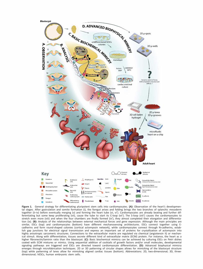
Figure 1. General strategy for differentiating pluripotent stem cells into cardiomyocytes. (A): Observation of the heart’s developmen-tal stages. After gastrulation and somite formation (i), the foregut arises and folding brings the two branches of splanchic mesodermtogether (ii–iv) before eventually merging (v) and forming the heart tube (vi, vi0). Cardiomyocytes are already beating and further dif-ferentiating but some keep proliferating (vii), cause the tube to start its C-loop (vii0). The S-loop (viii0) causes the cardiomyocytes tostretch even more (viii) and when the four chambers are finally formed (ix0), they almost completed their elongation and differentia-tion (ix). (B): Analysis of the relationships between external mechanical forces and gene expression. Although the main principles aresimilar, ESCs (top) and cardiomyocytes (bottom) have different mechanosensing architectures. ESCs connect together using E-cadherins and form round-shaped colonies (cortical actomyosin network), while cardiomyocytes connect through N-cadherins, estab-lish gap junctions for electrical signal transmission and express an important set of proteins for crystallization of actomyosin intohighly anisotropic sarcomeric structures. Connections to the extracellular matrix are regulated via chemical (angiotensin II) or mechan-ical stimuli. Along with differentiation, tissues secrete different kind of extracellular matrix (ECM) protein. For instance, the heart as ahigher fibronectin/laminin ratio than the blastocyst. (C): Basic biochemical mimicry can be achieved by culturing ESCs on Petri dishescoated with ECM mixtures or mimics. Using sequential addition of cocktails of growth factors and/or small molecules, developmentalsignaling pathways are triggered and ESCs are directed toward cardiomyocyte differentiation. (D): Advanced biophysical mimicryemerges through microfabrication techniques. 2D or 3D patterning of circular shapes allows for mimicking of the blastocyst structure(top) while patterning of lanes allow for mimicking aligned cardiac tissues (bottom). Abbreviations: 2D, two-dimensional; 3D, three-dimensional; hESCs, human embryonic stem cells.
compaction at the eight-cell stage, every single cell is onlyconnected to its neighbor through cell-cell interactions, usingproto E-cadherins and a few integrins [67, 68]. Upon cell com-paction, cells increasingly express E-Cadherins and startsecreting other types of cadherins [69], which can be seen asthe first differentiation step before implantation of theembryo in utero, as cells self-sort by cadherin-type affinity.Later, cells start to secrete more and more ECM components,like collagen, vitronectin, tenascin, elastin, fibronectin, hyal-uronic acid (HA), or laminin [70]. Cells then bind to thesecomponents through different mechanisms, for example, bycreating focal adhesion complexes through RGD (argininyl-glycyl aspartic acid. Peptide of sequence L-Arginine (R)-Glycyl(G)-Aspartic acid (D))-integrin interactions, or by other specificreceptors (like CD44 for HA) [71].
By the end of development, cells can sense externalforces either through cell-cell interactions, cell-ECM interac-tions, mechanosensitive ion channels, or by directly sensingthe force by wave propagation throughout the cell andtoward the nucleus [71]. Hence, in order to manipulate thestem cell niche, the surface chemistry at the interfacebetween the cell and the materials must reflect the integra-tion of all the coupled mechanical signals before it reachesthe cell membrane. Although not clearly demonstrated in theliterature, one can think that if ECM proteins are too weaklybound to the materials, cells will not be able to generate thesame amount of force, in similar ways as if it was linked to asoft material. Cells would then behave as if in suspension.Also, if ECM proteins are not abundant enough, integrin clus-ters and focal adhesions would be insufficient to form as effi-ciently as in a physiological context [72]. Cadherins andintegrins are both linked to the cytosolic network of actomyo-sin. When the tension of this network is changed, a signalingcascade occurs, involving the Rho pathway, directly deformingthe nucleus. These signals are then integrated within thenucleus leading to a differential gene expression and thusactivating or repressing various developmental pathways [73,74] (Fig. 1B).
Strategies for Mimicking Cell-Cell Communication andCell-ECM Coupling
Providing cell-cell communication capabilities and cell-ECMcoupling was the first strategy stem cell researchers haveused. In contrast to mESCs, one of the first attempts to cul-ture what we call now hESCs showed that regular Petri dishtreatments for cell adhesion allowed expansion but triggeredspontaneous differentiation as early as the second passage[75]. These dishes are made adherent by exhibiting positivelycharged functional groups. Although cells can form adhesions,they do not recapitulate the signals given by different ECMcomponents during embryogenesis. Additionally, the highbrand-variability in terms of nanotopography and surfacecharge can have dramatic effects on cell behavior [76].Because of their electrostatic properties, most of the ECMproteins can be adsorbed on the surface of tissue-culture-treated plates. Thomson et al. in 1998 [77], followed by Reu-binoff et al. in 2000 [78], were the first to demonstrate thathESCs could be cultured for months provided that they werecocultured on a layer of feeder cells. Later, feeder-free condi-tions were introduced by just coating a mixture of ECM pro-teins (Matrigel), or laminin on the surface of a Petri dish [67]
(Fig. 1C, top). However, it has been observed that ESCsbehaved differently depending on the coating protein: vitro-nectin would enhance self-renewal and proliferation of PSCwhile fibronectin, collagen IV, or laminin would drive moreeasily the differentiation of the cells toward various lineages.In addition to ECM proteins, the use of recombinant cadherincoating to mimic cell-cell interactions occurring betweenfeeders and ESCs has been reported [79].
Debate 2: Choice of the Right ECM. The choice of the rightECM proteins for cardiac SCD is intricate. Different ECM pro-teins at different densities are present in cardiac tissuesdepending on the developmental stage [70]. DifferentiatingPSCs on Matrigel by an Activin A/BMP4-based protocol, Chanet al. observed in vitro time-dependent levels of HA and versi-can in the cell culture [4]. This evolving composition in ECMcomponents not only affects the mechanical properties of theECM but also many signaling cascades triggered by these twoglycosaminoglycans (GAGs). Alternatively, Schenke-Laylandet al. [80] made murine embryonic bodies (mEBs) on disheseither coated with collagen I, collagen IV, laminin, or fibronec-tin, and evaluated mesodermal and cardiovascular markersafter culture in medium without any exogenous factor. Theyfound that collagen IV induced significantly more mesendo-dermal cells (characterized by high Flk1 expression) than otherECM proteins. After sorting these precursors and seedingthem again on the four coatings, cardiomyocytes appeared tobe significantly more present on fibronectin-coated dishes.Also for mESC, Stary et al. reported that the protein SPARCacted in similar ways as BMP2 by increasing cardiomyogenesisin EB with clear upregulation of Nkx2.5 [81]. These studieshighlight the fact that it might be questionable whether ornot it is best to let the cells generate their own ECM compo-nents or designing biomaterials to use them as source poly-mers to control simultaneously stiffness dynamics andchemical signaling.
INFLUENCE OF SUBSTRATE TOPOGRAPHY
General Overview
There is growing evidence that cell shape is an importantparameter during heart development (Fig. 1Avii–1Aix).Although cell proliferation may account for major mechanicalevents like asymmetric looping, it cannot explain the wholeprocess, such as how the looping direction is controlled orwhy the growing four chambers are different from oneanother. Indeed, oriented growth may explain how any of thefour chambers grow differently [82]. It has been reported thatthis kind of growth may be explained by mitotic spindle orien-tation [83], which can be directly correlated to cellular shapeand more generally ECM distribution [84]. Another event, car-diac looping, is also highly influenced by the cell’s geometricalshape. Studies in the chick have highlighted the fact that thecardiac tube starts looping not only by differential growth butmainly due to different morphologies of the cardiomyocytesat the outer curvature (elongated cells) as compared to theinner curvature (packed cuboidal cells) [85]. In the matureheart, cardiomyocytes are elongated and contract simultane-ously in the direction of elongation. A specific aspect ratiohas been correlated with healthy cardiomyocytes, and any
1024 Materials Design for Cardiac Differentiation
VC AlphaMed Press AlphaMed STEM CELLS
variation to that aspect ratio may mimic features of the failingmyocyte [86].
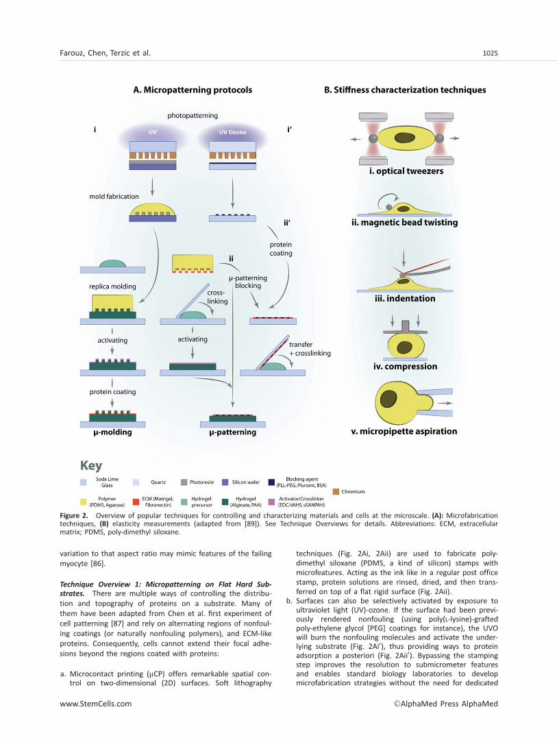
Technique Overview 1: Micropatterning on Flat Hard Sub-
strates. There are multiple ways of controlling the distribu-tion and topography of proteins on a substrate. Many ofthem have been adapted from Chen et al. first experiment ofcell patterning [87] and rely on alternating regions of nonfoul-ing coatings (or naturally nonfouling polymers), and ECM-likeproteins. Consequently, cells cannot extend their focal adhe-sions beyond the regions coated with proteins:
a. Microcontact printing (mCP) offers remarkable spatial con-trol on two-dimensional (2D) surfaces. Soft lithography
techniques (Fig. 2Ai, 2Aii) are used to fabricate poly-dimethyl siloxane (PDMS, a kind of silicon) stamps withmicrofeatures. Acting as the ink like in a regular post officestamp, protein solutions are rinsed, dried, and then trans-ferred on top of a flat rigid surface (Fig. 2Aii).
b. Surfaces can also be selectively activated by exposure toultraviolet light (UV)-ozone. If the surface had been previ-ously rendered nonfouling (using poly(L-lysine)-graftedpoly-ethylene glycol [PEG] coatings for instance), the UVOwill burn the nonfouling molecules and activate the under-lying substrate (Fig. 2Ai0), thus providing ways to proteinadsorption a posteriori (Fig. 2Aii0). Bypassing the stampingstep improves the resolution to submicrometer featuresand enables standard biology laboratories to developmicrofabrication strategies without the need for dedicated
Figure 2. Overview of popular techniques for controlling and characterizing materials and cells at the microscale. (A): Microfabricationtechniques, (B) elasticity measurements (adapted from [89]). See Technique Overviews for details. Abbreviations: ECM, extracellularmatrix; PDMS, poly-dimethyl siloxane.
Farouz, Chen, Terzic et al. 1025
www.StemCells.com VC AlphaMed Press AlphaMed

nano-fabrication clean rooms [88]. Nevertheless, one ofthe limitations might be that UV–ozone lamps still are animportant investment and still too few platforms for cellbiology provide them.
Technique Overview 2: Micropatterning on Soft Materials
and Curable Polymers.
a. Stamps can be fabricated by soft lithography (for microme-ter resolution) or by standard machining (micromilling,three-dimensional [3D] printer) for bigger features (Fig.2Ai). Instead of transferring proteins, they can act as rep-lica molds while a polymer or hydrogel polymerizes (Fig.2A, left). Depending on the depth and spacing of the fea-tures, it provides the materials with quasi-2D microstruc-tures (low spacing and height <100 nm) or with real 3D-cues (larger spacing and height >500 nm). Proteins arethen coated uniformly, and thus cells will not constrain byadapting to changes in chemical signals, but by sensinglocal variations in topography [90]. If deep enough, thesetechniques can be adapted for miniaturization of culturewells for high-throughput screening on 3D aggregates [91,92].
b. Coating proteins chemically on soft material is affected bythe structural properties of the material itself, and proteinsurface density will vary as a function of local porosity.When using polyacrylamide (PAA), rather than using opto-chemical reactions to bind the proteins as in Engler et al.in 2006 [2], it has been recently suggested that simpletransfer of proteins could be achieved by polymerizing asolution of acrylamide/bis-acrylamide sandwiched betweena micropatterned surface and a glass coverslip [93] (Fig.2A, right), or simply using mCP on polymerized PAA ren-dered adhesive with hydroxyl AOH groups [94] or withcoupled streptavidin-acrylamide [95] (in this case, proteinshould be biotinylated prior to coating) (Fig. 2A, center).Further studies are needed to confirm that these techni-ques overcome, as claimed, the artifact of the effect oncell mechanosensing of PAA’s variation in porosity withregard to elasticity.Many variations on this theme are proposed depending
on the way the hydrogel of interest is polymerized, like pho-tocuration for methacrylated or PEG-DA-based hydrogels [96,97]. Although many 3D-patterning techniques are emerging,their resolution still does not allow for single cell studies. Wewill briefly describe them in the Debate 4.
Influence of Topography on SCD into MesodermalProgenitors and Cardiomyocytes: The Importance ofColony Size
The first step in deriving cardiomyocytes from ESCs in vitro isa step of specification. Like during embryogenesis, cells firstdifferentiate into one of the three germ layers (namely ecto-derm, mesoderm, and endoderm, Fig. 1Ai). These three layersare more restricted in their fate and cardiomyocytes can onlyarise from mesodermal or mesendodermal cells. Peerani et al.showed that colony size mattered in driving this first specifica-tion [98]. They microcontact printed Matrigel islands of vary-ing diameters on glass coverslips and seeded them with ESCswithout any exogenous inductive signals (Fig. 1D, top). Byconstraining the size of the aggregates, they showed that,after 2 days in culture, smaller aggregates (200 mm in diame-ter) expressed more endodermal markers and had higher lev-els of BMP2 whereas bigger aggregates (1,000–1,200 mm in
diameter) expressed more pluripotent markers. This wasattributed to the modulation of the ratio of pSmad1 agonistsover pSmad1 antagonists. Indeed, while pSmad1 antagonistsincreased with colony size, no correlation was found forpSmad1 agonists, thus leading to lower levels of agonists insmaller colonies when compared with pSmad1 antagonists.Later, they repeated the same experiment but adding ActivinA and BMP2 in the culture medium to force mesendodermaldifferentiation [99]. This time, cells on smaller spots (200–400mm in diameter) were found to express more endodermalmarkers (GSC, Sox17, and Cer1) while bigger patterns (800–1,200 mm) led to more mesodermal cells (T, KDR). To investi-gate further the hypothesis that colony size influences differ-entiation by changing cell number and thus localconcentrations in proteins and chemical, they used microtiterplates coated with Pluronic F-127 (a poloxamer used to createnonfouling regions on glass) to generate EBs by centrifugalaggregation [100, 101]. After sorting these cells for KDR, c-kit,and cTnt (markers for early to late cardiomyocyte differentia-tion), they concluded that the largest number of cells wasreached at an optimum of approximately 1,000 cells peraggregate. Endodermal markers were predominantly found atthe periphery of the aggregates, creating a higher ratio ofendodermal cells over other cells for smaller aggregates(when calculating the surface/volume ratio).
Similar conclusions were drawn by Hwang et al. [102]after they cultured ESC on nonfouling micromolded poly-ethylene glycol (PEG) wells of different diameter (from 150 to450 mm). Endothelial cells emerged from cells that had beencultured in small aggregates while cardiomyocytes emergedfrom cells cultured in bigger aggregates. Interestingly, theyhave been able to correlate these differences with modulationin Wnt5a and Wnt11, two important regulators of the nonca-nonical Wnt pathway.
Together, these four studies suggest that differential differ-entiation emerges from size-induced variations in the concen-tration of modulators of key signaling pathways involved incardiogenesis (Nodal, BMP, and Wnt). Although qualitativelyconsistent with each other, these studies were not performedwith the same ranges of diameters, and in a similar experi-ment by Mohr et al., the greater relative number of cardiomy-ocytes was reached for 300 mm-diameter EBs [103].
An explanation could be that they neglected the impactof mechanotransduction. In these studies, radii of curvatureare different depending on the spot size and therefore a sur-face tension emerges at the periphery of the colony whereaspressure is increasing inside the colony due to cell prolifera-tion, as suggested by Nelson et al. [104]. Also, PEG microwells[102] might have been softer than gold-coated polystyrenewells [103].
At a completely different scale, Myers et al. compared sixof the most popular differentiation protocols (all based onexogenous signals modulating either the BMP or the Wntpathways) while constraining initial colony growth to 2-mmwide spots of Matrigel [105]. Clearly, micropatterningincreased homogeneity in the yields of differentiation. Mostimportantly, this study highlighted the important discrepanciesthat can affect cardiac differentiation depending on the choiceof the modulated pathway. Put together, these data empha-size the importance of controlling the size of cell aggregatesto optimize the cardiomyocyte yield.
1026 Materials Design for Cardiac Differentiation
VC AlphaMed Press AlphaMed STEM CELLS
Influence of Topography on Sarcomere Maturation andForce Generation of Cardiomyocytes: The Importanceof Anisotropy
Contact guidance and topographical effects on cardiomyocytematuration have been studied in many ways. At the single-cell level, in vitro studies on micropatterns [86, 106, 107]and in silico models of sarcomerogenesis [108] conducted byParker’s group suggested that a single cell will form moremature sarcomeres when its shape is constrained to a rec-tangle with an aspect ratio length/width of roughly 7:1. Ifthe ratio is bigger, interdisc space will become bigger andcells will behave as a hypertrophied cardiomyocyte. Ifsmaller (like in the case of circular patterns), sarcomeres willnot be able to align in the same direction. They will bemore randomly distributed within the cells, as characterizedby orientation factors, and they will hence generate lessforce. At the multicellular level, they made the same obser-vations regarding force generation (Fig. 1D, bottom). In thiscase, what mattered was less the aspect ratio than the elon-gated shape itself. If not all the cells were elongated in thesame direction, the gap junctions were less well establishedand electromechanical coupling was inefficient in the tissue.The force generated was then considerably smaller than foranisotropic tissues [109–111]. These results have also beenconfirmed in 3D collagen gels encapsulating neonatal rat car-diomyocytes (NRCMs) [112], thus confirming the importanceof anisotropy itself. A recent study by Wang et al. highlightsthat in the case of ESC-derived cardiomyocytes (ESCd-CM),seeding cells on a topographically aligned substrate did notimprove the maturation state of the cells. The improvementsin term of electrical stability and reduction in inducedreentrant arrhythmias were solely due to the spatial organi-zation, as assessed by monophasic action potential measure-ments [113].
Anisotropy in Early Differentiation and Isotropy inESCd-CM Studies
If many studies have shown similar results with NRCMs[114–120], ESCd-CMs [114, 119], or extracted CPCs [121],there is little evidence of any effect of anisotropy of the ECMproteins on earlier stages of mesodermal differentiation [122,123]. It could be interesting to know at which stage of devel-opment cells are able to sense lines of proteins and startaligning. This switch could correspond to the loss or the gainof a phenotype, like, one might suggest, the vanishing of theprimary cilium, an important mechanosensing feature involvedin cardiogenesis [124]. Cardiomyocytes could also start align-ing because fibroblasts first aligned and hence anisotropicallysecreted ECM proteins, driving the orientation of other cells[125–127]. Nevertheless, anisotropy is involved in many mor-phogenetic events and for instance, researchers recentlyreported that the culture of human PSCs (hPSCs) on nano-grooved surfaces let to rapid differentiation of the cells intoneurons [128]. In a completely different approach, it wasfound that reprogramming of fibroblasts into iPSCs wasenhanced on grooved substrates due to increased acetylationand methylation of histone H3 [129]. This phenomenon couldimply that anisotropy would act as a global inducer of epige-netic modifications leading to increased genetic sensitivity todifferentiation/reprogramming protocols.
As for culturing beating cardiomyocytes on circular spots,experiments reported above do not really encourage it in thatit would rather mimic pathological behaviors than physiologi-cal ones. However, in light of the high-throughput drug-screening platform designed by Serena et al. [130], one canargue that circular microtissues of cardiomyocytes might stillgive precious information on the relative behavior of cellssubjected to different kinds of drugs.
INFLUENCE OF TISSUE ELASTICITY
General Overview
As cells are differentiating, their phenotype considerablychanges, that is, not only their cytosolic composition isaffected (resulting in a different stiffness by polymerization ofthe cytoskeleton), but their proliferation rate as well. Regionsof differential growth can be observed, leading to differentcompressive or tensional forces applied to their neighbors.The composite material that is the cell environment consider-ably changes—the overall stiffness changes as well as thestretching forces. At the same time, cells are rearranged byaffinity due to the different expression and amounts of cad-herins. Mimicking these dynamic changes of highly nonlinearmaterials is obviously very challenging but simple models ofconstant linear elasticity have shown important results onSCD.
Not only the cell-cell interactions but also the cell-ECMinteractions play a large role in cardiac differentiation andmaturation. Particularly, the cardiac jelly, which interfaces theendocardium and the myocardium, is composed of GAGs(such as HA), proteoglycans, and proteins (such as fibronectin,collagen, or laminin). Based on the properties of GAGs toosmotically attract water, the cardiac jelly acquires a compres-sive strength and the internal pressure it generates on themyofibrillar architecture of the myocardium is thought todrive cardiac chamber expansion [20].
Technique Overview 3: Measurement of Tissues Elasticity in
the Developing and Adult Muscle. In order to mimic themechanical properties of cardiac tissues, it is important to beable to have precise measurements from tissue samples atdifferent stages of the development. The difficulty relies ondefining precisely the “substrate”: is the tissue in its whole,only the ECM or the ECM plus the support cells? And if wedeplete the tissue from their cells, what are the effects of thedecellularization techniques on the mechanical properties ofthe tissue? Many laboratories have tried doing such measure-ments, either using atomic force microscopy (AFM) or stand-ard rheology or tensile/compression testing. Moreover, elasticanisotropy (different stiffness in orthogonal directions) hasbeen identified for cardiac tissue but is rarely considered as arequirement for biomimetic cardiac constructs in the litera-ture [131]. Furthermore, measuring the stiffness of a materialwill not mean much if the bonding force with the coatedECM protein is too weak [132].
In the field of tissue engineering, the measure mostreported for characterization a material’s elasticity is theYoung’s modulus. However, depending on the technique used,it can be indirectly derived from more or less complex mathe-matical extrapolations of other mechanical characteristics.
Farouz, Chen, Terzic et al. 1027
www.StemCells.com VC AlphaMed Press AlphaMed
a. Nanoscale elasticity can be measured by magnetic/opticaltweezers. Nanobeads are constrained in a magnetic fieldand attached to the material. The material is then pulledaway from the beads and the force needed to maintainthe bead inside the field is measured (Fig. 2Bi). Similar set-ups can use twisting of the beads to measure shear elastic-ity (Fig. 2Bii).
b. Nanoscale and microscale elasticity can also be measuredby AFM in contact mode (Fig. 2Biii). The cantilever tip ismodeled as a pyramidal or spherical tip in Herzian contactwith the material. Measuring the deformation upon nano-indentation is directly related to the Young’s modulus andthe Poisson ratio of the material.
c. Microscale elasticity of cells or materials can be measuredby micrograph analysis of deformation upon an appliedforce. Cells and materials can be squished between plates(Fig. 2Biv), or aspirated through micropipettes (Fig. 2Bv).
d. Mesoscale elasticity (cell aggregates and hydrogel micro-spheres) can be measured by micropipette aspiration (Fig.2Bv) as well as microrheology. In microrheology, fluores-cent nanobeads displaced inside the tissue or material areoptically tracked and statistical analysis of their randommotion can be related to a measure of elasticity.
e. Mesoscale indentation can also be performed with simplehandmade setups by measuring the deformation of aweight or small tip on the material or by hooking a smallweight under the material to measure the implieddeformation.
f. Bulk elasticity of macroscale materials is often measuredby standard mechanical engineering testing machine, likethe Instron. It can be set to apply uniaxial or biaxial strainin tension or compression on the tissues.
Influence of Elasticity on Mesodermal SCD
In cardiac repair, it has initially been thought that the use ofstriated muscle, be it skeletal muscle or cardiac, would be suf-ficient for engraftment and contraction inside the infarctedheart. They have quite similar mechanical properties and onecan think that the properties of the matrix will feature aroughly similar elasticity. When Engler and Discher firstreported that matrix elasticity had a great influence on SCD[2], they showed they had been able to make MSCs differenti-ate into myoblast-like cells on polyacrylamide gels of about 10kPa in elasticity. From then on, material design studies for car-diac patches report measurements of the elastic properties oftheir materials in order to show that they closely matchnative heart muscle [90, 131, 133–136].
However, no strong evidence has been shown yet duringthe entire process of cardiac differentiation and experimentsare often performed only after exposition to exogenous differ-entiation chemicals. Culturing preimplantation stage embryoson 2D PDMS substrates of varying stiffness resulted in signifi-cantly greater frequency of development from the two-cellstage to the hatching blastocyst stage, as compared to cul-tures in standard Petri dishes [137]. Sun et al. linked higherlevels of Oct4 expression to higher stiffness when seedinghESCs on 2D PDMS micropillars but did not really look at thewhole panel of genes expressed by the three germ layers[138].
As explained in Debate 1, many differences can emergewhen switching from a mouse model to human cells. But asimportantly, researchers recently started to point out that the
influence of elasticity on SCD varies depending on the dimen-sionality (2D or 3D) of the materials [139]. Indeed, the distri-bution of ECM cues is completely different and affect theresponse of the membrane receptors (integrins for instance).It has been reported that cells in 2D tend to form more stressfibers than in 3D, which could prevent cardiomyocytes fromforming mature sarcomeres [140]. Also, if high stiffness allowscell to generate high traction forces in 2D, it is not true in 3Dwhere high stiffness can prevent the cell from moving aroundand force it to stay round. Interestingly, Zoldan et al. per-formed a strong investigation on the effect of elasticity onearly specification in the three germ layers by encapsulatinghESC into 3D polymers [141]. As suspected before, there is astrong correlation between the material’s stiffness and germlayer differentiation. Surprisingly, the ranges of measured elas-ticity are several orders of magnitude higher than in the otherstudies afore-mentioned. Also, few details are given on theimpact of the surface chemistry of the various polymers usedin this study.
One study, nevertheless, has been able to correlate car-diac differentiation to matrix stiffness, through the regulationof YAP/TAZ expression [62]. Using Sca11 adult CPCs seededon polyacrylamide gels of controllable stiffness, the authorshave showed that the control CPCs differentiated best intocardiomyocytes on gels of 10 kPa in elasticity, while YAP-silenced CPCs would not differentiate at all into cardiomyo-cytes under the same conditions.
Influence of Elasticity on Cardiomyocyte SarcomereMaturation
Sarcomere maturation is probably the most studied effectamong mechanical properties. By isolating cardiac cells at dif-ferent developmental stages of the mouse embryo, Engleret al. have been able to establish a relationship between thevariation in elasticity and the developmental stage of theheart [133]. Interestingly, it has been shown that embryoniccardiomyocytes beat best on substrates where the rigiditymatches the embryonic tissue rigidity. These cardiomyocyteswere only able to form mature sarcomeres on PAA gels ofapproximately 10 kPa. On softer gels, sarcomeres were lessspaced and not fully organized whereas on stiffer gels, sarco-meres were not present at all and some sarcomeric proteinswere completely diffuse in the cytoplasm. This result was alsoconfirmed for NRCMs [142] as well as for ESCd-CMs [134,143], although Hazeltine et al. found that the intermediatestiffness supporting best ESCd-CM differentiation from a pro-genitor stage would be approximately 50 kPa [143]. Previousstudies on the differentiation of adipose stem cells into myo-tubes had already shown that pathological tissue stiffness ledto reduced differentiation whereas tissue-like elasticity led tooptimal maturation and striation formation of the myotubes[144].
Not surprisingly, it has been shown that depending on thetype of extracellular element that was linked to the hydrogel,there could be different cellular responses to stiffness. Cardio-myocytes have been able to grow mature sarcomeres on HA-based gels of less than 500 Pa whereas it was only achievedat around 10 kPa with fibronectin-coated PAA gels [145]. Thisstarts to show the limits of considering the Young’s modulusas the principal mechanical parameter influencing cell pheno-type. It is thought that the HA signaling cascade could bypass
1028 Materials Design for Cardiac Differentiation
VC AlphaMed Press AlphaMed STEM CELLS
the mechanotransduction pathway and direct sarcomerogene-sis. One explanation could be that in this case, HA does notconvey its message through mechanical signaling but ratherthrough chemical interaction with the CD44 receptors [146].However, it was also shown that the dynamic properties ofHA gels are actually important in the cell response. WhereasPAA has a static elastic behavior, HA gels show time-dependent stiffening and thus can go from approximately 1kPa to almost 10 kPa depending on its fabrication process[147]. This phenomenon was also observed in 3D fibrin cul-tures of myoblasts, where the fibrin gel, initially softer than 5kPa, exhibited a stiffness of 15 kPa after several days [139].
This highlights the fact that static mechanical signals alonecannot act as a replacement of chemical induction of differen-tiation. Both signals are complementary and will act synergis-tically in a time-dependent manner. Furthermore, the point ofintroducing mechanical compliance and mechanical stimuli incell culture is to avoid mixed signals. As cells sense theirmechanical environment, adding promyogenic soluble factorscan be hindered by the pro-osteogenic mechanical signals ofthe rigid Petri dish.
Debate 3: Elasticity Versus Viscosity and Porosity. If currenttechniques of microfabrication are increasingly simple, a fewbiases arise from the technique itself. Trappmann et al.recently suggested that depending on the polymer (they com-pared PAA and PDMS), the results shown initially by Englerand Discher could not be reproduced in the case of PDMS[132]. This could be explained by a difference in porosity andtethering of the ECM proteins to the synthetic polymer. SoftPAA gels (that led to neuronal differentiation) were highlyporous and the ECM density at their surface was considerablylower than on stiffer gels, but these problems were notobserved for PDMS, which has a more constant porosity. Tofurther complexify this issue, a new study addressing theproblem of porosity in soft polyacrylamide gels has refutedthe hypothesis that tethering had biased the observation ofdifferential fates depending on elasticity [148]. Yet it is unclearwhether tethering of ECM is the only source of this differenceas very soft PDMS made out of big ratio of the PDMS baseand curing agent can exhibit significant viscous behaviors. Thebalance between the viscous modulus and the elastic modu-lus of this kind of soft PDMS directly influenced the cell’smigration properties [149]. Although other types of PDMShave been reported as good candidates for traction forcestudies in purely elastic conditions [150, 151], there is nodoubt that further studies will underpin the importance ofviscosity during guided differentiation of ESCs intocardiomyocytes.
PERSPECTIVES
Influence of Stretching and Shearing
In addition to designing biomimetic materials for stem cellculture and differentiation, other mechanical stimuli can bedynamically applied to the cells. While shearing is consideredto be of prime importance in vascular remodeling, cyclicstretching and static strains mimic more faithfully themechanical behavior of muscle tissues. The effect of stretchingon signaling pathways has been reported in review by Riehl
et al. [152]. When subjected to static stretching, ESCs andiPSCs increased their markers of (cardio-) myogenic differen-tiation [153]. Although many setups exist for stretching cells(in 2D or 3D), like magnetic stretcher, manual stretcher, orvacuum stretching [152], the results are usually consistent.
As for the cardiac cells, it has not been shown that stretch-ing PSCs would act synergistically with chemical factors for theinduction of cardiac differentiation. In this particular case, ithas only been shown that either NRCM, endogenous CPC, orstem cell-derived cardiomyocytes (SCd-CM) had better contrac-tility and sarcomere maturation when subjected to cyclicstretching [154, 155]. Cyclic stretching enhanced their align-ment and favored gap junction formation for a better electro-mechanical coupling. Not only can stretching improve thealignment of cells, but also of the 3D matrix that surrounds thecells. When static strain was applied on a polymerizing gel offibrin, the nanofibers of the mesh tended to align in thestretching direction [139, 156, 157]. A global review on someother physical signals that can affect cardiac differentiation pro-tocols can be found in the review by Ghafar-Zadeh et al. [158].
Influence of Electrical Signals on the Maturation ofCardiomyocytes
Many efforts have been made to develop either conductivescaffolds or electrically stimulated systems for recreating thecardiomyocyte’s natural environment [159–162]. In thesearticles, emphasis is put on trying to mimic a healthy environ-ment to avoid the appearance of diseased phenotypes. Thechanges that have been demonstrated using these manyproof-of-concept materials are more related to phenotypicchanges in already mature cardiomyocytes, rather than tospecification, differentiation, or maturation events in cardio-vascular progenitors or PSCs. One of the main improvementsobserved by electrical stimulation is an increase in connexin43(Cx43) expression. This is a phenotypic change that consider-ably improves cardiac contraction as compared to standard invitro controls (without electrical stimulation).
The goal of these new materials and systems is either tohelp implanted cardiomyocytes to integrate better with thehost’s cardiomyocytes or to drive pathological cardiomyocytesto regain a healthier phenotype by expressing more Cx43.Future studies involving the culture of immature cardiogeniccells or even PSC using these materials will be needed toaddress questions like the relationship of cardiac progenitorphenotype and the establishment of electrical currents in thedeveloping heart.
Debate 4: 2D Versus 3D Culture. 2D models helped under-stand the signaling cascades in mechanotransduction, but thisis still far from reality and adding a third dimension should bethe next step. However, although 2D soft and patternedmaterials can now be prepared in a relatively high scale, 3Dtechniques are still cost-prohibitive, time-consuming andpoorly resolved. Multi-photon technologies allow for 3D pat-terning by in situ chemical bonding of proteins to the back-bone materials [163] as well as for the localized cellencapsulation by polymerization of photosensitive materials[164]. If these techniques can be quite slow, complex micro-fluidic systems have been designed for high-throughput anddigitally tunable fabrication of 3D-patterned cell-laden fibers
Farouz, Chen, Terzic et al. 1029
www.StemCells.com VC AlphaMed Press AlphaMed
and sheets [165, 166]. Yet the impact on PSC viability, pluripo-tency, and differentiation potential remains to be addressed.
Another pitfall would be to expect the same effects thanpreviously shown on 2D substrates on 3D substrates from thesame material or a material with similar mechanical proper-ties. Indeed, the distribution (and production) of membranereceptors will be completely rearranged. Additionally, embed-ding cells in a 3D matrix will paradoxically decrease the num-ber of degrees of freedom of the cell system. While 2Dsubstrates allow cells to move freely on the topside, 3D mate-rials constrain the cell in all directions. The stress distributionwill be completely different. This is even more true when con-sidering the integration of matrix metalloproteinase-degradable sites inside 3D scaffolds, as it has been shownthat it would induce, at least in MSCs, traction forcesdifferent from those seen in nondegradable materials, thusleading to different fates [167].
As for in vivo cardiomyogenesis and embryogenesis, cellsare arranged either in 3D or in 2D assemblies depending onthe developmental stage. At first, cells are clustered in 3D butsoon form the three germinal layers, which can be roughlymodeled in 2D. Later on, gastrulation leads to the primitivestreak formation and the mesodermal cells that will becomethe heart form 3D structures again. But after cardiac loopingand the beginning of the formation of the four chambers, car-diomyocytes are stretched and eventually define the final myo-cardium. Although three dimensional in theory, ultrasoundsand diffusion tensor magnetic resonance imaging studies havehighlighted the fact that the myocardium is composed of layersof anisotropic cardiomyocytes oriented with a variable anglefrom the endocardium to the epicardium. Therefore, the myo-cardium can also be described as a monolayer of aligned cellswrapped around the ventricle with varying angles. In that case,one could argue that 2D models are better. Nevertheless, thequestion remains whether the best strategy would be to createa 3D scaffold and push it to self-organize into a multi-layeredstructure of aligned cells (like what happens during cardiogene-sis [168]) or to force the cells to align on multiple layers beforestacking them (as proposed by Takahashi [169] et al. for thedesign of cardiac patches [170]). In both cases though, the lim-its of oxygen and nutrient diffusion will have to be overcomein order for the construct to be viable, and this is particularlyrelevant to cells embedded in its core.
Comments on Nanofibers
Nanofibers have been extensively used to show the impor-tance of surface roughness and hydrophilicity in maintainingpluripotency in PSC cultures. They are often cited as a simpleway to have mesh-like topographies or substrates with alignedfibers [171]. Also, new techniques demonstrate the possibilityto overcome the slow speed, high variability [172, 173] andhigh cost of electrospinning [174]. However, generating fibersonly allows for two kinds of patterns: random fibers andaligned fibers. Whenever different shapes are needed, electro-spinning has still quite a poor resolution [175] and needs tobe combined with the most popular techniques describedhere above [176, 177].
Pharmacological and Medical Applications
In this review, we have seen that two parameters, substrateelasticity and ECM protein patterns, had a strong influence on
sarcomerogenesis and cardiac maturation. Recent advances inmaterials design have led to the possibility of studying thesynergetic effect of both properties [93–95, 136, 178] and itappears that appropriate stiffness (approximately 10 kPa) andaspect ratio (7:1) enabled optimal sarcomeric organizationand optimal contractility both at the single-cell level but alsoat the cell pair level [136]. This study highlights the fact thatcell-cell coupling also results from a balance of forces influ-enced by the environment.
Whether the end goal of producing striated muscle cellsis to develop toxico-pharmacological assays [11, 12, 179–182]or regenerative solutions [18, 183–185] does not change thefact that cells would have to behave like in a healthy muscle.The fabrication of scaffolds for regenerative medicine has thesame requirements than when mimicking developmentalmechanics, but with additional constraints. First, in the caseof surgical applications, structural integrity of the patch is ofprime importance. Although cell sheets are a tempting optiondue to their fully natural composition and their promisingresults in cardiac failure [169, 186–188], they were found tobe quite fragile and hard to handle. Second, to our knowl-edge, there is still no work reported on the combined influ-ence of the materials properties described here on earliermesodermal induction and cardiac differentiation. One cansuggest that ESCs or iPSCs will behave like MSCs and differen-tiate into cardiomyocytes at around 10 kPa. However, studiesreported above suggest that cell density will have an impor-tant impact as well. This complexity needs to be addressedwhen determining the whole range of materials that need tobe used from the extraction, reprogramming, or thawing ofstem cells to the patch fabrication. On one hand, scaffold-freetechniques would ideally emerge and give rise to bioreactorsthat act directly on the 3D stem cell aggregate by providingchemical, mechanical, and electrical stimulations to producehighly pure cardiomyocytes. These techniques would naturallyovercome additional constraints related to the “memory” acell can have of its previous mechanical environments, which,as described by Yang et al. [189], can affect cell’s fate in agiven physical environment. On the other hand, it might bemore realistic in a nearer future to develop multiphasic cul-ture systems: one biomaterial-based bioreactor for early com-mitment of ESC, then a second biomaterial-based bioreactoronto which cells would be transferred for cardiac maturation(in parallel to the production of endothelial and smooth mus-cle cells) and eventually, the three cell types would be mixedin a third bioreactor prior to implantation onto the failingmyocardium [190–192]. Notwithstanding the complexity ofstreamlining the translational process for adapting theseapproaches to wide-scale clinical applications, a key and yetunsettled issue is that one still ignores whether highly maturecardiomyocytes integrate better or not than earlier progenitorcells in the host myocardium.
In a nutshell, the ideal scaffold for differentiation andmaturation will likely be a scaffold that can diffuse some keygrowth factors, like BMP2 or Wnt inhibitors, have the appro-priate stiffness, and be coated with adhesion molecules thatcan trigger mechanosensitive events, such as ECM protein orGAGs, or, when considering upscale of these processes, shortmimicking peptides. These elements would have to be pat-terned to direct oriented proliferation and spreading of thedifferentiating cardiomyocytes. Such materials have been
1030 Materials Design for Cardiac Differentiation
VC AlphaMed Press AlphaMed STEM CELLS
partially reported by Agarwal, Farouz et al. when they showedthat NRCM could adhere, align, and spread on soft micropat-terned calcium-alginate scaffolds functionalized with fibronec-tin. These muscular thin films showed enhanced contractionwhen stimulated electrically as compared to isotropic equiva-lents. Thus, using these kinds of scaffold for ESC differentia-tion could be the beginning of new standardized culturingconditions for cardiac differentiation [90].
CONCLUSIONS
Whether mechanical forces arise by activation of pathwaysresulting in varied cytoskeleton behavior or developmentalpathways are modulated in response to cytoskeletal modifica-tions promoted by mechanical forces is unclear. It can beseen as a “chicken and the egg” kind of question and under-mining one or the other component can dramatically affectthe outcome of the experiments to be performed.
A few efforts have been made toward the establishmentof a global interactome of cardiopoiesis [193–196] and thiskind of approach keeps being expected by cardiovascularresearchers [197]. Additionally, in silico models are increas-ingly efficient in predicting cardiogenic events during develop-ment [198]. Combining the latest high-throughput and high-content technologies [25, 199] to the techniques described
here in the design of micropatterned stiffness-controlledmaterials will eventually unravel many of the mysteries of car-diac development and provide invaluable information for theestablishment of robust analytical models [200] and reliablepatient-specific therapies.
ACKNOWLEDGMENTS
This review has been partially funded by the Leducq Founda-tion through the ShapeHeart network. Y.F. is supported by the“Fondation pour la Recherche M�edicale” (FDT20140930860).YF is also supported by “Ecole Doctorale Frontieres du Vivant(FdV) – Programme Bettencourt”.
AUTHOR CONTRIBUTIONS
Y.F.: conception and design, manuscript writing, figure organi-zation and drawing, and final approval of manuscript; Y.C.:final approval of manuscript; A.T.: conception and design,manuscript writing, and final approval of manuscript; P.M.:conception and design and final approval of manuscript.
DISCLOSURE OF POTENTIAL CONFLICTS OF INTEREST
The authors indicate no potential conflicts of interest.
REFERENCES
1 Mummery CL, Zhang J, Ng ES et al. Dif-ferentiation of human embryonic stem cellsand induced pluripotent stem cells to cardio-myocytes: A methods overview. Circ Res2012;111:344–358.
2 Engler AJ, Sen S, Sweeney HL et al.Matrix elasticity directs stem cell lineagespecification. Cell 2006;126:677–689.
3 Radisic M, Park H, Chen F et al. Biomi-metic approach to cardiac tissue engineering:Oxygen carriers and channeled scaffolds. Tis-sue Eng 2006;12:2077–2091.
4 Chan CK, Rolle MW, Potter-Perigo Set al. Differentiation of cardiomyocytes fromhuman embryonic stem cells is accompaniedby changes in the extracellular matrix pro-duction of versican and hyaluronan. J CellBiochem 2010;111:585–596.
5 Tan G, Shim WS, Gu Y et al. Differentialeffect of myocardial matrix and integrins oncardiac differentiation of human mesenchymalstem cells. Differentiation 2010;79:260–271.
6 Schenke-Layland K, Angelis E, Rhodes KEet al. Collagen IV induces trophoectodermdifferentiation of mouse embryonic stemcells. Stem Cells 2007;25:1529–1538.
7 Zhang J, Klos M, Wilson GF et al. Extrac-ellular matrix promotes highly efficient car-diac differentiation of human pluripotentstem cells: The matrix sandwich method. CircRes 2012;111:1125–1136.
8 Uosaki H, Andersen P, Shenje LT et al.Direct contact with endoderm-like cells effi-ciently induces cardiac progenitors frommouse and human pluripotent stem cells.PLoS One 2012;7:e46413.
9 Brown K, Doss MX, Legros S et al. eXtra-embryonic ENdoderm (XEN) stem cells pro-
duce factors that activate heart formation.PLoS One 2010;5:e13446.10 Shimko VF, Claycomb WC. Effect ofmechanical loading on three-dimensional cul-tures of embryonic stem cell-derivedcardiomyocytes. Tissue Eng Part A 2008;14:49–58.11 Hansen A, Eder A, B€onstrup M et al.Development of a drug screening platformbased on engineered heart tissue. Circ Res2010;107:35–44.12 Grosberg A, Alford PW, McCain MLet al. Ensembles of engineered cardiac tis-sues for physiological and pharmacologicalstudy: Heart on a chip. Lab Chip 2011;11:4165–4173.13 Garbern JC, Lee RT. Cardiac stem celltherapy and the promise of heart regenera-tion. Cell Stem Cell 2013;12:689–698.14 Menasch�e P. Embryonic stem cells pacethe heart. Nat Biotechnol 2004;22:1237–1238.15 Menasch�e P. Stem cells for clinical usein cardiovascular medicine: Current limita-tions and future perspectives. Thromb Hae-most 2005;94:697–701.16 Laflamme MA, Chen KY, Naumova AVet al. Cardiomyocytes derived from humanembryonic stem cells in pro-survival factorsenhance function of infarcted rat hearts. NatBiotechnol 2007;25:1015–1024.17 Blin G, Nury D, Stefanovic S et al. Apurified population of multipotent cardiovas-cular progenitors derived from primate pluri-potent stem cells engrafts in postmyocardialinfarcted nonhuman primates. J Clin Invest2010;120:1125–1139.18 Chong JJH, Yang X, Don CW et al.Human embryonic-stem-cell-derived cardio-myocytes regenerate non-human primatehearts. Nature 2014;510:273–277.
19 Kirby ML, Waldo K. CardiacDevelopment. Oxford: Oxford UniversityPress, 2007.20 Rosenthal N, Harvey RP. Heart Develop-ment and Regeneration. Boston: AcademicPress, 2010.21 Malizia AP, Wang D-Z. MicroRNAs in car-diomyocyte development. Wires Syst BiolMed 2011;3:183–190.22 Lee S-Y, Ham O, Cha M-J et al. The pro-motion of cardiogenic differentiation ofhMSCs by targeting epidermal growth factorreceptor using microRNA-133a. Biomaterials2013;34:92–99.23 Eulalio A, Mano M, Dal Ferro M et al.Functional screening identifies miRNAs induc-ing cardiac regeneration. Nature 2012;492:376–381.24 Nazareth EJP, Ostblom JEE, L€ucker PBet al. High-throughput fingerprinting ofhuman pluripotent stem cell fate responsesand lineage bias. Nat Methods 2013;10:1225–1231.25 Willems E, Spiering S, Davidovics Het al. Small-molecule inhibitors of the Wntpathway potently promote cardiomyocytesfrom human embryonic stem cell-derivedmesoderm. Circ Res 2011;109:360–364.26 Gan L, Schwengberg S, Denecke B.MicroRNA profiling during cardiomyocyte-specific differentiation of murine embryonicstem cells based on two different miRNAarray platforms. PLoS One 2011;6:e25809.27 Lian X, Hsiao C, Wilson G et al. Robustcardiomyocyte differentiation from humanpluripotent stem cells via temporal modula-tion of canonical Wnt signaling. PNAS 2012;109:E1848–E1857.28 Lian X, Zhang J, Azarin SM et al.Directed cardiomyocyte differentiation from
Farouz, Chen, Terzic et al. 1031
www.StemCells.com VC AlphaMed Press AlphaMed
human pluripotent stem cells by modulatingWnt/b-catenin signaling under fully definedconditions. Nat Protoc 2013;8:162–175.29 Ni TT, Rellinger EJ, Mukherjee A et al.Discovering small molecules that promotecardiomyocyte generation by modulatingWnt signaling. Chem Biol 2011;18:1658–1668.30 Nakamura T, Sano M, Songyang Z et al.A Wnt- and b-catenin-dependent pathway formammalian cardiac myogenesis. PNAS 2003;100:5834–5839.31 Minami I, Yamada K, Otsuji TG et al. Asmall molecule that promotes cardiac differ-entiation of human pluripotent stem cellsunder defined, cytokine- and xeno-free con-ditions. Cell Rep 2012;2:1448–1460.32 He Z, Li H, Zuo S et al. Transduction ofWnt11 promotes mesenchymal stem celltransdifferentiation into cardiac phenotypes.Stem Cells Dev 2011;20:1771–1778.33 Pan�akov�a D, Werdich AA, MacRae CA.Wnt11 patterns a myocardial electrical gradi-ent through regulation of the L-type Ca21
channel. Nature 2010;466:874–878.34 Nagy II, Railo A, Rapila R et al. Wnt-11signalling controls ventricular myocardiumdevelopment by patterning N-cadherin andb-catenin expression. Cardiovasc Res 2010;85:100–109.35 Deb A, Davis BH, Guo J et al. SFRP2 reg-ulates cardiomyogenic differentiation byinhibiting a positive transcriptional autofeed-back loop of Wnt3a. Stem Cells 2008;26:35–44.36 Puc�eat M. TGFb in the differentiation ofembryonic stem cells. Cardiovasc Res 2007;74:256–261.37 Kattman SJ, Witty AD, Gagliardi M et al.Stage-specific optimization of activin/nodaland BMP signaling promotes cardiac differen-tiation of mouse and human pluripotentstem cell lines. Cell Stem Cell 2011;8:228–240.38 Leschik J, Stefanovic S, Brinon B et al.Cardiac commitment of primate embryonicstem cells. Nat Protoc 2008;3:1381–1387.39 de Pater E, Ciampricotti M, Priller Fet al. Bmp signaling exerts opposite effectson cardiac differentiation. Circ Res 2012;110:578–587.40 Weng Z, Kong C-W, Ren L et al. A sim-ple, cost-effective but highly efficient systemfor deriving ventricular cardiomyocytes fromhuman pluripotent stem cells. Stem Cells Dev2014;23:1704–1716.41 Yang L, Soonpaa MH, Adler ED et al.Human cardiovascular progenitor cellsdevelop from a KDR1 embryonic-stem-cell-derived population. Nature 2008;453:524–528.42 Burridge PW, Matsa E, Shukla P et al.Chemically defined generation of human car-diomyocytes. Nat Methods 2014;11:855–860.43 Amin N, Vincan E. The Wnt signalingpathways and cell adhesion. Front Biosci2012;17:784–804.44 Tanegashima K, Zhao H, Dawid IB. WGEFactivates Rho in the Wnt-PCP pathway andcontrols convergent extension in Xenopusgastrulation. EMBO J 2008;27:606–617.45 Wallingford JB. Planar cell polarity andthe developmental control of cell behavior in
vertebrate embryos. Annu Rev Cell Dev Biol2012;28:627–653.46 Kopf J, Petersen A, Duda GN et al.BMP2 and mechanical loading cooperativelyregulate immediate early signalling events inthe BMP pathway. BMC Biol 2012;10:37.47 Peiris D, Pacheco I, Spencer C et al. Theextracellular calcium-sensing receptor recip-rocally regulates the secretion of BMP-2 andthe BMP antagonist Noggin in colonic myofi-broblasts. Am J Physiol Gastrointest LiverPhysiol 2007;292:G753–G766.48 Kim SA, Tai C-Y, Mok L-P et al. Calcium-dependent dynamics of cadherin interactionsat cell-cell junctions. PNAS 2011;108:9857–9862.49 Kinney MA, Sargent CY, McDevitt TC.Temporal modulation of b-catenin signalingby multicellular aggregation kinetics impactsembryonic stem cell cardiomyogenesis. StemCells Dev 2013;22:2665–2677.50 Brembeck FH, Ros�ario M, Birchmeier W.Balancing cell adhesion and Wnt signaling,the key role of b-catenin. Curr Opin GenetDev 2006;16:51–59.51 Lickert H, Kutsch S, Kanzler B et al. For-mation of multiple hearts in mice followingdeletion of b-catenin in the embryonic endo-derm. Dev Cell 2002;3:171–181.52 Imanaka-Yoshida K, Knudsen KA, LinaskKK. N-cadherin is required for the differentia-tion and initial myofibrillogenesis of chickcardiomyocytes. Cell Motil Cytoskeleton1998;39:52–62.53 Iba~nes M, Izpisua Belmonte JC. Left–right axis determination. Wires Syst Biol Med2009;1:210–219.54 Mammoto T, Ingber DE. Mechanical con-trol of tissue and organ development. Devel-opment 2010;137:1407–1420.55 Xin M, Kim Y, Sutherland LB et al. Hippopathway effector Yap promotes cardiacregeneration. PNAS 2013;110:13839–13844.56 Dupont S, Morsut L, Aragona M et al.Role of YAP/TAZ in mechanotransduction.Nature 2011;474:179–183.57 Gaspar P, Tapon N. The local environ-ment: Actin architecture and Hippo signal-ling. Curr Opin Cell Biol 2014;31:74–83.58 Xin M, Kim Y, Sutherland LB et al. Regu-lation of insulin-like growth factor signalingby Yap governs cardiomyocyte proliferationand embryonic heart size. Sci Signal 2011;4:ra70.59 Beyer TA, Weiss A, Khomchuk Y et al.Switch enhancers interpret TGF-b and hipposignaling to control cell fate in humanembryonic stem cells. Cell Rep 2013;5:1611–1624.60 Hao J, Zhang Y, Wang Y et al. Role ofextracellular matrix and YAP/TAZ in cell fatedetermination. Cell Signal 2014;26:186–191.61 Varelas X. The Hippo pathway effectorsTAZ and YAP in development, homeostasisand disease. Development 2014;141:1614–1626.62 Mosqueira D, Pagliari S, Uto K et al.Hippo pathway effectors control cardiac pro-genitor cell fate by acting as dynamic sensorsof substrate mechanics and nanostructure.ACS Nano 2014;8:2033–2047.63 Halder G, Dupont S, Piccolo S. Transduc-tion of mechanical and cytoskeletal cues by
YAP and TAZ. Nat Rev Mol Cell Biol 2012;13:591–600.64 Sun Y, Yong KMA, Villa-Diaz LG et al.Hippo/YAP-mediated rigidity-dependentmotor neuron differentiation of humanpluripotent stem cells. Nat Mater 2014;13:599–604.65 Blin G, Nury D, Stefanovic S et al. Apurified population of multipotent cardiovas-cular progenitors derived from primate pluri-potent stem cells engrafts in postmyocardialinfarcted nonhuman primates. J Clin Invest2010;120:1125–1139.66 Vall�ee J-P, Hauwel M, Lepetit-Coiff�e Met al. Embryonic stem cell-based cardio-patches improve cardiac function in infarctedrats. Stem Cells Transl Med 2012;1:248–260.67 Xu C, Inokuma MS, Denham J et al.Feeder-free growth of undifferentiatedhuman embryonic stem cells. Nat Biotechnol2001;19:971–974.68 Tsai Z-Y, Singh S, Yu S-L et al. A feeder-free culture using autogeneic conditionedmedium for undifferentiated growth ofhuman embryonic stem cells: Comparativeexpression profiles of mRNAs, microRNAsand proteins among different feeders andconditioned media. BMC Cell Biol 2010;11:76.69 Kan NG, Stemmler MP, Junghans D et al.Gene replacement reveals a specific role forE-cadherin in the formation of a functionaltrophectoderm. Development 2007;134:31–41.70 Damsky C, Sutherland A, Fisher S.Extracellular Matrix-5—Adhesive interactionsin early mammalian embryogenesis, implan-tation, and placentation. FASEB J 1993;7:1320–1329.71 Kolahi KS, Mofrad MRK. Mechanotrans-duction: A major regulator of homeostasisand development. Wires Syst Biol Med 2010;2:625–639.72 Massia SP, MASSIA S, Hubbell JA et al.An RGD spacing of 440 nm is sufficient forintegrin amb3-mediated fibroblast spreadingand 140 nm for focal contact and stress fiberformation. J Cell Biol 1991;114:1089–1100.73 Bhadriraju K, Yang M, Alom Ruiz S et al.Activation of ROCK by RhoA is regulated bycell adhesion, shape, and cytoskeletal ten-sion. Exp Cell Res 2007;313:3616–3623.74 McBeath R, Pirone DM, Nelson CMet al. Cell shape, cytoskeletal tension, andRhoA regulate stem cell lineage commitment.Dev Cell 2004;6:483–495.75 Bongso A, Fong CY, Ng SC et al. Isolationand culture of inner cell mass cells fromhuman blastocysts. Hum Reprod 1994;9:2110–2117.76 Zeiger AS, Hinton B, Van Vliet KJ. Whythe dish makes a difference: Quantitativecomparison of polystyrene culture surfaces.Acta Biomater 2013;9:7354–7361.77 Thomson JA, Itskovitz-Eldor J, Shapiro SSet al. Embryonic stem cell lines derived fromhuman blastocysts. Science 1998;282:1145–1147.78 Reubinoff BE, Pera MF, Fong CY et al.Embryonic stem cell lines from human blas-tocysts: Somatic differentiation in vitro. NatBiotechnol 2000;18:399–404.79 Lambert M, Padilla F, Mege RM. Immo-bilized dimers of N-cadherin-Fc chimera
1032 Materials Design for Cardiac Differentiation
VC AlphaMed Press AlphaMed STEM CELLS
mimic cadherin-mediated cell contact forma-tion: Contribution of both outside-in andinside-out signals. J Cell Sci 2000;113:2207–2219.80 Schenke-Layland K, Nsair A, Van HandelB et al. Recapitulation of the embryonic car-diovascular progenitor cell niche. Biomateri-als 2011;32:2748–2756.81 Stary M, Pasteiner W, Summer A et al.Parietal endoderm secreted SPARC promotesearly cardiomyogenesis in vitro. Exp Cell Res2005;310:331–343.82 Meilhac SM. Oriented clonal cell growthin the developing mouse myocardium under-lies cardiac morphogenesis. J Cell Biol 2004;164:97–109.83 Strauss B, Adams RJ, Papalopulu N. Adefault mechanism of spindle orientationbased on cell shape is sufficient to generatecell fate diversity in polarised Xenopus blas-tomeres. Development 2006;133:3883–3893.84 Th�ery M, Racine V, P�epin A et al. Theextracellular matrix guides the orientation ofthe cell division axis. Nat Cell Biol 2005;7:947–953.85 Auman HJ, Coleman H, Riley HE et al.Functional modulation of cardiac formthrough regionally confined cell shapechanges. PLoS Biol 2007;5:e53.86 Geisse NA, Sheehy SP, Parker KK. Controlof myocyte remodeling in vitro with engi-neered substrates. In Vitro Cell Dev BiolAnim 2009;45:343–350.87 Chen CS, Mrksich M, Huang S et al.Micropatterned surfaces for control of cellshape, position, and function. BiotechnolProg 1998;14:356–363.88 Vignaud T, Ennomani H, Th�ery M. Poly-acrylamide hydrogel micropatterning. Meth-ods Cell Biol 2014;120:93–116.89 Lee DA, Knight MM, Campbell JJ et al.Stem cell mechanobiology. J Cell Biochem2011;112:1–9.90 Agarwal A, Farouz Y, Nesmith AP et al.Micropatterning alginate substrates for invitro cardiovascular muscle on a chip. AdvFunct Mater 2013;23:3738–3746.91 Cimetta E, Sirabella D, Yeager K et al.Microfluidic bioreactor for dynamic regula-tion of early mesodermal commitment inhuman pluripotent stem cells. Lab Chip 2013;13:355–364.92 Khademhosseini A, Ferreira L, Blumling Jet al. Co-culture of human embryonic stemcells with murine embryonic fibroblasts onmicrowell-patterned substrates. Biomaterials2006;27:5968–5977.93 Tang X, Ali MY, Saif MTA. A novel tech-nique for micro-patterning proteins and cellson polyacrylamide gels. Soft Matter 2012;8:7197–7206.94 Grevesse T, Versaevel M, Circelli G et al.A simple route to functionalize polyacryl-amide hydrogels for the independent tuningof mechanotransduction cues. Lab Chip 2013;13:777–780.95 Hynd MR, Frampton JP, Burnham M-Ret al. Functionalized hydrogel surfaces forthe patterning of multiple biomolecules.J Biomed Mater Res 2007;81:347–354.96 Jeon O, Alsberg E. Regulation of stemcell fate in a three-dimensional micropat-terned dual-crosslinked hydrogel system. AdvFunct Mater 2013;23:4765–4775.
97 Annabi N, Tsang K, Mithieux SM et al.Highly elastic micropatterned hydrogel forengineering functional cardiac tissue. AdvFunct Mater 2013;23:4950–4959.98 Peerani R, Rao BM, Bauwens CL et al.Niche-mediated control of human embryonicstem cell self-renewal and differentiation.EMBO J 2007;26:4744–4755.99 Lee LH, Peerani R, Ungrin M et al.Micropatterning of human embryonic stemcells dissects the mesoderm and endodermlineages. Stem Cell Res 2009;2:155–162.100 Ungrin MD, Joshi C, Nica A et al.Reproducible, ultra high-throughput forma-tion of multicellular organization from singlecell suspension-derived human embryonicstem cell aggregates. PLoS One 2008;3:e1565.101 Bauwens CL, Song H, Thavandiran Net al. Geometric control of cardiomyogenicinduction in human pluripotent stem cells.Tissue Eng Part A 2011;17:1901–1909.102 Hwang Y-S, Chung BG, Ortmann Det al. Microwell-mediated control of embry-oid body size regulates embryonic stem cellfate via differential expression of WNT5a andWNT11. PNAS 2009;106:16978–16983.103 Mohr JC, Zhang J, Azarin SM et al. Themicrowell control of embryoid body size inorder to regulate cardiac differentiation ofhuman embryonic stem cells. Biomaterials2010;31:1885–1893.104 Nelson CM, Jean RP, Tan JL et al. Emer-gent patterns of growth controlled by multi-cellular form and mechanics. Proc Natl AcadSci USA 2005;102:11594–11599.105 Myers FB, Silver JS, Zhuge Y et al.Robust pluripotent stem cell expansion andcardiomyocyte differentiation via geometricpatterning. Integr Biol 2013;5:1495–1506.106 Parker KK, Tan J, Chen CS et al. Myofi-brillar architecture in engineered cardiacmyocytes. Circ Res 2008;103:340–342.107 Bray M-A, Sheehy SP, Parker KK. Sarco-mere alignment is regulated by myocyte shape.Cell Motil Cytoskeleton 2008;65:641–651.108 Grosberg A, Kuo P-L, Guo C-L et al.Self-organization of muscle cell structure andfunction. PLoS Comput Biol 2011;7:e1001088.109 Alford PW, Feinberg AW, Sheehy SPet al. Biohybrid thin films for measuring con-tractility in engineered cardiovascular muscle.Biomaterials 2010;31:3613–3621.110 Feinberg AW, Alford PW, Jin H et al.Controlling the contractile strength of engi-neered cardiac muscle by hierarchal tissuearchitecture. Biomaterials 2012;33:5732–5741.111 Feinberg AW, Feigel A, Shevkoplyas SSet al. Muscular thin films for building actua-tors and powering devices. Science 2007;317:1366–1370.112 van Spreeuwel ACC, Bax NAM,Bastiaens AJ et al. The influence of matrix(an)isotropy on cardiomyocyte contraction inengineered cardiac microtissues. Integr Biol(Camb) 2014;6:422–429.113 Wang J, Chen A, Lieu DK et al. Effectof engineered anisotropy on the susceptibil-ity of human pluripotent stem cell-derivedventricular cardiomyocytes to arrhythmias.Biomaterials 2013;34:8878–8886.114 Luna JI, Ciriza J, Garcia-Ojeda ME et al.Multiscale biomimetic topography for the
alignment of neonatal and embryonic stemcell-derived heart cells. Tissue Eng Part C:Methods 2011;17:579–588.115 Kim D-H, Lipke EA, Kim P et al. Nano-scale cues regulate the structure and func-tion of macroscopic cardiac tissue constructs.PNAS 2010;107:565–570.116 Bursac N, Parker KK, Iravanian S et al.Cardiomyocyte cultures with controlled mac-roscopic anisotropy a model for functionalelectrophysiological studies of cardiac mus-cle. Circ Res 2002;91:e45–e54.117 Badie N, Bursac N. Novel micropatternedcardiac cell cultures with realistic ventricularmicrostructure. Biophys J 2009;96:3873–3885.118 Bian W, Juhas M, Pfeiler TW et al.Local tissue geometry determines contractileforce generation of engineered muscle net-works. Tissue Eng Part A 2012;18:957–967.119 Parrag IC, Zandstra PW, WoodhouseKA. Fiber alignment and coculture with fibro-blasts improves the differentiated phenotypeof murine embryonic stem cell-derived cardi-omyocytes for cardiac tissue engineering.Biotechnol Bioeng 2012;109:813–822.120 Orlova Y, Magome N, Liu L et al. Elec-trospun nanofibers as a tool for architecturecontrol in engineered cardiac tissue. Bioma-terials 2011;32:5615–5624.121 Kim D-H, Kshitiz, Smith RR et al. Nano-patterned cardiac cell patches promote stemcell niche formation and myocardial regener-ation. Integr Biol 2012;4:1019–1033.122 Massumi M, Abasi M, Babaloo H et al.The effect of topography on differentiationfates of matrigel-coated mouse embryonicstem cells cultured on PLGA nanofibrousscaffolds. Tissue Eng Part A 2012;18:609–620.123 Chen A, Lieu DK, Freschauf L et al.Shrink-film configurable multiscale wrinklesfor functional alignment of human embryonicstem cells and their cardiac derivatives. AdvMater 2011;23:5785–5791.124 Clement CA, Kristensen SG, Møllgard Ket al. The primary cilium coordinates earlycardiogenesis and hedgehog signaling in car-diomyocyte differentiation. J Cell Sci 2009;122:3070–3082.125 Radisic M, Park H, Martens TP et al.Pre-treatment of synthetic elastomeric scaf-folds by cardiac fibroblasts improves engi-neered heart tissue. J Biomed Mater Res2008;86:713–724.126 Ieda M, Tsuchihashi T, Ivey KN et al.Cardiac fibroblasts regulate myocardial prolif-eration through b1 integrin signaling. DevCell 2009;16:233–244.127 Liau B, Christoforou N, Leong KW et al.Pluripotent stem cell-derived cardiac tissuepatch with advanced structure and function.Biomaterials 2011;32:9180–9187.128 Lee MR, Kwon KW, Jung H et al. Directdifferentiation of human embryonic stemcells into selective neurons on nanoscaleridge/groove pattern arrays. Biomaterials2010;31:4360–4366.129 Downing TL, Soto J, Morez C et al. Bio-physical regulation of epigenetic state andcell reprogramming. Nat Mater 2013;12:1154–1162.130 Serena E, Cimetta E, Zatti S et al.Micro-arrayed human embryonic stem cells-derived cardiomyocytes for in vitro functionalassay. PLoS One 2012;7:e48483.
Farouz, Chen, Terzic et al. 1033
www.StemCells.com VC AlphaMed Press AlphaMed
131 Engelmayr GC, Cheng M, Bettinger CJet al. Accordion-like honeycombs for tissueengineering of cardiac anisotropy. Nat Mater2008;7:1003–1010.132 Trappmann B, Gautrot JE, Connelly JTet al. Extracellular-matrix tethering regulatesstem-cell fate. Nat Mater 2012;11:642–649.133 Engler AJ, Carag-Krieger C, Johnson CPet al. Embryonic cardiomyocytes beat beston a matrix with heart-like elasticity: Scar-likerigidity inhibits beating. J Cell Sci 2008;121:3794–3802.134 Jacot JG, Kita-Matsuo H, Wei KA et al.Cardiac myocyte force development duringdifferentiation and maturation. Ann N Y AcadSci 2010;1188:121–127.135 Jacot JG, McCulloch AD, Omens JH.Substrate stiffness affects the functional mat-uration of neonatal rat ventricular myocytes.Biophys J 2008;95:3479–3487.136 McCain ML, Lee H, Aratyn-Schaus Yet al. Cooperative coupling of cell-matrix andcell-cell adhesions in cardiac muscle. PNAS2012;109:9881–9886.137 Kolahi KS, Donjacour A, Liu X et al.Effect of substrate stiffness on early mouseembryo development. PLoS One 2012;7:e41717.138 Sun Y, Villa-Diaz LG, Lam RHW et al.Mechanics regulates fate decisions of humanembryonic stem cells. PLoS One 2012;7:e37178.139 Chiron S, Tomczak C, Duperray A et al.Complex interactions between human myo-blasts and the surrounding 3D fibrin-basedmatrix. PLoS One 2012;7:e36173.140 Legant WR, Miller JS, Blakely BL et al.Measurement of mechanical tractionsexerted by cells in three-dimensional matri-ces. Nat Methods 2010;7:969–971.141 Zoldan J, Lytton-Jean AKR, KaragiannisED et al. Directing human embryonic stemcell differentiation by non-viral delivery ofsiRNA in 3D culture. Biomaterials 2011;32:7793–7800.142 Bhana B, Iyer RK, Chen WLK et al.Influence of substrate stiffness on the pheno-type of heart cells. Biotechnol Bioeng 2010;105:1148–1160.143 Hazeltine LB, Badur MG, Lian X et al.Temporal impact of substrate mechanics ondifferentiation of human embryonic stemcells to cardiomyocytes. Acta Biomater 2014;10:604–612.144 Engler AJ, Griffin MA, Sen S et al. Myo-tubes differentiate optimally on substrateswith tissue-like stiffness: Pathological implica-tions for soft or stiff microenvironments.J Cell Biol 2004;166:877–887.145 Chopra A, Chopra A, Lin V et al.Reprogramming cardiomyocyte mechanosens-ing by crosstalk between integrins and hyal-uronic acid receptors. J Biomech 2012;45:824–831.146 Chopra A, Murray ME, Mendez MGet al. Augmentation of integrin-mediatedmechanotransduction by hyaluronic acid. Bio-materials 2014;35:71–82.147 Young JL, Engler AJ. Hydrogels withtime-dependent material properties enhancecardiomyocyte differentiation in vitro. Bioma-terials 2011;32:1002–1009.148 Wen JH, Vincent LG, Fuhrmann A et al.Interplay of matrix stiffness and protein teth-
ering in stem cell differentiation. Nat Mater2014;13:979–987.149 Murrell M, Kamm R, Matsudaira P.Substrate viscosity enhances correlation inepithelial sheet movement. Biophys J 2011;101:297–306.150 Mertz AF, Che Y, Banerjee S et al. Cad-herin-based intercellular adhesions organizeepithelial cell-matrix traction forces. PNAS2013;110:842–847.151 Mertz AF, Banerjee S, Che Y et al. Scal-ing of traction forces with the size of cohe-sive cell colonies. Phys Rev Lett 2012;108:198101.152 Riehl BD, Park J-H, Kwon IK et al.Mechanical stretching for tissue engineering:Two-dimensional and three-dimensional con-structs. Tissue Eng Part B: Rev 2012;18:288–300.153 Schmelter M, Ateghang B, Helmig Set al. Embryonic stem cells utilize reactiveoxygen species as transducers of mechanicalstrain-induced cardiovascular differentiation.FASEB J 2006;20:1182–1184.154 Gwak S-J, Bhang SH, Kim I-K et al. Theeffect of cyclic strain on embryonic stem cell-derived cardiomyocytes. Biomaterials 2008;29:844–856.155 Zhang T, Wan LQ, Xiong Z et al. Chan-nelled scaffolds for engineering myocardiumwith mechanical stimulation. J Tissue EngRegen Med 2011;6:748–756.156 Matsumoto T, Sasaki J-I, Alsberg Eet al. Three-dimensional cell and tissue pat-terning in a strained fibrin gel system. PLoSOne 2007;2:e1211.157 Boublik J, Park H, Radisic M et al.Mechanical properties and remodeling ofhybrid cardiac constructs made from heartcells, fibrin, and biodegradable, elastomericknitted fabric. Tissue Eng 2005;11:1122–1132.158 Ghafar-Zadeh E, Waldeisen JR, Lee LP.Engineered approaches to the stem cellmicroenvironment for cardiac tissue regener-ation. Lab Chip 2011;11:3031–3048.159 Radisic M, Marsano A, Maidhof R et al.Cardiac tissue engineering using perfusionbioreactor systems. Nat Protoc 2008;3:719–738.160 You J-OO, Rafat M, Ye GJC et al. Nano-engineering the heart: Conductive scaffoldsenhance connexin 43 expression. Nano Lett2011:110803085826058.161 Dvir T, Timko BP, Brigham MD et al.Nanowired three-dimensional cardiacpatches. Nat Nanotechnol 2011;6:720–725.162 Shin SR, Jung SM, Zalabany M et al.Carbon-nanotube-embedded hydrogel sheetsfor engineering cardiac constructs and bioac-tuators. ACS Nano 2013;7:2369–2380.163 Wylie RG, Ahsan S, Aizawa Y et al. Spa-tially controlled simultaneous patterning ofmultiple growth factors in three-dimensionalhydrogels. Nat Mater 2011;10:799–806.164 Chueh B-H, Zheng Y, Torisawa Y-S et al.Patterning alginate hydrogels using light-directed release of caged calcium in a micro-fluidic device. Biomed Microdevices 2010;12:145–151.165 Kang E, Jeong GS, Choi YY et al. Digi-tally tunable physicochemical coding ofmaterial composition and topography in con-tinuous microfibres. Nat Mater 2011;10:877–883.
166 Onoe H, Okitsu T, Itou A et al. Metre-long cell-laden microfibres exhibit tissue mor-phologies and functions. Nat Mater 2013;12:584–590.167 Khetan S, Guvendiren M, Legant WRet al. Degradation-mediated cellular tractiondirects stem cell fate in covalently crosslinkedthree-dimensional hydrogels. Nat Mater2013;12:458–465.168 Sedmera D, Pexieder T, Vuillemin Met al. Developmental patterning of the myo-cardium. Anat Rec 2000;258:319–337.169 Takahashi H, Takahashi H, Shimizu Tet al. The use of anisotropic cell sheets tocontrol orientation during the self-organization of 3D muscle tissue. Biomateri-als 2013;34:7372–7380.170 Takahashi H, Nakayama M, Shimizu Tet al. Anisotropic cell sheets for constructingthree-dimensional tissue with well-organizedcell orientation. Biomaterials 2011;32:8830–8838.171 Li D, Wang Y, Xia Y. Electrospinningnanofibers as uniaxially aligned arrays andlayer-by-layer stacked films. Adv Mater 2004;16:361–366.172 Badrossamay MR, McIlwee HA, Goss JAet al. Nanofiber assembly by rotary jet-spin-ning. Nano Lett 2010;10:2257–2261.173 Wang L, Shi J, Liu L et al. Fabricationof polymer fiber scaffolds by centrifugal spin-ning for cell culture studies. MicroelectronEng 2011:88;1718–1721.174 Alamein MA, Stephens S, Liu Q et al.Mass production of nanofibrous extracellu-lar matrix with controlled 3D morphologyfor large-scale soft tissue regeneration.Tissue Eng Part C: Methods 2013;19:458–472.175 Lee J, Lee SY, Jang J et al. Fabricationof patterned nanofibrous mats using direct-write electrospinning. Langmuir 2012:120427135255000.176 Sundararaghavan HG, Metter RB,Burdick JA. Electrospun fibrous scaffolds withmultiscale and photopatterned porosity. Mac-romol Biosci 2010;10:265–270.177 Liu J, Shi J, Secret E et al. Parametricoptimization of micro-contact printing basedthermal transfer of electrospun nanofibers.Microelectron Eng 2010;87:2513–2517.178 Agarwal A, Farouz Y, Nesmith AP et al.Micropatterning alginate substrates for invitro cardiovascular muscle on a chip. AdvFunct Mater 2013;23:3738–3746.179 Agarwal A, Goss JA, Cho A et al. Micro-fluidic heart on a chip for higher throughputpharmacological studies. Lab Chip 2013;13:3599–3608.180 Grosberg A, Nesmith AP, Goss JA et al.Muscle on a chip: In vitro contractility assaysfor smooth and striated muscle. J PharmacolToxicol Methods 2012;65:126–135.181 Schaaf S, Shibamiya A, Mewe M et al.Human engineered heart tissue as a versatiletool in basic research and preclinical toxicol-ogy. PLoS One 2011;6:e26397.182 Wang G, McCain ML, Yang L et al.Modeling the mitochondrial cardiomyopathyof Barth syndrome with induced pluripotentstem cell and heart-on-chip technologies. NatMed 2014;20:616–623.183 Shiba Y, Fernandes S, Zhu W-Z et al.Human ES-cell-derived cardiomyocytes
1034 Materials Design for Cardiac Differentiation
VC AlphaMed Press AlphaMed STEM CELLS
electrically couple and suppress arrhythmias ininjured hearts. Nature 2012;489:322–325.184 Stevens KR, Pabon L, Muskheli V et al.Scaffold-free human cardiac tissue patch cre-ated from embryonic stem cells. Tissue EngPart A 2009;15:1211–1222.185 Zimmermann W-H, Didi�e M, D€oker Set al. Heart muscle engineering: An updateon cardiac muscle replacement therapy. Car-diovasc Res 2006;71:419–429.186 Hamdi H, Planat-Benard V, Bel A et al. Epi-cardial adipose stem cell sheets results in greaterpost-infarction survival than intramyocardialinjections. Cardiovasc Res 2011;91:483–491.187 Bel A, Planat-Bernard V, Saito A et al.Composite cell sheets: A further step towardsafe and effective myocardial regeneration bycardiac progenitors derived from embryonicstem cells. Circulation 2010;122(11 suppl):S118–S123.188 Matsuura K, Haraguchi Y, Shimizu T.Cell sheet transplantation for heart tissuerepair. J Control Release 2013;169:336–340.189 Yang C, Tibbitt MW, Basta L et al.Mechanical memory and dosing influence
stem cell fate. Nat Mater 2014;13:645–652.190 Menasch�e P, Vanneaux V, FabreguettesJ-R et al. Towards a clinical use of humanembryonic stem cell-derived cardiac progeni-tors: A translational experience. Eur Heart J2014 May 16. pii: ehu192.191 Farouz Y, Chen Y, Menasch�e P et al.[Mending broken hearts and repairing damagedvessels] (French title: R�eparer les cœurs bris�eset les vaisseaux abım�es). Biofutur 2012:1–4.192 Bellamy V, Vanneaux V, Bel A et al. Longterm functional benefits of human embryonicstem cell-derived cardiac progenitors embed-ded into a fibrin scaffold. J Heart Lung Trans-plant. 2014 Nov 7. pii: S1053-2498(14)01432-6. doi: 10.1016/j.healun.2014.10.008.193 Faustino RS, Terzic A. Interactome of acardiopoietic precursor. J Cardiovasc TransRes 2008;1:120–126.194 Faustino RS, Chiriac A, Niederlander NJet al. Decoded calreticulin-deficient embry-onic stem cell transcriptome resolves latentcardiophenotype. Stem Cells 2010;28:1281–1291.
195 Faustino RS, Behfar A, Perez-Terzic Cet al. Genomic chart guiding embryonicstem cell cardiopoiesis. Genome Biol 2008;9:R6.196 Behfar A, Faustino RS, Arrell DK et al.Guided stem cell cardiopoiesis: Discovery andtranslation. J Mol Cell Cardiol 2008;45:523–529.197 Young DA, DeQuach JA, Christman KL.Human cardiomyogenesis and the need forsystems biology analysis. Wires Syst Biol Med2011;3:666–680.198 Gould RA, Aboulmouna LM, Varner JDet al. Hierarchical approaches for systemsmodeling in cardiac development. Wires SystBiol Med 2013;5:289–305.199 Colas AR, McKeithan WL, CunninghamTJ et al. Whole-genome microRNA screeningidentifies let-7 and mir-18 as regulators ofgerm layer formation during early embryo-genesis. Genes Dev 2012;26:2567–2579.200 Underhill GH. Stem cell bioengineeringat the interface of systems-based models andhigh-throughput platforms. Wires Syst BiolMed 2012;4:525–545.
Farouz, Chen, Terzic et al. 1035
www.StemCells.com VC AlphaMed Press AlphaMed