GroEL from the psychrophilic bacterium Pseudoalteromonas haloplanktis TAC 125: molecular...
12
ORIGINAL PAPER Alessandra Tosco Leila Birolo Stefania Madonna Graziano Lolli Giovanni Sannia Gennaro Marino GroEL from the psychrophilic bacterium Pseudoalteromonas haloplanktis TAC 125: molecular characterization and gene cloning Received: 6 March 2002 / Accepted: 19 July 2002 / Published online: 1 October 2002 Ó Springer-Verlag 2002 Abstract The heat shock response of the psychrophilic bacterium Pseudoalteromonas haloplanktis TAC 125 (PhTAC 125) gives rise to the production of several in- ducible proteins. Among these, the protein correspond- ing to a 55-kDa band on SDS-PAGE was purified to homogeneity and identified as a GroEL-like protein. The gene coding for this protein (PhGroEL) was cloned and sequenced; the deduced amino acid sequence shows 82% sequence identity to GroEL from Escherichia coli (EcGroEL). The ORF found in the 5’ upstream region codes for a homologue of the GroES from E. coli (PhGroES, 71% sequence identity to EcGroES). PhGroEL shows a chaperone activity and can use GroES from E. coli as a co-chaperone. PhGroEL melt- ing temperature, 6 °C lower than that of EcGroEL, and equilibrium unfolding experiments in urea showed a less stable protein architecture for the psychrophilic GroEL. The data herein reported demonstrate that PhGroEL cold adaptation consists in a shift of the protein prop- erties toward lower temperatures without increasing catalytic efficiency at low temperatures. Primary exten- sion analysis depicted a complex organization of regu- lative elements for the operon containing the genes coding for PhgroES and PhgroEL (PhgroE), suggesting that a fine-tuning of transcription can also be involved in thermal adaptation of PhTAC 125. Keywords Adaptation to cold Antarctic bacteria GroEL GroES Heat shock Pseudoalteromonas haloplanktis Introduction Induction of a specific subset of highly conserved pro- teins, i.e., heat-shock proteins (hsps), is a universal re- sponse among prokaryotic and eukaryotic organisms to the exposure to a mild, non lethal heat shock (Lindquist 1986). An understanding of the mechanisms whereby microorganisms from extreme environmental conditions adapt to temperature changes should further contribute to the knowledge of hsp functioning. In this respect, hsps from different thermophilic organisms have already been isolated and characterized (Osipiuk and Joachimiak 1997; Motohashi et al. 1994; Trent et al. 1991, 1994; Cross et al. 1996; Roy et al. 1999), whereas few data have been reported so far on the heat shock response in organisms growing at low temperatures. Specific heat shock-inducible protein patterns have been reported for different cold-adapted bacteria (McCallum et al. 1986; McCallum and Innis 1990) and yeasts (Berg et al. 1987; Julseth and Inniss 1990; Dee- genaars and Watson 1997, 1998), and some attempts have been made to demonstrate relationships to known mesophilic hsps. McCallum and coworkers (McCallum et al. 1986; McCallum and Innis 1990) have shown that two psychrophilic bacteria possess genomic DNA se- quences homologous to the dnaK gene of Escherichia coli. Different strains of Antarctic yeasts showed the presence of some proteins crossreacting with antibodies raised against the major hsps from Saccharomyces cerevisiae (Deegenaars and Watson 1997, 1998). Never- theless, no attempts have been made so far to purify homogeneous ‘‘cold’’ hsps in order to perform struc- tural-functional comparison with their mesophilic counterparts. During a study aimed at analyzing the heat shock response of the gram-negative bacterium Pseudoaltero- monas haloplanktis TAC 125 (PhTAC 125) isolated from Antarctic seawater, we purified a hsp that resulted in a GroEL homologue. An intriguing characteristic of PhTAC 125 is its ability to grow ‘‘optimally’’ in quite a Extremophiles (2003) 7:17–28 DOI 10.1007/s00792-002-0291-6 Communicated by K. Horikoshi A. Tosco L. Birolo (&) S. Madonna G. Lolli G. Sannia G. Marino Dipartimento di Chimica Organica e Biochimica, Universita` di Napoli Federico II, Complesso Universitario di Monte Sant’Angelo, Via Cynthia 4, 80126 Naples, Italy E-mail: [email protected] Tel.: +39-81-674315 Fax: +39-81-674313
-
Upload
independent -
Category
Documents
-
view
1 -
download
0
Transcript of GroEL from the psychrophilic bacterium Pseudoalteromonas haloplanktis TAC 125: molecular...

ORIGINAL PAPER
Alessandra Tosco Æ Leila Birolo Æ Stefania Madonna
Graziano Lolli Æ Giovanni Sannia Æ Gennaro Marino
GroEL from the psychrophilic bacterium Pseudoalteromonas haloplanktisTAC 125: molecular characterization and gene cloning
Received: 6 March 2002 /Accepted: 19 July 2002 / Published online: 1 October 2002� Springer-Verlag 2002
Abstract The heat shock response of the psychrophilicbacterium Pseudoalteromonas haloplanktis TAC 125(PhTAC 125) gives rise to the production of several in-ducible proteins. Among these, the protein correspond-ing to a 55-kDa band on SDS-PAGE was purified tohomogeneity and identified as a GroEL-like protein.The gene coding for this protein (PhGroEL) was clonedand sequenced; the deduced amino acid sequence shows82% sequence identity to GroEL from Escherichia coli(EcGroEL). The ORF found in the 5’ upstream regioncodes for a homologue of the GroES from E. coli(PhGroES, 71% sequence identity to EcGroES).PhGroEL shows a chaperone activity and can useGroES from E. coli as a co-chaperone. PhGroEL melt-ing temperature, 6 �C lower than that of EcGroEL, andequilibrium unfolding experiments in urea showed a lessstable protein architecture for the psychrophilic GroEL.The data herein reported demonstrate that PhGroELcold adaptation consists in a shift of the protein prop-erties toward lower temperatures without increasingcatalytic efficiency at low temperatures. Primary exten-sion analysis depicted a complex organization of regu-lative elements for the operon containing the genescoding for PhgroES and PhgroEL (PhgroE), suggestingthat a fine-tuning of transcription can also be involved inthermal adaptation of PhTAC 125.
Keywords Adaptation to cold Æ Antarctic bacteria ÆGroEL Æ GroES Æ Heat shock Æ Pseudoalteromonashaloplanktis
Introduction
Induction of a specific subset of highly conserved pro-teins, i.e., heat-shock proteins (hsps), is a universal re-sponse among prokaryotic and eukaryotic organisms tothe exposure to a mild, non lethal heat shock (Lindquist1986). An understanding of the mechanisms wherebymicroorganisms from extreme environmental conditionsadapt to temperature changes should further contributeto the knowledge of hsp functioning. In this respect, hspsfrom different thermophilic organisms have already beenisolated and characterized (Osipiuk and Joachimiak1997; Motohashi et al. 1994; Trent et al. 1991, 1994;Cross et al. 1996; Roy et al. 1999), whereas few datahave been reported so far on the heat shock response inorganisms growing at low temperatures.
Specific heat shock-inducible protein patterns havebeen reported for different cold-adapted bacteria(McCallum et al. 1986; McCallum and Innis 1990) andyeasts (Berg et al. 1987; Julseth and Inniss 1990; Dee-genaars and Watson 1997, 1998), and some attemptshave been made to demonstrate relationships to knownmesophilic hsps. McCallum and coworkers (McCallumet al. 1986; McCallum and Innis 1990) have shown thattwo psychrophilic bacteria possess genomic DNA se-quences homologous to the dnaK gene of Escherichiacoli. Different strains of Antarctic yeasts showed thepresence of some proteins crossreacting with antibodiesraised against the major hsps from Saccharomycescerevisiae (Deegenaars and Watson 1997, 1998). Never-theless, no attempts have been made so far to purifyhomogeneous ‘‘cold’’ hsps in order to perform struc-tural-functional comparison with their mesophiliccounterparts.
During a study aimed at analyzing the heat shockresponse of the gram-negative bacterium Pseudoaltero-monas haloplanktis TAC 125 (PhTAC 125) isolated fromAntarctic seawater, we purified a hsp that resulted in aGroEL homologue. An intriguing characteristic ofPhTAC 125 is its ability to grow ‘‘optimally’’ in quite a
Extremophiles (2003) 7:17–28DOI 10.1007/s00792-002-0291-6
Communicated by K. Horikoshi
A. Tosco Æ L. Birolo (&) Æ S. Madonna Æ G. Lolli Æ G. SanniaG. MarinoDipartimento di Chimica Organica e Biochimica,Universita di Napoli Federico II,Complesso Universitario di Monte Sant’Angelo,Via Cynthia 4, 80126 Naples, ItalyE-mail: [email protected].: +39-81-674315Fax: +39-81-674313
Verwendete Distiller 5.0.x Joboptions
Dieser Report wurde automatisch mit Hilfe der Adobe Acrobat Distiller Erweiterung "Distiller Secrets v1.0.5" der IMPRESSED GmbH erstellt. Sie koennen diese Startup-Datei für die Distiller Versionen 4.0.5 und 5.0.x kostenlos unter http://www.impressed.de herunterladen. ALLGEMEIN ---------------------------------------- Dateioptionen: Kompatibilität: PDF 1.2 Für schnelle Web-Anzeige optimieren: Ja Piktogramme einbetten: Ja Seiten automatisch drehen: Nein Seiten von: 1 Seiten bis: Alle Seiten Bund: Links Auflösung: [ 600 600 ] dpi Papierformat: [ 595.276 785.197 ] Punkt KOMPRIMIERUNG ---------------------------------------- Farbbilder: Downsampling: Ja Berechnungsmethode: Bikubische Neuberechnung Downsample-Auflösung: 150 dpi Downsampling für Bilder über: 225 dpi Komprimieren: Ja Automatische Bestimmung der Komprimierungsart: Ja JPEG-Qualität: Mittel Bitanzahl pro Pixel: Wie Original Bit Graustufenbilder: Downsampling: Ja Berechnungsmethode: Bikubische Neuberechnung Downsample-Auflösung: 150 dpi Downsampling für Bilder über: 225 dpi Komprimieren: Ja Automatische Bestimmung der Komprimierungsart: Ja JPEG-Qualität: Mittel Bitanzahl pro Pixel: Wie Original Bit Schwarzweiß-Bilder: Downsampling: Ja Berechnungsmethode: Bikubische Neuberechnung Downsample-Auflösung: 600 dpi Downsampling für Bilder über: 900 dpi Komprimieren: Ja Komprimierungsart: CCITT CCITT-Gruppe: 4 Graustufen glätten: Nein Text und Vektorgrafiken komprimieren: Ja SCHRIFTEN ---------------------------------------- Alle Schriften einbetten: Ja Untergruppen aller eingebetteten Schriften: Nein Wenn Einbetten fehlschlägt: Warnen und weiter Einbetten: Immer einbetten: [ ] Nie einbetten: [ ] FARBE(N) ---------------------------------------- Farbmanagement: Farbumrechnungsmethode: Alles für Farbverwaltung kennzeichnen (keine Konvertierung) Methode: Standard Arbeitsbereiche: Graustufen ICC-Profil: Dot Gain 10% RGB ICC-Profil: sRGB IEC61966-2.1 CMYK ICC-Profil: R705-Noco-gl-01-220499-ICC Geräteabhängige Daten: Einstellungen für Überdrucken beibehalten: Ja Unterfarbreduktion und Schwarzaufbau beibehalten: Ja Transferfunktionen: Anwenden Rastereinstellungen beibehalten: Ja ERWEITERT ---------------------------------------- Optionen: Prolog/Epilog verwenden: Nein PostScript-Datei darf Einstellungen überschreiben: Ja Level 2 copypage-Semantik beibehalten: Ja Portable Job Ticket in PDF-Datei speichern: Nein Illustrator-Überdruckmodus: Ja Farbverläufe zu weichen Nuancen konvertieren: Nein ASCII-Format: Nein Document Structuring Conventions (DSC): DSC-Kommentare verarbeiten: Nein ANDERE ---------------------------------------- Distiller-Kern Version: 5000 ZIP-Komprimierung verwenden: Ja Optimierungen deaktivieren: Nein Bildspeicher: 524288 Byte Farbbilder glätten: Nein Graustufenbilder glätten: Nein Bilder (< 257 Farben) in indizierten Farbraum konvertieren: Ja sRGB ICC-Profil: sRGB IEC61966-2.1 ENDE DES REPORTS ---------------------------------------- IMPRESSED GmbH Bahrenfelder Chaussee 49 22761 Hamburg, Germany Tel. +49 40 897189-0 Fax +49 40 897189-71 Email: [email protected] Web: www.impressed.de
Adobe Acrobat Distiller 5.0.x Joboption Datei
<< /ColorSettingsFile () /AntiAliasMonoImages false /CannotEmbedFontPolicy /Warning /ParseDSCComments false /DoThumbnails true /CompressPages true /CalRGBProfile (sRGB IEC61966-2.1) /MaxSubsetPct 100 /EncodeColorImages true /GrayImageFilter /DCTEncode /Optimize true /ParseDSCCommentsForDocInfo false /EmitDSCWarnings false /CalGrayProfile (Dot Gain 10%) /NeverEmbed [ ] /GrayImageDownsampleThreshold 1.5 /UsePrologue false /GrayImageDict << /QFactor 0.9 /Blend 1 /HSamples [ 2 1 1 2 ] /VSamples [ 2 1 1 2 ] >> /AutoFilterColorImages true /sRGBProfile (sRGB IEC61966-2.1) /ColorImageDepth -1 /PreserveOverprintSettings true /AutoRotatePages /None /UCRandBGInfo /Preserve /EmbedAllFonts true /CompatibilityLevel 1.2 /StartPage 1 /AntiAliasColorImages false /CreateJobTicket false /ConvertImagesToIndexed true /ColorImageDownsampleType /Bicubic /ColorImageDownsampleThreshold 1.5 /MonoImageDownsampleType /Bicubic /DetectBlends false /GrayImageDownsampleType /Bicubic /PreserveEPSInfo false /GrayACSImageDict << /VSamples [ 2 1 1 2 ] /QFactor 0.76 /Blend 1 /HSamples [ 2 1 1 2 ] /ColorTransform 1 >> /ColorACSImageDict << /VSamples [ 2 1 1 2 ] /QFactor 0.76 /Blend 1 /HSamples [ 2 1 1 2 ] /ColorTransform 1 >> /PreserveCopyPage true /EncodeMonoImages true /ColorConversionStrategy /UseDeviceIndependentColor /PreserveOPIComments false /AntiAliasGrayImages false /GrayImageDepth -1 /ColorImageResolution 150 /EndPage -1 /AutoPositionEPSFiles false /MonoImageDepth -1 /TransferFunctionInfo /Apply /EncodeGrayImages true /DownsampleGrayImages true /DownsampleMonoImages true /DownsampleColorImages true /MonoImageDownsampleThreshold 1.5 /MonoImageDict << /K -1 >> /Binding /Left /CalCMYKProfile (R705-Noco-gl-01-220499-ICC) /MonoImageResolution 600 /AutoFilterGrayImages true /AlwaysEmbed [ ] /ImageMemory 524288 /SubsetFonts false /DefaultRenderingIntent /Default /OPM 1 /MonoImageFilter /CCITTFaxEncode /GrayImageResolution 150 /ColorImageFilter /DCTEncode /PreserveHalftoneInfo true /ColorImageDict << /QFactor 0.9 /Blend 1 /HSamples [ 2 1 1 2 ] /VSamples [ 2 1 1 2 ] >> /ASCII85EncodePages false /LockDistillerParams false >> setdistillerparams << /PageSize [ 595.276 841.890 ] /HWResolution [ 600 600 ] >> setpagedevice
broad range of temperatures, thus adapting the wholecellular machinery to work efficiently in ‘‘deep’’ cold aswell as at moderate temperatures (Tutino et al. 1999a,where PhTAC 125 is still named Moraxella TAC 125).This feature makes PhTAC 125 a truly cold-adaptedbacterium, making it an interesting example of apsychrophilic organism that is suitable to investigationof the molecular basis of adaptation to life at low tem-peratures and to biotechnological exploitation.
The GroEL proteins, belonging to the class of hsp60chaperones, are essential for cell growth and for survivalin stress conditions in many bacteria, and their synthesisis highly sensitive to growth temperature and tempera-ture changes. GroEL, together with its co-chaperoneGroES, appears to be critical for proper protein foldingin vivo (Goloubinoff et al. 1989), and they have beenshown to promote the refolding of several denaturedproteins in vitro (Mendoza et al. 1992). The corre-sponding genes groEL and groES are typically arrangedas an operon (groE), and their translation products areassembled into single or double heptameric rings, re-spectively. In the presence of nucleotides, GroEL formsa 1:1 complex with GroES and binds the protein sub-strate in its central cavity. Substrate release is contingentupon ATP hydrolysis, and multiple cycles of bindingand release may be necessary for a protein to reach itsnative conformation (for recent reviews on structure andfunctioning of GroEL, see Sigler et al. 1998; Fenton andHorwich 1997; Grantcharova et al. 2001).
In this paper we report a molecular and functionalcharacterization of GroEL from P. haloplanktis TAC125 (PhGroEL). Moreover, the genes coding forPhGroEL and its co-chaperone PhGroES were clonedand sequenced, and primary extension analysis wasperformed at several temperature conditions in order togain information on the transcription of the corre-sponding operon (PhgroE).
Materials and methods
Bacterial sources and proteins
Pseudoalteromonas haloplanktis TAC 125 (PhTAC 125) (from theC. Gerday collection, University of Liege), was collected in 1992from seawater near the French Antarctic Station Dumont d’Ur-ville (60�40’;40�01’E). Escherichia coli DH5a (supE44, DlacU169[/80 lacZ D M 15], hsdR17, recA1, endA1, gyr A96, thi-1 relA1)was the host strain for DNA manipulation. GroEL and GroESfrom E.coli were a kind gift from Prof. J. Buchner (Institute ofOrganic and Biological Chemistry, Technical University of Mu-nich). Synthetic oligonucleotides were from Ceinge BiotecnologieAvanzate s.c.r.l.
Heat shock and cell labeling
P. haloplanktis TAC 125 cells were grown at 15 �C to mid-logphase, (optical density at 600 nm = 0.3), in the minimal mediumShatz (KH2PO4 1 g/l, NH4NO3 1 g/l, NaCl 10 g/l, MgSO4 7H2O0.2 g/l, FeSO4 7H2O 10 mg/l, CaCl2 2H2O 10 mg/l, pH 7.0) with0.2% of galactose and then rapidly shifted to 37 �C. As the desiredtemperature was reached, 19 lCi of [35S] methionine and [35S]
cysteine (Pro-Mix, Amersham) were added to 1 ml of cell culturethat was further incubated at 37 �C. Radiolabeled cells were har-vested by centrifugation at 4,000 g for 15 min at 4 �C, and pelletswere resuspended in Laemmli sample buffer (Laemmli 1970) andloaded on a 12.5% (w/v) SDS-PAGE. Radiolabeled proteins weredetected by exposing dried gels to Fuji Medical X-ray Films.
Purification of GroEL from P. haloplanktis TAC 125 (PhGroEL)
The pellet of 3 ml of cell culture submitted to heat shock and ra-diolabeling as described above was combined with the pellet of250 ml of cell culture that had been heat shocked for 30 min at37 �C but not radiolabeled. After a freeze and thawing cycle at–80 �C, cells were resuspended in 50 mM Tris-HCl pH 7.5,100 mM NaCl, 10 mM EDTA, and 1 mM phenylmethylsulfonylfluoride and disrupted by sonication with a Misonix, UltrasonicProcessor. Cell debris was removed by centrifugation at 11,000 gfor 20 min at 4 �C. The cell-free crude extract was fractionated withammonium sulfate at 30% of saturation, and the soluble super-natant, separated by centrifugation (11,000 g, 20 min, 4 �C), wasconcentrated by ammonium sulfate precipitation at 80% satura-tion. Protein pellet was resuspended, extensively dialyzed against50 mM Tris-HCl, pH 7.5, and loaded on a Mono-Q column (PC1.6/5 Pharmacia) equilibrated in the same buffer. Proteins wereeluted with a linear NaCl gradient (0–0.5 M in equilibration buf-fer), and fractions were analyzed for radioactivity in a BeckmanReady Safe liquid scintillation cocktail with a scintillation counter(Beckmann LS 6000 SC). The most radioactive fractions wereloaded on a SDS-PAGE, and those showing a major band at about55 kDa (eluting between 74% and 82% of the gradient) werepooled, concentrated (Amicon 30 kDa membrane filter), dialyzedagainst 50 mM Tris-HCl pH 7.5, 0.15 M NaCl, and loaded on aSuperose 6 (PC 3.2/30) gel-filtration column equilibrated in thesame buffer. The column was calibrated in the same buffer with thefollowing protein of known molecular mass: bovine thyroglobulin(669 kDa), equine apoferritin (420 kDa), aspartate aminotrans-ferase from E. coli (90 kDa), and bovine serum albumin (66 kDa).Fractions corresponding to a peak of radioactivity and containinga major band of 55 kDa when analyzed by SDS-PAGE werepooled, concentrated, and loaded again on Superose 6 in the sameconditions as above as a further purification step.
For preparative protein purification, cells were grown at 15 �Cin TYP medium (16 g/l yeast extract, 16 g/l tryptone, 10 g/l marinesalts, pH 7.5) to stationary phase and were not heat-shocked orradiolabeled. The cells were harvested and processed as above, thistime using a Poros 50 HQ (PerSeptive Biosystems) in the anionexchange step and a 125-ml Superose 6 as the first gel-filtrationcolumn. Protein concentration was determined with the Bio-RadProtein Assay (Bradford 1976) with BSA as standard.
Reduction and alkylation of cysteine residues
Protein samples were reduced in 0.25 M Tris-HCl pH 8.5 and1.25 mM EDTA, containing 6 M guanidinium chloride, by incu-bation at 37 �C for 2 h, under nitrogen atmosphere with a 10:1molar excess of dithiothreitol (25 mM). The free cysteine residueswere alkylated by using a 10:1 molar excess of iodoacetamide overthe total -SH groups at room temperature for 1 h, in the dark,under nitrogen atmosphere. Protein samples were freed from saltand reagent excess by passing the reaction mixture through a PD10prepacked column, equilibrated, and eluted in 0.4% ammoniumbicarbonate, pH 8.5.
Enzymatic hydrolysis and peptide purification
Endoprotease LysC digestion was performed on a carboxami-domethylated protein sample (100 lg) in 0.4% ammonium bicar-bonate, pH 8.5 at 37 �C, overnight, using an enzyme/substrateratio of 1:50. The peptide mixture was separated on a narrow boreVydac C18 column (The Separation Group), using a linear gradient
18
from 5% to 60% of acetonitrile in 0.1% trifluoroacetic acid, over aperiod of 80 min, at a flow rate of 0.2 ml/min.
Protein sequence analysis
Automated N-terminal degradation of the electroblotted protein orpurified peptides was performed using a Perkin Elmer-AppliedBiosystems 477A pulsed-liquid protein sequencer equipped with amodel 120A analyzer for the online identification and quantifica-tion of phenylthiohydantoin amino acids.
Mass spectrometric analysis
ESI-MS spectra were recorded by using an API-100 single quad-rupole mass spectrometer (Applied Biosystems) equipped with anatmospheric pressure ionization source or a BIO-Q triple quadru-pole mass spectrometer (Micromass) equipped with an electrosprayion source. Protein molecular weight was determined by injecting aprotein solution (10 pmol/ll) directly into the ion source at a flowof 5 ll/min. Data were elaborated using the BioMultiView pro-gram version 1.3 (Applied Biosystems). All masses were reported asaverage values.
MALDI spectra were recorded by using a Voyager DE MAL-DI-TOF spectrometer (Perkin Elmer-Perseptive Biosystem); amixture of analyte solution, a-ciano-4-hydroxy-cinnamic acid or2,5-dihydroxy-benzoic acid, bovine insulin, and horse heart myo-globin was applied to the sample plate and dried in vacuo. Masscalibration was performed using the molecular ions from the horsemyoglobin (16,951.50 m/z), the bovine insulin (5,734.59 m/z), andthe a-ciano-4-hydroxy-cinnamic acid (379.06 m/z) as internalstandards. Raw data were analyzed by using software provided bythe manufacturer and reported as average masses.
Cloning of P. haloplanktis TAC groESL operon
A DNA fragment of 1,130 bp of the gene coding for PhgroEL wasamplified by polymerase chain reaction (PCR) on genomic DNA(extracted as in Birolo et al. 2000). Two degenerated oligonucleo-tides were designed on the basis of amino acid sequence informa-tion [5’PCR, 5’-GARGAYAARTTYGARAAYATGGG; 3’PCR,5’-CCRTGRTTYTGRTCYTCRTTRTC (where Y=T, C; R=A,G)]. A mixture containing 1 lg genomic DNA, 2.5 pmol/ll oligo5’PCR and 5 pmol/ll oligo 3’PCR, 1.75 mM MgCl2, 50 mM KCl,10 mM Tris-HCl pH 9.0, 0.1% Triton X-100, and 200 lM dNTPin a final volume of 50 ll was incubated at 95 �C for 10 min, afterwhich 2.5 units Taq DNA polymerase were added. Thirty-five cy-cles of amplification consisting of 1 min at 95 �C, 1 min at 55 �C,and 2 min at 72 �C were carried out and followed by a cycle inwhich the extension reaction at 72 �C was prolonged for 15 min inorder to complete DNA synthesis. The amplified fragment wassubsequently purified by agarose gel electrophoresis, ligated into apCAPs plasmid (Boheringer), and sequenced. The nucleotide se-quence was determined by the dideoxynucleotide chain terminationmethod (Sanger et al. 1977) with T7 Sequenase (Amersham). Theregions upstream and downstream the amplified fragment wereobtained by inverse PCR. Ten micrograms of genomic DNA wasdigested with PstI in 1-ml final volume, phenol/chloroform/iso-amylic alcohol extracted, and ethanol precipitated. Five micro-grams of PstI-digested DNA was ligated in 66 mM Tris-HCl, pH7.5, 5 mM MgCl2, 1 mM dithiothreitol, and 1 mM ATP in 1-mlfinal volume with 60 units of T4 DNA ligase (Boehringer). LigatedDNA was precipitated with ethanol and resuspended in 100 ll50 mM Tris-HCl pH 7.5. A DNA fragment of about 2,000 bp wasamplified on ligated DNA using two primers that had beendesigned on the sequence of the 1,130-bp fragment (5’invPCR,5’-GTAGTACCTGGCGGCGGCG; 3’invPCR, 5’-CCATCGCC-GGCTGCATCGT). A mixture containing 230 ng ligated DNA,0.7 pmol/ll each primer, 14 mM (NH4)2SO4, 1.75 mM MgCl2,50 mM Tris-HCl pH 9.0, and 360 lM dNTP in a final volume of50 ll was incubated for 3 min at 93 �C, for 1 min at 60 �C, and for
10 min at 68 �C, during which 3.5 units of a mixture containingTaq and Pwo DNA polymerases (Boehringer) were added. Thirty-nine cycles of amplification consisting of 10 s at 93 �C, 1 min at60 �C, and 7 min at 68 �C were carried out. The amplified fragmentwas purified by agarose gel-electrophoresis, ligated into a pCAPs
plasmid, and sequenced, giving the entire groEL gene sequence anda fragment groES gene. The remainder of the operon was cloned intwo steps by single specific primer PCR.
In the first step, a PCR carried out on aHindIII-genomic libraryof Ph TAC125 in pGEM4Z (Promega) allowed us to amplify theremaining portion of the PhgroES gene. A DNA fragment of763 bp was amplified on the genomic library using the syntheticoligonucleotide 3’invPCR and the M13/pUC universal sequencingforward oligonucleotide (5’-GTTTTCCCAGTCACGAC) asprimers. The PCR was carried out in 50 ll of a mixture containing1.5 lg plasmid DNA, 1 pmol/ll oligo 3’ invPCR, 2 pmol/ll pUCforward oligonucleotide, 1.75 mM MgCl2, 50 mM KCl, 10 mMTris-HCl pH 9.0, 0.1% mM MgCl2, and 200 lM dNTP, followingthe same procedure as described above for the amplification on thegenomic DNA, except that the annealing temperature was set at62 �C. The amplified fragment was purified by agarose gel elec-trophoresis and sequenced, allowing us to complete the PhgroESgene sequence.
In the second step, a PCR carried out on a PstI-genomic libraryof Ph TAC125 in pGEM4Z allowed us to amplify a region up-stream of PhgroES. A DNA fragment of 274 bp was amplified onthe genomic library using the synthetic oligonucleotide Prom-RevGroE (5’ CGCGATCATGTAAAGGACG 3’) and the M13/pUC universal sequencing reverse oligonucleotide (5’ AG-CGGATAACAATTTCACACAGGA 3’) as primers. The PCRwas carried out following the same procedure as described abovefor the amplification on PstI-genomic library, except that the an-nealing temperature was set at 58 �C. The amplified fragment wasgel purified and sequenced, allowing us to complete the PhgroEsequence. A fragment containing the whole PhgroE operon wasthen amplified on genomic DNA and ligated to the vector pUC18(Stratagene), giving rise to plasmid pGTO. The EMBL Databankaccession number for GroEL and GroES is AJ243594.
Primer extension mapping
Duplicate cultures of PhTAC125 were grown at the desired tem-perature conditions in TYP medium to an optical density at600 nm (OD600) of 2.5, chilled briefly on ice, collected by centrif-ugation at 6,000 g, and stored at )80 �C. Total RNA was isolatedfrom 1.5-ml cultures. A 1-ml volume of TRIreagent (Sigma) wasadded to each cell pellet. Total RNA was separated from genomicDNA and proteins by centrifugation following the addition of0.2 ml of chloroform. After isopropyl alcohol precipitation, RNAsamples were resuspended in 30 ll of RNase-free water andrecovered as specified by the manufacturer.
Nine picomoles of the oligonucleotide PromRevGroE (5’CGCGATCATGTAAAGGACG 3’) was labeled with 30 lCi of(c-32 P)ATP (Amersham), 10 U of polynucleotide kinase (Roche),and 1 ll of 10X polynucleotide kinase buffer (500 mM Tris-HCl,100 mM MgCl2, 1 mM EDTA, 50 mM DTT, 1 mM spermidine,pH 8.2 ) in a final volume of 10 ll for 1 h at 37 �C. Unincorporatednucleotides were removed using the Removal Nucleotides KIT(Quiagen).
For the primer extension reactions, 50 lg of each total RNAwas incubated at 65 �C for 7 min with 1.6 ll of 5X avian myelo-blastosis virus reverse transcriptase (AMV) buffer (250 mM Tris-HCl, 250 mM KCl, 50 mM MgCl2, 50 mM DTT, 2.5 mMspermidine) and 2.5 · 105 cpm of oligonucleotide in a final volumeof 8 ll. Annealing was accomplished by freezing the total RNA-oligonucleotide solutions in a dry ice-ethanol bath and by slowlydefrosting in ice. The primer extension reactions were carried outfor 60 min at 42 �C following the addition of 3.0 ll of a master mixcontaining 1.4 mM dCTP, 1.4 mM dATP, 1.4 mM dTTP, 1.4 mMdGTP, 7 U of AMV (Promega), and 1X AMV buffer. Each reac-tion was stopped by addition of 8 ll of sequencing stop solution.
19

The length of the primer extension products was calculated bycomparing the electrophoretical mobility in a 6.0% polyacrylamidegel with that of the product of a sequencing reaction generated withplasmid pGTO template and the same primer as used for thereverse transcription reactions.
CD spectroscopy
A JASCO J-715 spectropolarimeter equipped with a Peltierthermostatic cell holder (Jasco model PTC-348) was used.Temperature was measured directly in the 0.1-cm path lengthquartz cell, the solutions were filtered just before use, and datawere corrected by subtracting a control from which the proteinwas omitted.
Spectra were recorded at 20 �C from 250 to 184 nm at0.2 nm resolution, 8 s response, and protein concentration was10 lM in 50 mM Tris-HCl pH 7.0. All data are averages of twomeasures. Thermal denaturation of the protein was followed byrecording the ellipticity at 220 nm as temperature was variedfrom 5 �C to 95 �C at a rate of 1 �C min–1. Linear baselineswere fitted above and below the transition zone, and theapparent fraction of molecules in the unfolded state (Fu) wasderived from the experimental mean residue ellipticity accordingto the equation:
Fu¼ yn�yobsð Þyn�yuð Þ
where yn and yu are the pre- and post-transitional baselines thatare assumed to depend linearly on temperature:
• yn = yn+mnT• yu = yu+muT
The midpoint of thermal transition, Tm, was defined as thetemperature at which the apparent fraction Fu is = 0.5.
Fluorescence spectroscopy
Fluorescence measurements were carried out on a Perkin ElmerLB50S fluorimeter in 10-mm cells with thermostatically controlledcell holder, and temperature was measured directly in the cuvette.The solutions were filtered just before use, and data were correctedby subtracting a control from which the enzyme was omitted.Native protein (0.45 lM as monomer concentration, in 50 mMTris-HCl, NaCl 0.15 M, pH 7.5) was incubated at 25 �C in thepresence of various concentrations of urea for 30 min before add-ing 1-anilinonapthalene-8-sulfonate (ANS) to a final concentrationof 5 lM. Fluorescence emission spectra were collected, between430 and 500 nm (emission slit 10 nm) with an excitation wave-length set at 395 nm (excitation slit 5 nm), and recorded as theaverage of two scans. Fluorescence at 460 nm was reported as afunction of urea concentration.
Chaperone activity of PhGroEL
Chaperone-mediated protein folding activity was performed inbuffer A (0.2 M KH2PO4, 1.8% glycerol, 7.7 mM NaCl, 5.4 mMMgCl2, 1.5 mM DTT, 10 mM EDTA). Porcine mitochondrialmalate dehydrogenase [mMDH (Sigma)], was denatured at roomtemperature in 4.0 M GdmCl, 38 mM Tris-HCl pH 7.5, and10 mM dithiothreitol (DTT) for 1 h. Refolding at 20 �C wasinitiated by 50-fold rapid dilution (to a subunit concentrationof 87 nM) in buffer A containing 0.30 lM PhGroEL orEcGroEL. After 120 min, 5 mM ATP and 0.86 lM EcGroESwere added. Aliquots were taken at different time intervals fromeach refolding mixture and assayed for mMDH activity bydiluting 8-fold into cuvettes containing 0.2 mM NADH and0.6 mM oxaloacetate and measuring the rate of conversion ofNADH to NAD+ at 340 nm with a DU7500 Beckmann spec-trophotometer.
ATPase assay
The malachite green phosphate assay developed to determinenanomolar amounts of inorganic phosphate (Lanzetta et al. 1979)was used to measure ATPase activity. Phosphate standard solutionswere prepared to give a linear standard curve from 0 to 4.0 lM. Onemicrogram of chaperone was incubated in 150 ll buffer containing20 mM Tris-HCl, pH 7.5, 10 mM MgCl2, 10 mM KCl, and 1 mMATP at the desired temperature. Aliquots of 50 ll were withdrawnat 20, 40, and 60 min, and phosphate content was determinedby adding 850 ll of assay solution (prepared as in Lanzetta et al.1979) and stopping the reaction after 1 min with 100 ll of 1.15 Msodium citrate. Absorbance was then recorded at 660 nm.
Polyacrylamide gel electrophoresis
Denaturing 12.5% PAGE was carried out in the presence of SDSaccording to Laemmli (1970). Nondenaturing PAGE was done atroom temperature as above but without SDS.
Computer data fitting
Micromath Scientist for Windows was used to analyze the data.
Results and discussion
Setting up of temperature up-shift conditions to purifyhsp from Pseudoalteromonas haloplanktis TAC 125
P. haloplanktis TAC 125 is a psychrotrophic bacteriumable to grow below 4 �C, with an optimal growth tem-perature at about 20 �C (Tutino et al. 1999a).
Patterns of protein synthesis in ‘‘control’’ and heat-shocked cells were examined using [35S]methionine and[35S]cysteine labeling as described in the Materials andmethods section. Changes in protein pattern were mostpronounced in cell cultures subjected to heat shock at37 �C for 30 min, probably also because of concomitantdenaturation of several proteins. Moreover, this hightemperature is in agreement with previous data on cold-adapted microorganisms that require a large up-shiftof temperature to induce massive synthesis of hsps(Deegenaars and Watson 1997). Autoradiography ofSDS-PAGE of the whole cell extract revealed that theorganism responds to the heat shock by inducing thesynthesis of a set of proteins. Several bands increased inintensity during shock treatment; the induction of thesynthesis of five of them, of approximately 65, 55, 50, 15,and 14 kDa, is most evident (Fig. 1).
We focused our attention on the protein of about55 kDa on the assumption that it was related to the classof hsp60. It was purified by taking advantage of theradioactivity incorporated during heat shock asdescribed in the Materials and methods section; about200 lg of pure protein was obtained from 15 g of wetcells of PhTAC 125.
The homogeneity of protein was assessed by SDS-PAGE. Electrospray mass spectrometry revealed a singlecomponent having a molecular mass of 57,011.9 ±4.3 kDa. N-terminal sequence of the 57-kDa electrob-lotted protein (double underlined sequence in Fig.2) was
20
compared with the protein entries of Swiss Prot DataBank and turned out to be 73.3% identical to theN-terminal sequence of GroEL from E. coli.
Some internal peptides of PhGroEL, obtained byendoprotease LysC digestion, were sequenced (Fig. 2).The fragments to be sequenced were selected on the basisof a MALDI mapping experiment among those peptideswith a mass that had no correspondence in the expectedmapping of EcGroEL.
Cloning of PhgroEL and PhgroES
Taking advantage of primary structure information, thesequence of the entire PhgroE operon was obtained bymeans of PCR and inverse PCR techniques. The nucle-otide sequence obtained is shown in Fig. 2 with itstranslation. Analysis of the sequence revealed two ORFsseparated by 48 nt. A complete ORF of 1,641 nt encodesa GroEL protein of 547 residues that exhibits 82%sequence identity to GroEL from E. coli (Hemmingsenet al. 1988). The expectedmolecularmass (57,012.4 kDa),calculated on the basis of the sequence reported in thefigure, was in agreement with the value experimentallydetermined. A secondORF of 288 nt encodes a protein of95 amino acids that appears to be a GroES homologuesince it exhibits 71% sequence identity to GroES fromE.coli (Hemmingsen et al. 1988).
The PhgroES and PhgroEL translational start codons(ATG) are preceded (10 nt) by putative ribosome-bind-
ing sites (AGGAG and AGAGG, respectively), while30 nt after the PhgroEL stop codon there is a putativehairpin or stem-loop structure that can be involved intranscription termination. No potential promoter se-quence was identified between the two genes, suggestingtheir organization in a bicistronic operon as found inmost bacterial species (Segal and Ron 1996).
Codon usage within the two ORFs shows a markedbias for A or T in the wobble position, as alreadyreported for the gene coding for the aspartate amino-transferase from the same organism (Tutino et al. 1999b)and in agreement with the frequencies observed for allthe other genes from PhTAC 125 so far identified,namely, the elongation factor Tu (EMBL accessionnumber AJ249258), the thioredoxin (data to be pub-lished), and several gene fragments (Tutino and Duilio,personal communications).
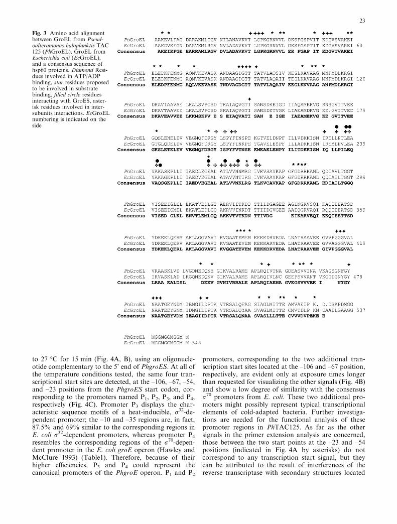
The deduced sequence of PhGroEL, confirmed byMALDI mapping experiments, shows considerablesimilarity to previously reported sequences from otherorganisms, as shown from the alignment of PhGroEL toa consensus sequence (Fig.3) derived by Brocchieri andKarlin from a multiple alignment of 43 hsp60 sequences(Brocchieri and Karlin 2000). This hsp60 multiplealignment consists of sequences from a-, b-, c-, and �-proteobacteria; Rickettsiales; high and low G+C gram-positive bacteria; other bacterial sequences, includingCyanobacteria; and eukaryotic sequences, such as Ara-bidopsis thaliana and Saccharomyces cerevisiae. In par-ticular, there are 21 gram-negative sequences in themultiple alignment; thus, they represent 49% of the se-quences aligned. This sequences analysis represents avalid tool for the comparison of PhGroEL to knownhsp60 sequences and for the integration and analysis ofsequence, structure, and function information.
All the amino acids that make contact with ATP/ADP in consensus sequence are conserved in PhGroEL;those amino acids interacting with GroES or thebound substrate that do not match the consensussequence are nevertheless the same as those found inEcGroEL (Brocchieri and Karlin 2000). A few substi-tutions with respect to both the consensus andEcGroEL sequences can be observed among theresidues at the inter-ring and within-ring contacts: inposition 438 there is a histidine instead of a valine, butthis position has quite a low conservation index(CI=0.16, Brocchieri and Karlin 2000); a methioninecan be found instead of a valine in position 387(CI=0.46, Brocchieri and Karlin 2000); an isoleucinesubstitutes the highly conserved valine 521 (CI=0.85,Brocchieri and Karlin 2000) that is at the interfacebetween the E domains of contiguous monomerswithin the same ring.
Transcriptional analysis of the PhgroE operon
The sequence of the entire regulatory region of thePhgroE operon was analyzed preliminarily for the
Fig. 1 Autoradiography of L-[35S]methionine pulse-labeled totalproteins of PhTAC 125 at normal growth temperature (15 �C) andafter 30 min of heat-shock exposure (30 �C). Arrows indicate someof the proteins induced by heat shock. Molecular weight standardpositions (kDa) are indicated on the left
21

presence of promoters (Neural Network Promoter:http://www.fruitfly.org/seq_tools/promoter.html), reve-aling the presence of four putative –10 and –35 regions(Fig. 4 C). When the same analysis was carried out onthe regulatory region of the E. coli groE operon, onlytwo putative promoter regions could be predicted,corresponding to the two regulative elements experi-mentally identified: a r70-dependent promoter and a r32-dependent promoter (Zhou et al. 1988). The latter plays
a key role in E. coli heat-shock response, since it isrecognized by the alternative factor r32, whose leveldramatically increases during the first few minutes fol-lowing heat shock (Yura et al. 1993).
A primer extension analysis was carried out in orderto verify the functionality of identified promoters. Re-action was carried out on RNA samples isolated fromcells permanently grown at different temperatures (4–15–20–25 �C) and from cells grown at 15 �C and shifted
Fig. 2 Nucleotide and aminoacid sequences of PhGroEL andPhGroES. Peptides sequencedare underlined (the N-terminalsequence is double underlined).Sequence of peptides used todesign primers for the PCRamplification are shown witharrows below the amino acidicsequence (oligo 5’PCR and oli-go 3’PCR), while oligonucleo-tides designed as primers forinverse PCR are indicated witharrows above the nucleotidesequence (oligo 5’invPCR andoligo 3’invPCR). Initiation andstop codons are in bold. Puta-tive regions of ribosome bind-ing site and palindromicsequences of transcriptionaltermination are boxed
22
to 27 �C for 15 min (Fig. 4A, B), using an oligonucle-otide complementary to the 5’ end of PhgroES. At all ofthe temperature conditions tested, the same four tran-scriptional start sites are detected, at the –106, –67, –54,and –23 positions from the PhgroES start codon, cor-responding to the promoters named P1, P2, P3, and P4,respectively (Fig. 4C). Promoter P3 displays the char-acteristic sequence motifs of a heat-inducible, r32-de-pendent promoter; the –10 and –35 regions are, in fact,87.5% and 69% similar to the corresponding regions inE. coli r32-dependent promoters, whereas promoter P4
resembles the corresponding regions of the r70-depen-dent promoter in the E. coli groE operon (Hawley andMcClure 1993) (Table1). Therefore, because of theirhigher efficiencies, P3 and P4 could represent thecanonical promoters of the PhgroE operon. P1 and P2
promoters, corresponding to the two additional tran-scription start sites located at the –106 and –67 position,respectively, are evident only at exposure times longerthan requested for visualizing the other signals (Fig. 4B)and show a low degree of similarity with the consensusr70 promoters from E. coli. These two additional pro-moters might possibly represent typical transcriptionalelements of cold-adapted bacteria. Further investiga-tions are needed for the functional analysis of thesepromoter regions in PhTAC125. As far as the othersignals in the primer extension analysis are concerned,those between the two start points at the –23 and –54positions (indicated in Fig. 4A by asterisks) do notcorrespond to any transcription start signal, but theycan be attributed to the result of interferences of thereverse transcriptase with secondary structures located
Fig. 3 Amino acid alignmentbetween GroEL from Pseud-oalteromonas haloplanktis TAC125 (PhGroEL), GroEL fromEscherichia coli (EcGroEL),and a consensus sequence ofhsp60 proteins. Diamond Resi-dues involved in ATP/ADPbinding, star residues proposedto be involved in substratebinding, filled circle residuesinteracting with GroES, aster-isk residues involved in inter-subunits interactions. EcGroELnumbering is indicated on theside
23
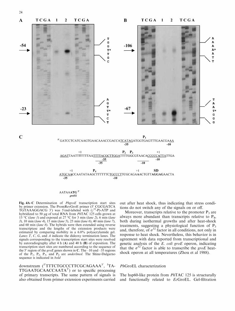
downstream (5’TTTCTGCCCTTCGCAGAAA3’, 5TA-TTGAATGCAACCAATA3’) or to specific processingof primary transcripts. The same pattern of signals isalso obtained from primer extension experiments carried
out after heat shock, thus indicating that stress condi-tions do not switch any of the signals on or off.
Moreover, transcripts relative to the promoter P3 arealways more abundant than transcripts relative to P4,both during isothermal growths and after heat-shocktreatments, suggesting a physiological function of P3
and, therefore, of r32 factor in all conditions, not only inresponse to heat shock. Nevertheless, this behavior is inagreement with data reported from transcriptional andgenetic analysis of the E. coli groE operon, indicatingthat the r32 factor is able to transcribe the groE heat-shock operon at all temperatures (Zhou et al 1988).
PhGroEL characterization
The hsp60-like protein from PhTAC 125 is structurallyand functionally related to EcGroEL. Gel-filtration
Fig. 4A–C Determination of PhgroE transcription start sitesby primer extension. The PromRevGroE primer (5’ CGCGATCATGTAAAGGACG 3’) was 5’end-labeled with (c32-P)-ATP andhybridized to 50 lg of total RNA from PhTAC 125 cells grown at15 �C (lane 1) and exposed at 27 �C for 3 min (lane 2), 6 min (lane3), 10 min (lane 4), 15 min (lane 5), 25 min (lane 6), 40 min (lane 7),and 60 min (lane 8). The hybrids were then extended using reversetranscriptase and the lengths of the extension products wereestimated by comparing mobility in a 6.0% polyacrylamide gel.Lanes T, C, G, and A indicate the dideoxy termination lanes. Thesignals corresponding to the transcription start sites were resolvedby autoradiography after 4 h (A) and 40 h (B) of exposition. Thetranscription start sites are numbered according to the sequence ofthe 5’ region of the groE genes shown in C. The –10 and –35 regionsof the P1, P2, P3, and P4 are underlined. The Shine-Dalgarnosequence is indicated in bold
24
chromatography analysis and far UV-CD spectra ofPhGroEL, in comparison to EcGroEL, suggest similarquaternary and secondary structure organization forthese two proteins (data not shown). PhGroEL pos-sesses a chaperone activity similar to that of EcGroEL.In fact, it is able to bind unfolded mMDH (an often-used in vitro substrate of chaperones [Miller et al 1993]),inhibiting its spontaneous refolding when it is presentalone and releasing it in a conformation committed torecover up to 93% of initial activity when Mg-ATP andEcGroES are added (data not shown). This experimentalso demonstrates that EcGroES can replace PhGroESas a co-chaperone of PhGroEL. Moreover, the samekinetics and yield of reactivation of mMDH at 20 �C inthe presence of PhGroEL or EcGroEL (with Mg-ATPand/or EcGroES added) were observed. This indicatesthat there is no difference between the psychrophilic andmesophilic chaperones in efficiency as a folding helper.
PhGroEL stability
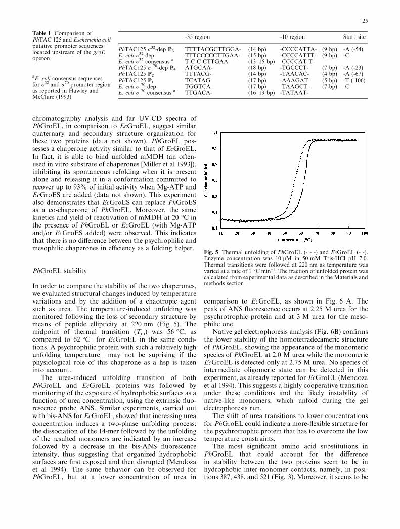
In order to compare the stability of the two chaperones,we evaluated structural changes induced by temperaturevariations and by the addition of a chaotropic agentsuch as urea. The temperature-induced unfolding wasmonitored following the loss of secondary structure bymeans of peptide ellipticity at 220 nm (Fig. 5). Themidpoint of thermal transition (Tm) was 56 �C, ascompared to 62 �C for EcGroEL in the same condi-tions. A psychrophilic protein with such a relatively highunfolding temperature may not be suprising if thephysiological role of this chaperone as a hsp is takeninto account.
The urea-induced unfolding transition of bothPhGroEL and EcGroEL proteins was followed bymonitoring of the exposure of hydrophobic surfaces as afunction of urea concentration, using the extrinsic fluo-rescence probe ANS. Similar experiments, carried outwith bis-ANS for EcGroEL, showed that increasing ureaconcentration induces a two-phase unfolding process:the dissociation of the 14-mer followed by the unfoldingof the resulted monomers are indicated by an increasefollowed by a decrease in the bis-ANS fluorescenceintensity, thus suggesting that organized hydrophobicsurfaces are first exposed and then disrupted (Mendozaet al 1994). The same behavior can be observed forPhGroEL, but at a lower concentration of urea in
comparison to EcGroEL, as shown in Fig. 6 A. Thepeak of ANS fluorescence occurs at 2.25 M urea for thepsychrotrophic protein and at 3 M urea for the meso-philic one.
Native gel electrophoresis analysis (Fig. 6B) confirmsthe lower stability of the homotetradecameric structureof PhGroEL, showing the appearance of the monomericspecies of PhGroEL at 2.0 M urea while the monomericEcGroEL is detected only at 2.75 M urea. No species ofintermediate oligomeric state can be detected in thisexperiment, as already reported for EcGroEL (Mendozaet al 1994). This suggests a highly cooperative transitionunder these conditions and the likely instability ofnative-like monomers, which unfold during the gelelectrophoresis run.
The shift of urea transitions to lower concentrationsfor PhGroEL could indicate a more-flexible structure forthe psychrotrophic protein that has to overcome the lowtemperature constraints.
The most significant amino acid substitutions inPhGroEL that could account for the differencein stability between the two proteins seem to be inhydrophobic inter-monomer contacts, namely, in posi-tions 387, 438, and 521 (Fig. 3). Moreover, it seems to be
Table 1 Comparison ofPhTAC 125 and Escherichia coliputative promoter sequenceslocated upstream of the groEoperon
aE. coli consensus sequencesfor r32 and r70 promoter regionas reported in Hawley andMcClure (1993)
-35 region -10 region Start site
PhTAC125 r32-dep P3 TTTTACGCTTGGA- (14 bp) -CCCCATTA- (9 bp) -A (-54)E. coli r32-dep TTTCCCCCTTGAA- (15 bp) -CCCCATTT- (9 bp) -CE. coli r32 consensus a T-C-C-CTTGAA- (13–15 bp) -CCCCAT-T-PhTAC125 r 70-dep P4 ATGCAA- (18 bp) -TGCCCT- (7 bp) -A (-23)PhTAC125 P2 TTTACG- (14 bp) -TAACAC- (4 bp) -A (-67)PhTAC125 P1 TCATAG- (17 bp) -AAAGAT- (5 bp) -T (-106)E. coli r 70-dep TGGTCA- (17 bp) -TAAGCT- (7 bp) -CE. coli r 70 consensus a TTGACA- (16–19 bp) -TATAAT-
Fig. 5 Thermal unfolding of PhGroEL (- - -) and EcGroEL (- -).Enzyme concentration was 10 lM in 50 mM Tris-HCl pH 7.0.Thermal transitions were followed at 220 nm as temperature wasvaried at a rate of 1 �C min–1. The fraction of unfolded protein wascalculated from experimental data as described in the Materials andmethods section
25
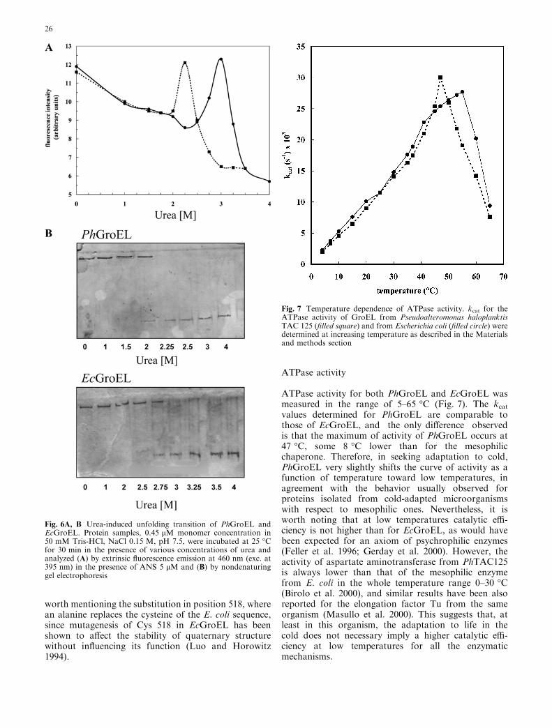
worth mentioning the substitution in position 518, wherean alanine replaces the cysteine of the E. coli sequence,since mutagenesis of Cys 518 in EcGroEL has beenshown to affect the stability of quaternary structurewithout influencing its function (Luo and Horowitz1994).
ATPase activity
ATPase activity for both PhGroEL and EcGroEL wasmeasured in the range of 5–65 �C (Fig. 7). The kcatvalues determined for PhGroEL are comparable tothose of EcGroEL, and the only difference observedis that the maximum of activity of PhGroEL occurs at47 �C, some 8 �C lower than for the mesophilicchaperone. Therefore, in seeking adaptation to cold,PhGroEL very slightly shifts the curve of activity as afunction of temperature toward low temperatures, inagreement with the behavior usually observed forproteins isolated from cold-adapted microorganismswith respect to mesophilic ones. Nevertheless, it isworth noting that at low temperatures catalytic effi-ciency is not higher than for EcGroEL, as would havebeen expected for an axiom of psychrophilic enzymes(Feller et al. 1996; Gerday et al. 2000). However, theactivity of aspartate aminotransferase from PhTAC125is always lower than that of the mesophilic enzymefrom E. coli in the whole temperature range 0–30 �C(Birolo et al. 2000), and similar results have been alsoreported for the elongation factor Tu from the sameorganism (Masullo et al. 2000). This suggests that, atleast in this organism, the adaptation to life in thecold does not necessary imply a higher catalytic effi-ciency at low temperatures for all the enzymaticmechanisms.
Fig. 6A, B Urea-induced unfolding transition of PhGroEL andEcGroEL. Protein samples, 0.45 lM monomer concentration in50 mM Tris-HCl, NaCl 0.15 M, pH 7.5, were incubated at 25 �Cfor 30 min in the presence of various concentrations of urea andanalyzed (A) by extrinsic fluorescence emission at 460 nm (exc. at395 nm) in the presence of ANS 5 lM and (B) by nondenaturinggel electrophoresis
Fig. 7 Temperature dependence of ATPase activity. kcat for theATPase activity of GroEL from Pseudoalteromonas haloplanktisTAC 125 (filled square) and from Escherichia coli (filled circle) weredetermined at increasing temperature as described in the Materialsand methods section
26

Conclusions
We have characterized a heat shock protein from thepsychrotrophic bacterium Pseudoalteromonas halo-planktis TAC 125, which turned out to be a GroEL-likeprotein. The primary structure of the protein from thecold-adapted bacterium was shown to share a high de-gree of identity with the mesophilic GroEL from E. coli(82%). This result is not unexpected, since chaperonesbelong to a very highly conserved class of proteins;furthermore, the proteins of PhTAC125 so far charac-terized show a high percentage of residues identical tothe corresponding gene products of E. coli.
The melting temperature of the psychrotrophicGroEL is 6 �C lower than that of the mesophilic one,and the maximum of ATPase activity of PhGroEL isalso 8 �C lower than that of EcGroEL. These data are inagreement with the general tendency of psychrophilicproteins to optimally shift their function to lower tem-peratures. However, the not-so-impressive differencesuggests that PhGroEL is not one of those classically‘‘cold-adapted’’ proteins for which an improvement inphysiological efficiency through optimization of thecatalytic parameters is claimed to compensate for thereduction of reaction rates induced by low temperatures.Moreover, the rather marginal lower stability of thepsychrophilic GroEL could well arise from a lack ofselective pressure toward thermostability rather thanfrom specific adaptation needs. Similarly, the aspartateaminotransferase form PhTAC 125 does not exhibit in-creased catalytic efficiency at low temperature and re-duced thermostability (Birolo et al. 2000), nor do theelongation factor Tu (Masullo et al. 2000) or the thior-edoxin from the same organism (data to be published).Should we conclude that the proteins in PhTAC 125 arenot cold-adapted? If our answer is positive, then weshould ask ourselves how PhTAC 125 managed to beamong the best cold-adapted bacteria in terms of du-plication times at low temperatures and range of growthtemperature (Tutino et al. 1999a, where PhTAC 125 isnamed Moraxella TAC 125). It seems to be a paradox;however, it is interesting to observe that the usuallyclaimed rules of protein cold adaptation are derivedfrom the characterization of enzymes that in severalcases were purified from organisms specifically selectedto fulfill biotechnological requests, namely, high cata-lytic performances. This is, for instance, the case ofb-galactosidase from P. haloplanktis TAE 79, a strainthat was selected after a screening of about 300 bacterialisolates collected in Antarctica for the highest intracel-lular b-galactosidase activity (Hoyoux et al. 2000). Thisis also the case of cold-adapted lipases (Feller et al. 1989)and of the alkaline metalloprotease from a psychrophilicPseudomonas species (Chessa et al. 2000). We wonderwhether a selective pressure seeking for biotechnologicalperformances could have forced the interpretation ofgeneral rules of protein cold adaptation. Anotherworking hypothesis is that the adaptation of extracel-lular proteins (such as lipases, a-amylase, proteases) can
differ from that of intracellular proteins (such as GroEL,aspartate aminotransferase, thioredoxin, and the elon-gation factor Tu from PhTAC 125), showing less pro-nounced molecular adaptations that nevertheless allowappropriate metabolic fluxes at the environmental cellworking temperatures. The set of available data is not,of course, large enough to formulate a consistentanswer. However, one can also guess that many of thecharacteristics of an organism are determined more bythe regulative features of gene expression than by onlythe protein’s structural aspects. In these respects, thecomplexity of the promoter region of PhGroESL operonwould suggest such a hypothesis.
Acknowledgments The authors are indebted to Prof. J. Buchner(University of Regensburg) for helpful discussions and suggestionsand for kindly providing anti-EcGroEL and EcGroES antibodiesand EcGroEL and EcGroES proteins. We thank Dr. M. LuisaTutino and Dr. Angela Duilio for many helpful discussions andfor critical reading of the manuscript. This work was supported bygrants from the European Union (program ‘‘COLDZYME’’,Contract ERB BIO4 CT96 0051; program ‘‘EUROCOLD’’ con-tract number ERB BIO4 CT95 0017, program ‘‘COLDNET’’contract ERB FMRX CT97 0131), from the Ministero dell’Universita e della Ricerca Scientifica (Progetti di RilevanteInteresse Nazionale 1999), and the Consiglio Nazionale delleRicerche (CNR contract 97.01138.PF49 Progetto Finalizzato‘‘Biotecnologie’’).
References
Berg GR, Innis WE, Heikkila JJ (1987) Stress proteins and ther-motolerance in psychrotrophic yeasts from arctic environments.Can J Microbiol 33:383–389
Birolo L, Tutino ML, Fontanella B, Gerday C, Mainolfi K, Pas-carella S, Sannia G, Vinci F, Marino G (2000) Aspartate am-inotransferase from the Antarctic bacterium Pseudoalteromonashaloplanktis TAC 125. Cloning, expression, properties, andmolecular modelling. Eur J Biochem 267:2790–2802
Bradford MM (1976) A rapid and sensitive method for the quan-tification of microgram quantities of protein utilising the prin-ciple of protein-dye binding. Anal Biochem 72:248–254
Brocchieri L, Karlin S (2000) Conservation among HSP60sequences in relation to structure, function, and evolution.Protein Sci 9:476–486
Chessa JP, Petrescu I, Bentahir M, Beeumen JV, Gerday C (2000)Purification, physico-chemical characterization and sequence ofa heat labile alkaline metalloprotease isolated from a psychro-philic Pseudomonas species. Biochim Biophys Acta 1479:265–274
Cross SJ, Ciruela A, Poomputsa K, Romaniec MP, Freedman RB(1996) Thermostable chaperonin from Clostridium thermocel-lum. Biochem J 316:615–622
Deegenaars ML, Watson K (1997) Stress proteins and stress tol-erance in an Antarctic, psychrophilic yeast, Candida psychro-phila. FEMS Microbiol Lett 151:191–196
Deegenaars ML, Watson K (1998) Heat shock response inpsychrophilic and psychrotrophic yeast from Antarctica.Extremophiles 2:41–49
Feller G, Thiry M, Arpigny JL, Mergeay M, Gerday C (1989)Lipases from psychrotrophic antartic bacteria. FEMS Micro-biol Lett 66:239–244
Feller G, Narinx E, Arpigny JL, Aittaleb M, Baise E, Genicot S,Gerday C (1996) Enzymes from psycrophilic organisms. FEMSMicrobiol Rev 18:189–202
Fenton WA, Horwich AL (1997) GroEL-mediated protein folding.Protein Sci 6:743–760
27

Gerday C, Aittaleb M, Bentahir M, Chessa JP, Claverie P, CollinsT, D’Amico S, Dumont J, Garsoux G, Georlette D, Hoyoux A,Lonhienne T, Meuwis MA, Feller G (2000) Cold-adaptedenzymes: from fundamentals to biotechnology. TrendsBiotechnol 18:103–107
Goloubinoff P, Gatenby AA, Lorimer GH (1989) GroE heat-shockproteins promote assembly of foreign prokaryotic ribulosebisphosphate carboxylase oligomers in Escherichia coli. Nature337:44–47
Grantcharova V, Alm EJ, Baker D, Horwich AL (2001) Mecha-nisms of protein folding. Curr Opin Struct Biol 11:70-82
Hawley DK, McClure WR (1993) Compilation and analysis ofE.coli promoter DNA sequences. Nucleic Acids Res 11:2237–2255
Hemmingsen SM, Woolford C, van der Vies SM, Tilly K, DennisDT, Georgopoulos CP, Hendrix RW, Ellis RJ (1988) Homol-ogous plant and bacterial proteins chaperone oligomeric pro-tein assembly. Nature 333:330–334
Hoyoux A, Jennes I, Dubois P, Genicot S, Dubail F, Francois JM,Biase E, Feller G, Gerday C (2000) Cold-adapted beta-galact-osidase from the Antarctic psychrophile Pseudoalteromonashaloplanktis. Appl Environ Microbiol 67:1529–1535
Julseth CR, Inniss WE (1990) Heat shock protein induction and theacquisition of thermotolerance in the psycrotrophic yeastTrichosporon pullulans. Curr Microbiol 20:391–396
Laemmli UK (1970) Cleavage of structural proteins during theassembly of the head of bacteriofage T4. Nature 227:680–685
Lanzetta PA, Alvarez LJ, Reinach PS, Candia OA (1979) An im-proved assay for nanomole amounts of inorganic phosphate.Anal Biochem 100:95–97
Lindquist S (1986) The heat-shock response. Annu Rev Biochem55:1151–1191
Luo GX, Horowitz PM (1994) J The stability of the molecularchaperonin cpn60 is affected by site-directed replacement ofcysteine 518. Biol Chem 269:32151–32154
Masullo M, Arcari P, de Paola B, Parmeggiani A, Bocchini V(2000) Psychrophilic elongation factor Tu from the antarcticMoraxella sp. Tac II 25: biochemical characterization andcloning of the encoding gene. Biochemistry 39:15531–15539
McCallum KL, Innis WE (1990) Thermotolerance, cell filamenta-tion and induced protein synthesis in psychrophilic and psy-chrotrophic bacteria. Arch Microbiol 153:585–590
McCallum KL, Heikkila JJ, Innis WE (1986) Temperature-dependent pattern of heat-shock protein synthesis in psychro-philic and psychrotrophic micro-organisms. Can J Microbiol32:516–521
Mendoza JA, Lorimer GH, Horowitz PM (1992) Chaperonincpn60 from Escherichia coli protects the mitochondrial enzymerhodanese against heat inactivation and supports folding atelevated temperatures. J Biol Chem 267:17631–17634
Mendoza JA, Demeler B, Horowitz PM (1994) Alteration of thequaternary structure of cpn60 modulates chaperonin-assisted
folding. Implications for the mechanism of chaperonin action.J Biol Chem 269:2447–2451
Miller, AD, Maghlaoui K, Albanese G, Kleinjan DA, Smith C(1993) Escherichia coli chaperonins cpn60 (groEL) and cpn10(groES) do not catalyse the refolding of mitochondrial malatedehydrogenase. Biochem J 291:139–144
Motohashi K, Taguchi H, Ishii N, Yoshida M (1994) Isolation ofthe stable hexameric DnaK.DnaJ complex from Thermus ther-mophilus. J Biol Chem 269:27074–27079
Osipiuk J, Joachimiak A (1997) Cloning, sequencing, and expres-sion of dnaK-operon proteins from the thermophilic bacteriumThermus thermophilus. Biochim Biophys Acta 1353:253–265
Roy SK, Hiyama T, Nakamoto H (1999) Purification and char-acterization of the 16-kDa heat-shock-responsive protein fromthe thermophilic cyanobacterium Synechococcus vulcanus,which is an alpha-crystallin-related, heat shock protein. EurJ Biochem 262:406–416
Sanger F, Nicklen S, Coulson AR (1977) DNA sequencing withchain-terminating inhibitors. Proc Natl Acad Sci USA 74:5463–5467
Segal G, Ron EZ (1996) Regulation and organization of the groEand dnaK operons in Eubacteria. FEMS Microbiol Lett 138:1–10
Sigler PB, Xu Z, Rye HS, Burston SG, Fenton WA, Horwich AL(1998) Structure and function in GroEL-mediated proteinfolding. Annu Rev Biochem 67:581–608
Trent JD, Nimmesgern E, Wall JS, Hartl FU, Horwich AL (1991)A molecular chaperone from a thermophilic archaebacterium isrelated to the eukaryotic protein t-complex polypeptide-1.Nature 354:490–493
Trent JD, Gabrielsen M, Jensen B, Nenhard J, Olsen J (1994)Acquired thermotolerance and heat shock proteins in thermo-philes from the three phylogenetic domains. J Bacteriol176:6148–6152
Tutino ML, Birolo L, Fontanella B, Mainolfi K, Vinci F, SanniaG, Marino G (1999a) Aspartate aminotransferase from Mor-axella TAC125: an unusual psychrophilic enzyme. In: MargesinR, Schinner F (eds) Cold-adapted organisms: ecology, physi-ology, enzymology and molecular biology. Springer, BerlinHeidelberg New York, pp 305–316
Tutino ML, Duilio A, Fontanella B, Moretti MA, Sannia G,Marino G (1999b) Plasmids from Antarctic bacteria. In: Mar-gesin R., Schinner F (eds) Cold-adapted organisms: ecology,physiology, enzymology and molecular biology. Springer,Berlin Heidelberg New York, pp 335–348
Yura T, Nagai N, Mori H (1993) Regulation of the heat-shockresponse in bacteria. Annu Rev Microbiol 47:321–350
Zhou YN, Kusukawa N, Erickson JW, Gross CA, Yura T (1988)Isolation and characterization of Escherichia coli mutants thatlack the heat shock sigma factor sigma 32. J Bacteriol 170:3640–3649
28