Gracilaria vermiculophylla: A western Pacific species of Gracilariaceae (Rhodophyta) first recorded...
11
Phycological Research 2004; 52: 69–79 Gracilaria vermiculophylla: A western Pacific species of Gracilariaceae (Rhodophyta) first recorded from the eastern Pacific Alexis M. Bellorin, 1 * Mariana C. Oliveira 2 and Eurico C. Oliveira 2 1 Departamento de Biología, Escuela de Ciencias, Universidad de Oriente, Cumaná, Venezuela and 2 Departamento de Botânica, Instituto de Biociências, Universidade de São Paulo, Caixa Postal 11461, CEP 05422-970, São Paulo, Brazil SUMMARY We report Gracilaria vermiculophylla (Ohmi) Papenfuss from the Pacific coast of North America based on a morphoanatomical revision and comparison of sequences of the nuclear gene coding for the small subunit of ribosomal RNA and the internal transcribed spacers of populations from Baja California, Mexico and Hiro- shima, Japan. It is the first convincing report of this species out of its center of distribution in western north Pacific, where it has been considered as a synonym of former ‘G. verrucosa’ records. G. vermiculophylla also occurs in central California as indicated by internal transcribed spacer sequences of a previously unknown Gracilaria Greville material. In the northeastern Pacific G. vermiculophylla is characterized by a robust some- what vermiform, well-branched cylindrical thallus, with gradual cell size transition from cortex to medulla, deep spermatangial conceptacles, regular chains of carposporangia and carposporangial initials, downwardly oriented tubular nutritive cells, but rarely with upwardly oriented tubular nutritive cells. In some features of its cystocarp anatomy G. vermiculophylla is related to Gracilariopsis E. Y. Dawson or Hydropuntia Montagne and it is a relevant species for discussions about Gracilariaceae genera. Key words: agarophytes, Gracilaria, Gracilariaceae, internal transcribed spacer, phylogeny, small subunit of ribosomal RNA. INTRODUCTION The well-known economic importance of gracilarioid algae (sensu Rice & Bird 1990) as the main global sources of agar (Oliveira et al. 2000) has stimulated a number of authors to search for reliable means to identify species in this group. This interest has also been fueled through decades by the complexity of the taxonomic problems in the gracilarioids, most of which are far from being resolved. Superficially similar species may have totally different agar yields and gel quality and thus there is a need for sound identification at the species level, for utilization and marketing. Since 1980, a vast literature dealing with new species proposals, species merging, nomenclatural changes, reviews of taxonomic criteria, genera characterization and regional floras has been published (reviews in Oliveira & Plastino 1994; Bird 1995). Almost all of the available taxonomic tools, such as anatomy and onto- geny, chemotaxonomy, antigenic studies, chromosome number, crossability, DNA fingerprinting and DNA sequencing, have been applied to species recognition, without much success except for DNA analysis and crossability tests. Recent studies have remarked that there are several species complexes, both in Gracilaria and Gracilariopsis, in which different species with similar gross morphology could be recognized only with higher-resolution taxonomic techniques (Bird et al. 1994; Goff et al. 1994; Wattier et al. 1997; Bellorin et al. 2002; Gurgel et al. 2003). Of these species complexes, the best studied has been the Gracilaria verrucosa complex (Bird & Rice 1990; Rice & Bird 1990), a group of cylindrical Gracilaria species with deep isolated spermatangial conceptacles recorded, erroneously in most cases, for most algal floras in the world. Since the 1980s we have kept in our laboratory a culture of a species of the G. verrucosa complex from Pacific Mexico, whose correct name was suspected to be G. pacifica I. A. Abbott (Nunez-Lopez & Valdez 1998; Garza-Sanchez et al. 2000). This name has been assumed to be the correct name for the G. verrucosa records throughout the northeastern Pacific. Hybridiza- tion trials in vitro showed that this species (as G. verrucosa Baja California) is reproductively isolated from species of the G. verrucosa complex from the Caribbean and South America (Plastino & Oliveira 1996). In a previous paper about the phylogeny and systemat- ics of Gracilariaceae based on DNA sequence compari- sons (Bellorin et al. 2002) we established that this species is distinct from G. pacifica. However, no name *To whom correspondence should be addressed. Email: [email protected] Communicating editor: J. A. West. Received 28 February 2003; accepted 29 September 2003.
-
Upload
independent -
Category
Documents
-
view
1 -
download
0
Transcript of Gracilaria vermiculophylla: A western Pacific species of Gracilariaceae (Rhodophyta) first recorded...

Phycological Research
2004;
52:
69–79
Gracilaria vermiculophylla
: A western Pacific species of Gracilariaceae (Rhodophyta) first recorded from the eastern Pacific
Alexis M. Bellorin,
1
* Mariana C. Oliveira
2
and Eurico C. Oliveira
2
1
Departamento de Biología, Escuela de Ciencias, Universidad de Oriente, Cumaná, Venezuela and
2
Departamento de Botânica, Instituto de Biociências, Universidade de São Paulo, Caixa Postal 11461, CEP 05422-970, São Paulo, Brazil
SUMMARY
We report
Gracilaria vermiculophylla
(Ohmi) Papenfussfrom the Pacific coast of North America based on amorphoanatomical revision and comparison of sequencesof the nuclear gene coding for the small subunit ofribosomal RNA and the internal transcribed spacers ofpopulations from Baja California, Mexico and Hiro-shima, Japan. It is the first convincing report of thisspecies out of its center of distribution in western northPacific, where it has been considered as a synonym offormer ‘
G. verrucosa
’ records.
G. vermiculophylla
alsooccurs in central California as indicated by internaltranscribed spacer sequences of a previously unknown
Gracilaria
Greville material. In the northeastern Pacific
G. vermiculophylla
is characterized by a robust some-what vermiform, well-branched cylindrical thallus, withgradual cell size transition from cortex to medulla,deep spermatangial conceptacles, regular chains ofcarposporangia and carposporangial initials, downwardlyoriented tubular nutritive cells, but rarely with upwardlyoriented tubular nutritive cells. In some features of itscystocarp anatomy
G. vermiculophylla
is related to
Gracilariopsis
E. Y. Dawson or
Hydropuntia
Montagneand it is a relevant species for discussions aboutGracilariaceae genera.
Key words: agarophytes,
Gracilaria
, Gracilariaceae,internal transcribed spacer, phylogeny, small subunit of
ribosomal RNA.
INTRODUCTION
The well-known economic importance of gracilarioidalgae (
sensu
Rice & Bird 1990) as the main globalsources of agar (Oliveira
et al
. 2000) has stimulated anumber of authors to search for reliable means toidentify species in this group. This interest has alsobeen fueled through decades by the complexity of thetaxonomic problems in the gracilarioids, most of whichare far from being resolved. Superficially similar speciesmay have totally different agar yields and gel qualityand thus there is a need for sound identification at the
species level, for utilization and marketing. Since1980, a vast literature dealing with new speciesproposals, species merging, nomenclatural changes,reviews of taxonomic criteria, genera characterizationand regional floras has been published (reviews inOliveira & Plastino 1994; Bird 1995). Almost all of theavailable taxonomic tools, such as anatomy and onto-geny, chemotaxonomy, antigenic studies, chromosomenumber, crossability, DNA fingerprinting and DNAsequencing, have been applied to species recognition,without much success except for DNA analysis andcrossability tests. Recent studies have remarked thatthere are several species complexes, both in
Gracilaria
and
Gracilariopsis
, in which different species withsimilar gross morphology could be recognized only withhigher-resolution taxonomic techniques (Bird
et al
.1994; Goff
et al
. 1994; Wattier
et al
. 1997; Bellorin
et al
. 2002; Gurgel
et al
. 2003). Of these speciescomplexes, the best studied has been the
Gracilariaverrucosa
complex (Bird & Rice 1990; Rice & Bird1990), a group of cylindrical
Gracilaria
species withdeep isolated spermatangial conceptacles recorded,erroneously in most cases, for most algal floras in theworld.
Since the 1980s we have kept in our laboratory aculture of a species of the
G. verrucosa
complex fromPacific Mexico, whose correct name was suspected tobe
G. pacifica
I. A. Abbott (Nunez-Lopez & Valdez 1998;Garza-Sanchez
et al
. 2000). This name has beenassumed to be the correct name for the
G. verrucosa
records throughout the northeastern Pacific. Hybridiza-tion trials
in vitro
showed that this species (as
G. verrucosa
Baja California) is reproductively isolatedfrom species of the
G. verrucosa
complex from theCaribbean and South America (Plastino & Oliveira 1996).In a previous paper about the phylogeny and systemat-ics of Gracilariaceae based on DNA sequence compari-sons (Bellorin
et al
. 2002) we established that thisspecies is distinct from
G. pacifica
. However, no name
*
To whom correspondence should be addressed.Email: [email protected] editor: J. A. West.Received 28 February 2003; accepted 29 September 2003.
70
A. M. Bellorin
et al
.
was applied to the material from Mexico until molecularand morphoanatomical data of other species of the
G. verrucosa
complex from other regions was available.Recently, we have determined the small subunit of
ribosomal RNA (SSU rDNA) and internal transcribedspacer (ITS) sequences of
G. vermiculophylla
from Japan,a species of the
G. verrucosa
complex, so far knownonly from the northwestern Pacific (Tseng & Xia 1999).The sequences we obtained are identical or nearlyidentical to those from Pacific Mexico. Likewise, nosignificant differences between these materials wereobserved in detailed morphoanatomical and repro-ductive ontogeny studies. Hence, we are extendingthe original distribution of
G. vermiculophylla
from thewestern Pacific to the Pacific coast of Mexico andCalifornia. We also discuss some reproductive anatom-ical features used commonly by species and genericdiscrimination in the Gracilariaceae.
MATERIALS AND METHODS
Anatomical observations were based on herbariumspecimens housed in the Phycological Herbarium ofthe University of São Paulo, Brazil (SPF). The selectedmaterials were soaked in seawater for 24 h and trans-verse or longitudinal sections were made with a razorblade or a microtome after embedding the materialwith historesin (Leica Instruments, Heidelberger,Germany). The sections were stained with aqueous 1%aniline blue (AB), aqueous 1.6% toluidine blue O (Cl52040) (TBO) adjusted to pH 1.0 with HCl, or withWittmann’s aceto-iron-hematoxylin-chloral hydrate (WH)(Wittmann 1965). Semipermanent slides of hand-made sections stained with aniline blue or toluidineblue were mounted in aqueous 30% corn syrup (Karo,São Paolo, Brazil). Hand-sections stained with hema-toxylin were mounted with 50 : 50 Hoyer’s mountingmedium (Hommersand & Fredericq 1988). Microtomesections were stored dried.
DNA was extracted from a plant from MiyajimaIsland, Hiroshima, Japan (collected by E. C. Oliveira on14 November, 2001; voucher specimen number SPF56151). DNA extraction and purification, polymerasechain reaction amplification of SSU rDNA and ITSregions, and sequencing were carried out as describedin Bellorin
et al
. (2002). The SSU rDNA and ITSsequences were compared with those from
Gracilaria
sp. México (GenBank accession no. AF468886 andAF468906). Phylogenetic inferences under maximumlikelihood, maximum parsimony and neighbor-joiningmethods were maded using PAUP* 4.0 (Swofford 1998),based on datasets of Bellorin
et al
. (2002), updatedwith new published SSU rDNA and ITS sequences of
Gracilaria
. Robustness of inferred trees was estimatedas bootstrap support values (Felsenstein 1985) with1000–2000 replicates of heuristic searches on the50% majority-rule consensus trees.
RESULTS
Gracilaria vermiculophylla
(Ohmi) Papenfuss, Papenfuss (1966)
Synomym:
Gracilariopsis vermiculophylla
Ohmi, Ohmi(1956): 271, figs 1–4, pls 1,2.
Geographical distribution: This species was originallydescribed from Akkeshi Lagoon, eastern Hokkaido, Japan(Ohmi 1956), and later for tropical and subtropicalregions in the northwestern Pacific (Korea, China,Vietnam; Tseng & Xia 1999). Here we extend thegeographic distribution to the northeastern Pacific(Mexico and California), as revealed by the nearlyidentical ITS sequences among the specimens fromMexico (Bellorin
et al
. 2002; as
Gracilaria
sp. Mexico),California (Goff
et al
. 1994; as
Gracilaria
sp. ElkhornSlough) and Japan (this work).
Habitat: This species forms extensive beds in theintertidal and upper sublittoral zones, attached to rocksor pebbles, often covered with sand and mud. Thecollection site in Baja California is an estuary. Therecords from California are also from an estuarinehabitat (Goff
et al
. 1994), and the material fromMiyajima Island, Japan, was found in the intertidal, onsmall pebbles, in the outflow of a small creek.
Specimens examined: Estero de Punta Banda(Ensenada, Baja California, Mexico), 9 October 1979,
leg
. M. A. Aguilar Rosas, SPF 55205 (infertile speci-mens); 28 April 1980,
leg
. R. Aguilar Rosas, SPF55207 (seven female specimens); 28 May 1986,
leg
.R. Aguilar Rosas, SPF 55206 (two sheets, tetraspor-angial, male and infertile specimens); July 1989,
leg
.E. C. Oliveira, SPF 54484 (two sheets, tetrasporangialspecimens); July 1989,
leg
. E. C. Oliveira, SPF 54485(seven sheets, tetrasporangial, female, male and infer-tile specimens obtained from
in vitro
cultivation);7 March 2000,
leg
. J. M. Guzmán, SPF 56131, SPF56139, SPF 56140 (tetrasporangial and femalespecimens); Miyajima Island (Hiroshima, Japan),14 November 2001,
leg
. E. C. Oliveira, SPF 56141(four sheets, female specimens), SPF 56142 (twosheets, male specimens), SPF 56143 (five sheets,tetrasporangial and infertile specimens).
Morphology and vegetative anatomy: This specieshas a thallus up to 100 cm long and 5 mm indiameter, but usually smaller and cylindrical through-out, with two to five orders of lateral branches ofvarious sizes produced at irregular intervals in analternate to unilateral pattern. The branches usuallylack basal constrictions and are tapered toward the tips(Figs 1–3). Fresh material is fleshy and robust, some-what vermiform, dark brown, becoming black ondrying. The cell size transition from the medulla to thecortex is gradual (Figs 4–6). The cortex has three tofour layers of well-pigmented cells (up to eight celllayers in tetrasporangial and spermatangial areas).Outer cortical cells are radially elongated, measuring

Gracilaria vermiculophylla
from eastern Pacific
71
7–18
×
3–8
µ
m (Figs 7,8). There is a subcortex ofthree to five layers of radially elongated cells in cross-sections (Figs 4–7). The medulla consists of large ellip-tical cells of 34–410
×
12–25
µ
m in cross-sections,
with four to five cell layers (up to 12 cell layers in olderaxis). Cells are highly vacuolated and have abundantstarch grains. Medullary cell walls are 12–20
µ
m thick(Figs 4–6). Rupture of some medullary cells can be
Figs 1–3.
Gracilaria vermiculophylla
. 1. Sterile plant obtained
in vitro
from tetrasporangia (SPF 54485). 2.
Mixed-phase specimen
(tetrasporangia and spermatangia) from Estero de Punta Banda, Mexico (SPF 55206). 3.
Large tetrasporangial plant from Estero de Punta
Banda, Mexico (SPF 54484).

72
A. M. Bellorin
et al
.
Figs 4–19.
Gracilaria vermiculophylla
. 4. Cross-section (AB) of female young axis (SPF 55207). 5.
Cross-section (AB) of tetrasporangial
old axis (SPF 54485). 6.
Cross-section (TBO) of female old axis (SPF 56139). 7.
Cortex and subcortex (AB) in transverse section show-
ing the basal cells of ephemeral hairs (arrowheads) (SPF 56139). 8.
Basal cell of deciduous hair (arrowhead) (WH). Note that undiffer-
entiated terminal cortical cells are multinucleated (SPF 55206). 9.
Dividing terminal cortical cell (WH) with a longitudinal oblique
concave/convex septum forming a spermatangial initial (arrowhead) (SPF 55206). 10.
Differentiated spermatangial initial (arrowhead)
(WH), which is a terminal cortical cell (SPF 55206). 11,12.
Dividing spermatangial initials by a longitudinal oblique concave/convex
septum (WH) (SPF 55206). 13.
Formation of spermatangial mother cells (arrowhead) (WH) from a spermatangial initial. Note that one
spermatangium (arrow) has been cut off at this stage (SPF 55206). 14.
Developing shallow spermatangial conceptacle (WH) with fila-
ments of spermatangial mother cells lining both floor and lateral walls of cavity. Note the enlarged basal cell of a deciduous hair (arrowhead)
(SPF 55206). 15.
Developing shallow spermatangial conceptacle (AB) with spermatangial mother cells lining only the floor of the cavity.
Note that some spermatangial mother cells bear columns of two or more spermatangia (SPF 55206). 16.
Developing spermatangial
conceptacle (WH). Note the dividing terminal cells of spermatangial/mother cell filaments (arrowhead) (SPF 55206). 17.
Deep isolated
spermatangial conceptacle (WH), showing a dividing terminal cell of a spermatangial/mother cell filament (arrowhead) (SPF 55206).
18. Coalescing deep male conceptacles resembling ‘henriquesiana’ type (AB). Some spermatangial mother cells bears two sequentially
produced spermatangia (SPF 55206). 19.
Cortical decussate/cruciate tetrasporangium (WH) (SPF 54484).

Gracilaria vermiculophylla
from eastern Pacific
73
seen on old branches resulting in a partially hollowthallus. Deciduous cortical hairs are produced near theapex, as indicated by the presence of large andmultinucleated, darkly stained hair basal cells(Figs 7,8,14).
Spermatangia: Spermatangia are produced in deepconceptacles (up to 90
µ
m deep, 60
µ
m wide) thatreach the subcortex and open by an external pore(‘
verrucosa
’ type; Yamamoto, 1984) (Fig. 17). Youngconceptacles may appear as shallow depressions withspermatangia limited to the cavity floor (resembling‘
textorii
’ type) (Fig. 15) or both the cavity floor andlateral walls (Figs 14,16), becoming deeper at matura-tion. Conceptacles are mostly single, although somemay coalesce (Fig. 18). Spermatangial mother cellsproduced from terminal uninucleate cortical cells(‘spermatangial initials’, Fredericq & Hommersand1989a) stain darkly with haematoxylin (Figs 9–10).Differentiated spermatangial initials can be seen nearbranch apices (approximately 10 mm), linked to sub-cortical cells. A spermatangial initial first cuts off aspermatangial mother cell by a longitudinal obliqueconcave/convex division (Figs 11,12). Later on, boththe subapical spermatangial initial and the daughterspermatangial mother cell, produce a system of fila-ments of spermatangial mother cells (Fig. 13). Theterminal cells of these fertile filaments are elongatedand divide actively, forming new spermatangial mothercells by longitudinally oblique concave/convex divisions(Figs 16–17). In a well-developed conceptacle thesedividing cells are positioned at the opening of theconceptacle. Spermatangia are produced distally by anoblique division of the elongated spermatangial mothercells (Fig. 13). Usually only one spermatangium is cutoff at a time from each spermatangial mother cell,although two or rarely more (up to four) sequentiallyproduced spermatangia can remain united, resemblingchains of spermatangia (Figs 15,16,18).
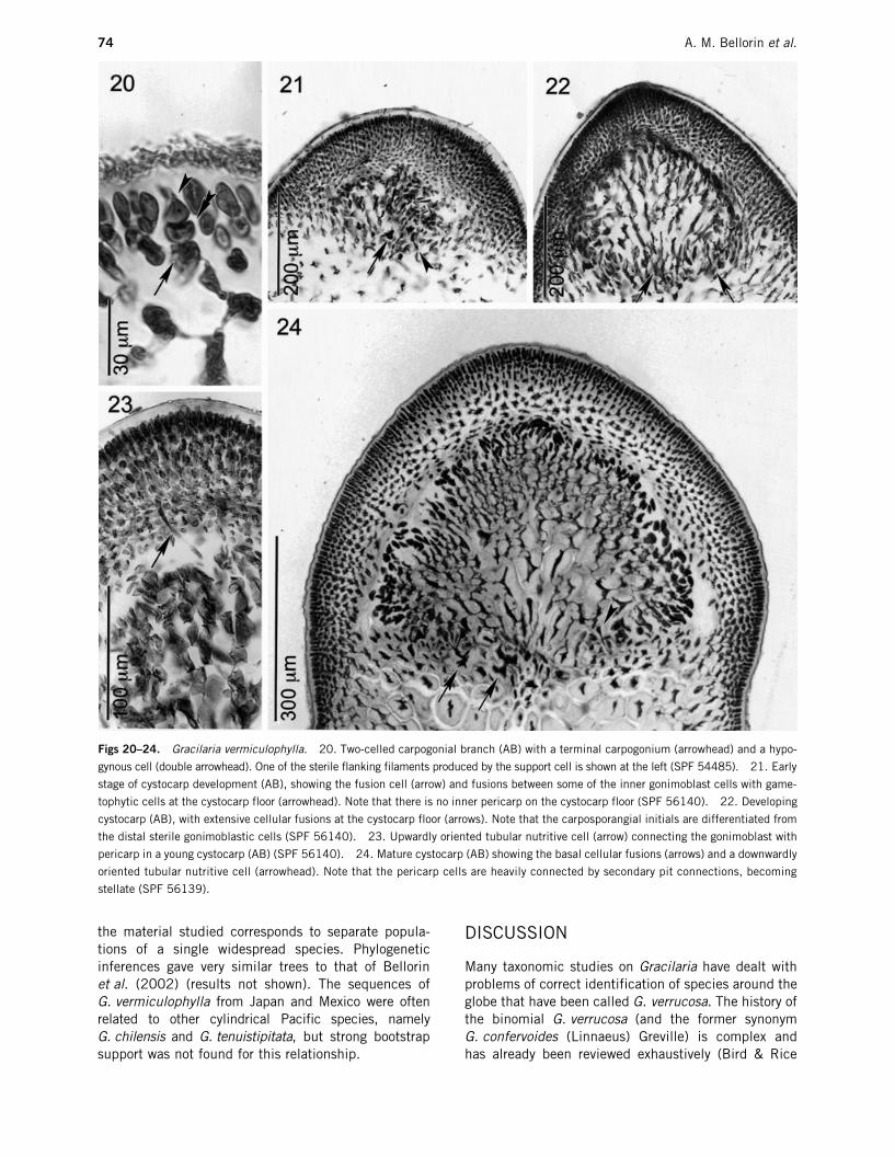
Female apparatus and cystocarp ontogeny: Typicalgracilariacean two-celled carpogonial branches, withshort trichogynes, are produced from a terminal corti-cal cell that acts as the supporting cell (Fig. 20). Thesupporting cell produces two short filaments of sterileflanking cells. A central, star-like, highly dissectedfusion cell is distinguishable only at the initial stagesof cystocarp development (Fig. 21). The cortical cellssurrounding the fusion cell divide periclinally to forman incipient pericarp prior to gonimoblast production.These incipient pericarp cells are radially elongated,linked by primary pit connections. As the gonimoblastinitials are produced from the fusion cell, the cystocarpcavity is produced progressively by a periclinal rupture,which separates the incipient pericarp from the game-tophytic cells at the cystocarp floor. Few pericarp cellsremain at the cystocarp floor and, therefore, no innerpericarp is formed as has been described for otherspecies (Fredericq & Norris 1985; Fredericq &
Hommersand 1989b). The gonimoblast filamentsarise first from the fusion cell. Later, fusions betweengonimoblastic cells and gametophytic cells at thecystocarp floor give rise to a basal nutritive tissue ofsecondarily interconnected and highly dissected cells(Fig. 22). At this stage the fusion cell becomes indis-tinguishable from other cellular fusions. Upwardlyoriented tubular nutritive cells (Fredericq & Hommersand1989a) connecting the gonimoblasts and the pericarpare observed rarely, and only in young cystocarps(Fig. 23). These cells remain linked by a primary pit-connection to sterile gonimoblastic cells close to carp-osporangial initials. The proximal region of tubularnutritive cells has a triangular outline and the distalregion is star-like, because of secondary fusions withseveral gametophytic cells. Downwardly orientedtubular nutritive cells are also produced (Fig. 24) andthe distal region of these cells is difficult to differenti-ate from other cellular fusions in the cystocarp floor.Mature cystocarps are prominent, up to 1.5 mm highin herbarium specimens, sometimes constricted at thebase and rostrate. Inner gonimoblast cells are radiallyelongated, radiating from the cystocarp floor and con-nected by abundant secondary pit connections (Fig. 24).The carposporangia mature terminally from radiallyelongated chains of carposporangial initials (up to fivecells) heavily stained with aniline blue. Mature carpo-sporangia measure 15–29
µ
m. Neither gonimoblastlobes nor upwardly oriented tubular nutritive cells wereobserved in mature cystocarps. Cellular fusions are stilldistinguishable at the floor of mature cystocarps(Fig. 24). Three cell types are usually recognized in thepericarp of mature cystocarps: (i) the outer cells (threeto four cell layers), which are rounded, except theterminal ones that are radially elongated (from 5–7 to10–17
µ
m) and not linked by secondary pit connec-tions; (ii) the intermediate cells (6–10 cell layers),which become star-shaped due to abundant secondarypit connections; and (iii) the innermost cells, (1–4cells in radial chains), rounded and connected only byprimary pit connections (Fig. 24).
Tetrasporangia: Radially elongated tetrasporangia,up to 65
µ
m long, are produced in the cortex (Fig. 19).Division is decussate cruciate or irregular. The corticalcells surrounding the tetrasporangia divide anticlinally,not forming a traditional nemathecial-type cortex.
Molecular data: The complete unambiguous sequencewas obtained for SSU rDNA from
G. vermiculophylla
from Japan (GenBank accession no. AY465828). TheITS sequence was determined with some ambiguouspositions (56 from 1640 bp) (GenBank accession no.AY465829). There are no differences between the SSUrDNA sequences of
G. vermiculophylla
from Mexicoand Japan in pairwise comparisons. The actual sequencedivergence between the ITS sequences in pairwisecomparisons, excluding ambiguous positions, was0.68%. Based on these observations, we assume that
74 A. M. Bellorin et al.
the material studied corresponds to separate popula-tions of a single widespread species. Phylogeneticinferences gave very similar trees to that of Bellorinet al. (2002) (results not shown). The sequences ofG. vermiculophylla from Japan and Mexico were oftenrelated to other cylindrical Pacific species, namelyG. chilensis and G. tenuistipitata, but strong bootstrapsupport was not found for this relationship.
DISCUSSION
Many taxonomic studies on Gracilaria have dealt withproblems of correct identification of species around theglobe that have been called G. verrucosa. The history ofthe binomial G. verrucosa (and the former synonymG. confervoides (Linnaeus) Greville) is complex andhas already been reviewed exhaustively (Bird & Rice
Figs 20–24. Gracilaria vermiculophylla. 20. Two-celled carpogonial branch (AB) with a terminal carpogonium (arrowhead) and a hypo-
gynous cell (double arrowhead). One of the sterile flanking filaments produced by the support cell is shown at the left (SPF 54485). 21. Early
stage of cystocarp development (AB), showing the fusion cell (arrow) and fusions between some of the inner gonimoblast cells with game-
tophytic cells at the cystocarp floor (arrowhead). Note that there is no inner pericarp on the cystocarp floor (SPF 56140). 22. Developing
cystocarp (AB), with extensive cellular fusions at the cystocarp floor (arrows). Note that the carposporangial initials are differentiated from
the distal sterile gonimoblastic cells (SPF 56140). 23. Upwardly oriented tubular nutritive cell (arrow) connecting the gonimoblast with
pericarp in a young cystocarp (AB) (SPF 56140). 24. Mature cystocarp (AB) showing the basal cellular fusions (arrows) and a downwardly
oriented tubular nutritive cell (arrowhead). Note that the pericarp cells are heavily connected by secondary pit connections, becoming
stellate (SPF 56139).

Gracilaria vermiculophylla from eastern Pacific 75
1990; Rice & Bird 1990; Irvine & Steentoft 1995). Inbrief, the type locality of G. verrucosa is the BritishIsles, although this name was applied somewhat indis-criminately to any stringy and freely ramified cylindri-cal Gracilaria with deep spermatangial conceptaclesworldwide (Fredericq & Hommersand 1989a). Never-theless, a convincing body of evidence from cross-ability tests, DNA methods and detailed morphologyshowed that most of these extra-European records aredistinct entities from what is considered to be authen-tic G. verrucosa from Britain (McLachlan 1979; Birdet al. 1982; Abbott 1985; Zhang & Xia 1985; Plastino& Oliveira 1997). Even in the type locality, thebinomial G. verrucosa was shown not to be mono-specific but to encompass two distinct entities (Bird &Rice 1990; Rice & Bird 1990), one of which belongsto a distinct genus, Gracilariopsis longissima (S. G.Gmelin) Steentoft, L. Irvine et Farnham, and the otherone to Gracilaria, now known as G. gracilis (Stack-house) Steentoft, L. Irvine et Farnham (Steentoft et al.1995). The former name G. verrucosa was abandonedbecause its lectotype belonged to a Gracilariopsisspecies. G. gracilis also occurs in Wales, Norway,France, Spain, Argentina and Namibia (Rice & Bird1990; Bird et al. 1994; Steentoft et al. 1995).
The most recent reviews of Gracilaria in the northPacific advocate recognition of two species within theG. verrucosa complex, both distinct from G. gracilis:G. pacifica in the North American side, and G. vermi-culophylla in the western side (Tseng & Xia 1999). Therecords of G. verrucosa along the Pacific coast ofNorth America were first treated as distinct fromBritish Isles material because of sexual incompatibilityin hybridization tests by Bird et al. (1982), based on aculture isolate from Vancouver Island. Later, Abbott(1985) proposed the binomial G. pacifica for theformer G. verrucosa from California, remarking on thefew morphoanatomical differences between the newspecies from the eastern Pacific and the British entity.Bird et al. (1992) ascribed their isolate from Vancou-ver Island to G. pacifica and showed that this species isclosely related to G. gracilis (as G. verrucosa) on thebasis of SSU rDNA sequences, which are nearlyidentical between these two species. The fast-evolvingsequences of ITS and Rubisco spacer (Goff et al.1994), as well as microsatellite comparisons (Wattieret al. 1997), confirmed later that G. pacifica is suffi-ciently distinct at the species level from G. gracilis,although both entities are closely related. Goff et al.(1994) also reported G. pacifica from Washington State,USA.
In the western side of the north Pacific, on the otherhand, the identity of G. verrucosa samples remainedlargely uncertain. Zhang and Xia (1984) comparedG. gracilis and G. pacifica (both as G. verrucosa) withthe G. verrucosa from China and Japan, noting somedifferences in cystocarp anatomy. The following year
these authors (Zhang & Xia 1985) establishedG. asiatica J. F. Zhang et B. M. Xia to accommodatethe specimens previously ascribed to G. verrucosa inChina, also including a collection from Shinori, Hokkaido,Japan. Yamamoto and Sasaki (1988) investigated col-lections referred to as G.verrucosa in Japan, employingcrossability tests and found interfertility between twopopulations identified as G. verrucosa from Hokkaido(including the material ascribed to G. asiatica fromShinori) and the topotype material of the poorly knownG. vermiculophylla. They concluded that G. verrucosashould be merged with G. vermiculophylla rather thanbe placed under the later name G. asiatica, given thenomenclatural priority. The binomial G. vermiculophyllawas later applied to all former G. verrucosa records inthe northwestern Pacific, including China, Korea, Vietnamand Japan (Yoshida et al. 1995; Yoshida 1998; Tseng& Xia 1999).
The first DNA sequence data of a G. verrucosacomplex species from Asia was the SSU rDNA sequenceof an isolate referred to as G. verrucosa from Hokkaido(unspecified locality), Japan (Bird et al. 1992). Thissequence was nearly identical to others of theG. gracilis group (as G. verrucosa from Europe andArgentina) and it was suspected that this Japaneseisolate belonged to the same species as European andArgentinean specimens (Bird et al. 1992). However, areanalysis of DNA samples in the C. J. Bird collectionusing four single loci microsatellites and ITS sizevariation (Wattier et al. 1997) showed that the isolatefrom Japan is distinct from authentic G. gracilis fromEurope, Argentina and Namibia. At the moment, noname has been applied to this isolate and its taxo-nomic status remains uncertain.
Recently, one of us (E. C. Oliveira) collectedG. verrucosa samples from Miyajima Island, Hiroshima,Japan. Our anatomical observation on this speciesconfirmed the presence of deep, separate sperma-tangial conceptacles typical of the G. verrucosa com-plex, but the cystocarps differed from typical G. verrucosain having few upwardly directed tubular nutritive cells(most were downwardly directed), small protoplasm-rich gonimoblasts cells and rather regular chains ofcarposporangia (A. Bellorin, unpubl. data, 2002),among other features. We ascribed this material toG. vermiculophylla (Ohmi) Papenfuss sensu Yamamoto(1978). The SSU rDNA of this material was completelydivergent from that of G. verrucosa from Japan deter-mined by Bird et al. (1992). Instead, the SSU rDNAand ITS sequences of this entity were identical ornearly identical to those of the unknown Gracilariaspecies from California and Mexico previously studiedby Goff et al. (1994; as Gracilaria sp. Elkhorn Slough)and by us (Bellorin et al. 2002; as Gracilaria sp. Mexico).We conclude that our materials from Mexico and Japanbelong to the same species (G. vermiculophylla) andthat this species is distinct from the G. verrucosa from

76 A. M. Bellorin et al.
Japan studied by Bird et al. (1992). This findingconfirms the previous suspicion that at least twospecies have been confused under G. verrucosa inJapan (Yamamoto & Sasaki 1988). The species studiedby Bird et al. (1992) appears to be closely related toG. gracilis and G. pacifica, according to SSU rDNAsequences, while G. vermiculophylla has been weaklyrelated to other Pacific species, namely G. chilensisand G. tenuistipitata (Goff et al. 1994; Bellorin et al.2002). This increases considerably the distribution ofG. vermiculophylla in the Pacific. The question of howmany species of the G. verrucosa complex occurs inJapan and, by extension, in Asia remains open. Like-wise, it should be concluded that the application ofbinomial G. vermiculophylla to all of the G. verrcuosamaterials in Asia, as proposed by Tseng and Xia(1999), is questionable. The confusion that resultsfrom combining superficially similar species into asingle taxon was already noticed for G. blodgettii inJapan (Terada & Yamamoto 2000), in which G. shimo-densis R. Terada and H. Yamamoto was confused withthe presumably authentic G. blodgettii Harvey.
Gracilaria vermiculophylla was first proposed asGracilariopsis vermiculophyla by Ohmi (1956), basedon a population from Akkeshi Lagoon, Hokkaido Island,Japan. At that time, the generic concept of Gracilariop-sis (Dawson 1949) was related to the absence oftubular nutritive cells and other cystocarpic features,without any consideration of spermatangial configura-tion. Thus, as Ohmi (1956) did not observe tubularnutritive cells in cystocarps of material from AkkeshiLagoon, he treated his new species as Gracilariopsis.When Papenfuss (1966) called for the submergence ofGracilariopsis into Gracilaria, he transferred Gracilari-opsis vermiculophylla to Gracilaria. Some downwardlydirected tubular nutritive cells were later observed inmaterial from Akkeshi Lagoon (Yamamoto 1978) and,as this species presents deep spermatangial concepta-cles, a feature associated with the modern Gracilariaconcept, the placement into Gracilaria was not furtherdiscussed. The SSU rDNA and ITS sequences ofG. vermiculophylla from Japan and Mexico determinedby us confirm this generic placement. Basic morpho-logical and anatomical features of G. vermiculophyllahave already been described for Japanese specimensby Ohmi (1956) and Yamamoto (1978). In this work,we provide a description of material from Mexico,describing for the first time the male apparatus andcystocarp formation in the species.
Gracilaria vermiculophylla was considered to berestricted to the type locality in the cold water regionsof Japan (Yamamoto 1978, 1984). In fact, its generalmorphology was described based on specimens from alagoon in which the plants maintain the vermiformmorphology, the scarce to absent tubular nutritive cellsin cystocarps and the small-celled gonimoblasts (Yama-moto 1978; Yamamoto & Sasaki 1988). However,
when G. verrucosa populations from other localitieswere studied and shown to be conspecific withG. vermiculophylla by Yamamoto and Sasaki (1988),it became clear just how morphologically variablethis species is. Interestingly, the hybrids betweenG. vermiculophyla from the type locality and the formerG. verrucosa from Japan showed intermediate numbersof tubular nutritive cells (Yamamoto & Sasaki 1988),which corroborate the marked intraspecific variation ofthis character (see Bird 1995). The data of Yamamoto(1984) showing that G. vermiculophylla is a euryhalineand eurythermal species may explain the wider geo-graphic distribution found in this study.
The weak evolutionary relationships among G. vermi-culophylla, G. chilensis and G. tenuistipitata inferredfrom SSU rDNA and ITS nucleotide sequences (Bel-lorin et al. 2002) were also inferred from Rubisco spacersequence comparisons (Goff et al. 1994, G. vermi-culophylla as Gracilaria sp. Elkhorn Slough). Gracilariachilensis and G. tenuistipitata were also considered tobe related in comparisons of rbcL sequences (Gurgel &Fredericq 2002). These evolutionary relationships aremorphologically supported by cystocarp anatomy,although these traits should be considered with cautionas they may be quite variable intraspecifically(G. vermiculophylla is a good example of this). All ofthese cylindrical species: (i) rarely produce tubularnutritive cells connecting the gonimoblast to the upperpericarp, although downwardly or laterally orientedtubular nutritive cells are frequent; (ii) have gonimo-blast inner cells highly connected by secondary pitconnections; (iii) have cellular fusions at the cystocarpfloor; and (iv) produce orderly arrays of carposporangialinitials and a non-deeply lobed gonimoblast (A. M.Bellorin, unpubl. data, 2002). However, there are someimportant differences: (i) in G. chilensis and G. tenuis-tipitata, the generative fusion cell can be generallyrecognized even in mature cystocarps (A. M. Bellorin,unpubl. data, 2002), which is not the case inG. vermiculophylla; (ii) in G. chilensis, a differentiatedinner pericarp (cytologically modified cells derivedfrom cortical gametophytic cells in the cystocarp floor,see Fredericq & Hommersand 1989b) is clearly distin-guishable in mature cystocarps, as well as verticallyelongated cellular fusions through secondary pit con-nections between some of the basal gonimoblast cellsand inner pericarp cells (Bird et al. 1990; Nelson &Ryan 1991; A. M. Bellorin, unpubl. data, 2002). Aninner pericarp is not present in G. vermiculophylla orG. tenuistipitata (A. M. Bellorin, unpubl. data, 2002).
The mature male apparatus of G. vermiculophylla isdistinct from G. chilensis and G. tenuistipitata in havingdeep male conceptacles rather than shallow ones.However, there are no fundamental differences in theontogeny of male conceptacles between G. vermiculo-phylla and G. chilensis. In both species the spermatangialinitials are terminal cortical cells that produce a system

Gracilaria vermiculophylla from eastern Pacific 77
of filaments of spermatangial mother cells (Ryan &Nelson 1991). In addition, the young spermatangialconceptacles of G. vermiculophylla are shallow and arequite similar to the textorii type described inG. chilensis (Nelson 1987; Ryan & Nelson 1991).Unfortunately, we do not know the spermatangialontogeny in G. tenuistipitata.
The position of the spermatangial initials was usedto define discrete types of male conceptacles (Freder-icq & Norris 1985; Fredericq & Hommersand 1989a,1989b). According to these authors, in the textorii typethe spermatangial initials are derived from terminalcortical cells, whereas in the verrucosa type the sperma-tangial initials are derived from intercalary cortical cells.G. vermiculophylla, however, has verrucosa-type sperma-tangia produced from terminal cortical cells. Moreover,we have observed verrucosa-type spermatangia origi-nated from terminal spermatangial initials in otherspecies (A. M. Bellorin, unpubl. data, 2002), thereaf-ter the verrucosa and textorii types are not necessarilydistinct in origin. These results, together with theconflicting observations about the male apparatusontogeny already reported in the literature (Yamamoto1973, Fredericq & Norris 1985; Fredericq & Hommer-sand 1989a; Abbott et al. 1991; Ryan & Nelson 1991;Terada & Yamamoto 2000), show that the recognizedpatterns of spermatangial types are not so clear cut asoriginally expected, at least among species that havespermatangia within conceptacles. Thus, althoughusually useful in species recognition, the ‘types’ ofspermatangial conceptacles may not have major evolu-tionary importance and, in fact, can not be establishedas synapomorphies (Bird et al. 1992; Bellorin et al.2002).
Gracilaria vermiculophylla presents some of thecystocarp features proposed as descriptors for thegenus Gracilariopsis (Fredericq & Hommersand 1990),as was discussed for G. chilensis by Nelson and Ryan(1991). The species also presents some features asso-ciated with the generic or subgeneric concept ofHydropuntia (Fredericq & Hommersand 1990), a taxonthat is currently unsustainable in light of morphological(Bird 1995) and molecular (Bellorin et al. 2002) data.Emphasis should be placed on the downwardly orientedtubular nutritive cells that were proposed as being ofdiagnostic value in the original Hydropuntia proposal(Chang & Xia 1963, as Polycavernosa C. F. Chang et B.M. Xia). Our studies with G. vermiculophylla show thatthe orientation of these cells may be quite variableamong populations. The other Hydropuntia basic fea-ture, the compound henriquesiana-type spermatangia,is widespread in several unrelated groups of Gracilariaspecies (Bellorin et al. 2002), and can not be utilizedas a descriptor for any generic or subgeneric taxon.
Gracilaria vermiculophylla and G. tenuistipitata, aswas previously remarked for G. chilensis (Nelson &Ryan 1991; Bird 1995), provide further support for the
submergence of Hydropuntia into Gracilaria. Addition-ally, as these three species also combine Gracilariopsisand Gracilaria features, two genera sufficiently distinctaccording to a strong body of molecular evidence (Birdet al. 1992, 1994; Goff et al. 1994; Bellorin et al.2002; Gurgel & Fredericq 2002), the morphologicalboundaries between these genera should be redefined,specially in regard to the inner pericarp differentiation(a proposed feature of Gracilariopsis widespread intoGracilaria, A. M. Bellorin, unpubl. data, 2002) and themode of production of carposporangia (as there areseveral Gracilaria species with straight chains of carpo-sporangia, e.g. Terada et al. 2000, a proposed featureof Gracilariopsis). The next step in the systematics ofgracilarioid algae should be the morphological charac-terization of the infrageneric lineages of Gracilariopsisand Gracilaria established in molecular comparisons,as well as a revision of the status of many poorly knownspecies.
ACKNOWLEDGMENTSWe thank J. M. Guzmán for supplying material for DNAanalysis and J. Zertuche for help in specimen collec-tion. Financial support was provided by FAPESP,Brazil. AMB acknowledge CONICIT, Venezuela, forPh.D. scholarship. MCO and ECO acknowledge CNPq,Brazil. We acknowledge C. J. Bird, M. Hommersandand W. Nelson for critically reviewing the manuscriptand for valuable suggestions.
REFERENCESAbbott, I. 1985. New species of Gracilaria Grev. (Graci-
lariaceae, Rhodophyta) from California and Hawaii. InAbbott, I. A. and Norris, J. N. (Eds) Taxonomy of EconomicSeaweeds. California Sea Grant College Program, La Jolla,California, pp. 115–21.
Abbott, I. A., Zhang, J. and Xia, B. 1991. Gracilaria mixta sp.nov. and other western Pacific species of the genus(Rhodophyta: Gracilariaceae). Pacific Sci. 45: 12–27.
Bellorin, A. M., Oliveira, M. C. and Oliveira, E. C. 2002.Phylogeny and systematics of the marine algal familyGracilariaceae (Gracilariales, Rhodophyta) based on SSUrDNA and ITS sequences of Atlantic and Pacific species.J. Phycol. 38: 551–63.
Bird, C. J. 1995. A review of recent taxonomic concepts anddevelopment in the Gracilariaceae (Rhodophyta). J. Appl.Phycol. 7: 255–67.
Bird, C. J., Nelson, W. A., Rice, E. L., Ryan, K. G. andVillemur, R. 1990. A critical comparison of Gracilariachilensis and G. sordida (Rhodophyta, Gracilariales). J.Appl. Phycol. 2: 375–82.
Bird, C. J., Ragan, M. A., Critchley, A. T., Rice, E. L. andGutell, R. R. 1994. Molecular relationships among theGracilariaceae (Rhodophyta): further observations on someundetermined species. Eur. J. Phycol. 29: 195–202.

78 A. M. Bellorin et al.
Bird, C. J. and Rice, E. L. 1990. Recent approaches to thetaxonomy of the Gracilariaceae (Gracilariales, Rhodophyta)and the Gracilaria verrucosa problem. Hydrobiologia 204/205: 111–18.
Bird, C. J., Rice, E. L., Murphy, C. A. and Ragan, M. A. 1992.Phylogenetic relationships in the Gracilariales (Rhodo-phyta) as determined by 18S rDNA sequences. Phycologia31: 510–22.
Bird, C. J., van der Meer, J. P. and McLachlan, J. 1982. Acomment on Gracilaria verrucosa (Huds.) Papenf. (Rhodo-phyceae, Gigartinales). J. Mar. Biol. Ass. UK 62: 453–9.
Chang, C. F. and Xia, B. 1963. Polycavernosa, a new genusof the Gracilariaceae. Stud. Mar. Sinica 3: 119–26.
Dawson, E. Y. 1949. Studies of northeast Pacific Graci-lariaceae. Allan Hancock Found. Publs Occ. Pap. 7:1–105.
Felsenstein, J. 1985. Confidence limits on phylogenies: anapproach using bootstrap. Evolution 39: 783–91.
Fredericq, S. and Hommersand, M. H. 1989a. Proposal of theGracilariales ord. nov. (Rhodophyta) based on an analysisof the reproductive development of Gracilaria verrucosa.J. Phycol. 25: 213–27.
Fredericq, S. and Hommersand, M. H. 1989b. Comparativemorphology and taxonomic status of Gracilariopsis(Gracilariales, Rhodophyta). J. Phycol. 25: 228–41.
Fredericq, S. and Hommersand, M. H. 1990. Diagnoses andkey to the genera of the Gracilariaceae (Gracilariales,Rhodophyta). Hydrobiologia 204/205: 173–8.
Fredericq, S. and Norris, J. N. 1985. Morphological studieson some tropical species of Gracilaria Grev. (Gracilar-iaceae, Rhodophyta): taxonomic concepts based on repro-ductive morphology. In Abbott, I. A. and Norris, J. N. (Eds)Taxonomy of Economic Seaweeds. California Sea GrantCollege Program, La Jolla, California, pp. 137–55.
Garza-Sanchez, F., Zertuche-Gonzalez, J. A. and Chapman D. J.2000. Effect of temperature and irradiance on the release,attachment and survival of spores of Gracilaria pacificaAbbott (Rhodophyta). Bot. Mar. 43: 205–12.
Goff, L. J., Moon, D. A. and Coleman, A. W. 1994. Moleculardelineation of species and species relationships in the redalgal agarophytes Gracilariopsis and Gracilaria (Gracilari-ales). J. Phycol. 30: 521–37.
Gurgel, C. F. D. and Fredericq, S. 2002. Filogenética, taxono-mia e biogeografia da família Gracilariaceae (Gracilariales,Rhodophyta). Abstract of IX Brazilian Meeting of Phycol-ogy. Brazilian Society of Phycology, Santa Cruz, Aracruz,Espíritu Santo, 261.
Gurgel C. F. D., Liao L. M., Fredericq S. and Hommersand, M. H.2003. Systematics of Gracilariopsis (Gracilariales, Rhodo-phyta) based on rbcL sequence analyses and morpho-logical evidence. J. Phycol. 39: 154–71.
Hommersand, M. and Fredericq, S. 1988. An investigation ofcystocarp development in Gelidium pteridifolium with arevised description of the Gelidiales (Rhodophyta). Phyco-logia 27: 254–72.
Irvine, L. and Steentoft, M. 1995. Proposal to reject the nameFucus verrucosus Huds. (Rhodophyta). Taxon 44: 223–4.
McLachlan, J. 1979. Gracilaria tikvahiae sp. nov. (Rhodo-phyta, Gigartinales, Gracilariaceae) from the northwesternAtlantic. Phycologia 18: 19–23.
Nelson, W. A. 1987. The New Zealand species of GracilariaGreville (Rhodophyta, Gigartinales). NZ J. Bot. 25: 87–98.
Nelson, W. A. and Ryan, K. G. 1991. Comparative study ofreproductive development in two species of Gracilaria(Gracilariales, Rhodophyta). II. Carposporogenesis. Crypt.Bot. 2: 234–41.
Nunez-Lopez, R. A. and Valdez, M. C. 1998. Seasonal varia-tion of seaweed biomass in San Ignacio Lagoon, BajaCalifornia Sur, Mexico. Bot. Mar. 41: 421–6.
Ohmi, H. 1956. On a new species of the genus Gracilariopsiswith some considerations on its ecology. Bull. Fac. Fish.Hokkaido University 6: 271–9.
Oliveira, E. C., Alveal, K. and Anderson, R. J. 2000. Maricul-ture of the agar-producing gracilarioid red algae. Rev.Fish. Sci. 8: 345–77.
Oliveira, E. C. and Plastino, E. M. 1994. Gracilariaceae. In:Akatsuka, I. (Ed.) Biology of Economic Algae. SPB Aca-demic Publishing, Netherlands, pp. 185–226.
Papenfuss, G. F. 1966. Notes on algal nomenclature V.Various Chlorophyceae and Rhodophyceae. Phykos 5:95–105.
Plastino, E. M. and Oliveira, E. C. 1996. Approaches to theidentification of terete Brazilian Gracilariaceae (Gracilari-ales, Rhodophyta). Hydrobiologia 326/327: 145–8.
Plastino, E. M. and Oliveira, E. C. 1997. Gracilaria caudataJ. Agardh (Gracilariales, Rhodophyta): restoring an oldname for a common western Atlantic alga. Phycologia 36:225–32.
Rice, E. L. and Bird, C. J. 1990. Relationships amonggeographically distant populations of Gracilaria verrucosa(Gracilariales, Rhodophyta) and related species. Phycolo-gia 29: 501–10.
Ryan, K. G. and Nelson, W. A. 1991. Comparative study ofreproductive development in two species of Gracilaria(Gracilariales, Rhodophyta). I. Spermatogenesis. Crypto.Bot. 2: 229–33.
Steentoft, M., Irvine, L. M. and Farnham, W. F. 1995. Twoterete species of Gracilaria and Gracilariopsis (Gracilari-ales, Rhodophyta) in Britain. Phycologia 34: 113–27.
Swofford, D. L. 1998. PAUP* Phylogenetic Analysis UsingParsimony (*and Other Methods), version 4. Sinauer Asso-ciates, Sunderland, MA, USA.
Terada, R., Baba, M. and Yamamoto, H. 2000. New record ofGracilaria firma Chang et Xia (Rhodophyta) from Okinawa,Japan. Phycol. Res. 48: 291–4.
Terada, R. and Yamamoto, H. 2000. A taxonomic study oftwo Japanese species of Gracilaria: Gracilaria shimodensissp. nov. and Gracilaria blodgettii (Gracilariales, Rhodo-phyta). Phycol. Res. 48: 189–98.
Tseng, C. K. and Xia, B. 1999. On the Gracilaria in thewestern Pacific and the southeastern Asia Region. Bot.Mar. 42: 209–17.
Wattier, R., Dallas, J. F., Destombe, C., Saumitou-Laprade, P.and Valero, M. 1997. Single locus microsatellites in

Gracilaria vermiculophylla from eastern Pacific 79
Gracilariales (Rhodophyta): high level of genetic variabilitywithin Gracilaria gracilis and conservation in relatedspecies. J. Phycol. 33: 868–80.
Wittmann, W. 1965. Aceto-iron-haematoxylin-chloral hydratefor chromosome staining. Stain Technol. 40: 161–4.
Yamamoto, H. 1973. The development of the male reproduc-tive organ of Gracilaria verrucosa (Huds.) Papenfuss. Bull.Jap. Soc. Phycol. 21: 130–2.
Yamamoto, H. 1978. Systematic and anatomical study of thegenus Gracilaria. Japan. Bull. Fac. Fish. Hokkaido Univer-sity 25: 97–152.
Yamamoto, H. 1984. An evaluation of some vegetative fea-tures and some interesting problems in Japanese popula-tions of Gracilaria. Hydrobiologia 116/117: 51–4.
Yamamoto, H. and Sasaki, J. 1988. Interfertility between so-called Gracilaria verrucosa (Huds.) Papenfuss and G.vermiculophylla (Ohmi) Papenfuss in Japan. Bull. Fac.Fish. Hokkaido University 39: 1–3.
Yoshida, T. 1998. Marine Algae of Japan. Uchida Roukakuho,Tokyo, 25 + 1222 pp. (in Japanese).
Yoshida, T., Yoshinaga, K. and Nakajima, Y. 1995. Check listof marine algae of Japan. Jpn J. Phycol. 43: 115–71.
Zhang, J. and Xia, B. 1984. Some problems in the taxonomyof Chinese species of Gracilaria (Rhodophyta). Hydrobio-logia 116/117: 59–62.
Zhang, J. and Xia, B. 1985. On Gracilaria asiatica sp. nov.and G. verrucosa (Huds.) Papenfuss. Oceanol. Limnol.Sinica 3: 175–80.