Gorokhova et al. 2013 PLo S ONE
11
Trade-Offs between Predation Risk and Growth Benefits in the Copepod Eurytemora affinis with Contrasting Pigmentation Elena Gorokhova 1,3 *, Maiju Lehtiniemi 2 , Nisha H. Motwani 3 1 Department of Applied Environmental Science, Stockholm University, Stockholm, Sweden, 2 Finnish Environment Institute, Marine Research Center, Helsinki, Finland, 3 Department of Ecology, Environment and Plant Sciences, Stockholm University, Stockholm, Sweden Abstract Intraspecific variation in body pigmentation is an ecologically and evolutionary important trait; however, the pigmentation related trade-offs in marine zooplankton are poorly understood. We tested the effects of intrapopulation phenotypic variation in the pigmentation of the copepod Eurytemora affinis on predation risk, foraging, growth, metabolic activity and antioxidant capacity. Using pigmented and unpigmented specimens, we compared (1) predation and selectivity by the invertebrate predator Cercopagis pengoi, (2) feeding activity of the copepods measured as grazing rate in experiments and gut fluorescence in situ, (3) metabolic activity assayed as RNA:DNA ratio in both experimental and field-collected copepods, (4) reproductive output estimated as egg ratio in the population, and (5) total antioxidant capacity. Moreover, mitochondrial DNA (mtDNA) COI gene variation was analysed. The pigmented individuals were at higher predation risk as evidenced by significantly higher predation rate by C. pengoi on pigmented individuals and positive selection by the predator fed pigmented and unpigmented copepods in a mixture. However, the antioxidant capacity, RNA:DNA and egg ratio values were significantly higher in the pigmented copepods, whereas neither feeding rate nor gut fluorescence differed between the pigmented and unpigmented copepods. The phenotypic variation in pigmentation was not associated with any specific mtDNA genotype. Together, these results support the metabolic stimulation hypothesis to explain variation in E. affinis pigmentation, which translates into beneficial increase in growth via enhanced metabolism and antioxidant protective capacity, together with disadvantageous increase in predation risk. We also suggest an alternative mechanism for the metabolic stimulation via elevated antioxidant levels as a primary means of increasing metabolism without the increase in heat absorbance. The observed trade-offs are relevant to evolutionary mechanisms underlying plasticity and adaptation and have the capacity to modify strength of complex trophic interactions. Citation: Gorokhova E, Lehtiniemi M, Motwani NH (2013) Trade-Offs between Predation Risk and Growth Benefits in the Copepod Eurytemora affinis with Contrasting Pigmentation. PLoS ONE 8(8): e71385. doi:10.1371/journal.pone.0071385 Editor: Syuhei Ban, University of Shiga Prefecture, Japan Received May 15, 2013; Accepted July 1, 2013; Published August 7, 2013 Copyright: ß 2013 Gorokhova et al. This is an open-access article distributed under the terms of the Creative Commons Attribution License, which permits unrestricted use, distribution, and reproduction in any medium, provided the original author and source are credited. Funding: The financial support was provided by The Swedish Research Council for Environment, Agricultural Sciences and Spatial Planning (FORMAS), Walter and Andre ´ e de Nottbeck Foundation, and Stockholm University’s strategic marine environmental research program ‘‘Baltic Ecosystem Adaptive Management’’. The funders had no role in study design, data collection and analysis, decision to publish, or preparation of the manuscript, Competing Interests: The authors have declared that no competing interests exist. * E-mail: [email protected] Introduction Variation in pigmentation among individuals, populations and species has been associated with various adaptations to predation, ultraviolet radiation (UVR) and diet, but the ultimate mechanisms and trade-offs regulating the observed patterns in the field are still unclear. Studies on pigment variation and its adaptive function in zooplankton have been focused on carotenoids in calanoid copepods [1,2] and melanisation of daphniids [3,4,5]. Most of these studies attributed pigmentation variability to the trade-offs between protection from UVR and predation [6,7], with weak pigmentation correlating with lower predation pressure but also with lower UVR tolerance [4]. Deeply colored populations are usually found at high-latitude or high-altitude lakes, but pigmen- tation is also observed in temperate regions [7,8]. In temperate marine zooplankton, the evidence for the linkage between pigmentation and UVR tolerance is, however, less compelling. For example, the pigmentation does not appear to play a role in UVR tolerance of marine copepods [9], whereas the opposite was observed in crab larvae [10]. In addition to the photoprotective function of pigments, a metabolic stimulation hypothesis has been advocated to explain the adaptive value of pigmentation by providing warmth through absorption of solar radiation [11,12]. Any increase in body temperature due to energy absorbance by body pigments under light conditions would thus have primarily positive effects, such as increased enzyme activities, metabolic rates and, ultimately, growth. Laboratory experiments have shown that under sunlight, respiration rate increased in pigmented copepods but not in those lacking visible pigmentation [11,12], and that average pigmenta- tion intensity was inversely related to temperature [6,12]. This has important implications for zooplankton performing diel vertical migrations (DVM) as demographic disadvantage to zooplankton that spend a daytime at deeper and cooler waters may actually be similar to or greater than the lethal effects of predation [13]. In fishless lakes, pigmented zooplankters remain in productive near- surface waters during the day and have higher reproduction rates PLOS ONE | www.plosone.org 1 August 2013 | Volume 8 | Issue 8 | e71385
Transcript of Gorokhova et al. 2013 PLo S ONE

Trade-Offs between Predation Risk and Growth Benefitsin the Copepod Eurytemora affinis with ContrastingPigmentationElena Gorokhova1,3*, Maiju Lehtiniemi2, Nisha H. Motwani3
1Department of Applied Environmental Science, Stockholm University, Stockholm, Sweden, 2 Finnish Environment Institute, Marine Research Center, Helsinki, Finland,
3Department of Ecology, Environment and Plant Sciences, Stockholm University, Stockholm, Sweden
Abstract
Intraspecific variation in body pigmentation is an ecologically and evolutionary important trait; however, the pigmentationrelated trade-offs in marine zooplankton are poorly understood. We tested the effects of intrapopulation phenotypicvariation in the pigmentation of the copepod Eurytemora affinis on predation risk, foraging, growth, metabolic activity andantioxidant capacity. Using pigmented and unpigmented specimens, we compared (1) predation and selectivity by theinvertebrate predator Cercopagis pengoi, (2) feeding activity of the copepods measured as grazing rate in experiments andgut fluorescence in situ, (3) metabolic activity assayed as RNA:DNA ratio in both experimental and field-collected copepods,(4) reproductive output estimated as egg ratio in the population, and (5) total antioxidant capacity. Moreover, mitochondrialDNA (mtDNA) COI gene variation was analysed. The pigmented individuals were at higher predation risk as evidenced bysignificantly higher predation rate by C. pengoi on pigmented individuals and positive selection by the predator fedpigmented and unpigmented copepods in a mixture. However, the antioxidant capacity, RNA:DNA and egg ratio valueswere significantly higher in the pigmented copepods, whereas neither feeding rate nor gut fluorescence differed betweenthe pigmented and unpigmented copepods. The phenotypic variation in pigmentation was not associated with any specificmtDNA genotype. Together, these results support the metabolic stimulation hypothesis to explain variation in E. affinispigmentation, which translates into beneficial increase in growth via enhanced metabolism and antioxidant protectivecapacity, together with disadvantageous increase in predation risk. We also suggest an alternative mechanism for themetabolic stimulation via elevated antioxidant levels as a primary means of increasing metabolism without the increase inheat absorbance. The observed trade-offs are relevant to evolutionary mechanisms underlying plasticity and adaptation andhave the capacity to modify strength of complex trophic interactions.
Citation: Gorokhova E, Lehtiniemi M, Motwani NH (2013) Trade-Offs between Predation Risk and Growth Benefits in the Copepod Eurytemora affinis withContrasting Pigmentation. PLoS ONE 8(8): e71385. doi:10.1371/journal.pone.0071385
Editor: Syuhei Ban, University of Shiga Prefecture, Japan
Received May 15, 2013; Accepted July 1, 2013; Published August 7, 2013
Copyright: � 2013 Gorokhova et al. This is an open-access article distributed under the terms of the Creative Commons Attribution License, which permitsunrestricted use, distribution, and reproduction in any medium, provided the original author and source are credited.
Funding: The financial support was provided by The Swedish Research Council for Environment, Agricultural Sciences and Spatial Planning (FORMAS), Walterand Andree de Nottbeck Foundation, and Stockholm University’s strategic marine environmental research program ‘‘Baltic Ecosystem Adaptive Management’’.The funders had no role in study design, data collection and analysis, decision to publish, or preparation of the manuscript,
Competing Interests: The authors have declared that no competing interests exist.
* E-mail: [email protected]
Introduction
Variation in pigmentation among individuals, populations and
species has been associated with various adaptations to predation,
ultraviolet radiation (UVR) and diet, but the ultimate mechanisms
and trade-offs regulating the observed patterns in the field are still
unclear. Studies on pigment variation and its adaptive function in
zooplankton have been focused on carotenoids in calanoid
copepods [1,2] and melanisation of daphniids [3,4,5]. Most of
these studies attributed pigmentation variability to the trade-offs
between protection from UVR and predation [6,7], with weak
pigmentation correlating with lower predation pressure but also
with lower UVR tolerance [4]. Deeply colored populations are
usually found at high-latitude or high-altitude lakes, but pigmen-
tation is also observed in temperate regions [7,8]. In temperate
marine zooplankton, the evidence for the linkage between
pigmentation and UVR tolerance is, however, less compelling.
For example, the pigmentation does not appear to play a role in
UVR tolerance of marine copepods [9], whereas the opposite was
observed in crab larvae [10].
In addition to the photoprotective function of pigments, a
metabolic stimulation hypothesis has been advocated to explain
the adaptive value of pigmentation by providing warmth through
absorption of solar radiation [11,12]. Any increase in body
temperature due to energy absorbance by body pigments under
light conditions would thus have primarily positive effects, such as
increased enzyme activities, metabolic rates and, ultimately,
growth. Laboratory experiments have shown that under sunlight,
respiration rate increased in pigmented copepods but not in those
lacking visible pigmentation [11,12], and that average pigmenta-
tion intensity was inversely related to temperature [6,12]. This has
important implications for zooplankton performing diel vertical
migrations (DVM) as demographic disadvantage to zooplankton
that spend a daytime at deeper and cooler waters may actually be
similar to or greater than the lethal effects of predation [13]. In
fishless lakes, pigmented zooplankters remain in productive near-
surface waters during the day and have higher reproduction rates
PLOS ONE | www.plosone.org 1 August 2013 | Volume 8 | Issue 8 | e71385

[14]. Therefore, by relaxing DVM and increasing pigmentation, a
zooplankter can minimize photodamage, enjoy high food avail-
ability and reproductive output in the illuminated trophogenic
zone, but at the cost of predation by visual planktivores. Although,
fish are commonly considered as the only visual predators in
aquatic ecosystems, there are marine and freshwater invertebrates
capable of visual predation, such as mysids [15] and onychopod
cladocerans [13,16]. The invasive onychopods Cercopagis pengoi and
Bythotrephes longimanus have been implicated in the reduction of
zooplankton diversity and abundance in the Laurentian Great
Lakes [17] and the Baltic Sea [18] as well as in the forcing
zooplankton to migrate below thermocline during daytime
[13,18]. By studying intraspecific and intrapopulation variability
in pigmentation in relation to growth and vulnerability to
predation, we can better understand mechanisms of these trade-
offs in specific populations and environmental conditions.
Many zooplankton pigments are important quenching agents
and antioxidants that neutralize reactive oxygen species; they also
have other biological functions, such as regulatory effects on cell
signaling [19]. The changes in the antioxidative status may thus
translate to fitness responses, and increase in investment in the
antioxidant system can come at a cost of an investment elsewhere,
e. g., it may shorten lifespan, decrease growth and reproductive
output, weaken immune system, etc. [19,20]. However, in
different species, elevated growth has been linked with both a
reduction and an increase in antioxidants [20]. This is related to a
complexity of antioxidant systems and interactions amongst
various antioxidants, implying that any specific antioxidative
compound may fail to indicate the overall defense state. Thus,
responses of individual antioxidants are generally more difficult to
interpret than those of overall antioxidant capacity when testing
specific relationships between antioxidants, metabolic activity and
growth, in order to understand mechanisms of trade-offs.
As far as we know, no studies have addressed pigmentation-
mediated trade-offs between fitness parameters and vulnerability
to predation by invertebrate predators in marine zooplankton.
Moreover, whereas genetic basis and developmental mechanisms
generating the diversity of pigmentation and color patterns are
well appreciated in vertebrates [21], much less is understood
regarding genetic versus environmental influence on pigment
variation in invertebrate species under various selection pressures
acting on pigmentation-related traits. In Daphnia, both genetic and
environmental effects were found to influence pigmentation
[14,22], with differences in melanisation among species, clones
and mitochondrial DNA (mtDNA) haplotypes [23,24]. Similarly, a
genetic divergence in mtDNA was found between differently
pigmented but otherwise morphologically identical varieties in
shrimps [25] and ascidians [26].
In our fieldwork in different areas of the Baltic Sea, we were
puzzled by variability in pigmentation in the calanoid copepod
Eurytemora affinis Poppe, raising questions about the factors that
influence pigmentation in this species. This copepod is one of the
few dominant mesozooplankton species in the northern Baltic and
a preferred prey for the dominant zooplanktivores [27] as well as
for C. pengoi [18]. Interestingly, a recent inventory using mtDNA,
revealed a high diversity in the Baltic lineages of E. affinis [28].
This species has been suggested to actually comprise a number of
cryptic species [29], two of which were recently reported from the
Baltic Sea [30,31]. Given the observed genetic variation in the
Baltic Eurytemora, differences in pigmentation between the cryptic
species and association between pigmentation and certain mtDNA
haplotypes in E. affinis are possible.
In this study, we investigated whether the observed variation in
pigmentation is related to interspecific variation between Eur-
ytemora affinis and a morphologically similar species E. carolleeae
recently reported from the Gulf of Finland [31]. When the
presence of only original Baltic lineage of E. affinis in our
collections was confirmed, we examined the intrapopulation
variation of the mitochondrial cytochrome oxidase c subunit 1
(COI) gene in relation to pigmentation, and tested effects of this
pigmentation on the predation and selectivity by C. pengoi and on
the variations in antioxidant capacity, feeding, metabolic activity
and reproduction of the copepods.
Methods
Ethics StatementThe sampling was conducted within national Finnish monitor-
ing in the Gulf of Finland and no specific permissions were
required for the sampling locations of this study. Also, we did not
require ethical approval to conduct this study as we did not handle
or collect animals considered in any animal welfare regulations
and no endangered or protected species were involved in the
samplings or the experiments.
Defining Pigmented and Unpigmented FormsThe term pigmentation is used here to describe the presence of
a dark pigment in the exoskeleton of E. affinis. There was a clear
distinction between pigmented and unpigmented individuals, with
the latter appearing opaque or transparent in transmitted light,
without visible dark coloration of any specific area, apart from the
eye, ovaries and egg sacks (in females). By contrast, the pigmented
individuals had dark brown coloration on either one side of the
body or on the both sides; the pigmentation was particularly strong
on antennae, ventro-lateral part of cephalothorax and furcal rami
(Figure S1).
Sampling Methods, Locations and Sample PreparationThe experimental animals were collected and the experiments
were conducted on board R/V ‘Aranda’ (SYKE, Finland) in August
2011 in the eastern and central Gulf of Finland, the northern
Baltic Sea (Table 1). Eurytemora affinis and Cercopagis pengoi were
collected from 20 m to the surface, using a WP-2 plankton net
(mesh size 100 mm) equipped with a cod end. The samples were
used to obtain animals for the experiments and to preserve them
for different measurements. For predation and grazing experi-
ments, E. affinis individuals with contrasting pigmentation were
sorted with a pipette under a dissecting microscope and kept in
groups of 10 individuals in 0.2 mm-filtered seawater (FSW) in
0.1 L vials. For the predation experiments, C. pengoi were carefully
picked under a dissecting microscope with forceps and wide-
mouth pipettes and transferred individually to FSW in 0.1 L vials;
only actively swimming individuals were used in the experiments.
All specimens were kept at 16uC approximating the temperature
in the mixing layer until the experiments commenced. The
remaining zooplankton collections were treated as follows. The
water was removed as much as possible with a filter paper and the
samples were preserved by: (1) freezing at 280uC for measure-
ments of gut fluorescence and antioxidant capacity; and (2) adding
RNAlater 10:1 (v/v) for genetic analysis and measurements of
nucleic acid concentrations; these samples were also used for
microscopic analysis to estimate sex- and age-related difference in
pigmentation and to calculate egg ratio.
Genetic AnalysesUsing a 10% Chelex buffer, genomic DNA was extracted from
pigmented and unpigmented individuals preserved in RNAlater (25
ind. group21; pooled from 6 stations; Table 1) and used for
Pigmentation Stimulates Growth in Copepods
PLOS ONE | www.plosone.org 2 August 2013 | Volume 8 | Issue 8 | e71385
amplification of COI (,570 bp) gene. Polymerase chain reactions
(PCR) were conducted using EuF1 and EuR2 primers [28]. The
products were separated in 1.5% (w/v) agarose gel with a 100-bp
ladder and visualized by staining with ethidium bromide. The
PCR products were purified using the QIAquick PCR Purification
Kit (QIAGEN) and sequenced in both directions with an ABI
3730 PRISMH DNA Analyzer at KIGene (Karolinska Institute,
Stockholm). The resulting nucleotide sequences were aligned using
BioEdit software [32] checking electropherograms to ensure
proper base calling. The sequences were compared to all
Eurytemora sequences reported for the Baltic Sea (Figure S2) and
all unique nucleotide sequences have been submitted to the
GenBank (accession numbers JQ822115-JQ822130).
Genetic diversity was compared between the pigmentation
phenotypes using estimators implemented in DnaSP version 5.10
[33], including the number of haplotypes (K), haplotype diversity
(Hd, i.e., probability that two randomly chosen haplotypes are
different in the sample) and nucleotide diversity (p, i.e., average
number of nucleotide differences per location between two
sequences), and diversity per site based on the number of
segregating sites (h). A statistical parsimony haplotype network
was constructed to show genetic relatedness among haplotypes
using the software package HapStar [34].
Sex- and Age- related Differences in PigmentationTo test whether the proportion of pigmented individuals
differed between males and females (CIV-VI), 25 individuals of
each sex per sample were counted in the 6 field samples preserved
with RNAlater and the pigmentation status and developmental
stage were noted for each individual. When no sex-related
difference was found, we examined the variation between the
immature (CIV-V) and mature (CVI) individuals using the same
counts.
Predation ExperimentsWe conducted two predation experiments using parthenogenic
females of Cercopagis pengoi (instars II and III) as predators and older
copepodites (CIV–VI, males and females without egg sacks) of
Eurytemora affinis as a prey. In Experiment 1, predation rates (PR) of
C. pengoi offered either pigmented or unpigmented copepods were
determined, whereas in Experiment 2, prey selection using a
mixed (1:1) assemblage of pigmented and unpigmented copepods
was determined. The experiments consisted of 6 replicate
incubations for each treatment and were conducted in either 0.1
(Experiment 1) or 0.2 (Experiment 2) L vials under constant
illumination with green light at 15uC and lasted ca. 20–22 h.
Incubations were started by adding the known number of prey (10
in Experiment 1or 20 in Experiment 2 to keep the same prey
concentration in both experiments) to a single C. pengoi. In each
experiment, there were two predator-free control incubations with
the same number of prey to correct for prey recovery (100% in all
cases). Recovered prey were counted directly upon the experiment
termination and the pigmentation status of each copepod was
noted. Body length (BL, from the top of the head to the base of the
caudal claws) of every C. pengoi and prosome length (PL) in a
subsample of 20–25 individuals for each E. affinis group were
measured under a dissecting microscope. PR was calculated as the
difference between the initial and final numbers of prey and scaled
to 24 h.
Grazing ExperimentExperiment 3 was conducted to test for differences in grazing,
metabolic activity and antioxidant capacity between the copepods
with contrasting pigmentation. As food, we used Rhodomonas salina
(Cryptophyceae; CCMP 1319) grown on f/2 medium at 17uC and
8% salinity in artificial seawater (Instant OceanTM, Aquarium
Systems). To minimize algal growth during the experiment and
increase accuracy of the grazing rate (GR) estimates, the algae
were pre-treated with c-radiation to prevent cell division [35].
Groups of pigmented and unpigmented copepods (CIV–VI, males
and females without egg sacks; 10 ind. vial21; 6 replicates per
treatment) were incubated during 22–24 h in 0.2 L of FSW with
600 mgC L21 of food; light regime was the same as in the
predation experiments. This food concentration is above the
saturation level for E. affinis [36] that ensures no decrease in the
feeding rate due to prey depletion. To determine algal concen-
trations (cells mL–1), 15 mL of the media were preserved with acid
Lugol’s solution and analyzed using a laser particle counter
Spectrex PC-2000 (Spectrex Corp.). The individual GR was
determined as a difference between the initial and final algal
concentrations in each vial and scaled to 24 h and individual
copepod.
Upon termination of the experiment, the copepods were
examined with a dissecting microscope, and dead individuals
were noted. In all incubations, survivorship was $95%, with no
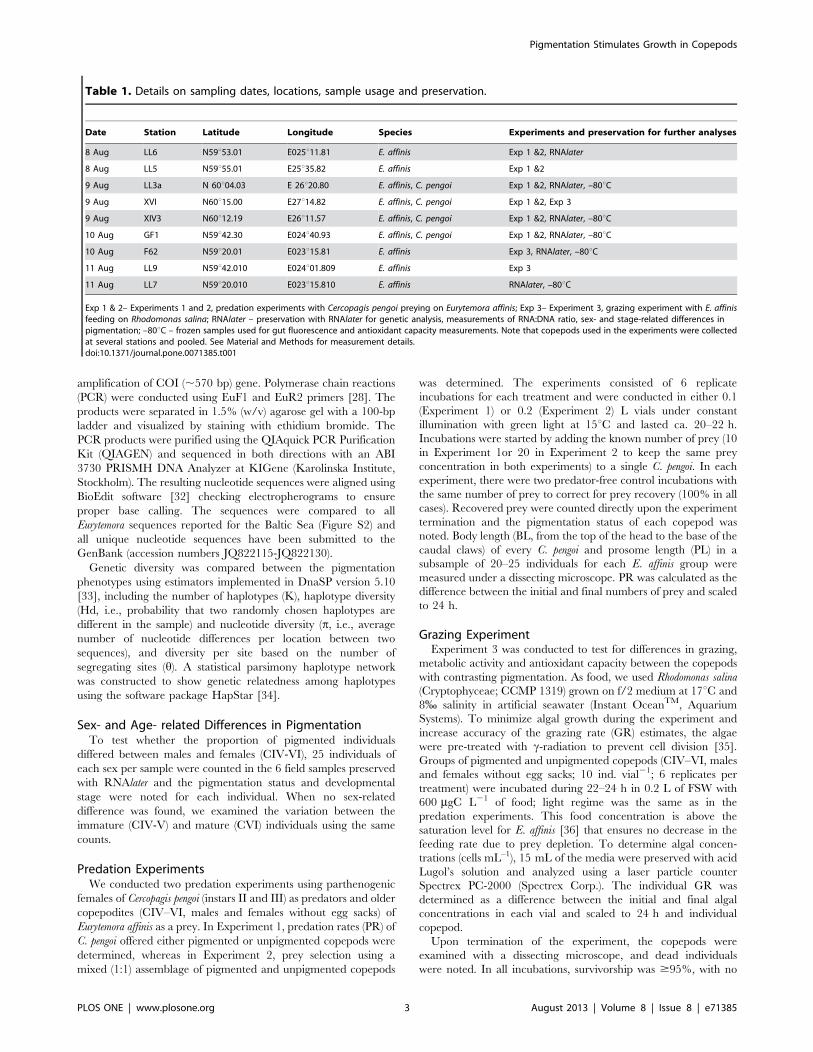
Table 1. Details on sampling dates, locations, sample usage and preservation.
Date Station Latitude Longitude Species Experiments and preservation for further analyses
8 Aug LL6 N59u53.01 E025u11.81 E. affinis Exp 1 &2, RNAlater
8 Aug LL5 N59u55.01 E25u35.82 E. affinis Exp 1 &2
9 Aug LL3a N 60u04.03 E 26u20.80 E. affinis, C. pengoi Exp 1 &2, RNAlater, –80uC
9 Aug XVI N60u15.00 E27u14.82 E. affinis, C. pengoi Exp 1 &2, Exp 3
9 Aug XIV3 N60u12.19 E26u11.57 E. affinis, C. pengoi Exp 1 &2, RNAlater, –80uC
10 Aug GF1 N59u42.30 E024u40.93 E. affinis, C. pengoi Exp 1 &2, RNAlater, –80uC
10 Aug F62 N59u20.01 E023u15.81 E. affinis Exp 3, RNAlater, –80uC
11 Aug LL9 N59u42.010 E024u01.809 E. affinis Exp 3
11 Aug LL7 N59u20.010 E023u15.810 E. affinis RNAlater, –80uC
Exp 1 & 2– Experiments 1 and 2, predation experiments with Cercopagis pengoi preying on Eurytemora affinis; Exp 3– Experiment 3, grazing experiment with E. affinisfeeding on Rhodomonas salina; RNAlater – preservation with RNAlater for genetic analysis, measurements of RNA:DNA ratio, sex- and stage-related differences inpigmentation; –80uC – frozen samples used for gut fluorescence and antioxidant capacity measurements. Note that copepods used in the experiments were collectedat several stations and pooled. See Material and Methods for measurement details.doi:10.1371/journal.pone.0071385.t001
Pigmentation Stimulates Growth in Copepods
PLOS ONE | www.plosone.org 3 August 2013 | Volume 8 | Issue 8 | e71385

significant difference between the pigmented and unpigmented
copepods (Kruskal-Wallis statistic = 0.91, p.0.93). Live copepods
recovered from each vial were split in two groups: (1) 5 individuals
designated for RNA:DNA ratio assay were transferred to
Eppendorf tubes containing 100 mL of RNAlater and stored at
4uC [37], and (2) 4–5 individuals designated for antioxidant
capacity measurements were transferred to Eppendorf tubes and
stored at –80uC.
Gut FluorescenceUsing the frozen zooplankton samples collected in the field, gut
pigment concentration (GPC) in Eurytemora affinis (females, CV-VI)
was analyzed fluorometrically. After thawing on ice, the copepods
were quickly sorted in groups of 5 individuals per pigmentation
type; 2 replicate samples per station, 5 stations in total (Table 1).
Gut pigments were extracted in 0.5 mL of 90% acetone for 24 h
at 4uC in the dark. After centrifugation for 3 min at 3000 rpm, the
fluorescence was measured using a Turner Designs TD700
fluorometer with a detection limit of 0.06 mg L–1 of chlorophyll-
a (Chl-a) and 0.08 mg L–1 of phaeophytin-a. Fluorescence was
measured before and after acidification with HCl and GPC, and
Chl-a was calculated as ng Chl-a eq ind –1 [38]. The duplicate
samples were averaged to provide a single value per station for
each pigmentation type.
RNA:DNA RatioThis ratio was used as a proxy for overall metabolic activity in
the copepods [39]. The nucleic acid concentrations were
measured in both field samples preserved in RNAlater (5 stations,
LL6 excluded; Table 1) and those collected upon termination of
the grazing experiment (6 replicates). For each replicate, a pooled
sample of 5 individuals was used to quantify RNA and DNA
concentrations (mg ind.21) using microplate fluorometric high-
range RiboGreen (Molecular Probes, Inc. Eugene, OR) assay [40]
optimized for Eurytemora affinis of similar size [41]. Test samples,
standards, and the negative controls were measured in duplicates
using FLUOstar Optima microplate reader in black solid flat-
bottom microplates (Greiner Bio-One GmbH) at excitation/
emission wavelengths of 485/590 nm (0.2 s well21, 10 measure-
ments well21). The DNA:RNA standard curve slope ratio was
1.76.
Total Antioxidant CapacityThese measurements were done using copepod samples
obtained in the Experiment 3 (6 replicates) and frozen samples
of field-collected copepods that were also used for the GPC (5
stations; Table 1). The total antioxidant capacity was measured as
oxygen radical absorbance capacity (ORAC), with fluorescein as a
fluorescent probe, AAPH (2,2-azobis(2-amidinopropane) dihydro-
chloride as a thermal free radical source and Trolox as a standard
[42]; all reagents were purchased from Sigma-Aldrich. The
copepods (4–5 individuals sample21) were homogenized in Tris
buffer with ,50 mg of glass beads (,100 mm) for 2 min at 4uCusing FastPrep, centrifuged at 150006g for 15 min at 4uC and
then 10 and 30 mL of the supernatant were taken for measure-
ments of ORAC and protein content, respectively. Protein
concentration (mg mL21) was measured using the bicinchoninic
acid assay (BCA, Pierce Ltd.) according to the manufacturer’s
instructions. The ORAC values were expressed in Trolox-
equivalents, mg mg protein21. All measurements were conducted
in duplicates that were averaged for statistical analysis.
Egg RatioEgg ratio was calculated using the field samples preserved in
RNAlater (6 stations; Table 1). In each sample, the ratio was
calculated using ,50 females for pigmented and unpigmented E.
affinis and dividing a cumulative number of eggs by a total number
of mature females in each category.
Data Analysis and StatisticsTo assess sex- and age-related differences in pigmentation
occurrence, Wilcoxon matched-pairs signed-ranks test was used.
Differences in PR (Experiment 1) and GR (Experiment 3) between
the pigmentation types were evaluated by an unpaired t-test. To
test for differences in prey preference, Manly’s a index [43] was
calculated for each prey type in the Experiment 2. This estimator
has been shown to be asymptotically distributed as a normal
random variate [43]; therefore to assess effects of pigmentation on
PR and prey preference, we compared PR observed in the mixed
prey incubations and mean a values for pigmented and
unpigmented prey using a paired t-test.
Pearson’s product-moment and correlation coefficients were
calculated for all pairs of measured physiological and biochemical
variables. To further evaluate effect of pigmentation on
RNA:DNA ratio and ORAC, 2-way ANOVA was used with
pigmentation status (pigmented or unpigmented) and animal
origin (field or experiment) as categorical predictors. To compare
egg ratio and GPC between the pigmentation types, paired t-test
was used with pairing by station. Finally, to establish a relationship
between the metabolic status and antioxidant capacity and to
evaluate effect of pigmentation on this relationship, we used a
general linear model (GLM) with the pigmentation status and the
animal origin as the categorical variables and ORAC as
continuous variables and RNA:DNA ratio as the dependent
variable. The data for the correlation and regression analyses were
Box-Cox transformed to improve distribution of the residuals. If
not specified otherwise, data are presented as means and standard
deviations (SD). Statistica version 8.0 (StatSoft, 2007) was used for
statistical analyses; the differences were considered significant at
p,0.05.
Results
Species Identity and COI Variability in E. affinisAll 50 sequences had high identity (98–100%) to COI sequences
reported for the Baltic lineage of Eurytemora affinis from coastal
areas of the Gulf of Finland (e.g., Vyborg Bay: HM473964-
HM473975, HM473980- HM473982; Luga Bay: HM473994,
HM473996- HM473998), but also from other parts of the Baltic
Sea (e.g., Swedish coastal waters: JF727517- JF727523; Gulf of
Riga: HM474023, HM474026- HM474027); see Figure S2. By
contrast, there were only 87–88% identity to the sequences
representing E. carolleeae of North American origin (Luga Bay:
HM474011- HM474012, HM474003; Neva Bay: HM474029).
Therefore, all species in our collections were assumed to belong to
the Baltic lineage of E. affinis.
The aligned COI sequence dataset contained 16 unique
haplotypes with 20 polymorphic sites sampled across 571–
574 bp. In both pigmentation groups, many closely related
haplotypes co-existed, with high haplotype diversity and low
nucleotide diversity (Table 2). Four haplotypes were shared
between the groups, whereas 7 and 5 haplotypes were private
for the pigmented and unpigmented copepods, respectively (Fig. 1).
There were two prevalent haplotypes, while rare haplotypes were
in general one to three mutational steps apart from the prevalent
haplotypes.
Pigmentation Stimulates Growth in Copepods
PLOS ONE | www.plosone.org 4 August 2013 | Volume 8 | Issue 8 | e71385

Sex- and Age- related Differences in PigmentationNeither sex- (Wilcoxon matched-pairs signed-ranks test: p.0.8)
nor age-related (p.0.5) differences in pigmentation were signif-
icant. The proportion of the pigmented individuals varied from
25% to 42% among the stations.
Predation on Pigmented and Unpigmented EurytemoraIn the single prey type experiment, the effect of pigmentation on
PR was significant (unpaired t-test; t10 = 3.503, p,0.006; Fig. 2A),
with the pigmented copepods being consumed at ,4-fold higher
rate than unpigmented ones. Similarly, in the mixed prey
experiment, the effect of pigmentation on PR was significant
(paired t-test; t5 = 5.000, p,0.003; Fig. 2A), with the pigmented
copepods being consumed at ,2-fold higher rate than unpig-
mented ones. This was reflected by the significantly higher
Manly’s a for the pigmented individuals in the mixed prey
incubations (paired t-test; t10 = 2.72, p,0.03; Fig. 2B) indicating
that pigmentation increases vulnerability to predation. The
observed differences were not related to body size variations in
either the predator (C. pengoi BL: t48 = 0.97, p.0.43) or the prey
(copepod PL: t48 = 1.01, p.0.32).
Copepod Grazing Rate in the Experiment and GutFluorescence in situ
There was no significant difference in GR between the
pigmented and unpigmented copepods fed Rhodomonas in Exper-
iment 3 (unpaired t-test: t10 = 1.40, p.0.19; Fig. 3A). Similarly,
GPC in field collected individuals showed no significant difference
between the pigmentation types (paired t-test: t4 = 0.78, p.0.49;
Fig. 3A), with mean values varying from 0.02 to 0.07 ng Chl-a eq
ind –1.
Egg Ratio, Metabolic State and Total AntioxidantCapacity
Pigmentation had significantly positive effects on egg ratio in the
field-collected E. affinis (paired t-test: t5 = 3.91, p,0.02; Fig. 3B).
Similarly, there were significantly higher values for RNA:DNA
ratio (2-way ANOVA; F1,20 = 16.39, p,0.007; Fig. 3C) and
Table 2. Eurytemora affinis.
Pigmentation type K Hd p h
Pigmented 11 0.860 0.0042 0.0074
Unpigmented 9 0.863 0.0039 0.0051
Total 16 0.859 0.0041 0.0077
Measures of genetic diversity of the cytochrome c oxidase subunit I (COI) in thecopepods with different pigmentation type.Number of haplotypes (K), haplotype diversity (Hd) and nucleotide diversity (p),and diversity per site (h).doi:10.1371/journal.pone.0071385.t002
Figure 1. Minimum spanning tree showing relationships among haplotypes of pigmented (black) and unpigmented (white)Eurytemora affinis. Size of circle is proportional to the frequency of the haplotype. Slash marks indicate the number of nucleotide substitutionsbetween haplotypes; no slash marks present indicates a single substitution separating haplotypes.doi:10.1371/journal.pone.0071385.g001
Pigmentation Stimulates Growth in Copepods
PLOS ONE | www.plosone.org 5 August 2013 | Volume 8 | Issue 8 | e71385

ORAC values (F1,18 = 107.3, p,0.0001; Fig. 3D) in the pigmented
copepods. Moreover, the experimental conditions with ad libitum
feeding affected positively the RNA:DNA ratio compared to the
field-collected copepods (F1,20 = 14.02, p,0.002), whereas no
significant differences in ORAC values between the animals used
in the experiment and those in the field samples were observed
(F1,18 = 1.505, p.0.3). In no case the interaction term was
significant (p,0.4 in all cases).
There were significant positive correlations between ORAC and
growth indices, but not with food intake (Table 3). Finally, there
was a significant relationship between the RNA:DNA ratio and
ORAC (GLM; F1,19 = 4.98, p,0.04), with a slope being signifi-
cantly higher in animals used in Experiment 3 compared to the
field samples (F1,19 = 15.33, p,0.002), whereas pigmentation did
not affect the relationship (F1,19 = 0.93, p.0.8).
Discussion
In Eurytemora affinis, pigmentation carries both penalty in terms
of the greater vulnerability to predation and fitness advantage in
terms of enhanced growth, reproduction and metabolism. Three
crucial results support this conclusion. First, higher predation rates
and positive selectivity for the pigmented copepods were observed
in the predation experiments with the visual predator Cercopagis
pengoi. Second, growth-related variables (egg ratio and RNA:DNA
ratio) were consistently higher in the pigmented copepods, whereas
the food intake was similar between the pigmentation types.
Consequently, the higher growth output in the pigmented
individuals at a given feeding rate implies higher growth efficiency,
the capacity to convert ingested energy into biomass. Third, the
pigmented copepods had higher antioxidant capacity, suggesting
that this phenotype is superior in handling excessive formation of
free-radicals, which is often a primary cause or a downstream
consequence of tissue damage due to various environmental
stressors, including predation pressure [44]. These findings have
important implications for food web interactions as increased
contribution of the pigmented forms in the population would have
positive effects on energy transfer from primary producers to
secondary consumers and prey quality, in terms of its antioxidant
content, for zooplanktivores.
Our results that pigmented individuals have higher egg
production, metabolic activity and antioxidant capacity lend
support to the metabolic stimulation hypothesis [12]. Despite
experimental evidence that pigmentation enhances survival of
zooplankton exposed to high irradiances, field studies often show
lack of significant relationships with irradiance, but strong inverse
relationships with temperature [6,12]. In Eurytemora affinis, juvenile
Figure 2. Predation rate and prey preference by Cercopagispengoi offered pigmented and unpigmented Eurytemora affinisin feeding trials (Experiments 1 and 2). (A) predation rate (PR) insingle-prey incubations (Experiment 1; mean 6SD; n = 12), and (B) preypreference index (Manley’s a) estimated from the mixed-prey incuba-tions (Experiment 2; Tukey’s box and whisker plot, n = 12); the indexranges from 0 to 1, with higher values indicating greater preference.Shown are the median value (horizontal line), 25% to 75% responseranges (top and bottom lines of boxes) and minima and maxima(whiskers). Asterisk indicate a significant difference (p,0.05) inpreference for the pigmented copepods when compared to thosewithout visible pigmentation.doi:10.1371/journal.pone.0071385.g002
Figure 3. Feeding, growth, reproduction and antioxidantcapacity in pigmented and unpigmented Eurytemora affinis infield and experimental studies. (A) grazing rate (GR; left axis, n = 12)on Rhodomonas salina and gut content fluorescence (GCF; right axis,n = 10) measured in the field collected copepods; (B) egg ratio in thesamples collected in the study area (n = 12); (C) RNA:DNA ratio in thecopepods (Experiment 3: n = 12; field samples: n = 10), and (D) totalantioxidant capacity assayed as ORAC (Experiment 3: n = 12; fieldsamples: n = 10). All data are shown as mean 6SD.doi:10.1371/journal.pone.0071385.g003
Pigmentation Stimulates Growth in Copepods
PLOS ONE | www.plosone.org 6 August 2013 | Volume 8 | Issue 8 | e71385

development is highly temperature dependent in the range of 7 to
18uC [45], i.e., common summer temperatures in the trophogenic
zone in the Baltic Sea. Therefore, increased metabolic stimulation
through absorption of solar radiation would translate into
increased individual and population growth. More recently, a
field study reporting lack of a direct relationship between levels of
photoprotective compounds in freshwater copepods and water
column radiation provided further support to Byron’s hypothesis
[46]. The metabolic stimulation hypothesis was, however,
criticized, arguing that the possible temperature gain is insignif-
icant due to the high heat transfer in small-sized planktonic
animals [47].
We suggest an alternative mechanism for the metabolic
stimulation that could involve elevated antioxidant levels as a
primary means of increasing metabolism without temperature
increase. As physiological functions of pigments include antioxi-
dant protection, the observed higher total antioxidant capacity
(Fig. 3D) in the pigmented copepods is likely to simply reflect
higher concentration of pigments contributing to the higher
antioxidant defences in this phenotype [48]. Recently, a number of
studies have attempted to reveal the relationships between
antioxidant capacity and fitness components in various animals
[20]. Although the connections appear to be stage- and species-
specific, higher antioxidant levels were linked with larger clutches
[49], greater reproductive success [50], improved offspring quality
[51], and increased life span [52]. The observed positive
associations between ORAC levels and growth-related variables
(reproductive output and RNA:DNA ratio) with no apparent
correlation to the food intake (Table 3) provides further evidence
for the positive effects of antioxidant capacity on the copepod
fitness. Moreover, the relationship between ORAC and
RNA:DNA ratio was not phenotype-specific, indicating common
underlying mechanisms.
The apparent lack of costs for pigment synthesis is in
disagreement with experimental studies on zooplankton inhabiting
high-UVR lakes and suffering growth penalties related to
production of photoprotective compounds [4,23]. In our study,
the in situ growth conditions for copepods were suboptimal as
indicated by the higher RNA:DNA ratio of the copepods
incubated at ad libitum food levels in the grazing experiment
compared to the field-collected copepods (Fig. 3B); hence, it is
difficult to assess costs because each phenotype is likely to face own
challenges. Moreover, our study design might have failed to detect
an existing cost: we measured feeding either in the surplus of high
quality food in the absence of competition or external stressors
(Experiment 3) or using a gut fluorescence (field-collected
copepods, pooled samples) without any knowledge on the gut
evacuation governed by ambient temperature and food levels.
Fitness trade-offs can be obscured in the presence of abundant
resources, whereas ambient conditions may differ in multiple
variables, such as temperature, food quality and quantity,
particularly if the pigmentation phenotypes resided at different
depths.
Our study was not designed to address UVR effects on
pigmentation in Eurytemora affinis, because it seemed unlikely that
these effects would be ecologically relevant in the system studied.
Indeed, detrimental UVR effects on copepods were found to be
restricted to the first meter of the water column [53], particularly
in sea areas with high primary productivity. The actual levels of
UVR that summer zooplankton communities in the Baltic Sea are
exposed to are relatively low compared to the oligotrophic and/or
high elevation shallow lakes in Scandinavia, with high light
penetration and a lack of depth refuge from UVB exposure for
zooplankton. In the Baltic Sea, plankton communities are more
protected from UV exposure, because of the greater depth and
high concentrations of colored dissolved organic matter contrib-
uting to high attenuation [53]. In the coastal areas, a typical 1%
penetration depth for UV-B is ,0.1 m in inner bays and 0.3 m in
outer areas, whereas near infrared wavelength range that
contributes most to the temperature-mediated metabolic stimula-
tion reaches deepest in the water column [54]. Moreover, our
study was conducted during a filamentous cyanobacteria bloom,
which substantially increases water turbidity (Secchi depth
,4.5 m) and thus decreases UVR penetration in the water
column. Recently, a counter-intuitive contrast in pigmentation of
the copepod Arctodiaptomus spinosus in shallow fishless lakes has been
reported, with unpigmented copepods inhabiting macrophyte-
dominated clear water lakes and pigmented forms occurring in
highly turbid and phytoplankton-rich lakes [55]. While the
penetration of UV radiation alone failed to explain the differences
in copepod pigmentation among these lakes, several possible
stressors, such as wind-induced turbulence combined with short-
term sunlight exposure and crowding, were advocated in
explaining this pattern [55]. In particular, the pigment accumu-
lation in the crowded populations occurring in the turbid lakes was
suggested to be a support for immune defense in copepods [55,56].
Thus, high UVR exposure is not the only factor favoring
carotenoid accumulation in zooplankton. Nevertheless, the UVR
effects on the copepod pigmentation in the Baltic Sea cannot be
ruled out completely. It could be hypothesized, for example, that
unpigmented phenotype (,70% of the population) would be more
sensitive to UVR avoiding illuminated waters during daytime,
whereas pigmented individuals (,30%) would stay in the more
productive upper part of the water column and produce more
offspring – all these at the cost of predation risk. In this context,
examining the vertical distribution of copepods with contrasting
pigmentation pattern, would help in (1) understanding proximate
causes of DVM, (2) establishing linkages between pigment
production and vertical position of the copepods in the water
column, and (3) revealing energetic costs related to migratory
activity, which would allow to compare growth efficiencies
between the phenotypes and to evaluate the trade-offs involved.
Whereas defence against predators is usually accompanied by
declining rates of growth or development in prey organisms, the
underlying physiological responses remain little understood and
often explained by reduced feeding and/or increased metabolic
costs due to the fight-or-flight response [57]. In our case, the lower
egg ratio in the unpigmented E. affinis cannot be explained by
reduced foraging under predation risk as indicated by similar gut
pigment content. Instead, we suggest that when predation pressure
Table 3. Eurytemora affinis.
Variable RNA:DNA Egg ratio GR GCF
ORAC 0.59 (22) 0.63 (10) 0.44 (12) 0.53 (10)
RNA:DNA 0.61 (10) 0.46 (12) 0.51 (10)
Egg ratio 0.44 (10)
Correlation matrix with Pearson correlation coefficients among the totalantioxidant capacity (ORAC), growth indices (RNA:DNA ratio and egg ratio), andfood intake (GR and GCF).ORAC: oxygen radical absorbance capacity (Experiment 3 and field collectedcopepods), GR: grazing rate (Experiment 3), egg ratio: number of eggs perfemale in a sample (field collected copepods), GCF: gut content fluorescence(field collected copepods). Significant correlations (p,0.05) are in bold face.Data were Box-Cox transformed to approach normal distribution. Number ofsamples is shown in parentheses.doi:10.1371/journal.pone.0071385.t003
Pigmentation Stimulates Growth in Copepods
PLOS ONE | www.plosone.org 7 August 2013 | Volume 8 | Issue 8 | e71385

is high, the unpigmented copepods have higher survival rate, but
also lower antioxidant capacity and reduced fitness. In line with
this, predation-induced decrease in antioxidants has been observed
in copepods [56] and damselfly [44], resulting in a weakened
immune status and a growth reduction, respectively. It remains to
be studied, whether each individual adjust the pigmentation and
related antioxidant reserves to the prevailing predation risk
according to its individual costs and benefits or, alternatively,
whether copepods with different pigmentation suffer differential
mortality as a result of selective predation on specific genotypes.
Although, mitochondrial polymorphisms have been found to be
linked with pigmentation patterns in various species (e.g.,
cladocerans [24], shrimps [25], insects [58], fish [59]), there was
no association between pigmentation and mtDNA genotypes in
the Baltic Eurytemora affinis. This does not, however, imply an
absence of genetic differences between the copepods with different
pigmentation phenotypes, both with regard to genes responsible
for positioning pigments in space and time and those responsible
for synthesis of pigments. Recently, the predation and photo-
protection concepts have been integrated [60], demonstrating that
zooplankters are plastic in their pigmentation, making a trade-off
between high and low pigmentation in relation to the prevalent
fish/UV threat ratio [2,8]. What remains to be integrated in this
theory is the genetic basis of pigmentation, and this calls for a new
approach to study adaptive variation in production of various
pigments and their relative importance in different species and
systems. Indeed, in some cases, pigmentation polymorphism could
be genetically determined, in others, it is a polyphenism, a
developmental difference in pigmentation pattern cued by physical
or chemical signals. With regard to dietary pigments, such as
carotenoids and mycosporine-like amino acids, the latter has been
found to be the case [5,56]. However, both production of the
cuticular pigments and pigment-dispersing hormone activities are
under genetic control [61,62] and, therefore, E. affinis pigmenta-
tion could be an example of a genetic system that leads to multiple
fitness peaks under multiple selection pressures.
In this respect, E. affinis could represent a particularly suitable
model, due to its occurrence worldwide and high evolutionary
diversification [28,29,63]. Together, the observed low nucleotide-
diversity and high haplotype-diversity indicate a co-existence of a
high number of closely related haplotypes (Fig. 1), which is
indicative of a recent population expansion. These findings are in
agreement with the results of a pairwise haplotype mismatch
analysis [28] suggesting that Baltic E. affinis has undergone a
recent expansion. High genetic diversity in such populations is the
raw material for natural selection, and therefore, potentially
underpins fitness and/or adaptive capacity. Recently, a compelling
evidence has been presented on the heritable genetic variation of
pigmentation as a fitness-related trait in populations of snakes [64]
and isopods [65]. Given the involvement of pigmentation in fitness
traits of E. affinis and a likely selection by predation, understanding
genetic basis of pigmentation in this species may provide a more
comprehensive view on ecological and evolutionary significance of
pigmentation patterns in copepods from temperate estuarine
systems, which might be different from those in clear lakes exposed
to high UVR levels.
Supporting Information
Figure S1 A schematic drawing showing pigmentationpattern in pigmented (A) and unpigmented (B) Euryte-mora affinis.
(PDF)
Figure S2 Maximum Likelihood (ML) tree for Euryte-mora affinis sequences reported from the Baltic Sea.
(PDF)
Acknowledgments
We thank crew of R/V Aranda for help in sampling, Jon Duberg and Lisa
Mattsson (Stockholm University) for sharing their collections and providing
laboratory assistance, and Prof. Ragnar Elmgren for fruitful discussions.
Author Contributions
Conceived and designed the experiments: EG ML. Performed the
experiments: EG ML. Analyzed the data: EG NHM. Contributed
reagents/materials/analysis tools: EG. Wrote the paper: EG ML.
Performed genetic analysis and sequencing: NHM. Commented on the
manuscript: NHM.
References
1. Hairston NG (1979) The adaptive significance of color polymorphism in two
species of Diaptomus (Copepoda). Limnol Oceanogr 24: 15–37.
2. Hylander S, Boeing WJ, Grane’li W, Karlsson J, von Einem J, et al. (2009)
Complementary UV protective compounds in zooplankton. Limnol Oceanogr
54: 1883–1893.
3. Hebert PDN, McWalter DB (1983) Cuticular pigmentation in Arctic Daphnia:
Adaptive diversification of asexual lineages? Amer Nat 122: 286–291.
4. Hessen DO (1996). Competitive trade-off strategies in Arctic Daphnia linked to
melanism and UV-B stress. Polar Biol 16: 573–576.
5. Hansson LA, Hylander S, Sommaruga R (2007) Escape from UV threats in
zooplankton: a cocktail of behavior and protective pigmentation. Ecology 88:
1932–1939.
6. Hairston NG (1979) The effect of temperature on carotenoid photoprotection in
the copepod Diaptomus nevadensis. Comp Biochem Physiol 62A: 445–448.
7. Hansson LA (2000) Induced pigmentation in zooplankton: a trade-off between
threats from predation and ultraviolet radiation. Proc R Soc Lond B 267: 2327–
2331.
8. Hansson LA (2004) Plasticity in pigmentation induced by conflicting threats
from predation and UV radiation. Ecology 85: 1005–1016.
9. Speekmann CL, Bollens SM, Avent SR (2000) The effect of ultraviolet radiation
on the vertical distribution and mortality of estuarine zooplankton. J Plankton
Res 22: 2325–2350.
10. Morgan SG, Christy JH (1996) Survival of marine larvae under the
countervailing selective pressures of photodamage and predation. Limnol
Oceanogr 41: 498–504.
11. Byron ER (1981) Metabolic stimulation by light in a pigmented freshwater
invertebrate. Proc Natl Acad Sci USA 78: 1765–1767.
12. Byron ER (1982) The adaptive significance of calanoid copepod pigmentation: a
comparative and experimental analysis. Ecology 63: 1871–1886.
13. Pangle KL, Peacor SD, Johannsson OE (2007) Large nonlethal effects of an
invasive invertebrate predator on zooplankton population growth rate. Ecology
88: 402–412.
14. De Meester L, Beenaerts N (1993) Heritable variation in carotenoid content in
Daphnia magna. Limnol Oceanogr 38: 1193–1199.
15. Ramcharan CW, Sprules WG (1986) Visual predation in Mysis relicta Loven.
Limnol Oceanogr 31: 414–420.
16. Rivier IK (1998) The predatory Cladocera (Onychopoda: Podonidae,
Polyphemidae, Cercopagidae) and Leptodorida of the world. In Dumont H,
editor. Guides to the Identification of the Micro-invertebrates of the Continental
Waters of the World 13. Backhuys Publishers, Leiden. 213 p.
17. Yan ND, Girard R, Boudreau S (2002) An introduced invertebrate predator
(Bythotrephes) reduces zooplankton species richness. Ecol Lett 5: 481–485.
18. Lehtiniemi M, Gorokhova E (2008) Predation of the introduced cladoceran
Cercopagis pengoi on the calanoid copepod Eurytemora affinis in the Gulf of Finland,
Baltic Sea. Mar Ecol Prog Ser 362: 193–200.
19. Sies H, Stahl W (2005) New horizons in carotenoid research. In Packer L,
Obermuller-Jevic U, Kraemer K, Sies H, editors. Carotenoids and Retinoids:
Molecular aspects and health issues. AOCS Press, Urbana, IL, 315–320.
20. Monaghan P, Metcalfe NB, Torres R (2009) Oxidative stress as a mediator of life
history trade-offs: mechanisms, measurements and interpretation. Ecol Lett 12:
75–92.
21. Hubbard JK, Uy JAC, Hauber ME, Hoekstra HE, Safran RJ (2010) Vertebrate
pigmentation: from underlying genes to adaptive function. Trends Genet 26:
231–239.
Pigmentation Stimulates Growth in Copepods
PLOS ONE | www.plosone.org 8 August 2013 | Volume 8 | Issue 8 | e71385

22. Gerrish GA, Caceres CE (2003) Genetic versus environmental influence on
pigment variation in the ephippia of Daphnia pulicaria. Freshwat Biol 48: 1971–1982.
23. Hebert PDN, Emery A (1990) The Adaptive Significance of Cuticular
Pigmentation in Daphnia. Functional Ecol 4: 703–710.24. Van Raay TJ, Crease TJ (1995) Mitochondrial DNA diversity in an apomictic
Daphnia complex from the Canadian High Arctic. Mol Ecol 4: 149–161.25. Tsoi KH, Wang ZY, Chu KH (2005) Genetic divergence between two
morphologically similar varieties of the kuruma shrimp Penaeus japonicus. Mar
Biol 147: 367–379.26. Tarjuelo I, Posada D, Crandall A, Pascual M, Turon X (2004) Phylogeography
and speciation of colour morphs in the colonial ascidian Pseudodistomacrucigaster. Mol Ecol 13: 3125–3136.
27. Viherluoto M, Viitasalo M (2001) Effect of light on the feeding rates of pelagicand littoral mysid shrimps: a trade-off between feeding success and predation
avoidance. J Exp Mar Biol Ecol 261: 237–244.
28. Winkler G, Souissi S, Poux C, Castric V (2011) Genetic heterogeneity amongEurytemora affinis populations in Western Europe. Mar Biol 158: 1841–1856.
29. Lee CE, Frost BW (2002) Morphological stasis in the Eurytemora affinis speciescomplex (Copepoda: Temoridae). Hydrobiologia 480: 111–128.
30. Alekseev VR, Abramson NI, Sukhikh NM (2009) Introduction of sibling species
to the ecosystem of the Baltic Sea. Dokl Biol Sci 429: 544–547.31. Alekseev VR, Souissi A (2011) A new species within the Eurytemora affinis complex
(Copepoda: Calanoida) from the Atlantic Coast of USA, with observations oneight morphologically different European populations. Zootaxa 2767: 41–56.
32. Hall TA (1999) BioEdit: a user-friendly biological sequence alignment editor andanalysis program for Windows 95/98/NT. Nucl Acid Symp Ser 41: 95–98.
33. Librado P, Rozas J. (2009) DnaSP v5: a software for comprehensive analysis of
DNA polymorphism data. Bioinformatics 25: 1451–1452.34. Teacher AGF, Griffiths DJ (2011) HapStar: automated haplotype network
layout and visualization. Mol Ecol Res 11: 151–153.35. Gorokhova E, Mattsson L, Sundstrom AM (2012). A comparison of TO-PRO-1
iodide and 5-CFDA-AM staining methods for assessing viability of planktonic
algae with epifluorescence microscopy. J Microbiol Methods 89: 216–221.36. Ban S (1994) Effect of temperature and food concentration on post-embryonic
development, egg production and adult body size of calanoid copepod Eurytemora
affinis. J Plankton Res 16: 721–735.
37. Gorokhova E (2005) Effects of preservation and storage of microcrustaceans inRNAlater on RNA and DNA degradation. Limnol Oceanogr: Methods 3: 143–
148.
38. Bamstedt U, Gifford DJ, Irigoien X, Atkinson A, Roman M, et al. (2000)Feeding. In: Harris RP, Wiebe PH, Lenz J, Skjoldal HR, Huntley M, editors.
ICES Zooplankton Methodology Manual. Academic Press, London, 297–380.39. Holmborn T, Lindell K, Holeton C, Hogfors H, Gorokhova E. (2009)
Biochemical proxies for growth and metabolism in Acartia bifilosa (Copepoda,
Calanoida). Limnol Oceanogr: Methods 7: 785–794.40. Gorokhova E, Kyle M (2002) Analysis of nucleic acids in Daphnia: development
of methods and ontogenetic variations in RNA-DNA content. J Plankton Res 24:511–522.
41. Hook T, Gorokhova E, Hansson S (2008) RNA:DNA ratios of Baltic herringlarvae and copepods in embayment and open sea habitats. Estuar Coast Shelf
Sci 76: 29–35.
42. Prior RL, Hoang H, Gu L, Wu X, Bacchiocca M, et al. (2003) Assays forhydrophilic and lipophilic antioxidant capacity (oxygen radical absorbance
capacity (ORACFL)) of plasma and other biological and food samples. J AgricultFood Chem 51: 3273–3279.
43. Manly BFJ (1974) A model for certain types of selection experiments. Biometrics
30: 281–294.44. Slos S, Stoks R (2008) Predation risk induces stress proteins and reduces
antioxidant defense. Funct Ecol 22: 637–642.
45. Vuorinen I (1982) The effect of temperature on the rates of development ofEurytemora hirundoides (Nordquist) in laboratory culture. Ann Zool Fenn 19: 129–
134.
46. Garcia PE, Perez AP, Dieguez MC, Ferraro MA, Zagarese HE (2008) Dual
control of the levels of photoprotective compounds by ultraviolet radiation andtemperature in the freshwater copepod Boeckella antiqua. J Plankton Res 30: 817–
827.
47. Hairston NG Jr (1981) The interaction of salinity, predators, light and copepod
color. Hydrobiologia 81: 151–158.
48. McGraw KJ (2005) The antioxidant function of many animal pigments: are
there consistent health benefits of sexually selected colourants? Animal
Behaviour 69: 757–764.
49. Bize P, Devevey G, Monaghan P, Doligez B, Christe P (2008) Fecundity and
survival in relation to resistance to oxidative stress in a free living bird. Ecology89: 2584–2593.
50. Safran RJ, McGraw KJ, Wilkins MR, Hubbard J, Marling J (2010) Positivecarotenoid balance over the breeding season predicts reproductive performance
in a wild bird. PLoS ONE 5: e9420.
51. Biard C, Surai PF, Møller AP (2005) Effects of carotenoid availability during
laying on reproduction in the blue tit. Oecologia 144: 32–44.
52. Martin I, Grotewiel MS (2006) Oxidative damage and age-related functional
declines. Mech Ageing Dev 127: 411–423.
53. Browman HI, Rodriguez CA, Beland F, Cullen JJ, Davis RF, et al. (2000)
Impact of ultraviolet radiation on marine crustacean zooplankton and
ichthyoplankton: a synthesis of results from the estuary and Gulf of St.Lawrence, Canada. Mar Ecol Prog Ser 199: 293–311.
54. Schubert H, Sagert S, Forster RM (2001) Evaluation of the different levels ofvariability in the underwater light field of a shallow estuary. Helgol Mar Res 55:
12–22.
55. Schneider T, Herzig A, Koinig KA, Sommaruga R (2012) Copepods in turbid
shallow soda lakes accumulate unexpected high levels of carotenoids. PLoS ONE7: e43063.
56. Van der Veen IT (2005) Costly carotenoids: a trade-off between predation andinfection risk? J Evol Biol 18: 992–999.
57. Creel S, Christianson D (2008). Relationships between direct predation and riskeffects. Trends Ecol Evol 23: 194–201.
58. Brisson JA, De ToniDC, Duncan I, Templeton AR (2005) AbdominalPigmentation Variation in Drosophila polymorpha: Geographic Variation in the
Trait, and Underlying Phylogeography. Evolution 59: 1046–1059.
59. Rodrıguez-Grana L, Herrera G, Herrera L, Castro LR (2004) Divergence of two
forms of Triphoturus in the eastern Pacific based on mtDNA cytochrome b gene
sequences and larval morphology. J Fish Biol 64: 1455–1461.
60. Williamson GE, Fischer JM, Bollens SM, Overholt EP, Breckenridge JK (2011)
Toward a more comprehensive theory of zooplankton diel vertical migration:Integrating ultraviolet radiation and water transparency into the biotic
paradigm. Limnol Oceanogr 56: 1603–1623.
61. Stevenson J R (1985) Dynamics of the integument. In Bliss DE, Mantel LH,
editors. The Biology of Crustacea, Vol. 9, Integument, Pigments, and HormonalProcesses, Academic Press, New York. Pp. 1–42.
62. Rao KR, Riehm JP (1988) Pigment-dispersing hormones: A novel family ofneuropeptides from arthropods. Peptides 9, Suppl.1: 153–159.
63. Winkler G, Dodson JJ, Lee CE (2008) Heterogeneity within the native range:population genetic analyses of sympatric invasive and noninvasive clades of the
freshwater invading copepod Eurytemora affinis. Mol Ecol 17: 415–430.
64. Westphal MF, Massie JL, Bronkema JM, Smith BE, Morgan TJ (2011) Heritable
Variation in Garter Snake Color Patterns in Postglacial Populations. PLoS ONE
6(9): e24199.
65. Protas ME, Trontelj P, Patel NH (2011) Genetic basis of eye and pigment loss in
the cave crustacean, Asellus aquaticus. Proc Natl Acad Sci 108: 5702–5707.
Pigmentation Stimulates Growth in Copepods
PLOS ONE | www.plosone.org 9 August 2013 | Volume 8 | Issue 8 | e71385

Supporting Information
Trade-offs between predation risk and growth benefits in the copepod Eurytemora affinis with
contrasting pigmentation
By Elena Gorokhova, Maiju Lehtiniemi, and Nisha H. Motwani
Figure S1. A schematic drawing showing pigmentation pattern in pigmented (A) and
unpigmented (B) Eurytemora affinis.
A B

Vybo_HM473986
Vybo_HM473979
Luga_HM474008
Himm JF727541
Askö_JF727529
Vybo_HM473972
Vybo_HM473987
Luga_HM473997
Riga_HM474026
U4
P8
P9
U6
Riga_HM474023
Luga_HM473996
Vybo_HM473985
Vybo_HM473971
Askö_JF727523
Himm JF727539
Himm JF727537
Himm JF727536
Himm JF727527
Riga_HM474019
Himm JF727519
Himm JF727542
Vist_HM473960
Vybo_HM473973
Vybo_HM473988
Luga_HM473998
Riga_HM474027
U2
P7
U1
U26
Luga_HM474000
Vybo_HM473989
Vybo_HM473974
Vybo_HM473964
Himm JF727543
Himm JF727522
Riga_HM474024
Himm JF727518
P2
P21
U22
Luga_HM474009
Vybo_HM473991
Vybo_HM473980
Vybo_HM473966
Vist_HM473959
Himm JF727547
P12
U20
Luga_HM474013
Vybo_HM473993
Vybo_HM473981
Vybo_HM473967
Himm JF727548
Himm JF727531
Luga_HM474006
Askö_JF727524
P4
P10
U8
Riga_HM474016
Luga_HM473994
Vybo_HM473984
Vybo_HM473970
Luga_HM474010
Askö_JF727520
P11
U9
Luga_HM474014
Luga_HM368364
Vybo_HM473982
Vybo_HM473968
Vist_HM473961
Askö_JF727517
P22
U24
Luga_HM474005
Vybo_HM473990
Vybo_HM473975
Vybo_HM473965
Himm JF727546
Himm JF727521
P6
P20
U3
Riga_HM474028
Riga_HM474021
Riga_HM474020
Riga_HM474017
Riga_HM474015
Vybo_HM473963
Askö_JF727544
Askö_JF727540
Askö_JF727535
P24
Himm JF727532
Vist_HM473958
Himm JF727516
Himm JF727525
Vybo_HM473992
Luga_HM473995
Luga_HM473999
Luga_HM474002
Luga_HM474007
Himm JF727530
Riga_HM474022
P23
Vybo_HM473978
Vybo_HM473977
Vybo_HM473976
Vybo_HM473983
Himm JF727538
Luga_HM474004
Riga_HM474018
Riga_HM474025
U21
U9
U7
P3
P1
P14
Elbe JF727313
Elbe JF727330
Elbe JF727348
Elbe JF727344
Riga_JF727528
Elbe JF727311
Elbe JF727310
Elbe JF727312
Elbe JF727314
Elbe JF727315
Elbe JF727316
Elbe JF727317
Elbe JF727319
Elbe JF727320
Elbe JF727321
Elbe JF727322
Elbe JF727323
Elbe JF727324
Elbe JF727325
Elbe JF727327
Elbe JF727329
Elbe JF727331
Elbe JF727347
Elbe JF727346
Elbe JF727345
Elbe JF727343
Elbe JF727318
Elbe JF727326
Elbe JF727328
Elbe JF727349
Luga_HM474001
U5
Askö_JF727534
Vybo_HM473969
Himm JF727545
Himm JF727526
Riga_JF727558
Riga_JF727557
Riga_JF727554
Riga_JF727553
Luga_HM474012
Luga_HM474003
Neva_HM474029
Luga_HM474011
lacustris HM474035
0.02
elenag
Text Box
Supporting Information Figure S2. Maximum Likelihood (ML) tree for Eurytemora affinis sequences reported from the Baltic Sea. Sequences are presented with their GenBank accession numbers preceded by geographic locations that are coded as follows: Askö: Swedish coastal area close to Askö station, northern Baltic proper; Elba: Elba estuary, southern Baltic Sea Himm: Himmerfjärden Bay, Swedish coastal area in Södertälje municipality, northern Baltic proper; Luga: Luga Bay, Gulf of Finland; Riga: Riga Bay, northern Baltic proper; Vist: Vistula Lagoon, south-east Baltic proper; Vybo: Vyborg Bay, Gulf of Finland. Sequences marked U and P are unpigmented and pigmented copepods, respectively, sequenced in this study, the number indicates isolate. Phylogenetic tree was constructed with the software MEGA 5.0.Identical sequences were not included in the tree. One thousand bootstrapped replicate resampled datasets were analyzed. Sequences representing Eurytemora carollee are marked in red. Eurytemora lacustris was used as an outgroup.

![One-pot synthesis of mesoporous [Al]-SBA-16 and acidity characterization by CO adsorption](https://static.fdokumen.com/doc/165x107/633e3ad94f039e2afc0181e4/one-pot-synthesis-of-mesoporous-al-sba-16-and-acidity-characterization-by-co-adsorption.jpg)