Genotoxicity of diuron and glyphosate in oyster spermatozoa and embryos
Upload
independentCategory
view
5download
0
Mutation Research 490 (2001) 141–158
Genotoxicity assessment in aquatic environments under theinfluence of heavy metals and organic contaminants
Vera Maria Ferrão Vargas∗, Sayonara Bresolin Migliavacca, Andréa Cássia de Melo,Rubem Cesar Horn, Regis Rolim Guidobono, Isabel Cristina Fernandes de Sá Ferreira,
Maria Heloisa Degrazia PestanaFundação Estadual de Proteção Ambiental Henrique Luis FEPAM, Av. Salvador França, 1707, 90690-000 Porto Alegre, RS, Brazil
Received 22 August 2000; received in revised form 13 November 2000; accepted 15 November 2000
Abstract
The genotoxicity of river water and sediment including interstitial water was evaluated by microscreen phage-inductionandSalmonella/microsome assays. Different processes used to fractionate the sediment sample were compared using solventswith different polarities. The results obtained for mutagenic activity using theSalmonella/microsome test were negative inthe water and interstitial water samples analysed using the direct concentration method. The responses in the microscreenphage-induction assay showed the presence of genotoxic or indicative genotoxic activity for at least one water sample of eachsite analysed using the same concentration method. Similar results were obtained for interstitial water samples, i.e. absence ofmutagenic activity in theSalmonella/microsome test and presence of genotoxic activity in the microscreen phage-inductionassay. Metal contamination, as evidenced by the concentrations in stream sediments, may also help explain some of thesegenotoxic results. Stream sediment organic extracts showed frameshift mutagenic activity in the ether extract detected bySalmonella/microsome assay. The concentrates evaluated by microscreen phage-induction assay identified the action of organiccompounds in the non-polar, medium polar and polar fractions. Thus, the microscreen phage-induction assay has proven to bea more appropriate methodology than theSalmonella/microsome test to analyse multiple pollutants in this ecosystem whereboth organic compounds and heavy metals are present. © 2001 Elsevier Science B.V. All rights reserved.
Keywords: Salmonella/microsome assay; Microscreen phage-induction assay; Water samples; Sediment; Heavy metals; Organic compounds
1. Introduction
This work is part of the studies to evaluate genotoxicactivity in water sources in the state of Rio Grande doSul, RS, Brazil. It is an assessment of the potential forcontamination in the Sinos River, whose basin coversa significant urban, agricultural and industrial area ofthe state, with a high concentration of different typesof industry (metallurgical, footwear, chemicals, tex-
∗ Corresponding author. Tel./fax:+55-5133-46765.E-mail address:[email protected] (V.M.F. Vargas).
tiles, foodstuffs), including an oil refinery and manytanneries. This river has been monitored because ithas a potential for contamination by toxic substances.The water of the Sinos River basin is used for a publicand industrial water supply, irrigation, watering ani-mals, primary contact recreation, artisanal fishing andnavigation, and to protect aquatic communities [1].
The evaluation of the genotoxic potential of thisriver is based on ongoing studies in an area influencedby petrochemicals [2–4]. The purpose is to provide ad-ditional information to integrate this level of biologi-cal evaluation in quality control parameters in regions
1383-5718/01/$ – see front matter © 2001 Elsevier Science B.V. All rights reserved.PII: S1383-5718(00)00159-5
142 V.M.F. Vargas et al. / Mutation Research 490 (2001) 141–158
subject to ecotoxicological risk. In order to study envi-ronmental genotoxicity, besides choosing the method-ologies for genetic evaluation, it should be understoodthat the environmental sample consists of a complexmixture of substances that requires different method-ologies for analysis [4,5].
Chemical compounds of urban-industrial originprovide a significant contribution to the contaminationof river water, both in the water column and sedi-ments, resulting in a potential ecotoxicological risk.Water-soluble contaminants can be persistent andmaintain their physical and chemical characteristicswhile they are transported and distributed throughoutthe aquatic environment. These non-degradable pol-lutants may accumulate in different compartments orundergo transformations resulting in compounds withmore or less bioavailability [6]. The use of streamsediments in environmental studies to assess aquaticmetal pollution, for example, is due mostly to theability of this compartment to concentrate metals,acting either as a sink or as a secondary source ofcontaminants in the water column and aquatic biota[7,8]. Heavy metals, in particular, are chemical ele-ments that occur in natural environments at low con-centrations and are essential to living beings, but inexcessive amounts may be toxic, mutagenic, carcino-genic, and/or teratogenic agents [9–11]. In addition,several studies have reported the presence of xenobi-otics of organic origin and genotoxic activity in theaquatic environment [2–4,13,36].
The definition of the class of compounds responsi-ble for the genotoxic contamination of complex sam-ples is a task with multiple approaches. Among theaspects to be considered in the chemical interactionsthat result in ecotoxicological damages are the effectsof synergy and antagonism that occur as a result of thecomplexity of the chemical compounds present, andconfer specific characteristics on the mixtures formed.
When possible, the evaluation of the sample as awhole, without chemical fractionation, prevents theloss or alteration of the substances present. However,the low concentration or excessive toxicity of thesample could influence the efficiency of assay diag-nosis (for a review, see [4,5]). Chemical fractionationaccording to acid–base properties or polarity of chem-icals has been used in association with bioassays,especially theSalmonella/microsome assay in an at-tempt to identify genotoxic organic compounds. This
fractionation seeks to simplify the composition of thesamples, as much as possible, in order to correlatemutagenicity and chemical composition [4,5,12–16].These answers are especially important in seekingmeasures to clean contaminated environments.
Several studies have shown the microscreenphage-induction assay to be an appropriate ana-lytic methodology to detect halogenated compounds,organochlorine compounds and metals components,with a low level of detection by theSalmonellaas-say [5,10,17,18]. In addition, this test was considereda good screening assay for genotoxic compoundspresent in small concentrations in environmentalsamples [4].
In the current study, the microscreen phage-inductionassay andSalmonella/microsome test were used toanalyse samples of river water and sediments byevaluating the interstitial water and organic fractionsobtained by different methods of extraction withsolvents that have different polarities [12,13]. Theobjective of the study was to investigate the method-ologies for examining the different environmentalcompartments associated with the choice of assaysand bacterial strains more sensitive to the diagnosisof the ecosystem studied. Total metal contents inthe stream sediments were also determined, in orderto investigate possible relations between genotoxic-ity tests in water (bulk and interstitial) samples andmetal concentrations in the subaquatic sediments.The study also attempted to evaluate the sensitivity ofthe microscreen phage-induction assay as a methodfor genotoxic diagnosis of the environmental samplespossibly contaminated by trace-elements associatedwith the presence of organic contaminants.
2. Materials and methods
2.1. Sample
Samples of water were collected in the Sinos River,RS, according to Standard Methods for the Examina-tion of Water and Wastewater [19], described by Var-gas et al. [3]. The stream sediment samples were col-lected with a Petersen grab, in a composite manner, atabout 20 cm depth from the river bed, put into plasticbags and kept on ice while being transported to thelaboratory. The water samples were stored at 4◦C for
V.M.F. Vargas et al. / Mutation Research 490 (2001) 141–158 143
4 days, and then divided into aliquots and kept in afreezer at−20◦C. After extraction, the sediment sam-ples were treated the same way.
2.2. Sampling sites
The sampling sites were chosen based on insti-tutional studies developed in the area of this riverbasin (Fig. 1 [20]). The sites evaluated consideredcritical to the Sinos River basin drain an area an-thropogenically contaminated by urban, agriculturaland industrial activities, located close to the citiesand towns with a population ranging from 34,177(Santo Antonio da Patrulha) to 294,125 (Canoas) in-habitants: Canoas (Sinos A); Esteio (Sinos B); Sa-pucaia (Sinos C); São Leopoldo (Sinos D and SinosE); Campo Bom (Sinos F) and Parobé (Sinos G). Asa site of reference, a location was chosen close tothe sources in the town of Santo Antônio da Patrulha(Sinos H). The sites are listed and characterised inTable 1.
2.3. Stages of the study
It was possible to divide the study in to five differ-ent stages (Table 2). During the first stage (samplingsI–IV, April, May, October and November 1992), wa-ter samples from the eight sampling points were stud-ied using the direct concentration method. During thesecond stage (samplings V, January, April, November1993 and VI, March, April, August 1994), water and
Table 1Contributions from the sampling sites: agricultural and industrialactivities
Samplings Main contributions
Sinos A Oil refinerySinos B Tanneries, agricultural pesticide
plant, recycled paper plant, steel millSinos C Tanneries, agricultural pesticide
plant, sand dredgingSinos D and E Recycled paper plant, sand dredging,
metallurgySinos F MetallurgySinos G Footwear metallurgical factories and
leather finishingSinos H Small farms, livestock
interstitial water were evaluated from four selectedsites using the direct concentration method. In the thirdstage (samplings V and VI), chemical determinationswere performed on sediment samples: organochlorinepesticides and total metal concentrations in sedimentsfrom Sinos C, E (both in the lower course, drainingsandstones, Quaternary river sediments and alluvialfan deposits), G (in the middle course, draining sand-stones and Quaternary river sediments) and H (locatedin the upper reaches, draining basalts and sandstones)[21]. During the fourth stage (sampling VI), organicextracts of sediment samples were studied, from sitesSinos E, G and H. During the fifth stage (samplingVII, July 1995) extracts obtained by chemical frac-tionation from site Sinos E were evaluated.
2.4. Sample preparation
2.4.1. Water samplesThey were sterilised using Millipore filters with
0.22mm pores and analysed at different volumes us-ing the direct concentration method of Vargas et al.[3,4] in Salmonella/microsome, and microscreenphage-induction assays.
2.4.2. Interstitial waterThe sediment samples were centrifuged at 1100g
for 10 min to extract interstitial water. The lat-ter was sterilised and analysed according to themethod of direct concentration of the sample [3,4]using Salmonella/microsome, and microscreenphage-induction assays. The pH of the supernatantwas measured.
2.4.3. Ether extraction by silica gel glass columnSediment samples were extracted using the ether
as a solvent for polar compounds by the method de-scribed by Sato et al. [12], modified. After the sam-ple was centrifuged, the stream sediment was driedat room temperature and one portion of the samplewas mixed with anhydrous sodium sulfate (1:1). Theextraction, at a neutral pH was performed in a col-umn and concentrated in a rotary evaporator. A por-tion of the extract was used to determine density. Theconcentrate was transferred, stored (4◦C), and beforethe assay, the extracts were evaporated until dry un-der a nitrogen gas flow, and then resuspended with theappropriate solvent to the assay.

144V.M
.F.V
arg
as
et
al./M
uta
tion
Re
sea
rch4
90
(20
01
)1
41
–1
58

V.M.F. Vargas et al. / Mutation Research 490 (2001) 141–158 145
Table 2Stages of the study
Stage Samplings Sample types Sites
1 I–IV (1992) River water (direct concentration method) A–H
2 V (1993) Water and interstitial water (direct concentration method) D, E, G, HVI (1994)
3 V Sediment (chemical determinations) C, E, G, HVI
4 VI Sediment (organic extracts) E, G, H
5 VII (1995) Sediment (organic extracts — chemical fractionation) E
2.4.4. Sediment extraction by sonicationThis method was performed as described in EPA
[22], modified. Sodium sulfate was added to the wetsediment samples. The organic solvent was added tothis mixture, shaken, and sonicated (100 W for 6 min);this operation was repeated three times. The combinedextracts were dried, concentrated, stored (4◦C) and thedensity was determined from a portion of this extract.At the time of analysis, the extracts were evaporateddry under a nitrogen gas flow and resuspended in ace-tone at 10%. Two different extraction solvents wereused in this process: dichloromethane for the extrac-tion of moderately polar compounds and ether [12] asa polar solvent.
2.4.5. Extraction by chemical fractionationThe procedure for this extraction followed the
methodology described by Grifoll et al. [13]. A20–40 g portion of sediments was extracted by sonica-tion with a dichloromethane methanol (2:1) mixture.For chemical fractionation, the organic extracts wereconcentrated to near dryness in a rotary evaporator,redissolved in 1 ml of dichloromethane and adsorbedonto 1 g of neutral alumina. The solvent was evapo-rated under a nitrogen gas flow, and the mixture wastransferred to a glass column packed with 8 g of silicagel and 7 g of neutral alumina. A total of six fractionswas eluted successively with the following solvents: I,20 ml hexane; II, 20 ml 10% dichloromethane in hex-ane; III, 40 ml 20% dichloromethane in hexane; IV,20 ml 75% dichloromethane in hexane; V, 40 ml 5%methanol in dichloromethane; VI, 40 ml diethylether.Next the density of the extracts obtained in a portionof the sample was determined. The subfractions ob-tained were transferred to the acetone 10% solvent, at
the time of the assay, as described previously. Blankextracts were obtained in parallel.
2.4.6. Total metal concentrations in sedimentsThe fine silt–clay fraction was wet-sieved,
oven-dried at 40◦C and analysed for metals (Fe, Mn,Cu, Cr, Pb, Zn, Ni) after total extraction with a hotmixture of HF/HCLO4/HNO3 (1:1:1) in duplicate(1 g samples). Metal determinations were performedby flame atomic absorption spectrophotometry atthe laboratory at Federal University of Rio Grandedo Sul (UFRGS), Centro de Estudos em Petrologiae Geoquımica/Instituto de Geociências (CPGq/IG).Further information on the methods applied to thesediment samples is given by Pestana et al. [23].
2.4.7. Sediment extraction for chromatographicanalysis
Sediment extraction for chromatographic analy-sis for the presence of organochlorine pesticides.The method described by EPA [24] was used, im-plemented by a procedure of extraction in silica gelglass column using 250 ml of hexane–acetone (1:1)mixture. The extract was concentrated for purificationthrough a florisil column for analysis by gas chro-matography. This extract obtained after purificationwas concentrated into 10 ml at 85◦C, and its densitywas determined at a later time.
2.5. Genotoxic evaluation tests
2.5.1. Salmonella/microsome assaySalmonella/microsome assay performed using the
pre-incubation procedure according to Maron andAmes [25] and described by Vargas et al. [2,3]. The

146 V.M.F. Vargas et al. / Mutation Research 490 (2001) 141–158
Salmonella typhimuriumstrains used were TA100 andTA1535 detecting base-pair substitution mutagens,strains TA97, TA98 for detecting frameshift mutagens,and strain TA102 for detecting a large variety of ox-idative mutagens and presenting intact excision repairmechanisms. Strain TA97 is described in literature asmore sensitive to heavy metals [26] and polyaromatichydrocarbons [25]. Negative (sterile distilled water ordimethylsulfoxide, DMSO, spectrophotometric grade)and positive (sodium azide, SAZ; 4-nitroquinolineoxide, 4NQO; UV irradiation, assays without S9 mix;2-aminofluorene, 2AF; 2-aminoanthracene, 2AA; as-says with S9 mix) controls were included in eachassay. The cellular viability test was performed tocontrol the cytotoxic potential of these samples [3].
2.5.1.1. Evaluation of results.In the screening as-says using a single dose, the sample was consideredmutagenic when the value of the induced mutation wasdouble the activity observed in the negative control.For the samples assessed at different concentrations,the significance of the dose–response curve was eval-uated using the Salmonel software [27]. The samplewas considered as indicating mutagenic activity, whenonly one of the criteria above was observed. In thecellular viability test, the responses were consideredcytotoxic when the percentage of sample survival wasless than 60% of that observed for negative control.
2.5.2. Microscreen phage-induction assayThis methodology was performed according to
Rossman et al. [28], as described by Vargas et al. [4].It is based on the response to induction of SOS func-tions mediated by proteaseRecA and cleavage of thephagel repressor. The strains used in this method arederived fromEscherichia coliB/r. WP2s (λ), is thelysogenic strain (trpE, uvrA) and RJF801, the indi-cating strain (trpE, uvrD3, Amp®). In the test carriedout in the presence of microsome activation, the totalvolume was maintained, the amount of strain addedwas reduced from 100 to 75ml, and 25ml of S9 mixprepared using the method of Maron and Ames [25]was added. The negative controls, distilled water oracetone 10% [29] and positive controls (4NQO, as-says without S9 mix or 2AF, assays with S9 mix) wereused in each test. A variation of the methodology inwhich a series of seven concentrations are used witha dilution factor 2, beginning with the concentration
of 50mg per well, as indicated in literature [10], wasemployed in the assays with the extracts obtained bychemical fractionation. The protocol of this assay wasmodified to utilise a constant volume of acetone of10% per well at the different concentrations tested.The optimum dosage of solvent to be used in thisstage of the work was established as 50ml of ace-tone 10% per well, because this does not interfere inspontaneous induction and allows sample solubility.
2.5.2.1. Evaluation of results.In order to comparethe different samples, the rate of lysogenic induction(induction factor, IF) was calculated by the ratio ofmean plaque-forming unit (lysis) (PFU) of the sampleto spontaneous mean PFU of the assay [4]. To evaluatethe dose–response relation, the linear portion of thecurve was used. According to the final criterion ofevaluation [10,17], described by Vargas et al. [4], thesamples that presented a reproducible and significantdose–response ratio with a lysogenic induction indexof three times in at least two dosages were consideredpositive. If the sample exceeded this value by one dose,it was considered weakly genotoxic. In the presentstudy, if the sample presented an index above threeor a significant and reproducible dose–response curvewith an index above 1.5, the result was consideredindicative.
2.5.3. Microsome activation systemThe assays were performed in the presence or ab-
sence of S9 mix metabolisation fraction (20ml of S9fraction) prepared from Sprague-Dawley rat livers,pre-treated with Aroclor 1254 (Moltox, SA, USA),The activation mixture was prepared according toMaron and Ames [25].
3. Results
3.1. First stage: river water
3.1.1. Salmonella/microsome assaySamples from four different samplings (I–IV) were
analysed. This was done at the collection sites of SinosA, B, C, D, E, F, G and H, using the direct concen-tration method with strains TA100 and TA98 in thepresence and absence of the S9 microsome fraction,at a screening dosage of 2000ml per plate. The results

V.M.F. Vargas et al. / Mutation Research 490 (2001) 141–158 147
Table 3Screening analysis of mutagenicity bySalmonella/microsome assay in the absence (without S9 mix) and presence (with S9 mix) ofmetabolic activation of water samples from the Sinos River basin
Samplesa TA98 TA97 TA100 TA1535 TA102
Rev/plb Ic Rev/pl I Rev/pl I Rev/pl I Rev/pl I
Without S9 mixNCd 34 ± 17.7 189± 25.3 200± 15.2 31± 3.0 306± 35.1Sinos A 29± 8.0 0.9 145± 6.0 0.8 92± 10.0 0.5 35± 3.5 1.1 405± 27.0 1.3Sinos B 65± 28.5 1.9 132± 19.0 0.7 214± 22.0 1.1 35± 9.5 1.1 435± 18.2 1.4Sinos C 25± 2.2 0.8 153± 6.0 0.8 112± 12.0 0.6 40± 6.0 1.3 483± 15.0 1.6Sinos D 12± 2.4 0.4 141± 27.0 0.7 95± 15.0 0.5 33± 7.5 1.1 334± 38.0 1.1Sinos E 21± 4.9 0.6 132± 31.0 0.7 121± 4.9 0.6 32± 3.0 1.0 309± 39.1 1.0Sinos F 16± 1.9 0.5 154± 53.0 0.8 104± 2.6 0.5 30± 4.6 1.0 435± 16.9 1.4Sinos G 17± 2.8 0.5 144± 33.0 0.8 101± 14.0 0.5 38± 12.0 1.2 353± 66.2 1.2Sinos H 26± 9.2 0.8 129± 31.0 0.7 188± 16.0 0.9 31± 7.0 1.0 382± 33.7 1.2
With S9 mixNC 39 ± 12.1 205± 10.1 269± 40.0 33± 2.0 305± 40.2Sinos A 26± 3.8 0.7 166± 26.8 0.8 150± 20 0.6 37± 2.3 1.1 350± 36.1 1.1Sinos B 26± 6.4 0.7 162± 11.0 0.8 214± 22.0 0.8 27± 1.1 0.8 329± 18.3 1.1Sinos C 23± 2.7 0.6 149± 10.6 0.7 170± 8.4 0.6 29± 6.0 0.9 319± 36.8 1.0Sinos D 30± 9.9 0.8 173± 8.7 0.8 247± 12.0 0.9 30± 7.8 0.9 496± 49.8 1.6Sinos E 23± 9.3 0.6 125± 10.0 0.6 200± 45.0 0.7 39± 3.0 1.2 370± 23.1 1.2Sinos F 25± 6.8 0.7 155± 17.6 0.7 150± 49.0 0.6 28± 7.0 0.8 371± 30.0 1.2Sinos G 25± 2.1 0.7 170± 14.6 0.8 163± 60.0 0.6 45± 15.0 1.4 360± 45.0 1.2Sinos H 17± 5.3 0.4 143± 6.7 0.7 205± 8.9 0.8 27± 4.0 0.8 358± 30.1 1.2
a 2000ml of samples per plate.b Revertants per plate.c Mutagenic index: no. of his+ induced in the sample per no. of spontaneous his+ in the negative control (NC).d Negative control: sterile distilled water; positive controls: TA98:4NQO (0.5mg per plate) 294± 38; +S9:2AF (10mg per plate)
2576± 52; TA97:4NQO (0.5mg per plate) 241± 63; +S9:2AF (10mg per plate) 601± 103; TA100:SAZ (5mg per plate) 636± 198;+S9:2AF (10mg per plate) 1679± 103; TA1535:SAZ (5mg per plate) 1551± 111; +S9:2AA (2.5mg) 1238± 113; TA102:UV (2 s)2950± 101; +S9:2AF (10mg) 620± 52.
were negative for mutagenic activity, but cytotoxicitywas detected in two samples for assays in the absenceof metabolisation in sampling D, i.e. Sinos A (2 mlper plate) 33% (1 ml per plate) 58%; Sinos H (2 mlper plate) 45% (data not shown). The response to mu-tagenicity remained negative, even when smaller vol-umes of these samples were tested. Sampling IV wasalso investigated using the strains TA1535, TA97 andTA102 (Table 3), as recommended for the diagnosis ofmutagenicity [30] and also presented negative results.
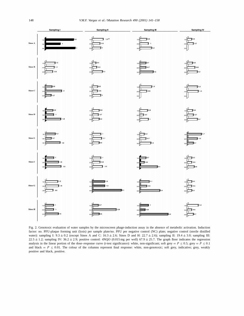
3.1.2. Microscreen phage-induction assayThe mutagenic evaluation data were compared to
the phage-induction responses. Figs. 2 and 3 showthe results of the assays in the absence and presenceof metabolic activation. Significant positive induct-ing activity could be detected at the sampling points
Sinos A (sampling I,-S9), Sinos G (II,-S9), Sinos H(III,-S9). The significant indicative responses werepresent at all of the other sites, in at least one sampling.Table 4 shows a summary of these results. Positive orindicative responses can be observed in the absenceof metabolic activation at sites Sinos A (sampling I),B (III), C (I), D (I), E (I, IV), F (I, III), G (II, III), H(I, II, III) which persist after metabolisation at SinosB, D, E, G (III), and H (III). Activity was observedonly for metabolites at the Sinos A (II), B (IV), E(II), and G (I) sites.
3.2. Second stage: water and interstitial water
During this stage (samplings V and VI), samples ofwater were evaluated from two environmental com-partments collected from site Sinos D, E, G and H
148 V.M.F. Vargas et al. / Mutation Research 490 (2001) 141–158
Fig. 2. Genotoxic evaluation of water samples by the microscreen phage-induction assay in the absence of metabolic activation. Inductionfactor: no. PFU-plaque forming unit (lysis) per sample plate/no. PFU per negative control (NC) plate; negative control (sterile distilledwater): sampling I: 9.3 ± 0.2 (except Sinos A and C: 16.3 ± 2.6; Sinos D and H: 22.7 ± 2.6); sampling II: 19.4 ± 3.0; sampling III:22.5 ± 1.2; sampling IV: 36.2 ± 2.9; positive control: 4NQO (0.015 mg per well) 67.9 ± 25.7. The graph floor indicates the regressionanalysis in the linear portion of the dose–response curve (t-test significance): white, non-significant; soft grey= P ≤ 0.5; grey= P ≤ 0.1and black= P ≤ 0.01. The colour of the columns represent final response: white, non-genotoxic; soft grey, indicative; grey, weaklypositive and black, positive.

V.M.F. Vargas et al. / Mutation Research 490 (2001) 141–158 149
Fig. 3. Genotoxic evaluation of water samples by the microscreen phage-induction assay in the presence of metabolic activation. Inductionfactor: no. PFU-plaque forming unit (lysis) per sample plate/no. PFU per negative control (NC) plate; negative control (sterile distilledwater): sampling I: 12.8± 0.1; sampling II: 25.0± 2.6; sampling III, 25.0± 2.6; sampling IV: 18.7± 0.2. Positive control: 2AF (5 mg perwell) 101.2 ± 23.0. The graph floor indicates the regression analysis in the linear portion of the dose–response curve (t-test significance):white, non-significant; soft grey= P ≤ 0.5; grey= P ≤ 0.1; and black= P ≤ 0.01. The colour of the columns represent final response:white, non-genotoxic; soft grey, indicative; grey, weakly positive; and black, positive.

150 V.M.F. Vargas et al. / Mutation Research 490 (2001) 141–158
Table 4Summary of the presence of significant phage-induction in water samples from samplings I–IV of the Sinos River basina
Sampling sites Treatment Sampling I (PFU/ml)b Sampling II (PFU/ml) Sampling III (PFU/ml) Sampling IV (PFU/ml)
Sinos A −S9 ++141.8 − − −+S9 − ±117.7 − −
Sinos B −S9 − − ±42.6 −+S9 − − ±71.9 ±83.2
Sinos C −S9 ±51.6 − − −+S9 − − − −
Sinos D −S9 ±24.0 − − −+S9 ±30.0 − − −
Sinos E −S9 ±27.0 − − ±49.1+S9 ±20.0 ±NSc − ±67.7
Sinos F −S9 ±34.0 − ±30.0 −+S9 − − − −
Sinos G −S9 − +86.3 ±43.1 −+S9 ±53.7 − +73.0 −
Sinos H −S9 ±17.0 ±59.5 +36.6 −+S9 − − ±86.5 −
a ‘−S9’, ‘+S9’ assays in absence or presence of metabolic activation; ‘−’ non-genotoxic, ‘±’ indicative, ‘+’ weakly positive or ‘++’positive genotoxic response.
b PFU/ml: plaque-forming unit (lysis)/ml of sample.c NS: non-significant.
(Table 5). Indicative significant phage-induction forriver water was observed only in the Sinos E sam-ple, sampling VI (-S9). Therefore, for samples of in-terstitial sediment water, the presence of positive orindicative activity is observed in interstitial sedimentwater in five assays at sites Sinos E (two responses),Sinos G (one response) and Sinos H (two responses).It is important to point out the presence of a signifi-cant indicative response in the samples of water andinterstitial water of site Sinos E, sampling VI, and thepersistence of induction activity observed in the pres-ence of metabolisation for Sinos H sampling V. Thesame sites evaluated in theSalmonella/microsome as-say also using the direct concentration method pre-sented a negative response (data not shown).
3.3. Third stage: chemical determinations insediment samples
3.3.1. Total metal concentrationsThe total metal concentrations in the stream sedi-
ment samples of Sinos River are shown in Table 6.The results obtained can be compared with mean shale(MS) concentrations [31], which are seen at the bot-tom of Table 6. MS is commonly used in environ-
mental geochemical studies as an indicator of naturalmetal concentrations in the fine (silt–clay) grain frac-tion of sediments [7,32]. Compared to MS, the sedi-ments from the Sinos River basin seem to be naturallyenriched in Fe, Mn and Cu, as shown by these metalsconcentrations at the most upstream site (Sinos H). Inthe medium course, site Sinos G shows an enrichmentin Cr and, to a lesser degree, in Ni concentrations,while the most contaminated site is Sinos E, for Cu,Cr, Pb and Zn, in the lower course. The maximumMn concentration appears at the most downstream siteSinos C.
Although agricultural activities have not been ex-cluded as a potential source of metal for the most up-stream site (Sinos H), it is more likely that the localbedrock, especially the Serra Geral formation basalts,provides a major contribution to the Fe, Cu and Mnenrichment observed. Iron was the only metal anal-ysed which represented a permanent downstream de-crease in concentration. This trend highlights a fullynatural origin for this metal (also observed by Hatje[33]), as it links the distance from the natural sourceto the decrease in sediment concentration. In general,our results agree with those obtained by Baisch [34]and Hatje [33].

V.M.F. Vargas et al. / Mutation Research 490 (2001) 141–158 151
Table 5Genotoxic evaluation for Sinos River samples of water and interstitial water by microscreen phage-induction assay in the absence andpresence of metabolic activation (sampling V–VI)
Sampling Sampling sites Dose perwell (ml)
Water Sediment interstitial water
If a Responseb,c,d
(PFU/ml)If Response
(PFU/ml)
Absence of metabolic activationV Sinos D 100 0.4 1.4
200 0.3 − 1.0 −300 0.7 1.4
Sinos E 100 0.5 1.0200 0.6 − 1.4300 0.8 2.5 ±63.0∗∗∗
Sinos G 100 0.7 1.2200 0.8 − 0.9 −300 1.2 1.1
Sinos H 100 0.4 0.4200 0.4 − 0.7300 1.0 2.4 ±56.0∗∗
VI Sinos E 100 1.3 1.0200 1.3 1.3300 2.0 ±60.0∗ 1.6 ±46.0∗
Sinos G 100 0.8 1.9200 1.2 − 3.5300 1.3 1.6 +56.0∗∗∗
Sinos H 100 0.4 0.6200 0.5 − 0.6 −300 0.4 0.7
Presence of metabolic activation
V Sinos D 100 0.8 1.0200 0.5 − 1.1 −300 0.5 1.7
Sinos E 100 0.3 0.9200 0.8 − 0.8 −300 1.2 1.1
Sinos H 100 0.4 1.5200 0.7 − 1.4300 1.0 3.0 +55.0∗
a Induction factor: no. PFU-plaque forming unit (lysis) per sample plate/no. PFU per negative control (NC) plate.b ‘−’ non-genotoxic, ‘±’ indicative, ‘+’ positive.c Regression analysis in the linear portion of the dose–response curve (t-test significance).d PFU/ml: plaque forming unit (lysis)/ml of sample; negative control (sterile distilled water): sampling V, absence (16.6 ± 0.9) and
presence (26.0± 11) of metabolic activation; sampling VI, Sinos H (25.0± 1), Sinos G (29.2± 3.3), Sinos E (23.0± 1.0); positive control:absence 4NQO (0.015 mg per well= 83.7±2.5, sampling V and 104.0±32.4, sampling VI) and presence 2AF (5 mg per well= 87.9±0.4)of metabolic activation.
∗ ≤0.5.∗∗ ≤0.1.∗∗∗ ≤0.01.

152 V.M.F. Vargas et al. / Mutation Research 490 (2001) 141–158
Table 6Total metal concentrations in sediments from the Sinos River basina
Sites Fe Mn Cu Cr Pb Zn Ni
Sinos C 37881± 1061 4163± 136 65.2± 0 154± 0.7 32.8± 0 113.5± 7 57 ± 0Sinos E 39881± 10606 3195± 2327 100.7± 23 168± 40 41± 11.6 127.8± 47 51.6± 14Sinos G (V)b 46881± 707 1123± 0 74.5± 0.6 146.7± 1.5 24.7± 3.9 76.9± 1.5 69.3± 0Sinos G (VI)b 83444± 18650 1162± 0 78 ± 0.6 156.1± 0 21.9± 0 81.5± 1.7 74.7± 1.1Sinos H 104756± 2121 1006± 0 78.9± 0.6 83.8± 0.7 21.9± 0 81.5± 5 60.8± 1.1MSc 48000 850 39 90 23 120 68
a Results expressed in parts per millions (ppm); duplicate analyses — total extraction.b G (V) and G (VI) were collected at the same site, but at different periods.c MS: mean shale according to Bowen, 1979 [31].
3.3.2. Analysis of organochlorine pesticidesThe extraction of sediment for chromatographic
analysis of organochlorines indicated values for DDTat Sinos G of up to 32mg/kg. The following com-pounds were analysed: aldrin, BHC, dieldrin, endrin,heptaclor, heptaclor epoxi and endosulfan and theirpresence was not found. Besides site Sinos G, pointsSinos E and Sinos H were evaluated.
3.4. Fourth stage: sediment organic extracts
Extracts of organic compounds were analysed forthe presence of genotoxic substances in the samples ofsediments from sites Sinos E and G, besides the pointof reference (H) chosen as two of the sites contami-nated by SOS function inducing substances in the firstand second parts of the study. The sediment extrac-tion methods were compared as to sonication, usingsolvents dichloromethane or ether and ether extractionby silica gel glass column.
Analysing the density results of the differentextracts (Table 7), it can be observed that the ex-traction methodologies showed different degrees ofefficiency at the sites evaluated. Taking into accountthe extraction yield, ether was the most appropri-ate solvent presenting polar fractions with a higherdensity as compared to the fractions obtained by ex-traction with dichloromethane. Measured similarly,column extraction presented results superior to thoseobserved during the sonication process.
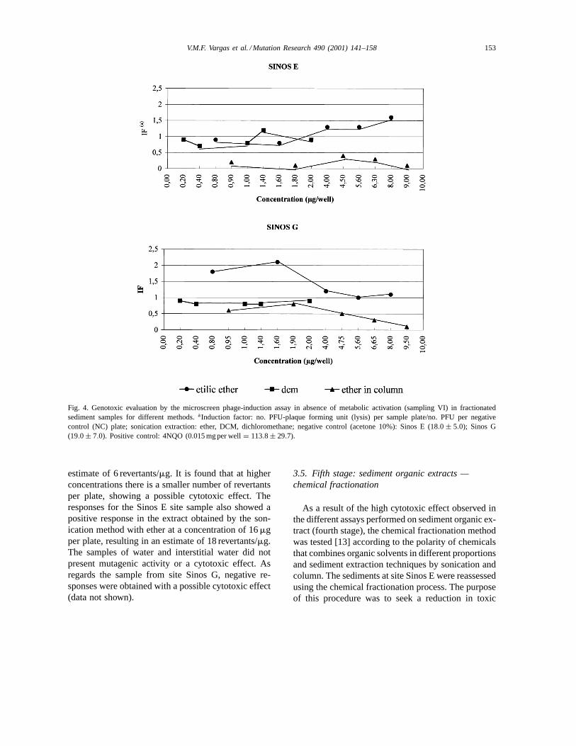
3.4.1. Genotoxic activityThe fractions obtained in the different extrac-
tion processes were evaluated by microscreen
phage-induction assay. Fig. 4 shows the very lowsignificant indicative responses observed for polarSinos E (2.0 PFU/mg) and Sinos G (16.0 PFU/mg)extracts obtained by sonication with the presence ofcytotoxicity at higher concentrations. The polar ex-tracts obtained by extraction in a column presented acytotoxic response. The moderately polar extracts donot present genotoxic activity at the concentrationsevaluated with an indication for cytotoxicity at higherconcentrations.
Site Sinos H presented genotoxic activity for mod-erately polar compounds at high concentrations (12.5,25.0 and 50.0mg per plate) with estimated values of2.0 PFU/mg. The polar extracts presented a higher cy-totoxic response (data not shown).
The extracts were also evaluated in theSalmonella/microsome assay, strain TA97, in the absence ofmetabolisation. Critical mutagenic activity (index 2)could be detected in the extract obtained by ethertreatment in a column in the Sinos H sample, at adosage of 3.5mg per plate resulting in the statistical
Table 7Extract densities obtained using different extraction methods insediment samples from the Sinos River (g/l) (sampling VI)
Sites Column extraction Sonication extraction
Ether Ether Dichloromethane
Sinos E 1.40 0.80 0.20Sinos Ga 1.60 0.10 0.10Sinos G 0.66 0.80 0.20Sinos H 0.70 0.23 0.17
a Sample tested inSalmonella/microsome assay.
V.M.F. Vargas et al. / Mutation Research 490 (2001) 141–158 153
Fig. 4. Genotoxic evaluation by the microscreen phage-induction assay in absence of metabolic activation (sampling VI) in fractionatedsediment samples for different methods.aInduction factor: no. PFU-plaque forming unit (lysis) per sample plate/no. PFU per negativecontrol (NC) plate; sonication extraction: ether, DCM, dichloromethane; negative control (acetone 10%): Sinos E (18.0 ± 5.0); Sinos G(19.0 ± 7.0). Positive control: 4NQO (0.015 mg per well= 113.8 ± 29.7).
estimate of 6 revertants/mg. It is found that at higherconcentrations there is a smaller number of revertantsper plate, showing a possible cytotoxic effect. Theresponses for the Sinos E site sample also showed apositive response in the extract obtained by the son-ication method with ether at a concentration of 16mgper plate, resulting in an estimate of 18 revertants/mg.The samples of water and interstitial water did notpresent mutagenic activity or a cytotoxic effect. Asregards the sample from site Sinos G, negative re-sponses were obtained with a possible cytotoxic effect(data not shown).
3.5. Fifth stage: sediment organic extracts —chemical fractionation
As a result of the high cytotoxic effect observed inthe different assays performed on sediment organic ex-tract (fourth stage), the chemical fractionation methodwas tested [13] according to the polarity of chemicalsthat combines organic solvents in different proportionsand sediment extraction techniques by sonication andcolumn. The sediments at site Sinos E were reassessedusing the chemical fractionation process. The purposeof this procedure was to seek a reduction in toxic

154 V.M.F. Vargas et al. / Mutation Research 490 (2001) 141–158
Fig. 5. Genotoxic evaluation by the microscreen phage-inductionassay in the absence of metabolic activation (sampling VII)for different fractions of the sediment chemical fractionationsample Sinos E. IF: Induction factor: no. PFU-plaque form-ing unit (lysis) per sample plate/no. PFU per negative control(NC) plate; graph bars represent crescent concentration F1 (0.39,0.78, 1.56, 3.13, 6.25, 12.5, 25.0, 50.0mg per well); F2–F4(0.78, 1.56, 3.13, 6.25, 12.5, 25.0, 50.0mg per well); nega-tive control (acetone 10%, 16.0 ± 1.0). Positive control: 4NQO(0.015 mg per well= 113.8 ± 29.7).
activity and a correlation between the extract compo-sition and genotoxic activity.
A series of seven concentrations with dilution fac-tor 2 was used in assays with the extracts obtained bychemical fractionation of site Sinos E, beginning withthe concentration of 50mg per well, as indicated inliterature [10], modified to use a constant volume ofacetone 10% per well (50ml) at the different concen-trations tested.
In the Fig. 5, the results obtained for fractions 1(hexane), 2 (10% DCM), 3 (20% DCM) and 4 (75%DCM) at site Sinos E are shown. The genotoxic activ-ity was identified in the non-polar–medium polar frac-tions. The higher values of PFU per plate (inductionfactor) at different fractions were observed for fraction1 (4.05–2.29), fraction 2 (3.06), fraction 3 (1.83) andfraction 4 (3.19–5.56). In fractions 1 and 4, two peaksof activity could be identified at the lower 12.5mg,6.25mg per well and higher 0.39mg, 50mg per wellconcentrations, respectively. The statistical analysisperformed on the linear portion of the dose–responsecurve, allowed the estimation of PFU/mg for thesefractions at the values of 0.29 ± 0.16, 2.24 ± 0.91,0.50±0.21, 1.26±0.34, respectively. Cytotoxicity wasobserved by turbidity analysis at all dilutions of frac-
tions 5 (5% methanol in DCM) and 6 (diethylether)(data not shown).
4. Discussion
In the study performed at the Sinos River basin,conclusions could be established regarding the meth-ods of approach to the samples used in environ-mental diagnosis of the region and the sensitiv-ity of the Salmonella/microsome and microscreenphage-induction assays for genotoxicity assessment.
The investigations performed during the first andsecond stages of the study (Tables 3–5 and Figs. 2and 3) clearly indicated the greater sensitivity ofthe microscreen phage-induction in relation to theSalmonella/microsome test in evaluating the geno-toxicity of the water and interstitial water samples. Itshould be pointed out that theSalmonella/microsomeassay was used for screening analysis in order to iden-tify the most significant sites for an initial assessmentof the basin as to the presence of mutagenic com-pounds. A similar method was used successfully [3,4]in an area subject to the influence of petrochemicals,defining the sites for a complementary dose–responseeffect study.
The comparative study using the lysogenic induc-tion assay in the samples of water and interstitial wa-ter also submit to the direct concentration method in-dicated sediment as the best compartment for evalu-ating genotoxicity. The samples of interstitial waterproved most appropriate for the study of genotoxic-ity in this river. The relatively low induction rates ob-served could be the result of the complexity of inter-actions of the compounds present in the sample. Thepresence of heavy metals, mainly at sites E and G,are indications that at least part of the contaminationdetected in these compartments is due to the pres-ence of metals resulting from anthropogenic factors.To this evidence is added the fact that the microscreenphage-induction assay has been utilised as a more ap-propriate assay than theSalmonella/microsome testto determine the genotoxic activity of samples con-taining halogenated compounds, organochlorine com-pounds and metals [10,17,18]. The association of thisassay with theSalmonella/microsome test is recom-mended for the study of environmental samples po-tentially contaminated by these compounds [4,5,17].
V.M.F. Vargas et al. / Mutation Research 490 (2001) 141–158 155
4.1. Contribution of heavy metals to Sinos Riverbasin contamination
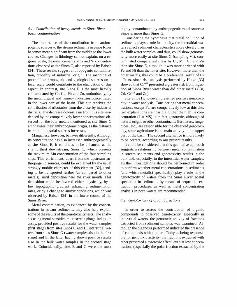
The importance of the contribution from anthro-pogenic sources to the stream sediments in Sinos Riverbecomes more significant from the middle to the lowercourse. Changes in lithology cannot explain, on a re-gional scale, the enhancements of Cr and Ni concentra-tions observed at site Sinos G, also reported by Baisch[34]. These results suggest anthropogenic contamina-tion, probably of industrial origin. The mapping ofpotential anthropogenic and geological sources on alocal scale would contribute to the elucidation of thisaspect. In contrast, site Sinos E is the most heavilycontaminated by Cr, Cu, Pb and Zn, undoubtedly, bythe metallurgical and tannery industries concentratedin the lower part of the basin. This site receives thecontribution of tributaries from the close-by industrialdistricts. The decrease downstream from this site, evi-denced by the comparatively lower concentrations ob-served for the four metals mentioned at site Sinos Cemphasises their anthropogenic origin, as the distancefrom the industrial sources increases.
Manganese, however, behaves differently. Althoughits concentration has also increased anthropogenicallyat site Sinos E, it continues to be enhanced at thesite furthest downstream, Sinos C, which presentsthe maximum Mn concentration of the four samplingsites. This enrichment, apart from the upstream an-thropogenic sources, could be explained by the usualstrongly mobile character of this element [32], tend-ing to be transported further (as compared to othermetals), until deposition near the river mouth. Thisdeposition could be favored either physically, by alow topographic gradient enhancing sedimentationrates, or by a change to anoxic conditions, which wasobserved by Baisch [34] in the lower course of theSinos River.
Metal contamination, as evidenced by the concen-trations in stream sediments, may also help explainsome of the results of the genotoxicity tests. The analy-sis using metal-sensitive microscreen phage-inductionassay, provided positive results for the water samples(first stage) from sites Sinos C and H, interstitial wa-ters from sites Sinos G (water samples also in the firststage) and E, the latter having shown positive resultsalso in the bulk water samples in the second stagework. Coincidentally, sites E and G were the most
highly contaminated by anthropogenic metal sources:Sinos E more than Sinos G.
Considering the hypothesis that metal pollution ofsediments plays a role in toxicity, the interstitial wa-ters reflect sediment characteristics more closely thanthe bulk water samples, and thus, could show genotox-icity more easily at site Sinos G (sampling VI), con-taminated comparatively less by Cr, Mn, Cu and Znthan site Sinos E, although it was more enriched withFe and Ni than the latter site. However, more than theother metals, this could be a preferential result of Creffects, since risk analysis performed by Hatge [33]showed that Cr+6 presented a greater risk from inges-tion of Sinos River water than did other metals (Cu,Cd, Cr+3 and Zn).
Site Sinos H, however, presented positive genotoxi-city in water analysis. Considering that metal concen-trations, except Fe, are comparatively low at this site,two explanations are possible. Either the high Fe con-centration (2× MS) is in fact genotoxic, although ofnatural origin, or other contaminants (fertilizers, fungi-cides, etc.) are responsible for the observed genotoxi-city, since agriculture is the main activity in the upperpart of the basin. The second alternative is more likelyto be correct, according to our present results.
It could be considered that this qualitative approachsuggests a relationship between metal contaminationin stream sediments and genotoxicity results in thebulk and, especially, in the interstitial water samples.Further investigations should be performed in orderto confirm whether metal concentrations in sediments(and which metal(s) specifically) play a role in thegenotoxicity of waters from the Sinos River. Metalspeciation in sediments by means of sequential ex-traction procedures, as well as metal concentrationanalysis in pore waters are recommended.
4.2. Genotoxicity of organic fractions
In order to assess the contribution of organiccompounds to observed genotoxicity, especially ininterstitial waters, the genotoxic activity of fractionsextracted from sediment samples was examined. Al-though the diagnosis performed indicated the presenceof compounds with a polar affinity as being responsi-ble for genotoxic activity, the fractions extracted withether presented a cytotoxic effect, even at low concen-trations (especially the polar fraction extracted by the
156 V.M.F. Vargas et al. / Mutation Research 490 (2001) 141–158
column process). The polar fractions analysed in themicroscreen phage-induction assay (Fig. 4) showedvery low significant indicative responses observed atSinos E (2.0 PFU/mg) and Sinos G (16.0 PFU/mg).These responses had already been observed (Table 5)in bulk water (60 PFU/ ml) and interstitial water(46 PFU/ml) at site Sinos E and G, in interstitialwater (56 PFU/ml). For site Sinos H, genotoxic ac-tivity was detected in the moderately polar fraction(2.0 PFU/mg). The presence was detected in the mod-erately polar fraction (2.0 PFU/mg). The presence ofconstant cytotoxicity in the sediment fractions is anindication of the characteristic of this environmentalcompartment as an accumulator and reprocessor ofchemical substances.
Low level direct mutagenesis results for po-lar extracts were also observed for site Sinos E(18 revertants/mg) and Sinos H (6 revertants/mg) afterusing the Salmonella/microsome assay with strainTA97. Water and interstitial water from the samesamplings and sampling sites, however, presentednegative mutagenic activity with signs of cytotoxi-city. The organic extraction of the samples enabledthe concentration and identification of mutageniccompounds in the polar fraction.
As a consequence of the cytotoxicity results ob-served in the sediment organic extract (fourth stage),an approach to sediment samples was assessed thatused a combination of organic solvents in differentproportions. This chemical fractionation by sequen-tial extraction (fifth stage) [13] separated the classesof compounds present in the complex sample (SinosE) according to polarity. The genotoxic activity ofthis sample was observed in the non-polar–mediumpolar fractions. The genotoxicity per plate increasesas the solvent polarity increases (hexane, 10%dichloromethane in hexane; 20% dichloromethanein hexane; 75% dichloromethane in hexane). It alsobecame clear that the most toxic compounds thatinterfere in cellular dynamics, with preponderantlycytotoxic activity are extracted in the polar fraction.The modification of the microscreen phage-inductionassay protocol using a constant volume of solventmade it possible to increase the concentrations tested,eliminating the interference caused by the variationof solvent volume and allowing the identification ofthe presence of genotoxicity at higher concentrationsin medium polar fractions (Fig. 5).
In characterisation studies of genotoxic com-ponents in fractions isolated from the river andmarine sediments collected close to the city ofBarcelona, Spain, in studies of polarity fractiona-tion of chemicals [13,14] site could be observed thatthese fractions may contain organohalogenated com-pounds (PCBs), chlorinated pesticides, hepta- andocta-chlorodibenzo-p-dioxins, polycyclic aromatichydrocarbons (PAHs) alkylated, PAH-containing sul-fur heterocycles related to low-polarity fractions,oxygenated-polycyclic aromatic compounds (PACs),nitroarenes (nitro-PACs), nitrogen-containing com-pounds present in the medium-polarity fractions andespecially nitro-PACs containing additional polarsubstitutes in the polar fraction.
5. Conclusions
It can be concluded that the analysis of water andinterstitial water proved to be an essential stage ofthe study to screen areas potentially contaminated bygenotoxic compounds from the different urban andindustrial contributions.
As a genotoxicity assessment method, the micro-screen phage-induction assay was sensitive to the com-plexity of compounds present in this river, both in thestudies of integral samples, bulk water and interstitialwater, and in the identification of the genotoxicity ofthe different sediment organic fractions.
However, the sequential extraction with chemicalfractionation of the sample proved to be an adequatemethod to evaluate the genotoxic activity in sedimentsamples, since it reduces the toxicity of this com-plex matrix and permits classes of organic compoundspresent to be estimated. The most significant genotox-icity results were identified in the non-polar–mediumpolar fractions and the cytotoxicity results are obtainedin the polar fraction. It should be pointed out that noextraction method recovers all the compounds present,and the results obtained correspond to a small portionof the matrix that does not reflect the true genotoxicityof the sample as a whole [4,35,36].
The distribution of genotoxicity in the Sinos Riverbasin illustrates the complexity of the study of regionsexposed to multiple pollutants. Although the identi-fication of areas contaminated by genotoxic agents,through biological assays, represents a significant
V.M.F. Vargas et al. / Mutation Research 490 (2001) 141–158 157
phase of environmental diagnosis, chemical fraction-ation, associated with the analysis and identificationof classes of compounds present in the samples in-vestigated, represent especially important stages inseeking clean-up measures.
Acknowledgements
We are grateful to Andrea da Silva Lopes, JaneBeatriz Fernandes and Inara Pedrotti, for their greatsupport during the development and finalisation of thestudy. We are also grateful to the sampling team ofFEPAM. This research was supported by the Con-selho Nacional de Desenvolvimento Cientıfico e Tec-nológico (CNPq), Fundação de Amparo à Pesquisa doRio Grande do Sul (FAPERGS) and Financiadora deEstudos e Projetos (CIAMB/PADCT/FINEP). CNPqand FAPERGS supplied the scholarships for scientificinitiation (1) and further training (5) that enabled theparticipation of students and graduates in developingthis study.
References
[1] Brazil, Conselho Nacional do Meio Ambiente, resolução n.20, de 18 de junho de 1986, Classifica as águas do TerritórioNacional, Diário Oficial da República Federativa do Brasil,Brasılia, 30 de junho, 1986.
[2] V.M.F. Vargas, V.E.P. Motta, J.A.P. Henriques, Analysisof mutagenicity of waters under the influence ofpetrochemical industrial complexes by the Ames test(Salmonella/microsome), Rev. Bras. Gen. 11 (3) (1988) 505–518.
[3] V.M.F. Vargas, V.E.P. Motta, J.A.P. Henriques, Mutagenicactivity detected by the Ames test in river water under theinfluence of petrochemical industries, Mutat. Res. 319 (1993)31–45.
[4] V.M.F. Vargas, R.R. Guidobono, C. Jordão, J.A.P. Henriques,Use of two short-term test to evaluate genotoxicity ofriver water treated with different concentration extractionprocedure, Mutat. Res. 343 (1995) 31–52.
[5] L. Claxton, V.S. Houk, S.E. George, Integration ofcomplex mixture toxicity and microbiological analyses forenvironmental remediation research, in: J. de Serres, A.D.Bloom (Eds.), Ecotoxicity and Human Health: A BiologicalApproach to Environmental Remediation, CRC Press Inc.,1995.
[6] G.M. Rand, S.R. Petrocelli, Fundamentals of AquaticToxicology — Methods and Applications, HemischerPublishing Corporation, Washington, USA, 1985.
[7] U. Förstner, G.T.M. Wittmann, Metal Pollution in the AquaticEnvironment, Springer, Heidelberg, 1979.
[8] W. Salomons, U. Förstner, Metals in the Hydrocycle, Springer,Berlin, 1984.
[9] D.L. Tsalev, Z.K. Zaprianov, Atomic Absorption Spectometryin Occupational and Environmental Health Practice, CRCPress Inc., USA, 1984.
[10] T.G. Rossman, M. Molina, L.W. Meyer, P. Boone, C.B. Klein,Z. Wang, F. Li, W.C. Lin, P.L. Kinney, Performance of 133compounds in the lambda prophage induction endpoint ofthe microscreen assay and a comparison withS. typhimuriummutagenicity and rodent carcinogenicity assays, Mutat. Res.260 (1991) 349–367.
[11] P.K. Wong, Mutagenicity of heavy metals, Bull. Environ.Contam. Toxicol. 40 (1988) 597–603.
[12] T. Sato, T. Mona, Y. Ose, T. Ishikama, K. Kato, Mutagenicityof Niagara River sediment, Mutat. Res. 118 (1983) 257–267.
[13] M. Grifoll, A.M. Solanas, J.M. Bayona, Characterization ofgenotoxic components in sediments by mass spectrometrictechniques combined withSalmonella/microsome test, Arch.Environ. Contam. Toxicol. 19 (1990) 175–184.
[14] P. Fernández, M. A Griffol, M. Solanas, J.M. Bayona, J.Albalgés, Bioassay-directed chemical analysis of genotoxiccomponents in coastal sediments, Environ. Sci. Technol. 26(1992) 817–829.
[15] G.U. Valent, M.I. Sato, M.C. Coelho, C.A. Coimbrão, P.S.Sanchez, Monitoring São Paulo state rivers in Brazil formutagenic activity using the Ames test, Environ. Toxicol.Water Qual. 8 (1993) 371–381.
[16] V.M.F. Vargas, R.C. Horn, R.R. Guidobono, B. Mittelstaedt,I.M.G. Azevedo, Mutagenic activity of airborne particulatematter from urban areas of Porto Alegre, Brazil, Genet. Mol.Biol. 21 (1998) 1–7.
[17] V.S. Houk, D.M. DeMarini, Use of the microscreenphage-induction assay to assess the genotoxicity of 14hazardous industrial wastes, Environ. Mol. Mutag. 11 (1988)13–29.
[18] V.S. Houk, The genotoxicity of industrial wastes and effluents:a review, Health Effects Research Laboratory, USEPA, Mutat.Res. 277 (1992) 91–138.
[19] Standard Methods for the Examination of Water andWastewater, M.A.H. Franson (Ed.), 18th Edition, AmericanPublic Health Association, American Water WorksAssociation and Water Environment Federation, Washington,1992.
[20] Pró-Guaiba, Rede de Monitoramento Ambiental: Monitora-mento das águas superficiais do Rio dos Sinos,FEPAM/CORSAN/DMAE, 1999.
[21] DNPM, Mapa Geológico do Estado do RGS, escalas1:1,000,000 e 1:600,000, Brasılia, DF, 1989.
[22] US Environmental Protection Agency, Guidelines forpreparing environmental and waste samples for mutagenicity(Ames) testing, in: Interim Procedures and Panel MeetingProceedings, Las Vegas, September 1985, EPA, p. 255 (EPA600/07/68-03-3136).
[23] M.H.D. Pestana, M.L.L. Formoso, E.C. Teixeira, Heavymetals in stream sediments from copper and gold mining areasin southern Brazil, J. Geoch. Explor. 58 (1997) 133–143.
158 V.M.F. Vargas et al. / Mutation Research 490 (2001) 141–158
[24] US Environmental Protection Agency, Manual of analyticalmethods for the analysis of pesticides in humans andenvironmental samples: a compilation of methods selectedfor use in pesticides monitoring programs, North Carolina,USA, June 1980, EPA 600/8-80-038.
[25] D.M. Maron, B.N. Ames, Revised methods for theSalmonellamutagenicity test, Mutat. Res. 113 (1983) 173–215.
[26] A.D. Pagano, E. Zeiger, Conditions for detecting themutagenicity of divalent metals inSalmonella typhimurium,Environ. Mol. Mutag. 19 (1992) 139–146.
[27] L.N. Myers, L. Adams, T.K. Kier, B. Rao, B. Shaw, L.Williams, Microcomputer software for data management andstatistical anlyses of the Ames/Salmonellatest, in: D. Krewski(Ed.), Statistical Methods in Toxicological Research, Gordonand Brech, New York, 1991.
[28] T.G. Rossman, M. Molina, L.W. Meyer, The genetictoxicology of metal compounds: I. Induction of prophage inE. coli WP2s (λ), Environ. Mol. Mutag. 6 (1984) 59–69.
[29] D.M. DeMarini, B.K. Lawrence, H.G. Brooks, V. Houk,Compatibility of organic solvents with the microscreenprophage-induction assay: solvent–mutagen interactions,Mutat. Res. 263 (1991) 107–113.
[30] D. Gatehouse, S. Harworth, T. Cebula, E. Gocke, L. Kier, T.Matsushima, C. Melcion, T. Nohmi, T. Ohta, S. Venitt, E.Zeiger, Recommendations for the performance of bacterialmutation assays, Mutat. Res. 312 (1994) 217–233.
[31] H.J.M. Bowen, Environmental Chemistry of the Elements,Academic Press, New York, 1979.
[32] M.H.D. Pestana, Partição geoquımica de metais pesados emsedimentos estuarinos nas Baıas de Sepetiba e da Ribeira,MS Thesis, Universidede Federal Fluminense, Rio de Janeiro,RJ, 1989.
[33] V. Hatje, Contaminação por metais pesados no Rio dos Sinos,RS: uma abordagem dinâmica a partir de balanços de massa,MS Thesis, Universidade Federal Fluminense, Rio de Janeiro,RJ, 1996.
[34] P. Baisch, Les Oligo-elements metalliques du systeme flúviolagunaire dos Patos (Brésil), Flux et Devenir, Doctoral Thesis,L’Université de Bordeaux I, Bordeaux, 1994.
[35] J.C. Loper, Mutagenic effects of organic compounds indrinking water, Mutat. Res. 76 (1980) 241–268.
[36] R.G. Stahl Jr., The genetic toxicology of organic compoundsin natural waters and wastewaters, Ecotoxicol. Environ. Safety22 (1991) 94–125.
Copyright © 2022 FDOKUMEN