Prevalence, indications, and outcomes of caesarean section ...
Upload
independentCategory
view
1download
0
Virology 378 (2008) 151–161
Contents lists available at ScienceDirect
Virology
j ourna l homepage: www.e lsev ie r.com/ locate /yv i ro
Genomic characterization of novel dolphin papillomaviruses provides indications forrecombination within the Papillomaviridae
Annabel Rector a,⁎, Hans Stevens a, Géraldine Lacave b,c, Philippe Lemey a,d, Sara Mostmans a, Ana Salbany c,Melissa Vos a, Koenraad Van Doorslaer a, Shin-Je Ghim e, Manuela Rehtanz e,f, Gregory D. Bossart f,g,A. Bennett Jenson e, Marc Van Ranst a
a Laboratory of Clinical Virology, Department of Microbiology and Immunology, Rega Institute for Medical Research, University of Leuven, Minderbroedersstraat 10, 3000 Leuven, Belgiumb Marine Mammal Veterinary Services, Daverlostraat 186, 8310 Brugge — Assebroek, Belgiumc Mundo Aquatico Zoomarine, Albufeira, Portugald Department of Zoology, University of Oxford, Oxford OX1 3PS, UKe The Brown Cancer Center, University of Louisville, Louisville, KY 40202, USAf Center for Coastal Research–Marine Mammal Research and Conservation Program, Harbor Branch Oceanographic Institution, Fort Pierce, FL 34946, USAg Florida Atlantic University, Boca Raton, FL 33431, USA
⁎ Corresponding author. Fax: +32 16 332131.E-mail address: [email protected] (A. R
0042-6822/$ – see front matter © 2008 Elsevier Inc. Aldoi:10.1016/j.virol.2008.05.020
a b s t r a c t
a r t i c l e i n f oArticle history:
Phylogenetic analysis of no Received 27 March 2008Returned to author for revision29 April 2008Accepted 16 May 2008Available online 24 June 2008Keywords:PapillomaviridaeCetaceaBottlenose dolphin (Tursiops truncatus)CondylomataEvolutionPhylogenyBootscanningRecombinationSelection
vel dolphin (Tursiops truncatus) papillomavirus sequences, TtPV1, -2, and -3,indicates that the early and late protein coding regions of their genomes differ in evolutionary history. Slidingwindow bootscan analysis showed a significant a change in phylogenetic clustering, in which the groupedsequences of TtPV1 and -3 move from a cluster with the Phocoena spinipinnis PsPV1 in the early region to acluster with TtPV2 in the late region. This provides indications for a possible recombination event near theend of E2/beginning of L2. A second possible recombination site could be located near the end of L1, in theupstream regulatory region. Selection analysis by using maximum likelihood models of codon substitutionsruled out the possibility of intense selective pressure, acting asymmetrically on the viral genomes, as analternative explanation for the observed difference in evolutionary history between the early and lategenomic regions of these cetacean papillomaviruses.
© 2008 Elsevier Inc. All rights reserved.
Introduction
The Papillomaviridae are a large family of epitheliotropic virusesthat can cause benign and malignant proliferations of the stratifiedsquamous epithelium in various vertebrate species. A wide geneticdiversity of different human papillomavirus (HPV) types has beendescribed, and is associated with a broad range of genotype-specificclinical conditions, ranging from genital and skin warts to invasivecervical carcinoma and skin cancer (Van Ranst et al., 1992). The limitednumber of non-human papillomaviruses (PVs) that has been char-acterized to date covers a broad range of host species, includingmostly domestic and wild mammals but also two bird species(Sundberg et al., 1997; Sundberg, 1987; Tachezy et al., 2002). Takentogether with the numerous partial sequences of putative novel non-
ector).
l rights reserved.
human PVs that have been reported (Antonsson and Hansson, 2002;Chan et al., 1997; Ogawa et al., 2004), these data suggest that everyvertebrate species could carry a set of species-specific PVs.
Papillomatous lesions including gastric papillomas and cutaneousand lingual papillomatosis, with PV infection as a suspected causebased on clinical, histopathological, transmission electronmicroscopicand immunohistochemical findings, have been described in severalmembers of the order Cetacea (Bossart et al., 1996, 2005; De Guiseet al., 1994; Geraci et al., 1987; Van Bressem et al., 1996, 1999).Furthermore, genital papillomatosis has been reported in a number ofcetacean species, such as the sperm whale, the Blainville's beakedwhale, the killer whale, the Atlantic white-sided dolphin, the long-beaked common dolphin, the Dusky dolphin, Burmeister's porpoise,and the bottlenose dolphin (Bossart et al., 2005; Flom et al., 1980;Geraci et al., 1987; Greenwood et al., 1974; Lambertsen et al., 1987; VanBressem et al., 1996). Whereas cutaneous papillomatosis has beendescribed in many orders of placental mammals, cetaceans representthe only non-primate mammalian order in which genital PV infection
152 A. Rector et al. / Virology 378 (2008) 151–161
has been confirmed. In some cetacean species genital warts are highlyprevalent (e.g. up to 66% in Dusky dolphins) (Van Bressem et al., 1996).In a recent survey, a high occurrence of related benign and malignantorogenital neoplastic epithelial lesions was found in bottlenosedolphins, indicating that these could represent an emerging infectiousdisease (Bossart et al., 2005). The same study provided arguments foran infectious etiology of these tumors, with an orogenital transmis-sion route (Bossart et al., 2005). Although these reports indicate thatPV infection might be widespread in cetaceans, PV etiology had untilrecently only been unequivocally demonstrated in the case of genitallesions in the Burmeister's porpoise (Phocoena spinipinnis), caused bythe Phocoena spinipinnis PV (PsPV1, GenBank accession numberNC_003348) (Van Bressem et al., 2007). It therefore remains to bedetermined whether genital papillomatosis within single species ofcetaceans is caused by a single PV type, or whether multiple types caninfect the same species, as is the case in humans.
Papillomaviruses use the high-fidelity DNA replication machineryof their eukaryotic host cells. Together with their slow replication,which is linked to the division of infected epithelial cells, this results inan evolutionary rate of only 1.95×10⁻⁸ nucleotide substitutions persite per year (Rector et al., 2007). The apparent absence of inter-typerecombination further contributes to the high degree of conservationamong PVs. Recently however, data indicating that recombinationcould have occurred in the evolutionary history of PVs have accumu-lated (Angulo and Carvajal-Rodriguez, 2007; Bravo and Alonso, 2007;Gottschling et al., 2007b; Narechania et al., 2005; Varsani et al., 2006),but no proven examples of recombination between established PVtypes have been reported so far.
We have isolated the genomes of three distinct PV types from genitalmucosal lesions in bottlenosedolphins (Tursiops truncatus). Phylogeneticcomparison of these dolphin PV types with PsPV1 indicates that thedifferent parts of their genomes have different evolutionary histories,andprovides a strong indication that recombination couldhaveoccurredbetween different (ancestral) types of the Papillomaviridae.
Results
TtPV1, TtPV2 and TtPV3 complete genomic sequence
The complete genomes of three distinct Tursiops truncatus PVshave been isolated: TtPV1 and TtPV3, isolated from a singlecondylomatous lesion on the penis of a captive bottlenose dolphinfrom the Mundo Aquatico Zoomarine in Portugal (described in thispaper), and TtPV2, which was isolated from a genital condyloma of afree-ranging bottlenose dolphin from Charleston Harbor, SouthCarolina, USA (described elsewhere) (Rehtanz et al., 2006). Thecomplete nucleotide sequence of TtPV1 counts 8089 base pairs (bp)and has a GC content of 42.1%, TtPV2 counts 7866 bp with a 46.0% GCcontent, and TtPV3 counts 7915 bp with 44.2% CG.
Like all other PVs that have been characterized to date, TtPV1 and -3have all their ORFs on the same coding strand of their dsDNA. Thelocation of the ORFs and the size of the corresponding proteinsequences, with comparison to the corresponding ORF data fromTtPV2 and PsPV1, are summarized in a table that is provided asSupplementary file 1. The dolphin PV genomes contain the classical PVORFs E6, E1, and E2 in their early region, and the major and minorcapsid proteins L1 and L2 in their late region. An additional E5 ORF,which is also present in the E2–L2 region of the genome of themucosalHPVs belonging to the genus Alphapapillomavirus (α-PV) and thecutaneous (fibro-) PVs of the genus Deltapapillomavirus (Garcia-Vallveet al., 2005), as well as in TtPV2, could be identified in TtPV1 and -3.
Typical features of the early region of the PVs of Cetacea
The E6 ORFs of TtPV1 and TtPV3 contain two zinc-binding domainswith the sequence C-X-X-C-X29-C-X-X-C, separated by 36 amino acids.
This motif is identical to those found in the other cetacean PVs, TtPV2and PsPV1 (Rehtanz et al., 2006; Van Bressem et al., 2007). Similarly toTtPV2 and PsPV1, the genomes of TtPV1 and TtPV3 encode E6 proteinswhich are larger than those generally found in PVs: 224 and 220amino acids for TtPV1 and TtPV3 respectively, in comparison to 206amino acids in TtPV2, 211 in PsPV1, and about 150 in most other PVs(Howley and Lowy, 2001). The predicted E6 proteins of TtPV1 and -3 donot contain a PDZ domain binding motif in their extreme C-terminus,which would enable E6 to bind to proteins containing a PDZ do-main and direct them to proteolysis. Such a motif, with the consensussequence X-S/T-X-V/L (in which X represents any amino acid), ispresent in the very C-terminus of the high risk but not of the low riskgenital HPVs (Gardiol et al.,1999; Jing et al., 2007; Kiyono et al.,1997). APDZ domain binding motif has also been identified in the E6 of TtPV2E6 (Rehtanz et al., 2006), but not in the PsPV1 E6 (Van Bressem et al.,2007).
In the early region of the PV genome, the E6 ORF is typicallyfollowed by an ORF coding for the papillomaviral E7 protein, whichcontains a zinc-binding domain and in some cases a pRb bindingdomain (containing the L-X-C-X-Emotif, critical for binding of pRb). InTtPV1 and TtPV3 however, no E7 ORFs can be identified. A similargenomic organization, with the absence of a classical E7, has pre-viously been reported for PsPV1 (Van Bressem et al., 2007) and TtPV2(Rehtanz et al., 2006). Whereas the PsPV1 genome still has thecapacity to encode a small peptide (26 amino acids) harbouring thepRb binding motif, through a small ORF overlapping with the 5′ end ofthe PsPV1 E1 (Narechania et al., 2004), this is not the case in TtPV1 and-3, nor in TtPV2 (Rehtanz et al., 2006). In TtPV3 however, an L-X-C-X-Emotif was identified in the E1 ORF, but since this was located upstreamof the E1 start codon it is not likely that this motif is effectivelytranslated. The absence of a canonical E7 ORF is highly uncommonamong PVs, and so far seems to be a unique feature of the PVs ofcetaceans.
Sequence similarity to other PVs and classification of dolphin PVs
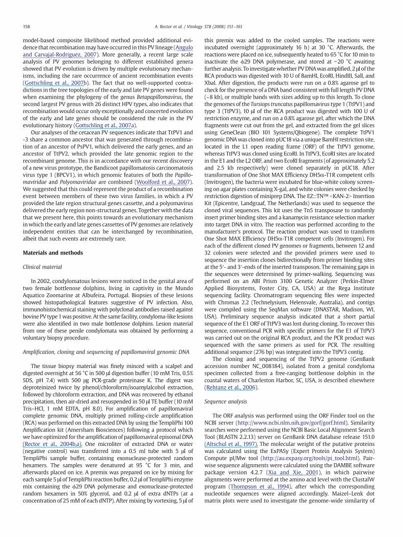
The similarities between the dolphin PVs mutually, and with theporpoise PsPV1 (which is the only other cetacean PV characterized todate), the manatee TmPV1 (representing a sea mammal PV isolatedfrom a member of the Sirenia, an order of aquatic mammals which isonly distantly related to the Cetacea order), the human low riskmucosal HPV11, and the high risk mucosal HPV16, were investigatedfor the different ORFs by pairwise nucleotide and amino acid sequencealignments. For all ORFs, the highest similarities were noted betweenTtPV1 and TtPV3, and the similarities to TmPV1, HPV11, and HPV16were lower and in the same range (Table 1). For the ORFs in the earlyregion, TtPV1 and TtPV3 show high similarities to PsPV1. In contrast,the late region of TtPV1 and TtPV3 displayed higher similarity toTtPV2as to PsPV1. This was even more apparent when the dolphin andporpoise PV genomes were analyzed in Maizel–Lenk dot matrix plots(Fig. 1). The plot illustrates the genome-wide high similarity betweenTtPV1 and TtPV3. Between TtPV1 and TtPV2, a high degree ofsimilarity is still visible across the late protein region, especially inthe L1 ORF, but not in the early region. With PsPV1 on the other hand,TtPV1 shared higher similarity across the early region compared to thelate region.
For classification purposes, only the sequence of the L1 ORF istaken into account. According to the current definition, PV typesbelong to the same genus when they share at least 60% nucleotidesequence identity across the entire L1 ORF (de Villiers et al., 2004).This places TtPV1, TtPV2, and TtPV3 in the same genus, with identitiesbetween 66 and 80% (Table 1). Since the similarities of the dolphin PVsto PsPV1, which is the next closest related PV type, are below 60%,TtPV1, -2, and -3 cannot be assigned to the genus Omikronpapilloma-virus (ο-PV), of which PsPV1 is to date the onlymember, but have to beclassified in a novel, yet unnamed genus.
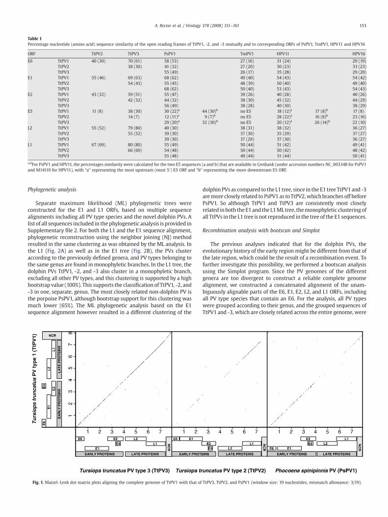
Table 1Percentage nucleotide (amino acid) sequence similarity of the open reading frames of TtPV1, -2, and -3 mutually and to corresponding ORFs of PsPV1, TmPV1, HPV11 and HPV16
ORF TtPV2 TtPV3 PsPV1 TmPV1 HPV11 HPV16
E6 TtPV1 40 (30) 70 (61) 58 (53) 27 (16) 31 (24) 29 (19)TtPV2 38 (30) 41 (32) 27 (20) 30 (23) 33 (23)TtPV3 55 (49) 26 (17) 35 (28) 29 (20)
E1 TtPV1 55 (46) 69 (63) 68 (62) 49 (40) 54 (43) 54 (42)TtPV2 54 (43) 55 (45) 48 (39) 50 (40) 49 (40)TtPV3 68 (62) 50 (40) 53 (43) 54 (43)
E2 TtPV1 43 (32) 59 (51) 55 (47) 39 (26) 40 (28) 40 (26)TtPV2 42 (32) 44 (32) 38 (30) 45 (32) 44 (29)TtPV3 56 (49) 38 (28) 40 (30) 38 (29)
E5 TtPV1 11 (8) 38 (30) 30 (22)a 44 (30)b no E5 18 (12)a 17 (8)b 17 (8)TtPV2 14 (7) 12 (11)a 9 (7)b no E5 28 (22)a 16 (8)b 23 (16)TtPV3 29 (20)a 32 (30)b no E5 20 (12)a 26 (14)b 22 (10)
L2 TtPV1 55 (52) 79 (80) 40 (30) 38 (31) 38 (32) 36 (27)TtPV2 55 (52) 39 (30) 37 (30) 35 (29) 37 (27)TtPV3 39 (30) 37 (29) 37 (30) 36 (27)
L1 TtPV1 67 (69) 80 (80) 55 (49) 50 (44) 51 (42) 49 (41)TtPV2 66 (69) 54 (48) 50 (44) 50 (42) 48 (42)TtPV3 55 (48) 49 (44) 51 (44) 50 (41)
a,bFor PsPV1 and HPV11, the percentages similarity were calculated for the two E5 sequences (a and b) that are available in Genbank (under accession numbers NC_003348 for PsPV1and M14119 for HPV11), with “a” representing the most upstream (most 5′) E5 ORF and “b” representing the more downstream E5 ORF.
153A. Rector et al. / Virology 378 (2008) 151–161
Phylogenetic analysis
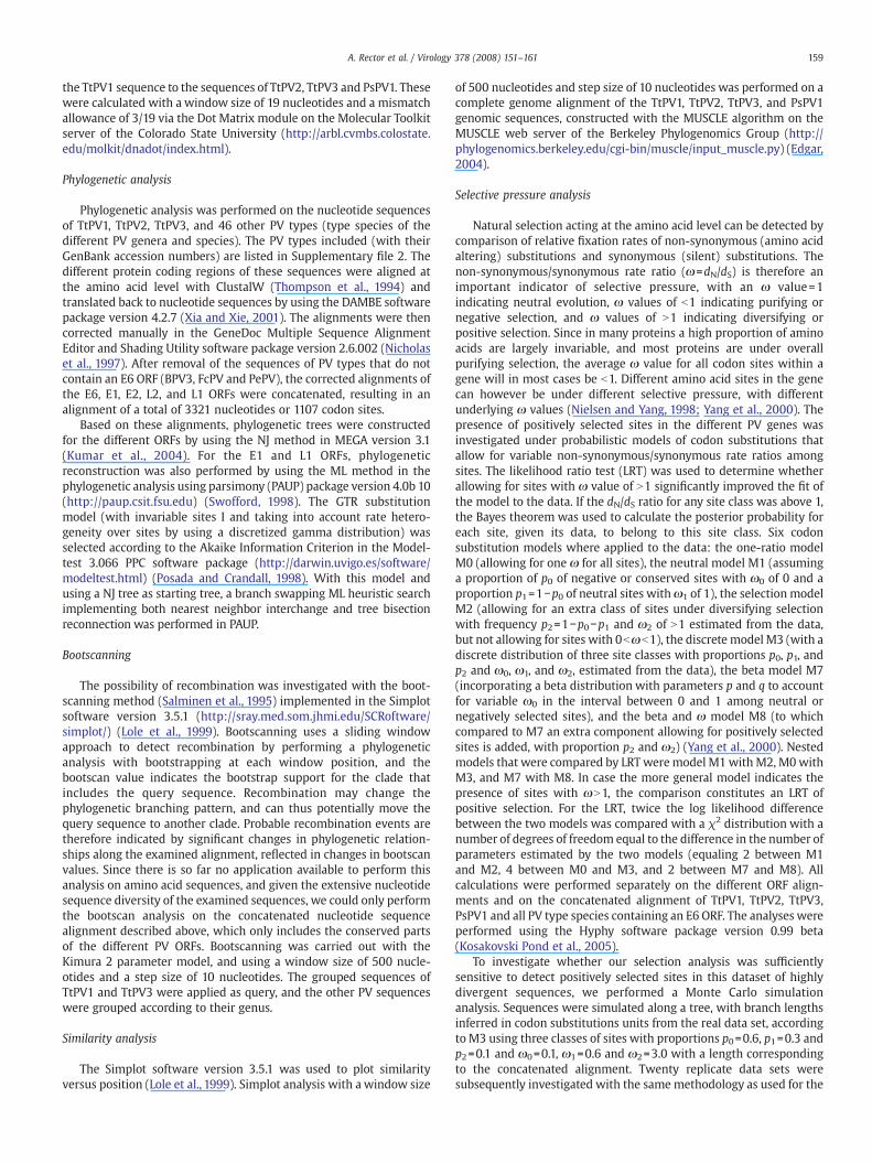
Separate maximum likelihood (ML) phylogenetic trees wereconstructed for the E1 and L1 ORFs, based on multiple sequencealignments including all PV type species and the novel dolphin PVs. Alist of all sequences included in the phylogenetic analysis is provided inSupplementary file 2. For both the L1 and the E1 sequence alignment,phylogenetic reconstruction using the neighbor joining (NJ) methodresulted in the same clustering as was obtained by the ML analysis. Inthe L1 (Fig. 2A) as well as in the E1 tree (Fig. 2B), the PVs clusteraccording to the previously defined genera, and PV types belonging tothe same genus are found inmonophyletic branches. In the L1 tree, thedolphin PVs TtPV1, -2, and -3 also cluster in a monophyletic branch,excluding all other PV types, and this clustering is supported by a highbootstrap value (100%). This supports the classification of TtPV1, -2, and-3 in one, separate, genus. The most closely related non-dolphin PV isthe porpoise PsPV1, although bootstrap support for this clustering wasmuch lower (65%). The ML phylogenetic analysis based on the E1sequence alignment however resulted in a different clustering of the
Fig. 1. Maizel–Lenk dot matrix plots aligning the complete genome of TtPV1 with that of
dolphin PVs as compared to the L1 tree, since in the E1 tree TtPV1 and -3aremore closely related to PsPV1 as toTtPV2,which branches off beforePsPV1. So although TtPV1 and TtPV3 are consistently most closelyrelated inboth theE1 and the L1ML tree, themonophyletic clusteringofall TtPVs in the L1 tree is not reproduced in the tree of the E1 sequences.
Recombination analysis with bootscan and Simplot
The previous analyses indicated that for the dolphin PVs, theevolutionary history of the early regionmight be different from that ofthe late region, which could be the result of a recombination event. Tofurther investigate this possibility, we performed a bootscan analysisusing the Simplot program. Since the PV genomes of the differentgenera are too divergent to construct a reliable complete genomealignment, we constructed a concatenated alignment of the unam-biguously alignable parts of the E6, E1, E2, L2, and L1 ORFs, includingall PV type species that contain an E6. For the analysis, all PV typeswere grouped according to their genus, and the grouped sequences ofTtPV1 and -3, which are closely related across the entire genome, were
TtPV3, TtPV2, and PsPV1 (window size: 19 nucleotides, mismatch allowance: 3/19).
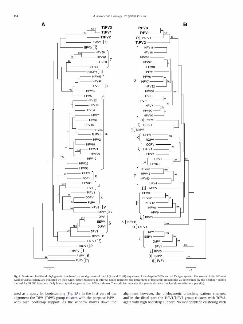
Fig. 2. Maximum likelihood phylogenetic tree based on an alignment of the L1 (A) and E1 (B) sequences of the dolphin TtPVs and all PV type species. The names of the differentpapillomavirus genera are indicated by their Greek letter. Numbers at internal nodes represent the percentage of bootstrap probabilities as determined by the neighbor-joiningmethod for 10 000 iterations. Only bootstrap values greater than 80% are shown. The scale bar indicates the genetic distance (nucleotide substitutions per site).
154 A. Rector et al. / Virology 378 (2008) 151–161
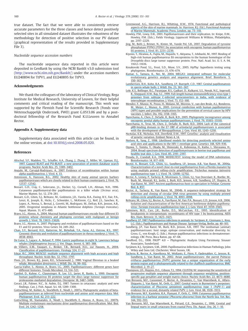
used as a query for bootscanning (Fig. 3A). In the first part of thealignment the TtPV1/TtPV3 group clusters with the porpoise PsPV1,with high bootstrap support. As the window moves down the
alignment however, the phylogenetic branching pattern changes,and in the distal part the TtPV1/TtPV3 group clusters with TtPV2,again with high bootstrap support. No monophyletic clustering with
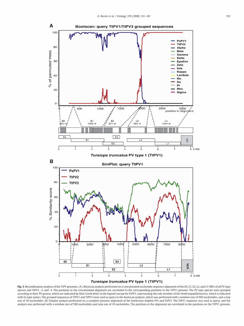
Fig. 3. Recombination analysis of the TtPV genomes. (A). Bootscan analysis performed on a concatenated nucleotide sequence alignment of the E6, E1, E2, L2, and L1 ORFs of all PV typespecies and TtPV1, -2, and -3. The positions in the concatenated alignment are correlated to the corresponding positions in the TtPV1 genome. The PV type species were groupedaccording to their PV genera, which are indicated by their Greek letter in the legend (except for PsPV1, representing the sole member of the Omikronpapillomavirus, which is indicatedwith its type name). The grouped sequences of TtPV1 and TtPV3 were used as query in the bootscan analysis, which was performed with awindow size of 500 nucleotides, and a stepsize of 10 nucleotides. (B) Simplot analysis performed on a complete genome alignment of the bottlenose dolphin PVs and PsPV1. The TtPV1 sequence was used as query, and theanalysis was performed with a window size of 500 nucleotides and step size of 10 nucleotides. The positions in the alignment are correlated to the positions on the TtPV1 genome.
155A. Rector et al. / Virology 378 (2008) 151–161
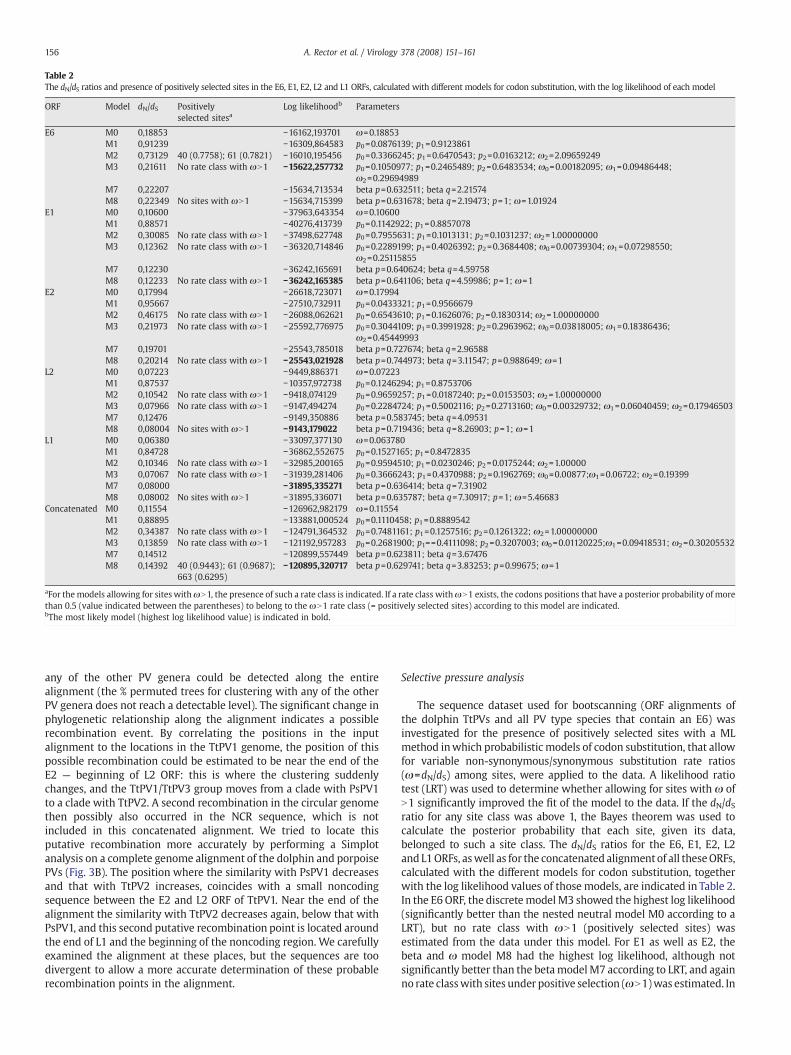
Table 2The dN/dS ratios and presence of positively selected sites in the E6, E1, E2, L2 and L1 ORFs, calculated with different models for codon substitution, with the log likelihood of each model
ORF Model dN/dS Positivelyselected sitesa
Log likelihoodb Parameters
E6 M0 0,18853 −16162,193701 ω=0.18853M1 0,91239 −16309,864583 p0=0.0876139; p1=0.9123861M2 0,73129 40 (0.7758); 61 (0.7821) −16010,195456 p0=0.3366245; p1=0.6470543; p2=0.0163212; ω2=2.09659249M3 0,21611 No rate class with ωN1 −15622,257732 p0=0.1050977; p1=0.2465489; p2=0.6483534; ω0=0.00182095; ω1=0.09486448;
ω2=0.29694989M7 0,22207 −15634,713534 beta p=0.632511; beta q=2.21574M8 0,22349 No sites with ωN1 −15634,715399 beta p=0.631678; beta q=2.19473; p=1; ω=1.01924
E1 M0 0,10600 −37963,643354 ω=0.10600M1 0,88571 −40276,413739 p0=0.1142922; p1=0.8857078M2 0,30085 No rate class with ωN1 −37498,627748 p0=0.7955631; p1=0.1013131; p2=0.1031237; ω2=1.00000000M3 0,12362 No rate class with ωN1 −36320,714846 p0=0.2289199; p1=0.4026392; p2=0.3684408; ω0=0.00739304; ω1=0.07298550;
ω2=0.25115855M7 0,12230 −36242,165691 beta p=0.640624; beta q=4.59758M8 0,12233 No rate class with ωN1 −36242,165385 beta p=0.641106; beta q=4.59986; p=1; ω=1
E2 M0 0,17994 −26618,723071 ω=0.17994M1 0,95667 −27510,732911 p0=0.0433321; p1=0.9566679M2 0,46175 No rate class with ωN1 −26088,062621 p0=0.6543610; p1=0.1626076; p2=0.1830314; ω2=1.00000000M3 0,21973 No rate class with ωN1 −25592,776975 p0=0.3044109; p1=0.3991928; p2=0.2963962; ω0=0.03818005; ω1=0.18386436;
ω2=0.45449993M7 0,19701 −25543,785018 beta p=0.727674; beta q=2.96588M8 0,20214 No rate class with ωN1 −25543,021928 beta p=0.744973; beta q=3.11547; p=0.988649; ω=1
L2 M0 0,07223 −9449,886371 ω=0.07223M1 0,87537 −10357,972738 p0=0.1246294; p1=0.8753706M2 0,10542 No rate class with ωN1 −9418,074129 p0=0.9659257; p1=0.0187240; p2=0.0153503; ω2=1.00000000M3 0,07966 No rate class with ωN1 −9147,494274 p0=0.2284724; p1=0.5002116; p2=0.2713160; ω0=0.00329732; ω1=0.06040459; ω2=0.17946503M7 0,12476 −9149,350886 beta p=0.583745; beta q=4.09531M8 0,08004 No sites with ωN1 −9143,179022 beta p=0.719436; beta q=8.26903; p=1; ω=1
L1 M0 0,06380 −33097,377130 ω=0.063780M1 0,84728 −36862,552675 p0=0.1527165; p1=0.8472835M2 0,10346 No rate class with ωN1 −32985,200165 p0=0.9594510; p1=0.0230246; p2=0.0175244; ω2=1.00000M3 0,07067 No rate class with ωN1 −31939,281406 p0=0.3666243; p1=0.4370988; p2=0.1962769; ω0=0.00877;ω1=0.06722; ω2=0.19399M7 0,08000 −31895,335271 beta p=0.636414; beta q=7.31902M8 0,08002 No sites with ωN1 −31895,336071 beta p=0.635787; beta q=7.30917; p=1; ω=5.46683
Concatenated M0 0,11554 −126962,982179 ω=0.11554M1 0,88895 −133881,000524 p0=0.1110458; p1=0.8889542M2 0,34387 No rate class with ωN1 −124791,364532 p0=0.7481161; p1=0.1257516; p2=0.1261322; ω2=1.00000000M3 0,13859 No rate class with ωN1 −121192,957283 p0=0.2681900; p1==0.4111098; p2=0.3207003; ω0=0.01120225;ω1=0.09418531; ω2=0.30205532M7 0,14512 −120899,557449 beta p=0.623811; beta q=3.67476M8 0,14392 40 (0.9443); 61 (0.9687);
663 (0.6295)−120895,320717 beta p=0.629741; beta q=3.83253; p=0.99675; ω=1
aFor themodels allowing for sites withωN1, the presence of such a rate class is indicated. If a rate class withωN1 exists, the codons positions that have a posterior probability of morethan 0.5 (value indicated between the parentheses) to belong to the ωN1 rate class (= positively selected sites) according to this model are indicated.bThe most likely model (highest log likelihood value) is indicated in bold.
156 A. Rector et al. / Virology 378 (2008) 151–161
any of the other PV genera could be detected along the entirealignment (the % permuted trees for clustering with any of the otherPV genera does not reach a detectable level). The significant change inphylogenetic relationship along the alignment indicates a possiblerecombination event. By correlating the positions in the inputalignment to the locations in the TtPV1 genome, the position of thispossible recombination could be estimated to be near the end of theE2 — beginning of L2 ORF: this is where the clustering suddenlychanges, and the TtPV1/TtPV3 group moves from a clade with PsPV1to a clade with TtPV2. A second recombination in the circular genomethen possibly also occurred in the NCR sequence, which is notincluded in this concatenated alignment. We tried to locate thisputative recombination more accurately by performing a Simplotanalysis on a complete genome alignment of the dolphin and porpoisePVs (Fig. 3B). The position where the similarity with PsPV1 decreasesand that with TtPV2 increases, coincides with a small noncodingsequence between the E2 and L2 ORF of TtPV1. Near the end of thealignment the similarity with TtPV2 decreases again, below that withPsPV1, and this second putative recombination point is located aroundthe end of L1 and the beginning of the noncoding region. We carefullyexamined the alignment at these places, but the sequences are toodivergent to allow a more accurate determination of these probablerecombination points in the alignment.
Selective pressure analysis
The sequence dataset used for bootscanning (ORF alignments ofthe dolphin TtPVs and all PV type species that contain an E6) wasinvestigated for the presence of positively selected sites with a MLmethod inwhich probabilistic models of codon substitution, that allowfor variable non-synonymous/synonymous substitution rate ratios(ω=dN/dS) among sites, were applied to the data. A likelihood ratiotest (LRT) was used to determine whether allowing for sites with ω ofN1 significantly improved the fit of the model to the data. If the dN/dSratio for any site class was above 1, the Bayes theorem was used tocalculate the posterior probability that each site, given its data,belonged to such a site class. The dN/dS ratios for the E6, E1, E2, L2and L1ORFs, aswell as for the concatenated alignmentof all theseORFs,calculated with the different models for codon substitution, togetherwith the log likelihood values of those models, are indicated in Table 2.In the E6 ORF, the discretemodelM3 showed the highest log likelihood(significantly better than the nested neutral model M0 according to aLRT), but no rate class with ωN1 (positively selected sites) wasestimated from the data under this model. For E1 as well as E2, thebeta and ω model M8 had the highest log likelihood, although notsignificantly better than the betamodelM7 according to LRT, and againno rate classwith sites under positive selection (ωN1)was estimated. In
157A. Rector et al. / Virology 378 (2008) 151–161
the L2ORF,modelM8was significantly better than its nestedmodelM7,and although a rate class with ωN1 was determined no sites wereattributed to this rate classwith a posterior probability ofmore than0.5.For the L1 ORF, the beta model M7, which does not allow for positivelyselected sites, bestfitted the data, althoughnot significantly better thanthemore complexmodelM8. No siteswith a dN/dS ratio of N1 (posteriorprobability cutoff value 0.5) could be identified in any of the separateORF alignments with the models that best fitted the data. For theconcatenated alignment however, the beta and ω model (M8) bestfitted the data (significantly better than model M7 according to LRT),and in the alignment of 1107 codon sites in total, 3 sites under positiveselectionwere identified under this model, with a posterior probabilityof at least 0.5. These sites were at codon positions 40 (posteriorprobability 0.944), 61 (posterior probability 0.969) and 663 (posteriorprobability 0.629) of the concatenated alignment, all three of whichwere located in the early region of the PV genome.
Although no major selective pressure was found, the bootscananalysis for detection of recombination was repeated on theconcatenated alignment fromwhich the sites under positive selectionwere removed, resulting in the same bootscan results as with theoriginal alignment (data not shown). This was done to rule out thepossibility that the presence of positively selected sites in the early butnot in the late genomic region, thus asymmetric selective pressure,could account for the observed difference in evolutionary clusteringbetween the early and late genomic regions of the cetacean PVs.
Discussion
Distinct PV types in genital condylomas of dolphins
Three different PV types were discovered in genital lesions ofbottlenose dolphins by examining biopsy material from two differentanimals. Together with the high prevalence of genital papillomatosisin dolphins this indicates that dolphins, like humans, could harbour amultitude of different genital PV types. Some mucosal HPV types areknown to cause genital lesions as well as infections of the respiratorytract and of the oral mucosa (Syrjänen and Syrjänen, 2000). In Atlanticbottlenose dolphins, lingual papillomas and squamous cell carcino-mas have been described, with pathological evidence suggesting aviral etiology and the possibility of orogenital transmission (Bossartet al., 2005). Whether the genital PV types that we have characterizednow are also responsible for (some of the) oral lesions in bottlenosedolphins still needs to be determined.
In humans, co-infection of the mucosa with different HPV types isfrequent (21.3% of cases) (Mendez et al., 2005). The fact that thegenomes of TtPV1 and TtPV3 were amplified from a single biopsyspecimenof a penile condylomatous lesionproves that also in dolphinsmultiple PV types can co-infect the genital mucosa.
A number of HPV types have the potential to induce neoplasticlesions of the human genital tract, most importantly cervical cancer.There are major concerns that also in dolphins, PV infection of thegenital mucosa could result in malignant lesions. The simultaneouspresence of benign oral papillomas and oral squamous cell carcinomasin bottlenose dolphins is highly suggestive of such a progressivepathologic process involving malignant transformation (Bossart et al.,2005).Whether there are certain PV types associatedwith an increasedrisk of progression of lesions tomalignancy in dolphins, as is the case intheir human counterparts which can be classified in high and low riskmucosal HPVs, remains to be determined.
Recombination in Papillomaviridae
Adifference in evolutionaryhistorybetween theearlyand late proteincoding regionsof the cetaceanPVswas consistentlydetectedbyanumberof different phylogenetic methods. The percentages pairwise identity(Table 1) and the Maizel–Lenk dot matrix plots (Fig. 1) show that the
dolphin PVs TtPV1 and -3 are most closely related to each other and thisclose relationship is maintained along the entire genome. In the earlyregion, PsPV1 is more similar to this TtPV1/TtPV3 group than TtPV2,whereas in the late region the similarities of TtPV2 to TtPV1 and -3 arehigher than those of PsPV1. These findings are confirmed by the NJ andML phylogenetic analyses (Fig. 2): TtPV1 and -3 are found in a mono-phyletic branch in both the phylogenetic tree based on the E1 sequencesand the phylogenetic tree based on L1, PsPV1 is most closely related tothis TtPV1/TtPV3 cluster in the E1 tree, and in the L1 tree TtPV2 is moreclosely related to the TtPV1/TtPV3 cluster than PsPV1. Sliding windowbootscan analysis on an E6–E1–E2–L2–L1 concatenated alignment of thedolphin PVs and all PV type species that contain an E6 in their genome(Fig. 3A) shows that this change inphylogenetic branching pattern occursnear the end of the E2 or the beginning of the L2 ORF. This position couldbenarroweddown to a small noncoding sequencebetween theE2 and L2ORFs by performing a sliding window Simplot analysis on a completegenome alignment of the cetacean PV genomes (Fig. 3B). This moreoverindicated that near the end of the alignment (around the end of L1 or inthe noncoding region) the phylogenetic clustering reverted to thesituation found in the early region. From these analyses, we deduce thehypothesis that recombination has occurred between ancestors of theextant cetacean PVs, in which the early region of an ancestor of PsPV1recombinedwith the late regionof anancestorof TtPV2, andTtPV1and -3are descendants of this recombinant virus.
The construction of phylogenetic trees, and hence also the bootscananalysis for detection of possible sites of recombination in sequences, isbasedon the assumption of stochastic evolution of the sequences underanalysis. Since positive selection in nucleotide sequences is a non-stochastic event, the presence of positively selected sites can result inphylogenetic incongruence for different parts of the sequences,possibly creating a false positive signal for recombination. If nucleotidepositions under positive selection are present, these should thereforebe removed from the alignment, and phylogenetic analyses should beperformed on a corrected dataset for which the assumption ofstochastic evolution is valid. We therefore examined our dataset forthe presence of codonpositions under positive selection. Although onlythree sites in the concatenated alignment of 1107 codon sites werepossibly positively selected (Table 2), we have repeated the bootscanand Simplot analyses on an alignment from which these sites werestripped.Weobtained the same resultswhenanalyzing this “corrected”alignment aswith the original alignment, indicating that the differencein phylogenetic clustering between the early and late genomic regionsof these cetacean PVs cannot be attributed to selective pressure actingasymmetrically on the viral genomes, but is most likely the conse-quence of an ancient recombination event.
Despite the large number of complete PV genomes that have beensequenced to date, no proven recombinations between extant PV typeshave been reported, and PV evolution is thought to proceed throughslow accumulation of point mutations, insertions, and deletions.Recently however, evidence for a non-monophyletic mode of evolutionwithin at least the genusα-PV has accumulated. Qualitative indicationsfor a different evolutionary pattern of the early and late genes of thesePVs were provided by Bravo and Alonso (Bravo and Alonso, 2004), andstatistical analysis of topology differences in the phylogenetic treesinferred fromdifferent ORF alignments of theα-PVs provided evidencefor a phylogenetic incongruence at the putative high risk node in theevolutionary tree (Narechania et al., 2005). These findings support theoccurrence of one or more early recombination events in theevolutionary history of this PV lineage, although alternative explana-tions such as asymmetric genome convergence driven by intenseselection and/or ecological niche changes could not be ruled out. Thepossibilityof recombinationwithin the genusα-PVwas later confirmedwith independent phylogeny-based statistical tests, which alsoprovided phylogenetic support for three additional ancient recombina-tions in the predecessors of PsPV1, HPV41 and HPV75 (Varsani et al.,2006). Furthermore, examination of humanα-PV sequences by using a
158 A. Rector et al. / Virology 378 (2008) 151–161
model-based composite likelihood method provided additional evi-dence that recombinationmayhave occurred in this PV lineage (Anguloand Carvajal-Rodriguez, 2007). More generally, a recent large scaleanalysis of PV genomes belonging to different established generashowed that PV evolution is driven by multiple evolutionary mechan-isms, including the rare occurrence of ancient recombination events(Gottschling et al., 2007b). The fact that no well-supported contra-dictions in the tree topologies of the early and late PV geneswere foundwhen examining the phylogeny of the genus Betapapillomavirus, thesecond largest PV genus with 26 distinct HPV types, also indicates thatrecombinationwould occur only exceptionally and concerted evolutionof the early and late genes should be considered the rule in the PVevolutionary history (Gottschling et al., 2007a).
Our analyses of the cetacean PV sequences indicate that TtPV1 and-3 share a common ancestor that was generated through recombina-tion of an ancestor of PsPV1, which delivered the early genes, and anancestor of TtPV2, which provided the late genomic region to therecombinant genome. This is in accordance with our recent discoveryof a new virus prototype, the Bandicoot papillomatosis carcinomatosisvirus type 1 (BPCV1), in which genomic features of both the Papillo-maviridae and Polyomaviridae are combined (Woolford et al., 2007).We suggested that this could represent the product of a recombinationevent between members of these two virus families, in which a PVprovided the late region structural genes cassette, and a polyomavirusdelivered the early region non-structural genes. Togetherwith the datathat we present here, this points towards an evolutionary mechanisminwhich the early and late genes cassettes of PV genomes are relativelyindependent entities that can be interchanged by recombination,albeit that such events are extremely rare.
Materials and methods
Clinical material
In 2002, condylomatous lesions were noticed in the genital area oftwo female bottlenose dolphins, living in captivity in the MundoAquatico Zoomarine at Albufeira, Portugal. Biopsies of these lesionsshowed histopathological features suggestive of PV infection. Also,immunohistochemical stainingwith polyclonal antibodies raised againstbovinePV type1waspositive.At the same facility, condyloma-like lesionswere also identified in two male bottlenose dolphins. Lesion materialfrom one of these penile condylomata was obtained by performing avoluntary biopsy procedure.
Amplification, cloning and sequencing of papillomaviral genomic DNA
The tissue biopsy material was finely minced with a scalpel anddigested overnight at 56 °C in 500 μl digestion buffer (10 mM Tris, 0.5%SDS, pH 7.4) with 500 μg PCR-grade proteinase K. The digest wasdeproteinized twice by phenol/chloroform/isoamylalcohol extraction,followed by chloroform extraction, and DNA was recovered by ethanolprecipitation, then air-dried and resuspended in 50 μl TE buffer (10 mMTris–HCl, 1 mM EDTA, pH 8.0). For amplification of papillomaviralcomplete genomic DNA, multiply primed rolling-circle amplification(RCA) was performed on this extracted DNA by using the TempliPhi 100Amplification kit (Amersham Biosciences) following a protocol whichwehave optimized for the amplification of papillomaviral episomalDNA(Rector et al., 2004b,a). One microliter of extracted DNA or water(negative control) was transferred into a 0.5 ml tube with 5 μl ofTempliPhi sample buffer, containing exonuclease-protected randomhexamers. The samples were denatured at 95 °C for 3 min, andafterwards placed on ice. A premix was prepared on ice by mixing foreach sample 5 μl of TempliPhi reaction buffer, 0.2 μl of TempliPhi enzymemix containing the ϕ29 DNA polymerase and exonuclease-protectedrandom hexamers in 50% glycerol, and 0.2 μl of extra dNTPs (at aconcentration of 25mMof each dNTP). Aftermixing by vortexing, 5 μl of
this premix was added to the cooled samples. The reactions wereincubated overnight (approximately 16 h) at 30 °C. Afterwards, thereactionswere placed on ice, subsequently heated to 65 °C for 10min toinactivate the ϕ29 DNA polymerase, and stored at −20 °C awaitingfurther analysis. To investigatewhether PVDNAwas amplified, 2 μl of theRCA products was digested with 10 U of BamHI, EcoRI, HindIII, SalI, andXbaI. After digestion, the products were run on a 0.8% agarose gel tocheck for the presence of a DNA band consistentwith full length PVDNA(~8 kb), or multiple bands with sizes adding up to this length. To clonethe genomes of the Tursiops truncatus papillomavirus type 1 (TtPV1) andtype 3 (TtPV3), 10 μl of the RCA product was digested with 100 U ofrestriction enzyme, and run on a 0.8% agarose gel, after which the DNAfragments were cut out from the gel, and extracted from the gel slicesusing GeneClean (BIO 101 Systems/Qbiogene). The complete TtPV1genomicDNAwas cloned into pUC18 via a unique BamHI restriction site,located in the L1 open reading frame (ORF) of the TtPV1 genome,whereas TtPV3 was cloned using EcoRI. In TtPV3, EcoRI sites are locatedin the E1 and the L2ORF, and two EcoRI fragments (of approximately 5.2and 2.5 kb respectively) were cloned separately in pUC18. Aftertransformation of One Shot MAX Efficiency DH5α-T1R competent cells(Invitrogen), the bacteria were incubated for blue-white colony screen-ing on agar plates containing X-gal, andwhite colonieswere checked byrestriction digestion of miniprep DNA. The EZ::TN™ bKAN-2N InsertionKit (Epicentre, Landgraaf, The Netherlands) was used to sequence thecloned viral sequences. This kit uses the Tn5 transposase to randomlyinsert primer binding sites and a kanamycin resistance selectionmarkerinto target DNA in vitro. The reaction was performed according to themanufacturer's protocol. The reaction product was used to transformOne Shot MAX Efficiency DH5α-T1R competent cells (Invitrogen). Foreach of the different cloned PV genomes or fragments, between 12 and32 colonies were selected and the provided primers were used tosequence the insertion clones bidirectionally from primer binding sitesat the 5′- and 3′-ends of the inserted transposon. The remaining gaps inthe sequences were determined by primer-walking. Sequencing wasperformed on an ABI Prism 3100 Genetic Analyzer (Perkin-ElmerApplied Biosystems, Foster City, CA, USA) at the Rega Institutesequencing facility. Chromatrogram sequencing files were inspectedwith Chromas 2.2 (Technelysium, Helensvale, Australia), and contigswere compiled using the SeqMan software (DNASTAR, Madison, WI,USA). Preliminary sequence analysis indicated that a short partialsequence of the E1 ORF of TtPV3was lost during cloning. To recover thissequence, conventional PCR with specific primers for the E1 of TtPV3was carried out on the original RCA product, and the PCR product wassequenced with the same primers as used for PCR. The resultingadditional sequence (276 bp) was integrated into the TtPV3 contig.
The cloning and sequencing of the TtPV2 genome (GenBankaccession number NC_008184), isolated from a genital condylomaspecimen collected from a free-ranging bottlenose dolphin in thecoastal waters of Charleston Harbor, SC, USA, is described elsewhere(Rehtanz et al., 2006).
Sequence analysis
The ORF analysis was performed using the ORF Finder tool on theNCBI server (http://www.ncbi.nlm.nih.gov/gorf/gorf.html). Similaritysearches were performed using the NCBI Basic Local Alignment SearchTool (BLASTN 2.2.13) server on GenBank DNA database release 151.0(Altschul et al., 1997). The molecular weight of the putative proteinswas calculated using the ExPASy (Expert Protein Analysis System)Compute pI/Mw tool (http://au.expasy.org/tools/pi_tool.html). Pair-wise sequence alignments were calculated using the DAMBE softwarepackage version 4.2.7 (Xia and Xie, 2001), in which pairwisealignments were performed at the amino acid level with the ClustalWprogram (Thompson et al., 1994), after which the correspondingnucleotide sequences were aligned accordingly. Maizel–Lenk dotmatrix plots were used to investigate the genome-wide similarity of
159A. Rector et al. / Virology 378 (2008) 151–161
the TtPV1 sequence to the sequences of TtPV2, TtPV3 and PsPV1. Thesewere calculated with a window size of 19 nucleotides and a mismatchallowance of 3/19 via the Dot Matrix module on the Molecular Toolkitserver of the Colorado State University (http://arbl.cvmbs.colostate.edu/molkit/dnadot/index.html).
Phylogenetic analysis
Phylogenetic analysis was performed on the nucleotide sequencesof TtPV1, TtPV2, TtPV3, and 46 other PV types (type species of thedifferent PV genera and species). The PV types included (with theirGenBank accession numbers) are listed in Supplementary file 2. Thedifferent protein coding regions of these sequences were aligned atthe amino acid level with ClustalW (Thompson et al., 1994) andtranslated back to nucleotide sequences by using the DAMBE softwarepackage version 4.2.7 (Xia and Xie, 2001). The alignments were thencorrected manually in the GeneDoc Multiple Sequence AlignmentEditor and Shading Utility software package version 2.6.002 (Nicholaset al., 1997). After removal of the sequences of PV types that do notcontain an E6 ORF (BPV3, FcPV and PePV), the corrected alignments ofthe E6, E1, E2, L2, and L1 ORFs were concatenated, resulting in analignment of a total of 3321 nucleotides or 1107 codon sites.
Based on these alignments, phylogenetic trees were constructedfor the different ORFs by using the NJ method in MEGA version 3.1(Kumar et al., 2004). For the E1 and L1 ORFs, phylogeneticreconstruction was also performed by using the ML method in thephylogenetic analysis using parsimony (PAUP) package version 4.0b 10(http://paup.csit.fsu.edu) (Swofford, 1998). The GTR substitutionmodel (with invariable sites I and taking into account rate hetero-geneity over sites by using a discretized gamma distribution) wasselected according to the Akaike Information Criterion in the Model-test 3.066 PPC software package (http://darwin.uvigo.es/software/modeltest.html) (Posada and Crandall, 1998). With this model andusing a NJ tree as starting tree, a branch swapping ML heuristic searchimplementing both nearest neighbor interchange and tree bisectionreconnection was performed in PAUP.
Bootscanning
The possibility of recombination was investigated with the boot-scanning method (Salminen et al., 1995) implemented in the Simplotsoftware version 3.5.1 (http://sray.med.som.jhmi.edu/SCRoftware/simplot/) (Lole et al., 1999). Bootscanning uses a sliding windowapproach to detect recombination by performing a phylogeneticanalysis with bootstrapping at each window position, and thebootscan value indicates the bootstrap support for the clade thatincludes the query sequence. Recombination may change thephylogenetic branching pattern, and can thus potentially move thequery sequence to another clade. Probable recombination events aretherefore indicated by significant changes in phylogenetic relation-ships along the examined alignment, reflected in changes in bootscanvalues. Since there is so far no application available to perform thisanalysis on amino acid sequences, and given the extensive nucleotidesequence diversity of the examined sequences, we could only performthe bootscan analysis on the concatenated nucleotide sequencealignment described above, which only includes the conserved partsof the different PV ORFs. Bootscanning was carried out with theKimura 2 parameter model, and using a window size of 500 nucle-otides and a step size of 10 nucleotides. The grouped sequences ofTtPV1 and TtPV3 were applied as query, and the other PV sequenceswere grouped according to their genus.
Similarity analysis
The Simplot software version 3.5.1 was used to plot similarityversus position (Lole et al., 1999). Simplot analysis with a window size
of 500 nucleotides and step size of 10 nucleotides was performed on acomplete genome alignment of the TtPV1, TtPV2, TtPV3, and PsPV1genomic sequences, constructed with the MUSCLE algorithm on theMUSCLE web server of the Berkeley Phylogenomics Group (http://phylogenomics.berkeley.edu/cgi-bin/muscle/input_muscle.py) (Edgar,2004).
Selective pressure analysis
Natural selection acting at the amino acid level can be detected bycomparison of relative fixation rates of non-synonymous (amino acidaltering) substitutions and synonymous (silent) substitutions. Thenon-synonymous/synonymous rate ratio (ω=dN/dS) is therefore animportant indicator of selective pressure, with an ω value=1indicating neutral evolution, ω values of b1 indicating purifying ornegative selection, and ω values of N1 indicating diversifying orpositive selection. Since in many proteins a high proportion of aminoacids are largely invariable, and most proteins are under overallpurifying selection, the average ω value for all codon sites within agene will in most cases be b1. Different amino acid sites in the genecan however be under different selective pressure, with differentunderlying ω values (Nielsen and Yang, 1998; Yang et al., 2000). Thepresence of positively selected sites in the different PV genes wasinvestigated under probabilistic models of codon substitutions thatallow for variable non-synonymous/synonymous rate ratios amongsites. The likelihood ratio test (LRT) was used to determine whetherallowing for sites with ω value of N1 significantly improved the fit ofthe model to the data. If the dN/dS ratio for any site class was above 1,the Bayes theorem was used to calculate the posterior probability foreach site, given its data, to belong to this site class. Six codonsubstitution models where applied to the data: the one-ratio modelM0 (allowing for one ω for all sites), the neutral model M1 (assuminga proportion of p0 of negative or conserved sites with ω0 of 0 and aproportion p1=1−p0 of neutral sites withω1 of 1), the selection modelM2 (allowing for an extra class of sites under diversifying selectionwith frequency p2=1−p0−p1 and ω2 of N1 estimated from the data,but not allowing for sites with 0bωb1), the discrete model M3 (with adiscrete distribution of three site classes with proportions p0, p1, andp2 and ω0, ω1, and ω2, estimated from the data), the beta model M7(incorporating a beta distribution with parameters p and q to accountfor variable ω0 in the interval between 0 and 1 among neutral ornegatively selected sites), and the beta and ω model M8 (to whichcompared to M7 an extra component allowing for positively selectedsites is added, with proportion p2 and ω2) (Yang et al., 2000). Nestedmodels that were compared by LRT weremodel M1 with M2, M0 withM3, and M7 with M8. In case the more general model indicates thepresence of sites with ωN1, the comparison constitutes an LRT ofpositive selection. For the LRT, twice the log likelihood differencebetween the two models was compared with a χ2 distribution with anumber of degrees of freedom equal to the difference in the number ofparameters estimated by the two models (equaling 2 between M1and M2, 4 between M0 and M3, and 2 between M7 and M8). Allcalculations were performed separately on the different ORF align-ments and on the concatenated alignment of TtPV1, TtPV2, TtPV3,PsPV1 and all PV type species containing an E6 ORF. The analyses wereperformed using the Hyphy software package version 0.99 beta(Kosakovski Pond et al., 2005).
To investigate whether our selection analysis was sufficientlysensitive to detect positively selected sites in this dataset of highlydivergent sequences, we performed a Monte Carlo simulationanalysis. Sequences were simulated along a tree, with branch lengthsinferred in codon substitutions units from the real data set, accordingto M3 using three classes of sites with proportions p0=0.6, p1=0.3 andp2=0.1 and ω0=0.1, ω1=0.6 and ω2=3.0 with a length correspondingto the concatenated alignment. Twenty replicate data sets weresubsequently investigated with the samemethodology as used for the
160 A. Rector et al. / Virology 378 (2008) 151–161
true dataset. The fact that we were able to consistently retrieveaccurate parameters for the three classes and hence detect positivelyselected sites in all simulated dataset illustrates the robustness of themethodology for detection of positive selection in our PV dataset(graphical representation of the results provided in SupplementaryFile 3).
Nucleotide sequence accession numbers
The nucleotide sequence data reported in this article weredeposited in GenBank by using the NCBI BankIt v3.0 submission tool(http://www.ncbi.nlm.nih.gov/BankIt/) under the accession numbersEU240894 for TtPV1, and EU240895 for TtPV3.
Acknowledgments
We thank the colleagues of the laboratory of Clinical Virology, RegaInstitute for Medical Research, University of Leuven, for their helpfulcomments and critical reading of the manuscript. This work wassupported by the Flemish Fund for Scientific Research (Fonds voorWetenschappelijk Onderzoek, FWO) grant G.0513.06 and by a post-doctoral fellowship of the Research Fund K.U.Leuven to AnnabelRector.
Appendix A. Supplementary data
Supplementary data associated with this article can be found, inthe online version, at doi:10.1016/j.virol.2008.05.020.
References
Altschul, S.F., Madden, T.L., Schaffer, A.A., Zhang, J., Zhang, Z., Miller, W., Lipman, D.J.,1997. Gapped BLAST and PSI-BLAST: a new generation of protein database searchprograms. Nucleic Acids Res. 25, 3389–3402.
Angulo, M., Carvajal-Rodriguez, A., 2007. Evidence of recombination within humanalpha-papillomavirus. J. Virol. 4, 33.
Antonsson, A., Hansson, B.G., 2002. Healthy skin of many animal species harborspapillomaviruses which are closely related to their human counterparts. J. Virol. 76,12537–12542.
Bossart, G.D., Cray, C., Solorzano, J.L., Decker, S.J., Cornell, L.H., Altman, N.H., 1996.Cutaneous papillomaviral-like papillomatosis in a killer whale (Orcinus orca).Marine Mamm. Sci. 12, 274–281.
Bossart, G.D., Ghim, S.J., Rehtanz, M., Goldstein, J.D., Varela, R.A., Ewing, R.Y., Fair, P.A.,Lenzi, R., Joseph, B., Hicks, C., Schneider, L., McKinnie, C.J., Reif, J.S., Sanchez, R.,Lopez, A., Novoa, S., Bernal, J., Goretti, M., Rodriguez, M., Defran, R.H., Jenson, A.B.,2005. Orogenital neoplasia in Atlantic bottlenose dolphins (Tursiops truncatus).Aquat. Mamm. 31, 473–480.
Bravo, I.G., Alonso, A., 2004. Mucosal human papillomaviruses encode four different E5proteins whose chemistry and phylogeny correlate with malignant or benigngrowth. J. Virol. 78, 13613–13626.
Bravo, I.G., Alonso, A., 2007. Phylogeny and evolution of papillomaviruses based on theE1 and E2 proteins. Virus Genes 34, 249–262.
Chan, S.Y., Bernard, H.U., Ratterree, M., Birkebak, T.A., Faras, A.J., Ostrow, R.S., 1997.Genomic diversity and evolution of papillomaviruses in rhesusmonkeys. J. Virol. 71,4938–4943.
De Guise, S., Lagace, A., Beland, P., 1994. Gastric papillomas in eight St. Lawrence belugawhales (Delphinapterus leucas). J. Vet. Diagn. Invest. 6, 385–388.
de Villiers, E.M., Fauquet, C., Broker, T.R., Bernard, H.U., zur Hausen, H., 2004.Classification of papillomaviruses. Virology 324, 17–27.
Edgar, R.C., 2004. MUSCLE: multiple sequence alignment with high accuracy and highthroughput. Nucleic Acids Res. 32, 1792–1797.
Flom, J.O., Brown, R.J., Jones, R.E., Schonewald, J., 1980. Vaginal fibromas in a beakedwhale, Mesoplodon densirostris. J. Wildl. Dis. 16, 99–102.
Garcia-Vallve, S., Alonso, A., Bravo, I.G., 2005. Papillomaviruses: different genes havedifferent histories. Trends Microbiol. 13, 514–521.
Gardiol, D., Kuhne, C., Glaunsinger, B., Lee, S.S., Javier, R., Banks, L., 1999. Oncogenichuman papillomavirus E6 proteins target the discs large tumour suppressor forproteasome-mediated degradation. Oncogene 18, 5487–5496.
Geraci, J.R., Palmer, N.C., St. Aubin, D.J., 1987. Tumors in cetaceans: analysis and newfindings. Can. J. Fish. Aquat. Sci. 44, 1289–1300.
Gottschling, M., Kohler, A., Stockfleth, E., Nindl, I., 2007a. Phylogenetic analysis of beta-papillomaviruses as inferred from nucleotide and amino acid sequence data. Mol.Phylogenet. Evol. 42, 213–222.
Gottschling, M., Stamatakis, A., Nindl, I., Stockfleth, E., Alonso, A., Bravo, I.G., 2007b.Multiple evolutionary mechanisms drive papillomavirus diversification. Mol. Biol.Evol. 24, 1242–1258.
Greenwood, A.G., Harrison, R.J., Whitting, H.W., 1974. Functional and pathologicalaspects of the skin of marine mammals. In: Harrison, R.J. (Ed.), Functional Anatomyof Marine Mammals. Academic Press, London, pp. 73–110.
Howley, P.M., Lowy, D.R., 2001. Papillomaviruses and their replication. In: Knipe, D.M.,Howley, P.M. (Eds.), Fields Virology. Lippincott Williams & Wilkins, Philadelphia,pp. 2197–2229.
Jing, M., Bohl, J., Brimer, N., Kinter, M., Vande Pol, S.B., 2007. Degradation of tyrosinephosphatase PTPN3 (PTPH1) by association with oncogenic human papillomavirusE6 proteins. J. Virol. 81, 2231–2239.
Kiyono, T., Hiraiwa, A., Fujita, M., Hayashi, Y., Akiyama, T., Ishibashi, M., 1997. Binding ofhigh-risk human papillomavirus E6 oncoproteins to the human homologue of theDrosophila discs large tumor suppressor protein. Proc. Natl. Acad. Sci. U. S. A. 94,11612–11616.
Kosakovski Pond, S.L., Frost, S.D., Muse, S.V., 2005. HyPhy: hypothesis testing usingphylogenies. Bioinformatics 21, 676–679.
Kumar, S., Tamura, K., Nei, M., 2004. MEGA3: integrated software for molecularevolutionary genetics analysis and sequence alignment. Brief. Bioinform. 5,150–163.
Lambertsen, R.H., Kohn, B.A., Sundberg, J.P., Buergelt, C.D., 1987. Genital papillomatosisin sperm whale bulls. J. Wildl. Dis. 23, 361–367.
Lole, K.S., Bollinger, R.C., Paranjape, R.S., Gadkari, D., Kulkarni, S.S., Novak, N.G., Ingersoll,R., Sheppard, H.W., Ray, S.C.,1999. Full-length human immunodeficiency virus type 1genomes from subtype C-infected seroconverters in India, with evidence ofintersubtype recombination. J. Virol. 73, 152–160.
Mendez, F., Munoz, N., Posso, H., Molano, M., Moreno, V., van den Brule, A.J., Ronderos,M., Meijer, C., Munoz, A., 2005. Cervical coinfection with human papillomavirus(HPV) types and possible implications for the prevention of cervical cancer by HPVvaccines. J. Infect. Dis. 192, 1158–1165.
Narechania, A., Chen, Z., DeSalle, R., Burk, R.D., 2005. Phylogenetic incongruence amongoncogenic genital alpha human papillomaviruses. J. Virol. 79, 15503–15510.
Narechania, A., Terai, M., Chen, Z., DeSalle, R., Burk, R.D., 2004. Lack of the canonicalpRB-binding domain in the E7 ORF of artiodactyl papillomaviruses is associatedwith the development of fibropapillomas. J. Gen. Virol. 85, 1243–1250.
Nicholas, K.B., Nicholas, H.B., Deerfield, D.W., 1997. GeneDoc: analysis and visualizationof genetic variation. Embnet News 4, 14.
Nielsen, R., Yang, Z., 1998. Likelihood models for detecting positively selected aminoacid sites and applications to the HIV-1 envelope gene. Genetics 148, 929–936.
Ogawa, T., Tomita, Y., Okada, M., Shinozaki, K., Kubonoya, H., Kaiho, I., Shirasawa, H.,2004. Broad-spectrum detection of papillomaviruses in bovine teat papillomas andhealthy teat skin. J. Gen. Virol. 85, 2191–2197.
Posada, D., Crandall, K.A., 1998. MODELTEST: testing the model of DNA substitution.Bioinformatics 14, 817–818.
Rector, A., Bossart, G.D., Ghim, S.J., Sundberg, J.P., Jenson, A.B., Van Ranst, M., 2004a.Characterization of a novel close-to-root papillomavirus from a Florida manatee byusing multiply primed rolling-circle amplification: Trichechus manatus latirostrispapillomavirus type 1. J. Virol. 78, 12698–12702.
Rector, A., Lemey, P., Tachezy, R., Mostmans, S., Ghim, S.J., Van Doorslaer, K., Roelke, M.,Bush, M., Montali, R.J., Joslin, J., Burk, R.D., Jenson, A.B., Sundberg, J.P., Shapiro, B.,Van Ranst, M., 2007. Ancient papillomavirus-host co-speciation in Felidae. GenomeBiol. 8, R57.
Rector, A., Tachezy, R., Van Ranst, M., 2004b. A sequence-independent strategy fordetection and cloning of circular DNA virus genomes by using multiply primedrolling-circle amplification. J. Virol. 78, 4993–4998.
Rehtanz,M., Ghim, S.J., Rector, A., Van Ranst,M., Fair, P.A., Bossart, G.D., Jenson, A.B., 2006.Isolation and characterization of the first American bottlenose dolphin papilloma-virus: Tursiops truncatus papillomavirus type 2. J. Gen. Virol. 87, 3559–3565.
Salminen, M.O., Carr, J.K., Burke, D.S., McCutchan, F.E., 1995. Identification ofbreakpoints in intergenotypic recombinants of HIV type 1 by bootscanning. AIDSRes. Hum. Retrovir. 11, 1423–1425.
Sundberg, J.P.,1987. Papillomavirus infections in animals. In: Syrjänen, K., Gissmann, L., Koss,L.G. (Eds.), Papillomaviruses and Human Disease. Springer-Verlag, Berlin, pp. 40–103.
Sundberg, J.P., Van Ranst, M., Burk, R.D., Jenson, A.B., 1997. The nonhuman (animal)papillomaviruses: host range, epitope conservation, and molecular diversity. In:Gross, G., von Krogh, G. (Eds.), Human papillomavirus infections in dermatovener-eology. CRC Press, Boca Raton, pp. 47–68.
Swofford, D.L., 1998. PAUP⁎ 4.0 — Phylogenetic Analysis Using Parsimony. SinauerAssociates, Sunderland.
Syrjänen, K.J., Syrjänen, S.M., 2000. Papillomavirus Infections in Human Pathology. JohnWiley and Sons Ltd, Chichester, West Sussex, UK.
Tachezy, R., Rector, A., Havelkova, M., Wollants, E., Fiten, P., Opdenakker, G., Jenson, B.,Sundberg, J., Van Ranst, M., 2002. Avian papillomaviruses: the parrot Psittacuserithacus papillomavirus (PePV) genome has a unique organization of the earlyprotein region and is phylogenetically related to the chaffinch papillomavirus. BMCMicrobiol. 2, 19–27.
Thompson, J.D., Higgins, D.G., Gibson, T.J., 1994. CLUSTALW: improving the sensitivity ofprogressive multiple sequence alignment through sequence weighting, position-specific gap penalties and weight matrix choice. Nucleic Acids Res. 22, 4673–4680.
Van Bressem, M.F., Cassonnet, P., Rector, A., Desaintes, C., Van Waerebeek, K., Alfaro-Shigueto, J., Van Ranst, M., Orth, G., 2007. Genital warts in Burmeister's porpoises:characterization of Phocoena spinipinnis papillomavirus type 1 (PsPV-1) andevidence for a second, distantly related PsPV. J. Gen. Virol. 88, 1928–1933.
Van Bressem, M.F., Kastelein, R.A., Flamant, P., Orth, G., 1999. Cutaneous papillomavirusinfection in a harbour porpoise (Phocoena phocoena) from the North Sea. Vet. Rec.144, 592–593.
Van Bressem, M.F., Van Waerebeek, K., Piérard, G.E., Desaintes, C., 1996. Genital andlingual warts in small cetaceans from coastal Peru. Dis. Aquat. Org. 26, 1–10.

161A. Rector et al. / Virology 378 (2008) 151–161
Van Ranst, M., Kaplan, J.B., Burk, R.D., 1992. Phylogenetic classification of humanpapillomaviruses: correlation with clinical manifestations. J. Gen. Virol. 73,2653–2660.
Varsani, A., van der Walt, E., Heath, L., Rybicki, E.P., Williamson, A.L., Martin, D.P., 2006.Evidence of ancient papillomavirus recombination. J. Gen. Virol. 87, 2527–2531.
Woolford, L., Rector, A., Van Ranst,M., Ducki, A., Bennett,M.D., Nicholls, P.K.,Warren, K.S.,Swan, R.A., Wilcox, G.E., O'Hara, A.J., 2007. A novel virus detected in papillomas and
carcinomas of the endangered western barred bandicoot (Perameles bougainville)exhibits genomic features of both the Papillomaviridae and Polyomaviridae. J. Virol.81, 13280–13290.
Xia, X., Xie, Z., 2001. DAMBE: software package for data analysis in molecular biologyand evolution. J. Heredity 92, 371–373.
Yang, Z., Nielsen, R., Goldman, N., Pedersen, A.M., 2000. Codon-substitution models forheterogeneous selection pressure at amino acid sites. Genetics 155, 431–449.
Copyright © 2022 FDOKUMEN