Ghana - Project for the Creation of Sustainable Tsetse and ...

DOI: 10.1126/science.1249656, 380 (2014);344 Science
International Glossina Genome InitiativeAfrican Trypanosomiasis
): Vector ofGlossina morsitansGenome Sequence of the Tsetse Fly (
This copy is for your personal, non-commercial use only.
clicking here.colleagues, clients, or customers by , you can order high-quality copies for yourIf you wish to distribute this article to others
here.following the guidelines
can be obtained byPermission to republish or repurpose articles or portions of articles
): April 26, 2014 www.sciencemag.org (this information is current as of
The following resources related to this article are available online at
http://www.sciencemag.org/content/344/6182/380.full.htmlversion of this article at:
including high-resolution figures, can be found in the onlineUpdated information and services,
http://www.sciencemag.org/content/suppl/2014/04/23/344.6182.380.DC1.html can be found at: Supporting Online Material
http://www.sciencemag.org/content/344/6182/380.full.html#relatedfound at:
can berelated to this article A list of selected additional articles on the Science Web sites
http://www.sciencemag.org/content/344/6182/380.full.html#ref-list-1, 30 of which can be accessed free:cites 96 articlesThis article
http://www.sciencemag.org/cgi/collection/geneticsGenetics
subject collections:This article appears in the following
registered trademark of AAAS. is aScience2014 by the American Association for the Advancement of Science; all rights reserved. The title
CopyrightAmerican Association for the Advancement of Science, 1200 New York Avenue NW, Washington, DC 20005. (print ISSN 0036-8075; online ISSN 1095-9203) is published weekly, except the last week in December, by theScience
on
Apr
il 26
, 201
4w
ww
.sci
ence
mag
.org
Dow
nloa
ded
from
o
n A
pril
26, 2
014
ww
w.s
cien
cem
ag.o
rgD
ownl
oade
d fr
om
on
Apr
il 26
, 201
4w
ww
.sci
ence
mag
.org
Dow
nloa
ded
from
o
n A
pril
26, 2
014
ww
w.s
cien
cem
ag.o
rgD
ownl
oade
d fr
om
on
Apr
il 26
, 201
4w
ww
.sci
ence
mag
.org
Dow
nloa
ded
from
o
n A
pril
26, 2
014
ww
w.s
cien
cem
ag.o
rgD
ownl
oade
d fr
om
on
Apr
il 26
, 201
4w
ww
.sci
ence
mag
.org
Dow
nloa
ded
from
o
n A
pril
26, 2
014
ww
w.s
cien
cem
ag.o
rgD
ownl
oade
d fr
om

5. J. Widom, A. Klug, Cell 43, 207–213 (1985).6. J. P. Langmore, J. R. Paulson, J. Cell Biol. 96, 1120–1131
(1983).7. J. T. Finch, A. Klug, Proc. Natl. Acad. Sci. U.S.A. 73,
1897–1901 (1976).8. C. L. Woodcock, L. L. Frado, J. B. Rattner, J. Cell Biol. 99,
42–52 (1984).9. S. P. Williams et al., Biophys. J. 49, 233–248 (1986).
10. M. F. Smith, B. D. Athey, S. P. Williams, J. P. Langmore,J. Cell Biol. 110, 245–254 (1990).
11. H. G. Davies, J. V. Small, Nature 217, 1122–1125 (1968).12. L. M. Carruthers, C. Tse, K. P. Walker 3rd, J. C. Hansen,
Methods Enzymol. 304, 19–35 (1999).13. P. T. Lowary, J. Widom, J. Mol. Biol. 276, 19–42 (1998).14. P. J. Robinson, L. Fairall, V. A. Huynh, D. Rhodes,
Proc. Natl. Acad. Sci. U.S.A. 103, 6506–6511 (2006).15. B. Dorigo et al., Science 306, 1571–1573 (2004).16. T. Schalch, S. Duda, D. F. Sargent, T. J. Richmond, Nature
436, 138–141 (2005).17. F. Thoma, T. Koller, A. Klug, J. Cell Biol. 83, 403–427
(1979).18. J. O. Thomas, Curr. Opin. Cell Biol. 11, 312–317 (1999).19. D. L. Bates, J. O. Thomas, Nucleic Acids Res. 9,
5883–5894 (1981).20. J. Allan, P. G. Hartman, C. Crane-Robinson, F. X. Aviles,
Nature 288, 675–679 (1980).21. D. Z. Staynov, Bioessays 30, 1003–1009 (2008).
22. T. D. Frouws, H. G. Patterton, B. T. Sewell, Biophys. J. 96,3363–3371 (2009).
23. K. Luger, A. W. Mäder, R. K. Richmond, D. F. Sargent,T. J. Richmond, Nature 389, 251–260 (1997).
24. S. H. Syed et al., Proc. Natl. Acad. Sci. U.S.A. 107,9620–9625 (2010).
25. D. Z. Staynov, C. Crane-Robinson, EMBO J. 7, 3685–3691(1988).
26. B. R. Zhou et al., Proc. Natl. Acad. Sci. U.S.A. 110,19390–19395 (2013).
27. G. J. Carter, K. van Holde, Biochemistry 37, 12477–12488(1998).
28. L. Fan, V. A. Roberts, Proc. Natl. Acad. Sci. U.S.A. 103,8384–8389 (2006).
29. S. H. Leuba, C. Bustamante, K. van Holde, J. Zlatanova,Biophys. J. 74, 2830–2839 (1998).
30. J. Bednar et al., Proc. Natl. Acad. Sci. U.S.A. 95,14173–14178 (1998).
31. A. Routh, S. Sandin, D. Rhodes, Proc. Natl. Acad. Sci. U.S.A.105, 8872–8877 (2008).
Acknowledgments: This work was supported by grants fromthe National Basic Research Program of China (2010CB912400to P.Z., 2011CB966300 to G.L., and 2009CB825500 toR.M.X. and P.Z.); the National Natural Science Foundationof China (91219202 to G.L., 31230018 to P.Z., 91019007 toG.L., 21261130090 to P.Z., and 31000566 to P.C.); Strategic
Priority Research Program (XDA01010304 to G.L. andXDB08010100 to P.Z. and R.M.X.) and Key ResearchProgram (KJZD-EW-L05 to P.Z., G.L., and R.M.X.) from theChinese Academy of Sciences; and the Scientific ResearchFoundation for the Returned Overseas Chinese Scholars,State Education Ministry, to P.C. All EM data were collectedand processed at the Center for Bio-imaging, Institute ofBiophysics, Chinese Academy of Sciences. We thank G. Jiand X. Huang for their technical help and support withelectron microscopy and L. Ling for technical help andsupport with the data processing in the High PerformanceComputing Service Station. We are also indebted to thecolleagues whose work could not be cited because of thelimitation of space. The cryo-EM maps for the 12 × 177 bp,12 × 187 bp and 24 × 177 bp chromatin fibers weredeposited into the Electron Microscopy Data Bank with theaccession codes EMD-2600, EMD-2601 and EMD-2602,respectively. The authors declare no conflicts of interest.
Supplementary Materialswww.sciencemag.org/content/344/6182/376/suppl/DC1Materials and MethodsFigs. S1 to S8References (32–41)Movies S1 to S3
28 January 2014; accepted 18 March 201410.1126/science.1251413
Genome Sequence of the TsetseFly (Glossina morsitans): Vector ofAfrican TrypanosomiasisInternational Glossina Genome Initiative*†
Tsetse flies are the sole vectors of human African trypanosomiasis throughout sub-Saharan Africa.Both sexes of adult tsetse feed exclusively on blood and contribute to disease transmission. Notabledifferences between tsetse and other disease vectors include obligate microbial symbioses, viviparousreproduction, and lactation. Here, we describe the sequence and annotation of the 366-megabaseGlossina morsitans morsitans genome. Analysis of the genome and the 12,308 predictedprotein–encoding genes led to multiple discoveries, including chromosomal integrations of bacterial(Wolbachia) genome sequences, a family of lactation-specific proteins, reduced complement ofhost pathogen recognition proteins, and reduced olfaction/chemosensory associated genes. Thesegenome data provide a foundation for research into trypanosomiasis prevention and yield importantinsights with broad implications for multiple aspects of tsetse biology.
African trypanosomiasis is transmitted bythe tsetse fly to humans (sleeping sick-ness) and livestock (nagana) throughout
sub-Saharan Africa, with an estimated 70 millionpeople at risk of infection. Rearing livestock inendemic areas is difficult to impossible and re-sults in an economic loss in agricultural output ofseveral billion U.S. dollars per year. Human in-fections are fatal if untreated, but tools for diseasecontrol are limited because it has not been pos-sible to develop vaccines and current trypanocidaldrug treatments result in undesirable side effectswith growing reports of drug resistance. The re-duction or elimination of tsetse populations is aneffective method for disease control that could be
improved with greater knowledge of their biol-ogy and genetics (1).
Tsetse flies are key representatives of thedipteran clade Calyptratae, which represents 12%of the known diversity within the dipteran order.Many of the calyptrate species are blood feedersof biomedical importance (2). In addition, mem-bers of the calyptrate family of Glossinidae andsuperfamily Hippoboscoidea, to which tsetse be-long (fig. S1) (3), are defined by the ability tonourish intrauterine offspring from glandular se-cretions and give birth to fully developed larvae(obligate adenotrophic viviparity). Tsetse flies liveconsiderably longer than other vector insects,whichsomewhat compensates for their slow rate of repro-duction. Trypanosome infections in tsetse are ac-quired by blood feeding from an infected vertebratehost, and trypanosomes have to overcomemultipleimmune barriers to establish an infection withinthe fly. As a result, trypanosome infection prev-alence is low in field populations and in experi-
mentally infected tsetse (4). Tsetse have symbiontsthat compensate for their nutritionally restricteddiet by the production of specific metabolites andinfluence multiple other aspects of the fly’s im-mune and reproductive physiology (5).
In 2004, the International Glossina GenomeInitiative (IGGI) was formed (6) to expand re-search capacity for Glossina, particularly in sub-Saharan Africa, through the generation anddistribution of molecular resources, including bio-informatics training. An outcome of the effortundertaken by IGGI is the annotated Glossinamorsitans genome presented here and furtherdeveloped in satellite papers on genomic andfunctional biology findings that reflect the uniquephysiology of this disease vector (7–14).
Characteristics of the Glossina GenomeA combination of sequencingmethods were usedto obtain theGlossina morsitans morsitans (Gmm)genome, including Sanger sequencing of bacte-rial artificial chromosomes (BACs), small-insertplasmid and large-insert fosmid libraries, and 454and Illumina sequencing (tables S1 and S2). Thesequences were assembled into 13,807 scaffoldsof up to 25.4 Mb, with a mean size of 27 kb andhalf the genome present in scaffolds of at least120 kb. The 366-Mb genome is more than twicethe size of the Drosophila melanogaster genome(fig. S2A and table S3). Clear conservation ofsynteny was detected betweenGlossina andDro-sophila, but with the blocks of synteny tendingto be twice as large in Glossina due to larger in-trons and an increase in the size of intergenicsequences, possibly as a result of transposonactivity and/or repetitive sequence expansions.Sequences from most of the major groups ofretrotransposons andDNA transposons are foundin the Glossina genome (table S4). These se-quences comprise ~14% of the assembled ge-nome, in contrast to 3.8% of the Drosophila
*Members of the International Glossina Genome Initiative,affiliations, and individual contributions appear at the end ofthis paper.†Corresponding author. E-mail: [email protected](Serap Aksoy); [email protected] (G.M.A.); [email protected] (M.B.)
25 APRIL 2014 VOL 344 SCIENCE www.sciencemag.org380
RESEARCH ARTICLES

euchromatic genome (15). TheGlossina genomeis estimated to contain 12,308 protein-encodinggenes based on automated and manual annota-tions. Although this number is fewer than Dro-sophila, the average gene size in Glossina isalmost double that of Drosophila (fig. S2B).The number of exons and their average size isroughly equivalent in both fly species (fig. S2C),but the average intron size inGlossina appears tobe roughly twice that of Drosophila (fig. S2D).
Orthologous clusters of proteins were gener-ated by comparing the predictedGlossina proteinsequences to five other complete Dipteran ge-nomes (Drosophila melanogaster,Aedes aegypti,Anopheles gambiae,Culex quinquefasciatus, andPhlebotomus papatasi). Each cluster containedmembers from at least two taxa; groups from a sin-gle taxonwere considered species-specific paralogs.
In total, 9172 (74%) of Glossina genes (from8374 orthologous clusters) had aDipteran ortholog,
2803 genes (23%) had no ortholog/paralog, and482 (4%) had a unique duplication/paralog inGlossina. The ortholog analysis across the Diptera(fig. S3A) shows that 94% (7867 of 8374) ofclusters that contain aGlossina gene also containan ortholog with Drosophila (fig. S3B).
Blood Feeding and NutritionBlood feeding has originated at least 12 timesin Diptera, and this genome facilitates a perspec-tive for the comparative evolutionary biology ofhematophagy (2). Unlike its distantly relatedblood-feeding relatives in the suborder Nema-tocera (such as mosquitoes and sandflies), whichsupplement their diet with plant nectar, both maleand female Glossina use blood as their solesource of nutrients and energy.
Adult tsetse have several salivary moleculesthat are essential for efficient blood feeding anddigestion because they counteract the complex
physiological responses of the host that impedeblood feeding, including coagulation, blood plate-let aggregation, and vasoconstriction (table S5and Fig. 1) (16). One gene family, tsal, is the mostabundant in theGlossina sialome (16) and encodeshigh-affinity nucleic acid–binding proteins thatlack strong endonuclease activity (17). Orthologsto tsal are not found inDrosophila, but they arepresent in sandflies (Phlebotomus genus) andmosquitoes (Culex species only). In mosquitoesand sandflies, a single gene is responsible for theproduction of salivary endonucleases with hydro-lysis activity (18). Glossina carries three distincttsal genes (GMOY012071, GMOY012361, andGMOY012360) that colocalize to a 10-kb locus.
Another family of abundant salivary proteins,related to adenosine deaminases and insect growthfactors (ADGFs) are thought to reduce the in-flammation and irritation resulting from adeno-sine and inosine-induced mast cell activation. In
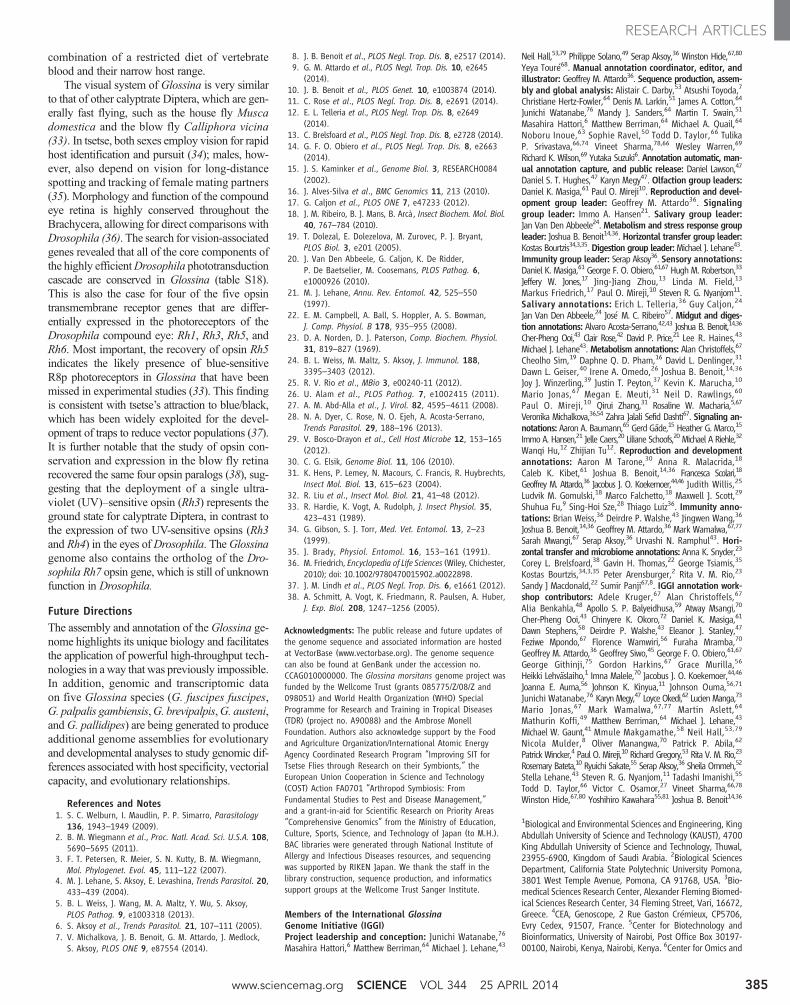
Fig. 1. Diagrammatic presentation of major findings regarding theeffects of trypanosome infection of the salivary glands and midgut,proteomic analysis of the peritrophicmatrix, and the role of aquaporinproteins in blood meal digestion and diuresis. (Top) comparison oftrypanosome-uninfected and -infected states of Glossina salivary glands. (Left)
Representative protein components of Glossina salivary secretions. (Right)Pathogenic effects of trypanosome infection on salivary gland function. (Bottom)Glossina digestive physiology and the infection process by trypanosomes. As-sociated satellite references for these findings are listed within the figure asnumbers in parentheses and correspond to the reference list (8, 11, 12).
www.sciencemag.org SCIENCE VOL 344 25 APRIL 2014 381
RESEARCH ARTICLES

tsetse, theADGF genes are uniquely organized asa cluster of four genes in a 20-kb genomic locus(GMOY002973, GMOY012372, GMOY012373,and GMOY012374). An adenosine deaminase(ada) gene (GMOY008741) without the putativegrowth factor domain is encoded elsewhere in thegenome. InDrosophila, five ADGF genes can befound in various loci and are associated with de-velopmental regulation (19). NematoceranDiptera,including sandflies and mosquitoes, have a maxi-mum of three ADGF genes. Other arthropods,such as Ixodes scapularis, Rhodnius prolixus, andPediculus humanus, have only bona fide adeno-sine deaminases.
Recent studies show that specific genes andproteins are down-regulated in salivary glands dur-ing parasite infection,which promotes trypanosometransmission because feeding efficiency is reducedand feeding time is extended (20). RNA-seq analy-
sis of salivary gland gene expression during para-site infection confirmed the reduction of transcriptabundance for previously identified genes, suchas ada, tsal1, tsal2, and 5′ nucleotidase, as well asof many other putative secreted salivary proteingenes (12). Additionally, genes involved in stresstolerance and cell repair showed increased expres-sion, indicating that considerable salivary gland tis-sue damage is caused by trypanosome infections.
Upon blood-meal ingestion, the peritrophicmatrix (PM), which separates the midgut epithe-lium from the blood bolus, protects gut cells fromdamaging or toxic dietary elements, allows forcompartmentalized digestion and metabolism ofthe blood meal, and is a barrier against infection(5). Glossina produces a type-II PM, which is se-creted continuously as concentric sleeves by theproventriculus and separates the lumen of themidgut (endoperitrophic space) from the mono-
layer of epithelial cells (21). Type-II PMs are gen-erally composed of chitin, peritrophin proteins,glycosaminoglycans (GAGs), and mucin-likemolecules. Analysis of isolated PMs of male fliesby mass spectrometry identified ~300 proteins,including multiple uncharacterized peritrophinsand peritrophin-like glycoproteins. This pro-teomic data identified the corresponding genesin the Glossina genome. Three of these genesare exclusively expressed by the proventriculus(table S6) (11).
Glossina takes a blood meal that is almostequivalent to its own weight, and excess wateris rapidly excreted by means of the Aquaporinfamily of transport proteins (22). Ten aqua-porin genes (aqps) were identified in Glossina,compared with six and eight in mosquitoes andDrosophila, respectively (table S7). InGlossina,two aqp genes are duplicates: the orthologs
Fig. 2. Diagrammatic pre-sentation of the Glossinamicrobiome, tissue locali-zationofbacterial symbionts,physiological importance,and summary of genomicinteractions. (Top) Glossinareproductive physiology andassociated symbiont localiza-tions. (Bottom) Glossina di-gestive physiology and theassociated symbiont localiza-tions. Associated text describessignificant findings regard-ing the Glossina microbiomeand the associated impacts interms of Glossina immunity,nutrition, vectorial capacity,and vector control. Associatedsatellite references are listedwithin the figure as numbersin parentheses and correspondto the reference list (11, 13).
25 APRIL 2014 VOL 344 SCIENCE www.sciencemag.org382
RESEARCH ARTICLES

of the aqp2 and the Drosophila integral pro-tein (drip) genes. Knockdown of aquaporinsinhibited post–blood meal diuresis, increaseddehydration tolerance, reduced heat tolerance,and extended the duration of lactation andpregnancy in females. The drip orthologs areparticularly abundant in the female accessorygland (milk glands), suggesting a role in hy-dration of glandular secretions (8).
In comparison with mosquitoes and sand-flies, Glossina has a marked reduction in genesassociated with carbohydrate metabolism, insteadusing a proline-alanine shuttle system for energydistribution and triglycerides and diglycerides forstorage in the fat body and milk secretions. Littleto no sugar nor glycogen is detectable in these
flies (23). Genes involved in lipid metabolismare generally conserved, with gene expansionsassociated with fatty acid synthase, fatty acyl-CoA reductase, and 3-keto acyl-CoA synthasefunctions (table S8). In addition, three multi-vitamin transporters from the solute:sodiumsymporter (SSS) family are found in Glossinaand mosquitoes, but not in Drosophila, suggest-ing an association with blood-meal metabolism(table S9).
MicrobiomeGlossina harbor multiple maternally transmittedmutualistic and parasitic microorganisms, in-cluding the obligate Wigglesworthia glossinidia,which reside intracellularly in cells that com-
promise the midgut-associated bacteriome or-gan as well as extracellularly in the milk glandlumen (Fig. 2). In the absence ofWigglesworthia,female flies tend to prematurely abort their larvaloffspring unless they receive dietary supplements(18). However, the larvae that have under-gone intrauterine development in the absenceof Wigglesworthia metamorphose into immune-compromised adults (24).
The predicted proteome of Wigglesworthiaindicates a capacity for B vitamin biosynthesis(25) and synthesis of thiamine monophosphate(TMP). Glossina lacks this capacity; however, itcarries genes for thiamine transporters, a memberof the reduced folate carrier family (GMOY009200),and a folate transporter (GMOY005445).
Fig. 3. Diagrammatic presentation of milk gland secretory cell physiologyand milk production during lactation and after parturition. (Left) Lactation.Nutrients including lipids, amino acids, and water are taken up by the cell throughvarious transporters. Lipids are aggregated into droplets while amino acids areincorporated into the synthesis of a battery of milk proteins. During pregnancy,milk protein genes are up-regulated by the Ladybird Late homeodomain protein.Lipids, proteins, and water are combined to form the milk constituents, which arestored in a large extracellular secretory reservoir. Stored milk is secreted into the
lumen, which also houses the extracellular obligate bacterial symbiontWigglesworthia.The milk and symbionts are transported through the gland to the uterus, wherethe developing larvae feeds upon these secretions. (Right) Involution and re-covery. After parturition, milk gland cells shrink, undergo autophagy, and expressantioxidant enzymes to inhibit oxidative damage. The recovered cells prepare forthe next round of lactation by regeneration of protein synthesis and structuralcomponents. Associated satellite references for these findings are listed within thefigure as numbers in parentheses and correspond to the reference list (7–10).
www.sciencemag.org SCIENCE VOL 344 25 APRIL 2014 383
RESEARCH ARTICLES

Wolbachia is another symbiont present insome wildGlossina populations (and in the strainsequenced here), which resides in gonadal tissues.Laboratory studies have shown that this associatedWolbachia strain induces cytoplasmic incompat-ibility (CI) in Glossina morsitans (26). Further-more, at least three horizontal transfer events(HTEs) from Wolbachia were detected in Glossinachromosomes. The two largest insertions carry atotal of 159 and 197 putative functional protein-coding genes, whereas the third lacks any proteincoding genes. In situ staining of Glossinamitot-ic chromosomes with Wolbachia-specific DNAprobes identified multiple insertions on the X, Y,and B chromosomes (table S10) (13). Althoughno Wolbachia-specific transcripts were detectedarising from chromosomal insertions, the func-tional and evolutionary implications of these in-sertions require study.
Many Glossina species, including the strainsequenced here, harbor a large DNAhytrosavirus,the Glossina pallidipes salivary gland hypertro-phy virus (GpSGHV) (27). The virus can reducefecundity and life span in Glossina and causesalivary gland pathology and swelling at highdensities. Also, the analysis of a group of geneslacking dipteran orthologs revealed many pu-tative bracoviral genes [Basic Local AlignmentSearch Tool (BLAST) E values of <1 × 10–50)]spread over 151 genomic scaffolds (tables S11and S12). The putative bracoviral sequences bearhighest homology to those identified from the par-asitic braconid waspsGlyptapanteles flavicoxis andCotesia congregata. This suggests that Glossinawas or is parasitized by an as-yet-unidentifiedbraconid wasp.
ImmunityMultiple factors, including age, sex, nutritionalstatus, and symbionts, influence tsetse’s compe-tence as a vector for trypanosomes. Peptidoglycan(PGN) recognition proteins (PGRPs), antimicro-bial effector peptides (AMPs) produced by im-mune deficiency (IMD) pathway, midgut lectins,antioxidants, EP protein (defined by its glutamicacid and proline repeats), and the gut-associatedperitrophic matrix structure are all componentsthat regulate the nature of the interactions betweenthe fly and its symbionts (28).
Microbial detection is a multistep processthat requires direct contact between host patternrecognition receptors (PRRs) and pathogen-associated molecular patterns (PAMPs). Dro-sophila has 13 PGRPs that play a role in the re-cognition of PGN, an essential component ofthe cell wall of virtually all bacteria. InGlossina,only six pgrp genes were identified, four in thelong subfamily (pgrp-la, -lb, -lc, and -ld) and twoin the short subfamily (pgrp-sa and -sb), whereasDrosophila has a gene duplication resulting intwo related forms of pgrp-sb. Based on both ge-nome annotation and transcriptome data,Glossinalacks orthologs of the PGN receptors, -le, -lf,-sc, and -sd, found in Drosophila. The reducedpgrp repertoire ofGlossinamay reflect its blood-
specific diet, which likely exposes its gut to fewermicrobes thanDrosophila. In theDrosophila gut,PGRP-LE functions as the master bacterial sen-sor, which induces both responses to infectiousbacteria and tolerance tomicrobiota by up-regulatingsuppressors of the IMDpathway, including PGRP-LB (29). In the case of Glossina, loss of amidase-SC1 along with PGRP-LE may indicate a hostimmune response that has evolved to protect thesymbiosis withWigglesworthia. Reduced immunecapacity is also observed in aphids that also har-bor obligate symbionts (30). A complete listing oforthologs toDrosophila immune genes is presentedin table S13.
Reproduction and Developmental BiologyThe reproductive biology of tsetse is unique tothe Hippoboscoidea superfamily. The evolutionof adenotrophic viviparity (intrauterine larval de-velopment and nourishment by glandular secre-tions) has required ovarian follicle reduction (twofollicles per ovary compared with 30 to 40 inDrosophila), expansion and adaptation of theuterus to accommodate developing larvae, andadaptation of the female accessory gland to func-tion as a nutrient synthesis and delivery system.
Glossina, Drosophila, and other Brachyceranflies use lipase-derived yolk proteins for vitello-genesis, unlike non-Brachyceran flies that usethe vitellogenin family of yolk proteins (31).Drosophila and Brachyceran flies outside ofthe Hippoboscidae superfamily produce multi-ple oocytes per gonotrophic cycle. However,Glossina only develops a single oocyte each cy-cle. Unlike Drosophila, which has three yolk pro-tein genes (yp1, yp2, and yp3) localized on theX chromosome, Glossina has a single yolk pro-tein gene, which is orthologous to Drosophilayp2 (GMOY002338), expressed only in the ova-ries, and lacks fat body–associated expression.Multiple yolk proteins have been identified inother Brachyceran flies, indicating that Glossinamay have lost these genes in association with itsreduction in reproductive capacity (31).
Glossina larvae are dependent on their moth-er’s milk gland secretions for nutrition, as wellas for transfer of symbiotic fauna (Fig. 3). Thisgland is highly specialized and secretes a com-plex mixture of stored lipids and milk proteins.Analysis of differential gene expression in lac-tating versus nonlactating females confirmedthe presence of previously characterized milkprotein genes—including a lipocalin (mgp1), atransferrin (trf ), an acid sphingomyelinase(asmase), milk proteins 2 + 3 (mgp2 and -3), andpgn recognition protein lb (pgrp-lb) (28)—butalso revealed a previously undiscovered suite ofeight paralogs to the mgp2 and -3 genes. Annota-tion of the 40-kb genomic loci encompassingmgp2 and mgp3 revealed that these genes havearisen via gene duplication events. These geneshave similar exon/intron structures and are ex-pressed in the same stage- and tissue-specificmanner as mgp2 and mgp3 (10). The newly iden-tified milk proteins may function as lipid emul-
sification agents, sources of amino acids, andphosphate carriers. The 12 genes associated withmilk synthesis make up almost half of all mater-nal transcriptional activity during lactation (tableS14) (10). The combined suite of Glossina milkproteins bear remarkable functional similaritiesto those of placental mammals and marsupials(Fig. 3).
The massive level of protein synthesis duringlactation generates substantial oxidative stress intsetse females, but females can undergo this pro-cess 8 to 10 times during their life spans withoutevidence of reproductive senescence. Transcrip-tional analysis of antioxidant enzyme (AOE) geneexpression revealed an increase in abundance ofthese genes during lactation and after birth (7)(table S15), such that knockdown of these en-zymes decreases fecundity in subsequent repro-ductive cycles. The mediation of oxidative stressby AOEs at key points in tsetse reproductionappears critical to preservation of fecundity lateinto Glossina’s life span (7) (Fig. 3).
The milk proteins produced by tsetse are un-der tight transcriptional regulation and are onlyexpressed in the female accessory gland. The ex-pression level of these genes is coordinated withthe stage of pregnancy and increases with larvaldevelopment. The system regulating these genesappears conserved as transgenic Drosophila car-rying the mgp1 gene promoter sequence drivethe expression of a green fluorescent protein re-porter gene exclusively in the female accessoryglands in coordination with oogenesis/ovulation.Comparative analysis of the promoter sequencesfrom multiple milk protein genes revealed thepresence of conserved binding sites for homeo-domain transcription factors. Analysis of thehomeodomain transcription factors in Glossina(table S16) identified a gene, ladybird late (lbl),which is expressed exclusively in the milk glandof adult female flies and the female accessoryglands of Drosophila. Knockdown of lbl re-sults in a global reduction of milk gland proteinexpression in tsetse and causes loss of fecundity(9) (Fig. 3).
Sensory Genes as Targets for GlossinaControl StrategiesGlossina species differ in host preferences andvary in their response to chemical and visual cuesfrom different mammalian hosts or for mate find-ing. Sensory proteins range from odorant bindingproteins (OBP), chemosensory proteins (CSP),odorant receptors (OR), gustatory receptors (GR),and ligand-gated ionotropic receptors (IR) to sen-sory neuron membrane proteins (SNMP) (32).
Detailed annotation of Glossina sensory re-ceptors reveals that they have fewer olfactoryproteins relative to Drosophila, An. gambiae, andApis mellifera (table S17) (14). Of note, six ORsare homologous to a singleDrosophilaOR, whichis associated with female mating deterrence. Inaddition, GR genes associated with sweet tastes,present in all other Diptera, are missing in tsetse.These genetic differences are consistent with the
25 APRIL 2014 VOL 344 SCIENCE www.sciencemag.org384
RESEARCH ARTICLES
combination of a restricted diet of vertebrateblood and their narrow host range.
The visual system of Glossina is very similarto that of other calyptrate Diptera, which are gen-erally fast flying, such as the house fly Muscadomestica and the blow fly Calliphora vicina(33). In tsetse, both sexes employ vision for rapidhost identification and pursuit (34); males, how-ever, also depend on vision for long-distancespotting and tracking of female mating partners(35). Morphology and function of the compoundeye retina is highly conserved throughout theBrachycera, allowing for direct comparisons withDrosophila (36). The search for vision-associatedgenes revealed that all of the core components ofthe highly efficientDrosophila phototransductioncascade are conserved in Glossina (table S18).This is also the case for four of the five opsintransmembrane receptor genes that are differ-entially expressed in the photoreceptors of theDrosophila compound eye: Rh1, Rh3, Rh5, andRh6. Most important, the recovery of opsin Rh5indicates the likely presence of blue-sensitiveR8p photoreceptors in Glossina that have beenmissed in experimental studies (33). This findingis consistent with tsetse’s attraction to blue/black,which has been widely exploited for the devel-opment of traps to reduce vector populations (37).It is further notable that the study of opsin con-servation and expression in the blow fly retinarecovered the same four opsin paralogs (38), sug-gesting that the deployment of a single ultra-violet (UV)–sensitive opsin (Rh3) represents theground state for calyptrate Diptera, in contrast tothe expression of two UV-sensitive opsins (Rh3and Rh4) in the eyes ofDrosophila. TheGlossinagenome also contains the ortholog of the Dro-sophila Rh7 opsin gene, which is still of unknownfunction in Drosophila.
Future DirectionsThe assembly and annotation of theGlossina ge-nome highlights its unique biology and facilitatesthe application of powerful high-throughput tech-nologies in a way that was previously impossible.In addition, genomic and transcriptomic dataon five Glossina species (G. fuscipes fuscipes,G. palpalis gambiensis,G. brevipalpis,G. austeni,andG. pallidipes) are being generated to produceadditional genome assemblies for evolutionaryand developmental analyses to study genomic dif-ferences associatedwith host specificity, vectorialcapacity, and evolutionary relationships.
References and Notes1. S. C. Welburn, I. Maudlin, P. P. Simarro, Parasitology
136, 1943–1949 (2009).2. B. M. Wiegmann et al., Proc. Natl. Acad. Sci. U.S.A. 108,
5690–5695 (2011).3. F. T. Petersen, R. Meier, S. N. Kutty, B. M. Wiegmann,
Mol. Phylogenet. Evol. 45, 111–122 (2007).4. M. J. Lehane, S. Aksoy, E. Levashina, Trends Parasitol. 20,
433–439 (2004).5. B. L. Weiss, J. Wang, M. A. Maltz, Y. Wu, S. Aksoy,
PLOS Pathog. 9, e1003318 (2013).6. S. Aksoy et al., Trends Parasitol. 21, 107–111 (2005).7. V. Michalkova, J. B. Benoit, G. M. Attardo, J. Medlock,
S. Aksoy, PLOS ONE 9, e87554 (2014).
8. J. B. Benoit et al., PLOS Negl. Trop. Dis. 8, e2517 (2014).9. G. M. Attardo et al., PLOS Negl. Trop. Dis. 10, e2645
(2014).10. J. B. Benoit et al., PLOS Genet. 10, e1003874 (2014).11. C. Rose et al., PLOS Negl. Trop. Dis. 8, e2691 (2014).12. E. L. Telleria et al., PLOS Negl. Trop. Dis. 8, e2649
(2014).13. C. Brelsfoard et al., PLOS Negl. Trop. Dis. 8, e2728 (2014).14. G. F. O. Obiero et al., PLOS Negl. Trop. Dis. 8, e2663
(2014).15. J. S. Kaminker et al., Genome Biol. 3, RESEARCH0084
(2002).16. J. Alves-Silva et al., BMC Genomics 11, 213 (2010).17. G. Caljon et al., PLOS ONE 7, e47233 (2012).18. J. M. Ribeiro, B. J. Mans, B. Arcà, Insect Biochem. Mol. Biol.
40, 767–784 (2010).19. T. Dolezal, E. Dolezelova, M. Zurovec, P. J. Bryant,
PLOS Biol. 3, e201 (2005).20. J. Van Den Abbeele, G. Caljon, K. De Ridder,
P. De Baetselier, M. Coosemans, PLOS Pathog. 6,e1000926 (2010).
21. M. J. Lehane, Annu. Rev. Entomol. 42, 525–550(1997).
22. E. M. Campbell, A. Ball, S. Hoppler, A. S. Bowman,J. Comp. Physiol. B 178, 935–955 (2008).
23. D. A. Norden, D. J. Paterson, Comp. Biochem. Physiol.31, 819–827 (1969).
24. B. L. Weiss, M. Maltz, S. Aksoy, J. Immunol. 188,3395–3403 (2012).
25. R. V. Rio et al., MBio 3, e00240-11 (2012).26. U. Alam et al., PLOS Pathog. 7, e1002415 (2011).27. A. M. Abd-Alla et al., J. Virol. 82, 4595–4611 (2008).28. N. A. Dyer, C. Rose, N. O. Ejeh, A. Acosta-Serrano,
Trends Parasitol. 29, 188–196 (2013).29. V. Bosco-Drayon et al., Cell Host Microbe 12, 153–165
(2012).30. C. G. Elsik, Genome Biol. 11, 106 (2010).31. K. Hens, P. Lemey, N. Macours, C. Francis, R. Huybrechts,
Insect Mol. Biol. 13, 615–623 (2004).32. R. Liu et al., Insect Mol. Biol. 21, 41–48 (2012).33. R. Hardie, K. Vogt, A. Rudolph, J. Insect Physiol. 35,
423–431 (1989).34. G. Gibson, S. J. Torr, Med. Vet. Entomol. 13, 2–23
(1999).35. J. Brady, Physiol. Entomol. 16, 153–161 (1991).36. M. Friedrich, Encyclopedia of Life Sciences (Wiley, Chichester,
2010); doi: 10.1002/9780470015902.a0022898.37. J. M. Lindh et al., PLOS Negl. Trop. Dis. 6, e1661 (2012).38. A. Schmitt, A. Vogt, K. Friedmann, R. Paulsen, A. Huber,
J. Exp. Biol. 208, 1247–1256 (2005).
Acknowledgments: The public release and future updates ofthe genome sequence and associated information are hostedat VectorBase (www.vectorbase.org). The genome sequencecan also be found at GenBank under the accession no.CCAG010000000. The Glossina morsitans genome project wasfunded by the Wellcome Trust (grants 085775/Z/08/Z and098051) and World Health Organization (WHO) SpecialProgramme for Research and Training in Tropical Diseases(TDR) (project no. A90088) and the Ambrose MonellFoundation. Authors also acknowledge support by the Foodand Agriculture Organization/International Atomic EnergyAgency Coordinated Research Program “Improving SIT forTsetse Flies through Research on their Symbionts,” theEuropean Union Cooperation in Science and Technology(COST) Action FA0701 “Arthropod Symbiosis: FromFundamental Studies to Pest and Disease Management,”and a grant-in-aid for Scientific Research on Priority Areas“Comprehensive Genomics” from the Ministry of Education,Culture, Sports, Science, and Technology of Japan (to M.H.).BAC libraries were generated through National Institute ofAllergy and Infectious Diseases resources, and sequencingwas supported by RIKEN Japan. We thank the staff in thelibrary construction, sequence production, and informaticssupport groups at the Wellcome Trust Sanger Institute.
Members of the International GlossinaGenome Initiative (IGGI)Project leadership and conception: Junichi Watanabe,76
Masahira Hattori,6 Matthew Berriman,64 Michael J. Lehane,43
Neil Hall,53,79 Philippe Solano,49 Serap Aksoy,36 Winston Hide,67,80
Yeya Touré68. Manual annotation coordinator, editor, andillustrator: Geoffrey M. Attardo36. Sequence production, assem-bly and global analysis: Alistair C. Darby,53 Atsushi Toyoda,7
Christiane Hertz-Fowler,64 Denis M. Larkin,51 James A. Cotton,64
Junichi Watanabe,76 Mandy J. Sanders,64 Martin T. Swain,51
Masahira Hattori,6 Matthew Berriman,64 Michael A. Quail,64
Noboru Inoue,63 Sophie Ravel,50 Todd D. Taylor,66 TulikaP. Srivastava,66,74 Vineet Sharma,78,66 Wesley Warren,69
Richard K. Wilson,69 Yutaka Suzuki6. Annotation automatic, man-ual annotation capture, and public release: Daniel Lawson,47
Daniel S. T. Hughes,47 Karyn Megy47. Olfaction group leaders:Daniel K. Masiga,61 Paul O. Mireji10. Reproduction and devel-opment group leader: Geoffrey M. Attardo36. Signalinggroup leader: Immo A. Hansen21. Salivary group leader:Jan Van Den Abbeele24.Metabolism and stress response groupleader: Joshua B. Benoit14,36. Horizontal transfer group leader:Kostas Bourtzis34,3,35. Digestion group leader:Michael J. Lehane43.Immunity group leader: Serap Aksoy36. Sensory annotations:Daniel K. Masiga,61 George F. O. Obiero,61,67 Hugh M. Robertson,33
Jeffery W. Jones,17 Jing-Jiang Zhou,13 Linda M. Field,13
Markus Friedrich,17 Paul O. Mireji,10 Steven R. G. Nyanjom11.Salivary annotations: Erich L. Telleria,36 Guy Caljon,24
Jan Van Den Abbeele,24 José M. C. Ribeiro57. Midgut and diges-tion annotations: Alvaro Acosta-Serrano,42,43 Joshua B. Benoit,14,36
Cher-Pheng Ooi,43 Clair Rose,42 David P. Price,21 Lee R. Haines,43
Michael J. Lehane43.Metabolism annotations: Alan Christoffels,67
Cheolho Sim,19 Daphne Q. D. Pham,16 David L. Denlinger,31
Dawn L. Geiser,40 Irene A. Omedo,26 Joshua B. Benoit,14,36
Joy J. Winzerling,39 Justin T. Peyton,37 Kevin K. Marucha,10
Mario Jonas,67 Megan E. Meuti,31 Neil D. Rawlings,60
Paul O. Mireji,10 Qirui Zhang,31 Rosaline W. Macharia,5,67
Veronika Michalkova,36,54 Zahra Jalali Sefid Dashti67. Signaling an-notations: Aaron A. Baumann,65 Gerd Gäde,15 Heather G. Marco,15
Immo A. Hansen,21 Jelle Caers,20 Liliane Schoofs,20 Michael A Riehle,32
Wanqi Hu,12 Zhijian Tu12. Reproduction and developmentannotations: Aaron M Tarone,30 Anna R. Malacrida,18
Caleb K. Kibet,61 Joshua B. Benoit,14,36 Francesca Scolari,18
Geoffrey M. Attardo,36 Jacobus J. O. Koekemoer,44,46 Judith Willis,25
Ludvik M. Gomulski,18 Marco Falchetto,18 Maxwell J. Scott,29
Shuhua Fu,9 Sing-Hoi Sze,28 Thiago Luiz36. Immunity anno-tations: Brian Weiss,36 Deirdre P. Walshe,43 Jingwen Wang,36
Joshua B. Benoit,14,36 Geoffrey M. Attardo,36 Mark Wamalwa,67,77
Sarah Mwangi,67 Serap Aksoy,36 Urvashi N. Ramphul43. Hori-zontal transfer and microbiome annotations: Anna K. Snyder,23
Corey L. Brelsfoard,38 Gavin H. Thomas,22 George Tsiamis,35
Kostas Bourtzis,34,3,35 Peter Arensburger,2 Rita V. M. Rio,23
Sandy J Macdonald,22 Sumir Panji67,8. IGGI annotation work-shop contributors: Adele Kruger,67 Alan Christoffels,67
Alia Benkahla,48 Apollo S. P. Balyeidhusa,59 Atway Msangi,70
Cher-Pheng Ooi,43 Chinyere K. Okoro,72 Daniel K. Masiga,61
Dawn Stephens,58 Deirdre P. Walshe,43 Eleanor J. Stanley,47
Feziwe Mpondo,67 Florence Wamwiri,56 Furaha Mramba,70
Geoffrey M. Attardo,36 Geoffrey Siwo,45 George F. O. Obiero,61,67
George Githinji,75 Gordon Harkins,67 Grace Murilla,56
Heikki Lehväslaiho,1 Imna Malele,70 Jacobus J. O. Koekemoer,44,46
Joanna E. Auma,56 Johnson K. Kinyua,11 Johnson Ouma,56,71
Junichi Watanabe,76 Karyn Megy,47 Loyce Okedi,62 Lucien Manga,73
Mario Jonas,67 Mark Wamalwa,67,77 Martin Aslett,64
Mathurin Koffi,49 Matthew Berriman,64 Michael J. Lehane,43
Michael W. Gaunt,41 Mmule Makgamathe,58 Neil Hall,53,79
Nicola Mulder,8 Oliver Manangwa,70 Patrick P. Abila,62
Patrick Wincker,4 Paul O. Mireji,10 Richard Gregory,53 Rita V. M. Rio,23
Rosemary Bateta,10 Ryuichi Sakate,55 Serap Aksoy,36 Sheila Ommeh,52
Stella Lehane,43 Steven R. G. Nyanjom,11 Tadashi Imanishi,55
Todd D. Taylor,66 Victor C. Osamor,27 Vineet Sharma,66,78
Winston Hide,67,80 Yoshihiro Kawahara55,81 Joshua B. Benoit14,36
1Biological and Environmental Sciences and Engineering, KingAbdullah University of Science and Technology (KAUST), 4700King Abdullah University of Science and Technology, Thuwal,23955-6900, Kingdom of Saudi Arabia. 2Biological SciencesDepartment, California State Polytechnic University Pomona,3801 West Temple Avenue, Pomona, CA 91768, USA. 3Bio-medical Sciences Research Center, Alexander Fleming Biomed-ical Sciences Research Center, 34 Fleming Street, Vari, 16672,Greece. 4CEA, Genoscope, 2 Rue Gaston Crémieux, CP5706,Evry Cedex, 91507, France. 5Center for Biotechnology andBioinformatics, University of Nairobi, Post Office Box 30197-00100, Nairobi, Kenya, Nairobi, Kenya. 6Center for Omics and
www.sciencemag.org SCIENCE VOL 344 25 APRIL 2014 385
RESEARCH ARTICLES

Bioinformatics, Department of Computational Biology, Grad-uate School of Frontier Sciences, The University of Tokyo, 5-1-5Kashiwanoha, Kashiwa, Chiba, 277-8561, Japan. 7ComparativeGenomics Laboratory, National Institute of Genetics, 411-8540Yata 1111, Mishima, Shizuoka, 411-8540, Japan. 8Com-putational Biology Group, IIDMM, University of Cape TownFaculty of Health Sciences, Cape Town, 7925, South Africa.9Department of Biochemistry and Biophysics, Texas A&MUniversity, 328B TAMU, College Station, TX 77843, USA. 10De-partment of Biochemistry and Molecular Biology, EgertonUniversity, Post Office Box 536, Njoro, Kenya. 11Department ofBiochemistry, Jomo Kenyatta University of Agriculture andTechnology (JKUAT), Post Office Box 62000-00200, Nairobi,Kenya. 12Department of Biochemistry, Virginia PolytechnicInstitute and State University, 309 Fralin Hall, Blacksburg, VA24061, USA. 13Department of Biological Chemistry and CropProtection, Rothamsted Research, West Common, Harpenden,Herts, AL5 2JQ, UK. 14Department of Biological Sciences,McMicken College of Arts and Sciences, University of Cincinnati,Cincinnati, OH 45221, USA. 15Department of Biological Sci-ences, University of Cape Town, Private Bag, Rondebosch,ZA-7700, South Africa. 16Department of Biological Sciences,University of Wisconsin–Parkside, 900 Wood Road, Kenosha,WI 53144, USA. 17Department of Biological Sciences, WayneState University, 5047 Gullen Mall, Detroit, MI 48202, USA.18Department of Biology and Biotechnology, University ofPavia, Via Ferrata 9, Pavia, 27100, Italy. 19Department of Bi-ology, Baylor University, Waco, TX 76798, USA. 20Departmentof Biology, KU Leuven, Naamsestraat 59, Leuven, B-3000,Belgium. 21Department of Biology, New Mexico State University,Foster Hall 263, Las Cruces, NM 88003, USA. 22Department ofBiology, University of York, Wentworth Way, York, Y010 5DD,UK. 23Department of Biology, West Virginia University, 53Campus Drive, 5106 LSB, Morgantown, WV, USA. 24Depart-ment of Biomedical Sciences, Institute of Tropical MedicineAntwerp, Nationalestraat 155, Antwerp, B-2000, Belgium.25Department of Cellular Biology, University of Georgia, 302Biological Sciences Building, Athens, GA 30602, USA. 26De-partment of Clinical Research, KEMRI-Wellcome Trust Programme,CGMRC, Post Office Box 230-80108, Kilifi, Kenya. 27Depart-ment of Computer and Information Sciences, College of Sci-ence and Technology, Covenant University, P.M.B. 1023, Ota,Ogun State, Nigeria. 28Department of Computer Science andEngineering, Department of Biochemistry and Biophysics,Texas A&M University, HRBB 328B TAMU, College Station, TX77843, USA. 29Department of Entomology, North Carolina StateUniversity, Campus Box 7613, Raleigh, NC 27695–7613, USA.30Department of Entomology, Texas A&M University, 2475TAMU, College Station, TX 77843, USA. 31Department ofEntomology, The Ohio State University, 400 Aronoff Labora-tory, 318 West 12th Avenue, Columbus, OH 43210, USA.32Department of Entomology, University of Arizona, 1140 EastSouth Campus Drive, Forbes 410, Tucson, AZ 85721, USA.33Department of Entomology, University of Illinois at Urbana-Champaign, 505 South Goodwin Avenue, Urbana, IL 61801,USA. 34Insect Pest Control Laboratory, Joint FAO/IAEA Pro-gramme of Nuclear Techniques in Food and Agriculture, Vienna,1220, Austria. 35Department of Environmental and NaturalResources Management, University of Patras, 2 Seferi Street,Agrinio, 30100, Greece. 36Department of Epidemiology ofMicrobial Diseases, Yale School of Public Health, 60 CollegeStreet, New Haven, CT 06520, USA. 37Department of Evolution,Ecology, and Organismal Biology, The Ohio State University,300 Aronoff Laboratory, 318 West 12th Avenue, Columbus, OH43210, USA. 38Department of Natural Sciences, St. CatharineCollege, 2735 Bardstown Road., St. Catharine, KY 40061, USA.39Department of Nutritional Sciences, University of Arizona,Career and Academic Services, College of Agriculture and LifeSciences, Forbes Building, Room 201, Post Office Box 210036,Tucson, AZ 85721–0036, USA. 40Department of NutritionalSciences, University of Arizona, Shantz 405, 1177 East 4thStreet, Tucson, AZ 85721–0038, USA. 41Department of Patho-gen Molecular Biology, London School of Hygiene and TropicalMedicine, Keppel Street, London, WC1E 7HT, UK. 42Depart-ment of Parasitology, Liverpool School of Tropical Medicine,Pembroke Place, Liverpool, L3 5QA, UK. 43Department of Vec-tor Biology, Liverpool School of Tropical Medicine, PembrokePlace, Liverpool, L3 5QA, UK. 44Entomology Section, OnderstepoortVeterinary Institute, Private Bag X5, Onderstepoort, 110, SouthAfrica. 45Eck Institute for Global Health, Department of Bio-
logical Sciences, University of Notre Dame, Notre Dame, IN 46556,USA. 46Department of Veterinary Tropical Diseases, Universityof Pretoria, Private Bag X04, Onderstepoort, 110, South Africa.47European Molecular Biology Laboratories, European Bio-informatics Institute (EMBL-EBI), Wellcome Trust Genome Cam-pus, Hinxton, Cambridge, Cambridgeshire, CB10 1SA, UK.48Group of Bioinformatics and Modeling, Laboratory of Med-ical Parasitology, Biotechnology, and Biomolecules, Institut Pasteurde Tunis, 13, Place Pasteur, BP74, Belvédère, Tunis, 1002,Tunisia. 49Institut de Recherche pour le Développement (IRD),UMR 177 IRD-CIRAD INTERTRYP, CIRDES Bobo-Dioulasso,Burkina Faso. 50Institut de Recherche pour le Développement(IRD), UMR 177 IRD-CIRAD INTERTRYP, LRCT Campus Inter-national de Baillarguet, Montpellier, France. 51Institute of Bi-ological, Environmental, and Rural Sciences, University ofAberystwyth, Old College, King Street, Aberystwyth, Ceredigion,SY23 3FG, UK. 52Institute of Biotechnology Research, JomoKenyatta University of Agriculture and Technology (JKUAT),Post Office Box 62000-00200, Nairobi, Kenya. 53Institute ofIntegrative Biology, The University of Liverpool, Crown Street,Liverpool, L69 7ZB, UK. 54Institute of Zoology, Slovak Academyof Sciences, Dúbravská cesta 9, Bratislava, 845 06, Slovakia.55Integrated Database Team, Biological Information ResearchCenter, National Institute of Advanced Industrial Science andTechnology, Aomi 2-4-7, Koto-ku, Tokyo, 135-0064, Japan.56Kenya Agricultural Research Institute Trypanosomiasis Re-search Centre, Post Office Box 362, Kikuyu, 902, Kenya. 57Lab-oratory of Malaria and Vector Research, National Institute ofAllergy and Infectious Diseases, 12735 Twinbrook Parkway,Room 2E-32D, Rockville, MD 20852, USA. 58TechnologyInnovation Agency, National Genomics Platform, Post OfficeBox 30603, Mayville, Durban, 4058, South Africa. 59Depart-ment of Biochemistry and Sports Science, Makerere University,Post Office Box 7062, Kampala, Uganda. 60Bateman Group,Wellcome Trust Sanger Institute, EMBL European Bioinfor-matics Institute, Wellcome Trust Genome Campus, Hinxton,Cambridge, Cambridgeshire, CB10 1SA, UK. 61Molecular Bi-ology and Bioinformatics Unit, International Center of InsectPhysiology and Ecology, Duduville Campus, Kasarani, PostOffice Box 30772-00100, Nairobi, Kenya. 62National LivestockResources Research Institute (NaLIRRI), Post Office Box 96,Tororo, Uganda. 63National Research Center for ProtozoanDiseases, Obihiro University of Agriculture and VeterinaryMedicine, Inada-cho, Obihiro, Hokkaido, 080-8555, Japan.64Parasite Genomics Group, Wellcome Trust Sanger Institute,Wellcome Trust Genome Campus, Hinxton, Cambridge,
Cambridgeshire, CB10 1SA, UK. 65Riddiford Laboratory, JaneliaFarm Research Campus, Howard Hughes Medical Institute,19700 Helix Drive, Ashburn, VA 20147, USA. 66RIKEN Centerfor Integrative Medical Sciences, 1-7-22 Suehiro-cho, Tsurumi-ku, Yokohama, Kanagawa, 230-0045, Japan. 67South AfricanNational Bioinformatics Institute, South African MRC Bio-informatics Unit, University of the Western Cape, 5th Floor LifeSciences Building, Modderdam Road, Bellville 7530, SouthAfrica. 68Special Programme for Research and Training inTropical Diseases (TDR), WHO, Avenue Appia 20, 1211 Geneva27, Switzerland. 69The Genome Institute, Washington Univer-sity School of Medicine, St. Louis, MO 63110, USA. 70Tsetseand Trypanosomiasis Research Institute (TTRI), Majani Mapana,Off Korogwe Road, Post Office Box 1026, Tanga, Tanzania.71Vector Health International, Post Office Box 15500, Arusha,Tanzania. 72Wellcome Trust Sanger Institute, Wellcome TrustGenome Campus, Hinxton, Cambridge, Cambridgeshire, CB101SA, UK. 73WHO Regional Office for Africa, WHO, Cité duDjoué, Post Office Box 06, Brazzaville, Congo. 74School ofBasic Sciences, Indian Institute of Technology, Mandi 175001,Himachal Pradesh, India. 75Department of Parasite, Vector,and Human Biology, KEMRI-Wellcome Trust Programme,CGMRC, Post Office Box 230-80108, Kilifi, Kenya. 76Instituteof Medical Science, The University of Tokyo, 4-6-1 Shirokanedai,Minato-ku, Tokyo, 108-8639, Japan. 77Department of Bio-chemistry and Biotechnology, Kenyatta University, Post OfficeBox 43844-00100, Nairobi, Kenya. 78Department of Biolog-ical Sciences, Indian Institute of Science Education and Re-search, Indore Bypass Road, Bhauri District, Bhopal, MadhyaPradesh, 462066, India.79Faculty of Science, King AbdulazizUniversity, Jeddah, 21589, SA. 80Department of Biostatistics,Harvard School of Public Health, 655 Huntington Ave. Boston,MA 02461. 81Bioinformatics Research Unit, AgrogenomicsResearch Center, National Institute of Agrobiological Sciences,2-1-2, Kannondai, Tsukuba, Ibaraki 305-8602, Japan.
Supplementary Materialswww.sciencemag.org/content/344/6182/380/suppl/DC1Materials and MethodsSupplementary TextFigs. S1 to S9Tables S1 to S43References (39–101)
12 December 2013; accepted 19 March 201410.1126/science.1249656
Discovery of BrainwideNeural-BehavioralMaps via Multiscale UnsupervisedStructure LearningJoshua T. Vogelstein,1,2* Youngser Park,1* Tomoko Ohyama,3* Rex A. Kerr,3 James W. Truman,3
Carey E. Priebe,1†‡ Marta Zlatic3†‡
A single nervous system can generate many distinct motor patterns. Identifying which neurons and circuitscontrol which behaviors has been a laborious piecemeal process, usually for one observer-definedbehavior at a time. We present a fundamentally different approach to neuron-behavior mapping.We optogenetically activated 1054 identified neuron lines in Drosophila larvae and tracked thebehavioral responses from 37,780 animals. Application of multiscale unsupervised structure learningmethods to the behavioral data enabled us to identify 29 discrete, statistically distinguishable,observer-unbiased behavioral phenotypes. Mapping the neural lines to the behavior(s) they evokeprovides a behavioral reference atlas for neuron subsets covering a large fraction of larval neurons.This atlas is a starting point for connectivity- and activity-mapping studies to further investigate themechanisms by which neurons mediate diverse behaviors.
Nervous systems can generate a wide rangeof motor outputs, depending on their in-coming sensory inputs and internal state.
A comprehensive understanding of how behav-
ioral diversity and selection is achieved requiresthe identification of neural circuits that mediatemany distinct motor patterns in a given nervoussystem. Mapping a functional circuit for one be-
25 APRIL 2014 VOL 344 SCIENCE www.sciencemag.org386
RESEARCH ARTICLES
Copyright © 2022 FDOKUMEN