Genetic, Functional and Compleme - CIB (CSIC)
17
ORIGINAL RESEARCH published: 05 December 2017 doi: 10.3389/fmicb.2017.02393 Edited by: Tatiana Venkova, Fox Chase Cancer Center, United States Reviewed by: Maria Jesus Yebra, Instituto de Agroquímica y Tecnología de Alimentos (CSIC), Spain Bopda Waffo Alain, Alabama State University, United States Antonius Suwanto, Bogor Agricultural University, Indonesia *Correspondence: Paloma López [email protected] Specialty section: This article was submitted to Evolutionary and Genomic Microbiology, a section of the journal Frontiers in Microbiology Received: 28 September 2017 Accepted: 20 November 2017 Published: 05 December 2017 Citation: Pérez-Ramos A, Werning ML, Prieto A, Russo P, Spano G, Mohedano ML and López P (2017) Characterization of the Sorbitol Utilization Cluster of the Probiotic Pediococcus parvulus 2.6: Genetic, Functional and Complementation Studies in Heterologous Hosts. Front. Microbiol. 8:2393. doi: 10.3389/fmicb.2017.02393 Characterization of the Sorbitol Utilization Cluster of the Probiotic Pediococcus parvulus 2.6: Genetic, Functional and Complementation Studies in Heterologous Hosts Adrian Pérez-Ramos 1 , Maria L. Werning 1,2 , Alicia Prieto 1 , Pasquale Russo 3 , Giuseppe Spano 3 , Mari L. Mohedano 1 and Paloma López 1 * 1 Biological Research Center (CIB), Consejo Superior de Investigaciones Científicas, Madrid, Spain, 2 Center of Research and Transfer of Catamarca (CITCA), Consejo Nacional de Investigaciones Científicas y Técnicas, Catamarca, Argentina, 3 Department of Agricultural, Food and Environmental Sciences, University of Foggia, Foggia, Italy Pediococcus parvulus 2.6 secretes a 2-substituted (1,3)-β-D-glucan with prebiotic and immunomodulatory properties. It is synthesized by the GTF glycosyltransferase using UDP-glucose as substrate. Analysis of the P. parvulus 2.6 draft genome revealed the existence of a sorbitol utilization cluster of six genes (gutFRMCBA), whose products should be involved in sorbitol utilization and could generate substrates for UDP-glucose synthesis. Southern blot hybridization analysis showed that the cluster is located in a plasmid. Analysis of metabolic fluxes and production of the exopolysaccharide revealed that: (i) P. parvulus 2.6 is able to metabolize sorbitol, (ii) sorbitol utilization is repressed in the presence of glucose and (iii) sorbitol supports the synthesis of 2-substituted (1,3)- β-D-glucan. The sorbitol cluster encodes two putative regulators, GutR and GutM, in addition to a phosphoenolpyruvate-dependent phosphotransferase transport system and sorbitol-6-phosphate dehydrogenase. Therefore, we investigated the involvement of GutR and GutM in the expression of gutFRMCBA. The promoter-probe vector pRCR based on the mrfp gene, which encodes the fluorescence protein mCherry, was used to test the potential promoter of the cluster (P gut ) and the genes encoding the regulators. This was performed by transferring by electrotransformation the recombinant plasmids into two hosts, which metabolize sorbitol: Lactobacillus plantarum and Lactobacillus casei. Upon growth in the presence of sorbitol, but not of glucose, only the presence of P gut was required to support expression of mrfp in L. plantarum. In L. casei the presence of sorbitol in the growth medium and the pediococcal gutR or gutR plus gutM in the genome was required for P gut functionality. This demonstrates that: (i) P gut is required for expression of the gut cluster, (ii) P gut is subjected to catabolic repression in lactobacilli, (iii) GutR is an activator, and (iv) in the presence of sorbitol, trans-complementation for activation of P gut exists in L. plantarum but not in L. casei. Keywords: Pediococcus parvulus, exopolysaccharides, β-glucans, sorbitol, lactic acid bacteria, probiotic Frontiers in Microbiology | www.frontiersin.org 1 December 2017 | Volume 8 | Article 2393
-
Upload
khangminh22 -
Category
Documents
-
view
0 -
download
0
Transcript of Genetic, Functional and Compleme - CIB (CSIC)

fmicb-08-02393 December 2, 2017 Time: 15:55 # 1
ORIGINAL RESEARCHpublished: 05 December 2017
doi: 10.3389/fmicb.2017.02393
Edited by:Tatiana Venkova,
Fox Chase Cancer Center,United States
Reviewed by:Maria Jesus Yebra,
Instituto de Agroquímica y Tecnologíade Alimentos (CSIC), Spain
Bopda Waffo Alain,Alabama State University,
United StatesAntonius Suwanto,
Bogor Agricultural University,Indonesia
*Correspondence:Paloma López
Specialty section:This article was submitted to
Evolutionary and GenomicMicrobiology,
a section of the journalFrontiers in Microbiology
Received: 28 September 2017Accepted: 20 November 2017Published: 05 December 2017
Citation:Pérez-Ramos A, Werning ML,Prieto A, Russo P, Spano G,
Mohedano ML and López P (2017)Characterization of the Sorbitol
Utilization Cluster of the ProbioticPediococcus parvulus 2.6: Genetic,
Functional and ComplementationStudies in Heterologous Hosts.
Front. Microbiol. 8:2393.doi: 10.3389/fmicb.2017.02393
Characterization of the SorbitolUtilization Cluster of the ProbioticPediococcus parvulus 2.6: Genetic,Functional and ComplementationStudies in Heterologous HostsAdrian Pérez-Ramos1, Maria L. Werning1,2, Alicia Prieto1, Pasquale Russo3,Giuseppe Spano3, Mari L. Mohedano1 and Paloma López1*
1 Biological Research Center (CIB), Consejo Superior de Investigaciones Científicas, Madrid, Spain, 2 Center of Research andTransfer of Catamarca (CITCA), Consejo Nacional de Investigaciones Científicas y Técnicas, Catamarca, Argentina,3 Department of Agricultural, Food and Environmental Sciences, University of Foggia, Foggia, Italy
Pediococcus parvulus 2.6 secretes a 2-substituted (1,3)-β-D-glucan with prebiotic andimmunomodulatory properties. It is synthesized by the GTF glycosyltransferase usingUDP-glucose as substrate. Analysis of the P. parvulus 2.6 draft genome revealed theexistence of a sorbitol utilization cluster of six genes (gutFRMCBA), whose productsshould be involved in sorbitol utilization and could generate substrates for UDP-glucosesynthesis. Southern blot hybridization analysis showed that the cluster is located in aplasmid. Analysis of metabolic fluxes and production of the exopolysaccharide revealedthat: (i) P. parvulus 2.6 is able to metabolize sorbitol, (ii) sorbitol utilization is repressedin the presence of glucose and (iii) sorbitol supports the synthesis of 2-substituted (1,3)-β-D-glucan. The sorbitol cluster encodes two putative regulators, GutR and GutM, inaddition to a phosphoenolpyruvate-dependent phosphotransferase transport systemand sorbitol-6-phosphate dehydrogenase. Therefore, we investigated the involvementof GutR and GutM in the expression of gutFRMCBA. The promoter-probe vector pRCRbased on the mrfp gene, which encodes the fluorescence protein mCherry, was used totest the potential promoter of the cluster (Pgut) and the genes encoding the regulators.This was performed by transferring by electrotransformation the recombinant plasmidsinto two hosts, which metabolize sorbitol: Lactobacillus plantarum and Lactobacilluscasei. Upon growth in the presence of sorbitol, but not of glucose, only the presence ofPgut was required to support expression of mrfp in L. plantarum. In L. casei the presenceof sorbitol in the growth medium and the pediococcal gutR or gutR plus gutM in thegenome was required for Pgut functionality. This demonstrates that: (i) Pgut is required forexpression of the gut cluster, (ii) Pgut is subjected to catabolic repression in lactobacilli,(iii) GutR is an activator, and (iv) in the presence of sorbitol, trans-complementation foractivation of Pgut exists in L. plantarum but not in L. casei.
Keywords: Pediococcus parvulus, exopolysaccharides, β-glucans, sorbitol, lactic acid bacteria, probiotic
Frontiers in Microbiology | www.frontiersin.org 1 December 2017 | Volume 8 | Article 2393

fmicb-08-02393 December 2, 2017 Time: 15:55 # 2
Pérez-Ramos et al. Characterization of Sorbitol Utilization by Pediococcus parvulus 2.6
INTRODUCTION
Sorbitol, also named D-glucitol, is a six-carbon sugar polyolwidespread in plants, particularly in fruits, such as berries,cherries, plums, pears and apples. However, sorbitol isobtained industrially, by catalytic hydrogenation of glucoseor glucose/fructose mixtures. This polyol has a relative sweetnessof about 60% compared to that of sucrose, high-water solubilityand is largely used as a low calorie sweetener, humectant,texturizer and softener (Zumbé et al., 2001). In addition, sorbitolis used in the production of pharmaceutical compounds, such assorbose and ascorbic acid, and as a vehicle for drug-suspension(Silveira and Jonas, 2002). Sorbitol has also a potential prebioticeffect in vivo, since it does not contribute to the formation ofdental caries, is slowly and only partially absorbed in the smallintestine and can reach the colon where it can act as substratefor bacterial fermentation. Supplementation with sorbitolresulted in enrichment of lactobacilli in rat colon and cecum(Sarmiento-Rubiano et al., 2007).
Sorbitol absorption is mediated by dose and concentration.Doses greater than 30 g can cause water retention, resulting inosmotic diarrhea, bloating, flatulence, cramping and abdominalpain (Fernández-Bañares et al., 2009). These doses varydepending on the condition of the intestinal absorption surface.In patients with malabsorption, the ingestion of 5–20 g, provokeddiarrhea and gastrointestinal complications (Montalto et al.,2013). In the colon, this sugar alcohol is metabolized by somespecies of Lactobacillus and is also a preferred carbon source forhuman intestinal bifidobacteria (Sarmiento-Rubiano et al., 2007).
Furthermore, utilization of sorbitol as a carbon sourcehas been described in a variety of bacteria within the filaproteobacteria (Yamada and Saier, 1988; Aldridge et al., 1997)and firmicutes (Tangney et al., 1998; Boyd et al., 2000; Yebraand Pérez-Martínez, 2002). Among the firmicutes, there are somelactic acid bacteria (LAB) with catabolic pathways for sorbitolmetabolism (Rhodes and Kator, 1999; Sarmiento-Rubiano et al.,2007). These pathways are encoded by genes organized in gutoperons, and include the sorbitol transport system, sorbitol-6-phosphate dehydrogenase (S6PD) as well as regulatory protein(s),and those of Lactobacillus casei and Lactobacillus plantarumhave been characterized (Nissen et al., 2005; Ladero et al., 2007;Alcantara et al., 2008).
Sorbitol is transported into the cells and phosphorylatedto sorbitol-6-phosphate by a phosphopyruvate-dependentphosphotransferase (PTS) sorbitol system (PTSgut). Each PTSis composed of two cytoplasmic enzymes, common to thetransport of different compounds (EI and HPr) and of differentmembrane-associated enzyme complexes (EII), specific for one,or several substrates. The genes gutC, gutB and gutA encode theEII domain of a sorbitol PTS (Alcantara et al., 2008). The gutFgene encodes a sorbitol-6-P dehydrogenase, which catalyzes theconversion of sorbitol-6-phosphate to fructose-6-phosphate, acompound that is introduced into the glycolytic pathway withNADH regeneration (Nissen et al., 2005). The gutR and gutMgenes encode two regulatory proteins. The role of the GutM andGutR proteins has been studied in Escherichia coli, operatingGutM as an activator and GutR as a repressor (Yamada and
Saier, 1988). In the firmicutes group, the analyzed gut operonscontain homologs to the gutM and gutR genes, but the roleof GutR regulator is different from that of E. coli. The GutRof L. casei has been functionally characterized and it has beenshown to be a PTS-controlled transcriptional activator, via a PTSregulation binding domain (PRD) (Stülke et al., 1998). Also, boththe GutR binding sequence and the PRD domain are conservedin firmicutes. GutM encodes a highly conserved protein infirmicutes and in L. casei plays a regulatory role (Alcantara et al.,2008).
Pediococcus parvulus 2.6 (Werning et al., 2006) (previouslynamed Pediococcus damnosus) is a lactic acid bacteria isolatedfrom a ropy cider (Fernández et al., 1995). This LABproduces a 2-substituted (1,3)-β-D-glucan exopolysaccharide(EPS) (Dueñas-Chasco et al., 1997), with high molecular mass(>106 Da), and whose rheological properties showed its potentialutility as a biothickening agent (Velasco et al., 2009). Thepresence of this EPS improves some probiotic features ofP. parvulus 2.6, including tolerance to simulated gastrointestinalconditions and adherence to Caco-2 cell lines and reducesinflammation-related cytokine levels produced by polarizedmacrophages (Fernández de Palencia et al., 2009; Immerstrandet al., 2010). Moreover, the purified EPS improves the growth,viability and adhesion capability of probiotic microorganisms(Russo et al., 2012), also it activates macrophages with anti-inflammatory effects (Notararigo et al., 2014), and decreases thelevels of the proinflammatory IL8 in human intestine cultures(Notararigo et al., unpublished data). The draft genome ofP. parvulus 2.6 has been determined (Pérez-Ramos et al., 2016),and its analysis showed the existence of a putative sorbitolutilization gut operon in this bacterium. Thus, this currentwork focuses on the genomic location, expression and metabolicinvolvement of the gut operon of P. parvulus 2.6 in sorbitolcatabolism, as well as its interplay with EPS production by thisbacterium.
MATERIALS AND METHODS
Bacterial Strains and Growth ConditionsThe bacteria used in this work are listed in Table 1. Pediococcusand Lactobacillus strains were routinely grown in de ManRogosa Sharpe (MRS) broth (Pronadisa, Madrid, Spain) at30◦C and 37◦C, respectively. Lactococcus lactis strains weregrown in ESTY broth (Pronadisa) supplemented with 0.5%glucose at 30◦C. When bacteria carried the pRCR plasmid or itsderivatives the medium was supplemented with chloramphenicol(Cm) at 5 µg mL−1 for L. lactis and at 10 µg mL−1 for lactobacilli.E. coli V517 was grown in LB broth and incubated at 37◦C.
For evaluation of sorbitol utilization, P. parvulus strains weregrown in a MRS broth made by components (de Man et al.,1960) without glucose, pH was adjusted to 5.2 and the mediumsupplemented with 10 mM glucose (MRSG), 30 mM sorbitol(MRSS) or 10 mM glucose plus 30 mM sorbitol (MRSGS) at 30◦C.Prior selection of conditions for growth in presence of sorbitolseveral tests were performed. First various carbon sources weretested (10 mM glucose, 10 mM fructose or 10 mM maltose) and
Frontiers in Microbiology | www.frontiersin.org 2 December 2017 | Volume 8 | Article 2393

fmicb-08-02393 December 2, 2017 Time: 15:55 # 3
Pérez-Ramos et al. Characterization of Sorbitol Utilization by Pediococcus parvulus 2.6
TABLE 1 | Bacteria used in this work.
Bacteria Plasmid Resistance Characteristics Reference
Pediococcus parvulus 2.6 pPP1, pPP2, andpPP3
– 2-substituted (1,3)-β-D-glucan producer Pérez-Ramos et al., 2016
P. parvulus 2.6NR pPP1 and pPP3 – Non-EPS-producing strain. Derivative of 2.6strain by pPP2 plasmid curing
Fernández et al., 1995
Lactococcus lactis subsp. cremoris MG1363 – – Plasmid free type strain used for plasmid cloning Wegmann et al., 2007
L. lactis subsp. cremoris MG1363[pRCR] pRCR CmR Source of promoter probe pRCR containing themrfp gene, which encodes the fluorescentmCherry protein
Mohedano et al., 2015
Escherichia coli V517 8 plasmidspVA517A throughpVA517H
ND Source of plasmids used as references in agarosegel analysis
Macrina et al., 1978
Lactobacillus casei BL23 – – Bacteria used for heterologous gene expression Mazé et al., 2010
L. casei BL23[pRCR16] [pRCR16] CmR Derivative of pRCR by cloning of Pgut upstream ofmrfp
This study
L. casei BL23[pRCR17] [pRCR17] CmR Derivative of pRCR16 by cloning of gutRupstream of mrfp
This study
L. casei BL23[pRCR18] [pRCR18] CmR Derivative of pRCR16 by cloning of gutMupstream of mrfp
This study
L. casei BL23[pRCR19] [pRCR19] CmR Derivative of pRCR16 by cloning of gutMRupstream of mrfp
This study
Lactobacillus plantarum 90 1 uncharacterizedplasmid
ND Bacteria used for heterologous gene expression Lamontanara et al., 2015
L. plantarum 90[pRCR16] [pRCR16] CmR Derivative of pRCR by cloning of Pgut upstream ofmrfp
This study
L. plantarum 90[pRCR17] [pRCR17] CmR Derivative of pRCR16 by cloning of gutRupstream of mrfp
This study
L. plantarum 90[pRCR18] [pRCR18] CmR Derivative of pRCR16 by cloning of gutMupstream of mrfp
This study
L. plantarum 90[pRCR19] [pRCR19] CmR Derivative of pRCR16 by cloning of gutMRupstream of mrfp
This study
ND, no determined; CmR, resistance to chloramphenicol.
pH at 6.8, 5.2 or 4.0 and then influence of aeration was evaluatedin presence of 10 mM glucose at either pH 6.8 and 5.2 (results notshow).
For evaluation of mCherry expression, Lactobacillus strainswere grown in a MRSG containing 55 mM (1% w/v) glucose orin a MRSS containing 55 mM (1% w/v) sorbitol at 37◦C.
Plasmidic DNA PreparationsTotal plasmidic DNA preparations of P. parvulus 2.6 and2.6NR strains were prepared as follows. Bacterial cultures weregrown to an optical density at 600 nm (OD600 nm) of 2.5, and100 mL of each culture were sedimented by centrifugation at10,000 × g for 20 min at 4◦C. The cells were resuspendedin 4 mL of a solution containing 50 mM Tris/HCl pH 8.0,10 mM EDTA, lysozyme (30 mg mL−1) and RNasa A (10 µgmL−1), and incubated for 30 min at 37◦C. Then, 4 mL of asolution containing 220 mM NaOH and 1.33% sodium dodecylsulfate) were added and samples were incubated for 5 min atroom temperature. Upon addition of 5 M potassium acetatepH 5.0 (4 mL), samples were centrifugated at 10,000 × g for15 min at 21◦C. The DNA present in the supernatants wasprecipitated, concentrated by addition of 8.7 mL of isopropanol,sedimented by centrifugation at 10,000 × g for 15 min at 4◦C,and resuspended in 10 mM Tris, 1 mM EDTA buffer (4.3 mL).
The DNA preparation was deproteinated by treatment with 7.5 Mammonium acetate (2.7 mL) and phenol (4.3 mL) during 5 min atroom temperature and then sedimented at 10,000 × g for 5 minat 21◦C. The aqueous phase containing total plasmidic DNA wasfurther purified by isopycnic CsCl density gradient centrifugationand dialysis as previously described (López et al., 1989). Thefinal recovery was 54 µg and 58 µg for 2.6 and 2.6NR DNApreparations, respectively.
The recombinant plasmids from the lactococcal andlactobacilli strains were isolated using the High pure plasmidisolation kit (Roche) as follows. Bacteria were grown untilstationary phase (109 colony forming units mL−1) and 1 mL ofeach culture were sedimented by centrifugation at 10,000× g for10 min at 4◦C. Cells were resuspended in solution I of the kitsupplemented with lysozyme (30 mg mL−1) and were incubatedfor 30 min at 37◦C. Then, plasmid isolation were performedas described in the kit protocol, eluting the plasmidic DNA in100 µL at approximately 100 ng µL−1.
SequencingDNA sequencing was performed by the dideoxy methodat Secugen (Madrid, Spain). The sequencing of the sorbitolutilization cluster and the flanking regions of pPP1 of P. parvulus2.6 was performed using total plasmidic DNA preparations
Frontiers in Microbiology | www.frontiersin.org 3 December 2017 | Volume 8 | Article 2393

fmicb-08-02393 December 2, 2017 Time: 15:55 # 4
Pérez-Ramos et al. Characterization of Sorbitol Utilization by Pediococcus parvulus 2.6
of the bacterium (see above) with the walking strategy andthe sequence has been deposited in GenBank (accession NoMF766019). The lack of sorbitol cluster in the 2.6NR strainwas confirmed by sequencing of its pPP1 plasmid by using assubstrates a total plasmidic preparation of 2.6NR strain andeither pPP1∗F or pPP1∗R primers (see Table 2) In addition,in the case of sequencing with pPP1∗F, it was also used assubstrate the product of a polymerization reaction catalyzedby the bacteriophage 829 DNA polymerase with plasmidicDNA of P. parvulus 2.6NR and hexamers containing randomsequences.
Construction of pRCR16, pRCR17,pRCR18, and pRCR19A region located upstream of the P. parvulus 2.6 gut operoncarrying the putative Pgut promoter and the gutR and gutMgenes was cloned into the promoter probe pRCR vector.To this end, three DNA regions of pPP1 plasmid wereamplified with Phusion High Fidelity Polymerase (PHFP,ThermoFisher Scientific) by using a plasmidic DNA preparationof P. parvulus 2.6 and the primers depicted in Table 2,which have homology with pPP1 DNA and carry restrictionsites suitable for cloning. Plasmid pRCR16 (Figure 1) wasgenerated by ligation of the Pgut promoter to the pRCRpromoter probe vector (Mohedano et al., 2015), after doubledigestion of both DNAs with BglII and XmaI (New EnglandBiolabs, Ipswich, MA, United States), with the T4 DNA ligase(New England Biolabs). Then, between the XmaI and XbaIrestriction sites of pRCR16 three amplicons were independentlycloned, containing gutR, gutM or gutRM, generating plasmidspRCR17, pRCR18 and pRCR19, respectively. The clonings wereperformed in L. lactis MG1363, the ligations mixtures wereused to transform the bacteria by electroporation (25 µF,
2.5 kV and 200 � in 0.2 cm cuvettes), as previously described(Dornan and Collins, 1987) and transformants were selectedin ESTY-agar plates supplemented with Cm at 5 µg mL−1.The inserts present in the new four recombinant plasmids wereconfirmed by automated sequencing. Then, DNA preparationsof pRCR17, pRCR18 and pRCR19 obtained from L. lactisMG1363 (0.5 µg) were used for transfer to lactobacilli byelectroporation (25 µF, 1.3 kV and 200 � in 0.1 cm cuvettes)as previously described (Berthier et al., 1996) and transformantswere selected in MRSG-agar plates supplemented with Cm at10 µg mL−1.
Southern HybridizationPlasmid samples were fractionated by electrophoresis in a 0.7%agarose gel and DNA molecules were revealed by stainingwith ethidium bromide at 0.5 µg mL−1. The image ofthe gels was obtained with GelDoc 200 (BioRad) and thebands were quantitated with the Quantity One 4.5.2 software(BioRad). The DNA fragments were transferred to a nylonmembrane Biodyne A (PALL Gelman Laboratory, AnnArbor,MI, United States) by 5 inches Hg of vacuum for 2 husing the Vacuum Blotter model 785 (Bio-Rad). Internalregions of gutF, gutR and gutB genes were amplified by PCRgenerating amplicons 1, 2, and 3, respectively, in reactionscatalyzed by PHFP, and by using as substrate total plasmidicDNA preparation of P. parvulus 2.6 and the primer pairsshown in Table 2. Then, the amplicons were labeled withdigoxigenin-dUTP by using the DIG high prime DNA labelingand detection starter kit II (Roche, Mannheim, Germany).Each DIG-labeled DNA probe (25 ng mL−1) was used forhybridization at 45◦C following the specifications of the kit’ssupplier. The hybridization bands were revealed with thechemiluminescent substrate CSPD, and the signals were detected
TABLE 2 | Oligonucleotides used in this work.
Primers Sequence (5′- 3′) Utilization Amplicon size (bp)
pts1F TGCGGAAGCGGTTAATCGGCT gutF DNA probea 618
pts1R CCACGACTCTTGCCTCCCGCA
ptsRF CGAACTGGAAGCAACCTGGGA gutR DNA probea 652
ptsRR CCGATGAATAATTGGCGCTGC
ptsBF GGAATGGAAGCTGTTGATGGC gutB DNA probea 643
ptsBR CAACGCCAATCAAGGTCCCGA
pgutBglIIF GAAGATCTACCATATGGCGATAATGAAAA Cloning of Pgut in pRCRa 186
pgutXmaIR TCGCTCCCGGGTCATTTCTTTTC
gutRXmaIF CGTGGTTAACCCGGGAATTTTAGTTG Cloning of gutR in pRCR16a 1952
gutRXbaIR GCTCTAGAAACGCACTGACTAGGATCA
gutMXmaIF TCCCCCCGGGTTAAATCAGTTGATGGA Cloning of gutM in pRCR16a 597
gutMXbaIR GCTCTAGAACAGCCCATAAAGCCC
gutRXmaIF CGTGGTTAACCCGGGAATTTTAGTTG Cloning of gutRM in pRCR16a 2472
gutMXbaIR GCTCTAGAACAGCCCATAAAGCCC
pPP1∗F CATAGTTCACTGGGCTACCA Sequencing of pPP1∗b –
pPP1∗R TAGCGGTGCCTCCCTTTAAT –
aPlasmidic DNA preparations of P. parvulus 2.6 was used as substrate for the PCR reactions. bPlasmidic DNA preparations of P. parvulus 2.6NR was used as substratefor the DNA sequencing.
Frontiers in Microbiology | www.frontiersin.org 4 December 2017 | Volume 8 | Article 2393

fmicb-08-02393 December 2, 2017 Time: 15:55 # 5
Pérez-Ramos et al. Characterization of Sorbitol Utilization by Pediococcus parvulus 2.6
FIGURE 1 | Scheme of construction of plasmids pRCR16, pRCR17, pRCR18, and pRCR19. Maps of these plasmids and of the parental pRCR are depicted. Thepertinent restriction sites are shown, as well as the putative promoter of the gut operon (Pgut ) and the following genes: mrfp, cat, gutR and gutM encoding themCherry, a chloramphenicol acetyltransferase, GutR and GutM, respectively.
with the LAS-3000 imaging system (Fujifilm, Stamford, CT,United States).
Analysis of the Metabolic Fluxes ofP. parvulus and Its EPS ProductionP. parvulus 2.6 and 2.6NR strains were grown in either MRSGor MRSGS under aerobic conditions (shaking at 180 rpm),at 30◦C during 66 h, and samples were taken at the timesindicated in Figure 2 to monitor growth by determination ofoptical density at 600 nm and of acidification of the media bymeasuring pH. Also, samples were centrifuged at 16,000 × g for30 min at 4◦C, and the levels of glucose, sorbitol, lactic acid andEPS in the supernatants were analyzed. The experiments wereperformed in triplicate for each strain and in each condition ofgrowth.
Analysis of Culture Supernatants by GasChromatography-Mass Spectrometry (GC-MS)The concentration of glucose, sorbitol and lactic acid wasdetermined by GC–MS using myo-inositol as internal standard.For this analysis, myo-inositol (100 µg) was first added to aliquotsof the bacterial culture supernatants. The mixture was lyophilizedand derivatized with 2.5% hydroxylamine chloride in pyridinefor 30 min at 70◦C, to form the sugar oximes. Afterward,bis-trimethylsilyl trifluoroacetamide (BSTFA) was added and
samples were incubated for 45 min at 80◦C, to form thetrimethylsilylated derivatives. Identification and quantification ofthe compounds were performed by GC–MS on a 7980A-5975Cinstrument (Agilent, Santa Clara, CA, United States) equippedwith a HP-5MS column (30 m × 0.25 mm I.D. × 0.2 µmfilm thickness) with helium as the carrier gas. Injector anddetector were set at 275◦C. Samples (1 µL) were injectedwith a split ratio of 1:50 with a temperature program: 80◦Cfor 4 min, then 15◦C min−1 to 270◦C and finally 30◦Cmin−1 to 310◦C (2 min). The peaks in the chromatogramscorresponding to sugars and lactic acid were identified bytheir retention times. Quantifications were calculated usingthe peak areas and the calibration standard curve for eachcompound.
Quantification of the 2-Substituted (1,3)-β-D-GlucanProduced by P. parvulusA competition (ELISA) method for the specific detectionof the EPS synthesized by P. parvulus 2.6, based onStreptococcus pneumoniae serotype 37 antibodies, was performedas previously described (Werning et al., 2014). Briefly, the ELISAassay was carried out in 96-Well Nunc-Immuno MicroWellMaxiSorp plates (Thermo Fisher Scientific), and the EPS ofP. parvulus 2.6, purified as previously described (Notararigoet al., 2013), was immobilized in each well (62.5 ng per well).
Frontiers in Microbiology | www.frontiersin.org 5 December 2017 | Volume 8 | Article 2393

fmicb-08-02393 December 2, 2017 Time: 15:55 # 6
Pérez-Ramos et al. Characterization of Sorbitol Utilization by Pediococcus parvulus 2.6
FIGURE 2 | Analysis of metabolism of P. parvulus 2.6 (A,B) and 2.6NR (C)strains. Bacteria were grown in the indicated media. Symbols: OD600 nm (N),glucose ( ), sorbitol (4), lactic acid (#) and pH (�). The experiments wereperformed in triplicate and the mean value and standard deviation is depicted.
Culture supernatants [diluted with phosphate-buffered saline(PBS) pH 7.2 when necessary] were used as competitor forbinding to the primary antibody (dilution 1:800 of anti-serotype 37, Statens Serum Institut, Copenhagen, Denmark).Then, primary antibody was conjugated with a secondaryantibody, polyclonal Anti-Rabbit IgG alkaline phosphatase(Sigma–Aldrich, Saint Louis, MO, United States) diluted1:25,000, and finally was revealed with p-nitrophenylphosphatein diethanolamine buffer (Sigma–Aldrich). Reaction signals
were detected with a microtiter plate reader model 680(Bio-Rad, Hercules, CA, United States), measuring the ODat 415 nm. Quantification was performed using a standardcurve generated by the competition for the primary antibodyof serial dilutions of the purified P. parvulus 2.6 EPS dissolvedin PBS.
Detection of mCherry Fluorescence inLAB Carrying pRCR16, pRCR17,pRCR18, or pRCR19To detect the expression levels of the mCherry fluorescentprotein, L. plantarum Lp90 strains carrying the pRCR derivativeswere diluted 1:100 and grown in MRS supplemented with1% glucose in static mode at 37◦C, until mid-exponentialphase. Then the cultures were centrifuged at 9,000 × g for10 min at room temperature, and the cells were washed withone volume of PBS pH 7.2 prewarmed at 37◦C. Then, thebacteria were resuspended in the same volume of MRS brothsupplemented with 1% sorbitol or 1% glucose prewarmedat 37◦C. Cultures were incubated at 37◦C with agitation of180 rpm, and samples were taken each hour. Two hundredmicroliter of all chilled samples were centrifuged at 9,000 × gfor 10 min at 4 ◦C and cells were washed once withchilled PBS buffer pH 7.2. Samples were resuspended in200 µL of PBS buffer pH 7.2 and used to measure thefluorescence levels of mCherry protein in a 96-Well NuncU96 MicroWell plate (Thermo Fisher Scientific) in a VarioskanFlash equipment (Thermo Fisher Scientific), using 587 and610 nm wavelengths for excitation and detection of emission,respectively. In addition, appropriate dilutions were preparedto estimate culture biomass by measuring the OD600 nm.Three independent trials were performed and the same freshsuspensions, without fixing, were used for phase contrast andfluorescent microscopy analysis with a Leica DM1000 modelmicroscope (Leica Microsystems, Mannheim, Germany) with alight source EL6000 and a filter system TX2 ET for detection ofred fluorescence. The microscope was connected to a DFC3000Gcamera (Leica Microsystems) with a CCD sensor. Image analysiswas performed using Leica Application Suite X Software (LeicaMicrosystems).
To detect the expression of the mCherry fluorescent protein,L. casei BL23 strains carrying the pRCR derivatives weregrown and processed in the same manner as the L. plantarumcultures, except that preinoculum cultures were diluted inMRS supplemented with 1% glucose or 1% sorbitol to anOD600 nm = 0.1 and then were incubated at 37◦C with agitationof 180 rpm for 16 h, until they reached early stationary phase.Then, 1 mL of each culture was centrifuged and washed withPBS as above. Samples were concentrated five-fold and used tomeasure the fluorescence levels and to take fluorescence imagesas described above.
Bioinformatic AnalysisThe DNA sequence of plasmid pPP1 was analyzed with theprograms included in the DNASTAR Lasergene 12 (DNAstarInc. Madison, WI, United States). Homologies of pPP1 DNA
Frontiers in Microbiology | www.frontiersin.org 6 December 2017 | Volume 8 | Article 2393
fmicb-08-02393 December 2, 2017 Time: 15:55 # 7
Pérez-Ramos et al. Characterization of Sorbitol Utilization by Pediococcus parvulus 2.6
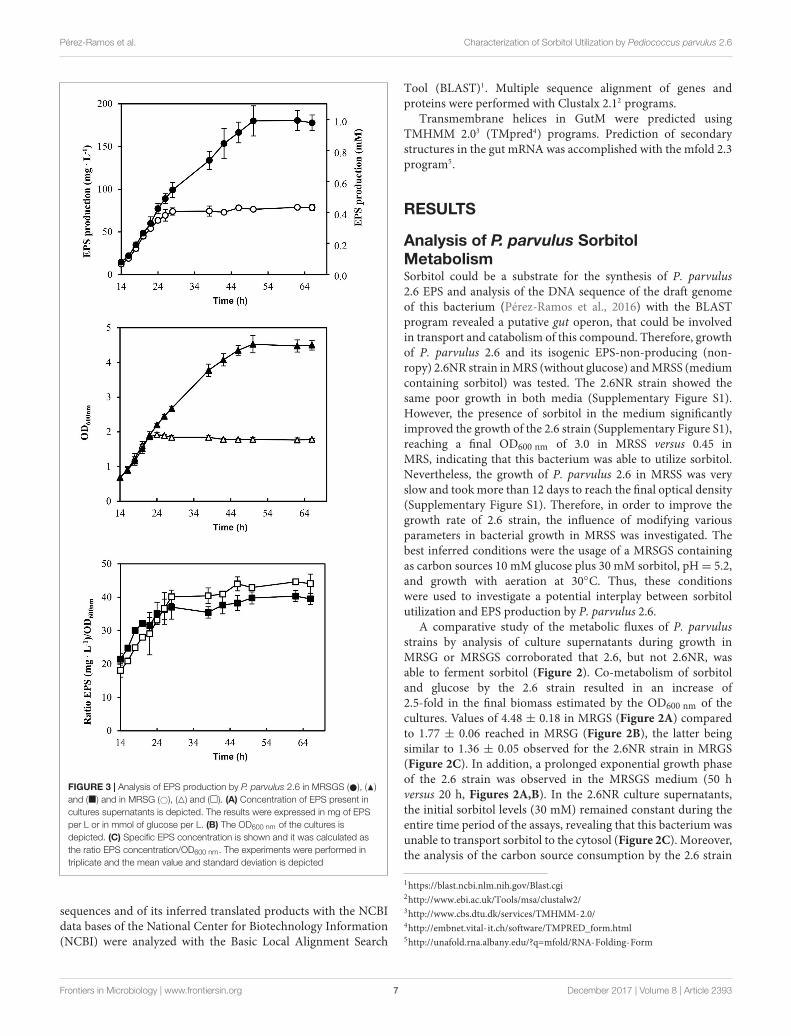
FIGURE 3 | Analysis of EPS production by P. parvulus 2.6 in MRSGS ( ), (N)and (�) and in MRSG (#), (4) and (�). (A) Concentration of EPS present incultures supernatants is depicted. The results were expressed in mg of EPSper L or in mmol of glucose per L. (B) The OD600 nm of the cultures isdepicted. (C) Specific EPS concentration is shown and it was calculated asthe ratio EPS concentration/OD600 nm. The experiments were performed intriplicate and the mean value and standard deviation is depicted
sequences and of its inferred translated products with the NCBIdata bases of the National Center for Biotechnology Information(NCBI) were analyzed with the Basic Local Alignment Search
Tool (BLAST)1. Multiple sequence alignment of genes andproteins were performed with Clustalx 2.12 programs.
Transmembrane helices in GutM were predicted usingTMHMM 2.03 (TMpred4) programs. Prediction of secondarystructures in the gut mRNA was accomplished with the mfold 2.3program5.
RESULTS
Analysis of P. parvulus SorbitolMetabolismSorbitol could be a substrate for the synthesis of P. parvulus2.6 EPS and analysis of the DNA sequence of the draft genomeof this bacterium (Pérez-Ramos et al., 2016) with the BLASTprogram revealed a putative gut operon, that could be involvedin transport and catabolism of this compound. Therefore, growthof P. parvulus 2.6 and its isogenic EPS-non-producing (non-ropy) 2.6NR strain in MRS (without glucose) and MRSS (mediumcontaining sorbitol) was tested. The 2.6NR strain showed thesame poor growth in both media (Supplementary Figure S1).However, the presence of sorbitol in the medium significantlyimproved the growth of the 2.6 strain (Supplementary Figure S1),reaching a final OD600 nm of 3.0 in MRSS versus 0.45 inMRS, indicating that this bacterium was able to utilize sorbitol.Nevertheless, the growth of P. parvulus 2.6 in MRSS was veryslow and took more than 12 days to reach the final optical density(Supplementary Figure S1). Therefore, in order to improve thegrowth rate of 2.6 strain, the influence of modifying variousparameters in bacterial growth in MRSS was investigated. Thebest inferred conditions were the usage of a MRSGS containingas carbon sources 10 mM glucose plus 30 mM sorbitol, pH= 5.2,and growth with aeration at 30◦C. Thus, these conditionswere used to investigate a potential interplay between sorbitolutilization and EPS production by P. parvulus 2.6.
A comparative study of the metabolic fluxes of P. parvulusstrains by analysis of culture supernatants during growth inMRSG or MRSGS corroborated that 2.6, but not 2.6NR, wasable to ferment sorbitol (Figure 2). Co-metabolism of sorbitoland glucose by the 2.6 strain resulted in an increase of2.5-fold in the final biomass estimated by the OD600 nm of thecultures. Values of 4.48 ± 0.18 in MRGS (Figure 2A) comparedto 1.77 ± 0.06 reached in MRSG (Figure 2B), the latter beingsimilar to 1.36 ± 0.05 observed for the 2.6NR strain in MRGS(Figure 2C). In addition, a prolonged exponential growth phaseof the 2.6 strain was observed in the MRSGS medium (50 hversus 20 h, Figures 2A,B). In the 2.6NR culture supernatants,the initial sorbitol levels (30 mM) remained constant during theentire time period of the assays, revealing that this bacterium wasunable to transport sorbitol to the cytosol (Figure 2C). Moreover,the analysis of the carbon source consumption by the 2.6 strain
1https://blast.ncbi.nlm.nih.gov/Blast.cgi2http://www.ebi.ac.uk/Tools/msa/clustalw2/3http://www.cbs.dtu.dk/services/TMHMM-2.0/4http://embnet.vital-it.ch/software/TMPRED_form.html5http://unafold.rna.albany.edu/?q=mfold/RNA-Folding-Form
Frontiers in Microbiology | www.frontiersin.org 7 December 2017 | Volume 8 | Article 2393

fmicb-08-02393 December 2, 2017 Time: 15:55 # 8
Pérez-Ramos et al. Characterization of Sorbitol Utilization by Pediococcus parvulus 2.6
FIGURE 4 | Detection of plasmids of P. parvulus 2.6 and 2.6NR strains.Plasmids preparations of P. parvulus strains and of E. coli V517 were analyzedin a 0.7% agarose gel. (A) The image of the gel. The three samples were runin the same gel. In (B) the calibration curve for plasmid size determination isdepicted. Symbols: plasmids from E. coli V517 (�), plasmids from P. parvulusstrains (�).
showed that glucose started to be transported to the cytosol after2 h of growth, and upon 26 h of incubation the monosaccharidewas undetectable in the culture supernatants (Figures 2A,B).Furthermore, only after 20 h of incubation did the 2.6 strainstart to internalize the sorbitol and presumably to metabolize it,because the bacterium did not enter into the stationary phaseuntil the sorbitol was consumed (Figure 2A). The metabolicactivity of the two strains was monitored by detecting the lacticacid production, since it is the main metabolic end-productbecause pediococci are homofermentative bacteria. The resultsshowed that the 2.6 strain grown in MRSG (Figure 2B) and the
2.6NR strain grown in MRSGS (Figure 2C) released to the culturemedia similar amounts of lactic acid, the maximum levels being18.45± 0.45 mM and 20.19± 0.42 mM, respectively. By contrast,the 2.6 strain grown in the presence of both carbon sourcesshowed a higher lactic acid production, up to 76.03 ± 0.43 mM(Figure 2A). Correlating with these results, the final pH of the2.6 cultures in MRSG and of the 2.6NR cultures in MRSGS wassimilar (4.81 ± 0.02 versus 4.78 ± 0.02), and higher than that ofthe 2.6 cultures in MRSGS (4.23± 0.02).
Furthermore, the EPS production by P. parvulus 2.6 in thepresence or absence of sorbitol was investigated. Significant EPSlevels were detected after 14 h of growth in MRSG and MRSGSmedia. Therefore, the data depicted in Figure 3 correspond tothose obtained within the 14–62 h incubation period. The resultsrevealed that the bacterium produced EPS during the growthin MRSGS and synthesized higher levels of the polymer in thismedium than in MRSG (Figure 3A). Thus, after 62 h of growthin MRSG, the 2.6 strain produced 78.6 ± 3.7 mg L−1 of EPS,while in MRSGS synthesized 180.5 ± 11.8 mg L−1. Additionally,in order to evaluate the specific efficiency of the EPS productiondepending on the carbon source used, the ratio between EPSconcentration and the biomass estimated from the OD600 nm(Figure 3B) was calculated (Figure 3C). The results showedthat irrespectively of the carbon source, the bacteria had almostidentical efficiency of EPS production, which increased duringthe exponential and stationary phases of growth (Figure 3C).
Determination of Genomic Location ofthe gut OperonP. parvulus 2.6 probably carries three natural plasmids, whichwere previously named pPP1, pPP2 and pPP3 (Werning et al.,2006), and we have identified only three plasmid replicationmachineries in the P. parvulus draft genome (Pérez-Ramos et al.,2016). In addition, the P. parvulus 2.6 EPS is synthesized bythe GTF glycosyltransferase encoded by the gtf gene, which islocated in the pPP2 plasmid (Werning et al., 2006). Thus, the2.6NR strain was generated from 2.6 by pPP2 plasmid curing aftertreatment with the DNA intercalating agent ethidium bromideand the gyrase inhibitor novobiocin (Fernández et al., 1995).
Consequently, given that 2.6NR does not utilize sorbitol, itwas feasible that the gut operon was encoded by pPP2 andthis hypothesis was investigated. First, total plasmidic DNApreparations of the two Pediococcus strains were purified byfractionation in a CsCl gradient to eliminate non-supercoiled(open circles and linear) forms of the plasmids. Then, the purifiedplasmidic DNA preparations were analyzed in an agarose gel(Figure 4). Four and three bands were detected, respectively,in preparations of the 2.6 and 2.6NR strains. The sizes of thebands were inferred from their migration using a calibrationcurve (Figure 4B) generated with the plasmids of the E. coli V517strain and are shown in Figure 4A. Two of the bands apparentlywere shared by 2.6 and 2.6NR, and were initially ascribed tothe monomeric forms of pPP1 (39.1 kpb in 2.6 and 40.0 kpb in2.6NR) and pPP3 (12.7 kpb). As expected, pPP2 (24.5 kpb) wasnot detected in 2.6NR DNA preparations. Moreover, we couldnot ascribe to any plasmid the band with less mobility and a
Frontiers in Microbiology | www.frontiersin.org 8 December 2017 | Volume 8 | Article 2393

fmicb-08-02393 December 2, 2017 Time: 15:55 # 9
Pérez-Ramos et al. Characterization of Sorbitol Utilization by Pediococcus parvulus 2.6
FIGURE 5 | Detection of the P. parvulus 2.6 gut operon by Southern blot hybridization. Plasmids preparations of the P. parvulus strains were fractionated byelectrophoresis in agarose gel, transferred to membranes and hybridized for detection of gutF, gutR and gutB with the probes 1, 2, and 3, respectively. (A) Physicalmap of the gut operon, including the putative promoter of the gut operon (Pgut ), the operon genes (gutF, gutR, gutM gutC, gutB and gutA), a secondary structureincluding the 3′-end of gutR and the 5′-end of gutM (coordinates 3483-3512 in GenBank accession No MF766019) as well as a transcriptional terminator(coordinates 6103-6027 in GenBank accession No MF766019) downstream of gutA. In addition, location of probes 1, 2, and 3 is indicated. (B) Images of thecorresponding gels and hybridized membranes. The double headed arrows indicate the position of the hybridized bands in the corresponding gel.
theoretical molecular weight of 56.8 kbp that was present in DNApreparations of both strains. Quantification of the bands fromagarose gels (Figures 5, 6) revealed different proportions of theplasmidic forms in 2.6 (0.3:5.9:2.5:1.0) and 2.6NR (0.8:1.0:0.0:1.0)samples.
The gut operon of P. parvulus 2.6 (Figures 5A, 6A) iscomposed of six genes, of which gutF encodes a sorbitol-6-phosphate dehydrogenase; gutRM encodes two putativeregulators; and gutCBA encodes the proteins EIIC, EIIBCand EIIA which are components of a phosphoenolpyruvate-dependent sorbitol phosphotransferase system (PTSgut). Thus, todetect the location of the gut operon, Southern blot hybridizationof total plasmidic DNA preparations was performed using as aprobe internal regions of gutF, gutR or gutB. One hybridizationsignal was observed with the three probes at the position ofthe 39.1 kb pPP1 plasmid in the 2.6 DNA sample (Figure 5B).Surprisingly, this plasmid was apparently present in bothP. parvulus strains, but in the 2.6NR DNA sample no signalwas observed. Nevertheless, the results demonstrated that thegut operon was not located in the pPP2 plasmid, but rather wascarried by the pPP1 plasmid of the 2.6 strain and not of the newlydesignated pPP1∗ plasmid of 2.6NR strain.
Analysis of Plasmids pPP1 of P. parvulus2.6 and pPP1∗ of P. parvulus 2.6NRThe results obtained by Southern blot analysis prompted usto obtain further information of pPP1 and pPP1∗ plasmids.Thus, the total plasmidic DNA preparation of the 2.6 strain
was used as a substrate to confirm the sequence of the gutoperon and to determine the unknown nucleotide sequenceof the flanking regions (undetected in the draft genome ofthe bacterium) by the dideoxynucleotide method and with thewalking strategy. The sequence of a DNA segment of 11,746 bp(Figure 6A and GenBank accession No MF766019) was obtainedand its analysis revealed the existence of nine open readingframes (ORF), in addition to the 6 genes (gutFRMCBA) ofthe gut operon (Figure 6A and Supplementary Table S1).One open reading frame was detected upstream of the gutoperon and was designated tnp, since its product has 100%identity with a multispecies transposase (Genbank accessionNo WP_003606336.1) widely distributed in the Lactobacillaceaefamily. Downstream of the gut operon were detected fourORF named orf1, orf2, orf3 and orf4, which could encodehypothetical proteins conserved in other LAB. In addition,the product of the named res gene belongs to the Ser-recombinase superfamily (cl02788) and specifically to the PinEconserved protein domain family (COG1961), showing morethan 90% amino acid identity with proteins from oenococci,lactobacilli and pediococci annotated as Pin-related site-specificrecombinases/DNA invertases. Also, two divergent genes namedtauE and tetR seem to encode a TauE sulfite exporter whichbelongs to the TauE conserved domain family (pfam01925) and atranscriptional regulator belonging to the TetR family (domainarchitecture ID 11442015), and both proteins have more than95% amino acid identity with their homologues in Oenoccocusoeni and lactobacilli.
Frontiers in Microbiology | www.frontiersin.org 9 December 2017 | Volume 8 | Article 2393

fmicb-08-02393 December 2, 2017 Time: 15:55 # 10
Pérez-Ramos et al. Characterization of Sorbitol Utilization by Pediococcus parvulus 2.6
FIGURE 6 | The sorbitol operon and flanking DNA regions of P. parvulus 2.6. (A) Sequenced regions of pPP1 and pPP1∗ plasmids are shown. Genes and ORF aredepicted and homologies at the nucleotide level are indicated (see details in the text). ? Indicates unknown sequence. Homology of regulatory genetic regions (B)and proteins (C) involved in sorbitol utilization between P. parvulus 2.6, L. plantarum 90 and L. casei BL23 is shown. (B) DNA sequences of: the putative Pgut
promoters (–10 boxes) and the GutR operator of L. casei are underlined; the putative cre sequences for catabolic repression are overlined; also the start codon (ATG)of gutF gene and the nucleotides between this and the –10 boxes are depicted. Red letters indicate mismatches with the P. parvulus 2.6 DNA sequence. ∗Meansidentity of nucleotides. (C) Identity of the amino acid sequence of the Gut proteins from L. plantarum 90 and L. casei BL23 with those of P. parvulus 2.6 is depicted.
Based on the DNA sequence of the gut operon flanking regionsin pPP1, and on the lack of the gut operon in 2.6NR, primerswere designed and used to try to detect if there exists anyidentity between pPP1 and pPP1∗ by DNA sequencing. Twoof these, pPP1∗F and pPP1∗R, located respectively upstreamand downstream of the gut operon, provided the desiredinformation (Figure 6A). A good chromatogram of the DNAsequencing of pPP1∗ using the 2.6NR plasmidic preparationand pPP1∗F primer with 100% identity with pPP1 was obtaineduntil nucleotide 156 in the chromatogram (548 nt in Genbankaccession No WP_003606336.1), then at least two overlappingsequences were observed (Supplementary Figure S2A), andit was not possible from this point to deduce a furthercorrect DNA sequence. This was not the case when DNAfrom the 2.6 strain was used as substrate, since a goodchromatogram of the pPP1 DNA sequencing was obtained(Supplementary Figure S2B). However, the usage of pPP1∗Rallowed not only to determine that the homology betweenpPP1 and pPP1∗ starts again at nucleotide 10,021 (in Genbankaccession No WP_003606336.1), but also that upstream ofthis position in pPP1∗ there exists a region including a uvrXputative gene identical to those of other pediococci (i.e., inpPC892-2 plasmid, Genbank accession No CP021472.1) andLactobacilli (i.e., in pH10 plasmid, Genbank accession No
CP002430.1) plasmids, which do not carry orf2, orf3 andorf4.
With regard to the gut operon of P. parvulus 2.6, theidentity of the region including the genes and the upstreamregulatory regions with the homologues of L. plantarum strainswas 99% (Figures 6A,B) and nucleotides from 694-to 60020in GenBank accession No MF766019), consequently the aminoacid sequence of the Gut proteins of P. parvulus showed anidentity ranging from 95 to 100%, with those of L. plantarum 90(Figure 6C). No significant homology at the DNA sequence levelwas detected between the characterized operons of L. casei andthose of P. parvulus (Figure 6B and results not shown). However,presumably due to convergent evolution, homology ranging from68 to 24% amino acid identity was detected between the Gutproteins of P. parvulus 2.6 and of L. casei BL23 (Figure 6C).
Analysis of the Gut Operon RegulationGutR and GutM of P. parvulus could be involved in regulationof the gut operon expression and upstream of the start codon ofP. parvulus 2.6 gutF gene, a TATAtT sequence was detected thatonly deviates one nucleotide from the consensus −10 promoterregion (Figure 6B). Thus, to gain insight into this potentialregulation, complementation studies in heterologous LAB hostsable to utilize sorbitol were carried out. First, we cloned
Frontiers in Microbiology | www.frontiersin.org 10 December 2017 | Volume 8 | Article 2393

fmicb-08-02393 December 2, 2017 Time: 15:55 # 11
Pérez-Ramos et al. Characterization of Sorbitol Utilization by Pediococcus parvulus 2.6
FIGURE 7 | Detection of fluorescence in L. casei BL23 strains at stationary phase. Cultures of the indicated strains in MRSS (sorbitol) or MRSG (glucose) wereanalyzed by phase contrast (left panels) or fluorescence (right panels) microscopy.
independently the putative promoter sequence (designated Pgut)and its upstream region (Figure 6B), as well as the transcriptionalfusions Pgut-gutR, Pgut-gutM and Pgut-gutRM into the pRCRpromoter probe vector (Mohedano et al., 2015) upstream of themrfp, generating the pRCR16, pRCR17, pRCR18 and pRCR19plasmids, respectively (Figure 1). Thus, functionality of thepromoter and influence of GutR and GutM could be detectedby measuring the levels of fluorescence of the mCherry encodedby the mrfp gene. As hosts to perform the studies, we chose: (i)the plasmid free L. casei BL23, because its sorbitol utilizationand the regulation of its gut operon is known (Yebra and Pérez-Martínez, 2002; Nissen et al., 2005; Alcantara et al., 2008) and,(ii) L. plantarum 90, because we have previously detected inthis bacterium efficient functional expression of mCherry froma pRCR derivative, without problems of plasmid incompatibilityand that the copy number of the plasmid was 62 ± 2 moleculesper bacterial genome (Russo et al., 2015).
The well characterized transcriptional activator GutR ofL. casei BL23 controls expression of the gut operon of this bacteriaand its operator site upstream of the Pgut has been identified as
well as a catabolite repression element (cre) overlapping the −10region of the promoter (Alcantara et al., 2008) (Figure 6B). TheP. parvulus 2.6 GutR has only a low homology of amino acids(24%) with its homologue of L. casei, but like its counterpartbelongs to the BglG transcriptional antiterminators family,possesses the PRD domain and the DNA helix turn helix bindingdomain. Therefore, both proteins could have a similar role.Alignment of the L. casei and P. parvulus -10 regions revealedthat the upstream regulatory regions of BL23 strain has no clearhomologs in the 2.6 strain (Figure 6B). Consequently, crosstalk between transcriptional signals of P. parvulus and L. caseiregulators should not take place, and influence of the pediococcalGutR and GutM in expression of Pgut from the 2.6 straincould be investigated in the BL23 strain without interferences.Thus, the pRCR derivatives were transferred independently tothe BL23 strain and the recombinant bacteria were grown inMRS supplemented with either 1% glucose or 1% sorbitol untilstationary phase prior to analysis. Examination of the culturesby fluorescent and phase contrast optical microscopy revealedthat only bacteria carrying pRCR17 and pRCR19 and grown in
Frontiers in Microbiology | www.frontiersin.org 11 December 2017 | Volume 8 | Article 2393

fmicb-08-02393 December 2, 2017 Time: 15:55 # 12
Pérez-Ramos et al. Characterization of Sorbitol Utilization by Pediococcus parvulus 2.6
TABLE 3 | Heterologous expression of components of the P. parvulus 2.6 gutoperon in L. casei BL23 carrying pRCR derivatives plasmids grown in either MRSSor MRSG.
L. casei strains DNA insert inpRCR derivatives
Specific fluorescencea
MRSS MRSG
BL23[pRCR16] Pgut 0.14 ± 0.07 0.14 ± 0.01
BL23[pRCR17] Pgut-gutR 5.69 ± 0.44 0.07 ± 0.09
BL23[pRCR18] Pgut-gutM 0.19 ± 0.06 0.11 ± 0.02
BL23[pRCR19] Pgut-gutRM 3.58 ± 0.06 0.05 ± 0.02
aThe specific fluorescence is depicted and it was calculated as the ratio of thedetected fluorescence (5×) and the bacterial biomass estimated from the OD600 nmof the culture.
presence of sorbitol have fluorescence (Figure 7). In addition,fluorescence as well as the optical density of the cultures wasmeasured and the specific fluorescence, referred to the biomass,was calculated. The fluorescence quantification confirmed thatthe Pgut-gutRmrfp, and Pgut-gutRMmrfp transcriptional fusionsare activated upon growth in the presence of sorbitol (Table 3).Thus, these results revealed that expression from the Pgutrequired the activation by GutR and the presence of sorbitol inthe growth medium. Moreover, they indicated that activation byGutR decreased, when GutM was present (5.69 ± 0.44 versus3.58± 0.06).
Concerning the L. plantarum 90 host, its GutR has 98%homology to that of P. parvulus 2.6 (Figure 6C) and theDNA sequence of the region located upstream of the twoPgut promoters only differs in one nucleotide (Figure 6B).Consequently, both operons must have the same regulatorygene system, which implies that both systems could recognizeeach other. Thus, a trans-complementation process was expectedbetween the regulatory proteins of Lp90 and the promoterregion of 2.6. Therefore, the pRCR derivatives were transferredindependently to the 90 strain and, since a cross talk is morecomplex situation, its comprehension required a more detailedanalysis. For this reason, the recombinant bacteria, after growthin MRS supplemented with 1% glucose, were transferred toMRS fresh medium supplemented with either 1% sorbitol or1% glucose and a time course assay of fluorescence and growthof the cultures was performed. The results revealed that allrecombinant strains became fluorescent, when grown in thepresence of sorbitol and, with the time of incubation thefluorescence increased (Figure 8 and Table 4). In addition,analysis of the bacterial growth showed that all cultures inMRSG have very similar exponential growth rates (rangingfrom 0.889 ± 0.059 to 0.803 ± 0.049) and all entered slowlyinto stationary phase after 2 h of incubation (Figure 8F andTable 4). Initial transfer of the cultures to MRSS resulted ina similar decrease (around 50%) of the growth rate (valuesfrom 0.416 ± 0.045 to 0.495 ± 0.011) during the first 2 h ofinduction. Then, probably after consumption of the residualintracellular glucose or due to the induction process, bacteriadecreased their growth rate to levels ranging from 0.259 ± 0.020to 0.251± 0.034, besides the 90[pRCR18] (GutM overexpressor),that after stalling its growth from 2 h to 3 h incubation time
decreased its growth rate to 0.239 ± 0.048, indicating thatoverexpression of GutM in absence of high levels of GutR has anegative impact for the cells. Furthermore, analysis of the specificlevels of fluorescence of the cultures referred to their biomass(Table 4) showed different levels for the different fusions (Pgut-mrfp < Pgut-gutRMmrfp < Pgut-gutRmrfp < Pgut-gutMmrfp),showing that overexpression of GutM provokes the highestinduction of expression from Pgut . In addition, the highest levelswere observed after 4 h of induction for cells carrying eitherpRCR17 (22.38± 2.02) or pRCR19 (19.26± 2.10) versus the endof the incubation (6 h) for cells carrying pRCR16 (14.81 ± 0.66)and pRCR18 (26.83± 1.83).
Thus, the results revealed a trans-complementation ofthe L. plantarum regulatory proteins on expression driven fromthe P. parvulus Pgut promoter. Moreover, the results confirmedthe role of inducer of GutR as well as requirement of sorbitol forexpression from Pgut and support that co-expression of GutR andGutM decrease the activation mediated by GutR.
DISCUSSION
The overall metabolic results obtained here support thatP. parvulus is able to synthesize EPS in MRS mediumusing either glucose or sorbitol as carbon sources. We havepreviously demonstrated that the 2-substituted (1,3)-β-D-glucanof P. parvulus 2.6 is synthesized by the GTF glycosyltransferaseutilizing UDP-glucose as substrate (Werning et al., 2014). Inaddition, Velasco et al. (2007) determined that the 2.6 straintransport the glucose by a PMF-permease and possesses theα-phosphoglucomutase and the UDP-glucose pyrophosphorylaseactivities responsible for the conversion of glucose-6-P toglucose-1-P and further conversion of this compound to UDP-glucose. Thus, Velasco et al. (2007) showed how the 2.6 strainuses the glucose, not only for the central metabolism, viathe glycolytic pathway, but also for the secondary metabolisminvolving a biosynthetic pathway for its EPS synthesis. Inaddition, the detection of the genetic determinants of sorbitolutilization by the 2.6 strain obtained in this work supportsthat the bacterium transports sorbitol by a PTSgut system andconverts sorbitol-6-P into fructose-6-P by the action of sorbitol-6-P dehydrogenase. Fructose-6-P can be converted to glucose-6-Pby a reaction catalyzed by phosphoglucose isomerase, enzymaticactivity that was also previously detected in the 2.6 strain(Velasco et al., 2007). Therefore, the 2.6 strain possesses thetransport and enzymatic machineries for synthesis of the EPSfrom sorbitol. In addition, we have detected that aeration ofthe cultures during the growth improves sorbitol consumption(results not shown). Accordingly, the conversion of sorbitol-6-P into fructose-6-P requires NAD+ as an oxidative co-factorto produce NADH (Zarour et al., 2017). Analysis of the draftgenome of the 2.6 strain showed the existence of a putativeNADH oxidase coding gene. If this enzyme exists, it couldunbalance the NAD+/NADH equilibrium toward the oxidizedform NAD+.
The P. parvulus 2.6 2-substituted (1,3)-β-D-glucan iscomposed of molecules of glucose and consequently the
Frontiers in Microbiology | www.frontiersin.org 12 December 2017 | Volume 8 | Article 2393

fmicb-08-02393 December 2, 2017 Time: 15:55 # 13
Pérez-Ramos et al. Characterization of Sorbitol Utilization by Pediococcus parvulus 2.6
FIGURE 8 | Trans complementation of the sorbitol utilization regulatory machinery in L. plantarum 90. Bacteria carrying (A,E,F) pRCR16 (�,�), (B,E,F) pRCR17 (#, ), (C,E,F) pRCR18 (♦, �) or (D,E,F) pRCR19 (4, N) were grown in MRSG and at time 0 were transferred to fresh MRSS (open symbols) or to MRSG (closedsymbols). OD600nm and fluorescence of the cultures were monitored every hour. Overlays of images of the cultures taken at time 0 and after 2, 4, and 6 h incubationby phase contrast and fluorescent microscopy are depicted. The experiments were performed in triplicate and the mean value and standard deviation is depicted.
Frontiers in Microbiology | www.frontiersin.org 13 December 2017 | Volume 8 | Article 2393

fmicb-08-02393 December 2, 2017 Time: 15:55 # 14
Pérez-Ramos et al. Characterization of Sorbitol Utilization by Pediococcus parvulus 2.6
TAB
LE4
|Het
erol
ogou
sex
pres
sion
ofco
mpo
nent
sof
P.pa
rvul
us2.
6gu
tope
ron
inL.
plan
taru
m90
carr
ying
pCR
Cde
rivat
ives
plas
mid
s.
L.p
lan
taru
mst
rain
sD
NA
inse
rtin
pR
CR
der
ivat
ives
Med
ium
Init
ialg
row
thra
tea
(µ)
So
rbit
oli
nduc
edg
row
thra
teb
(µ)
Sp
ecifi
cfl
uore
scen
cec
2h
3h
4h
5h
6h
90[p
RC
R16
]P
gut
MR
SS
0.41
6±
0.04
50.
251±
0.03
41.
48±
1.03
5.86±
1.43
10.4
2±
1.56
13.4
4±
0.51
14.8
1±
0.66
90[p
RC
R17
]P
gut-
gutR
MR
SS
0.45
5±
0.06
50.
259±
0.02
04.
42±
1.22
13.4
1±
0.90
22.3
8±
2.02
20.3
0±
1.62
17.2
8±
1.65
90[p
RC
R18
]P
gut-
gutM
MR
SS
0.42
9±
0.02
50.
239±
0.04
81.
62±
0.69
10.2
6±
2.30
18.0
5±
2.85
24.2
6±
1.51
26.8
3±
1.83
90[p
RC
R19
]P
gut-
gutR
MM
RS
S0.
495±
0.01
10.
256±
0.01
62.
80±
0.77
11.7
4±
1.51
19.2
6±
2.10
17.2
0±
1.71
15.9
8±
1.21
90[p
RC
R16
]P
gut
MR
SG
0.88
9±
0.05
9N
D0.
06±
0.03
0.04±
0.00
0.04±
0.00
0.04±
0.01
0.04±
0.01
90[p
RC
R17
]P
gut-
gutR
MR
SG
0.82
5±
0.02
9N
D0.
07±
0.06
0.06±
0.02
0.04±
0.01
0.03±
0.00
0.03±
0.02
90[p
RC
R18
]P
gut-
gutM
MR
SG
0.80
3±
0.04
9N
D0.
06±
0.03
0.03±
0.03
0.03±
0.01
0.04±
0.01
0.03±
0.01
90[p
RC
R19
]P
gut-
gutR
MM
RS
G0.
832±
0.03
4N
D0.
04±
0.02
0.03±
0.02
0.03±
0.01
0.04±
0.02
0.03±
0.02
aG
row
thra
teof
the
cultu
res
grow
nin
MR
SS
was
calc
ulat
edfro
mth
eO
D60
0nm
obta
ined
durin
gth
efir
st2
hof
indu
ctio
n.bG
row
thra
teof
the
cultu
res
grow
nin
MR
SS
was
calc
ulat
edfro
mth
eO
D60
0nm
obta
ined
durin
gth
e2–
6h
ofin
duct
ion,
besi
des
for
90[p
RC
R18
],th
atdu
eto
the
stal
ling
ofgr
owth
from
2h
to3
hof
incu
batio
nth
egr
owth
rate
was
calc
ulat
edfro
mth
eda
taob
tain
edfro
m3
hto
6h
ofin
cuba
tion.
ND
,the
grow
thra
tew
asno
tdet
erm
ined
beca
use
the
cultu
res
have
ente
red
inth
est
atio
nary
phas
eof
grow
th.c
Spe
cific
fluor
esce
nce
was
calc
ulat
edas
the
ratio
ofth
ede
tect
edflu
ores
cenc
e(5
x)an
dth
eba
cter
ialb
iom
ass
estim
ated
from
the
OD
600
nmof
the
cultu
re.
EPS concentration can be calculated as molarity of thismonosaccharide (see secondary Y axis in Figure 3A). Thiscalculation revealed that in both media this bacterium onlyused a small percentage of the substrate molecules (10 mMglucose plus 30 mM sorbitol in MRSGS or 10 mM glucose inMRSG) for synthesis of EPS (0.99 or 0.45 mM, respectively),whereas more than 90% was utilized in the glycolytic pathway tosynthesize pyruvic acid (2 molecules per 1 molecule of substrate)and by action of the lactate dehydrogenase to finally generatelactic acid (1 molecule per 1 molecule of pyruvate, 79 mMor 18 mM). Moreover, the specific quantification method for2-substituted (1,3)-β-D-glucan used here and the estimationof the specific concentration of EPS synthesized (Figure 3C)showed that, using as substrate either glucose or sorbitol, thebacterium synthesizes the same polymer and suggests that withthe same efficiency. This was not the case when synthesis ofthis EPS utilizing fructose was tested, since levels were lowcompared with that obtained from glucose (Velasco et al.,2007). We have also observed a temporal delay of 2.6 to startto consume sorbitol in MRSGS (Figure 3A). This could bedue to the existence of a catabolite repression of sorbitolutilization by glucose. Supporting this hypothesis, we havedetected a potential cre operator (Figure 6) for the CcpA, whichmediates with HPR this regulation in firmicutes (Deutscher,2008).
In P. parvulus 2.6, the gut operon, as in other LAB, constitutesthe genetic determinant for sorbitol transport and conversioninto fructose-6-P. In addition, we have established here thatit is located in a plasmid named pPP1 (Figure 6) which isunusual, since the almost identical operon of L. plantarum andthat of Lactobacillus pentosus strain SLC13 (82% homologous,Genbank accession No CP022130.1) as well as the unrelatedone from L. casei are located in the chromosome. As faras we know, only the location of an unrelated gut operonin the megaplasmid pMP118 from L. salivarius UCC118 hasbeen previously described (Claesson et al., 2006). A search ofthe protein data banks revealed that L. salivarius 5713 andJCM1046 strains possess, respectively, the pHN3 and pMP1046Amegaplasmids which carry gut operons homologous to that ofpMP118 (Jiménez et al., 2010; Raftis et al., 2014). Flankingthe operon two inverted repeat sequences (nucleotides 604-627 and 6612-6635 of Genbank accession No WP_003606336.1)were identified, which are also present at the same relativelocation in L. plantarum strains and at various locations inlactobacilli chromosomes and plasmids (even more than onecopy per genome). The upstream region is preceded by a tnp geneencoding a putative transposase, which could be responsible formobilization of the gut operon from plasmid to chromosome orvice versa.
Lactic acid bacteria are prone to carry more than onecompatible plasmid and this facilitates exchange of differentregions with physiological significance, that later on canbe transferred to other bacteria by plasmid conjugation ormobilization (Cui et al., 2015). Thus, downstream of the gutoperon of pPP1 there are DNA regions almost identical to thatpresent in plasmids of lactobacilli, which along with P. parvuluscan be contaminants of alcoholic beverages. Furthermore, the
Frontiers in Microbiology | www.frontiersin.org 14 December 2017 | Volume 8 | Article 2393

fmicb-08-02393 December 2, 2017 Time: 15:55 # 15
Pérez-Ramos et al. Characterization of Sorbitol Utilization by Pediococcus parvulus 2.6
FIGURE 9 | Predicted folding of the gut mRNA region including the 3′-end ofgutR and the 5′-end of gutM. (A) Secondary structure of the wild-typeP. parvulus 2.6 and L. plantarum 90 gut operons. The arrow and the Gindicate that in the insert of pRCR18 G substitute to the A in the indicatedposition. (B) Secondary structure of the L. casei BL23 gut operon.
Oenococcus oeni pOENI-1 and pOENI-1v2 plasmids (Favieret al., 2012) and pPP1 carry a region containing amongothers the res, tauE and tetR genes. The putative TauE sulfiteexporter is possibly involved in adaptation to stress conditionsduring alcoholic beverage production (Favier et al., 2012).Thus, the recombinase or invertase site specific Res could beresponsible for a mobilization of an element composed of atruncated res, tauE and tetR to a stable location, since atthe 3′-end region of res and downstream of tetR unit existinverted complementary sequences 5′-TTTTAAAGC-3′ and 5′-GCTTTAAAA-3′ (nucleotides 7774–7778 and 10021–10029 ofGenbank accession No WP_003606336.1).
Another instance of plasmids rearrangement in P. parvulus isthat which generated the profile and DNA sequence of 2.6NRstrain plasmids (Figure 6). The initial isolate of 2.6NR straingenerated in the Basque country University (BCU, Spain) anddescribed in Fernández et al. (1995), kindly provided by Dr.Maria Teresa Dueñas (BCU) was studied in this work. Thus, thechanges in plasmid cassettes were not produced in our laboratory,and presumably they took place upon treatment of 2.6 strain withethidium bromide and novobiocin and was selected for the lossof the ropy phenotype. Thus, it is feasible that a formation ofa co-integrate of pPP1 with other plasmid, may be pPP2, tookplace and convergent replication from two origins prompted to adeletion of one of the replicons, may be the pPP2, since its losswas envisaged, to generate pPP1∗.
Concerning the regulation of expression of the gut operon,the overall results showed that it is repressed in the absence
of sorbitol in the growth medium and that the P. parvulusGutR is an activator like the L. casei BL23 regulator (Alcantaraet al., 2008). In this system, it has been proposed that GutMis involved in the activation, since a decreased expressionof the gut operon was detected in a GutM deficient mutant(Alcantara et al., 2008). Furthermore, the complementationstudies in L. plantarum 90 performed here showed a heterologousregulation of gene expression from the pediococcal Pgut promoterby the GutR from Lactobacillus, and a positive effect whenonly the pediococcal GutM was overexpressed (Figure 8 andTable 4). Thus, these results suggest that a protein-proteininteraction between the P. parvulus GutM and the L. plantarumGutR could potentiate the activation of the Pgut promoter, since,P. parvulus 2.6 GutR and GutM have 98% identity with theirhomologues of L. plantarum 90. In addition, either in L. caseiBL23 and in L. plantarum a decrease of expression from Pgut wasobserved when GutM was overexpressed in combination withGutR (Figure 8 and Tables 3, 4). This prompted us to analyzethe genetic environment of gutR and gutM. An overlapping ofthe last nucleotide of the termination codon (TAA) of gutRand the first nucleotide of the start codon (ATG) of gutM wasdetected in P. parvulus 2.6 and L. plantarum 90 genomes. Thisindicated that post-transcriptional regulation of the gut operoncould exist in this bacterium. For this reason, the secondarystructure of the region surrounding the overlapping in thegut mRNA was folded with the Mfol program (Figure 9A).A secondary stem-loop structure was predicted with a 1G =−5.6 kcal mol−1, the ribosomal binding site (RBS) of gutM(5′-GGAGG-3′) was located at the loop and partially blockedin the stem of the structure. Thus, even though the sequenceof the RBS of gutM indicates a high efficiency of utilizationfor the ribosome, the initiation of translation of gutM could bepartially impaired by the partial RBS blockage, which would bereleased by the opening provoked by the passage of the ribosomestranslating gutR. In addition, the overlapping of gutR and gutMis located at the end of the stem of the structure. Thus, twopost-transcriptional regulations could take place: (i) translationof gutR can act by favoring translation of gutM by expositionof its RBS and (ii) a -1 frameshift (Atkins et al., 2016) couldhappen at the TAA termination codon of gutR and ribosomestranslating this could step back one nucleotide and upon chargingthe corresponding tRNA read the Leu (TTA) codon and continuetranslating gutM. In this way a fused peptide GutR-M couldbe synthesized. The same structure could be formed in thetranscript encoded by the plasmid pRCR19 with a 1G = −5.9kcal mol−1, containing gutRM and which could be a substrate forthe two proposed post-transcriptional regulations. Furthermore,the DNA fragment cloned in pRCR18, lacks most of the gutRgene but still retains some of the 3′-end region of this geneand the encoded mRNA can form a secondary structure almostidentical to the wild-type structure (with only a change of A-U byG-U pairing at the end of the stem, Figure 9). Thus in bacteriacarrying pRCR18 partial blockage of the RBS could take place,but synthesis of GutR-M could not occur. This could explainthe antagonistic effect of overexpression of gutM from pRCR18(increase of expression from Pgut) and pRCR19 (decrease ofexpression from Pgut), if GutR-M exists and has a role.
Frontiers in Microbiology | www.frontiersin.org 15 December 2017 | Volume 8 | Article 2393

fmicb-08-02393 December 2, 2017 Time: 15:55 # 16
Pérez-Ramos et al. Characterization of Sorbitol Utilization by Pediococcus parvulus 2.6
Prediction of transmembrane regions in the regulatoryproteins with the TM-Pred revealed that GutR is a soluble proteinand that the first amino acids from 1 to 21 of GutM constituteda transmembrane region also predicted for the GutR-M fusedpolypeptide. This fused polypeptide could provide an efficientanchoring of the regulator to the membrane bringing it closeto the PTSgut system facilitating the phosphorylation of GutRand resulting in the physiological optimal expression of theoperon. This generation of a fused polypeptide could also takeplace in L. plantarum but does not seems to occur in L. casei,since in this bacterium the TAA translational stop codon ofGutR and the ATG start codon of GutM are adjacent and notoverlapped (Figure 9). However, the L. casei gut transcript canform a secondary structure with a 1G = −9.8 kcal mol−1
which could block the RBS of gutM gene, couple translation ofGutR and GutM could take place, and protein-protein interactioncould be responsible for higher activation of the system at thebeginning of the induction process. Our results indicate thathigh levels of GutM synthesized from a multicopy plasmid havea deleterious effect for the bacteria (Figure 8) and probablythe proposed models of posttranscriptional regulation aredesigned to have the right concentration of regulatory proteins.Nevertheless, further experiments are required to pinpoint therole of GutM and of the putative GutR-M polypeptide ofP. parvulus.
AUTHOR CONTRIBUTIONS
AP-R contributed to all parts of the experimental work and wrotea draft of the manuscript. MW performed the initial detection
of sorbitol utilization and characterization of gut genes. APcontributed to the characterization of the sorbitol metabolism.PR participated in the elaboration of the manuscript and analysisof the DNA sequences. GS contributed to the design andanalysis of the experimental work involving characterization ofregulation of gut operon expression. MM contributed to thedesign of strategies to determine trans complementation of thegut operon and corrected the manuscript. PL participated instudy conception, data interpretation and generated the finalversion of the manuscript. All authors have read and approvedthe final manuscript.
FUNDING
This work was supported by the Spanish Ministry of Economyand Competitiveness (grant AGL2015-65010-C3-1-R).
ACKNOWLEDGMENTS
The authors thank Dr. Stephen W. Elson for critical reading ofthe manuscript and to Dr. Ma Teresa Dueñas for providing theoriginal P. parvulus 2.6NR isolate to perform this study.
SUPPLEMENTARY MATERIAL
The Supplementary Material for this article can be foundonline at: https://www.frontiersin.org/articles/10.3389/fmicb.2017.02393/full#supplementary-material
REFERENCESAlcantara, C., Sarmiento-Rubiano, L. A., Monedero, V., Deutscher, J., Perez-
Martinez, G., and Yebra, M. J. (2008). Regulation of Lactobacillus casei sorbitolutilization genes requires DNA-binding transcriptional activator GutR and theconserved protein GutM.Appl. Environ.Microbiol. 74, 5731–5740. doi: 10.1128/AEM.00230-08
Aldridge, P., Metzger, M., and Geider, K. (1997). Genetics of sorbitol metabolismin Erwinia amylovora and its influence on bacterial virulence. Mol. Gen. Genet.256, 611–619.
Atkins, J. F., Loughran, G., Bhatt, P. R., Firth, A. E., and Baranov, P. V.(2016). Ribosomal frameshifting and transcriptional slippage: from geneticsteganography and cryptography to adventitious use. Nucleic Acids Res. 44,7007–7078. doi: 10.1093/nar/gkw530
Berthier, F., Zagorec, M., Champomier-Vergès, M., Ehrlich, S. D., andMorel-Deville, F. (1996). Efficient transformation of Lactobacillus sake byelectroporation. Microbiology 142, 1273–1279. doi: 10.1099/13500872-142-5-1273
Boyd, D. A., Thevenot, T., Gumbmann, M., Honeyman, A. L., and Hamilton,I. R. (2000). Identification of the operon for the sorbitol (glucitol)phosphoenolpyruvate: sugar phosphotransferase system in Streptococcusmutans. Infect. Immun. 68, 925–930.
Claesson, M. J., Li, Y., Leahy, S., Canchaya, C., van Pijkeren, J. P., Cerdeño-Tárraga, A. M., et al. (2006). Multireplicon genome architecture of Lactobacillussalivarius. Proc. Natl. Acad. Sci. U.S.A. 103, 6718–6723. doi: 10.1073/pnas.0511060103
Cui, Y., Hu, T., Qu, X., Zhang, L., Ding, Z., Dong, A., et al. (2015). Plasmids fromfood lactic acid bacteria: diversity, similarity, and new developments. Int. J. Mol.Sci. 16, 13172–13202.
de Man, J. C., Rogosa, M., and Sharpe, M. E. (1960). A medium for the cultivationof lactobacilli. J. Appl. Microbiol. 23, 130–135. doi: 10.1111/j.1365-2672.1960.tb00188.x
Deutscher, J. (2008). The mechanisms of carbon catabolite repression in bacteria.Curr. Opin. Microbiol. 11, 87–93. doi: 10.1016/j.mib.2008.02.007
Dornan, S., and Collins, M. (1987). High efficiency electroporation of Lactococcuslactis subsp. lactis LM0230. Lett. Appl. Microbiol. 11, 62–64. doi: 10.1111/j.1472-765X.1990.tb01275.x
Dueñas-Chasco, M. T., Rodríguez-Carvajal, M. A., Mateo, P. T., Franco-Rodríguez, G., Espartero, J. L., Irastorza-Iribas, A., et al. (1997). Structuralanalysis of the exopolysaccharide produced by Pediococcus damnosus 2.6.Carbohydr. Res. 303, 453–458. doi: 10.1016/S0008-6215(97)00192-4
Favier, M., Bilhère, E., Lonvaud-Funel, A., Moine, V., and Lucas, P. M.(2012). Identification of pOENI-1 and related plasmids in Oenococcus oenistrains performing the malolactic fermentation in wine. PLOS ONE 7:e49082.doi: 10.1371/journal.pone.0049082
Fernández-Bañares, F., Esteve, M., and Viver, J. M. (2009). Fructose-sorbitolmalabsorption. Curr. Gastroenterol. Rep. 11, 368–374.
Fernández, K., Dueñas, M. T., Irastorza, A., Bilbao, A., and del Campo, G. (1995).Characterization and DNA plasmid analysis of ropy Pediococcus spp. strainsisolated from Basque Country ciders. J. Food Prot. 59, 35–40.
Fernández de Palencia, P., Werning, M. L., Sierra-Filardi, E., Dueñas, M. T.,Irastorza, A., Corbí, A. L., et al. (2009). Probiotic properties of the 2-substituted (1,3)-β-d-Glucan-producing bacterium Pediococcus parvulus 2.6.Appl. Environ. Microbiol. 75, 4887–4891. doi: 10.1128/AEM.00394-09
Immerstrand, T., Paul, C. J., Rosenquist, A., Deraz, S., Mårtensson, O. B.,Ljungh, A., et al. (2010). Characterization of the properties of Pediococcusparvulus for probiotic or protective culture use. J. Food Prot. 73,960–966.
Frontiers in Microbiology | www.frontiersin.org 16 December 2017 | Volume 8 | Article 2393

fmicb-08-02393 December 2, 2017 Time: 15:55 # 17
Pérez-Ramos et al. Characterization of Sorbitol Utilization by Pediococcus parvulus 2.6
Jiménez, E., Martín, R., Maldonado, A., Martín, V., Gómez de Segura, A.,Fernández, L., et al. (2010). Complete genome sequence of Lactobacillussalivarius CECT 5713, a probiotic strain isolated from human milk and infantfeces. J. Bacteriol. 192, 5266–5267. doi: 10.1128/jb.00703-10
Ladero, V., Ramos, A., Wiersma, A., Goffin, P., Schanck, A., Kleerebezem, M., et al.(2007). High-level production of the low-calorie sugar sorbitol by Lactobacillusplantarum through metabolic engineering. Appl. Environ. Microbiol. 73,1864–1872. doi: 10.1128/AEM.02304-06
Lamontanara, A., Caggianiello, G., Orrù, L., Capozzi, V., Michelotti, V., Bayjanov,J. R., et al. (2015). Draft genome sequence of Lactobacillus plantarum Lp90isolated from wine. Genome Announc. 3:e00097-15. doi: 10.1128/genomeA.00097-15
López, P., Martínez, S., Diaz, A., Espinosa, M., and Lacks, S. A. (1989).Characterization of the polA gene of Streptococcus pneumoniae and comparisonof the DNA polymerase I it encodes to homologous enzymes from Escherichiacoli and phage T7. J. Biol. Chem. 264, 4255–4263.
Macrina, F. L., Kopecko, D. J., Jones, K. R., Ayers, D. J., and McCowen, S. M. (1978).A multiple plasmid-containing Escherichia coli strain: convenient source of sizereference plasmid molecules. Plasmid 1, 417–420. doi: 10.1016/0147-619X(78)90056-2
Mazé, A., Boël, G., Zúñiga, M., Bourand, A., Loux, V., Yebra, M. J., et al. (2010).Complete genome sequence of the probiotic Lactobacillus casei strain BL23. J.Bacteriol. 192, 2647–2648. doi: 10.1128/JB.00076-10
Mohedano, M. L., García-Cayuela, T., Pérez-Ramos, A., Gaiser, R., Requena, T.,and López, P. (2015). Construction and validation of a mCherry protein vectorfor promoter analysis in Lactobacillus acidophilus. J. Ind. Microbiol. Biotechnol.42, 247–253. doi: 10.1007/s10295-014-1567-4
Montalto, M., Gallo, A., Ojetti, V., and Gasbarrini, A. (2013). Fructose, trehaloseand sorbitol malabsorption. Eur. Rev. Med. Pharmacol. Sci. 17, 26–29.
Nissen, L., Perez-Martinez, G., and Yebra, M. J. (2005). Sorbitol synthesis byan engineered Lactobacillus casei strain expressing a sorbitol-6-phosphatedehydrogenase gene within the lactose operon. FEMS Microbiol. Lett. 249,177–183. doi: 10.1016/j.femsle.2005.06.010
Notararigo, S., Casas-Engel, M., Fernández de Palencia, P., Corbí, A. L., andLópez, P. (2014). Immunomodulation of human macrophages and myeloid cellsby 2-substituted (1-3)-β-D-glucan from P. Parvulus 2.6. Carbohydr. Polym. 112,109–113. doi: 10.1016/j.carbpol.2014.05.073
Notararigo, S., Nacher-Vazquez, M., Ibarburu, I., Werning, M. L., de Palencia,P. F., Duenas, M. T., et al. (2013). Comparative analysis of production andpurification of homo- and hetero-polysaccharides produced by lactic acidbacteria. Carbohydr. Polym. 93, 57–64. doi: 10.1016/j.carbpol.2012.05.016
Pérez-Ramos, A., Mohedano, M. L., Puertas, A., Lamontanara, A., Orru, L.,Spano, G., et al. (2016). Draft genome sequence of Pediococcus parvulus2.6, a probiotic β-glucan producer strain. Genome Announc. 4:e01381-16.doi: 10.1128/genomeA.01381-16
Raftis, E. J., Forde, B. M., Claesson, M. J., and O’Toole, P. W. (2014). Unusualgenome complexity in Lactobacillus salivarius JCM1046. BMC Genomics15:771. doi: 10.1186/1471-2164-15-771
Rhodes, M. W., and Kator, H. (1999). Sorbitol-fermenting bifidobacteria asindicators of diffuse human faecal pollution in estuarine watersheds. J. Appl.Microbiol. 87, 528–535.
Russo, P., Iturria, I., Mohedano, M. L., Caggianiello, G., Rainieri, S., Fiocco, D.,et al. (2015). Zebrafish gut colonization by mCherry-labelled lactic acid bacteria.Appl. Microbiol. Biotechnol. 99, 3479–3490. doi: 10.1007/s00253-014-6351-x
Russo, P., López, P., Capozzi, V., Férnandez de Palencia, P., Dueñas, M. T.,Spano, G., et al. (2012). Beta-glucans improve growth, viability and colonizationof probiotic microorganisms. Int. J. Mol. Sci. 13, 6026–6039. doi: 10.3390/ijms13056026
Sarmiento-Rubiano, L. A., Zuniga, M., Perez-Martinez, G., and Yebra, M. J.(2007). Dietary supplementation with sorbitol results in selective enrichment oflactobacilli in rat intestine. Res. Microbiol. 158, 694–701. doi: 10.1016/j.resmic.2007.07.007
Silveira, M. M., and Jonas, R. (2002). The biotechnological production of sorbitol.Appl. Microbiol. Biotechnol. 59, 400–408. doi: 10.1007/s00253-002-1046-0
Stülke, J., Arnaud, M., Rapaport, G., and Martin-Verstraete, I. (1998). PRD —a protein domain involved in PTS-dependent induction and carboncatabolite repression of catabolic operons in bacteria. Mol. Microbiol. 28,865–874.
Tangney, M., Brehm, J. K., Minton, N. P., and Mitchell, W. J. (1998). A gene systemfor glucitol transport and metabolism in Clostridium beijerinckii NCIMB 8052.Appl. Environ. Microbiol. 64, 1612–1619.
Velasco, S. E., Areizaga, J., Irastorza, A., Dueñas, M. T., Santamaria, A., andMuñoz, M. E. (2009). Chemical and rheological properties of the β- glucanproduced by Pediococcus parvulus 2.6. J. Agric. Food Chem. 57, 1827–1834.doi: 10.1021/jf803065w
Velasco, S. E., Yebra, M. J., Monedero, V., Ibarburu, I., Dueñas, M. T., andIrastorza, A. (2007). Influence of the carbohydrate source on β-glucanproduction and enzyme activities involved in sugar metabolism in Pediococcusparvulus 2.6. Int. J. Food Microbiol. 115, 325–334. doi: 10.1016/j.ijfoodmicro.2006.12.023
Wegmann, U., O’connell-Motherway, M., Zomer, A., Buist, G., Shearman, C.,Canchaya, C., et al. (2007). Complete genome sequence of the prototype lacticacid bacterium Lactococcus lactis subsp. cremoris MG1363. J. Bacteriol. 189,3256–3270. doi: 10.1128/JB.01768-06
Werning, M. L., Ibarburu, I., Dueñas, M. T., Irastorza, A., Navas, J., and López, P.(2006). Pediococcus parvulus gtf gene encoding the GTF glycosyltransferase andits application for specific PCR detection of beta-D-glucan-producing bacteriain foods and beverages. J. Food Prot. 69, 161–169.
Werning, M. L., Pérez-Ramos, A., Fernández de Palencia, P., Mohedano, M. L.,Dueñas, M. T., Prieto, A., et al. (2014). A specific immunological method todetect and quantify bacterial 2-substituted (1,3)-β-d-glucan. Carbohydr. Polym.113, 39–45. doi: 10.1016/j.carbpol.2014.06.072
Yamada, M., and Saier, M. H. J. (1988). Positive and negative regulators for glucitol(gut) operon expression in Escherichia coli. J. Mol. Biol. 203, 569–583.
Yebra, M. J., and Pérez-Martínez, G. (2002). Cross-talk between the L-sorbose andD-sorbitol (D-glucitol) metabolic pathways in Lactobacillus casei. Microbiology148, 2351–2359.
Zarour, K., Vieco, N., Pérez-Ramos, A., Nácher-Vázquez, M., Mohedano, M. L.,and López, P. (2017). “Food ingredients synthesised by lactic acid bacteria,” inMicrobial Production of Ingredients and Additives, eds A. M. Holban and A. M.Grumezescu (Cambridge: Elsevier), 89–124.
Zumbé, A., Lee, A., and Storey, D. (2001). Polyols in confectionery: the route tosugar-free, reduced sugar and reduced calorie confectionery. Br. J. Nutr. 85,S31–S45.
Conflict of Interest Statement: The authors declare that the research wasconducted in the absence of any commercial or financial relationships that couldbe construed as a potential conflict of interest.
Copyright © 2017 Pérez-Ramos, Werning, Prieto, Russo, Spano, Mohedano andLópez. This is an open-access article distributed under the terms of the CreativeCommons Attribution License (CC BY). The use, distribution or reproduction inother forums is permitted, provided the original author(s) or licensor are creditedand that the original publication in this journal is cited, in accordance with acceptedacademic practice. No use, distribution or reproduction is permitted which does notcomply with these terms.
Frontiers in Microbiology | www.frontiersin.org 17 December 2017 | Volume 8 | Article 2393