Schistosome and liver fluke derived catechol-estrogens and helminth associated cancers
Upload
independentCategory
view
0download
0
FULL MANUSCRIPT
Gender-Associated Differential Expression of Cytokinesin Specific Areas of the Brain During Helminth Infection
Lorena Lopez-Griego,1 Karen Elizabeth Nava-Castro,2 Valeria Lopez-Salazar,1
Rosalıa Hernandez-Cervantes,1 Nelly Tiempos Guzman,1 Sae Muniz-Hernandez,3
Romel Hernandez-Bello,4 Hugo O. Besedovsky,5 Lenin Pavon,6
Luis Enrique Becerril Villanueva,6 and Jorge Morales-Montor1
Intraperitoneal infection with Taenia crassiceps cysticerci in mice alters several behaviors, including sexual,aggressive, and cognitive function. Cytokines and their receptors are produced in the central nervous system(CNS) by specific neural cell lineages under physiological and pathological conditions, regulating such pro-cesses as neurotransmission. This study is aimed to determine the expression patterns of cytokines in variousareas of the brain in normal and T. crassiceps-infected mice in both genders and correlate them with thepathology of the CNS and parasite counts. IL-4, IFN-g, and TNF-a levels in the hippocampus and olfactorybulb increased significantly in infected male mice, but IL-6 was downregulated in these regions in female mice.IL-1b expression in the hippocampus was unaffected by infection in either gender. Our novel findings dem-onstrate a clear gender-associated pattern of cytokine expression in specific areas of the brain in mammals thatparasitic infection can alter. Thus, we hypothesize that intraperitoneal infection is sensed by the CNS of thehost, wherein cytokines are important messengers in the host–parasite neuroimmunoendocrine network.
Introduction
The immune and neuroendocrine systems commu-nicate through a network in which hormones, antigens,
receptors, cytokines, antibodies, and neuropeptides modu-late the immune response, in connection with neuroendo-crine changes, while maintaining homeostasis (Besedovskyand del Rey 1996, 2000; Buckingham and others 1996).
Cytokines are highly inducible secretory proteins thatmediate intercellular communication in the immune, endo-crine, and nervous systems. They are grouped into severalfamilies: tumor necrosis factors, interleukins, interferons,and colony-stimulating factors (Dinarello 2007). Certaincytokines and their receptors have recently been demon-strated to be produced in the central nervous system (CNS)by specific neural cell lineages under physiological andpathological conditions (Deverman and Patterson 2009).Cytokines regulate various processes in the CNS, includingneurotransmission (Camacho-Arroyo and others 2009).
Thus, this interneuronal communication through synaptic,presynaptic, and parasynaptic interactions is complicated bythe potential involvement of cytokines in these processes,requiring this network to be delicately balanced throughoutthe ontogeny of an organism (Deverman and Patterson2009). The mutual regulatory influences between neuro-transmitters, hormones, and cytokines demonstrate that un-der normal conditions, they function in concert. Theincrease in the discovery of the number of factors that de-termine the result of these neuronal interactions expands ourunderstanding of brain function (Deverman and Patterson2009).
Murine intraperitoneal cysticercosis is caused by thetaeniid, Taenia crassiceps. This experimental system hasbeen used to explore the physiological host factors that areassociated with porcine cysticercosis and human neurocys-ticercosis (Larralde and others 1990). Male and female micethat are infected with T. crassiceps experience significantchanges in sex steroid levels (Morales-Montor and Larralde
1Departamento de Inmunologıa, Instituto de Investigaciones Biomedicas, Universidad Nacional Autonoma de Mexico, Mexico City,Mexico.
2Centro de Investigacion Sobre Enfermedades Infecciosas, Instituto Nacional de Salud Publica, Secretarıa de Salud (SSA), Cuernavaca,Morelos, Mexico.
3Subdireccion de Investigacion Basica, Instituto Nacional de Cancerologıa, Secretarıa de Salud (SSA), Mexico City, Mexico.4Departamento de Microbiologıa, Facultad de Medicina, Universidad Autonoma de Nuevo Leon, Monterrey, Nuevo Leon, Mexico.5Department of Immunophysiology, Institute of Physiology, Philipps-University, Marburg, Germany.6Direccion de Investigaciones en Neurociencias, Instituto Nacional de Psiquiatrıa Ramon de la Fuente, Mexico City, Mexico.
JOURNAL OF INTERFERON & CYTOKINE RESEARCHVolume 00, Number 00, 2014ª Mary Ann Liebert, Inc.DOI: 10.1089/jir.2013.0141
1

2005; Arteaga-Silva and others 2009), affecting their sexualbehavior (Morales and others 1996), aggressiveness (Gourbaland others 2001), social status (Gourbal and others 2002), andprey avoidance of predator (Gourbal and others 2002).
Recently, it has been demonstrated that chronic cysti-cercotic infection impairs short-term memory in male andfemale mice—more so in the latter (Morales-Montor andothers 2014). Correlating with these changes, serotonin levelsdecline in the hippocampus of infected females, whereasnoradrenaline levels rise significantly in infected males(Morales-Montor and others 2014). Notably, infected femalesshowed a significant increase in the number of counts in theforced swimming tests and had decreased counts in immo-bility, whereas male mice showed an increment in the numberof counts in total activity and ambulation tests. Serotoninlevels fell by 30% in the hippocampus of infected females,and noradrenaline levels increased significantly in infectedmales (Morales-Montor and others 2014). Concomitantly, IL-6, IFN-g, and TNF-a in the hippocampus were upregulated ininfected male and female mice, and IL-4 expression increasedin infected females, but decreased in infected males (Mor-ales-Montor and others 2014). Moreover, c-fos and proges-terone receptor expression in the hypothalamus (HYP), braincortex, and preoptic area (POA) of infected mice oscillateduring the infection, which does not occur in other areas ofthe brain or in other organs (Morales-Montor and others2004a; Rodriguez-Dorantes and others 2007). This findingindicates that the brain senses intraperitoneal infections andresponds to it.
Thus, during chronic murine cysticercosis, a host–parasiteNIE network is established, involving the brain. Yet, we donot know whether classical messengers of the immunesystem, such as cytokines, mediate communication in thisnetwork with the host brain.
Because intraperitoneal infection with T. crassiceps altersvarious behaviors in mice, in association with changes in theexpression of soluble messengers in the NIE network invarious areas of the brain, we hypothesized that this infec-tion also affects the levels of molecules that mediate inter-cellular communication in the NIE network in these regions,such as cytokines. Thus, we examined the changes in IL-1b,IL-2, IL-4, IL-6, IL-10, IL-12, TNF-a, and IFN-g expressionin the POA, olfactory bulb (OB), hippocampus, HYP, lateralcortex (LC), and frontal cortex (FC) of mice during intra-peritoneal T. crassiceps infection and correlated these datawith brain inflammation and the parasite burdens in theperitoneum. Our results demonstrate that as the infectionadvances and the intraperitoneal parasite load increases,cytokine expression levels in various areas of the brainundergo persistent, sexually dimorphic changes, without anyhistological damage to the brain. Thus, we report novelexpression patterns of cytokines in the brain of a host duringa helminth infection.
Materials and Methods
Ethics statement
The Animal Care and Use Committee at the Instituto deInvestigaciones Biomedicas evaluated the animal care andexperimentation practices per Mexican regulations (NOM-062-ZOO-1999). These regulations are in strict accordancewith the recommendations in the Guide for the Care and Use
of Laboratory Animals of the National Institutes of Health(NIH and The Weatherall Report), ensuring compliancewith international regulations and guidelines. The ethicscommittee of the Instituto de Investigaciones Biomedicasapproved this protocol (Permission Number: 2009-16).
Animals and experimental infection
Male and female Balb/c AnN (H2-d) inbred mice, ob-tained from Harlan (Mexico City), were used for all ex-periments and housed in the animal care facilities at theInstituto de Investigaciones Biomedicas, UNAM, at con-trolled temperatures and under 12-h dark–12-h light cycles,with the lights on between 07:00 and 19:00. They were fedsterilized Purina Diet 5015 (Purina, St. Louis, MO) andgiven sterilized tap water ad libitum. Estrous was monitoredin females.
The fast-growing ORF strain of T. crassiceps was usedfor infection in all experiments (Smith and others 1972).Larvae were obtained from 3- to 6-month-infected femaledonor mice. Ten nonbudding T. crassiceps larvae (*2 mmin diameter) were suspended in 0.3 mL sterile phosphatebuffered saline (0.15 M NaCl, 0.01 M sodium phosphatebuffer, pH 7.2) and injected intraperitoneally into 42-day-old male and female mice using a 0.25-gauge needle. Eightuninfected mice of each sex were used as age-matchedcontrols.
Mice were euthanized by cervical dislocation after anes-thesia with ketamine (Pfizer, Mexico D.F., Mexico) at 4, 8,and 16 weeks of infection. All tissue sections were collectedimmediately after rinsing. To avoid variations due to cir-cadian rhythms, all animals were sacrificed at the same timeeach day (08:00). Females were sacrificed in the same phaseof estrous (proestrus).
Collection and processing of brain tissue
The POA, HYP, OB, hippocampus, and FC and LC fromcontrol and infected male and female mice were obtained at4, 8, and 16 weeks postinfection as per the The Mouse Brainin Stereotaxic Coordinates. Briefly, POA, and HYP weredissected by making a razor cut just rostral to the opticchiasm to the anterior commissure, the anteroventral limit ofwhich was the nucleus of the diagonal band of the lateralBroca. The hippocampus was obtained by making a razorcut underneath the FC, and the FC was excised directly fromthe frontal lobes; the LC was excised directly from thelateral lobes.
The brain tissue from 5 animals was pooled by region toincrease the sample quantity. All experiments were per-formed in triplicate.
RNA extraction
Total RNA was isolated from the POA, HYP, hippo-campus, OB, LC, FC, and spleen (positive control tissue forcytokine expression) of control and infected mice by usingTRIzol (Gibco-BRL, Grand Island, NY). Briefly, each tissuesample was removed and disrupted immediately in TRIzol(1 mL/0.1 g tissue); 0.2 mL chloroform was then added per1 mL TRIzol. The aqueous phase was recovered after a 10-min centrifugation at 14,000 g. RNA was precipitated withisopropyl alcohol, washed with 75% ethanol, and dissolvedin RNase-free water. RNA concentration was measured,
2 LOPEZ-GRIEGO ET AL.

based on its absorbance at 260 nm, and its purity was veri-fied after electrophoresis in a 1.0% denaturing agarose gel inthe presence of 2.2 M formaldehyde. Total RNA from allextracted tissues was reverse-transcribed, from which IL-1b,IL-2, IL-4, IL-6, IL-10, IL-12, IFN-g, TNF-a, and 18S ri-bosomal RNA (control) were PCR-amplified.
IL-1b, IL-2, IL-4, IL-6, IL-10, IL-12, IFN-c, and TNF-aexpression in brain tissues
The sequences of the primers that were used for ampli-fication have been published (Rodriguez-Dorantes and oth-ers 2007). Briefly, 1 mg of total RNA from each tissue wasincubated at 37�C for 1 h with 400 units of M-MLV reverse-transcriptase (Applied Biosystems, Boston, MA) in a 50-mLreaction, containing 50 mM of each dNTP and 0.05 mg oligo(dt) primer (Gibco-BRL). The 50-mL PCR reaction com-prised 10 mL of the cDNA, 5 mL 10 · PCR buffer (Bio-tecnologıas Universitarias, Mexico) 1 mM MgCl, 0.2 mM ofeach dNTP, 0.05 mM of each primer, and 2.5 units of TaqDNA polymerase (Biotecnologıas Universitarias, Mexico).
Twenty microliters of each PCR reaction was electro-phoresed on a 2% agarose gel and visualized with ethidiumbromide. A single band was detected in all cases, as ex-pected. To determine whether all reactions were in the ex-ponential phase of amplification and ensure that the changesin expression were not artifacts (eg, 18S rRNA in the sta-tionary phase), we performed RNA, cycling, and tempera-ture curves for each gene.
Densitometric analysis
PCR bands were quantified by densitometric scanning ofseveral autoradiograms at various exposures and expressedas the ratio of the signal of the target gene (cytokines) to thatof 18S Ribosomal RNA gene (constitutively expressedcontrol gene).
Experimental design and statistical analysis
We designed the study as a 2-factor experiment. The in-dependent variables were infection (Yes, No) and gender(Male or Female). The dependent variables were the numberof parasites and the expression of IL-1b, IL-2, IL-4, IL-6,IL-10, IL-12, IFN-g, and TNF-a in the tissue sample, ex-pressed as the ratio of the optical density of the corre-sponding gel to that of 18S rRNA.
The complete design was repeated 3 times (5 animals/treatment); the tissues for each experiment were obtainedfrom normal males (n = 15) and females (n = 15) and infectedmales (n = 15) and infected females (n = 15) mice. Statisticalanalysis of variance components was performed using thePrism 2.01 (GraphPad Software Incorporated). When applied,post hoc individual contrasts of group means were analyzedby Tukey test using the sum of residual and 3-factor inter-action variance to test for significant differences.
Results
Parasite loads
As expected, the parasite load differed between males andfemales, wherein females harbored more parasites, startingas early as 4 weeks after infection (Fig. 1). Also, the in-
crease in the number of parasites from the peritoneal cavitycorrelated with the time of infection, peaking at 1,450 – 321per male at 16 weeks postinfection, whereas females had 1.3times as many parasites (2,240 – 642) (Fig. 1). No parasiteswere detected outside of the peritoneal cavity.
Histomorphology of the brain
To determine whether the infection induced any brainpathology, we performed a histopathological analysis ofbrains from control and infected mice. Acute (4 weeks) andchronic (8 and 16 weeks) infection did not induce inflam-mation in the brain, particularly in the areas that we ana-lyzed. In both genders, there was no inflammation or highleukocyte infiltration in any brain sample, despite changes inproinflammatory cytokine expression, even in mice with ahigh parasite load in the peritoneal cavity, regardless ofgender (data not shown).
Cytokine expression in POA
Figure 2 shows the relative expression patterns of theproinflammatory cytokines, IL-1b, TNF-a, and IFN-g andthe Th2 cytokines IL-4 and IL-6 in the POA in male andfemale mice, either uninfected or infected. All cytokineswere detected in control and infected mice in both sexes inall brain areas analyzed, varying by gender and infectionstatus. IL-2, IL-10, and IL-12 were undetectable in everyarea of the brain in control and infected mice. In the positivecontrol (spleen of infected and control animals), these cy-tokines were always detected (not shown), suggesting thatthese findings are not artifacts.
The expression of IL-1b was higher in females versusmales at 4 weeks in control animals. However, among in-fected animals, males had higher IL-1b levels, rising sig-nificantly compared with control males, beginning at 4weeks and remaining until week 16. TNF-a expression inthe POA was not dimorphic, rising 8 weeks after infection(P < 0.05) (Fig. 2).
FIG. 1. Parasite loads in male and female mice infected withTaenia crassiceps cysticerci at various times of infection. Micewere sacrificed after 4, 8, and 16 weeks of infection. *P < 0.05,**P < 0.01, both compared between genders.
CYTOKINE EXPRESSION IN THE BRAIN OF CYSTICERCOTIC MICE 3

Although IFN-g expression did not differ between gen-ders in control animals, parasitic infection induced gender-specific patterns. IFN-g was upregulated in infected malesand females at 4 weeks, being higher in females. Interest-ingly after 8 weeks of infection and until 16 weeks, malesshowed higher levels of this cytokine than females(P < 0.05). IL-4 expression was dimorphic at 4 and 8 weeksin control and infected animals, with males expressing moreIL-4. But, this pattern disappeared after 16 weeks postin-fection, at which point IL-4 expression rose in control fe-males, but declined in infected females (P < 0.05).
IL-6 expression in the POA did not differ between gen-ders in control mice, but was upregulated on infection inmales and females at the same rate (P < 0.05) (Fig. 2).
Expression of cytokines in the HYP
IL-1b, TNF-a, IFN-g, IL-4, and IL-6 were analyzed in theHYP from uninfected and infected mice. IL-1b expressionwas constant in both genders and was not dimorphic, apattern that was unaltered on infection. We found onlysignificant differences in infected mice at 4 weeks postin-fection, where females showed higher levels of IL-1b thanmales. TNF-a expression in the HYP was dimorphic incontrol and infected animals during the early stages of in-fection (4 weeks), though in an opposite pattern: in controlmice, males have higher expression, whereas in infectedmice, females had higher expression. However, at 8 weeksno differences between genders are observed, but again,females had higher expression of TNF-a 8 weeks postin-
fection. No gender differences exist in TNF-a relative ex-pression at 16 weeks in either control or infected mice (Fig.3). IFN-g expression was not gender-specific in controlanimals, but infection induced a sex-associated pattern at4 weeks of infection, IFN-g was upregulated in femalesversus males after 4 weeks postinfection (pi). Interestinglythis pattern changed in infected mice since males showedhigher levels of IFN-g than females (P < 0.05).
IL-4 expression was dimorphic at 4 and 8 weeks in in-fected animals, but not in control ones, a pattern that dis-appeared at 16 weeks postinfection. In females, infectionupregulated IL-4 (P < 0.05) whereas in males, this was notobserved. IL-6 expression in the HYP did not differ betweengenders in control mice, but increased at the same rate oninfection in males and females (P < 0.05) (Fig. 3).
Cytokine expression pattern in the OB
All 5 cytokines were detected in the OB in both gendersand in control and infected mice. IL-1b expression wasconstant in males and females. A dimorphic pattern wasobserved at 8 weeks, where control males showed higherlevels than females; infection did not alter this pattern, ex-cept at 8 weeks, when females upregulated IL-1b. IL-1bdecreased significantly in males starting at 4 weeks and thispattern remained until 16 weeks. TNF-a expression in theOB was not dimorphic, although it decreased in males androse in females at 16 weeks pi (P < 0.05) (Fig. 4).
IFN-g expression was not gender-specific in control ani-mals. But, infection induced a sex-associated pattern at 4
FIG. 2. Expression of IL-1b, TNF-a, IFN-g, IL-4, and IL-6 in the preoptic area (POA) of male and female mice infectedwith T. crassiceps at various times of infection. Data are presented as mean – SD. *P < 0.05, ***P < 0.001. ANOVA andTukey test were performed to compare selected pairs of columns.
4 LOPEZ-GRIEGO ET AL.

FIG. 3. Expression of IL-1b, TNF-a, IFN-g, IL-4, and IL-6 in the hypothalamus (HYP) of male and female mice infectedwith T. crassiceps at various times of infection. Data are presented as mean – SD. **P < 0.01, ***P < 0.001. ANOVA andTukey test were performed to compare selected pairs of columns.
FIG. 4. Expression of IL-1b, TNF-a, IFN-g, IL-4, and IL-6 in the olfactory bulb (OB) of male and female mice infectedwith T. crassiceps at various times of infection. Data are presented as mean – SD. **P < 0.01, ***P < 0.001. ANOVA andTukey test were performed to compare selected pairs of columns.
5

weeks of infection, IFN-g was upregulated in females andmales, decreasing in females after 8 and 16 weeks postin-fection, but maintained in males (P < 0.05). IL-4 expressionwas dimorphic at 4 and 8 weeks in control animals, but thispattern disappeared at 16 weeks of infection. In females,infection upregulated IL-4, which was not observed after 16weeks (P < 0.05).
IL-6 expression in the OB did not differ in control malesand females, but increased at the same rate on infection inboth genders (P < 0.05) (Fig. 4).
Cytokine expression pattern in the FC
Figure 5 shows the relative expression patterns of IL-1b,TNF-a, IFN-g, IL-4, and IL-6 in the FC in uninfected andinfected males and females. IL-1b was not dimorphic, andinfection increased its expression at 4 weeks postinfection inboth genders. TNF-a expression in the FC was not dimorphic,infection downregulated it in males, but increased its ex-pression in females at every time point after infection (Fig. 5).
IFN-g expression was higher in control males than fe-males. However, infection upregulated the expression ofIFN-g in both males and in females, a pattern that wasmaintained until 16 weeks postinfection. IL-4 expressionwas dimorphic at 4 and 8 weeks in control animals, but thispattern disappeared at 16 weeks. Notably, the infected ani-mal lost this dimorphism. In males, IL-4 expression declinedand IL-6 expression rose due to infection in males and fe-males at the same rate (P < 0.05) (Fig. 5).
Cytokine expression pattern in the LC
In the LC, IL-1b was dimorphic at 4 weeks, whereinmales had greater expression. However, in infected animals,this pattern was inverted. There were no gender- or infec-tion-related differences at 8 or 16 weeks. TNF-a expressionwas not dimorphic, but infection upregulated it in females,but decreased its expression in males at 4 and 8 weeks pi(P < 0.05) (Fig. 6).
IFN-g expression was similar between genders in controlanimals, but upregulated in infected mice in males and fe-males (P < 0.05). IL-4 expression was dimorphic at 4 and 8weeks in control animals, and this pattern waned in week 16.Infection upregulated IL-4 in both genders at weeks 4 and 8,which was maintained only in males until 16 weeks post-infection (P < 0.05). IL-6 had a similar expression pattern inmale and female controls, and infection increased its ex-pression in both genders (P < 0.05) (Fig. 6).
Cytokine expression of in the hippocampus
We examined IL-1b, TNF-a, IFN-g, IL-4, and IL-6 ex-pression in the hippocampus (HC) in uninfected and infectedmice. IL-1b did not differ between genders and did notchange on infection. TNF-a was equally expressed in malesand females and increased depending on the infection(P < 0.05) (Fig. 7). IFN-g expression was similar betweengenders in control mice, but rose on infection at everystage in females and only at 4 weeks in males (P < 0.05). IL-4did not differ between male and female control mice, but
FIG. 5. Expression of IL-1b, TNF-a, IFN-g, IL-4, and IL-6 in the frontal cortex (FC) of male and female mice infectedwith T. crassiceps at various times of infection. Data are presented as mean – SD. *P < 0.05, **P < 0.01, ***P < 0.001.ANOVA and Tukey test were performed to compare selected pairs of columns.
6 LOPEZ-GRIEGO ET AL.
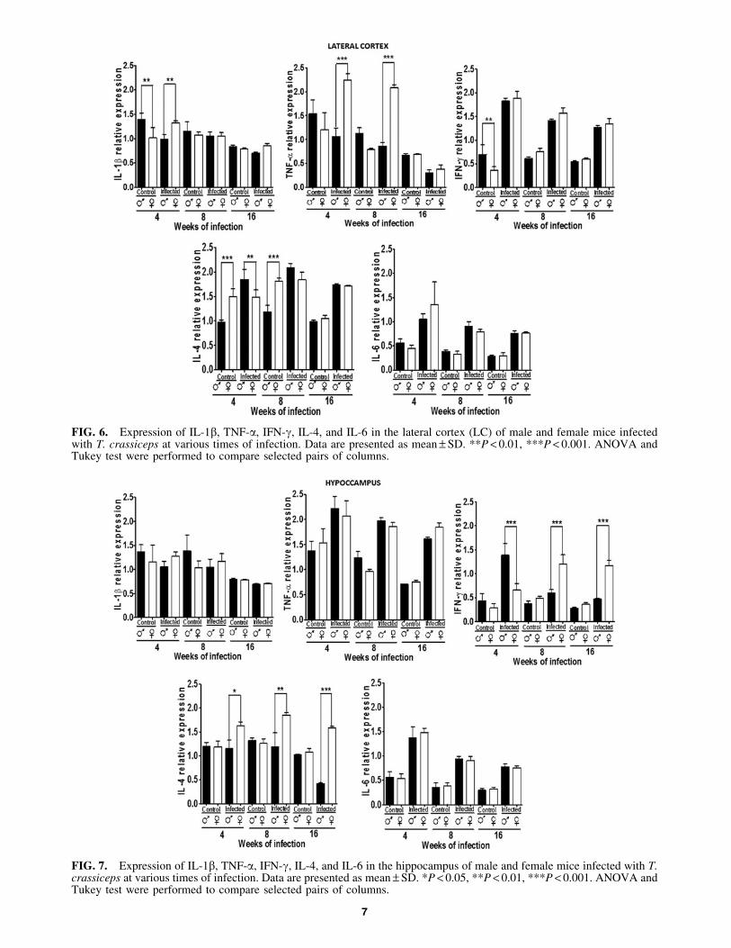
FIG. 6. Expression of IL-1b, TNF-a, IFN-g, IL-4, and IL-6 in the lateral cortex (LC) of male and female mice infectedwith T. crassiceps at various times of infection. Data are presented as mean – SD. **P < 0.01, ***P < 0.001. ANOVA andTukey test were performed to compare selected pairs of columns.
FIG. 7. Expression of IL-1b, TNF-a, IFN-g, IL-4, and IL-6 in the hippocampus of male and female mice infected with T.crassiceps at various times of infection. Data are presented as mean – SD. *P < 0.05, **P < 0.01, ***P < 0.001. ANOVA andTukey test were performed to compare selected pairs of columns.
7

increased in females at 8 and 16 weeks postinfection(P < 0.05). IL-6 was not dimorphic and was upregulated oninfection in both genders (P < 0.05) (Fig. 7).
Discussion
In this study, we report novel findings on an importantneuroimmune interaction in male and female mice that havebeen infected with T. crassiceps cysticerci, noting signifi-cant changes in cytokine expression in their brains. Wefound that cytokine expression was up or downregulated inthe hippocampus, LC, FC, HYP, OB, and POA of infectedmales and females to various extents. Also, the cytokineexpression patterns in the brain were gender-specific, inde-pendent of infection.
These changes in expression followed various dynamics,based on time, infection, and gender, reflecting specific al-terations in the function of each region in the brain.
Changes in cytokine expression in mice that are chroni-cally infected with T. crassiceps are likely caused bymodulation in steroid levels (Larralde and others 1995;Morales and others 1996; Arteaga-Silva and others 2009),consistent with the findings that estradiol activates cytokinegene transcription in rodents (Loose-Mitchell and others1988; Travers and others 1988; Nephew and others 1993;Bigsby and Li 1994) and in the spleen and thymus of cy-sticercotic mice (Morales-Montor and others 1998). In T.crassiceps cysticercosis-infected male mice, sexual behavioris abolished. The region-specific changes in cytokine ex-pression in the brain that we observed during cysticercosiscould explain behavioral disruptions, because cytokinesmediate brain-specific control of neurotransmission (Ca-macho-Arroyo and others 2009).
Although cytokines modulate brain development, regen-eration, and synapses, their origin in the brain is unclear—astrocytes produce cytokines and chemokines that attractvarious immune cell types that facilitate their access to thebrain through the blood–brain barrier during inflammatoryresponses (Yoshida and others 1992; Benveniste and others1994; Hu and others 1994; Feldhaus and others 2004). No-tably, astrocytes constitutively produce and secrete cytokines,suggesting that these messengers modulate CNS function.
Our goal is to determine the implications of changes incytokine expression in the brain in an infected host for itsphysiology. In the mammalian brain, neurons communicatepredominantly through chemical synapses, mediated byvarious neurotransmitters. The steps in communicationthrough chemical synapses—neurotransmitter synthesis,storage, release, uptake, degradation, interaction with spe-cific receptors, and transduction—are crucial in the regula-tion of brain function, all of which can be regulated bycytokines (Camacho-Arroyo and others 2009). For instance,the neurotransmitter serotonin is an immune modulator thatis necessary for optimal synthesis of IL-6 and TNF-a in thebrain; under physiological concentrations, it can increasetheir production by stimulating its cytokine receptors.Conversely, extracellular serotonin concentrations abovebaseline physiological levels can suppress the production ofIL-6 and TNF-a (Kubera and others 2005).
Neurotransmission by glutamate and g-aminobutyric(GABA), key excitatory and inhibitory neurotransmitters,respectively, is modulated by cytokines. In vitro, IL-1b in-creases GABA-A-mediated inward chloride currents in
synaptosomes and cultured neurons of the amygdala andcerebral cortex in rats (Miller and others 1991; Hu and others2000). Moreover, in vivo, systemically injected IL-1b upre-gulates c-fos in GABAergic, enkephalinergic, neurotensiner-gic, and CRHergic neurons of the amygdaloid complex(Miller and Fahey 1994; Yu and Shinnick-Gallagher 1994;Mazzoni and Kenigsberg 1997). Our result of increased IL-1bexpression in various areas of the brain in an infected hostsupport the finding that c-fos expression rises in the hippo-campus during chronic infection with T. crassiceps (Morales-Montor and others 2004b).
IL-6 also participates in neurochemical communicationand undergoes changes in its expression in the brain duringinfection; this cytokine potentiates evoked GABA releasefrom mediobasal hypothalamic explants and posterior pitu-itary cells in culture (De Laurentiis and others 2000).Moreover, long-term exposure of the brain to IL-6, as ob-served in certain degenerative disorders and infections,impedes adult hippocampal neurogenesis (Vallieres andothers 2002). Thus, our results support and extend a modelin which chronically infected animals that experience in-creased IL-6 expression in the brain have impaired short-term memory (Morales-Montor and others 2014).
IFN-g was also altered during cysticercosis. Interferonsregulate neural transmission—application of IFN-g duringpeak synaptogenesis reduces the frequency of spontaneousexcitatory activity, but increases that of spontaneous inhib-itory postsynaptic currents, upsetting the balance betweenexcitatory and inhibitory neurotransmission (Brask andothers 2004).
TNF-a regulates important functions in the brain, whichmight correlate with the alterations in TNF-a expression ininfected host brains that we observed. For instance, TNF-aincreases Ca2 + current density through L-type voltage-dependent channels, but it decreases currents that are in-duced by glutamate, NMDA, AMPA, and kainite in culturedneurons (Furukawa and Mattson 1998; Liu and others 2002).TNF-a also modulates synaptic maturation and neuronalbranching in the hippocampus by controlling the expressionof synaptic vesicle-associated proteins (Golan and others2004). Moreover, TNF-a alters the electrophysiologicalproperties of myenteric neurons through cyclooxygenasemetabolites and protein tyrosine phosphorylation (Son andothers 2004).
In conclusion, cytokines interact with the brain in a sys-temic and complex manner, influencing the development,function, and hormone production in the host. Thus, manyclinical situations might be attributed to cytokine activity inthe brain, and therapeutic manipulation of the immunesystem might affect brain function. Further work is neededto determine the exact function of cytokines and determinewhether other factors are associated with the parasite or hostin the feminization that occurs during experimental malemurine T. crassiceps-induced cysticercosis.
Acknowledgments
Financial support: Grant # IN-214011 from Programa deApoyo a Proyectos de Investigacion e Innovacion Tecnolo-gica (PAPIIT) from Direccion General de Asuntos delPersonal Academico (DGAPA), Universidad Nacional Au-tonoma de Mexico (UNAM), and Grant 176803, from Pro-grama de Fondos Sectoriales CB-SEP, Consejo Nacional de
8 LOPEZ-GRIEGO ET AL.

Ciencia y Tecnologıa (CONACyT), both to Jorge MoralesMontor. Karen Nava-Castro has a postdoctoral fellowshipfrom CONACyT. Rosalıa Hernandez-Cervantes and NellyTiempos Guzman are PhD students at Programa de Doctor-ado en Ciencias Biomedicas, Universidad Nacional Autono-ma de Mexico, and both have scholarships from CONACyT.Lenin Pavon is supported by PROMEP NETWORK No.14411150.
Author Disclosure Statement
No competing financial interests exist.
References
Arteaga-Silva M, Vargas-Villavicencio JA, Vigueras-VillasenorRM, Rodriguez-Dorantes M, Morales-Montor J. 2009. Taeniacrassiceps infection disrupts estrous cycle and reproductivebehavior in BALB/c female mice. Acta Trop 109(2):141–145.
Benveniste EN, Kwon J, Chung WJ, Sampson J, Pandya K, TangLP. 1994. Differential modulation of astrocyte cytokine geneexpression by TGF-beta. J Immunol 153(11):5210–5221.
Besedovsky HO, del Rey A. 1996. Immune-neuro-endocrineinteractions: facts and hypotheses. Endocr Rev 17(1):64–102.
Besedovsky HO, del Rey A. 2000. The cytokine-HPA axis feed-back circuit. Z Rheumatol 59 (Suppl 2):II/26–30.
Bigsby RM, Li A. 1994. Differentially regulated immediate earlygenes in the rat uterus. Endocrinology 134(4):1820–1826.
Brask J, Kristensson K, Hill RH. 2004. Exposure to interferon-gamma during synaptogenesis increases inhibitory activityafter a latent period in cultured rat hippocampal neurons. EurJ Neurosci 19(12):3193–3201.
Buckingham JC, Loxley HD, Christian HC, Philip JG. 1996.Activation of the HPA axis by immune insults: roles and in-teractions of cytokines, eicosanoids, glucocorticoids. Pharma-col Biochem Behav 54(1):285–298.
Camacho-Arroyo I, Lopez-Griego L, Morales-Montor J. 2009.The role of cytokines in the regulation of neurotransmission.Neuroimmunomodulation 16(1):1–12.
De Laurentiis A, Pisera D, Lasaga M, Diaz M, Theas S, Du-vilanski B, Seilicovich A. 2000. Effect of interleukin-6 andtumor necrosis factor-alpha on GABA release from medio-basal hypothalamus and posterior pituitary. Neuroimmuno-modulation 7(2):77–83.
Deverman BE, Patterson PH. 2009. Cytokines and CNS de-velopment. Neuron 64(1):61–78.
Dinarello CA. 2007. Historical insights into cytokines. Eur JImmunol 37(S1):S34–S45.
Feldhaus B, Dietzel ID, Heumann R, Berger R. 2004. Effects ofinterferon-gamma and tumor necrosis factor-alpha on sur-vival and differentiation of oligodendrocyte progenitors. JSoc Gynecol Investig 11(2):89–96.
Furukawa K, Mattson MP. 1998. The transcription factor NF-kappaB mediates increases in calcium currents and decreasesin NMDA- and AMPA/kainate-induced currents induced bytumor necrosis factor-alpha in hippocampal neurons. J Neu-rochem 70(5):1876–1886.
Golan H, Levav T, Mendelsohn A, Huleihel M. 2004. In-volvement of tumor necrosis factor alpha in hippocampaldevelopment and function. Cereb Cortex 14(1):97–105.
Gourbal BE, Lacroix A, Gabrion C. 2002. Behavioural domi-nance and Taenia crassiceps parasitism in BALB/c malemice. Parasitol Res 88(10):912–917.
Gourbal BE, Righi M, Petit G, Gabrion C. 2001. Parasite-altered host behavior in the face of a predator: manipulationor not? Parasitol Res 87(3):186–192.
Hu S, Martella A, Anderson WR, Chao CC. 1994. Role ofcytokines in lipopolysaccharide-induced functional andstructural abnormalities of astrocytes. Glia 10(3):227–234.
Hu S, Sheng WS, Ehrlich LC, Peterson PK, Chao CC. 2000.Cytokine effects on glutamate uptake by human astrocytes.Neuroimmunomodulation 7(3):153–159.
Kubera M, Maes M, Kenis G, Kim YK, Lason W. 2005. Effectsof serotonin and serotonergic agonists and antagonists on theproduction of tumor necrosis factor alpha and interleukin-6.Psychiatry Res 134(3):251–258.
Larralde C, Morales J, Terrazas I, Govezensky T, Romano MC.1995. Sex hormone changes induced by the parasite lead tofeminization of the male host in murine Taenia crassicepscysticercosis. J Steroid Biochem Mol Biol 52(6):575–580.
Larralde C, Sotelo J, Montoya RM, Palencia G, Padilla A,Govezensky T, Diaz ML, Sciutto E. 1990. Immunodiagnosisof human cysticercosis in cerebrospinal fluid. Antigens frommurine Taenia crassiceps cysticerci effectively substitutethose from porcine Taenia solium. Arch Pathol Lab Med114(9):926–928.
Liu B, Li H, Brull SJ, Zhang JM. 2002. Increased sensitivity ofsensory neurons to tumor necrosis factor alpha in rats withchronic compression of the lumbar ganglia. J Neurophysiol88(3):1393–1399.
Loose-Mitchell DS, Chiappetta C, Stancel GM. 1988. Estrogenregulation of c-fos messenger ribonucleic acid. Mol En-docrinol 2(10):946–951.
Mazzoni IE, Kenigsberg RL. 1997. Microglia from the devel-oping rat medial septal area can affect cholinergic and GA-BAergic neuronal differentiation in vitro. Neuroscience76(1):147–157.
Miller LG, Fahey JM. 1994. Interleukin-1 modulates GA-BAergic and glutamatergic function in brain. Ann N Y AcadSci 739:292–298.
Miller LG, Galpern WR, Dunlap K, Dinarello CA, Turner TJ.1991. Interleukin-1 augments gamma-aminobutyric acidAreceptor function in brain. Mol Pharmacol 39(2):105–108.
Morales J, Larralde C, Arteaga M, Govezensky T, Romano MC,Morali G. 1996. Inhibition of sexual behavior in male miceinfected with Taenia crassiceps cysticerci. J Parasitol 82(5):689–693.
Morales-Montor J, Arrieta I, Del Castillo LI, Rodriguez-Dorantes M, Cerbon MA, Larralde C. 2004a. Remote sensingof intraperitoneal parasitism by the host’s brain: regionalchanges of c-fos gene expression in the brain of feminizedcysticercotic male mice. Parasitology 128 (Pt 3):343–351.
Morales-Montor J, Escobedo G, Rodriguez-Dorantes M, Tellez-Ascencio N, Cerbon MA, Larralde C. 2004b. Differentialexpression of AP-1 transcription factor genes c-fos and c-junin the helminth parasites Taenia crassiceps and Taenia so-lium. Parasitology 129 (Pt 2):233–243.
Morales-Montor J, Larralde C. 2005. The role of sex steroids inthe complex physiology of the host-parasite relationship: thecase of the larval cestode of Taenia crassiceps. Parasitology131 (Pt 3):287–294.
Morales-Montor J, Picazo O, Besedovsky HO, Hernandez-BelloR, Lopez-Griego L, Becerril LE, Moreno J, Pavon L, Nava-Castro K, Camacho-Arroyo I. 2014. Helminth infection altersmood and short-term memory as well as levels of neuro-transmitters and cytokines in the mouse hippocampus. Neu-roimmunomodulation 21(4):195–205.
Morales-Montor J, Rodriguez-Dorantes M, Mendoza-RodriguezCA, Camacho-Arroyo I, Cerbon MA. 1998. Differential ex-pression of the estrogen-regulated proto-oncogenes c-fos, c-jun,and bcl-2 and of the tumor-suppressor p53 gene in the male
CYTOKINE EXPRESSION IN THE BRAIN OF CYSTICERCOTIC MICE 9

mouse chronically infected with Taenia crassiceps cysticerci.Parasitol Res 84(8):616–622.
Nephew KP, Webb DK, Akcali KC, Moulton BC, Khan SA.1993. Hormonal regulation and expression of the jun-Dprotooncogene in specific cell types of the rat uterus. JSteroid Biochem Mol Biol 46(3):281–287.
Rodriguez-Dorantes M, Cerbon MA, Larralde C, Morales-Montor J. 2007. Modified progesterone receptor expression inthe hypothalamus of cysticercotic male mice. Acta Trop103(2):123–132.
Smith JK, Esch GW, Kuhn RE. 1972. Growth and developmentof larval Taenia crassiceps (cestoda). I. Aneuploidy in theanomalous ORF strain. Int J Parasitol 2(2):261–263.
Son DS, Arai KY, Roby KF, Terranova PF. 2004. Tumor ne-crosis factor alpha (TNF) increases granulosa cell prolife-ration: dependence on c-Jun and TNF receptor type 1.Endocrinology 145(3):1218–1226.
Travers MT, Wakeling AE, Knowler JT. 1988. The isolation ofrecombinant RNA species responsive to oestrogen and ta-moxifen in rat uterus and MCF-7 cells. Mol Cell Endocrinol57(3):179–186.
Vallieres L, Campbell IL, Gage FH, Sawchenko PE. 2002.Reduced hippocampal neurogenesis in adult transgenic mice
with chronic astrocytic production of interleukin-6. J Neu-rosci 22(2):486–492.
Yoshida K, Kakihana M, Chen LS, Ong M, Baird A, Gage FH.1992. Cytokine regulation of nerve growth factor-mediatedcholinergic neurotrophic activity synthesized by astrocytesand fibroblasts. J Neurochem 59(3):919–931.
Yu B, Shinnick-Gallagher P. 1994. Interleukin-1 beta inhibits syn-aptic transmission and induces membrane hyperpolarization inamygdala neurons. J Pharmacol Exp Ther 271(2):590–600.
Address correspondence to:Dr. Jorge Morales-Montor
Departamento de InmunologıaInstituto de Investigaciones Biomedicas
Universidad Nacional Autonoma de MexicoAP 70228
Mexico D.F. 04510Mexico
E-mail: [email protected];[email protected]
Received 13 December 2013/Accepted 22 July 2014
10 LOPEZ-GRIEGO ET AL.
Copyright © 2022 FDOKUMEN