
Chloro-oxime derivatives as novel small molecule chaperone amplifiers

Hindawi Publishing CorporationInternational Journal of SpectroscopyVolume 2012, Article ID 847676, 8 pagesdoi:10.1155/2012/847676
Research Article
Gas Chromatography Electron Ionization Mass Spectral Analysisof Thio Analogues of Pyrimidine Bases: 5-Bromo-2,4-di-o-(m-and p-) chloro- (bromo-)benzylthiouracils and 6-methyluracils
G. Bartkowiak, E. Wyrzykiewicz, and G. Schroeder
Faculty of Chemistry, Adam Mickiewicz University, Grunwaldzka 6, 60-780 Poznan, Poland
Correspondence should be addressed to G. Bartkowiak, [email protected]
Received 14 July 2011; Accepted 30 September 2011
Academic Editor: Hans Riesen
Copyright © 2012 G. Bartkowiak et al. This is an open access article distributed under the Creative Commons Attribution License,which permits unrestricted use, distribution, and reproduction in any medium, provided the original work is properly cited.
Electron ionization (EI) mass spectral fragmentation routes of twelve 5-bromo-2,4-di-o-(m- and p-) chloro- (bromo-)benzyl-thiouracils and 6-methyluracils are investigated. The compounds studied are analyzed using gas chromatography/mass spectrom-etry (GC/MS). Fragmentation pathways, whose elucidation is assisted by accurate mass measurements and metastable transitions,are discussed. Correlation between the abundances of the selected fragment ions of the compounds investigated is discussed. Thedata obtained make grounds for distinction of structural isomers.
1. Introduction
Thio derivatives of pyrimidine bases are of interest becauseof their biological and pharmacological activities, for exam-ple, as minor components of t-RNA or as antithyroidaland anticancer drugs as well as sedatives [1–7]. Essentialrole in biological systems play compounds, which con-tain 5-bromopyrimidine moiety, like 5-bromouracil and 5-bromouridine. It is well known that these compounds aremutagenic [8–10] and able to replace thymine residue in theDNA molecule. On the other hand, C-5 bromo group is ahydrophobic substituent having also electron-negative prop-erties, very important for anaesthetic and anticonflict activ-ities [11]. The presence of benzyl group also influences theactivity of pyrimidine derivatives, for example, the pyrim-idine thioethers with 2-benzylthio substituent have beenreported as a novel nonnucleoside HIV-1 reverse transferaseinhibitors (NNRTIs) with activity against BHAP-resistantHIV [12, 13]. Because of biological importance of themodified thio analogues of pyrimidine bases, much attentionhas been devoted to recognize their properties. Mass spec-trometry continues to be a convenient and effective methodfor determination of nature of covalent modifications to thioanalogues of nucleobases and for distinction of structuralisomers [14–17]. The mass spectral behavior of modified
derivatives of thionucleobases, for example, 2-benzylthioand 4-benzylthiouracils which may appear in the gas phasein various tautomeric forms has been studied previously.However, to the best of our knowledge, no work has beenpublished about the mass spectrometric behavior of fullyaromatic 2,4-dibenzylthio-5-bromouracils. The hereby pre-sented study of mass fragmentation of the title compoundswere undertaken to examine the influence of C-5 bromo sub-stitution in pyrimidine ring on their EI mass fragmentation.
Isomeric organic compounds discrimination is a chal-lenging task for mass spectrometrists because of the identicalmolecular masses and similar fragmentation pathways ofthese species. However, differences in the nature and elec-tronic effects of substituents, depending on their locationin the molecule, lead to different stabilization possibilitiesand influence the kinetics of mass dissociation of molecularand fragment ions. These factors are strongly reflected by theabundances of ions in the electron-ionization mass spectra ofsuch molecules. Comparison of the abundances of selectedfragment and molecular ions reveals the preferences of themolecule’s breakup and often allows isomers differentiation[18, 19].
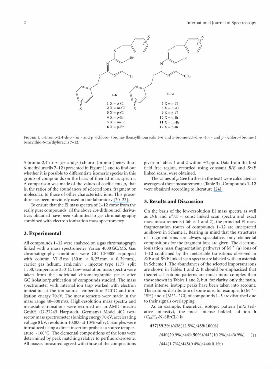
The aim of this investigation was to elucidate the EImass spectrometric fragmentations of 5-bromo-2,4-di-o-(m- and p-) chloro- (bromo-)benzylthiouracils 1–6 and
2 International Journal of Spectroscopy
N
N
S
S
Br
H
XX
N
N
S
S
Br
CH3
XX
X = o-ClX =m-ClX = p-ClX = o-BrX =m-BrX = p-Br
X = o-ClX =m-ClX = p-ClX = o-BrX =m-BrX = p-Br
1–6 7–12
123456
789
101112
Figure 1: 5-Bromo-2,4-di-o -(m - and p -)chloro- (bromo-)benzylthiouracils 1–6 and 5-bromo-2,4-di-o -(m - and p -)chloro-(bromo-)benzylthio-6-methyluracils 7–12.
5-bromo-2,4-di-o- (m- and p-) chloro- (bromo-)benzylthio-6-methyluracils 7–12 (presented in Figure 1) and to find outwhether it is possible to differentiate isomeric species in thisgroup of compounds on the basis of their EI mass spectra.A comparison was made of the values of coefficients μ, thatis, the ratios of the abundances of selected ions, fragment ormolecular, to those of other characteristic ions. This proce-dure has been previously used in our laboratory [20–23].
To ensure that the EI mass spectra of 1–12 come from thereally pure compounds, all the above 2,4-dithiouracil deriva-tives obtained have been submitted to gas chromatographycombined with electron ionization mass spectrometry.
2. Experimental
All compounds 1–12 were analyzed on a gas chromatographlinked with a mass spectrometer Varian 4000 GC/MS. Gaschromatography conditions were GC CP3800 equippedwith column VF-5 ms (30 m × 0, 25 mm × 0, 39 mm),carrier gas helium, 1 mL min−1, injector type 1177, split1 : 50, temperature 250◦C. Low-resolution mass spectra weretaken from the individual chromatographic peaks afterGC isolation/purification of compounds studied. The massspectrometer with internal ion trap worked with electronionization at the ion source temperature 220◦C and ion-ization energy 70 eV. The measurements were made in themass range 40–800 m/z. High-resolution mass spectra andmetastable transitions were recorded on an AMD-IntectraGmbH (D-27243 Harpstedt, Germany) Model 402 two-sector mass spectrometer (ionizing energy 70 eV, acceleratingvoltage 8 kV, resolution 10.000 at 10% valley). Samples wereintroduced using a direct insertion probe at a source temper-ature ∼160◦C. The elemental compositions of the ions weredetermined by peak matching relative to perfluorokerosene.All masses measured agreed with those of the compositions
given in Tables 1 and 2 within ±2 ppm. Data from the firstfield free region, recorded using constant B/E and B2/Elinked scans, were obtained.
The values of μ (see further in the text) were calculated asaverages of three measurements (Table 3) . Compounds 1–12were obtained according to literature [24].
3. Results and Discussion
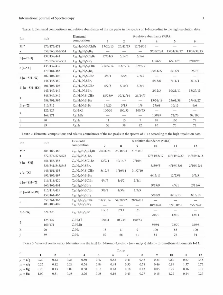
On the basis of the low-resolution EI mass spectra as wellas B/E and B2/E = const linked scan spectra and exactmass measurements (Tables 1 and 2), the principal EI massfragmentation routes of compounds 1–12 are interpretedas shown in Scheme 1. Bearing in mind that the structuresof fragment ions are always speculative, only elementalcompositions for the fragment ions are given. The electron-ionization mass fragmentation pathways of M+• (a) ions of1–12 confirmed by the metastable transitions observed inB/E and B2/E linked scan spectra are labeled with an asteriskin Scheme 1. The abundances of the selected important ionsare shown in Tables 1 and 2. It should be emphasized thattheoretical isotopic patterns are much more complex thanthose shown in Tables 1 and 2, but, for clarity, only the main,most intense, isotopic peaks have been taken into account.The isotopic distribution of some ions, for example, b (M+•–•SH) and c (M+•– •Cl) of compounds 1–3 are disturbed dueto their signals overlapping.
As an example, theoretical isotopic pattern [m/z (rel-ative intensity), the most intense bolded] of ion b(C18H12N2SBrCl2) is
437(59.2%)/438(12.5%)/439(100%)
/440(20.9%)/441(50%)/442(10.2%)/443(9%)
/444(1.7%)/445(0.4%)/446(0.1%)
(1)
International Journal of Spectroscopy 3
Table 1: Elemental compositions and relative abundances of the ion peaks in the spectra of 1–6 according to the high-resolution data.
Ion m/zElementalcomposition
% relative abundance (%RA)
1 2 3 4 5 6
M+• 470/472/474 C18H13N2S2Cl2Br 13/20/13 23/42/23 12/24/16 — — —
a 558/560/562/564 C18H13N2S2Br3 — — — 9/30/23/8 15/31/34/17 13/37/38/13
b [a–•SH]437/439/441 C18H12N2SCl2Br 27/14/3 6/14/5 6/5/4
525/527/529/531 C18H12N2SBr3 1/5/6/2 6/7/12/5 2/10/9/3
c [a–•X]435/437/439 C18H13N2S2ClBr 21/27/14 0.6/6/14 0.9/6/5
479/481/483 C18H13N2S2Br2 25/44/27 6/14/9 2/2/2
d [a–•SH–•X]402/404/406 C18H12N2SClBr 3/4/1 2/5/3 2/2/3 — — —
446/448/450 C18H12N2SBr2 — — — 5/18/6 7/11/4 5/14/4
d′ [a–•SH–HX]401/403/405 C18H11N2SClBr 5/7/3 5/10/4 3/8/4
445/447/449 C18H11N2SBr2 2/12/3 10/21/11 13/27/15
e345/347/349 C11H7N2S2ClBr 18/25/9 32/42/14 21/24/7 — — —
389/391/393 C11H7N2S2Br2 — — — 13/54/18 25/61/38 27/48/27
f [e–•X] 310/312 C11H7N2S2Br 19/20 5/13 1/9 53/68 10/13 6/6
g125/127 C7H6Cl 100/36 100/33 100/33 — — —
169/171 C7H6Br — — — 100/99 72/70 99/100
h 90 C7H6 11 15 7 99 100 79
j 89 C7H5 58 58 42 85 73 72
Table 2: Elemental compositions and relative abundances of the ion peaks in the spectra of 7–12 according to the high-resolution data.
Ion m/zElementalcomposition
% relative abundance (%RA)
7 8 9 10 11 12
M+• 484/486/488 C19H15N2S2Cl2Br 20/41/24 25/48/24 21/33/16 — — —
a 572/574/576/578 C19H15N2S2Br3 — — — 17/54/53/17 13/44/49/20 14/35/44/18
b [a–•SH]451/453/455 C19H14N2SCl2Br 12/9/4 10/14/7 7/10/4
539/541/543/545 C19H14N2SBr3 3/5/9/3 4/19/15/6 2/10/12/4
c [a–•X]449/451/453 C19H15N2S2ClBr 3/12/9 1/10/14 0.1/7/10
493/495/497 C19H15N2S2Br2 6/15/11 12/23/8 3/5/3
d [a–•SH–•X]416/418/420 C19H14N2SClBr 4/4/3 1/4/2 1/1/1
460/462/464 C19H14N2SBr2 9/19/9 4/9/1 2/11/6
d′ [a–SH–HX]415/417/419 C19H13N2SClBr 3/6/2 4/5/4 1/3/3
459/461/463 C19H13N2SBr2 5/10/9 8/18/13 3/13/10
e359/361/363403/405/407
C12H9N2S2ClBrC12H9N2S2Br2
31/33/14 54/78/22 28/46/12 — — —
— — — 48/81/44 52/100/57 35/72/44
f [e–•X] 324/326 C12H9N2S2Br18/18 2/13 1/5 — — —
— — — 70/70 12/10 12/11
g125/127 C7H6Cl 100/31 100/34 100/33 — — —
169/171 C7H6Br — — — 89/91 73/70 98/95
h 90 C7H6 13 11 9 100 85 100
j 89 C7H5 57 66 41 81 76 94
Table 3: Values of coefficients μ (definitions in the text) for 5-bromo-2,4-di-o - (m - and p -) chloro- (bromo)benzylthiouracils 1–12.
μComp
1 2 3 4 5 6 7 8 9 10 11 12
μ1 = a/g 0.20 0.42 0.24 0.30 0.47 0.38 0.41 0.48 0.33 0.60 0.67 0.45
μ2 = e/g 0.25 0.42 0.24 0.54 0.84 0.48 0.33 0.78 0.46 0.89 1.37 0.73
μ3 = f/g 0.20 0.13 0.09 0.68 0.18 0.48 0.18 0.13 0.05 0.77 0.16 0.12
μ4 = f/a 1.00 0.31 0.38 2.26 0.38 0.16 0.43 0.27 0.15 1.29 0.24 0.27

4 International Journal of Spectroscopy
f
C7H5+
−HX∗CH2
[M−•C11H6N2S2RXBr]
C7H6
h
C11H6N2S2RBr
C11H5N2SRBr
f1
[M−•C7H6X−•X]
= H
= CH3
X =C18H12N2S2RX2Br
[M−•SH−HX]
[M−•C7H6X]
C11H6N2S2RXBr
e1
C18H10N2SRXBr
M
[M−•SH]
[M−•X]
[M−•SH−•X]
C18H12N2S2RXBr
C18H11N2SRX2Br
C18H10N2RX2Brb1
−X•
C18H11N2SRXBr −C7H5XS
−•SH−•X −HX
d1d2 d3
d4
+ •
d5
−•C7H6X
−•SH−•X
−HX
j
g
∗
∗
+ •
∗
+
∗
∗
e
+
∗
+
∗
∗
∗
∗
∗
∗
+ •
b
+ •
+ •
+
c+
∗+
∗ −•SH
+ •
∗∗
d
a
−•SH
−•SH
N
N
R
Br
S
S−•SH
∗
∗
1–6
7–12
R
R1–3, 7–9) or Br (4–6, 10–12)
X
−•X
∗−•X
XCl (
d∗
X
Scheme 1: Pathways of the EI mass fragmentation of the molecular ions M+• (a) of 1–12.
and, for ion c (C18H13N2S2BrCl)
435(70.9%)/436(15.6%)/437(100%)
/438(21.6%)/439(32.8%)/440(6.7%)
/441(2.8%)/442(0.4%)/443(0.1%),
(2)
so the abundances of ions at m/z 437 and 439 add up.As can be seen from Scheme 1 and the data presented in
Tables 1 and 2, the principal mass fragmentation pathwaysof 1–6 and 7–12 are similar but show differences in theabundances of important fragment ions. The fragmentationof molecular ion of the compounds studied 1–12 proceedsaccording to the same pattern (as can be seen fromFigures 2(a) and 2(b), presenting EI mass spectra of two ofthe compounds studied as representative examples), in spiteof the different substituents (chloro- or bromo-) and differ-ent positions (ortho or para) of halogen in the phenyl ring.Figure 2(a) shows the EI mass spectrum of 5-bromo-2,4-di-o-chlorobenzylthio-6-methyluracil (compound 7) andFigure 2(b) the EI mass spectrum of 5-bromo-2,4-di-p-bromobenzylthio-6-methyluracil (compound 12). In thefigures mentioned above, the main fragmentation features ofM+• are clearly seen: both molecular ions easily lose •SH and•X (•Cl or •Br, resp.) radicals as well as •CH2C6H4X, thatis, the whole halobenzyl group. Other losses are loss of •SH
radical and neutral molecule HX (M+•– •SH–HCl or M+•–•SH–HBr) and combined loss of halobenzyl radical andhalogen from the other halobenzyl group in the molecule,however, it seems preferred that the loss of •Br occursrather from bromobenzyl derivatives and the loss of HCloccurs more likely from chlorobenzyl compounds. The sametendency is seen in the second step of fragmentation: theloss of HCl from the ions g m/z 125 (CH2C6H4Cl) givesdaughter ion j m/z 89 (C7H5
+) and loss of •Br from theions at m/z 169/171 (CH2C6H4Br) yields ion h at m/z 90(C7H6
+•), which confirms a known fact that HCl formationis thermochemically favoured.
The B/E = const linked scan spectrum of molecularion of compound 12 (Figure I, see Supplementary Materialavailable online at doi: 10.1155/2012/847676), with regardto the lowest mass isotope of bromine, that is, 79Br, presentsthe first step of fragmentation, that is, daughter ions formedthrough the dissociation of ion a (C19H15N2S2
79Br3, m/z572). It indicates that most of important and abundantfragment ions, listed in Table 2, originate directly from themolecular ion. A comparative set of the EI mass spectra of4–6 is presented in Figure II(A–C), Supplementary Material.Note different abundances of corresponding fragment ionsand different retention times.
One of the essential processes of EI-MS decompositionof molecular ions 1–12 is the elimination of •SH radicals.

International Journal of Spectroscopy 5
Cl
SH–HCl]
0
25
50
75
100
100 200 300 400 500
(%)
63
89
125.1
127.1
121.1 155.1
325
328
361
363
417.1451
484
486
•C12 N2S2
•CH2C6H4 −H
•CH2C6H4
••
•SH]
[M–
[M–
[M–
[M–
[M–
[M–
ClClBr]
Cl]
Cl]
Cl]
H9
(a)
0
25
50
75
100
100 200 300 400 500 600
(%)
40 83
90.1
73.2
121.1 170
169
247
281.1
282.1324
404.9
406.9
405.9
•C12 N2S2Br2]
•CH2C6H4
•CH2C6H4Br]
•
•Br]
•SH]
460.9 542.9
575.9
517.8
[M–
[M–
[M–
[M–
[M–
[M–
H9
371.9
SH–HBr]
Br–• Br]
(b)
Figure 2: GC-EI mass spectra of: (a) 5-bromo-2,4-di-(ortho-chlorobenzylthio)-6-methyluracil 7, retention time 36.983 min; (b) 5-bromo-2,4-di-(para-bromobenzylthio)-6-methyluracil 12, retention time 57.366 min.
For the loss of •SH, a skeletal rearrangement is requiredwhich implies the formation of new carbon-carbon andcarbon-nitrogen bonds. The •SH elimination occurs directlyfrom molecular ions a, leading to the even-electron fragmentions b, as well as from the many fragment ions, givingsecond generation of daughter ions of low abundance inthe EI mass spectrum. Fragment ions b, that is, [M+•–•SH], are precursors of ions d, created through the loss of•X from b. According to the metastable transition spectra(B/E linked scan method), the loss of •SH is a widespreadprocess in the fragmentation routes of 1–12 because ittakes place also for ions c, d, e, and f , although thegenerated daughter ions d1, e1, f1 are of low abundanceand low significance for differentiation of isomers, andtherefore they are not listed in Tables 1 and 2. It should bementioned that the loss of a sulfhydryl radical is commonfor aromatic thioethers [25] and is a characteristic featureof EI mass fragmentation of molecular ions of alkylthio-5-bromo and alkoxycarbonylalkylthiouracils [23, 26] as well as2-benzylthioorotic acids [27].
Molecular ions a decompose also with halogen radicalelimination. In the EI mass spectra of compounds 4–6and 9–12, the evident loss of •Br from molecular ion is
seen, especially pronounced for the ortho isomers 4 and10. The bromine loss can be noticed also in the furthersteps of fragmentation, for example, the B/E = const linkedscan spectrum of ion c of compound 12 presents intense•Br elimination. It remains unknown if there is a loss ofbromine radical from the uracil or phenyl ring. Analysis ofEI mass spectra of 1–3 and 7–9, where the [M–•Br] ions arealmost completely absent, indicates that bromine originatesfrom benzyl moiety. Analogously, ions e of 10–12, that is,C12H9N2S2Br2 lose bromine radical giving f, but ions e of 7–9 lose •Cl not •Br. It means that in this series of uracil deriva-tives bromine radicals are more easily abstracted from phenylthan from pyrimidine ring and the X-Csp2 bond in phenylring is more prone to cleave than in heteroaromatic one. Thisis the difference between disubstituted 5-bromodithiouracilsand monosubstituted 5-bromothiouracils. It was mentionedearlier that •Br loss from C-5 position of pyrimidine ringis characteristic of many bromouracil and bromothiouracilderivatives and that •Br radical elimination is more intensefor 4-thio- than for 2-thioderivatives [23, 26]. For molecularions of 5-bromo-2,4-di-o- (m- and p-)chloro- (bromo-)benzylthiouracils, the elimination of bromine radical fromposition 5 of pyrimidine ring does not occur in the EI-MS
6 International Journal of Spectroscopy
NR1S
H
N
O
Br
R2
I
(a)
O
Br
R2
N
SR1
H
N
II
(b)
N
N
SR1
R1S R2
Br
III
(c)
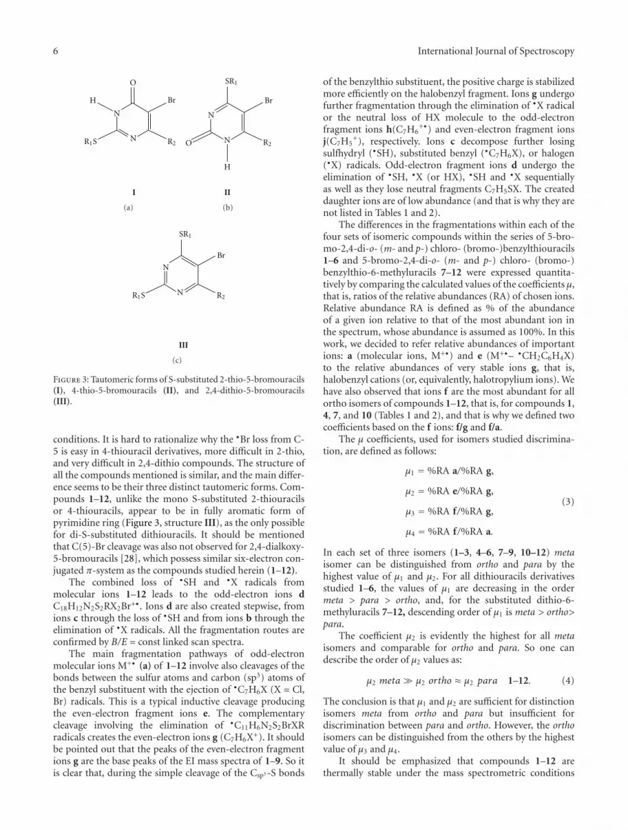
Figure 3: Tautomeric forms of S-substituted 2-thio-5-bromouracils(I), 4-thio-5-bromouracils (II), and 2,4-dithio-5-bromouracils(III).
conditions. It is hard to rationalize why the •Br loss from C-5 is easy in 4-thiouracil derivatives, more difficult in 2-thio,and very difficult in 2,4-dithio compounds. The structure ofall the compounds mentioned is similar, and the main differ-ence seems to be their three distinct tautomeric forms. Com-pounds 1–12, unlike the mono S-substituted 2-thiouracilsor 4-thiouracils, appear to be in fully aromatic form ofpyrimidine ring (Figure 3, structure III), as the only possiblefor di-S-substituted dithiouracils. It should be mentionedthat C(5)-Br cleavage was also not observed for 2,4-dialkoxy-5-bromouracils [28], which possess similar six-electron con-jugated π-system as the compounds studied herein (1–12).
The combined loss of •SH and •X radicals frommolecular ions 1–12 leads to the odd-electron ions dC18H12N2S2RX2Br+•. Ions d are also created stepwise, fromions c through the loss of •SH and from ions b through theelimination of •X radicals. All the fragmentation routes areconfirmed by B/E = const linked scan spectra.
The main fragmentation pathways of odd-electronmolecular ions M+• (a) of 1–12 involve also cleavages of thebonds between the sulfur atoms and carbon (sp3) atoms ofthe benzyl substituent with the ejection of •C7H6X (X = Cl,Br) radicals. This is a typical inductive cleavage producingthe even-electron fragment ions e. The complementarycleavage involving the elimination of •C11H6N2S2BrXRradicals creates the even-electron ions g (C7H6X+). It shouldbe pointed out that the peaks of the even-electron fragmentions g are the base peaks of the EI mass spectra of 1–9. So itis clear that, during the simple cleavage of the Csp3 -S bonds
of the benzylthio substituent, the positive charge is stabilizedmore efficiently on the halobenzyl fragment. Ions g undergofurther fragmentation through the elimination of •X radicalor the neutral loss of HX molecule to the odd-electronfragment ions h(C7H6
+•) and even-electron fragment ionsj(C7H5
+), respectively. Ions c decompose further losingsulfhydryl (•SH), substituted benzyl (•C7H6X), or halogen(•X) radicals. Odd-electron fragment ions d undergo theelimination of •SH, •X (or HX), •SH and •X sequentiallyas well as they lose neutral fragments C7H5SX. The createddaughter ions are of low abundance (and that is why they arenot listed in Tables 1 and 2).
The differences in the fragmentations within each of thefour sets of isomeric compounds within the series of 5-bro-mo-2,4-di-o- (m- and p-) chloro- (bromo-)benzylthiouracils1–6 and 5-bromo-2,4-di-o- (m- and p-) chloro- (bromo-)benzylthio-6-methyluracils 7–12 were expressed quantita-tively by comparing the calculated values of the coefficients μ,that is, ratios of the relative abundances (RA) of chosen ions.Relative abundance RA is defined as % of the abundanceof a given ion relative to that of the most abundant ion inthe spectrum, whose abundance is assumed as 100%. In thiswork, we decided to refer relative abundances of importantions: a (molecular ions, M+•) and e (M+•– •CH2C6H4X)to the relative abundances of very stable ions g, that is,halobenzyl cations (or, equivalently, halotropylium ions). Wehave also observed that ions f are the most abundant for allortho isomers of compounds 1–12, that is, for compounds 1,4, 7, and 10 (Tables 1 and 2), and that is why we defined twocoefficients based on the f ions: f/g and f/a.
The μ coefficients, used for isomers studied discrimina-tion, are defined as follows:
μ1 = %RA a/%RA g,
μ2 = %RA e/%RA g,
μ3 = %RA f/%RA g,
μ4 = %RA f/%RA a.
(3)
In each set of three isomers (1–3, 4–6, 7–9, 10–12) metaisomer can be distinguished from ortho and para by thehighest value of μ1 and μ2. For all dithiouracils derivativesstudied 1–6, the values of μ1 are decreasing in the ordermeta > para > ortho, and, for the substituted dithio-6-methyluracils 7–12, descending order of μ1 is meta > ortho>para.
The coefficient μ2 is evidently the highest for all metaisomers and comparable for ortho and para. So one candescribe the order of μ2 values as:
μ2 meta� μ2 ortho ≈ μ2 para 1–12. (4)
The conclusion is that μ1 and μ2 are sufficient for distinctionisomers meta from ortho and para but insufficient fordiscrimination between para and ortho. However, the orthoisomers can be distinguished from the others by the highestvalue of μ3 and μ4.
It should be emphasized that compounds 1–12 arethermally stable under the mass spectrometric conditions
International Journal of Spectroscopy 7
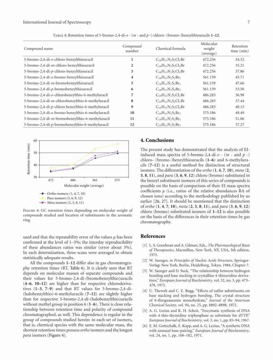
Table 4: Retention times of 5-bromo-2,4-di-o - (m - and p -) chloro- (bromo-)benzylthiouracils 1–12.
Compound nameCompound
numberChemical formula
Molecularweight
(average)
Retentiontime (min)
5-bromo-2,4-di-o-chloro-benzylthiouracil 1 C18H13N2S2Cl2Br 472.256 34.52
5-bromo-2,4-di-m-chloro-benzylthiouracil 2 C18H13N2S2Cl2Br 472.256 35.21
5-bromo-2,4-di-p-chloro-benzylthiouracil 3 C18H13N2S2Cl2Br 472.256 37.86
5-bromo-2,4-di-o-bromo-benzylthiouracil 4 C18H13N2S2Br3 561.159 45.71
5-bromo-2,4-di-m-bromobenzylthiouracil 5 C18H13N2S2Br3 561.159 47.66
5-bromo-2,4-di-p-bromobenzylthiouracil 6 C18H13N2S2Br3 561.159 53.50
5-bromo-2,4-di-o-chlorobenzylthio-6-methyluracil 7 C19H15N2S2Cl2Br 486.283 36.98
5-bromo-2,4-di-m-chlorobenzylthio-6-methyluracil 8 C19H15N2S2Cl2Br 486.283 37.44
5-bromo-2,4-di-p-chloro benzylthio-6-methyluracil 9 C19H15N2S2Cl2Br 486.283 40.13
5-bromo-2,4-di-o-bromo benzylthio-6-methyluracil 10 C19H15N2S2Br3 575.186 49.49
5-bromo-2,4-di-m-bromobenzylthio-6-methyluracil 11 C19H15N2S2Br3 575.186 51.06
5-bromo-2,4-di-p-bromobenzylthio-6-methyluracil 12 C19H15N2S2Br3 575.186 57.27
30
40
50
60
472 486 561 575
Ret
enti
on t
ime
(min
)
Ortho isomers (1, 4, 7, 10)
Meta isomers (2, 5, 8, 11)Para isomers (3, 6, 9, 12)
Molecular weight (average)
Figure 4: GC retention times depending on molecular weight ofcompounds studied and location of substituents in the aromaticring.
used and that the repeatability error of the values μ has beenconfirmed at the level of 1–3%; the interday reproducibilityof these abundances ratios was similar (error about 3%).In each determination, three scans were averaged to obtainstatistically adequate results.
All the compounds 1–12 differ also in gas chromatogra-phy retention times (RT, Table 4). It is clearly seen that RTdepends on molecular masses of separate compounds andtheir values for 5-bromo-2,4-di-(bromobenzylthio)uracils(4–6, 10–12) are higher than for respective chloroderiva-tives (1–3, 7–9) and that RT values for 5-bromo-2,4-di-(halobenzylthio)-6-methyluracils (7–12) are slightly higherthan for respective 5-bromo-2,4-di-(halobenzylthio)uracilswithout methyl group in position 6 (1–6). There is close rela-tionship between retention time and polarity of compoundchromatographed, as well. This dependence is regular in thegroup of compounds studied herein: in each set of isomers,that is, chemical species with the same molecular mass, theshortest retention times possess ortho isomers and the longestpara isomers (Figure 4).
4. Conclusions
The present study has demonstrated that the analysis of EI-induced mass spectra of 5-bromo-2,4-di-o - (m - and p -)chloro- (bromo-)benzylthiouracils (1–6) and 6-methylura-cils (7–12) is a useful method for distinction of structuralisomers. The differentiation of the ortho (1, 4, 7, 10), meta (2,5, 8, 11), and para (3, 6, 9, 12) chloro (bromo) substituted inthe benzyl substituent isomers of this series of compounds ispossible on the basis of comparison of their EI mass spectracoefficients μ (i.e., ratios of the relative abundances RA ofchosen ions) according to the methodology published by usearlier [26, 27]. It should be mentioned that the distinctionof ortho (1, 4, 7, 10), meta (2, 5, 8, 11), and para (3, 6, 9, 12)chloro (bromo) substituted isomers of 1–12 is also possibleon the basis of the differences in their retention times by gaschromatography.
References
[1] L. S. Goodman and A. Gilman, Eds., The Pharmacological Basisof Therapeutics, Macmillan, New York, NY, USA, 5th edition,1975.
[2] W. Saenger, in Principles of Nucleic Acids Structure, Springer-Verlag: New York, Berlin, Heidelberg, Tokyo, 1984, Chapter 7.
[3] W. Saenger and D. Suck, “The relationship between hydrogenbonding and base stacking in crystalline 4-thiouridine deriva-tives,” European Journal of Biochemistry, vol. 32, no. 3, pp. 473–478, 1973.
[4] U. Thewalt and C. E. Bugg, “Effects of sulfur substituents onbase stacking and hydrogen bonding. The crystal structureof 6-thioguanosine monohydrate,” Journal of the AmericanChemical Society, vol. 94, no. 25, pp. 8892–8898, 1972.
[5] A. G. Lezius and K. H. Scheit, “Enzymatic synthesis of DNAwith 4-thio-thymidine triphosphate as substitute for dTTP,”European Journal of Biochemistry, vol. 3, no. 1, pp. 85–94, 1967.
[6] E. M. Gottschalk, E. Kopp, and A. G. Lezius, “A synthetic DNAwith unusual base-pairing,” European Journal of Biochemistry,vol. 24, no. 1, pp. 168–182, 1971.

8 International Journal of Spectroscopy
[7] K. H. Scheit and E. Gaertner, “Die Polymerization von 4-Thiouridin-5’-diphosphat und 4-thiothymidin-5’-diphosphatdurch Polynucleotidphosphorylase aus Micrococcus lysodeik-ticus,” Biochimica et Biophysica Acta, vol. 182, no. 1, 1969.
[8] E. Freese, “The specific mutagenic effect of base analogues onphage T4,” Journal of Molecular Biology, vol. 1, no. 2, pp. 87–105, 1959.
[9] E. Freese, “The difference between spontaneous and base-analogue induced mutations of phage T4,” Proceedings of theNational Academy of Sciences of the United States of America,vol. 45, no. 4, pp. 622–633, 1959.
[10] S. Y. Wang, “Pyrimidine bimolecular photoproducts,” inPhotochemistry and Photobiology of Nucleic Acids, vol. 1, pp.295–326, Academic Press, New York, NY, USA, 1976.
[11] M. Imaizumi, F. Kano, and S. Sakata, “Novel uracil derivatives:newly synthesized centrally acting agents,” Chemical andPharmaceutical Bulletin, vol. 40, no. 7, pp. 1808–1813, 1992.
[12] I. W. Althaus, K.-C. Chou, R. J. Lemay et al., “The benzylthio-pyrimidine U-31,355, a potent inhibitor of HIV-1 reversetranscriptase,” Biochemical Pharmacology, vol. 51, no. 6, pp.743–750, 1996.
[13] R. A. Nugent, S. T. Schlachter, M. J. Murphy et al., “Pyrim-idine thioethers: a novel class of HIV-1 reverse transcriptaseinhibitors with activity against BHAP-resistant HIV,” Journalof Medicinal Chemistry, vol. 41, no. 20, pp. 3793–3803, 1998.
[14] P. Crain, T. Hasizume, and J. A. McCloskey, in Biological MassSpectrometry, ed. by A. L. Burlingame and J. A. McCloskey,671–692, Elsevier, Amsterdam [1990].
[15] K. Schram, “Mass spectrometry of nucleic acid components,”in Biological Application of Mass Spectrometry, C. H. Suelterand J. T. Watson, Eds., pp. 203–288, John Wiley, New York,NY, USA, 1990.
[16] J. A. McCloskey, in Mass Spectrometry in the Health and LifeSciences, ed. by A. L. Burlingame and N. Castagnoli Jr., ElsevierAmsterdam [1985].
[17] J. A. McCloskey, in Mass Spectrometry in Biomedical Research,ed. by S. J. Gaskell, Wiley, New York [1986].
[18] J. Garin, J. Orduna, J. M. Royo, A.-M. Le Quere, and H. Muller,“Differentiation of isomeric sulfur heterocycles by electronionization mass spectrometry: 1,4-dithiins, 1,4-dithiafulvenesnad their analogues tetrathianaphtalenes, tetrathiafulvalenesand tetrathiapentalenes,” Rapid Communications in MassSpectrometry, vol. 17, no. 6, pp. 547–552, 2003.
[19] V. Ovcharenko, M. Rzadkowska, T. Tkaczynski, D. Matosiuk,and K. Pihlaja, “Electron impact mass spectra of substituted1-aryl-2-arylsulphonylamino-δ2-imidzazolines,” Rapid Com-munications in Mass Spectrometry, vol. 11, no. 9, pp. 1043–1045, 1997.
[20] E. Wyrzykiewicz and Z. Nowakowska, “Mass spectrometry ofthio analogues of pyrimidine bases: correlation of the intensi-ties of the M+• and selected fragment ions of o-(m- and p-)substituted benzylthiouracils,” Journal of Mass Spectrometry,vol. 30, no. 2, pp. 269–274, 1995.
[21] E. Wyrzykiewicz and G. Bartkowiak, “Mass spectrometry ofthio analogues of pyrimidine bases: Correlation of the intensi-ties of the M+• and the [M–•SH]+ ions of 2-alkylthiouracils,”Organic Mass Spectrometry, vol. 27, no. 12, pp. 1377–1380,1992.
[22] G. Bartkowiak, “Differentiation of 2-alkylthioorotic acids,methyl and ethyl 2-alkylthioorotates and hydrazides of 2-alkylthioorotic acids by using electron ionization mass spec-tra,” European Journal of Mass Spectrometry, vol. 14, no. 1, pp.27–35, 2008.
[23] J. Wybieralska, “Electron impact induced mass spectral studyof 2- and 4- ethoxycarbonylalkylthio-5-bromo-6-methylura-cils,” Rapid Communications in Mass Spectrometry, vol. 14, no.12, pp. 1074–1076, 2000.
[24] G. Bartkowiak, E. Wyrzykiewicz, G. Schroeder, A. Walkowiak,A. Szponar, and I. Pawlak, “Thio analogues of pyrimidinebases: Syntheses and spectral study of new potentially biolog-ically active 2,4-di-ortho-(meta- and para-) bromo- (chloroand nitro)-benzylthio-5-bromouracils (and 6-methyluracils),”Phosphorus, Sulfur and Silicon and the Related Elements, vol.185, no. 7, pp. 1429–1436, 2010.
[25] F. W. McLafferty and F. Turecek, Interpretation of Mass Spectra,University Science Books, Mill Valley, Calif, USA, 4th edition,1993.
[26] G. Bartkowiak, “Electron ionization induced mass spectralstudy of 2-alkylthio- and 4-alkykthio-5-bromouracils,” RapidCommunications in Mass Spectrometry, vol. 19, no. 9, pp.1207–1212, 2005.
[27] E. Wyrzykiewicz and G. Bartkowiak, “EIMS and C-13 NMRstudy of new ortho (meta and para) substituted derivatives of2-benzylthioorotic acids,” Polish Journal of Chemistry, vol. 69,no. 4, p. 566, 1995.
[28] A. S. Plaziak, H. Wojtowicz-Rajchel, and K. Golankiewicz,“Mass spectrometry of 2,4-dialkoxy-5-bromopyrimidines,”Polish Journal of Chemistry, vol. 72, no. 4, pp. 719–724, 1998.

Submit your manuscripts athttp://www.hindawi.com
Hindawi Publishing Corporationhttp://www.hindawi.com Volume 2014
Inorganic ChemistryInternational Journal of
Hindawi Publishing Corporation http://www.hindawi.com Volume 2014
International Journal ofPhotoenergy
Hindawi Publishing Corporationhttp://www.hindawi.com Volume 2014
Carbohydrate Chemistry
International Journal of
Hindawi Publishing Corporationhttp://www.hindawi.com Volume 2014
Journal of
Chemistry
Hindawi Publishing Corporationhttp://www.hindawi.com Volume 2014
Advances in
Physical Chemistry
Hindawi Publishing Corporationhttp://www.hindawi.com
Analytical Methods in Chemistry
Journal of
Volume 2014
Bioinorganic Chemistry and ApplicationsHindawi Publishing Corporationhttp://www.hindawi.com Volume 2014
SpectroscopyInternational Journal of
Hindawi Publishing Corporationhttp://www.hindawi.com Volume 2014
The Scientific World JournalHindawi Publishing Corporation http://www.hindawi.com Volume 2014
Medicinal ChemistryInternational Journal of
Hindawi Publishing Corporationhttp://www.hindawi.com Volume 2014
Chromatography Research International
Hindawi Publishing Corporationhttp://www.hindawi.com Volume 2014
Applied ChemistryJournal of
Hindawi Publishing Corporationhttp://www.hindawi.com Volume 2014
Hindawi Publishing Corporationhttp://www.hindawi.com Volume 2014
Theoretical ChemistryJournal of
Hindawi Publishing Corporationhttp://www.hindawi.com Volume 2014
Journal of
Spectroscopy
Analytical ChemistryInternational Journal of
Hindawi Publishing Corporationhttp://www.hindawi.com Volume 2014
Journal of
Hindawi Publishing Corporationhttp://www.hindawi.com Volume 2014
Quantum Chemistry
Hindawi Publishing Corporationhttp://www.hindawi.com Volume 2014
Organic Chemistry International
ElectrochemistryInternational Journal of
Hindawi Publishing Corporation http://www.hindawi.com Volume 2014
Hindawi Publishing Corporationhttp://www.hindawi.com Volume 2014
CatalystsJournal of
Copyright © 2022 FDOKUMEN


![Design, stereoselective synthesis, configurational stability and biological activity of 7-chloro-9-(furan-3-yl)-2,3,3a,4-tetrahydro-1H-benzo[e]pyrrolo[2,1-c][1,2,4]thiadiazine 5,5-dioxide](https://static.fdokumen.com/doc/165x107/632c1e54677f861b9c010883/design-stereoselective-synthesis-configurational-stability-and-biological-activity.jpg)

![Antitubercular effect of 8-[(4-Chloro phenyl) sulfonyl]-7-Hydroxy-4-Methyl-2H-chromen-2-One in guinea pigs](https://static.fdokumen.com/doc/165x107/63336ec5b6829c19b80c6a0b/antitubercular-effect-of-8-4-chloro-phenyl-sulfonyl-7-hydroxy-4-methyl-2h-chromen-2-one.jpg)


![Tris(4-bromo-1 H-pyrazol-1-yl)borato derivatives of first-row transition and group 12 and 14 metals. X-ray crystal structure of [HB(4-Brpz) 3] 2 Cd. 113Cd solution NMR study of bis[poly(pyrazolyl)borato]cadmium](https://static.fdokumen.com/doc/165x107/631ca5f3a1cc32504f0c95bd/tris4-bromo-1-h-pyrazol-1-ylborato-derivatives-of-first-row-transition-and-group.jpg)