The follow-up of patients with head and neck cancer: an analysis of 1,039 patients
Upload
khangminh22Category
view
0download
0
Treatment of
Gallstone patients
Torben Jørgensen
Danish Institute forHealth Technology Assessment
A health technology assessment
Treatm
ent of Gallstone patients
NIP
H &
Danish Institute for H
ealth Technology A
ssessment 2000
In the Danish health care system, more than DKK 100 million per year are
spent on treating patients with benign biliary tract diseases.
This report is the first attempt at performing a health technology
assessment of treatment of gallstone patients. By including the technology,
the patient, the organisation as well as the economy, a health technology
assessment is an analytical tool that serves as basis for ensuring an optimal
treatment of patients within the given economical framework.
The report is based on a systematic review of the literature and a
thorough analysis of data from The National Hospital Register where all
hospital admissions in Denmark are recorded. By reviewing the literature
using explicit criteria, the fundament for performing an objective assessment
of the various treatment modalities has been established. At the same time,
analyses of data from The National Hospital Register form a clear picture of
the development of treatment of gallstone patients throughout Denmark.
By combining the technology with the economical analyses, the limited
knowledge of patients’ preferences for the various treatment modalities
as well as the organisational aspects, the report questions a number of
well-established treatment modalities and the organisation of these.
It is the hope, that this report will form part of a knowledge-based
fundament for the development of a national strategy for prevention,
diagnosis and treatment of patients with biliary tract disease.
National Institute of Public Health
25, Svanemøllevej
DK-2100 Copenhagen , Denmark
Phone: +45 39 20 77 77
Fax: +45 39 20 80 10
E-mail: [email protected]
Homepage : www.dike.dk
Counselling and advising in connection with HTA projects is given by:
Danish Institute for Health Technology Assessment
National Board of Health
13, Amaliegade
PO. Box 2020
DK-1012 Copenhagen
Denmark
Phone: + 45 33 91 16 01
Fax: + 45 33 91 70 61
E-mail: [email protected]
Homepage: http://www.dihta.dk
Further copies of this publication can be bought at:
Sundhedsstyrelsens Publikationer
c/o Schultz Information
12, Herstedvang
DK-2620 Albertslund
Denmark
Phone: + 45 70 26 26 36
Fax: + 45 43 63 62 45HI TD A

2000
Treatment of
Gallstone patients
Torben Jørgensen
Danish Institute forHealth Technology Assessment
A health technology assessment
TI HD A

TREATMENT OF GALLSTONE PATIENTSA health technology assessment
ISBN (printed version): 87-90951-44-1
ISBN (electronic version): 87-7676-381-1
© Torben Jørgensen
Copenhagen 2000
Published by:
National Institute of Public Health
25, Svanemøllevej
DK-2100 Copenhagen, Denmark
Phone: + 45 39 20 77 77
Fax: + 45 39 20 80 10
E-mail: [email protected]
Homepage: www.dike.dk
and
Danish Institute for Health Technology Assessment
National Board of Health
13, Amaliegade
PO. Box 2020
DK-1012 Copenhagen
Denmark
Phone: + 45 33 91 16 01
Fax: + 45 33 91 70 61
E-mail: [email protected]
Homepage: http://www.dihta.dk
Layout: Peter Dyrvig Grafisk Design
Print: P.J. Schmidt A/S, Vojens
Production: Danish Committee for Health Education
Printed witout solvents, using only natural vegetable colours,on environmentally approved paper.
Further copies of this publication can be bought at:
Sundhedsstyrelsens Publikationer
c/o Schultz Information
12, Herstedvang
DK-2620 Albertslund
Denmark
Phone: + 45 70 26 26 36
Fax: + 45 43 63 62 45
Cover photo: Professor Carl Langenbuch, who in Berlin in 1882 carried out the first cholecystectomy.
2
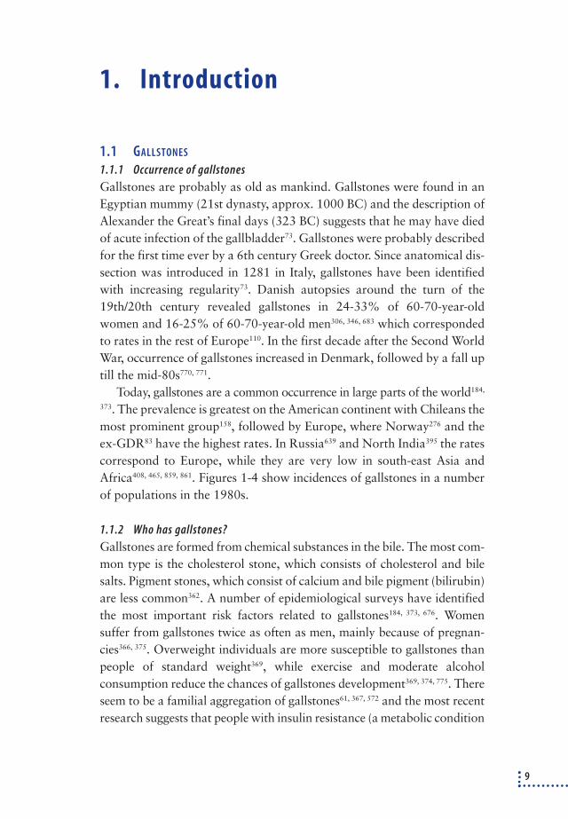
Institutional foreword
This report started as a joint clinical epidemiological project involving theNational Institute of Public Health (NIPH) and consultant surgeon Tor-ben Jørgensen during his tenure in the Surgical Department at BispebjergHospital. The NIPH has traditionally been adept at analysing data fromthe National Hospital Discharge Register for health service research pro-jects and health technology assessments. Torben Jørgensen has a long-standing interest in gallstone epidemiology and has conducted critical re-search into surgical treatment of gallstones. Torben Jørgensen worked atthe NIPH during part of the project and it was finished after his appoint-ment as head of the Copenhagen County Centre of Preventative Medicine,Glostrup University Hospital.
The project was financed by the NIPH’s basic grant and the Health In-surance Fund
It was decided that the appropriate form for the project was a healthtechnology assessment (HTA), i.e. an assessment in which clinicians, epi-demiologists, statisticians and health service economists co-operate tostudy a particular clinical field.
Due to the current heavy interest in HTA projects, it was agreed withthe Danish Institute for Health Technology Assessment (HTA Institute)that we would finance the expansion of the project to encompass healtheconomics and organisational elements, etc. As a result of this decision,co-operation was also established with the health service economists atthe Centre for Health and Social Policy, University of Southern Denmark.
In other words, the project has grown from a somewhat narrow clini-cal epidemiological project to a project that aspires to the breadth of anHTA. The emphasis in the report is, however, still on the clinical and epi-demiological areas.
NIPH and the HTA Institute found it appropriate to co-operate on thepublication of the overall HTA report. The report will be supplementedby scientific articles in magazines in Danish and English.
For further details, please refer to the author’s foreword.
Finn Kamper-Jørgensen, Director Finn Børlum Kristensen, Head of Institute
National Institute of Public Health Danish Institute for Health Technology Assessment
3

Author’s foreword
The idea for this report emerged during my tenure as consultant in theSurgical Gastroenterology Ward K at Bispebjerg Hospital in Copenhagen.It started out as a clinical epidemiological project and as a natural pro-gression of my research into biliary tract disorders. During the prelimi-nary research, it became evident that the whole area was crying out for in-depth study, hence the decision to expand the project into a health tech-nology assessment in order to lay the foundations for the rational, patient-friendly and economical treatment of patients with biliary tract disorders.The concept was presented at a meeting of Dansk Kirurgisk Selskab (Danish Society of Surgeons) in spring 1997 and the Society expressed thewish that the report would form the basis for national clinical guidelines.
In order to include a maximum number of aspects of the treatment ofbiliary tract disorders, the report has become very comprehensive. As a re-sult, those with no knowledge of the field will find the report heavy read-ing. Chapters 1-4 ought to be read, while chapters 5-8 can be read sepa-rately or used for reference purposes, as they concern particular aspects ofbiliary tract disorder. Chapters 9, 10 and 11 cover the other aspects of anHTA: the patient, the organisation and the finances. As so much docu-mentation has been included, some of it has been included in appendices,which can be read as required. The summary (chapter 12) and the syn-thesis and recommendations (chapter 13) can be read on their own.
A number of people have contributed to this report. Firstly, I wouldlike to thank chief surgeon Johan Kjærgaard for his willingness to grantme leave for seven months, for all our in-depth discussions and for hiswillingness to read and comment critically on the report. I would also liketo thank my ex-colleague in department K, development consultant IngridWillaing for our fruitful discussions as well as her stringent and alwayspositive critical study of my draft manuscript.
The work was done at the National Institute for Public Health, whichpossesses the necessary expertise in the processing and analysis of datafrom the National Hospital Discharge Register. Director Mette Madsen,MA, NIPH provided invaluable help interpreting and analysing data fromthe National Hospital Discharge Register and discussing and revising thereport critically. IT consultant Lene Bjørk Nielsen, NIPH was responsible
4

for the huge task of transforming the data from the National Hospital Dis-charge Register into a form that can be analysed and Søren Rasmussen,MSc, NIPH, performed the statistical analyses in exemplary fashion andwrote appendix 4 of this report. Health economist Jørgen Clausen fromCHS at the University of Southern Denmark did a marvellous job beingresponsible for the financial analyses. Jørgen Clausen’s detailed financialanalyses form the basis for chapter 11 and enclosure 5 in the report. Iwould like to avail myself of this opportunity to thank them all for theirpositive and constructive co-operation.
I would also like to thank the many surgical and medical departmentsall over the country who worked long and hard in attempt to validate theNational Hospital Discharge Register and to assess the time and staff de-mands of the different surgical and endoscopic operations. Thanks aredue to secretary Margit Christiansen, NIPH, for all her work sending outand sorting questionnaires and medical commentaries. I would also like toexpress my gratitude to Anne Eliasen, medical student Sine Wanda Jør-gensen, and medical student Jette Nielsen for all their help with the med-ical commentaries. Thanks are also due to Sine Wanda Jørgensen for herpatience and stamina photocopying the many thousands of articles uponwhich the report is based. Secretary Ulla Jørgensen deserves a specialthank you for her assiduous proof-reading and ward doctor Dina Haugefor her translations of Italian articles.
Finally, I would like to express my sincere gratitude to Director FinnKamper-Jørgensen and the staff of NIPH for providing a positive, hos-pitable and inspiring working environment.
The work was financed in part by the Health Insurance Fund (11/249-95) and the Danish Institute for Health Technology Assessment (J.no.3126-2-1997).
NIPH, 4 December 1998
Torben Jørgensen
Note: This report was translated from Danish by “The Translation Centre,University of Copenhagen”.
5

Content
1. Introduction . . . . . . . . . . . . . . . . . . . . . . . . . . . . . . . . . . . . . . . . . . . . . . . . . . . . . . . . . . . . . . .91.1 Gallstones . . . . . . . . . . . . . . . . . . . . . . . . . . . . . . . . . . . . . . . . . . . . . . . . . . . . . . . . . . . . . . . . . . . . . . . .9
1.1.1 Occurrence of gallstones . . . . . . . . . . . . . . . . . . . . . . . . . . . . . . . . . . . . . . . . . . . . . . . . . . . . . . . . .9
1.1.2 Who has gallstones? . . . . . . . . . . . . . . . . . . . . . . . . . . . . . . . . . . . . . . . . . . . . . . . . . . . . . . . . . . . .9
1.1.3 The disease spectrum . . . . . . . . . . . . . . . . . . . . . . . . . . . . . . . . . . . . . . . . . . . . . . . . . . . . . . . . . .11
1.2 Developments in the treatment of gallstone patients . . . . . . . . . . . . . . . . . . . . . . . . . . . . . . . . .11
1.2.1 Cholecystectomy (removal of the gallbladder) . . . . . . . . . . . . . . . . . . . . . . . . . . . . . . . . . . . . .12
1.2.2 Cholecystolithotomy (removal of gallbladder stone) . . . . . . . . . . . . . . . . . . . . . . . . . . . . . . .13
1.2.3 Bile salts and ESWL (medical dissolution of gallstones) . . . . . . . . . . . . . . . . . . . . . . . . . . . . .14
1.2.4 Choledocholithotomy (removal of stones in the bile duct) . . . . . . . . . . . . . . . . . . . . . . . . . . .14
1.3 Health technology assessment . . . . . . . . . . . . . . . . . . . . . . . . . . . . . . . . . . . . . . . . . . . . . . . . . . . . .16
2. Material and methods . . . . . . . . . . . . . . . . . . . . . . . . . . . . . . . . . . . . . . . . . . . . . . . . . . . . . .192.1 The National Hospital Discharge Register . . . . . . . . . . . . . . . . . . . . . . . . . . . . . . . . . . . . . . . . . . . .19
2.1.1 Courses of treatment . . . . . . . . . . . . . . . . . . . . . . . . . . . . . . . . . . . . . . . . . . . . . . . . . . . . . . . . . . .19
2.1.2 The validity of the National Hospital Discharge Register . . . . . . . . . . . . . . . . . . . . . . . . . . . .22
2.2 Literature . . . . . . . . . . . . . . . . . . . . . . . . . . . . . . . . . . . . . . . . . . . . . . . . . . . . . . . . . . . . . . . . . . . . . . .24
2.3 Danish population studies . . . . . . . . . . . . . . . . . . . . . . . . . . . . . . . . . . . . . . . . . . . . . . . . . . . . . . . . .25
2.4 Staff and staff time in the treatment of gallstone patients in Denmark . . . . . . . . . . . . . . . . . .25
2.5 The National Register for Laparoscopic Cholecystectomy . . . . . . . . . . . . . . . . . . . . . . . . . . . . . . .25
2.6 Analyses . . . . . . . . . . . . . . . . . . . . . . . . . . . . . . . . . . . . . . . . . . . . . . . . . . . . . . . . . . . . . . . . . . . . . . . .26
3. Occurrence, natural history and prevention . . . . . . . . . . . . . . . . . . . . . . . . . . . . . . . . . . .283.1 Gallstone disorders in the Danish population . . . . . . . . . . . . . . . . . . . . . . . . . . . . . . . . . . . . . . . . .28
3.2 The natural history of people with untreated gallstones . . . . . . . . . . . . . . . . . . . . . . . . . . . . . . .29
3.3 Prevention of gallstones . . . . . . . . . . . . . . . . . . . . . . . . . . . . . . . . . . . . . . . . . . . . . . . . . . . . . . . . . .30
3.3.1 Primary prevention . . . . . . . . . . . . . . . . . . . . . . . . . . . . . . . . . . . . . . . . . . . . . . . . . . . . . . . . . . . .30
3.3.2 Secondary prevention . . . . . . . . . . . . . . . . . . . . . . . . . . . . . . . . . . . . . . . . . . . . . . . . . . . . . . . . . .31
4. Treatment of patients with benign biliary tract disorders in Denmark 1978-95 . . . . .33
5. Treatment of patients with non-complicated gallbladder stones . . . . . . . . . . . . . . . . .475.1 Developments in Denmark, 1978-95 . . . . . . . . . . . . . . . . . . . . . . . . . . . . . . . . . . . . . . . . . . . . . . . .47
5.1.1 The frequency of simple cholecystectomy . . . . . . . . . . . . . . . . . . . . . . . . . . . . . . . . . . . . . . . . .47
5.1.2 Regional variations in Denmark . . . . . . . . . . . . . . . . . . . . . . . . . . . . . . . . . . . . . . . . . . . . . . . . .51
5.2 Which symptoms are due to stones in the gallbladder? . . . . . . . . . . . . . . . . . . . . . . . . . . . . . . . .60
5.3 Indication for treatment . . . . . . . . . . . . . . . . . . . . . . . . . . . . . . . . . . . . . . . . . . . . . . . . . . . . . . . . . .63
5.4 Methods of treatment . . . . . . . . . . . . . . . . . . . . . . . . . . . . . . . . . . . . . . . . . . . . . . . . . . . . . . . . . . . .66
5.4.1 Cholecystectomy . . . . . . . . . . . . . . . . . . . . . . . . . . . . . . . . . . . . . . . . . . . . . . . . . . . . . . . . . . . . . .66
5.4.2 Cholecystolithotomy . . . . . . . . . . . . . . . . . . . . . . . . . . . . . . . . . . . . . . . . . . . . . . . . . . . . . . . . . . .86
5.4.3 ESWL/bile salts . . . . . . . . . . . . . . . . . . . . . . . . . . . . . . . . . . . . . . . . . . . . . . . . . . . . . . . . . . . . . . . .87
5.4.4 Comparison between cholecystectomy, cholecystolithotomy and ESWL/bile salts . . . . . .89
5.4.5 Cholecystectomy for acalculous pains . . . . . . . . . . . . . . . . . . . . . . . . . . . . . . . . . . . . . . . . . . . .90
6

6. Treatment of patients with acute cholecystitis . . . . . . . . . . . . . . . . . . . . . . . . . . . . . . . . .926.1 Developments in Denmark 1978-95 . . . . . . . . . . . . . . . . . . . . . . . . . . . . . . . . . . . . . . . . . . . . . . . . .92
6.2 Indication for treatment . . . . . . . . . . . . . . . . . . . . . . . . . . . . . . . . . . . . . . . . . . . . . . . . . . . . . . . . . .95
6.3 Methods of treatment . . . . . . . . . . . . . . . . . . . . . . . . . . . . . . . . . . . . . . . . . . . . . . . . . . . . . . . . . . . .95
6.3.1 Cholecystectomy . . . . . . . . . . . . . . . . . . . . . . . . . . . . . . . . . . . . . . . . . . . . . . . . . . . . . . . . . . . . . .95
6.3.2 Cholecystolithotomy and partial cholecystectomy . . . . . . . . . . . . . . . . . . . . . . . . . . . . . . . . .98
6.3.3 Ultrasonic drainage . . . . . . . . . . . . . . . . . . . . . . . . . . . . . . . . . . . . . . . . . . . . . . . . . . . . . . . . . . . .98
6.3.4 Acalculous cholecystitis . . . . . . . . . . . . . . . . . . . . . . . . . . . . . . . . . . . . . . . . . . . . . . . . . . . . . . . .99
7. Treatment of patients with choledochal stones . . . . . . . . . . . . . . . . . . . . . . . . . . . . . . .1007.1 Developments in Denmark, 1978-95 . . . . . . . . . . . . . . . . . . . . . . . . . . . . . . . . . . . . . . . . . . . . . . .100
7.2 Indication for examination and treatment of stones in the bile ducts . . . . . . . . . . . . . . . . . . .104
7.3 Methods of treatment . . . . . . . . . . . . . . . . . . . . . . . . . . . . . . . . . . . . . . . . . . . . . . . . . . . . . . . . . . .106
7.3.1 Surgical methods of treatment . . . . . . . . . . . . . . . . . . . . . . . . . . . . . . . . . . . . . . . . . . . . . . . . .107
7.3.2 Endoscopic methods of treatment . . . . . . . . . . . . . . . . . . . . . . . . . . . . . . . . . . . . . . . . . . . . . .107
7.3.3 Comparison between surgical and endoscopic treatment . . . . . . . . . . . . . . . . . . . . . . . . . .108
7.3.4 Thirty-day mortality rates in Denmark, 1978-95 . . . . . . . . . . . . . . . . . . . . . . . . . . . . . . . . .109
8. Treatment of patients with gallstone pancreatitis . . . . . . . . . . . . . . . . . . . . . . . . . . . . .1118.1 Development in Denmark, 1978-95 . . . . . . . . . . . . . . . . . . . . . . . . . . . . . . . . . . . . . . . . . . . . . . . .111
8.2 Indication for treatment . . . . . . . . . . . . . . . . . . . . . . . . . . . . . . . . . . . . . . . . . . . . . . . . . . . . . . . . .111
8.3 Methods of treatment . . . . . . . . . . . . . . . . . . . . . . . . . . . . . . . . . . . . . . . . . . . . . . . . . . . . . . . . . . .111
8.3.1 Cholecystectomy . . . . . . . . . . . . . . . . . . . . . . . . . . . . . . . . . . . . . . . . . . . . . . . . . . . . . . . . . . . . .112
8.3.2 Endoscopic treatment . . . . . . . . . . . . . . . . . . . . . . . . . . . . . . . . . . . . . . . . . . . . . . . . . . . . . . . . .113
8.3.3 Treatment strategies for gallstone pancreatitis – summary . . . . . . . . . . . . . . . . . . . . . . . .113
9. The patient . . . . . . . . . . . . . . . . . . . . . . . . . . . . . . . . . . . . . . . . . . . . . . . . . . . . . . . . . . . . . .1159.1 The patient’s choice of treatment procedure . . . . . . . . . . . . . . . . . . . . . . . . . . . . . . . . . . . . . . . .115
9.2 Patient expectations . . . . . . . . . . . . . . . . . . . . . . . . . . . . . . . . . . . . . . . . . . . . . . . . . . . . . . . . . . . .116
9.3 Patient information . . . . . . . . . . . . . . . . . . . . . . . . . . . . . . . . . . . . . . . . . . . . . . . . . . . . . . . . . . . . .116
9.4 The patient’s assessment of the given treatment . . . . . . . . . . . . . . . . . . . . . . . . . . . . . . . . . . . .117
10. Organisation . . . . . . . . . . . . . . . . . . . . . . . . . . . . . . . . . . . . . . . . . . . . . . . . . . . . . . . . . . . . .11910.1 Administrative rules for the treatment of patients with gallstones in Denmark . . . . . . . . . .119
10.2 Gallstone treatments in Denmark . . . . . . . . . . . . . . . . . . . . . . . . . . . . . . . . . . . . . . . . . . . . . . . . .119
10.2.1 Developments in Denmark, 1978-95 . . . . . . . . . . . . . . . . . . . . . . . . . . . . . . . . . . . . . . . . . . . .119
10.2.2 Morbidity and mortality in relation to the number of operations . . . . . . . . . . . . . . . . . . .120
10.3 Training for gallstone surgeons . . . . . . . . . . . . . . . . . . . . . . . . . . . . . . . . . . . . . . . . . . . . . . . . . . .120
11. Economy . . . . . . . . . . . . . . . . . . . . . . . . . . . . . . . . . . . . . . . . . . . . . . . . . . . . . . . . . . . . . . . .12311.1 Introduction . . . . . . . . . . . . . . . . . . . . . . . . . . . . . . . . . . . . . . . . . . . . . . . . . . . . . . . . . . . . . . . . . . . .123
11.2 Developments in treatment costs in Denmark, 1978-1995 . . . . . . . . . . . . . . . . . . . . . . . . . . . .124
11.2.1 All treatments for benign biliary tract disorderse . . . . . . . . . . . . . . . . . . . . . . . . . . . . . . . . .124
11.2.2 Simple cholecystectomy . . . . . . . . . . . . . . . . . . . . . . . . . . . . . . . . . . . . . . . . . . . . . . . . . . . . . . .126
11.2.3 Discussion . . . . . . . . . . . . . . . . . . . . . . . . . . . . . . . . . . . . . . . . . . . . . . . . . . . . . . . . . . . . . . . . . . .127
11.3 Economical models for gallstone treatments . . . . . . . . . . . . . . . . . . . . . . . . . . . . . . . . . . . . . . . .128
11.3.1 Treatment of patients with stones in the gallbladder . . . . . . . . . . . . . . . . . . . . . . . . . . . . . .129
11.3.2 Treatment of patients with acute cholecystitis . . . . . . . . . . . . . . . . . . . . . . . . . . . . . . . . . . .140
11.3.3 Treatment of patients with stones in the bile ducts . . . . . . . . . . . . . . . . . . . . . . . . . . . . . . .141
7

12. Summary of the elements in the Health Technology Assessment . . . . . . . . . . . . . . . .14212.1 Developments in the incidence and treatment of gallstones . . . . . . . . . . . . . . . . . . . . . . . . . . .142
12.2 Material and methods . . . . . . . . . . . . . . . . . . . . . . . . . . . . . . . . . . . . . . . . . . . . . . . . . . . . . . . . . . .144
12.3 Occurrence, natural history and prevention . . . . . . . . . . . . . . . . . . . . . . . . . . . . . . . . . . . . . . . . .145
12.4 Overall treatment of biliary tract disorders in Denmark . . . . . . . . . . . . . . . . . . . . . . . . . . . . . . .146
12.5 Treatment of patients with uncomplicated gallbladder stones . . . . . . . . . . . . . . . . . . . . . . . .147
12.6 Treatment of patients with acute cholecystitis . . . . . . . . . . . . . . . . . . . . . . . . . . . . . . . . . . . . . .151
12.7 Treatment of patients with stones in the bile ducts . . . . . . . . . . . . . . . . . . . . . . . . . . . . . . . . . .152
12.8 Treatment of patients with gallstone pancreatitis . . . . . . . . . . . . . . . . . . . . . . . . . . . . . . . . . . .154
12.9 The patient . . . . . . . . . . . . . . . . . . . . . . . . . . . . . . . . . . . . . . . . . . . . . . . . . . . . . . . . . . . . . . . . . . . .154
12.10 The organisation . . . . . . . . . . . . . . . . . . . . . . . . . . . . . . . . . . . . . . . . . . . . . . . . . . . . . . . . . . . . . . .155
12.11 Economy . . . . . . . . . . . . . . . . . . . . . . . . . . . . . . . . . . . . . . . . . . . . . . . . . . . . . . . . . . . . . . . . . . . . . . .156
13. Synthesis and recommendations . . . . . . . . . . . . . . . . . . . . . . . . . . . . . . . . . . . . . . . . . . . .158
Appendices1 Literature list . . . . . . . . . . . . . . . . . . . . . . . . . . . . . . . . . . . . . . . . . . . . . . . . . . . . . . . . . . . . . . . . . . .164
2 The National Hospital Discharge Register . . . . . . . . . . . . . . . . . . . . . . . . . . . . . . . . . . . . . . . . . . .201
Extract from the National Hospital Discharge Register . . . . . . . . . . . . . . . . . . . . . . . . . . . . .201
Validation of the National Hospital Discharge Register . . . . . . . . . . . . . . . . . . . . . . . . . . . .209
Developments in the use of different technologies for treatment of patients
with bile duct disorders, 1978-95 . . . . . . . . . . . . . . . . . . . . . . . . . . . . . . . . . . . . . . . . . . . . . . .210
3 Literature . . . . . . . . . . . . . . . . . . . . . . . . . . . . . . . . . . . . . . . . . . . . . . . . . . . . . . . . . . . . . . . . . . . . . .214
Search of literature . . . . . . . . . . . . . . . . . . . . . . . . . . . . . . . . . . . . . . . . . . . . . . . . . . . . . . . . . . .214
Critical evaluation of the literature . . . . . . . . . . . . . . . . . . . . . . . . . . . . . . . . . . . . . . . . . . . . .215
Selected literature . . . . . . . . . . . . . . . . . . . . . . . . . . . . . . . . . . . . . . . . . . . . . . . . . . . . . . . . . . . .217
4 Statistical analyses . . . . . . . . . . . . . . . . . . . . . . . . . . . . . . . . . . . . . . . . . . . . . . . . . . . . . . . . . . . . . .236
Analysis of developments in surgical rate . . . . . . . . . . . . . . . . . . . . . . . . . . . . . . . . . . . . . . . .236
Regional variations in operation rates . . . . . . . . . . . . . . . . . . . . . . . . . . . . . . . . . . . . . . . . . .241
Analyses of 30-day mortality rates . . . . . . . . . . . . . . . . . . . . . . . . . . . . . . . . . . . . . . . . . . . . .244
Analyses of complicated courses of treatment in cases of simple cholecystectomy . . . . .245
The distribution of municipalities between admissions areas . . . . . . . . . . . . . . . . . . . . . .246
5 Economical analyses . . . . . . . . . . . . . . . . . . . . . . . . . . . . . . . . . . . . . . . . . . . . . . . . . . . . . . . . . . . . .250
Costs of gallstone treatment in Denmark . . . . . . . . . . . . . . . . . . . . . . . . . . . . . . . . . . . . . . . .250
Sensitivity analyses of the economical models . . . . . . . . . . . . . . . . . . . . . . . . . . . . . . . . . . . .254
8
1. Introduction
1.1 GALLSTONES
1.1.1 Occurrence of gallstones
Gallstones are probably as old as mankind. Gallstones were found in anEgyptian mummy (21st dynasty, approx. 1000 BC) and the description ofAlexander the Great’s final days (323 BC) suggests that he may have diedof acute infection of the gallbladder73. Gallstones were probably describedfor the first time ever by a 6th century Greek doctor. Since anatomical dis-section was introduced in 1281 in Italy, gallstones have been identifiedwith increasing regularity73. Danish autopsies around the turn of the19th/20th century revealed gallstones in 24-33% of 60-70-year-oldwomen and 16-25% of 60-70-year-old men306, 346, 683 which correspondedto rates in the rest of Europe110. In the first decade after the Second WorldWar, occurrence of gallstones increased in Denmark, followed by a fall uptill the mid-80s770, 771.
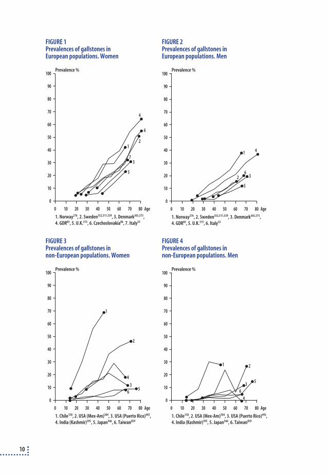
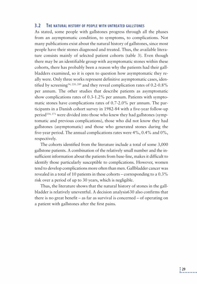
Today, gallstones are a common occurrence in large parts of the world184,
373. The prevalence is greatest on the American continent with Chileans themost prominent group158, followed by Europe, where Norway276 and theex-GDR83 have the highest rates. In Russia639 and North India395 the ratescorrespond to Europe, while they are very low in south-east Asia andAfrica408, 465, 859, 861. Figures 1-4 show incidences of gallstones in a numberof populations in the 1980s.
1.1.2 Who has gallstones?
Gallstones are formed from chemical substances in the bile. The most com-mon type is the cholesterol stone, which consists of cholesterol and bilesalts. Pigment stones, which consist of calcium and bile pigment (bilirubin)are less common362. A number of epidemiological surveys have identifiedthe most important risk factors related to gallstones184, 373, 676. Women suffer from gallstones twice as often as men, mainly because of pregnan-cies366, 375. Overweight individuals are more susceptible to gallstones thanpeople of standard weight369, while exercise and moderate alcoholconsumption reduce the chances of gallstones development369, 374, 775. Thereseem to be a familial aggregation of gallstones61, 367, 572 and the most recentresearch suggests that people with insulin resistance (a metabolic condition
9
10
FIGURE 1Prevalences of gallstones inEuropean populations. Women
FIGURE 2Prevalences of gallstones inEuropean populations. Men
FIGURE 3Prevalences of gallstones innon-European populations. Women
FIGURE 4Prevalences of gallstones innon-European populations. Men
100
Age
90
80
70
60
50
40
30
20
10
0
0 10 20 30 40 50 60 70 80
12
3
4
5
7
1. Norway276, 2. Sweden352,511,539, 3. Denmark365,375,4. GDR83, 5. U.K.315, 6. Czechoslovakia86, 7. Italy53
Prevalence %100
Age
90
80
70
60
50
40
30
20
10
0
0 10 20 30 40 50 60 70 80
1
1. Norway276, 2. Sweden352,511,539, 3. Denmark365,375,4. GDR83, 5. U.K.315, 6. Italy53
2 3
4
5
6
Prevalence %
6
100
Age
90
80
70
60
50
40
30
20
10
0
0 10 20 30 40 50 60 70 80
1
1. Chile158, 2. USA (Mex-Am)304, 3. USA (Puerto Rico)492,4. India (Kashmir)395, 5. Japan566, 6. Taiwan859
2
3
4
56
Prevalence %100
Age
90
80
70
60
50
40
30
20
10
0
0 10 20 30 40 50 60 70 80
1
1. Chile158, 2. USA (Mex-Am)304, 3. USA (Puerto Rico)492,4. India (Kashmir)395, 5. Japan566, 6. Taiwan859
2
3
4
5
6
Prevalence %

that increases the risk of arteriosclerosis, cardiovascular disease, obesityand non-insulin-dependent diabetes) have a far higher occurrence of gall-stones than non-insulin-resistant372. This corresponds to the very high incidence of gallstones among Native Americans, the majority of whomare insulin resistant677. Gallstones are rarely encountered in children andyoung people, but the prevalence increases with age585, 832.
1.1.3 The disease spectrum
Gallstone disease is a chronic condition, which starts - and usually ends- as an asymptomatic condition, i.e. a condition that the person never dis-covers. The development is probably as follows:
❖ Stones are formed in the gallbladder, where they remain asymptomatic for a long
time, perhaps permanently. Small gallstones can disappear again spontaneously.
❖ Stones can cause pain, which is unpleasant, but not dangerous.
❖ After an certain number of years, the stones can cause complications, which as a
rule can be easily treated and are rarely life-threatening:
- Acute cholecystitis
- Choledochal stone
Icterus/cholangitis
Acute pancreatitis
- Fistulation, if a chronic infection involves the neighbouring organs.
1.2 DEVELOPMENTS IN THE TREATMENT OF GALLSTONE PATIENTS
Doctors did not attribute any significance to gallstones in relation tosymptoms such as abdominal pains, jaundice and infection until the 16thcentury73. In principle, there are two methods of treating stones in thegallbladder:
❖ Removal of the gallbladder including the stone (cholecystectomy)
❖ Removal of the stone from the gallbladder
- invasive technique (cholecystolithotomy or contact dissolution)
- non-invasive technique (ESWL/bile salts). .
In principle stones in the bile duct should be removed (choledocholitho-tomy). Below is a short description of historical developments in the dif-ferent forms of treatment.
11

1.2.1 Cholecystectomy (removal of the gallbladder)
Three different technologies are used: traditional open cholecystectomy,cholecystectomy by minilaparotomy and laparoscopic cholecystectomy.
The first cholecystectomy was performed by Langenbuch in Berlin in 1882429, while the first cholecystectomy in Denmark was performed in1887 by Iversen at Copenhagen District General Hospital25. Removal ofgallbladder and stones remains the standard treatment to this day. Thegallbladder was removed through a 10-20 cm long subcostal or verticalmidline incision (traditional open cholecystectomy). The argument infavour of the large incision was that it afforded the surgeon a better viewand that the whole of the abdominal cavity could be properly examined.
In the early 70s, a technique was developed in which the surgeon useslong instruments to remove the gallbladder through a 3-6 cm long incisionand does not insert his hands into the abdomen (cholecystectomy by mini-laparotomy)197, 282. The argument in favour of minilaparotomy is that it re-duces surgical trauma and, consequently, the time spent in hospital andthe convalescence period (the period from the operation to resumption ofwork/normal activities). It is difficult to define exactly which incisionsqualify as traditional laparotomy and which constitute minilaparotomy.Besides the length of the incision, it has been discussed whether the pro-cedure also should spare the muscles (i.e. not separate the muscles in theabdominal wall) before it can be defined as minilaparotomy. The major-ity defines an operation as minilaparotomy if the incision is under 6 cm,while others accept 8 cm483, 656 or even 10 cm435, 504. Some point out the im-portance of the location of the incision, which ought to be on the right ofthe epigastrium, just above the point where ductus cysticus joins ductuscholedochus. This is where the difficult dissection occurs793. If a minila-parotomy runs into technical or other problems, the incision is lengthenedand the operation is converted into a traditional open cholecystectomy. Itis uncertain, when the first cholecystectomy during a minilaparotomy wasperformed in Denmark733.
Mühe introduced laparoscopic cholecystectomy in Germany in 1985545,
546. The method was refined by Mouret in France in 1987198, 601. The op-eration is performed with special instruments and video equipment in-serted through three or four trochars (“doors”) in the abdominal wall,each 1⁄2-1 cm in diameter. Thus, the total incision is 3-4 cm. The argumentsin favour of laparoscopic cholecystectomy correspond to those in favourof cholecystectomy by minilaparotomy: it reduces surgical trauma, thetime spent in hospital and the convalescence period. If technical or other
12

difficulties occur, the operation is converted into open surgery – usuallytraditional open cholecystectomy. This requires new instruments, a newincision and the removal of old instruments. Outside Denmark, the la-paroscopic technique soon gave cause for concern because of a suspectedrise in the number of bile duct lesions166 and at the same time, the numberof gallstone operations increased149. Laparoscopic cholecystectomy wasintroduced to Denmark in January 1991. During the first year, only asmall proportion of the total number of cholecystectomies were per-formed laparoscopically, but in the ensuing year the number increased toconstitute the majority.
1.2.2 Cholecystolithotomy (removal of gallbladder stone)
Surgical removalIn 1667, an abscess in the abdominal wall containing a gallstone was emp-tied322. It is probably the first recorded case of cholecystolithotomy as atreatment for acute infection of the gallbladder. The first planned opera-tion to remove a stone from a gallbladder was performed by Bobbs in1867 in the USA169. When Langenbuch later carried out the first chole-cystectomy, most surgeons converted to removal of the gallbladder alongwith the stones due to the large number of patients who experienced a re-currence of their gallstones after cholecystolithotomy603. Langenbuch isquoted as saying: “The gallbladder has to be removed – not because it contains stones, but because it generates stones.” Cholecystolithotomystill had its advocates, however, and as late as World War Two many sur-geons in Denmark insisted on removing only the gallstones25, 603. Alreadyin 1955 it was described that the procedure could be performed by mini-laparotomy670 and even under local anaesthesia645 for patients unable totolerate general anaestesia. Later, a laparoscopic method was developed inwhich a scope is inserted through a 2-3 cm incision, attaches itself to thegallbladder using suction, opens the gallbladder and empties it of stones434,
481. This operation can also be performed under local anaesthesia. The de-velopment of ultrasound techniques made it possible to puncture the gall-bladder. In the beginning, this method was used on patients with acutecholecystitis to combat infection in advance of subsequent operations623.The latter method was further refined by dilating the puncture canal andinserting instruments to remove the stones or laser/ultrasound probes topulverise the stones16, 341, 386, 392, 833. The method has been used in Den-mark734.
13

Contact dissolutionSince the 19th century, it has been a well-known fact that ether dissolvesgallstones814. Methyl tertbutyl ether (MTBE) has been particularly well re-searched because it stays in liquid form at body temperature (boiling point55). MTBE is injected via a catheter guided into the gallbladder by ultra-sound equipment22. It can take from a few hours to a couple of days to dis-solve the stones and flush them out207, 318. The method is described in depthby Thistle777. Attempts have been made to combine the method withESWL (see next section)562, 597 and other solvents351, 553 – but to no great ef-fect. Often, the procedure is followed up with bile salts193. Instead of per-cutaneous (and therefore invasive) incision, reports have been publishedof inserting a naso-biliary catheter into the gallbladder by ERCP and dis-solving gallbladder stones with MTBE234. MTBE has a number of side ef-fects in the form of lethargy and nausea. Recently, positive tests have beenperformed using ethyl propionate without the side effects of MTBE. How-ever, the tests only comprised five patients330. It is not known to what ex-tent the method has been tried in Denmark.
1.2.3 Bile salts and ESWL (medical dissolution of gallstones)
Bile salts reduce cholesterol concentration in the bile331. The first attemptto dissolve gallstone with bile-salt treatment was performed in 1970171 us-ing chenodeoxycholic acid, but the initial results were not promising691,partly because of side-effects such as diarrhoea, liver damage and in-creased levels of cholesterol in the blood254. In 1975, a new chemical waslaunched on the market (ursodeoxycholic acid), which was more efficientand had fewer side effects430, 672. Treatment with bile salts takes severalmonths to years485.
In 1985, bile salt treatment was combined with ESWL (ExtracorporealShockWave Lithotripsy)680, which uses sound waves to pulverise galls-tones. ESWL produces a blast wave that works in fluids. A targeting sys-tem makes sure that the blast wave hits the gallstones and does not causelesions to the surrounding tissue. This combination of bile salts and ESWLhas been used in Denmark31, 47.
1.2.4 Choledocholithotomy (removal of stones in the bile duct)
Stones in the bile duct almost always stem from the gallbladder. Surgicalremoval of stones in the bile duct was done for the first time by Cour-voisier in Basle in 189073. From then on, open surgery to remove the gall-bladder and the stones in the bile duct (traditional open bile duct surgery)
14

became the standard treatment. Often, patients do not have bile ductstones diagnosed until they are X-rayed during the operation (intraopera-tive cholangiography). Sometimes the number of number or size of bileduct stones is so great, or the alterations of the bile duct so severe, that ithas to be dissected and sewn onto the intestines (biliodigestive anastomo-sis). A 1978-85 Danish survey revealed that 20% of patients who under-went cholecystectomy also had stones removed from ductus choledochusand a further 5% underwent biliodigestive anastomosis106. Open bile ductsurgery has traditionally been performed through a 10-20 cm long sub-costal or vertical midline incision. In 1982, the first report was publishedconcerning the removal of stones in the bile duct through a small incisionof 4-5 cm (removal of bile duct stone through minilaparotomy)197, 532. In1991, the first report340 was published concerning removal of stones fromthe bile duct with the help of laparoscopic surgery (laparoscopic removalof stones from the bile duct). The latter method has not been adopted inDenmark37.
The first ERCP (Endoscopic Retrograde Cholangiopancreatography)was presented in 1970575. Later, instruments were developed to performsphincterotomy and remove stones from the bile duct143, 383. ERCP was in-troduced to Denmark in 1973412, 413, 491, but initial progress was slow. Inthe late 80s, the pace at which ERCP technology spread began to pick upand then accelerate after the introduction of laparoscopic cholecystec-tomy in 1991. As ERCP technology was developed, so was a number ofother treatments for stones in the bile duct, e.g.: contact dissolution156, di-rect dissolution by introduction of various medicaments into the bileduct544 and ESWL738. The latter method is used in Denmark24. In cases,where it proved impossible to remove the gallstones, methods were devel-oped to insert a drain and allow the bile to pass125. Instead of a sphinc-terotomy, proposals have been mooted to dilate the sphincter with a bal-loon and then remove the stones474.
Recently, MR scanning (magnetic resonance), which unlike ERCP is anon-invasive technique, has been used to produce a three-dimensional im-age of the biliary tracts (MRC) and the results have been promising727.
1.3 HEALTH TECHNOLOGY ASSESSMENT
Health technology assessment is defined by the Danish National Board ofHealth as an all-round, systematic evaluation of the preconditions for, andthe consequences of, using health technology. Health technology includesall forms of diagnostics, prevention, treatment and care of patients, which
15

means that it is not necessarily associated with technical equipment. In re-cent years, health technology assessment has gained a foothold in a num-ber of health services all over the world. This development has coincidedwith rapid technological progress within the health service, demands frompatients and health-service personnel for evidence-based treatment, pluslimited financial resources within the health sector. Patients have benefitedfrom many new technologies, but new technology cannot always beequated with improved treatment.
In most fields, the supply of health technology exceeds the health ser-vice’s financial resources. In principle, this means that all new technologyought to be subjected to health technology assessment. Health technologyassessment is, therefore, a tool that analyses all the available data andreaches decisions about which forms of health technology are expedientwithin the health service. Thus, a health technology assessment is not aready-made checklist of the extent to which a particular type of technol-ogy should be used and how, but a basis upon which health-service staff,administrators and politicians can make decisions.
Ideally, the HTA process includes the five steps, illustrated in figure 5.
FIGURE 5
The five steps involved in health technology assessment
The content of the four traditional main elements in a health technologyassessment: the technology, the patient, the organisation and the finan-ces, are described in figures 6-934.
Justification for a health technology assessment of the gallstone fieldThe many new methods of treatment developed in the 70s and 80s were in-troduced both abroad and in Denmark after trial runs in clinical series. Ran-domised studies of the relevance of the technologies in comparison to one an-other or to traditional methods of treatment were rarely conducted. The num-
16
Problem identification
Planning
Summary
Presentation and communication
Analysis of the elements
Technology Patient Organisation Economy

17
FIGURE 6
The Technology
Area of application◆ What are the indications?◆ Is there consensus on the indication?◆ How many patients are involved?◆ What are the relevant alternatives?◆ Is the technology a substitute or
supplement to existing technologies?
Effectiveness◆ Is there a documented effect?◆ Is it more effective than the alternatives?◆ Can the documented effect be attained in
daily practice?
Risk assessment◆ Are there any undesired effects?◆ Does the risk bear a reasonable
proportional relationship to the gain?
FIGURE 7
The Patient
Psychological aspects◆ Does the patient receive an optimal level
of information?◆ Is insecurity created/experienced?◆ Is discomfort or anxiety created/
experienced?
Effect aspects◆ How are the effects and side-effects
perceived?
Social aspects◆ Will it affect daily life?◆ Will it affect employment capacity?
Ethical aspects◆ Is the technology acceptable to the
individual patient?◆ Is it acceptable to society?◆ Are there special ethical issues?
FIGURE 8
The Organisation
Structure◆ Should the technology be centralised to a
single or a few centres?◆ Is decentralisation possible?◆ Will it affect the division of work between
hospital and primary healthcare?◆ Will new specialised functions emerge?◆ Will it change visitation criteria?
Staff◆ Will it affect work routines?◆ Will it affect the division of work among
professional groups?◆ Is supplemental/developmental staff
training needed?◆ Are there any consequences for the staff in
terms of employment?
Environment◆ Is there an environmental risk within the
workplace?◆ Is there a risk to the external environment?
FIGURE 9
The Economy
Social and health economic appraisal◆ What are the costs and benefits for
society?◆ In comparison to the alternatives,
does the gain justify the effort?◆ Is there a demonstrable health gain?
Operational economic appraisal◆ What are the investment and operational
costs?◆ Are there any possible cost-savings or
income generation?◆ Who is the immediate purchaser?◆ What accounts are affected? ◆ Are there any economic consequences for
the patient?

ber of new treatments has been particularly high for gallstone complications(acute cholecystitis and bile duct stones).
Several reports have cast doubt on the benefit of introducing laparoscopiccholecystectomy, which is expensive, may have lowered the threshold for ope-ration and may have increased the number of serious complications duringbiliary tract surgery194, 489. In addition, a number of randomised surveys haveraised doubts about the value of ERCP in relation to open surgery as treat-ment of stones in the bile duct394, 557, 737, 748.
Under these circumstances, the author, in collaboration with National In-stitute of Public Health (NIPH) and the Danish Institute for Health Techno-logy Assessment, found it relevant to perform a health technology assessmentof the whole gallstone field. On the basis of this HTA report, national guide-lines (reference programmes) can be developed for diagnostics, preventionand treatment of gallstones.
18

2. Material and methods
2.1 THE NATIONAL HOSPITAL DISCHARGE REGISTER
Each person living in Denmark has a unique 10-digit person number,which follows him throughout life. The person number is used for all re-gistrations in Danish registers, which makes linkage across time and re-gisters highly accurate.
2.1.1. Courses of treatment
Data from the National Hospital Discharge Register was used to acquirean overall impression of developments in the treatment of patients withgallstones in Denmark in the period 1978-95. A total of 99,803 hospitaladmissions were identified, including 87,007 patients of relevance to thisreport (see enclosure 2). As a rule, a patient’s treatment for gallstones iscompleted during a single stay in hospital, but sometimes a patient is ad-mitted several times during the same course of his or her treatment, partlyfor diagnostic examinations (ERCP), partly because the treatment is com-plicated and complications arise during it. In order to identify thesecourses of treatment, all hospital admissions for biliary tract treatmentlisted under the same patients person number with less than a year be-tween discharge date and subsequent admission date have been consid-ered as a single course of treatment. A single patient may have undergoneseveral courses of treatment. For the 87,007 patients, 90,582 courses oftreatment were identified - 96.3% of the patients underwent only a singlecourse of treatment, 3.2% underwent two, while 0.4% underwent threeor more. The treatments have been divided into a number of clinically rele-vant groups according to the index admission that refers to the primarytreatment (the index treatment). This included the following index admis-sions (table 1).
Simple cholecystectomy These patients, who make up the vast majority of cases, had their gall-bladders removed because of pains or infection. The majority of these pa-tients were admitted once only.
19

Cholecystectomy plus endoscopic bile duct treatment during the samestay in hospital These patients had the gallbladder as well as stone from the bile ducts removed during the first course of treatment or experienced complicationsduring the primary cholecystectomy that were treated endoscopically dur-ing the same stay in hospital.
Cholecystectomy plus open bile duct surgery during the same stay in hospitalMost of the patients in this group had their gallbladder and stones in thebile duct removed. The group also includes patients whose choledochuswas examined in greater detail during the operation (explorative chole-dochotomy), or who suffered a lesion of the choledochus that was diag-nosed and treated during the same stay in hospital.
Cholecystectomy with biliodigestive anastomosis This group consists mainly of patients with stones in both the gallbladderand the bile duct and whose bile duct was altered to such an extent thatthe surgeon deemed it necessary to perform an anastomosis of the bileduct and intestines. The group may also contain patients who suffer le-sions to the bile ducts during simple cholecystectomy.
Endoscopic bile duct treatment without simultaneous cholecystectomyThis group consists mainly of patients who only have stones removedfrom the bile ducts. Occasionally, a simple cholecystectomy is performedduring a later admission.
Open bile duct surgery without simultaneous cholecystectomyThis group consists mainly of patients who only have stones removedfrom the bile ducts and who have previously had their gallbladder re-moved.
Biliodigestive anastomosis without simultaneous cholecystectomyThis group consists of patients whose bile ducts are altered to such an ex-tent that the surgeon deemed it necessary to perform an anastomosis ofthe bile duct and intestines. The majority of these patients have a biliarytract diagnosis.
20

Other treatmentThese courses of treatment represent rare procedures or wrong codes, asa result of which the patient cannot be categorised in any of the groupslisted above.Diagnostic ERCPThese courses of treatment represent patients whose bile duct is examinedbut not treated immediately or subsequently. These are exploratory pro-cedures in cases where disease of the liver, pancreas or biliary tracts is sus-pected. However, as no therapeutic operation is performed, they seldomrepresent patients with gallstones. The group is included to show howwidespread the use of this technology is, even though it is not without risk.(Please refer to 7.3.2).
Each course of treatment can be divided according to whether the indexadmission is:
❖ the only admission to hospital during the course of treatment,
❖ preceded or followed by an admission for a diagnostic procedure (ERCP)
❖ followed by an admission for a cholecystectomy (only for some courses of treat-
ment)
❖ followed by one or more admissions for endoscopic or open surgery on the bile
duct within a year.
The latter category is assumed to include patients whose course of treat-ment is complicated, either because of complications to the biliary tractdisease (e.g. stones in the bile ducts) or because of procedure-related com-plications during the index treatment.
All courses of treatment are mutually exclusive and exhaustive – inother words, each course of treatment can be placed in one and only onecategory.
To assess the total number of hospital admissions accounted for bygallstone diseases, all admissions for a period of three months before andone year after the index admission (regardless of the nature of the treat-ment) were identified in the National Hospital Discharge Register.
Of the 90,582 courses of treatment, 12,262 only involved diagnosticERCP without treatment of the biliary tracts. Table 1 breaks down the re-maining 78,320 courses of treatment.
In-depth details of how the data from the National Hospital DischargeRegister was processed can be found in enclosure 2.
21

TABLE 1
Breakdown of 78,320 courses of treatment from 1978-95
The treatment during Admissions for biliary tract treatment after the index admission
the index admission
None Diagnostic ERCPa Cholecystectomy Bile duct Total
treatment % (N)
n n n n
Cholecystectomy 83.8
Only procedure 52,132 1,535 469 69.1 (54,136)
with endoscopic treatment 430 43 49 0.7 (522)
with open bile duct surgery 8,810 329 280 12.0 (9,419)
with biliodigestive anastomosis 1,414 104 25 2.0 (1,543)
Bile duct treatment 12.7
without cholecystectomy
Endoscopic 5,509 941 764 394 9.7 (7,608)
open surgery 969 71 46 1.4 (1,086)
with biliodigestive anastomosis 1,140 160 18 1.7 (1,318)
Other procedures 3.4
Exploration of gallbladder 1,244 21 22 1.6 (1,287)
other 1,155 95 67 84 1.8 (1,401)
Total 72,803 3,278 852 1,387 (78,320)
a: ERCP either before or after the index admission.
2.1.2 The validity of the National Hospital Discharge Register
As far as biliary tract treatments are concerned, the National HospitalDischarge Register has provided national data throughout the relevant pe-riod except 1978 and 1979 when St. Joseph’s Hospital in Copenhagenwas not included. Thus, there is a minor level of underreporting (<2%) inthe first two years.
To validate the data in the National Hospital Discharge Register (en-closure 2), a random sample was extracted including the years 1979, 1985and 1993 of approx. 10% of the courses of treatment and approx. 2% ofall patients with a biliary tract diagnosis who were admitted to surgicaldepartments, but who were not treated according to the register. Allcourses of treatment involving more than one admission to hospital forbiliary tract procedures were also extracted. In total, 3,570 commentarieswere requisitioned from hospitals around the country. As we go to press,71% of the commentaries have been received and reviewed to make a pro-visional assessment. The most significant results are:
❖ 96% of the courses of treatment were classified correctly. In the remaining 4% of
cases, patients had a different form of biliary tract treatment than the one regi-
22

stered in the National Hospital Discharge Register. No cases were identified in
which the patient’s biliary tracts were not treated.
❖ 99% of admissions recorded in the National Hospital Discharge Register with sim-
ple cholecystectomy as index treatment (n=682) were classified correctly. The final
1% consists of patients who had other operations performed on the bile ducts as
well as cholecystectomy. No cases were identified of patients who did not have a
cholecystectomy.
❖ All the entries in the National Hospital Discharge Register recorded as cholecyste-
ctomy combined with one or another form of bile duct treatment (n=122) were
found to be classified correctly.
❖ 95% of the courses of treatment involving endoscopic bile duct treatment but not
cholecystectomy (n=102) were classified correctly. In the remaining 5% of cases,
diagnostic ERCP was performed but not endoscopic bile duct treatment (1%), or
tubulation of the bile duct without sphincterotomy (4%).
❖ 84% of the courses of treatment classified in the National Hospital Discharge Re-
gister as exclusively diagnostic ERCP were classified correctly, with a clear trend
towards poorer validity over the years. In 1979, 96% of the classifications were cor-
rect, but only 80% in 1993. Among those wrongly classified, 36% of the commen-
taries contain no information about diagnostics or treatment of the biliary tracts,
while the rest had endoscopic treatment (sphincterotomy or tubulation of the bile
ducts). None of them had a cholecystectomy or an open operation on the bile
ducts.
❖ 91% of hospital admissions with a gallstone diagnosis but no treatment code
were classified correctly. The level of accuracy diminishes over the years, since 94%
were correct for the first two years, but only 82% correct in 1993. Among those
wrongly classified, 25% had diagnostic ERCP, while the rest had endoscopic treat-
ment (sphincterotomy or tubulation) of the biliary tracts. None had the gallblad-
der removed or open surgery of the biliary tracts.
It can be concluded that the data in the National Hospital Discharge Reg-ister forms a particularly suitable basis for the assessment of how oftencholecystectomy and open bile duct surgery are performed.
As regard therapeutic ERCP the underreporting is big. The scale of un-derreporting can be calculated on the basis of the proportion of wronglyclassified therapeutic ERCP in the groups where they were observed (en-doscopic procedure without cholecystectomy, diagnostic ERCP and staysin hospital without any treatment of the biliary tract) and comparing this
23

proportion with the response rate and total number of admissions regis-tered in the National Hospital Discharge Register in the relevant cate-gories. This suggests an underreporting of therapeutic ERCP levelsamounting to 75% in 1979, 42% in 1985 and 23% in 1993. The esti-mates for 1979 and 1985 were based on very small numbers (since fewsphincterotomies were performed) and, as such, are subject to a certaindegree of uncertainty. The relatively lower level of underreporting in 1993than in 1985 and 1979 has to be compared with the much higher numberof sphincterotomies in 1993 (n=1091) than in 1985 (n=233) and 1979(n=63). As a result of this, the level of underreporting has a greater impactin 1993 than in the other two years in question.
The validation survey is badly suited to assessing the extent to which di-agnostic ERCP is underreported or overreported, since the majority of di-agnostic ERCP is exploratory and only remains diagnostic if the patient hasnothing wrong with his or her biliary tract. A survey of this correlationwould require an extract of admissions classified as neither biliary tracttreatment nor a biliary tract diagnosis. In other words, to find admissionsfor ERCP not registered in the National Discharge Register would requirean extract of several thousands additional commentaries. However, it canbe surmised that the pattern of reporting will be the same as for therapeu-tic ERCP since both procedures are registered by the same staff.
Once the remaining commentaries have been received, a total analysisof the validation will be sent to the Weekly Journal of the Danish MedicalAssociation for potential publication.
2.2 LITERATURE
To identify what evidence exists for the best treatment of the differenttypes of gallstone disorders, an in-depth study was conducted of the sci-entific literature in the field. So much literature now exists that it was nec-essary to lay down principles for the method in which they were studiedin order to avoid misinterpretations. Thus, it is important that all articlesregarding a given subject are identified and assessed according to fixed cri-teria. This work compares with a scientific survey. Criteria for selectionand assessment of articles and research results are listed in appendix 3.
The following themes have been studied systematically:
❖ Natural history of gallstone disorders
❖ Symptoms of stones in the gallbladder
24

❖ Cholecystectomy rates since 1978
❖ Comparisons of different technologies for elective treatment of stones in the gall-
bladder
❖ Randomised surveys comparing different technologies for the treatment of stones
in the bile ducts.
2.3 DANISH POPULATION STUDIES
At the Copenhagen County Centre of Preventative Medicine, GlostrupUniversity Hospital, the occurrence of gallstones in the Danish populationhas been studied since 1982. These epidemiological data include randomsamples from the population in the western area of Copenhagen County.A total of 5,936 people have undergone ultrasound scanning for gall-stones. Many of them have been examined three times over a ten-year pe-riod. The cohorts have been linked to the National Hospital DischargeRegister and the Civil Registration System and constitute a major sourceof data for a more in-depth study of the natural history of gallstones andtheir importance in terms of general morbidity and mortality. A certainamount of unpublished data from these studies has been used in this re-port and marked with a specific reference371.
2.4 STAFF AND STAFF TIME IN THE TREATMENT OF GALLSTONE PATIENTS IN
DENMARK
In 1997, a questionnaire was sent out to all the surgical departments in thecountry and to specially selected medical gastroenterological departmentsregarding the number of staff and the amount of staff time spent on bil-iary tract operations and on diagnostic and therapeutic ERCP. Recipientswere not sent reminder letters. The response rates were 67% for the sur-gical departments and 100% for the medical gastroenterological depart-ments. The results of this survey are included in enclosure 5.
2.5 THE NATIONAL REGISTER FOR LAPAROSCOPIC CHOLECYSTECTOMY
The National Register for Laparoscopic Cholecystectomy is a clinicaldatabase kept by Dansk Kirurgisk Selskab (Danish Society of Surgeons).It registers all the laparoscopic cholecystectomies performed in Denmarksince 1991. The database contains information about patients who havehad a laparoscopic cholecystectomy. At an early stage of the work on thisreport, we contacted the National Register for Laparoscopic Cholecystec-tomy to find out whether we could work closely together. The National
25

Register covers a selected section of the patient population and does notcontain data from before 1991. Publications from the Register are re-ferred to in a number of situations in this report.
2.6 ANALYSES
The analyses of data from the National Hospital Discharge Register coverthe period 1978-95. In several tables and figures, this 18-year period hasbeen divided up into four sub-sections: i.e. 1978-83, 1984-87, 1988-91and 1992-95. This was done in order to provide sufficient material for theanalyses and to take into account technological progress. Period one rep-resents a stable period during which gallstone treatment did not changesignificantly, while the two middle periods cover the spread of ultrasonicdrainage in the treatment of acute cholecystitis and ERCP. The final pe-riod represents the period when laparoscopic cholecystectomy was intro-duced in the majority of departments in Denmark.
Calculation of rates in relation to period, gender and ageThe population of Denmark rose from 4,937,579 (1 January 1978) to5,251,027 (1 January 1996). The proportion of elderly citizens also roseduring this period. In order to compare developments in the various treat-ments, the number of patients has been calculated per 100,000 and stan-dardised in terms of age in relation to the 1995 population. Gender andage-specific operation rates have been calculated for each of the four pe-riods. Changes in operation rates for age groups and periods have beenanalysed more closely with the help of a statistical model described in en-closure 4. The model takes into account that the development in opera-tion rates according to age is different for men and women and can alsovary between the four time periods.
Regional variationsSub-division into the four periods of time was used when estimating re-gional variations. Population-based operation rates were calculated forgeographic areas roughly corresponding to a given hospital for each pe-riod. These hospital admission area was defined by studying which hos-pital carried out the majority of the operations in question in any givenmunicipality. Grouping municipalities mainly served by the same hospitalconstitutes a single hospital admission area. If a hospital closed during anyof the periods, it was grouped together with the hospital that took overthe majority of patients. The municipalities of Copenhagen and Frederiks-
26

berg were considered a single admission area. A statistical model (Poisson)was used to estimate the age-standardised rates. Since the admission areasvary in size and the statistical uncertainty varies accordingly, a so-called‘random effect model’ was used to align operation rates based on smallnumbers with the national average. The relative differences in the fre-quencies of operations described in the report are thus cautious estimatesof the regional variation. This has proven to be an acceptable method ofapproximation109. The statistical method is described in more depth in enclosure 4.
Mortality and morbidityLogistic regression analysis was used to calculate mortality and morbid-ity. Tests were run to identify significant interactions. The results are givenas odds ratio (OR) with 95% confidence limits. OR stipulates the relativedifference in morbidity and mortality between the groups being com-pared.
27

3. Occurrence, natural history and prevention
3.1 GALLSTONE DISORDERS IN THE DANISH POPULATION
Gallstones are common in the Danish population. By comparing twoscreening surveys in Denmark (1982-84365, 375 and 1991371, respectively) afall of 11% was detected in gallstone disorders (both people with gall-stones and people who had a cholecystectomy) (table 2). The fall is in-significant but corresponds to the fall observed in Denmark since WorldWar II770, 771. The proportion of people with gallstone disorders who under-go a cholecystectomy also fell insignificantly during the period (table 2),corresponding to the falling cholecystectomy rate in the country until1991 (see section 5.1).
TABLE 2
Age-standardised occurrence of gallstone disorders (both people with gallstones and those
who had a cholecystectomy) and the proportion who have been cholecystectomised in a
random cross-section of the Danish population (N=5,936) resident in the Western part of
Copenhagen County.
Prevalence of gallstone disorders a The proportion who have a cholecystectomy b
1982-84 1991 OR (95% c.l.)c 1982-84 1991 OR (95% c.l.)c
% % 1991 >< 1982-4 % % 1991 >< 1982-4
Men 7.2 6.5 0.91 (0.66-1.25) 23.3 22.2 1.07 (0.53-2.17)
Women 13.7 12.0 0.88 (0.69-1.13) 38.7 31.9 0.66 (0.41-1.06)
Total 10.4 9.2 0.89 (0.73-1.08) 33.2 28.4 0.77 (0.52-1.14)
a: The presence of gallstones was detected by ultrasound scan 365, 371, 375.
b: Among those with gallstone disorders.
c: Standardised for age (and gender).
On the basis of the above-mentioned cohort studies from CopenhagenCounty Centre of Preventative Medicine, Glostrup University Hospital371, 373
(see section 3.2) and information from Statistics Denmark regarding the ageand gender composition of the Danish population, it was estimated that ap-prox. 450,000 people suffered from gallstone disorders in 1991, and that ofthose approx. one third had a cholecystectomy. The rest (approx. 300,000)had stones in the gallbladder. Since gallstones are usually asymptomatic,practically none of these people will be aware of their gallstones368.
28
3.2 THE NATURAL HISTORY OF PEOPLE WITH UNTREATED GALLSTONES
As stated, some people with gallstones progress through all the phasesfrom an asymptomatic condition, to symptoms, to complications. Notmany publications exist about the natural history of gallstones, since mostpeople have their stones diagnosed and treated. Thus, the available litera-ture consists mainly of selected patient cohorts (table 3). Even thoughthere may be an identifiable group with asymptomatic stones within thesecohorts, there has probably been a reason why the patients had their gall-bladders examined, so it is open to question how asymptomatic they re-ally were. Only three works represent definitive asymptomatic cases, iden-tified by screening54, 220, 289 and they reveal complication rates of 0.2-0.8%per annum. The other studies that describe patients as asymptomaticshow complications rates of 0.3-1.2% per annum. Patients with sympto-matic stones have complications rates of 0.7-2.0% per annum. The par-ticipants in a Danish cohort survey in 1982-84 with a five-year follow-upperiod354, 371 were divided into those who knew they had gallstones (symp-tomatic and previous complications), those who did not know they hadgallstones (asymptomatic) and those who generated stones during thefive-year period. The annual complications rates were 4%, 0.4% and 0%,respectively.
The cohorts identified from the literature include a total of some 3,000gallstone patients. A combination of the relatively small number and the in-sufficient information about the patients from base-line, makes it difficult toidentify those particularly susceptible to complications. However, womentend to develop complications more often than men. Gallbladder cancer wasrevealed in a total of 10 patients in these cohorts – corresponding to a 0.3%risk over a period of up to 30 years, which is negligible.
Thus, the literature shows that the natural history of stones in the gall-bladder is relatively uneventful. A decision analysis630 also confirms thatthere is no great benefit – as far as survival is concerned – of operating ona patient with gallstones after the first pains.
29

TABLE 3
Follow-up of patient cohorts with stones in the gallbladder.
Author Country Year a N Follow-up b Complications
Asymptomatic gallstones
Comfort152 USA 1925-34 112 15 years 4.5% (0.3%/year)
Graciec, 289 USA 1956-69 123 11 years 2.4% (0.2%/year)
McSherry508 USA - 135 5 years 3.0 % (0.6%/year)
Wolpers854 Germany 1950-80 145 13.5 years 16.7% (1.2%/year)
Friedmandd, 251 USA 1967-73 123 20 years (1%/year)
Cucchiaro164 USA 1982-83 125 5 years 1.6% (0.3%/year)
delFaveroe, 220 Italy 1984-85 47 5 years 4.2% (0.8%/year)
Attilic, 54 Italy 1980 118 10 years 3.0% (0.3%/year)
Both asymptomatic and symptomatic gallstones
Lund469 Denmark 1936-50 296 13 years about 25% (1.9%/year)
Thistle776 USA - 305 2 years 1.3% (0.7%/year)
Symptomatic gallstones
Ralston629 USA 1930-45 116 221⁄2 years (2%/year)
Wenckert831 Sweden 1951-52 781 11 years 18% (1.6%/year)
McSherry508 USA - 556 6 years 10% (1.7%/year)
Friedmandd, 251 USA 1967-73 298 25 years (1%/year)
Attilic, 54 Italy 1980 33 10 years 6.5% (0.7%/year)
A further seven surveys were identified but the information was either insufficient 93, 564, 608, 791, 853 or written in a language not covered by
this report 285, 639.
a: The period of time during which the cohort was formed.
b: Attempts have been made to estimate a median follow-up time.
c: Healthy people screened for gallstones.
d: This study is the only one that used a correct method of analysis (life-table analyses), in which account is taken of the fact that not all
patients are observed for the same length of time. The consequence of not using the correct method is that the frequency of complica-
tions is underestimated.
e: Diabetes patients screened for gallstones.
3.3 PREVENTION OF GALLSTONES
3.3.1 Primary prevention
Primary prevention removes or modifies the risk factors associated withgallstone formation in such a way that stones do not form. The modifi-able risk factors are obesity42, 62, 369, smoking369, 375, lack of exercise371 plusa low-fibre diet, rich in saturated fats611, 784. These are the same lifestylefactors involved in the prevention of cardiovascular diseases, a topical pri-ority in Denmark. No studies have attempted to document the extent towhich changes in these lifestyle factors lead to a reduction in the incidenceof gallstones.
Primary prevention has been applied to overweight people who plan-ned a comprehensive weight loss. They run a particularly high risk of de-
30

veloping gallstones, since some of the excess cholesterol from the weightloss is secreted through the bile331. In theory, adjuvant bile salt therapyought to reduce the risk of gallstone formation. Two randomised surveysof this subject were found. In one of them,709 the patients participated(N=1,004) in a low calorie weight reduction programme, while the pa-tients (N=233) in the other study760 underwent operations on the stomach(gastric bypass) to reduce the intake of food. Both studies show that adaily dosage of 600 mg ursodeoxycholic acid during the weight reductionperiod reduced gallstone lithiasis to 2% compared with 28-32% in theplacebo group. However, there are no follow-up studies to cast light uponthe clinical significance of this difference in gallstone formation. Sponta-neous dissolution of gallstones after weight loss was identified in otherstudies451. By ensuring a moderate weight loss (<1.5 kg/per week), the riskof gallstone formation should be reduced827. However, this has not beentested in a randomised design.
Patients subjected to long periods of parenteral alimentation run a highrisk of forming gallstones because of lack of gallbladder contraction487. Asingle randomised study revealed that a daily dosage of cholecystokinin (ahormone that causes contraction of the gallbladder) prevents the prelimi-nary stages of gallstone formation during parenteral alimentation715.
3.3.2 Secondary prevention
Secondary prevention identifies and removes the gallstones before symp-toms or complications develop. In the 60s, several authorities279, 469 advo-cated prophylactic cholecystectomy - in other words cholecystectomy forpeople with gallstones but without characteristic symptoms. The argu-ment was that performing a cholecystectomy while the patient is youngand healthy and has not yet developed complications to the gallstone dis-orders, will cause lower morbidity and mortality than performing an op-eration when the person is older, has developed other illnesses and has amore complicated biliary tract disease. Since a lot of stones remain asymp-tomatic, this approach would lead to a high proportion of superfluouscholecystectomies. A single study revealed that greater reluctance to per-form cholecystectomy lead to an increased complication rate of 22%, in-creased morbidity, but unchanged mortality187. The question is whether alot of people should undergo surgical procedures to lower the postopera-tive morbidity in the few. Decision analyses based on the literature revealfor both traditional open cholecystectomy and laparoscopic cholecystec-tomy222, 252, 631 that prophylactic cholecystectomy leads to a slight increase
31

in mortality. Because of these results, prophylactic cholecystectomy is notrecommended in the international recommendations44, 45, 46.
No articles were found that describe systematic attempts at secondaryprevention using medical dissolution of gallstones, even though medicaltreatment probably has the highest success rate at the point in time whenthe stones have just formed and have not yet calcified. A secondary pro-phylaxis would require screening of sections of the population for gall-stones, which would be very expensive331. Selective screening of high-riskgroups may be justifiable; e.g. screening of pregnant women. Pregnancyseems to be the largest single risk factor in gallstone formation, and insome studies the gender difference in the incidence of gallstones is ex-plained exclusively by pregnancy366, 375, which means that up to half ofgallstones in women can be ascribed to pregnancy. One single largestudy792 found that 2% formed stones during pregnancy, but that some ofthese stones may disappear again after the birth802. No studies have as-sessed the possibility of screening and subsequent ESWL treatment rightafter the birth and bile salts once breast feeding has stopped. The methodmight prove cost-efficient if it halves the number of women operated onat a later date.
32

4. Treatment of patients with benign biliary tract disordersin Denmark 1978-95
This chapter looks at all 78,320 courses of treatment for biliary tract dis-orders in Denmark in the period 1978-95 (figure 10) and gives an overallpicture of the volume of treatment. Diagnostic ERCP (N=12,262) is dis-cussed separately. The subsequent chapters (5-8) describe the variousmain areas within biliary tract treatment.
Treatment of biliary tract disordersThe 78,320 courses of treatment correspond to 4,351 annual treatments inDenmark, and approx. 85% of these included a cholecystectomy. The num-ber did not remain constant throughout the period. From a relatively highrate for women (figure 10) in 1978 there was a fall of 30% (2.3% per an-num) until 1991, after which the rate rose by 25% (6.3% per annum). Thecorresponding figure for men (figure 10) showed a fall of 21% (1.6% perannum) from 1978 to 1991 followed by a rise of 22% (5.5% per annum).
FIGURE 10
All operations with biliary tract diagnosis
33
0
25
50
75
100
125
150
Rate per 100,000
78 79 80 81 82 83 84 85 86 87 88 89 90 91 92 93 94 95Year
MenWomen

Treatment for biliary tract disorders is rare in females under age 10-15years. The number of treatments rises until 30, interrupted by a minor falluntil 40, only to rise again after 40 (figure 11). The age distributionchanged during the period studied. In the first three sub-periods (until1991), more or less the same number of young women were treated,whereas the number of women over 40 undergoing treatment declinedover the years. During the final sub-period (1992-95), there was a signif-icant rise among all age groups except 70+, in which the number of treat-ments remained the same as the previous periods. In men, gallstone treat-ment starts a bit later than in women (age 20-25 years) but there is nobreak in the age curve (figure 12), only an exponential rise with age. Asfar as the tendency in the different sub-periods is concerned, the pattern isthe same as for women. While the number of gallstone treatment shows atendency to recede in older women, it continues to rise in men.
FIGURE 11
All operations with biliary tract diagnosis - women
34
0
80
160
200
240
280
320
Rate per 100,000
0 10 20 30 40 50 60 70 80
120
40
Age
1988-1991 1992-19951984-19871978-1983
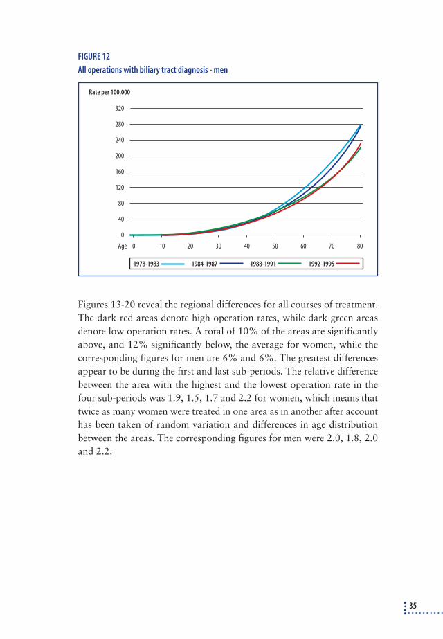
FIGURE 12
All operations with biliary tract diagnosis - men
Figures 13-20 reveal the regional differences for all courses of treatment.The dark red areas denote high operation rates, while dark green areasdenote low operation rates. A total of 10% of the areas are significantlyabove, and 12% significantly below, the average for women, while thecorresponding figures for men are 6% and 6%. The greatest differencesappear to be during the first and last sub-periods. The relative differencebetween the area with the highest and the lowest operation rate in thefour sub-periods was 1.9, 1.5, 1.7 and 2.2 for women, which means thattwice as many women were treated in one area as in another after accounthas been taken of random variation and differences in age distributionbetween the areas. The corresponding figures for men were 2.0, 1.8, 2.0and 2.2.
35
0
80
160
200
240
280
320
Rate per 100,000
0 10 20 30 40 50 60 70 80
120
40
Age
1988-1991 1992-19951984-19871978-1983

FIGURE 13
All operations on biliary tracts – women, 1978-1983
36
SMR < 0.7 0.7 - 0.8 0.8 - 0.9 0.9 - 1.01 - 1.1 1.1 - 1.2 1.2 - 1.3 > 1.3
min = 0.72 max = 1.35

FIGURE 14
All operations on biliary tracts – women, 1984-1987
37
SMR < 0.7 0.7 - 0.8 0.8 - 0.9 0.9 - 1.01 - 1.1 1.1 - 1.2 1.2 - 1.3 > 1.3
min = 0.83 max = 1.22

38
FIGURE 15
All operations on biliary tracts – women, 1988-1991
SMR < 0.7 0.7 - 0.8 0.8 - 0.9 0.9 - 1.01 - 1.1 1.1 - 1.2 1.2 - 1.3 > 1.3
min = 0.79 max = 1.33

FIGURE 16
All operations on biliary tracts – women, 1992-1995
39
SMR < 0.7 0.7 - 0.8 0.8 - 0.9 0.9 - 1.01 - 1.1 1.1 - 1.2 1.2 - 1.3 > 1.3
min = 0.65 max = 1.42

FIGURE 17
All operation on biliary tracts – men, 1978-1983
40
SMR < 0.7 0.7 - 0.8 0.8 - 0.9 0.9 - 1.01 - 1.1 1.1 - 1.2 1.2 - 1.3 > 1.3
min = 0.70 max = 1.38

FIGURE 18
All operation on biliary tracts – men, 1984-1987
41
SMR < 0.7 0.7 - 0.8 0.8 - 0.9 0.9 - 1.01 - 1.1 1.1 - 1.2 1.2 - 1.3 > 1.3
min = 0.73 max = 1.29

FIGURE 19
All operation on biliary tracts – men, 1988-1991
42
SMR < 0.7 0.7 - 0.8 0.8 - 0.9 0.9 - 1.01 - 1.1 1.1 - 1.2 1.2 - 1.3 > 1.3
min = 0.72 max = 1.42
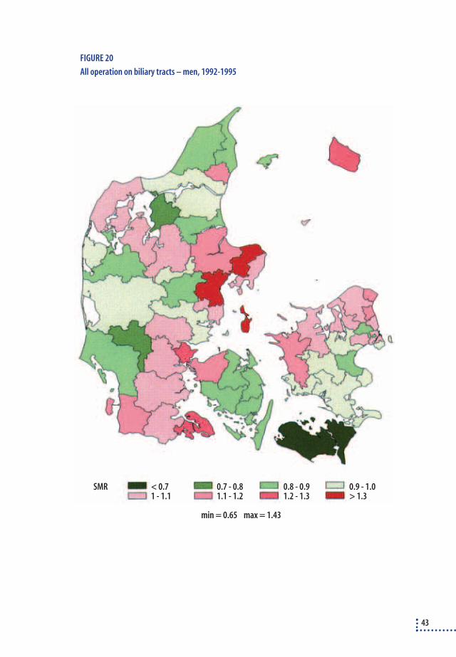
FIGURE 20
All operation on biliary tracts – men, 1992-1995
43
SMR < 0.7 0.7 - 0.8 0.8 - 0.9 0.9 - 1.01 - 1.1 1.1 - 1.2 1.2 - 1.3 > 1.3
min = 0.65 max = 1.43

MortalityThe 30-day mortality rates exhibit a minor fall in the period 1978-95 if alloperations are considered (figure 21). However, the curves do reveal somevariation.
The rates are standardised for age and gender. To assess the changes inmortality in greater depth and take into account differences in other formsof ill-health (co-morbidity), the four sub-periods were compared by meansof logistic regression analysis (table 4). These analyses showed a tendencyfor mortality to rise among acute admissions over the years, while in a single period (1988-91) there was a significant fall in mortality among theelective admissions. No international literature was found which analysesmortality rates associated with all biliary tract procedures.
TABLE 4
The 30-day mortalitya after operation on biliary tracts in relation to diagnosis and period of
time in Denmark, 1978-95. Only courses involving biliary tract diagnoses have been included
and the analyses (multiple logistic regression analyses) take into account age, gender and
co-morbidity.
All operations on biliary tracts
Elective admission Acute admission Total
Period OR (95% c.l.) OR (95% c.l.) OR (95% c.l.)
1978-1983 1.00 1.00 1.00
1984-1987 0.92 (0.69-1.22) 1.22 (1.03-1.44) 1.11 (0.96-1.28)
1988-1991 0.68 (0.47-0.97) 1.25 (1.05-1.49) 1.08 (0.93-1.26)
1992-1995 0.75 (0.52-1.07) 1.18 (0.98-1.42) 1.04 (0.88-1.22)
a: Mortality is measured from the date of admission, as the actual date of operation is not stipulated in the National Hospital Discharge
Register
Diagnostic ERCPThe 12,262 admissions correspond to 681 patients per annum undergoingdiagnostic ERCP not followed by treatment of the biliary tracts. There isa significant change during the period of 300% for women and 250% formen (figure 22). The rise is particularly pronounced after the introductionof laparoscopic cholecystectomy.The findings correspond to international literature232, 698. Since diagnosticERCP does not usually lead to a gallstone diagnosis, the rise must be dueto the fact that this technology is being used increasingly often to exam-ine patients suspected of having biliary tract disorders.
44

FIGURE 21
Mortality rates standardised for gender and age
FIGURE 22
ERCP
45
0
50
Number per 1,000
45
40
35
30
25
20
15
10
5
Total Acute Elective
78 79 80 81 82 83 84 85 86 87 88 89 90 91 92 93 94 95Year
0
30
Rate per 100,000
25
20
15
10
5
78 79 80 81 82 83 84 85 86 87 88 89 90 91 92 93 94 95Year
MenWomen
ConclusionIt can be concluded that the total number of treatments for biliary tractdisorders in Denmark fell during the 80s but rose again after the intro-duction of laparoscopic cholecystectomy. Due to the underreporting of en-doscopic bile duct surgery, it can only be presumed that the rise after 1991has been even more pronounced than the curves suggest. The regionalvariations, which reflect differences in ill-health and patient behaviour aswell as lack of consensus about examination and treatment indication,show a moderate variation pattern. The overall mortality showed no sig-nificant difference during the time periods with a tendency towards de-creasing mortality among patients admitted electively and increasing mor-tality in patients admitted acutely. The use of diagnostic ERCP rosesteadily during the whole period, more rapidly after the introduction of la-paroscopic cholecystectomy.
46

5. Treatment of patients withnon-complicated gallbladderstones
5.1 DEVELOPMENTS IN DENMARK, 1978-955.1.1 The frequency of simple cholecystectomy
The treatment rate for simple cholecystectomy (including patients withacute cholecystitis) has changed noticeably during the period 1978-95(figure 23). A relatively constant operation rate for women in the earlyyears was followed by a fall of 21% (3% per annum) from 1984 to 1991and a rise of 27% (9% per annum) to 1994. For men, a corresponding fallof 26% (3.7% per annum) was recorded from 1984 to 1991, followed bya rise of 18% (4.5% per annum) until 1995.
FIGURE 23
Simple cholecystectomy - with biliary tract diagnosis
The treatment rates for simple elective cholecystectomy (figure 24) revealmore pronounced variations than the rates for all simple cholecystec-
47
0
110
Rate per 100,000
78 79 80 81 82 83 84 85 86 87 88 89 90 91 92 93 94 95Year
100
90
80
70
60
50
40
30
20
10
MenWomen

tomies (figure 23). The fall for women from 1984 to 1991 was 24%(3.4% per annum) and the subsequent rise until 1994 was 44% (14.7%per annum). For men, a corresponding fall of 35% (5% per annum) from1984 to 1991 was followed by a rise of 21% (5.3% per annum) until1995.
FIGURE 24
Simple cholecystectomy - without acute cholecystitis - with biliary tract diagnosis
The cholecystectomy rate for women (including operation for acute chole-cystitis) in different age groups (figure 25) fell during the first three sub-periods for the over-40s and rose during the final sub-period for the 40-70s. The most prominent feature in the younger groups was a steep riseafter the introduction of laparoscopic cholecystectomy – a rise of almost50%. All the curves break, with the cholecystectomy rate rising until 30,falling slightly until 40 and then rising again. This break is particularlypronounced in the final sub-period. The break in the curve is equally pro-nounced all over the country. There is no break in the age curve for men(figure 26). It continues to rise with age. The variation over the four sub-periods was the same as for women.
48
0
Rate per 100,000
78 79 80 81 82 83 84 85 86 87 88 89 90 91 92 93 94 95Year
90
80
70
60
50
40
30
20
10
MenWomen
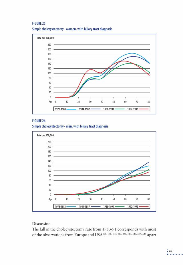
FIGURE 25
Simple cholecystectomy - women, with biliary tract diagnosis
FIGURE 26
Simple cholecystectomy - men, with biliary tract diagnosis
DiscussionThe fall in the cholecystectomy rate from 1983-91 corresponds with mostof the observations from Europe and USA128, 186, 187, 417, 426, 510, 540, 605, 648 apart
49
0
80
140
160
180
200
220
Rate per 100,000
0 10 20 30 40 50 60 70 80
120
40
Age
100
60
20
1988-1991 1992-19951984-19871978-1983
0
80
140
160
180
200
220
Rate per 100,000
0 10 20 30 40 50 60 70 80
120
40
Age
100
60
20
1988-1991 1992-19951984-19871978-1983

from one single study745 that revealed no change. The fall in the cholecys-tectomy rate until the introduction of laparoscopic cholecystectomy wasprobably due to the increasing use of new technologies instead of tradi-tional open cholecystectomy, e.g. ESWL and bile salts, ultrasounddrainage of the gallbladder in the event of acute cholecystitis and percu-taneous dissolution of gallstones. There was probably a minor fall in theincidence of gallstones as well (see section 3.1).
The rise in the cholecystectomy rate after the introduction of laparo-scopic cholecystectomy followed the international pattern. A systematicliterature search identified six studies of cholecystectomy rates coveringwell-defined regions (even whole countries) that uses standardisation inrelation to the size of the population, age and gender (table 5). Resultsfrom individual hospitals667, 682, 746 were not included since the size of thepatient base can change. To ensure comparability with the other studies,the genders have been mixed and all cholecystectomies (regardless of si-multaneous bile duct surgery) included in the Danish figures in the table.The data reveals that the rise in the Danish cholecystectomy rate is greaterthan in Scotland and than in one of the American studies, but also farsmaller than in two other areas in the USA. Other studies149, 426 show in ac-cordance with this study that the rise in the cholecystectomy rate was par-ticularly pronounced among young women.
TABLE 5
Changes in the cholecystectomy rate (per 1,000 people) after the introduction of laparoscopic
cholecystectomy.
Author 1985 1986 1987 1988 1989 1990 1991 1992 1993 1994 Rise Country
Legoretta438 1.37 1.35 1.59a 2.01 2.15 59.3% USA
Cobb146 3.87 7.22 86.5% USAb, c
Steiner745 1.65 1.65 1.68 1.68 1.69 1.84a 2.16 2.17 28.4% USA
Escarce213 3.4 3.5 3.5 3.3 3.6a 4.1 4.4 4.2 33.3% USA; >64 år c
Cohen149 2.75 2.82a 3.01 3.25 3.04 18.8% USA
Lam426 0.90 0.91 0.89 0.88 0.86 0.85a 0.80 0.93 1.03 19.8% The whole of Scotland
This studyd 0.60 0.58 0.59 0.57 0.54 0.53 0.53a 0.58 0.66 0.67 26.4% The whole of Denmark
All studies covered an average of more than 900 cholecystectomies per annum. The major difference in the cholecystectomy rates
between USA on the one hand and Scotland and Denmark on the other is due to a higher prevalence of gallstones in the USA (section 1.1).
a: States the year in which laparoscopic cholecystectomy was introduced.
b: The exact periods of time are 01.07.84-30.06.85 and 01.07.91-30.06.92. Laparoscopic cholecystectomy was introduced between the
two periods
c: Only patients insured by Medi Care. Thus, the base population is variable and theoretically there could have been a larger increase in
patient numbers after the introduction of laparoscopic cholecystectomy, which would lead to over-estimating.
d: Includes all cholecystectomies – including patients who also received treatment of the bile duc.
50

A series of American and Australian reports showed the rise in the num-ber of cholecystectomies without taking into account any changes in theage and gender composition of the total population3, 206, 232, 554, 577, 658, 660.These rises vary from 28% to 39.6%, but since the population in the ar-eas in question must be expected to have grown older, the estimates willbe slightly too high. In addition to the present study, and the Scottishstudy426, two other national studies have been conducted into changes incholecystectomy rates. The cholecystectomy rates have, however, not beenstandardised, so the statistical rises of 24% in Australia489 and 17% inCanada489 are presumably slightly too high.
The cause of the rise in the cholecystectomy rate after the introductionof laparoscopic cholecystectomy is not known, but is presumed to be dueto a lower threshold for treatment. One article reported that an informalsurvey of 19 gastroenterologists revealed that 17 had modified the indica-tions for cholecystectomy578.
The lower of the high points in the cholecystectomy rate around 30 yearsfor women has not been described in the literature before. It may be due togallstones formed during pregnancy. Ultrasound scanning is frequent duringpregnancies, and as a result, asymptomatic gallstones may be discoveredmore frequently in pregnant women because it is difficult to avoid the gall-bladder when conducting an ultrasound examination of the abdomen.
ConclusionThe cholecystectomy rate in Denmark and abroad fell throughout the 80s.After the introduction of laparoscopic cholecystectomy, the cholecystec-tomy rate rose noticeably. The rise in Denmark was greater than in Scot-land, Canada and Australia, but less than in certain areas of USA.
5.1.2 Regional variations in Denmark
Regional variations for simple cholecystectomy (including acute cholecystitis)in Denmark (figure 27-34) were moderate. Among women 10% of the areaswere significantly below and 15% significant above the average. The corre-sponding figures for men were 3% and 6%. There was a distinct tendencyfor regional variations to be greatest in the first and last sub-periods. Thedifferences between the areas with the lowest and highest cholecystectomyrates for women during the periods 1978-83, 1984-87, 1988-91 and 1992-95 were 2.2; 1.6; 1.8 and 2.5, respectively, while the corresponding numbersfor men were 2.1; 1.4; 1.7 and 2.1. Thus, taking into account random varia-tion and age differences, more than twice as many people had a cholecys-tectomy in one area of Denmark during the final sub-period as in another.
51

FIGURE 27
Simple cholecystectomy – women 1978-1983
52
SMR < 0.7 0.7 - 0.8 0.8 - 0.9 0.9 - 1.01 - 1.1 1.1 - 1.2 1.2 - 1.3 > 1.3
min = 0.66 max = 1.47

FIGURE 28
Simple cholecystectomy – women 1984-1987
53
SMR < 0.7 0.7 - 0.8 0.8 - 0.9 0.9 - 1.01 - 1.1 1.1 - 1.2 1.2 - 1.3 > 1.3
min = 0.80 max = 1.25

FIGURE 29
Simple cholecystectomy – women 1988-1991
54
SMR < 0.7 0.7 - 0.8 0.8 - 0.9 0.9 - 1.01 - 1.1 1.1 - 1.2 1.2 - 1.3 > 1.3
min = 0.80 max = 1.47

FIGURE 30
Simple cholecystectomy – women 1992-1995
55
SMR < 0.7 0.7 - 0.8 0.8 - 0.9 0.9 - 1.01 - 1.1 1.1 - 1.2 1.2 - 1.3 > 1.3
min = 0.63 max = 1.59
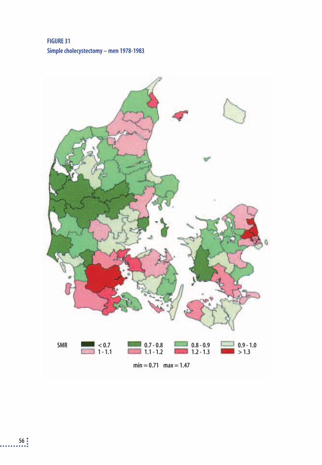
FIGURE 31
Simple cholecystectomy – men 1978-1983
56
SMR < 0.7 0.7 - 0.8 0.8 - 0.9 0.9 - 1.01 - 1.1 1.1 - 1.2 1.2 - 1.3 > 1.3
min = 0.71 max = 1.47

FIGURE 32
Simple cholecystectomy – men 1984-1987
57
SMR < 0.7 0.7 - 0.8 0.8 - 0.9 0.9 - 1.01 - 1.1 1.1 - 1.2 1.2 - 1.3 > 1.3
min = 0.83 max = 1.16

FIGURE 33
Simple cholecystectomy – men 1988-1991
58
SMR < 0.7 0.7 - 0.8 0.8 - 0.9 0.9 - 1.01 - 1.1 1.1 - 1.2 1.2 - 1.3 > 1.3
min = 0.80 max = 1.34

FIGURE 34
Simple cholecystectomy – men 1992-1995
59
SMR < 0.7 0.7 - 0.8 0.8 - 0.9 0.9 - 1.01 - 1.1 1.1 - 1.2 1.2 - 1.3 > 1.3
min = 0.68 max = 1.43

DiskussionRegional variations have been described in many countries. Most worksstem from the 60s and 70s239, 449, 507, 834, but a few later publications showeither the same variation393 or slightly greater variations610 than in Den-mark. One Nordic report478 revealed regional variations in cholecystec-tomy rates in the other Nordic countries corresponding to those in Den-mark. The variation may simply reflect variations in gallstone incidence indifferent regions, which is probably not the case in Denmark367. In Den-mark, the differences are probably due to differences in patient behaviouror in definitions of indications for diagnostics and treatment.
5.2 WHICH SYMPTOMS ARE DUE TO STONES IN THE GALLBLADDER? There is international consensus (see section 3.3.2) that patients shouldnot be treated for stones in the gallbladder until they develop symptoms.Since 30-40% of the population suffer gastrointestinal symptoms382 andup to 20-30% have stones in the gallbladder370, coincidence will often occur. The challenge for clinicians is to find out which symptoms arecaused by gallstones and which are not45. This area has been the object ofmajor disagreement over the years, ranging from the extreme view that allgallstones cause symptoms “as long as you interrogate the patient thoroughly “496 to the other extreme view that stones do not cause symp-toms until complications set in2.
In an attempt to discover which symptoms are due to stones in the gall-bladder and, therefore, which symptoms should be included in the indi-cation for cholecystectomy, a thorough study was conducted of articlesconcerning:
❖ The description of gallstone symptoms in uncontrolled clinical series
❖ Comparison of symptoms in people with and without gallstones:
- in populations screened for gallstones
- among patients examined for suspected gallstones
❖ Incidence of - and predictors for - continued symptoms after removal of gallstones
Excepting articles describing gallstone symptoms in uncontrolled series,the study tried to trace all articles (see however appendix 3) that fulfil thefollowing requirements: a) screening surveys of at least 100 people fromrandom samples of the population; b) consecutive series of at least 100 pa-tients examined for gallstones during well-defined periods and, finally c)
60

articles about persistent pain after cholecystectomy had to cover consecu-tive series of at least 50 patients in well-defined time periods.
Description of gallstone symptoms in uncontrolled clinical series No attempt was made to conduct a systematic literature search of this cat-egory, since these studies are unsuitable to identify which pains are specificto stones in the gallbladder. However, some of the older works containvery detailed descriptions of symptoms in patients with gallstones, symp-toms which it might be beneficial to compare with findings from newer,controlled series. The traditional gallstone pains are described as very se-vere - comparable with birth pains249, 295. The pains either start suddenlyor reach a climax during a period of 10-60 minutes, after which they re-main present and constant for several hours before fading away slowly.The pain is in the top of the stomach or the top right-hand side of the ab-domen, often radiates towards the back and/or right shoulder/shoulderblade. Attacks are usually weeks or even years apart. Gallstone patientsexperience more constant pains than patients with kidney stones249. Com-pared with patients who suffer abdominal pains for other reasons, gall-stone patients usually suffer pains at night with a tendency for the major-ity of the attacks to occur at the same time of day643.
Comparison of symptoms in people with and without gallstonesSeven out of 16 screening studies revealed a significant relationship be-tween gallstone-like pains or pains in the upper abdomen and the inci-dence of stones in the gallbladder. Twelve of the studies also assessed therelationship between dyspepsia and gallstones, which was significant intwo of the studies. The studies are summarised in appendix 3 (table 1).
Seven studies were identified in which patients referred to a clinicaldepartment or to an X-ray department were asked about their symptomsbefore the actual examination for gallstone was conducted. Most of theseries revealed that patients with gallstones suffer upper abdominal painsmore often than those who do not have gallstones, whereas only one studyout of six identified a relationship between dyspepsia and gallstones. Thestudies are summarised in appendix 3 (table 2).
Occurrence of persistent symptoms in patients after cholecystectomyIf a symptom persists after cholecystectomy, the gallstones or the gall-bladder may not have been the cause of the symptoms. Conversely, thefact that symptoms disappear after a cholecystectomy does not necessar-
61

ily mean that the symptoms were caused by the gallstones/gallbladder. Acertain level of placebo effect cannot be discounted. Furthermore, ab-dominal symptoms are not constant in individual patients, but show greatvariation over time382. In 19 clinical series identified, the incidence of per-sistent symptoms varied from 6% to 41% (average 22%); though onlyfrom 3% to 17% (average 9%), if symptoms had to have the samestrength and regularity as before the operation. The only predictors forpersistent pains are dyspepsia along with pains70, 468, atypical pains alongwith typical pains 409, longstanding history of pains70, 742 plus psychic vul-nerability or mental illness376, 468. The studies are summarised in appendix3 (table 3).
Discussion Some of the works mentioned above have been collated in a meta-analysis 414,which concluded that there was some sort of interrelationship betweengallstones and severe upper abdominal pains. However, none of the sur-veys were ideal because they did not properly identify symptoms specificto stones in the gallbladder, so it is difficult to reach an unambiguous con-clusion. The screening studies constitute snapshots of the general popula-tion, so a high incidence of disease was not expected. These surveys donot, therefore, reflect the clinical working day. Studies of groups of pa-tients referred to a clinical department or to an X-ray department mayalso be an inadequate way of studying gallstone symptoms, as the patientshave been pre-selected on the basis of symptoms that suggest bile disease.To a certain extent, clinical series studying persistent pains may identifywhich symptoms are not associated with gallstones by focusing on pat-terns of symptoms that most commonly cause persistent pain. However,abdominal symptoms vary greatly382, so the findings are ambiguous. Amore definitive answer would be found, if a group of patients with gall-stones and upper abdominal pains was randomised for either cholecys-tectomy or no treatment. Such a survey has been conducted721, but it hadmajor in-built problems in the form of large-scale exclusions (63%) and ahigh drop-out rate after the randomisation (12-24%). The results of thesurvey have not yet been published.
Whenever scientific studies fail to provide unambiguous answers, thereis a tradition of delineating the area at consensus conferences. These con-ferences have defined gallstone pains as “a relatively strong pain that oc-curs in attacs, is located in the epigastrium or in the upper right quadrant,lasts 1-5 hours and often wakes the patient during the night”45. Even
62

though the literature search have demonstrated a significant relationshipbetween this pattern of symptoms and the incidence of gallstones, the re-sults can only be used in the clinical working day with the proviso thatthese pains are also experienced by people without gallstones. In the Dan-ish cohort surveys, 2.7% of the population suffered from attacks of severepains that lasted for hours in the epigastrium or upper right quadrant, re-gardless of whether or not they had gallstones368, 371. An Italian study 53revealed that 5% of Italian men and 8% of women have “gallstone pains”without gallstones. Gallstone pains in the Italian studies were defined as“pains in the epigastrium or upper right quadrant within the last fiveyears. The pains must last more than half an hour and must not be easedby bowel movements/wind”.
Conclusions❖ Hour-long bouts of severe pains in the upper abdomen (the top of the stomach or
upper right quadrant) may be associated with gallstones. The pain starts relati-
vely abruptly and occasionally radiates towards the back. This corresponds more
or less to the symptoms described in the early literature. These symptoms are rela-
tively rare. The symptoms do, however, also occur in people without gallstones, so
it is impossible to define exactly when these symptoms are due to gallstones and
when they are not.
❖ Other abdominal pains are probably not due to gallstones.
❖ Dyspepsia is not caused by gallstones (see section 5.4.1; page 85).
5.3 INDICATION FOR TREATMENT
Pains are the main indication for treating stones in the gallbladder. Theproblems listed above that are associated with identifying symptoms spe-cific to stones in the gallbladder explains why the indication for the treat-ment of patients with gallstones has always been open to debate. The fa-ther of modern cholecystectomy, Langenbuch, is supposed to have said:“In my opinion, cholecystectomy should be used in cases where both pa-tient and doctor have exhausted their patience.”
The regional variations in cholecystectomy rates documented in thisreport reflect the fact that relationships between gallstones and symptomsare open to interpretation, as the individual doctor and patient have dif-ferent opinions about when a symptom is due to gallstones – it is also pos-sible that they have different levels of patience. The differences in indica-tion should also be considered in a historical and international light:
63

❖ Historically, treatment has changed. A Danish autopsy study showed that the pro-
portion operated for gallstone disorders rose from 2-3% in 1920 to 40% in 1985 772.
❖ A systematic study of the literature covering screening surveys of random samples
of the population, in which the cholecystectomy rate for people with gallstone dis-
orders (both people with gallstones and those who had a cholecystectomy ) was
estimated, revealed that the rates vary among men from 5-36% and among wo-
men from 5-55%. In other words, in some countries (Norway) only 5% of those
who have gallstones are operated on, while in other countries (USA, Italy, Sweden)
more than 50% have an operation. In addition, women are treated for gallstones
far more often than men. It is unlikely that these pronounced variations can be ex-
plained solely by different symptoms. They have to be ascribed to different attitu-
des – among both patients and doctors – to when a patient should be examined
and treated for gallstones. A summary of these studies is included in appendix 3
(table 4).
ConsensusA number of attempts have been made to reach a consensus about the in-dication for cholecystectomy. A model with two consensus panels – oneconsisting of nine surgeons and one consisting of various specialists (sur-geons, medical gastroenterologists, internal physicians, general practitio-ners and radiologists) was developed in the USA720 and later tested in Israel242, 243, 244 and Great Britain699. On the basis of the available literature,a number of patient histories were drawn up regarding gallstone disorders.Each member of the panels had first to assess each patient history themsel-ves, after which the panel attempted to reach an agreement about a treat-ment indication. In general, the surgical panel was more disposed to find indications for cholecystectomy than the mixed panel. In almosthalf of the patient histories, agreement could not be reached within the panels699. There was, however, general agreement that people with asymp-tomatic gallstones or vague symptoms should not undergo a cholecystect-omy, whereas people with stones in the gallbladder and one or more attackof gallstone pains should be offered a cholecystectomy. The indication for cholecystectomy fell with increased incidence of co-morbidity in the patient377. These recommendations correspond to international recom-mendations44, 45, 46. This represents a significant tightening up in relation tothe recommendations of earlier consensus panels who tended to agree thatoperations should be offered to patients with gallstones and dyspepsia(without pains)805.
64

AuditOnly one single study700 looked at the extent to which indications forcholecystectomy provided by the above-mentioned consensus are ob-served in daily life. Information was extracted from journals on 252 pa-tients, who had had a cholecystectomy and the information was comparedwith the above-mentioned panel consensus 699. The panel consisting ofdifferent specialists would have agreed on operation for 41% of cases andno operation for 30%. The corresponding figures for the surgical panelwould have been 52% and 2%. In the remaining cases (29% and 46%,respectively) it would not have been possible to reach a consensus. Itproved impossible to find publications dealing with audits of gallstonetreatment in Denmark.
Even though a consensus has been reached about indications for chole-cystectomy, it can be difficult to comply with in daily life. One study re-vealed that if a computer program with recommendations for cholecys-tectomy was used simultaneous with indication for operation, the recom-mendations were followed far more often than before the computer pro-gramme was used102.
DiscussionIn contrast to medicinal treatment, in which the effect of a chemical sub-stance can be tested and subsequently stopped if it does not work, a chole-cystectomy is non-reversible. Thus, there is good reason to warn against theso-called “diagnostic cholecystectomy” in which the gallbladder is removedto see whether or not it caused the symptoms 730. If a patient suffers symp-toms, then the patient and doctor must decide when they are so troublesomethat treatment ought to be offered. Even if a symptom is thought to be as-sociated with gallstones, it is not necessarily an indication for treatment.This depends on a number of things, such as the patient’s wishes, fear of newattacks, etc. The patient’s overall health also plays a role. Observation of apatient after the first pains seems justifiable, since the spontaneous course ofstones in the gallbladder is relatively peaceful (see section 3,2).
Conclusion❖ Despite attempts to reach international consensus, a great deal of disagreement
still surrounds cholecystectomy indications.
❖ A discrepancy exists between the indications for cholecystectomy defined by con-
sensus panels or international recommendations and the indications used under
everyday clinical conditions.
65
❖ Cholecystectomy ought to be limited to patients with the type of pains that the
consensus has defined as being due to stones in the gallbladder.
❖ The large proportion of patients with persistent pains, audit surveys and studies
showing rises in cholecystectomy rates suggest that too many people undergo
cholecystectomy.
5.4 METHODS OF TREATMENT
The technologies used to treat patients with symptomatic gallstones arecholecystectomy, cholecystolithotomy or stone dissolution (ESWL/bilesalts). The primary purpose of the treatment is to remove symptoms, whilethe secondary purpose is to remove gallstones. Ideally, the treatmentshould not lead to complications or mortality, there should be few post-operative pains, the duration of the stay in hospital and of the convales-cence should be short and the cosmetic result satisfactory. The next sec-tion compares the individual treatments with these objectives.
5.4.1 Cholecystectomy
The gallbladder can be removed either by traditional open cholecystec-tomy, cholecystectomy by minilaparotomy or laparoscopic cholecystec-tomy.
Cure from symptomsAmong the randomised surveys of the different access to cholecystectomy(appendix 3) only one study dealt with persistent pains505. It revealed nodifference between cholecystectomy by minilaparotomy and laparoscopiccholecystectomy after 6-12 months as far as the incidence of persistentpains, the regularity and type of pains or visits to the doctor because ofpains were concerned.
The clinical series (appendix 3, table 3) revealed persistent pains aftercholecystectomy in 7-41% (average 22%) after traditional open chole-cystectomy and in 6-37% (average 21%) after laparoscopic cholecystec-tomy. If the data are restricted to patients who suffer persistent pains ofthe same or greater magnitude, the proportion falls to 4-17% (average8%) after traditional open cholecystectomy and to 3-11% (average 7%)after laparoscopic cholecystectomy. None of the clinical series concerningcholecystectomy by minilaparotomy stipulated the proportion sufferingfrom persistent pains.
These numbers suggest that the degree of persistent pains is probablynot dependent on the size of the incision. This is corroborated by the fact
66

that persistent pains after contusion (ESWL) and after medicinal dissolu-tion of gallstones are the same (6-31%; average 19%) as after cholecys-tectomy8, 436, 754, 825.
Thus, it can be concluded that the two new access to remove the gall-bladder (minilaparotomy and laparoscopy) have not affected the primarypurpose of the treatment.
There has been a longstanding debate among surgeons about the extentto which a long cysticus stump can cause persistent pains after a cholecys-tectomy. A randomised survey with an 8-year follow-up359 revealed that40% of patients had persistent pains after traditional cholecystectomycompared with 11% in a group that had the whole ductus cysticus re-moved. The survey was blinded. It is, however, rare for a long cysticusstump to be the sole explanation for persistent pains646 and no special pat-tern of pain has been associated with the length of a remaining cysticusstump864.
Removal of gallstonesCholecystectomy by traditional open laparotomy and by minilaparotomyare equally good as far as removal of gallstones (with gallbladder) is con-cerned. The same can more or less be said about laparoscopic cholecys-tectomy, but since defects are often made in the gallbladder during dissec-tion357, stones are sometimes lost and left behind in the abdominal ca-vity357, 399, 765. A number of casuistic studies describe the many ways thesestones manifest themselves in the form of intra-abdominal abscesses616,ileus594, fistula to the urinary bladder139, fistula to the pulmonary cavity842
and coughing up of stones108. Clinical series show that left stones rarelycause complications357, 399 but that the individual complications can be se-rious765.
Time spent in hospital and postoperative pains No scientific evidence exists to define exactly how long patients ought toremain in hospital after a cholecystectomy. The median time for electivesimple cholecystectomy fell in Denmark from 11 days in 1978, to 6 daysin 1991, to 3 days in 1995. There was no significant gender difference (figure 35). The shorter hospital stay was accompanied by a minor rise inthe frequency of readmissions to hospital, as the proportion of patientsreadmitted within a month after discharge rose from 9.5 % in 1978-83;to 11.3% in 1984-87, to 12.1% in 1988-91 and 13.6% in 1992-95. Thereduction in the time spent in hospital in the period before the introduc-
67
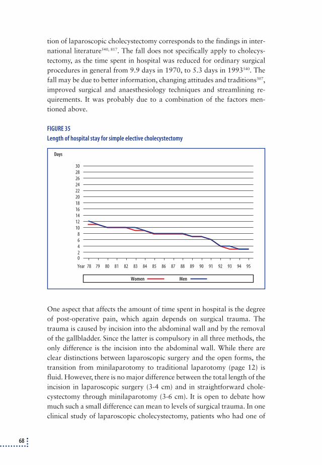
tion of laparoscopic cholecystectomy corresponds to the findings in inter-national literature540, 817. The fall does not specifically apply to cholecys-tectomy, as the time spent in hospital was reduced for ordinary surgicalprocedures in general from 9.9 days in 1970, to 5.3 days in 1993540. Thefall may be due to better information, changing attitudes and traditions307,improved surgical and anaesthesiology techniques and streamlining re-quirements. It was probably due to a combination of the factors men-tioned above.
FIGURE 35
Length of hospital stay for simple elective cholecystectomy
One aspect that affects the amount of time spent in hospital is the degreeof post-operative pain, which again depends on surgical trauma. Thetrauma is caused by incision into the abdominal wall and by the removalof the gallbladder. Since the latter is compulsory in all three methods, theonly difference is the incision into the abdominal wall. While there areclear distinctions between laparoscopic surgery and the open forms, thetransition from minilaparotomy to traditional laparotomy (page 12) isfluid. However, there is no major difference between the total length of theincision in laparoscopic surgery (3-4 cm) and in straightforward chole-cystectomy through minilaparotomy (3-6 cm). It is open to debate howmuch such a small difference can mean to levels of surgical trauma. In oneclinical study of laparoscopic cholecystectomy, patients who had one of
68
0
Days
78 79 80 81 82 83 84 85 86 87 88 89 90 91 92 93 94 95Year
3028262422201816141210
8642
MenWomen

the incisions lengthened to remove a large gallbladder (because of inflam-mation or stones)100, did not take more painkilling medicine than thosewho did not have the incision lengthened.
The literature shows that it is not only surgical trauma that affects theamount of analgesics and the length of hospital stay. The information thatthe patient receives about the procedure from the surgeon and the rest ofthe medical staff, and which reflects their attitudes to the procedure, isprobably a critical factor241, 536. One study suggested that positive hospitalsurroundings reduce the need for painkilling medicine and the length of thestay in hospital795. A systematic study of the literature showed that by pri-oritising efforts – including early postoperative feeding and in-depth infor-mation – for patients after traditional open cholecystectomy lead to verylow consumption of analgesics and patients being discharged 1-4 days af-ter the operation447, 533, 534, 550, 584, 635. In a clinical series starting in 1980, con-secutive patients were offered discharge the day after traditional opencholecystectomy with a success rate of 92%533. The effect was originally as-cribed to early feeding after the operation, but a randomised trial rejectedthis241. Since the first reports, several of which exist only as abstracts, fiveclinical works have been found in which consecutive patients were dis-charged quickly after a traditional open cholecystectomy with great successand low readmission frequency (table 6). A further two examinationsshowed that it was possible to treat these patients as outpatients. These ex-aminations are not included in table 6, since it is not known to what extentthey represent consecutive patient material785, 813. What all these workshave in common is that a great deal of effort was invested in informing thepatients about the procedure.
TABLE 6
Clinical series of consecutive patients, in which early discharge is planned after traditional
open cholecystectomy.
Post-operative discharge (cumulated percentages)
Author Period N Same day 1st day 2nd day 3rd day Average Re-admissions Notes
Hall299 1980-84 100 - - 51% - 3.2 dag 0 a)
Saltzstein674 1988-90 500 - 23% 56% - 2.8 dag 2 (0.4%) b) d)
Chaudhary132 1986-89 340 - - - 86% - 0 a)
Saltzstein675 1990 64 69% - - - < 1 dag 1 (1.6%) c) d)
Moss535 1983-87 160 - 100% - - 1 dag 3 (1.9%) a)
a) Elective, simple cholecystectomies
b) All patients including acute cholecystitis and choledochal surgery. If the material is restricted to elective simple cholecystectomies in
people under age 35, 44% were discharged after the first day and 83% after the second day
c) Simple cholecystectomies covering both elective and acute patients. Limited to people under 56 years of age
d ) The two groups of patients published by Saltzstein were not the same
69

In the first clinical series studying cholecystectomy by minilaparo-tomy, the short hospital stay from 1-3 days was emphasised282, 514.532 – andlater, same-day surgery was described in a series of 200 selected patientsdischarged 3-10 hours after the operation435.
Clinical series studying laparoscopic cholecystectomy also highlightthe short hospital stay of 1-2 days 43. A number of studies have aimed atsame-day surgery among selected patients427, 619, 696, 747 with a success rateof 50-100%, and among consecutive patients for simple elective chole-cystectomy230, 524, 767, 810, 811 with a corresponding rate of 61-94%.
In other words, it is possible for all three access to cholecystectomy tosend patients home either on the same day or 1-2 days after the operation.
Comparison between forms of accessA reliable comparison of the three access to cholecystectomy, as far asanalgesics and the amount of time spent in hospital are concerned, canonly be conducted in randomised surveys in which patient and staff arenot informed of the method applied582. Of the randomised examinationscomparing laparoscopic cholecystectomy with cholecystectomy by mini-laparotomy, only one fulfils this criteria for blinding483 and it showed nodifference in the length of hospital stay. No information was includedabout analgesics. Among randomised studies comparing laparoscopiccholecystectomy with traditional open cholecystectomy, only one lived upto the criteria for blinding579 and it did not reveal any difference in thelength of hospital stay either, although it did identify more post-operativepain in the group subjected to open cholecystectomy. The material was,however, very small (2 x 10 patients).
Among the non-blinded studies, significantly more time was spent in hos-pital after traditional open laparotomy than after laparoscopy79, 337, 789,while the results for laparoscopy vs. minilaparotomy500, 504 and minila-parotomy vs. traditional laparotomy574, 703 were ambiguous. The use of anal-gesics was significantly greater after traditional open laparotomy79, 337, 789
and after minilaparotomy503 than after laparoscopy. No significant differ-ence in the use of analgesics was registered after traditional laparotomycompared with minilaparotomy574, 690. Table 5 in appendix 3 illustratesthese small differences and since the studies were not blinded and the re-sults, therefore, susceptible to staff and patient attitudes to the differentoperations, the results have to be interpreted carefully.
A number of randomised trials revealed a whole string of other cir-cumstances that affect the use of analgesics and the amount of time spent
70

in hospital. They included the location of the incision, transcutaneouselectrical nerve stimulation, epidural anaesthesia, epidural morphine pluslocal anaesthesia in the wound, intrapleurally (in the pulmonary cavity),intraperitoneally (in the abdominal cavity) and intercostally (between theribs). Time constraints prohibit this report from studying these interrela-tionships systematically, but the randomised surveys are listed in appen-dix 3.
ConclusionThe amount of time spent in hospital after cholecystectomy has fallensteadily since 1978. The literature suggests that the length of the stay inhospital depends on factors other than just surgical trauma. Even if theamount of time spent in hospital fell after the introduction of laparoscopiccholecystectomy, no documentary evidence exists to prove that the fallwas caused by the laparoscopic procedure. As far as the use of postoper-ative analgesics is concerned, laparoscopic cholecystectomy seems to be aminor improvement over open surgery. However, analgesics are taken forsuch a short time that it ought not to affect the choice of procedure.
Complications to Cholecystectomy The vast majority of cholecystectomy complications are banal. Among themore serious complications, such as lesions of the bile ducts, major arter-ies and the gastrointestinal tract, the former is by far the most common333
and this section will concentrate on them.International literature states that the introduction of laparoscopic
cholecystectomy led to a rise in the number of bile duct lesions and thatthere may even be a reason to fear underreporting471, 756, 848. Many re-searchers have attributed the rise in complications to insufficient trainingof surgeons who felt pressurised to start using the new method too soon137,
165 and consequently, there was an initial call for caution. One Danish con-sensus report37 states that the number of bile duct lesions during laparo-scopic surgery in Denmark is no higher than the number of lesions suf-fered during traditional open cholecystectomy on Funen in 1953-57477.The data has not been compared with more recent material.
The next two chapters study the available literature in greater depthand assess the extent to which lesions of the bile ducts have increased inDenmark after the introduction of laparoscopic cholecystectomy. Wher-ever possible, a differentiation is made between:
71

❖ peripheral lesions (cysticus leak, leak from aberrant/accessory bile duct, unidenti-
fiable leak), which either leads to re-operation, ERCP with the insertion of a stent
or percutaneous drainage. These lesions are not serious enough to cause lasting
damage to the bile ducts per se.
❖ central lesions, which involve the deep bile ducts (ductus choledochus, ductus he-
paticus communis and ductus hepaticus dxt. and sin.). Lesions of these organs can
cause lasting damage to the bile ducts in the form of contraction and impaired
liver function.
Analysis of bile duct lesions – literature reviewThe literature was studied systematically on the basis of the principles dis-cussed in appendix 3. The summary of the various complications consid-ered:
❖ all randomised trials
❖ all surveys of all cholecystectomies in well-defined geographical areas during the
period when laparoscopy was being introduced
❖ all consecutive series of over 200 patients in which the operations were performed
after 1978 (see, however, page 75).
Randomised surveysThe randomised surveys were too small to evaluate differences in com-plication rates accurately. A total of 500 patients were covered by rand-omised surveys comparing laparoscopic cholecystectomy with cholecys-tectomy by minilaparotomy483, 504. There were two central bile duct lesionsin each treatment group and three peripheral lesions in the laparoscopicgroup against one in the minilaparotomy group. The total of three bileduct lesions in the minilaparotomy group (1.2%) and five in the lapar-oscopy group (2.0%) was not significant. The very high complicationrate was due to one of the studies, which accounted for seven of the eightlesions504. There is a temptation to suspect that the surgeons involved inthat study were not trained in minilaparotomy; at any rate it is not obvi-ous from the article. This in contrast to the study with the low compli-cation rate483, which specifically mentioned that the surgeons were trai-ned in both procedures before the survey started. Among 151 patientsrandomised for laparoscopic cholecystectomy or traditional open chole-cystectomy79, 337, 579, 789 there was one peripheral bile duct lesion in theopen group, but none in the laparoscopic group. Of 341 patients rando-
72
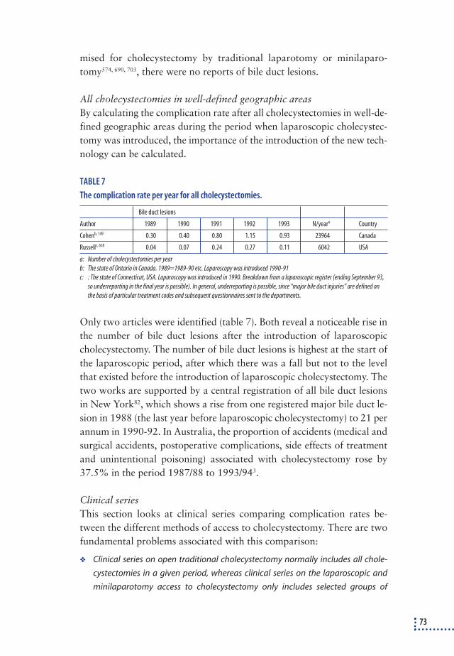
mised for cholecystectomy by traditional laparotomy or minilaparo-tomy574, 690, 703, there were no reports of bile duct lesions.
All cholecystectomies in well-defined geographic areasBy calculating the complication rate after all cholecystectomies in well-de-fined geographic areas during the period when laparoscopic cholecystec-tomy was introduced, the importance of the introduction of the new tech-nology can be calculated.
TABLE 7
The complication rate per year for all cholecystectomies.
Bile duct lesions
Author 1989 1990 1991 1992 1993 N/yeara Country
Cohenb, 149 0.30 0.40 0.80 1.15 0.93 23964 Canada
Russellc, 658 0.04 0.07 0.24 0.27 0.11 6042 USA
a: Number of cholecystectomies per year
b: The state of Ontario in Canada. 1989=1989-90 etc. Laparoscopy was introduced 1990-91
c: : The state of Connecticut, USA. Laparoscopy was introduced in 1990. Breakdown from a laparoscopic register (ending September 93,
so underreporting in the final year is possible). In general, underreporting is possible, since “major bile duct injuries” are defined on
the basis of particular treatment codes and subsequent questionnaires sent to the departments.
Only two articles were identified (table 7). Both reveal a noticeable rise inthe number of bile duct lesions after the introduction of laparoscopiccholecystectomy. The number of bile duct lesions is highest at the start ofthe laparoscopic period, after which there was a fall but not to the levelthat existed before the introduction of laparoscopic cholecystectomy. Thetwo works are supported by a central registration of all bile duct lesionsin New York82, which shows a rise from one registered major bile duct le-sion in 1988 (the last year before laparoscopic cholecystectomy) to 21 perannum in 1990-92. In Australia, the proportion of accidents (medical andsurgical accidents, postoperative complications, side effects of treatmentand unintentional poisoning) associated with cholecystectomy rose by37.5% in the period 1987/88 to 1993/943.
Clinical seriesThis section looks at clinical series comparing complication rates be-tween the different methods of access to cholecystectomy. There are twofundamental problems associated with this comparison:
❖ Clinical series on open traditional cholecystectomy normally includes all chole-
cystectomies in a given period, whereas clinical series on the laparoscopic and
minilaparotomy access to cholecystectomy only includes selected groups of
73

patients, leaving a group of patients, who are cholecystectomised by traditional
laparotomy. This latter group of patients both have a more advanced stage of
their gallstone disease378 and more co-morbidity225,378 compared to the group ope-
rated on by the laparoscopic method. This means that, when clinical series of chol-
ecystectomy by minilaparotomy or laparoscopy are compared with clinical series
where a traditional laparotomy is used, a group of more healthy patient are com-
pared with a group of more ill patients,
❖ Before introduction of the laparoscopic technique cholecystectomy was perfor-
med by younger surgeons in contrast to the laparoscopic operations, which
mostly is performed by more senior surgeons682,692. Also in the comparisons be-
tween cholecystectomy by laparoscopy and minilaparotomy the distribution of
senior surgeons is in favour of laparoscopy 190,500.
Due to these two problems, it must be expected – à priori – that patientsshould experience less complications in the clinical series of cholecystec-tomy by minilaparotomy and laparoscopy compared to the clinical seriesof cholecystectomy by traditional laparotomy.
Before the era of laparoscopic cholecystectomy very few articles dealtwith complications in relation to cholecystectomy, whereas after the in-troduction several articles and reviews on complications to cholecystec-tomy emerged. In the following review only clinical series on traditionalcholecystectomy in the period 1978-1990 were included. This restrictionis done in order to include only clinical series as close to the laparoscopicera as possible, as elder series represent another time with another organ-isation, which hardly can be compared with the 90’ies. The lower limit of1978 was chosen, as it from this year is possible to compare with Danishdata from the National Hospital Discharge Registry and as the relativelong period (13 years) will make it possible to have sufficient material.Open cholecystectomies after 1990 are not included, as many countries atthat time had started laparoscopic surgery, making cholecystectomies per-formed by traditional laparotomy a selected group. This was evident fromthe two-fold rise in postoperative mortality after open traditional chole-cystectomy after 1990745. The review of open traditional cholecystectomyis divided into series only including simple cholecystectomies (or where itwas possible to extract these operations from larger series also includingcholecystectomy with bile duct surgery) and series, where it was not pos-sible to distinguish between simple cholecystectomy and cholecystectomywith bile duct surgery. The former series are more comparable with serieson laparoscopy and minilaparotomy than the latter.
74

As very few series have been published on cholecystectomy by minila-parotomy, all series are included independent on number of operationsand when the surgery was done. Separate analyses are, however, per-formed of series representing more than 50, more than 100 and more than200 patients. Two studies being part of randomised trials in comparisonwith laparoscopic cholecystectomy are not included in this review, butdealt with elsewhere in this report.
Literature on clinical series including laparoscopic cholecystectomies ishuge. To minimise the work only articles published in an early period(1990-1992) and a late period (1995-97) are included. Besides clinical se-ries, results from questionnaire reviews, registers and audit are included.
A summarised review concerning bile duct lesions is shown in table 8,whereas results for each study is shown in appendix 3 (table 6-13). Cen-tral bile duct lesions are far more common in the laparoscopic series com-pared to traditional laparotomy and minilaparotomy, whereas the differ-ence in periferal bile duct lesions are not than big.
TABLE 8
Total summary of central and peripheral biliary tract lesions in materials consisting of 200(+)
consecutive patients operated since 1978a.
Access to cholecystectomy Number of Number of Lesions of the bile ductsstudies patients b Central % Peripheral
(95% c.l.) % (95% c.l.)
Open (simple cholecystectomy) 10 4,804 0.04 (0.01-0.18) 0.26 (0.15-0.47)
Open (all cholecystectomies) c 18 15,208 0.13 (0.08-0.20) 0.10 (0.06-0.16)
Minilaparotomy >=50 ptt./serie 16 3,553 0.03 (0.00-0.20) 0.17 (0.08-0.38)
Minilaparotomy >=100 ptt./serie 7 2,921 0.03 (0.00-0.24) 0.21 (0.09-0.46)
Minilaparotomy >=200 ptt./serie 3 2,307 0.04 (0.01-0.31) 0.04 (0.01-0.31)
Laparoscopic (1990-92) 47 19,234 0.32 (0.25-0.41) 0.46 (0.39-0.59)
Laparoscopic (1995-97) 26 24,342 0.26 (0.20-0.33) 0.39 (0.32-0.48)
Laparoscopic (questionnaires) 15 130,320 0.37 (0.34-0.40) 0.30 (0.27-0.33)
Laparoscopic (register) 16 110,939 0.47 (0.43-0.51) 0.28 (0.25-0.31)
Laparoscopic (audit) 6 22,671 0.52 (0.43-0.62) 0.57 (0.48-0.68)
a: : The material for cholecystectomy by minilaparotomy consists of 50, 100, and 200+ patients
b: States the number of patients about whom information is available concerning central bile duct lesions. When calculating confidence
limits for peripheral bile duct lesions, only the number of patients is used about whom information concerning this type of lesion is
available (table 6-13, appendix 3)
c: Including choledochal surgery
Assessment of bile duct lesions in Denmark 1978-95 By using the material used to validate the National Hospital Discharge Reg-ister (see section 2.1.2), an assessment was made of developments in bile
75

duct lesions in Denmark from 1978-95. Courses of treatment, during whichthe patient – after an initial admission for cholecystectomy with or withoutbile duct surgery – was readmitted or transferred to another department forendoscopic or open biliary tract surgery, must be presumed to include themajority of patients who have suffered a lesion of the bile ducts. Thesecourses of treatment rose fourfold after the introduction of laparoscopiccholecystectomy (figure 36, page 77). All commentaries on these courses oftreatment together with a random cross-section of all other commentariesin 1979, 1985 and 1993 were requisitioned from the surgical departments.The detailed breakdown is covered on page 22 and in appendix 2.
In approximately 1/3 of the cases, the treatments in figure 36 representedlesions of the bile ducts during simple cholecystectomy. This proportion re-mained constant throughout the whole period. More or less all the othercases involved patients who subsequently had a stone removed from the bileducts or were treated for biliary tract dyskinesia (page 83). By counting thenumber of bile duct lesions found in the commentaries received and predict-ing the same regularity of bile duct lesions in commentaries not received, theannual frequency of bile duct lesions was calculated for patients who had asimple cholecystectomy. In order to achieve reasonable quantities of data, theperiod was divided into three sub-periods, of which the final period includesall the years during which laparoscopic cholecystectomy has been performedin Denmark, while the first period is divided into two equally large halves.The bile duct lesions are divided into central and peripheral according to thesame principle described under the literature survey (page 72).
Because of size of the random sample, the data has to be interpretedcarefully, but the calculations suggest that there has been a two- to three-fold increase in the number of central lesions, while the number of pe-ripheral lesions has multiplied since the introduction of laparoscopic tech-nology (table 9).
TABLE 9
The estimated incidence of bile duct lesions during simple cholecystectomy in Danish
hospitals, 1978-95.
Localisation of bile duct lesion
Peripheral Central Uncertain* Total
Period % (95% c.l.) % (95% c.l.) % (95% c.l.) % (95% c.l.)
1978-83 0.02 (0.01-0.06) 0.16 (0.11-0.23) 0.00 (0.00-0.02) 0.18 (0.13-0.25)
1984-90 0.04 (0.02-0.08) 0.11 (0.07-0.16) 0.01 (0.00-0.04) 0.15 (0.11-0.22)
1991-95 0.39 (0.31-0.51) 0.33 (0.25-0.44) 0.10 (0.06-0.17) 0.83 (0.70-0.99)
* The medical commentaries did not specify peripheral or central.
76

The data in table 9 is underestimated. During the study of the commen-taries from the random extract from 1979, 1985 and 1993 a further fourcomplications were identified: one in 1979, one in 1985 and two in 1993.This may explain some of the discrepancy between the results in this re-port for 1991-95 and the breakdown of central bile duct lesions from thelaparoscopic register in Denmark, in which the percentage is 0.74%10. Another possible explanation is that the results from the laparoscopic reg-ister only include patients who had a laparoscopic cholecystectomy. Theresults show that this screening method using patients causes coveringmultiple admissions will identify many of bile duct injuries, but that thereis a certain amount of underreporting.
DiscussionThe data on complications after traditional open cholecystectomy is con-firmed by a number of studies of major series (3,403 to approx. 90,000 pa-tients), which show a bile duct lesions frequency of 0.05-0.07%29, 163, 268.However, the articles do not contain enough details to be included in thetotal result for all clinical series. In one review502, the proportion of bileduct lesions after traditional open cholecystectomy was estimated at0.2%. The work lacks a number of articles, includes older series, includesseries after the introduction of laparoscopic cholecystectomy and includes
77
FIGURE 36
Complicated courses after simple cholecystectomy
0
Percent
78 79 80 81 82 83 84 85 86 87 88 89 90 91 92 93 94 95Year
4
3
2
1

a major register study that overestimates the complication rate, since itdoes not differentiate between bile duct lesions and other peroperative le-sions to the gastrointestinal system. Review articles about bile duct lesionsafter laparoscopic cholecystectomy using criteria other than those used inthis study, or in which the criteria are not mentioned at all, show numberscorresponding to the result of this survey, with bile duct lesions rangingfrom 0.30-0.63%333, 391, 502. In a 1991 national questionnaire survey in theNetherlands with an response rate of 88%287, bile duct lesions were foundin 0.51% of open and 1.09% of laparoscopic surgery. Both rates are sig-nificantly higher than the results in table 8, which is partly explained inthe case of the open operations by the fact that 15% also had bile ductsurgery and that those who had a traditional open cholecystectomy dur-ing the laparoscopic era were at more advanced stages of illness378.
There was no significant difference in complication rates between tra-ditional open cholecystectomy and cholecystectomy by minilaparotomy.The data for minilaparotomy is, however, small and one major French se-ries197 in particular is very poorly described. It is, however, conspicuousthat none of the clinical series reveal the high incidences of lesions seen inthe laparoscopic series.
Clinical series, questionnaire surveys and register studies have all beencriticised for underreporting bile duct lesions471, 756, 848 compared with anaudit, in which an unbiased panel studies all the available patient journals.This report (table 8) partially supports this criticism, as the incidence ofboth central and peripheral bile duct lesions is somewhat higher in the au-dit series than in the other series concerning laparoscopic cholecystec-tomy. No audit studies were found of cholecystectomy by traditional opensurgery or minilaparotomy.
The rise in bile duct lesions has to be compared with the fact that thesurgeons performing biliary tract procedures in the laparoscopic periodare more experienced than in the period before the introduction of la-paroscopy682, 692.
None of the applied methods are ideal for calculating the frequency ofbile duct lesions. This would require a clinical database of all biliary tracttreatments (not just laparoscopic cholecystectomy) listed systematicallyaccording to criteria fixed in advance. Thus, it must be expected that allthe figures shown underestimate the real incidence of bile duct lesions.However, the calculations have been made the same way, so the registeredrise can be assumed to reflect reality.
78

It has been stated that the frequency of bile duct lesions after laparo-scopic cholecystectomy falls over time, a fact partly confirmed by the clin-ical series in table 8 and the summary in table 7. However, the frequencyof bile duct lesions does not fall to the level experienced before the intro-duction of laparoscopic cholecystectomy149, 658. Data from the Danish la-paroscopic register shows that there has not been any reduction in thenumber of bile duct lesions in Denmark in the period 1991-94 among pa-tients who had a laparoscopic cholecystectomy10.
A survey by an intensive care unit showed that complications after la-paroscopic cholecystectomy are more severe than after cholecystectomyby minilaparotomy or traditional laparotomy515.
Conclusion❖ The expected fall in the number of complications (due to the pre-selection of pati-
ents and more experienced surgeons) after the introduction of laparoscopic chol-
ecystectomy did not occur - on the contrary, there was a noticeable rise which cor-
responds to the international literature.
❖ In several studies, the complication rate fell again after a couple of years but never
to the same low level as before the introduction of laparoscopic cholecystectomy.
No fall was registered in Denmark.
❖ Data from the National Hospital Discharge Register together with findings from
the literature survey provide ample evidence that there was also an increase in
complication rates in Denmark after the introduction of laparoscopic surgery.
❖ The extent to which cholecystectomy by minilaparotomy leads to a lower frequ-
ency of bile duct lesions than laparoscopic cholecystectomy cannot be calculated
definitively. The available clinical series suggest that this is the case, but the qua-
lity of the series is poor. The randomised studies also show a tendency towards
fewer bile duct lesions during minilaparotomy, but the series are small. However,
there is no evidence to suggest that the minilaparotomy leads to a greater num-
ber of bile duct lesions than laparoscopic cholecystectomy.
ConvalescenceThere is no scientific definition for how long a patient should stay offwork after an operation. A survey of patients who had a cholecystectomyat Copenhagen University Hospital from 1980-81480 showed median sickleave of 25-65 days, dependent on the physical nature of the work. A ran-domised survey of patients operated for inguinal hernia showed that itwas possible to encourage a group of patients to return to work signifi-
79

cantly faster by simply asking them to – without any adverse effects on theresults of the operation103. Corresponding surveys for cholecystectomy pa-tients were not traced.
When laparoscopic cholecystectomy was introduced, short convales-cence was stressed as one of the major advantages166, and one Danish sur-vey11 puts the median time for resumption of normal activities at 8 days(range 1-165). These conditions are, however, not unique for laparoscopiccholecystectomy. After ambulatory traditional open cholecystectomy, thepatients were able to return to work after 7-12 days675, 813 and in series cov-ering cholecystectomy by minilaparotomy, patients were able to return towork after 4-5 days if they had a non-physical job and after 21 days if theyhad a highly physical job435.
A reliable comparison of the length of convalescence after the differentmethods of cholecystectomy can only be conducted by means of ran-domised surveys in which both patients and staff are blinded as far as themethod is concerned. However, it would be difficult to blind an operationtotally until the end of a period of convalescence since this would involvethe patient not removing the dressing for several days. Two randomised sur-veys comparing minilaparotomy with laparoscopy483, 504 showed no signifi-cant difference in the length of convalescence (table 5, appendix 3), whereasrandomised studies comparing traditional open and laparoscopic cholecys-tectomy79, 789 revealed significantly longer convalescence in the open group.
ConclusionThe clinical series divulge that informed and motivated patients can returnto work quickly, even after traditional open cholecystectomy. The ran-domised examinations disclosed no differences between laparoscopiccholecystectomy or cholecystectomy by minilaparotomy but longer con-valescence after traditional open cholecystectomy. These studies were,however, not blinded, so it is impossible to decide the extent to which it isthe laparoscopic procedure or the attitudes of the staff and patients thathas made the convalescence shorter.
MortalityThe 30-day mortality rate after a simple cholecystectomy is increasedcompared with the general population106. Only rarely is this excess mor-tality directly related to biliary tract disease. It is ascribed to a combina-tion of co-morbidity and surgical trauma. Since laparoscopic surgery hasbeen regarded as less traumatic than traditional open cholecystectomy, a
80

fall in mortality might have been expected when the technology was in-troduced.
Mortality rates, 1978-1995 Data from the National Hospital Discharge Register discloses no majorfluctuations in the 30-day mortality rate after simple cholecystectomy foreither elective simple cholecystectomy or all simple cholecystectomies (fi-gures 37). In analyses that make allowances for the presence of other med-ical conditions (measured as the number of diagnoses registered at the in-dex admission), the mortality rate is significantly higher in the last threesub-periods compared with 1978-83 as far as all simple cholecystectomiesare concerned (table 10). The increased mortality rate refers primarily topatients with acute cholecystitis. No reduction in mortality can be de-tected after the introduction of laparoscopic cholecystectomy.
TABLE 10
The 30-day mortality rate a after simple cholecystectomy in relation to diagnosis and period of
time in Denmark in 1978-95. Only patients with biliary tract diagnoses were included and in
the logistic regression analysis account was taken of age, gender and co-morbidity (number of
diagnoses)
Simple cholecystectomy as index operation
Elective operation Acute cholecystitis Total
Period OR (95% c.l.) OR (95% c.l.) OR (95% c.l.)
1978-1983 1.00 1.00 1.00
1984-1987 1.44 (1.09-1.89) 1.44 (1.07-1.94) 1.41 (1.17-1.70)
1988-1991 1.10 (0.79-1.53) 1.48 (1.08-2.03) 1.42 (1.16-1.74)
1992-1995b 1.03 (0.71-1.51) 1.85 (1.34-2.57) 1.62 (1.31-2.00)
a: Mortality is measured from the date of admission, as the actual date of operation is not stipulated in the National Hospital Discharge
Register
b: Some elective patients may have been given the wrong codes and been registered as having acute cholecystitis in 1994-95 (page 93).
DiscussionIt is difficult to compare the Danish mortality figures with the literaturefor the following reasons:
❖ As a rule, mortality is listed during the stay in hospital, and since the time spent in ho-
spital has fallen throughout the last 20 years, the mortality rate will be lower per se.
❖ It is rare to find works that standardise mortality according to age, gender, gene-
ral medical condition and how advanced the gallstone disease actually is. After
the introduction of laparoscopic surgery, the proportion of young women has
risen, which will lower the mortality rate per se.
81

Thus, none of the available studies are directly comparable with the mor-tality data in this study for the period 1978 to 1990. Roos648 detects aslight rise in the age and gender-standardised 90-day mortality rate from1977-8 to 1982-3, while an older Swedish study128 detects a fall in stan-dardised mortality until 1981 but does not stipulate a post-operative timeframe. In the period around the introduction of laparoscopic cholecystec-tomy, the mortality rate fell in some studies213, 577, 745, while it rose slightlyin one of the others554. All these works either omit relevant parameters inthe standardisation of the mortality rates or list only mortality duringstays in hospital. Only one work, which calculated the 30-day mortalityand performed a standardisation, was comparable with the Danishdata149. Like the Danish data, this work revealed no significant changes inmortality rates from 1989 to 1993 (table 11).
The reason why the introduction of laparoscopic surgery did not leadto a fall in the 30-day mortality may be that the increased complicationsrate after the introduction of laparoscopic cholecystectomy counters theexpected fall in the mortality rate, or that the difference in the levels of sur-gical trauma between laparoscopic surgery and traditional open cholecys-tectomy is not significant enough to effect the mortality rate.
82
0
Number per 1,000
40
35
30
25
20
15
10
5
78 79 80 81 82 83 84 85 86 87 88 89 90 91 92 93 94 95Year
Total Acute Elective
FIGURE 37
The 30-day mortality rate after simple cholecystectomy standardised for gender and age

TABLE 11
The 30-day mortality rate after simple cholecystectomy in Canada and Denmark. The estimates
are standardised for age, gender and co-morbidity
Canada149 Denmark
Period OR (95% c.l.) OR (95% c.l.)
1989 1.00 1.00
1990 0.84 (0.57-1.24)a 1.38 (0.84-2.27)
1991 0.89 (0.59-1.32) 1.02 (0.59-1.74)a
1992 1.03 (0.68-1.56) 1.18 (0.70-2.01)
1993 1.10 (0.72-1.69) 1.21 (0.71-2.07)
1994 - 1.65 (0.99-2.74)
1995 - 1.29 (0.75-2.22)
a: The year laparoscopic cholecystectomy was introduced
ConclusionThere was an inexplicable and significant rise in the 30-day mortality rateafter simple cholecystectomy at the start of the period, after which the ratehas remained more or less constant. There was no fall in mortality ratesin Denmark after the introduction of laparoscopic cholecystectomy,which corresponds with international literature on the topic.
Sequela after cholecystectomyThe normal physiological mechanism by which the release of the hormonecholecystokinin is stimulated by food in the duodenum and makes thegallbladder contract, the sphincter Oddi relax and the pylorus close –mechanisms that allow bile to pass from the gallbladder down to the foodin the intestines – do not function after a cholecystectomy. The question iswhether this drastic change of normal physiological conditions causes sideeffects.
Post-cholecystectomy syndrome (pains after cholecystectomy)Persistent pains are suffered by a large number of patients after cholecys-tectomy (see section 5.2). In a smaller group, the pain is so severe that fur-ther examination and treatment is necessary. It is generally agreed thatERCP can play a central role in diagnosing these pains127, 188, 488, 713, mainlyto examine whether choledochal stones have been left behind in the pa-tient and are causing the pains. It has been suggested that patients with nosign of stones in the bile ducts but with continued pains may be sufferingfrom disfunction of sphincter Oddi. The extent to which the pains that ledto the cholecystectomy, were originally due to sphincter disfunction and
83

not gallstones has never been studied in-depth, but it is known that a pro-portion of gallstone patients experience abnormal sphincter pressure be-fore a cholecystectomy439. Due to the altered physiological conditions af-ter a cholecystectomy, it could also be argued that the disfunction mayarise as a result of the cholecystectomy.
The condition is classified in three groups: Groups 1 and 2 exhibitquantifiable changes in liver function and bile duct to a greater or lesser degree, while group 3 only suffer pains332. The dysfunction can be diag-nosed either by invasive methods (measuring pressure during ERCP) or non-invasive methods (scintigraphy)439. Few surveys have compared the methodsand found that they agree in the classification of the patients599, 726. The arearequires further research in order to standardise the diagnostic methods.
There are no fixed guidelines for the treatment of these patients. Sincethe condition is unpleasant, but not dangerous, it is suggested that medi-cinal treatment be tried first439. Individual studies have shown effect of calcium channel blockers294, 396, 678, nitrate60 and lipid-lowering sub-stances (statins)661. Two of the works were randomised trials396, 678. Whenmedical treatment has no effect, ERCP with sphincterotomy may be tried.A number of clinical series studying the effect of sphincterotomy showthat pains persists in 12-92%101, 104, 259, 555, 644, 647, 826, 829 of patients. Onework differentiates between the different groups suffering from the dys-function and finds that the effect on group 3 (those who only have pains)is very low (92% persistent pains), while it is better for group 2 (40% per-sistent pains). In a randomised trials269 in which only some patients weregiven a sphincterotomy, sphincterotomy was seen to have a positive effecton pains. A corresponding study has allegedly been conducted but not yetpublished260.
There is a potential risk of sphincterotomy spreading in an unsystem-atic fashion in the event of post-cholecystectomy pains, as more depart-ments master ERCP technology than master the technology for properlyexamining bile duct dysfunction. Often the clinical situation is that ERCPis performed on a patient with post-cholecystectomy pains to search forresidual choledochal stones. If no stones are found, it may perhaps betempting to conduct a sphincterotomy now – while the scope is in place –to avoid subjecting the patient to yet another unpleasant examination.However, sphincterotomy in this patient category is associated with a sig-nificant number of complications (see section 7.3.2) and the effect on thepains is dependent on the patient being thoroughly examined and treatedin accordance with the diagnosis reached.
84

Within the time scale of this report, it would be impossible to conductan in-depth study of this aspect. It must simply be ascertained that thereare no fixed guidelines for this area and that the literature is now so ex-tensive that the subject would make a relevant theme for a health tech-nology assessment of its own, so that guidelines could be established forhow this technology should be used in Danish hospitals in the future
Changes in bile reflux (flowback of bile in the stomach and oesophagus) With exception of a single study460, population studies that have screenedfor gallstones have detected a significantly higher incidence of dyspepsiaamong people who have undergone a cholecystectomy than among thosewho have gallstones or a normal gallbladder62, 352, 371, 611, 840. This correla-tion may be due to the fact that some people underwent a cholecystectomybecause of dyspeptic symptoms (with no effect on the symptoms) but itmay also be due to the fact that the cholecystectomy has caused dyspep-sia. Nearly all the studies agree that people with dyspepsia suffer bile re-flux significantly more often than people without dyspepsia338, 356, 467, 571, 600,
844, while a single study depicts this as merely a tendency820. Seventeenstudies were identified that measured the incidence of bile reflux beforeand after cholecystectomy48, 124, 134, 218, 219, 338, 353, 388, 461, 462, 463, 464, 466, 475, 549, 653, 820.Fifteen studies disclosed significant increases in bile reflux after cholecys-tectomy, one study revealed an insignificant tendency820 and anotherfound no change338. In the latter work, measurements were taken as earlyas nine days after the operation, which may be too soon for reflux to es-tablish itself. The remaining studies took measurements from two to sev-eral months after the operation. Some of the works also studied whetherthe incidence of gastritis (irritation of the stomach) increased. All of themconfirmed an increase219, 353, 463, 464, 549. No difference was detected in the in-cidence of bile reflux after traditional open cholecystectomy and laparo-scopic cholecystectomy, so the effect has to be ascribed to the lack of agallbladder356. Individual studies revealed increased reflux in people withgallstones48, 124, 466 and a connection between gallstones and dyspepsia460.This may be due to the fact that the incidence of gallstones sometimescauses the gallbladder functions to cease, which can, in physiologicalterms, correspond to the conditions for a cholecystectomy.
ConclusionThe findings quoted above suggest that cholecystectomy does have someside effects, which can be particularly troublesome for a small group of
85
patients. This should be taken into consideration before cholecystectomyis performed.
5.4.2 Cholecystolithotomy
A number of technologies can be used for cholecystolithotomy: traditionalopen surgery, minilaparotomy, laparoscopy and percutaneous insertion ofprobes with subsequent direct pulverisation with lasers or ultrasound orimmediate dissolution with medical substances. Some of the procedurescan be performed under local anaesthesia.
The National Hospital Discharge Register contains treatment numbersfor these technologies. They account for a total of about 1.6% of all treat-ments (table 1) with a downward tendency from 1978-95.
Cure from symptomsNo randomised surveys are available to illustrate which of the methodsmentioned above is best as far as the primary purpose of the treatment–removal of pain – is concerned. Clinical series studying surgical removal(table 14, appendix 3) on 25 patients or more showed persistent pains af-ter 8-48 months in 5-21%, while series studying 25 patients or moretreated by MTBE show persistent pains after 6-35 months in 7-33% ofcases (table 15, appendix 3).
Removal of gallstonesSurgical procedures remove gallstones with a success rate of 71-100%(table 14, appendix 3) but stones recur in 5-44% (follow-up 8-236months). Dissolution by ether has a success rate of 29-96% (table 15, ap-pendix 3) but stones recur in 19-70% (follow-up 6-42 months).
ComplicationsThe complications usually consist of bile leaking from the gallbladder. Thefrequency varies from 0-16% in the different series studied (table 14 and15, appendix 3).
Time spent in hospital, convalescence and mortality ratesThese aspects cannot be assessed, partly because of the poor quality of theavailable information, partly because the clinical series were very selectiveand most of them included extremely sick patients.
86

DiscussionIn general, the surveys published cover smaller, selected groups of patients,especially including patients unable to tolerate general anaesthetics. Norandomised studies have been published in which the technologies usedfor cholecystolithotomy are compared with cholecystectomy. The effecton the primary objective of treatment – absence of pain – is similar tocholecystectomy. On the other hand, as far as the secondary objective ofthe treatment is concerned, i.e. removal of the gallstones, the success rateis not as high as for cholecystectomy and there is also a great risk of stonerecurrence. The fact that cholecystolithotomy is not so widespread canthus be associated with the low success rate as far as the removal of gall-stones is concerned. However, there is a great deal of evidence to suggestthat stones do not recur in half of the patients and, in the light of the long-term side effects of cholecystectomy, further study is recommended intowhether some patients would find it more beneficial to only have theirgallstones removed. Too few epidemiological surveys exist, however, towork out which patients are least likely to reproduce stones. A ran-domised trial should be conducted of this group of patients to comparecholecystolithotomy with cholecystectomy.
In addition, the method should be refined for the small group of pa-tients who are too sick to tolerate an anaesthetic and who would benefitfrom the fact that this method can be used under local anaesthesia.
5.4.3 ESWL/bile salts
Not all gallstone patients can be treated with bile salts. The gallstoneshave to be X-Ray negative (i.e. mainly cholesterol stones), must not beover 15(20) mm485, 672 and the gallbladder has to function. A total of only10-15% of gallstone patients fulfil these requirements. After the intro-duction of the use of ESWL (Extracorporeal Shock Wave Lithotripsy) –originally devised to pulverised kidney stones133 – for stones in the gall-bladder in 1985680, it was possible to expand the indication to includestones of up to 30 mm as long as they did not fill more than 30% of thebiliary-bladder lumen334. After that, it was assessed that approx. 20% ofgallstone patients were suitable for ESWL and bile acid treatment663.
The literature includes several hundred studies of which several arerandomised trials. A systematic study of this literature would per se con-stitute a comprehensive work. This report uses data from a meta-analysisof randomised surveys of treatment with bile salts before September1992493 as well as reviews published since 1991. Thus, an in-depth study
87

of the literature in this area has not been performed except for clinical se-ries describing the incidence of persistent pains after stones have been dis-solved.
Cure from symptomsPersistent pains after ESWL and medical dissolution of gallstones are ofthe same extent (6-31%) as after cholecystectomy8, 436, 754, 825. During theactual treatment – before the stones are dissolved – there is an inexplica-ble improvement in the symptoms672.
Removal of gallstonesA meta-analysis493 of 23 randomised surveys covering 1,949 patients con-cluded that the optimal treatment is either ursodoxycholic-acid or a com-bination of urso- and chenodeoxycholic acid. Combination treatment dis-solved 63% of gallstones. If the treatment is combined with ESWL, ahigher success rate is achieved. ESWL alone has a poorer success rate thanESWL followed by treatment with bile salts47. In one randomised Danishstudy, in which there was a low success rate as far as stone dissolution wasconcerned, it was concluded that the method was not very suitable47.
After treatment, stones will recur in some patients. The relapse ratesare 31-61% after 5-11 years573, 657, 664, 806. One index article concludes431
that approximately half of the patients suffer a recurrence, the majority ofthem during the first five years. Continued treatment with low-dosage ur-sodeoxycholic acid and anti-inflammatory drugs is suggested to reducethe risk of stones reforming672. Very few relapses cause symptoms duringthe first years431. It proved impossible to find a straightforward epidemio-logical work that – with correct analyses – attempted to identify in whichpatients stones recur with the purpose to improve the selection of patientfor treatment.
Complications that required intervention were limited to gallstonepancreatitis or cholestasis. These complications occur in 1-2.5% of thepatients and are dealt with by endoscopic procedure665, 693.
DiscussionThe literature is comprehensive and nobody has really tried to summariseit systematically. If we presume that 20% of all gallstone patients are suit-able for this treatment, that the treatment is a success in 50% of cases andthat stones recur in 50% of patients within 10 years, then 5% of thosewho could be offered operation will experience a lasting effect. A further
88
5% would experience a long-term effect in the form of absence of pain.The technology is used abroad. However – apart from one Japanesestudy794 which suggests that just under 30% of the gallstone patients inJapan are treated with ESWL and bile salts – no surveys have been identi-fied about how frequently the technology is used. However, the volume ofarticles suggests it is widespread in the USA and in certain parts of Europe.Because of the lack of specific treatment codes in the National HospitalDischarge Register, the extent of the use of this technology in Denmarkcannot be assessed, but it is probably not widespread31, 47.
ConclusionThe technology is widespread abroad, but has not gained a foothold inDenmark. In relation to the long-term side effects after cholecystectomyand the opportunities for early tracing and screening (see section 3.3.2),this technology and its application in practice ought to be studied in greaterdepth.
5.4.4 Comparison between cholecystectomy, cholecystolithotomy and ESWL/bile
salts
No randomised trials were found in which cholecystectomy is comparedwith cholecystolithotomy or direct dissolution of stones. Seven publica-tions were identified, in which cholecystectomy was compared withESWL/bile salts64, 172, 281, 565, 613, 614, 615. The articles represent three ran-domised trials64, 565, 613, 614, 615 and two non-randomised172, 281. The ran-domised trials cover 260 patients, of whom 163 were in one study565. Twoof the studies dealt with traditional open cholecystectomy and had a fol-low-up of one year, while one64 dealt with laparoscopic cholecystectomywhich had a follow-up of three months. The primary objective of the treat-ment –removal of pain – was identical in the two treatment groups afterthree months in two of the studies64, 565 but significantly lower in the ESWLgroup in the third study614. After a year, there was no difference in the in-cidence of persistent gallstone pains in the two treatment groups565, 614. Asfar as other gastrointestinal symptoms were concerned, there were no sig-nificant differences between the two groups either64, 565, 614. Severe treat-ment-related complications occurred with equal regularity in the two treat-ment groups. The two non-randomised studies172, 281 were not analysed ingreater depth.
Theoretical models (decision analyses) built on available informationabout natural history, morbidity and mortality showed that there either is
89

no significant difference in expected mortality when choosing eithercholecystectomy or ESWL722 or a tendency to increased mortality with op-erations. By using the quality of life of patients as a parameter69, ESWLcan be seen as having a better effect than traditional open cholecystectomyin people with only one gallstone, but not in young people with multiplestones69.
ConclusionAs far as the primary purpose of the treatment is concerned –removal ofpain – there does not seem to be any difference between cholecystectomyand ESWL/bile salts after one year. Longer observation would, however,be desirable as the people who receive ESWL/bile salts treatment are stillat risk of new/more stones and complications to their stones. Greater in-depth study is called for to compare the technologies used to surgically re-move or dissolve gallbladder stones with cholecystectomy.
5.4.5 Cholecystectomy for acalculous pains
Even though this report is a health technology assessment of the treatmentof patients with gallstones, it is natural to include the above-mentionedtechnology, as it contains several of the aspects that have been reviewedunder indications for gallstone treatment (page 63). This group includespatients with severe “gallstone-like pains” but without gallstones.
As previously described, a small proportion of the population has“gallstone-like pains” without having gallstones53, 368, 371. People who suf-fer this type of pains sufficiently often, will consult their doctor and somewill be offered a cholecystectomy. One randomised survey and more than20 clinical series were identified, in which patients without gallstones butwith gallstone-like pains had their gallbladder removed (table 16, appen-dix 3). Summarising the clinical results reveals two things: firstly, the pri-mary purpose of the treatment – removal of pain – is achieved in roughlythe same number of patients as with cholecystectomy for gallstones, witha variation of persistent pains of 5-33%. Secondly, the surveys suggest –even though they refer to small, selected groups – that those with a patho-logical physiological test (reduced emptying of the gallbladder, pains bycontraction) are more likely to benefit from the treatment, than those witha normally functioning gallbladder. One randomised survey856 covered 21patients with pains and reduced emptying of the gallbladder, in which halfhad a cholecystectomy and the other half serves as a control group. The
90

patients were followed for just under three years with significant im-provement in symptoms in those who had a cholecystectomy.
The technology is gaining a foothold and there is a potential risk ofover-treatment if the indication is not precisely defined. From the patient’spoint of view, the rationale seems sensible, since it is abdominal pains –and not gallstones – that cause discomfort. The extent to which this treat-ment is used in Denmark is not known. The number of treatments that in-clude simple cholecystectomy but no gallstone diagnosis remained con-stant from 1978-95 at around 2%.
ConclusionThe technology seems to be relevant for specially selected patients. How-ever, it needs to be tested in further randomised surveys to limit its use inthe treatment of patients without gallstones and with gallstone-like pains.The use of the technology has to be monitored carefully to avoid over-treatment, which could have serious consequences.
91

6. Treatment of patients withacute cholecystitis
In the vast majority of cases, acute cholecystitis is caused by stones in thegallbladder. Acute cholecystitis reflects complications to gallbladder stones.The disease can be divided into complicated acute cholecystitis (for example perforation, abscess formation) and uncomplicated acute chole-cystitis.
6.1 DEVELOPMENTS IN DENMARK, 1978-95The survey of cholecystectomy rates for acute cholecystitis, 1978-95, (figure 38) was based on patients who had a simple cholecystectomy astheir index operation combined with the diagnosis acute cholecystitis (appendix 2). In women, there was a rise of 10% (1.1% per annum) from1978 to 1987, then a fall of 27% (4.5% per annum) to 1993 followed bya new rise of 42% (21% per annum) until 1995. A corresponding patternwas revealed in men, albeit less pronounced.
FIGURE 38
Simple cholecystectomy in patients with acute cholecystitis
92
0
Rate per 100,000
78 79 80 81 82 83 84 85 86 87 88 89 90 91 92 93 94 95Year
20
18
16
14
12
10
8
6
2
4
MenWomen

These curves are different than the curves for simple cholecystectomy forelective conditions (figure 24). On the basis of the two sets of curves, it canbe calculated that the ratio between acute and elective cholecystectomyfor women has varied from 1:5 in 1978 to 1:4 in 1987, 1:7 in 1993 and1:4 in 1995. The ratio for men remained more or less constant at 1:2.5.
The curves for age-related cholecystectomy rates (figures 39 & 40) re-veal that the rise in cholecystectomy rates for acute cholecystitis amongwomen until 1987 was mainly due to the increased number of operationson elderly patients. In the two last sub-periods, the tendency was thatmore younger and fewer older patients were operated. The tendency forthe number of operations to peak to a top at 30 was not as pronouncedas it was for elective cholecystectomies (figure 25). No major fluctuationswere revealed for men, apart from fewer cholecystectomies in the elderlyage groups in the final sub-period.
DiscussionThe figures do not include all treatments for acute cholecystitis. Some pa-tients also underwent surgery to the bile ducts as well as a cholecystectomyand a new technology emerged during the period, i.e. ultrasound drainageof the gallbladder. This operation does not have an independent treatmentcode, which is why the treatment does not feature in the National PatientDischarge Register. Some of these patients were later operated in an elec-tive phase, but for some the drainage was the only treatment, especially inthe case of elderly patients. This may explain the fall in the number ofcholecystectomies for acute cholecystitis in the last two sub-periods amongthe elderly. The rise in acute cholecystitis in the last two years is probablynot real, but reflects uncertainty about the use of the new diagnosis codes(ICD10), which were introduced in 1994. This is supported by the factthat there was no rise in the proportion of patients admitted acutely (datanot shown).
Developments in Denmark correspond to the international literature, inwhich the share of cholecystectomies caused by acute cholecystitis rose be-fore the introduction of laparoscopic cholecystectomy417, 540, 605, and subse-quently fell149, 213, 658. The changes may be due to a more conservative atti-tude towards surgical gallstone treatment in the 80s (figure 24), when theshare of elective operations fell, only to rise again after the introduction oflaparoscopic cholecystectomy.
93

FIGURE 39
Simple cholecystectomy in women with acute cholecystitis
FIGURE 40
Simple cholecystectomy in men with acute cholecystitis
94
0
60
Rate per 100,000
0 10 20 30 40 50 60 70 80Age
50
40
30
20
10
1988-1991 1992-19951984-19871978-1983
0
60
Rate per 100,000
0 10 20 30 40 50 60 70 80Age
50
40
30
20
10
1988-1991 1992-19951984-19871978-1983
6.2 INDICATION FOR TREATMENT
In contrast to courses of treatment for gallbladder stones without compli-cations, where the indication is relative, there are always indications forone or other of the forms of treatment for acute cholecystitis, rangingfrom antibiotics to drainage and surgery.
6.3 METHODS OF TREATMENT
The primary purpose of treatment is to stop the acute infection. The sec-ondary purpose is to prevent acute cholecystitis from recurring. In this sec-tion, there has been a systematic literature search for randomised trials.The technologies described are:
❖ cholecystectomy
❖ cholecystolithotomy or partial cholecystectomy
❖ drainage with or without dissolution/pulverisation of the stones.
6.3.1 Cholecystectomy
When should patients with acute cholecystitis be operated?Patients with complications to acute cholecystitis (e.g. perforated chole-cystitis) have to be dealt with acutely, whereas different strategies havebeen applied to patients with uncomplicated acute cholecystitis.
Sub-acute or electiveFor many years, patients with uncomplicated acute cholecystitis have beentreated conservatively (e.g. with antibiotics) until the infection subsided.Subsequently, patients were offered operations 3-6 months later120, 205.However, there were also advocates of operation during the acute phase627.In the 70s and 80s, a number of randomised surveys were conducted inwhich patients with acute cholecystitis either had sub-acute operations (afew days after the symptoms started) or elective operation later on (3-6months after the initial symptoms). A total of five works were identified364,
423, 455, 497, 568, which all showed that sub-acute operation entails the samecomplication and mortality rates as late operation. The early-operationstrategy did, however, lead to a reduced number of days spent in hospital,shorter convalescence and lower morbidity. A large proportion (15-24%)of the patients who were operated later found that their cholecystitis recurred during the waiting period and they had to be re-admitted. An observational study of all patients with acute cholecystitis in the periodbefore and after the introduction of the new early-surgery strategy in one
95

department showed that the sub-acute operation strategy was also supe-rior to the late operation strategy in the everyday clinical situation454.
“Timing” of sub-acute cholecystectomyThus, it is agreed that patients with uncomplicated acute cholecystitisought to be operated sub-acutely during the same admission. However,the infection must not have been present for too long, since that makes ittechnically more difficult to perform the operation89. The literature revealsdisagreement as to when a sub-acute cholecystectomy ought to be per-formed in order to achieve the lowest complication frequency, since opin-ions vary from no more than 4 days497 to 7 days90, 568 to 10 days89, 655 afterthe initial symptoms. Since a steep increase in the number of complica-tions occurs if the operation is performed later than 10 days after the ini-tial symptoms89, 655, operation is recommended to be performed not laterthan 7-10 days after symptoms started. If this is not possible, the patientshould be discharged and operated electively approximately 3 months after the initial symptoms552.
Access for cholecystectomy for acute cholecystitis Cholecystectomy for acute cholecystitis can be performed by traditionalopen laparotomy, minilaparotomy and laparoscopy. When laparoscopiccholecystectomy was introduced, acute cholecystitis was a relative con-traindication, but the indication area was gradually expanded to includethis condition too89, 233. Both the conversion rate to open cholecystectomyand the number of complications are higher for operations for acutecholecystitis than for elective cholecystectomy418. The rate for conversionto open surgery depends on the degree of cholecystitis and duration of thesymptoms160, 210, 265, 347.
No studies exist that compare laparoscopic cholecystectomy for acutecholecystitis with cholecystectomy by minilaparotomy or traditional openlaparotomy. A single study compares cholecystectomy by traditional openlaparotomy with minilaparotomy. There were 30 patients in each group51.The duration of the operation, the technical difficulties and complicationswere identical in the two groups. Minilaparotomy lead to lower con-sumption of analgesics, less time spent in hospital (3.1 day vs. 4.7 days)and quicker resumption of normal activities compared to traditional openlaparotomy (73% vs. 40% after 2 weeks). The survey was not blinded.
96

Preoperative treatmentIt has been discussed, to what extent the gallbladder should be drained asa matter of routine in patients with acute cholecystitis. A single ran-domised survey673 showed that patients drained by ultrasound were freeof fever and pain, could eat and were discharged from hospital signifi-cantly faster than those who only received systemic antibiotics. However,no randomised surveys have been conducted to study the extent to whichdrainage with subsequent cholecystectomy is a better solution than a sub-acute cholecystectomy without drainage. Thus, the extent to whichdrainage is necessary if a decision to operate has been taken anyway isopen to question. In certain – more complicated – cases it could be an ad-vantage to bring the patient in optimal health before an operation, but theliterature does not provide proper evidence in favour of this. Since preop-erative drainage is used in many hospitals, a more in-depth study of thistechnology is needed.
In one randomised double-blind study, preoperative treatment of pa-tients with acute cholecystitis with non-steroid anti-inflammatory drugsled to a significant reduction of temperature and pains compared with theplacebo group. In those who were not operated, it also reduced the lengthof the stay in hospital781. This corresponds to two other randomised trials,in which patients who were admitted acutely with gallstone pains, wererandomised for non-steroid anti-inflammatory drugs or placebo18, 283. In both cases, progression to acute cholecystitis was significantly lower inthe group that was treated. Non-steroid anti-inflammatory drugs reducethe pressure in the gallbladder during acute cholecystitis780, a further signthat the drugs impede or delay the development of acute cholecystitis.
Time spent in hospital, sick leave and complicationsThere are too few larger consecutive series (>200 patients) of newer dateto give an impression of the time spent in hospital, duration of sick leaveand the complication rates for the different methods of cholecystectomy90,
638, 655, 796, 809. Furthermore, the materials differ a lot due to the broad clini-cal spectrum of acute cholecystitis. The single randomised comparison of traditional open laparotomy and minilaparotomy51 suggested that patients with uncomplicated acute cholecystitis spend more or less thesame amount of time in hospital and convalescing as patients with an elec-tive cholecystectomy.
Lesion of the bile ducts is 2-3 times more common after operations onpatients with acute cholecystitis than after elective simple cholecystec-
97

tomy. There are no signs that this over-frequency of bile duct lesions hasdiminished since the introduction of laparoscopic cholecystectomy658.
MortalityThe 30-day mortality rate for simple cholecystectomy after acute chole-cystitis shows a number of fluctuations throughout the period studied (figure 37). If the patients’ general health is included, the mortality rate issignificantly higher in the last three sub-periods compared with the firstone (table 10). The 30-day mortality rate has also tended to rise over theyears. Introduction of laparoscopic cholecystectomy has not reduced mor-tality after acute cholecystitis. By comparing the higher mortality rate af-ter cholecystectomy for acute cholecystitis with elective cholecystectomy,a rise can be detected over the years (1978-83: OR=1.25 (95% c.l. 0.93-1.67); 1984-87: OR=1.44 (95% c.l. 1.07-1.94); 1988-91: OR=1.85 (95%c.l. 1.29-2.65); 1992-95: OR=2.62 (95% c.l. 1,72-3,98)). One has tomake certain reservations in regard to the data for the final sub-period be-cause patients with acute cholecystitis were not always classified correctlyin 1994-95 (page 93). It is impossible to make comparisons with the avail-able literature, since no valid recent studies exist of 30-days mortalityrates after cholecystectomy for acute cholecystitis.
6.3.2 Cholecystolithotomy and partial cholecystectomy
Cholecystolithotomy and partial cholecystectomy are well-known tech-nologies, if the anatomical circumstances are such that the gallbladdercannot be removed without grave risk of lesion to the bile duct 704. Chole-cystolithotomy is also used for particularly sick patients, as it can be per-formed under a local anasthesia118. A number of series have been pub-lished which show that this technology is used to a certain extent297, 381, 850,but no comparable studies have been made.
6.3.3 Ultrasonic drainage
Drainage of acute cholecystitis was not a readily available option until theintroduction of ultrasound. The first description is of ultrasonic drainageof an empyema (an abscess in the gallbladder)623. A number of series havebeen published112, 339, 499, 804, 808 showing that the technology can be used,and that it is used in Denmark735. The greatest problem is the risk of thedrain sliding out of position and causing a leak of bile into the abdominalcavity. As with elective conditions (page 85), ultrasonic drainage can befollowed by dilation of the canal, pulverisation (laser/ultrasound) of the
98

gallstones and subsequent flushing out of the remains of stones345. Becauseof the low mortality rate (0.4%)773 after percutaneous cholecystotomy,this treatment is recommended6 for patients who are too sick to undergoan operation. Literature about larger clinical series and especially aboutfollowing-up on these patients is scarce.
DiscussionA number of technologies exist to relieve acute infection of the gallblad-der. However, the literature is incredibly weak, since nobody has con-ducted comparable studies to find out which treatment is best underwhich circumstances. Acute infection of the gall bladder stretches from a reasonably straightforward condition – which can be dealt with by acholecystectomy without causing greater problems than elective cholecys-tectomy – to acute life-threatening conditions. Furthermore, the patients’conditions vary from no co-morbidity to a high degree of co-morbidity.The literature provides no evidence of when cholecystectomy ought to beperformed, when drainage ought to be used and followed up with a chole-cystectomy, or when drainage alone is sufficient. In the absence of clear-cut recommendations, many departments have devised their own methodsof solving the problem.
6.3.4 Acalculous cholecystitis
Patients with acalculous cholecystitis make up a special sub-group who donot have stones in the gallbladder and who are often very sick for otherreasons. The majority of cases arise after major traumas and major ope-rations. The acute treatment consists of drainage of the gallbladder. Thequestion is: to what extent this treatment ought to be followed up with a cholecystectomy during a calm phase? In a single series covering 19 patients who were followed for a median 26 months, 17 remained free ofsymptoms and had properly functioning gallbladders78. There are no com-parable studies.
99

7. Treatment of patients withcholedochal stones
The vast majority of stones in the bile ducts stem from the gallbladder andcan be seen as a complication to gallbladder stones (see section 1.1.3).Stones generated primarily in the bile ducts are usually formed during in-fections.
7.1 DEVELOPMENTS IN DENMARK, 1978-95Courses of treatment that include bile duct treatment as part of the indextreatment make up 27% of the total number (table 1). Slightly over halfof the patients also had a cholecystectomy. The vast majority of the pa-tients (94.4%) had stones in the bile ducts, while the rest had benign tu-mours and biliary tract dyskinesia. Among the latter there was a rise from4.5% in 1978-83 to 7.4% in 1992-95.
The survey of bile duct treatment from 1978-95 was based on patientswho had open or endoscopic treatment for bile duct disorders indepen-dent of simultaneous cholecystectomy (figure 41).
FIGURE 41
All operations on bile duct - with biliary tract diagnosis
100
0
Rate per 100,000
78 79 80 81 82 83 84 85 86 87 88 89 90 91 92 93 94 95Year
40
35
30
25
20
15
10
5
MenWomen

The rate for women fell by 30% from 1978 to 1991 (2.3% per annum)and rose by 22% until 1995 (5.6% per annum). The rate for men fell 13%from 1978 to 1991 (1.0% per annum) and rose by 31% until 1995 (7.7%per annum).
The curves indicate a number of characteristic changes in the patternof treatment. The most prominent is a fall in the rate of traditional openbile duct surgery (figure 42) and a rise in the rate of endoscopic bile ducttreatment (figure 43).
FIGURE 42
Open bile duct surgery - with biliary tract diagnosis
The declining rate of open bile duct surgery was due in particular to a noticeable reduction in the proportion of patients who had an open chole-cystectomy simultaneous with bile duct surgery, while the increase in en-doscopic bile duct treatment was predominantly due to the use of endo-scopic sphincterotomy as the only treatment. A total picture of develop-ments in the different types of bile duct treatment is included in appendix 3.
101
0
Rate per 100,000
78 79 80 81 82 83 84 85 86 87 88 89 90 91 92 93 94 95Year
35
30
25
20
15
10
5
MenWomen

FIGURE 43
Endoscopic bile duct surgery - with biliary tract diagnosis
The age-related rates (figure 44 & 45) show that bile duct treatment ismost prevalent among the elderly. The treatment rate reaches a minorpeak for 30-year-old women.
FIGURE 44
All operations on bile ducts - women, with biliary tract diagnosis
102
0
Rate per 100,000
78 79 80 81 82 83 84 85 86 87 88 89 90 91 92 93 94 95Year
35
30
25
20
15
10
5
MændKvinder
0
80
140
160
180
Rate per 100,000
0 10 20 30 40 50 60 70 80
120
40
Age
100
60
20
1988-1991 1992-19951984-19871978-1983

FIGURE 45
All operations on bile ducts - men, with biliary tract diagnosis
DiscussionThe switch from open to endoscopic treatment corresponds to interna-tional developments232. The fall in the number of operations on the bileducts in the 80s also corresponds to the literature605. No internationalstudies were found that describe possible changing operation rates on thebile duct since the introduction of laparoscopic cholecystectomy.
The decrease in the number of operations on the bile ducts in the 80smay be due to the decrease in incidence of gallstones. Another explana-tion may be that attitudes to examining patients with stones in the gall-bladder changed from routine examination for stones in the bile ducts inall patients who had a cholecystectomy, to examining only selected pa-tients who displayed signs of stones in the bile ducts21, 311. The reason forthis change in attitudes was that the routine examination gave occasion tounnecessary operations on the bile ducts, partly because of a number offalse positive findings and partly because of findings of small gallstones, alarge number of which would have passed spontaneously311, 543. After theintroduction of laparoscopic cholecystectomy, reintroduction of routineintraoperative cholangiography or preoperative ERCP was advocated bysome77, 113, 662. This may have increase the rates of unnecessary ERCP andintraoperative cholangiography and associated treatments. Since endo-
103
0
80
140
160
180
Rate per 100,000
0 10 20 30 40 50 60 70 80
120
40
Age
100
60
20
1988-1991 1992-19951984-19871978-1983

scopic and open treatment of bile ducts are associated with few, but seri-ous complications, it is important to draw up appropriate guidelines fordiagnostics of stones in the bile ducts.
ConclusionWhile the number of open operations on bile ducts (with or without chole-cystectomy) fell throughout the whole period – coinciding with the spreadof ERCP – the rate of endoscopic bile duct surgery rose. The pattern inthese changes was expected. While the total number of patients who hadbile duct treatment fell in the 80s, the number rose after the introductionof laparoscopic cholecystectomy.
7.2 INDICATION FOR EXAMINATION AND TREATMENT OF STONES IN THE BILE DUCTS
Stones in the bile ducts are diagnosed either because of symptoms (jaun-dice, infection of the bile ducts, pains and pancreatitis) or by a routine sur-vey of the bile ducts during cholecystectomy. In the case of symptoms,there is always an indication for treatment, while routine examination andtreatment of randomly discovered stones can be discussed. Many of thestones in the bile ducts probably pass spontaneously448. The question is:what is the best strategy for examining a patient for stones in the bile ductsin connection with a cholecystectomy?
Examination of the bile ducts in connection with cholecystectomy canbe intraoperative cholangiography, ERCP or MRC. Only ERCP has atreatment code in the National Hospital Discharge Register, while theother two are not registered (there would, however, not be many withMRC before 1995). The proportion of simple cholecystectomy patientswho undergo diagnostic ERCP is illustrated in figure 46. The curve showsa clearly rising tendency - especially after the introduction of laparoscopiccholecystectomy. To what extent the rise is exclusively due to ERCP re-placing intraoperative cholangiography or reflects changes of the indica-tions cannot be assessed.
104

FIGURE 46
ERCP during hospitalisation for simple cholecystectomy
A lot of articles mention the lack of agreement about which patientsshould be examined for bile duct stones in connection with an impendingcholecystectomy. A systematic study of the literature - involving both ob-servational studies and randomised surveys – does provide a clear picture,however:
❖ Patients shown by ultrasound not to have a broad bile duct (>6-10 mm), patients
who do not have jaundice or liveraffection, do not have a broad ductus cysticus,
do not have a pancreatitis and do not have palpable stones in ductus choledo-
chus intraoperatively, very rarely have bile duct stones1, 178, 310, 761, 857. These findings
suggest that not all cholecystectomy patients need to be examined for stones in
the bile ducts.
❖ The consequence of conducting intraoperative cholangiography on all patients
are a number of false positive findings and consequent unnecessary choledochal
exploration857.
❖ Follow-up of groups of patients, whose bile ducts were not examined in conne-
ction with cholecystectomy, revealed no292 patients or only one single patient with
choledochal stones293.
❖ Patients with no signs of bile duct stones were randomised for intraoperative chol-
angiography or not311, 543, 723. In the control group, no patients had residual stones in
ductus choledochus - not even after 6-8 years follow-up309. In one of the studies543,
105
0
Percent
78 79 80 81 82 83 84 85 86 87 88 89 90 91 92 93 94 95Year
10
9
8
7
6
5
4
3
2
1

12% of those who had intraoperative cholangiography had choledochus stones.
Thus a number of unnecessary operations was the consequence of routine exami-
nation for choledochal stones.
❖ Comparison of routine intraoperative cholangiography with selective intraopera-
tive cholangiography showed - in randomised trials - that the latter strategy was
justifiable 21, 706.
Thus, simple techniques make it possible to identify approx. 80%769 of thepatients who do not need a closer examination of the bile ducts in con-nection with a cholecystectomy. However, surgeons do not always adhereto these simple rules. A questionnaire survey in the USA showed that ap-proximately half of the surgeons recommended routine intraoperativecholangiography111.
Comparison of diagnostic methodsThe comparison between MRC and ERCP attracts special interest, as theformer method is non-invasive and, therefore, not associated with thecomplications caused by ERCP. The method is promising727 but would beexpensive, if new equipment had to be purchased exclusively to examinebiliary tracts. In recent years, it has been proven that intraoperative ultra-sound scanning is excellent for routine identification of stones in the bileducts66, 652 but the examination ought not to be performed unless indica-tions are clear.
ConclusionWithout reducing the safety and quality of the treatment, the proportionof operations on the bile ducts could be reduced significantly by intro-ducing fixed guidelines for examinations for stones in the bile ducts in pa-tients due to undergo a cholecystectomy. The new MRC technology needsto be assessed in relation to ERCP but should not entail changes to the ac-tual indication for examination of the bile ducts.
7.3 METHODS OF TREATMENT
The primary purpose of the treatment is to provide a drain for the bile,while the secondary purpose is to remove gallstones. For this section, asystematic literature search has been conducted in order to identify all ran-domised trials . The technologies described are:
❖ surgical removal (traditional open bile duct surgery, bile duct surgery by minila-
parotomy and laparoscopic bile duct surgery)
106

❖ endoscopic removal (via ERCP or percutaneous methods).
7.3.1 Surgical methods of treatment
Only a few recent (post-1978) clinical series covering more than 200 pa-tients have dealt with traditional open choledochal surgery237, 561, 580. Theyall reveal good results for removal of the stones in 96-98% of the cases.Correspondingly, only two studies of more than 200 patients were identi-fied in which laparoscopic choledochal surgery was consistently used onbile duct stones76, 523 with a success rate of 96-97%. Laparoscopic chole-dochal surgery has not been as widespread as laparoscopic cholecystec-tomy and has not yet been introduced in Denmark37. Choledochal surgeryby minilaparotomy is mentioned only casuistically197, 532.
Complications – mainly in the form of sepsis, biliary leak, pneumoniaand haemorrhaging – were calculated either very differently or not at all,so it has not been possible on the basis of such sparse material to comparethe three methods. No randomised surveys have been conducted thatcompare the three methods. Several sources stress that the laparoscopicapproach is still at the experimental stage37.
7.3.2 Endoscopic methods of treatment
A number of clinical series have been published that deal with endoscopictreatment of stones in the bile ducts. A study of series of 200+ consecutivepatients treated after 1978 reveals that the technology has the same suc-cess rate as open surgery as far as removal of stones is concerned - vary-ing from 82% to 97%52, 85, 851. The complications are different than in thecase of open surgery and mainly consist of haemorrhaging, pancreatitis,infection of the bile ducts and perforation of the duodenum - varying from5% to 14%. The most serious complication, perforation of the duode-num, was seen in 0.1%-3%74, 85, 97, 131, 136, 154, 238, 248, 442, 459, 707, 708, 800, 843, 851.
The extent to which endoscopic operation for stones in normally cali-brated bile ducts causes more complications than operations in dilatedbile ducts has been discussed and conflicting results emerge from the lit-erature136, 843. On the other hand, it is generally agreed that endoscopictreatment of a slender bile duct without stones (e.g. for sphincter Oddidyskinesia - page 83) is associated with a two-fivefold increased risk ofcomplications compared with a stone-filled bile duct248, 708. There is con-siderable disagreement about the extent to which prophylactic antibiotictreatment should be administered to patients suspected of having stones
107

in the bile ducts in order to avoid subsequent infection. One randomisedsurvey shows a positive effect122 while two others failed to concur229, 314.
The 30-day mortality rate was calculated at 1.2%-4.7%248, 459, 708, 800.
Alternative or supplementary technologies along with endoscopic treatmentIn order to avoid complications associated with sphincterotomy, a methodhas been developed whereby the bile duct sphincter is dilated with a bal-loon before the bile duct stones are removed474. In addition, a number oftechnologies have been developed to deal with the more complicatedcholedochus stones that cannot be removed by general sphincterotomy orballoon dilatation:
❖ Application of stent (plastic tube so bile can pass the stones) 125, 397, 741.
❖ Contact pulverisation (laser) through a so-called mother-child scope (thin scope is
passed through the normal-size scope up into the bile duct) 156, 563, through a drai-
nage tract (after an operation) 617 or transheptically 342, 753.
❖ Mechanical pulverisation 142, 710 by ERCP or drainage tract.
❖ Dissolution of the stones by injecting ether or other solvents 544, 559, 751, 786 into the bile
duct;
❖ ESWL (extracorporeal contusion) 7, 517, 738.
These technologies have rarely been compared in clinical series. One ran-domised survey showed that balloon angioplasty was just as good assphincterotomy 81, while another randomised survey showed thatlithotripsy (contusion of the stones via ERCP) was better than ESWL be-cause fewer courses of treatment and examinations were necessary348.
Subsequent cholecystectomyShould patients subsequently have their gallbladders removed after suc-cessful endoscopic treatment of bile duct stones? A number of clinical se-ries show a cholecystectomy rate of 6-20% in a follow-up of 3-4 years 191,
302, 305, 326, 344, 415, 558, 763. The vast majority of the cholecystectomies were per-formed within 1-2 years302, 326. The majority of the indications for subse-quent cholecystectomy consisted of acute cholecystitis that was treatedwithout any problems.
It can be concluded that subsequent cholecystectomy is not justifiedunless the patient develops symptoms that can be ascribed to stones in thegallbladder.
108

7.3.3 Comparison between surgical and endoscopic treatment
Surgical treatment has been compared with endoscopic treatment in anumber of randomised trials:
❖ In four of these trials (230 patients), the patients were randomised to preoperative
endoscopic removal of stones in the bile duct followed by traditional open chole-
cystectomy or cholecystectomy and removal of bile duct stone by traditional open
laparotomy 394, 557, 737, 748. An overall evaluation of the complication rates, mortality
rates and direct costs, revealed that using the endoscopic method as opposed to
open surgery did not offer any advantages.
❖ Three studies representing 263 patients 303, 424, 764 compared endoscopic treatment
(with no immediate plan for a subsequent cholecystectomy) with open surgery.
One of the studies, which covered patients with acute infection of the bile ducts 424,
showed significantly lower mortality rates for acute endoscopy compared with
acute open surgery, while the other two 303, 764 both suggest surgery as the initial tre-
atment.
❖ One single randomised trial 167 representing 207 patients found that preoperative
endoscopic treatment and subsequent laparoscopic cholecystectomy are no bet-
ter than a laparoscopic operation that removes both gallbladder and bile duct
stones, as the total time spent in hospital by the endoscopic group was 9.5 days
compared to 6.5 days for the laparoscopic group.
ConclusionEndoscopic treatment of stones in the bile ducts is probably best suited toacute infection of the bile ducts but otherwise does not appear to be anybetter than either open surgery or laparoscopic surgery. The source mate-rial is, however, small and varied in nature, so it is difficult to draw defin-itive conclusions. A major randomised survey of endoscopic vs. opentreatment is needed. It might be worth considering whether the open treat-ment should follow the principles as minilaparotomy.
7.3.4 Thirty-day mortality rates in Denmark, 1978-95
As endoscopic bile duct treatment is presumed to cause less surgicaltrauma than corresponding open surgery, a fall was expected in the 30-day mortality rate after treatment for stones in the bile ducts during theperiod studied because the proportion of endoscopic bile duct treatmentsincreased. The mortality rate decreased throughout the period for patientsadmitted electively but there is no clear tendency for acute admissions (fig-ure 47). The 30-day mortality rate (table 12) also decreased in the analy-
109

sis that took account of age, gender and co-morbidity. The decrease wasmost pronounced among elective patients.
TABLE 12
The 30-day mortalitya in all patients who had operations on biliary ducts in relation to the
method of admission and period. Only cases with biliary tract diagnoses were included and in
the multiple logistic regression analysis account was taken of age, gender and co-morbidity.
All operations on the bile ducts
Elective admission Acute admission Total
Period OR (95% c.l.) OR (95% c.l.) OR (95% c.l.)
1978-1983 1.00 1.00 1.00
1984-1987 0.59 (0.36-0.97) 0.95 (0.73-1.23) 0.84 (0.67-1.05)
1988-1991 0.54 (0.31-0.95) 1.11 (0.86-1.44) 0.95 (0.76-1.19)
1992-1995 0.59 (0.33-1.04) 0.85 (0.64-1.11) 0.76 (0.60-0.97)
a: The mortality is measured from the date of admission as the actual date of operation is not stipulated in the National Hospital
Discharge Register
FIGURE 47
All operations on bile ducts - mortality rates standardised for gender and age
The literature contains insufficient information about the 30-day mortal-ity rate after operation on bile duct
110
0
70
Number per 1,000
60
50
40
30
20
10
78 79 80 81 82 83 84 85 86 87 88 89 90 91 92 93 94 95Year
Total Acute Elective

8. Treatment of patients withgallstone pancreatitis
Gallstone pancreatitis is probably caused by an acute obstruction at theconfluence of the choledochal and pancreatic duct either because of astone blocking the passage or because of the oedema that occurs when astone is passed4, 576.
Cases of gallstone pancreatitis are classified as mild or severe in accor-dance with recognised guidelines – the Glasgow criteria95 or the Ransoncriteria632. In mild cases, pancreatitis passes quickly, while severe casesmay cause necrosis of the pancreas and abscesses and develop into a life-threatening condition.
8.1 DEVELOPMENT IN DENMARK, 1978-95No separate curves have been drawn up for treatment of patients withgallstone pancreatitis, since the patient category cannot be identified with100% accuracy in the National Hospital Discharge Register. Treatmentform part of the curves for simple cholecystectomy and for treatment ofstones in the bile ducts.
8.2 INDICATION FOR TREATMENT
As a general principle, there is always an indication for treatment when apatient is diagnosed as having gallstone pancreatitis as the risk of recur-rence is 25-50%211, 495, 586.
8.3 METHODS OF TREATMENT
The primary purpose of the treatment is to make sure that the pancreati-tis does not develop into a serious condition. The secondary purpose is toremove the cause of the pancreatitis, so the patient does not suffer a re-lapse.
The technologies used are:
❖ Cholecystectomy (in the form of traditional open laparotomy, minilaparotomy or
laparoscopy) perhaps combined with open or endoscopic bile duct treatment.
❖ Endoscopic operation (ERCP with sphincterotomy) perhaps followed by simple
cholecystectomy.
111

8.3.1 Cholecystectomy
Until the late 70s, the standard treatment was traditional open cholecys-tectomy combined with choledochal surgery if stones were found intra-operatively.
Timing of cholecystectomy in relation to pancreatitisBecause of the high risk of relapse, it was suggested as early as 1964 thatcholecystectomy should be performed during the same stay in hospitalwhen the pancreatitis had subsided instead of waiting for a subsequent ad-mission277. A randomised trial755 supported this treatment policy, as itshowed that patients operated during the same stay in hospital were ill fora shorter period of time than those operated six weeks later. There was nodifference in the complication rates for the two groups. However, thestudy is not up-to-date, since open sphincterotomy was consistently per-formed on all – an operation only extremely rarely carried out these days.A corresponding randomised survey is mentioned in the literature butdoes not appear to have been published473. The principle of treatment dur-ing the same stay in hospital has evidently worked well according to awhole string of clinical series – summed up in a review article593.
One suggestion in favour of sub-acute operation within 48 hours5 wascontradicted by others389, 632. A randomised trial390, in which 165 patientswere allocated to either early (<2 days) or late (4-10 days after admission)cholecystectomy, showed that there was no significant difference betweenthe two treatment strategies in the group with mild pancreatitis. Thegroup with severe pancreatitis did, however, suffer a significantly greaternumber of complications and deaths using the early strategy than it didusing the later strategy.
Cholecystectomy by traditional open laparotomy, minilaparotomy or laparoscopyAfter the laparoscopic method was introduced, it gradually became com-mon for cholecystectomy after acute pancreatitis177. A survey of all chole-cystectomies in the state of Connecticut in the USA658 from 1989 to 1993showed that the occurrence of bile duct lesions in cholecystectomy foracute pancreatitis was significantly higher than for elective cholecystec-tomies and that the complications seem to be greater for laparoscopictechnology than for traditional open technology. No series analysed the30-day mortality rate. No randomised surveys exist that compare thethree methods of cholecystectomy.
112

8.3.2 Endoscopic treatment
Endoscopic sphincterotomy for acute gallstone pancreatitis was not de-scribed until the late 70s and early 80s144, 668, 731. The philosophy behindthis technology is to provide a drain for the bile duct early to make thepancreatitis subside. A whole string of clinical series show that this tech-nology can be used – summed up in a review845.
Acute endoscopy versus ‘wait-and-see’ attitudeFour randomised trials have been identified216, 235, 556, 560, 570 covering 742patients. In each of these surveys, the patients were randomised either tosub-acute endoscopy with sphincterotomy (if the examination revealedstones in the bile duct) or expectant treatment supplemented with later en-doscopy if the clinical situation necessitated it. The effect was measured interms of the length of admission, complications and mortality rates. It wasgenerally agreed that patients with mild gallstone pancreatitis do not ben-efit from early endoscopy and sphincterotomy – there may even be an in-creased risk of complications in the group who undergo endoscopyearly216. As far as patients with a severe pancreatitis are concerned, the re-sults diverge from a distinct positive effect of early endoscopy556, 570 to adistinct tendency216 towards a negative effect because of too many severecomplications235. All the works can be criticised on one or more points, in-cluding the way they delineate the patient population.
It should be noted that the earlier patients with gallstone pancreatitishave their bile duct examined, the greater the probability of finding thestone(s) before it/they pass spontaneously. In patients examined withintwo days of initial symptoms, stones were found in the bile ducts in 44-70% of cases, while in patients examined 3-7 days after the initial symp-toms, stones were found in the bile ducts in 20% of cases865, 866.
8.3.3 Treatment strategies for gallstone pancreatitis – summary
Treatment within 48 hoursIn the acute phase, no indication was found for specific treatment of mildgallstone pancreatitis. A single randomised survey showed that acutecholecystectomy ought not to be performed for severe gallstone pancre-atitis390, while four randomised examinations failed to give a straightfor-ward picture of the extent to which a sphincterotomy should be per-formed216, 235, 556, 560, 570.
113

Treatment before dischargeBoth cholecystectomy and endoscopic sphincterotomy seem to prevent re-currence of pancreatitis, so it is recommended that all patients with gall-stone pancreatitis have performed one of these treatments before dis-charge. However, there is a shortage of randomised surveys to provewhich treatment is best. It is suggested494 that at least those patients whohave difficulty coping with an operation should have a sphincterotomy af-ter the pancreatitis has been dealt with successfully (regardless of whetherthere are stones in the bile duct or not).
Treatment after dischargeShould patients who have had a sphincterotomy subsequently undergo acholecystectomy? A single work was identified covering 51 patients830 inwhich the patients were followed after sphincterotomy without a subse-quent cholecystectomy. A total of two developed new cases of pancreati-tis in a follow-up period of 27 months. Both cases occurred in a group ofthree patients for whom the initial sphincterotomy was stipulated as be-ing insufficient. No information was provided about other complicationsassociated with the fact that the gallbladder was not removed.
ConclusionAs a main rule, patients with gallstone pancreatitis should undergo finaltreatment (cholecystectomy or sphincterotomy) before discharge. It is notknown which method is best. General agreement has not been reachedabout the initial treatment of severe gallstone pancreatitis. Further re-search is needed in the area. Patients who undergo sphincterotomy do notneed a subsequent routine cholecystectomy.
114

9. The patient
A number of the factors normally covered in the chapter about the patienthave been included in the technology section of this assessment. This ap-proach was chosen because it seemed the most natural. The topics con-cerned were postoperative pains, length of hospitalisation, convalescenceand persistent pains.
9.1 THE PATIENT’S CHOICE OF TREATMENT PROCEDURE
Literature is particularly sparse in this area. Even though many articlesabout laparoscopic cholecystectomy start off by stating that laparoscopiccholecystectomy has now become a patient demand, the evidence for thisis very scarce. A single work has been identified in which the patient’s viewof different gallstone treatments has been investigated by means of an in-terview survey68. Forty patients were selected with various medical ill-nesses but not with gallstones. Patients without gallstones were chosen be-cause they are assumed not to be prejudiced. A panel consisting of clinicalexperts in gallstone treatment (two surgeons, a gastroenterologist, a radi-ologist and a nurse), three health service researchers and a lay person,drew up descriptions of traditional open cholecystectomy, laparoscopiccholecystectomy and ESWL as well as of possible temporary consequences(acute cholecystitis, gallstone pancreatitis, postoperative sepsis , post-op-erative bile duct stones and post-operative bile duct stricture) as well as ofchronic results (post-cholecystectomy pains, biliary colic, persistentasymptomatic gallstones and surgical scars). The preference for eachtreatment and result was to be stated on a scale from 0 (death) to 100 (per-fect health). ESWL was deemed slightly better than laparoscopic chole-cystectomy and both of these technologies were deemed preferable to tra-ditional open cholecystectomy. The preferences were far from definitiveand depended on the possible temporary and chronic consequences. Forboth post-cholecystectomy pains and biliary colic, the preferences de-pended on the regularity of the symptoms – the more often, the lower thepreference. The preferences were not dependent on gender, age and race.The article does not divulge the nature of the detailed descriptions of theindividual procedures. Thus, it does not reveal what information was pro-vided about the difference between, for example, serious complications.
115

Cholecystectomy by minilaparotomy and “watchful waiting” were not in-cluded in the assessment. It was impossible to identify works that analysegallstone patients’ preferences for the different methods after they havebeen provided with detailed information.
Knowledge of patient preferences ought to be expanded. Bass’ method68 could be adapted to include a greater number of aspects and used witha group of patients due for gallstone treatment. This would require a de-partment that masters all the different potential forms of treatment.
9.2 PATIENT EXPECTATIONS
A single work dealt with patient expectations of gallstone treatments649.All the patients expected to be cured of their pains as well as of any com-plications to their gallstone disorders and 86% of the patients with dys-peptic symptoms expected to be cured of these too – either because the pa-tients thought that the symptoms were connected with their gallstone dis-orders or because their doctor had said they were
9.3 PATIENT INFORMATION
According to the Danish National Board of Health38, the patient has theright to be informed about his health and about treatment options, in-cluding the risk of complications and side effects. Health service employ-ees are also obliged to inform patients of serious and common complica-tions at all times and to inform them of serious and rare complications inmost cases. Information about complications has to be weighed in relationto the seriousness of the disease and the expected effect of a given treat-ment. Thus, staff can weigh the information about the possible treatmentsincluding advantages and disadvantages. No studies exist about the extentto which patients are informed about the risk of complications and whatinfluence this has on the patients’ choice of procedure.
Few studies abroad have looked at the patients’ assessment of the in-formation provided. A total of 77% considered the information providedbefore a cholecystectomy to be sufficient92. In another study, the patientsfound that they received adequate information about the necessity of theiroperation378, although the percentage was slightly higher for laparoscopiccholecystectomy (96%) than for traditional open cholecystectomy (94%).This high degree of satisfaction with general questions is well-known fromother patient satisfaction surveys. When posing more specific questions, adifferent assessment is often seen. In a detailed postoperative study of la-paroscopic cholecystectomy (the patients were interviewed on days 1, 2,
116

3, 4 and 7 after the operation), the majority of patients stated that theywere suffering from greater pain, more nausea, vomiting and fatigue thanthey had been led to expect by the information provided in advance of theoperation130.
9.4 THE PATIENT’S ASSESSMENT OF THE GIVEN TREATMENT
Assessment of persistent pains has been thoroughly discussed earlier (sec-tion 5.2 & table 3 in appendix 3). This section will, therefore, focus onother ways of measuring levels of satisfaction.
Complete or partial success/satisfaction was claimed after 86-94% oflaparoscopic and 82-95% of traditional open cholecystectomies378, 846. Ina multi-centre study involving eight European countries, 91% said thatthe result of their laparoscopic cholecystectomy was as expected and 79%said that they recovered as quickly as they had expected92. No special agedifference was experienced as far as general satisfaction with the giventreatment was concerned (84% in patients over 60 year and 79% amongthe rest)531. A series of quality targets concerning pre-operative expecta-tions, choice of the same operation again, satisfaction with the stay in hos-pital plus time spent with the doctor(s) and nurse(s) varied from 88-97%– all with a tendency to slightly poorer results in those who had traditionalopen cholecystectomy than in those who had laparoscopic cholecystec-tomy. This could be interpreted as greater interest in the latter patient cat-egory378. A large proportion (26% and 23% after traditional and laparo-scopic cholecystectomy, respectively) thought that they were sent homefrom the hospital too soon (4 days and 1 day after the operation, respec-tively).
Comparison between cholecystectomy by minilaparotomy and by la-paroscopy in a blinded study showed that an equally large share of pa-tients in the two groups considered the result excellent or good after 1 and12 weeks504. There was a tendency for the laparoscopy group to be moresatisfied than the minilaparotomy group, but after a year505 there was nodifference in the success rates (table 13). A significantly larger number ofthe laparoscopy group were satisfied with their scar after 12 weeks (84%)compared with the minilaparotomy group (74%). While 97% of patientsin the laparoscopy group said they would have chosen the same operationagain, the corresponding figure for the minilaparotomy group was94%504. In other words, the all-round level of satisfaction was very highindeed.
117

TABLE 13
Patients’ general satisfaction with the result of cholecystectomy by laparoscopic method or
minilaparotomy.
Method of access
Satisfaction at different times after the operation Laparoscopic Minilaparotomy
One week after the operation
Excellent 60% 41%
Good 26% 45%
Satisfactory or poor a 14% 14%
12 weeks after the operation
Excellent 69% 54%
Good 23% 39%
Satisfactory or poor a 8% 7%
1 year after the operation
Excellent 59% 58%
Good 25% 21%
Satisfactory 11% 14%
Poor 5% 7%
a: The author does not differentiate between “satisfactory” and “poor” in the surveys after 1 and 12 weeks. McMahon 504, 505
Two of the randomised surveys comparing cholecystectomy with ESWLshowed equally satisfactory quality of life measured by two different ques-tionnaires64 and by the Nottingham Health Profile565, while the third re-vealed significantly higher quality of life after traditional open cholecys-tectomy than after ESW 615.
DiscussionIn general, patients are highly satisfied with the biliary tract treatmentthey have received. Just after the treatment, there is a tendency towardsgreater satisfaction among those who undergo laparoscopic cholecystec-tomy and ESWL compared to the open forms of operation where patientscan be affected by staff attitudes to the new forms of treatment. After ayear, there was no difference in assessments of the treatments. Many feltthey were discharged from hospital too quickly.
118

10. Organisation
10.1 ADMINISTRATIVE RULES FOR THE TREATMENT OF PATIENTS WITH GALLSTONES IN
DENMARK
According to the National Board of Health’s ‘Special planning and na-tional and regional functions in the hospital service’36, cholecystectomywith associated uncomplicated open bile duct surgery is a basic treatment.Thus, in principle, it has to be available in every surgical department thatprovides a basic service. The guidelines stipulate that ERCP should beconcentrated at one central location in each county, since at least 100 ex-aminations must be conducted per annum. The treatment of severe chole-dochus stones, bile duct stricture and bile duct lesions is, however, a na-tional/regional function and should be concentrated in four specific de-partments around the country. Examinations for, and treatment of, biliarytract dyskinesia should be available in two specific departments in thecountry.
Due partly to regional variations in the services on offer, the DanishNational Board of Health set up a working party to recommend ways ofdrawing up definitive indications for specific treatments. In 1995, theworking party recommended33 that efforts in the area be intensified – e.g.by drafting reference programmes and holding consensus conferences. Asfar as gallstone treatment is concerned, these recommendations were notfollowed up, although this HTA report could be considered part of theprocess.
10.2 GALLSTONE TREATMENTS IN DENMARK
10.2.1 Developments in Denmark, 1978-95
In 1978-83, 75 surgical departments performed biliary tract surgery inDenmark, falling to 58 in. If all biliary tract operations were taken to-gether, an average of 81 operations were performed per department perannum in 1992-95, varying from 10 to over 100 per annum. A total of 5departments performed less than 25 operations per annum, while 14 de-partments performed more than 100. As far as simple cholecystectomywas concerned, an average of 55 operations were performed per depart-ment per annum in 1992-95, varying from under 10 to over 100 opera-tions per annum. A total of 9 departments performed less than 25 chole-
119

cystectomies per annum, while 6 conducted more than 100 per annum.No figures are available for the number of doctors performing gallstoneoperations in Denmark during the period in question. If the average num-ber of surgeons performing biliary tract surgery per department was four,the average number of simple cholecystectomies per surgeon would havebeen approximately one per month.
10.2.2 Morbidity and mortality in relation to the number of operations
The surgical departments were divided into four equally large groups(quartiles) based on the number of cholecystectomies performed per an-num (appendix 4). The proportion of simple cholecystectomies afterwhich the patient was readmitted for new biliary tract procedures was sig-nificantly higher in the hospitals that performed the fewest operations.The material does not reveal the extent to which the re-admissions weredue to residual choledochal stones or bile duct lesions (table 14). On theother hand, there was a tendency for the 30-day mortality rate to be high-est in those hospitals, in which the greatest number of gallstone patientswere treated per annum (table 14). This report is unable to explain the dif-ferences found.
TABLE 14
Morbidity and 30-day mortality after simple cholecystectomy in relation to the number of
surgical procedures per annum.
Morbidity Mortality
Quartiles OR (95% c.l.) OR (95% c.l.)
1st quartile a 1,45 (1,02-2,07) 0,94 (0,67-1,34)
2nd quartile 1,11 (0,86-1,43) 0,78 (0,62-0,99)
3rd quartile 0,93 (0,74-1,17) 0,85 (0,71-1,03)
4th quartile 1,00 1,00
a: The departments were divided up into four equal parts based on the number of simple cholecystectomies performed per annum.
The first quartile represents the quarter of the departments that perform the fewest operations.
10.3 TRAINING FOR GALLSTONE SURGEONS
CholecystectomyAccording to the Danish National Board of Health, simple cholecystec-tomy is one of the operations that surgeons have to learn to perform at“high level” during their training35. Approx. 50 surgeons graduate per an-num. No official data exists for how many cholecystectomies a surgeonhas to conduct before s/he is deemed able to perform the operation at a“high level”. The introduction of laparoscopic cholecystectomy has really
120

brought training into focus because it is a brand new operating technique.The steep learning curve is used to explain the high incidence of compli-cations526. In one study covering 8,839 cholecystectomies, it was calcu-lated 526 that the risk of bile duct lesion was 1.7% in the first operationperformed by a surgeon, and 0.17% by operation number 50. Anotherstudy577 showed that the curve for technical complications did not fall un-til after the first 30 operations. If all newly graduated surgeons in Den-mark had to learn laparoscopic cholecystectomy, then the figures quotedabove would mean that approx. 1,500-2,500 (corresponding to 50-80%)of all cholecystectomies per annum would be part of their training.
No randomised studies have been conducted into the importance oftraining. A comparison between two training strategies in Japan showedsignificantly fewer complications in operations carried out by the group ofsurgeons who were supervised during their first ten laparoscopic chole-cystectomies compared with the group who were only supervised duringthe first two855. Another survey showed no difference in the complicationrates for surgeons trained “according to normal surgical training” as op-posed to the same training with an extra course in laparoscopic technol-ogy and practical exercises in operations on pigs253. However, the com-parison was only based on 2 x 48 operations.
Nobody is suggesting that surgeons should not be trained in the tech-nique202. The debate is about the extent to which training should be on pa-tients and according to the apprenticeship principle705, on patients and un-der mutual supervision227, on animal models457 or in specially built simu-lators729. A simulator is a sealed box with a full set of laparoscopic equip-ment. An animal liver and gallbladder are placed in the box and can be re-moved under circumstances that simulate an operation on a patient. Thisis an inexpensive training model. It uses old instruments and the animallivers are also cheap. In principle, the procedure can be repeated ‘infi-nitely’, until the surgeon has learned the technique properly. The largestsingle cost involved is the surgeon’s time. This method is used in Den-mark63.
A questionnaire survey in the USA50 of 285 specially selected surgeons(response rate 52%) showed that 74% were of the opinion that trainingon animal models was necessary and 66% that new surgeons should besubjected to a supervisory period (average 12 patients) before final certi-fication. A questionnaire survey of 1,031 surgeons in Great Britain (re-sponse rate 76%) showed that 9% had trained on animal models, 32%had attended courses that included practical exercises, 52% had visited
121

and observed other surgeons and 67% had invited experienced surgeonsto attend their hospital 470.
DiscussionIt is been discussed whether the number of surgeons who perform chole-cystectomies ought to be restricted312. However, within oncologicalsurgery, it has been impossible to identify any relationship in traditionalopen laparotomy between the number of operations per surgeon and thesuccess rates for the various operations321, 363, 739. The literature suggeststhat the situation may be different as far as the laparoscopic technique isconcerned526. Training in biliary tract surgery used to be part of generalsurgical training, but the introduction of the laparoscopic method broughtinto question the feasibility of this situation in Denmark. On the face ofit, there seems to be a disparity between the number of surgeons who haveto be trained and the number operations available to them. A debate isneeded about the structure of this complicated medical field if the healthservice is to continue to concentrate on the laparoscopic method.
ERCPERCP also requires training and regular updates. The literature suggeststhat it takes 100-120 ERCP procedures before an endoscope operator canbe considered fully trained689, 818. One survey also showed that doctorswho perform more than one sphincterotomy per week encounter fewercomplications than those who perform fewer248. This suggests a need forcentralisation, which is also foreseen in the Danish National Board ofHealth guidelines36.
122

11. Economy
11.1 INTRODUCTION
The financial assessment covers the total cost of gallstone treatment inDenmark. In order to identify the most cost-effective methods, the costsof alternative treatments were also compared with the benefits in healthterms.
The basic elements in a financial assessment are the costs and the ef-fects. The relevant costs were divided up into three categories:
❖ Direct costs such as wages, equipment, materials, etc. for all those involved in the
treatment.
❖ Indirect costs as a result of the loss of productivity due to illness, including lost wor-
king hours and reduced spare time for patients and next-of-kin.
❖ Intangible human costs such as nervousness about the result of the treatment,
pains, dissatisfaction, etc. which cannot be calculated in monetary value.
The costs represent the total (social) cost of a given treatment. The analy-ses did not include sickness benefit, pensions, etc. since these represent aredistribution of social resources.
The consequences of the treatment (effects) is the second element in afinancial assessment. The effects can be measured in terms of: increasedlongevity, improved quality of life, absence of symptoms, risk of futurecomplications, successful treatment, etc. A number of financial analyseshave been developed for this purpose. This report used a cost-effectivenessanalysis, which compared alternative interventions with different effects.The decision criteria were: to choose the alternative that minimises costsfor a given effect, or conversely, maximises effects within a given budget(efficiency).
This section calculates the direct costs of biliary tract treatment in Den-mark and a number of financial analyses are made of simple cholecystec-tomy, which accounts for 69% of the total number of treatments in thisfield.
123

11.2 DEVELOPMENTS IN TREATMENT COSTS IN DENMARK, 1978-199511.2.1 All treatments for benign biliary tract disorderse
These calculations only include hospital admissions with a diagnosis re-lated to the biliary tracts (figure 48). The costs cover bed-days, the costsof different types of diagnostic and therapeutic operation (ERCP andsurgery) and expenses for apparatus and instruments. The calculationswere based on a questionnaire survey sent to all surgical departments andto selected medical departments in Denmark, information about averagewages as well as prices for the purchase, maintenance and consumption ofapparatus and instruments all collated in 1997. A detailed breakdown ispresented in appendix 5. All the costs are expressed in 1997 prices in Dan-ish kroner (DKK).
FIGURE 48
Total costs of biliary tract treatments in Denmark, 1978-1995
The total cost of treatment for biliary tract disorders fell by 46% from1978 to 1991 (3.5% per annum) and rose by 8% until 1995 (2% per an-num). The total fall from 1978 to 1995 was 41%. The only category oftreatment in which costs rose before the introduction of laparoscopiccholecystectomy was diagnostic ERCP. The total costs in 1995 were due
124
0
Costs (Mill. DKK – 1997 prices)
78 79 80 81 82 83 84 85 86 87 88 89 90 91 92 93 94 95Year
250
200
150
100
50
Simple cholecystectomyDiagnostic ERCP
Other bile duct treatmentCholecystectomy with bile duct treatment
Totalt

mainly to simple cholecystectomies (55%) and endoscopic bile ductsurgery without cholecystectomy (20%).
By dividing the costs between bed-days and the actual treatment (fig-ure 49), it becomes clear that the cost per bed-day fell by 49% from 1978to 1991 (3.8% per annum) followed by a minor fall of 5% until 1995(1.2% per annum). The fall during the whole period was 52%. The costsassociated with operations including equipment – remained more or lessconstant until 1990, after which they rose by 146% over the whole pe-riod.
FIGURE 49
Total costs divided between bed-days and operations, 1978-1995
If the costs are calculated per admission, then they decrease over the wholeperiod (figure 50). The fall was 3.3% per annum until 1991, after whichit accelerated to 5.6% per annum. The decrease was particularly notice-able just after the introduction of laparoscopic cholecystectomy.
125
0
Costs (Mill. DKK – 1997 prices)
78 79 80 81 82 83 84 85 86 87 88 89 90 91 92 93 94 95Year
200
180
160
140
120
100
80
60
40
20
Costs due to treatmentCosts due to bed-days

FIGURE 50
Cost per admission, 1978-1995
11.2.2 Simple cholecystectomy
The total costs for courses of treatment with simple cholecystectomy as in-dex treatment fell by 37% (2.9% per annum) until 1991, after which thecurve flattened out and they fell slightly by 2.3% (0.6% per annum) (fig-ure 51). If admissions three months before and one month after the indexadmission are included, then the pattern does not change. Costs per pa-tient remained the same during the first 6 years followed by a fall of 24%(3.4% per annum) until 1991. After that they fell by 26% (6.4%/year)(figure 52). The fall was particularly noticeable just after the introductionof laparoscopic cholecystectomy.
126
0
Costs in Mill. DKK (1997-prices)
78 79 80 81 82 83 84 85 86 87 88 89 90 91 92 93 94 95Year
45,000
40,000
35,000
30,000
25,000
20,000
15,000
10,000
5,000

FIGURE 51
Total costs of simple cholecystectomy, 1978-1995
FIGURE 52
Cost per patient with simple cholecystectomy, 1978-1995
11.2.3 Discussion
The calculations shown above are relatively rough, as they mainly focuson the length of stay and the costs associated with the actual operation. In
127
0
Mill. DKK (1997-prices)
78 79 80 81 82 83 84 85 86 87 88 89 90 91 92 93 94 95Year
120
100
80
60
40
20
0
Costs in DKK (1997-prices)
78 79 80 81 82 83 84 85 86 87 88 89 90 91 92 93 94 95Year
60,000
50,000
40,000
30,000
20,000
10,000

other words, these analyses only account for a proportion of the totalcosts. These components do, however, account for a significant share ofthe cost of treating biliary tract disorders. If both an average bed-day priceand the direct cost of the actual operation are included, a certain amountof double registering may occur, since the operation affects the price perday of a hospital bed to a certain degree. However, the average bed-dayprice includes all admissions to Danish hospitals separate figures are notavailable for biliary tract surgery. The calculations do not include indirectcosts in the form of sick leave after the operation since no definitive datais available about sick leave in Denmark in the period in question. As dis-cussed later in this chapter, sick leave costs only make up a small propor-tion of the total costs, so they would have little effect on the conclusions.
The changes in costs for all admissions and for courses of treatment in-volving simple cholecystectomy can be attributed to three components:the time spent in hospital, the number of periods spent in hospital and ap-paratus (ERCP and laparoscopic cholecystectomy). A significant cause ofthe fall in costs until 1991 was the reduced number of days spent in hos-pital, which may have been due to a change in attitudes or a desire forstreamlining. Data about the number of bed-days in the National Hospi-tal Discharge Register has proven particularly valid32.
The large number of admissions for diagnostic ERCP that do not leadto a biliary tract diagnosis are avoided, by only including admissions re-gistered under a biliary tract diagnosis. These admissions would weighheavily, since they often involve patients who are ill for a variety of otherreasons and for whom diagnostic ERCP is only one of many examina-tions.
The developments in Denmark correspond to those in other countries,where unit costs have fallen but total costs have risen from 6-11% afterthe introduction of laparoscopic cholecystectomy71, 438, 578.
11.3 ECONOMICAL MODELS FOR GALLSTONE TREATMENTS
In the following section, a number of alternative treatments for biliarytract disorders are assessed by means of financial models. Financial as-sessments of different treatment options often start with a decision tree de-scribing alternative strategies. Figure 53 includes an example of a decisiontree for patients with symptomatic uncomplicated gallstones. The decisiontree is used to identify the costs and to define probabilities during thecourse of treatment. There are five steps in the decision analysis:
128

❖ Identification of the specific decisions and time limits.
❖ Estimation of the different results in the decision model in the light of original
studies, the literature or expert opinion.
❖ Fixing of the costs associated with the alternative treatment options and the
consequences of the alternative treatments.
❖ Setting the value of the possible outcome of the treatment.
❖ Comparing the cost efficiency of the alternatives.
FIGURE 53
Decision tree for the treatment of patients with symptomatic but uncomplicated gallstones
The financial models are based on data extracted from the literature, datafrom the National Hospital Discharge Register and questionnaires to sur-gical and medical departments in Denmark (appendix 5).
11.3.1 Treatment of patients with stones in the gallbladder
Elective simple cholecystectomy accounted for 69% of all the courses oftreatment from 1978-95, making it by far the most important group in afinancial context.
129
Laparoscopic cholecystectomy Minilaparotomy Open cholecystectomy
Patients with symptomaticuncomplicated gallstones
Success
Complication, reoperation
Peripheral bile duct lesions
Central bile duct lesions
Conversion
Success
Complication, reoperation
Peripheral bile duct lesions
Central bile duct lesions
Conversion
Success
Complication, reoperation
Peripheral bile duct lesions
Central bile duct lesions

Cholecystectomy by laparoscopy, minilaparotomy and traditional openlaparotomyThe total costs of laparoscopic cholecystectomy and cholecystectomy byminilaparotomy were calculated on the basis of two randomised studies483,
504. The costs were based on information in the articles about the durationof operations; length of stay in hospital, bile duct lesions discovered afterthe operation that required either re-operation or therapeutic ERCP, othercomplications that required re-operation and, finally, the length of theconvalescence. The specific expenses for the individual components weretaken from Danish calculations (appendix 5). Cholecystectomy by la-paroscopy was DKK 1,163 cheaper per patient than minilaparotomy inone of the studies504, but DKK 1,210 more expensive in the other483. Thelatter must in this context be considered the most valid because it wasblinded. Both the randomised surveys have been criticised for includingexcessively long stays in hospital and long periods of convalescence; oneof them has been criticised for having a very high complication rate andnot being blinded504, and they constitute a very flimsy basis (approx. 500patients), so the results should be interpreted with caution. Randomisedsurveys in which cholecystectomy by traditional open laparotomy is com-pared with minilaparotomy and laparoscopy respectively are even morescarce, which is why no financial analysis has been attempted on the ba-sis of those figures.
Instead a financial model was used in which the three methods ofcholecystectomy are compared. The model was based on a number of sup-positions listed in table 15.
130

TABLE 15
The baseline estimates in the model for cholecystectomy of patients with symptomatic
uncomplicated gallstones divided up according to the three methods of access.
LAPAROSCOPY
Results Probability Bed-days Costs c in DKK Sick leave
Success a 0.8835 2 9,251 8
Peripheral bile duct lesion 0.0039 7 23,513 14
Central bile duct lesion 0.0026 14 39,308 42
Conversion 0.1000 6 18,656 28
Re-operation (other cause) 0.0100 10 29,870 28
Average b 2.53 10,532 10.31
MIINILAPAROTOMY
Results Probability Bed-days Costs c in DKK Sick leave
Success a 0.8880 2 6,169 8
Peripheral bile duct lesion 0.0017 7 20,412 14
Central bile duct lesion 0.0003 14 36,333 42
Conversion 0.1000 6 15,591 28
Re-operation (other cause) 0.0100 10 26,790 28
Average b 2.49 7,351 10.22
TRADITIONAL LAPAROTOMY
Results Probability Bed-day Costs c in DKK Sick leave
Success a 0.9870 6 15,189 28
Peripheral bile duct lesion 0.0026 7 20,423 28
Central bile duct lesion 0.0004 14 36,250 42
Conversion 0.0000 - - -
Re-operation (other cause) 0.0100 10 26,790 28
Average b 6.05 15,327 28.01
a: Operations which go according to plan without conversion and/or subsequent re-operation
b: The average is weighted against the probability of different results
c: Direct costs
Background for the suppositions:
❖ The prices of the operations were taken from a national survey (appendix 5).
❖ The prices for bed-days and sick leave were taken from national data (appendix 5).
❖ The incidence of peripheral and central bile lesions was derived from a systematic
literature search (table 8). Costs of treatment for peripheral lesions were estimated
to be the costs of therapeutic ERCP (since that is the most common way of treating
them), while those for central lesions were estimated to be the costs of an open
operation. If bile duct lesions are ignored, then there is no evidence to suggest that
there is any difference in the rate of re-operation among the three methods.
131

❖ Systematic study of the available literature does not suggest any particular diffe-
rence in the time spent in hospital and convalescence after cholecystectomy by la-
paroscopy or by minilaparotomy.
❖ The length of time spent in hospital after traditional open cholecystectomy is esti-
mated to be the median time spent in hospital according to the National Hospital
Discharge Register around the time that laparoscopic cholecystectomy was intro-
duced (figure 35).
❖ Conversion to open surgery seems to be equally frequent for both the minimal in-
vasive techniques483, 504. In the event of conversion, the length of stay in hospital
and the length of the convalescence are presumed to be just as long as after tra-
ditional open laparotomy.
The model does not include operation-related mortality rates. Other costsare, of course, associated with gallstone treatment, e.g. prehospital exam-inations. However, they can be assumed to be identical for all three meth-ods and, as such, have no bearing on the results of the financial analysis.
Based on the prerequisites mentioned above, the direct costs of an un-problematic operation were DKK 9,251 for a laparoscopic cholecystec-tomy, DKK 6,169 for a cholecystectomy by minilaparotomy and DKK15,189 for a traditional open cholecystectomy. The differences are due todifferences in the amount of time spent in hospital and the differences inthe cost of equipment. In the event of problems or complications, the fi-nancial consequences are adapted, paying due heed to the alternatives i.e.conversion, re-operation or endoscopic treatment. This gives an expectedaverage direct cost for the three forms of access of DKK 10,532, DKK7,351 and DKK 15,327, respectively (table 15). When calculating the in-direct costs, the convalescence period was used and the costs per day ofsick leave were calculated at just under DKK 300. This figure was basedon an aggregate consisting of the average hourly wage, proportion of thepopulation in the workforce, proportion of the workforce in work and av-erage number of working hours per day (appendix 5). Based on this cal-culation, the expected average total costs for the three methods of accesswork out at DKK 13,603, DKK 10,394 and DKK 23,668, respectively(table 16).
The effect(Ei) is the probability of a successful intervention, in this casedefined as a cholecystectomy performed without complications or otherproblems for the patient. The expected costs (Ci) are calculated as the sumof the costs for the possible results weighted against the probability ofthese results. Average cost-effectiveness expresses the cost (in DKK) per
132

successful treatment (Ci/Ei) and, as such, only assesses the individualmethod and does not involve the alternatives. The marginal cost efficiencyexpresses the cost of increasing the probability for successful operation byone unit. In other words, the ratio for the difference in costs and the ef-fects between the two alternatives (Clap. – Cmini/Elap – Emini).
TABLE 16
Result of the financial model for cholecystectomy
Cost effectiveness
Expected effect (E) Expected costs (C) Average (C/E) Marginala
Direct costs only for cholecystectomy by
Laparoscopy 0.8835 10,532 11,920
Minilaparotomy 0.8880 7,351 8,278
Traditional laparotomy 0.9870 15,327 15,529 46,334
Both direct and indirect b costs for cholecystectomy by
Laparoscopy 0.8835 13,603 15,396
Minilaparotomy 0.8880 10,394 11,705
Traditional laparotomy 0.9870 23,668 23,979 97,246
a: The costs of increasing the probability of a successful operation by one unit (for example: Clap.-Cmini/Elap.-Emini).
The data was compared with laparoscopic cholecystectomy
b: The indirect costs were calculated by multiplying the average sick leave by DKK 297.81 (appendix 5)
The estimates in tables 15 and 16 show that cholecystectomy by minila-parotomy is both better (in the sense of a higher success rate) and cheaperthan laparoscopic cholecystectomy, while cholecystectomy by open la-parotomy is better (fewer bile duct lesions) than by laparoscopy but alsomore expensive. In the latter comparison, there is a so-called dominant so-lution, in which it is relevant to calculate the cost by increasing the prob-ability that an operation will be a success (the marginal cost effectiveness).According to table 16, the price is just under DKK 100,000.
If it is assumed that 75% of all simple cholecystectomies are laparo-scopic and the calculation is based on the expected average total costs intable 16, it can be calculated that the total saving by switching from la-paroscopic cholecystectomy to cholecystectomy by minilaparotomy inDenmark would be just under DKK 8 million per annum.
Sensitivity analysisThe assumptions used in the model can always be discussed. In order tocomply with well-founded objections and test the durability and conclu-sions of the model, a number of sensitivity analyses were conducted inwhich the central prerequisites were varied. The preconditions can be var-
133

ied infinitely and some examples will be given in the next section. In ap-pendix 5 (table 5 and 6) a number of estimates have been calculated bychanging the direct and indirect costs as well as the prices for days spentin bed and convalescing.
Changes in pricesEven relatively major changes in the prices do not change the conclusion.Minilaparotomy still comes out better (fewer bile duct lesions) andcheaper than laparoscopy, and traditional open laparotomy is still better(fewer bile duct lesions) but more expensive than laparoscopy. By reduc-ing the price per day of a hospital bed to DKK 1,000, traditional open la-parotomy does, however, become almost as cost-effective as laparoscopy.The unit price for the laparoscopic columns is calculated at approximatelyDKK 2,000. Even if this price were lowered which would correspond toperforming more operations per column laparoscopic cholecystectomywould never turn out cheaper than cholecystectomy by minilaparotomy(figure 54). Conversely, the figure shows that in departments that performvery few cholecystectomies per annum (approximately 20) the cost of la-paroscopic cholecystectomy approaches that of traditional open chole-cystectomy.
FIGURE 54
Adjustment of the unit cost for laparoscopic columns
134
0
Costs per success
500 3500Unit price
18,000
16,000
14,000
12,000
10,000
8,000
6,000
4,000
2,000
1000 1500 2000 2500
CE-lap CE-minilap CE-open
Changes in the number of days spent in bed and convalescingA difference in the time spent in hospital between minilaparotomy and la-paroscopy might offer up different conclusions, as a difference of over twodays would make laparoscopic access cheaper than minilaparotomy. If thetrue time spent in hospital for laparoscopic cholecystectomy is three days,then the number of bed-days would have to rise to five days for cholecys-tectomy by minilaparotomy, before laparoscopy would be the least ex-pensive solution. If, on the other hand, the stay in hospital was three daysfor minilaparotomy, then the time spent in hospital for a laparoscopic op-eration would have to be reduced to one day before it would be the leastexpensive solution.
The time spent convalescing is not as significant. For example, sickleave after cholecystectomy by minilaparotomy would have to rise to 21days instead of 8, before laparoscopic cholecystectomy would be cheaper.
Changes in the number of complicationsThe final option is to change the estimates of the number of complica-tions. Even if the success rate for laparoscopic cholecystectomy was raisedto 1 in other words all laparoscopic cholecystectomies would have to be asuccess without any complications, conversions or re-operations thencholecystectomy by minilaparotomy would still have the lowest expectedcost (DKK 10,394 versus DKK 11,633).
ConclusionLaparoscopic cholecystectomy is only cheaper than cholecystectomy byminilaparotomy if the difference in number of days spent in hospital ismore than two. Even in a hypothetical situation without conversion orcomplications to laparoscopic cholecystectomy, minilaparotomy wouldstill be a better financial alternative.
DiscussionA number of articles have dealt with economical analyses that calculatedthe costs of alternative procedures. The majority of them compared tradi-tional open cholecystectomy with the laparoscopic procedure. The ma-jority only analysed actual hospital costs. Bearing in mind the differencesin the way the studies were designed American surveys, in particular, arebased on the hospitals’ prices, which do not necessarily reflect the realcosts – as well as the operation itself and the time perspective, an am-biguous picture emerges. Laparoscopic cholecystectomy varies from ap-
135

prox. 60% cheaper to 30% more expensive compared with open sur-gery718, 752, 803. However, hospital costs tend to be lower for the laparo-scopic procedure because of the shorter stay in hospital26, 153, 258, 307, 387, 452,
501, 606, 686, 835.As far as the scope of financial assessments that explicitly compare the
costs with effects are concerned, the literature is more limited. A cost-min-imisation analysis comparing laparoscopic and traditional open cholecys-tectomy concluded80 that traditional open cholecystectomy is cheaperthan laparoscopy in regard to hospital costs. If direct and indirect costs areincluded, the laparoscopic procedure proves to be the best alternative infinancial terms – subject, however, to a minimum of 68 patients per an-num because of the investment costs for laparoscopic equipment. Thisconclusion was supported by another study67, which, however, found theconclusion to be sensitive to changes in the estimates.
One argument against the financial models presented here is the wayin which the model is constructed, i.e. with some estimates of probabilitytaken from the literature and others calculated specifically for the pur-poses of this report. This method of constructing models involves a num-ber of elements of uncertainty. The sensitivity analyses do, however, takethis into account by adapting the values applied. As mentioned above, theinformation used about the frequency of complications after cholecystec-tomy was based on retrospective results and have to be interpreted withcaution. However, the sensitivity analyses showed that even without con-versions or complications to laparoscopic surgery, cholecystectomy byminilaparotomy would still be the best alternative in financial terms.
As stated, it has not been possible to take into account the intangiblecosts. However, there was no difference in, for example, general satisfac-tion and persistent pains some months after the operation505. Patients didtend to be more satisfied with their scars 12 weeks after laparoscopicsurgery than after minilaparotomy (10 cm)504.
Disposable versus reusable instruments for laparoscopic cholecystectomyThe instruments used for laparoscopic removal of the gallbladder areavailable in disposable and reusable forms. A simple cost breakdown con-cluded that disposable instruments are 7-12 times more expensive to usethan reusable instruments even when maintenance costs for the reusableinstruments are factored in327. Disposable instruments cost between DKK3,911 and DKK 5,560. (including special agreements and volume dis-counts). The purchase price for a set of reusable instruments is around
136

DKK 32,000 (1996 prices), which gives a unit cost per operation of be-tween DKK 479-834 including cleaning and maintenance costs327. How-ever, a randomised survey has shown that the use of disposable instru-ments causes significantly fewer surgical/technical problems and an in-significant tendency towards faster operations and fewer conversions toopen surgery than reusable instruments588. Despite these differences,reusable equipment was on average some DM 1,000 cheaper to use. An-other survey found similar differences437. Common for both surveys wasthat they did not link the higher cost of surgical equipment directly withthe potential benefits of using them. The next section consists of a cost-ef-fectiveness analysis of the use of disposable versus reusable equipment.
An economic model In this model, technical problems with instruments are included under theterm ‘successful treatment’, since they have no adverse effect on the pa-tient’s health. Technical problems cause longer operations, which havebeen incorporated into the model. The costs of a successful operation con-sist of the actual surgical equipment, surgical staff costs and days spent inbed. The financial consequences of the alternative results would consistmainly of longer operations and extra days in bed. The increased numberof bed-days as a result of the different clinical situations and their respec-tive probabilities are included in table 17.
137

TABLE 17
The baseline estimates in the model for the use of disposable or reusable equipment for
laparoscopic cholecystectomy.
DISPOSABLE EQUIPMENT d
Operation result Probability Bed-days Costs c Sick leave
Success a 0.7686 2 11,591 8
Surgical/ technical problem 0.1533 2 12,035 8
Post-operative complication 0.0122 5 18,356 10
Central bile duct lesion 0.0026 14 42,314 42
Conversion 0.0635 6 20,833 28
Average b 2.32 12,407 9.38
REUSABLE EQUIPMENT d
Operation result Probability Bed-days Costs c Sick leave
Success a 0.5494 2 7,131 8
Surgical/ technical problem 0.3119 2 7,575 8
Post-operative complication 0.0494 5 13,896 10
Central bile duct lesion 0.0026 14 37,853 42
Conversion 0.0869 6 16,373 28
Average b 2.53 8,485 9.93
a: Operations that go according to plan without conversion and/or subsequent re-operation
b: The average is weighted against the probability of different results
c: Direct costs
d: The unit price for disposable equipment is DKK 5,560 and for reusable equipment: DKK 834.
The cost per day of sick leave was calculated at just under DKK 300 (ap-pendix 5). The economical model (table 18) shows that disposable equip-ment is better but also more expensive. The marginal cost efficiency ofsome DKK 64,000 shows the monetary value (the alternative cost) of us-ing the next best alternative (reusable instruments). The total financialanalysis shows that as far as the expected costs are concerned, the use ofdisposable instruments costs some DKK 9 million more per annum inDenmark than the use of reusable instruments would cost. This calcula-tion is based on the number of simple cholecystectomies in 1995 and un-der the proviso that 75% of all cholecystectomies were laparoscopic.
138

TABLE 18
Results from the financial model for comparison of disposable and reusable equipment.
Cost effectiveness
Expected effect (E)C Expected costs (C) Average (C/E) Marginala
Direct costs only for laparoscopic cholecystectomy using
Disposable equipment 0,9218 12.407 13.460 64.719
Reusable equipment 0,8612 8.485 9.853
Both direct and indirect bcosts for laparoscopic cholecystectomy using
Disposable equipment 0,9218 15.200 16.489 62.013
Reusable equipment 0,8612 11.442 13.286
a: The costs of increasing the probability for successful operation by one unit (for example: Cdisp-Creus/Edisp-Ereus).
The data is compared with reusable equipment
b: The indirect costs are calculated by multiplying the average sick leave by DKK 297.81 (appendix 5).
c: The expected effect includes successful operation as well as technical problems with the instruments (see text).
Sensitivity analyses To assess the durability of the model and, therefore, the conclusions, a sen-sitivity analysis was conducted during which the model estimates werevaried. It would be possible to use an infinite number of alternatives andcombinations of both probabilities and cost targets. Variations are madein the probabilities of clinical events, the price per day of a hospital bed,the price of the equipment, the staff costs plus the time spent in hospital,while other parameters remain constant. Some of the estimates are listedin table 7, appendix 5.
As far as the price per day of a hospital bed and the staff costs are con-cerned, reusable equipment will still be the best financial alternative un-less prices are multiplied, which is unrealistic.
The price for a complete set of disposable equipment would have to belower than DKK 1,639 before the disposable equipment would becomethe least expensive alternative or, conversely, the costs associated withreusable equipment would have to exceed DKK 4,756 per operation.
If the success rate for disposable instruments is set at 1 (i.e. every treat-ment a success with no conversions), then the total direct costs would beexactly DKK 11,591, which is still higher than for the reusable instru-ments.
Conclusion Reusable equipment is the least expensive solution, even if it does causemore conversions and subsequent longer stays in hospital and longer con-valescence. The price of disposable instruments would have to fall belowDKK 1,700 per operation for this alternative to be cheaper and better than
139

reusable instruments. It would not be feasible to try and put a value on theextra level of job satisfaction the staff would experience using disposableequipment.
Cholecystectomy versus ESWLTwo studies compared ESWL with laparoscopic and traditional opencholecystectomy153, 281. In a cost-utility analysis153, the laparoscopic proce-dure was found to be the best alternative, since it cost less and the effectswere better. On the other hand, the results were sensitive to the inclusionof, for example, indirect costs. In a sort of cost-effectiveness analysis281 itwas concluded that the laparoscopic procedure is the most cost-effectiveprocedure for the majority of patients with symptomatic gallstones, whileESWL seems to be more or just as cost-efficient as traditional open chole-cystectomy; especially for elderly patients828 and patients with small gall-stones221, 565. In 1990, the Swedish Institute for Medical Technology As-sessment (SBU) assessed gallstone treatments based on the use of ESWL30.One of their conclusions was that the costs associated with shock wavetreatments were reasonable in relation to the other alternatives and thismethod ought, therefore, to be tested in-depth.
Conclusion While ESWL seems to be more cost-efficient than traditional open chole-cystectomy, laparoscopic cholecystectomy is superior to ESWL. No finan-cial comparisons were traced that compare ESWL with cholecystectomyby minilaparotomy.
11.3.2 Treatment of patients with acute cholecystitis
The early strategy versus the later strategy for cholecystectomy in the eventof acute cholecystitis does not need to be subjected to a comprehensive fi-nancial analysis, since all the randomised studies unanimously showed thatthe early strategy entailed less time spent in hospital and shorter convales-cence periods with no difference in the complication rates (page 95).
This patient category constitutes a large proportion a total of 21% ofall treatments from 1978-95. It would have been interesting to assess thefinancial consequences of different strategies in the form of immediate op-eration, drainage in conjunction with subsequent operation and drainagealone followed by operation if necessary at a later phase. However, the lit-erature in the area is so sparse that a responsible model could not be con-structed.
140

Similarly, the literature does not contain sufficient information to as-sess the financial consequences of different methods of cholecystectomy inthe event of acute cholecystitis.
11.3.3 Treatment of patients with stones in the bile ducts
The proportion of courses of treatment primarily involving bile duct dis-orders was 27%.
Screening for choledochal stones during cholecystectomyThe following three strategies were compared for the examination of pa-tients for stones in the bile ducts during cholecystectomy: preoperativeERCP for all patients, intraoperative cholangiography for all patients andintraoperative cholangiography for selected patients where there is partic-ular reason to suspect choledochal stones. Selective intraoperative cholan-giography proved the most cost-effective strategy702. Choledochoscopy asa means of searching for residual choledochal stones was not cost-effec-tive548.
141

12. Summary of the elements in the Health Technology Assessment
This chapter summarises the four elements of a health technology assess-ment – the technology, the patient, the organisation and the finances. Thechapter can be read independently along with the synthesis in chapter 13.References do, however, only figure in the main report and appendices.
12.1 DEVELOPMENTS IN THE INCIDENCE AND TREATMENT OF GALLSTONES
GallstonesGallstones are common in America, Europe, Australia and large parts ofAsia, while they are rare in the Far East and Africa. Twice as many womenhave gallstones as men. The greatest risk factor among women is preg-nancy, which probably accounts for most of the gender difference. Theother risk factors are closely related to Western lifestyle, e.g. low-fibre andfatty foods, lack of exercise, obesity, smoking and type-2 diabetes.
As a rule, stones in the gallbladder do not provoke symptoms. The clin-ical spectrum stretches from this asymptomatic condition, which oftenlasts for the rest of the patient’s a life; to symptomatic gallstones, whichare unpleasant but benign; to gallstone complications in the form of in-fection of the gallbladder, stones in the bile ducts and pancreatitis. As arule, the complications can be dealt with easily enough, but on rare occa-sions they can be life-threatening or disabling.
Developments in treatmentIn the early 70s, work started on the development of a whole series oftechnologies that changed the traditional methods of treating gallstonepatients. The most important technologies are summed up in figures 55and 56. The year denotes the year of publication in the international lit-erature.
As a rule, the technologies were introduced after clinical testing butwere not tested right away in randomised studies to compare their roles intreatment strategies for gallstone patients. A lot of technologies were in-troduced because they were considered to entail less hardship for the pa-
142

tient. Later, randomised studies have, however, questioned whether this isin fact the case. There was also an inexplicable rise in the number of op-erations and a suspected increase in the number of complications in theform of lesions of the bile ducts after the introduction of laparoscopiccholecystectomy.
FIGURE 55
The introduction of different technologies for the treatment of stones in the gallbladder
Year of publication Technology
1867 Surgical removal of stones in the gallbladder (cholecystolithotomy)
1882 Surgical removal of the gallbladder (cholecystectomy)
1955 Surgical removal of stones in the gallbladder by minilaparotomy
1972 Bile salts to dissolve stones in the gallbladder
1980 Ultrasonic puncturing of the gallbladder
1982 Surgical removal of the gallbladder by minilaparotomy
1985 Percutaneous removal of stones in the gallbladder through a probe inserted ultrasonically
1985 Dissolution of gallbladder stones with ether and flushing out through probe
1985 Pulverisation of gallbladder stones with sound waves (ESWL)
1986 Laparoscopic removal of the gallbladder
1991 Laparoscopic removal of stones in the gallbladder
FIGURE 56
The introduction of different technologies for the treatment of stones in the bile ducts
Year of publication Technology
1890 Surgical removal of stones in the bile ducts (cholecystolithotomy)
1970 Diagnostic ERCP
1974 Removal of stones in the bile ducts after sphincterotomy by ERCP
1982 Surgical removal of stones in the bile ducts by minilaparotomy
1988 Dissolution of stones in the bile ducts by ether
1990 Contact pulverisation of stones in the bile ducts
1991 Laparoscopic removal of stones in the bile ducts
1992 Pulverisation of stones in the bile ducts by sound waves (ESWL)
1994 Removal of stones in the bile ducts after balloon angioplasty of the bile duct sphincter
With these developments in mind, the author, the National Institute ofPublic Health (NIPH) and the Danish Institute for Health TechnologyAssessment decided it would be relevant to conduct a health technologyassessment of gallstone treatments. An HTA is a comprehensive, syste-matic evaluation of the preconditions for, and consequences of, using health technologies.
143

12.2 MATERIAL AND METHODS
The National Hospital Discharge Register All admissions registered in the National Hospital Discharge Register be-tween 1978-95 with a treatment code related to biliary tracts were ex-tracted. After excluding cancer of the liver, pancreas and biliary tracts,99,803 admissions were left, spread between 87,007 patients. A more in-depth study of the admissions identified the number of courses of treat-ment – a total of 90,582 because some patients could have more than onecourse. Among the 90,582 courses of treatment, 12,262 consisted exclu-sively of diagnostic ERCP. The remaining 78,320 courses of treatmentwere classified in the following categories:
❖ Simple cholecystectomy. This category makes up the vast majority (69%) and re-
presents patients who only had their gallbladders removed.
❖ Cholecystectomy with simultaneous bile duct treatment. The bile duct treatment
was either endoscopic, by open surgery or by biliodigestive anastomosis. The ca-
tegory represents patients who were treated for stones both in the gallbladder
and the bile duct but also includes patients who suffered complications to a sim-
ple cholecystectomy.
❖ Bile duct treatment without simultaneous cholecystectomy. Bile duct treatment
can be by endoscopic surgery, open surgery or biliodigestive anastomosis. The ca-
tegory includes patients who either only suffer from stones in the bile ducts (they
may have had a cholecystectomy earlier) or who do not need to have the stones in
their gallbladder treated at the same time.
❖ Other treatments.
The vast majority of patients were only admitted to hospital once butsome were readmitted later for one or more new biliary tract procedures.Patients who were readmitted usually suffered either a complication totheir biliary tract disease or a complication to the primary treatment.
The validity of the information from the National Hospital DischargeRegister was tested by extracting 3,570 commentaries from the respectivehospitals. The response rate at the time we were going to press was 71%.As far as the surgical operations such as cholecystectomy and open bileduct surgery are concerned, there level of validity was particularly high(above 95%), while therapeutic ERCP was underreported throughout theperiod. The importance of the underreporting has increased over theyears, which means that the increase in bile duct treatment in the 90s wasgreater than stated in the report.
144

Literature An in-depth study of the available literature was conducted in order to as-sess the evidence of the different technologies for the treatment of biliarytract disorders. The scientific literature dealing with the treatment of gall-stone patients is very comprehensive. In order to avoid bias in the selec-tion of the literature and consequently in the conclusions, the report fol-lowed fixed criteria. A total of more than 23,000 abstracts were studiedand more than 3,000 articles reviewed.
Danish population studiesSince 1982, several screening have been done for gallstones in Danishpopulations. A total of 5,936 randomly selected people were scanned byultrasound for gallstones. The results of these examinations make it pos-sible to assess the occurrence of gallstones in the Danish population.
Staff costs and time spent treating gallstone patients For use in the financial analyses, a questionnaire was sent to all surgicaldepartments in 1997 (response rate 67%) and selected medical depart-ments (response rate 100%). A number of questions were posed to helpestimate staff costs and time spent on different biliary tract operations.
12.3 OCCURRENCE, NATURAL HISTORY AND PREVENTION
In Denmark, approximately 10% of the population have gallstone disor-ders (both people who have undergone a cholecystectomy and those whohave stones in the gallbladder). The vast majority of people with gall-stones have no symptoms, so it is estimated that approximately 300,000people in Denmark have stones in the gallbladder but are not aware ofthem. The proportion of the population with gallstone disorders has de-clined in the 80s but not significantly.
People with asymptomatic gallstones and symptomatic gallstones de-velop complications with a frequency of 0.2-1.2% and 0.7-2.0% per an-num, respectively. These low levels of risk show that gallstones are a rela-tively harmless condition. Thus, there is no great risk associated withwaiting to see what happens in respect to gallstone patients.
No documentary knowledge exists of the extent to which modifyingthe lifestyle factors that increase the risk of gallstone formation effects theincidence of gallstones (primary prevention). Prevention of cardiovasculardisease focuses on lifestyle factors that also affect gallstone formation.Changes in lifestyle in recent years may have caused the reduction in the
145

incidence of gallstones. Severe weight loss can cause gallstones to form. Ithas been proven that formation of gallstones can be reduced significantly,if bile salts are administered to overweight patients during rapid weightloss.
Secondary prevention identifies and removes gallstones before symp-toms or complications develop. It is generally agreed that cholecystectomyis not well-suited as secondary prophylaxis because it has been calculatedthat operating has a tendency to increase the mortality rate compared withnot operating. The extent to which medical dissolution of asymptomaticgallstones as secondary prophylaxis is cost-effective has not been studied.The method requires samples of the population to be screened for gall-stones, which requires identification of high-risk groups. Pregnant womenwould be one relevant target group, as pregnancies are the largest singlerisk factor for lithiasis in women. No studies have been conducted of thepotential for secondary prophylaxis among pregnant women.
12.4 OVERALL TREATMENT OF BILIARY TRACT DISORDERS IN DENMARK
From 1978 to 1991, when laparoscopic cholecystectomy was introducedin Denmark, the rate of treatment for biliary tract disorders (numbertreated per 100,000 of the population) fell steadily. The rate fell 30% forwomen and 21% for men. After 1991, the rate rose 25% for women and22% for men. There is no evidence to suggest a corresponding rise in theincidence of biliary tract disorders. The rise may be due to the change inindications when laparoscopic cholecystectomy was introduced. The in-crease in the frequency of treatment included both simple cholecystectomyand treatment for bile duct diseases. Also the frequency of diagnosticERCP rose, especially after the introduction of laparoscopic cholecystec-tomy. The rise may reflect a changed strategy for the diagnosis and treat-ment of gallstones, if ERCP to a certain extent replaced intraoperativecholangiography as a method of examination for stones in the bile ducts.The rise in ERCP may also reflect changed indications for the examina-tion of patients.
12.5 TREATMENT OF PATIENTS WITH UNCOMPLICATED GALLBLADDER STONES
Developments in Denmark, 1978-95After a stable period from 1978-84, the cholecystectomy rate fell steadilyuntil 1991. The fall was 21% for women and 26% for men. After 1991the rate rose 27% for women and 18% for men. The rise was particularlypronounced for women under 40, i.e. almost 50%. The cholecystectomy
146

rate increases with age but the curve for women also peaks at around 30,which may be due to gallstones formed during pregnancy. The top of thecurve was more pronounced in the last sub-period studied, which mayhave been due to a higher degree of diagnosing. The fall in the cholecys-tectomy rate until the introduction of laparoscopic cholecystectomy, aswell as the subsequent rise, correspond to findings abroad. The rise in cer-tain areas of the USA was greater than in Denmark, but compared withthree national surveys in Canada, Scotland and Australia, the rise wasgreatest in Denmark.
In some areas of Denmark more than twice as many operations areperformed as in other areas – after taking account of random variationand age differences in the population. The variations were greatest at thestart of the period and after the introduction of laparoscopic cholecystec-tomy. The variation – which has also been experienced abroad – reflectsdifferences in overall ill-health, patient behaviour and disagreementamong doctors about the indications for diagnostics and treatment.
Which symptoms are due to stones in the gallbladderThere is a great deal of disagreement about which symptoms can be as-cribed to gallstones. This is due partly to the fact that 30-40% of the pop-ulation have symptoms in the abdomen and that up to 20-30% havestones in the gallbladder, giving rise to random coincidence. A systematicreview was conducted of the literature on symptoms in persons with andwithout gallstones as well as persons with persistent pains after cholecys-tectomy. The following conclusions were reached:
❖ Hours lasting attacks of severe pains in the upper right-hand side of the abdomen
or the epigastrium may be associated with gallstones. The pain starts relatively
abruptly and may radiate out to the back or right shoulder. These symptoms are
relatively rare. However, the symptoms also occur in people without gallstones.
Thus, it is not possible to specify a watertight definition of when these symptoms
are due to gallstones and when they are not.
❖ Other abdominal pains are probably not due to gallstones.
❖ Dyspeptic pains are not caused by gallstones
Indications for treatmentRegional, historical and international variations in cholecystectomy ratescan be interpreted as disagreement among both patients and doctorsabout when there is an indication for examination and treatment of gall-
147

stones. This is confirmed by studies of the disagreement among expertpanels about when there is an indication for the treatment of patients withgallstones. The only audit carried out on patients showed that two expertpanels only agreed with the surgeon’s indication in approximately half ofthe cases. These circumstances, combined with the fact that many patientssuffer persistent pains after cholecystectomy, suggest that too many chole-cystectomies are performed. Instead of regular audits, it has been pro-posed that the guidelines be programmed into computer software whichcould store data about all patients with gallstones. This would register thedepartment’s practice in relation to the recommended guidelines.
Methods of treatmentThe technologies used to treat stones in the gallbladder are: cholecystec-tomy, cholecystolithotomy and dissolution/pulverisation. The primarypurpose of the treatment is absence of pain, the secondary purpose is toremove the gallstones. The importance of the different technologies forpost-operative pains, length of time spent in hospital, complications,length of convalescence and mortality are described below.
CholecystectomyCholecystectomy is the most common treatment for patients with symp-tomatic gallstones. The method can either be traditional open laparotomy,minilaparotomy or laparoscopy. The length of the incisions are respec-tively >10 cm, 3-6 (10) cm and 3-4 cm. The proportion of patients whosuffer persistent pains after 1-2 years is typically 20-25%, regardless of themethod. Thus, the length of the incision has no significance as far as theprimary purpose of the treatment is concerned. All three methods removethe gallstones, but in some cases gallstones are lost in the abdominal ca-vity during laparoscopic cholecystectomy. These stray gallstones rarelycause problems but any problems they do cause can be serious.
The length of the stay in hospital has fallen steadily since 1978 in Den-mark and was 6-7 days before the introduction of laparoscopic cholecys-tectomy. When laparoscopic cholecystectomy was introduced, there wasa further reduction in the number of days spent in hospital. A number ofarticles have been published about clinical series covering all three meth-ods in which patients were sent home the same day or the day after theiroperation. Double-blinded randomised studies (i.e. both patient and staffblinded with the aid of a dressing covering the whole abdomen) revealedno difference in the length of hospital stay for the three different methods.
148

In non-blinded randomised studies, no difference was detected betweenminilaparotomy and laparoscopy, but patients did spend longer time inhospital after traditional open cholecystectomy.
Bile duct lesions are among the most serious complications associatedwith cholecystectomy. The lesions are divided up into central bile duct le-sions, which are the most serious since they can cause contraction of thebile duct and affect the liver, and peripheral bile duct lesions, which re-quire treatment, but which rarely cause contraction of the bile duct. Therandomised trials were too small to assess differences in complicationrates between the three methods of access. Systematic reviews of clinicalseries published in the international literature revealed that central bileduct lesions occur in 0.26-0.52% of laparoscopic cholecystectomies, andin 0.03-0.13% of open cholecystectomies (traditional open laparotomyand minilaparotomy). The corresponding numbers for peripheral lesionsare 0.28-0.57% and 0.10-0.26%. A number of international studies alsorevealed a distinct rise (4-6 times) in the number of bile duct lesions afterthe introduction of laparoscopic cholecystectomy. The proportion of le-sions fell a couple of years after the introduction of the laparoscopy, butnot to the same level as before the introduction. During the validation ofthe information in the National Hospital Discharge Register, it was foundthat there was also a rise in both central and peripheral bile duct lesionsin Denmark after the introduction of laparoscopic cholecystectomy. Stud-ies from the national register for laparoscopic cholecystectomy suggestthat no subsequent fall in the number of bile duct lesions has occurred inDenmark.
One argument in favour of the introduction of laparoscopic cholecys-tectomy was that patients could return to work sooner. A systematic re-view of the international literature shows that informed and motivatedpatients can return to work quickly, even after traditional open surgery.Randomised trials show no difference in the length of convalescence aftercholecystectomy by laparoscopy or minilaparotomy, while the convales-cence is longer after traditional open laparotomy. When laparoscopiccholecystectomy was introduced, attention was focused on short conva-lescence but there is no documentary evidence to show whether it was thelaparoscopic procedure or the attitudes of staff and patients that made theconvalescence shorter.
The mortality rate did not change after the introduction of laparo-scopic cholecystectomy – either in Denmark or abroad.
149

Some chronic sequelae after cholecystectomy have been described,which may be a consequence of the procedure. A small group of patientsexperience persistent pains that call for a diagnosis and treatment not cov-ered by fixed procedural guidelines. The absence of the gallbladder causesincreased reflux of bile to the stomach and oesophagus, which may be thecause of the very high incidence of dyspeptic discomfort in people whohave had a cholecystectomy. Neither of the sequelae depend on the accessfor cholecystectomy.
CholecystolithotomyCholecystolithotomy can be performed either by traditional open surgery,by minilaparotomy, by laparoscopy and by percutaneous insertions ofprobes followed by pulverisation with laser or ultrasound or direct disso-lution with medicinal substances. Some of the operations can be per-formed under local anaesthesia. The method is used only to a limited ex-tent (<2% of the operations in Denmark) and is reserved mainly for pa-tients who are too sick to tolerate anaesthesia and for patients who wishto preserve their gallbladder.
The primary purpose of the treatment, absence of pain, is achieved asoften as it is achieved by cholecystectomy. The secondary purpose of thetreatment, removal of stones, is achieved in the vast majority of cases butstones recur in approximately half of the patients. Complications consistmainly of leakage of bile from the gallbladder. There is no description ofbile duct lesions in association with the procedure. The clinical series weretoo small and the groups of patients too selective to assess the length ofstay in hospital, the length of convalescence and the mortality rate. Norandomised trials exist which compare the different methods of cholecys-tolithotomy.
ESWL/bile saltsPulverisation of gallstones with ESWL ought to be followed by treatmentwith bile salts lasting 1/2-2 years. The treatment is only suitable for 20%of gallstone patients, as a number of preconditions have to be fulfilled.The method is widespread abroad but has not been used particularly of-ten in Denmark.
The primary objective of the treatment, absence of pain, is achieved tothe same degree as it is achieved by cholecystectomy. Absence of pain has– for inexplicable reasons – nothing to do with whether the stones are pul-verised or not. Stones are pulverised in over 60% of the patients, but in
150

approximately half of them stones reoccur. However, many patients re-main asymptomatic for a long time after the stones reform. Routine use ofESWL and bile salts would have a lasting effect on 5% of all gallstone pa-tients, while a further 5% would experience a long-term effect in the formof absence of pain. Of the potential complications, stones stuck in the bileduct with ensuing pancreatitis is the most serious and occurs in 1-2% ofcases. The extent of sick leave and hospitalisation correspond to out-pa-tient treatment and potential complications.
Comparisons between cholecystectomy, cholecystolithotomy and ESWL/bile salts No randomised trials exist which compare cholecystectomy with chole-cystolithotomy. There are three randomised trials which compare chole-cystectomy with treatment by ESWL/bile salts. As far as absence of painis concerned, they reveal no differences. The observation period was,however, too short.
Cholecystectomy in patients with pains but without gallstonesA small group of patients has gallstone-like pains without having gall-stones. Some of these patients suffer from abnormal emptying of the gall-bladder. The literature reveals that cholecystectomy achieves the primarypurpose of the treatment in these patients, i.e. absence of pain, just as of-ten as cholecystectomy in patients with gallstones. In order to avoidovertreatment, the indication for treatment should be subjected to verystrict guidelines. Further studies are required.
12.6 TREATMENT OF PATIENTS WITH ACUTE CHOLECYSTITIS
The operation rate for acute cholecystitis in men remained the same, whileit rose for women until 1987, followed by a fall in the subsequent period.The development reflects the generally conservative attitude towardscholecystectomy in 80s. As a result, a higher proportion of those operatedhad acute cholecystitis. A new technology emerged during the period stud-ied, i.e. ultrasonic drainage of the gallbladder. Since this treatment doesnot have an independent code in the National Hospital Discharge Regis-ter, the proportion with acute cholecystitis has been underreported. El-derly patients in particular are only treated by drainage, which may ex-plain the fall observed in operation rates in this group of patients in thesecond half of the period studied.
151

There is always indication for treatment in patients with acute chole-cystitis either in the form of antibiotics, drainage or surgery.
The technologies used to treat acute cholecystitis are cholecystectomy,cholecystolithotomy, partial cholecystectomy or drainage with or withoutdissolution or pulverisation of the stones. The primary purpose of thetreatment is to stop the acute infection. The secondary purpose is to stopacute cholecystitis recurring.
Patients with complications to an acute cholecystitis (for example per-forated cholecystitis) have to be operated acutely. Randomised trials haveshown that it is preferable to operate on patients with uncomplicatedacute cholecystitis during the same stay in hospital instead of letting theinfection recede and operate some months later. The sub-acute operationought, however, to take place not more than 7-10 days after the initialsymptoms, otherwise the changes are so pronounced that the risk of com-plications increases. Randomised studies have shown that treatment withnon-steroid anti-inflammatory drugs delays or stops acute cholecystitisbecoming more serious.
Cholecystectomy can be performed by traditional open laparotomy,minilaparotomy or laparoscopy. Apart from one single open randomisedsurvey, which showed that minilaparotomy is better than traditional la-parotomy, there is no evidence to prove which method of incision is best.Complications in the form of lesions of the bile ducts are seen more oftenin acute than in elective cholecystectomy. The mortality rate after simplecholecystectomy for acute cholecystitis did not fall in Denmark after theintroduction of laparoscopic cholecystectomy.
Cholecystolithotomy or partial cholecystectomy can be performed, ifthe anatomical conditions are complicated or if the patient is very sick andcannot tolerate a general anaesthetic.
Ultrasonic drainage can make cholecystitis recede faster. However, it isnot known whether there is anything to be gained by draining an acutecholecystitis, if a cholecystectomy is to be performed within a couple ofdays of the drainage. Further research is required.
12.7 TREATMENT OF PATIENTS WITH STONES IN THE BILE DUCTS
Treatment rates for stones in the bile ducts fell by 30% for women and by13% for men from 1978-91, then rose by 22% for women and 31% formen until 1995. The curves conceal a fall in the proportion of patientswho underwent surgery and a rise in the proportion of patients who weretreated endoscopically. The fall in the number of patients who received
152
bile duct treatment in the 80s and the subsequent rise in the 90s may bedue to changed attitudes to routine examinations for stones in the bileducts in connection with cholecystectomy (see below).
The need for examination and treatment of stones in the bile ducts isindicated when the patient suffers symptoms of stones in the bile ducts.There is, however, no need for routine examination of the bile ducts inconnection with cholecystectomy. Only if certain requirements are ful-filled (dilated choledochus, liver symptoms , acute pancreatitis), should abile duct examination be performed in connection with a cholecystec-tomy. Thus, examinations of the bile duct can be avoided in 80% of pa-tients who have a cholecystectomy. The risk involved in routine examina-tion is that it can lead to unnecessary operations on the bile ducts becauseof unfounded suspicions of stones or discovery of small stones, whichwould have passed by themselves.
Besides intraoperative cholangiography and ERCP a new technologyMRC (magnetic resonance) has been developed for examining bile ducts.In contrast to ERCP, it is non-invasive and does not, therefore, cause thecomplications that ERCP does. Provisional surveys suggest that the diag-nostic safety of MRC is on par with ERCP, but financial surveys are re-quired, since the equipment is expensive.
The treatment technologies in use are a) surgical removal (traditionalopen bile duct surgery, minilaparotomy bile-duct surgery and laparo-scopic bile duct surgery) and b) endoscopic removal (via ERCP or percu-taneous methods). The primary purpose of treatment is to provide an out-let for the bile, while the secondary purpose is to remove the gallstones.
No studies have compared the three surgical methods. Minilaparo-tomy has only been tested casuistically and the laparoscopic methodseems to be difficult and has not become as widespread as laparoscopiccholecystectomy.
The endoscopic method of treatment has become widespread but awhole series of supplementary technologies for the endoscopic treatmentof complicated bile duct stones (stones that cannot be removed by a sim-ple sphincterotomy) have made it difficult to keep tabs on all the literaturein this field. There is no documentary evidence in favour of cholecystec-tomy after successful endoscopic treatment.
A number of randomised trials in which open surgery was comparedwith endoscopic treatment showed that endoscopic treatment is not bet-ter than open surgery per se. Further research is needed in the area.
153

The 30-day mortality rate after bile duct treatment in Denmark fell forpatients treated electively during the period studied.
12.8 TREATMENT OF PATIENTS WITH GALLSTONE PANCREATITIS
This group of patients cannot be identified with certainty in the NationalHospital Discharge Register, which is why no treatment rates are stated.Due to the high risk of recurrence of pancreatitis (25-50%) treatment isalways indicated for this group of patients. The treatment technologies,used are a) cholecystectomy (in the form of traditional open laparotomy,minilaparotomy or laparoscopy) perhaps combined with open or endo-scopic bile duct treatment, b) endoscopic operation perhaps followed bysimple cholecystectomy. The primary purpose of the treatment is to makesure that the pancreatitis does not develop into a more serious condition.The secondary purpose of the treatment is to remove the cause of the pan-creatitis so the patient does not suffer a relapse.
The treatment depends on the severity of the pancreatitis. In mildcases, the patient ought to be observed in hospital until the pancreatitissubsides, after which a cholecystectomy or sphincterotomy should be per-formed. General agreement has not been reached about the initial treat-ment of severe pancreatitis, as three randomised studies showed that sub-acute sphincterotomy is best, while a fourth study showed the opposite.All the materials were open to criticism, so new examinations are re-quired. If a sphincterotomy is not performed sub-acutely on a patient withsevere pancreatitis, the patient ought to be observed and treated like a pa-tient with mild pancreatitis.
If a sphincterotomy is performed on the patient there is no reason fora routine cholecystectomy to be performed later.
12.9 THE PATIENT
Post-operative pains, length of hospital stay, length of convalescence andpersistent pains are discussed under the individual technologies.
Gallstone patients’ choices of methods of treatment for stones in thegallbladder and stones in the bile duct were not revealed by the literature.One study of a group of patients without gallstones showed that morewould prefer ESWL instead of laparoscopic cholecystectomy, while boththese methods were deemed preferable to traditional open cholecystec-tomy. A single survey showed that patients expect a cholecystectomy tocure 100% of their pains.
154

According to Danish legislation, patients are entitled to informationabout any serious complications associated with treatment. The literaturedoes not reveal the extent to which patients are informed about the risk ofcomplications.
International literature shows that patients are generally very satisfiedwith cholecystectomy – regardless of the type of incision. More detailedquestions posed to patients after laparoscopic cholecystectomy showedthat the patients thought they were suffering more pains afterwards thanthey had been led to expect.
Randomised studies of cholecystectomy by laparoscopy versus minila-parotomy show the same degree of patient satisfaction after a few months.Significantly more patients were satisfied with their scar after 12 weeks inthe laparoscopy group (84%) than in the minilaparotomy group (74%).A large proportion of patients thought that they were sent home too soon,after laparoscopic as well as after traditional open cholecystectomy.
There is a clear need to study patient preferences after they have beenprovided with in-depth information about the advantages and disadvan-tages of different methods of treatment.
12.10 THE ORGANISATION
According to the Danish National Board of Health, cholecystectomy andnon-complicated bile duct surgery should be performed in all surgical de-partments, ERCP should be centred on one location per county, whiletreatment of difficult bile duct stones and examination for and treatmentof biliary tract dyskinesia should be limited to specially selected depart-ments throughout the country.
The number of surgical departments performing biliary tract surgeryfell from 75 in 1978-83 to 58 in 1992-95. During the latter period, an av-erage of 81 operations was performed per department per year, varyingfrom 10 to over 100. The average number for simple cholecystectomy was55 with a variation from under 10 to over 100 operations per annum. Nosignificant systematic relationship was identified between mortality andthe number of annual operations, but a tendency towards a higher 30-daymortality rate was identified in the quarter of the hospitals in which thehighest number of operations were performed. On the other hand, thequarter of departments that performed the fewest operations undertooksignificantly more complicated courses of treatment than the quarter thatperformed most operations. This assessment is unable to identify thecauses of these differences. As far open surgery is concerned, there is no
155

evidence to link the number of operations per surgeon with the number ofcomplications. On the other hand, the literature does suggest that com-plications after laparoscopic cholecystectomy fall according to the num-ber of operations performed by the surgeon. The international literaturesuggests that each surgeon has to perform 30-50 laparoscopic operationsbefore the complication level is brought down to a reasonably low level.If these findings are transferred to Danish conditions, then only a few sur-geons should perform the operations in order to maintain a low compli-cation rate. Thus, it is necessary to centralise cholecystectomy operationsif the laparoscopic method is to be continued.
Performing ERCP also requires training and updates to a degree thatrequires centralisation, something which is also recommended by theDanish National Board of Health.
12.11 ECONOMY
The costs of biliary tract treatments can be divided into direct costs (thestay in hospital, staff costs for the surgery plus the technological equip-ment), indirect expenses (sickness benefit) and intangible costs (fear, anx-iety, etc). The latter category is not included in this assessment. The costsof time spent in hospital, staff, technological equipment and sick leave arecalculated partly on the basis of a questionnaire survey sent to all surgicaland selected medical departments in Denmark and partly on the basis ofpublicly available national data.
The direct costs for all biliary tract treatments in Denmark fell steadilyfrom 1978 to 1991, after which they rose slightly. Calculated as costs peradmission to hospital, they fell throughout the period, slightly more pro-nounced around the time of the introduction of laparoscopic cholecystec-tomy. For simple cholecystectomy, the fall in direct costs in the 80s was re-placed by stagnation. The fall in the 80s was due to shorter stays in hos-pital, while the rise or stagnation in the 90s was due to a combination ofa continued reduction in the time spent in hospital combined with an in-crease in the number of patients treated and the use of more expensivetechnology. This corresponds to the international literature.
The direct costs for a simple cholecystectomy without complicationswere calculated at DKK 9,251 for a laparoscopic cholecystectomy, DKK6,169 for a cholecystectomy by minilaparotomy and DKK 15,189 for atraditional open cholecystectomy.
The costs and effects of the three methods of access for cholecystec-tomy were compared with the help of financial models (cost-effectiveness
156
analyses). The analyses showed that cholecystectomy by minilaparotomyis both cheaper and better than laparoscopic cholecystectomy. This fact isconsistent in sensitivity analyses in which conditions are varied. Only ifthe difference in the length of the stay in hospital exceeds 2 days, does la-paroscopic cholecystectomy become cheaper than cholecystectomy byminilaparotomy. The annual costs for the current number of simple chole-cystectomies would be reduced by just under DKK 8 million if laparo-scopic cholecystectomy was replaced by cholecystectomy by minilaparo-tomy. Cholecystectomy by traditional open laparotomy is better (becauseof fewer bile duct lesions), but also more expensive than laparoscopiccholecystectomy. This fact was also consistent in the sensitivity analyses,as only a significant reduction in the number of operations per annum(less than 20) would make this method cheaper than laparoscopic chole-cystectomy.
A financial model was drawn up regarding use of disposable versusreusable equipment for laparoscopic cholecystectomy. The model showedthat reusable equipment was poorer as it resulted in a greater number ofconversions to open surgery but was also cheaper than disposable equip-ment. Again, this stood up well in sensitivity analyses. After account hadbeen taken of the higher number of conversions and longer operations,longer stays in hospital and longer convalescence occasioned by the use ofreusable equipment, the annual costs would be approximately DKK 9 mil-lion less if reusable equipment was consistently used compared with con-sistent use of disposable equipment at current operation rates.
The literature also included financial comparisons between ESWL/bilesalts and cholecystectomy, which showed that laparoscopic cholecystec-tomy was more cost-efficient than ESWL/bile salts, which on the otherhand was slightly more cost-effectiveness than traditional open cholecys-tectomy. ESWL/bile salts were not compared with minilaparotomy.
As far as treatment of acute cholecystitis is concerned, sub-acute chole-cystectomy is cheaper than performing an operation some months later.
As far as indications for bile duct stone examination during cholecys-tectomy are concerned, a cost-effectiveness analysis suggested that selec-tive intraoperative cholangiography would be a better strategy than rou-tine cholangiography.
157

13. Synthesis and recommendations
This chapter presents a synthesis based on the four standard elements in ahealth technology assessment: the technology, the patient, the organisa-tion and the finances; and considers what recommendations could bebased on the HTA analysis. The synthesis is marked by the fact that in-depth analysis and extensive information about the technology and fi-nancial aspects of simple cholecystectomy are available, while informa-tion is scarce about the patient and organisation. The synthesis culminatesin a proposal for a new structure which ensures the future use of evidence-based practices in the treatment of patients with biliary tract disorders.
The report should be used by scientific societies and health authoritiesin their attempts to define guidelines for diagnostics, prevention, treat-ment and care of patients with biliary tract disorders.
PreventionSince the direct costs of biliary tract treatment in Denmark are approx.DKK 120 million per annum, preventative options ought to be examined.At the moment, there is a lot of focus on lifestyle in Denmark, e.g. fattyfood, lack of exercise, smoking and obesity as causes of cardiovasculardisease and cancer. Since gallstones are associated with these lifestyle fac-tors, the attention paid to these areas may help reduce the incidence ofgallstones.
It is possible to reduce the risk of forming gallstones significantly bytaking bile salts during heavy weight loss. The clinical significance and fi-nancial consequences have, however, not been studied in sufficient depth.Further research is necessary.
Secondary prevention in the form of prophylactic cholecystectomy(cholecystectomy in people with gallstones without characteristic symp-toms) cannot be recommended.
Pregnancy is the greatest single risk factor for gallstones in women. Asurvey is recommended of the extent to which screening of pregnantwomen and subsequent extracorporeal pulverisation and/or medicinaldissolution of any newly-formed gallstones after the birth would be cost-efficient and acceptable to the patients.
158

Uncomplicated gallbladder stonesThe international literature and developments in cholecystectomy rates inDenmark both point clearly to the fact that too many cholecystectomiesare performed. It is recommended that national guidelines be drawn upfor diagnostics and indications for treatment. It is recommended that thedepartments which treat patients with gallstones draw up a system to en-sure that the guidelines are adhered to. This could either take the form oflocal audits at regular intervals or a computer programme containing datafrom all gallstone patients. This would register the department’s practicesin relation to the guidelines. Since the natural history of gallstones is rela-tively peaceful, a conservative treatment strategy is justified.
The recommended standard treatment is cholecystectomy and thechoice is between three methods: laparoscopy, minilaparotomy or tradi-tional open laparotomy:
❖ As far as the primary purpose of the treatment is concerned (absence of pain),
there are no differences between the three methods
❖ As far as the length of time spent in hospital and the length of the convalescence
are concerned, there is no difference between laparoscopy and minilaparotomy,
while traditional open laparotomy probably lengthens the time spent in hospital
and the convalescence period.
❖ The transition from traditional open laparotomy to the laparoscopic method cau-
sed an increase in bile ducts lesions. There is no evidence to suggest that cholecys-
tectomy by minilaparotomy should lead to more bile duct lesions than laparos-
copy - the literature points to the opposite, but the documentation is not solid. For
the time being, these two methods of access for cholecystectomy must be consi-
dered equal as far as bile duct lesions are concerned.
❖ The laparoscopic method is more expensive than minilaparotomy and the lapar-
oscopic method requires longer training.
❖ No surveys have been conducted into patient preferences between the two
methods.
On the face of it, cholecystectomy by minilaparotomy seems to be a safeand financially advantageous alternative to laparoscopic cholecystectomy.
If it is decided to continue using laparoscopic surgery, this HTA rec-ommends that the operations are centralised in few locations. A debateshould be encouraged to assess the training requirements and populationbase necessary to make sure that surgeons and their staff receive the nec-
159

essary training and routine in laparoscopic cholecystectomy. However, la-paroscopic cholecystectomy and cholecystectomy by minilaparotomy arenot mutually self-exclusive. It is recommended that randomised trials beconducted comparing the two methods and examining patient prefer-ences.
A small group of patients experiences dysfunction of the bile ductsphincter after cholecystectomy. It is recommended that national guide-lines be drawn up for the examination and treatment of this condition.
Alternatives to cholecystectomy include the removal or dissolution ofgallbladder stones. These methods are used on patients who are too sickto tolerate a general anaesthetic. The procedures can be performed underlocal anaesthesia and, in certain cases, as medicinal treatment. It is rec-ommended that guidelines be drawn up for which of the many methodsought to be used as a matter of routine. Cholecystectomy is frequently ac-companied by dyspepsia and pains, and in approximately half of the pa-tients gallstones do not recur after dissolution or surgical removal. As aresult, a reassessment of the use of cholecystolithotomy or ESWL/bile saltsis recommended.
Some patients experience violent pains reminiscent of gallstone attackswithout having gallstones. No correct method of examination and treat-ment has been defined for this group of patients. It is recommended thatthe group be examined in-depth in order to optimise treatment strategies.
Acute cholecystitisIt has been documented that the treatment of uncomplicated acute chole-cystitis in the form of cholecystectomy not more than 7-10 days after ini-tial symptoms is better than cholecystectomy after some months. If thetime limit of 7-10 days is exceeded, the operation ought, however, to waituntil the patient enters a calmer phase.
Ultrasonic drainage of the gallbladder may be sufficient treatment forparticularly ill people who cannot tolerate sub-acute operation. Any sub-sequent treatment for gallbladder stones ought to follow the same guide-lines as treatment for uncomplicated gallbladder stones.
The extent to which ultrasonic drainage is necessary in patients due toundergo a cholecystectomy has not been documented. Further research inthis field is recommended.
Stones in the bile ductsExamination for stones in the bile ducts can be done by ERCP or MRC.
160

The extent to which MRC provides sufficiently accurate diagnoses is un-clear. It is recommended that this question be studied in greater depthsince MRC is non-invasive and, thus, does not entail the complicationscaused by ERCP. Since the number of patients whose bile ducts are exam-ined by means of ERCP has risen sharply, this HTA recommends thatguidelines be drawn up for when indications exist for examination of thebile duct. Regular audits ought to be conducted to make sure that theguidelines are adhered to.
It is well documented that there is no indication in favour of routineexamination of the bile duct for stones in connection with cholecystec-tomy; on the contrary, this gives rise to unnecessary treatment. The avail-able literature and the data from the National Hospital Discharge Regis-ter suggest that a number of patients have their bile ducts examined un-necessarily. With simple methods, it is possible before an operation is per-formed to identify the patients who need to be examined. It is recom-mended that guidelines be drawn up for when the bile duct should be ex-amined in connection with cholecystectomy and that regular audits beconducted to make sure that the guidelines are adhered to.
The recommended treatment of symptomatic stones in the bile duct issub-acute ERCP with sphincterotomy and extraction of the stones. Theliterature suggests that this is the correct treatment if the patient also suf-fers from cholangitis (infection of the bile ducts). For the time being, thisought to be the treatment for patients with bile duct stones withoutcholangitis as well, but the results of a number of randomised trials sug-gest that the use of open surgery ought to be reassessed. It is recommendedthat this reassessment take the form of randomised trials.
Very few patients who have stones removed from their bile duct by en-doscopy experience later symptoms from stones in the gallbladder. As aresult, routine cholecystectomy for patients after successful removal ofstones is not recommended.
The introduction of laparoscopy in Denmark for removal of stonesfrom the bile ducts cannot be recommended on the basis of the current lit-erature.
A whole series of technologies have been developed for the removal ofcomplicated bile duct stones. It is recommended that the relevant tech-nologies be studied in order to decide which ones should be used in Den-mark and to devise the correct organisational structure for their use. Thenecessary patient base and degree of centralisation ought to be discussed.
161

Gallstone pancreatitisIt has been documented that patients admitted with gallstone pancreatitisrun a great risk of recurrence and, therefore, should receive treatment de-signed to prevent any relapse before they are discharged from hospital.
Initially, conservative treatment is recommended for patients with lesssevere cases of gallstone pancreatitis, but their condition should be moni-tored in case it deteriorates. Before discharge – but not until after the pan-creatitis has subsided – the patient ought to be offered a cholecystectomy.If the patient is unable to tolerate surgery, endoscopic sphincterotomyshould be offered instead.
If a patient with gallstone pancreatitis also exhibits signs of obstructionof the bile duct or cholangitis, a sub-acute sphincterotomy ought to beperformed.
The international literature about patients with severe gallstone pan-creatitis reveals diverging opinions about the initial treatment, which caneither be conservative or take the form of a sub-acute sphincterotomy. It isrecommended that a randomised trial look into this difference of opinion.
If a sphincterotomy is performed on a patient with gallstone pancre-atitis, the only reason for subsequent cholecystectomy is if the patient getssymptoms of, or complications to, gallbladder stones.
Proposals for the quality control of treatment of patients with biliarytract disorders in DenmarkTechnology for the treatment of patients with gallstones will probablycontinue to develop. To make sure that future treatment of biliary tractdisorders in Denmark is evidence-based – involving all four elements of ahealth technology assessment – it would be appropriate to structure thework and introduce continuous quality control. A plan of action for qual-ity control could, for example, include the following:
❖ Establishment of a national database of all diagnoses and treatments of biliary
tract disorders. It should be managed and operated by professional groups with
insight into the four elements in a health technology assessment. Funds should be
procured for the continuous operation and use of the database.
❖ Establishment of a mechanism to ensure that new literature is read and assessed
according to explicit criteria. This work should be co-ordinated with the hepato-
biliary group in the Cochrane Collaboration.
❖ Initiation and co-ordination of randomised trials into different diagnostic, pre-
ventative, therapeutic and care options. .
162

❖ Establishment of local quality control systems.
Until an acceptable quality control system has been established, it is rec-ommended that developments in the treatment of biliary tract disorders befollowed in the National Hospital Discharge Register and according to themethods applied in this report.
The relevant scientific societies ought to collaborate with the DanishNational Board of Health to reassess the need for training in biliary tracttreatments – especially the number of specialists who need to be trained inthe different functions. In this context, the establishment of one or morecentres for biliary tract treatment might be considered.
163

Appendix 1. Literature list
The Literature is in alphabetic order, except the last seven references.
1. Abboud PA, Malet PF, Berlin JA, et al. Predictors of common bile duct stones prior to cholecystectomy: a meta-analysis.
Gastrointest Endosc 1996; 44:450-5.
2. Achord JL. Are all gallstones “silent” until acute cholecystitis occurs? Gastroenterology 1989; 97:1591-2.
3. Ackland MJ, Jolley DJ, Ansari MZ. Postoperative complications of cholecystectomy in Victorian public hospitals. Aust N Z J
Public Health 1996; 20:583-8.
4. Acosta JM, Ledesma CL. Gallstone migration as a cause of acute pancreatitis. N Engl J Med 1974;290:484-7.
5. Acosta JM, Rossi R, Galli OMR, Pellegrini CA, Skinner DB. Early surgery for acute gallstone pancreatitis: Evaluation of a
systematic approach. Surgery 1978;83:367-70.
6. Adam A, Roddie ME. Acute cholecystitis: radiological management. Baillieres Clin Gastroenterol 1991; 5:787-816.
7. Adamek HE, Maier M, Jakobs R, Wessbecher FR, Neuhauser T, Riemann JF. Management of retained bile duct stones:
a prospective open trial comparing extracorporeal and intracorporeal lithotripsy. Gastrointest Endosc 1996; 44:40-7.
8. Adamek HE, Sorg S, Bachor OA, Riemann JF. Symptoms of post-extracorporeal shock wave lithotripsy: long-term analysis of
gallstone patients before and after successful shock wave lithotripsy. Am J Gastroenterol 1995; 90:1125-9.
9. Adams WJ, Avramovic J, Barraclough BH. Wound infiltration with 0.25% bupivacaine not effective for postoperative
analgesia after cholecystectomy. Aust N Z J Surg 1991; 61:626-30.
10. Adamsen S, Hansen OH, Funch Jensen P, Schulze S, Stage JG, Wara P. Bile duct injury during laparoscopic cholecystectomy:
a prospective nationwide series. J Am Coll Surg 1997; 184:571-8.
11. Adamsen S, Hansen OH, Funch Jensen P, Schulze S, Stage JG, Wara P, Jensen LP. Laparoskopisk kolecystektomi i Danmark.
En prospektiv registrering. Ugeskr Læg 1995;157:4449-54.
12. Agnifili A, Ibi I, Guadagni S, et al.Perioperative pain and stress: a comparison between video laparoscopic cholecystectomy
and “open” cholecystectomy. G Chir 1993; 14:344-8.
13. Aguilar JL, Montes A, Montero A, Vidal F, F Llamazares J, Pastor C. Continuous pleural infusion of bupivacaine offers better
postoperative pain relief than does bolus administration. Reg Anesth 1992; 17:12-4.
14. Airan M, Appel M, Berci G et al. Retrospective and prospective multi-institutional laparoscopic cholecystectomy study
organized by the Society of American Gastrointestinal Endoscopic Surgeons. Surg Endosc 1992;6:169-76.
15. Airan MC, Ko ST. Assessment of quality of care in laparoscopic cholecystectomy. Am Coll Med Qual 1992;7:85-7.
16. Akiyama H, Nagusa Y, Fujita T. A new method for non-surgical cholecystolithotomy. Surg Gyn Obst 1985;161:77-4
17. Akiyama H, Okazaki T, Takashima I, et al. Percutaneous treatments for biliary diseases. Radiology 1990; 176:25-30.
18. Akriviadis EA, Hatzigavriel M, Kapnias D, Kirimlidis J, Markantas A, Garyfallos A. Treatment of biliary colic with diclofenac:
a randomized, double-blind, placebo-controlled study. Gastroenterology 1997; 113:225-31.
19. Alexander DJ, Ngoi SS, Lee L, et al. Randomized trial of periportal peritoneal bupivacaine for pain relief after laparoscopic
cholecystectomy. Br J Surg 1996; 83:1223-5.
164

20. Ali J, Khan TA. The comparative effects of muscle transection and median upper abdominal incisions on postoperative
pulmonary function. Surg Gyn Obst 1979;148:863-6.
21. Alinder G, Nilsson U, Lunderquist A, Herlin P, Holmin T. Pre-operative infusion cholangiography compared to routine
operative cholangiography at elective cholecystectomy. Br J Surg 1986;73:383-7.
22. Allen MJ, Borody TJ, Bugliosi TF, May GR, LaRusso NF, Thistle JL. Rapid dissolution of gallstones by methyl tert-butyl ether.
N Eng J Med 1985;312:217-20.
23. Amaral JF. Laparoscopic cholecystectomy in 200 consecutive patients using an ultrasonically activated scalpel. Surg
Laparosc Endosc 1995; 5:255-62.
24. Amstrup JH, Eldrup J, Wille-Jørgensen PA. Ekstrakorporal shockbølgelitotripsi ved behandling af sten i ductus choledochus.
Ugeskr Læg 1995;157:2143-6.
25. Andersen OP, Hansen GL. Galdestenskirurgiens historie. Med Forum 1985;38:161-4.
26. Anderson RE, Hunter JG. Laparoscopic cholecystectomy is less expensive than open cholecystecto-my. Surg Laparosc
Endosc 1991; 1:82-4.
27. Andersson A, Bergdahl L. Disease of the gallbladder in patients with normal cholecystograms. Am J Surg 1976;132:322-4.
28. Andersson A, Bergdahl L, Boquist L. Acalculous cholecystitis. Am J Surg 1971;122:3-7
29. Andrén-Sandberg Å, Alinder G, Bengmark S. Accidential lesions of the common bile duct at cholecystectomy. Ann Surg
1985;201:328-32.
30. Anonym. Stötvågsbehandling af njursten och gallsten. Statens Beredning för Utvärdering av medicisnk teknologi. SBU 1990.
31. Anonym. Ekstrakorporal galdestensknusning. En præliminær rapport. Hvidovre Hospitals galdestens-gruppe. Ugeskr Læg
1991;153:332-5.
32. Anonym. Sundhedsstyrelsen. Evaluering af Landspatientregisteret 1990. Sygehusstatistik 1993;II:57.
33. Anonym. Indikationer og henvisningspraksis. Sundhedsstyrelsen 1995.
34. Anonym. National strategi for medicinsk teknologivurdering. Sundhedsstyrelsen 1996.
35. Anonym. Målbeskrivelse for speciallægeuddannelsen i kirurgi. Sundhedsstyrelsen, marts 1996.
36. Anonym. Specialeplanlægning og lands- og landsdelsfunktioner i sygehusvæsenet. Vejledning. Sundhedsstyrelsen 1996.
37. Anonym. Kikkertkirurgi i bughulen. Rapport fra en konsensus-konference 3.-5- marts 1997, København. DSI 1997.
ISBN 87-7488-332-1.
38. Anonym. Vejledning om information og samtykke og om videregivelse af helbredsoplysninger mv. Bekendtgørelse om in-
formation og samtykke og om videregivelse af helbredsoplysninger mv. Lov om patientrs retsstilling. Sundhedsstyrelsen
1998.
39. Anonymous. (GREPCO) Prevalence of gallstone disease in an Italian adult female population. Am J Epidemiol 1984;119:
796-805.
40. Anonymous. Epidemiology of gallstone disease in Italy: comparison between a rural and urban female population. DISCO &
GREPCO. Ital J Gastroenterology 1987;19:129-33.
41. Anonymous. (GREPCO) The epidemiology of gallstone disease in Rome, Italy. Part I. Prevalence data in men. Hepatology
1988;8:904-6.
42. Anonymous. (GREPCO) The epidemiology of gallstone disease in Rome, Italy. Part II. Factors associated with the disease.
Hepatology 1988;8:907-13.
43. Anonymous. A prospective analysis of 1518 laparoscopic cholecystectomies. The Southern Surgeons Club published
erratum appears in N Engl J Med 1991 Nov 21;325(21):1517-8 see comments. N Engl J Med 1991; 324:1073-8.
44. Anonymous. Guidelines for the treatment of gallstones. American College of Physicians. Ann Int med 1993;119:620-2.
45. Anonymous. NIH consensus conference. Gallstones and laparoscopic cholecystectomy. JAMA 1993;269:1018-24.
165

46. Anonymous. The E.A.E.S. consensus development conference on laparoscopic cholecystectomy, appendectomy, and hernia
repair. Surg Endosc 1995;9:550-63.
47. Anonymous. Bile acid therapy versus placebo before and after extracorporeal shock wave lithotripsy of gallbladder stones.
The East-Danish Gallstone Study Group. Aliment Pharmacol Ther 1996; 10:651-7.
48. Anselmi M, Milos C, Schultz H, Munoz MA, Alvarez R, Maturana J. Effect of cholelithiasis and cholecystectomy on
duodenogastric biliary reflux. Rev Med Chil 1993; 121:1118-22.
49. Armstrong PJ, Burgess RW. Choice of incision and pain following gallbladder surgery. Br J Surg 1990;77:746-8.
50. Asbun HJ, Reddick EJ. Credentialing in laparoscopic surgery: a survey of physicians. J Laparoendosc Surg 1992; 2:27-32.
51. Assalia A, Schein M, Kopelman D, Hashmonai M. Minicholecystectomy vs conventional cholecystecto-my: a prospective
randomized trial – implications in the laparoscopic era. World J Surg 1993; 17:755-9.
52. Assouline Y, Liguory C, Ink O, et al. Current results of endoscopic sphincterotomy for lithiasis of the common bile duct.
Gastroenterol Clin Biol 1993; 17:251-8.
53. Attili AF, Carulli N, Roda E, et al. Epidemiology of gallstone disease in Italy: prevalence data of the Multicenter Italian Study on
Cholelithiasis (M.I.COL.). Am J Epidemiol 1995; 141:158-65.
54. Attili AF, De Santis A, Capri R, Repice AM, Maselli S. The natural history of gallstones: the GREPCO experience. The GREPCO
Group. Hepatology 1995; 21:655-60.
55. Azurin DJ, Go LS, Maslack M, Kirkland ML. Bile leak following laparoscopic cholecystectomy. J Laparoendosc Surg
1995;5:233-6.
56. Bailey RW, Zucker KA, Flowers JL, Scovill WA, Graham SM, Imbembo AL. Laparoscopic cholecystectomy. Experience with
375 consecutive patients. Ann Surg 1991; 214:531-40.
57. Bainton D, Davies GT, Evans KT, Gravelle IH. Gallbladder disease. Prevalence in a South Wales industrial town. N Engl J Med
1976;294:1147-9.
58. Baird DR, Wilson JP, Mason EM, et al. An early review of 800 laparoscopic cholecystectomies at a university-affiliated
community teaching hospital. Am Surg 1992; 58:206-10.
59. Bar-Meir S, Halpern Z, Bardan E, Gilat T. Frequency of papillary dysfunction among cholecystectomi-zed patients.
Hepatology 1984;4:328-30.
60. Bar-Meir S, Halpern Z, Bardan E. Nitrate therapy in a patient with papillary dysfunction. Am J Gastroenterol 1983;78:94-5.
61. Barbara L, Festi D Morselli Labate AM, Roda E, Rusticali AG, Sama C, Sapio C, Taroni F. The Sirmione study: Familial frequency
of gallstone disease. Hepatology 1984;4:1086.
62. Barbara L, Sama C, Labate AMM, Taroni F, Rusticali AG, Festi D, Sapio C, Roda E, Banterle C, Puci A, Formentini F, Colasanti S,
Nardin F. A population study on the prevalence of gallstone disease: The Sirmione study. Hepatology 1987;7:913-7.
63. Bardram L, Jacobsen B, Jensen PMF, Edvardsen L. Lokalt kursus i konventionel og laparoskopisk kirurgisk teknik. Ugeskr Læg
1995;157:5845.
64. Barkun AN, Barkun JS, Sampalis JS, and the McGill gallstone treatment trial group. Gallbladder stone shock wave litotripsy
vs. laparoscopic cholecystectomy. A randomized clinical trial. Clin Invest Med 1992;15(suppl):A44 (Abstract).
65. Barkun JS, Barkun AN, Sampalis JS, et al. Randomised controlled trial of laparoscopic versus mini cholecystectomy.
The McGill Gallstone Treatment Group see comments. Lancet 1992; 340:1116-9.
66. Barteau JA, Castro D, Arregui ME, Tetik C. A comparison of intraoperative ultrasound versus cholangiography in the
evaluation of the common bile duct during laparoscopic cholecystectomy. Surg Endosc 1995; 9:490-6.
67. Bass EB, Pitt HA, Lillemoe KD. Cost-effectiveness of laparoscopic cholecystectomy versus open cholecystectomy. Am J Surg
1993; 165:466-71.
166

68. Bass EB, Steinberg EP, Pitt HA, et al. Comparison of the rating scale and the standard gamble in measuring patient
preferences for outcomes of gallstone disease [see comments]. Med Decis Making 1994; 14:307-14.
69. Bass EB, Steinberg EP, Pitt HA, et al. Cost-effectiveness of extracorporeal shock-wave lithotripsy versus cholecystectomy for
symptomatic gallstones. Gastroenterology 1991; 101:189-99.
70. Bates T, Ebbs SR, Harrison M, A’Hern RP. Influence of cholecystectomy on symptoms. Br J Surg 1991; 78:964-7.
71. Bateson M. Second opinions in laparoscopic cholecystectomy [see comments]. Lancet 1994; 344:76
72. Battersby C, Askew A. Experience with biliary audit [see comments]. Aust N Z J Surg 1991; 61:570-5.
73. Beal JM. Historical perspectives of gallstone disease. Surg Gyn Obst 1984;158:181-9.
74. Bell RC, Van Stiegmann G, Goff J, Reveille M, Norton L, Pearlman NW. Decision for surgical management of perforation
following endoscopic sphincterotomy. Am Surg 1991; 57:237-40.
75. Bellon JM, Manzano L, Bernardos L, et al. Cytokine levels after open and laparoscopic cholecystecto-my. Eur Surg Res 1997;
29:27-34.
76. Berci G, Morgenstern L. Laparoscopic management of common bile duct stones. A multi-institutional SAGES study. Society
of American Gastrointestinal Endoscopic Surgeons see comments. Surg Endosc 1994; 8:1168-74.
77. Berci G, Sackier JM, Paz Partlow M. Routine or selected intraoperative cholangiography during laparoscopic
cholecystectomy? Am J Surg 1991; 161:355-60.
78. Berger H, Leibl A, Kohz P, Briegel T, Pratschke E. [The results of a long-term follow-up of patients with a percutaneous
cholecystostomy]. Rofo Fortschr Geb Rontgenstr Neuen Bildgeb Verfahr 1993; 159:518-21.
79. Berggren U, Gordh T, Grama D, Haglund U, Rastad J, Arvidsson D. Laparoscopic versus open cholecystectomy:
hospitalization, sick leave, analgesia and trauma responses see comments. Br J Surg 1994; 81:1362-5.
80. Berggren U, Zethraeus N, Arvidsson D, Haglund U, Jonsson B. A cost-minimization analysis of laparoscopic cholecystectomy
versus open cholecystectomy. Am J Surg 1996; 172:305-10.
81. Bergman JJ, Rauws EA, Fockens P, et al. Randomised trial of endoscopic balloon dilation versus endoscopic sphincterotomy
for removal of bileduct stones see comments. Lancet 1997; 349:1124-9.
82. Bernard HR, Hartman TW. Complications after laparoscopic cholecystectomy. Am J Surg 1993; 165:533-5.
83. Berndt H, Nurnberg D, Pannwitz H. Prevalence of cholelithiasis. Results of an epidemiologic study using sonography in East
Germany. Z Gastroenterol 1989; 27:662-6.
84. Bero L, Rennie D. The Cochrane Collaboration. Preparing, maintaining, and disseminating systematic reviews of the effects of
health care. JAMA 1995;274:1935-8.
85. Bickerstaff KI, Berry AR, Chapman RW, Britton J. Endoscopic sphincterotomy for bile duct stones: an institutional review of
272 patients. Ann R Coll Surg Engl 1989; 71:384-6.
86. Bielik J. Asymptomatic cholecystolithiasis in an ultrasonographic investigation in Trencín. Vnitrní Lékarství 1992;38:378-83
87. Bing Z et al. Percutaneous cholecystolithocenosis: Report og 421 cases. Chinese J Surg 1991;29:350-6.
88. Birdi I, Hunt TM, Veitch PS, Armon M, Jervis P, Barr C. Laparoscopic cholecystectomy in Leicester: an audit of 555 patients.
Ann R Coll Surg Engl 1994; 76:390-5.
89. Bittner R, Leibl B, Kraft K, Butters M, Nick G, Ulrich M. Laparoscopic cholecystectomy in therapy of acute cholecystitis:
immediate versus interval operation. Chirurg 1997; 68:237-43.
90. Bjerkeset T, Edna TH, Drogset JO, Svinsas M. Early elective cholecystectomy in acute stone-related cholecystitis. Tidsskr Nor
Laegeforen 1997; 117:2941-3.
91. Bjerkeset T, Drogset JO, Svinsas M. Kan dødeligheten ved gallsteinssykdom reduseres? Tidsskr Nor Laegeforen 1997;
117:2944-6.
92. Black NA, Thompson E, Sanderson CF. Symptoms and health status before and six weeks after open cholecystectomy:
a European cohort study.ECHSS Group. European Collaborative Health Services Study Group. Gut 1994; 35:1301-5.
167

93. Blackford SD, Bird RM, Casscells SW. Non-operative results in ninety patients with abnormal cholecystograms. Ann Int Med
1942;16:1118-22.
94. Blake DW, Donnan G, Novella J. Interpleural administration of bupivacaine after cholecystectomy: a comparison with
intercostal nerve block. Anaesth Intensive Care 1989; 17:269-74.
95. Blamey SL, Imrie CW, O’Neill J, Gilmour WH, Carter DC. Prognostic factors in acute pancreatitis. Gut 1984;25:1340-6.
96. Bodvall B, Övergaard B. Computer analysis of postcholecystectomy biliary tract symptoms. Surg Gyn Obstet 1967;124:
723-32.
97. Boender J, Nix GA, de Ridder MA, et al. Endoscopic papillotomy for common bile duct stones: factors influencing the
complication rate. Endoscopy 1994; 26:209-16.
98. Bond G, De Costa A. Laparoscopic cholecystectomy: the experience of community hospital. Aust N Z J Surg 1996; 66:14-7.
99. Bongard FS, Pianim NA, Leighton TA, Dubecz S, Davis IP, Lippmann M, Klein S, Liu S-Y. Helium insufflation for laparoscopic
operation. Surg Gyn Obst 1993;177:140-6.
100. Bordelon BM, Hobday KA, Hunter JG. Incision extension is the optimal method of difficult gallbladder extraction at
laparoscopic cholecystectomy. Surg Endosc 1992; 6:225-7.
101. Botoman VA, Kozarek RA, Novell LA, et al. Long-term outcome after endoscopic sphincterotomy in patients with biliary
colic and suspected sphincter of Oddi dysfunction. Gastrointest Endosc 1994; 40:165-70.
102. Bouhaddou O, Frucci L, Cofrin K, et al. Implementation of practice guidelines in a clinical setting using a computerized
knowledge base (Iliad). Proc Annu Symp Comput Appl Med Care 1993; 258-62.
103. Bourke JB, Lear PA, Taylor M. Effect of early return after elective repair of inguinal hernia: Clinical and financial consequences
at one year and three years. Lancet 1981;II:623-5
104. Bozkurt T, Orth KH, Butsch B, Lux G. Long-term clinical outcome of post-cholecystectomy patients with biliary-type pain:
results of manometry, non-invasive techniques and endoscopic sphincterotomy. Eur J Gastroenterol Hepatol 1996; 8:245-9.
105. Bradbury AW, Stonebridge PA, Wallace IW, Macleod DA, Rainey JB. Open biliary surgery and the use of routine inpatient
audit. J R Coll Surg Edinb 1993; 38:86-8.
106. Bredesen J, Jorgensen T, Andersen TF, et al. Early postoperative mortality following cholecystectomy in the entire female
population of Denmark, 1977-1981. World J Surg 1992; 16:530-5.
107. Bremner DN, McCormick JSC, Thomson JWW, McNair TJ. A study of cholecystectomy. Surg Gyn Obst 1974;138:752-4.
108. Breslin AB, Wadhwa V. Cholelithoptysis: a rare complication of laparoscopic cholecystectomy. Med J Aust 1996; 165:373-4.
109. Breslow N, Claiton D. Approximate inference in generalized linear mixed models. J Am Statist Ass 1993;88:9-25.
110. Brett M, Barker JP. The world distribution of gallstones. Int J Epidemiol 1976;5:335-41.
111. Brodish RJ, Fink AS. ERCP, cholangiography, and laparoscopic cholecystectomy. The Society of American Gastrointestinal
Endoscopic Surgeons (SAGES) opinion survey. Surg Endosc 1993; 7:3-8.
112. Browning PD, McGahan JP, Gerscovich EO. Percutaneous cholecystostomy for suspected acute cholecystitis in the
hospitalized patient. J Vasc Interv Radiol 1993; 4:531-7.
113. Bruhn EW, Miller FJ, Hunter JG. Routine fluoroscopic cholangiography during laparoscopic cholecystectomy: an argument.
Surg Endosc 1991; 5:111-5.
114. Brune IB, Schonleben K, Omran S. Complications after laparoscopic and conventional cholecystectomy: a comparative study.
HPB Surg 1994; 8:19-25.
115. Buanes T, Mjaland O. Complications in laparoscopic and open cholecystectomy: a prospective comparative trial. Surg
Laparosc Endosc 1996;6:266-72.
116. Buanes T, Mjaland O, Waage A, Solheim K, Faerden AE. A national registry for cholecystectomy. Quality assurance with
practical consequences? Landsregister for kolecystektomi. Kvalitetssikringstil-tak med praktiske konsekvenser. Tidsskr Nor
Laegeforen 1995; 115:2236-9.
168

117. Buhler L, Mentha G, Borst F, Roche B, Morel P, Rohner A. [Safety of cholecystectomy by laparotomy in elective situation and
in emergency]. J Chir Paris 1992; 129:466-70.
118. Burhenne HJ, Stoller JL. Minicholecystectomy and radiologic stone extraction in high-risk cholelithiasis patients. Am J Surg
1985;149:632-5.
119. Burnett W, Shield R. Symptoms after cholecystektomy. Lancet 1958;i:923-5.
120. Burnett W. The management of acute cholecystitis. Aust NZ J Surg 1971;41:25.
121. Burnstein MJ, Vassal KP, Strasberg SM. Results of Combined drainage and cholecystokinin cholecystography in 81 patients
with normal oral cholecystogram. Ann Surg 1982;196:627-32.
122. Byl B, Deviere J, Struelens MJ, et al. Antibiotic prophylaxis for infectious complications after therapeutic endoscopic retro-
grade cholangiopancreatography: a randomized, double-blind, placebo-controlled study. Clin Infect Dis 1995; 20:1236-40.
123. Cabrera JC, Matute E, Escolano F, Castillo J, Santiveri X, Castano J. [Efficacy of ondansetron in the prevention of nausea and
vomiting after laparoscopic cholecystectomy]. Rev Esp Anestesiol Reanim 1997; 44:36-8.
124. Cabrol J, Navarro X, Simo Deu J, Segura R. Evaluation of duodenogastric reflux in gallstone disease before and after simple
cholecystectomy. Am J Surg 1990; 160:283-6.
125. Cairns SR, Dias L, Cotton PB, Salmon PR, Russell RC. Additional endoscopic procedures instead of urgent surgery for retained
common bile duct stones. Gut 1989; 30:535-40.
126. Caputo L, Aitken DR, Mackett MC, Robles AE. Iatrogenic bile duct injuries. The real incidence and contributing factors
implications for laparoscopic cholecystectomy. Am Surg 1992; 58:766-71.
127. Carlson GL, Rhodes M, Stock S, Lendrum R, Lavelle MI, Venables CW. Role of endoscopic retrograde
cholangiopancreatography in the investigation of pain after cholecystectomy. Br J Surg 1992; 79:1342-5.
128. Carlsson P. Minskar gallstenssjukdomarna i samhället? Nord Med 1984;99:170-3.
129. Carroll BJ, Friedman RL, Liberman MA, Phillips EH. Routine cholangiography reduces sequelae of common bile duct
injuries. Surg Endosc 1996; 10:1194-7.
130. Cason CL, Seidel SL, Bushmiaer M. Recovery from laparoscopic cholecystectomy procedures. AORN J 1996; 63:
1099-103,1106-8,1111-2.
131. Catalano O, Lapiccirella G, Rotondo A. Papillary injuries and duodenal perforation during endoscopic retrograde
sphincterotomy (ERS): radiological findings. Clin Radiol 1997; 52:688-91.
132. Chaudhary A, Khanna R, Salunkhe S, Kapoor R, Sachdev A, Aranya RC. Tubeless, drainless, short-stay cholecystectomy.
Indian J Gastroenterol 1992; 11:9-10.
133. Chaussy CG, Brendel W, Schmiedt E. Extracorporeally induced destruction of kidney stones by shockwaves. Lancet
1980;2:1265-8.
134. Chen MF, Wang CS. A prospective study of the effect of cholecystectomy on duodenogastric reflux in humans using 24 hour
gastric hydrogen monitoring. Surg Gynecol Obstet 1992; 175:52-6.
135. Chen XR, Lou D, Li SH, et al. Avoiding serious complications in laparoscopic cholecystectomy – lessons learned from an
experience of 2428 cases. Ann Acad Med Singapore 1996; 25:635-9.
136. Chen YK, Foliente RL, Santoro MJ, Walter MH, Collen MJ. Endoscopic sphincterotomy-induced pancreatitis: increased risk
associated with nondilated bile ducts and sphincter of Oddi dysfunction [see comments]. Am J Gastroenterol 1994; 89:327-
33.
137. Cheslyn-Curtis S, Emberton M, Ahmed H, Williamson RCN, Habib NA. Bile duct injury following laparoscopic
cholecystectomy. Br J Surg 1992;79:231-2.
138. Cheslyn Curtis S, Gillams AR, Russell RC, et al. Selection, management, and early outcome of 113 patients with
symptomatic gall stones treated by percutaneous cholecystolithotomy. Gut 1992; 33:1253-9.
139. Chia JK, Ross M. Gallstones exiting the urinary bladder: a complication of laparoscopic cholecystectomy letter. Arch Surg
1995; 130:677
169

140. Chiverton SG, Inglis JA, Hudd C, Kellett MJ, Russell RC, Wickham JE. Percutaneous cholecystolithotomy: the first 60 patients
see comments. BMJ 1990; 300:1310-2.
141. Chundrigar T, Hedges AR, Morris R, Stamatakis JD. Intraperitoneal bupivacaine for effective pain relief after laparoscopic
cholecystectomy. Ann R Coll Surg Engl 1993; 75:437-9.
142. Chung SC, Leung JW, Leong HT, Li AK. Mechanical lithotripsy of large common bile duct stones using a basket. Br J Surg
1991; 78:1448-50.
143. Classen M, Demling L. Endoscopische sphinkterotomie der papilla vateri. Dsch Med Wochenschr 1974;99:496-7.
144. Classen M, Ossenberg W, Wurbs D, Dammerman R, Hagenmuller. Pancreatitis: An indication for endoscopic papillotomy?
Endoscopy 1978;10:223.
145. Clavien PA, Sanabria JR, Mentha G, et al. Recent results of elective open cholecystectomy in a North American and a
European center. Comparison of complications and risk factors see comments. Ann Surg 1992; 216:618-26.
146. Cobb AB, Sanchez N, Miller D. Gallbladder surgery for Medicare patients in Mississippi. J Miss State Med Assoc 1994;
35:293-7.
147. Cocks J, Johnson W, Cade R, et al. Bile duct injury during laparoscopic cholecystectomy: a report of the Standards Sub-
committee of the Victorian State Committee of the Royal Australasian College of Surgeons see comments. Aust N Z J Surg
1993; 63:682-3.
148. Coelho JC, de Araujo RP, Marchesini JB, Coelho IC, de Araujo LR. Pulmonary function after cholecystectomy performed
through Kocher’s incision, a mini-incision, and laparoscopy. World J Surg 1993; 17:544-6.
149. Cohen MM, Young W, Theriault ME, Hernandez R. Has laparoscopic cholecystectomy changed patterns of practice and
patient outcome in Ontario? Can Med Assoc J 1996; 154:491-500.
150. Collet D. Laparoscopic cholecystectomy in 1994. Results of a prospective survey conducted by. Surg Endosc 1997; 11:56-63.
151. Collet D. Celioscopic cholecystectomy. A survey of the French Society of Endoscopic Surgery and Operative Radiology.
Apropos of 937 cases. Gastroenterol Clin Biol 1992; 16:302-8.
152. Comfort MW, Gray HK, Wilson JM. The silent gallstone: A ten to twenty year follow-up study of 112 cases. Ann Surg
1948;128:931-7.
153. Cook J, Richardson J, Street A. A cost utility analysis of treatment options for gallstone disease: methodological issues and
results. Health Econ 1994; 3:157-68.
154. Coppola R, Riccioni ME, Ciletti S, et al. Analysis of complications of endoscopic sphincterotomy for biliary stones in a
consecutive series of 546 patients. Surg Endosc 1997; 11:129-32.
155. Corbitt JD, Jr. Laparoscopic cholecystectomy: laser versus electrosurgery [see comments]. Surg Laparosc Endosc 1991;
1:85-8.
156. Cotton PB, Kozarek RA, Schapiro RH, et al. Endoscopic laser lithotripsy of large bile duct stones. Gastroenterology 1990;
99:1128-33.
157. Courtois CS, Picus DD, Hicks ME, et al. Percutaneous gallstone removal: long-term follow-up. J Vasc Interv Radiol 1996;
7:229-34.
158. Covarrubias C, Valdivieso V, Nervi F. Epidemiology of gallstone disease in Chile. I: Cappocaccia L, Ricci G, Angelico F et al, eds.
Epidemiology and prevention of gallstone disease. Lancaster: MTP Press Limited, 1984:26-30.
159. Cox MR, Gunn IF, Eastman MC, Hunt RF, Heinz AW. Open cholecystectomy: a control group for comparison with laparoscopic
cholecystectomy. Aust N Z J Surg 1992; 62:795-801.
160. Cox MR, Wilson TG, Luck AJ, Jeans PL, Padbury RT, Toouli J. Laparoscopic cholecystectomy for acute inflammation of the
gallbladder see comments. Ann Surg 1993; 218:630-4.
161. Cozzolino HJ, Goldstein F, Greening RR, Wirtz CW. The cystic duct syndrome JAMA 1974;185:920-4.
162. Croce E, Azzola M, Golia M, Russo R, Pompa C. Laparocholecystectomy. 6,865 cases from Italian institutions. Surg Endosc
1994; 8:1088-91.
170

163. Csendes A, Diaz JC, Burdiles P, Maluenda F. Late results of immediate primary end to end repair in accidental section of the
common bile duct. Surg Gynecol Obstet 1989; 168:125-30.
164. Cucchiaro G, Rossitch JC, Bowie J, et al. Clinical significance of ultrasonographically detected coincidental gallstones. Dig Dis
Sci 1990; 35:417-21.
165. Cuschieri A. The laparoscopic revolution – walk carefully before we run [editorial] [see comment]. J R Coll Surg Edinb 1989;
34:295
166. Cuschieri A, Berci G, McSherry CK. Laparoscopic cholecystectomy editorial. Am J Surg 1990; 159:273
167. Cuschieri A, Croce E, Faggioni A, et al. EAES ductal stone study. Preliminary findings of multi-center prospective randomized
trial comparing two-stage vs single-stage management see comments. Surg Endosc 1996; 10:1130-5.
168. Cuschieri A, Dubois F, Mouiel J, et al. The European experience with laparoscopic cholecystectomy. Am J Surg 1991;
161:385-7.
169. Cutter IS. John Stough Bobbs and lithotomy of the gallbladder. Int Abst Surg 1928;47:409-11.
170. Dallemagne B, Weerts JM, Jehaes C, Markiewicz S, Lombard R. [Cholecystectomy using celioscopy: analysis of 368
operations]. Acta Gastroenterol Belg 1992; 55:4-10.
171. Danzinger RG, Hofmann AF, Schoenfield LJ, Thistle JL. Dissolution of cholesterol gallstones by chenodeoxycholic acid. N Engl
J Med 1972;286:1-8.
172. Darzi A, Sheehan SS, Geraghty JG, Tanner AN, Williams NN, Keane FB. The pros and cons of laparoscopic cholecystectomy
and extracorporeal shock wave lithotripsy in the management of gallstone disease. Ann R Coll Surg Engl 1994;76:42-6.
173. Dashow L, Friedman I, Kempner R, Rudick J, McSherry C. Initial experience with laparoscopic cholecystectomy at the Beth
Israel Medical Center. Surg Gynecol Obstet 1992; 175:25-30.
174. Dauleh MI, Rahman S, Townell NH. Open versus laparoscopic cholecystectomy: a comparison of postoperative temperature.
J R Coll Surg Edinb 1995; 40:116-8.
175. Davies MG, O’Broin E, Mannion C, et al. Audit of open cholecystectomy in a district general hospital. Br J Surg 1992; 79:
314-6.
176. Davis CJ, Arregui ME, Nagan RF, Shaar C. Laparoscopic cholecystectomy: the St. Vincent experience. Surg Laparosc Endosc
1992; 2:64-8.
177. DeIorio AV, Jr., Vitale GC, Reynolds M, Larson GM. Acute biliary pancreatitis. The roles of laparoscopic cholecystectomy and
endoscopic retrograde cholangiopancreatography. Surg Endosc 1995; 9:392-6.
178. del Santo P, Kazarian KK, Rogers JF, Bevins PA, Hall JR. Prediction of operative cholangiography in patients undergoing
elective cholecystectomy with routine liver function chemistries. Surgery 1985;98:7-11.
179. Delaitre B, Testas P, Dubois F, et al. [Complications of cholecystectomy by laparoscopic approach. Apropos of 6512 cases].
Chirurgie 1992; 118:92-9.
180. Denning DA, Lipshy KA. Missed pathology following laparoscopic cholecystectomy: a cause for concern? Am Surg 1995;
61:117-20.
181. Deveney KE. The early experience with laparoscopic cholecystectomy in Oregon. Arch Surg 1993; 128:627-32.
182. Dexter SP, Martin IG, Marton J, McMahon MJ. Long operation and the risk of complications from laparoscopic
cholecystectomy. Br J Surg 1997; 84:464-6.
183. Deziel DJ, Millikan KW, Economou SG, Doolas A, Ko ST, Airan MC. Complications of laparoscopic cholecystectomy: a national
survey of 4,292 hospitals and an analysis of 77,604 cases. Am J Surg 1993; 165:9-14.
184. Diehl AK. Epidemiology and natural history of gallstone disease. Gastroenterol Clin North Am 1991; 20:1-19.
185. Diehl AK, Sugarek NJ, Todd KH. Clinical evaluation for gallstone disease: usefulness of symptoms and signs in diagnosis. Am J
Med 1990; 89:29-33.
186. Diehl AK. Trends in cholecystectomy rates in the United States. Lancet 1987;II:683.
171

187. Diettrich NA, Cacioppo JC, Davis RP. The vanishing elective cholecystectomy. Trends and their consequences. Arch Surg
1988;123:810-4.
188. Dilawari JB, Chawla YK, Singhal AK, Kataria S. Postcholecystectomy syndrome in northern India – study on the diagnostic
and therapeutic role of ERCP. Gastroenterol Jpn 1990; 25:394-9.
189. Dion YM, Morin J. Laparoscopic cholecystectomy: a review of 258 patients. Can J Surg 1992; 35:317-20.
190. Dionigi R, Dominioni L, Benevento A, et al. Effects of surgical trauma of laparoscopic vs. open cholecystectomy. Hepato-
gastroenterology 1994; 41:471-6.
191. Dobronte Z. [Late results of endoscopic sphincterotomy in patients with intact gallbladder]. Z Gastroenterol 1993; 31
Suppl 2.
192. Dobson AJ, Kuulasmaa K, Eberle E, Scherer J. Confidence interval for weighted sums of poisson parameters. Stat Med
1991;10:457-62.
193. Donald JJ, Cheslyn Curtis S, Gillams AR, Russell RC, Lees WR. Percutaneous cholecystolithotomy: is gall stone recurrence
inevitable? Gut 1994; 35:692-5.
194. Downs SH, Black NA, Devlin HB, Royston CMS, Russell RCG. A systematic review of the effectiveness and safety of
laparoscopic cholecystectomy. Ann R Coll Surg Eng 1996;78 no 3 (part II):241-323.
195. Dorazio RA. Selective operative cholangiography in laparoscopic cholecystectomy. Am Surg 1995; 61:911-3.
196. Dubois B, Nagy AG, Anderson D, Simpson WT, Appleby JP. Comparison of initial laparoscopic cholecystectomy at a
community hospital versus a teaching hospital. Can J Surg 1995; 38:439-44.
197. Dubois F, Berthelot B. Cholécystectomie par mini-laparotomie. Nouv Presse Med 1982;11:1139-41.
198. Dubois F, Berthelot G, Levard H. [Cholecystectomy by coelioscopy (see comments]). Presse Med 1989;18:980-2.
199. Dubois F, Berthelot G, Levard H. Cholécystectomie sous caelioscopie. 330 cas. Chirurgie 1990;116:248-50.
200. Dubois F, Berthelot G, Levard H. Laparoscopic cholecystectomy: Historic perspective and personal experience. Surg Laparosc
Endosc 1991;1:52-7.
201. Dubois F, Berthelot G, Levard H. Cholécystectomie sous caelioscopie. Technique et complications. A propos de 2665 cas. Bull
Acad Natle Méd 1995;179:1059-68.
202. Dunham R, Sackier JM. Is there a dilemma in adequately training surgeons in both open and laparoscopic biliary surgery?
Surg Clin North Am 1994; 74:913-21.
203. Dunn D, Nair R, Fowler S, McCloy R. Laparoscopic cholecystectomy in England and Wales: results of an audit by the Royal
College of Surgeons of England see comments. Ann R Coll Surg Engl 1994; 76:269-75.
204. Dunn FH, Christensen EC, Reynolds J, Jones V, Fordtran JS. Cholecystokinin cholecystography. JAMA 1974;228:997-1003.
205. duPlessis DJ, Jersky J. The management of acute cholecystitis. Surg Clin North Am 1973;53:1071.
206. Eggleston JM, London SD, Glasheen WP, et al. A retrospective analysis of 6,387 cholecystectomies. Med Prog Technol 1995;
21:85-90.
207. Eidsvoll BE, Aadland E, Stiris M, Lunde OC. Dissolution of cholesterol gallbladder stones with methyl tert-butyl ether in
patients with increased surgical risk. Scand J Gastroenterol 1993; 28:744-8.
208. Eidsvoll BE, Aadland E, Stiris M, Lunde OC. [Dissolution of gallstones with methyl-tert-butyl ether. An alternative to surgery
in high risk patients]. Tidsskr Nor Laegeforen 1994; 114:567-9.
209. Einarsson K, Angelin B, Kelter U, Nyberg B, Sonnenfeld T. Biliary colic without evidience of gallstones: Diagnosis, biliary lipid
metabolism and treatment. Acta Chir Scand 1986;530(suppl):31-4.
210. Eldar S, Sabo E, Nash E, Abrahamson J, Matter I. Laparoscopic cholecystectomy for acute cholecystitis: prospective trial.
World J Surg 1997; 21:540-5.
211. Elfström J. The timing of cholecystektomy in patients with gallstone pancreatitis. A retrospective analysis of 89 patients. Acta
Chir Scand 1978;144:487-90.
172

212. Elfving G, Lehtonen T, Her H. Clinical significance of primary hyperplasia of gallbladder mucosa. Ann Surg 1967;165:61-68
213. Escarce JJ, Chen W, Schwartz JS. Falling cholecystectomy thresholds since the introduction of laparoscopic cholecystectomy
[see comments]. JAMA 1995; 273:1581-5.
214. Ewing HP, Cade RJ, Cocks JR, Collopy BT, Thompson GA. Developing clinical indicators for cholecystectomy. Aust N Z J Surg
1993; 63:181-5.
215. Fabre JM, Fagot H, Domergue J, et al. [Celioscopic cholecystectomy in simple and complicated cholelithiasis. Apropos of 392
cases]. Ann Chir 1992; 46:330-4.
216. Fan ST, Lai EC, Mok FP, Lo CM, Zheng SS, Wong J. Early treatment of acute biliary pancreatitis by endoscopic papillotomy,
see comments. N Engl J Med 1993; 328:228-32.
217. Farrell T, Mahon T, Daly L, Masterson J. Identification of inappropriate radiological referrals with suspected gallstones:
a prospective audit. Br J Radiol 1994; 67:32-5.
218. Farsakh NAA, Roweily E, Steitieh M, Butchoun R, Khalil B. Prevalence of Helicobacter pylori in patients with gall stones
before and after cholecystectomy: a longitudinal study. Gut 1995; 36:675-8.
219. Farsakh NAA, Stietieh M, Farsakh FAA. The postcholecystectomy syndrome. A role for duodenogastric reflux. J Clin
Gastroenterol 1996;22:197-201.
220. delFavero G, Caroli A, Meggiato T, Volpi A, Scalon P, Puglisi A, di Mario F. Natural history of gallstones in non-insulin-
dependent diabetes mellitus. A prospective 5-year follow-up. Dig Dis Sci 1994;39:1704-7.
221. Fendrick AM, de Pouvourville G, Bitker C, Pelletier G. Treatment of symptomatic cholelithiasis in France. A decision analysis
comparing cholecystectomy and biliary lithotripsy. Int J Technol Assess Health Care 1992; 8:166-84.
222. Fendrick AM, Gleeson SP, Cabana MD, Schwartz JS. Asymptomatic gallstones revisited. Is there a role for laparoscopic
cholecystectomy? Arch Fam Med 1993; 2:959-68.
223. Fenster LF, Lonborg R, Thirlby RC, Traverso LW. What symptoms does cholecystectomy cure? Insights from an outcomes
measurement project and review of the literature. Am J Surg 1995; 169:533-8.
224. Ferraris VA. The empty gallbladder syndrome – results of operation for noncalculous biliary disorders. West J Med
1986;144:437-40.
225. Ferzli G, Massaad A, Piperno B, Fiorillo M, Kiel T. Changing experiences with 1848 cholecystectomies at a single institution.
J Laparoendosc Surg 1996; 6:1-11.
226. Feussner H, Ungeheuer A, Lehr L, Siewert JR. [Laparoscopic cholecystectomy – personal results of the first 500 operations.]
Bildgebung 1992; 59 Suppl 1:54-6.
227. Fiallo VM, O’Connor FX, Reed WP. Preceptored introduction of laparoscopic techniques for cholecystectomy into a large
university-affiliated medical center. Surg Endosc 1994; 8:1063-6.
228. Fielding GA. Laparoscopic cholecystectomy. Aust N Z J Surg 1992; 62:181-7.
229. Finkelstein R, Yassin K, Suissa A, Lavy A, Eidelman S. Failure of cefonicid prophylaxis for infectious complications related to
endoscopic retrograde cholangiopancreatography [see comments]. Clin Infect Dis 1996; 23:378-9.
230. Fiorillo MA, Davidson PG, Fiorillo M, D’Anna JA, Jr., Sithian N, Silich RJ. 149 ambulatory laparoscopic cholecystectomies. Surg
Endosc 1996; 10:52-6.
231. Fitzgibbons RJ, Jr., Schmid S, Santoscoy R, et al. Open laparoscopy for laparoscopic cholecystectomy. Surg Laparosc Endosc
1991; 1:216-22.
232. Fletcher DR. Changes in the practice of biliary surgery and ERCP during the introduction of laparoscopic cholecystectomy to
Australia: their possible significance. Aust N Z J Surg 1994; 64:75-80.
233. Flowers JL, Bailey RW, Scovill WA, Zucker KA. The Baltimore experience with laparoscopic management of acute
cholecystitis. Am J Surg 1991; 161:388-92.
234. Foerster EC, Matek W, Domschke W. Endoscopic retrograde cannulation of the gallbladder: direct dissolution of gallstones.
Gastrointest Endosc 1990; 36:444-50.
173

235. Folsch UR, Nitsche R, Ludtke R, Hilgers RA, Creutzfeldt W. Early ERCP and papillotomy compared with conservative
treatment for acute biliary pancreatitis. The German Study Group on Acute Biliary Pancreatitis [see comments]. N Engl J Med
1997; 336:237-42.
236. Forse A, El Beheiry H, Butler PO, Pace RF. Indomethacin and ketorolac given preoperatively are equally effective in reducing
early postoperative pain after laparoscopic cholecystectomy. Can J Surg 1996; 39:26-30.
237. Forster R, Lindlar R, Vorbeck B, Rothmund M. Is simultaneous cholecystectomy and choledochal exploration obsolete?. Dtsch
Med Wochenschr 1990; 115:563-9.
238. Foutch PG. A prospective assessment of results for needle-knife papillotomy and standard endoscopic sphincterotomy.
Gastrointest Endosc 1995; 41:25-32.
239. Fowkes FGR. Cholecystectomy and surgical ressources in Scotland. Health Bull 1980;38:126-32.
240. Frank ED, McKay W, Rocco A, Gallo JP. Interpleural bupivacaine for postoperative analgesia following cholecystectomy:
a randomized prospective study. Reg Anesth 1990; 15:26-30.
241. Frankel AM, Horowitz GD. Nasoduodenal tubes in short-stay cholecystectomy. Surg Gynecol Obstet 1989; 168:433-6.
242. Fraser GM, Pilpel D, Kosecoff J, Brook RH. Effect of panel composition on appropriateness ratings. Int J Qual Health Care 1994;
6:251-5.
243. Fraser GM, Pilpel D, Hollis S, Kosecoff J, Brook RH. Indications for cholecystectomy: the results of a consensus panel approach.
Qual Assur Health Care 1993; 5:75-80.
244. Fraser GM, Pilpel D, Hollis S, Kosecoff J, Brook RH. The agreed indications and contra-indications for cholecystectomy.
Qual Assur Health Care 1993; 5:81-5.
245. Frazee RC, Roberts JW, Symmonds R, et al. What are the contraindications for laparoscopic cholecystectomy? Am J Surg
1992; 164:491-4.
246. Frazee RC, Roberts JW, Okeson GC, et al. Open versus laparoscopic cholecystectomy. A comparison of postoperative
pulmonary function. Ann Surg 1991; 213:651-3.
247. Freeman JB, Cohen WN. Cholecystokinin cholangiography and analysis of duodenal bile in the investigation of pain in the
right upper quadrant of the abdomen without gallstones. Surg Gyn Obst 1975;140:371-6.
248. Freeman ML, Nelson DB, Sherman S, et al. Complications of endoscopic biliary sphincterotomy [see comments]. N Engl J
Med 1996; 335:909-18.
249. French EB, Robb WAT. Biliary and renal colic. BMJ 1963;2:135-8.
250. Frenette L, Boudreault D, Guay J. Interpleural analgesia improves pulmonary function after cholecystectomy. Can J Anaesth
1991; 38:71-4.
251. Friedman GD, Raviola CA, Fireman B. Prognosis of gallstones with mild or no symptoms: 25 years of follow-up in a health
maintenance organization. J Clin Epidemiol 1989; 42:127-36.
252. Friedman LS, Roberts MS, Brett AS, Marton KI. Management of asymptomatic gallstones in the diabetes patient.
A decision analysis. Ann Int Med 1988;109:913-9.
253. Friedman RL, Pace BW, Tallon RW. Resident education in laparoscopic cholecystectomy Laparoscopic cholecystectomy:
a technologic assessment. Surg Endosc 1996; 27:52-4.
254. Fromm H, Malavolti M. Bile acid dissolution therapy of gallbladder stones. Baillieres Clin Gastroenterol 1992; 6:689-95.
255. Frykberg ER, Duong TC, LaRosa JJ, Etienne HB. Chronic acalculous gallbladder disease: A clinical variant. South Med J
1988;81:13537.
256. Fujii Y, Tanaka H, Toyooka H. Granisetron reduces the incidence and severity of nausea and vomiting after laparoscopic
cholecystectomy. Can J Anaesth 1997; 44:396-400.
257. Fullarton GM, Bell G. Prospective audit of the introduction of laparoscopic cholecystectomy in the west of Scotland. West of
Scotland Laparoscopic Cholecystectomy Audit Group. Gut 1994; 35:1121-6.
174

258. Fullarton GM, Darling K, Williams J, MacMillan R, Bell G. Evaluation of the cost of laparoscopic and open cholecystectomy
[see comments]. Br J Surg 1994; 81:124-6.
259. Fullarton GM, Murray WR. Evaluation of endoscopic sphincterotomy in sphincter of Oddi dysfunction. Endoscopy 1992;
24:199-202.
260. Funch Jensen P, Ebbehoj N. Sphincter of Oddi motility. Scand J Gastroenterol Suppl 1996; 216:
261. Gaetini A, Camandona M, De Simone M, Giaccone M. Cholecystectomy by minilaparotomy. Minerva Chir 1997; 52:13-6.
262. Gai H, Thiele H. Ultrasound selection criteria for laparoscopic cholecystectomy. Chirurg 1992; 63:426-31.
263. Galatola G, Jazrawi RP, Kupfer RM, Maudgal DP, Lanzini A, Joseph AEA, Northfield TC. Value of different symptom complexes
for clinical diagnosis of gallstones in outpatients presenting with abdominal pain. Eur J Gastroenterol Hepatol 1991;3:
623-5.
264. Ganey JB, Johnson PA, Prillaman PE, McSwain GR. Cholecystectomy: Clinical experience with a large series. Am J Surg
1986;151:352-6.
265. Garber SM, Korman J, Cosgrove JM, Cohen JR. Early laparoscopic cholecystectomy for acute cholecystitis. Surg Endosc 1997;
11:347-50.
266. Garcia Caballero M, Vara Thorbeck C. The evolution of postoperative ileus after laparoscopic cholecystectomy. A comparative
study with conventional cholecystectomy and sympathetic blockade treatment. Surg Endosc 1993; 7:416-9.
267. Garcia-Valdecasas JC, Almenara R, Cabrer C, deLacy AM, Sust M, Taurá P, Fuster J, Grande L, Pera M, Sentis J, Visa J.
Subcostal incision versus midline laparotomy in gallstone surgery: A prospective and randomized trial. Br J Surg
1988;75:473-5.
268. Gebhardt C, Meinl P. Lesions of the bile ducts in open cholecystectomy. Chirurg 1994; 65:741-7.
269. Geenen JE, Hogan WJ, Dodds WJ, Toouli J, Venu RP. The efficacy of endoscopic sphincterotomy after cholecystectomy in
patients with sphincter-of-oddi dysfunction. N Eng J Med 1989;320:82-7.
270. Gibney RG, Chow K, So CB, Rowley VA, Cooperberg PL, Burhenne HJ. Gallstone recurrence after cholecystolithotomy. AJR Am
J Roentgenol 1989; 153:287-9.
271. Gilks WR, Richardson S, Spigelhalter DJ. Markov Chain Monte Carlo in practice 1996:359-79, Chapman&Hall.
272. Gillams A, Curtis SC, Donald J, Russell C, Lees W. Technical considerations in 113 percutaneous cholecystolithotomies.
Radiology 1992; 183:163-6.
273. Gilliland TM, Traverso LW. Modern standards for comparison of cholecystectomy with alternative treatments for
symptomatic cholelithiasis with emphasis on long-term relief of symptoms. Surg Gynecol Obstet 1990; 170:39-44.
274. Gilliland TM, Traverso LW. Cholecystectomy provides long-term symptom relief in patients with acalculous gallbladders [see
comments]. Am J Surg 1990; 159:489-92.
275. Glambek I, Arnesjo B, Soreide O. Correlation between gallstones and abdominal symptoms in a random population. Results
from a screening study. Scand J Gastroenterol 1989; 24:277-81.
276. Glambek I, Kvaale G, Arnesjö B, Søreide O. Prevalence of gall stones in a Norwegian population. Scand J Gastroenterol
1987;22:1089-94.
277. Glenn F, Frey C. Re-evaluation of the treatment of pancreatitis associated with biliary tract disease. Ann Surg 1964;160:
723-36.
278. Glenn F, Mannix H. The acalculous gallbladder. Ann Surg 1956;144:670-8.
279. Glenn F, McSherry K. Etiological factors in fatal complications following operations upon the biliary tract. Ann Surg
1963;157:695-706.
280. Go PM, Schol F, Gouma DJ. Laparoscopic cholecystectomy in The Netherlands. Br J Surg 1993; 80:1180-3.
281. Go PM, Stolk MF, Obertop H, et al. Symptomatic gallbladder stones. Cost-effectiveness of treatment with extracorporeal
shock-wave lithotripsy, conventional and laparoscopic cholecystectomy. Surg Endosc 1995; 9:37-41.
175

282. Goco IR, Chambers LG. “Mini-cholecystectomy” and operative cholangiography. A means of Cost Containment. Am Surg
1983;49:143-5.
283. Goldman G, Kahn PJ, Alon R, Wiznitzer T. Biliary colic treatment and acute cholecystitis prevention by prostaglandin
inhibitor [see comments]. Dig Dis Sci 1989; 34:809-11.
284. Goldstein F, Grunt R, Margulies M. Cholecystokinin cholecystography in the differentiel diagnosis of acalculous gallbladder
disease. Am J Dig Dis 1974;19:835-49.
285. Gomand L, Vandenbroucke J, DeGroote J. De natuurlijke evolutie van colelithiase. Tijdschr Gastrenterol 1966;9:594-608.
286. Gough MH. “The cholecystogram is normal” ... but ... BMJ 1977;1:960-2
287. Gouma DJ, Go PM. Bile duct injury during laparoscopic and conventional cholecystectomy. J Am Coll Surg 1994; 178:
229-33.
288. Graber JN, Schultz LS, Pietrafitta JJ, Hickok DF. Complications of laparoscopic cholecystectomy. Las Surg Med 1992;12:92-7.
289. Gracie WA, Ransohoff DF. The natural history of silent gallstones. The innocent gallstone is not a myth. N Engl J Med
1982;307:798-800.
290. Graves HA, Jr., Ballinger JF, Anderson WJ. Appraisal of laparoscopic cholecystectomy. Ann Surg 1991; 213:655-62.
291. Greenhalgh T. Papers that summarise other papers (systematic reviews and meta-analyses). BMJ 1997;315:672-5.
292. Gregg RO. The case for selective cholangiography. Am J Surg 1988;155:540-5.
293. Grogono JL, Woods WGA. Selective use of operative cholangiography. World J Surg 1986;10:1009-13.
294. Guelrud M, Mendoza S, Rossiter G, Ramirez L, Barkin J. Effect of nifedipine on sphincter of Oddi motor activity: Studies in
healthy volunteers and patients with biliary dyskinesia. Gastroenterology 1988;95:1050-8.
295. Gunn A, Keddie N. Some clinical observations on patients with gallstones. Lancet 1972;ii:239-41.
296. Gunn A, Keddie NC, Box H. Acalculous gall-bladder disease. Br J Surg 1973;60:213-5.
297. Hafif A, Gutman M, Kaplan O, Winkler E, Rozin RR, Skornick Y. The management of acute cholecystitis in elderly patients.
Am Surg 1991; 57:648-52.
298. Hakanson E, Bengtsson M, Rutberg H, Ulrick AM. Epidural morphine by the thoracic or lumbar routes in cholecystectomy.
Effect on postoperative pain and respiratory variables. Anaesth Intensive Care 1989; 17:166-9.
299. Hall RC. Short Hospital stay: Two hospital days for cholecystectomy. Am J Surg 1987;154:510-5.
300. Halverson JD, Garner BA, Siegel BA, et al. The use of hepatobiliary scintigraphy in patients with acalculous biliary colic. Arch
Intern Med 1992; 152:1305-7.
301. Hamilton S, Leahy AL, Darzi A, Keane FB. Biliary intervention via minicholecystostomy published erratum appears in Clin
Radiol 1991 Apr;43(4):289. Clin Radiol 1990; 42:418-22.
302. Hammarström L-E, Holmin T, Stridbeck H. Endoscopic treatment of bile duct calculi in patients with gallbladder in situ:
Long-term outcome and factors predictive of recurrent symptoms. Scan J Gastroenterol 1996;31:294-301.
303. Hammarström L-E, Holmin T, Stridbeck H, Ihse I. Long-term follow-up of a prospective randomized study of endoscopic
versus surgical treatment of bile duct calculi in patients with gallbladder in situ. Br J Surg 1995;82:1516-21.
304. Hanis CL, Hewett Emmett D, Kubrusly LF, et al. An ultrasound survey of gallbladder disease among Mexican Americans in
Starr County, Texas: frequencies and risk factors. Ethn Dis 1993; 3:32-43.
305. Hansell DT, Millar MA, Murray WR, Gray GR, Gillespie G. Endoscopic sphincterotomy for bile duct stones in patients with
intact gallbladders [see comments]. Br J Surg 1989; 76:856-8.
306. Hansen S. Undersøgelser over cholelithiasis. Ugeskr Læg 1922;84:405-21.
307. Hardy KJ, Miller H, McNeil J, Shulkes A. Measurement of surgical costs: a clinical analysis. Aust N Z J Surg 1994; 64:607-11.
308. Hardy KJ, Miller H, Fletcher DR, Jones RM, Shulkes A, McNeil JJ. An evaluation of laparoscopic versus open cholecystectomy.
Med J Aust 1994; 160:58-62.
176

309. Hauer Jensen M, Karesen R, Nygaard K, et al. Prospective randomized study of routine intraoperative cholangiography
during open cholecystectomy: long-term follow-up and multivariate analysis of predictors of choledocholithiasis. Surgery
1993; 113:318-23.
310. Hauer-Jensen M, Kåresen R, Nygaard K, Solheim K, Amlie E, Havig Ø, Viddal KO. Predictive ability of choledocholithiasis
indicators. A prospective evaluation. An Surg 1985;202:64-8.
311. Hauer-Jensen M, Kåresen R, Nygaard K, Solheim K, Amlie E, Havig Ø, Viddal KO. Consequences of routine peroperative
cholangiography during cholecystectomy for gallstone disease: A prospective, randomized study. World J Surg 1986;10:996-
1002.
312. Harvey MH, Ross AHMcL, Sauven PD. Introduction of laparoscopic cholecystectomy in a large teaching hospital:
Independent audit of the first three year. Br J Surg 1996;83:132-8.
313. Hawasli A. To drain or not to drain in laparoscopic cholecystectomy: rationale and technique. Surg Laparosc Endosc 1992;
2:128-30.
314. Hazel SJ van der, Speelman P, Dankert J, Huibregtse K, Tytgat GNJ, Leeuwen DJ van. Piperacillin to prevent cholangitis after
endoscopic retrograde cholangiopancreatography. A randomized, controlled trial. Ann Intern Med 1996;125:442-7.
315. Heaton KW, Braddon FEM, Mountford RA, Hughes AO, Emmett PM. Symptomatic and silent gall stones in the community.
Gut 1991;32:316-20.
316. Heberer G, Paumgartner G, Sauerbruch T, Sackmann M, Rrämling H-J, Delius M, Brendel W. A retrospective analysis of 3
year’s experience of an interdisciplinary approach to gallstone disease including shock-waves. Ann Surg 1988;208:274-8.
317. Hederström E, Forsberg L, Herlin P, Holmin T. Fatty meal provocation monitored by ultrasonography. Acta Radiologica
1988;29:207-10.
318. Hellstern A, Leuschner M, Frenk H, Dillinger HW, Caspary W, Leuschner U. Gall stone dissolution with methyl tert-butyl
ether: how to avoid complications. Gut 1990; 31:922-5.
319. Hellstern A, Leuschner U, Gatzen M. Percutaneous transhepatic litholysis--indications, results, risks. Langenbecks Arch Chir
Suppl II Verh Dtsch Ges Chir 1990; 1217-8.
320. Hellstern A, Rubesam D, Leuschner M, Wendt T, Fuchs H, Leuschner U. Percutaneous transhepatic gallstone dissolution with
methyl tert-butyl ether in complicated stone diagnosis and gallbladder anomalies. Endoscopy 1990; 22:254-8.
321. Hermanek P, Hohenberger W. The importance of volume in colorectal cancer surgery. Eur J Surg Onc 1996;22:213-5.
322. Hermann RE. Surgery for acute and chronic cholecystitis. Surgical Clinics of North America 1990;70:1263-75.
323. Hershman MJ, Rosin RD. Laparoscopic laser cholecystectomy: our first 200 patients. Ann R Coll Surg Engl 1992; 74:242-7.
324. Herzog U, Kocher T, Looser C, Schuppisser JP, Ackermann C, Tondelli P. Laparoscopic cholecystectomy. Initial experiences
and results in 278 patients. Dtsch Med Wochenschr 1992; 117:775-81.
325. Herzog U, Messmer P, Sutter M, Tondelli P. Surgical treatment for cholelithiasis. Surg Gynecol Obstet 1992; 175:238-42.
326. Hill J, Martin DF, Tweedle DE. Risks of leaving the gallbladder in situ after endoscopic sphincterotomy for bile duct stones.
Br J Surg 1991; 78:554-7.
327. Hilsberg P. Det betaler sig at bruge flergangsudstyr. Sygeplejersken 1997;97:32-7
328. Hinkel CL, Moller GA. Correlation of symptoms, age, sex, and habitus with cholecystografic findings in 1000 consecutive
examinations. Gastroenterology 1957;32:807-15
329. Hjelmqvist B, Gustavson K. Svenska laparoskopiregistret. Hygiea 1995;104:215.
330. Hofmann AF, Amelsberg A, Esch O, et al. Successful topical dissolution of cholesterol gallbladder stones using ethyl
propionate. Dig Dis Sci 1997; 42:1274-82.
331. Hofmann AF. Primary and secondary prevention of gallstone disease: implications for patient management and research
priorities. Am J Surg 1993; 165:541-8.
332. Hogan WJ, Greenen JE. Biliary dyskinesia. Endoscopy 1988;20:179-83.
177

333. Holbling N, Pilz E, Feil W, Schiessel R. Laparoscopic cholecystectomy – a meta-analysis of 23,700 cases and status of a
personal patient sample Laparoskopische Cholezystektomie – eine Metaanalyse von 23,700 Fallen und der Stellenwert im
eigenen Patientenkollektiv. Wien Klin Wochenschr 1995; 107:158-62.
334. Holmes SA, Whitfield HN. The current status of lithotripsy. Br J Urol 1991; 68:337-44.
335. Huang CS, Tai FC, Shi MY, Chen DF, Wang NY. Complications of laparoscopic cholecystectomy: an analysis of 200 cases. J
Formos Med Assoc 1992; 91:785-92.
336. Huang FY, Fan SZ, Wang MS, Chen TL, Sun WZ, Lin SY. Comparison of continuous epidural infusion of fentanyl and
fentanyl-bupivacaine for post cholecystectomy pain control. Ma Tsui Hsueh Tsa Chi 1990; 28:9-13.
337. Huang SM, Wu CW, Lui WY, P’eng FK. A prospective randomised study of laparoscopic v. open cholecystectomy in aged
patients with cholecystolithiasis. S Afr J Surg 1996; 34:177-9.
338. Hubens A, Van de Kelft E, Roland J. The influence of cholecystectomy on the duodenogastric reflux of bile.
Hepatogastroenterology 1989; 36:384-6.
339. Hultman CS, Herbst CA, McCall JM, Mauro MA. The efficacy of percutaneous cholecystostomy in critically ill patients.
Am Surg 1996; 62:263-9.
340. Hunter JG. Laparoscopic transcystic common bile duct exploration. Am J Surg 1992;163:53-8.
341. Hwang M-H, Mo L-R, Chen G-D, Yang J-C, Lin C, Yueh S-K. Percutaneous transhepatic cholecystic ultrasonic lithotripsy.
Gastrointest Endoscopy 1987;33:301-3.
342. Hwang MH, Tsai CC, Mo LR, et al. Percutaneous choledochoscopic biliary tract stone removal: experience in 645 consecutive
patients. Eur J Radiol 1993; 17:184-90.
343. Ihasz M, Hung CM, Regoly Merei J, et al. Complications of laparoscopic cholecystectomy in Hungary: a multicentre study of
13,833 patients. Eur J Surg 1997; 163:267-74.
344. Ingoldby CJ, el Saadi J, Hall RI, Denyer ME. Late results of endoscopic sphincterotomy for bile duct stones in elderly patients
with gall bladders in situ. Gut 1989; 30:1129-31.
345. Inui K, Nakazawa S, Yoshino J, et al. Percutaneous cholecystoscopy. Endoscopy 1989; 21 Suppl 1:
346. Iversen A. VII Cholecystectomia. Gynaekologiske og obstetriciske meddelelser 1889;7:106-40.
347. Jacobs M, Verdeja JC, Goldstein HS. Laparoscopic cholecystectomy in acute cholecystitis. J Laparoendosc Surg 1991; 1:
175-7.
348. Jakobs R, Adamek HE, Maier M, et al. Fluoroscopically guided laser lithotripsy versus extracorporeal shock wave lithotripsy for
retained bile duct stones: a prospective randomised study. Gut 1997; 40:678-82.
349. Jan YY, Chen HM, Wang CS, Chen MF. Biliary complications during and after laparoscopic cholecystectomy.
Hepatogastroenterology 1997; 44:370-5.
350. Jan YY, Chen MF. Laparoscopic versus open cholecystectomy: a prospective randomized study. J Formos Med Assoc 1993;
92 Suppl 4:243-9
351. Janowitz P, Schumacher KA, Swobodnik W, Kratzer W, Tudyka J, Wechsler JG. Transhepatic topical dissolution of gallbladder
stones with MTBE and EDTA. Results, side effects, and correlation with CT imaging. Dig Dis Sci 1993; 38:2121-9.
352. Janzon L, Aspelin P, Eriksson S, Hildell J, Trell E, Östberg H. Ultrasonographic screening for gallstone disease in middle-aged
women. Detection rate, symptoms, and biochemical features. Scand J Gastroenterol 1985;20:706-10.
353. Jazrawi S, Walsh TN, Byrne PJ, et al. Cholecystectomy and oesophageal reflux: a prospective evaluation. Br J Surg 1993;
80:50-3.
354. Jensen KH, Jørgensen T. Incidence of gallstones in a Danish population. Gastroenterology 1991;100:790-4.
355. Johansson B, Glise H, Hallerback B, Dalman P, Kristoffersson A. Preoperative local infiltration with ropivacaine for
postoperative pain relief after cholecystectomy [see comments]. Anesth Analg 1994; 78:210-4.
356. Johnson AG. Cholecystectomy and gallstone dyspepsia. Ann R Coll Surg Engl 1975;56:69-80.
178
357. Jones DB, Dunnegan DL, Soper NJ. The influence of intraoperative gallbladder perforation on long-term outcome after
laparoscopic cholecystectomy. Surg Endosc 1995;9:977-80.
358. Jones DB, Soper NJ, Brewer JD, et al. Chronic acalculous cholecystitis: laparoscopic treatment. Surg Laparosc Endosc 1996;
6:114-22.
359. Jonson G, Nilsson DM, Nilsson T. Cystic duct remnants and biliary symptoms after cholecystectomy. A randomised
comparison of two operative techniques. Eur J Surg 1991; 157:583-6.
360. Jorgensen JO, Gillies RB, Hunt DR, Caplehorn JR, Lumley T. A simple and effective way to reduce postoperative pain after
laparoscopic cholecystectomy. Aust N Z J Surg 1995; 65:466-9.
361. Joris J, Thiry E, Paris P, Weerts J, Lamy M. Pain after laparoscopic cholecystectomy: characteristics and effect of
intraperitoneal bupivacaine. Anesth Analg 1995; 81:379-84.
362. Juvonen T. Pathogenesis of gallstones. Scand J Gastroenterol 1994;29:577-82.
363. Järhult J. The importance of volume for outcome in cancer surgery – an overview. Eur J Surg Onc 1996;22:205-10
364. Järvinen HJ, Hästbacka J. Early cholecystectomy for acute cholecystitis. A prospective randomized study. Ann Surg
1980;191:501-5.
365. Jørgensen T. Prevalence of gallstones in a Danish population. Am J Epidemiol 1987;126:912-21.
366. Jørgensen T. Gall stones in a Danish population: Fertility period, pregnancies, and exogenous female sex hormones. Gut
1988;29:433-9.
367. Jørgensen T. Gallstones in a Danish population: Familial occurrence and social factors. J Biosoc Sci 1988;20:111-20.
368. Jørgensen T. Abdominal symptoms and gallstone disease: an epidemiological investigation. Hepatology 1989; 9:856-60.
369. Jørgensen T. Gall stones in a Danish population. Relation to weight, physical activity, smoking, coffee consumption, and
diabetes mellitus. Gut 1989; 30:528-34.
370. Jørgensen T. Gallstones. An epidemiological investigation. Dan Med Bull 1990; 37:336-46.
371. Jørgensen T. Center for Sygdomsforebyggelse. 1997.
372. Jørgensen T, Borch-Johnsen K. Gallstones and glucose metabolism. An epidemiological investigation. Gut
1995;37(suppl2):A194.
373. Jørgensen T, Jensen KH. Hvem har galdesten? Nyere epidemiologiske undersøgelser. Ugeskr Læg 1991;153:2612-7.
374. Jørgensen T, Jørgensen LM. Gallstones and diet in a Danish population. Scand J Gastroenterol 1989; 24:821-6.
375. Jørgensen T, Kay L, Schultz Larsen K. The epidemiology of gallstones in a 70-year-old Danish population. Scand J
Gastroenterol 1990; 25:335-40.
376. Jørgensen T, Stubbe-Teglbjærg J, Wille-Jørgensen PA, Bille T, Thorvaldsen P. Persistent pain after cholecystectomy.
A prospective investigation. Scand J Gastroenterol 1991;26:124-8.
377. Kahn KL, Park RE, Brook RH, Chassin MR, Kosecoff J, Fink A, Keesey JW, Solomon DH. The effect of comorbidity on
appropriateness ratings for two gastrointestinal procedures. J Clin Epidemiol 1988;41:115-122.
378. Kane RL, Lurie N, Borbas C, et al. The outcomes of elective laparoscopic and open cholecystectomies [see comments]. J Am
Coll Surg 1995; 180:136-45.
379. Karayiannakis AJ, Makri GG, Mantzioka A, Karousos D, Karatzas G. Postoperative pulmonary function after laparoscopic and
open cholecystectomy. Br J Anaesth 1996; 77:448-52.
380. Kastrissios H, Mogg GA, Triggs EJ, Higbie JW. Interpleural bupivacaine infusion compared with intravenous pethidine
infusion after cholecystectomy. Anaesth Intensive Care 1991; 19:539-45.
381. Kaufman M, Weissberg D, Schwartz I, Moses Y. Cholecystostomy as a definitive operation. Surg Gynecol Obstet 1990;
170:533-7.
179

382. Kay L, Jørgensen T, Jensen KH. Epidemiology of abdominal symptoms in a random population: Prevalence, incidence, and
natural history. Eur J Epidemiol 1994;10:559-66.
383. Kawai K, Akasaka Y, Murakami M. Endoscopic sphincterotomy of the ampulla of vater. Gastrointestinal Endosc 1974;20:
148-51.
384. Keddie NC, Gough AL, Galland RB. Acalculous gallbladder disease: a prospective study. Br J Surg 1976;63:797-8.
385. Kellett MJ, Russell RC, Wickham JE. Percutaneous cholecystolithotomy. Endoscopy 1989; 21 Suppl 1:
386. Kellett MJ, Wickham JEA, Russell RCG. Percutaneous cholecystolithotomy. Br med J 1988;296:453-5.
387. Kelley JE, Burrus RG, Burns RP, Graham LD, Chandler KE. Safety, efficacy, cost, and morbidity of laparoscopic versus open
cholecystectomy: a prospective analysis of 228 consecutive patients. Am Surg 1993; 59:23-7.
388. Kellosalo J, Alavaikko M, Laitinen. Effect of biliary tract procedures on duodenogastric reflux and the gastric mucosa. Scand J
Gastroenterol 1991;26:1272-8.
389. Kelly TR. Gallstone pancreatitis: The timing of surgery. Surgery 1980;88:345-50.
390. Kelly TR, Wagner DS. Gallstone pancreatitis: A prospective randomized trial of the timing of surgery. Surgery 1988;
104:600-5.
391. Kerin MJ, Gorey TF. Biliary injuries in the laparoscopic era. Eur J Surg 1994; 160:195-201.
392. Kerlan RK Jr, LaBerge JM, Ring EJ. Percutaneous cholecystolithotomy: Preliminary experience. Radiology 1985;157:653-6.
393. Keskimäki I, Aro S, Teperi J. Regional variation in surgical procedure rates in Finland. Scand J Soc Med1994;22:132-8.
394. Khuroo MS, Mahajan R, Zargar SA, Javid G, Banday M. Endoscopic vs surgical drainage of biliary tract in acute pyogenic
cholangitis: a controlled study letter [see comments]. Indian J Gastroenterol 1989; 8:119
395. Khuroo MS, Mahajan R, Zargar SA, Javid G, Sapru S. Prevalence of biliary tract disease in India: a sonographic study in adult
population in Kashmir. Gut 1989; 30:201-5.
396. Khuroo MS, Zargar SA, Yattoo GN. Efficacy of nifedipine therapy in patients with sphincter of Oddi dysfunction:
a prospective, double-blind, randomized, placebo-controlled, cross over trial. Br J Clin Pharmacol 1992; 33:477-85.
397. Kiil J, Kruse A, Rokkjaer M. Large bile duct stones treated by endoscopic biliary drainage. Surgery 1989; 105:51-6.
398. Kimura K, Ido K, Taniguchi Y, et al. Prospective study of laparoscopic cholecystectomy in two hundred and fifty patients
[see comments]. Endoscopy 1992; 24:740-4.
399. Kimura T, Goto H, Takeuchi Y, et al. Intraabdominal contamination after gallbladder perforation during laparoscopic
cholecystectomy and its complications. Surg Endosc 1996; 10:888-91.
400. Kimura T, Kimura K, Suzuki K, et al. Laparoscopic cholecystectomy: the Japanese experience. Surg Laparosc Endosc 1993;
3:194-8.
401. Kitano S, Iso Y, Tomikawa M, Moriyama M, Sugimachi K. A prospective randomized trial comparing pneumoperitoneum and
U-shaped retractor elevation for laparoscopic cholecystectomy. Surg Endosc 1993; 7:311-4.
402. Kiviluoto T, Luukkonen P, Haapiainen R, Kruuna O, Kivilaakso E. Laparoscopic cholecystectomy for symptomatic gallstone
disease. Experience of the first 200 cases [see comments]. Ann Chir Gynaecol 1992; 81:343-8.
403. Ko ST, Airan MC. Review of 300 consecutive laparoscopic cholecystectomies: development, evolution, and results. Surg
Endosc 1991; 5:103-8.
404. Koch JP, Donaldson RM. A survey of food intolerances in hospitalized patients. N Engl J Med 1964;271:657-60
405. Kocher M, Herzog U, Schuppisser JP, Ackermann Ch, Tondelli P. Was leistet die chirurgie der cholelithiasis heute? Helv chir
Acta 1991;58:969-76.
406. Koivusalo AM, Kellokumpu I, Scheinin M, Tikkanen I, Halme L, Lindgren L. Randomized comparison of the neuroendocrine
response to laparoscopic cholecystectomy using either conventional or abdominal wall lift techniques. Br J Surg 1996;
83:1532-6.
180

407. Koivusalo AM, Kellokumpu I, Lindgren L. Gasless laparoscopic cholecystectomy: comparison of postoperative recovery with
conventional technique. Br J Anaesth 1996; 77:576-80.
408. Kono S, Kochi S, Ohyama S, Wakisaka A. Gallstones, serum lipids, and glucose tolerance among male officials of self-defense
forces in Japan. Dig Dis Sci 1988;33:839-44.
409. Konsten J, Gouma DJ, von Meyenfeldt MF, Menheere P. Long-term follow-up after open cholecystec-tomy. Br J Surg 1993;
80:100-2.
410. Koprulu G, Esen F, Pembeci K, Denkel T. Pulmonary mechanics during laparoscopic surgery. Adv Exp Med Biol 1996; 388:
411. Kozarek R, Gannan R, Baerg R, Wagonfeld J, Ball T. Bile leak after laparoscopic cholecystectomy. Diagnostic and therapeutic
application of endoscopic retrograde cholangiopancreatogrphy. Arch Intern Med 1992;152:1040-3.
412. Kruse A. Endoskopisk papillotomi og stenekstraktion som behandling af choledocholithiasis. Ugeskr Læg 1978;140:395-9.
413. Kruse A, Mathiasen MS. Endoskopisk retrograd kolangio-pancreatografi (ERCP). Hundrede undersøgelser. Ugeskr Læg
1976;138:971-7
414. Kraagg N, Thijs C, Knipschild P. Dyspepsia--how noisy are gallstones? A meta-analysis of epidemiologic studies of biliary
pain, dyspeptic symptoms, and food intolerance. Scand J Gastroenterol 1995; 30:411-21.
415. Kullman E, Borch K, Dahlin LG, Liedberg G. Long-term follow-up of patients with gallbladder in situ after endoscopic
sphincterotomy for choledocholithiasis. Eur J Surg 1991; 157:131-5.
416. Kullman E, Borch K, Lindström E, Svanvik J, Anderberg B. Value of routine intraoperative cholangiography in detecting
aberrant bile ducts and bile duct injuries during laparoscopic cholecystectomy. Br J Surg 1996;83:171-5.
417. Kullman E, Dahlin LG, Hallhagen S, Segersvardh R, Borch K. Trends in incidence, clinical findings and outcome of acute and
elective cholecystectomy, 1970-1986. Eur J Surg 1994; 160:605-11.
418. Kum CK, Eypasch E, Lefering R, Paul A, Neugebauer E, Troidl H. Laparoscopic cholecystectomy for acute cholecystitis: is it
really safe? World J Surg 1996; 20:43-8.
419. Kum CK, Eypasch E, Aljaziri A, Troidl H. Randomized comparison of pulmonary function after the ’French’ and ’American’
techniques of laparoscopic cholecystectomy. Br J Surg 1996; 83:938-41.
420. Kum CK, Goh PM. Laparoscopic cholecystectomy: the Singapore experience. Surg Laparosc Endosc 1994; 4:22-4.
421. Kunz R, Orth K, Vogel J, et al. Laparoscopic cholecystectomy versus mini-lap-cholecystectomy. Results of a prospective,
randomized study ([see comments]). Chirurg 1992; 63:291-5.
422. Ladocsi LT, Benitez LD, Filippone DR, Nance FC. Intraoperative cholangiography in laparoscopic cholecystectomy: a review of
734 consecutive cases. Am Surg 1997; 63:150-6.
423. Lahtinen J, Alhava EM, Aukee S. Acute cholecystitis treated by early and delayed surgery. A controlled clinical trial. Scand J
Gastreoenterol 1978;13:673-8.
424. Lai EC, Mok FP, Tan ES, et al. Endoscopic biliary drainage for severe acute cholangitis [see comments]. N Engl J Med 1992;
326:1582-6.
425. Laitinen J, Nuutinen L. Failure of transcutaneous electrical nerve stimulation and indomethacin to reduce opiate requirement
following cholecystectomy. Acta Anaesthesiol Scand 1991; 35:700-5.
426. Lam CM, Murray FE, Cuschieri A. Increased cholecystectomy rate after the introduction of laparoscopic cholecystectomy in
Scotland. Gut 1996; 38:282-4.
427. Lam D, Miranda R, Hom SJ. Laparoscopic cholecystectomy as an outpatient procedure. J Am Coll Surg 1997; 185:152-5.
428. Lane GE, Lathrop JC, Boysen DA, Lane RC. Effect of intramuscular intraoperative pain medication on narcotic usage after
laparoscopic cholecystectomy. Am Surg 1996; 62:907-10.
429. Langenbuch C. Ein Fall von Exstirpation der Gallenblase wegen chronischer Cholelithiasis: Heilung. Berliner Klin
Wochenschr 1882;19:725-7.
430. Lanzini A, Northfield TC. Pharmacological treatment of gallstones. Practical guidelines. Drugs 1994; 47:458-70.
181
431. Lanzini A, Northfield TC. Management of recurrent gallstones. Baillieres Clin Gastroenterol 1992; 6:767-83.
432. Larson GM, Vitale GC, Casey J, et al. Multipractice analysis of laparoscopic cholecystectomy in 1,983 patients. Am J Surg
1992; 163:221-6.
433. Leahy AL, Bouchier Hayes DB, Hyland JM, Delaney PV, O’Sullivan G, Keane FB. Early experiences of laparoscopic
cholecystectomy in five Irish hospitals. Irish Laparoscopic Group. Ir J Med Sci 1992; 161:410-3.
434. Leahy AL, Darzi AW, Murchan PM, et al. A safe new procedure for high-risk patients with symptomatic gallstones. Br J Surg
1991; 78:1319-20.
435. Ledet WP, Jr. Ambulatory cholecystectomy without disability. Arch Surg 1990; 125:1434-5.
436. Lee SH, Burhenne HJ. Persistence of symptoms after gall bladder clearance with cholecystolithotripsy [see comments].
Gut 1991; 32:536-8.
437. Lefering R, Troidl H, Ure BM. Do costs decide? Disposable or reusable instruments in laparoscopic cholecystectomy?
Chirurg 1994; 65:317-25.
438. Legorreta AP, Silber JH, Costantino GN, Kobylinski RW, Zatz SL. Increased cholecystectomy rate after the introduction of
laparoscopic cholecystectomy [see comments]. JAMA 1993; 270:1429-32.
439. Lehman GA, Sherman S. Sphincter of Oddi dysfunction. Int J Pancreatol 1996; 20:11-25.
440. Lennard TWJ, Farndon JR, Taylor RMR. Acalculous biliary pain: Diagnosis and selection for cholecystectomy using the
cholecystokinin test for pain reproduction. Br J Surg 1984;71:368-70.
441. Lennard TWJ, Farndon JR, Taylor RMR. Right upper quadrant pain and no gallstones: A clinical problem. Surv Dig Dis
1984;2:226-32.
442. Lenriot JP, Le Neel JC, Hay JM, Jaeck D, Millat B, Fagniez PL. Retrograde cholangiopancreatography and
endoscopic sphincterotomy for biliary lithiasis. Prospective evaluation in surgical circle. Gastroenterol Clin Biol 1993;
17:244-50.
443. Leung KL, Kwong KH, Lau WY, Chung SC, Li AK. Absorbable clips for cystic duct ligation in laparoscopic cholecystectomy.
Surg Endosc 1996; 10:49-51.
444. Leuschner U, Hellstern A, Ansell A, Gatzen M, Guldutuna S, Leuschner M. Manual and automatic gallstone dissolution with
methyl tert-butyl ether. Dig Dis Sci 1994; 39:1302-8.
445. Leuschner U, Hellstern A, Guldutuna S, Gatzen M, Leuschner M. Direct contact dissolution of gallbladder stones with
methyl tert-butyl ether: experience in 209 patients. Z Gastroenterol Verh 1991; 26:
446. Leuschner U, Hellstern A, Schmidt K, et al. Gallstone dissolution with methyl tert-butyl ether in 120 patients – efficacy and
safety. Dig Dis Sci 1991; 36:193-9.
447. Levesque RM. Immediate enteral feeding following cholecystectomy. J Am Coll Nutr 1982;1:388.
448. Levine SB, Lerner HJ, Leifer ED, Lindheim SR. Intraoperative cholangiography. A review of indications and analysis of age-sex
groups. Ann Surg 1983;198:692-7.
449. Lewis CE. Variation in the incidence of surgery. N Engl J Med 1969;281:880-4.
450. Lewis RT, Goodall RG, Marien B, Park M, Lloyd-Smith W, Wiegand FM. Simple elective cholecystecto-my: To drain or not.
Am J Surg 1990;159:241-5.
451. Liddle RA, Goldstein RB, Saxton J. Gallstone formation during weight-reduction dieting. Arch Intern Med 1989;149:1750-3.
452. Lill H, Sitter H, Klotter HJ, Nies C, Guntert Gomann K, Rothmund M. What is the cost of laparoscopic cholecystectomy?
Chirurg 1992; 63:1041-4.
453. Lillemoe KD, Yeo CJ, Talamini MA, Wang BH, Pitt HA, Gadacz TR. Selective cholangiography. Current role in laparoscopic
cholecystectomy. Ann Surg 1992; 215:669-74.
454. Linden W van der, Edlund G. Early versus delayed cholecystectomy: The effect of a change in management. Br J Surg
1981;68:753-7.
182

455. Linden W van der, Sunzel H. Early versus delayed operation for acute cholecystitis. A controlled clinical trial. Am J Surg
1970;120:7-13.
456. Lindgren L, Koivusalo A-M, Kellokumpu I. Conventional pneumoperitoneum compared with abdominal wall lift for
laparoscopic cholecystectomy. Br J Anaesthesia 1995;75:567-72.
457. Lindlar R, Forster R, Krug D, Ballast A, Rothmund M. Laparoscopic cholecystectomy and continuing surgical education. Dtsch
Med Wochenschr 1991; 116:1777-82.
458. Litwin DE, Girotti MJ, Poulin EC, Mamazza J, Nagy AG. Laparoscopic cholecystectomy: trans-Canada experience with 2201
cases. Can J Surg 1992; 35:291-6.
459. Lo CY, Lai EC, Lo CM, et al. Endoscopic sphincterotomy: 7-year experience. World J Surg 1997; 21:67-71.
460. Loria P, Dilengite MA, Bozzoli M, et al. Prevalence rates of gallstone disease in Italy. The Chianciano population study. Eur J
Epidemiol 1994; 10:143-50.
461. Lorusso D, Misciagna G, Mangini V, et al. Duodenogastric reflux of bile acids, gastrin and parietal cells, and gastric acid
secretion before and 6 months after cholecystectomy. Am J Surg 1990; 159:575-8.
462. Lorusso D, Pezzolla F, Linsalata M, et al. Duodenogastric reflux and gastric mucosal cell proliferation after cholecystectomy or
Billroth II gastric resection. Gastroenterol Clin Biol 1994; 18:927-31.
463. Lorusso D, Pezzolla F, Messa C, et al. Cholecystectomy and duodenogastric reflux. Minerva Chir 1992; 47:1771-5.
464. Lorusso D, Pezzolla F, Cavallini A, et al. A prospective study on duodenogastric reflux and on histological changes in gastric
mucosa after cholecystectomy. Gastroenterol Clin Biol 1992; 16:328-33.
465. Lu SN, Chang WY, Wang LY, et al. Risk factors for gallstones among Chinese in Taiwan. A community sonographic survey. J
Clin Gastroenterol 1990; 12:542-6.
466. Lujan Mompean JA, Robles Campos R, Parrilla Paricio P, Liron Ruiz R, Torralba Martinez JA, Cifuentes Tebar J. Duodenogastric
reflux in patients with biliary lithiasis before and after cholecystecto-my. Surg Gynecol Obstet 1993; 176:116-8.
467. Lujan Mompean JA, Torralba Martinez JA, Parrilla Paricio P, Robles Campos R, Liron Ruiz R, Ramirez Romero P. Quantification
of duodenogastric reflux in patients with choledochoduodenostomy. J Am Coll Surg 1994; 179:193-6.
468. Luman W, Adams WH, Nixon SN, et al. Incidence of persistent symptoms after laparoscopic cholecystectomy: a prospective
study. Gut 1996; 39:863-6.
469. Lund J. Surgical indications in cholelithiasis: Prophylactic cholecystectomy elucidated on the basis of long-term follow up on
526 nonoperated cases. Ann Surg 1960;151:153-62.
470. Macintyre IM, Wilson RG. Impact of laparoscopic cholecystectomy in the UK: a survey of consultants. Br J Surg 1993; 80:346
471. Macintyre IMC, Wilson RG. Laparoscopic cholecystectomy. Br J Surg 1993; 80:552-9.
472. Mackey WA. Cholecystitis without stones. Br J Surg 1934;22:274-95.
473. Mackie CR, Wood RAB, Preece PE, Cuschieri A. Surgical pathology at early elective operation for suspected acute gallstone
pancreatitis: preliminary report of a prospective clinical trial. Br J Surg 1985;72:179-81.
474. MacMathuna P, White P, Clarke E, Lennon J, Crowe. Endoscopic sphincteroplasty: A novel and safe alternative to papillotomy
in the management of bile duct stones. Gut 1994;35:127-9.
475. Maddern GJ, Baxter PS. The effect of laparoscopic cholecystectomy on gastroduodenal reflux. Aust N Z J Surg 1997;
67:703-5.
476. Madhavan KK, Macintyre IM, Wilson RG, Saunders JH, Nixon SJ, Hamer Hodges DW. Role of intraoperative cholangiography
in laparoscopic cholecystectomy. Br J Surg 1995; 82:249-52.
477. Madsen CM, Sørensen HR, Truelsen F. The frequency of operative bile duct injuries, illustrated by a Danish county survey.
Acta Chir Scand 1960;119:110-1.
478. Madsen M, Andersen TF, Roepstorff C, Jørgensen S, Keskimäki I, Johnsson M, Paulson E, Nielsen HB. Rates of surgery in the
Nordic countries. Variation between and within nations. Nordic Medico-Statistical Committee NOMESCO 43:1994. DIKE.
183
479. Madsen PER, Ander JF, Jensen KB, Andersen D. Kolecystokinin-kolecystografi og kolecystokinin smerteprovokationstest.
Ugeskr Læg 1981;143:2968-70.
480. Madsen PV, Christensen T. Arbejdsfravær efter abdominalkirurgi. Ugeskr læg 1985;147:706-7.
481. Majeed AW, Reed MW, Watkin DF, Smart JG, Johnson AG. Sheffield cholecystoscope: new instrument for minimally invasive
gallbladder surgery. Br J Surg 1991; 78:557-8.
482. Majeed AW, Reed MW, Ross B, Peacock J, Johnson AG. Gallstone removal with a modified cholecystoscope: an alternative to
cholecystectomy in the high-risk patient. J Am Coll Surg 1997; 184:273-80.
483. Majeed AW, Troy G, Nicholl JP, Smythe A, Reed MWR, Stoddard CJ, Peacock J, Johnson AG. Randomised, prospective,
single-blind comparison of laparoscopic versus small-incision cholecystec-tomy. Lancet 1996;347:989-94.
484. Makino I, Shinozaki K, Yoshino K, Nakagawa S. Dissolution of cholesterol gallstones by ursodeoxycho-lic acid. Jap J Gastroen-
terol 1975;72:690-702.
485. Malet PF. Medical dissolution of gallstones with bile salts. Semin Roentgenol 1991; 26:239-44.
486. Manger T, Wolff H. Experiences and results in 200 laparoscopic cholecystectomies. Zentralbl Chir 1991; 116:1173-9.
487. Manji N, Bistrian BR, Mascioli EA, Benotti PA, Blackburn GL. Gallstone disease in patients with severe short bowel syndrome
dependent on parenteral nutrition. JPEN J Parenter Enteral Nutr 1989; 13:461-4.
488. Marotta F, Hada R, Morello P, et al. ERCP in the assessment of patients with post-cholecystectomy syndrome: benefits and
limitations. Neth J Med 1989; 35:232-40.
489. Marshall D, Hailey D, Hirsch N, Clark E, Menon D. The introduction of laparoscopic cholecystectomy in Canada and Australia.
Canadian Coordinating Office for Health Technology Assessment 1994. ISBN 1 895561 21 3.
490. Maruszynski M, Pojda Z. Interleukin 6 (IL-6) levels in the monitoring of surgical trauma. A comparison of serum IL-6
concentrations in patients treated by cholecystectomy via laparotomy or laparoscopy. Surg Endosc 1995;9:882-5.
491. Matzen P, Haubek A. Endoskopisk retrograd kolangio-pankreatografi (ERCP). Beskrivelse af en diagnostisk metodes
indkøringsfase. Ugeskr Læg 1978;140:404-7.
492. Maurer KR, Everhart JE, Ezzati TM, et al. Prevalence of gallstone disease in Hispanic populations in the United States
published erratum appears in Gastroenterology 1989 Jun;96(6):1630. Gastroen-terology 1989; 96:487-92.
493. May GR, Sutherland LR, Shaffer EA. Efficacy of bile acid therapy for gallstone dissolution: a meta-analysis of randomized
trials. Aliment Pharmacol Ther 1993; 7:139-48.
494. May GR, Shaffer EH. Should elective endoscopic sphincterotomy replace cholecystectomy for the treatment of high-risk
patients with gallstone pancreatitis? editorial [see comments]. J Clin Gastroenterol 1991; 13:125-8.
495. Mayer AD, McMahon MJ, Benson EA, Axon ATR. Operations upon the biliary tract in patients with acute pancreatitis: aims,
indications and timing. Ann Royal Coll Surg Eng 1984;66:179-83.
496. Mayo WJ. “Innocent” gallstones a myth. JAMA 1911;61:1021-4.
497. McArthur P, Cusschieri A, Sells RA, Shields R. Controlled clinical trial comparing early with interval cholecystectomy for acute
cholecystitis. Br J Surg 1975;62:850-2.
498. McDermott VG, Arger P, Cope C. Gallstone recurrence and gallbladder function following percutaneous cholecystolithotomy.
J Vasc Interv Radiol 1994; 5:473-8.
499. McGahan JP, Lindfors KK. Percutaneous cholecystostomy: an alternative to surgical cholecystostomy for acute cholecystitis?
Radiology 1989; 173:481-5.
500. McGinn FP, Miles AJG, Uglow M, Ozmen M, Terzi C, Humby M. Randomized trial of laparoscopic cholecystectomy and
minicholecystectomy. Br J Surg 1995;82:1374-7.
501. McIntyre RC, Jr., Zoeter MA, Weil KC, Cohen MM. A comparison of outcome and cost of open vs. laparoscopic
cholecystectomy. J Laparoendosc Surg 1992; 2:143-8.
184

502. McMahon AJ, Fullarton G, Baxter JN, O’Dwyer PJ. Bile duct injury and bile leakage in laparoscopic cholecystectomy. Br J Surg
1995; 82:307-13.
503. McMahon AJ, Russell IT, Ramsay G, et al. Laparoscopic and minilaparotomy cholecystectomy: a randomized trial comparing
postoperative pain and pulmonary function. Surgery 1994; 115:533-9.
504. McMahon AJ, Russell IT, Baxter JN, et al. Laparoscopic versus minilaparotomy cholecystectomy: a randomised trial
[see comments]. Lancet 1994; 343:135-8.
505. McMahon AJ, Ross S, Baxter JN, Russell IT, Anderson JR, Morran CG, Sunderland GT, Galloway DJ, O’Dwyer PJ. Symptomatic
outcome 1 year after laparoscopic and minilaparotomy cholecystectomy: A randomized trial. Br J Surg 1995;82:1378-82.
506. McNulty J, Chua A, Keating J, Ah Kion S, Weir DG, Keeling PW. Dissolution of cholesterol gall stones using methyltertbutyl
ether: a safe effective treatment. Gut 1991; 32:1550-3.
507. McPherson K, Strong PM, Jones L. Britton BJ. Do cholecystectomy rates correlate with geographic variations in the
prevalence of gallstones? J Epidemiol Commun Health 1985;39:179-82.
508. McSherry CK, Ferstenberg H, Calhoun F, Lahman E, Virshup M. The natural history of diagnosed gallstone disease in
symptomatic and asymptomatic patients. Ann Surg 1985;202:59-63.
509. Meijer DW, Rademaker BP, Schlooz S, et al. Laparoscopic cholecystectomy using abdominal wall retraction. Hemodynamics
and gas exchange, a comparison with conventional pneumoperitoneum. Surg Endosc 1997; 11:645-9.
510. Meijer WS. Gallbladder extirpation in The Netherlands; frequency, surgical mortality and postoperative complications
(1987-1989). Ned Tijdschr Geneeskd 1991; 135:1688-91.
511. Mellström D, Asztély M, Svanvik J. Gallstones and previous cholecystectomy in 77- to 78-year-old women in an urban
population in Sweden. Scand J Gastroenterol 1988;23:1241-4.
512. Mentha G, Vettorel D, Meyer P, Klopfenstein CE, Rohner A. Results of biliary surgery in a teaching center. Schweiz Med
Wochenschr 1990; 120:226-30.
513. Merrie AE, Booth MW, Shah A, Pettigrew RA, McCall JL. Bile duct imaging and injury: a regional audit of laparoscopic
cholecystectomy. Aust N Z J Surg 1997; 67:706-11.
514. Merrill JR. Minimal trauma cholecystectomy (A “No-Touch” procedure in a “Well”). Am Surg 1988;54:256-61.
515. Messahel FM. Post cholecystectomy admission to the intensive care unit. Comparison between open, mini-lap and
laparoscopic techniques. Anaesthesia 1995;50:901-4.
516. Metzger J, Muller C. 1-year follow-up of laparoscopic cholecystectomy in an unselected patient sample. Objective findings
and subjective status. Helv Chir Acta 1994; 60:767-72.
517. Meyenberger C, Meierhofer U, Michel Harder C, et al. Long-term follow-up after treatment of common bile duct stones by
extracorporeal shock-wave lithotripsy. Endoscopy 1996; 28:411-7.
518. Meyer C, De Manzini N, Rohr S, Thiry CL, Perim Kalil FC, Bachellier Billot C. 1000 cases of cholecystectomy: 500 by laparo-
tomy versus 500 by laparoscopy. J Chir Paris 1993; 130:501-6.
519. Michaloliakou C, Chung F, Sharma S. Preoperative multimodal analgesia facilitates recovery after ambulatory laparoscopic
cholecystectomy. Anesth Analg 1996; 82:44-51.
520. Middleton GW, Williams JH. Is gall bladder ejection fraction a reliable predictor of acalculous gall bladder disease?
[see comments]. Nucl Med Commun 1992; 13:894-6.
521. Miles RH, Carballo RE, Prinz RA, et al. Laparoscopy: the preferred method of cholecystectomy in the morbidly obese.
Surgery 1992; 112:818-22.
522. Milheiro A, Sousa FC, Manso EC, Leitao F. Metabolic responses to cholecystectomy: open vs. laparoscopic approach
[see comments]. J Laparoendosc Surg 1994; 4:311-7.
523. Millat B, Atger J, Deleuze A, et al. Laparoscopic treatment for choledocholithiasis: a prospective evaluation in 247
consecutive unselected patients. Hepatogastroenterology 1997; 44:28-34.
524. Mjaland O, Raeder J, Aasboe V, Trondsen E, Buanes T. Outpatient laparoscopic cholecystectomy. Br J Surg 1997; 84:958-61.
185

525. Monson JRT, Guillou PJ, Keane FBV, Tanner WA, Brennan TG. Cholecystectomy is safer without drainage: The results of a
prospective, randomized clinical trial. Surgery 1991;109:740-6.
526. Moore MJ, Bennett CL. The learning curve for laparoscopic cholecystectomy. The Southern Surgeons Club. Am J Surg 1995;
170:55-9.
527. Morgan R, Lauffer G, Northfield T, Grundy A. Radiological aspects of solvent dissolution of gall-stones. Clin Radiol 1993;
48:172-5.
528. Morgenstern L, McGrath MF, Carroll BJ, Paz-Partlow M, Berci G. Continuing hazards of the learning curve in laparoscopic
cholecystectomy. Am Surg 1995;61:914-8.
529. Morgenstern L, Wong L, Berci G. Twelve hundred open cholecystectomies before the laparoscopic era. A standard for
comparison. Arch Surg 1992; 127:400-3.
530. Morino M, Festa V, Garrone C. Survey on Torino courses. The impact of a two-day practical course on apprenticeship and
diffusion of laparoscopic cholecystectomy in Italy. Surg Endosc 1995; 9:46-8.
531. Mort EA, Guadagnoli E, Schroeder SA, et al. The influence of age on clinical and patient-reported outcomes after
cholecystectomy. J Gen Intern Med 1994; 9:61-5.
532. Morton CE. Cost containment with the use of “mini-cholecystectomy” and intraoperative cholangio-graphy. Am Surg
1985;51:168-9.
533. Moss G. Discharge within 24 hours of elective cholecystectomy. Arch Surg 1986;121:1159-61. (a)
534. Moss G. Mini-trauma cholecystectomy. J Abdominal Surg 1983;25:66-74.
535. Moss G. Raising the outcome standards for conventional open cholecystectomy. Am J Surg 1996; 172:383-5.
536. Moss G, Regal ME, Lichtig L. Reducing postoperative pain, narcotics, and length of hospitalization. Surgery 1986;99:206-10.
(b)
537. Mraovic B, Jurisic T, Kogler Majeric V, Sustic A. Intraperitoneal bupivacaine for analgesia after laparoscopic cholecystectomy.
Acta Anaesthesiol Scand 1997; 41:193-6.
538. Mucio M, Felemovicius J, De la Concha F, Cabello R, Zamora A. The Mexican experience with laparoscopic cholecystectomy
and common bile duct exploration. A multicentric trial. Surg Endosc 1994; 8:306-9.
539. Muhrbeck O, Ahlberg J. Prevalence of gallstone disease in a Swedish populatio. Scand J Gastroenterol 1995;30:1125-8.
540. Muhrbeck O, Sahlin S, Ahlberg J. Rise and fall in the number of cholecystectomies: Stockholm 1932-1993. Eur J Surg 1996;
162:199-204.
541. Muller GP. The noncalculous gall-bladder. JAMA 1927;89:786-9.
542. Munster AM, Brown JR. Acalculous cholecystitis. Am J Surg 1967;113:730-4.
543. Murison MS, Gartell PC, McGinn FP. Does selective peroperative cholangiography result in missed common bile duct
stones? J R Coll Surg Edinb 1993; 38:220-4.
544. Murray WR, Laferla G, Fullarton GM. Choledocholithiasis – in vivo stone dissolution using methyl tertiary butyl ether (MTBE).
Gut 1988;29:143-5.
545. Mühe E. Die erste Cholecystektomie durch das laparoskop. Langenbecks Archiv für Chirurgie 1986;369:804.
546. Mühe E. Laparoscopic cholecystectomy – late results. Langenbecks Arch Chir Suppl Kongressbd 1991;
547. Mühe E. Long-term follow-up after laparoscopic cholecystectomy. Endoscopy 1992; 24:754-8.
548. Nagorney DM, Lohmuller JL. Choledochoscopy. A cost-minimization analysis. Ann Surg 1990; 211:354-9.
549. Nano M, Palmas F, Giaccone M, et al. Biliary reflux after cholecystectomy: a prospective study. Hepatogastroenterology 1990;
37:233-4.
550. Nassif AC. Peri-operative accelerated recovery technique (art) with Moss feeding-decompression regimen. J Am Coll Nutr
1984;3:281.
186
551. Nechis M, King CM, Murphy RA. Cholecystokinin cholecystography in the diagnosis of acalculous extrahepatic biliary tract
disorders. Am J Gastroenterol 1978;70:593-9.
552. Nel CJ, Grobler SP. Management of acute calculous cholecystitis. Surg Annu 1991; 23 Pt 1:
553. Nelson PE, Moyer TP, Thistle JL. Dissolution of calcium bilirubinate and calcium carbonate debris remaining after methyl tert-
butyl ether dissolution of cholesterol gallstones. Gastroenterology 1990; 98:1345-50.
554. Nenner RP, Imperato PJ, Rosenberg C, Ronberg E. Increased cholecystectomy rates among Medicare patients after the
introduction of laparoscopic cholecystectomy. J Community Health 1994; 19:409-15.
555. Neoptolemos JP, Bailey IS, Carr-Locke DL. Sphincter of Oddi dysfunction: results of treatment by endoscopic
sphincterotomy. Br J Surg 1988a;75:454-9.
556. Neoptolemos JP, Carr-Locke DL, London NJ, Bailey IA, James D, Fossard DP. Controlled trial of urgent endoscopic retrograde
cholengiopancreatography and endoscopic sphincterotomy versus conservative treatment for acute pancreatitis due to
gallstones. Lancet 1988b;II:979-83.
557. Neoptolemos JP, Carr-Locke DL, Fossard DP. Prospective randomised study of preoperative endoscopic sphincterotomy
versus surgery alone for common bile duct stones. Br Med J 1987;294:470-4.
558. Neoptolemos JP, Carr-Locke DL, Fraser I, Fossard DP. The management of common bile duct calculi by endoscopic
sphincterotomy in patients with gallbladders in situ. Br J Surg 1984;71:69-71.
559. Neoptolemos JP, Hall C, O’Connor HJ, Murray WR, Carr Locke DL. Methyl-tert-butyl-ether for treating bile duct stones:
the British experience. Br J Surg 1990; 77:32-5.
560. Neoptolemos JP, London N, Slater ND, Carr-Locke DL, Fossard DP, Moosa AR. A prospective study of ERCP and endoscopic
sphincterotomy in the diagnosis and treatment of gallstone acute pancreatitis. A rational and safe approach to
management. Arch Surg 1986;121:697-702.
561. Neoptolemos JP, Shaw DE, Carr Locke DL. A multivariate analysis of preoperative risk factors in patients with common bile
duct stones. Implications for treatment. Ann Surg 1989; 209:157-61.
562. Neubrand M, Holl J, Sackmann M, et al. Combination of extracorporeal shock-wave lithotripsy and dissolution of
gallbladder stones with methyl tert-butyl ether: a randomized study. Hepatology 1994; 19:133-7.
563. Neuhaus H, Hoffmann W, Classen M. Laser lithotripsy of pancreatic and biliary stones via 3.4 mm and 3.7 mm miniscopes:
first clinical results. Endoscopy 1992; 24:208-14.
564. Newman HF, Northup JD, Rosenblum M, Abrams H. Complications of cholelithiasis 1968;50:476-96.
565. Nicholl JP, Brazier JE, Milner PC, et al. Randomised controlled trial of cost-effectiveness of lithotripsy and open
cholecystectomy as treatments for gallbladder stones. Lancet 1992; 340:801-7.
566. Nomura H, Kashiwagi S, Hayashi J, Kajiyama W, Ikematsu H, Noguchi A, Tani S, Goto M. Prevalence of gallstone disease in a
general population of Okinawa, Japan. Am J Epidemiol 1988;128:598-605.
567. Nora PF, Davis RP, Fernandez MJ. Chronic acalculous gallbladder disease: A clinical enigma. World J Surg 1984;8:106-12.
568. Norrby S, Herlin P, Holmin T, Sjödahl R, Tagesson C. Early or delayed cholecystectomy in acute cholecystitis? A clinical trial.
Br J Surg 1983;70:163-.
569. Nottle PD. Percutaneous laparoscopic cholecystectomy: indications, contraindications and complications. Aust N Z J Surg
1992; 62:188-92.
570. Nowak A, Nowakowska-Duzawa E, Rybicka J. Urgent endoscopic sphincterotomy vs. conservative treatment in acute biliary
pancreatitis – prospective controlled trial. Hepatogastroentology 1990;37(suppl II):A5.
571. Nudo R, Pasta V, Monti M, Vergine M, Picardi N. Correlation between “post-cholecystectomy sindrome” and biliary reflux
gastritis. Endoscopic study. Ann Ital Chir 1989;60:291-302.
572. Nurnberg D, Berndt H, Pannwitz H. Familial incidence of gallstones. Dtsch Med Wochenschr 1989; 114:1059-63.
573. O’Donnall LDJ, Heaton KW. Recurrence and re-recurrence of gall stones after medical dissolution: A longterm follow up.
Gut 1988;29:655-8.
187

574. O’Dwyer PJ, McGregor JR, McDermott EW, Murphy JJ, O’Higgins NJ. Patient recovery following cholecystectomy through a
6 cm or 15 cm transverse subcostal incision: a prospective randomized clinical trial. Postgrad Med J 1992; 68:817-9.
575. Oi I. Fiberduodenoscopy and endoscopic pancreato-cholangiography. Gastrointest Endosc 1970;17:59-62.
576. Opie EL. The etiology of acute hemorrhagic pancreatitis. Bull John Hopkins Hosp 1901;12:182.
577. Orlando R, 3d, Russell JC, Lynch J, Mattie A. Laparoscopic cholecystectomy. A statewide experience. The Connecticut
Laparoscopic Cholecystectomy Registry. Arch Surg 1993; 128:494-8.
578. Orlando R, Russell JC. Managing gallbladder disease in a cost-effective manner. Surg Clin North Am 1996; 76:117-28.
579. Ortega AE, Peters JH, Incarbone R, et al. A prospective randomized comparison of the metabolic and stress hormonal
responses of laparoscopic and open cholecystectomy. J Am Coll Surg 1996; 183:249-56.
580. O’Sullivan ST, Hehir DJ, O’Sullivan GC, Kirwan WO. Open common bile duct exploration – end of an epoch? I J M S
1996;165:32-4.
581. Ovaska J, Airo I, Haglund C, et al. Laparoscopic cholecystectomy: the Finnish experience. Ann Chir Gynaecol 1996; 85:208-11.
582. Oxman A. Preparing and maintaining systematic reviews. In: The Cochrane Collaboration Handbook 1994;VI:35.
583. Oxorn DC, Whatley GS. Post-cholecystectomy pulmonary function following interpleural bupivacaine and intramuscular
pethidine [see comments]. Anaesth Intensive Care 1989; 17:440-3.
584. Page DW. Immediate enteral feeding and early discharge after cholecystectomy. J Am Coll Nutr 1983;2:310-1.
585. Palasciano G, Portincasa P, Vinciguerra V, et al. Gallstone prevalence and gallbladder volume in children and adolescents:
an epidemiological ultrasonographic survey and relationship to body mass index. Am J Gastroenterol 1989; 84:1378-82.
586. Paloyan D, Simonowitz D, Skinner DB. The timing of biliary tract operations in patients with pancreatitis associated with
gallstones. Surg Gyn Obst 1975;141:737-9.
587. Panton ON, Nagy AG, Scudamore CH, Panton RJ. Laparoscopic cholecystectomy: a continuing plea for routine
cholangiography. Surg Laparosc Endosc 1995; 5:43-9.
588. Paolucci V, Schaeff B, Gutt CN, Encke A. Disposable versus reusable instruments in laparoscopic cholecystectomy.
A prospective, randomised study. Endosc Surg Allied Technol 1995; 3:147-50.
589. Pasquale MD, Nauta RJ. Selective vs routine use of intraoperative cholangiography. An argument. Arch Surg 1989;
124:1041-2.
590. Pasqualucci A, Contardo R, Da Broi U, et al. The effects of intraperitoneal local anesthetic on analgesic requirements and
endocrine response after laparoscopic cholecystectomy: a randomized double-blind controlled study. J Laparoendosc Surg
1994; 4:405-12.
591. Pasqualucci A, de Angelis V, Contardo R, et al. Preemptive analgesia: intraperitoneal local anesthetic in laparoscopic
cholecystectomy. A randomized, double-blind, placebo-controlled study. Anesthesiology 1996; 85:11-20.
592. Patel JM, Lanzafame RJ, Williams JS, Mullen BV, Hinshaw JR. The effect of incisional infiltration of bupivacaine hydrochloride
upon pulmonary functions, atelectasis and narcotic need following elective cholecystectomy. Surg Gyn Obst 1983;
157:338-40.
593. Patti MG, Pellegrini CA. Gallstone pancreatitis. Surg Clin North Am 1990; 70:1277-95.
594. Paul A, Eypasch EP, Holthausen U, Troidl H. Bowel obstruction caused by a free intraperitoneal gallstone – a late
complication after laparoscopic cholecystectomy. Surgery 1995; 117:595-6.
595. Paul A, Troidl H, Gay K, Viell B, Bode C. Dyspepsia and food intolerance in symptomatic gallstone disease. Does cholecystec-
tomy help? Chirurg 1991; 62:462-6.
596. Pauletzki J, Holl J, Sackmann M, et al. Gallstone recurrence after direct contact dissolution with methyl tert-butyl ether.
Dig Dis Sci 1995; 40:1775-81.
597. Peine CJ, Petersen BT, Williams HJ, et al. Extracorporeal shock-wave lithotripsy and methyl tert-butyl ether for partially
calcified gallstones. Gastroenterology 1989; 97:1229-35.
188

598. Pelissier EP, Blum D, Meyer JM, Girard JF. Cholecystectomy by minilaparotomy without muscle section: a short-stay
procedure. Hepatogastroenterology 1992; 39:294-5.
599. Peng NJ, Lai KH, Tsay DG, Liu RS, Su KL, Yeh SH. Efficacy of quantitative cholescintigraphy in the diagnosis of sphincter of
Oddi dysfunc tion. Nucl Med Commun 1994; 15:899-904.
600. Perdikis G, Wilson P, Hinder R, et al. Altered antroduodenal motility after cholecystectomy. Am J Surg 1994; 168:609-14.
601. Perissat J, Collet DR, Belliard R. Gallstones: Laparoscopic treatment, intracorporeal lithotripsy followed by cholecystostomy or
cholecystectomy – a personal technique. Endoscopy 1989;21:373-4.
602. Perissat J, Collet D, Edye M, Magne E, Belliard R, Desplantez J. Laparoscopic cholecystectomy: an analysis of 777 cases.
Baillieres Clin Gastroenterol 1992; 6:727-42.
603. Pers M, Baden H. On the frequency of recurrence of calculi in the gall bladder after cholecystolithoto-my. Acta Chir Scand
1951;102:260-6.
604. Persson G, Sloth M, Skold S, Thulin A. Evaluation of anamnestic data in patients referred for oral cholecystography. Scand J
Gastroenterol 1989; 24:550-6.
605. Persson GE, Thelin AG, Thulin AJ. Changes in the surgical treatment of gallstones during a 10 year period. Eur J Surg 1993;
159:409-13.
606. Peters JH, Ellison EC, Innes JT, et al. Safety and efficacy of laparoscopic cholecystectomy. A prospective analysis of 100 initial
patients [see comments]. Ann Surg 1991; 213:3-12.
607. Peters JH, Gibbons GD, Innes JT, et al. Complications of laparoscopic cholecystectomy. Surgery 1991; 110:769-77.
608. Peterson R. Gall-stones during the course of 1,066 abdominal sections for pelvic disease. Surg Gyn Obst 1915;20:284-91.
609. Picus D, Hicks ME, Darcy MD, et al. Percutaneous cholecystolithotomy: analysis of results and complications in 58 consecutive
patients. Radiology 1992; 183:779-84.
610. Pilpel D, Fraser GM, Kosecoff J, Weitzman S, Brook RH. Regional differences in appropriateness of cholecystectomy in a
prepaid health insurance system. Public Health Rev 1992; 20:61-74.
611. Pixley F, Mann J. Dietary factors in the aetiology of gallstones: a case control study. Gut 1988;29:1511-5.
612. Pixley F, Wilson D, McPherson K, Mann J. Effect of vegetarianism on development of gall stones in women. BMJ
1985;291:11-12.
613. Plaisier PW, Berger MY, van der Hul RL, et al. Unexpected difficulties in randomizing patients in a surgical trial: a prospective
study comparing extracorporeal shock wave lithotripsy with open cholecystectomy. World J Surg 1994; 18:769-72.
614. Plaisier PW, van der Hul RL, Nijs HG, den Toom R, Terpstra OT, Bruining HA. The course of biliary and gastrointestinal
symptoms after treatment of uncomplicated symptomatic gallstones: results of a randomized study comparing
extracorporeal shock wave lithotripsy with conventional cholecystecto-my. Am J Gastroenterol 1994; 89:739-44.
615. Plaisier PW, van der Hul RL, Nijs HG, den Toom R, Terpstra OT, Bruining HA. Quality of life after treatment of gallstones:
results of a randomised study of lithotripsy and open cholecystectomy. Eur J Surg 1994; 160:613-7.
616. Ponce J, Cutshall KE, Hodge MJ, Browder W. The lost laparoscopic stone. Potential for long-term complications. Arch Surg
1995; 130:666-8.
617. Ponchon T, Gagnon P, Valette PJ, Henry L, Chavaillon A, Thieulin F. Pulsed dye laser lithotripsy of bile duct stones.
Gastroenterology 1991; 100:1730-6.
618. Pongchairerks P. The impact of laparoscopic cholecystectomy on the practice and training of gallbladder surgery in
Thailand. Ann Acad Med Singapore 1996; 25:629-34.
619. Prasad A, Foley RJ. Day case laparoscopic cholecystectomy: a safe and cost effective procedure. Eur J Surg 1996; 162:43-6.
620. Price WH. Gall-bladder dyspepsia. BMJ 1963;2:138-41.
621. Putensen Himmer G, Putensen C, Lammer H, Lingnau W, Aigner F, Benzer H. Comparison of postoperative respiratory
function after laparoscopy or open laparotomy for cholecystectomy. Anesthesiology 1992; 77:675-80.
189

622. Qureshi MA, Burke PE, Brindley NM, et al. Post-cholecystectomy symptoms after laparoscopic cholecystectomy
[see comments]. Ann R Coll Surg Engl 1993; 75:349-53.
623. Radder RW. Ultrasonically guided percutaneous catheter drainage for gallbladder empyema. Diagn Imaging (Basel)
1980;49:330-3.
624. Rademaker BM, Kalkman CJ, Odoom JA, de Wit L, Ringers J. Intraperitoneal local anaesthetics after laparoscopic
cholecystectomy: effects on postoperative pain, metabolic responses and lung function. Br J Anaesth 1994; 72:263-6.
625. Rademaker BM, Sih IL, Kalkman CJ, et al. Effects of interpleurally administered bupivacaine 0.5% on opioid analgesic
requirements and endocrine response during and after cholecystectomy: a randomized double-blind controlled study.
Acta Anaesthesiol Scand 1991; 35:108-12.
626. Raetzell M, Maier C, Schröder D, Wulf H. Intraperitoneal application of bupivacaine during laparoscopic cholecystectomy –
risk or benefit? Anesth Analg 1995;81:967-72.
627. Raine PAM, Gunn AA. Acute cholecystitis. Br J Surg 1975;62:697-99.
628. Rajagopalan AE, Pickleman J. Biliary colic and functional gallbladder disease. Arch Surg 1982;117:1005-8.
629. Ralston DE, Smith LA. The natural history of cholelithiasis. Minn Med 1965;48:327-32.
630. Ransohoff DF, Gracie WA. Management of patients with symptomatic gallstones: a quantitative analysis. Am J Med 1990;
88:154-60.
631. Ransohoff DF, Gracie WA, Wolfenson LB, Neuhauser D. Prophylactic cholecystectomy or expectant management for persons
with silent gallstones: A decision analysis to assess survival. Ann Int Med 1983;99:199-204.
632. Ranson JHC. The timing of biliary surgery in acute pancreatitis. Ann Surg 1979;189:654-63.
633. Ratliff DS, Denning DA, Canterbury TD, Walker JT. Laparoscopic cholecystectomy: a community experience. South Med J
1992; 85:942-5.
634. Redmond HP, Watson RW, Houghton T, Condron C, Watson RG, Bouchier Hayes D. Immune function in patients undergoing
open vs laparoscopic cholecystectomy. Arch Surg 1994; 129:1240-6.
635. Regal ME, Lichtig LK. Immediate & sustained nutrition leads to safe early discharge after cholecystec-tomy. J Am Coll Nutr
1983;2:319.
636. Regoly Merei J, Ihasz M. Bile duct injuries in laparoscopic cholecystectomy. A computerized analysis of 148 lesions in 24,440
operations in 89 Hungarian institutes (publication by the Hungarian Society of Surgery). Langenbecks Arch Chir Suppl Kon-
gressbd 1996; 113:
637. Reid DRK, Rogers IM, Calder JF. The cholecystokinin test;: An assessment Br J Surg 1975;62:317-9.
638. Reiss R, Nudelman I, Gutman C, Deutsch AA. Changing trends in surgery for acute cholecystitis [see comments]. World J
Surg 1990; 14:567-70.
639. Reshetnikov EA, Denisova AL. Clinical course of asymptomatic cholecystolithiasis. Khirurgiia Mosk 1993; 3-7.
640. Rhind JA, Watson L. Gall stone dyspepsia. BMJ 1968;1:32.
641. Rhodes M, Lennard TWJ, Farndon JR, Taylor RMR. Cholecystokinin (CCK) provocation test: long-term follow-up after
cholecystectomy. Br J Surg 1988;75:951-3.
642. Richardson MC, Bell G, Fullarton GM. Incidence and nature of bile duct injuries following laparoscopic cholecystectomy:
an audit of 5913 cases. West of Scotland Laparoscopic Cholecystectomy Audit Group. Br J Surg 1996; 83:1356-60.
643. Rigas B, Torosis J, McDougall CJ, Vener KJ, Spiro HM. The circadian rhythm of biliary colic [see comments]. J Clin
Gastroenterol 1990; 12:409-14.
644. Roberts-Thomson IC, Toouli J. Is endoscopic sphincterotomy for disabling biliary-type pain after cholecystectomy
effective? Gastrointestinal endoscopy 1985;31:370-3.
645. Roesel RW. Experience with mini-invasive endoscopic removal of gallstones. Surg Endosc 1989; 3:124-5.
190
646. Rogy MA, Fugger R, Herbst F, Schulz F. Reoperation after cholecystectomy. The role of the cystic duct stump. HPB Surg 1991;
4:129-34.
647. Rolny P, Geenen JE, Hogan WJ. Post-cholecystectomy patients with “objective signs” of partial bile outflow obstruction:
clinical characteristics, sphincter of Oddi manometry findings, and results of therapy. Gastrointest Endosc 1993; 39:778-81.
648. Roos LL, Roos NP, Sharp SM. Monitoring adverse outcomes of surgery using administrative data. Health Care Finan Rew.
Annual supplementum 1987:5-16.
649. Ros E, Zambon D. Postcholecystectomy symptoms. A prospective study of gallstone patients before and two years after
surgery. Gut 1987;28:1500-4.
650. Ross WB, Tweedie JH, Leong YP, Wyman A, Smithers BM. Intercostal blockade and pulmonary function after
cholecystectomy. Surgery 1989; 105:166-9.
651. Rothenbuhler JM, Chevalley JP, Famos M. Postcholecystectomy syndrome after simple cholecystec-tomy. Helv Chir Acta
1989; 56:175-8.
652. Rothlin MA, Schlumpf R, Largiader F. Laparoscopic sonography. An alternative to routine intraoperative cholangiography?
Arch Surg 1994; 129:694-700.
653. Rothwell JF, Lawlor P, Byrne PJ, Walsh TN, Hennessy TP. Cholecystectomy-induced gastroesopha-geal reflux: is it reduced by
the laparoscopic approach? Am J Gastroenterol 1997; 92:1351-4.
654. Rovina N, Bouros D, Tzanakis N, et al. Effects of laparoscopic cholecystectomy on global respiratory muscle strength. Am J
Respir Crit Care Med 1996; 153:458-61.
655. Rozsos I, Rozsos T. The applicability of micro- and minilaparotomy in the management of obstructive cholecystitis. Acta Chir
Hung 1994; 34:95-101.
656. Rozsos I, Rozsos T. Micro- and modern minilaparotomy cholecystectomy. Acta Chir Hung 1994; 34:11-6.
657. Ruppin DC, Dowling RH. Is recurrence inevitable after gallstone dissolution by bile-acid treatment? Lancet 1982;i:181-5.
658. Russell JC, Walsh SJ, Mattie AS, Lynch JT. Bile duct injuries, 1989-1993. A statewide experience. Connecticut Laparoscopic
Cholecystectomy Registry. Arch Surg 1996; 131:382-8.
659. Russell WC, Ramsay AH, Fletcher DR. The effect of incisional infiltration of bupivacaine upon pain and respiratory function
following open cholecystectomy [see comments]. Aust N Z J Surg 1993; 63:756-9.
660. Rutledge R. Can medical school-affiliated hospitals compete with private hospitals in the age of managed care? An 11-state,
population-based analysis of 351,201 patients undergoing cholecystecto-my [see comments]. J Am Coll Surg 1997;
185:207-17.
661. Saadah HA. Post-cholecystectomy biliary pain and dyspepsia (response to 3-hydroxy-3-methylglutaryl coenzyme A
reductase inhibitors). J Okla State Med Assoc 1994; 87:315-8.
662. Sackier JM, Berci G, Phillips E, Carroll B, Shapiro S, Paz Partlow M. The role of cholangiography in laparoscopic
cholecystectomy. Arch Surg 1991; 126:1021-5.
663. Sackmann M. Gallbladder stones: shockwave therapy. Baillieres Clin Gastroenterol 1992; 6:697-714.
664. Sackmann M, Niller H, Klueppelberg U, et al. Gallstone recurrence after shock-wave therapy [see comments].
Gastroenterology 1994; 106:225-30.
665. Sackmann M, Pauletzki J, Sauerbruch T, Holl J, Schelling G, Paumgartner G. The Munich Gallbladder Lithotripsy Study.
Results of the first 5 years with 711 patients. Ann Intern Med 1991; 114:290-6.
666. Safatle NF, da Costa Filho J, Ciasca DV, Ribeiro AV. Cholecystectomy using median minilaparotomy. A new method.
Arq Gastroenterol 1991; 28:119-23.
667. Safran DB, Sullivan BS, Leveque JE, Williams MD. Cholecystectomy following the introduction of laparoscopy: more, but for
the same indication. Am Surg 1997; 63:506-11.
668. Safrany L, Neuhaus B, Krause S, Portocarrero G, Schott B. Endoskopische Papillotomie bei akuter, biliär bedingter
Pankreatitis. Dtsch Med Wochenschr 1980;105:115-9.
191

669. Salamon V, Simunic S, Radanovic B. Percutaneous transhepatic dissolution of gallbladder stones. Z Gastroenterol 1992;
30:459-62.
670. Salembier MY. Présentation ’une technique de cholécystostomie par une incision minime. Lille-Chir 1955;10:16-22.
671. Salembier Y. Cholécystectomie par une courte incision transversale. Press Med 1986;15:210-1.
672. Salen G, Tint GS, Shefer S. Treatment of cholesterol gallstones with litholytic bile acids. Gastroenterol Clin North Am 1991;
20:171-82.
673. Salim AS. Percutaneous aspiration, lavage and antibiotic instillation. New approach in the management of acute calculous
cholecystitis. HPB Surg 1991; 3:167-75.
674. Saltzstein EC, Mercer LC, Peacock JB, Dougherty SH. Twenty-four hour hospitalization after cholecystectomy. Surg Gyn Obst
1991;173:367-70.
675. Saltzstein EC, Mercer LC, Peacock JB, Dougherty SH. Outpatient open cholecystectomy. Surg Gynecol Obstet 1992; 174:
173-5.
676. Sama C, Labate AM, Taroni F, Barbara L. Epidemiology and natural history of gallstone disease. Semin Liver Dis 1990;
10:149-58.
677. Sampliner RE, Bennett PH, Comess LJ, Rose FA, Burch TA. Gallbladder disease in Pima indians. Demonstration of high
prevalence and early onset by cholecystography. N Engl J Med 1970;283:1358-64.
678. Sand J, Nordback I, Koskinen M, Matikainen M, Lindholm TS. Nifedipine for suspected type II sphincter of Oddi dyskinesia
[see comments]. Am J Gastroenterol 1993; 88:530-5.
679. Sarac AM, Aktan AO, Baykan N, Yegen C, Yalin R. The effect and timing of local anesthesia in laparoscopic cholecystectomy.
Surg Laparosc Endosc 1996; 6:362-6.
680. Sauerbruch T, Delius M, Holl J et al. Fragmentation of gallstones in humans by extracorporal shock wave treatment.
Hepatology 1985;5:977.
681. Schauer PR, Luna J, Ghiatas AA, Glen ME, Warren JM, Sirinek KR. Pulmonary function after laparoscopic cholecystectomy.
Surgery 1993; 114:389-97.
682. Schauer PR, Page CP, Stewart RM, Schwesinger WH, Sirinek KR. The effect of laparoscopic cholecystectomy on resident
training. Am J Surg 1994; 168:566-9.
683. Scheel V. Undersøgelser over cholelithiasis. Ugeskr Læg 1911;73:1757-74.
684. Scheinin B, Kellokumpu I, Lindgren L, Haglund C, Rosenberg PH. Effect of intraperitoneal bupivacaine on pain after
laparoscopic cholecystectomy. Acta Anaesthesiol Scand 1995; 39:195-8.
685. Scher KS, Scott-Conner CEH. Complication of biliary surgery. Am Surg 1987;53:16-21.
686. Schirmer BD, Dix J. Cost effectiveness of laparoscopic cholecystectomy. J Laparoendosc Surg 1992; 2:145-50.
687. Schlumpf R, Klotz HP, Wehrli H, Herzog U. A nation’s experience in laparoscopic cholecystectomy. Prospective multicenter
analysis of 3722 cases. Surg Endosc 1994; 8:35-41.
688. Schlumpf R, Klotz HP, Wehrli H, Herzog U. Laparoscopic cholecystectomy in Switzerland. Critical retrospective evaluation of
the first 3,722 cases. Swiss Professional Society of Laparoscopic and Thoracoscopic Surgery. Chirurg 1993; 64:307-13.
689. Schlup MM, Williams SM, Barbezat GO. ERCP: a review of technical competency and workload in a small unit. Gastrointest
Endosc 1997; 46:48-52.
690. Schmitz R, Rohde V, Treckmann J, Shah S. Randomized clinical trial of conventional cholecystectomy versus
minicholecystectomy. Br J Surg 1997;84:1683-6.
691. Schoenfield LJ, Lachin JM. Chenodiol (chenodeoxycholic acid) for dissolution of gallstones: the National Cooperative
Gallstone Study. Ann Int Med 1981;95:257-82.
692. Schol FP, Go PM, Gouma DJ, Kootstra G. Laparoscopic cholecystectomy in a surgical training programme. Eur J Surg 1996;
162:193-7.
192

693. Schreiber F, Steindorfer P, Pristautz H, Gurakuqi GC, Schnedl W, Trauner M. Complications and surgical interventions during
4 years of biliary extracorporeal shockwave lithotripsy. Hepatogastroen-terology 1996; 43:1124-8.
694. Schrenk P, Woisetschlager R, Wayand WU. Laparoscopic cholecystectomy. Cause of conversions in 1,300 patients and
analysis of risk factors. Surg Endosc 1995; 9:25-8.
695. Schroeder D, Baker P. Interpleural catheter for analgesia after cholecystectomy: the surgical perspective. Aust N Z J Surg
1990; 60:689-94.
696. Schweins M, Edelmann M. Ambulatory laparoscopic cholecystectomy. Chirurg 1997; 68:613-7.
697. Scott AD, Greville AC, McMillan L, Wellwood JM. Laparoscopic laser cholecystectomy: results of the technique in 210
patients. Ann R Coll Surg Engl 1992; 74:237-41.
698. Scott BB. Gastroenterology in the Trent Region in 1992 and a review of changes since 1975. Gut 1995; 36:468-72.
699. Scott EA, Black N. Appropriateness of cholecystectomy in the United Kingdom--a consensus panel approach. Gut 1991;
32:1066-70.
700. Scott EA, Black N. Appropriateness of cholecystectomy: the public and private sectors compared. Ann R Coll Surg Engl 1992;
74:97-101.
701. Scott NB, Mogensen T, Bigler D, Kehlet H. Comparison of the effects of continuous intrapleural vs epidural administration of
0.5% bupivacaine on pain, metabolic response and pulmonary function following cholecystectomy. Acta Anaesthesiol
Scand 1989; 33:535-9.
702. Scott TE, Jacoby I. Clinical decision analysis as a means of technology assessment. The effectiveness of intraoperative
cholangiography. Int J Technol Assess Health Care 1992; 8:185-97.
703. Seenu V, Misra MC. Mini-lap cholecystectomy – an attractive alternative to conventional cholecystectomy. Trop
Gastroenterol 1994; 15:29-31.
704. Sen M, Williamson RC. Acute cholecystitis: surgical management. Baillieres Clin Gastroenterol 1991; 5:817-40.
705. Sequeira R, Weinbaum F, Satterfield J, Chassin J, Mock L. Credentialing physicians for new technology: the physician’s
learning curve must not harm the patient. Am Surg 1994; 60:821-3.
706. Sharma AK, Cherry R, Fielding JW. A randomised trial of selective or routine on-table cholangio-graphy. Ann R Coll Surg Engl
1993; 75:245-8.
707. Sherman S, Lehman GA. ERCP- and endoscopic sphincterotomy-induced pancreatitis published erratum appears in
Pancreas 1992;7(3):402. Pancreas 1991; 6:350-67.
708. Sherman S, Ruffolo TA, Hawes RH, Lehman GA. Complications of endoscopic sphincterotomy. A prospective series with
emphasis on the increased risk associated with sphincter of Oddi dysfunction and nondilated bile ducts. Gastroenterology
1991; 101:1068-75.
709. Shiffman ML, Kaplan GD, Brinkman Kaplan V, Vickers FF. Prophylaxis against gallstone formation with ursodeoxycholic acid
in patients participating in a very-low-calorie diet program. Ann Intern Med 1995; 122:899-905.
710. Siegel JH, Ben Zvi JS, Pullano WE. Mechanical lithotripsy of common duct stones. Gastrointest Endosc 1990; 36:351-6.
711. Sigman HH, Fried GM, Hinchey EJ, et al. Role of the teaching hospital in the development of a laparoscopic
cholecystectomy program [see comments]. Can J Surg 1992; 35:49-54.
712. Sim RR, Nowicky DJ, McAlhany JC, Jr., Blouin GS, Blackhurst DW. Laparoscopic cholecystectomy in a community hospital
setting. Surg Gynecol Obstet 1992; 175:161-6.
713. Singh V, Kumar P, Rai HS, Singh K. Postcholecystectomy problems and the role of endoscopic retrograde
cholangiopancreatography. Br J Clin Pract 1996; 50:183-6.
714. Singson DC, Balan A. Laparoscopic cholecystectomy: the Medical Center of Delaware experience. Del Med J 1992; 64:
497-504.
715. Sitzmann JV, Pitt HA, Steinborn PA, Pasha ZR, Sanders RC. Cholecystokinin prevents parenteral nutrition induced biliary
sludge in humans. Surg Gynecol Obstet 1990; 170:25-31.
193

716. Slim K, Pezet D, Stencl J, Jr., et al. Laparoscopic cholecystectomy: an original three-trocar technique. World J Surg 1995;
19:394-7.
717. Smith EB. Complications of laparoscopic cholecystectomy. J Natl Med Assoc 1992; 84:880-2.
718. Smith JF, Boysen D, Tschirhart J, Williams T. Risks and benefits of laparoscopic cholecystectomy in the community hospital
setting. J Laparoendosc Surg 1991; 1:325-32.
719. Soehendra N, Schulz H, Nam VC, et al. ESWL and gallstone dissolution with MTBE via a naso-vesicular catheter. Endoscopy
1990; 22:176-9.
720. Solomon DH, Brook RH, Fink A. Indications for selected medical and surgical procedures – a litterature review and ratings of
appropriateness: Cholecystectomy. R-3204/3- CWF/HF/PMT/RWJ. Santa Monica: The Rand Corporation, 1986.
721. Sondenaa K, Nesvik I, Solhaug JH, Soreide O. Randomization to surgery or observation in patients with symptomatic
gallbladder stone disease. The problem of evidence-based medicine in clinical practice. Scand J Gastroenterol 1997;
32:611-6.
722. Sonnenberg A, Derfus GA, Soergel KH. Lithotripsy versus cholecystectomy for management of gallstones. A decision
analysis by Markov process. Dig Dis Sci 1991; 36:949-56.
723. Soper NJ, Dunnegan DL. Routine versus selective intra-operative cholangiography during laparoscopic cholecystectomy
[see comments]. World J Surg 1992; 16:1133-40.
724. Soper NJ, Stockmann PT, Dunnegan DL, Ashley SW. Laparoscopic cholecystectomy. The new ’gold standard’? Arch Surg
1992; 127:917-21.
725. Sorenson MK, Fancher S, Lang NP, Eidt JF, Broadwater JR. Abnormal gallbladder nuclear ejection fraction predicts success of
cholecystectomy in patients with biliary dyskinesia. Am J Surg 1993; 166:672-4.
726. Sostre S, Kalloo AN, Spiegler EJ, Camargo EE, Wagner HN, Jr. A noninvasive test of sphincter of Oddi dysfunction in
postcholecystectomy patients: the scintigraphic score [see comments]. J Nucl Med 1992; 33:1216-22.
727. Soto JA, Barish MA, Ferrucci JT. Magnetic resonance imaging of the bile ducts. Semin Roentgenol 1997; 32:188-201.
728. Spaw AT, Reddick EJ, Olsen DO. Laparoscopic laser cholecystectomy: analysis of 500 procedures. Surg Laparosc Endosc 1991;
1:2-7.
729. Spencer JA. Laparoscopic cholecystectomy letter. Can J Surg 1992; 35:346-7.
730. Spiro HM. Diagnostic laparoscopic cholecystectomy. Lancet 1992; 339:167-8.
731. Spuy S van der. Endoscopic sphincterotomy in the management of gallstone pancreatitis. Endoscopy 1981;13:25-6.
732. Stabenow Lohbauer U, Bozkurt T, Langer M, Lux G. Lysis of gallstones with methyl tert-butyl ether: percutaneous
transhepatic or transpapillary?. Dtsch Med Wochenschr 1991; 116:1537-42.
733. Stage JG, Hjortso NC, Dahl JB, Damgaard B, Hansen B, Kehlet H. Minicholecystectomy. Ugeskr Laeger 1991; 153:3228-31.
734. Stage JG, Rasmussen SG, Damgaard B, Kehlet H. Percutaneous cholecystolithotripsy. Ugeskr Laeger 1991; 153:3231-3.
735. Stage JG, Rasmussen SG, Hojgaard L, Kehlet H. Percutaneous cholecystectomy in acute cholecystitis. Ugeskr Laeger 1993;
155:3477-80.
736. Stahlschmidt M, Lotz GW, Moergel K, Maurer T. Results of conventional and laparoscopic cholecystectomy. Z Gastroenterol
1992; 30:713-6.
737. Stain SC, Cohen H, Tsuishoysha M, Donovan AJ. Choledocholithiasis. Endoscopic sphincterotomy or common bile duct
exploration [see comments]. Ann Surg 1991; 213:627-33.
738. Staritz M, Grosse A, Alkier R, Krzoska B, Meyer zum Buschenfelde KH. Therapy of choledocholithia-sis using extracorporeal
shock wave lithotripsy and adjuvant surgical endoscopy. Z Gastroenterol 1992; 30:156-61.
739. Steel RJC. The influence of surgeon case volume on outcome in site-specific cancer surgery. Eur J Surg Onc 1996;22:211-3.
740. Steele RJ, Marshall K, Lang M, Doran J. Introduction of laparoscopic cholecystectomy in a large teaching hospital: indepen-
dent audit of the first 3 years. Br J Surg 1995; 82:968-71.
194

741. Steenbergen W van, Pelemans W, Fevery J. Endoscopic biliary endoprosthesis in elderly patients with large bile duct stones:
Long-term follow-up. JAGS 1992;40:57-60.
742. Stefanini P, Carboni M, Patrassi N, Loriga P, De Bernardinis G, Negro P. Factors influencing the long term results of
cholecystectomy. Surg Gyn Obst 1974;139:734-8.
743. Steger AC, Moore KM, Hira N. Contact laser or conventional cholecystectomy: A controlled trial. Br J Surg 1988;75:223-5.
744. Steinbrook RA, Freiberger D, Gosnell JL, Brooks DC. Prophylactic antiemetics for laparoscopic cholecystectomy: ondansetron
versus droperidol plus metoclopramide. Anesth Analg 1996; 83:1081-3.
745. Steiner CA, Bass EB, Talamini MA, Pitt HA, Steinberg EP. Surgical rates and operative mortality for open and laparoscopic
cholecystectomy in Maryland [see comments]. N Engl J Med 1994; 330:403-8.
746. Steinle EW, VanderMolen RL, Silbergleit A, Cohen MM. Impact of laparoscopic cholecystectomy on indications for surgical
treatment of gallstones. Surg Endosc 1997; 11:933-5.
747. Stephenson BM, Callander C, Sage M, Vellacott KD. Feasibility of ’day case’ laparoscopic cholecystectomy. Ann R Coll Surg
Engl 1993; 75:249-51.
748. Stiegmann GV, Goff JS, Mansour A, Pearlman N, Reveille RM, Norton L. Precholecystectomy endoscopic cholangiography
and stone removal is not superior to cholecystectomy, cholangiography, and common duct exploration. Am J Surg 1992;
163:227-30.
749. Stiff G, Rhodes M, Kelly A, Telford K, Armstrong CP, Rees BI. Long-term pain: Less common after laparoscopic than open
cholecystectomy. Br J Surg 1994;81:1368-70.
750. Stirnemann H, Aebersold P, Bader M, Biaggi J, Büchler U, Nussberger P, Wyler A. Was bestimmt die prognose nach
Cholezystektomie: Anamnese, Operationsbefunde, postoperative Komplikationen? Schweiz med Wschr 1983;113:448-53.
751. Stock SE, Carlson GL, Lavelle MI, Lendrum R, Venables CW. Treatment of common bile duct stones using mono-octanoin.
Br J Surg 1992; 79:653-4.
752. Stoker ME, Vose J, O’Mara P, Maini BS. Laparoscopic cholecystectomy. A clinical and financial analysis of 280 operations.
Arch Surg 1992;127: 589-95.
753. Stokes KR, Clouse ME. Biliary duct stones: percutaneous transhepatic removal. Cardiovasc Intervent Radiol 1990; 13:240-4.
754. Stolzel U, Koszka C, Wolfer B, Kleine U, Pommerien W, Riecken EO. Relief of heterogeneous symptoms after successful gall
bladder stone lithotripsy and complete stone disappearance. Gut 1994; 35:819-21.
755. Stone HH, Fabian TC, Dunlop WE. Gallstone pancreatitis. Biliary tract pathology in relation to time of operation. Ann Surg
1981;194:305-12.
756. Strasberg SM, Hertl M, Soper NJ. An analysis of the problem of biliary injury during laparoscopic cholecystectomy
[see comments]. J Am Coll Surg 1995; 180:101-25.
757. Stromskag KE, Minor BG, Lindeberg A. Comparison of 40 milliliters of 0.25% intrapleural bupivacaine with epinephrine with
20 milliliters of 0.5% intrapleural bupivacaine with epinephrine after cholecystec-tomy. Anesth Analg 1991; 73:
397-400.
758. Suarez CA, Blandford JM, Suarez EM, Ocariz Monzon HB, Suarez DL. Limited incision cholecystecto-my. J Fla Med Assoc
1992; 79:459-63.
759. Suc B, Fontes Dislaire I, Fourtanier G, Escat J. 3606 cholecystectomies under celioscopy. The Register of the French Society of
Digestive Surgery. Ann Chir 1992; 46:219-26.
760. Sugerman HJ, Brewer WH, Shiffman ML, et al. A multicenter, placebo-controlled, randomized, double-blind, prospective
trial of prophylactic ursodiol for the prevention of gallstone formation following gastric-bypass-induced rapid weight loss.
Am J Surg 1995; 169:91-6.
761. Suits GS, Wills JT, Long III JM. More evidence for selective use of intraoperative cholangiography. J South Car Med Ass
1987;83:477-80.
195

762. Szem JW, Hydo L, Barie PS. A double-blinded evaluation of intraperitoneal bupivacaine vs saline for the reduction of
postoperative pain and nausea after laparoscopic cholecystectomy. Surg Endosc 1996; 10:44-8.
763. Tanaka M, Ikeda S, Yoshimoto H, Matsumoto S. The long-term fate of gallbladder after endoscopic sphincterotomy.
Complete follow-up study of 122 patients. Am J Surg 1987;154:505-9.
764. Targarona EM, Ayuso RM, Bordas JM, et al. Randomised trial of endoscopic sphincterotomy with gallbladder left in situ
versus open surgery for common bileduct calculi in high-risk patients [see comments]. Lancet 1996; 347:926-9.
765. Targarona EM, Balagué C, Cifuentes A, Martínez J, Trías M. The spilled stone. A potential danger after laparoscopic
cholecystectomy. Surg Endosc 1995;9:768-73.
766. Tate JJ, Lau WY, Leung KL, Li AK. Laparoscopic versus mini-incision cholecystectomy letter; comment. Lancet 1993;
341:1214-5.
767. Taylor E, Gaw F, Kennedy C. Outpatient laparoscopic cholecystectomy feasibility. J Laparoendosc Surg 1996; 6:73-7.
768. Taylor OM, Sedman PC, Jones BM, Royston CM, Arulampalam T, Wellwood J. Laparoscopic cholecystectomy without opera-
tive cholangiogram: 2038 cases over a 5-year period in two district general hospitals. Ann R Coll Surg Engl 1997; 79:376-80.
769. Taylor TV, Armstrong CP, Rimmer S, Lucas SB, Jeacock J, Gunn AA. Prediction of choledocholithiasis using a pocket
microcomputer. Br J Surg 1988;75:138-40.
770. Teilum D. Prevalence of gallstones at autopsy at the Institutes of Forensic Medicine in Aarhus and Copenhagen, Denmark,
in 1944-1985. Scand J Gastroenterol 1990; 25:401-4.
771. Teilum D, Olsen B. The prevalence of gallstones in autopsies from a Danish urban area. Frederiks-berg, 1959-85. Scand J
Gastroenterol 1988;23:813-6.
772. Teilum D, Olsen B. Cholecystectomy rate in autopsies from a Danish urban area. Frederiksberg 1914-1987. Acta Chir Scand
1989; 155:273-5.
773. Teplick SK, Brandon JC, Wolferth CC, Amron G, Gambescia R, Zitomer N. Percutaneous interventional gallbladder procedures:
personal experience and literature review. Gastrointest Radiol 1990; 15:133-6.
774. Thanh LN, Houry S, Huguier M. Cholécystectomie par laparoscopie. Complications vasculaires et biliaires. Ann Chir
1997;51:237-42.
775. Thijs C, Knipschild P, Leffers P. Does alcohol protect against the formation of gallstones? A demonstration of protopathic bias.
J Clin Epidemiol 1991; 44:941-6.
776. Thistle JL, Cleary PA, Lachin JM, Tyor MP, Hersh T. The natural history of cholelithiasis: The natioanl cooperative gallstone
study. Ann Int Med 1984;101:171-5
777. Thistle JL. Direct contact dissolution of gallstones. Sem Liv Dis 1987;7:311-6.
778. Thistle JL, May GR, Bender CE, et al. Dissolution of cholesterol gallbladder stones by methyl tert-butyl ether administered by
percutaneous transhepatic catheter. N Engl J Med 1989; 320:633-9.
779. Thorn SE, Wattwil M, Naslund I. Postoperative epidural morphine, but not epidural bupivacaine, delays gastric emptying on
the first day after cholecystectomy. Reg Anesth 1992; 17:91-4.
780. Thornell E, Jahnsson R, Svanvik J. Indomethacin reduces raised intraluminal gallbladder pressure in acute cholecystitis.
Acta Chir Scand 1985;151:261-5.
781. Thornell E, Nilsson B, Jansson R, Svanvik J. Effect of short-term indomethacin treatment on the clinical course of acute
obstructive cholecystitis. Eur J Surg 1991; 157:127-30.
782. Thune A, Appelgren L, Haglind E. Prevention of postoperative nausea and vomiting after laparoscopic cholecystectomy.
A prospective randomized study of metoclopramide and transdermal hyoscine. Eur J Surg 1995; 161:265-8.
783. Toth C. Percutaneous cholecysto-lithotripsy. Acta Chir Hung 1991; 32:69-75.
784. Trautwein EA. Dietetic influences on the formation and prevention of cholesterol gallstones. Z Ernahrungswiss 1994;
33:2-15.
196

785. Treen DC, Jr., Downes TW, 3d, Hayes DH, McKinnon WM. Outpatient cholecystectomy simulated in an inpatient population.
Am Surg 1991; 57:39-45.
786. Tritapepe R, Piro D. Mono-octanoin and methyl tert-butyl ether mixture for bile duct stones. Panminerva Med 1993;
35:22-7.
787. Troidl H, Spangenberger W, Langen R, et al. Laparoscopic cholecystectomy: technical performance, safety and patient’s
benefit. Endoscopy 1992; 24:252-61.
788. Troidl H, Spangenberger W, Dietrich A, Neugebauer E. Laparoscopic cholecystectomy. Initial experiences and results in 300
operations: a prospective follow-up study. Chirurg 1991; 62:257-65.
789. Trondsen E, Reiertsen O, Andersen OK, Kjaersgaard P. Laparoscopic and open cholecystectomy. A prospective, randomized
study. Eur J Surg 1993; 159:217-21.
790. Trondsen E, Ruud TE, Nilsen BH, et al. Complications during the introduction of laparoscopic cholecystectomy in Norway.
A prospective multicentre study in seven hospitals. Eur J Surg 1994; 160:145-51.
791. Truensdell ED. The frequency and future of gallstones believed to be quiescent or symptomless. Ann Surg 1944;119:232-45.
792. Tsimoyiannis EC, Antoniou NC, Tsaboulas C, Papanikolaou N. Cholelithiasis during pregnancy and lactation. Prospective
study. Eur J Surg 1994; 160:627-31.
793. Tyagi NS, Meredith MC, Lumb JC, et al. A new minimally invasive technique for cholecystectomy. Subxiphoid “minimal
stress triangle”: microceliotomy [see comments]. Ann Surg 1994; 220:617-25.
794. Uchiyama K, Tanimura H, Ishimoto K, Murakami K, Nakai T, Yamazaki S. Extracorporeal shock wave lithotripsy (ESWL) for
biliary stones: a nationwide survey in Japan. Nippon Geka Hokan 1994; 63:199-207.
795. Ulrich RS. View through a window may influence recovery from surgery. Science 1984;224:420-1.
796. Unger SW, Nguyen N, Edelman DS, Unger HM. Laparoscopic approach to acute cholecystitis: a four year retrospective
review. Int Surg 1994; 79:209-12.
797. Ure BM, Troidl H, Spangenberger W, et al. Long-term results after laparoscopic cholecystectomy. Br J Surg 1995; 82:267-70.
798. Ure BM, Troidl H, Spangenberger W, et al. Preincisional local anesthesia with bupivacaine and pain after laparoscopic chole-
cystectomy. A double-blind randomized clinical trial. Surg Endosc 1993; 7:482-8.
799. VadeBoncouer TR, Riegler FX, Gautt RS, Weinberg GL. A randomized, double-blind comparison of the effects of interpleural
bupivacaine and saline on morphine requirements and pulmonary function after cholecystectomy. Anesthesiology 1989;
71:339-43.
800. Vaira D, D’Anna L, Ainley C, et al. Endoscopic sphincterotomy in 1000 consecutive patients [see comments]. Lancet 1989;
2:431-4.
801. Valberg LS, Jabbari M, Kerr JW, Curtis AC, Ramchaud S, Prentice RSA. Biliary pain in young women in the absence of
gallstones. Gastroenterology 1970;60:1020-6.
802. Valdivieso V, Covarrubias C, Siegel F, Cruz F. Pregnancy and cholelithiasis: pathogenesis and natural course of gallstones
diagnosed in early puerperium [see comments]. Hepatology 1993; 17:1-4.
803. Vanek VW, Bourguet CC. The cost of laparoscopic versus open cholecystectomy in a community hospital. Surg Endosc 1995;
9:314-23.
804. vanSonnenberg E, D’Agostino HB, Goodacre BW, Sanchez RB, Casola G. Percutaneous gallbladder puncture and chole-
cystostomy: results, complications, and caveats for safety. Radiology 1992; 183:167-70.
805. Vayda E, Mindell WR, Mueller CB. Use of hypothetical cases to investigate indications for surgery. Can J Surg 1981;24:19-21.
806. Villanova N, Bazzoli F, Taroni F, et al. Gallstone recurrence after successful oral bile acid treatment. A 12-year follow-up study
and evaluation of long-term postdissolution treatment [see comments]. Gastroenterology 1989; 97:726-31.
807. Vincent Hamelin E, Pallares AC, Felipe JA, et al. National survey on laparoscopic cholecystectomy in Spain. Results of a multi-
institutional study conducted by the Committee for Endoscopic Surgery (Associacion Espanola de Cirujanos). Surg Endosc
1994; 8:770-6.
197

808. Visset J, Hamy A, Likholatnikov D, Lerat F, Jurczak F, Paineau J. Echo-guided percutaneous cholecystostomy in the treatment
of acute cholecystitis. Apropos of 41 cases. Chirurgie 1994; 120:61-5.
809. Vogal J, Orth K, Buchler M, Beger HG. Surgical therapy of acute cholecystitis--advantages of early operation.
Z Gastroenterol 1992; 30:463-8.
810. Voitk AJ. Establishing outpatient cholecystectomy as a hospital routine [see comments]. Can J Surg 1997; 40:284-8.
811. Voitk AJ. Routine outpatient laparoscopic cholecystectomy [see comments]. Can J Surg 1995; 38:262-5.
812. Voyles CR, Petro AB, Meena AL, Haick AJ, Koury AM. A practical approach to laparoscopic cholecystectomy. Am J Surg 1991;
161:365-70.
813. Waldmeier V, Shope B. Open cholecystectomy: a same-day surgical experience. Todays OR Nurse 1992; 14:23-8.
814. Walker JW. The removal of gallstones by ether solution. Lancet 1891;1:874-5.
815. Warren BL, Marais AW. Elective cholecystectomy via a 5 cm subcostal incision. S Afr Med J 1992; 82:349-50.
816. Warwick DJ, Thompson MH. Six hundred patients with gallstones. Ann R Coll Surg Engl 1992; 74:218-21.
817. Wastell C. Laparoscopic cholecystectomy. Better for patients and health service. BMJ 1991;302:303-4.
818. Watkins JL, Etzkorn KP, Wiley TE, DeGuzman L, Harig JM. Assessment of technical competence during ERCP training.
Gastrointest Endosc 1996; 44:411-5.
819. Watson A, Better N, Kalff V, Nottle P, Scelwyn M, Kelly MJ. Cholecystokinin (CCK)-HIDA scintigraphy in patients with
suspected gall-bladder dysfunction [see comments]. Australas Radiol 1994; 38:30-3.
820. Watson RGP, Love AHG. Intragastric bile acid concentrations are unrelated to symptoms of flatulent dyspepsia in patients
with and without gallbladder disease and postcholecystectomy. Gut 1987;28:131-6.
821. Wayand W, Rieger R, Woisetschlager W. Laparoscopic surgery--the revolution in gallstone treatment. Acta Med Austriaca
1992; 19:41-4.
822. Wayand WU, Gitter T, Woisetschlager R. Laparoscopic cholecystectomy: the Austrian experience. J R Coll Surg Edinb 1993;
38:152-3.
823. Weber A, Muñoz J, Garteiz D, Cueto J. Use of subdiaphragmatic bupivacaine installation to control postoperative pain after
laparoscopic surgery. Surg Lap Endosc 1997;7:6-8.
824. Wegge C, Kjærgaard J. Evaluation of symptoms and signs of gallstone disease in patients admitted with upper abdominal
pain. Scand J Gastroenterol 1985;20:933-6.
825. Wehrmann T, Marek S, Hanisch E, Lembcke B, Caspary WF. Causes and management of recurrent biliary pain after successful
nonoperative gallstone treatment. Am J Gastroenterol 1997; 92:132-8.
826. Wehrmann T, Wiemer K, Lembcke B, Caspary WF, Jung M. Do patients with sphincter of Oddi dysfunction benefit from
endoscopic sphincterotomy? A 5-year prospective trial. Eur J Gastroenterol Hepatol 1996; 8:251-6.
827. Weinsier RL, Wilson LJ, Lee J. Medically safe rate of weight loss for the treatment of obesity: a guideline based on risk of
gallstone formation. Am J Med 1995; 98:115-7.
828. Weinstein MC, Coley CM, Richter JM. Medical management of gallstones: a cost-effectiveness analysis. J Gen Intern Med
1990; 5:277-84.
829. Weitemeyer R. The treatment of ampullary stenosis by endoscopic sphincterotomy (EST). Am J Surg 1994; 167:493-6.
830. Welbourn CR, Beckly DE, Eyre Brook IA. Endoscopic sphincterotomy without cholecystectomy for gall stone pancreatitis.
Gut 1995; 37:119-20.
831. Wenckert A, Robertson B. The natural course of gallstone disease. Eleven-year review of 781 nonoperated cases.
Gastroenterology 1966;50:376-81.
832. Wendtland Born A, Wiewrodt B, Bender SW, Weitzel D. Prevalence of gallstones in the neonatal period. Ultraschall Med
1997; 18:80-3.
198

833. Wenk H, Thomas S, Schmeller N, Lange V, Schildberg FW. Percutaneous transhepatic cholecysto-lithotripsy (PTCL).
Endoscopy 1989; 21:221-2.
834. Wennberg J, Gittelsohn A. Small area variations in health care delivery. Science 1973;182:1102-8.
835. Wenner J, Graffner H, Lindell G. Laparoscopic cholecystectomy. Cost-effective gallstone surgery Laparoskopisk
kolecystektomi. Kostnadseffektiv gallstenskirurgi. Lakartidningen 1995; 92:763-5.
836. Westlake PJ, Hershfield NB, Kelly JK, et al. Chronic right upper quadrant pain without gallstones: does HIDA scan predict
outcome after cholecystectomy? Am J Gastroenterol 1990; 85:986-90.
837. Wherry DC, Marohn MR, Malanoski MP, Hetz SP, Rich NM. An external audit of laparoscopic cholecystectomy in the steady
state performed in medical treatment facilities of the Department of Defense. Ann Surg 1996; 224:145-54.
838. Wherry DC, Rob CG, Marohn MR, Rich NM. An external audit of laparoscopic cholecystectomy performed in medical
treatment facilities of the department of Defense. Ann Surg 1994; 220:626-34.
839. Wieden TE, Gerberding J, Weiser HF. Laparoscopic cholecystectomy--benefit or risk?. Leber Magen Darm 1992; 22:22-6.
840. Wilbur RS, Bolt RJ. Incidence of gall bladder disease in “normal” men. Gastroenterology 1959;36:251-5.
841. Williams HJ, Bender CE, leRoy AJ. Dissolution of cholesterol gallstones using Methyl Tert-Butyl Ether. Cardiovasc Intervent
Radiol 1990;13:272-7.
842. Willekes CL, Widmann WD. Empyema from lost gallstones: a thoracic complication of laparoscopic cholecystectomy.
J Laparoendosc Surg 1996; 6:123-6.
843. Wilson MS, Tweedle DE, Martin DF. Common bile duct diameter and complications of endoscopic sphincterotomy. Br J Surg
1992; 79:1346-7.
844. Wilson P, Jamieson JR, Hinder RA, et al. Pathologic duodenogastric reflux associated with persistence of symptoms after
cholecystecto my. Surgery 1995; 117:421-8.
845. Wilson PG, Ogunbiyi O, Neoptolemos JP. The timing of endoscopic sphincterotomy in gallstone acute pancreatitis. Eur J
Gastroenterol Hepatol 1997; 9:137-44.
846. Wilson RG, Macintyre IM. Symptomatic outcome after laparoscopic cholecystectomy [see comments]. Br J Surg 1993;
80:439-41.
847. Wilson YG, Rhodes M, Ahmed R, Daugherty M, Cawthorn SJ, Armstrong CP. Intramuscular diclofenac sodium for
postoperative analgesia after laparoscopic cholecystectomy: a randomised, controlled trial. Surg Laparosc Endosc 1994;
4:340-4.
848. Windsor JA, Vokes DE. Early laparoscopic biliary injury: experience in New Zealand. Br J Surg 1994; 81:1208-11.
849. Windsor JA, Vokes DE. Early experience with minimally invasive surgery: a New Zealand audit. Aust N Z J Surg 1994; 64:81-7.
850. Winkler E, Kaplan O, Gutman M, Skornick Y, Rozin RR. Role of cholecystostomy in the management of critically ill patients
suffering from acute cholecystitis [see comments]. Br J Surg 1989; 76:693-5.
851. Wojtun S, Gil J, Gietka W, Gil M. Endoscopic sphincterotomy for choledocholithiasis: a prospective single-center study on the
short-term and long-term treatment results in 483 patients. Endoscopy 1997; 29:258-65.
852. Wolfe BM, Gardiner BN, Leary BF, Frey CF. Endoscopic cholecystectomy. An analysis of complications. Arch Surg 1991;
126:1192-6.
853. Wollesen JM. Efterundersøgelse af 193 medicinsk behandlede galdestenspatienter. Ugeskr Læg 1945;107:149-53.
854. Wolpers C. Veränderungen an “stillen” gallenblasensteinen. DMW 1986;111:1186-91.
855. Yamashita Y, Kurohiji T, Kakegawa T. Evaluation of two training programs for laparoscopic cholecystectomy: incidence of
major complications. World J Surg 1994; 18:279-85.
856. Yap L, Wycherley AG, Morphett AD, Toouli J. Acalculous biliary pain: cholecystectomy alleviates symptoms in patients with
abnormal cholescintigraphy [see comments]. Gastroenterology 1991; 101:786-93.
857. Yip A, Lam KH. An evaluation of routine operative cholangiography. Aust N Z J Surg 1988;58:391-5
199

858. Yoshida T, Kobayashi E, Suminaga Y, et al. Hormone-cytokine response. Pneumoperitoneum vs abdominal wall-lifting in
laparoscopic cholecystectomy. Surg Endosc 1997; 11:907-10.
859. Yuan C-Y, Yuan C-C, Yuan T-K, Wei T-C, Obata H, Hanyu F, Takasaki K, Kobayashi S. Epidemiologi-cal survey of biliary
disease in southern Taiwan – an ultrasonic study of 3004 asymptomatic subject from a general population. J Tokyo Wom
Med Coll 1987;57:807-12.
860. Zech ER, Simmons LB, Kendrick RR, Soballe PW, Olcese JAM, Goff II WB, Lawrence DP, deWeese RA. Cholecystokinin
enhanced hepatobiliary scanning with ejection fraction calculation as an indicator of disease of the gallbladder. Surg Gyn
Obst 1991;172:21-4.
861. Zhao Y, Zhang R, Hu Y, Li R, Liang L, Gang Y. An epidemiological survey of gallstones with gray-scale ultrasound. Hua Hsi I
Ko Ta Hsueh Hsueh Pao 1990; 21:217-20.
862. Zisman A, Gold Deutch R, Zisman E, et al. Is male gender a risk factor for conversion of laparoscopic into open
cholecystectomy? Surg Endosc 1996; 10:892-4.
863. Zoubek V. Incidence of cholecystolithiasis in a population 20-59 years of age. Cas Lek Cesk 1992; 131:268-70.
864. Äärimaa M, Mäkelä P. The cystic duct stump and the postcholecystectomy syndrome. Ann Chir Gynaecol 1981;70:297-303.
865. Schwesinger WH, Page CP, Sirinek KR, Levine BA, Aust B. Biliary pancreatits. Operative outcome with a selective approach.
Arch Surg 1991;126:836-40.
866. de Waele B, Peterson T, Smekens L, Willems G. Common bile duct stones in acute biliary pancreatits: An endoscopic study.
Surg Lap Endoscop 1997;7:248-50.
867. Sohi HS, Heipel J, Inman KJ, Chinnick B, Cunningham DG, Holliday RL, Girotti MJ. Preoperative transdermal scopolamine
does not reduce the level of nausea and frequency of vomiting after laparoscopic cholecystectomy. CJS 1994;37:307-18.
868. Orth K, Haas S, Oettinger W, Beger HG. Da risiko der elektiven Cholezystectomie. Z Gastroenterol 1990;28:616-20.
869. Reddick EJ, Olsen DO. Laparoscopic laser cholecystectomy. Surg Endosc 1989;3:131-133.
870. O’Dwyer PJ, Murphy JJ, O’Higgins NJ. Cholecystectomy through a 5 cm subcostal incision. Br J Surg 1990;77:1189-90.
871. Kumar N, Annudath KB, Shukla HS, Singh A, Kumar K. Postoperative intravenous drip infusion is not required after
minilaparotomy cholecystectomy. HPB Surgery 1997;10:279-81.
200

Appendix 2. The National Hospital Discharge Register
EXTRACT FROM THE NATIONAL HOSPITAL DISCHARGE REGISTER
Data from the National Hospital Discharge Register was used to acquire ageneral view of developments in the treatment of biliary tract disorders inDenmark. All the hospital admissions in the period 1978-95 that includedone or more codes for treatment of the biliary tracts were studied and thefollowing information extracted: admission date, discharge date, dischargediagnoses, treatments, method of admission (acute, elective), name of ho-spital, name of department, age, gender and municipality of residence. Thetreatments were classified according to the National Health Board's classi-fication of operations and treatments (1st version in the period 1978-80;2nd version in the period 1981-88; and 3rd version in the period 1989-95.Note: the codes are national and will hardly be comparable with other na-tional codes), and the diagnoses were classified according to the interna-tional classification of diseases (ICD-8 in the period 1978-93 and ICD-10in the period 1994-95). ICD-9 was never introduced in Denmark.
Basic material (figure 1) was collated. It includes all hospital admissi-ons containing treatment codes or codes for diagnostic invasive exami-nations that can be related to biliary tracts. Excluded were diagnosis co-des for malign liver, pancreas and biliary tract conditions.
FIGURE 1
The basic material
Treatment codes or codes for diagnostic invasive procedures related to biliary tracts:
1978-80: 4732-4814; 4829; 9105
1981-88: 4732-4829;9105
1989-95: 47320-48291;91050,91059
Excluding diagnosis codes for malign liver, pancreas or biliary tract conditions:
1978-93: 155.00 – 157.99; 197.79; 197.89; 230.50 – 230.69
1994-95: C22.0 – C25.9; C78.7; D01.5; D37.6
A total of 111,769 admissions were identified, 11,397 of them with one ofthe malign diagnosis codes mentioned in figure 1. These diagnosis groups
201

were excluded because the main object of the treatment received by thevast majority of these patients was the aforementioned malign conditi-ons, not gallstones.
A further 475 cases were excluded because the patients were from the Faeroe Islands, Greenland or abroad, and 94 were excluded because ofobvious mistakes in the register. These consist mainly of patients admittedto medical or anaesthesiology departments after a stay in a surgical depart-ment and who were accorded the same treatment code on both occasions,or of re-admissions to surgical departments for repeat cholecystectomies.This left a total of 99,803 hospital admissions covering 87,007 patients.
Classification of hospital admissionsBased on the treatment codes, it was possible to place each admission inone of the categories listed below. When defining categories, the treatmentcodes are stated for each of the three sub-periods. In many cases, they arealmost identical, but as a rule the number of treatment codes has increa-sed over the years. Overall, they can be divided into diagnostic ERCP andinvasive procedures (figure 2), with diagnostic ERCP including treatmentcodes 9105 and 91050-91059 with a total of 17,548 admissions, and theinvasive procedures covering the remaining 82,255 admissions.
FIGURE 2
All invasive procedures
1978-80: 4732-4814; 4829
1981-88: 4732-4829
1989-95: 47320-48291
The invasive procedures can be divided into a number of typical treat-ments (figures 3-14).
FIGURE 3
Simple cholecystectomy
Hospital stays during which the patient had a gallbladder removed without simultaneous choledochal exploration or biliodigestive
anastomosis.
Treatment codes:
1978-80: 4736, 4742
1981-88: 4736, 4742
1989-95: 47360, 47365, 47420
Without simultaneous occurrence of the following treatment codes:
1978-80: 4756; 4758; 4760; 4764-4798; 4808-4809; 4829
1981-88: 4756; 4758; 4760; 4764-4798; 4806-4809; (4827 & 9105); 4829
1989-95: 47560; 47580; 47600; 47640-47980; 48051-48090; (48270 & 91050-9); 48271-48275; 48290-48291
202

FIGURE 4
Cholecystectomy with endoscopic choledochal exploration
Hospital stays during which the patient had the gallbladder removed and during which an endoscopic procedure was performed on
the bile duct. Open sphincterotomy or biliodigestive anastomosis were not performed.
Treatment codes:
1978-80: 4736
1981-88: 4736
1989-95: 47360 og 47365
as well as at least one of following treatment codes
1978-80: (4786 & 9105); 4787
1981-88: (4786 & 9105); 4787; 4809; (4827 & 9105)
1989-95: 47702; 47711; (47860 & 91050); 47870; 47961; (48270 & 91050-9); 48051; 48052; 48090; 48095; 48271; 48291
Without simultaneous occurrence of the following treatment codes:
1978-80: 4776, 4794, 4796, 4798
1981-88: 4776, 4785, 4791, 4794, 4796, 4798
1989-95: 47760, 47801, 47850, 47910, 47940, 47960, 47980
FIGURE 5
Cholecystectomy with open choledochal exploration
Hospital stay during which the patient had the gallbladder removed and had an open operation performed on the bile duct during
the same stay. Open sphincterotomy or biliodigestive anastomosis were not performed.
Treatment codes:
1978-80: 4736
1981-88: 4736
1989-95: 47360 og 47365
as well as at least one of the following treatment codes:
1978-80: 4764, 4766, 4768, 4769, 4774, 4778, 4780, 4790, 4792, 4808
1981-88: 4764, 4766, 4768, 4769, 4774, 4778, 4780, 4790, 4792, 4808
1989-95: 47640, 47660, 47680, 47690, 47740, 47780, 47800, 47809, 47900, 47920, 48080
Without simultaneous occurrence of the following treatment codes:
1978-80: 4776, (4786 & 9105), 4794, 4796, 4798
1981-88: 4776, 4785, (4786 & 9105), 4791, 4794, 4796, 4798
1989-95: 47760, 47801, 47850, (47860 & 91050-9), 47910, 47940, 47960, 47980
203

FIGURE 6.
Cholecystectomy with open biliodigestive anastomosis
Hospital stay during which the patient had the gallbladder removed and open biliodigestive anastomosis was performed during the
same stay.
Treatment codes:
1978-80: 4736
1981-88: 4736
1989-95: 47360 og 47365
as well as at least one of the following treatment codes:
1978-80: 4776, 4794, 4796, 4798
1981-88: 4776, 4785, 4791, 4794, 4796, 4798
1989-95: 47760, 47801, 47850, 47910, 47940, 47960, 47980
No treatment codes were excluded
FIGURE 7.
Endoscopic choledochal exploration without simultaneous cholecystectomy
Hospital stay during which the patient had an endoscopic operation performed on the biliary tracts. During the same stay in hospital,
neither cholecystectomy, nor open sphincterotomy nor biliodigestive anastomosis were performed.
Treatment codes:
1978-80: (4786 & 9105), 4787
1981-88: (4786 & 9105), 4787, 4809, (4827 & 9105)
1989-95: 47702, 47711, (47860 & 91050-9), 47870, 47961, (48270 & 91050-9), 48051, 48052, 48090, 48095, 48271,
48291
Without simultaneous occurrence of the following treatment codes:
1978-80: 4736, 4756, 4758, 4760, 4776, 4794, 4796, 4798/
1981-88: 4736, 4756, 4758, 4760, 4776, 4785, 4791, 4794, 4796, 4798/
1989-95: 47360, 47365, 47560, 47580, 47600, 47760, 47801, 47850, 47910, 47940, 47960, 47980
FIGURE 8
Open choledochal exploration without simultaneous cholecystectomy
Hospital stay during which the patient had an open operation performed on the biliary duct. During the same stay in hospital, neither
cholecystectomy, open sphincterotomy nor bilio-digestive anastomosis were performed.
Treatment codes:
1978-80: 4764, 4766, 4768, 4774, 4778, 4780, 4790, 4792, 4808
1981-88: 4764, 4766, 4768, 4769, 4774, 4778, 4780, 4790, 4792, 4808
1989-95: 47640, 47660, 47680, 47690, 47740, 47780, 47800, 47809, 47900, 47920, 48080
Without simultaneous occurrence of the following treatment codes:
1978-80: 4736, 4756, 4758, 4760, 4776, (4786 & 9105), 4794, 4796, 4798
1981-88: 4736, 4756, 4758, 4760, 4776, 4785, (4786 & 9105), 4791, 4794, 4796, 4798
1989-95: 47360, 47365, 47560, 47580, 47600, 47760, 47801, 47850, (47860 & 91050-9), 47910, 47940, 47960, 47980
204

FIGUR 9
Open biliodigestive anastomosis without simultaneous cholecystectomy
Hospital stay during which the patient had open biliodigestive anastomosis performed without having a cholecystectomy performed
during the same stay.
Treatment codes:
1978-80: 4756, 4758, 4760, 4776, 4794, 4796, 4798
1981-88: 4756, 4758, 4760, 4776, 4785, 4791, 4794, 4796, 4798
1989-95: 47560, 47580, 47600, 47760, 47801, 47850, 47910, 47940, 47960, 47980
Without simultaneous occurrence of the following treatment codes:
1978-80: 4736
1981-88: 4736
1989-95: 47360 og 47365
FIGURE 10
Endoscopic biliodigestive anastomosis
Hospital stay during which the patient had a biliodigestive anastomosis performed endoscopically (cholecysto-gastrostomy or chole-
cystoduodenotomy) without simultaneous exploration of the choledochus duct or open bilidigestive anastomosis.
Treatment codes:
1989-95: 47561, 47581
Without simultaneous occurrence of the following treatment codes:
1989-95: 47360, 47365, 47420, 47560, 47580, 47600-48090; 48270-48275; 48290-48291
FIGURE 11
Endoscopic tubulation of the choledochus duct (without invasive procedures)
Hospital stay during which the patient had an endoscopic tubulation of the biliary tracts performed without other forms of invasive
operation.
Treatment codes:
1978-80: 4804
1981-88: 4804
1989-95: 48039, 48040
Without simultaneous occurrence of the following treatment codes:
1978-80: 4732 – 4803; 4806 – 4829
1981-88: 4732 – 4803; 4806 – 4829
1989-95: 47320 – 48030; 48051 – 48291
205

FIGURE 12
Exploration of the gallbladder
Hospital stay during which the patient had the gallbladder opened without stones being found. The gallbladder was not removed
and neither exploration of the choledochus nor biliodigestive anastomosis were performed.
Treatment codes:
1978-80: 4732, 4734
1981-88: 4732, 4734
1989-95: 47320, 47340
Without simultaneous occurrence of the following treatment codes:
1978-80: 4736 – 4798; 4808; 4829
1981-88: 4736 – 4798; 4806 – 4809; (4827 & 9105); 4829
1989-95: 47360-47980; 48051 – 48090; (48270 & 91050-9); 48271- 48275; 48290 – 48291
FIGURE 13
Cholecystolithotomy
Hospital stay during which the patient had stones removed from the gallbladder. The gallbladder was not removed and neither ex-
ploration of the choledochus nor biliodigestive anastomosis were performed.
Treatment codes:
1978-80: 4738
1981-88: 4738
1989-95: 47380
Without simultaneous occurrence of the following treatment codes:
1978-80: 4736; 4742 – 4798; 4808; 4829
1981-88: 4736; 4742 – 4798; 4806 – 4809; (4827 & 9105); 4829
1989-95: 47360; 47365; 47420 – 47980; 48051 – 48090; (48270 & 91050-9); 48271- 48275; 48290 – 48291
FIGURE 14
Percutaneous cholangiography (as only procedure)
Hospital stay during which the patient had percutaneous cholangiography performed without any other form of invasive treatment.
Treatment codes:
1978-80: 4803, 4810
1981-88: 4803, 4810
1989-95: 48030, 48100
Without simultaneous occurrence of the following treatment codes:
1978-80: 4732 – 4798; 4808; 4814 – 4829
1981-88: 4732 – 4798; 4808; 4809; 4814 – 4829
1989-95: 47320 – 47980; 48051 – 48090; 48140 – 48291
206

Table 1 shows the overall distribution of all admissions.
TABLE 1
99,803 admissions in the period 1978-95
Procedure Antal %a %a
(N=99,803) (N=82,255)
Cholecystectomy 66,9 81,2
as only procedure 55,021 55.1 66.9
with endoscopic bile duct treatment 565 0.6 0.7
with open bile duct treatment 9,248 9.3 11.2
with open and endoscopic bile duct treatment 335 0.3 0.4
with biliodigestive anastomosis 1,597 1.6 1.9
Bile duct treatment without simultaneous cholecystectomy 13,0 15,7
Endoscopic bile duct treatment or tubulation 10,160 10.1 12.4
Open bile duct treatment 1,130 1.1 1.4
Both endoscopic and open bile duct treatment 124 0.1 0.2
Biliodigestive anastomosis 1,518 1.5 1.8
Miscellaneous
Diagnostic ERCP 17,548 17.6
Exploration of the gallbladder and cholecystolithotomy 1,303 1.3 1.6
Percutaneous tubulation 432 0.4 0.5
Unclassifiable or rare operations 822 0.8 1.0
Total 99,803
a: In the first column, the percentage is calculated on the basis of all hospital admissions, while in the other column it is only calculated
on the basis of those stays in hospital during which invasive operations (i.e. excluding ERCP) were performed.
Multiple admissions of the same patient are counted as one course of tre-atment, provided less than a year elapsed between the separate admissi-ons. During this process, the following index admissions were combined:
❖ “Cholecystectomy with both open and endoscopic choledochus treatment” has
been combined with “cholecystectomy with open bile duct treatment”
❖ “Both endoscopic and open choledochus treatment” have been placed under
“open bile duct treatment”
❖ “Cholecystolithotomy” has been combined with “exploration of the gallbladder”
❖ “Percutaneous tubulation of biliary tracts” has been combined with “miscellaneous”
CLASSIFICATION ACCORDING TO DIAGNOSIS
The basic material and the different treatment groups can be divided ac-cording to diagnosis and whether or not ERCP was performed. The dia-gnosis codes help identify those patients who were treated because of an
207

illness related to the biliary tracts, and whether or not stones were pres-ent in the gallbladder, in the bile ducts or there was a cholecystitis.
The diagnosis codes were changed during the survey period. ICD8was used in 1978-1993, while ICD10 was used in 1994-1995. ICD9 wasnever introduced in Denmark.
FIGURE 15
Biliary tract diagnoses
1978-93: 574.00 – 574.99; 575.00 – 575.99; 576.00 – 576.99
1994-95: K80.0 – K83.9; K87.0
Stones in the gallbladder
1978-93: 574.00, 574.01, 574.02, 574.07, 574.08, 574.09
1994-95: K80.0 – K80.2, K80.8
Acute cholecystitis
1978-93: 574.01, 574.02, 575.00, 575.02, 575.03, 575.08, 575.09
1994-95: K80.0, K80.4, K81.0, K81.8, K81.9
Note: K81.8 & K81.9 can represent chronic cholecystitis but have been included here in order not to overlook some cases. They constitute
only 1% of the total
Stones in the bile ducts
1978-93: 574.03, 574.04, 574.05, 574.06
1994-95: K80.3, K80.4, K80.5
Acute cholangitis
1978-93: 574.04, 574.05, 575.04, 575.08, 575.09
1994-95: K80.3, K80.4, K83.0
Benign tumours in liver/bile-tract/pancreas
1978-93: 211.50 – 211.69
1994-95: D13.4; D13.5; D13.6; D13.7
Acute pancreatitis
1978-93: 577.00 – 577.09
1994-95: K85.9
Dyskinesia/post-cholecystectomy syndrome
1978-93: 576.00
1994-95: K83.4, K91.5
Abdominal pains
1978-93: 785.51, 785.52, 785.59
1994-95: R10.0 – R10.8
The change from ICD8 to ICD10 meant that in 1994 and 1995 it was notpossible to differentiate with 100% certainty between stones in the gall-bladder and stones in the bile ducts. In addition, problems may have ari-
208

sen with the diagnosis acute cholecystitis, as there was an appreciable risein the registration of acute cholecystitis in 1994 and 95 without any cor-responding rise in the proportion of patients admitted as acute cases. Asa result, this report is somewhat reserved in its interpretation of resultsthat are dependent on diagnoses from the National Hospital DischargeRegister in the years 1994 and 1995.
VALIDATION OF THE NATIONAL HOSPITAL DISCHARGE REGISTER
In order to validate the information from the National Hospital DischargeRegister the following extracts were taken from the 90,582 treatmentcourses (see section 2.1.1):
1. A random extract of approx. 10% of treatments in each of the years of discharge
1979, 1985 and 1993. A total of 1,327 courses of treatment
2. A random extract of approx. 10% of all courses of treatment only consisting of dia-
gnostic ERCP in each of the years of discharge 1979, 1985 and 1993. A total of 229
courses of treatment.
3. All courses of treatment in the years 1978-1995 where the index treatment was
cholecystectomy with or without operation on the bile ducts, and where the origi-
nal stay in hospital was followed by one or more re-admissions for other treat-
ment of the biliary tract. The diagnosis according to the index treatment had to be
gallbladder stones (because of problems with the new diagnosis classification sy-
stem all gallstone diagnoses have, however, been included for the years 1994 and
1995). A total of 539 courses of treatment.
4. A random extract of approx. 2% of all admissions for each of the years 1979, 1985
and 1993 to surgical departments in cases where a gall-stone diagnosis was made
but where no treatment code was stipulated. A total of 202 admissions.
A total of 3,570 medical comments were recalled from the respective ho-spitals for all admissions in the (1,327+229+539) 2,095 courses of treat-ment (3,368 hospital admissions) covered in points 1-3 plus the 202 co-vered in point 4.
The validation procedure has not yet been completed, but at the timeof writing, 71% of the medical comments have been returned and re-viewed. Thus, the breakdown of the figures represents a provisional re-sult. In this report, points 1, 2 and 4 have been used to validate the tre-atment codes in the National Hospital Discharge Register, while point 3supplemented by point 1 has been used to assess the extent to whichcourses of treatment involving multiple admissions are due to complica-
209

tions related to the disease or to the procedure. If the latter is the case, classification of courses of treatment could be used as a screening instru-ment for monitoring complications related to procedures related to sim-ple cholecystectomy. The results are covered in this report, section 2.1.2.
DEVELOPMENTS IN THE USE OF DIFFERENT TECHNOLOGIES FOR TREATMENT OF PATIENTS
WITH BILE DUCT DISORDERS, 1978-95In this section, the curves show developments in a number of index tre-atments for bile duct diseases that are only referred to summarily in thereport.
FIGURE 16.
Cholecystectomy with open bile duct surgery – with biliary tract diagnoses
210
0
Rate per 100.000
78 79 80 81 82 83 84 85 86 87 88 89 90 91 92 93 94 95Year
30
25
20
15
10
5
MenWomen

FIGURE 17.
Cholecystectomy with endoscopic bile duct surgery – with biliary tract diagnoses
FIGURE 18.
Cholecystectomy with biliodigestive anastomosis – with biliary tract diagnoses
211
0
Rate per 100.000
78 79 80 81 82 83 84 85 86 87 88 89 90 91 92 93 94 95Year
5
4
3
2
1
MenWomen
0
Rate per 100.000
78 79 80 81 82 83 84 85 86 87 88 89 90 91 92 93 94 95Year
5
4
3
2
1
MenWomen

FIGURE 19.
Cholecystectomy with operation on the bile ducts – with biliary tract diagnoses
FIGURE 20.
Open operation on the bile ducts without cholecystectomy – with biliary tract diagnoses
212
0
Rate per 100,000
78 79 80 81 82 83 84 85 86 87 88 89 90 91 92 93 94 95Year
6
4
3
2
1
5
MenWomen
0
Rate per 100,000
78 79 80 81 82 83 84 85 86 87 88 89 90 91 92 93 94 95Year
30
25
20
15
10
5
MenWomen
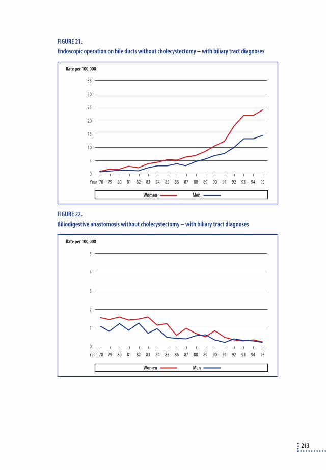
FIGURE 21.
Endoscopic operation on bile ducts without cholecystectomy – with biliary tract diagnoses
FIGURE 22.
Biliodigestive anastomosis without cholecystectomy – with biliary tract diagnoses
213
0
Rate per 100,000
78 79 80 81 82 83 84 85 86 87 88 89 90 91 92 93 94 95Year
35
20
15
10
5
25
30
MenWomen
0
Rate per 100,000
78 79 80 81 82 83 84 85 86 87 88 89 90 91 92 93 94 95Year
5
2
1
3
4
MenWomen

Appendix 3Literature
SEARCH OF LITERATURE
Index Medicus On a previous occasion, a search was conducted of Index Medicus for theperiod 1978-1990 for all keywords concerning bile, biliary tracts, gall-stones and treatment thereof.
MEDLINE A search was performed for the period January 1989 – November 1997.Search criteria: ❖ explode "BILIARY-TRACT-SURGERY"/ without-subheadings, adverse-effects, clas-
sification, contraindications, economics, history, instrumentation, mortality, met-
hods, nursing, psychology, rehabilitation, statistics-and-numerical-data, stan-
dards, trends, utiliszation - in MJME (Major Mesh).
❖ however, without "PORTOENTEROSTOMY,-HEPATIC"/ all subheadings
❖ explode "BILIARY-TRACT-DISEASES"/ without-subheadings, blood, blood-sup-
ply, cerebrospinal-fluid, chemistry, chemically-induced, classification, congenital,
complications, diet-therapy, diagnosis, drug-therapy, economics, ethnology, em-
bryology, enzymology, epidemiology, etiology, genetics, history, immunology,
metabolism, microbiology, mortality, nursing, pathology, prevention-and-con-
trol, physiopathology, parasitology, psychology, radiography, rehabilitation,
radio-nuclide-imaging, radiotherapy, secondary, secretion, surgery, therapy,
ultrastructure, urine, ultrasonography, virology - in MJME (Major Mesh).
Approx. 23,000 abstracts were reviewed and a total of 7,039 weredeemed relevant to this report and transferred into a RefMan database.
EMBASE A search was conducted for randomised surveys involving laparoscopiccholecystectomy for the period up to and including November 1997(Search criteria: ?olecystectomy, randomi?ed og laparoscop?).
214

Cochrane Library A search was conducted for randomised surveys involving diagnostics,prevention and treatment of biliary tract diseases.
Bibliographies The bibliographies in the articles read were studied to identify additionalreferences.
CRITICAL EVALUATION OF THE LITERATURE
In order to avoid biased interpretations, it was necessary to define in ad-vance the principles to be used for the literature study. There is now somuch literature about gallstone treatments that by accentuating the im-portance of different articles, support can be found for widely divergentpoints of view regarding the treatment of the same categories of illness.For this report, it has not been possible to conduct an in-depth study ofthe literature relating to all areas since the effort involved would corre-spond to several years of work. This work is being done at the momentunder the auspices of the Cochrane Collaboration. The purpose of theCochrane Collaboration84 is to collate all randomised surveys of diagnos-tics, prevention, treatment and care of diseases and to review them sys-tematically – in other words, to review articles according to fixed criteria.One reason for the international scope of the collaboration is to make surethat all language areas are covered. There are a number of methodologi-cal problems involved in collating the literature291.
Principles for evaluating individual articles
Randomised surveys1. Inclusion and exclusion criteria must be well defined
2. The randomisation has to be consistent
- Specification of the duration of the study; the proportion of relevant patients
randomised; exclusion after randomisation plus drop-out rate during treat-
ment.
- Patients must be allocated to the different interventions on the basis of the
chance principle and be blinded (computerised, closed envelopes, etc.). Quasi
randomisation (e.g. on surgeon or department) is unacceptable.
- The analyses have to be made according to the principle of ‘intention to treat’,
i.e. once a patient has been allocated to a treatment, he is included in this treat-
ment group regardless of how his course of treatment develops.
215

- The number of patients who are excluded or who drop out after randomisation
has to be less than 10% and the drop-out rate must not be spread unequally be-
tween the groups.
3. The extent to which the treatment is blind must be stated, and this is particularly
important in surveys about pain, bed days and convalescence582.
4. The method of treatment has to be well described and the extent to which the do-
ctors treating the patients have been trained in the procedure must also be clearly
stipulated.
The above criteria were used in the assessment of the randomised surveysand lead to either exclusion (if points 1 and 2 were not fulfilled) or to ex-planation of differences in the results achieved by the different surveys(points 3 and 4).
Clinical seriesIdeally, the following criteria ought to be fulfilled:
❖ Inclusion and exclusion criteria must be well defined
❖ The series must be consecutive and the time interval stipulated
❖ The series must be of a certain size
❖ The method of treatment must be well described and the extent to which the
doctors treating the patients have been trained in the procedure must also be sti-
pulated.
Deviations from the above mentioned criteria must be mentioned in thetext or in the relevant tables.
In which areas has the available literature been systematically studied?
❖ Follow up studies of patients with stones in the gallbladder
❖ Descriptions of gallstone symptoms
❖ Comparisons between different technologies for simple cholecystectomy
❖ Randomised surveys of different technologies for treatment of stones in the biliary
tracts
This report covered the following language areas: Danish, Norwegian,Swedish, English, German and French. Articles pertaining to randomisedsurveys in other languages have been translated into English or Danish.
216
SELECTED LITERATURE
Randomised trials of different forms of access to cholecystectomy
Cholecystectomy by laparoscopy versus minilaparotomySeven randomised trials that compared laparoscopic cholecystectomy tocholecystectomy by minilaparotomy were identified. The surveys covereda total of 1,011 patients65, 148, 421, 483, 500, 504, 766. Four of the surveys, i.e. rep-resenting 199 (20%) of the patients, did not fulfil the randomisation re-quirements65, 148, 421, 766. One500 of the three remaining works only per-formed an analysis according to the “intention to treat” principle for dataconcerning length of stay in hospital. Only one study483 was blinded andonly one work stated that the surgeons were trained in both procedures483.
Cholecystectomy by laparoscopic versus traditional open cholecystectomyNineteen randomised trials were identified that compare laparoscopiccholecystectomy with traditional open cholecystectomy. They cover 959patients12, 75, 79, 174, 190, 246, 266, 337, 350, 379, 410, 490, 522, 579, 621, 634, 654, 681, 789. The vastmajority of works in this category are quite small and are about anaes-thesiology and physiological trials in which no attempt was made to ex-plain the randomisation procedure. Sufficient randomisation procedureswere only stipulated in four works79, 337, 579, 789 covering 151 patients. Onlyone study579 was blinded.
Cholecystectomy by minilaparotomy versus traditional open cholecystec-tomyFour randomised trials were identified that compare cholecystectomy byminilaparotomy with traditional open cholecystectomy. They covered 391patients51, 574, 690, 703. One of the works covering 50 patients51 did not de-scribe the randomisation procedure sufficiently. None of the studies wereblinded.
Randomised trials regarding pain relief and nausea/discomfort after simple chole-
cystectomy
Various initiatives designed to reduce postoperative pain, nausea andother forms of discomfort have been analysed in several randomised tri-als. Within the time framework of this report, it has not been possible tostudy them all systematically. The references are stated here for use in anyfollow-up work on reference programmes about gallstone treatment.
217

The location of the incision Ali20, Garcia-Valdecasas267, Armstrong49
Laser vs. traditional surgerySteger743
Local anaesthesia in the wound Patel592, Adams9, Russell659, Ure798, Johansson355, Alexander19, Sarac679
Intrapleural local anaesthesia Aguilar13, Blake94, Frank240, Frenette250, Kastrissios380, Oxorn583, Radema-ker625, Schroeder695, Scott701, Strömskag757,VadeBoncouer799
Local anaesthesia above the lobe of the liver Chundrigar141, Joris361, Mraovic537, Pasqualuci590, Pasqualucci591, Radema-ker624, Raetzell626, Scheinin684, Szem762, Weber823
Intercostal local anaesthesia Blake94, Ross650
Epidural anaesthesia/morphine Hakanson298, Huang336, Scott701, Thörn779
Transcutaneous electric nerve stimulation Laitinen425
Preoperative and intraoperative analgetics Forse236, Lane428, Michaloliakon519, Wilson847
Suprahepatic drain Jorgensen360
Reduction of nausea/discomfort postoperativeCabrera123, Fujii256, Sohi867, Steinbook744, Thune782
Special circumstances associated with laparoscopic surgery
CO2 vs. abdominal-lift Kitano401, Koivusalo406, Koivusalo407, Lindgren456, Meijer509, Yoshida858
218

CO2 vs. helium Bongard99
French vs. American method Kum419
Clinical series covering symptoms and indication for treatment
This chapter contains a detailed breakdown of clinical series coveringsymptoms and indications for gallstone treatment. The tables are sum-marily treated in the main report.
TABLE 1
Association between symptoms and gall stones in population studies. The studies represent
well-defined population groups that have been screened for gallstones by X-ray or ultrasound.
Only series covering more than 100 persons have been included.
The occurrence of gall stones is associated with
Author Country N Biliary pain a Pains in upper abdomen Dyspepsiab
Wilbur840 USA 1,233 Nejc Yes Yes
Price620 UK 204 - No No
Sampliner677 USA 395 - Yes No
Bainton57 UK 956 - Noc No
GREPCO39 Italy 1,081 Noc - No
Janzon352 Sweden 424 - No No
Barbara62 Italy 1,911 Yes - No
Pixleyd 611 UK 242 - No No
GREPCO42 Italy 1,239 No - No
Mellström511 Sweden 109 Yes - -
Glambek275 Norway 1,329 - No No
Jørgensen368 Denmark 3,416 No No No
Heatone 315 UK 1,896 Yes (women) Noc (women) -
No (men) No (men) -
Loria460 Italy 1,807 Yes - Yes
Attili53 Italy 29,739 Yes - -
a: Biliary pain is defined differently in different studies, and in certain studies are not defined at all. The Italian studies agree on the fol-
lowing definition: Pains in the epigastrium or upper right quadrant within the last five years. The pains must last more than half an
hour and must not by eased by bowel movements/flatus.
b: Dyspepsia involves a whole series of symptoms like nausea, heartburn, eructation of acid fluid, bloatedness, rumbling, pressure in the
epigastrium, flatulence and food intolerance.
c: A distinct tendency towards a link between symptoms and gallstones but the link is not significant.
e: Heaton’s work was considered as two studies (men and women).
219

TABLE 2
Relationships between symptoms and gallstones in consecutive patient cohorts referred to an
X-ray department or a clinical department. Only series involving more than 100 patients were
included.
The occurrence of gall stones is associated with
Author Country N Biliary pain a Pains in upper abdomen Dyspepsiab
Hinkel328 1,000 Yes Yes No
Koch404 USA 655 - - No
Wegge824 Denmark 192 - Yes Yes
Persson604 Sweden 817 - Noc Nod
Diehl185 USA 300 Yes Yes No
Galatola263 UK 282 Yes - -
Farrell217 Ireland 300 - Yes Nejd
a: Biliary pain is defined differently in different studies, and in certain studies are not defined at all.
b: Dyspepsia covers a whole string of symptoms such as nausea, heartburn, eructation of acid fluid, bloatedness, rumbling, pressure in
the epigastrium, flatulence and food intolerance
c: The occurrence of abdominal pains was not different in people with and without gallstones, but maximal pain in the upper right qua-
drant was seen significantly more often in patients with gallstones than in patients without gallstones
d: A significant negative association between gall stones and certain dyspeptic symptoms
220

TABLE 3
Clinical series studying the frequency of persistent symptoms after cholecystectomy. The series
are consecutive with a follow-up of at least 3 months and a response rate of at least 70% a.
Follow-up has to have been conducted on at least 50 patients.
Persistent symptoms
Author Country N/nb Follow-up months Painsc Dyspepsiac
Burnett119 UK 200/141(o) 12-60 14% 11%
Rhind640 UK 72/66 (o) 12- 38% (17%) 40% (9%)
Gunn295 UK 107/105 (o) 12-24 23% (7%) -
Bremner107 UK 207/207 (o) 8-35 8% (5%) -
Johnson356 UK 108/108 (o) 3-39 - 54% (30%)
Ros649 Spain 130/92 (o) 24 22% 24%
Gilliland273 USA 650/525 (o) 15-79 12% 25%
Jørgensen376 Denmark 122/115 (o) 6-12 21% (7%) -
Batesd, 70 UK 292/278/274 (o) 12 & 24 34%/27% > 50%
Paul595 Germany 60/51 (o) 4 18% 18%
Mühe547 Germany 94/93 (l) 49-67 27% -
136/130 (o) 25% -
Konsten409 HongKong 351/325 (o) 120 19% -
Qureshi622 Ireland 100/100 (l) 12 13% 20%
Wilson846 UK 115/100 (l) 14 6% -
200/167 (o) 28 7% -
Mort531 USA 372/312 (o) 3 20-36% -
Stiff749 UK 250/205 (l) 15 37% (3%) -
200/155 (o) 32 41%(10%) -
Fenster223 USA 225/164 (l) 3 18% 54%
Kane378 USA 2.490/72% (l) 6 23% (11%) -
958/72% (o) 6 24% (11%) -
Ure797 Germany 508/468 (l) 15-23 26% (6%) -
(o): Traditional open cholecystectomy; (l): Laparoscopic cholecystectomy
a: A number of works have been omitted because of lack of information about the follow-up, follow-up rates below 70% 59, 96, 651, 742, 750,
insufficient follow-up periods (5-7 weeks)92 or selected follow-up468, 516 omitting patients converted to open cholecystectomy
b: N/n: Number of patients at the start of the study/number of patients followed up
c: The proportion with persisting symptoms (the proportion with unchanged or aggravated conditions).
d: Follow-up after 1 and 2 years respectively.
221
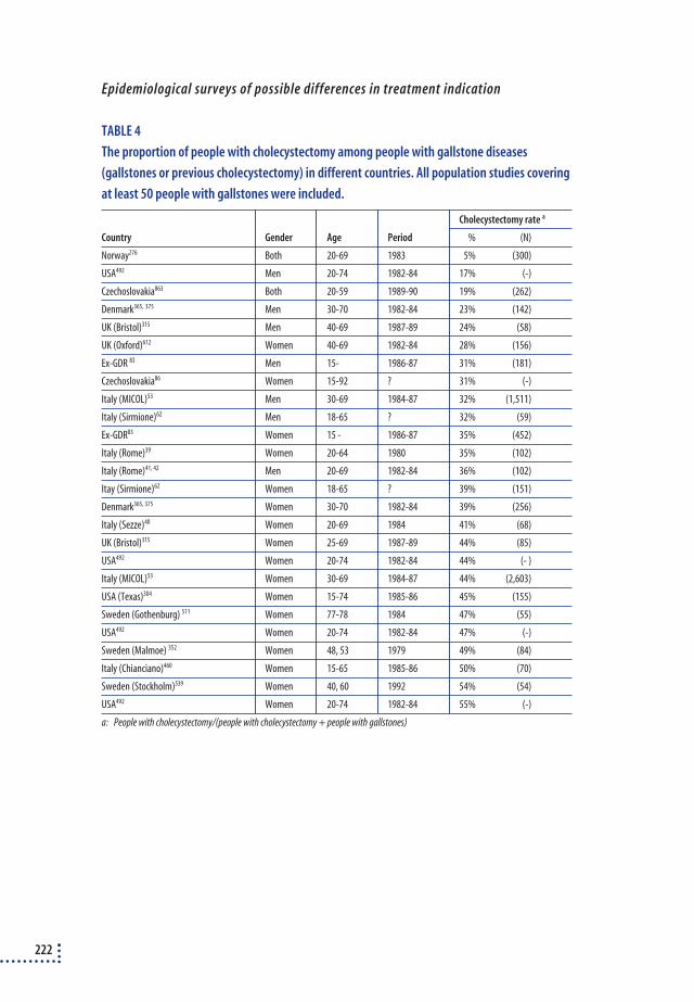
Epidemiological surveys of possible differences in treatment indication
TABLE 4
The proportion of people with cholecystectomy among people with gallstone diseases
(gallstones or previous cholecystectomy) in different countries. All population studies covering
at least 50 people with gallstones were included.
Cholecystectomy rate a
Country Gender Age Period % (N)
Norway276 Both 20-69 1983 5% (300)
USA492 Men 20-74 1982-84 17% (-)
Czechoslovakia863 Both 20-59 1989-90 19% (262)
Denmark365, 375 Men 30-70 1982-84 23% (142)
UK (Bristol)315 Men 40-69 1987-89 24% (58)
UK (Oxford)612 Women 40-69 1982-84 28% (156)
Ex-GDR 83 Men 15- 1986-87 31% (181)
Czechoslovakia86 Women 15-92 ? 31% (-)
Italy (MICOL)53 Men 30-69 1984-87 32% (1,511)
Italy (Sirmione)62 Men 18-65 ? 32% (59)
Ex-GDR83 Women 15 - 1986-87 35% (452)
Italy (Rome)39 Women 20-64 1980 35% (102)
Italy (Rome)41, 42 Men 20-69 1982-84 36% (102)
Itay (Sirmione)62 Women 18-65 ? 39% (151)
Denmark365, 375 Women 30-70 1982-84 39% (256)
Italy (Sezze)40 Women 20-69 1984 41% (68)
UK (Bristol)315 Women 25-69 1987-89 44% (85)
USA492 Women 20-74 1982-84 44% (- )
Italy (MICOL)53 Women 30-69 1984-87 44% (2,603)
USA (Texas)304 Women 15-74 1985-86 45% (155)
Sweden (Gothenburg) 511 Women 77-78 1984 47% (55)
USA492 Women 20-74 1982-84 47% (-)
Sweden (Malmoe) 352 Women 48, 53 1979 49% (84)
Italy (Chianciano)460 Women 15-65 1985-86 50% (70)
Sweden (Stockholm)539 Women 40, 60 1992 54% (54)
USA492 Women 20-74 1982-84 55% (-)
a: People with cholecystectomy/(people with cholecystectomy + people with gallstones)
222

Postoperative pain, hospital stay and convalescence
TABLE 5
Postoperative pain, length of hospital stay and convalescence in randomised trials of different
forms of access to cholecystectomy
Study N Blinding Pain Analgesia Hospital stay Absence due to
(days) illness (days)
Laparoscopy vs. traditional open laparotomy
Ortega579 20 Yes 5 vs. 12aa - 1,2 vs. 1,1 -
Trondsen789 72 No Sign.aa 4 vs. 6bb 2 vs. 4 11 vs. 34
Berggren79 30 No - 125mg vs. 200mgcc 1,8 vs. 2,8 12 vs. 24
Huang337 29 No - 0,5 vs. 2bb 3,9 vs. 7,9 -
Laparoscopy vs. minilaparotomy
Majeed483 200 Yes - - 3 vs. 3 35 vs. 28
McMahon504 302 No 40 vs. 59aa 22mg vs. 40mgdd 2 vs. 4 38 vs. 38
McGinnee 500 310 No - - 4 vs. 3 -
Minilaparotomy vs. open laparotomy
O’Dwyer574 30 No - 2,5 vs. 4,5bb 3 vs. 5 -
Seenu703 181 No - - 2,6 vs. 4 -
Schmitz690 130 No Not sign. Not sign.
Significant differences are written in italics
a: Visual analogue scale – significantly greater pain after open laparotomy
b: Median number of painkilling injections
c: mg of pethidin 13-24 hours after operation. Not sign. from 0-12 hours after operation
d: Median volume morphine
e: McGinn only treats the length of hospital stay according to the "intention to treat" principle
Number of biliary tract lesions in connection with cholecystectomy
In general, the following requirements were stipulated for all surveys:
❖ Consecutive groups of 200+ patients.
❖ Evenly distributed patient material (e.g. not only elderly patients or only young
patients).
❖ The material has to be well described.
❖ Biliary tract lesions have to be well described.
Any deviations will be stipulated in the text accompanying the individualtables.
The following types of works are differentiated:❖ Clinical series, in which one or more hospital departments or surgeons record their
own results. Randomised trials are included, if the randomisation does not cover dif-
ferent surgical technologies, and if it is clearly stated that all patients were included.
223
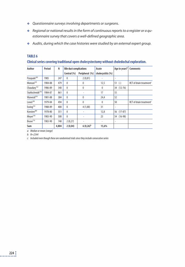
❖ Questionnaire surveys involving departments or surgeons.
❖ Regional or national results in the form of continuous reports to a register or a qu-
estionnaire survey that covers a well-defined geographic area.
❖ Audits, during which the case histories were studied by an external expert group.
TABLE 6
Clinical series covering traditional open cholecystectomy without choledochal exploration.
Author Period N Bile duct complications Acute Age in yearsb Comments
Central (%) Peripheral (%) cholecystitis (%)
Pasquale589 1985 247 0 2 (0,81) - -
Monson525 1984-88 479 0 0 12,5 51 (-) RCT of drain treatmentc
Chaudary132 1986-89 340 0 0 0 34 (12-76)
Stahlschmidt736 1984-87 861 0 - 17 55
Warwick816 1981-88 384 0 0 24,4 52
Lewis450 1979-84 494 0 0 0 50 RCT of drain treatmentc
Ewing214 1988-89 400 0 4 (1,00) 31 -
Konsten409 1978-80 351 0 - 12,8 56 (17-87)
Meyer518 1983-90 500 0 - 23 54 (16-98)
Brune114 1983-90 748 2 (0,27) - - -
Sum 4,804 2 (0,04) 6 (0,26)b 15,6%
a: Median or mean (range)
b: N=2344
c: Included even though these are randomised trials since they include consecutive series
224

TABLE 7
Clinical series covering traditional open cholecystectomy in which it is not possible to
differentiate between simple cholecystectomy and cholecystectomy with choledochal surgery
Author Period N Bile duct complications Acute Age in yearsb Comments
Central (%) Peripheral (%) cholecystitis (%)
Ganey264 1978-83 1,035 0 0 16 (15-92)
Scher685 1979-82 432 0 1 (0.23) - -
Gregg292 1979-80 765 1 (0.13) - - - The damage occurs du-
ring cholangiography
Heberer316 1985-87 544 0 - 0 -
Orth868 1982-89 773 1 (0.13) 2 (0.26) 0 56
Battersby72 1986-90 346 2 (0.58) - - -
Kocher405 1984-87 1,631 1 (0.06) 2 (0.12) 15.5 58 (15-91)
Saltzstein674 1988-90 500 0 0 29.8 40 (10-96)
Caputo126 1980-87 1,617 7 (0.43) 1 (0.06) 25.2 -
Clavienb, 145 1984-89 1,252 0 2 (0.16) 0 52 (16-88)
Cox159 1985-89 457 1 (0.22) - 36.4 53 (12-87)
Davies175 1985-90 722 0 0 24.9 49
Herzog325 1984-90 1,631 1 (0.06) - 15 -
Morgenstern529 1982-88 1,200 2 (0.17) - - 56 (3-96)
Stoker752 1989-90 304 0 - 38.3 53
Wieden839 1985-89 921 3 (0.33) 1 (0.11) - -
Bradbury105 1989-90 246 0 0 - 48k/57m
Bjerkeset91 1980-89 832 1 (0.12) - 49 66 (18-100)
Sum 15,208 20 (0.13) 9 (0.10)c 19.4%
a: Median or mean (range)
b: Two publications 117, 512 make up a subset of Clavien 145 and have, therefore, been omitted.
c: N=9129
225

TABLE 8
Cholecystectomy by minilaparotomy. Clinical series and randomised trialsa.
Author Period N Bile duct complications Acute Age in yearsb Comments
Central (%) Peripheral (%) cholecystitis (%)
Dubois197 1973-80 1.500 0 0 - -
Goco282 15 month 56 0 0 - -
Morton532 2 years 96 0 0 - - (21-82)
Salembier671 - 125 0 0 - -
Merrill514 3 years 91 0 0 28,8 57 (16-84)
Reddick469 1988-89 25 0 0 - 40 Part of RCT
Ledet435 1986-90 200 0 0 - (16-82)
O’Dwyer870 - 55 0 0 - 48 (13-81)
Safatle666 1990-91 21 0 0 - -
Stage733 - 24 0 0 - 56 (22-72)
Barkun65 1990-91 25 0 1 (4.00) - - Part of RCT
Kunz421 1990- ? 37 1 (2.70) 0 - - Part of RCT
O’Dwyer574 - 16 0 0 31,3 46 (27-74) Part of RCT
Pélissier598 1983-90 191 0 0 - 46 (9-82)
Suarez758 1984- ? 50 0 0 48 47 (-) Not “int. to treat”
Warren815 1990-91 20 0 0 - 55 (30-81)
Assalia51 18 months 24 0 0 - 60 (-) Part of RCT
Coelho148 1992 15 0 0 - 43 (25-66) Part of RCT
Rozsos656 1990-93 607 1 (0.16) 1 (0.16) - - (15-87)
Seenu703 1990-92 97 0 0 - - Part of RCT
Tyagi793 - 143 0 4 (2.80) - - (20-83)
McGinn500 1991-95 155 0 1 (0.65) 0 57 (26-84) Part of RCT
Gaetini261 1992-94 62 0 0 - -
Kumar871 1995-96 60 0 0 0 39 (24-70) Part of RCT
Schmitz690 1991-94 65 0 0 - 53 (24-66) Part of RCT
Sum 3,760 2 (0.05) 7 (0.19) 14.8%
Sum >= 50 3,553 1 (0.03) 6 (0.17) 14.0%
Sum >=100 2,921 1 (0.03) 6 (0.21) 0.0%
Sum >=200 2,307 1 (0.04) 1 (0.04) -
In this material, series covering less than 200 patients were also accepted because relatively little has been written about the subject. The
complication rates were, however, calculated on materials of more than 50, 100 and 200.
a: Two randomised trials were not included483, 504, since they are part of a separate assessment. The rest of the randomised trials do not
live up to the requirements of sufficient randomisation. They are included as clinical series.
b: Median or mean (range).
226

TABLE 9
Laparoscopic cholecystectomy. Early clinical series, published 1990-92.
Author Period N Bile duct complications Acute Age in yearsb Comments
Central (%) Peripheral (%) cholecystitis (%)
Duboisb, 200 1988-90 690 3 (0.43) 2 (0.29) - -
Bailey56 1989-91 375 1 (0.27) 5 (1.33) 6.4 47 (16-94)
Corbitt155 - 300 0 0 16 (15-90)
Cuschieri168 - 1,236 4 (0.32) 2 (0.16) 2.3 47 (13-86) 7 depts. in 3 countries
in Europe
Fitzgibbon231 - 350 0 0 14.7 -
Graves290 1989-90 304 1 (0.33) 0 3.3 50 (14-83)
Ko403 1989-91 300 1 (0.33) 0 5.3 (12-85) Personal series
Manger486 1990-91 200 0 1 (0.50) 3.0 47 (13-78)
Peters607 1990 283 1 (0.35) 3 (1.06) 4.9 46 (-)
Sackier662 1989-90 516 1 (0.19) 1 (0.19) - -
Spaw728 - 500 0 1 (0.20) 5.8 46 (17-86)
Voyles812 - 453 0 1 (0.22) 23.4 52 (15-98)
Wolfe852 1990-91 381 0 4 (1.05) 7.6 45
Baird58 1989-91 800 0 3 (0.38) 13.0 -
Dallemagne170 1990-91 368 1 (0.27) 1 (0.27) 1.6 56 (-)
Dashow173 1990-91 250 0 2 (0.80) - 49 (15-97)
Davis176 1989- 622 1 (0.16) 1 (0.16) 10.0 48 (17-97)
Dion189 1990-91 258 0 1 (0.39) 1.2 44 (14-82)
Fabre215 1989-91 392 4 (1.02) 0 16.6 54 (17-85)
Feussner226 - 500 0 4 (0.80) - -
Fielding228 1990-91 220 0 1 (0.45) 6.8 - (12-75)
Frazee245 1990-92 706 2 (0.28) - 14.2 51 (-)
Gai262 1990-91 340 0 1 (0.29) 6.8 49 (16-87)
Graber288 - 300 4 (1.33) - - -
Hawasli313 1989-91 480 0 2 (0.42) - - Personal series
Hershman323 - 200 1 (0.50) 2 (1.00) 24.5 49 (19-86)
Herzog325 1990-91 278 1 (0.36) 1 (0.36) 0.0 53 (18-86)
Huang335 1990-91 200 1 (0.50) 1 (0.50) 15.5 51 (27-83)
Kimura398 1990-91 250 0 0 0.0 49 (24-78)
Kiviluoto402 1991-92 200 1 (0.50) 0 16.0 48 (16-83)
Kozarekc. 411 1990 597 11 (1.84) 6 (1.01) - -
Leahy433 1990 308 1 (0.32) 4 (1.30) 13.6 48
Lillemoe453 1989-91 400 2 (0.50) 5 (1.25) 2.5 48 (15-96)
Miles521 - 201 1 (0.50) 1 (0.50) - -
Nottle569 1990-91 280 0 3 (1.07) - -
Pérrisatd, 602 1988-91 696 3 (0.43) 4 (0.57) - (13-83)
Ratliff633 1990-91 230 0 2 (0.87) 20.0 56 (10-86)
Scott697 1990-91 210 0 1 (0.48) 11.4 46 (17-85)
Sigman711 1990-? 500 2 (0.40) - 9.1 48 (7-93)
Sim712 1990 271 1 (0.37) - - -
227

(Tabel 9 continued)
Author Period N Bile duct complications Acute Age in yearsb Comments
Central (%) Peripheral (%) cholecystitis (%)
Singson714 1990-91 329 0 4 (1.22) 9.1 47 (15-83)
Soper724 1989-91 618 2 (0.32) 1 (0.16) 6.0 47 (15-82)
Stahlschmidte, 736 1990-92 812 3 (0.37) 6 (0.74) - -
Stoker752 1990-91 280 0 1 (0.36) 27.5 51 (-)
Troidlf, 787 1989-91 400 3 (0.75) 0 2 52 (18-87)
Wayand821 1990-91 250 1 (0.40) 2 (0.80) 19.2 52 m/46 k
Wieden839 1990-91 600 4 (0.67) 1 (0.17) 6.1 -
Sum 19,234 62 (0.32) 81 (0.46)g 8.9%
a: Median or mean (range)
b: An earlier publication199 contains a subset of this material
c: 13 of the 17 lesions occurred during surgeons' first 20 operations
d: Includes a large proportion of the patients from another series151, which has, therefore, been excluded
e: 3 bile duct lesions is a minimum number, since the 6 other lesions probably includes at least one bile duct lesion
f: Includes an earlier publication by the same author787.
g: N=17.457
A study covering 1,009 patients717 includes 6 injuries (0.59%), but it cannot be determined whether they are central or peripheral
228

TABLE 10
Laparoscopic cholecystectomy. Clinical series published 1995-97 a
Author Period N Bile duct complications Acute Age in yearsb Comments
Central (%) Peripheral (%) cholecystitis (%)
Amaral23 1991-92 200 0 1 (0.50) 24.0 46 (-) Personal series
Azurin55 1990-95 1,400 0 2 (0.14) 27.0 -
Denning180 1990-92 894 3 (0.34) 4 (0.45) - -
Dorazio195 1990-94 1,344 1 (0.07) - - - (9-90)
Dubois201 1988-94 2,665 6 (0.23) 13 (0.49) 12.5 51 (6-90)
Dubois196 1990-93 500 2 (0.40) 8 (1.60) 13.2 45
Hölbling333 1992-94 455 2 (0.44) 1 (0.22) 11.0 -
Madhavan476 1990-91 400 0 - 30.0 53 (9-90)
Panton587 1991-93 228 0 4 (1.75) 18 50 (15-84)
Schrenkc, 694 1990-93 1,300 3 (0.23) - - -
Slim716 1989- ? 710 4 (0.56) 0 7.9 57 (17-93)
Bond98 1990-93 529 4 (0.76) 3 (0.57) 4 44 (13-85)
Buanes115 1991-94 277 0 4 (1.44) 6.9 46 (18-83)
Carrolld, 129 1989-95 3,242 12 (0.37) - - -
Chen135 1991-95 2,428 5 (0.21) 0 1 (15-77)
Ferzli225 1990-4 1,442 2 (0.14) 2 (0.14) - 44 (13-94)
Kullman416 1991-94 630 4 (0.63) 3 (0.48) - 53 (11-86)
Leung443 1990- ? 297 1 (0.34) 1 (0.34) 22.6 53 (-)
Zisman862 - 329 0 - - -
Dexter182 1990-94 411 0 4 (0.97) - 53 (-)
Jan349 1991-95 1,115 5 (0.45) 3 (0.27) - 47 (28-70)
Ladocsi422 1991-93 734 1 (0.14) - 12.1 49 (-)
Lam427 1994-96 213 0 - - -
Mjåland524 1994-95 200 2 (1.00) 1 (0.50) - 49 (17-82)
Taylor768 1990-95 2,038 2 (0.10) - - -
Thanh774 1991-95 361 4 (1.11) 4 (1.11) 31 50 (16-85)
Sum 24,342 63 (0.26) 58 (0.39)e 12.7%
a: The search for relevant literature ended in November 1997
b: Median or mean (range).
c: Counts only injuries that lead to conversion (does not look at injuries not discovered until after the operation).
d: ): The series includes an earlier publication528, which has, therefore, been excluded
e: N=14.742
229

TABLE 11
Laparoscopic cholecystectomy. Results from questionnaire surveys.
Author Period N Bile duct complications Acute Age in yearsa Comments
Central (%) Peripheral (%) cholecystitis (%)
Airan14 -91 2,671 5 (0.19) 25 (0.94) - 52 m/45wb Response rate not
stated
Delaitre179 - 6,512 29 (0.45) 18 (0.28) - 51 (13-95) questionnaire and
seriesc
Larson432 - 1,983 5 (0.25) 7 (0.35) - - d
Cocks147 1990-92 5,927 12 (0.20) - - 74% response rate
Devency181 -92 9,597 27 (0.28) 69 (0.72) - - e
Deziel183 1990 77,604 317 (0.41) 142 (0.18) - - 41% response rate
Kimura400 1990- ? 1,989 9 (0.45) 1 (0.05) 0 - f
Croce162 1990-92 6,865 18 (0.26) - - g
Dunn203 1990-91 3,319 11 (0.33) 35 (1.05) - - Response rate not
stated
Mucio538 1990-91 2,399 6 (0.25) 10 (0.42) 22 - 72% response rate
Trondsen790 - 527 3 (0.57) 5 (0.95) 5.3 - 7 departments
Vincent- 1989-92 2,432 11 (0.45) 5 (0.21) 7.7 50 (9-91) Response rate not
Hamelin807 stated
Morino530 - 2,127 8 (0.38) - -
Pongchairerks618 - 1,744 14 (0.80) 11 (0.63) - -
Collet150 1994 4,624 7 (0.15) - 25.9 56m/53wb h
Sum 130,320 482 (0.37) 328 (0.30)i 16.2%
One questionnaire survey covering 36,232 patients15 has not been included in the table, since it was impossible to differentiate between
central and peripheral bile duct lesions (a total of 0.50%).
a: Median or mean (range).
b: m=men, w=women
c: Two questionnaire surveys linked to three clinical series.
d: No information about how the surgeons were chosen and how many declined to participate.
e: Questionnaire for surgeons and surgical managers in Oregon. Response rate 69% and 53%, respectively.
f: Eight institutions. How they were chosen and what response rate was achieved are not described in detail.
g: 19 groups. How they were chosen and what response rate was achieved are not described in detail.
h: 150 surgeons. It is not stated how many were asked.
i: N=110,777
230
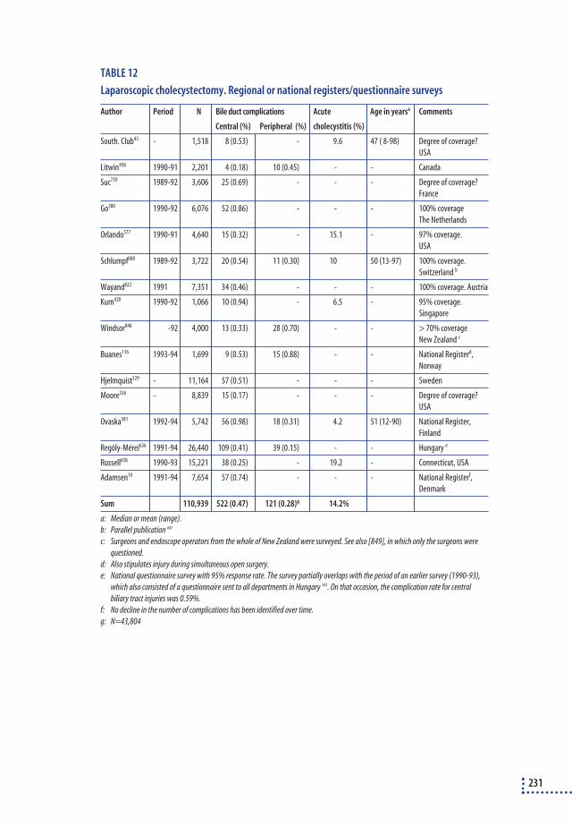
TABLE 12
Laparoscopic cholecystectomy. Regional or national registers/questionnaire surveys
Author Period N Bile duct complications Acute Age in yearsa Comments
Central (%) Peripheral (%) cholecystitis (%)
South. Club43 - 1,518 8 (0.53) - 9.6 47 ( 8-98) Degree of coverage?
USA
Litwin458 1990-91 2,201 4 (0.18) 10 (0.45) - - Canada
Suc759 1989-92 3,606 25 (0.69) - - - Degree of coverage?
France
Go280 1990-92 6,076 52 (0.86) - - - 100% coverage
The Netherlands
Orlando577 1990-91 4,640 15 (0.32) - 15.1 - 97% coverage.
USA
Schlumpf688 1989-92 3,722 20 (0.54) 11 (0.30) 10 50 (13-97) 100% coverage.
Switzerland b
Wayand822 1991 7,351 34 (0.46) - - - 100% coverage. Austria
Kum420 1990-92 1,066 10 (0.94) - 6.5 - 95% coverage.
Singapore
Windsor848 -92 4,000 13 (0.33) 28 (0.70) - - > 70% coverage
New Zealand c
Buanes116 1993-94 1,699 9 (0.53) 15 (0.88) - - National Registerd,
Norway
Hjelmquist329 - 11,164 57 (0.51) - - - Sweden
Moore526 - 8,839 15 (0.17) - - - Degree of coverage?
USA
Ovaska581 1992-94 5,742 56 (0.98) 18 (0.31) 4.2 51 (12-90) National Register,
Finland
Regöly-Mérei636 1991-94 26,440 109 (0.41) 39 (0.15) - - Hungary e
Russell658 1990-93 15,221 38 (0.25) - 19.2 - Connecticut, USA
Adamsen10 1991-94 7,654 57 (0.74) - - - National Registerf,
Denmark
Sum 110,939 522 (0.47) 121 (0.28)g 14.2%
a: Median or mean (range).
b: Parallel publication 687
c: Surgeons and endoscope operators from the whole of New Zealand were surveyed. See also [849], in which only the surgeons were
questioned.
d: Also stipulates injury during simultaneous open surgery.
e: National questionnaire survey with 95% response rate. The survey partially overlaps with the period of an earlier survey (1990-93),
which also consisted of a questionnaire sent to all departments in Hungary 343. On that occasion, the complication rate for central
biliary tract injuries was 0.59%.
f: No decline in the number of complications has been identified over time.
g: N=43,804
231

TABLE 13
Laparoscopic cholecystectomy. Audit
Author Period N Bile duct complications Acute Age in yearsa Comments
Central (%) Peripheral (%) cholecystitis (%)
Birdi88 1990-92 555 1 (0.18) 5 (0.90) - 51 (16-85) one town
Wherry838 1990-92 5,642 32 (0.57) - - - US-army
Steele740 1991-94 502 6 (1.20) 3 (0.60) - - one department
Richardson642 1990-95 5,913 37 (0.63) - - - West of Scotlandb
Wherry837 1993-94 9,130 35 (0.38) 50 (0.55) - - US-army
Merrie513 1991-95 929 8 (0.86) 5 (0.54) - - New Zealand
Sum 22,671 119 (0.52) 63 (0.57)c
a: Median or mean (range).
b: The results of the first 1,683 cases from the period 1990-92 were published 257 and the proportion suffering central complications was
0.48%.
c: N=11,116
232

Results for cholecystolithotomy
TABLE 14
Clinical series of cholecystolithotomy (elective surgery). Only series involving more than
25 patients were included
Author Period N/na Studyb Successc Follow-up Comments
Persistent Recur- Months
pains rence (median)
Pers603 1915-37 667/504 o - - 21% 236
Gibney270 1983-87 63/48 o - 11% 27% 18 After acute cholecystitis
Hamilton301 - 25/ m 96% 12% 8% 8 (4-18) Local anaesthetic
Leahy434 - 26/ l 96% - - - Local anaesthetic
8% biliary fistula
Majeed482 - 81/62 l 81% 5% 10% 18 (5-36) Intercostal/
epidural anaesthesia
Kellett385 - 46/ ll 91% - - - 4% biliary leak
Akiyama17 1981-89 28/ ll 71% - 5% 36 7% biliary leak/
7% cholecystectomy
Chiverton140 1986-89 60/ ll 93% 2% 0% 3 3% biliary leak
Stage734 1990-91 32 ll 90% - - - 3% biliary fistula
Tóth783 1989-90 40 ll 98% - - -
Cheslyn- 1988-90 113/ ll 88% 21% (7%) 10% 14 Bile accumulation (4%),
Curtis138+ perforated gall bladder
Gillams272 (2%)
Picus609 1987-91 58 ll 97% - - - Local anaesthesia
7% biliary leak
Donald193 1988-90 100 - - - 44% 48 Follow-up from
Cheslyn-Cutis92
McDermott498 1988-91 37/31 ll 86% 13% 16% 4-46 Bile salts afterwards
Courtois157 1987-92 /65 - - 12% 40% 33/14 See 609
Bing87 - 421/ ? 97% - - - Chinese – only abstract
a: N and n represent the number of patients included and the number of patients at follow-up, respectively.
b: ): o =open surgery, m=minilaparotomy, l=laparoscopy, ll=laparoscopy plus laser, ultrasound or other form of pulverization.
c: Proportion who have stones removed.
233
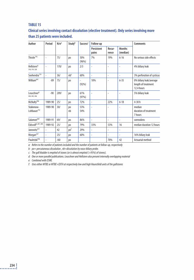
TABLE 15
Clinical series involving contact dissolution (elective treatment). Only series involving more
than 25 patients were included.
Author Period N/na Studyb Successc Follow-up Comments
Persistent Recur- Months
pains rence (median)
Thistle778 - 75/ po 50% 7% 19% 6-16 No serious side effects
(96%)
Hellsternd, - 170/ po 2/3 - - - 4% biliary leak318, 319, 320
Soehendra719 - 36/ nbe 60% - - - 3% perforation of cysticus
William841 -89 75/ po - 10% - 6-35 0% biliary leak/average
(92%) length of treatment
12.4 hours
Leuschnerd, -90 209/ po 61% - - - 5% biliary leak444, 445, 446 (97%)
McNulty506 1989-90 25/ po 72% - 22% 6-18 4-30 h
Stabenow- 1989-90 30/ po 53% - - - median
Lohbauer732 nb 54% duration of treatment
7 hours
Salamon669 1989-91 69/ po 86% - - - somnolens
Eidsvolld, 207, 208 1989-92 25/ po 79% 33% 53% 16 median duration 12 hours
Janowitz351 - 42 pof 29% - - -
Morgan527 - 25/ po 60% - - - 16% biliary leak
Pauletzki596 - /60 po 70% 42 Actuarial method
a: Refers to the number of patients included and the number of patients at follow-up, respectively
b: po= percutaneous dissolution , nb=dissolution by naso-biliary probe.
c: The gall bladder is emptied of stones (or is almost emptied (>95%) of stones).
d: One or more parallel publications. Leuschner and Hellstern also present internally overlapping material
e: Combined with ESWL
f: Uses either MTBE or MTBE+EDTA at respectively low and high Haunsfield units of the gallstones
234

Study of cholecystectomy in patients with acalculous biliary pain
TABLE 16
Clinical series covering cholecystectomy for gallstone-like pains in patients without gallstones
(acalculous biliary pains). Only series with a follow-up rate of at least 70% were included a.
Author Country N/nb Follow-up Persistent Preoperative
monthsc painsd physiological
measuremente
Nechis551 USA 18/18 1-60 - (5%) CCK-test
Madsen479 Denmark 9/9 32 (13-36) 33% (22%) CCK-testf
Burnstein121 Canada 41/37 18 32% (3%) CCK - bileg
Rajagopalan628 USA 21/16 22 6% (0%) CCK in some
Nora567 USA 30/30 - (12-39) - (0%) CCK (not emptied)
Einarsson209 Sweden 15/15 9-27 20% (7%) CCK - bile
Ferraris224 USA 42/38 6 - ? 29% (-) clinic
Rhodesh, 641 UK 90/81 35 (12-66) 33% (9%) CCK - pain
Gilliland274 USA 60/43 47 (16-79) 23% (- ) CCK
Westlake836 Canada 26/26 25 (5-48) 31% HIDA-EF
Zech860 USA 60 12 5% (2%) CCK-EF
Halverson300 USA 12/11 12-69 18% CCK-EF < 35%
Middleton520 UK 34/34 20 (3-36) 6% EF < 35%
Sorenson725 USA 11/11 10 (2-17) - ( 0%) EF <35%
Watson819 Australia 14/14 7% (0%) Dependent on EFi
Jones358 USA 36/35 14 (2-40) - (9%) Dependent on EFi
a: (a): Two studies were not included since the requirements set were not fulfilled255, 317
b: N and n represent the number of patients included and the number patients at follow-up, respectively
c: Median or mean (range)
d: Persistent pains (pains of same strength as before the treatment or worse)
e: The physiological measurement mainly consists of CCK (cholecystokinin) stimulation, in which the measurement of the effect is either
the level of pains or the degree of emptying of the gall bladder. In recent years, there has been a consensus that an EF (ejection
fraction) under 35% is pathological.
f: T he two who had unchanged pains both had positive provocative tests
g: Had examined both contraction and crystals. The one with unchanged pains was negative as far as the above mentioned tests are
concerned.
h: Two studies by Lennard440, 441 are omitted since they are a part of Rhodes641
i: The proportion of patients with persistent pains was smallest with pathological EF.
Before 1978, 16 surveys27, 28, 161, 204, 212, 247, 278, 284, 286, 296, 384, 472, 541, 542, 637, 801 are reported in which physiological measurements were rarely taken to
verify whether the gall bladder was diseased. Freeman247 did, however, conduct cholecystokinin cholangiography to see whether the pain
could be reproduced at the same time as he measured the gall bladder contraction. Persistent pains in these series vary from 0-40%.
235

Appendix 4Statistical analyses
ANALYSIS OF DEVELOPMENTS IN SURGICAL RATES
The analyses of data from the National Hospital Discharge Register co-ver the period 1978-95. The period has been divided into four sub-peri-ods: 1978-83, 1984-87, 1988-91 and 1992-95. This subdivision is justi-fied in the methodology section of Chapter 2. Developments in the frequ-ency of operations were identified by calculating age-standardised ope-ration rates for the individual types of operations throughout the periodand by comparing age-related operation rates for each of the four sub-periods. The analyses were based on the number of operations in 10-yearage-span groups for each calendar year compared with the overall size ofthe population in the year in question. In all the analyses, men and wo-men were analysed separately.
Age-standardised operation ratesThe age-standardised operation rates were calculated using the 1995 po-pulation as the standard, i.e. on the basis of the age-related operation ra-tes in the individual year, the expected frequency was calculated as if theage composition had been the same as the age distribution of the wholepopulation (men and women) in 1995 (direct age-standardisation). Byusing Dobson’s method192 95% safety margins were also calculated foreach year.
Age-related operation ratesIf developments in the frequency of operations differ in the different agegroups, the age-standardised curves will conceal important informationabout the developments. Therefore, age-related operation rates have alsobeen calculated for each of the four sub-periods. The number of opera-tions in the individual age groups was relatively modest, which can causesignificant random fluctuations from age group to age group. Consequ-ently, the curves need to be ‘smoothed out’ using a statistical model. Thestatistical model also makes it possible to test the differences in the frequ-ency of operations between the four sub-periods.
236

Statistical model The number of operations in age group a and period p is assumed to bePoisson distributed by the mean value m. The correlation between frequ-ency of operation, age and period is then described by the following ba-sis model for men:
log µ = k + α1 a + α2 a2 + α3 a3 + β p + λ1 a p + λ2 a2p + log (nap), where
where k is the constant, , α1 – α3 are parameters indicating age, β the pe-riod and λ1 and λ2 are parameters indicating an eventual interaction be-tween age and period. If both λ1 and λ2 are 0, the development during thefour time-periods is the same in different age-groups. nap indicates thenumber of persons in the various age-groups in the four time-periods andis used as a so called offset. The model is a third degree polynomium, asage is included to the third power. Age is in years and used as a continu-ous variable, whereas period is included as a categorical variable withfour classes.
In women the age dependent surgical rates are more complex, at pre-valence of surgery is not constant increasing with age, but shows aplateau between age 25 and 40. The model for women, therefore, inclu-des two age parameters, one below and one above 40 years.
log µ = k + α1 a1 + α2 a12 + α3 a13 + κ1 a2 + κ2 a22 + κ3 a23 + β p + λ1 a1 +λ2 a12 p + γ1 a2 p + γ2 a22 p + log(nap),
where a1 is age in years in women below or equal to 40, otherwise 0,whereas a2 is age in years in women above 40, otherwise 0.
Besides this the model is completely parallel to the model for men, buthas two sets of age parameters and two sets of interaction parameters.
Backward elimination and likelihood ratio tests reduce the start mo-del to a final model that only includes parameters with a significant ef-fect on the frequency of operations. A 5% level of significance was usedin the tests.
An example of the analyses is shown below. The example refers to theanalysis of simple cholecystectomy for women. Figure 1 shows the age-related frequency of cholecystectomy for women in the four sub-periods.
237

FIGURE 1
Observed rate for simple cholecystectomy – women
Table 1 shows the final model for cholecystectomy for women, i.e. themodel that only includes significant parameters. The final model revealsa complicated association with age, a significant difference in the frequ-ency of operations between the periods and a significant interaction be-tween age and period (for both age parameters). Thus, developments inoperation rates were significantly different for the different age groups.
TABLE 1
Simple cholecystectomy – women, final model.
df Chi-Square P-value
Age1 1 840,1624 0.0001
Period 3 129,4166 0.0001
Age1*Period 3 81,7824 0.0001
Age12 1 441,1829 0.0001
Age13 1 284,0963 0.0001
Age2 1 729,2097 0.0001
Age22 1 509,4697 0.0001
Age2*Period 3 170,1024 0.0001
Age1: Age ≤ 40 and Age2: Age > 40
238
0
80
140
160
260
Rate per 100,000
0 10 20 30 40 50 60 70 80
120
40
Age
100
60
20
180
200
220
240
1988-1991 1992-19951984-19871978-1983

Table 2 tests how well the model describes the observed rates (model fit).As the deviance/df is close to 1, the model prerequisites have almost beenmet.
TABLE 2
Simple cholecystectomy – women. Test for model fit.
df Chi-Square Chi-Square/DF
Deviance 309 370.2908 1.1984
Pearson Chi—Square 309 1194.5825 3.8660
The fitted curve for the 1992-95 cholecystectomy rate has been insertedin figure 2 along with the observed rates. The figure shows that the mo-del is capable of describing the decrease in operation rates for women ar-ound 40. Table 3 shows the corresponding parameter estimates and table4 tests whether the operation rates were identical in the four sub-periods.
FIGURE 2
Simple cholecystectomy – women. Model fit.
239
0
80
140
160
200
Rate per 100,000
0 10 20 30 40 50 60 70 80
120
40
Age
100
60
20
180

TABLE 3
Simple cholecystectomy - women. Estimated parameters.
Parameter df Estimate Std. Err Chi--Square P-value
Intercept 1 -22.9421 0.6283 1333.4683 0.0001
Age 1 1 1.4442 0.0679 451.9523 0.0001
Period 1 1 -1.1327 0.1040 118.7091 0.0001
Period 2 1 -0.9029 0.1161 60.5060 0.0001
Period 3 1 -0.5472 0.1122 23.7926 0.0001
Period 4 0 0.0000 0.0000 - -
Age1*Period1 1 0.0258 0.0030 73.8022 0.0001
Age1*Period2 1 0.0158 0.0033 22.3024 0.0001
Age1*Period3 1 0.0065 0.0033 3.9276 0.0475
Age1*Period4 0 0.0000 0.0000 - -
Age12 1 -0.0425 0.0024 316.6298 0.0001
Age13 1 0.0004 0.0000 228.6763 0.0001
Age2 1 0.0407 0.0021 375.6272 0.0001
Age22 1 -0.0011 0.0000 493.7108 0.0001
Age2*Period1 1 0.0140 0.0013 111.9328 0.0001
Age2*Period2 1 0.0176 0.0015 146.3164 0.0001
Age2*Period3 1 0.0108 0.0015 52.1207 0.0001
Age2*Period4 0 0.0000 0.0000 . .
Age1: Age <= 40 and Age2: Age > 40
TABLE 4
Simple cholecystectomy – women. Test for similarities between sub-periods
df Chi-Square P-value
Per1=Per2 3 55.0417 0.0001
Per2=Per3 3 91.7580 0.0001
Per3=Per4 3 231.6486 0.0001
Per1=Per2=Per3 6 268.0509 0.0001
Per2=Per3=Per4 6 467.2084 0.0001
Per1=Per2=Per3=Per4 8 710.4773 0.0001
Per1= period 1 etc.
Finally, figure 3 shows the estimated curves for all four sub-periods. Themodel showed an interaction between age and period, which was revealedin the figure as a very different age distribution in the four sub-periods.
240

FIGURE 3
Simple cholecystectomy – women
The model described was used for all types of operations. The smaller thenumber of operations, the simpler the model, because only a few para-meters were significant.
REGIONAL VARIATIONS IN OPERATION RATES
Hospital areasSub-division into the four periods of time was used to help estimate theregional variations. For each period, population-based operation rateswere calculated for geographic areas that correspond approximately tothe area covered by a given hospital. The areas were defined on the basisof which hospital carried out the majority of the operations in questionin the municipality. Municipalities mainly served by the same hospitalconstitute one area. The municipalities of Copenhagen and Frederiksbergwere considered as a single area covering several hospitals. The areaswere defined on the basis of all biliary tract procedures, but each of thefour sub-periods was calculated separately. If a hospital closed duringone of the periods, it was calculated along with the hospital that tookover the majority of its patients. The number of areas fell from 75 in1978-83 to 53 in 1992-95 (table 5). The areas are listed in the appendix(tables 9-12). As not all the patients in a given area were operated on in
241
0
90
150
Rate per 100,000
0 10 20 30 40 50 60 70 80
120
Age
60
30
180
210
1988-1991 1992-19951984-19871978-1983

the local hospital, the operation rate in the area cannot be ascribed sol-ely to the activity in this hospital.
TABLE 5
Number of hospital areas for gallstone treatment during different periods.
Periods 1978-1983 1984-1987 1988-1991 1992-1995
Number of areas 75 75 65 53
Calculation of index for operation ratesSince the areas vary greatly in size and the statistical uncertainty, there-fore, varies greatly, a so-called ‘random effect model’ was used which re-aligns estimated operation rates based on small numbers with the nationalaverage. The relative differences in the frequency of operations are, there-fore, cautious estimates of the regional variation.
Calculation of simple index without random effectA simple age standardised operation index is calculated. If Oi is assumedto be Poisson distributed, the standardised operation rate can be calcu-lated as θi = Oi/Ei with a standard deviance of std (θi) = 1/ √ Oi , where Oi
is the observed number of operations in an area i and Ei is the expectednumber, if operations rates were similar to the average in the whole coun-try.
Calculation of index with random effectIn the model with random effect it is supposed that Oi is normal distri-buted with mean θi Ei.
If µi = θi Ei then the link function is log (µi) = log (Ei) + vi , where vi = log(θi) is normal distributed N (0,σ2)271.
exp(vi) shows the estimates for the operation index for area i adjustedfor random variation. If Ei is big then exp(vi) becomes close to the simpleindex θi, but if Ei is small then exp(vi) will approach to 1.
The variance component σ2 is an overall estimate of the regional va-riation. In tabel 6a-d the development of this component is seen duringthe four time-periods, both for simple cholecystectomy and for all biletract operations. As seen from the tables, there is a trend towards less va-riation in the middle of the period followed by an increase in the latestperiods.
242

TABLE 6.a
Simple cholecystectomy women 20-79. Variance components (σ2) in random effect model in
the four sub-periods.
Period 1978-1983 1984-1987 1988-1991 1992-1995
Variance component σ2 0.027 0.020 0.026 0.045
TABLE 6.b
Simple cholecystectomy men 20-79. Variance components (σ2) in random effect model in the
four sub-periods.
Period 1978-1983 1984-1987 1988-1991 1992-1995
Variance component σ2 0.044 0.017 0.029 0.035
TABLE 6.c
All biliary tract procedures, women 20-79. Variance components (σ2) in random effect model
in the four sub-periods.
Period 1978-1983 1984-1987 1988-1991 1992-1995
Variance component σ2 0.020 0.014 0.021 0.027
TABLE 6.d
All biliary tract procedures, men 20-79. Variance components (σ2) in random effect model in
the four sub-periods.
Period 1978-1983 1984-1987 1988-1991 1992-1995
Variance component σ2 0.034 0.019 0.039 0.031
Table 7 shows an example of the effect of using a random effect modelinstead of simple standardisation. The table shows the calculated indexvalues for simple cholecystectomy for men from 1978-83 in selectedareas.
243

TABLE 7
Estimated operation index for simple cholecystectomy for men, 1978-83. Estimates with and
without random effect.
Area code With random effect Without random effect No. of op. Population
Index 95% Index 95%
conf. Lim. conf. Lim
5503 0.71 (0.53-0.97) 0.42 (0.23-0.76) 11 55211
3002 0.71 (0.58-0.89) 0.63 (0.48-0.82) 53 180315
8007 1.00 (0.73-1.38) 1.00 (0,60-1.66) 15 29978
8001 1.00 (0.88-1.14) 1.00 (0.88-1.15) 209 450917
1309 1.16 (1.07-1.25) 1.17 (1.08-1.26) 597 1047560
3505 1.18 (0.93-1.51) 1.29 (0.97-1.72) 46 66970
The table illustrates the difference between the two estimates. Using the‘random’ effect model, the same estimates are obtained for areas 5503 and3002, while there is a big difference in the ‘raw’ SMR value. Area 5503 is‘smoothed in’ towards the middle because the population base in area3002 is more than 3 times greater than in area 5503. If you compare ar-eas 8007 and 8001 they both have a ‘raw’ SMR value of 1, which they re-tain in the ‘random’ effect model. The change lies in the confidence inter-vals. Area 8001’s interval is retained because of the area’s large popula-tion, while area 8007 is narrowed when the ‘random’ effect model is used.Area 1309 (the municipality of Copenhagen) has the largest population,so there is (almost) no change between the two models’ estimates. Area3505 shows that for ‘raw’ SMR-values, the ‘smoothing out’ moves to-wards 1 and that, compared with area 1309, it is dependent on the size ofthe population.
The maps in this report (section 4 and 5.1.2) only use the “conserva-tive” estimates from the random effect model.
ANALYSES OF 30-DAY MORTALITY RATES
Developments in 30-day mortality rates were analysed using two differentmodels.
First, developments in 30-day mortality for different operation groupswere analysed by a logistic regression. The analysis used the following in-dependent variables: gender, age on admission, the index operation’smethod of admission (elective versus acute), acute or no cholecystitis atthe time of index operation, number of diagnoses during the index ope-ration and period. The dependent variable is death within 30 days of admission for the index operation.
244

245
The model was chosen by defining a base model that included the vari-ables listed above. The possible interaction points between the base mod-els’ variables were then found by means of a likelihood ratio test. Wher-ever an interaction occurs, the results are presented by means of a strati-fication.
Thirty-day mortality was then analysed by means of a Poisson regres-sion to illustrate developments during the individual year controlled forage, method of admission, cholecystitis and gender. The number of opera-tions leading to mortality within 30-days is the dependent variable and thenumber of operations in the defined strata is offset so that the analysisrefers to the proportion of cases with fatal consequences. The number ofdiagnoses is not included in this analysis, since the available data materialis smaller when single years are introduced as a parameter.
Analysis of the importance of the hospital for 30-day mortality rates
Over the four sub-periods, the hospitals that performed the index opera-tion were divided into four quartiles (for each period). On the basis of thisclassification, the following cut points were defined regarding the numberof operations, so that for first period, hospitals that conducted <=84 ope-rations were placed in the first quartile. Hospitals that performed morethan 84, but less than 155, were placed in second quartile etc.
TABLE 8
The number of operations which define quartiles for each period.
Periods Number of hospitals Cutpoints for number of operations
1978-1983 88 84-155-294
1984-1987 84 55-106-192
1988-1991 78 55-121-164
1992-1995 65 110-164-256
The analysis that included hospitals as independent variable was onlyperformed for simple cholecystectomy in a logistic regression that also in-cluded the other variables described.
ANALYSES OF COMPLICATED COURSES OF TREATMENT IN CASES OF SIMPLE CHOLECYSTECTOMY
The proportion of hospitalisations after which the patient is readmitted tohospital for new treatment on biliary tracts within one year of an opera-tion was analysed by logistic regression using the same independent vari-ables used in the analysis of 30-day mortality. The analysis was only per-formed for simple cholecystectomy.
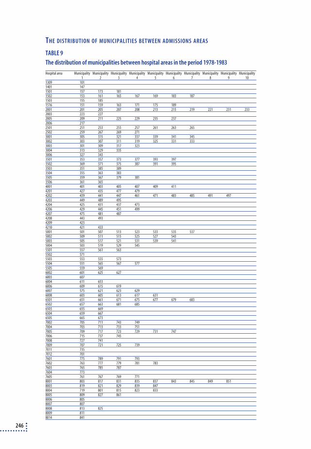
246
TABLE 9
The distribution of municipalities between hospital areas in the period 1978-1983
Hospital area Municipality Municipality Municipality Municipality Municipality Municipality Municipality Municipality Municipality Municipality1 2 3 4 5 6 7 8 9 10
1309 1011401 1471501 157 173 1811502 153 161 165 167 169 183 1871503 155 1851516 151 159 163 171 175 1892001 201 205 207 208 213 215 219 221 231 2332003 223 2272005 209 211 225 229 235 2372006 2172501 251 253 255 257 261 263 2652502 259 267 269 2713001 305 315 321 337 339 341 3453002 303 307 311 319 325 331 3333003 301 309 317 3233004 313 329 3353006 327 3433501 353 357 373 377 393 3973502 369 371 375 387 391 3953503 351 385 3893504 355 363 3833505 359 367 379 3813506 361 3654001 401 403 405 407 409 4114201 427 435 477 4794202 439 441 447 461 471 483 485 491 4974203 449 489 4954204 425 431 437 4734206 429 445 451 4994207 475 481 4874208 443 4934209 4234210 421 4335001 501 507 513 523 533 535 5375002 509 511 515 525 527 5435003 505 517 521 531 539 5415004 503 519 529 5455501 557 561 5635502 5715503 553 555 5735504 551 565 567 5775505 559 5696002 601 625 6276003 6076004 611 6536006 609 615 6196007 575 621 623 6296008 603 605 613 617 6316501 651 661 671 675 677 679 6836502 657 663 681 6856503 655 6696504 659 6676505 665 6737002 705 711 743 7497004 703 713 733 7517005 709 717 723 729 731 7477006 715 737 7457008 727 7417009 707 721 725 7397011 7357012 7017601 775 789 791 7937602 763 777 779 781 7837603 765 785 7877604 7737605 761 767 769 7718001 803 817 831 835 837 843 845 849 8518003 819 821 829 839 8478004 719 801 815 823 8338005 809 827 8618006 8058007 8078008 813 8258009 8118014 841
THE DISTRIBUTION OF MUNICIPALITIES BETWEEN ADMISSIONS AREAS

TABLE 10
The distribution of municipalities between hospital areas in the period 1984-1987
Hospital area Municipality Municipality Municipality Municipality Municipality Municipality Municipality Municipality Municipality1 2 3 4 5 6 7 8 9
1309 101 1671401 1471501 157 173 1811502 153 161 165 169 183 1871503 155 1851516 151 159 163 171 175 1892001 201 207 211 213 215 219 221 231 2332003 205 208 223 2272005 209 225 229 235 2372006 2172501 251 253 255 257 261 263 265 267 2692502 259 2713001 305 315 321 339 341 3453002 303 307 311 319 325 331 333 3373003 301 309 317 3233004 313 329 3353006 327 3433501 353 357 373 377 393 3973502 369 371 375 387 391 3953503 351 385 3893504 355 363 3833505 359 367 379 3813506 361 3654001 401 403 405 407 4094201 427 435 477 4794202 447 461 471 483 485 491 4974203 439 441 449 489 4954204 425 431 433 437 4734206 429 445 451 4994207 475 481 4874208 443 4934209 4234210 4215001 501 507 513 523 533 535 5375002 509 511 515 525 527 5435003 505 517 521 531 539 5415004 503 519 529 5455501 557 561 5635502 5715503 553 555 567 5735504 551 565 5775505 559 569 5756002 601 6276003 6076004 6116006 609 6156007 621 623 6296008 603 605 613 617 619 625 6316501 651 661 671 675 677 679 6836502 653 657 663 681 6856503 655 6696504 659 6676505 665 6737002 705 711 737 743 7497003 703 713 733 7517005 709 717 723 729 731 735 7477006 715 7457008 727 7417009 707 721 725 7397012 7017601 775 789 791 7937602 763 777 779 781 7837603 765 785 7877604 7737605 761 767 769 7718001 817 831 835 837 845 849 8518003 819 821 829 839 8478004 719 801 815 823 8338005 809 827 8618006 8058007 8078008 813 8258009 803 8118013 8438014 841
247
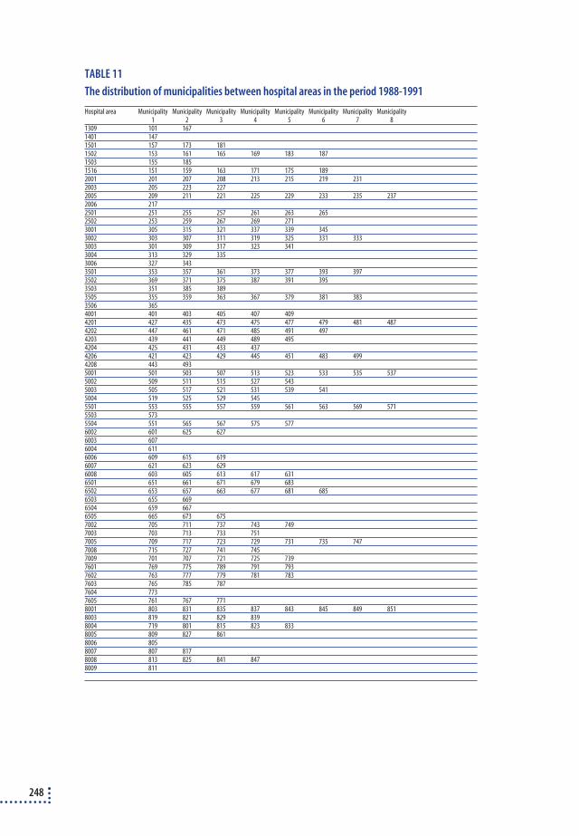
248
TABLE 11
The distribution of municipalities between hospital areas in the period 1988-1991
Hospital area Municipality Municipality Municipality Municipality Municipality Municipality Municipality Municipality1 2 3 4 5 6 7 8
1309 101 1671401 1471501 157 173 1811502 153 161 165 169 183 1871503 155 1851516 151 159 163 171 175 1892001 201 207 208 213 215 219 2312003 205 223 2272005 209 211 221 225 229 233 235 2372006 2172501 251 255 257 261 263 2652502 253 259 267 269 2713001 305 315 321 337 339 3453002 303 307 311 319 325 331 3333003 301 309 317 323 3413004 313 329 3353006 327 3433501 353 357 361 373 377 393 3973502 369 371 375 387 391 3953503 351 385 3893505 355 359 363 367 379 381 3833506 3654001 401 403 405 407 4094201 427 435 473 475 477 479 481 4874202 447 461 471 485 491 4974203 439 441 449 489 4954204 425 431 433 4374206 421 423 429 445 451 483 4994208 443 4935001 501 503 507 513 523 533 535 5375002 509 511 515 527 5435003 505 517 521 531 539 5415004 519 525 529 5455501 553 555 557 559 561 563 569 5715503 5735504 551 565 567 575 5776002 601 625 6276003 6076004 6116006 609 615 6196007 621 623 6296008 603 605 613 617 6316501 651 661 671 679 6836502 653 657 663 677 681 6856503 655 6696504 659 6676505 665 673 6757002 705 711 737 743 7497003 703 713 733 7517005 709 717 723 729 731 735 7477008 715 727 741 7457009 701 707 721 725 7397601 769 775 789 791 7937602 763 777 779 781 7837603 765 785 7877604 7737605 761 767 7718001 803 831 835 837 843 845 849 8518003 819 821 829 8398004 719 801 815 823 8338005 809 827 8618006 8058007 807 8178008 813 825 841 8478009 811

TABEL 12
The distribution of municipalities between hospital areas in the period 1992-1995
Hospital area Municip. Municip. Municip. Municip. Municip. Municip. Municip. Municip. Municip. Municip. Municip. Municip. Municip.1 2 3 4 5 6 7 8 9 10 11 12 13
1330 101 1671401 1471501 157 173 1811502 153 161 165 169 183 1871503 155 1851516 151 159 163 171 175 1892001 201 207 208 213 215 219 2312005 209 211 221 225 229 233 235 2372012 205 217 223 2272501 251 253 255 257 261 263 2652502 259 267 269 2713001 305 315 321 327 339 343 3453002 303 307 325 3313003 301 309 311 317 319 323 333 3413004 313 329 335 3373501 351 353 357 361 365 373 377 385 393 3973502 355 359 363 367 369 371 375 379 381 383 387 391 3953503 3894001 401 403 405 407 4094201 427 435 443 475 479 481 4874202 447 461 471 485 4974203 439 441 449 477 489 4954204 421 425 431 433 437 473 4934206 423 429 445 451 483 491 4995001 501 507 513 523 533 535 5375002 509 511 515 525 527 5435003 505 517 521 531 5415004 503 519 529 539 5455501 553 555 557 561 563 567 569 571 5735504 551 559 565 5776003 603 6076006 601 609 615 619 6256007 575 605 621 623 6296008 611 613 617 627 6316501 651 661 671 675 679 6836502 653 655 657 659 663 667 669 677 681 6856505 665 6737002 705 711 737 743 7497003 703 713 715 721 725 739 741 7517005 709 717 719 723 729 731 733 735 7477008 727 7457009 701 7077601 761 769 775 789 791 7937602 763 777 779 781 7837603 765 785 7877604 7737605 767 7718001 803 811 817 831 837 843 845 849 8518003 805 819 821 829 835 8398004 801 815 823 8338005 809 827 8618007 8078008 813 825 841 847
249
Appendix 5. Economical analyses
COSTS OF GALLSTONE TREATMENT IN DENMARK
The calculated cost of treating patients with gallstones includes the ma-jority of expenses as amounts spent on technological equipment, theatrestaff and days spent in hospital are included. Estimates of the indirectcosts associated with convalescence are also included.
Costs of technological equipment
Laparoscopic equipmentThe substantial difference in the direct cost of different biliary tract inter-ventions is due to the equipment. Laparoscopic equipment consists of in-sufflation equipment for inserting CO2, video equipment (camera, recorderand monitor) as well as the laparoscopic instruments (trochars, forceps,etc.) which are available in one-off and reusable form. In the models, theinvestment cost per laparoscopic column was set at DKK 440,000. (Bispe-bjerg Hospital). With a life span of five years and an interest rate of 5%,this gave a depreciation of DKK 101,629 p.a. If 47 operations are per-formed per annum (an average number of laparoscopic cholecystectomiesfor the country’s surgical departments), that gives a unit cost of DKK2,162. On top of that there is also the surgical equipment, which for dis-posable instruments was set at DKK 5,561, and for reusable equipment atDKK 834 per operation327. In this breakdown, the starting point was the aforementioned price for reusable equipment, but in the sensitivityanalysis in the economical models a number of different prices were usedbecause a different number of operations and various discount schemes fordisposable equipment give different unit costs. The calculations were basedon the assumption that laparoscopic cholecystectomies made up 25% of allsimple cholecystectomies in 1991 and 75% in the subsequent years.
ERCPThe purchase price of ERCP equipment was set at DKK 520,000. With alife span of five years and an interest rate of 5%, this gave an annual de-preciation of DKK 120,107. Divided up between 300 interventions (diag-
250

nostic and therapeutic) per annum, this gave a unit cost of DKK 400. Sincethe equipment is relatively sensitive, it often requires repair. Repair costswere set at DKK 423 per use (based on average calculations over the pe-riod 1992-97, Bispebjerg Hospital). In the case of diagnostic ERCP, thecost of utensils was set at DKK 100. In case of a therapeutic ERCP the costwas set at DKK 1,500. Thus, the unit cost for equipment for diagnosticERCP was DKK 923, while the corresponding price for therapeutic ERCPwas DKK 2,323.
Theatre staff costs
Some operations take longer than others and the composition of the ope-rating team depends on the nature of the operation. Thus, the staff costsfor the different gallstone interventions consist of an average hourly wage(table 1) and the time spent on the operation by the different staff cate-gories.
TABLE 1
Hourly wages per staff category
Staff category Annual income Hourly waged Staff category Annual income Hourly waged
Chief surgeona 543,231 319.08 Theatre nursea 246,544 144.81
Consultantb 450,516 264.62 Assistant nurse a 246,544 144.81
Anaesthetistc 450,516 264.62 Anaesthetics nursea 246,544 144.81
Senior registrar b 316,171 185.71 Radiographera 214,925 126.24
Registrarb 274,393 161.17 Portera 206,169 121.10
a: The Government Wages and Conditions Statistics Office. Collective bargaining statistics, 1995
b: Collective bargaining agreements for doctors, pay scale 6 + supplement. The Danish Medical Association.
c: Considered equal to consultants.
d: Average hourly wage (in DKK) calculated of annual income (227 working days per annum á 7.5 hours).
The time spent on different gallstone related interventions (surgery andERCP) was calculated by sending a questionnaire survey to all surgical de-partments and selected medical gastroenterological departments. The re-sponse rates were 67% and 100%, respectively. The surgical departmentsthat did not reply were evenly distributed in terms of geography and typeof hospital. The responses formed the basis for the calculation of staffcosts for the different interventions (table 2 & 3). The departments werenot asked to do detailed time and motion studies, but only to provide anassessment of time and staff resources.
251

TABLE 2
Staff involved in gallstone-related interventions. Average number of minutes
Staff category Laparoscopic cholecystectomy Open cholecystectomyb Other biliary tract surgery
Surgeon a 80.59 74.33 124.38
Assistant a 77.06 72.41 125.00
Theatre nurse 30.68 33.06 53.53
Assistant nurse 133.13 124.14 176.87
Anaesthesia nurse 140.45 130.19 176.38
Anaesthetist 131.68 121.85 172.96
Radiographer 11.90 18.00 17.50
Porter 18.74 18.91 20.00
a: Later calculations of unit prices were based on characteristics of the surgeon and the assistant, respectively. i.e. account was taken of
whether it was a department's consultants, senior registrar or registrar. Thus, the cost consists of a weighted average for the relevant
staff categories.
b: Including cholecystectomy by minilaparotomy
TABLE 3
Staff involved in gallstone-related interventions. Average number of minutes
Staff category ERCP – Diagnostic ERCP – Therapeutic
Endoscope operator 35.67 55.26
1st assistant 60.75 79.91
2nd assistant 43.46 57.74
3rd assistant 3.75 5.22
Porter 17.53 17.67
The endoscope operator corresponds to a chief surgeon/consultant; 1st and 2nd assistant to a nurse and 3rd assistant to a radiographer.
The average number of minutes decline for the 2nd and 3rd assistant as different departments use different numbers of assistants.
One question posed in the questionnaire was in how many of the opera-tions the surgeon and assistant were consultants, senior registrars or registrars. The departments were also asked to state how frequently intra-operative cholangiography was used. An expected price was then calcu-lated on the basis of the hourly rates for the groups of doctors and theprobability that the surgeon can be ascribed to the different categories.The estimated costs of different operations were calculated on the basis ofthe hourly wages for the different staff groups.
When calculating staff costs for different operations (table 4), the av-erage time spent by the different staff categories and the respective hourlywages were used. Staff costs were also calculated for the departments withthe lowest and highest staff costs. Average costs were used in the financialcalculations, while minimum and maximum values were used in the sen-sitivity analyses.
252

TABLE 4
Staff costs at various interventions (in DKK).
Cholecystectomy ERCP
Opena Laparoscopy Otherb Diagnostic Therapeutic
Average 1659.08 1744.76 2580.77 484.45 672.74
Maximum 3404.49 3021.37 4347.49 1597.41 2109.69
Minimum 586.39 773.17 1063.43 172.36 252.13
a: Including cholecystectomy by minilaparotomy
b: Other biliary tract surgery
Costs associated with hospitalisation
Ideally, when calculating the costs, bed price per day should reflect costsassociated with nursing, food, administration, etc. The economical mod-els used the general rate for patients with a free choice of hospital, whichwere fixed at DKK 2,255/day (1997 figures).
Average hospital day costs can conceal major variations in the individ-ual treatment costs for the same indication and for bed days. Thus, a re-duction in the time a patient is confined to bed will often mean a reduc-tion in the number of least expensive bed days. It is also open to questionwhether the general price for bed-days also reflect the cost of the actualoperation, which would mean some costs were registered twice. And finally, a given bed-day price will not only reflect and relate to, for exam-ple, gallstone operations. It will also reflect the costs associated with thedays spent in bed after other operations in the same ward. In the eco-nomical models, hospital bed-day rates were varied by means of sensitiv-ity analyses in order to identify these uncertainty factors.
Costs related to absence due to illness
The indirect costs include the loss production due to sick leave. These include the patient (and his/her immediate family) devoting workinghours and spare time to convalescence. It is difficult to measure indirectcosts exactly, partly because of the difficulties involved in placing a valueon ‘productive activity’ in the home.
Few gallstone studies have calculated indirect costs. In a Swedish survey[80], the indirect costs were estimated on the basis of the average hourlywage in different businesses (industry, mining and quarrying) (SKr 139.13),the gender-standardised average number of working hours per day (4.1hour/day), the proportion of the population in the workforce (0.779) and
253

finally, the proportion of the workforce in work (0.915). The result was aproduction loss per day of SKr 406.6 (139.19 x 4.1 x 0.779 x 0.915).
If these calculations were transferred to Denmark (based on 1994 fig-ures), the indirect costs per sick day could be estimated based on the pro-portion of the population in the workforce at 0.5597, the proportion ofthe workforce in employment at 0.89, the average hourly wage at DKK125.60 and the average number of man hours per day at 4.76. In total,this would give an average production loss of DKK 297.81 per day due toillness. If the proportion of the population in workforce was put at 0.789instead, the cost per day due to illness would be DKK 419.82. If no cor-rections were made for the size of the workforce and level of unemploy-ment, then the cost per day due to illness would be DKK 597.86.
In principle, the so-called intangible costs such as pain, anxiety, etc.should also be included in a socio-economic analysis. Estimates for theseelements have not been included in this report.
Comments
The costs included in these financial models were based on a number ofestimates which, in the nature of things, were subject to a number of un-certainty factors. Consequently, sensitivity analyses have been conductedin the estimations of the specific models, which amongst other things variesthe cost estimates.
It seemed justifiable to omit expenses incurred during the preoperativephase, since they were estimated at only 10% and 14% of total costs307 fortraditional open cholecystectomy and laparoscopic cholecystectomy, respectively.
SENSITIVITY ANALYSES OF THE ECONOMICAL MODELS
This section shows in schematic form, how the results of the financialmodels were altered by adjustments in the individual parameter values.The main conclusions are summed up in chapter 11 of the report.
254

TABLE 5
Cholecystectomy by laparoscopy, minilaparotomy and traditional open laparotomy.
Sensitivity analyses of the three alternatives. Variations in unit costs.
Parameter New value Average expected direct costs – DKK Marginal cost
effective-
Laparoscopy Minilaparotomy Trad. open lap ness lap. vs. open
Baseline values 10,532 7,351 15,327 46,334
Bed-day price (2,255) 1,000 7,356 4,223 8,325 3,711
4,000 14,948 11,699 25,877 105,598
Staff costs 773 9,560 7,351 15,327 55,721
(laparoscopic cholecystectomy) 3,021 11,808 7,351 15,327 34,000
Staff costs (cholecystectomy by 586 10,532 6,278 15,327 -
minilaparotomy) 3,404 10,532 9,096 15,327 -
Staff costs (cholecystectomy 586 10,532 7,351 14,254 35,970
by trad. open lap.) 3,404 10,532 7,351 17,073 63,198
Unit price for the laparoscopic 0 8,369 7,351 15,327 67,226
equipment (the number of 500 8,869 7,351 15,327 62,395
operations per column) 1,000 9,369 7,351 15,327 57,564
3,000 11,369 7,351 15,327 38,241
Equipment for minilaparotomy 500 10,532 7,851 15,327 -
Indirect costs 100 11,563 8,373 18,128 63,430
500 15,687 12,461 29,330 131,811
TABLE 6
Cholecystectomy by laparoscopy, minilaparotomy and traditional open laparotomy.
Sensitivity analyses of the three alternatives. Variations in time spent in hospital.
Parameters New value Average expected direct costs (in DKK)
Laparoscopy Minilaparotomy Trad. Open lap.
Baseline values 10,532 7,351 15,327
Successful treatment 1 day 8,539 5,348 4,199
3 days 12,524 9,353 8,650
5 days 16,509 13,357 13,101
7 days 20,493 17,363 17,553
Complication (re-operation) 8 days 10,486 7,306 15,282
10 daysa 10,532 7,351 15,327
12 days 10,577 7,396 15,372
Peripheral bile duct lesion 5 days 10,514 7,343 15,315
7 daysa 10,532 7,351 15,327
9 days 10,549 7,358 15,339
Central bile duct lesion 10 days 10,508 7,348 15,324
14 daysa 10,532 7,351 15,327
18 days 10,555 7,353 15,331
Conversion 4 days 10,081 6,900 -
6 daysa 10,532 7,351 15,327
8 days 10,983 7,802 15,327
Indirect costs DKK 100 11,563 8,373 18,128
DKK 500 15,687 12,461 29,330
a: Baseline estimat
255

TABLE 7
Disposable vs. reusable instruments for laparoscopic cholecystectomy.
Sensitivity analyses of the alternatives. Variations in unit costs and time spent in hospital.
Parameters New value Instruments (in DKK) Marg. cost
Disposable Reusable efficiency
Baseline values 12,407 8,485
Bed-day price DKK 1,000 9,494 5,315 68,968
DKK 4,000 16,458 12,894 58,810
Disposable equipment DKK 1,000 7,846 8,485 - 10,545
DKK 6,000 12,846 8,485 71,963
Reusable equipment DKK 500 12,407 8,151 70,230
DKK 1,000 12,407 8,651 61,979
Hospitalisation if success 1 dag 10,674 7,247 56,562
3 days 14,141 9,724 72,875
Hospitalisation for post-operative 3 days 12,353 8,263 67,487
complications 7 days 12,462 8,708 61,950
Hospitalisation for conversion 4 days 12,121 8,094 66,460
8 days 12,694 8,877 62,977
Hospitalisation for central 10 days 12,384 8,462 64,719
bile duct lesion 18 days 12,431 8,509 64,719
256

Treatment of
Gallstone patients
Torben Jørgensen
Danish Institute forHealth Technology Assessment
A health technology assessment
Treatm
ent of Gallstone patients
NIP
H &
Danish Institute for H
ealth Technology A
ssessment 2000
In the Danish health care system, more than DKK 100 million per year are
spent on treating patients with benign biliary tract diseases.
This report is the first attempt at performing a health technology
assessment of treatment of gallstone patients. By including the technology,
the patient, the organisation as well as the economy, a health technology
assessment is an analytical tool that serves as basis for ensuring an optimal
treatment of patients within the given economical framework.
The report is based on a systematic review of the literature and a
thorough analysis of data from The National Hospital Register where all
hospital admissions in Denmark are recorded. By reviewing the literature
using explicit criteria, the fundament for performing an objective assessment
of the various treatment modalities has been established. At the same time,
analyses of data from The National Hospital Register form a clear picture of
the development of treatment of gallstone patients throughout Denmark.
By combining the technology with the economical analyses, the limited
knowledge of patients’ preferences for the various treatment modalities
as well as the organisational aspects, the report questions a number of
well-established treatment modalities and the organisation of these.
It is the hope, that this report will form part of a knowledge-based
fundament for the development of a national strategy for prevention,
diagnosis and treatment of patients with biliary tract disease.
National Institute of Public Health
25, Svanemøllevej
DK-2100 Copenhagen , Denmark
Phone: +45 39 20 77 77
Fax: +45 39 20 80 10
E-mail: [email protected]
Homepage : www.dike.dk
Counselling and advising in connection with HTA projects is given by:
Danish Institute for Health Technology Assessment
National Board of Health
13, Amaliegade
PO. Box 2020
DK-1012 Copenhagen
Denmark
Phone: + 45 33 91 16 01
Fax: + 45 33 91 70 61
E-mail: [email protected]
Homepage: http://www.dihta.dk
Further copies of this publication can be bought at:
Sundhedsstyrelsens Publikationer
c/o Schultz Information
12, Herstedvang
DK-2620 Albertslund
Denmark
Phone: + 45 70 26 26 36
Fax: + 45 43 63 62 45HI TD A
Copyright © 2022 FDOKUMEN