Functional morphology of the feeding apparatus of the nymph of Farrodes sp. (Ephemeroptera:...
11
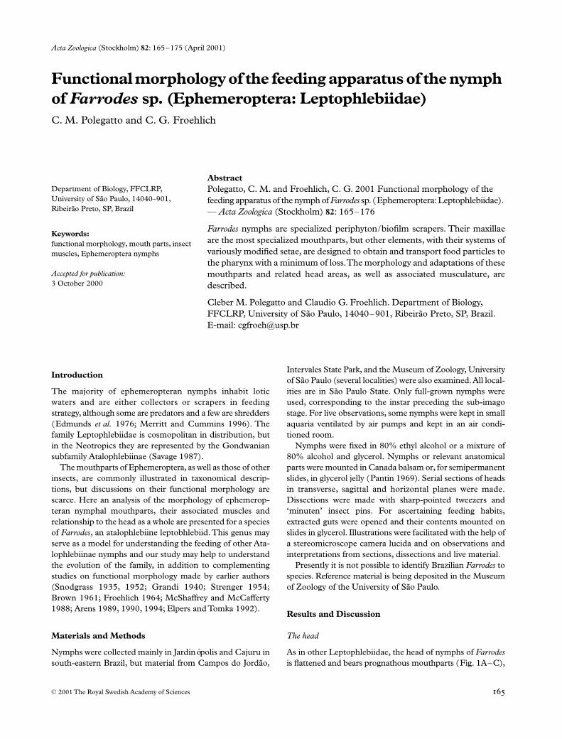
Acta Zoologica (Stockholm) 82 : 165 – 175 (April 2001) © 2001 The Royal Swedish Academy of Sciences Abstract Polegatto, C. M. and Froehlich, C. G. 2001 Functional morphology of the feeding apparatus of the nymph of Farrodes sp. (Ephemeroptera: Leptophlebiidae). — Acta Zoologica (Stockholm) 82 : 165–176 Farrodes nymphs are specialized periphyton/biofilm scrapers. Their maxillae are the most specialized mouthparts, but other elements, with their systems of variously modified setae, are designed to obtain and transport food particles to the pharynx with a minimum of loss. The morphology and adaptations of these mouthparts and related head areas, as well as associated musculature, are described. Cleber M. Polegatto and Claudio G. Froehlich. Department of Biology, FFCLRP, University of São Paulo, 14040–901, Ribeirão Preto, SP, Brazil. E-mail: [email protected] Blackwell Science, Ltd Functional morphology of the feeding apparatus of the nymph of Farrodes sp. (Ephemeroptera: Leptophlebiidae) C. M. Polegatto and C. G. Froehlich Department of Biology, FFCLRP, University of São Paulo, 14040–901, Ribeirão Preto, SP, Brazil Keywords: functional morphology, mouth parts, insect muscles, Ephemeroptera nymphs Accepted for publication: 3 October 2000 Introduction The majority of ephemeropteran nymphs inhabit lotic waters and are either collectors or scrapers in feeding strategy, although some are predators and a few are shredders ( Edmunds et al. 1976; Merritt and Cummins 1996). The family Leptophlebiidae is cosmopolitan in distribution, but in the Neotropics they are represented by the Gondwanian subfamily Atalophlebiinae (Savage 1987). The mouthparts of Ephemeroptera, as well as those of other insects, are commonly illustrated in taxonomical descrip- tions, but discussions on their functional morphology are scarce. Here an analysis of the morphology of ephemerop- teran nymphal mouthparts, their associated muscles and relationship to the head as a whole are presented for a species of Farrodes , an atalophlebiine leptobhlebiid. This genus may serve as a model for understanding the feeding of other Ata- lophlebiinae nymphs and our study may help to understand the evolution of the family, in addition to complementing studies on functional morphology made by earlier authors (Snodgrass 1935, 1952; Grandi 1940; Strenger 1954; Brown 1961; Froehlich 1964; McShaffrey and McCafferty 1988; Arens 1989, 1990, 1994; Elpers and Tomka 1992). Materials and Methods Nymphs were collected mainly in Jardinópolis and Cajuru in south-eastern Brazil, but material from Campos do Jordão, Intervales State Park, and the Museum of Zoology, University of São Paulo (several localities) were also examined. All local- ities are in São Paulo State. Only full-grown nymphs were used, corresponding to the instar preceding the sub-imago stage. For live observations, some nymphs were kept in small aquaria ventilated by air pumps and kept in an air condi- tioned room. Nymphs were fixed in 80% ethyl alcohol or a mixture of 80% alcohol and glycerol. Nymphs or relevant anatomical parts were mounted in Canada balsam or, for semipermanent slides, in glycerol jelly (Pantin 1969). Serial sections of heads in transverse, sagittal and horizontal planes were made. Dissections were made with sharp-pointed tweezers and ‘minuten’ insect pins. For ascertaining feeding habits, extracted guts were opened and their contents mounted on slides in glycerol. Illustrations were facilitated with the help of a stereomicroscope camera lucida and on observations and interpretations from sections, dissections and live material. Presently it is not possible to identify Brazilian Farrodes to species. Reference material is being deposited in the Museum of Zoology of the University of São Paulo. Results and Discussion The head As in other Leptophlebiidae, the head of nymphs of Farrodes is flattened and bears prognathous mouthparts ( Fig. 1A – C),
Transcript of Functional morphology of the feeding apparatus of the nymph of Farrodes sp. (Ephemeroptera:...
Acta Zoologica
(Stockholm)
82
: 165–175 (April 2001)
© 2001 The Royal Swedish Academy of Sciences
Abstract
Polegatto, C. M. and Froehlich, C. G. 2001 Functional morphology of the feeding apparatus of the nymph of
Farrodes
sp. (Ephemeroptera: Leptophlebiidae). —
Acta Zoologica
(Stockholm)
82
: 165–176
Farrodes
nymphs are specialized periphyton/biofilm scrapers. Their maxillaeare the most specialized mouthparts, but other elements, with their systems ofvariously modified setae, are designed to obtain and transport food particles tothe pharynx with a minimum of loss. The morphology and adaptations of thesemouthparts and related head areas, as well as associated musculature, aredescribed.
Cleber M. Polegatto and Claudio G. Froehlich. Department of Biology, FFCLRP, University of São Paulo, 14040–901, Ribeirão Preto, SP, Brazil. E-mail: [email protected]
Blackwell Science, Ltd
Functional morphology of the feeding apparatus of the nymph of
Farrodes
sp. (Ephemeroptera: Leptophlebiidae)
C. M. Polegatto and C. G. Froehlich
Department of Biology, FFCLRP, University of São Paulo, 14040–901, Ribeirão Preto, SP, Brazil
Keywords:
functional morphology, mouth parts, insect muscles, Ephemeroptera nymphs
Accepted for publication:
3 October 2000
Introduction
The majority of ephemeropteran nymphs inhabit loticwaters and are either collectors or scrapers in feedingstrategy, although some are predators and a few are shredders(Edmunds
et al.
1976; Merritt and Cummins 1996). Thefamily Leptophlebiidae is cosmopolitan in distribution, butin the Neotropics they are represented by the Gondwaniansubfamily Atalophlebiinae (Savage 1987).
The mouthparts of Ephemeroptera, as well as those of otherinsects, are commonly illustrated in taxonomical descrip-tions, but discussions on their functional morphology arescarce. Here an analysis of the morphology of ephemerop-teran nymphal mouthparts, their associated muscles andrelationship to the head as a whole are presented for a speciesof
Farrodes
, an atalophlebiine leptobhlebiid. This genus mayserve as a model for understanding the feeding of other Ata-lophlebiinae nymphs and our study may help to understandthe evolution of the family, in addition to complementingstudies on functional morphology made by earlier authors(Snodgrass 1935, 1952; Grandi 1940; Strenger 1954;Brown 1961; Froehlich 1964; McShaffrey and McCafferty1988; Arens 1989, 1990, 1994; Elpers and Tomka 1992).
Materials and Methods
Nymphs were collected mainly in Jardinópolis and Cajuru insouth-eastern Brazil, but material from Campos do Jordão,
Intervales State Park, and the Museum of Zoology, Universityof São Paulo (several localities) were also examined. All local-ities are in São Paulo State. Only full-grown nymphs wereused, corresponding to the instar preceding the sub-imagostage. For live observations, some nymphs were kept in smallaquaria ventilated by air pumps and kept in an air condi-tioned room.
Nymphs were fixed in 80% ethyl alcohol or a mixture of80% alcohol and glycerol. Nymphs or relevant anatomicalparts were mounted in Canada balsam or, for semipermanentslides, in glycerol jelly (Pantin 1969). Serial sections of headsin transverse, sagittal and horizontal planes were made.Dissections were made with sharp-pointed tweezers and‘minuten’ insect pins. For ascertaining feeding habits,extracted guts were opened and their contents mounted onslides in glycerol. Illustrations were facilitated with the help ofa stereomicroscope camera lucida and on observations andinterpretations from sections, dissections and live material.
Presently it is not possible to identify Brazilian
Farrodes
tospecies. Reference material is being deposited in the Museumof Zoology of the University of São Paulo.
Results and Discussion
The head
As in other Leptophlebiidae, the head of nymphs of
Farrodes
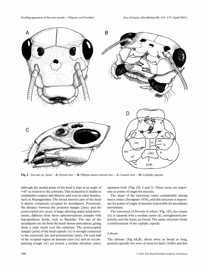
is flattened and bears prognathous mouthparts (Fig. 1A–C),
AZO083.fm Page 165 Thursday, March 15, 2001 11:09 AM
Feeding apparatus of
Farrodes nymph
•
Polegatto and Froehlich Acta Zoologica
(Stockholm)
82
: 165–175 (April 2001)
© 2001 The Royal Swedish Academy of Sciences
although the medial plane of the head is kept at an angle of
≈
45
°
in relation to the substrate. This inclination is similar inconfamilial scrapers and filterers, and even in other families,such as Heptageniidae. The broad anterior part of the headis almost completely occupied by mouthparts. Posteriorly,the distance between the posterior margin (pmc) and thepostoccipital one (pom) is large, allowing ample head move-ments, different from those ephemeropteran nymphs withhypognathous heads, such as Baetidae. The tips of themouthparts are far from the head–thorax articulation, givingthem a wide reach over the substrate. The postoccipitalmargin (pom) of the head capsule (cc) is strongly connectedto the tentorium (tn) and postmentum (psm). On each halfof the occipital region an internal crest (oc) and an accom-panying trough (of ) are found; a median elevation (emo)
separates both (Figs 1D, 2 and 3). These crests are impor-tant as points of origin for muscles.
The shape of the tentorium varies considerably amonginsect orders (Snodgrass 1935), and this structure is import-ant for points of origin of muscles responsible for mouthpartmovements.
The tentorium of
Farrodes
is robust (Fig. 1D); the corpus(ct) is squarish with a median carina (k), strengthened pos-teriorly, and the bases are broad. The entire structure formsa reinforcement of the cephalic capsule.
Labrum
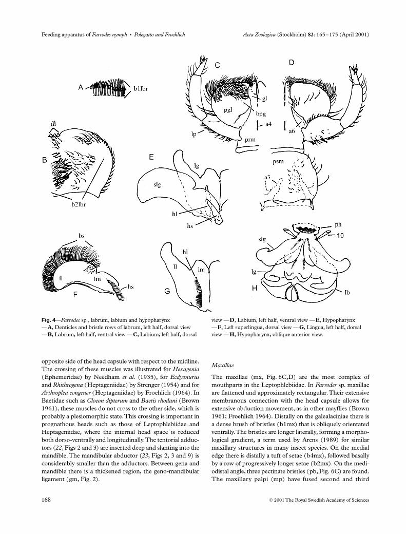
The labrum (Fig. 4A,B), about twice as broad as long,presents apically two rows of close-set hairs (b1lbr) and five
Fig. 1—Farrodes sp., head —A, Dorsal view —B, Oblique antero-lateral view —C, Lateral view —D, Cephalic capsule.
AZO083.fm Page 166 Thursday, March 15, 2001 11:09 AM
Acta Zoologica
(Stockholm)
82
: 165–175 (April 2001)
Polegatto and Froehlich
•
Feeding apparatus of
Farrodes nymph
© 2001 The Royal Swedish Academy of Sciences
denticles (dl) along a shallow anterior emargination. Theepipharynx is minimally sclerotized and possesses severalfields of hairs or bristles mostly directed mesially (Fig. 4B).
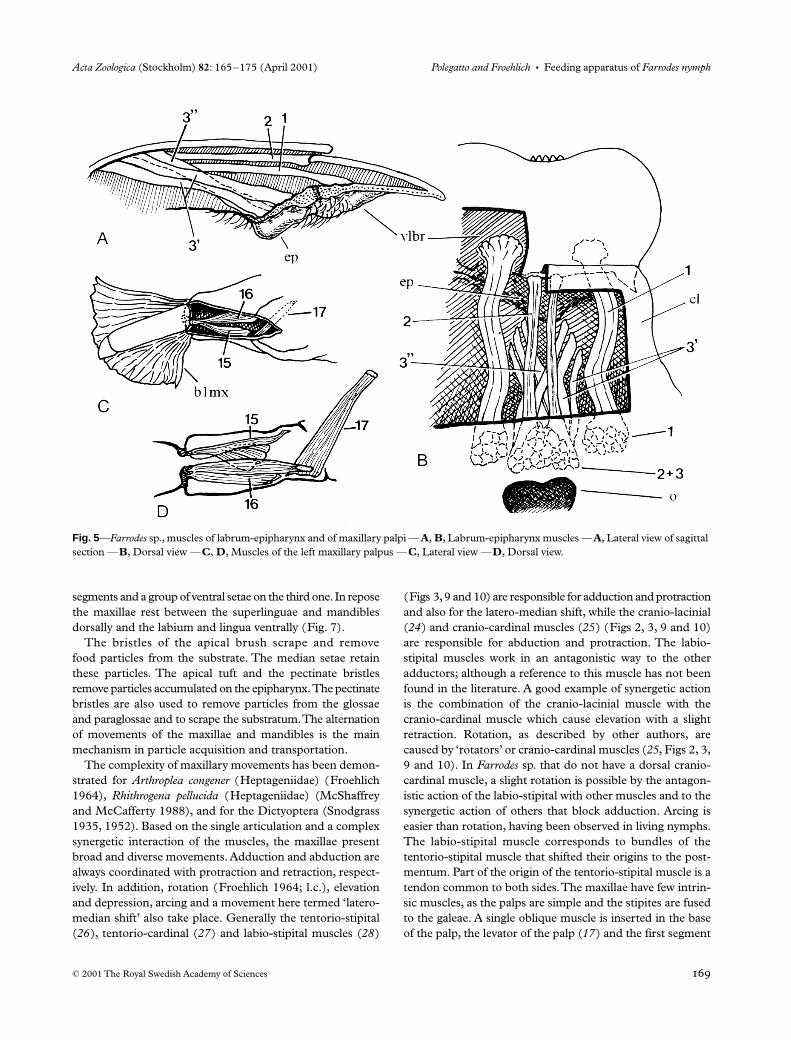
The labrum is moved by two pairs of muscles, a pair offlexors (depressors,
1
) and a pair of extensors (levators,
2
)(Figs 2, 3 and 5A,B). In addition to these, a set of smallmuscles (
3
), not yet referred to in the Ephemeroptera, areinserted in the posterior epipharynx, of which some arerestricted to the same side (
3
′
) but others cross from one sideto the other (
3
′′
) (Fig. 5). Labrum compressor muscles werenot found. These muscles were described by Snodgrass(1935) for several groups of insects and for
Arthropleacongener
(Heptageniidae) by Froehlich (1964), but were not
referred to by Strenger (1954) for
Rhithrogena
sp. and
Ecdy-onurus
sp. (Heptageniidae) and by Brown (1961) for
Cloeondipterum
and
Baetis rhodani
(Baetidae).The movements of the labrum are chiefly dorsoventral;
pendular movements or retraction also would be possible,but were not observed in live nymphs. Muscles
3
would act aselevators-retractors of the epipharynx, dilating the preoral cavity.
The labrum shields the other mouthparts from the watercurrent, reducing the loss of food particles. Food particlesaccumulating among the epipharynx hairs are periodicallyremoved by galealacinial bristles. The dorsal hairs of thelabrum could retain substrate food particles, but these couldbe used only if removed naturally. The denticles could havelimited use in the scraping of irregular surfaces.
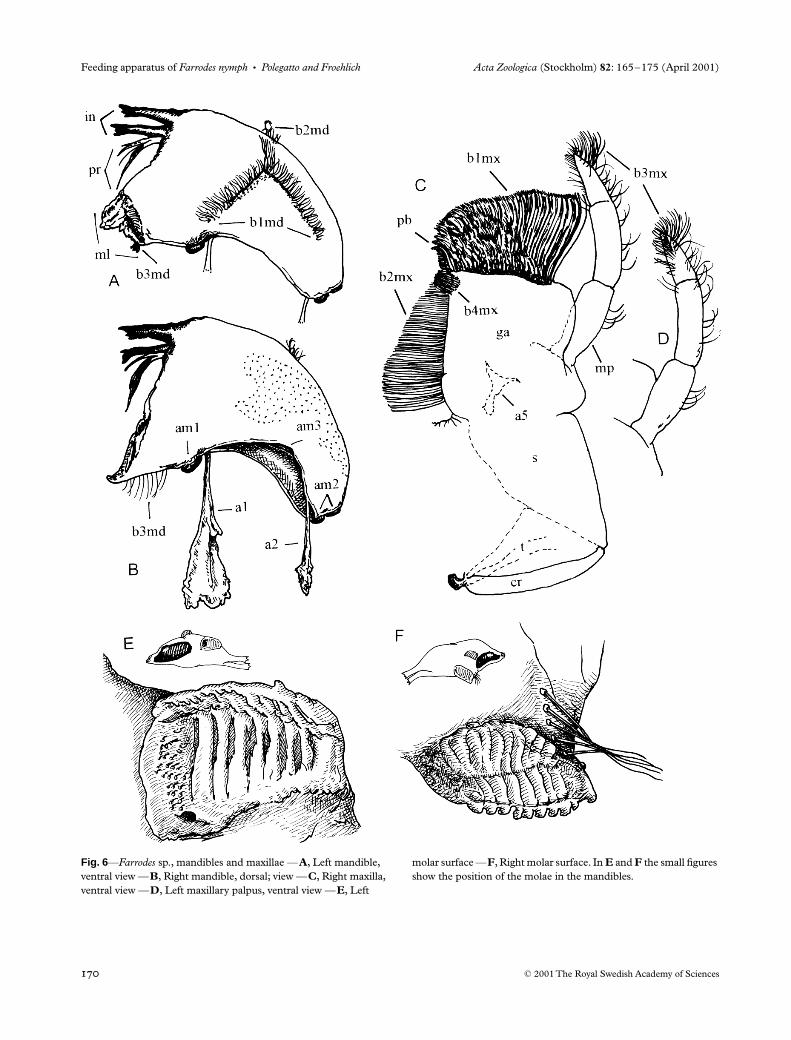
Mandibles
The mandibles (md, Fig. 6A,B) are approximately cres-centic with two incisors (in) and a mola (m). The molae(Fig. 6A,B,E,F) are asymmetrical and provided with asystem of meshing crests. Situated basally to the incisors isa prostheca (pr). A set of bristles (b3md) occurs basally toeach mola. An L-shaped row of bristles (b1md, Figs 6A and7) is found on the ventral surface of the mandibles; thesebristles are directed outwards.
The anterior articular surface (am1) is strong and bearsmost of the load of the mandibular action. The posteriorone (am2) is much smaller and works as an auxiliary support.An axis (e) through both shows the movements to be veryoblique (Fig. 8), an important adaptation in prognathousforms, especially those with dorso-ventrally flattened heads,allowing for a wide gape. A third articular surface (am3) isseen in an intermediate position. Some authors (Grandi1940; Arens 1989) have considered the anterior articulationas a ‘pseudo-articulation’, having appeared secondarily, andthe median articulation to be the true articulation that hasshifted laterally. This topic needs more studies for its elucida-tion. Two apodemes serve as insertion points for the muscles(Fig. 6B).
The mandibles are responsible principally for taking upfood from the maxillae. They exhibit alternate movementsduring the grinding of food by the molae. The progressiveclosing of the molae during adduction allows a precisegrinding of the food particles, which are pressed against thepharynx. The bristles basal to the molae help to retain the food,and those of the left mola remove food adhering to the rightone. The incisors help to comminute food lumps and, onirregular surfaces, they may have a scraping role. The L-shaped row of bristles on the ventral surface captures manyfood particles, preventing their loss during mouthpart move-ment. The outward orientation of these bristles may beexplained because they are occasionally cleaned by maxillarybristles.
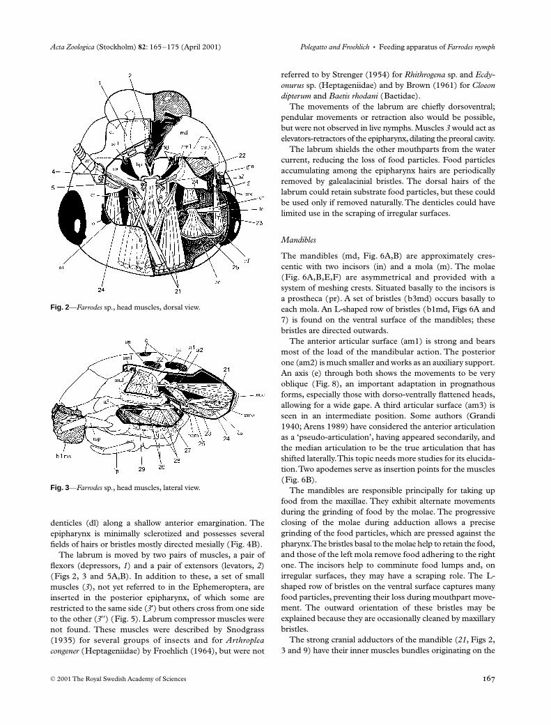
The strong cranial adductors of the mandible (
21
, Figs 2,3 and 9) have their inner muscles bundles originating on the
Fig. 2—Farrodes sp., head muscles, dorsal view.
Fig. 3—Farrodes sp., head muscles, lateral view.
AZO083.fm Page 167 Thursday, March 15, 2001 11:09 AM
Feeding apparatus of
Farrodes nymph
•
Polegatto and Froehlich Acta Zoologica
(Stockholm)
82
: 165–175 (April 2001)
© 2001 The Royal Swedish Academy of Sciences
opposite side of the head capsule with respect to the midline.The crossing of these muscles was illustrated for
Hexagenia
(Ephemeridae) by Needham
et al
. (1935), for
Ecdyonurus
and
Rhithrogena
(Heptageniidae) by Strenger (1954) and for
Arthroplea congener
(Heptageniidae) by Froehlich (1964). InBaetidae such as
Cloeon dipterum
and
Baetis rhodani
(Brown1961), these muscles do not cross to the other side, which isprobably a plesiomorphic state. This crossing is important inprognathous heads such as those of Leptophlebiidae andHeptageniidae, where the internal head space is reducedboth dorso-ventrally and longitudinally. The tentorial adduc-tors (
22
, Figs 2 and 3) are inserted deep and slanting into themandible. The mandibular abductor (
23
, Figs 2, 3 and 9) isconsiderably smaller than the adductors. Between gena andmandible there is a thickened region, the geno-mandibularligament (gm, Fig. 2).
Maxillae
The maxillae (mx, Fig. 6C,D) are the most complex ofmouthparts in the Leptophlebiidae. In
Farrodes
sp. maxillaeare flattened and approximately rectangular. Their extensivemembranous connection with the head capsule allows forextensive abduction movement, as in other mayflies (Brown1961; Froehlich 1964). Distally on the galealaciniae there isa dense brush of bristles (b1mx) that is obliquely orientatedventrally. The bristles are longer laterally, forming a morpho-logical gradient, a term used by Arens (1989) for similarmaxillary structures in many insect species. On the medialedge there is distally a tuft of setae (b4mx), followed basallyby a row of progressively longer setae (b2mx). On the medi-odistal angle, three pectinate bristles (pb, Fig. 6C) are found.The maxillary palpi (mp) have fused second and third
Fig. 4—Farrodes sp., labrum, labium and hypopharynx —A, Denticles and bristle rows of labrum, left half, dorsal view —B, Labrum, left half, ventral view —C, Labium, left half, dorsal
view —D, Labium, left half, ventral view —E, Hypopharynx —F, Left superlingua, dorsal view —G, Lingua, left half, dorsal view —H, Hypopharynx, oblique anterior view.
AZO083.fm Page 168 Thursday, March 15, 2001 11:09 AM
Acta Zoologica
(Stockholm)
82
: 165–175 (April 2001)
Polegatto and Froehlich
•
Feeding apparatus of
Farrodes nymph
© 2001 The Royal Swedish Academy of Sciences
segments and a group of ventral setae on the third one. In reposethe maxillae rest between the superlinguae and mandiblesdorsally and the labium and lingua ventrally (Fig. 7).
The bristles of the apical brush scrape and removefood particles from the substrate. The median setae retainthese particles. The apical tuft and the pectinate bristlesremove particles accumulated on the epipharynx. The pectinatebristles are also used to remove particles from the glossaeand paraglossae and to scrape the substratum. The alternationof movements of the maxillae and mandibles is the mainmechanism in particle acquisition and transportation.
The complexity of maxillary movements has been demon-strated for
Arthroplea congener
(Heptageniidae) (Froehlich1964),
Rhithrogena pellucida
(Heptageniidae) (McShaffreyand McCafferty 1988), and for the Dictyoptera (Snodgrass1935, 1952). Based on the single articulation and a complexsynergetic interaction of the muscles, the maxillae presentbroad and diverse movements. Adduction and abduction arealways coordinated with protraction and retraction, respect-ively. In addition, rotation (Froehlich 1964; l.c.), elevationand depression, arcing and a movement here termed ‘latero-median shift’ also take place. Generally the tentorio-stipital(
26
), tentorio-cardinal (
27
) and labio-stipital muscles (
28
)
(Figs 3, 9 and 10) are responsible for adduction and protractionand also for the latero-median shift, while the cranio-lacinial(
24
) and cranio-cardinal muscles (
25
) (Figs 2, 3, 9 and 10)are responsible for abduction and protraction. The labio-stipital muscles work in an antagonistic way to the otheradductors; although a reference to this muscle has not beenfound in the literature. A good example of synergetic actionis the combination of the cranio-lacinial muscle with thecranio-cardinal muscle which cause elevation with a slightretraction. Rotation, as described by other authors, arecaused by ‘rotators’ or cranio-cardinal muscles (
25
, Figs 2, 3,9 and 10). In
Farrodes
sp. that do not have a dorsal cranio-cardinal muscle, a slight rotation is possible by the antagon-istic action of the labio-stipital with other muscles and to thesynergetic action of others that block adduction. Arcing iseasier than rotation, having been observed in living nymphs.The labio-stipital muscle corresponds to bundles of thetentorio-stipital muscle that shifted their origins to the post-mentum. Part of the origin of the tentorio-stipital muscle is atendon common to both sides. The maxillae have few intrin-sic muscles, as the palps are simple and the stipites are fusedto the galeae. A single oblique muscle is inserted in the baseof the palp, the levator of the palp (
17
) and the first segment
Fig. 5—Farrodes sp., muscles of labrum-epipharynx and of maxillary palpi —A, B, Labrum-epipharynx muscles —A, Lateral view of sagittal section —B, Dorsal view —C, D, Muscles of the left maxillary palpus —C, Lateral view —D, Dorsal view.
AZO083.fm Page 169 Thursday, March 15, 2001 11:09 AM
Feeding apparatus of
Farrodes nymph
•
Polegatto and Froehlich Acta Zoologica
(Stockholm)
82
: 165–175 (April 2001)
© 2001 The Royal Swedish Academy of Sciences
Fig. 6—Farrodes sp., mandibles and maxillae —A, Left mandible, ventral view —B, Right mandible, dorsal; view —C, Right maxilla, ventral view —D, Left maxillary palpus, ventral view —E, Left
molar surface —F, Right molar surface. In E and F the small figures show the position of the molae in the mandibles.
AZO083.fm Page 170 Thursday, March 15, 2001 11:09 AM
Acta Zoologica
(Stockholm)
82
: 165–175 (April 2001)
Polegatto and Froehlich
•
Feeding apparatus of
Farrodes nymph
© 2001 The Royal Swedish Academy of Sciences
contains a flexor (
15
) and an extensor (
16
) of the distal palp(Fig. 5C,D). Some of the muscles have been observed tolack specific antagonists.
Labium
The postmentum (psm, Fig. 4D) of
Farrodes
sp. is approx-imately trapezoidal and the obliquely orientated lateralmargins (Fig. 11) allow the passage of the important labio-stipital muscle (
28
, Fig. 10). The glossae (gl) and paraglossae(pgl) are the main ventral structures of the head. The 3-segmented labial palps (lp) are similar to the maxillary ones,including in the fusion of the second and third segments.
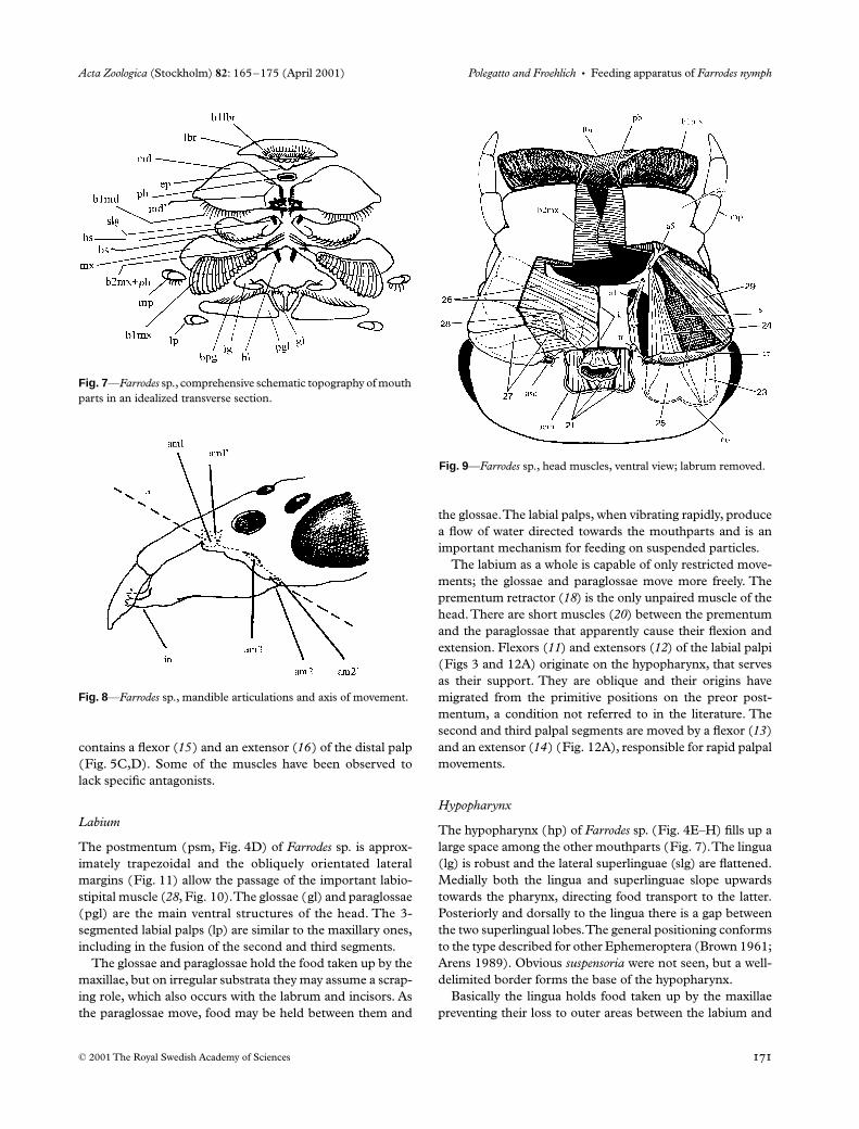
The glossae and paraglossae hold the food taken up by themaxillae, but on irregular substrata they may assume a scrap-ing role, which also occurs with the labrum and incisors. Asthe paraglossae move, food may be held between them and
the glossae. The labial palps, when vibrating rapidly, producea flow of water directed towards the mouthparts and is animportant mechanism for feeding on suspended particles.
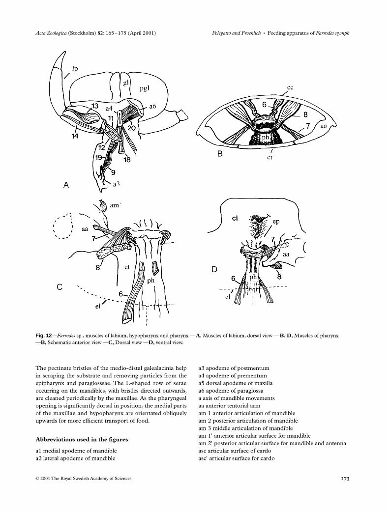
The labium as a whole is capable of only restricted move-ments; the glossae and paraglossae move more freely. Theprementum retractor (
18
) is the only unpaired muscle of thehead. There are short muscles (
20
) between the prementumand the paraglossae that apparently cause their flexion andextension. Flexors (
11
) and extensors (
12
) of the labial palpi(Figs 3 and 12A) originate on the hypopharynx, that servesas their support. They are oblique and their origins havemigrated from the primitive positions on the preor post-mentum, a condition not referred to in the literature. Thesecond and third palpal segments are moved by a flexor (
13
)and an extensor (
14
) (Fig. 12A), responsible for rapid palpalmovements.
Hypopharynx
The hypopharynx (hp) of
Farrodes
sp. (Fig. 4E–H) fills up alarge space among the other mouthparts (Fig. 7). The lingua(lg) is robust and the lateral superlinguae (slg) are flattened.Medially both the lingua and superlinguae slope upwardstowards the pharynx, directing food transport to the latter.Posteriorly and dorsally to the lingua there is a gap betweenthe two superlingual lobes. The general positioning conformsto the type described for other Ephemeroptera (Brown 1961;Arens 1989). Obvious
suspensoria
were not seen, but a well-delimited border forms the base of the hypopharynx.
Basically the lingua holds food taken up by the maxillaepreventing their loss to outer areas between the labium and
Fig. 7—Farrodes sp., comprehensive schematic topography of mouth parts in an idealized transverse section.
Fig. 8—Farrodes sp., mandible articulations and axis of movement.
Fig. 9—Farrodes sp., head muscles, ventral view; labrum removed.
AZO083.fm Page 171 Thursday, March 15, 2001 11:09 AM

Feeding apparatus of
Farrodes nymph • Polegatto and Froehlich Acta Zoologica (Stockholm) 82: 165–175 (April 2001)
© 2001 The Royal Swedish Academy of Sciences
the other mouthparts. The hypopharynx, especially its pos-terior portion, takes the food towards the pharynx directedby its shape and the arrangement of hairs. The gap betweenthe superlinguae serves as a food channel.
The action of the hypopharynx is restricted and depends onthe action of the other mouthparts. It is moved by retractors-depressors (9) and by mandibulo-hypopharyngeals or levatorsof the hypopharynx (10) (Figs 2 and 11); the latter were alsoobserved in Arthroplea congener by Froehlich (1964).
Pharynx
The pharynx (Figs 2, 3, 9, 11 and 12B–D), as the head, isflattened dorso-ventrally. It has simple movements, causedby simpler and fewer muscles than those described by
Snodgrass (1935) for other insects. The retractors-levators(6) could open the pharyngeal lumen; the depressors (7) andlevators (8) seem to act at the pharyngeal entrance. All areimportant in maintaining the position of the pharynx.
Conclusions
The nymphs of Farrodes sp., as those of many Leptophle-biidae, are specialized scrapers or brushers of the biofilm,which consists mainly of diatoms and other algae, includingcolonial algae, fungi, minute animals, unidentifiable detritus,all items referred as FPOM and UPOM (fine and ultra-fine particulate organic matter), differently from somespecies of the subfamily Leptophlebiinae, opportunisticfeeders that deals with CPOM (coarse particulate organicmatter) too (Williams and Feltmate 1992; Mattingly 1987).Each of the mouthparts presents very specific adaptationsto obtain and transport food particles. The distal brushesof bristles and hairs on the distal border of the maxillae areused to obtain food from the substrate. The maxillary palpiremove the food particles from the brushes taking themtowards the mandibles and hypopharynx. The labrumand labium assist in retaining food. The labial palpi areimportant in producing a water current towards the pre-buccal cavity. The mandibles, besides fragmenting lumps offood with the incisors and molae, are important, in alternatingmovements with the maxillae, to press the food towardsthe pharynx. The prosthecae help in retaining particles.
Most setae on the mouthparts are directed medially and/or posteriorly, channeling food towards the pharynx. Theprincipal setal fields are those of the epipharynx andlabrum, the dorsal ones of the paraglossae, those of the medianborder of the galealacinia, and those of the hypopharynx.
Fig. 10—Farrodes sp., maxillary and mandibular muscles, schematic transverse sections —A, B, Maxillary muscles —C, Tentorial adductor of the mandible and geno-mandibular ligament.
Fig. 11—Farrodes sp., head muscles, lateral view; mandibles and maxillae removed.
AZO083.fm Page 172 Thursday, March 15, 2001 11:09 AM
Acta Zoologica (Stockholm) 82: 165–175 (April 2001) Polegatto and Froehlich • Feeding apparatus of Farrodes nymph
© 2001 The Royal Swedish Academy of Sciences
The pectinate bristles of the medio-distal galealacinia helpin scraping the substrate and removing particles from theepipharynx and paraglosssae. The L-shaped row of setaeoccurring on the mandibles, with bristles directed outwards,are cleaned periodically by the maxillae. As the pharyngealopening is significantly dorsal in position, the medial partsof the maxillae and hypopharynx are orientated obliquelyupwards for more efficient transport of food.
Abbreviations used in the figures
a1 medial apodeme of mandiblea2 lateral apodeme of mandible
a3 apodeme of postmentuma4 apodeme of prementuma5 dorsal apodeme of maxillaa6 apodeme of paraglossaa axis of mandible movementsaa anterior tentorial armam 1 anterior articulation of mandibleam 2 posterior articulation of mandibleam 3 middle articulation of mandibleam 1′ anterior articular surface for mandibleam 2′ posterior articular surface for mandible and antennaasc articular surface of cardoasc′ articular surface for cardo
Fig. 12—Farrodes sp., muscles of labium, hypopharynx and pharynx —A, Muscles of labium, dorsal view —B, D, Muscles of pharynx —B, Schematic anterior view —C, Dorsal view —D, ventral view.
AZO083.fm Page 173 Thursday, March 15, 2001 11:09 AM
Feeding apparatus of Farrodes nymph • Polegatto and Froehlich Acta Zoologica (Stockholm) 82: 165–175 (April 2001)
© 2001 The Royal Swedish Academy of Sciences
b1lbr dorsal bristles of labrumb2lbr ventral bristles of labrum (epipharynx)b1md mandibular ventral bristles in L-shaped rowb2md marginal bristles of mandibleb3md molar bristlesb1mx bristle brush of galealaciniab2mx median bristle row of maxillab3mx bristles of maxillary palpusb4mx dorsal bristles of the median row of maxillabpg dorsal bristles of paraglossabs marginal bristles of superlinguacc cephalic capsulecl clypeus cm cavity for the maxillaco occipital crest of occipital fossacr cardoct corpus tentoriumdl denticleel ecdysial lineemo median occipital ridgeep epipharynxg genaga galealaciniagl glossagm geno-mandibular ligamenthl dorsal hair row of linguahp hypopharynxhs dorsal hair row of superlingua in incisorsk tentorial keellb labiumlbr labrumlc limit of cavity for maxillalg linguall lateral lobelm median lobelp labial palpusmd mandiblemd′ incisors, mola and prosthecaml molamp maxillary palpusmt lateral margin of tentoriummx maxillao ocellusoc occipital crest of occipital fossaofm occipital foramenpb pectinate bristles of maxillapgl paraglossaph pharynxpmc posterior margin of cephalic capsulepom postoccipital marginpr prosthecaprm prementumpsm postmentums stipessa1 anterior articulation of mandiblesg suboesophageal ganglion
slg superlinguat ventral thickening of cardotn tentoriumtr posterior tentorium rootvlbr ventral portion of labrum
Muscles
1 flexor of labrum2 extensor of labrum3 levators-retractors of epipharynx3′ simple3′′ crossed4 flexor of antenna5 extensor of antenna6 levators-retractors of pharynx7 depressors of pharynx8 levators of pharynx9 retractors-depressors of hypopharynx10 mandibulo-hypopharyngean; levator of hypopharynx11 flexor of labial palpus12 extensor of labial palpus13 flexor of middle and distal segments of labial palpus14 extensor of middle and distal segments of labial palpus15 flexor of middle and distal segments of maxillary palpus16 extensor of middle and distal segments of maxillary palpus17 depressor of maxillary palpus18 retractor of prementum19 levator-retractor of prementum20 muscles of paraglossa21 cranial adductors of mandible22 tentorial adductor of mandible23 abductor of mandible24 cranio-lacinial muscle25 cranio-cardinal muscle26 tentorio-stipital muscles27 tentorio-cardinal muscles28 labio-stipital muscle29 stipito-lacinial muscle
Acknowledgements
C.M.P. is grateful to CAPES for a Master Fellowship. C.G.F.is grateful to CNPq (Brazilian Council for Scientific andTechnological Development) for a Research Fellowship (No.301247/1996–0). Both authors are grateful to ConradLabandeira and to an anonymous reviewer for the commentsand improvements to the manuscript.
References
Arens, W. 1989. Comparative functional morphology of themouthparts of stream animals feeding on epilithic algae.(Monographische Beiträge). – Archiv für Hydrobiologie (Suppl.)Band83(3): 253–534.
AZO083.fm Page 174 Thursday, March 15, 2001 11:09 AM
Acta Zoologica (Stockholm) 82: 165–175 (April 2001) Polegatto and Froehlich • Feeding apparatus of Farrodes nymph
© 2001 The Royal Swedish Academy of Sciences
Arens, W. 1990. Wear and tear of mouthparts: a critical problem instream animals feeding on epilithic algae. – Canadian Journal ofZoology 68: 1896–1914.
Arens, W. 1994. Striking convergence in the mouthpart evolution ofstream-living algae grazers. – Journal of Zoological Systematics andEvolutionary Research 32: 319–343.
Brown, D. S. 1961. The morphology and functioning of the mouth-parts of Chloeon dipterum L. & Baetis rhodani (Pictet), (Insecta,Ephemeroptera). – Proceedings of the Zoological Society of London136(2): 147–176.
Edmunds, G. F., Jensen, S. L. and Berner, L. 1976. The Mayflies of Northand Central America. University of Minnesota Press, Minneapolis.
Elpers, C. and Tomka, I. 1992. Struktur der Mundwerkzeuge undNahrungsaufnahme bei den Larven von Oligoneuriella rhenana Imhoff(Ephemeroptera: Oligoneuriidae). Mitteilungen der SchweizerischenEntomologischen Gesellschaft 65: 119–139.
Froehlich, C. G. 1964. The feeding apparatus of the nymph of Arthropleacongener Bengtsson (Ephemeroptera). – Opuscula Entomologica29(3): 188–208.
Grandi, M. 1940. Contributi allo studio degli efemerotteri italiani.I. Note di morfologia e di etologia comparate su alcune specie diBetidi, Efemerelledi ed Eptagenidi. – Bollettino di EntomologiaBologna 12: 1–62.
Mattingly, R. L. 1987. Handling of coarse and fine particulate
organic matter by the aquatic insects Paraleptophlebia gregalis andP. Temporalis (Ephemeroptera: Leptophlebiidae). – FreshwaterBiology 18: 255–265.
McShaffrey, D. and McCafferty, W. P. 1988. Feeding behavior ofRhithrogena pellucida (Ephememeroptera, Heptageniidae). –Journal of the North American Benthological Society 7(2): 87–99.
Merritt, R. W. and Cummins, K. W. 1996. An Introduction to the AquaticInsects of North America, 3rd edn. Kendal/Hunt Publishers,Dubuque, Iowa.
Pantin, C. F. A. 1969. Notes on Microscopical Technique for Zoologists.University of Cambridge, Cambridge.
Savage, H. M. 1987. Biogeographic classification of the neotropicalLeptophlebiidae (Ephemeroptera) based upon geological centersof ancestral origin and ecology. – Studies on Neotropical Fauna andEnvironment 22(4): 199–222.
Snodgrass, R. E. 1935. Principles of Insect Morphology. McGraw-HillBook Company, New York..
Snodgrass, R. E. 1952. Arthropod Anatomy. Comstock PublishingAssociates, Cornell University Press, Ithaca, New York.
Strenger, A. 1954. Zur Kopfmorphologie der Ephemeridenlarven.I, Ecdyonurus und Rhithrogena. – Österreichische ZoologischeZeitschrift 4: 191–228.
Williams, D. D. and Feltmate, B. W. 1992. Aquatic Insects. Universityof Toronto, Toronto.
AZO083.fm Page 175 Thursday, March 15, 2001 11:09 AM