Functional conservation of PAS–LuxR transcriptional regulators in polyene macrolide biosynthesis
12
Functional conservation of PAS–LuxR transcriptional regulators in polyene macrolide biosynthesis Javier Santos-Aberturas a,b , Tamara D. Payero a,b , Cla ´ udia M. Vicente b , Susana M. Guerra b , Carmen Can ˜ ibano b , Juan F. Martı ´n b , Jesu ´ s F. Aparicio a,b,n a Area of Microbiology, Faculty of Biology, University of Leo ´n, 24071 Leo ´n, Spain b Institute of Biotechnology INBIOTEC, 24006 Leo ´n, Spain article info Article history: Received 20 May 2011 Received in revised form 27 September 2011 Accepted 28 September 2011 Available online 7 October 2011 Keywords: Antibiotic biosynthesis Antifungal agent PAS domain Polyene macrolide Regulator binding site Streptomyces abstract Control of polyene macrolide production in Streptomyces natalensis is mediated by the PAS–LuxR transcriptional activator PimM. Expression of target genes in this strain is positively regulated by binding of the regulator to 14-nucleotide sites showing dyad symmetry, and overlapping the -35 element of each promoter. These sequences have been found in the upstream regions of genes belonging to different polyene biosynthetic gene clusters. All the sequences in the amphotericin, nystatin, and filipin clusters were cloned and the binding of PimM to all of them has been shown by electrophoretic mobility shift assays. The precise binding regions were investigated by DNaseI protection studies. Results indicated that PAS–luxR regulators share the same regulatory pattern in different polyene-producing strains, these genes being responsible for polyketide chain construction, and when available, the genes for sugar dehydration and attachment, and the ABC transporters, the targets for regulation. Information content analysis of the 24 sequences protected in target promoters was used to refine the information-based model of the binding site. This site now spans 16 nucleotides and adjusts to the consensus CTVGGGAWWTCCCBAG. Gene complementation of S. natalensis DpimM with a single copy of heterologous regulators of the PAS/LuxR class integrated into the chromosome, such as amphRIV, nysRIV, or pteF, restored antifungal production, thus proving the functional conservation of these regulators. Introduction of a single copy of pimM into the amphotericin producing strain Streptomyces nodosus, or into the filipin producing strain S. avermitilis, boosted the production of both polyenes, thus indicating that the expression of the PAS–LuxR regulator constitutes a bottleneck in the biosynthesis of the antifungal, and also that these regulators are fully exchangeable. This work is the first report of a general mechanism regulating polyene production. & 2011 Elsevier Inc. All rights reserved. 1. Introduction Streptomycetes are filamentous soil-dwelling bacteria that have a complex life cycle involving differentiation and sporula- tion. These bacteria are well-known for their ability to produce a great variety of secondary metabolites, including therapeutic molecules like polyene macrolide antibiotics. These constitute a large group of antifungal agents (Aparicio et al., 2004), whose production, as occurs with other secondary metabolites, is regu- lated in response to nutritional status alteration and a variety of environmental conditions, and hence occurs in a growth-phase- dependent manner, at the transition between the rapid growth phase and the stationary growth phase (Bibb, 2005). The control of secondary metabolite production is therefore a complex process involving multiple levels of intertwined regulation. Typi- cally the lowest level is composed of pathway-specific transcrip- tional regulators, which are encoded within the respective biosynthetic gene cluster. PimM constitutes one of the two pathway-specific transcrip- tional regulators of pimaricin biosynthesis (Anto ´ n et al., 2007; Santos-Aberturas et al., 2011), a tetraene macrolide antifungal antibiotic produced by Streptomyces natalensis (Martı ´n and Aparicio, 2009). As other macrocyclic polyketides, pimaricin is synthesized by the action of so-called type I modular polyketide synthases (Caffrey et al., 2008), and its biosynthetic gene cluster has been characterized (Mendes et al., 2001, 2005, 2007a; Anto ´ n et al., 2004; Vicente et al., 2009; Kells et al., 2010). PimM is a transcrip- tional activator (its inactivation from the S. natalensis chromosome resulted in complete loss of pimaricin production (Anto ´ n et al., 2007)) with a peculiar architecture, combining an N-terminal PAS sensory domain (Hefti et al., 2004) with a C-terminal HTH motif of the LuxR type for DNA binding. PAS domains were first found in eukaryotes, and were named after homology to the Drosophila Contents lists available at SciVerse ScienceDirect journal homepage: www.elsevier.com/locate/ymben Metabolic Engineering 1096-7176/$ - see front matter & 2011 Elsevier Inc. All rights reserved. doi:10.1016/j.ymben.2011.09.011 n Corresponding author at: Institute of Biotechnology INBIOTEC, Avda. Real, No. 1, 24006 Leo ´ n, Spain. Fax: : þ34 987 210388. E-mail address: [email protected] (J.F. Aparicio). Metabolic Engineering 13 (2011) 756–767
-
Upload
sistemaieu -
Category
Documents
-
view
0 -
download
0
Transcript of Functional conservation of PAS–LuxR transcriptional regulators in polyene macrolide biosynthesis

Metabolic Engineering 13 (2011) 756–767
Contents lists available at SciVerse ScienceDirect
Metabolic Engineering
1096-71
doi:10.1
n Corr
24006 L
E-m
journal homepage: www.elsevier.com/locate/ymben
Functional conservation of PAS–LuxR transcriptional regulators in polyenemacrolide biosynthesis
Javier Santos-Aberturas a,b, Tamara D. Payero a,b, Claudia M. Vicente b, Susana M. Guerra b,Carmen Canibano b, Juan F. Martın b, Jesus F. Aparicio a,b,n
a Area of Microbiology, Faculty of Biology, University of Leon, 24071 Leon, Spainb Institute of Biotechnology INBIOTEC, 24006 Leon, Spain
a r t i c l e i n f o
Article history:
Received 20 May 2011
Received in revised form
27 September 2011
Accepted 28 September 2011Available online 7 October 2011
Keywords:
Antibiotic biosynthesis
Antifungal agent
PAS domain
Polyene macrolide
Regulator binding site
Streptomyces
76/$ - see front matter & 2011 Elsevier Inc. A
016/j.ymben.2011.09.011
esponding author at: Institute of Biotechnolog
eon, Spain. Fax: :þ34 987 210388.
ail address: [email protected] (J.F. Ap
a b s t r a c t
Control of polyene macrolide production in Streptomyces natalensis is mediated by the PAS–LuxR
transcriptional activator PimM. Expression of target genes in this strain is positively regulated by
binding of the regulator to 14-nucleotide sites showing dyad symmetry, and overlapping the -35
element of each promoter. These sequences have been found in the upstream regions of genes
belonging to different polyene biosynthetic gene clusters. All the sequences in the amphotericin,
nystatin, and filipin clusters were cloned and the binding of PimM to all of them has been shown by
electrophoretic mobility shift assays. The precise binding regions were investigated by DNaseI
protection studies. Results indicated that PAS–luxR regulators share the same regulatory pattern in
different polyene-producing strains, these genes being responsible for polyketide chain construction,
and when available, the genes for sugar dehydration and attachment, and the ABC transporters, the
targets for regulation. Information content analysis of the 24 sequences protected in target promoters
was used to refine the information-based model of the binding site. This site now spans 16 nucleotides
and adjusts to the consensus CTVGGGAWWTCCCBAG. Gene complementation of S. natalensis DpimM
with a single copy of heterologous regulators of the PAS/LuxR class integrated into the chromosome,
such as amphRIV, nysRIV, or pteF, restored antifungal production, thus proving the functional
conservation of these regulators. Introduction of a single copy of pimM into the amphotericin producing
strain Streptomyces nodosus, or into the filipin producing strain S. avermitilis, boosted the production of
both polyenes, thus indicating that the expression of the PAS–LuxR regulator constitutes a bottleneck in
the biosynthesis of the antifungal, and also that these regulators are fully exchangeable. This work is
the first report of a general mechanism regulating polyene production.
& 2011 Elsevier Inc. All rights reserved.
1. Introduction
Streptomycetes are filamentous soil-dwelling bacteria thathave a complex life cycle involving differentiation and sporula-tion. These bacteria are well-known for their ability to produce agreat variety of secondary metabolites, including therapeuticmolecules like polyene macrolide antibiotics. These constitute alarge group of antifungal agents (Aparicio et al., 2004), whoseproduction, as occurs with other secondary metabolites, is regu-lated in response to nutritional status alteration and a variety ofenvironmental conditions, and hence occurs in a growth-phase-dependent manner, at the transition between the rapid growthphase and the stationary growth phase (Bibb, 2005). The controlof secondary metabolite production is therefore a complex
ll rights reserved.
y INBIOTEC, Avda. Real, No. 1,
aricio).
process involving multiple levels of intertwined regulation. Typi-cally the lowest level is composed of pathway-specific transcrip-tional regulators, which are encoded within the respectivebiosynthetic gene cluster.
PimM constitutes one of the two pathway-specific transcrip-tional regulators of pimaricin biosynthesis (Anton et al., 2007;Santos-Aberturas et al., 2011), a tetraene macrolide antifungalantibiotic produced by Streptomyces natalensis (Martın andAparicio, 2009). As other macrocyclic polyketides, pimaricin issynthesized by the action of so-called type I modular polyketidesynthases (Caffrey et al., 2008), and its biosynthetic gene cluster hasbeen characterized (Mendes et al., 2001, 2005, 2007a; Anton et al.,2004; Vicente et al., 2009; Kells et al., 2010). PimM is a transcrip-tional activator (its inactivation from the S. natalensis chromosomeresulted in complete loss of pimaricin production (Anton et al.,2007)) with a peculiar architecture, combining an N-terminal PASsensory domain (Hefti et al., 2004) with a C-terminal HTH motif ofthe LuxR type for DNA binding. PAS domains were first found ineukaryotes, and were named after homology to the Drosophila

J. Santos-Aberturas et al. / Metabolic Engineering 13 (2011) 756–767 757
period protein (Per), the aryl hydrocarbon receptor nuclear translo-cator protein (ARNT), and the Drosophila single minded protein(Sim). These domains are widespread in nature, and are thought tomonitor changes in light, redox potential, oxygen, overall energylevel of a cell, and small ligands (Taylor and Zhulin, 1999). Unlikemost other sensors, proteins containing PAS domains are located inthe cytosol, and therefore they detect internal signals, but they canalso sense environmental factors that cross the cell membrane.Notably, PAS domain containing proteins are modular; the PASdomain detects a physical or chemical stimulus and regulates, inresponse, the activity of an effector domain(s) (Moglich et al., 2009).Recently, we have carried out the molecular characterization ofPimM mode of action and determined the canonical binding site ofthis regulator (Santos-Aberturas et al., 2011). The pimM regulatorymodel is especially attractive because PimM homologous regulatoryproteins have been found to be encoded in all known biosyntheticgene clusters of antifungal polyketides, such as the amphotericin(AmphRIV; Carmody et al., 2004), candicidin (FscRI; Chen et al.,2003), nystatin (NysRIV; Sekurova et al., 2004), or filipin (PteF;Omura et al., 2001) clusters.
Several strategies have been used for engineering production ofsecondary metabolites, including: combinatorial biosynthesis (Liet al., 2011), heterologous expression of entire pathways in indus-trial strains (Li et al., 2009), or elimination of unwanted genes(Pickens and Tang, 2009), among others. However, one of the mostobvious ways of increasing production of a particular metabolite isderegulating the expression of its biosynthetic pathway (Olano et al.,2008); thus overproduction of the antifungal pimaricin has beenachieved by overexpression of the activator PimM (Anton et al.,2007) or by deletion of the phoR–phoP two component system(Mendes et al., 2007b). However, the general applicability of thisapproach has been always hampered by the need to know theregulatory genes participating in the biosynthesis of a particularcompound. We now provide evidence that PAS/LuxR regulatorsdevoted to the biosynthesis of polyene antibiotics are functionallyequivalent, to the extent that the production of a given compoundcan be boosted by the introduction of a heterologous regulator ofthis class into the producing strain. Electrophoretic mobility shiftassays (EMSA) and footprinting analyses have been used to provethe conservation of the regulatory pattern followed by PimM inother polyene gene clusters.
2. Materials and methods
2.1. Bacterial strains and cultivation
The following polyene macrolide producers were used:S. natalensis ATCC 27448 (pimaricin), Streptomyces nodosus
DSM 40109 (amphotericin), Streptomyces noursei DSM 40635(nystatin), Streptomyces avermitilis ATCC 31267 (filipin), andStreptomyces rimosus DSM 40260 (rimocidin). Streptomycesstrains were routinely grown in YEME medium (Kieser et al.,2000) without sucrose. Sporulation was achieved in TBO medium(Aparicio et al., 1999) at 30 1C. Escherichia coli strain DH5a wasused as a host for DNA manipulation. E. coli BL21 (DE3) was usedfor expression studies. E. coli ET12567 [pUZ8002] was used as adonor in intergeneric conjugations. Candida utilis (syn. Pichia
jadinii) CECT 1061 was used for bioassay experiments asdescribed (Nic Lochlainn and Caffrey, 2009). S. natalensis DpimM
(Anton et al., 2007) was used for complementation studies.
2.2. Plasmids and DNA manipulation procedures
pUC19 (New England Biolabs) and pBluescript (Stratagene)were used as the routine cloning vectors, pSET152 (AmR, pUC18
replicon, FC31 attP; Bierman et al., 1992) and pSETpimM (Antonet al., 2007) were used for intergeneric conjugation, and pJM andpJMDBD the vectors used for protein expression (Santos-Aberturaset al., 2011). Plasmid and genomic DNA preparation, DNA diges-tion, fragment isolation, and transformation of E. coli wereperformed by standard procedures (Sambrook and Russell,2001). Polymerase chain reactions were carried out using PhusionDNA polymerase as described by the enzyme supplier (Finn-zymes). DNA sequencing was accomplished by the dideoxynu-cleotide chain-termination method using a Perkin Elmer AmplitaqGold Big Dye-terminator sequencing system with an AppliedBiosystems ABI 3130 DNA genetic analyzer (Foster City, CA,USA). DNA delivery into Streptomyces strains was accomplishedby intergeneric conjugation as described in Enrıquez et al. (2006).
2.3. Expression and purification of GST fusion proteins
PimM and PimMDBD (PimM DNA binding domain) were over-expressed in E. coli BL21(DE3) cells as GST fusion proteins.Expression vectors were constructed based on the pGEX-2T (GE-Healthcare) vector as described elsewhere (Santos-Aberturaset al., 2011). E. coli transformants were grown at 18 1C in600 ml LB medium containing 100 mg/ml of ampicillin until anOD600 of 0.7 was reached and then induced by adding isopropyl1-thio-b-D-galactopyranoside to a final concentration of 0.1 mM,and grown for an additional 14 h at 18 1C. Cells were harvested,resuspended in 50 mM Tris–HCl pH 8.0, and lysed by sonicationusing an ultrasonic processor XL apparatus (Misonix Inc.). Theinsoluble material was separated by centrifugation, and thesoluble fraction was applied to a Glutathione sepharose 4B(Pharmacia biotech) column. Protein was eluted with 10 mMreduced glutathione in 50 mM Tris–HCl pH 8.0, and conservedin 20% glycerol at �80 1C before use.
2.4. DNA–protein binding assays
DNA binding tests were performed by EMSA. The DNA frag-ments used for EMSA were amplified by PCR using the primerslisted in Table S1, and labeled at both ends with digoxigenin and aDIG Oligonucleotide 30-End Labeling Kit, 2nd Generation (RocheApplied Science). Binding assays were performed with the GST–PimM protein as described previously (Santos-Aberturas et al.,2011).
2.5. Footprinting assays
DNase I footprinting assays were performed by the fluorescentlabeling procedure, using the GST–PimMDBD protein as describedin Santos-Aberturas et al. (2011). The DNA fragments used werethe same as those used for EMSA experiments, cloned into pUC19,and amplified by PCR using the universal and reverse primers;one of them was labeled with 6-carboxyfluorescein. In each case,the same labeled oligonucleotide served to prime the sequencingreaction used as the molecular size marker. The PCR productswere purified after agarose-gel electrophoresis and DNA concen-trations were determined with a NanoDrop ND-1000 spectro-photometer (Thermo Scientific).
DNase I footprinting was performed by incubating 0.28 pmolof the DNA probe and GST– PimMDBD protein for 10 min at 30 1C.Lyophilized bovine pancreas DNase I (Roche grade I) was recon-stituted in 20 mM Tris–HCl pH 7.0, 50 mM NaCl, 100 mg/ml BSA,1 mM DTT, and 10% glycerol to a final concentration of2.5�10�3 units/ml. Nuclease digestions were carried out with0.01 units (4 ml) at 30 1C for 1 min and stopped with 120 ml of40 mM EDTA in 9 mM Tris–HCl pH 8.0. After phenol–chloroformpurification and ethanol precipitation, samples were loaded in an

Table S1Primers and probes used for the EMSA assays.
Primer Sequence Probe size
(bp)
amphA-DIp F GCGCCCATGGACAACACCCTCCG 477
amphA-DIp R ACCAGCTCCCACAGGTCCTCGG
amphBp F GCGTGGAATTCAGTGACCGCCTCACC 471
amphBp R GCGTCGCAGAAGCTTGGTGACCCGGCG
amphCp F CGCCCTCGGGCTGCCCCTGC 528
amphCp R CCACAGGTCCTCGGGGGAGACG
amphIp F GACTGGAAGGAATTCGTCGTCGTCGACCC 427
amphIp R GCCGTCGCAAGCTTGAGGTAATCC
amphJp F GCCGCCTGGATCCCGTCGTGGACC 513
amphJp R CAACTACGTTCTCAGCGGGGGCG
amphKp F GGTGTGCGGATCCCGTCCACGC 451
amphKp R GGTCTGGTGGGGATCCGCCGTGACC
amphH-DIIIp F TACAGGATCCGCAGCCGCCCGTTGG 257
amphH-DIIIp R TACAAAGCTTCCATCCCCATCTCAGACCACG
pteA1p F CATGAGAGAGGAATTCAACTGGCATGACCC 724
pteA1p R CTGCCGAAGCTTAACAGTCCCGATTCC
pteA2p F CGCGACCGGATCCTGTCAAAGCTCCAG 417
pteA2p R CGCCTGCGACGAATTCCCACAGATCCTCG
pteA3p F CACCCGAACCAGGATCCACATGCGTCTCCAGG 542
pteA3p R GGCGAAATCCCGAAGAATTCGG
pteA4p F CCACAGGATCCGGTACGGGACAAGATC 457
pteA4p R GTCGGTGGGGAATTCGGACACGCC
pteA5p F GAGGCGGCTGCAGGAACTGCTGTC 264
pteA5p R GGCGCTCACGCACCTGACGAAG
nysA-DIp F CGATTCGCGCCCATGGACAACAC 478
nysA-DIp R CGGTGCGGACGAATTCCCACAGGTC
nysBp F CCCTCGTCTTAAGCTTCCCCACCCCC 404
nysBp R GAGATAGTCGACGATTTTCTCCTGCTGGTC
nysCp F GCCGCGCTAAGCTTCGACCACCCC 399
nysCp R CGCAGCCGCTCGATCTCCTTCAGG
nysIp F ACCCCGACCTGGATCCGCCCGCCGAGG 416
nysIp R GCCGTCGCAAGCTTGAGGTAATCCCG
nysJp F GCCGCCTGGATCCCGTTGTGGACC 535
nysJp R GCGGAGTGCGGCGACTACGTTGTTCTC
nysKp F AACCGGCTGGAATTCGACTCGCTGATG 518
nysKp R GGGTCTGGTGGGGATCCTTCGTGAC
nysH-DIIIp F GCCCAGGATCCCGACGAGTTGAAG 474
nysH-DIIIp R CACCTGGTAAGCTTGGGACAGCAG
J. Santos-Aberturas et al. / Metabolic Engineering 13 (2011) 756–767758
Applied Biosystems ABI 3130 DNA genetic analyzer (Foster City,CA, USA). Results were analyzed with the PEAK SCANNER pro-gram (Applied Biosystems).
2.6. Constructs for gene complementation
In order to complement S. natalensis DpimM mutant, threeconstructs were made, one for each of PimM orthologous reg-ulators studied, as follows. For AmphRIV, a 874 bp DNA fragmentcontaining the entire amphRIV gene was amplified by PCR withprimers amphRIVD (50–TACAGGATCCATGCCGCAGGTCATCAACT-CAG-30) and amphRIVR (50-GCCCCGGAATTCGCGTCATGTCC-30)using S. nodosus chromosomal DNA as the template. The PCRproduct was digested with BamHI and EcoRI and ligated into aBamHI–EcoRI-cut pSET152. This vector was then digested withBamHI, and ligated to a 441 bp DNA fragment containing PimMpromoter amplified by PCR with primers pimMpd (50–TACAG-GATCCCATGCACAGGCCCGGAGCTG-30) and pimMpr (50-GGTTGGATCCTTGCGGTCGGTGGTGCGGGCATTACGG-30) using S. natalensis
chromosomal DNA as the template. The construct with thepromoter in the right orientation was selected and namedpMamphRIV.
For PteF, a similar strategy was used. A 1118 bp DNA fragmentcontaining the entire pteF gene was amplified by PCR withprimers pteFD (50-TATAGGATCCATGACGCCCCTGTTGACG-30) andpteFR (50-TACAGAATTCGGGGCAGAACGGACCGCATG-30) usingS. avermitilis chromosomal DNA as the template, cloned into
pSET152, and placed under the control of pimM promoter asbefore, to yield vector pMpteF.
For nyRIV, a 812 bp DNA fragment containing the entire nysRIV
gene was amplified by PCR with primers nysRIVD (50-TACACTC-GAGGTGATATCCGCTCAGACCGCACC-30) and nysRIVR (50-GCGCAT-GAGAATTCCGGGTTCGTAAC-30) using S. noursei chromosomalDNA as the template. The PCR product was digested with XhoIand EcoRI and ligated into a XhoI–EcoRI-cut pBluescript. Thisvector was then digested with XhoI, and ligated to a 441 bpDNA fragment containing PimM promoter amplified by PCR withprimers pimMpd2 (50-TACACTCGAGCATGCACAGGCCCGGAGCTG-30) and pimMpr3 (50-GGTTCTCGAGTTGCGGTCGGTGGTGCGGG-CATTACGG-30) using S. natalensis chromosomal DNA as thetemplate. The construct with the promoter in the right orientationwas selected, PCR amplified with primers pimMpr3 and nysRIVR,and cloned into SmaI-cut pSET152, to yield pMnysRIV.
2.7. Polyene production assessment
Production of the different polyenes was studied in YEMEmedium in liquid cultures using baffled flasks except for filipin,which was assessed in R5 medium. The production of thepolyenes was routinely quantified by spectrophotometric deter-mination except for filipin. An aliquot of the culture was extractedwith 10 volumes of methanol and further diluted with methanol(if necessary). The absorption was determined at 319 nm forpimaricin, nystatin, and rimocidin, and at 405 nm for amphoter-icin. Concentration was determined by comparison with a sampleof pure polyene (Sigma) except for rimocidin, where, since itsmolar extinction value was unknown, the unit was defined as theamount of polyene giving an absorption of 1 at 319 nm. Produc-tion of filipin (filipin III) was assessed by reverse phase HPLCusing a Waters 600 unit coupled to a PDA 996 detector equippedwith a Mediterranea Sea C18 column (4.6�150 mm2, particlesize, 3 mm; Teknokroma). Elution was performed with a gradient(0.8 ml/min) of methanol–0.1% formic acid according to thefollowing program (50:50 v/v 0–3 min, up to 90:10 v/v3–12 min, 90:10 12–20 min, down to 50:50 v/v 20–27 min,50:50 v/v 27–32 min). Retention time for Filipin III was 18.2 min.
2.8. Bioinformation analysis
To calculate the information content (Ri value) of individualsequences (Schneider, 1997), and to obtain the logo of the bindingsite of the regulator PimM we used a BiPad server (Bi and Rogan,2006). Candidate sequences to contain promoters were analyzedusing the Patser algorithm (Hertz and Stormo, 1999), implemen-ted in the web resource Regulatory Sequence Analysis Tools(van Helden, 2003). The pseudocount value was set to 10, andthe alphabet parameter was adjusted to the GC content ofStreptomyces genome: AT, 0.15; CG, 0.35. The matrices used tosearch for regions -35 and -10 were those derived from thealignments of class C and class A promoters of Bourn and Babb(1995). To search for a combination of ‘class C–n nucleotides ofseparation-class A’, we included n columns of null values in thecombined matrix. Phylogenetic trees were constructed using theMEGA4 software (Tamura et al., 2007).
3. Results
3.1. Polyene PAS–LuxR transcriptional regulators are highly
conserved
Protein databases searches revealed nine putative regulatorswith a PAS–LuxR architecture similar to that of PimM. Seven of

J. Santos-Aberturas et al. / Metabolic Engineering 13 (2011) 756–767 759
them were encoded by regulatory genes of polyene biosyntheticclusters. These were SgnNRII from the pimaricin-producingStreptomyces gilvosporeus (99% identity; Du et al., 2009), AURJ3Mfrom Streptomyces aureofuscus (98% identity; aureofuscin; Weiet al., 2011), ScnRII from Streptomyces chattanoogensis (97%identity; pimaricin; Du et al., 2009), PteF from S. avermitilis
(94% identity; filipin; Omura et al., 2001), AmphRIV fromS. nodosus (70% identity; amphotericin; Carmody et al., 2004),NysRIV from S. noursei (68% identity; nystatin; Sekurova et al.,2004), and FscRI from Streptomyces albus (69% identity; FR008/candicidin; Chen et al., 2003). The remaining two (66% identity)were identified upon genome sequencing of Streptomyces viola-
ceusniger Tu 4113, and the milbemycin-producing Streptomyces
bingchenggensis, and their affiliation to a particular metabolite isunknown. All of them have a PAS sensor-binding domain at theN-terminus (Taylor and Zhulin, 1999; Hefti et al., 2004; SMART00091) as well as an HTH motif of the LuxR type at the C-terminus(SMART 00421; Fig. 1). This degree of conservation is the highestat the DNA binding domain, ranging from 84% to 100% identity.Conservation at the PAS domain is lower, ranging from 58% to 99%identity. Fig. 1 shows a phylogenetic tree of the proteins. Inter-estingly, the regulators controlling production of small-size ringpolyenes are more closely related than those devoted to thebiosynthesis of polyenes with large aglycones. PimM, SgnRII, andScnRII are all involved in the control of the biosynthesis of thetetraene pimaricin, although in different strains: S. natalensis,S. gilvosporeus, and S. chattanoogensis, respectively; they havebeen described to be closely related from a phylogenetic point ofview (Du et al., 2009), and are clustered in the tree. AURJ3M isinvolved in the biosynthesis of the pimaricin isomer aureofuscin(Wei et al., 2011), and PteF in the production of the pentaenefilipin, and both are clustered with the pimaricin regulators.Regulators AmphRIV, NysRIV, and FscRI are more distantlyrelated. The clustering of the regulators from S. violaceusniger Tu4113 and S. bingchenggensis with them suggests that theseputative regulators could be involved in the biosynthesis ofpolyenes with large macrolactone rings.
3.2. GST–PimM binds several promoter regions of the amphotericin,
nystatin, and filipin clusters
Given the high degree of conservation among PimM homo-logous regulators from different polyene producers, and in orderto check the functional conservation of these regulators, wesearched for the presence of sequences similar to PimM bindingsite (Santos-Aberturas et al., 2011) in the upstream regions of
Fig. 1. Top: Domain structure of PimM and its counterparts. PAS, PAS sensory domain (
Bottom: Neighbor-joining tree of PAS–LuxR regulators of polyene biosynthesis. SgnRII,
pimaricin (in S. gilvosporeus), aureofuscin, pimaricin (in S. chattanoogensis), amphoteric
genes belonging to the different polyene biosynthetic geneclusters. We thus found three candidate promoter regions in theamphotericin cluster from S. nodosus, one in the filipin clusterfrom S. avermitilis, and three in the nystatin cluster fromS. noursei. Fig. 2 shows the location of the sequences found. Allthese sequences fitted well to the consensus, which prompted usto use these regions to perform EMSA assays with GST–PimM.
Incubation of GST–PimM with each labeled DNA fragmentfrom the putative promoter regions selected was assessed (Fig. 2)using an electrophoretic mobility shift assay (EMSA) (see Section2.4). For each experiment two negative control reactions wereperformed: absence of protein, and use of GST (isolated sepa-rately). The appearance of retarded band(s) was observed uponincubation of GST–PimM with all the promoter regions selectedwhereas it was not with the upstream regions of genes, which donot contain sequences that fit the consensus (Fig. 2). The intensityof the retarded band(s) was diminished by the addition of thesame unlabeled DNA (not shown). The regions retarded were theamphI promoter (one retardation band), the bidirectional amphA–
DI promoter (two retardation bands), the amphH–DIII promoter(two retardation bands), the pteA1 promoter (two retardationbands), the nysA–DI promoter (two retardation bands), the nysH–
DIII (two shifted bands), and the nysI promoter (two shiftedbands; Fig. 2). Upstream regions of the genes amphK, amphJ,amphC, amphB, pteA2, pteA3, pteA4, pteA5, nysJ, nysK, nysB, or nysC
were not retarded, indicating that PimM does not interact withthem. In all cases, control reactions made with pure GST proteinwere negative, excluding a possible binding of this protein to thepromoters. The specificity of binding of GST–PimM to targetpromoters was previously demonstrated by competition EMSAassays (Santos-Aberturas et al., 2011).
3.3. DNase I protection studies reveal binding sites in target
promoters
To determine the PimM binding sequences, the promoterregions shown above to be retarded in EMSA were studied byDNase I protection analysis. GST–PimMDBD protein (25.5 mM) wastested using 50-end fluorescein-labeled DNA fragments. All ana-lyses were carried out by triplicate.
Results of the analysis of the amphI promoter region showed aprotected stretch extending for 28 bp of the coding strand. Thisprotected region is located at nucleotide positions �149 to �122with respect to the amphI translational ATG start site. Theprotection of the reverse strand of amphI was five nucleotides
SMART 00091); LuxR HTH, DNA binding domain of the LuxR type (SMART 00421).
AURJ3M, ScnRII, AmphRIV, NysRIV, FscRI, and PteF are transcriptional activators of
in, nystatin, candicidin, and filipin clusters, respectively.

Fig. 2. GST–PimM DNA binding assay results. Electrophoretic mobility analysis (EMSA) of GST–PimM binding to different putative promoter regions. The arrows indicate
the DNA–protein complexes. Promoter names are indicated above the picture. All experiments were carried out with 2 ng labeled DNA probe. Left lane, control without
protein; right lane, 60 mM of GST–PimM protein. The organization of the amphotericin (A), filipin (B), and nystatin (C) gene clusters is indicated above. Pointed boxes
indicate the direction of transcription. Regulatory genes are indicated in red, and the polyketide synthase genes in green. Square boxes indicate DNA fragments used in
mobility shift experiments (sizes (bp) are indicated below). (For interpretation of the references to color in this figure legend, the reader is referred to the web version of
this article.)
J. Santos-Aberturas et al. / Metabolic Engineering 13 (2011) 756–767760
shorter than that of the coding strand (positions �149 to �127;Fig. S1).
It is noteworthy that when we carried out the footprintinganalysis with the bidirectional amphH–DIII promoter region, twoprotected areas were observed in each strand, in agreement withthe appearance of two retardation bands in EMSA experiments.The two areas are 13–23 nucleotides apart in the S. nodosus
chromosome. The first protected area, extending for 20 bp ofamphH coding strand, is located at nucleotide positions �31 to�12 with respect to the amphH ATG translational start site.The protection of the reverse strand was also 23 bp long (posi-tions �38 to �16), both regions being displaced by 4–5 nucleo-tides. The second protected area extended for 27 bp of the codingstrand of amphDIII, at positions �160 to �134 from amphDIII
translational start site. In the bottom strand the protectedsequence was 19 bp long, spanning from position �157 to�139 (Fig. 3A and B). In this case, both regions were displacedby three nucleotides.
Footprinting assays of the amphA–DI bidirectional promoter regionrevealed two protected areas in each strand, both 55 nucleotidesapart in the chromosome. The region closest to amphDI coding strandis 23-nucleotide long (positions �156 to �134 with respect to thetranslation start site). In the bottom strand the protected sequencewas one nucleotide shorter, spanning from position �159 to �138(Fig. S1). In this case, both protected regions were displaced by 3–4nucleotides. The second protected area extended for 22 bp of thecoding strand of amphA, at positions �88 to �67 from amphA
translational start site. In the bottom strand the protected sequencewas 25 bp long, spanning from position �94 to �70 (Fig. S1). In thiscase, both regions were displaced by three nucleotides.
Results of the analysis of the pteA1 promoter region showedtwo protected areas separated by some 25–29 bp, in agreementwith the appearance of two retardation bands in EMSA experi-ments. The region closest to pteAI coding strand is 21-nucleotidelong (positions �168 to �149 with respect to the translationstart site). The length of the protection of the reverse strand of the

Fig. 3. DNaseI footprints of the GST–PimMDBD protein bound to the promoter regions of amphH–DIII (A,B), pteA1 (C,D), and nysI (E,F). In each panel, the upper
electropherogram (blue line) is the control reaction. The protected nucleotide sequence is boxed; hyper-sensitive sites (arrows) are also indicated. Sequencing reactions are
not included except in panel A. Coordinates are from the translation start point. In the cases of the bidirectional promoter amphH–DIIIp, the gene to which coordinates are
referred to is indicated between brackets. (For interpretation of the references to color in this figure legend, the reader is referred to the web version of this article.)
J. Santos-Aberturas et al. / Metabolic Engineering 13 (2011) 756–767 761

Fig. 3. (continued)
J. Santos-Aberturas et al. / Metabolic Engineering 13 (2011) 756–767762
pteA1 promoter was 25 bp (positions �172 to �148) both regionsbeing slightly displaced. The second protected region alsoextended for 25 bp of the coding strand (positions �221 to
�197 with respect to pteA1 translational start site). The lengthof the protection of the reverse strand of the pteA1 promoter was22 bp (positions �223 to �202; Fig. 3C and D).

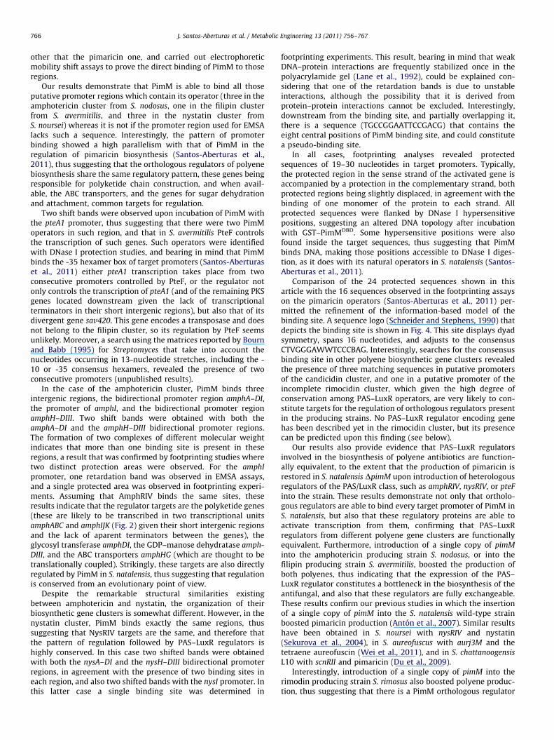
Fig. 4. Sequence logo of the nucleotides sequences that constitute PimM binding
site. The logo was constructed with the 16 protected regions observed in the
footprinting assays on the pimaricin operators (Santos-Aberturas et al., 2011), and
the 24 protected sequences identified in this work. The height of each letter is
proportional to the frequency of the base, and the height of the letter stack is the
conservation in bits at that position (Schneider and Stephens, 1990). The total
information (Rsequence) for the binding site is 12.01 bits (0.75 bits per base).
Table S2Binding sites and their information content.
Promoter Strand Sequence Ri score
amphAp þ TTAGGGAAAAGTACAG 5.04
amphAp � CTAAGGAATCTCAGTG 8.47
amphDIp þ CTAGGGATTCCCCGGA 15.75
amphDIp � CCGGGGAATCCCTAGT 7.61
amphIp þ CTAGGGATTTCCTGCC 13.65
amphIp � GGCAGGAAATCCCTAG 12.66
amphHp þ CTAGGGAAATCCGGGT 14.33
amphHp � ACCCGGATTTCCCTAG 12.11
amphDIIIp þ CCCGGGAATTCCCTAG 19.08
amphDIIIp � CTAGGGAATTCCCGGG 20.18
pteA1p (1) þ CTAGGGTTCTCCTTAG 10.77
pteA1p (1) � TTAGGGGAATCCCCAA 12.19
pteA1p (2) þ TTGGGGATTCCCCTAA 12.91
pteA1p (2) � CTAAGGAGAACCCTAG 11.36
nysAp þ TTAGGGAAAAGTACAG 5.04
nysAp � CTAAGGAATGACGGGC 6.32
nysDIp þ CTAGTGATTCCCCGGT 12.77
nysDIp � ACCGGGGAATCACTAG 9.84
nysIp þ CTAGTGAATTCCTGCC 11.49
nysIp � GGCAGGAATTCACTAG 10.24
nysHp þ CTAGTGAAATTTCGGA 8.45
nysHp � TCCGAAATTTCACTAG 5.98
nysDIIIp þ CTCGTGAATTCCCTAG 17.51
J. Santos-Aberturas et al. / Metabolic Engineering 13 (2011) 756–767 763
In the case of the nystatin promoters, results were very similarto those of amphotericin promoters. Thus nysI promoter, showeda single protected region of 30 nucleotides in the coding strand ofnysI (positions �178 to �149 from nysI translation start codon).In the bottom strand the protected sequence was 22 bp long, atpositions �165 to �144 (Fig. S1).
Results of the analysis of the nysH–DIII bidirectional regionshowed two protected regions in each strand, in agreement withthe appearance of two retardation bands in EMSA experiments.The two areas are 25–30 nucleotides apart in the S. noursei
chromosome. The protected area closest to nysH extends for23 bp and is located at nucleotide positions �114 to �92 withrespect to the nysH GTG translational start site. The protection ofthe reverse strand was 21 bp long (positions �110 to �90), bothregions being displaced 2–4 nucleotides. The second protectedarea extended for 22 bp of the coding strand of nysDIII, atpositions �157 to �136 from nysDIII translational start site. Inthe bottom strand the length of the protected sequence was thesame 21 bp, spanning from position �158 to �138 (Fig. S1). Inthis case, both regions were displaced by two nucleotides.
When we carried out the footprinting analysis with thebidirectional nysDI-A promoter, two protected areas wereobserved in each strand, in agreement with the appearance oftwo retardation bands in EMSA experiments. The two areas are56–57 nucleotides apart in the chromosome. The first protectedarea, extending for 22 bp of nysDI coding strand, is located atnucleotide positions �80 to �59 with respect to the nysDI GTGtranslational start site. The protection of the reverse strand wasone nucleotide larger (positions �83 to �61), both regions beingdisplaced by 2–3 nucleotides. The second protected area extendedfor 25 bp of the coding strand of nysA, at positions �89 to �65from nysA translational start site. In the bottom strand theprotected sequence was 23 bp long, spanning from position�93 to �71 (Fig. 3E and F). In this case, both regions were alsoslightly displaced.
In almost all cases DNase I hypersensitive positions flanked theprotected sequence, indicating altered DNA topology after incu-bation with GST–PimMDBD. Some hypersensitive positions werealso found inside the target sequences, typically two areas (Fig. 3),thus suggesting that PimM bends DNA, making those positionsaccessible to DNase I digestion.
nysDIIIp � CTAGGGAATTCACGAG 18.09
pimKp þ CTAAGGATTTCCCGAC 16.87
pimKp � GTCGGGAAATCCTTAG 15.94
pimS2p þ TCATGGAATTCCCTAG 13.59
pimS2p � CTAGGGAATTCCATGA 15.82
pimIp þ CTAGGTGTTTTCCGGC 7.57
pimIp � GCCGGAAAACACCTAG 8.67
pimJp þ CCCGGAATTTCTTGAC 9.49
pimJp � CTAGGGAATTCCCGAG 21.39
pimAp þ ACCGGAAAACCCCTAG 11.89
pimAp � CTAGGGGTTTTCCGGT 11.72
pimEp þ CTGGCATTTCCCCCAG 4.42
pimEp � GGGGGAAATGCCAGAG 6.22
pimS1p þ CTGCGGGTTTCCCTAG 12.14
pimS1p � CTAGGGAAACCCGCAG 14.48
pimDp þ GTAGGGATTTCCTGGT 13.52
pimDp � ACCAGGAAATCCCTAC 12.07
3.4. Information content analysis of the nucleotide sequences
The initial information-based model of the binding site(Santos-Aberturas et al., 2011) was refined taking into accountthe newly discovered binding sites. This new model takes intoaccount the 16 protected regions observed in the footprintingassays on the pimaricin operators (Santos-Aberturas et al., 2011),and the 24 protected sequences identified in this work. A newsequence logo (Schneider and Stephens, 1990) that depicts thebinding site is shown in Fig. 4. This new site spans 16 nucleotides,expanding the previous one by two nucleotides, and adjusts to theconsensus CTVGGGAWWTCCCBAG (where V is A, C, or G; W is Aor T; and B is C, G, or T). All newly identified operators showedhigh Ri values ranging from 20.18 bits in the case of the bindingsite at the reverse strand of the amphDIII promoter to 5.04 bits inthe operator of the nysA and amphA promoters, which showexactly the same binding site sequence (Table S2). The totalinformation (Rsequence) for the binding site showed a mean valueof 12.0174.26 (0.75 bits per base), even higher than the valueobtained when taking into account just the pimaricin operators.The fact that the introduction of 24 additional sequences in thealignment that is used to generate the weight matrix did notsubstantially alter the structure of the binding sequence
demonstrates the conservation of binding sites in the four speciesof Streptomyces studied.
3.5. Genetic complementation of S. natalensis DpimM by
orthologous regulators restores pimaricin production
Above results indicated that PimM could bind in vitro targetoperators of its homologous regulators, thus providing the firstevidence of a functional conservation of polyene PAS–LuxR

Fig. 5. Complementation of S. natalensis DpimM mutant with heterologous regulators restores pimaricin biosynthesis. Left: Bioassay for pimaricin production in S.
natalensis (A), S. natalensis DpimM (B), S. natalensis DpimM [pSETpimM] (C), S. natalensis DpimM [pMamphRIV] (D), S. natalensis DpimM [pMnysRIV] (E), and S. natalensis
DpimM [pMpteF] (F). Right: Quantification of the pimaricin production attained by the complemented strains after 60 and 84 h of growth. Data are the average of three
flasks. Vertical bars indicate the standard deviation values.
J. Santos-Aberturas et al. / Metabolic Engineering 13 (2011) 756–767764
regulators. In order to check what happened in vivo, we studiedthe exchangeability of PimM with its orthologous regulators byheterologous gene complementation of S. natalensis DpimM
mutant (Anton et al., 2007). To do that, and given that expressionfrom pimM promoter is controlled by other S. natalensis regulators(Santos-Aberturas et al., unpublished), we cloned DNA fragmentscontaining either amphRIV, nysRIV, or pteF genes into the inte-grative vector pSET152, and placed them under the control ofpimM promoter, giving rise to pMamphRIV, pMnysRIV, andpMpteF, respectively (see Section 2.6). These plasmids were thentransferred from E. coli ET12567 [pUZ8002] to S. natalensis DpimM
by conjugation (Enrıquez et al., 2006). In all cases, introduction ofthe vector restored the ability of S. natalensis DpimM to producepimaricin (Fig. 5). As expected, given its highest identity to PimM,pimaricin yield was the highest in the strain complemented withpteF (reaching 94% of the antifungal produced by the same straincomplemented with pimM (control) after 84 h of growth; Fig. 5).Production of the strain complemented with amphRIV or nysRIV,which are more distantly related to pimM, was somewhat lower,reaching 47% or 61% of the control levels, respectively (Fig. 5).
3.6. Expression of PAS–LuxR regulators constitutes a bottleneck for
polyene production
Previous results with PimM had shown that this regulator isnot present in saturating amounts in S. natalensis, and thatincreasing its gene dosage boosted pimaricin production (Antonet al., 2007). A similar result was obtained with NysRIV, whichconstitutes a bottleneck for nystatin production in S. noursei, andwhose gene dosage increase also boosted nystatin production(Sekurova et al., 2004). Taking this into account, and to corrobo-rate further the functional conservation of these regulators, westudied the effect of introducing one copy of pimM into thegenomes of S. nodosus and S. avermitilis. For that purpose, wetransferred pSETpimM, an integrative plasmid that contains pimM
under the control of its native promoter (Anton et al., 2007), byconjugation from E. coli ET12567 [pUZ8002] to the abovesaidstrains as described (Enrıquez et al., 2006). pSET152 was alsointroduced into the different strains as a control.
When we introduced pSETpimM into S. nodosus, whereas nosignificant change in the growth curve in flask cultures wasobserved, a substantial increase in the specific production ofamphotericin was detected (Fig. 6), thus suggesting that AmphRIVavailability does constitute a bottleneck in the biosynthesis of theantifungal, and proving that both regulators are fully exchange-able. The increase in amphotericin production ranged between50% at 72 h of growth and 60% at 84 h.
A similar result was observed when we introduced pSETpimMinto S. avermitilis. In this case, the biosynthesis of filipin was alsoboosted (Fig. 6), suggesting that PteF availability is also a bottle-neck for filipin production. The increase in filipin productionranged between 130% at 60 h of growth and 100% at 84 h.
These results indicate that expression of these regulatorsconstitutes a general bottleneck in polyene production, andconfirm their functional conservation among polyene biosyn-thetic gene clusters.
3.7. In silico search for binding sites in other polyene biosynthetic
gene clusters
Searches for the consensus binding site in other availablepolyene biosynthetic gene clusters revealed the presence of threematching sequences in putative promoters of the candicidincluster in Streptomyces sp. FR-008, and one in a putative promoterof the rimocidin cluster in Streptomyces diastaticus var. 108.Sequences at the candicidin cluster were the following: CTAGG-GAAAACACGGG (Ri of 5.98) in the promoter region of the poly-ketide synthase gene fscA (62 bp upstream fscA translationcodon), ACGGGGAATTCACCAG (Ri of 4.46) in the promoter regionof the polyketide synthase fscB (240 bp upstream from fscB), andCTAGGGATTTCCAAAG (Ri of 11.32) in the promoter of the poly-ketide synthase fscD (318 bp upstream from the gene). Thesequence at the incomplete rimocidin cluster was CTAAG-GATTTTCCGGT (Ri of 8.39) in the promoter region of the glycosyltransferase rimE.
In order to confirm the validity of the search carried out, wedecided to analyze the effect of introducing one copy of pimM intoS. diastaticus var. 108 or Streptomyces sp. FR-008; however giventhat both strains are not available commercially, we decided to

Fig. 6. pimM triggers polyene production in S. nodosus, S. avermitilis, and S. rimosus. Production of amphotericin B (A), filipin III (B), and rimocidin (C) [expressed as mg
polyene per ml] in YEME medium without sucrose (A and C) and R5 medium (B). Note that S. nodosus produces a mixture of the heptaene amphotericin B (98%) and the
tetraene amphotericin A (2%). Introduction of pimM did not vary the ratio between amphotericins. Black squares indicate the production by the wild type strain harboring
pSET152 (control) and white squares the production by the same strain with one copy of pimM (pSETpimM). Data are the average of three flasks. Vertical bars indicate the
standard deviation values. Growth curves are shown in the right panels.
J. Santos-Aberturas et al. / Metabolic Engineering 13 (2011) 756–767 765
introduce one copy of pimM into S. rimosus (a rimocidin producerthat also produces oxytetracycline as S. diastaticus var. 108) asindicated above (Section 3.6). Interestingly, results showed asubstantial increase in the specific production of rimocidin(Fig. 6), thus suggesting that there is a PimM orthologousregulator in S. rimosus genome, and that its availability alsoconstitutes a bottleneck in the biosynthesis of the antifungal.The increase in rimocidin production ranged between 24% at 84 hof growth and 29% at 96 h.
4. Discussion
All polyene macrolide biosynthetic gene clusters known up todate include a gene coding for a PAS–LuxR regulator. These typeof regulators have a peculiar architecture, combining an N-term-inal PAS sensor domain (Taylor and Zhulin, 1999) with a C-term-inal HTH motif of the LuxR type for DNA binding. The PAS domain
would detect an as yet unidentified stimulus, and in responseregulate the activity of the DNA binding domain. In PimM wehave recently described that one of the roles of the PAS domain isto reduce the affinity of the DNA binding domain towards itstarget promoters (Santos-Aberturas et al., 2011), so it is verylikely that all PAS–LuxR regulators share the same behavior.
These regulators are highly conserved, especially at theC-terminal domain for DNA binding, among polyene macrolidebiosynthetic gene clusters, and also seem to be confined to theregulation of the biosynthesis of this class of bioactive com-pounds, since they have not been found to regulate any otherbiological process.
Given the high degree of conservation between PimM and itscounterparts from other polyene producers, it was likely afunctional conservation among them. In order to provide experi-mental evidence of it, we searched for the presence of sequencessimilar to the recently described PimM canonical operator(Santos-Aberturas et al., 2011) in polyene biosynthetic clusters
J. Santos-Aberturas et al. / Metabolic Engineering 13 (2011) 756–767766
other that the pimaricin one, and carried out electrophoreticmobility shift assays to prove the direct binding of PimM to thoseregions.
Our results demonstrate that PimM is able to bind all thoseputative promoter regions which contain its operator (three in theamphotericin cluster from S. nodosus, one in the filipin clusterfrom S. avermitilis, and three in the nystatin cluster fromS. noursei) whereas it is not if the promoter region used for EMSAlacks such a sequence. Interestingly, the pattern of promoterbinding showed a high parallelism with that of PimM in theregulation of pimaricin biosynthesis (Santos-Aberturas et al.,2011), thus suggesting that the orthologous regulators of polyenebiosynthesis share the same regulatory pattern, these genes beingresponsible for polyketide chain construction, and when avail-able, the ABC transporters, and the genes for sugar dehydrationand attachment, common targets for regulation.
Two shift bands were observed upon incubation of PimM withthe pteA1 promoter, thus suggesting that there were two PimMoperators in such region, and that in S. avermitilis PteF controlsthe transcription of such genes. Such operators were identifiedwith DNase I protection studies, and bearing in mind that PimMbinds the -35 hexamer box of target promoters (Santos-Aberturaset al., 2011) either pteA1 transcription takes place from twoconsecutive promoters controlled by PteF, or the regulator notonly controls the transcription of pteA1 (and of the remaining PKSgenes located downstream given the lack of transcriptionalterminators in their short intergenic regions), but also that of itsdivergent gene sav420. This gene encodes a transposase and doesnot belong to the filipin cluster, so its regulation by PteF seemsunlikely. Moreover, a search using the matrices reported by Bournand Babb (1995) for Streptomyces that take into account thenucleotides occurring in 13-nucleotide stretches, including the -10 or -35 consensus hexamers, revealed the presence of twoconsecutive promoters (unpublished results).
In the case of the amphotericin cluster, PimM binds threeintergenic regions, the bidirectional promoter region amphA–DI,the promoter of amphI, and the bidirectional promoter regionamphH–DIII. Two shift bands were obtained with both theamphA–DI and the amphH–DIII bidirectional promoter regions.The formation of two complexes of different molecular weightindicates that more than one binding site is present in theseregions, a result that was confirmed by footprinting studies wheretwo distinct protection areas were observed. For the amphI
promoter, one retardation band was observed in EMSA assays,and a single protected area was observed in footprinting experi-ments. Assuming that AmphRIV binds the same sites, theseresults indicate that the regulator targets are the polyketide genes(these are likely to be transcribed in two transcriptional unitsamphABC and amphIJK (Fig. 2) given their short intergenic regionsand the lack of aparent terminators between the genes), theglycosyl transferase amphDI, the GDP–manose dehydratase amph-
DIII, and the ABC transporters amphHG (which are thought to betranslationally coupled). Strikingly, these targets are also directlyregulated by PimM in S. natalensis, thus suggesting that regulationis conserved from an evolutionary point of view.
Despite the remarkable structural similarities existingbetween amphotericin and nystatin, the organization of theirbiosynthetic gene clusters is somewhat different. However, in thenystatin cluster, PimM binds exactly the same regions, thussuggesting that NysRIV targets are the same, and therefore thatthe pattern of regulation followed by PAS–LuxR regulators ishighly conserved. In this case two shifted bands were obtainedwith both the nysA–DI and the nysH–DIII bidirectional promoterregions, in agreement with the presence of two binding sites ineach region, and also two shifted bands with the nysI promoter. Inthis latter case a single binding site was determined in
footprinting experiments. This result, bearing in mind that weakDNA–protein interactions are frequently stabilized once in thepolyacrylamide gel (Lane et al., 1992), could be explained con-sidering that one of the retardation bands is due to unstableinteractions, although the possibility that it is derived fromprotein–protein interactions cannot be excluded. Interestingly,downstream from the binding site, and partially overlapping it,there is a sequence (TGCCGGAATTCCGACG) that contains theeight central positions of PimM binding site, and could constitutea pseudo-binding site.
In all cases, footprinting analyses revealed protectedsequences of 19–30 nucleotides in target promoters. Typically,the protected region in the sense strand of the activated gene isaccompanied by a protection in the complementary strand, bothprotected regions being slightly displaced, in agreement with thebinding of one monomer of the protein to each strand. Allprotected sequences were flanked by DNase I hypersensitivepositions, suggesting an altered DNA topology after incubationwith GST–PimMDBD. Some hypersensitive positions were alsofound inside the target sequences, thus suggesting that PimMbinds DNA, making those positions accessible to DNase I diges-tion, as it does with its natural operators in S. natalensis (Santos-Aberturas et al., 2011).
Comparison of the 24 protected sequences shown in thisarticle with the 16 sequences observed in the footprinting assayson the pimaricin operators (Santos-Aberturas et al., 2011) per-mitted the refinement of the information-based model of thebinding site. A sequence logo (Schneider and Stephens, 1990) thatdepicts the binding site is shown in Fig. 4. This site displays dyadsymmetry, spans 16 nucleotides, and adjusts to the consensusCTVGGGAWWTCCCBAG. Interestingly, searches for the consensusbinding site in other polyene biosynthetic gene clusters revealedthe presence of three matching sequences in putative promotersof the candicidin cluster, and one in a putative promoter of theincomplete rimocidin cluster, which given the high degree ofconservation among PAS–LuxR operators, are very likely to con-stitute targets for the regulation of orthologous regulators presentin the producing strains. No PAS–LuxR regulator encoding genehas been described yet in the rimocidin cluster, but its presencecan be predicted upon this finding (see below).
Our results also provide evidence that PAS–LuxR regulatorsinvolved in the biosynthesis of polyene antibiotics are function-ally equivalent, to the extent that the production of pimaricin isrestored in S. natalensis DpimM upon introduction of heterologousregulators of the PAS/LuxR class, such as amphRIV, nysRIV, or pteF
into the strain. These results demonstrate not only that ortholo-gous regulators are able to bind every target promoter of PimM inS. natalensis, but also that these regulatory proteins are able toactivate transcription from them, confirming that PAS–LuxRregulators from different polyene gene clusters are functionallyequivalent. Furthermore, introduction of a single copy of pimM
into the amphotericin producing strain S. nodosus, or into thefilipin producing strain S. avermitilis, boosted the production ofboth polyenes, thus indicating that the expression of the PAS–LuxR regulator constitutes a bottleneck in the biosynthesis of theantifungal, and also that these regulators are fully exchangeable.These results confirm our previous studies in which the insertionof a single copy of pimM into the S. natalensis wild-type strainboosted pimaricin production (Anton et al., 2007). Similar resultshave been obtained in S. noursei with nysRIV and nystatin(Sekurova et al., 2004), in S. aureofuscus with aurj3M and thetetraene aureofuscin (Wei et al., 2011), and in S. chattanoogensis
L10 with scnRII and pimaricin (Du et al., 2009).Interestingly, introduction of a single copy of pimM into the
rimodin producing strain S. rimosus also boosted polyene produc-tion, thus suggesting that there is a PimM orthologous regulator

J. Santos-Aberturas et al. / Metabolic Engineering 13 (2011) 756–767 767
in S. rimosus genome, and that its availability also constitutes abottleneck in the biosynthesis of the antifungal.
More than 60 years after being discovered, polyenes stillremain as the ‘‘gold standard’’ against severe systemic fungalinfections. These compounds also show activity against parasites,enveloped viruses, and prion diseases (Caffrey et al., 2008), butdespite their interesting properties, the regulation of their bio-synthesis remains obscure. Here we provide the first evidence of ageneral mechanism that regulates polyene production. Thisknowledge will be valuable for maximizing yields of theseimportant compounds.
Acknowledgments
This work was supported by the Spanish Ministerio de Cienciae Innovacion (BIO2007-67585 and BIO2010-19911 to J.F.A.), F.P.U.fellowships of the Ministerio de Ciencia e Innovacion (AP2005-3644 to J.S-.A., AP2007-02055 to T.D.P.), a fellowship from thePortuguese Fundac-ao para a Ciencia e a Tecnologia (SFRH/BD/64006/2009 to C.M.V.), and a contract Juan de la Cierva (JCI-2009-05157 to C.C.). We thank J. Merino, B. Martın, Rosario Perez-Redondo, and A. Casenave for excellent technical assistance.
Appendix A. Supporting information
Supplementary data associated with this article can be foundin the online version at doi:10.1016/j.ymben.2011.09.011.
References
Anton, N., Mendes, M.V., Martın, J.F., Aparicio, J.F., 2004. Identification of PimR as apositive regulator of pimaricin biosynthesis in Streptomyces natalensis. J.Bacteriol. 186, 2567–2575.
Anton, N., Santos-Aberturas, J., Mendes, M.V., Guerra, S.M., Martın, J.F., Aparicio,J.F., 2007. PimM, a PAS domain positive regulator of pimaricin biosynthesis inStreptomyces natalensis. Microbiology 153, 3174–3183.
Aparicio, J.F., Colina, A.J., Ceballos, E., Martın, J.F., 1999. The biosynthetic genecluster for the 26-membered ring polyene macrolide pimaricin. A newpolyketide synthase organization encoded by two subclusters separated byfunctionalization genes. J. Biol. Chem. 274, 10133–10139.
Aparicio, J.F., Mendes, M.V., Anton, N., Recio, E., Martın, J.F., 2004. Polyenemacrolide antibiotic biosynthesis. Curr. Med. Chem. 11, 1645–1656.
Bi, C., Rogan, P.K., 2006. BIPAD: a web server for modeling bipartite sequenceelements. BMC Bioinf. 7, 76.
Bibb, M.J., 2005. Regulation of secondary metabolism in Streptomycetes. Curr.Opin. Microbiol. 8, 208–215.
Bierman, M., Logan, R., O’Brien, K., Seno, E.T., Rao, R.N., Schoner, B.E., 1992. Plasmidcloning vectors for the conjugal transfer of DNA from Escherichia coli toStreptomyces spp. Gene 116, 43–49.
Bourn, W.R., Babb, B., 1995. Computer assisted identification and classification ofstreptomycete promoters. Nucleic Acids Res. 23, 3696–3703.
Caffrey, P., Aparicio, J.F., Malpartida, F., Zotchev, S.B., 2008. Biosynthetic engineer-ing of polyene macrolides towards generation of improved antifungal andantiparasitic agents. Curr. Top. Med. Chem. 8, 639–653.
Carmody, M., Byrne, B., Murphy, B., Breen, C., Lynch, S., Flood, E., Finnan, S., Caffrey,P., 2004. Analysis and manipulation of amphotericin biosynthetic genes bymeans of modified phage KC515 transduction techniques. Gene 343, 107–115.
Chen, S., Huang, X., Zhou, X., Bai, L., He, J., Jeong, K.J., Lee, S.Y., Deng, Z., 2003.Organisational and mutational analysis of a complete FR-008/candicidin genecluster encoding a structurally related polyene complex. Chem. Biol. 10,1065–1076.
Du, Y.L., Chen, S.F., Cheng, L.Y., Shen, X.L., Tian, Y., Li, Y.Q., 2009. Identification of anovel Streptomyces chattanoogensis L10 and enhancing its natamycin produc-tion by overexpressing positive regulator ScnRII. J. Microbiol. 47, 506–513.
Enrıquez, L.L., Mendes, M.V., Anton, N., Tunca, S., Guerra, S.M., Martın, J.F., Aparicio,J.F., 2006. An efficient gene transfer system for the pimaricin producerStreptomyces natalensis. FEMS Microbiol. Lett. 257, 312–318.
Hefti, M.H., Francoijs, K.-J., de Vries, S.C., Dixon, R., Vervoort, J., 2004. The PAS fold.A redefinition of the PAS domain based upon structural prediction. Eur. J.Biochem. 271, 1198–1208.
van Helden, J., 2003. Regulatory sequence analysis tools. Nucleic Acids Res. 31,3593–3596.
Hertz, G.Z., Stormo, G.D., 1999. Identifying DNA and protein patterns withstatistically significant alignments of multiple sequences. Bioinformatics 15,563–577.
Kells, P.M., Ouellet, H., Santos-Aberturas, J., Aparicio, J.F., Podust, L.M., 2010.Structure of cytochrome P450 PimD suggests epoxidation of the polyenemacrolide pimaricin occurs via a hydroperoxoferric intermediate. Chem. Biol.17, 841–851.
Kieser, T., Bibb, M.J., Buttner, M.J., Chater, K.F., Hopwood, D.A., 2000. PracticalStreptomyces Genetics. The John Innes Foundation, Norwich, UK.
Lane, D., Prentki, P., Chandler, M., 1992. Use of gel retardation to analyze protein–nucleic acid interactions. Microbiol. Rev. 56, 509–528.
Li, C., Hazzard, C., Florova, G., Reynolds, K.A., 2009. High titer production oftetracenomycins by heterologous expression of the pathway in a Streptomycescinnamonensis industrial monensin producer strain. Metab. Eng. 11, 319–327.
Li, J., Li, L., Tian, Y., Niu, G., Tan, H., 2011. Hybrid antibiotics with the nikkomycinnucleoside and polyoxin peptidyl moieties. Metab. Eng. 13, 336–344.
Martın, J.F., Aparicio, J.F., 2009. Enzymology of the polyenes pimaricin andcandicidin biosynthesis. Methods Enzymol. 459, 215–242.
Mendes, M.V., Recio, E., Fouces, R., Luiten, R., Martın, J.F., Aparicio, J.F., 2001.Engineered biosynthesis of novel polyenes: a pimaricin derivative produced bytargeted gene disruption in Streptomyces natalensis. Chem. Biol. 8, 635–644.
Mendes, M.V., Anton, N., Martın, J.F., Aparicio, J.F., 2005. Characterization of thepolyene macrolid P450 epoxidase from Streptomyces natalensis that convertsde-epoxypimaricin into pimaricin. Biochem. J. 386, 57–62.
Mendes, M.V., Recio, E., Anton, N., Guerra, S.M., Santos-Aberturas, J., Martın, J.F.,Aparicio, J.F., 2007a. Cholesterol oxidases act as signalling proteins for thebiosynthesis of the polyene macrolide pimaricin. Chem. Biol. 14, 279–290.
Mendes, M.V., Tunca, S., Anton, N., Recio, E., Sola-Landa, A., Aparicio, J.F., Martın,J.F., 2007b. The two-component phoR–phoP system of Streptomyces natalensis:inactivation or deletion of phoP reduces the negative phosphate regulation ofpimaricin biosynthesis. Metab. Eng. 9, 217–227.
Moglich, A., Ayers, R.A., Moffat, K., 2009. Structure and signaling mechanism ofPer-ARNT-Sim domains. Structure 17, 1282–1294.
Nic Lochlainn, L., Caffrey, P., 2009. Phosphomannose isomerase and phosphoman-nomutase gene disruptions in Streptomyces nodosus: impact on amphotericinbiosynthesis and implications for glycosylation engineering. Metab. Eng. 11,40–47.
Olano, C., Lombo, F., Mendez, C., Salas, J.A., 2008. Improving production ofbioactive secondary metabolites in actinomycetes by metabolic engineering.Metab. Eng. 10, 281–292.
Omura, S., Ikeda, H., Ishikawa, J., Hanamoto, A., Takahashi, C., Shinose, M.,Takahasi, Y., Horikawa, H., Nakazawa, H., Osonoe, T., Kikuchi, H., Shiba, T.,Sakaki, Y., Hattori, M., 2001. Genome sequence of an industrial microorganismStreptomyces avermitilis: deducing the ability of producing secondary meta-bolites. Proc. Natl. Acad. Sci. USA 98, 12215–12220.
Pickens, L.B., Tang, Y., 2009. Decoding and engineering tetracycline biosynthesis.Metab. Eng. 11, 69–75.
Sambrook, J., Russell, D.W., 2001. Molecular Cloning: A Laboratory Manual. ColdSpring Harbor Laboratory Press, Cold Spring Harbor, NY.
Santos-Aberturas, J., Vicente, C.M., Guerra, S.M., Payero, T.D., Martın, J.F., Aparicio,J.F., 2011. Molecular control of polyene macrolide biosynthesis: direct bindingof the regulator PimM to eight promoters of pimaricin genes and identificationof binding boxes. J. Biol. Chem. 286, 9150–9161.
Schneider, T.D., 1997. Information content of individual genetic sequences. J.Theor. Biol. 189, 427–441.
Schneider, T.D., Stephens, R.M., 1990. Sequence logos: a new way to displayconsensus sequences. Nucleic Acids Res. 18, 6097–6100.
Sekurova, O.N., Brautaset, T., Sletta, H., Borgos, S.E.F., Jakobsen, O.M., Ellingsen, T.E.,Strom, A.R., Valla, S., Zotchev, S.B., 2004. In vivo analysis of the regulatorygenes in the nystatin biosynthetic gene cluster of Streptomyces noursei ATCC11455 reveals their differential control over antibiotic biosynthesis. J. Bacter-iol. 186, 1345–1354.
Tamura, K., Dudley, J., Nei, M., Kumar, S., 2007. MEGA4: Molecular EvolutionaryGenetics Analysis (MEGA) software version 4.0. Mol. Biol. Evol. 24, 1596–1599.
Taylor, B.L., Zhulin, I.B., 1999. PAS domains: internal sensors of oxygen, redoxpotential, and light. Microbiol. Mol. Biol. Rev. 63, 479–506.
Vicente, C.M., Santos-Aberturas, J., Guerra, S.M., Payero, T.D., Martın, J.F., Aparicio,J.F., 2009. PimT, an amino acid exporter controls polyene production viasecretion of the quorum sensing pimaricin-inducer PI-factor in Streptomycesnatalensis. Microb. Cell Fact. 8, 33.
Wei, J., Meng, X., Wang, Q., 2011. Enhanced production of aureofuscin by over-expression of AURJ3M, positive regulator of aureofuscin biosynthesis inStreptomyces aureofuscus. Lett. Appl. Microbiol. 47, 506–513.